Diagnosis of lumbar spinal stenosis: an updated systematic review

Open AccessOriginal ArticlePublished 01/02/2014DOI: 10.7759/cureus.152
Copyright © 2014 Mohi Eldin et al.
Distributed under Creative Commons CC-BY 3.0
Coflex Dynamic DistractionStabilization Device for LumbarDegenerative Diseases
Mohamed Mohi Eldin Corresponding Author: Mohamed Mohi Eldin
Categories: Neurological SurgeryKeywords: coflex, dynamic, lumbar, canal stenosi
How to cite this articleMohi eldin M (2014) Coflex Dynamic Distraction Stabilization Device for LumbarDegenerative Diseases . Cureus 6(1): e152. doi:10.7759/cureus.152
Abstract
Aim of the study: The purpose of this study was to assess the safety andeffectiveness of Coflex Dynamic Distraction Stabilization (DDS) devicein treating patients with degenerative diseases of the lumbar spine(DDLS), especially lumbar canal stenosis (LCS), to confirm itsindications for implantation, and to evaluate the clinical outcomes ofpatients. Material & Methods Our study is part of a multi centreprospective, case controlled studies in Egypt to determine the safetyand efficacy of minimally invasive spinal procedures, of these, theCoflex implant, a functionally dynamic interspinous implant (U-shapedtitanium) is included in the present study. From June 2008 until July2013 we treated 42 patients with this Coflex procedure. Median follow-up 22.5 months. At the time of follow-up all patients hadquestionnaires, clinical examination and x-ray taken. Results Asignificant number of patients showed pain relief. Preoperatively 30/42(71%) patients with moderate or severe low back pain (LBP).Postoperatively, the LBP in 6 (14%) patients did improve, 24 (57%)even showed no low back pain anymore. Mean preoperative walkingdistance was < 1000m in 36 (86%) patients. Postoperatively 42 (100%)patients could walk >1000m. Significant pain relief (> 50%) in monthswas calculated. Radiological results showed that endplate Angles whenwere acute preoperatively, always become less acute postoperatively;and the foraminal height always increases. Segmental range of motion(ROM) showed maintenance of the dynamic movements at theoperated level. Disc height showed significant changes after theprocedure in both anterior and posterior disc heights. Conclusion Thedata provided have demonstrated that the coflex implant provides painrelief for patients with indicated DDLS cases. The most commonindications for implantation were spinal stenosis and spinal stenosiswith lumbar disc herniation. There were no device-relatedcomplications. Using coflex is a safe option in the selection of

complications. Using coflex is a safe option in the selection ofinstrumentation for spinal stabilization.
IntroductionThe most severe primary spinal degenerative changes are found inthe lumbar spine region. Degenerative diseases of the lumbar spine(DDLS) are among the most common clinical entities. DDLS affectsthe intervertebral discs, the vertebral endplates, the facet joints andthe intervertebral posterior ligaments. These changes are well-known and often grouped under the broad definition of vertebralarthrosis, 2ry to degeneration of the ligaments and cartilage. Froma functional standpoint, the first event is hypermobility of the jointsdue to weak ligaments and the no longer perfect coaptation of thejoint surfaces. These changes continue to morphological changescausing narrowing of the affected segment and its exit foramina,and ending in some form of degenerative instability[1,2,3,4,5,6,7,8,9].
The Coflex® Dynamic Distraction Stabilization (DDS) Device(Paradigm Spine GmbH, Germany) offered both dynamic stabilizationand interlaminar orientation. It is indicated in patients whosesymptoms are exacerbated in extension and relieved in flexion.Implanted between the spinous processes, and reaching down to thelaminar level, the Coflex® DDS device is functionally dynamic, beingcompressible in extension, limiting flexion, with increased rotationalstability; the center of rotation being close to spinal canal.Protection of posterior elements through stress reduction on facetjoints & maintenance of foraminal height is also offered. Moreadvantages of the device include its ease of use, in a less invasive,tissue-sparing procedure with precise application of the device. Weconsider degenerative lumbar stenosis as the main indication forimplanting the dynamic interspinous Coflex device[10,11,12,13,14,15,16,17,18,19,20].
This study is a multi-center, prospective case-controlled study,designed to assess the safety and effectiveness of Coflex® DDSDevice in treating patients with DDLS, especially LCS, to confirm itsindications for implantation, and to evaluate the clinical outcomes ofpatients.. We intend to understand the changes in the disc height,segmental dynamic movement ability before and after Coflex®implantation in patients with symptomatic lumbar degenerativediseases. One limitation to this study is that it is a non-randomizedstudy. Further studies need to be done in a randomized, prospective
2014 Mohi Eldin et al. Cureus 6(1): e152. DOI 10.7759/cureus.152 Page 2 of 16

manner, with respect to long-term clinical outcomes.
Materials And MethodsOur study is part of a multi centre prospective, case controlledstudies in Egypt to determine the safety and efficacy of minimallyinvasive spinal procedures, of these, the Coflex implant, afunctionally dynamic interspinous implant (U-shaped titanium) isincluded in the present study. From June 2008 until July 2013 wetreated 42 patients with this Coflex procedure. Median follow-up22.5 months. At the time of follow-up all patients hadquestionnaires, clinical examination and x-ray taken.
Patient selection
The inclusion criteria were patients at 40 years of age or older withleg, buttock, or groin pain, with or without back pain, that could berelieved during flexion. Neurogenic claudication should be anintegral part of the patient's complaint. Patients had to havecompleted at least 3-6 months of non-operative treatment, with noimprovement. Stenosis was confirmed by CT or MRI scans at one ortwo levels. Primary exclusion criteria included a fixed motor deficit,cauda equina syndrome, frank instability, previous lumbar surgery,significant peripheral neuropathy, scoliosis, pathological fractures,severe osteoporosis, obesity, active infections, active systemicdisease, or vertebral metastasis.
Evaluation & Outcomes assessment
Data were collected preoperatively and postoperatively at 4 weeks,6 months, 1 year and 3 years following the initial treatment. Patientswere asked to grade their low-back and leg pain using the numericverbal rating (NVR) scale for pain [3]. Patients were asked abouttheir satisfaction with the surgical procedure using the validatedoutcome measurement specific to lumbar spinal stenosis, namely,the Oswestry Disability Index Questionnaire (ODI) [21,22,23].
Using numeric verbal rating (NVR) scale for pain intensity,significant relief was defined as pain relief of 50% or greater;otherwise it is non-significant (less than 50%). Significant pain reliefincludes patients in the pain free & improved pain categories, whilenon-significant pain relief includes patients in the fair & badcategories. Duration of pain relief was judged to be short-term, ifrelief was less than 6 months. If relief lasted for at least 6 up to 12
2014 Mohi Eldin et al. Cureus 6(1): e152. DOI 10.7759/cureus.152 Page 3 of 16

months, it was considered long-term. Success was defined as all ofthe following: 1) no or minimal remaining pain; 2) work not adverselyaffected; 3) no use of analgesic medications; and 4) patientsatisfaction with the procedure [21,23].
ODI scoring 0% to 20% means minimal disability (patient can copewith most living activities, no treatment is indicated apart fromexercise), 21%-40% moderate disability (patient experiences morepain and difficulty with sitting lifting and standing, patient managedby conservative means), 41%-60% severe disability (pain remainsthe main problem, activities of daily living are affected, require adetailed investigation), 61%-80% crippled (back pain impinges on allaspects of the patient's life, intervention is required), and 81%-100are either bed-bound or exaggerating their symptoms [22,23].
At each follow-up visit, pain and function evaluations is performed toconfirm the absence of complications. Clinical outcome scores andincidence rates of adverse effects, device failures, and revisionsurgeries, if any, are calculated.
Imaging Interpretation
Postoperatively a follow-up MRI scan is done at 6 months. X-rays aredone at 1 week, 3 month and yearly visits. Follow-up X-ray films areanalyzed to confirm maintenance of distraction and the absence ofdevice-related radiological complications. The changes in the discheight, endplate angles, and dynamic lumbar movement wereassessed before and after the placement of the Coflex® implant. Distances were measured between the most anterior or posteriorpoints on each vertebral body, excluding osteophytes. Endplateangles and foraminal height were measured. Endplate angles weremeasured on the images between adjacent vertebral bodies at thetreated level.
Procedure
The devices were implanted by, or under the direct supervision of asingle surgeon. The patients had the procedure under a lightgeneral anesthetic. Patients were placed on a radiolucent table inthe prone position, on surgical frame avoiding hyperlordosis of thespinal segment(s) to be operated upon. A neutral position or a slightkyphosis may be advantageous for surgical decompression as wellas for appropriate interspinous distraction. The level to be treated isidentified by fluoroscopy. Routine (midline) skin incision of
2014 Mohi Eldin et al. Cureus 6(1): e152. DOI 10.7759/cureus.152 Page 4 of 16

approximately 3 cm (per level), is performed over the spinousprocesses of the stenotic level(s), (Fig.: 1). This is carried down tothe fascia, which is split longitudinally 1 cm to the right and 1 cm tothe left of midline. It is extremely important to keep thesupraspinous ligament intact. The muscle is sharply dissected to thelevel of laminae and facets, lateral to the supraspinous ligamentpreserving the entire thickness of the supraspinous ligament andthe facet capsules. If possible a small portion of the spinous bony tipcan be resected together with the supraspinous ligament. This willaid a faster healing after reconstruction of the ligament. Theinterspinous ligament is removed any bony overgrowth of thespinous process that may interfere with insertion is resected.
After preparation of the implantation site, Trial instruments areutilized to define appropriate implant size, evaluate proper contactwith spinous process and amount of interspinous distraction. Somebony resection of the spinous process may be needed to ensureproper contact of the implant. To ensure proper depth of implantinsertion a small portion of the laminar surface may need partialresurfacing. The implant wings may need to be opened slightlyusing bending pliers at the mid portion of the wing to ensureappropriate depth of insertion. Implant is then introduced viaimpaction utilizing a mallet. Proper depth is determined if a beadedtip probe can be passed freely leaving 3-4 mm separation from thedura. If the implant is not seated appropriately further resurfacingor slightly more impaction force may be utilized. If the wings are nothaving sufficient bony contact additional stability can be achievedby slightly crimping the wings. Anteroposterior and lateralfluoroscopy views are taken to verify the proper position. Theincision is closed in the usual fashion. The drain is not routinelyutilized. The procedure is typically performed in less than an hour.Postoperatively, patients were mobilized immediately once they hadrecovered from the effects of any anesthetic or sedation anddischarged within 2 days, with external brace for 4 weekspostoperatively.
2014 Mohi Eldin et al. Cureus 6(1): e152. DOI 10.7759/cureus.152 Page 5 of 16

Figure 1:Routine (midline) skin incision of approximately 3 cm (per level), is performed over thespinous processes of the stenotic level(s)
ResultsFrom June 2008 until July 2013, 42 patients with degenerative spinalstenosis were treated with the Coflex procedure, 18 (43%) male, 24(57%) female. The average length of follow-up was 22.5 months(range 13 to 44) and the average age at the time of the operationwas 45 years (range 40 to 70). Thirty six out of 42 patients had theCoflex implanted at 1 level (mainly L3-4, or L4-5). There were siximplantations at the L5- S1 level in the study. Six out of 42 patientshad the Coflex implanted at 2 levels (L3-4 and L4-5). Thirty out of 42patients in the study had a segmental form of stenosis, (discogenic,liganentous, bony, or mixtures), nine cases had mild degree ofdegenerative retrololisthesis, and three case had mild 1st degree ofdegenerative spondylolisthesis .
A significant number of patients showed pain relief. Preoperatively30/42 (71%) patients with moderate or severe low back pain (LBP).Postoperatively, the LBP in 6 (14%) patients did improve, 24 (57%)
2014 Mohi Eldin et al. Cureus 6(1): e152. DOI 10.7759/cureus.152 Page 6 of 16
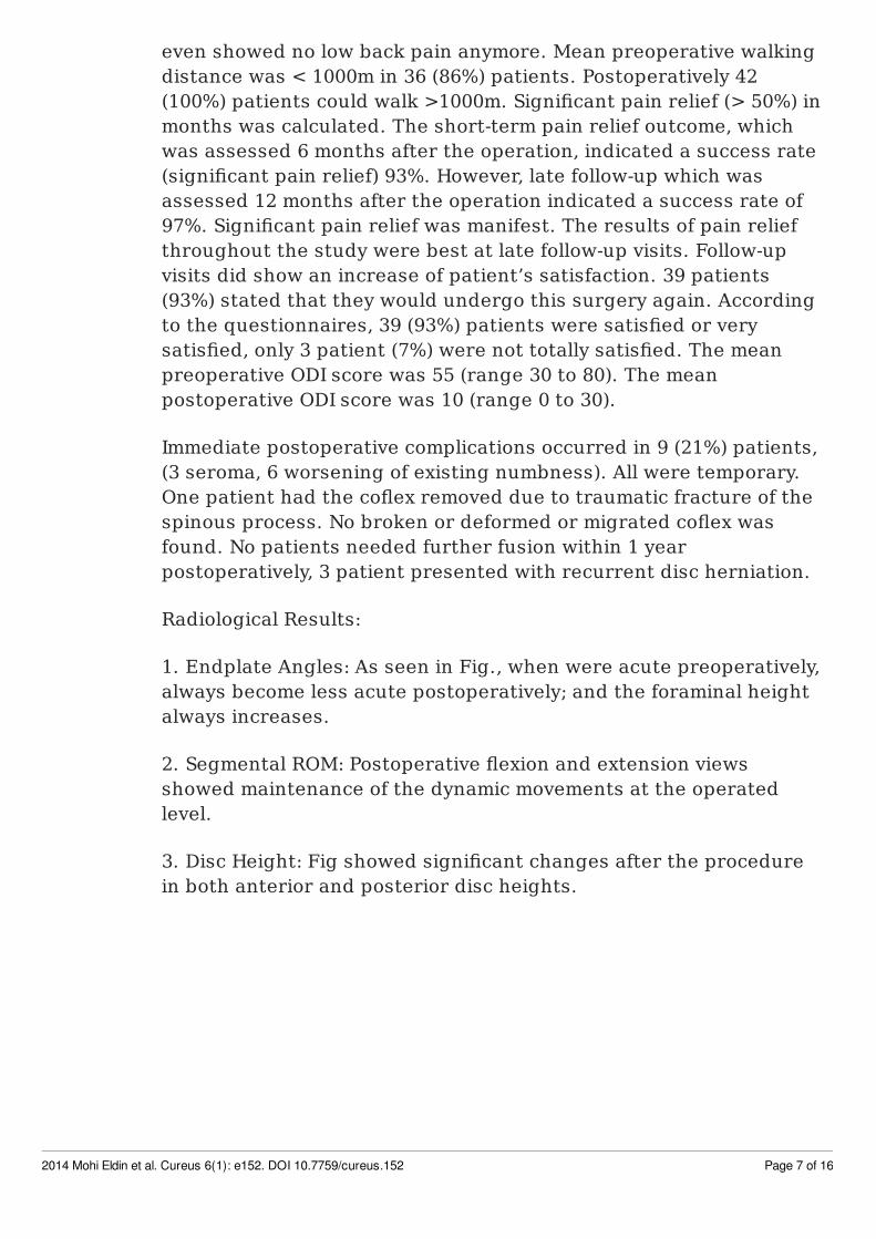
even showed no low back pain anymore. Mean preoperative walkingdistance was < 1000m in 36 (86%) patients. Postoperatively 42(100%) patients could walk >1000m. Significant pain relief (> 50%) inmonths was calculated. The short-term pain relief outcome, whichwas assessed 6 months after the operation, indicated a success rate(significant pain relief) 93%. However, late follow-up which wasassessed 12 months after the operation indicated a success rate of97%. Significant pain relief was manifest. The results of pain reliefthroughout the study were best at late follow-up visits. Follow-upvisits did show an increase of patient’s satisfaction. 39 patients(93%) stated that they would undergo this surgery again. Accordingto the questionnaires, 39 (93%) patients were satisfied or verysatisfied, only 3 patient (7%) were not totally satisfied. The meanpreoperative ODI score was 55 (range 30 to 80). The meanpostoperative ODI score was 10 (range 0 to 30).
Immediate postoperative complications occurred in 9 (21%) patients,(3 seroma, 6 worsening of existing numbness). All were temporary.One patient had the coflex removed due to traumatic fracture of thespinous process. No broken or deformed or migrated coflex wasfound. No patients needed further fusion within 1 yearpostoperatively, 3 patient presented with recurrent disc herniation.
Radiological Results:
1. Endplate Angles: As seen in Fig., when were acute preoperatively,always become less acute postoperatively; and the foraminal heightalways increases.
2. Segmental ROM: Postoperative flexion and extension viewsshowed maintenance of the dynamic movements at the operatedlevel.
3. Disc Height: Fig showed significant changes after the procedurein both anterior and posterior disc heights.
2014 Mohi Eldin et al. Cureus 6(1): e152. DOI 10.7759/cureus.152 Page 7 of 16

Figure 2:Preoperative bilateral foraminal narrowing is shown to widen postoperatively by theinterspinous distraction COFLEX DEVICE.
Figure 3:Preoperative unilateral foraminal narrowing is shown to widen postoperatively
2014 Mohi Eldin et al. Cureus 6(1): e152. DOI 10.7759/cureus.152 Page 8 of 16

Figure 4:Collapsed disc space is obviously opened by Coflex.
Figure 5:The AP endplate angle deviation is corrected postoperatively.
Figure 6:The Lat endplate angle & foraminal height are corrected postoperatively.
2014 Mohi Eldin et al. Cureus 6(1): e152. DOI 10.7759/cureus.152 Page 9 of 16
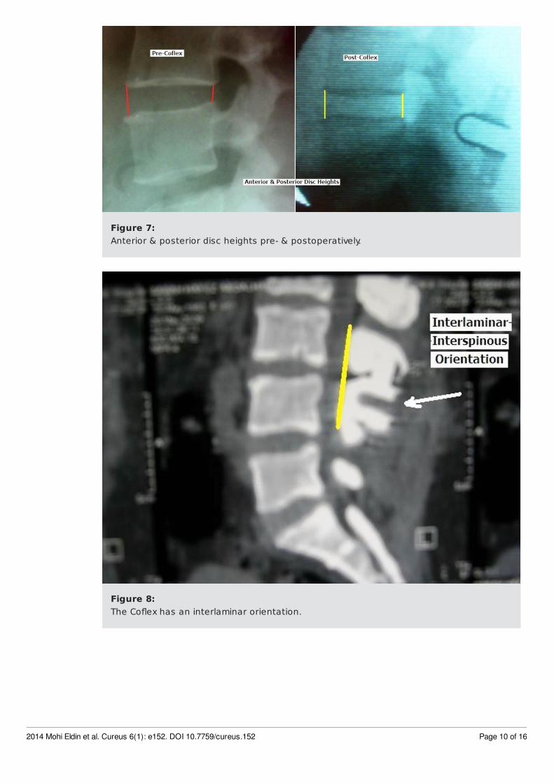
Figure 7:Anterior & posterior disc heights pre- & postoperatively.
Figure 8:The Coflex has an interlaminar orientation.
2014 Mohi Eldin et al. Cureus 6(1): e152. DOI 10.7759/cureus.152 Page 10 of 16
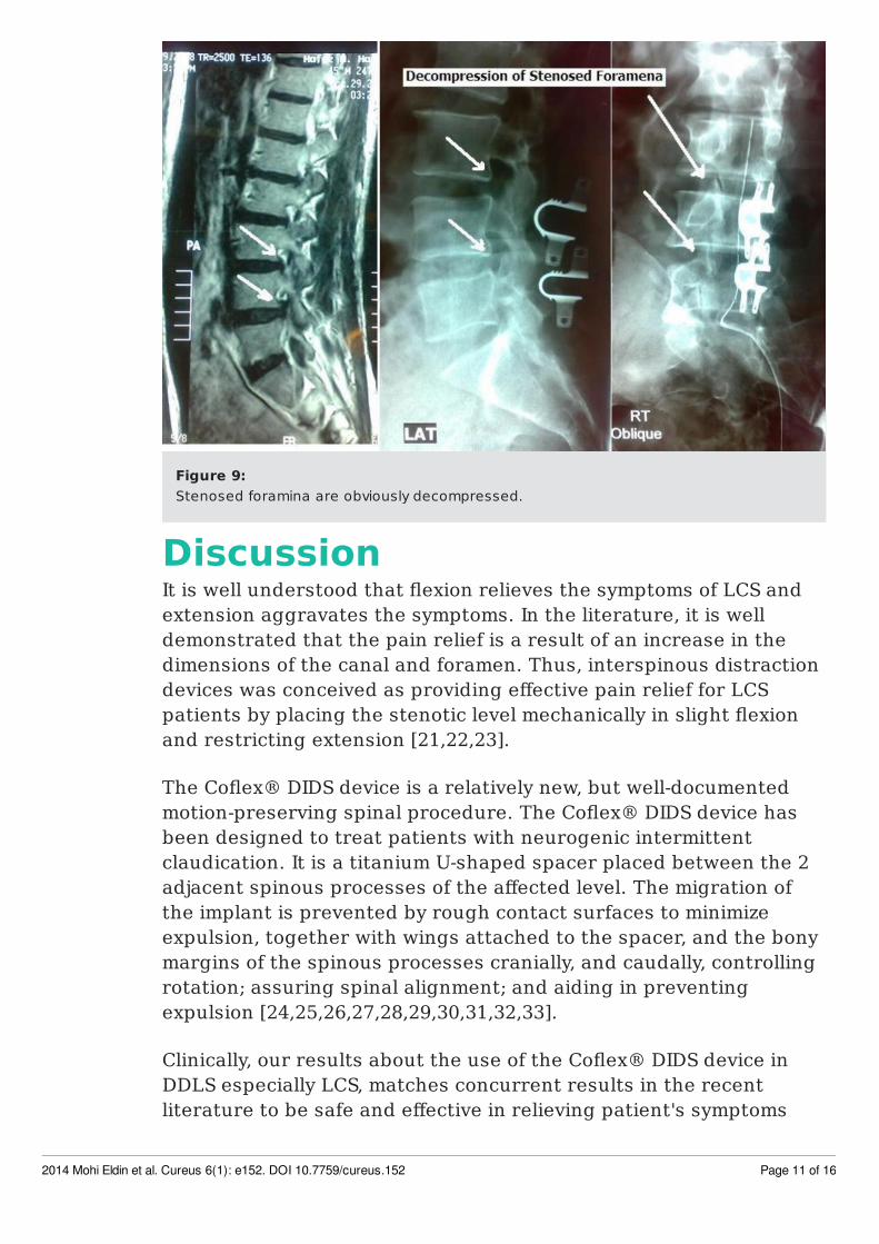
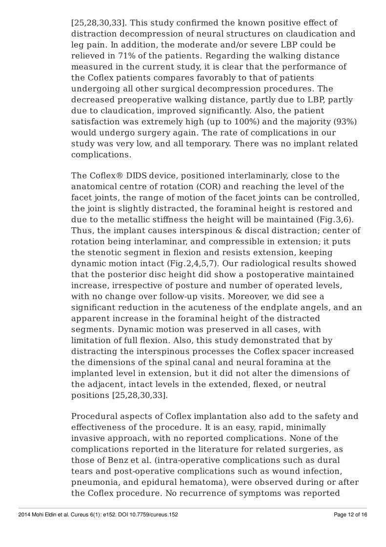
Figure 9:Stenosed foramina are obviously decompressed.
DiscussionIt is well understood that flexion relieves the symptoms of LCS andextension aggravates the symptoms. In the literature, it is welldemonstrated that the pain relief is a result of an increase in thedimensions of the canal and foramen. Thus, interspinous distractiondevices was conceived as providing effective pain relief for LCSpatients by placing the stenotic level mechanically in slight flexionand restricting extension [21,22,23].
The Coflex® DIDS device is a relatively new, but well-documentedmotion-preserving spinal procedure. The Coflex® DIDS device hasbeen designed to treat patients with neurogenic intermittentclaudication. It is a titanium U-shaped spacer placed between the 2adjacent spinous processes of the affected level. The migration ofthe implant is prevented by rough contact surfaces to minimizeexpulsion, together with wings attached to the spacer, and the bonymargins of the spinous processes cranially, and caudally, controllingrotation; assuring spinal alignment; and aiding in preventingexpulsion [24,25,26,27,28,29,30,31,32,33].
Clinically, our results about the use of the Coflex® DIDS device inDDLS especially LCS, matches concurrent results in the recentliterature to be safe and effective in relieving patient's symptoms
2014 Mohi Eldin et al. Cureus 6(1): e152. DOI 10.7759/cureus.152 Page 11 of 16
[25,28,30,33]. This study confirmed the known positive effect ofdistraction decompression of neural structures on claudication andleg pain. In addition, the moderate and/or severe LBP could berelieved in 71% of the patients. Regarding the walking distancemeasured in the current study, it is clear that the performance ofthe Coflex patients compares favorably to that of patientsundergoing all other surgical decompression procedures. Thedecreased preoperative walking distance, partly due to LBP, partlydue to claudication, improved significantly. Also, the patientsatisfaction was extremely high (up to 100%) and the majority (93%)would undergo surgery again. The rate of complications in ourstudy was very low, and all temporary. There was no implant relatedcomplications.
The Coflex® DIDS device, positioned interlaminarly, close to theanatomical centre of rotation (COR) and reaching the level of thefacet joints, the range of motion of the facet joints can be controlled,the joint is slightly distracted, the foraminal height is restored anddue to the metallic stiffness the height will be maintained (Fig.3,6).Thus, the implant causes interspinous & discal distraction; center ofrotation being interlaminar, and compressible in extension; it putsthe stenotic segment in flexion and resists extension, keepingdynamic motion intact (Fig.2,4,5,7). Our radiological results showedthat the posterior disc height did show a postoperative maintainedincrease, irrespective of posture and number of operated levels,with no change over follow-up visits. Moreover, we did see asignificant reduction in the acuteness of the endplate angels, and anapparent increase in the foraminal height of the distractedsegments. Dynamic motion was preserved in all cases, withlimitation of full flexion. Also, this study demonstrated that bydistracting the interspinous processes the Coflex spacer increasedthe dimensions of the spinal canal and neural foramina at theimplanted level in extension, but it did not alter the dimensions ofthe adjacent, intact levels in the extended, flexed, or neutralpositions [25,28,30,33].
Procedural aspects of Coflex implantation also add to the safety andeffectiveness of the procedure. It is an easy, rapid, minimallyinvasive approach, with no reported complications. None of thecomplications reported in the literature for related surgeries, asthose of Benz et al. (intra-operative complications such as duraltears and post-operative complications such as wound infection,pneumonia, and epidural hematoma), were observed during or afterthe Coflex procedure. No recurrence of symptoms was reported
2014 Mohi Eldin et al. Cureus 6(1): e152. DOI 10.7759/cureus.152 Page 12 of 16

[24,25,26,27,28].
Merging the clinical and radiological results of the current studysuggest that these effects produce a clinical benefit for LCS patientstreated with the Coflex spacer. Though this series has limitations ofa smaller sample size, it nevertheless confirms the satisfactoryresults. We will continue to follow the patients enrolled in this study,together with new cases and will report on the longer follow-up.
ConclusionsThe current study provides evidence that immediate pain relief andincrease in function can be provided by the Coflex Device with a lowrate of morbidity. It is considered a minimally-invasivearmamentarium in the scope of managing lumbar degenerativediseases especially LCS.
Additional InformationDisclosuresThe authors have no conflict of interest to disclose.
References1. Leone et al.: Lumbar Intervertebral Instability: A Review;Radiology: 2007(1), 245.
2. Fujiwara A, Lim TH, An HS, et al. The effect of disc degenerationand facet joint osteoarthritis on the segmental flexibility of thelumbar spine. Spine 2000;25:3036–3044.
3. Fujiwara A, Tamai K, An HS, et al. The relationship between discdegeneration, facet joint osteoarthritis, and stability of thedegenerative lumbar spine. J Spinal Disord 2000;13:444–450.
4. Miyasaka K, Ohmori K, Suzuki K, Inoue H. Radiographic analysis oflumbar motion in relation to lumbosacral stability. Spine 2000:25:732–737.
5. Berlemann U, Jeszenszky DJ, Buhler DW, Harms J. Mechanism ofretrolisthesis in the lower lumbar spine: a radiographic study. ActaOrthop Belg 1999;65: 472–477.
2014 Mohi Eldin et al. Cureus 6(1): e152. DOI 10.7759/cureus.152 Page 13 of 16

6. Axelsson P, Karlsson BS. Intervertebral mobility in the progressivedegenerative process: a radiostereometric analysis. Eur Spine J2004;13:567–572.
7. van Tulder MW, Assendelft WJ, Koes BW, Bouter LM. Spinalradiographic findings and nonspecific low back pain: a systematicreview of observational studies. Spine 1997;22:427–434.
8. Pitkanen MT, Manninen HI, Lindgren KA, Sihvonen TA, AiraksinenO, Soimakallio S. Segmental lumbar spine instability at flexion-extension radiography can be predicted by conventionalradiography. Clin Radiol 2002;57:632–639.
9. Nizard RS, Wybler M, Laredo JD. Radiologic assessment of lumbarintervertebral instability and degenerative spondylolisthesis. RadiolClin North Am 2001;39:55–71.
10. Hans-Joachim Wilke et al.: Biomechanical effect of differentlumbar interspinous implants on flexibility and intradiscal pressure,Eur Spine J 2008: 17:1049–1056
11. Amundsen T, Weber H, Nordal HJ, Magnaes B, Abdelnoor M,Lilleas F: Lumbar spinal stenosis: conservative or surgicalmanagement?: A prospective 10-year study. Spine 2000: 25(11):1424–1435 (discussion 1435–1436)
12. Christie SD, Song JK, Fessler RG: Dynamic Interspinous processtechnology. Spine 2005: 30(16 Suppl):S73–S78
13. Lindsey DP, Swanson KE, Fuchs P, Hsu KY, Zucherman JF, YerbySA: The effects of an interspinous implant on the kinematics of theinstrumented and adjacent levels in the lumbar spine. Spine 2003:28(19):2192–2197
14. Minns RJ, Walsh WK: Preliminary design and experimentalstudies of a novel soft implant for correcting sagittal planeinstability in the lumbar spine. Spine 1997: 22(16):1819–1825(discussion 1826–1827)
15. Schmoelz W, Huber JF, Nydegger T, Dipl I, Claes L, Wilke HJ:Dynamic stabilization of the lumbar spine and its effects on adjacentsegments: an in vitro experiment. J Spinal Disord Tech 2003:16(4):418–423
2014 Mohi Eldin et al. Cureus 6(1): e152. DOI 10.7759/cureus.152 Page 14 of 16

16. Swanson KE, Lindsey DP, Hsu KY, Zucherman JF, Yerby SA: Theeffects of an interspinous implant on intervertebral disc pressures.Spine 2003: 28(1):26–32
17. Whitesides TE Jr: The effect of an interspinous implant onintervertebral disc pressures. Spine 2003: 28(16):1906–1907 (authorreply 1907–1908)
18. Wiseman CM, Lindsey DP, Fredrick AD, Yerby SA: The effect of aninterspinous process implant on facet loading during extension.Spine 2005: 30(8):903–907
19. Kai-Jow Tsai et al.: A Biomechanical Evaluation of an InterspinousDevice (coflex™ Device ) used to Stabilize the Lumbar Spine.Paradigm Spine Journal 2006: 1: 1-4.
20. Skidmore G, Hsu KY, Zucherman JF, et al. The Quality of life ofneurogenic intermittent claudication patients treated withinterspinous process decompression. International Society for theStudy of the Lumbar Spine, 32nd Annual Meeting Abstracts. 2005;365.
21. Ware, J. Epps, C., Herr, K., & Packard, A.: Evaluation of therevised faces pain scale, verbal descriptor scale, numeric ratingscale, and Iowa pain thermometer in older minority adults. PainManagement Nursing, 2006: 7(3), 117-125.
22. Fairbank JC, Pynsent PB. The Oswestry Disability Index. Spine2000: 25: 2940–2952.
23. Stucki G, Daltroy L, Liang MH et al.: Measurement properties ofa self-administered outcome measure in lumbar spinal stenosis.Spine 1969: 21:796–803.
24. Anderson, P.A., Tribus, C.B. & Kitchel, S.H.: 'Treatment ofneurogenic claudication by interspinous decompression: applicationof the X STOP device in patients with lumbar degenerativespondylolisthesis', Journal of Neurosurgery, Spine 2006: 4 (6), 463–471.
25. Kaech, D.L., Fernandez, C. & Lombardi-Weber, D.: 'Theinterspinous 'U': A new restabilization divide for the lumbar spine'.In: Kaech, D.L. and Jinkins, J.R. (eds), Spinal restabilizationprocedures, Elsevier Science, Amsterdam 2002.
2014 Mohi Eldin et al. Cureus 6(1): e152. DOI 10.7759/cureus.152 Page 15 of 16

26. Christie, S.D., Song, J.K. & Fessler, R.G.: 'Dynamic interspinousprocess technology', Spine, 2005: 30 (16 Suppl), S73–78.
27. Dubois, G., De Germay, B. et al.: 'Dynamic neutralization: a newconcept for restabilization of the spine'. In: Szpalski, M., Gunzburg,R. and Pope, M.H. (eds), Lumbar segmental instability, Lippincott &Wilkins, Philadelphia, 1999: 233–240.
28. Taylor Bryant : Coflex application, Spine Motion 2005.
29. Atlas, S. e. "Long-Term Outcomes of Surgical and NonsurgicalManagement of Lumbar Spinal Stenosis: 8 to 10 Year Results fromthe Maine Lumbar Spine Study." Spine 2005:30(8): 8.
30. Doo-Sik K, Eun-Sang K, Whan E: One-year Outcome Evaluationafter Interspinous Implantation for Degenerative Spinal Stenosiswith Segmental Instability. J Korean Med Sci 2007; 22: 330-5.
31. Dimitriy G. K., Matthew ., Ken Y. H., and James F. Z.: InterspinousProcess Decompression With the X-STOP Device for Lumbar SpinalStenosis, A 4-Year Follow-Up Study. J Spinal Disord Tech 2006;19:323–327.
32. Kim KA, McDonald M, Pik JH, et al.: Dynamic intraspinous spacertechnology for posterior stabilization: case-control study on thesafety, sagittal angulation, and pain outcome at 1-year follow-upevaluation. Neurosurg Focus 2007; 22(1):E7.
33. Dieter A., Jacques S., Woo-Kyung K., Marcus E., et Al.: Coflex®Interspinous Stabilization: Clinical and Radiographic Results from anInternational Multicenter Retrospective Study. Paradigm SpineJournal 2007: 1: 1-4.
2014 Mohi Eldin et al. Cureus 6(1): e152. DOI 10.7759/cureus.152 Page 16 of 16
Copyright © 2022 FDOKUMEN