Nanobiocatalysis for protein digestion in proteomic analysis
21
Nanobiocatalysis for protein digestion in proteomic analysis Jungbae Kim 1,* , Byoung Chan Kim 2 , Daniel Lopez-Ferrer 3 , Konstantinos Petritis 4 , and Richard D. Smith 3 1 Department of Chemical and Biological Engineering, Korea University, Anam-dong, Seongbuk- gu, Seoul 136-701, Republic of Korea 2 Institut Pasteur Korea, 696, Sampyeong-dong, Bundang-gu, Seongnam-si, Gyeonggi-do, 463-400, Republic of Korea 3 Pacific Northwest National Laboratory, Richland, WA 99352, USA 4 Translational Genomic Research Institute, Phoenix, AZ, 85004, USA Abstract The process of protein digestion is a critical step for successful protein identification in the bottom-up proteomic analysis. To substitute the present practice of in-solution protein digestion, which is long, tedious, and difficult to automate, a lot of efforts have been dedicated for the development of a rapid, recyclable and automated digestion system. Recent advances of nanobiocatalytic approaches have improved the performance of protein digestion by using various nanomaterials such as nanoporous materials, magnetic nanoparticles, and polymer nanofibers. Especially, the unprecedented success of trypsin stabilization in the form of trypsin-coated nanofibers, showing no activity decrease under repeated uses for one year and retaining good resistance to proteolysis, has demonstrated its great potential to be employed in the development of automated, high-throughput, and on-line digestion systems. This review discusses recent developments of nanobiocatalytic approaches for the improved performance of protein digestion in speed, detection sensitivity, recyclability, and trypsin stability. In addition, we also introduce the protein digestions under unconventional energy inputs for protein denaturation and the development of microfluidic enzyme reactors that can benefit from recent successes of these nanobiocatalytic approaches. Keywords Enzyme coating; Nanobiocatalysis; Nanostructured materials; Protein digestion; Trypsin stabilization 1 Introduction In a typical shotgun proteomics study, proteins are digested into peptides by a protease such as trypsin prior to the MS analysis of peptides. The smaller peptides facilitate protein identification by tandem MS and also allow coverage of proteins that would be problematic due to e.g. solubility and heterogeneity. However, the present practice of protein digestion in solution is long, tedious, and difficult to automate. The development of a rapid, recyclable and automated digestion system has been a topic of many research trials, such as enzyme immobilization, employment of protein-denaturing conditions, and development of enzyme * Corresponding Author: Jungbae Kim, Tel) +82-2-3290-4850, Fax) +82-2-926-6102, [email protected]. The authors have declared no conflict of interest. NIH Public Access Author Manuscript Proteomics. Author manuscript; available in PMC 2010 July 21. Published in final edited form as: Proteomics. 2010 February ; 10(4): 687–699. doi:10.1002/pmic.200900519. NIH-PA Author Manuscript NIH-PA Author Manuscript NIH-PA Author Manuscript
-
Upload
independent -
Category
Documents
-
view
0 -
download
0
Transcript of Nanobiocatalysis for protein digestion in proteomic analysis
Nanobiocatalysis for protein digestion in proteomic analysis
Jungbae Kim1,*, Byoung Chan Kim2, Daniel Lopez-Ferrer3, Konstantinos Petritis4, andRichard D. Smith31 Department of Chemical and Biological Engineering, Korea University, Anam-dong, Seongbuk-gu, Seoul 136-701, Republic of Korea2 Institut Pasteur Korea, 696, Sampyeong-dong, Bundang-gu, Seongnam-si, Gyeonggi-do,463-400, Republic of Korea3 Pacific Northwest National Laboratory, Richland, WA 99352, USA4 Translational Genomic Research Institute, Phoenix, AZ, 85004, USA
AbstractThe process of protein digestion is a critical step for successful protein identification in thebottom-up proteomic analysis. To substitute the present practice of in-solution protein digestion,which is long, tedious, and difficult to automate, a lot of efforts have been dedicated for thedevelopment of a rapid, recyclable and automated digestion system. Recent advances ofnanobiocatalytic approaches have improved the performance of protein digestion by using variousnanomaterials such as nanoporous materials, magnetic nanoparticles, and polymer nanofibers.Especially, the unprecedented success of trypsin stabilization in the form of trypsin-coatednanofibers, showing no activity decrease under repeated uses for one year and retaining goodresistance to proteolysis, has demonstrated its great potential to be employed in the developmentof automated, high-throughput, and on-line digestion systems. This review discusses recentdevelopments of nanobiocatalytic approaches for the improved performance of protein digestionin speed, detection sensitivity, recyclability, and trypsin stability. In addition, we also introducethe protein digestions under unconventional energy inputs for protein denaturation and thedevelopment of microfluidic enzyme reactors that can benefit from recent successes of thesenanobiocatalytic approaches.
KeywordsEnzyme coating; Nanobiocatalysis; Nanostructured materials; Protein digestion; Trypsinstabilization
1 IntroductionIn a typical shotgun proteomics study, proteins are digested into peptides by a protease suchas trypsin prior to the MS analysis of peptides. The smaller peptides facilitate proteinidentification by tandem MS and also allow coverage of proteins that would be problematicdue to e.g. solubility and heterogeneity. However, the present practice of protein digestion insolution is long, tedious, and difficult to automate. The development of a rapid, recyclableand automated digestion system has been a topic of many research trials, such as enzymeimmobilization, employment of protein-denaturing conditions, and development of enzyme
*Corresponding Author: Jungbae Kim, Tel) +82-2-3290-4850, Fax) +82-2-926-6102, [email protected] authors have declared no conflict of interest.
NIH Public AccessAuthor ManuscriptProteomics. Author manuscript; available in PMC 2010 July 21.
Published in final edited form as:Proteomics. 2010 February ; 10(4): 687–699. doi:10.1002/pmic.200900519.
NIH
-PA Author Manuscript
NIH
-PA Author Manuscript
NIH
-PA Author Manuscript
columns as well as other digestion platforms on chips or plates. However, a broadly usefulapproach has not been defined, and one key reason for this appears to be the poor stability oftrypsin through the various sample manipulations. As an example, while several companieshave tried to develop trypsin digestion columns, their success has been limited by the shortlifetime of column due to the poor stability of trypsin in the column [1,2].
Recent successes with nanobiocatalytic enzyme stabilization have indicated great potentialfor improving trypsin stability [1]. Nanobiocatalytic approaches have used variousnanostructured materials, such as nanoporous materials, nanoparticles, nanofibers andnanotubes, as a host for enzyme immobilization and stabilization [3]. Nanostructuredmaterials have several advantages over conventional solid support materials for enzymeimmobilization. First, they provide larger surface areas for the immobilization of enzymes,leading to improved enzyme loading and apparent enzyme activity per unit mass or volumeof immobilization host. Second, they have uniform and well-controlled size distribution thatenables the systematic enzyme activation and stabilization based on the synergeticinteractions between enzyme immobilization techniques and nanostructures. Third, theyretain useful properties, such as magnetism and conductivity, which can be effectivelyemployed in improving various enzyme applications [1,3,4].
These features of nanostructured materials have been employed in immobilizing andstabilizing trypsin for rapid and recyclable protein digestion. Various nanobiocatalyticapproaches have been developed by using various enzyme immobilization techniques, suchas enzyme adsorption, covalent attachment, enzyme crosslinking, and combinations of theaforementioned methods. They have shown utility for activation, stabilization, recycleduses, magnetic separation, and proteolytic resistance of nanobiocatalytic trypsin systems.Especially, the recent success of nanobiocatalytic trypsin stabilization in a form of enzymecoating [5] has demonstrated potential for use in rigorous denaturing conditions and foronline digestion columns.
In this review, we discuss recent developments of nanobiocatalysis for the improvedperformance of protein digestion, together with its status and potentials to be used in variousconditions and platforms, such as microwave, ultrasound, high pressure and microfluidicreactors. It must be noted that most of nanobiocatalytic approaches cannot be directlyemployed for in-gel digestions because they generated large-sized enzyme immobilizationsthat might be unable to penetrate into the gel matrix. However, once proteins are eluted fromthe gel matrix via various elution techniques such as electroelution [68,69], passive elution[70], or ultrasound assisted elution [71], nanobiocatalytic systems can be used for thefollow-up protein digestion in solution.
2 Nanostructured materials employed in protein digestionThe process of protein digestion has been improved by using three different nanostructuredmaterials, such as nanoporous materials, nanoparticles, and nanofibers. Figure 1 showsvarious immobilization approaches using these nanostructured materials for efficient proteindigestion. Nanoporous materials have been used as nanoscale reactors for expedited proteindigestion by simply adsorbing trypsin and proteins into nanometer scale pores, whichenables much better contact between trypsin and proteins due to their high concentrationsand proximity in a confined environment of nanopores (Section 2.1). Nanoparticles,especially magnetic nanoparticles, have been widely used for an easy recycle of covalently-attached trypsin on nanoparticles (Section 2.2). Finally, the trypsin coating on nanofibers hasdemonstrated its unprecedented success in stabilizing the enzyme activity by showing noactivity decrease under recycled uses for one year and impressive proteolytic resistance(section 3) [5].
Kim et al. Page 2
Proteomics. Author manuscript; available in PMC 2010 July 21.
NIH
-PA Author Manuscript
NIH
-PA Author Manuscript
NIH
-PA Author Manuscript
2.1 Nanoporous materialsNanoporous materials have attracted a lot of attention as a host for enzyme immobilizationdue to their controlled porosity and high surface area. Various approaches have beenreported for the immobilization of enzymes into nanoporous materials, including adsorption,covalent attachment, and ‘ship-in-a-bottle’ approaches [3]. However, only a simpleadsorption approach has been used in immobilizing trypsin for the improvements of proteindigestion in proteomic analysis. On the other hand, nanoporous materials have also beenused for the enrichment of protein substrates and peptide products to improve the proteindigestion and the detection sensitivity, respectively. In this section, we discuss the uses ofnanoporous materials not only as nanoscale reactors for protein digestion, but also as pre-concentrators for the enrichment of digested peptides.
2.1.1 Nanoporous materials as nanoscale reactors for protein digestion—Thenanometer scale pores of nanoporous materials have been used as enzyme reactors toimprove the detection sensitivity as well as expedite the speed of protein digestion. Twodifferent approaches have been used as shown in Figure 2. The first approach is to initiallyadsorb trypsin into cyano-functionalized nanoporous silica (CNS), and then incubate thetrypsin-adsorbed CNS (trypsin-CNS) in the protein solution (Fig. 2A). Trypsin-CNS showedmuch more efficient performance of protein digestion than in-solution digestion, bysuccessfully identifying 2 ng/μL proteins after digestion for 20 min. The trypsin-CNSdigestion of biological complex sample was also successful by identifying 165 proteins outof a sample extracted from the cytoplasm of human liver tissue. The improved performanceof protein digestion was explained by the nanoscopic confinement and enrichment of proteinsubstrates within nanopores [6].
The second approach is to initially adsorb proteins into nanoporous silica, such as SBA-15or FDU-12, and then incubate the nanoporous silica enriched with proteins in the trypsinsolution (Fig. 2B) [7–9]. This approach was first proposed by using SBA-15, consisting ofone-dimensional nanochannels with a pore diameter of 8 nm that are packed in a two-dimensional hexagonal structure. The trypsin digestion of myoglobin for 10 min led to theidentification of eight peptides covering 58% of the protein sequence with an intense signal,while the conventional overnight in-solution digestion under identical conditions resulted inthe generation of only three peptides representing the sequence coverage of 27%. Thisimproved performance of protein digestion in nanopores was explained by the enrichment ofprotein substrates followed by the trypsin adsorption within nanoporous silica channels [7].This successful protocol was further investigated and optimized for the digestion of twomodel proteins, myoglobin and cytochrome c, and the higher peptide sequence coverage of98% for myoglobin could be achieved by increasing the time spans of both enrichment anddigestion from 10 min to 15 min [9].
In an extended study, new nanoporous silica, called FDU-12, has been used to achieve thedesired features of protein digestion, such as rapid digestion, larger sequence coverage, andhigh sensitivity. FDU-12 consists of highly ordered three-dimensional interconnectednanopore networks, in which large nanocellular pores (27 nm) are connected by expandedentrances (17 nm). In a comparative study with the other nanoporous silica such as SBA-15and MCF, FDU-12 showed much better performance in the nanopore digestion of variousmodel proteins and biological complex sample of nuclear protein fraction from mouse livercells. From the Raman spectroscopy results, the mechanism for the ‘reagent-free’denaturation of protein substrates was proposed when enriched in the nanopores of FDU-12.In other words, the unique three-dimensional nanopore structure of FDU-12 has facilitated athree-step process: substrate enrichment, reagent-free protein denaturation, and efficientproteolytic digestion of denatured protein substrates in the nanopores of FDU-12. The
Kim et al. Page 3
Proteomics. Author manuscript; available in PMC 2010 July 21.
NIH
-PA Author Manuscript
NIH
-PA Author Manuscript
NIH
-PA Author Manuscript
overall process of in-nanopore digestion took less than 15 min due to high surface area,favorable interactions of FDU-12 nanopores with protein substrates, and a highly-permeablestructure for trypsin, protein substrates and protein digests. The 3-D interconnected porestructure of FDU-12 allowed better performance than the 2-D pore structure of SBA-15 byimproving the protein sequence coverage of myoglobin from 31% to 84%. The use ofFDU-12 enabled substrate enrichment, efficient proteolysis in nanopores, and facile peptiderelease after digestion, leading to high sensitivity by detecting protein digests at lowfemtomoles using MALDI-MS/MS[8].
As described above, the approaches of using nanoporous materials as nanoscale reactorshave generated successful results in expediting the protein digestion and improving thedetection sensitivity. However, there are still some issues to be addressed for the realizationof automated and high-throughput process of protein digestion. The use of nanoporousmaterials requires centrifugation and tedious handling to recover them after each step ofprotein immobilization and enrichment. Nanoporous materials need to be excessivelywashed before their recycled uses in order to prevent any problem of sample carry-overs.The time span of in-nanopore digestion should be further reduced for the successful on-linedigestion in an automated and high-throughput way. It is anticipated that the incorporationof nanoporous materials into columns or microfluidic systems can possibly solve thesepotential problems in the system development for efficient protein digestion.
2.1.2 Nanoporous materials for enrichment of digested peptides—Nanoporousmaterials provide uniform and well-controlled nanopores with large pore volume and highsurface area, which can be used for the enrichment of proteins and peptides. Proteinprofiling and peptidome analysis of biological samples could be successfully achieved byusing functionalized nanoporous silica with different pore sizes and structures [10–13]. In asimilar approach, the protein digestion was followed by the selective enrichment of proteindigests before the MS analysis, leading to the great improvement of detection sensitivity.Zou and coworkers immobilized Fe3+ on the nanopore surface of nanoporous silica, calledMCM-41, with the particle size of 600 nm and the pore size of 3 nm, and used the Fe3+-immobilized MCM-41 to selectively enrich phosphopeptides from the tryptic digests of α-casein and β-casein by taking advantage of the strong affinity between the Fe3+ ion and thephosphate group. Abundant non-phosphopeptides were effectively removed, and thedetection sensitivity of phosphopeptides was improved by more than one order of magnitude[14]. In an extended study, they functionalized MCM-41 with titanium phosphonate toselectively capture phosphopeptides from the tryptic digests of β-casein, and the detectionlimit of as low as 1.25 fM phosphopeptides could be achieved based on the MALDI-TOFMS analysis [15].
Kim and coworkers reported the use of magnetically-separable nanoporous carbon foams,called Mag-MCF-C, for the efficient enrichment and desalting of protein digests in theMALDI-TOF MS analysis [16]. The quick capture of Mag-MCF-C using a magnet enabledthe easy and simple enrichment and desalting process, comprising three steps of adsorption,washing, and separation. The detection sensitivity could be improved by generating distinctMALDI mass spectra of peptides even at a peptide concentration as low as 50 pM, while thesequence coverage for protein identification was also significantly improved whencompared to other conventional methods [16]. Han et al. synthesized nanoporous Fe2O3microspheres with the particle size of 3 nm and the large inter-particle pores of 48 nm, andused them for the selective enrichment of phosphopeptides from the tryptic digests ofcaseins [17]. High sensitivity, selectivity and capacity of phosphopeptides could be achievedunder a mild condition in a relative short time.
Kim et al. Page 4
Proteomics. Author manuscript; available in PMC 2010 July 21.
NIH
-PA Author Manuscript
NIH
-PA Author Manuscript
NIH
-PA Author Manuscript
2.2 NanoparticlesMagnetic nanoparticles are nanostructured materials that have been most frequently used fortrypsin immobilization because they enable the easy recovery of enzyme immobilizationsystems after protein digestion. In most cases, trypsin was immobilized onto magneticnanoparticles via a conventional approach of covalent attachment. High trypsin loading onmagnetic nanoparticles, due to large surface area of nanoparticles for trypsin immobilizationwhen compared to micrometer-sized magnetic beads, enabled effective protein digestionwithin a short time ranging from 10 s to 5 min [18–20]. Covalently-attached trypsin onmagnetic nanoparticles showed good stability, leading to a good performance of proteindigestion even at elevated temperature such as 57 °C [18]. Trypsin-immobilized magneticnanoparticles have been employed mostly for on-chip digestion [20–22], and an easyrecovery via simple magnetic capture played a key role in the facile separation of peptides aswell as recycled uses of immobilized trypsin.
Li et al. developed an approach for on-plate digestion by using trypsin-immobilizedmagnetic nanoparticles [23]. The solution of protein substrates was firstly loaded onto theplate, followed by the addition of trypsin-immobilized magnetic nanoparticles. The platewas kept in a humidifier chamber at 50 °C. After the protein digestion was completed within5 min, a magnetized needle was used to remove magnetic nanoparticles from the plate.Finally, the matrix solution was added, and the samples were subsequently analyzed byMALDI-TOF-MS. Within only 5 min digestion, the peptide sequence coverage was higherthan or equal to that of traditional in-solution digestion that took place for 12 h. This on-plate digestion was successful in analyzing the RPLC fractions of the rat live extract as wellas model proteins such as myoglobin and cytochrome c. This approach has a great potentialto be used for the automated and high-throughput processes of protein digestion.
Protein digestion on a microchip was performed by packing the capillary channel withtrypsin-immobilized magnetic microparticles or nanoparticles [20–22,24]. From acomparative study of particle sizes in their digestion efficiency, sub-micrometer particles(500–1000 nm) were selected and packed into the channel for on-chip digestion of fivemodel proteins [21]. In another study, magnetic nanoparticles of 50 nm in diameter wereused for successful digestion of an RPLC fraction of the rat liver extract as well as threemodel proteins [22]. In more detail, trypsin was covalently attached onto magneticnanoparticles, and the trypsin-immobilized magnetic nanoparticles were then packed into acapillary column of microchip by applying a magnetic field. Finally, the protein solutionwas added and allowed to flow through the reactor, during which the proteins were digested.On-chip protein digestion with high sequence coverage could be completed in a short timesuch as 10 s under a flow rate of 5 μL/min. This rapid digestion could be achieved due toenhanced contact between trypsin and protein substrates through much smaller poresbetween magnetic nanoparticles when compared to magnetic microparticles. The controlleduse of magnetic field enabled the new packing of trypsin-immobilized magneticnanoparticles on a chip in less than 1 min. The successful on-chip protein digestion by theserecyclable and easily-replaceable trypsin microreactors on a chip has opened up the path tothe automated and high-throughput processes of protein digestion.
3 Nanobiocatalytic stabilization in a form of enzyme coatingInterestingly, not many research efforts have been dedicated to the stabilization of trypsinactivity in the field of proteomics even though the poor stability of trypsin is one of the mostserious hurdles against automating the process of protein digestion. As an example, thepresent failure of trypsin digestion column mostly results from the poor stability of trypsinin rigorous conditions of operation and/or the proteolytic digestion of trypsin by theproteases in proteomic samples. The addition of protease inhibitors in the samples can be a
Kim et al. Page 5
Proteomics. Author manuscript; available in PMC 2010 July 21.
NIH
-PA Author Manuscript
NIH
-PA Author Manuscript
NIH
-PA Author Manuscript
partial solution, but the activity of immobilized trypsin is decreased or eventually shut off inthe presence of trypsin inhibitors. Recent developments of nanobiocatalytic enzymestabilization have opened up the route to an eventual solution for automated on-linedigestion by generating the promising results, such as high enzyme loading, activity,stability, recyclability, and resistance against proteolysis [1,5]. During the last few years,several review papers have been published on the subject of nanobiocatalytic approaches forthe stabilization of enzyme activity [1,3,4,25]. Therefore, this section is dedicated to the in-depth discussion on one of those approaches, called the ‘enzyme coating’ approach, whichcan possibly revolutionize the process of protein digestion in proteomic analysis.
3.1 Enzyme coating approachThe improvement of protein digestion has been achieved by immobilizing trypsin innanoporous materials or on nanoparticles via conventional techniques of adsorption andcovalent attachment. These two approaches can marginally stabilize the trypsin activity, butthe ultimate stability of trypsin for repetitive uses could not be accomplished, particularly, inthe presence of proteolytic activities. The approach of enzyme coating carefully combinesthe two approaches of covalent attachment and enzyme crosslinking, which led to anunprecedented success in enzyme stabilization [5]. The fabrication of enzyme coatingconsists of two steps (Fig. 3). In the first step, the seed enzyme molecules are covalentlyattached to functionalized surfaces of nanostructured materials, leading to monolayercoverage of enzyme molecules. In the second step, after the addition of a highlyconcentrated enzyme solution, the enzyme crosslinking via the glutaraldehyde (GA)treatment is performed to fabricate the enzyme aggregate coating onto the covalently-attached seed enzyme molecules on nanostructured materials.
The approach of enzyme coating has been successful in the stabilization of trypsin,chymotrypsin, lipase, and glucose oxidase on various nanostructured materials, such aselectrospun polystyrene-based nanofibers, polyaniline nanofibers, magnetic nanoparticles,and carbon nanotubes [5,26–30]. This suggests that the success of enzyme coating is neitherenzyme-specific nor nanomaterial-specific, but rather versatile in developingnanobiocatalytic systems with stabilized enzyme activity. The enzyme coating of sub-micrometer thickness would place much less mass-transfer limitation in digesting proteinmolecules than the crosslinked enzymes of micrometer or millimeter size. In addition, thefabrication of enzyme coating on nanomaterials provides opportunities to employ theadvantageous features of nanomaterials, such as durability of polymer nanofibers, magneticseparation of magnetic nanoparticles, and conductivity of carbon nanotubes. For thesereasons, the enzyme coating approach has great potentials to be used in various enzymeapplications, such as bioconversion [28], biosensors, and biofuel cells [30], as well asprotein digestion in proteomic analysis [5].
3.2 Trypsin coatings on electrospun nanofibersThe approach of enzyme coating was employed to develop the stable and robust trypsincoating on electrospun polymer nanofibers (Fig. 4) [5]. First, polymer nanofibers wereprepared by electrospinning the mixture of polystyrene and poly(styrene-co-maleicanhydride). Trypsin molecules were covalently attached to electrospun polymer nanofibersthrough the reaction between the amino groups of trypsin and the maleic anhydride groupsof poly(styrene-co-maleic anhydride). The enzyme crosslinking via the glutaraldehydetreatment in a highly concentrated trypsin solution resulted in the trypsin-aggregate coatednanofibers. The SEM images of trypsin-coated nanofibers vividly showed the thick but stillsub-micrometer scale coating of trypsin aggregates on nanofibers, while the covalently-attached trypsin could not be observed in the same scale of SEM images (Fig. 4). Theactivity of trypsin coating was 300 times higher than that of covalently-attached trypsin
Kim et al. Page 6
Proteomics. Author manuscript; available in PMC 2010 July 21.
NIH
-PA Author Manuscript
NIH
-PA Author Manuscript
NIH
-PA Author Manuscript

because the thick coating significantly increased the enzyme loading when compared to theapproach of covalent attachment.
Trypsin coating on nanofibers resulted in an unprecedented stability by showing no decreasein activity even after repeated uses for one year. This highly stable form of trypsinimmobilization enabled the repeated digestions of bovine serum albumin over 40 days andsuccessful peptide identification by LC-MS/MS, revealing the resistance of trypsin coatingto autolysis. Trypsin-coated nanofibers also showed good resistance to the proteolyticactivity of chymotrypsin, and were successfully employed in digesting the yeast proteomeextract with high reproducibility and within a shorter time frame than in-solution digestion.Biocatalytic nanofibers with trypsin aggregate coating, which retain high enzyme activityand stability, are resistant to autolysis and proteolysis, and successfully digest proteins in areproducible and rapid manner, have demonstrated a great potential for repeated andautomated protein digestion in proteomic analysis [1,5].
4 Protein digestions under ‘non classical conditions’Protein digestion is one of the most important, yet time-consuming, steps in proteomicworkflows, in which trypsin is most widely used to transform proteins into peptides.Because the protein digestion is generally slow, this portion of the proteomic workflowdetermines the overall rate of protein identification and often limits the throughput of theanalysis. One of the strategies involves increasing the temperature to speed the kinetics ofprotein denaturation and digestion. However, increased temperature can also denature thetrypsin, which is a key reason for interest in thermo-stable organisms (i.e. to understand andengineer more heat-stable enzymes [31,32]). These efforts have resulted in the production ofenzymes that can function at higher temperature, where the sample proteins of interest canbe completely denatured allowing the easy access of heat stable protease to the cleavagesites of protein substrates. Using this approach, Havlis and coworkers demonstrated thatprotein digestions within 30 min could be achieved [31]. Nevertheless, 30 min may beexcessive for many applications involving very high sample numbers or ‘on-line’applications. For this reason, there is a strong focus on looking at other energy inputs inorder to accelerate the protein digestion. In this section, we discuss these other energy inputsfor rapid protein digestion, which can be called ‘non-classical conditions’ (Fig. 5) todifferentiate them from the classical condition of high temperature. It should be pointed outthat various approaches of nanobiocatalytic enzyme stabilization can contribute to theimproved performance of protein digestion in rigorous conditions, including these non-classical conditions, by stabilizing the trypsin activity as exemplified in the case of trypsin-coated nanofibers.
4.1 Microwave-assisted digestionMicrowave irradiation has been broadly explored to digest several proteins in gel orcomplex protein extracts in solution [33]. There are two main hypotheses to explain theaccelerated protein digestion under microwave irradiation. One assumes that it is just atransfer of heat based on traditional conduction/convention heating. The other hypothesis,pointing to a nonthermal effect, is based on non-ionizing radiation that interacts with ionpairs and organic molecules retaining a dipole. In this case, the microwave energy is directlytransferred to these species by a different means. To dissect the effect of microwaveirradiation, Pramanik et al. used a microwave apparatus to control the level of microwaveirradiation, and maintained the reaction temperature at 37 °C by using a reaction chamber.They investigated the effect of microwave irradiation in assisting the digestion of proteinssuch as cytochrome c, ubiquitin, lysozyme, and myoglobin. According to this study, heat byitself does not accelerate the reaction [34]. Microwave-assisted digestion was performed tospeed up the in-gel reaction by digesting proteins that were previously separated by the SDS
Kim et al. Page 7
Proteomics. Author manuscript; available in PMC 2010 July 21.
NIH
-PA Author Manuscript
NIH
-PA Author Manuscript
NIH
-PA Author Manuscript
gel [35]. The protocol of microwave-assisted digestion was further optimized either by usingdifferent organic co-solvents or by applying microwave energy to clinical samples [36,37].
4.2 Ultrasound-assisted digestionIn recent years, ultrasonic energy has also been explored for accelerating protein digestion[38] for either in-gel or in-solution digestion of single proteins and complex proteinmixtures. Effectively complete digestion could be achieved in 1 min, with similar results asconventional protocols. Interestingly, the addition of organic co-solvents such as acetonitrileshowed no influence on digestion yields.
Ultrasound-assisted digestion was also demonstrated for discovery proteomics [39]. In anextended study, accurate protein quantification could be achieved in investigating thedifferences in mouse macrophage proteomes after low dose irradiation exposure [40].
Ultrasonic energy generates the mechanical phenomena of microjetting and microstreaming,leading to the cavitation effect when the bubbles collapse. This cavitation effect produceslocal regions of extremely high pressure and temperature, which can aid mixing between theenzyme molecules and the proteins in the sample. Ultrasound increases the temperatureslowly in equilibrium with the surrounding solution while the temperature increases veryquickly under microwave irradiation. This fact is especially important in proteomics becausetemperature is a key factor in maintaining the selectivity of the method and avoiding the lossof enzyme activity. Ultrasound-assisted digestion can be performed in an automated andhigh-throughput way by using robotic stations with an array of probes.
4.3 Pressure-assisted digestionIn 2008, Lopez et al. reported a method for ultrafast digestion by using high pressure in theprocess of trypsin digestion [41]. This method used the pressure cycling technology, whichhad been developed as an efficient method for cell lysis. The protein digestion wasperformed at the pressures ranging from 5000 to 35,000 psi, and the pressure effect onvarious parameters such as addition of organic solvents, number of cycles, and digestiontime were investigated. Pressure-assisted digestions could be successfully conducted at roomtemperature, which is advantageous for limited chemical artifacts ascribed to heating.Carbamylation of cysteines as well as methionine oxidation are the most commonmodifications found in typical proteome analyses. The system was coupled to LC-MSsystems to demonstrate fast on-line digestion steps for both individual proteins as well ascomplex protein mixtures [42]. It was found that reductively-methylated and TPCK-treatedtrypsin seems to retain its activity under pressures as high as 20,000 psi, allowing betterprotein coverage than a conventional digestion procedure.
4.4 Nanobiocatalytic systems in unconventional digestion conditionsIt is expected that the use of nanobiocatalytic systems can further improve the performanceof in-solution protein digestion in unconventional conditions described in the formersections. Microwave energy has been extensively applied to several proteomic studies andproven to be a powerful means to reduce the time span of protein digestion down to a matterof seconds. Lin and coworkers [43–45] have demonstrated that trypsin-immobilizedmagnetic microspheres and nanoparticles resulted in a successful digestion under microwaveirradiation. The process was optimized by using standard proteins, and the method wasevaluated by digesting the protein extract from rat liver. All digestions were performedwithin 15 sec, and resulted in similar number of identified proteins when compared to in-solution digestion at 37 °C for 16 h. Lopez-Ferrer et al. have demonstrated that thecombination of trypsin-immobilized silica microspheres with ultrasound irradiation is a verypromising technology [46]. This approach was employed for the quantitative analysis in
Kim et al. Page 8
Proteomics. Author manuscript; available in PMC 2010 July 21.
NIH
-PA Author Manuscript
NIH
-PA Author Manuscript
NIH
-PA Author Manuscript

combination with 18O labeling, and showed a high degree of reproducibility, as well as veryefficient digestions using a minimum amount of enzyme. The simplicity of this approachfurther makes it amenable to automation and large-scale multiple sample analyses.
5 Fabrication and application of microfluidic enzymatic reactorsImmobilized enzymes have been used for several decades to fabricate large-scale reactorsfor several industrial processes [47]. The introduction of proteomics has renewed interest forenzyme immobilization and enzymatic reactors, but this time in the micro/nano scale.Several papers have been published on the subject most of which have been summarized inseveral excellent review papers that have been published in the past ten years [48–52]. Theapplication of enzymatic reactors in proteomics hold the promise of faster digestions,resistance to autolysis, repeated use, cost minimization, ease of automation, compatibilitywith very small amount of sample, and high digestion reproducibility.
Several techniques and number of supports have been used for the fabrication of enzymereactors. These reactors can be prepared either using the ‘in batch’ approach where theenzyme is firstly immobilized on the support and then is packed into the column using aslurry packing technique or the ‘in situ’ approach where the enzyme is directly immobilizedon the pre-packed column. Comparison of those two approaches by Massolini et al. [53]favors the ‘in situ’ approach due to the loss of catalytic activity during the ‘in batch’ packingprocess. As a result, the ‘in situ’ immobilization technique has been adopted by most groupsworking in the field of enzymatic reactors. Among the different immobilization supportsused for enzymatic microreactors, monolithic supports were found to be the most suitable,enabling the fabrication of highly active enzymatic reactors, especially in the capillarycolumn format. The major advantage of monolithic supports is the outstanding mass transferthey enable due to the convective flow through the pores.
Enzyme reactors used for protein characterization are usually positioned pre- or post-separation column (or capillary electrophoresis device), and coupled on-line or off-line withsome type of mass spectrometers. The majority of those applications use the reactor pre-column in order to digest proteins into peptides, followed by trapping/pre-concentration andseparation of these peptides before analysis by mass spectrometry [54–60] (Fig. 6A). Mostof the reports demonstrate the performance of the reactors with peptide mapping of smallstandard proteins and only a few papers have demonstrated results from a wholemicroorganism cell extract [57]. In addition to peptide mapping, post-column digestion hasalso been used in order to deglycosylate proteins and analyze the glycans by massspectrometry [61].
Unlike pre-column/separation enzymatic reactors, very few reports have been published onpost-column enzymatic reactors for proteome analysis [62]. This is due to several challengessuch as (i) band broadening caused by post-column volumes, connections, etc., (ii) the needto modify the experimental conditions for the separate step such as pH, solvent, temperature,etc., to those required for an enzymatic post-column reaction and analysis by massspectrometry, and (iii) separation of complex mixture of proteins that presents its owndifficulties. Although challenging, such an approach could be very powerful in proteomicsas the protein chromatographic separation information is maintained (i.e., peptides fromeach protein are eluted under a very narrow window, identical to the proteinchromatographic peak). This can significantly increase the confidence of peptideidentifications or even allow integration between top-down and bottom-up proteomics byperforming two chromatographic runs, with and without post-column digestion (Fig. 6B). In2006, Petritis et al. demonstrated an approach for protein/peptide identification by separatingpeptides, digesting them post-column to their smaller homologues, and subsequently
Kim et al. Page 9
Proteomics. Author manuscript; available in PMC 2010 July 21.
NIH
-PA Author Manuscript
NIH
-PA Author Manuscript
NIH
-PA Author Manuscript
analyzing them with high resolution mass spectrometry [63]. By obtaining accurate massinformation of the undigested and digested peptides it was possible to identify them withoutthe use of tandem mass spectrometry due to the increased specificity of the approach. Mostrecently, Petritis et al. demonstrated single injection top-down and bottom-up proteomicson-line integration by using a device post-column called ReplayTM (Advion, Ithaca, NY,USA) [64]. Briefly, a protein mixture was separated and split into two separate streams ofpost-columns. One was directed towards the mass spectrometer for analysis while thesecond was digested by pepsin and captured in a long capillary. When the LC-MS analysisof the first stream (intact proteins) was finished, a valve was switched directing the secondstream of protein digests for analysis. As the separation was maintained in the capturecapillary, the two chromatograms of the eluted intact proteins and their corresponding digestpeptides were similar, allowing the correlation between them.
Although enzymatic reactors have attracted significant attention in the academic circles,which is evidenced by the hundreds of relevant published papers, they are still notcommercially available. This is likely due to the short life-span of enzymatic reactors whencomplex proteomic mixtures such as plasma or whole cell lysate are used. One of the keyissues would be the activity of proteases present in those samples, which can digest theimmobilized trypsin. When simpler protein mixtures with no proteases are used, satisfactoryreactor life-span is observed. Bynum et al. demonstrated an integrated microfluidic LC/MSchip for the rapid on-line deglycosylation and characterization of N-glycans fromrecombinant IgG antibodies [65]. The chip was re-usable for at least 300 hundred times(Kevin Killen, personal communication) when simple mixtures of antibodies were used assamples. When considering the complexity of most proteome samples containing variousproteases, the new immobilization technique of enzyme coating with high stability and goodproteolytic resistance [5] and offers new promise for applications with more complex anddifficult mixtures.
6 ConclusionVarious nanobiocatalytic approaches, using nanoporous materials, magnetic nanoparticles,and polymers nanofibers, have improved the performance of protein digestion in speed,detection sensitivity, recyclability, and trypsin stability. Especially, trypsin-coatednanofibers demonstrated unprecedented success in trypsin stabilization with no activitydecrease under repeated uses for one year and good resistance to proteolysis. The presentsuccesses of nanobiocatalytic approaches will lead to the development of automated, high-throughput, and possibly on-line digestion systems that can expedite the total process ofprotein identification in proteomic analysis. It is also anticipated that nanobiocatalyticenzyme stabilization can effectively improve the approaches with unconventional energyinputs and microfluidic enzyme reactors by extending the lifetime of trypsin activity in thoserigorous conditions of operation. When considering various nanomaterials withadvantageous features, the composites of various nanomaterials are expected to play a rolefor further improvement of present nanobiocatalytic successes in protein digestion. As anexample, magnetic nanoparticles can be incorporated into polymer nanofibers, and theresulting magnetically-separable nanofibers can be used for the fabricating of stable trypsincoating that can be easily recovered by a simple magnetic capture. On the other direction,other nanobiocatalytic approaches of enzyme stabilization such as single enzymenanoparticles [66] can be used for the development of a stable trypsin column because thesingle trypsin nanoparticles with stable enzyme activity can be further immobilized intonanoporous structures of the silica column for the development of on-line digestion systemswith a long lifetime and reduced mass transfer limitation [67].
Kim et al. Page 10
Proteomics. Author manuscript; available in PMC 2010 July 21.
NIH
-PA Author Manuscript
NIH
-PA Author Manuscript
NIH
-PA Author Manuscript
In conclusion, nanobiocatalytic approaches have manifested their uses and potentials inimproving the performance of protein digestion for efficient proteomic analysis, and moreinnovative approaches in the next few years are anticipated to play a key role in replacingthe present practice of in-solution protein digestion with automated and high-throughputdigestion systems.
AcknowledgmentsParts of this work were supported by the Korea Science and Engineering Foundation (KOSEF) grant funded by theKorean Government (Ministry of Education, Science and Technology: MEST) [2009-0059861] and by the NationalCenter for Research Resources (RR18522), Battelle’s Internal Research and Development Funds. The research wasperformed in part at the W. R. Wiley Environmental Molecular Sciences Laboratory, a national scientific-userfacility sponsored by the U. S. Department of Energy’s Office of Biological and Environmental Research, andlocated at the Pacific Northwest National Laboratory. The authors would like to acknowledge B. Lee and S. -H. Junfor their help in the preparation of figures and Kim K. Hixson for her helpful assistance during the writing of themanuscript.
Abbreviations
CNS cyano-functionalized nanoporous silica
FDU-12 Fudan University-12
GA glutaric dialdehyde
Mag-MCF-C magnetically-separable mesocellular carbon foams
MCF mesocellular siliceous foams
MCM-41 Mobil composition of matters-41
RPLC reversed-phase liquid chromatography
SBA-15 University of California at Santa Barbara-15
TPCK L-1-tosylamido-2-phenylethyl chloromethyl ketone
References1. Kim J, Grate JW, Wang P. Nanobiocatalysis and its potential applications. Trends Biotechnol
2008;26:639–646. [PubMed: 18804884]2. Mouradian S. Lab-on-a-chip: applications in proteomics. Curr Opin Chem Biol 2002;6:51–56.
[PubMed: 11827823]3. Kim J, Grate JW, Wang P. Nanostructures for enzyme stabilization. Chem Eng Sci 2006;61:1017–
1026.4. Kim J, Jia HF, Wang P. Challenges in biocatalysis for enzyme-based biofuel cells. Biotechnol Adv
2006;24:296–308. [PubMed: 16403612]5. Kim BC, Lopez-Ferrer D, Lee SM, Ahn HK, et al. Highly stable trypsin-aggregate coatings on
polymer nanofibers for repeated protein digestion. Proteomics 2009;9:1893–1900. [PubMed:19288524]
6. Qiao L, Liu Y, Hudson SP, Yang PY, et al. A nanoporous reactor for efficient proteolysis. Chem-Eur J 2008;14:151–157.
7. Fan J, Shui WQ, Yang PY, Wang XY, et al. Mesoporous silica nanoreactors for highly efficientproteolysis. Chem-Eur J 2005;11:5391–5396.
8. Shui WQ, Fan J, Yang PY, Liu CL, et al. Nanopore-based proteolytic reactor for sensitive andcomprehensive proteomic analyses. Anal Chem 2006;78:4811–4819. [PubMed: 16841899]
9. Zuo C, Yu WJ, Zhou XF, Zhao DY, Yang PY. Highly efficient enrichment and subsequent digestionof proteins in the mesoporous molecular sieve silicate SBA-15 for matrix-assisted laser desorption/
Kim et al. Page 11
Proteomics. Author manuscript; available in PMC 2010 July 21.
NIH
-PA Author Manuscript
NIH
-PA Author Manuscript
NIH
-PA Author Manuscript
ionization mass spectrometry with time-of-flight/time-of-flight analyzer peptide mapping. RapidCommun Mass Spectrom 2006;20:3139–3144. [PubMed: 16986211]
10. Terracciano R, Gaspari M, Testa F, Pasqua L, et al. Selective binding and enrichment for low-molecular weight biomarker molecules in human plasma after exposure to nanoporous silicaparticles. Proteomics 2006;6:3243–3250. [PubMed: 16645983]
11. Terracciano R, Pasqua L, Casadonte F, Frasca S, et al. Derivatized mesoporous silica beads forMALDI-TOF MS profiling of human plasma and urine. Bioconjugate Chem 2009;20:913–923.
12. Tian RJ, Ren LB, Ma HJ, Li X, et al. Selective enrichment of endogenous peptides by chemicallymodified porous nanoparticles for peptidome analysis. J Chromatogr A 2009;1216:1270–1278.[PubMed: 18937950]
13. Tian RJ, Ye ML, Hu LH, Li X, Zou HF. Selective extraction of peptides in acidic human plasmaby porous silica nanoparticles for peptidome analysis with 2-D LC-MS/MS. J Sep Sci2007;30:2204–2209. [PubMed: 17683044]
14. Pan CS, Ye ML, Liu YG, Feng S, et al. Enrichment of phosphopeptides by Fe3+-immobilizedmesoporous nanoparticles of MCM-41 for MALDI and nano-LC-MS/MS analysis. J Proteome Res2006;5:3114–3124. [PubMed: 17081063]
15. Hu LH, Zhou HJ, Li YH, Sun ST, et al. Profiling of endogenous serum phosphorylated peptides bytitanium (IV) immobilized mesoporous silica particles enrichment and MALDI-TOFMS detection.Anal Chem 2009;81:94–104. [PubMed: 19117447]
16. Kim YP, Cho K, Lee D, Piao Y, et al. Efficient enrichment and desalting of protein digests usingmagnetic mesocellular carbon foams in matrix-assisted laser desorption/ionization massspectrometry. Rapid Commun Mass Spectrom 2007;21:3435–3442. [PubMed: 17902195]
17. Han L, Shan Z, Chen DH, Yu XJ, et al. Mesoporous Fe2O3 microspheres: Rapid and effectiveenrichment of phosphopeptides for MALDI-TOF MS analysis. J Colloid Interface Sci2008;318:315–321. [PubMed: 18001758]
18. Jeng J, Lin MF, Cheng FY, Yeh CS, Shiea J. Using high-concentration trypsin-immobilizedmagnetic nanoparticles for rapid in situ protein digestion at elevated temperature. Rapid CommunMass Spectrom 2007;21:3060–3068. [PubMed: 17705254]
19. Li Y, Xu XQ, Deng CH, Yang PY, Zhang XM. Immobilization of trypsin on superparamagneticnanoparticles for rapid and effective proteolysis. J Proteome Res 2007;6:3849–3855. [PubMed:17676785]
20. Li Y, Yan B, Deng C, Yu W, et al. Efficient on-chip proteolysis system based on functionalizedmagnetic silica microspheres. Proteomics 2007;7:2330–2339. [PubMed: 17570518]
21. Bilkova Z, Slovakova M, Minc N, Futterer C, et al. Functionalized magnetic micro- andnanoparticles: Optimization and application to mu-chip tryptic digestion. Electrophoresis2006;27:1811–1824. [PubMed: 16645945]
22. Liu J, Lin S, Qi D, Deng C, et al. On-chip enzymatic microreactor using trypsin-immobilizedsuperparamagnetic nanoparticles for highly efficient proteolysis. J Chromatogr A 2007;1176:169–177. [PubMed: 18021785]
23. Li Y, Yan B, Deng C, Tang J, et al. On-plate digestion of proteins using novel trypsin-immobilizedmagnetic nanospheres for MALDI-TOF-MS analysis. Proteomics 2007;7:3661–3671. [PubMed:17853514]
24. Slovakova M, Minc N, Bilkova Z, Smadja C, et al. Use of self assembled magnetic beads for on-chip protein digestion. Lab Chip 2005;5:935–942. [PubMed: 16100577]
25. Wang P. Nanoscale biocatalyst systems. Curr Opin Biotechnol 2006;17:574–579. [PubMed:17084611]
26. Kim BC, Nair S, Kim J, Kwak JH, et al. Preparation of biocatalytic nanofibres with high activityand stability via enzyme aggregate coating on polymer nanofibres. Nanotechnology2005;16:S382–S388.
27. Lee JH, Hwang ET, Kim BC, Lee SM, et al. Stable and continuous long-term enzymatic reactionusing an enzyme-nanofiber composite. Appl Microbiol Biotechnol 2007;75:1301–1307. [PubMed:17404727]
Kim et al. Page 12
Proteomics. Author manuscript; available in PMC 2010 July 21.
NIH
-PA Author Manuscript
NIH
-PA Author Manuscript
NIH
-PA Author Manuscript
28. Lee G, Joo H, Kim J, Lee JH. Development of magnetically separable immobilized lipase by usingcellulose derivatives and their application in enantioselective esterification of ibuprofen. JMicrobiol Biotechnol 2008;18:465–471. [PubMed: 18388463]
29. Lee J, Lee Y, Youn JK, Bin Na H, et al. Simple synthesis of functionalized superparamagneticmagnetite/silica core/shell nanoparticles and their application as magnetically separable high-performance biocatalysts. Small 2008;4:143–152. [PubMed: 18189246]
30. Fischback MB, Youn JK, Zhao XY, Wang P, et al. Miniature biofuel cells with improved stabilityunder continuous operation. Electroanal 2006;18:2016–2022.
31. Havlis J, Thomas H, Sebela M, Shevchenko A. Fast-response proteomics by accelerated in-geldigestion of proteins. Anal Chem 2003;75:1300–1306. [PubMed: 12659189]
32. Sebela M, Stosova T, Havlis J, Wielsch N, et al. Thermostable trypsin conjugates for high-throughput proteomics: synthesis and performance evaluation. Proteomics 2006;6:2959–2963.[PubMed: 16637014]
33. Lill JR, Ingle ES, Liu PS, Pham V, Sandoval WN. Microwave-assisted proteomics. Mass SpectromRev 2007;26:657–671. [PubMed: 17474122]
34. Pramanik BN, Mirza UA, Ing YH, Liu YH, et al. Microwave-enhanced enzyme reaction forprotein mapping by mass spectrometry: a new approach to protein digestion in minutes. ProteinSci 2002;11:2676–2687. [PubMed: 12381849]
35. Shevchenko A, Wilm M, Vorm O, Mann M. Mass spectrometric sequencing of proteins silver-stained polyacrylamide gels. Anal Chem 1996;68:850–858. [PubMed: 8779443]
36. Juan HF, Chang SC, Huang HC, Chen ST. A new application of microwave technology toproteomics. Proteomics 2005;5:840–842. [PubMed: 15693069]
37. Sun W, Gao S, Wang L, Chen Y, et al. Microwave-assisted protein preparation and enzymaticdigestion in proteomics. Mol Cell Proteomics 2006;5:769–776. [PubMed: 16339992]
38. Lopez-Ferrer D, Capelo JL, Vazquez J. Ultra fast trypsin digestion of proteins by high intensityfocused ultrasound. J Proteome Res 2005;4:1569–1574. [PubMed: 16212408]
39. Lopez-Ferrer D, Heibeck TH, Petritis K, Hixson KK, et al. Rapid sample processing for LC-MS-based quantitative proteomics using high intensity focused ultrasound. J Proteome Res2008;7:3860–3867. [PubMed: 18686986]
40. Smallwood HS, Lopez-Ferrer D, Eberlein PE, Watson DJ, Squier TC. Calmodulin mediates DNArepair pathways involving H2AX in response to low-dose radiation exposure of RAW 264.7macrophages. Chem Res Toxicol. 2009
41. Lopez-Ferrer D, Petritis K, Hixson KK, Heibeck TH, et al. Application of pressurized solvents forultrafast trypsin hydrolysis in proteomics: proteomics on the fly. J Proteome Res 2008;7:3276–3281. [PubMed: 18605748]
42. Lopez-Ferrer D, Petritis K, Lourette NM, Clowers B, et al. On-line digestion system for proteincharacterization and proteome analysis. Anal Chem. 2008
43. Lin S, Lin ZX, Yao GP, Deng CH, et al. Development of microwave-assisted protein digestionbased on trypsin-immobilized magnetic microspheres for highly efficient proteolysis followed bymatrix-assisted laser desorption/ionization time-of-flight mass spectrometry analysis. RapidCommun Mass Spectrom 2007;21:3910–3918. [PubMed: 17990248]
44. Lin S, Yao G, Qi D, Li Y, et al. Fast and efficient proteolysis by microwave-assisted proteindigestion using trypsin-immobilized magnetic silica microspheres. Anal Chem 2008;80:3655–3665. [PubMed: 18407620]
45. Lin S, Yun D, Qi D, Deng C, et al. Novel microwave-assisted digestion by trypsin-immobilizedmagnetic nanoparticles for proteomic analysis. J Proteome Res 2008;7:1297–1307. [PubMed:18257514]
46. Lopez-Ferrer D, Hixson KK, Smallwood HS, Squier TC, et al. Evaluation of a high-intensityfocused ultrasound-immobilized trypsin digestion and 18O-labeling method for quantitativeproteomics. Anal Chem. 2009 In press.
47. Cao, L. Carrier-bound Immobilized Enzymes: Principles, Application and Design. Wiley; NewYork: 2005.
Kim et al. Page 13
Proteomics. Author manuscript; available in PMC 2010 July 21.
NIH
-PA Author Manuscript
NIH
-PA Author Manuscript
NIH
-PA Author Manuscript
48. Krenkova J, Svec F. Less common applications of monoliths: IV. Recent developments inimmobilized enzyme reactors for proteomics and biotechnology. J Sep Sci 2009;32:706–718.[PubMed: 19194973]
49. Svec F. Less common applications of monoliths: I. Microscale protein mapping with proteolyticenzymes immobilized on monolithic supports. Electrophoresis 2006;27:947–961. [PubMed:16470758]
50. Ma J, Zhang L, Liang Z, Zhang W, Zhang Y. Monolith-based immobilized enzyme reactors: recentdevelopments and applications for proteome analysis. J Sep Sci 2007;30:3050–3059. [PubMed:18027897]
51. Ma J, Zhang L, Liang Z, Zhang W, Zhang Y. Recent advances in immobilized enzymatic reactorsand their applications in proteome analysis. Anal Chim Acta 2009;632:1–8. [PubMed: 19100875]
52. Girelli AM, Mattei E. Application of immobilized enzyme reactor in on-line high performanceliquid chromatography: a review. J Chromatogr B 2005;819:3–16.
53. Massolini G, Calleri E, De Lorenzi E, Pregnolato M, et al. Immobilized penicillin G acylase asreactor and chiral selector in liquid chromatography. J Chromatogr A 2001;921:147–160.[PubMed: 11471798]
54. Dormady SJ, Lei J, Regnier FE. Eliminating disulfide exchange during glutamyl endopeptidasedigestion of native protein. J Chromatogr A 1999;864:237–245. [PubMed: 10669291]
55. Peterson DS, Rohr T, Svec F, Frechet JM. Enzymatic microreactor-on-a-chip: protein mappingusing trypsin immobilized on porous polymer monoliths molded in channels of microfluidicdevices. Anal Chem 2002;74:4081–4088. [PubMed: 12199578]
56. Krenkova J, Bilkova Z, Foret F. Characterization of a monolithic immobilized trypsin microreactorwith on-line coupling to ESI-MS. J Sep Sci 2005;28:1675–1684. [PubMed: 16224961]
57. Feng S, Ye M, Jiang X, Jin W, Zou H. Coupling the immobilized trypsin microreactor ofmonolithic capillary with muRPLC-MS/MS for shotgun proteome analysis. J Proteome Res2006;5:422–428. [PubMed: 16457609]
58. Duan J, Liang Z, Yang C, Zhang J, et al. Rapid protein identification using monolithic enzymaticmicroreactor and LC-ESI-MS/MS. Proteomics 2006;6:412–419. [PubMed: 16342240]
59. Duan J, Sun L, Liang Z, Zhang J, et al. Rapid protein digestion and identification using monolithicenzymatic microreactor coupled with nano-liquid chromatography-electrospray ionization massspectrometry. J Chromatogr A 2006;1106:165–174. [PubMed: 16364346]
60. Ma J, Liang Z, Qiao X, Deng Q, et al. Organic-inorganic hybrid silica monolith based immobilizedtrypsin reactor with high enzymatic activity. Anal Chem 2008;80:2949–2956. [PubMed:18333626]
61. Krenkova J, Lacher NA, Svec F. Multidimensional system enabling deglycosylation of proteinsusing a capillary reactor with peptide-N-glycosidase F immobilized on a porous polymer monolithand hydrophilic interaction liquid chromatography-mass spectrometry of glycans. J Chromatogr A2009;1216:3252–3259. [PubMed: 19268959]
62. Slysz GW, Lewis DF, Schriemer DC. Detection and identification of sub-nanogram levels ofprotein in a nanoLC-trypsin-MS system. J Proteome Res 2006;5:1959–1966. [PubMed: 16889418]
63. Petritis, K.; Maympurath, AM.; Jaitly, N.; Monroe, ME., et al. Advantages of a new proteomicapproach that uses accurate mass measurements, LC retention time, isoelectric point and dualenzymatic digestion. 54th American Society for Mass Spectrometry Conference on MassSpectrometry and Allied Topics; 2006.
64. Petritis, K.; López-Ferrer, D.; Zhixin, T.; Robinson, E., et al. Integration of top-down and bottom-up approaches: Application of RePlay for confident protein/peptide identifications by on-line post-column digestion and high-resolution MS. 57th American Society for Mass SpectrometryConference on Mass Spectrometry and Allied Topics; 2009.
65. Bynum, MA.; Yin, H.; Felts, K.; Lee, YM.; Killeen, K. An integrated microfluidic LC/MS chipworkflow for rapid on-line deglycosylation and characterization of N-glycans from IgG antibodies.57th American Society for Mass Spectrometry Conference on Mass Spectrometry and AlliedTopics; 2009.
66. Kim J, Grate JW. Single-enzyme nanoparticles armored by a nanometer-scale organic/inorganicnetwork. Nano Lett 2003;3:1219–1222.
Kim et al. Page 14
Proteomics. Author manuscript; available in PMC 2010 July 21.
NIH
-PA Author Manuscript
NIH
-PA Author Manuscript
NIH
-PA Author Manuscript
67. Kim J, Jia HF, Lee CW, Chung SW, et al. Single enzyme nanoparticles in nanoporous silica: Ahierarchical approach to enzyme stabilization and immobilization. Enzyme Microb Technol2006;39:474–480.
68. Jacobs E, Clad A. Electroelution of fixed and stained membrane-proteins from preparative sodiumdodecyl-sulfate polyacrylamide gels into a membrane trap. Anal Biochem 1986;154:583–589.[PubMed: 2425657]
69. Kim YK, Kwon YJ. Isolation of intact proteins from acid-degradable polyacrylamide gel.Proteomics 2009;9:3765–3771. [PubMed: 19609972]
70. Cohen SL, Chait BT. Mass spectrometry of whole proteins eluted from sodium dodecyl sulfate-polyacrylamide gel electrophoresis gels. Anal Biochem 1997;247:257–267. [PubMed: 9177686]
71. Retamal CA, Thiebaut P, Alves EW. Protein purification from polyacrylamide gels by sonicationextraction. Anal Biochem 1999;268:15–20. [PubMed: 10036156]
Kim et al. Page 15
Proteomics. Author manuscript; available in PMC 2010 July 21.
NIH
-PA Author Manuscript
NIH
-PA Author Manuscript
NIH
-PA Author Manuscript
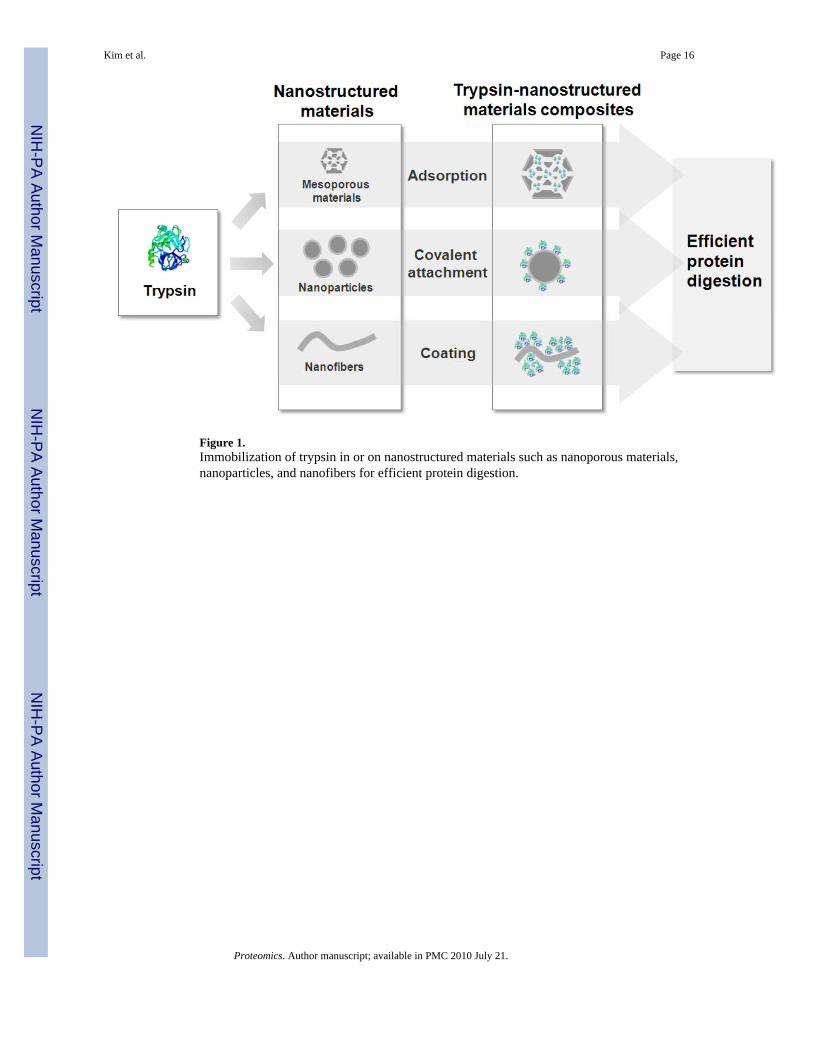
Figure 1.Immobilization of trypsin in or on nanostructured materials such as nanoporous materials,nanoparticles, and nanofibers for efficient protein digestion.
Kim et al. Page 16
Proteomics. Author manuscript; available in PMC 2010 July 21.
NIH
-PA Author Manuscript
NIH
-PA Author Manuscript
NIH
-PA Author Manuscript
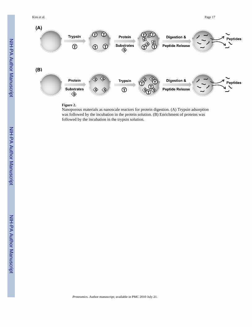
Figure 2.Nanoporous materials as nanoscale reactors for protein digestion. (A) Trypsin adsorptionwas followed by the incubation in the protein solution. (B) Enrichment of proteins wasfollowed by the incubation in the trypsin solution.
Kim et al. Page 17
Proteomics. Author manuscript; available in PMC 2010 July 21.
NIH
-PA Author Manuscript
NIH
-PA Author Manuscript
NIH
-PA Author Manuscript
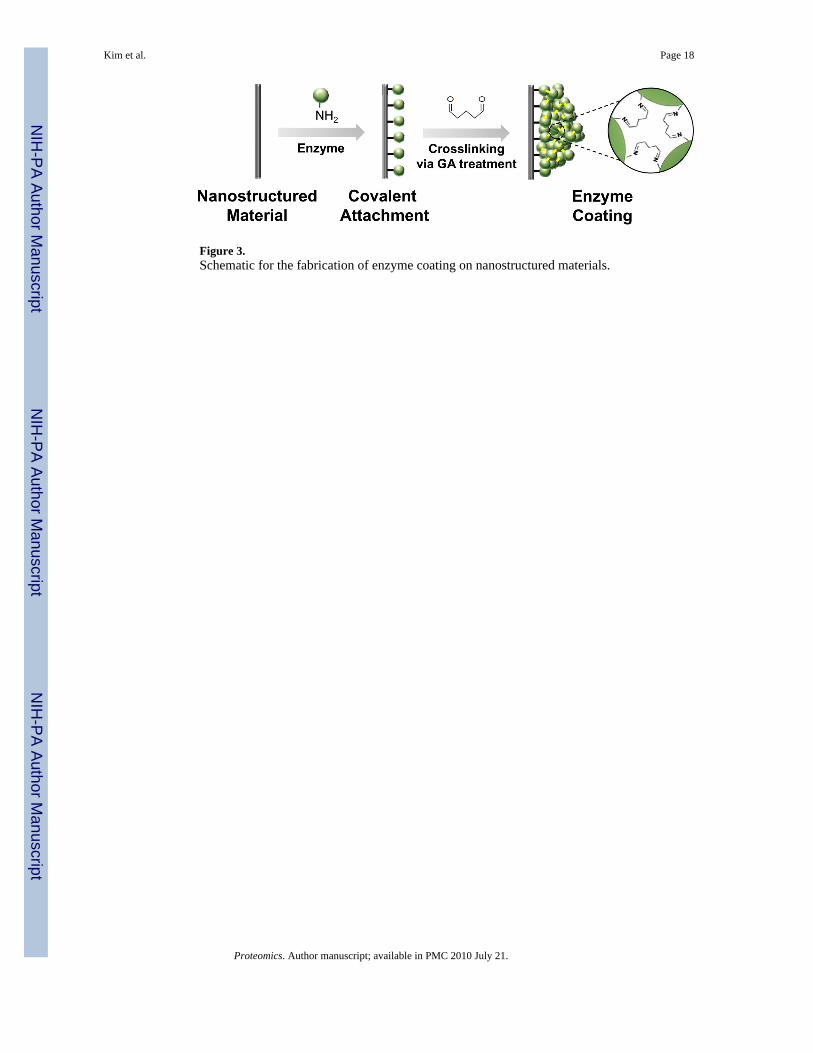
Figure 3.Schematic for the fabrication of enzyme coating on nanostructured materials.
Kim et al. Page 18
Proteomics. Author manuscript; available in PMC 2010 July 21.
NIH
-PA Author Manuscript
NIH
-PA Author Manuscript
NIH
-PA Author Manuscript
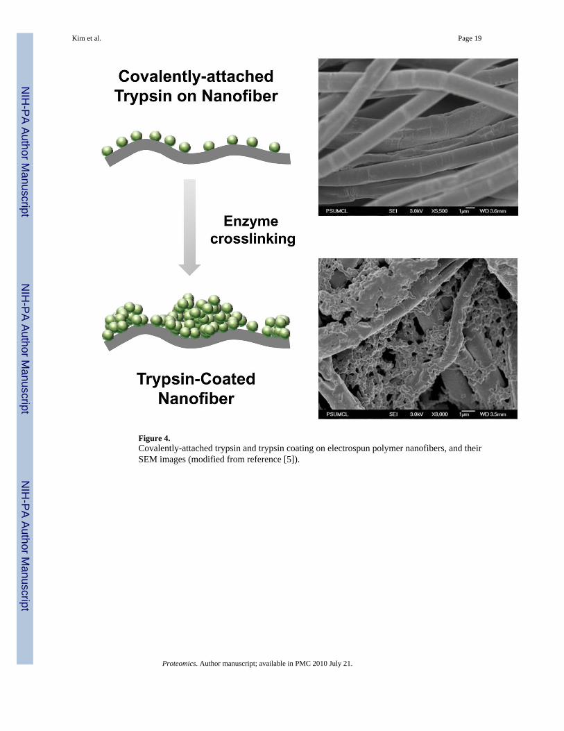
Figure 4.Covalently-attached trypsin and trypsin coating on electrospun polymer nanofibers, and theirSEM images (modified from reference [5]).
Kim et al. Page 19
Proteomics. Author manuscript; available in PMC 2010 July 21.
NIH
-PA Author Manuscript
NIH
-PA Author Manuscript
NIH
-PA Author Manuscript
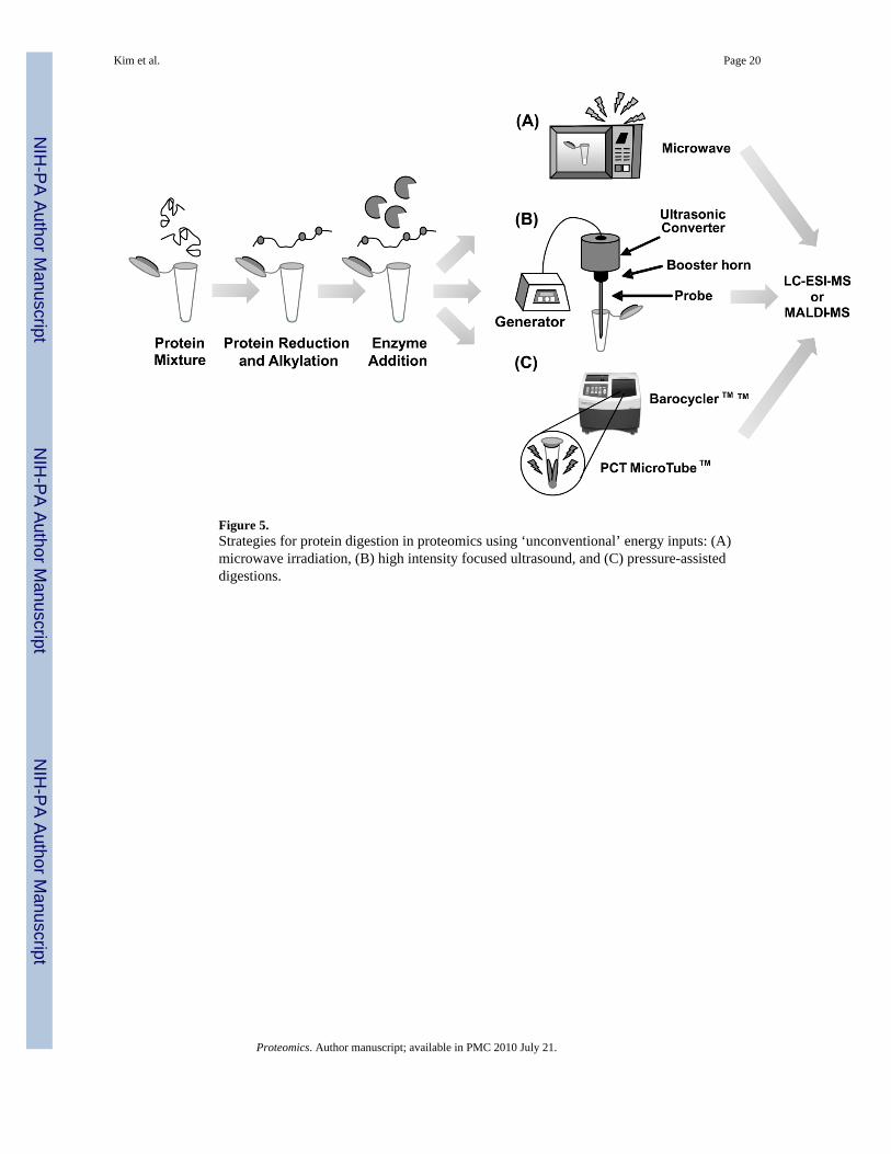
Figure 5.Strategies for protein digestion in proteomics using ‘unconventional’ energy inputs: (A)microwave irradiation, (B) high intensity focused ultrasound, and (C) pressure-assisteddigestions.
Kim et al. Page 20
Proteomics. Author manuscript; available in PMC 2010 July 21.
NIH
-PA Author Manuscript
NIH
-PA Author Manuscript
NIH
-PA Author Manuscript

Figure 6.(A) Pre-column immobilized enzyme reactor configuration. A protein is loaded in thereactor and digested to peptides, and then loaded in the analytical column. The valve isswitched and a gradient elution is performed; peptides are separated, eluted, and detected bythe mass spectrometry. (B) Post-column immobilized enzyme reactor configuration. (B1) Aprotein is retained in an analytical column, and then analyzed by mass spectrometry with nodigestion. (B2) A protein is retained in an analytical column and digested in a post-column,and the peptides are analyzed by mass spectrometry. The protein from path B1 and itscorresponding peptides from path B2 will have approximately the same retention time andelution profile enabling the correlation between them.
Kim et al. Page 21
Proteomics. Author manuscript; available in PMC 2010 July 21.
NIH
-PA Author Manuscript
NIH
-PA Author Manuscript
NIH
-PA Author Manuscript