Cigarette Smoke Induces an Unfolded Protein Response in the Human Lung A Proteomic Approach
10
Cigarette Smoke Induces an Unfolded Protein Response in the Human Lung A Proteomic Approach Steven G. Kelsen* 1 , Xunbao Duan* 2 , Rong Ji 1 , Oscar Perez 2 , Chunli Liu 2 , and Salim Merali 2 2 Department of Biochemistry and 1 Department of Medicine, Division of Pulmonary Critical Care and Pulmonary Medicine, Temple University School of Medicine, Philadelphia, Pennsylvania Cigarette smoking, which exposes the lung to high concentrations of reactive oxidant species (ROS) is the major risk factor for chronic obstructive pulmonary disease (COPD). Recent studies indicate that ROS interfere with protein folding in the endoplasmic reticulum and elicit a compensatory response termed the ‘‘unfolded protein re- sponse’’ (UPR). The importance of the UPR lies in its ability to alter expression of a variety of genes involved in antioxidant defense, inflammation, energy metabolism, protein synthesis, apoptosis, and cell cycle regulation. The present study used comparative proteomic technology to test the hypothesis that chronic cigarette smoking induces a UPR in the human lung. Studies were performed on lung tissue samples obtained from three groups of human subjects: nonsmokers, chronic cigarette smokers, and ex-smokers. Proteomes of lung samples from chronic cigarette smokers demonstrated 26 differentially expressed proteins (20 were up-regulated, 5 were down-regulated, and 1 was detected only in the smoking group) com- pared with nonsmokers. Several UPR proteins were up-regulated in smokers compared with nonsmokers and ex-smokers, including the chaperones, glucose-regulated protein 78 (GRP78) and calreticulin; a foldase, protein disulfide isomerase (PDI); and enzymes involved in antioxidant defense. In cultured human airway epithelial cells, GRP78 and the UPR-regulated basic leucine zipper, transcription factors, ATF4 and Nrf2, which enhance expression of important anti- oxidant genes, increased rapidly (, 24 h) with cigarette smoke extract. These data indicate that cigarette smoke induces a UPR response in the human lung that is rapid in onset, concentration dependent, and at least partially reversible with smoking cessation. We speculate that activation of a UPR by cigarette smoke may protect the lung from oxidant injury and the development of COPD. Keywords: oxidant defense; lung injury; COPD Cigarette smoking is the major risk factor for several forms of lung disease, including chronic obstructive pulmonary disease (COPD) (1–3). In the United States alone, more than 45 million people or 20.9% of the population are chronic smokers (4, 5). The gaseous and particulate phases of cigarette smoke contain more than 4,500 separate compounds, many of which are highly toxic reactive oxygen/nitrogen species (RONS) and xenobiotic materials (2, 6–8). In fact, cigarette smoke contains more than 10 14 free radicals in each puff of smoke (6, 7). Although the molecular mechanisms underlying development of COPD re- main incompletely understood, these toxic substances are be- lieved to induce an inflammatory response by adversely affecting oxidant/antioxidant and protease/anti-protease balance in the lung (1–3, 9–12). Of considerable importance, the propensity to develop COPD varies widely across cigarette smokers and correlates only weakly with the smoking history as reflected in the number of cigarette pack-years (13, 14). In fact, it is estimated that only a minority (i.e., 15–35%) of chronic, continuous cigarette smok- ers develop COPD (14, 15). That the majority of long-term smokers do not develop COPD suggests that failure of compen- satory mechanisms that protect the lung from ROS or xenobiotic materials contributes to development of the disease. In support of this concept, the expression of antioxidant genes believed to be important in protection of the lung from cigarette smoke– induced injury (e.g., thioredoxin, peroxiredoxin and glutathione- S-transferase, glutathione peroxidase) varies widely in airway epithelial cells harvested from chronic cigarette smokers (16, 17). Recent studies indicate that a complex molecular cascade termed the ‘‘unfolded protein response’’ (UPR) plays an impor- tant role in the regulation of expression of a variety of antiox- idant, xenobiotic metabolizing and pro- and anti-inflammatory genes (18–24). The UPR is activated in response to cellular stressors such as RONS, increases in cytosolic calcium, and hypoxia, which impair protein folding in the lumen of the en- doplasmic reticulum (ER) (18, 19, 22). Activation of the UPR compensates for abnormalities in protein folding by increasing the expression of genes involved in protein chaperoning and folding, protein translation, and protein degradation (18, 19, 25). Moreover, since the processes involved in protein transport and folding consume ATP and generate ROS, the UPR re- sponse induces expression of genes involved in energy synthesis and ROS quenching (21). Of interest, many basic cellular processes which depend on an adequate supply of fully func- tional membrane and secreted proteins (e.g., cell cycle regula- tion, apoptosis, energy metabolism, inflammation, and acute phase reactants) are also regulated by the UPR (20, 26–28). The effect of cigarette smoke exposure, which is likely to create conditions that foster development of a UPR by virtue of its effects on the cell RONS burden and cytosolic calcium, has not, however, been studied (29, 30). In the present study, we tested the following hypotheses: (1) chronic cigarette smoke exposure activates a UPR in the human lung; (2) the UPR is reversible with smoking cessation; and (3) UPR induction by cigarette smoke is dose-dependent and rapid in onset. Studies were performed on lung tissue samples obtained from three CLINICAL RELEVANCE Chronic cigarette smoking induces an endoplasmic reticu- lum stress response in the human lung termed the unfolded protein response. Failure of this compensatory system in some cigarette smokers may contribute to the development of chronic obstructive pulmonary disease. (Received in original form June 13, 2007 and in final form November 7, 2007) *These authors contributed equally to this work. Correspondence and requests for reprints should be addressed to Steven G. Kelsen, M.D., 761 Parkinson Pavilion, Temple University Hospital, 3401 N. Broad Street, Philadelphia, PA 19140. E-mail: [email protected] Am J Respir Cell Mol Biol Vol 38. pp 541–550, 2008 Originally Published in Press as DOI: 10.1165/rcmb.2007-0221OC on December 13, 2007 Internet address: www.atsjournals.org
-
Upload
independent -
Category
Documents
-
view
0 -
download
0
Transcript of Cigarette Smoke Induces an Unfolded Protein Response in the Human Lung A Proteomic Approach

Cigarette Smoke Induces an Unfolded Protein Responsein the Human LungA Proteomic Approach
Steven G. Kelsen*1, Xunbao Duan*2, Rong Ji1, Oscar Perez2, Chunli Liu2, and Salim Merali2
2Department of Biochemistry and 1Department of Medicine, Division of Pulmonary Critical Care and Pulmonary Medicine, Temple
University School of Medicine, Philadelphia, Pennsylvania
Cigarette smoking, whichexposes the lung to high concentrations ofreactive oxidant species (ROS) is the major risk factor for chronicobstructive pulmonary disease (COPD). Recent studies indicate thatROS interfere with protein folding in the endoplasmic reticulum andelicit a compensatory response termed the ‘‘unfolded protein re-sponse’’ (UPR). The importance of the UPR lies in its ability to alterexpression of a variety of genes involved in antioxidant defense,inflammation, energy metabolism, protein synthesis, apoptosis, andcell cycle regulation. The present study used comparative proteomictechnology to test the hypothesis that chronic cigarette smokinginduces a UPR in the human lung. Studies were performed on lungtissue samples obtained from three groups of human subjects:nonsmokers, chronic cigarette smokers, and ex-smokers. Proteomesof lung samples from chronic cigarette smokers demonstrated26 differentially expressed proteins (20 were up-regulated, 5 weredown-regulated, and 1 was detected only in the smoking group) com-pared with nonsmokers. Several UPR proteins were up-regulated insmokers compared with nonsmokers and ex-smokers, including thechaperones, glucose-regulated protein 78 (GRP78) and calreticulin;a foldase, protein disulfide isomerase (PDI); and enzymes involved inantioxidant defense. In cultured human airway epithelial cells,GRP78 and the UPR-regulated basic leucine zipper, transcriptionfactors, ATF4 and Nrf2, which enhance expression of important anti-oxidant genes, increased rapidly (, 24 h) with cigarette smokeextract. These data indicate that cigarette smoke induces a UPRresponse in the human lung that is rapid in onset, concentrationdependent, and at least partially reversible with smoking cessation.We speculate that activation of a UPR by cigarette smoke mayprotect the lung from oxidant injury and the development of COPD.
Keywords: oxidant defense; lung injury; COPD
Cigarette smoking is the major risk factor for several forms oflung disease, including chronic obstructive pulmonary disease(COPD) (1–3). In the United States alone, more than 45 millionpeople or 20.9% of the population are chronic smokers (4, 5).The gaseous and particulate phases of cigarette smoke containmore than 4,500 separate compounds, many of which are highlytoxic reactive oxygen/nitrogen species (RONS) and xenobioticmaterials (2, 6–8). In fact, cigarette smoke contains more than1014 free radicals in each puff of smoke (6, 7). Although themolecular mechanisms underlying development of COPD re-main incompletely understood, these toxic substances are be-lieved to induce an inflammatory response by adversely affecting
oxidant/antioxidant and protease/anti-protease balance in thelung (1–3, 9–12).
Of considerable importance, the propensity to developCOPD varies widely across cigarette smokers and correlatesonly weakly with the smoking history as reflected in the numberof cigarette pack-years (13, 14). In fact, it is estimated that onlya minority (i.e., 15–35%) of chronic, continuous cigarette smok-ers develop COPD (14, 15). That the majority of long-termsmokers do not develop COPD suggests that failure of compen-satory mechanisms that protect the lung from ROS or xenobioticmaterials contributes to development of the disease. In supportof this concept, the expression of antioxidant genes believed tobe important in protection of the lung from cigarette smoke–induced injury (e.g., thioredoxin, peroxiredoxin and glutathione-S-transferase, glutathione peroxidase) varies widely in airwayepithelial cells harvested from chronic cigarette smokers (16, 17).
Recent studies indicate that a complex molecular cascadetermed the ‘‘unfolded protein response’’ (UPR) plays an impor-tant role in the regulation of expression of a variety of antiox-idant, xenobiotic metabolizing and pro- and anti-inflammatorygenes (18–24). The UPR is activated in response to cellularstressors such as RONS, increases in cytosolic calcium, andhypoxia, which impair protein folding in the lumen of the en-doplasmic reticulum (ER) (18, 19, 22). Activation of the UPRcompensates for abnormalities in protein folding by increasingthe expression of genes involved in protein chaperoning andfolding, protein translation, and protein degradation (18, 19,25). Moreover, since the processes involved in protein transportand folding consume ATP and generate ROS, the UPR re-sponse induces expression of genes involved in energy synthesisand ROS quenching (21). Of interest, many basic cellularprocesses which depend on an adequate supply of fully func-tional membrane and secreted proteins (e.g., cell cycle regula-tion, apoptosis, energy metabolism, inflammation, and acutephase reactants) are also regulated by the UPR (20, 26–28).
The effect of cigarette smoke exposure, which is likely tocreate conditions that foster development of a UPR by virtue ofits effects on the cell RONS burden and cytosolic calcium, hasnot, however, been studied (29, 30). In the present study, wetested the following hypotheses: (1) chronic cigarette smokeexposure activates a UPR in the human lung; (2) the UPR isreversible with smoking cessation; and (3) UPR induction bycigarette smoke is dose-dependent and rapid in onset. Studieswere performed on lung tissue samples obtained from three
CLINICAL RELEVANCE
Chronic cigarette smoking induces an endoplasmic reticu-lum stress response in the human lung termed the unfoldedprotein response. Failure of this compensatory system insome cigarette smokers may contribute to the developmentof chronic obstructive pulmonary disease.
(Received in original form June 13, 2007 and in final form November 7, 2007)
*These authors contributed equally to this work.
Correspondence and requests for reprints should be addressed to Steven G.
Kelsen, M.D., 761 Parkinson Pavilion, Temple University Hospital, 3401 N. Broad
Street, Philadelphia, PA 19140. E-mail: [email protected]
Am J Respir Cell Mol Biol Vol 38. pp 541–550, 2008
Originally Published in Press as DOI: 10.1165/rcmb.2007-0221OC on December 13, 2007
Internet address: www.atsjournals.org
groups of human subjects: nonsmokers, chronic cigarette smok-ers, and ex-smokers. Comparative proteomic technology wasused to examine the expression of several proteins that arehallmarks of a UPR and its downstream targets (18, 26–28).Specifically, activation of a UPR was tested by examining theexpression of the chaperones, GRP78, calnexin and calreticulin,and a foldase, protein disulfide isomerase. Cultured humanairway epithelial cells (i.e., the 16-HBE cell line), a cell whichplays an important role in the pathogenesis of COPD, wereused to examine the UPR response to a range of concentrationsof cigarette smoke extract (CSE), and its time course (1–3). Inaddition, experiments in 16-HBE cells sought to determine ifCSE activates one of several ER sensors that detect misfoldedproteins (i.e., the protein kinase-R-like endoplasmic reticulumkinase [PERK]) (18, 19). The PERK pathway is of considerableimportance in the cellular response to oxidant stress, since itaugments nuclear expression of the basic leucine zipper tran-scription factors, nuclear factor, erythroid factor 2 p45 relatedfactor 2 (Nrf2), and ATF4 (21, 26, 31–33). In particular, Nrf2heterodimerizes with several different transcription factors,including ATF4, to regulate a large number of genes involvedin protection of the lung against oxidant injury and metabolismof xenobiotic substances (12, 34–38).
The results of the present study indicate that the severalchaperones and foldase, which are markers of a UPR, are up-regulated in the lungs of chronic cigarette smokers, and thatseveral downstream targets of the UPR including the antioxi-dant gene, thioredoxin-dependent peroxidase reductase, are up-regulated. Moreover, ex-smokers demonstrate a UPR responseintermediate between that of nonsmokers and that of activesmokers, supporting the idea that the UPR induced by chroniccigarette smoking is at least partially reversible. Finally, incultured airway epithelial cells, the UPR is dose-dependentlyactivated by CSE, occurs in less than 24 hours, and specificallyinvolves PERK activation and its downstream targets, thetranscription factors Nrf2 and ATF4.
MATERIALS AND METHODS
Clinical Study Material
Lungs were obtained by the region’s transplant network, Gift of LifeInc., under a protocol approved by the Temple University InstitutionalReview Board. The lungs were obtained from three chronic smokers;three lifelong, nonsmoking subjects; and three ex-smokers. Criteria forinclusion included a history negative for lung disease and normal lungradiography.
All lungs were obtained from beating heart donors, immediatelyplaced in sterile, ice-cold preservation medium (39), and shipped to ourlaboratory on ice within 12 hours of collection. Radiologically andmacroscopically normal-appearing regions were selected for this study.The region selected, usually an entire lobe, was slowly inflated usinga cannula inserted into the supplying airway to instill a 1:1 mix of cryo-embedding medium (Cryomatrix; Thermo Electron Corp., Waltham,MA) and 50% sucrose in distilled water.
Portions of the inflated organ were then frozen by suspension inliquid nitrogen fumes for 20 to 25 minutes, as previously described (40),then stored at 2808C until used. Samples of frozen tissues were cut into1 3 1 3 1 cm blocks and embedded in OCT (Tissue Tek, ElectronMicroscopy Sciences, Hatfield, PA). Sample sections (6 mm) were cutfrom each block using a cryostat at 2208C, then stained with hematoxylinand eosin for inspection. Only those blocks showing normal lungarchitecture microscopically were selected for subsequent proteomicanalysis. Since whole lung tissue is composed of at least six major celltypes, considerable effort was made to obtain approximately equalamounts of lung parenchyma, small airways, and pulmonary vessels. Inthis regard, the amount (z 1 cm3) and location (peripheral tissueextending to include the pleural surface) of the sample were standardized.
Human Airway Epithelial Cell Culture
Human airway epithelial cells, the 16-HBE cell line, were cultured in100-mm tissue culture dishes coated with type VI human placentalcollagen (Sigma/Aldrich, St. Louis, MO) in Dulbecco’s modified Eagle’smedium containing 10% fetal bovine serum (FBS), penicillin (100 U/ml),and streptomycin (100 mg/ml) in 5% CO2 at 378C. Cells were grown to80% to 100% confluence before study. At the time of study, medium wasreplaced with medium containing CSE.
CSE was prepared as previously described (41, 42). Briefly, smokefrom two unfiltered, research cigarettes (2R4; University of Kentucky,Lexington, KY) was bubbled through 25 ml of cell culture medium ina 50-ml plastic centrifuge tube at a flow rate of 50 ml/minute. Smokewas allowed to equilibrate in the medium for 10 to 15 minutes, and theresultant suspension was then passed through a 0.2-mm pore filter(Nalgene, Rochester, NY). CSE prepared in this manner was consid-ered to have a concentration of 100%. From this stock, serial dilutionswere made with cell culture medium to achieve final concentrations of15, 30, and 45% CSE. CSE was applied to cell cultures within 30minutes of preparation and maintained for 24 hours. Cells exposed tomedium alone served as control.
Cultured cells were washed twice with cold PBS, scraped and lysedin 1% SDS, 4 mg/ml aprotinin, 4 mg/ml leupeptin, 4 mg/ml pepstatin,2 mM phenylmethylsulfonyl fluoride, 6 mM Na3VO4, and 6 mM NaF.Protein concentrations were determined by DC protein assay (Bio-Rad, Hercules, CA).
Sample Preparation for Two-Dimensional Gel Analysis
Frozen lung tissue from three nonsmokers, three chronic smokers, andthree ex-smokers was individually processed by grinding with a mortarand pestle cooled with liquid nitrogen. Frozen powders from eachindividual tissue block were thawed by adding 0.8 ml of cold lysis buffer(7 M urea, 2 M thiourea, 4% CHAPS, 40 mM Tris, and 60 mM DTT) andsonicated in an ice bath for approximately 1 minute at 5 watts and 70%duty cycle (Sonic Dismembrator; Fisher Scientific, Pittsburgh, PA). Soni-cates were centrifuged at 10,000 3 g for 12 minutes at 48C. Supernatantswere collected and acetone added to precipitate proteins and remove cryo-embedding medium and sucrose solution. Precipitated proteins wereresolubilized using the above lysis buffer. Protein concentrations in theextracts were measured in triplicate using a Bio-Rad Bradford-basedprotein assay with bovine serum albumin as the standard.
Two-Dimensional Gel Electrophoresis
Lung lysates were processed individually (nine in total; three from eachgroup of subjects). For first-dimension separation, 40 mg of sample proteinwas diluted in 125 ml with rehydration buffer and loaded onto animmobilized pH gradient (IPG) strip by overnight passive in-gel re-hydration. A global view of the proteome was obtained initially using IPGstrips of pI 3-10. To enhance resolution and sensitivity, narrow range IPGstrips (pI 4–7 and 6–10) were subsequently used. The rehydration buffercontained 8 M urea, 2% CHAPS, 0.2% carrier ampholytes, and 10 mMDTT for pH 4–7 linear and pH 3–10 nonlinear IPG strips. For pH 6–10IPG strips, rehydration was with 15 mg/ml Destreak reagent. Isoelectricfocusing (IEF) within the strips was performed at 208C with a MultiPhor IIsystem (Amersham Biosciences Corp., Piscataway, NJ) using a total of12,000 V�h with a maximum of 5,000 V.
For second-dimensional separation, the IPG strips were removedfrom the MultiPhor II chamber, soaked for 15 minutes in 10 ml of anequilibration buffer (6 M urea, 30% glycerol, 2% SDS, 1% DTT, and0.05 M pH 8.8 Tris), then for 15 minutes in 10 ml of a second equil-ibration buffer (with 2.5% iodoacetamide substituted for 1% DTT),and positioned against 10 to 14% SDS polyacrylamide gels in a BioRadMini-PROTEAN 3 System at 200 V for 45 minutes. Polyacrylamidegels were then fixed twice using 50% methanol, 7% acetic acid, balancewater. Protein spots in the gels were revealed by staining with eitherSypro-Ruby fluorescent total protein stain or Pro-Q Diamond phos-phoprotein stain.
Image Analysis of Two-Dimensional Gels
Fluorescence images of individual gels from the nine lung lysates werecaptured with a FLA-5000 Fluor Imager (Fuji Photo Film Co., Ltd.,Tokyo, Japan) and analyzed using PDQuest Software (Version 8.0).
542 AMERICAN JOURNAL OF RESPIRATORY CELL AND MOLECULAR BIOLOGY VOL 38 2008

After automatic detection of spots by PDQuest software, the files wereinspected manually to assess accuracy of computer-generated images.The software calculated individual spot ‘‘volumes’’ in each gel bydensity/area integration. To control for slight differences in proteinloading across gels, the spot volume density obtained from eachindividual lung lysate was calculated by image analysis software andnormalized to total spot volume on that gel.
In-Gel Trypsin Digestion
Differentially expressed spots were excised using an Xcise automatedrobotic system (Shimadzu Biotech, Columbia, MD). Destaining ofexcised gel pieces was performed by two 30-minute washes with 50%acetonitrile containing 50 mM ammonium bicarbonate. After dehydra-tion with 100% acetonitrile, 10 ml of 12.5 ng/ml sequencing grade trypsin(Promega, Madison, WI) was added to the gel pieces and incubatedovernight at 378C. Resulting tryptic peptides were extracted twice with15 ml (5% formic acid, 50% acetonitrile, balance water) for 20 minutesand the pooled extracts were processed with desalting ZipTips.
MALDI-TOF-TOF Analysis
The desalted peptides from each spot were mixed 1:1 with matrixsolution (1% a-cyano-4-hydroxy cinnamic acid in 50% acetonitrile and50% 0.1% trifluoroacetic acid) and 1.0 ml volumes were applied to wellsof an AnchorChip sample target plate used for the Bruker Auto-flexMALDI-TOF-TOF instrument. Peptide mass fingerprints were obtainedusing the reflective and positive ion mode. Mass spectra were collectedfrom the sum of 100 to 400 laser shots and mono-isotopic peaks weregenerated by FlexAnalysis software with signal-to-noise ratio of 2:1.Mass peak value calculations used two trypsin auto-digestion peptideswith M1H values 842.509 and 2211.104 as internal standards. Proteinswere identified by matching the calibrated peptide mass values withineither Swiss-Prot or NCBInr protein databases for Homo sapiens usingan in-house version of Mascot Server 2.1 imbedded in Bruker’s Biotoolsoftware. Match variances allowed were a mass tolerance of 50 ppm,one missed trypsin cleavage, fixed modification of carbamidomethylcysteine, and variable modification of methionine oxidation. For thesamples that did not produce a ‘‘hit’’ with a confident score, peptidepeaks with good signal were further fragmented using ‘‘Laser-induceddecomposition’’ to obtain LIFT-TOF/TOF spectra, and these MS/MSdata alone or combined with the previously produced MS data were usedto search against the protein database through the Mascot.
Western Blot Analysis
Proteins (30 to 80 mg) from the same lung lysates as used for the abovetwo-dimensional gels or from CSE-exposed 16 HBE cells was sepa-rated by 10 to 14% gradient SDS-PAGE. The separated proteins weretransferred to a nitrocellulose membrane in a semi-dry blottingchamber according to the manufacturer’s protocol (Bio-Rad) or ina transfer apparatus in CAPS buffer (10 mM 3-cyclohexylamino-1-propanosulfonic acid in 15% methanol, pH 10.6).
Blots were blocked with 5% milk in Tris-buffer saline solution (pH7.6) containing 0.05% Tween-20 (TBS/T), and probed with the followingrabbit anti-human antibodies from Santa Cruz Biotechnology (SantaCruz, CA) at a concentration of 0.4 mg/ml: GRP-78 (SC-13968), PDI(SC-20132), calreticulin (SC-11398), calnexin (SC-11397), GAPDH(SC-25778), Nrf2 (SC-13032), and ATF4 (SC-200). In addition, rabbitanti-human total eIF2 a (cat# 9722) and anti-phospho eIF2a (cat# 9721)from Cell Signaling Technology (Beverly, CA) were used at 1:1,000 di-lution. Blots were incubated with primary antibody overnight at 48C atwith gentle shaking, then incubated with a mouse anti-rabbit horseradishperoxidase–conjugated secondary Ab (1:10,000) (Biomeda Corp., Fos-ter City, CA) for 1 hour at room temperature. Blots were exposed usinga chemiluminescent detection method (Enhanced ECL Detection Sys-tem, Amersham Biosciences).
In some 16-HBE cell experiments, cytoplasmic and nuclear frac-tions were prepared to examine expression, respectively, of the trans-lational initiation factor, eIF2a, a classical target of activated PERK, aswell as the transcription factors, Nrf2 and ATF4. Cells were washed withcold PBS twice, scraped in 1 ml PBS/1 mM EDTA buffer, and thencentrifuged. The pellet was resuspended in ice-cold harvest buffer (10mM HEPES, pH 7.9, 50 mM NaCl, 0.5 M Sucrose, 0.1 mM EDTA, 0.5%
Triton X-100, 1 mM dithiothreitol, and proteinase inhibitor [cat#78410;Pierce, Rockford, IL]) for 10 minutes. The lysate was centrifuged and thesupernatant containing the cytoplasmic fraction was collected andcleared at 14,000 rpm. The pellet containing the nuclear fraction waswashed in ice-cold buffer A (10 mM HEPES, pH 7.9, 10 mM KCl, 0.1 mMEDTA, 0.1 mM EGTA, 1 mM dithiothreitol, and proteinase inhibitor),pelleted and then resuspended in ice-cold buffer C (10 mM HEPES, pH7.9, 500 mM NaCl, 0.1 mM EDTA, 0.1 mM EGTA, 0.1% NP-40, with 1 mMdithiothreitol, and proteinase inhibitor). The suspension was vortexed,vigorously rocked, and then centrifuged. The supernatant containing thenuclear proteins was removed. Nuclear and cytoplasmic fractions werestored at 2808C before Western blotting.
Statistical Analysis
Group mean 6 SD values of normalized spot volumes in two-dimensional gels of individual smokers, nonsmokers, and ex-smokerswere determined. Statistical significance of differences in group meanvalues was determined by one-way ANOVA and Student’s t test withstatistical significance accepted at the P , 0.05 level. Spots on two-dimensional gels, which were statistically significantly different (P ,
0.05), were considered differentially expressed.Western blots for proteins of interest were scanned and differences in
band density assessed statistically by one-way ANOVA and Students’ ttest. Statistical significance was accepted at the P , 0.05 level.
RESULTS
Subject Groups
Demographics, smoking history, co-morbidities, and cause of deathare shown in Table 1. Smokers and ex-smokers had similar,heavy smoke exposure (36 6 19 SE pack-years, range 12–75 yr,and 38 6 13 SE pack-years, range 15–60 yr, for smokers and ex-smokers, respectively; P . 0.44). Ex-smokers had stopped smokingfrom 8 to 18 years before death.
The three groups appeared evenly matched in terms of ageand sex. All were white except for one African American in thesmoker group. Subjects in all three groups had co-morbidities,but these appeared to be similar.
The microscopic appearance of the lung in the region fromwhich samples were obtained was normal (Figure 1).
Proteomic Analysis
Gels performed over a wide isoelectric focusing range of pI 3–10 produced approximately 700 individual protein spots (datanot shown). Gels performed with narrower pI ranges of 4 to 7and 6 to 10 provided greater resolution and produced a total ofapproximately 900 protein spots in each gel (Figure 2).
A full comparison of all spots visualized was performed inthe smoker and nonsmoker groups only. A total of 41 spots weresignificantly different in the cigarette smoking versus the non-smoking groups (P , 0.05). Of these, 40 spots representing26 unique proteins were identified. Fourteen of the spots weremultiple isoforms of nine proteins (protein disulfide isomerase,GRP78, glyceraldehyde 3-phosphate dehydrogenase, actin, apoli-poprotein A, albumin, gelsolin, transferrin, and annexin).
Of the differentially expressed proteins observed in the lungsof smokers compared with those of the nonsmokers, 20 were up-regulated, 5 were down-regulated and 1 (major vault protein)was detected only in the smoking group. The differentiallyexpressed proteins, their identity, and the magnitude of differ-ence with the nonsmoker group are shown in Table 2.
The differentially expressed proteins were grouped into thefollowing categories: ER stress, energy metabolism, structural,protein synthesis/degradation, acute phase reactants, and mis-cellaneous (Table 2). Of note, several proteins involved in theUPR were up-regulated in the smoking group. These includedthe ER chaperones, GRP 78 and calreticulin, and the ER
Kelsen, Duan, Ji, et al.: Cigarette Smoke Induces an Unfolded Protein Response 543

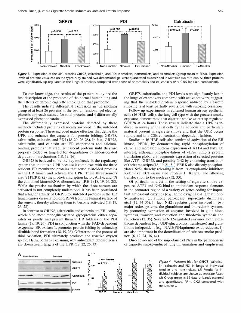
foldase, PDI (Table 2). The magnitude of increase of theseseveral UPR-induced proteins ranged from 1.8- to 2.4-fold inthe lungs of smokers (P , 0.05 for each; Figure 3).
Selected spots characterizing the UPR response (i.e.,GRP78, calreticulin, and PDI) were examined in ex-smokersand compared with smokers and nonsmokers. Of interest, levelsof expression of GRP78, calreticulin, and PDI were significantlylower in ex-smokers compared with smokers (P , 0.05 for each;Figure 3).
Up-regulation of the UPR chaperones and foldase in thelungs of smokers was confirmed by Western blotting forGRP78, calreticulin, and PDI (Figure 4). Another UPR markernot detected on two-dimensional gels, the membrane-boundchaperone calnexin, was also up-regulated as detected byWestern blotting (Figure 4).
In addition, several downstream pathways controlled by theUPR were differentially expressed in the lungs of smokers. These
included up-regulation of the antioxidant enzyme, thioredoxin-dependent peroxidase reductase, and several components of thepolyribosome that regulate protein translation (i.e., elongationfactor-1b, elongation factor-1d, 60S acidic ribosomal protein P2,and heat shock protein 27).
Phosphoproteome
Additional gels performed at pI 4 to 7 two-dimensional gels,which were stained for phosphoproteins, demonstrated fourdifferentially expressed spots in the lungs of smokers (P , 0.05),one of which was not previously identified in the total proteinstain (Figure 5). Of the four proteins, three were up-regulatedand one was down-regulated. The three up-regulated phospho-proteins (elongation factor-1b, elongation factor-1d, and 60Sacidic ribosomal protein P2) were also identified in gels stainedfor total proteins (Table 2). In contrast, the one phosphoproteinnot detected in the gels stained for total protein, calgranulin-B/
TABLE 1. SUBJECT DEMOGRAPHICS AND SMOKING HISTORY
Subject No. Age (yr) Sex Ethnicity Cause of Death Smoking History Co-Morbidities
Nonsmokers
1 76 M C ICH None Diabetes mellitus
Hip fracture with prosthesis
2 68 M AA ICH None Diabetes mellitus
Hypertension
Sleep apnea
3 68 F C ICH None Cerebrovascular accident
Seizures
Esophageal stricture
Smokers
4 46 M C Cardiac arrest 1 PPD 3 12 yr Allergic rhinitis
5 45 F C ICH 1 PPD 3 20 yr Diabetes mellitus
Seizures
Alcohol abuse
Pancreatitis
6 69 M C ICH 1.5 PPD 3 50 yr Hypertension;
Knee replacement
Ex-smokers
7 61 M C ICH, CVA 15 PY, quit 15 yr ago Hypertension;
C-Spinal surgery
8 76 M C Head 1 PPD 3 40 yr, quit 18 yr ago Polio
Trauma Bladder spasm
Knee replacement
Hemorrhoidectomy
Appendectomy
9 74 F C ICH 1.5 PPD 3 40 yr,quit 8 yr ago Hypertension
Osteoporosis
Osteoarthritis
Hypercholesterolemia
Coronary artery disease
Hysterectomy
Definition of abbreviations: AA, African American; C, white; ICH, intracranial hemorrhage; PPD, packs per day; PY, pack-year.
Figure 1. Typical lung architecture in representative
smoker and nonsmoker. The lungs were inflated with
cryo-embedding medium and frozen over liquid nitrogen
fumes. Frozen sections (6 mM) were stained with hema-toxylin and eosin. Each field shows normal-appearing
small intra-pulmonary airways (large arrows), pulmonary
arterioles (small arrows), and adjacent parenchyma(31,000 magnification).
544 AMERICAN JOURNAL OF RESPIRATORY CELL AND MOLECULAR BIOLOGY VOL 38 2008

Figure 2. Differential expression profiling of lung homogenates from smoker and nonsmoker using two-dimensional electrophoresis. Proteins were
separated by isoelectric focusing (pI) and by molecular weight as described in MATERIALS AND METHODS. The sub-proteome from each sample wasassessed using a pI range of 4 to 7 (A) or of 6 to 10 (B). The proteins were stained with sypro-ruby and images compared by PDQuest software. The
red arrows indicate up-regulated proteins and the blue arrows down-regulated proteins in lungs of smokers.
Kelsen, Duan, Ji, et al.: Cigarette Smoke Induces an Unfolded Protein Response 545

S-100 A9, was down-regulated. Interestingly, all four proteinswere annotated as phospho-serine proteins in Swiss-Prot pro-tein knowledge database.
Effect of CSE on 16-HBE Cells
Treatment with CSE for 24 hours increased GRP78 protein inconcentration-dependent fashion (P , 0.005) (Figure 6). Themaximum increase was observed at CSE concentrations of 15 to30% CSE.
CSE extract (30%) also rapidly increased phosphorylation ofeIF2a, a target of PERK, without affecting total eIF2a (Figure7A). CSE (30%) also increased nuclear expression of the PERK-regulated transcription factors, ATF4 and Nrf2 (Figure 7B).
DISCUSSION
Because cigarette smoking is strongly associated with thedevelopment of a variety of lung diseases (especially COPD),
yet not all who smoke develop these diseases, it has beenpostulated that protective mechanisms are activated in smokerswho do not develop these diseases. In the present study, weexamined the proteomes of lungs from chronic smokers, non-smokers, and ex-smokers for evidence of a UPR which mayrepresent one such mechanism.
The lungs studied were obtained from donors who had beenscrutinized by history and chest X-ray and felt to be free ofchronic lung disease. The lung was preserved in the usualmanner for transplanted organs and quick-frozen within 12hours of removal from the body. The region of lung sampledwas macroscopically and microscopically normal and standard-ized to minimize variability in cell composition across subjects.However, given the manner in which the lungs were obtained,lung function data (FEV1, FVC, etc.) were not available in anyof the subjects. Hence, we cannot rule out the possibility thatsome subjects may have had COPD. However, if present,COPD was almost certainly mild, since it was not associatedwith symptoms or abnormalities in chest X-ray or histology.
TABLE 2. PROTEINS DIFFERENTIALLY EXPRESSED IN SYPRO-RUBY–STAINED TWO-DIMENSIONAL GELS
Normalized Spot Volumes 6 SD
Spot No. Protein ID
SwissProt
Accession No.
Peptides
Matched Nonsmoker Smoker P Value
ER Stress
7 Calreticulin [ P27797 5 5,102 6 1,400 9,364 6 1,524 0.023
10 Protein disulfide-isomerase (PDI)[ P07237 25 536 6 169 1,167 6 35 0.003
11 979 6 313 2,064 6 411 0.022
12 1377 6 424 2,885 6 369 0.010
17 78 kD glucose-regulated protein (GRP 78) [ P11021 11 767 6 336 1,534 6 143 0.022
18 1,078 6 606 2,251 6 333 0.043
30 Thioredoxin-dependent peroxide reductase [ P30048 4 1,392 6 259 2,513 6 180 0.004
Energy Metabolism
16 ATP synthase subunit beta [ P06576 5 3,414 6 988 6,335 6 1,163 0.029
39 Glyceraldehyde-3-phosphate dehydrogenase [ P04406 9 2,515 6 337 5,294 6 1,334 0.025
40 P04406 8 1,502 6 1,136 6,163 6 2,316 0.035
41 Malate dehydrogenase[ P40926 6 1,199 6 203 2,316 6 20 0.001
Structural
8 Actin, cytoplasmic 1 Y P60709 7 3,062 6 512 1,638 6 340 0.016
9 P60709 6 5,627 6 1,006 3,603 6 518 0.036
15 Tubulin beta-2 chain [ P07437 16 2,107 6 456 4,670 6 1,283 0.031
21 Microfibril-associated glycoprotein [ 338 6 96 971 6 300 0.025
27 Gelsolin [ P06396 7 124 6 90 819 6 166 0.003
28 157 6 126 921 6 220 0.006
29 217 6 154 1,024 6 201 0.005
36 Annexin A2 [ P07355 15 2,355 6 939 6,041 6 2055 0.048
37 P07355 20 3,379 6 1,364 9,584 6 3,558 0.047
38 Cofilin-I [ P23528 5 346 6 242 183 6 304 0.020
Protein Synthesis/Degradation
2 60S acidic ribosomal protein P2 [ P05387 5 576 6268 1,434 6 456 0.048
4 Elongation factor 1b [ P24534 4 852 6286 1,708 6 84 0.008
14 Elongation factor 1d [ P29692 8 997 6 453 2,236 6 384 0.022
24 Cathepsin D [ P07339 14 2,094 6 899 5,934 6 2,129 0.045
31 Heat shock protein 27 [ 4 2,302 6 409 3,664 6 527 0.024
Acute Phase Reactants
19 Apolipoprotein A-I Y P02647 23 4,777 6 885 3,142 6 366 0.042
20 P02647 17 10,805 6 3,381 4,865 6 1,283 0.047
22 Albumin Y P02768 19 10,921 6 4,849 2,370 6 1,717 0.045
23 P02768 19 19,980 6 9,709 3,832 6 1,430 0.046
32 Albumin Y P02768 12 10,496 6 3,584 3,399 6 775 0.029
33 Serotransferrin (transferrin) Y P02787 15 569 6 165 82 6 143 0.018
34 681 6 299 64 6 58 0.021
35 543 6 269 62 6 57 0.045
Miscellaneous
3 Heme-binding protein 2 [ Q9Y5Z4 6 533 6 29 816 6 89 0.006
5 14-3-3 protein sigma (Epithelial cell marker protein 1) [ P31947 11 1,098 6 289 2,579 6 326 0.004
6 Reticulocalbin-1 [ Q15293 6 661 6 392 1,545 6 212 0.027
13 Rho GDP-dissociation inhibitor 1 [ P52565 9 2,372 6 370 4,200 6 577 0.010
25 Major vault protein (Lung resistance-related protein) [ Q14764 10 0 197 6 107 0.033
26 0 209 6 86 0.013
546 AMERICAN JOURNAL OF RESPIRATORY CELL AND MOLECULAR BIOLOGY VOL 38 2008
To our knowledge, the results of the present study are thefirst description of the proteome of the normal human lung andthe effects of chronic cigarette smoking on that proteome.
The results indicate differential expression in the smokinggroup of at least 26 proteins in the two-dimensional gel electro-phoresis approach stained for total proteins and 4 differentiallyexpressed phosphoproteins.
The differentially expressed proteins detected by thesemethods included proteins classically involved in the unfoldedprotein response. These included major effectors that define theUPR and enhance the capacity for protein folding: GRP78,calreticulin, calnexin, and PDI (18–20, 26–28). In fact, GRP78,calreticulin, and calnexin are ER chaperones and calcium-binding proteins that stabilize nascent proteins until they areproperly folded or targeted for degradation by ER-associateddegradation mechanisms (18, 19, 26).
GRP78 is believed to be the key molecule in the regulatorysystem that initiates a UPR because it complexes with the threeresident ER membrane proteins that sense misfolded proteinsin the ER lumen and activate the UPR. These three sensorsare: (1) PERK; (2) the proto-transcription factor, ATF6; and (3)the combined kinase/RNA ribonuclease, IRE-1 (18, 19, 26, 28).While the precise mechanism by which the three sensors areactivated is not completely understood, it has been postulatedthat a higher affinity of GRP78 for unfolded proteins in the ERlumen causes dissociation of GRP78 from the luminal surface ofthe sensors, thereby allowing them to become activated (18, 19,26, 28).
In contrast to GRP78, calreticulin and calnexin are ER lectins,which bind most monoglucosylated glycoproteins either sepa-rately or jointly, and present them to ER foldases of the PDIfamily (18, 19, 26). PDI in conjunction with the FAD-dependentoxygenase, ER oxidase 1, promotes protein folding by enhancingdisulfide bond formation (18, 19, 26). Of interest, in the process ofthiol oxidation, PDI ultimately produces the reactive oxygenspecie, H2O2, perhaps explaining why antioxidant defense genesare downstream targets of the UPR (18, 22, 26, 43).
GRP78, calreticulin, and PDI levels were significantly less inthe lungs of ex-smokers compared with active smokers, suggest-ing that the unfolded protein response induced by cigarettesmoking is at least partially reversible with smoking cessation.
Follow-up experiments in cultured human airway epithelialcells (16-HBE cells), the lung cell type with the greatest smokeexposure, demonstrated that cigarette smoke extract up-regulatedGRP78 at 24 hours. These results indicate that a UPR is in-duced in airway epithelial cells by the aqueous and particulatematerial present in cigarette smoke and that the UPR occursrapidly and in a CSE concentration–dependent fashion.
Studies in 16-HBE cells also confirmed activation of the ERkinase, PERK, by demonstrating rapid phosphorylation ofeIF2a and increased nuclear expression of ATF4 and Nrf2. Ofinterest, although phosphorylation of eIF2a inhibits proteintranslation globally, it augments expression of selected proteinslike ATF4, GRP78, and possibly Nrf2 by enhancing translationof their transcripts (18, 19, 21, 31). PERK also directly phosphor-ylates Nrf2, thereby releasing it from its cytoplasmic inhibitor,Kelch-like ECH–associated protein 1 (Keap1) and allowingtranslocation to the nucleus (32, 33).
Of particular interest in the setting of cigarette smoke ex-posure, ATF4 and Nrf2 bind to antioxidant response elementsin the promoter region of a variety of genes coding for impor-tant antioxidant enzymes (e.g., heme oxygenase-1, glutathione-S-transferase, glutathione peroxidase, superoxide dismutase,etc.) (12, 34–36). In fact, Nrf2 regulates genes involved in twomajor redox systems, the glutathione and thioredoxin systems,by promoting expression of enzymes involved in glutathionesynthesis, transfer, and reduction and thiodoxin synthesis andreduction (12, 35). Several Nrf2-regulated enzymes, both gluta-thione dependent (e.g., UDP-glucuronosyl transferase) and gluta-thione independent (e.g., NAD(P)H:quinone oxidoreductase1),are also important in the detoxification of tobacco smoke prod-ucts (6, 12, 24, 36, 44).
Direct evidence of the importance of Nrf2 in the pathogenesisof cigarette smoke–induced lung inflammation and emphysema
Figure 4. Western blot for GRP78, calreticu-lin, calnexin and PDI in lungs of individual
smokers and nonsmokers. (A) Results for in-
dividual subjects are shown as separate lanes.
(B) Group mean 6 SE data of bands scannedand quantitated. *P , 0.05 compared with
nonsmokers.
Figure 3. Expression of the UPR proteins GRP78, calreticulin, and PDI in smokers, nonsmokers, and ex-smokers (group mean 6 SEM). Expression
levels of proteins visualized on the sypro-ruby stained two-dimensional gel were quantitated as described in MATERIALS AND METHODS. All three proteins
were significantly up-regulated in the lungs of smokers compared with those of nonsmokers and ex-smokers (P , 0.05 for each comparison).
Kelsen, Duan, Ji, et al.: Cigarette Smoke Induces an Unfolded Protein Response 547

has been provided in animal models and the protection of lungcells against oxidant injury (12, 37). For example, Nrf2 knock-out mice demonstrate enhanced susceptibility to cigarettesmoke–induced emphysema and lung inflammation comparedwith wild-type mice (12, 36, 37). Moreover, type II pneumocytesfrom Nrf2 knockout mice demonstrate impaired growth andincreased sensitivity to oxidant-induced cell death (38).
In the present study, lungs from the chronic smokers demon-strated up-regulation of the Nrf2-regulated antioxidant enzyme,mitochondrial thioredoxin-dependent peroxide reductase (12).Thioredoxin-dependent peroxide reductase scavenges mito-chondrial hydrogen peroxide by NAPDH-dependent oxidationof the two thiol groups in thioredoxin, the ubiquitous intracel-lular reducing agent (11). Increases in thioredoxin peroxidereductase would be expected, therefore, to prevent accumula-
tion of oxidized thioredoxin, redox imbalance in the mitochon-dria, and cytochrome c–induced apoptosis (45–47).
As mentioned, the UPR response involves depression ofoverall protein translation while selectively enhancing trans-lation of selected proteins (18, 19, 26). It is of interest, therefore,that four of the differentially expressed proteins in chronicsmokers are involved in translation and ribosome formation(60S acidic ribosomal protein P2, heat shock protein 27, andelongation factors-1b and -1d). In particular, heat shock protein27 inhibits formation of the large and small ribosomal complex(48), and 60S acidic ribosomal protein P2 associates with elon-gation factor-2 to form the large and small ribosomal complex(38, 39). Of note, 60S acidic ribosomal protein P2 is increasedwhen the UPR is induced by hypoxia, a condition associated witha global inhibition of protein synthesis (31, 49).
Protein folding and the correction of protein misfolding isan ATP-dependent process. It is of interest, therefore, thatseveral enzymes involved in energy synthesis (glyceraldehyde-3-phosphate dehydrogenase, malate dehydrogenase, and ATPsynthase subunit beta) were up-regulated in the lungs of chronic
Figure 5. Differentially expressed phosphopro-teins. The two-dimensional gels (pI 4–7) of smokers
and nonsmokers were prepared as described in
MATERIALS AND METHODS. Phosphoproteins were la-
beled using Pro-Q diamond staining. The three up-regulated proteins identified were: (1) 60S acidic
ribosomal protein P2, (2) elongation factor 1-b,
and (3) elongation factor 1-d. The decreased phos-phoprotein (4) was protein S100-A9.
Figure 6. Effect of cigarette smoke extract (CSE) on GRP78 proteinexpression in cultured 16 HBE cells. Cells were treated with a range of
CSE concentrations for 24 hours. GRP78 levels were assessed by
Western blotting. (Top) One experiment representative of five. (Bottom)
Group mean 6 SEM data of five experiments of bands scanned andquantitated. Note CSE concentration-dependent increase in GRP78
levels (*P 5 0.002 by one-way ANOVA).
Figure 7. Effect of CSE on eIF2a phosphorylation and nuclear expression
of ATF4 and Nrf2 in cultured 16 HBE cells. eIF2a, ATF4, and Nrf2 were
assessed by Western blotting. (A) CSE increased phosphorylated eIF2a inthe cytoplasmic fraction without affecting total eIF2a. One experiment
representative of four. (B) CSE (30%) increased Nrf2 and ATF4 in the
nuclear fraction. Lamin A/C was used as a loading control for nuclearprotein. Cells were harvested at 6 hours of exposure to CSE, tBHQ (100
mM), or TG (0.3 mM). tBHQ 5 tert-butyl hydroquinone, an organic
oxidant known to activate Nrf2 (44). TG 5 thapsigargin, a known
activator of ATF4 (21). One experiment representative of two.
548 AMERICAN JOURNAL OF RESPIRATORY CELL AND MOLECULAR BIOLOGY VOL 38 2008

cigarette smokers. These may represent downstream targets ofthe UPR.
Finally, a protein with inflammatory activity, S100-A9/calgra-nulin C, a member of the S100 family of EF hand calcium-bindingproteins, was down-regulated in the lungs of chronic smokers.Among other targets, S100-A9 binds the transcription factorsNF-kB and P53, and activates the P38 and ERK MAPK kinasesand the receptor for advanced glycated end products (RAGE)(50). Down-regulation of S100-A9 suggests a tilt toward an anti-inflammatory response in the smokers. To our knowledge, therelationship of S100-A9 to the ER stress response is unstudied.
The present study did not identify the nature of the stimulus/stimuli that initiated the UPR in smokers. However, a variety oforganic and inorganic oxidants induce a UPR by increasingcytosolic calcium and interfering with protein folding directly(21, 51–53). It seems possible, therefore, that ROS present in cig-arette smoke may induce a UPR in the lung directly. In addi-tion, nicotine per se induces a UPR response in several cell types,presumably by increasing cytosolic calcium (29, 30).
The overall consequences of the UPR in the lung of smokerswere not elucidated in this study. However, the pattern of proteinsaffected—namely, up-regulation of antioxidant/xenobiotic de-fense molecules (i.e., ATF4, Nrf2, and mitochondrial thioredoxin-dependent peroxide reductase)—suggest that the UPR mayincrease the capacity of the lung to maintain redox homeostasisand degrade xenobiotic material contained in cigarette smoke. Infact, induction of a UPR in the setting of oxidant stress promotescell survival, whereas knockdown or knockout of GRP78,calreticulin, or PDI increases ROS burden and promotes celldeath (21, 52, 53). Moreover, activation of a UPR may decreasethe inflammatory response to cigarette smoke. In fact, inhibitionof the UPR in IRE-1B knockout mice promotes colonic in-flammation in response to application of irritant chemicals to thecolonic mucosa (54). Accordingly, cigarette smoke–inducedactivation of a UPR response may be a protective mechanismdefending the lung against the deleterious effects of cigarettesmoke. If so, differences in the magnitude of a UPR couldcontribute, in part at least, to observed individual differences insusceptibility to cigarette smoke–induced lung diseases likeCOPD. The sample size in this study was relatively small (threein each group), however. Accordingly, larger population studieswill be required to test these concepts by characterizing theeffects of cigarette smoke on the UPR in the lung and the role ofthe UPR in the susceptibility to cigarette smoke–induced lungdisease.
In summary, the present study indicates that a number ofproteins are differentially regulated in the lungs of cigarettesmokers. These include proteins involved in the unfolded pro-tein response (GRP78, calreticulin, calnexin, and PDI) andseveral downstream targets involved in defense against antiox-idant/xenobiotic injury, protein synthesis and degradation, in-flammation, and cell structure.
Conflict of Interest Statement: None of the authors has a financial relationshipwith a commercial entity that has an interest in the subject of this manuscript.
References
1. Barnes PJ, Shapiro SD, Pauwels RA. Chronic obstructive pulmonary dis-ease: molecular and cellular mechanisms. Eur Respir J 2003;22:672–688.
2. MacNee W. Pathogenesis of chronic obstructive pulmonary disease.Proc Am Thorac Soc 2005;2:258–266.
3. Shapiro SD. The pathogenesis of chronic obstructive pulmonary disease:advances in the past 100 years. Am J Respir Cell Mol Biol 2005;32:367–372.
4. Tobacco use among adults–United States, 2005. MMWR Morb MortalWkly Rep 2006;55:1145–1148.
5. National Center for Health Statistics (U.S.). National hospital discharge
survey, 2003. Hyattsville, MD. Ann Arbor, MI: U.S. Dept. of Healthand Human Services Inter-University Consortium for Political andSocial Research [distributor]; 2005.
6. Church DF, Pryor WA. Free-radical chemistry of cigarette smoke and its
toxicological implications. Environ Health Perspect 1985;64:111–126.7. Pryor WA, Stone K. Oxidants in cigarette smoke: radicals, hydrogen
peroxide, peroxynitrate, and peroxynitrite. Ann N Y Acad Sci 1993;686:12–27.
8. Zang LY, Stone K, Pryor WA. Detection of free radicals in aqueous
extracts of cigarette tar by electron spin resonance. Free Radic BiolMed 1995;19:161–167.
9. Drost EM, Skwarski KM, Sauleda J, Soler N, Roca J, Agusti A, MacNee
W. Oxidative stress and airway inflammation in severe exacerbationsof COPD. Thorax 2005;60:293–300.
10. Kinnula VL, Ilumets H, Myllarniemi M, Sovijarvi A, Rytila P. 8-Isoprostane
as a marker of oxidative stress in nonsymptomatic cigarette smokers andCOPD. Eur Respir J 2007;29:51–55.
11. Rahman I, Biswas SK, Kode A. Oxidant and antioxidant balance in the
airways and airway diseases. Eur J Pharmacol 2006;533:222–239.12. Rangasamy T, Cho CY, Thimmulappa RK, Zhen L, Srisuma SS, Kensler
TW, Yamamoto M, Petrache I, Tuder RM, Biswal S. Genetic ablationof Nrf2 enhances susceptibility to cigarette smoke-induced emphy-sema in mice. J Clin Invest 2004;114:1248–1259.
13. Burrows B, Knudson RJ, Cline MG, Lebowitz MD. Quantitative relation-
ships between cigarette smoking and ventilatory function. Am RevRespir Dis 1977;115:195–205.
14. Rennard SI, Vestbo J. COPD: the dangerous underestimate of 15%.
Lancet 2006;367:1216–1219.15. Fletcher CM. The natural history of chronic bronchitis and emphysema: an
eight-year study of early chronic obstructive lung disease in working men inLondon. Oxford, [Eng.]; New York: Oxford University Press; 1976.
16. Hackett NR, Heguy A, Harvey BG, O’Connor TP, Luettich K, Flieder
DB, Kaplan R, Crystal RG. Variability of antioxidant-related geneexpression in the airway epithelium of cigarette smokers. Am J RespirCell Mol Biol 2003;29:331–343.
17. Spira A, Beane J, Shah V, Liu G, Schembri F, Yang X, Palma J, Brody
JS. Effects of cigarette smoke on human airway epithelial cell tran-scriptome. Proc Natl Acad Sci USA 2004;101:10143–10148.
18. Schroder M, Kaufman RJ. ER stress and the unfolded protein response.
Mutat Res 2005;569:29–63.19. Marciniak SJ, Ron D. Endoplasmic reticulum stress signaling in disease.
Physiol Rev 2006;86:1133–1149.20. Lee AH, Iwakoshi NN, Glimcher LH. XBP-1 regulates a subset of
endoplasmic reticulum resident chaperone genes in the unfoldedprotein response. Mol Cell Biol 2003;23:7448–7459.
21. Harding HP, Zhang Y, Zeng H, Novoa I, Lu PD, Calfon M, Sadri N,
Yun C, Popko B, Paules R, et al. An integrated stress responseregulates amino acid metabolism and resistance to oxidative stress.Mol Cell 2003;11:619–633.
22. Gorlach A, Klappa P, Kietzmann T. The endoplasmic reticulum: folding,
calcium homeostasis, signaling, and redox control. Antioxid RedoxSignal 2006;8:1391–1418.
23. Gargalovic PS, Gharavi NM, Clark MJ, Pagnon J, Yang WP, He A, Truong
A, Baruch-Oren T, Berliner JA, Kirchgessner TG. The unfolded proteinresponse is an important regulator of inflammatory genes in endothelialcells. Arterioscler Thromb Vasc Biol 2006;26:2490–2496.
24. Cribb AE, Peyrou M, Muruganandan S, Schneider L. The endoplasmic
reticulum in xenobiotic toxicity. Drug Metab Rev 2005;37:405–442.25. Zhang K, Kaufman RJ. The unfolded protein response: a stress signal-
ing pathway critical for health and disease. Neurology 2006;66:S102–S109.26. Schroder M, Kaufman RJ. The mammalian unfolded protein response.
Annu Rev Biochem 2005;74:739–789.27. Schroder M, Clark R, Liu CY, Kaufman RJ. The unfolded protein
response represses differentiation through the RPD3-SIN3 histonedeacetylase. EMBO J 2004;23:2281–2292.
28. Rutkowski DT, Arnold SM, Miller CN, Wu J, Li J, Gunnison KM, Mori
K, Sadighi Akha AA, Raden D, Kaufman RJ. Adaptation to ERstress is mediated by differential stabilities of pro-survival and pro-apoptotic mRNAs and proteins. PLoS Biol 2006;4:e374.
29. Yang YM, Liu GT. Damaging effect of cigarette smoke extract on
primary cultured human umbilical vein endothelial cells and itsmechanism. Biomed Environ Sci 2004;17:121–134.
30. Chowdhury P, MacLeod S, Udupa KB, Rayford PL. Pathophysiological
effects of nicotine on the pancreas: an update. Exp Biol Med (May-wood) 2002;227:445–454.
Kelsen, Duan, Ji, et al.: Cigarette Smoke Induces an Unfolded Protein Response 549

31. Koritzinsky M, Magagnin MG, van den Beucken T, Seigneuric R,Savelkouls K, Dostie J, Pyronnet S, Kaufman RJ, Weppler SA,Voncken JW. Gene expression during acute and prolonged hypoxiais regulated by distinct mechanisms of translational control. EMBO J2006;25:1114–1125.
32. Cullinan SB, Zhang D, Hannink M, Arvisais E, Kaufman RJ, Diehl JA.Nrf2 is a direct PERK substrate and effector of PERK-dependent cellsurvival. Mol Cell Biol 2003;23:7198–7209.
33. Cullinan SB, Diehl JA. PERK-dependent activation of Nrf2 contributesto redox homeostasis and cell survival following endoplasmic re-ticulum stress. J Biol Chem 2004;279:20108–20117.
34. He CH, Gong P, Hu B, Stewart D, Choi ME, Choi AM, Alam J.Identification of activating transcription factor 4 (ATF4) as an Nrf2-interacting protein. Implication for heme oxygenase-1 gene regula-tion. J Biol Chem 2001;276:20858–20865.
35. Cho HY, Reddy SP, Kleeberger SR. Nrf2 defends the lung fromoxidative stress. Antioxid Redox Signal 2006;8:76–87.
36. Cho HY, Reddy SP, Debiase A, Yamamoto M, Kleeberger SR. Geneexpression profiling of NRF2-mediated protection against oxidativeinjury. Free Radic Biol Med 2005;38:325–343.
37. Iizuka T, Ishii Y, Itoh K, Kiwamoto T, Kimura T, Matsuno Y,Morishima Y, Hegab AE, Homma S, Nomura A. Nrf2-deficient miceare highly susceptible to cigarette smoke-induced emphysema. GenesCells 2005;10:1113–1125.
38. Reddy NM, Kleeburger SR, Cho HY, Yamamoto M, Kensler TW,Biswal S, Reddy SP. Deficiency in Nrf2-GSH signaling impairs type IIcell growth and enhances sensitivity to oxidants. Am J Respir Cell MolBiol 2007;37:3–8.
39. Hall SM, Komai H, Reader J, Haworth SG. Donor lung preservation:effect of cold preservation fluids on cultured pulmonary endothelialcells. Am J Physiol 1994;267:L508–L517.
40. Gonzalez S, Hards J, van Eeden S, Hogg JC. The expression of adhesionmolecules in cigarette smoke-induced airways obstruction. Eur RespirJ 1996;9:1995–2001.
41. Mio T, Romberger DJ, Thompson AB, Robbins RA, Heires A, RennardSI. Cigarette smoke induces interleukin-8 release from human bron-chial epithelial cells. Am J Respir Crit Care Med 1997;155:1770–1776.
42. Kim H, Liu X, Kobayashi T, Conner H, Kohyama T, Wen FQ, Fang Q,Abe S, Bitterman P, Rennard SI. Reversible cigarette smoke extract-induced DNA damage in human lung fibroblasts. Am J Respir CellMol Biol 2004;31:483–490.
43. Tu BP, Weissman JS. The FAD- and O(2)-dependent reaction cycle ofEro1- mediated oxidative protein folding in the endoplasmic re-ticulum. Mol Cell 2002;10:983–994.
44. Bloom D, Dhakshinamoorthy S, Jaiswal AK. Site-directed mutagenesisof cysteine to serine in the DNA binding region of Nrf2 decreasesits capacity to up-regulate antioxidant response element-mediatedexpression and antioxidant induction of NAD(P)H:quinone oxidore-ductase1 gene. Oncogene 2002;21:2191–2200.
45. Szegezdi E, Logue SE, Gorman AM, Samali A. Mediators of endoplas-mic reticulum stress-induced apoptosis. EMBO Rep 2006;7:880–885.
46. Slebos DJ, Ryter SW, van der Toorn M, Liu F, Guo F, Baty CJ, KarlssonJM, Watkins SC, Kim HP, Wang X. Mitochondrial localization andfunction of heme oxygenase-1 in cigarette smoke-induced cell death.Am J Respir Cell Mol Biol 2007;36:409–417.
47. Faitova J, Krekac D, Hrstka R, Vojtesek B. Endoplasmic reticulumstress and apoptosis. Cell Mol Biol Lett 2006;11:488–505.
48. Doerwald L, van Genesen ST, Onnekink C, Marin-Vinader L, de LangeF, de Jong WW, Lubsen NH. The effect of alphaB-crystallin andHsp27 on the availability of translation initiation factors in heat-shocked cells. Cell Mol Life Sci 2006;63:735–743.
49. Vard C, Guillot D, Bargis P, Lavergne JP, Reboud JP. A specific role forthe phosphorylation of mammalian acidic ribosomal protein P2. J BiolChem 1997;272:20259–20262.
50. Hermani A, De Servi B, Medunjanin S, Tessier PA, Mayer D. S100A8and S100A9 activate MAP kinase and NF-kappaB signaling pathwaysand trigger translocation of RAGE in human prostate cancer cells.Exp Cell Res 2006;312:184–197.
51. van der Vlies D, Pap EH, Post JA, Celis JE, Wirtz KW. Endoplasmicreticulum resident proteins of normal human dermal fibroblasts arethe major targets for oxidative stress induced by hydrogen peroxide.Biochem J 2002;366:825–830.
52. Liu H, Miller E, van de Water B, Stevens JL. Endoplasmic reticulumstress proteins block oxidant-induced Ca21 increases and cell death.J Biol Chem 1998;273:12858–12862.
53. Hung CC, Ichimura T, Stevens JL, Bonventre JV. Protection of renalepithelial cells against oxidative injury by endoplasmic reticulumstress preconditioning is mediated by ERK1/2 activation. J Biol Chem2003;278:29317–29326.
54. Bertolotti A, Wang X, Novoa I, Jungreis R, Schlessinger K, Cho JH,West AB, Ron D. Increased sensitivity to dextran sodium sulfatecolitis in IRE1beta-deficient mice. J Clin Invest 2001;107:585–593.
550 AMERICAN JOURNAL OF RESPIRATORY CELL AND MOLECULAR BIOLOGY VOL 38 2008