RelB Is Differentially Regulated by I B Kinase in B Cells and Mouse Lung by Cigarette Smoke
12
RelB Is Differentially Regulated by IkB Kinase-a in B Cells and Mouse Lung by Cigarette Smoke Se-Ran Yang 1 , Hongwei Yao 1 , Saravanan Rajendrasozhan 1 , Sangwoon Chung 1 , Indika Edirisinghe 1 , Samantha Valvo 1 , George Fromm 2 , Michael J. McCabe Jr. 1 , Patricia J. Sime 1 , Richard P. Phipps 1,2,3 , Jian-Dong Li 3 , Michael Bulger 4 , and Irfan Rahman 1 1 Department of Environmental Medicine, Lung Biology and Disease Program, and 2 Departments of Pediatrics, 3 Microbiology and Immunology, and 4 Biochemistry and Biophysics, University of Rochester Medical Center, Rochester, New York The activation of transcription factor NF-kB is controlled by two main pathways: the classical canonical (RelA/p65-p50)- and the alterna- tive noncanonical (RelB/p52)–NF-kB pathways. RelB has been shown to play a protective role in RelA/p65-mediated proinflamma- tory cytokine release in immune–inflammatory lymphoid cells. Increased infiltration of macrophages and lymphoid cells occurs in lungs of patients with chronic obstructive pulmonary disease, leading to abnormal inflammation. We hypothesized that RelB, and its signaling pathway, is differentially regulated in macrophages and B cells and in lung cells, leading to differential regulation of proinflammatory cytokines in response to cigarette smoke (CS). CS exposure increased the levels of RelB and NF-kB–inducing kinase associated with recruitment of RelB on promoters of the IL-6 and macrophage inflammatory protein-2 genes in mouse lung. Treat- ment of macrophage cell line, MonoMac6, with CS extract showed activation of RelB. In contrast, RelB was degraded by a proteasome- dependent mechanism in B lymphocytes (human Ramos, mouse WEHI-231, and primary mouse spleen B cells), suggesting that RelB is differentially regulated in lung inflammatory and lymphoid cells in response to CS exposure. Transient transfection of dominant nega- tive IkB-kinase-a and double mutants of NF-kB–inducing kinase partially attenuated the CS extract–mediated loss of RelB in B cells and normalized the increased RelB level in macrophages. Taken together, these data suggest that RelB is differentially regulated in response to CS exposure in macrophages, B cells, and in lung cells by IkB-kinase-a–dependent mechanism. Rapid degradation of RelB signals for RelA/p65 activation and loss of its protective ability to suppress the proinflammatory cytokine release in lymphoid B cells. Keywords: chronic obstructive pulmonary disease; cigarette smoke; NF-kB; RelB; B cells Cigarette smoke (CS) is a major etiologic factor in the patho- genesis of chronic obstructive pulmonary disease (COPD), which is characterized by a chronic inflammatory response in the lungs with a progressive and irreversible airflow limitation (1). We and others have shown that CS exposure resulted in lung inflamma- tion with an increase in inflammatory cell influx that includes macrophages, neutrophils, CD8 1 lymphocytes, and increased release of proinflammatory mediators (2–10). The numbers of neutrophils, macrophages, and lymphocytes have been shown to be increased in both airways and parenchyma of patients with COPD (11). Recently, it has been reported that the progression and severity of COPD is associated with increasing infiltration of airways by innate and adaptive inflammatory immune cells, such as polymorphonuclear leukocytes, macrophages, lymphocyte subtypes CD4 1 and CD8 1 T cells, and B lymphocytes, which are accumulated in absolute volume in the pool of inflammatory cells and form lymphoid follicles (12, 13). These lymphoid cells, such as CD4 1 and CD8 1 T cells and B cells, are involved in adaptive immune responses that are highly specific and exhibit a specific ‘‘memory’’ to previous insults. Furthermore, these cells are involved in airway obstruction of the small airways and are associated with a thickening of the airway wall in patients with COPD (12). Recently, Van der Strate and coworkers (13) have reported that B-cell follicles are detectable in lung sections of mice with airspace enlargement and in patients with emphysema, associated with increased release of proinflammatory mediators (IL-4, IL-6, keratinocyte chemoattractant [KC, IL-8], regulated on activation, normal T-cell expressed and secreted [RANTES], monocyte chemoattractant protein [MCP]-1, interferon-inducible protein [IP]-10, and IL-13), suggesting that proliferating B cells contribute to the inflammatory process in the aggregates of lym- phoid follicles and/or development and perpetuation of em- physema. However, the presence of these B-cell follicles in CS-mediated, NF-kB–driven inflammatory process, specific antigen- driven reactions, or autoantibody production in the pathogen- esis of COPD is not known. The NF-kB family of transcription factors is essential for the control of immune and inflammatory responses as well as cell survival, proliferation and differentiation. The classical pathway requires IkB-kinase (IKK)-b activity, whereas the alternative pathway involves selective nuclear translocation of p52:RelB dimer upon NF-kB–inducing kinase (NIK)-mediated phosphor- ylation of IKK-a. We have recently shown that RelA/p65 subunit of NF-kB is activated in response to CS and oxidants, leading to increased release of inflammatory mediators that are involved in airway inflammation and pathogenesis of COPD (4, 14–19). The alternative NF-kB pathway activated by CD40 ligand, B-cell activating factor, IL-1b, and lymphotoxin-b is important in lymphoid organogenesis, B-lymphocyte differentiation, immune response, and antibody production (20–23). Furthermore, it has also been shown that RelB inhibits NF-kB–dependent proin- flammatory mediator gene expression, and RelB is inducibly degraded upon activation of lymphoid cells (24, 25), whereas CLINICAL RELEVANCE RelB is rapidly degraded in B cells, but not in macrophages and mouse lungs, in response to cigarette smoke exposure. RelB may be a potential target in controlling inflammation in lymphoid immune cells of patients with chronic obstruc- tive pulmonary disease. (Received in original form June 5, 2008 and in final form July 6, 2008) This work was supported by National Institutes of Health grants R01-HL085613 (I.R.), HL088325 (P.J.S.), and DE011390 (R.P.P.), and National Institute of Environmental Health Sciences Center grant ES-01247. Correspondence and requests for reprints should be addressed to Irfan Rahman, Ph.D., Department of Environmental Medicine, University of Rochester Medical Center, Box 850, 601 Elmwood Ave., Rochester, NY 14642. E-mail: irfan_rahman@ urmc.rochester.edu Am J Respir Cell Mol Biol Vol 40. pp 147–158, 2009 Originally Published in Press as DOI: 10.1165/rcmb.2008-0207OC on August 7, 2008 Internet address: www.atsjournals.org
Transcript of RelB Is Differentially Regulated by I B Kinase in B Cells and Mouse Lung by Cigarette Smoke
RelB Is Differentially Regulated by IkB Kinase-a inB Cells and Mouse Lung by Cigarette Smoke
Se-Ran Yang1, Hongwei Yao1, Saravanan Rajendrasozhan1, Sangwoon Chung1, Indika Edirisinghe1,Samantha Valvo1, George Fromm2, Michael J. McCabe Jr.1, Patricia J. Sime1, Richard P. Phipps1,2,3,Jian-Dong Li3, Michael Bulger4, and Irfan Rahman1
1Department of Environmental Medicine, Lung Biology and Disease Program, and 2Departments of Pediatrics, 3Microbiology and Immunology,
and 4Biochemistry and Biophysics, University of Rochester Medical Center, Rochester, New York
Theactivationof transcriptionfactorNF-kB is controlledby twomainpathways: the classical canonical (RelA/p65-p50)- and the alterna-tive noncanonical (RelB/p52)–NF-kB pathways. RelB has beenshown to play a protective role in RelA/p65-mediated proinflamma-tory cytokine release in immune–inflammatory lymphoid cells.Increased infiltration of macrophages and lymphoid cells occurs inlungs of patients with chronic obstructive pulmonary disease,leading to abnormal inflammation. We hypothesized that RelB,and its signaling pathway, is differentially regulated in macrophagesand B cells and in lung cells, leading to differential regulation ofproinflammatory cytokines in response to cigarette smoke (CS). CSexposure increased the levels of RelB and NF-kB–inducing kinaseassociated with recruitment of RelB on promoters of the IL-6 andmacrophage inflammatory protein-2 genes in mouse lung. Treat-ment of macrophage cell line, MonoMac6, with CS extract showedactivation of RelB. In contrast, RelB was degraded by a proteasome-dependent mechanism in B lymphocytes (human Ramos, mouseWEHI-231, and primary mousespleen B cells), suggesting that RelB isdifferentially regulated in lung inflammatory and lymphoid cells inresponse to CS exposure. Transient transfection of dominant nega-tive IkB-kinase-a and double mutants of NF-kB–inducing kinasepartially attenuated the CS extract–mediated loss of RelB in B cellsand normalized the increased RelB level in macrophages. Takentogether, these data suggest that RelB is differentially regulated inresponse to CS exposure in macrophages, B cells, and in lung cells byIkB-kinase-a–dependent mechanism. Rapid degradation of RelBsignals for RelA/p65 activation and loss of its protective ability tosuppress the proinflammatory cytokine release in lymphoid B cells.
Keywords: chronic obstructive pulmonary disease; cigarette smoke;
NF-kB; RelB; B cells
Cigarette smoke (CS) is a major etiologic factor in the patho-genesis of chronic obstructive pulmonary disease (COPD), whichis characterized by a chronic inflammatory response in the lungswith a progressive and irreversible airflow limitation (1). We andothers have shown that CS exposure resulted in lung inflamma-tion with an increase in inflammatory cell influx that includesmacrophages, neutrophils, CD81 lymphocytes, and increasedrelease of proinflammatory mediators (2–10). The numbers ofneutrophils, macrophages, and lymphocytes have been shown tobe increased in both airways and parenchyma of patients with
COPD (11). Recently, it has been reported that the progressionand severity of COPD is associated with increasing infiltration ofairways by innate and adaptive inflammatory immune cells, suchas polymorphonuclear leukocytes, macrophages, lymphocytesubtypes CD41 and CD81 T cells, and B lymphocytes, whichare accumulated in absolute volume in the pool of inflammatorycells and form lymphoid follicles (12, 13). These lymphoid cells,such as CD41 and CD81 T cells and B cells, are involved inadaptive immune responses that are highly specific and exhibita specific ‘‘memory’’ to previous insults. Furthermore, these cellsare involved in airway obstruction of the small airways and areassociated with a thickening of the airway wall in patients withCOPD (12). Recently, Van der Strate and coworkers (13) havereported that B-cell follicles are detectable in lung sections ofmice with airspace enlargement and in patients with emphysema,associated with increased release of proinflammatory mediators(IL-4, IL-6, keratinocyte chemoattractant [KC, IL-8], regulatedon activation, normal T-cell expressed and secreted [RANTES],monocyte chemoattractant protein [MCP]-1, interferon-inducibleprotein [IP]-10, and IL-13), suggesting that proliferating B cellscontribute to the inflammatory process in the aggregates of lym-phoid follicles and/or development and perpetuation of em-physema. However, the presence of these B-cell follicles inCS-mediated, NF-kB–driven inflammatory process, specific antigen-driven reactions, or autoantibody production in the pathogen-esis of COPD is not known.
The NF-kB family of transcription factors is essential for thecontrol of immune and inflammatory responses as well as cellsurvival, proliferation and differentiation. The classical pathwayrequires IkB-kinase (IKK)-b activity, whereas the alternativepathway involves selective nuclear translocation of p52:RelBdimer upon NF-kB–inducing kinase (NIK)-mediated phosphor-ylation of IKK-a. We have recently shown that RelA/p65 subunitof NF-kB is activated in response to CS and oxidants, leading toincreased release of inflammatory mediators that are involved inairway inflammation and pathogenesis of COPD (4, 14–19). Thealternative NF-kB pathway activated by CD40 ligand, B-cellactivating factor, IL-1b, and lymphotoxin-b is important inlymphoid organogenesis, B-lymphocyte differentiation, immuneresponse, and antibody production (20–23). Furthermore, it hasalso been shown that RelB inhibits NF-kB–dependent proin-flammatory mediator gene expression, and RelB is induciblydegraded upon activation of lymphoid cells (24, 25), whereas
CLINICAL RELEVANCE
RelB is rapidly degraded in B cells, but not in macrophagesand mouse lungs, in response to cigarette smoke exposure.RelB may be a potential target in controlling inflammationin lymphoid immune cells of patients with chronic obstruc-tive pulmonary disease.
(Received in original form June 5, 2008 and in final form July 6, 2008)
This work was supported by National Institutes of Health grants R01-HL085613
(I.R.), HL088325 (P.J.S.), and DE011390 (R.P.P.), and National Institute of
Environmental Health Sciences Center grant ES-01247.
Correspondence and requests for reprints should be addressed to Irfan Rahman,
Ph.D., Department of Environmental Medicine, University of Rochester Medical
Center, Box 850, 601 Elmwood Ave., Rochester, NY 14642. E-mail: irfan_rahman@
urmc.rochester.edu
Am J Respir Cell Mol Biol Vol 40. pp 147–158, 2009
Originally Published in Press as DOI: 10.1165/rcmb.2008-0207OC on August 7, 2008
Internet address: www.atsjournals.org
activation of RelB leads to proinflammatory cytokine release innonlymphoid cells (21). Moreover, IKK-a plays a critical role inactivation of the RelB/p52 pathway by processing p100 to formp52, which then associates predominantly with RelB in thecytoplasm (23, 26–28). The roles of RelB and its signalingpathway in response to environmental agents, particularly inresponse to CS in different immune–inflammatory and lymphoidcell types, are not known. We hypothesized that CS activates thealternative RelB/p52 pathway, leading to proinflammatory cyto-kine release in mouse lung, B-lymphocytes, and in humanmonocyte/macrophage (MonoMac6). Since IKK-a and NIK playcritical roles in alternative NF-kB pathway, we studied the role ofIKK-a and NIK in CS-mediated RelB activation in bothlymphoid and nonlymphoid cells.
MATERIALS AND METHODS
Reagents
Unless otherwise stated, all biochemical reagents used in this studywere purchased from Sigma Aldrich, Inc. (St. Louis, MO). Antibodiesused include the following: b-actin (CP-01; Oncogene, San Diego, CA),NIK (A-12), RelB (C-19), NF-kBp52 (K-27), RelA/p65, and CD19(SC-8417, SC-226, SC-298X, SC-372, and SC-8498; Santa Cruz Bio-technology Inc., Santa Cruz, CA), and IKK-a (05-536; Upstate,Charlottesville, VA).
Animals
Adult male C57BL/6J mice (8–10 wk old, 37 6 1.5 g; JacksonLaboratory, Bar Harbor, ME) were housed in the Inhalation CoreFacility of the University of Rochester. The Animal Research Com-mittee of the University of Rochester approved all animal experimen-tal procedures described here.
CS Exposure
Mice (6–8/group) were used for acute (3 d) CS exposure. The micewere placed in individual compartments of a wire cage, which wasplaced inside an aerated plastic box connected to the smoke source.The CS was generated from 2R4F research cigarettes (total particulatematter [TPM] concentration, 11.7 mg/cigarette; tar, 9.7 mg/cigarette;nicotine, 0.85 mg/cigarette; University of Kentucky, Lexington, KY).CS exposure was performed according to the Federal Trade Commis-sion protocol (1 puff/min of 2-s duration and 35 ml volume) in anautomatic Baumgartner-Jaeger CSM2082i CS machine (CH Technol-ogies, Westwood, NJ). Mainstream CS was diluted with filtered air anddirected into the exposure chamber. The smoke exposure (TPM percubic meter of air, mg/m3) was monitored in real time with a MicroDustPro-aerosol monitor (Casella CEL, Bedford, UK) and verified daily bygravimetric sampling. The smoke concentration was set at a nominalvalue of z 300 mg/m3 TPM by adjusting the flow rate of the dilution air(4, 29–33). Sham control animals were exposed only to filtered air inthe same manner for the same duration. Mice received two 1-hourexposures (1 h apart) per day for 3 days, and were killed 24 hours afterthe last exposure. The concentration of carbon monoxide in the CS-filled chamber was approximately 350 ppm. The dosimetry of carbonmonoxide in CS was estimated by measuring blood carboxyhemoglobinlevels. Mice tolerated CS without evidence of toxicity (carboxyhemo-globin levels z 17%, and no body weight loss).
Tissue Harvest and Bronchoalveolar Lavage
Mice were injected with 100 mg/kg body weight of pentobarbiturate(Abbott laboratories, Abbott Park, IL) intraperitoneally, and thenkilled by exsanguination. The heart and lungs were removed en bloc,and the lungs were lavaged three times with 0.5 ml of 0.9% sodiumchloride. The lavage fluid was centrifuged, and the cell-free super-natants were frozen at 2808C for luminex-based multiplex assay.
Analysis of Proinflammatory Mediators in Bronchoalveolar
Lavage Fluid
Mouse bronchoalveolar lavage (BAL) fluid (150 ml) was analyzed forthe cytokine levels using the sensitive mice Multi-Analyte Profile
(version 1.6) screening by Luminex (Rules-Based Medicine, comarketedwith Charles River Laboratories, Austin, TX). The assays permitsimultaneous cytometric quantification of multiple chemokines/cyto-kines with minimal sample volume.
Immunohistochemistry
The expression and levels of RelB, RelA/p65, and CD191 B cells (34)were measured in fixed mouse lung sections (4 mm thick) by immuno-histochemical staining using specific antibodies (1:100 dilution), withthe avidin–biotin–peroxidase complex method followed by hematoxy-lin counter staining. Preimmune serum was used as negative control.Appearance of dark brown color represents the presence of RelB,RelA/p65, and B cells in the lung sections.
B-Cell Isolation from Mouse Spleen
B lymphocytes were isolated from mouse spleen using a B-cell isolationkit (MACS, mouse B-cell isolation kit no. 130–090–862; MiltenyiBiotec, Auburn, CA), according to the manufacturer’s instructions.The spleens were removed from 6- to 8-week-old C57BL/6J mice, andsplenic cells were dispersed by grinding between glass slides withMACS running buffer (0.5% FBS and 2 mM EDTA in PBS) to obtaina single-cell suspension. Cells were resuspended with MACS runningbuffer (40 ml) with a B-cell biotin antibody cocktail (10 ml; MiltenyiBiotech) per 1 3 107 cells, followed by removing erythrocyte lysisbuffer (Sigma). After 15-minute incubation on ice, MACS antibiotinbeads (20 ml/1 3 107 cells; Miltenyi Biotech) and MACS running buffer(30 ml/1 3 107 cells) were added to the cell suspension for magneticlabeling. Cell pellets were washed and then resuspended with MACSrunning buffer for sorting using an AutoMACS Separator (MiltenyiBiotech). Flow cytometry was used to determine the purity of enrichedB cells (B220 versus CD3), which was . 98%.
Cell Culture
The MonoMac6 cell line, which was established from peripheral bloodof a patient with monoblastic leukemia (35, 36), were grown in RPMI1640 medium supplemented with 10% FBS, 2 mM L-glutamine, 100 mg/mlpenicillin, 100 U/ml streptomycin, 0.1 mM nonessential amino acids,1 mM sodium pyruvate, 1 mg/ml human holotransferrin, and 1 mMoxaloacetic acid. These cells do not require phorbol myristate acetateto differentiate into the macrophages, thus avoiding any stress to thecells. Human Burkitt B lymphoma cells (Ramos B cells), the line ofwhich was established from the ascitic fluid of a 3-year-old boy withAmerican-type Burkitt lymphoma (37), were grown in RPMI 1640medium supplemented with 5 to 10% FBS, 0.1 mM nonessential aminoacids, 1 mM sodium pyruvate, 2 mM L-glutamine, 10 mM HEPES,100 mg/ml penicillin, 100 U/ml streptomycin and 50 mM 2-mercapthoe-thanol. Mouse immature B cells (WEHI-231) were grown in RPMI1640 medium supplemented with 10% FBS, 100 mg/ml penicillin, 100 U/mlstreptomycin, and 50 mM 2-mercapthoethanol (38). The cells werecultured at 378C in a humidified atmosphere containing 7.5% CO2.
Preparation of Aqueous CS Extract
Research-grade cigarettes (1R3F) were obtained from the KentuckyTobacco Research and Development Center at the University ofKentucky (Lexington, KY). Tar and nicotine contents of 1R3F were15 mg/cigarette and 1.16 mg/cigarette, respectively. CS extract (CSE)(10%) was prepared by bubbling smoke from 1 cigarette into 10 ml ofculture medium at a rate of 1 cigarette per 2 minutes, as describedpreviously (39), with modifications (4, 15, 40, 41). The pH was adjustedto 7.4 in the CSE, which was sterile filtered through a 0.45-mm filter
TABLE 1. PRIMER SEQUENCES USED IN CHROMATINIMMUNOPRECIPITATION ASSAY
Gene Primer Sequence
MIP-2 Sense 59-CAA CAG TGT ACT TAC GCA GAC G-39
Antisense 59-CTA GCT GCC TGC CTC ATT CTA C-39
IL-6 Sense 59-GAC ATG CTC AAG TGC TGA GTC AC-39
Antisense 59-AGA TTG CAC AAT GTG ACG TCG-39
Definition of abbreviation: MIP 5 macrophage inflammatory protein.
148 AMERICAN JOURNAL OF RESPIRATORY CELL AND MOLECULAR BIOLOGY VOL 40 2009
(25-mm Acrodisc; Pall, Ann Arbor, MI). The CSE preparation wasstandardized by monitoring the absorbance at 320 nm (optical densityof 0.74 6 0.05). The spectral variations observed between differentCSE preparations at 320-nm wavelength were found to be withinacceptable limits. CSE was freshly prepared for each experiment anddiluted with culture medium containing 1% FBS immediately beforeuse. Control medium was prepared by bubbling air through 10 ml of
culture medium supplemented with 1% FBS, adjusting pH to 7.4, andsterile filtering as described for CSE preparation.
Treatments
MonoMac6, Ramos B, and WEHI-231 B cells were seeded at a density ofless than 1 3 106 cells/well (total final volume 5 2 ml), grown to z 80–90%confluency in six-well plates containing RPMI 1640 medium with 10%
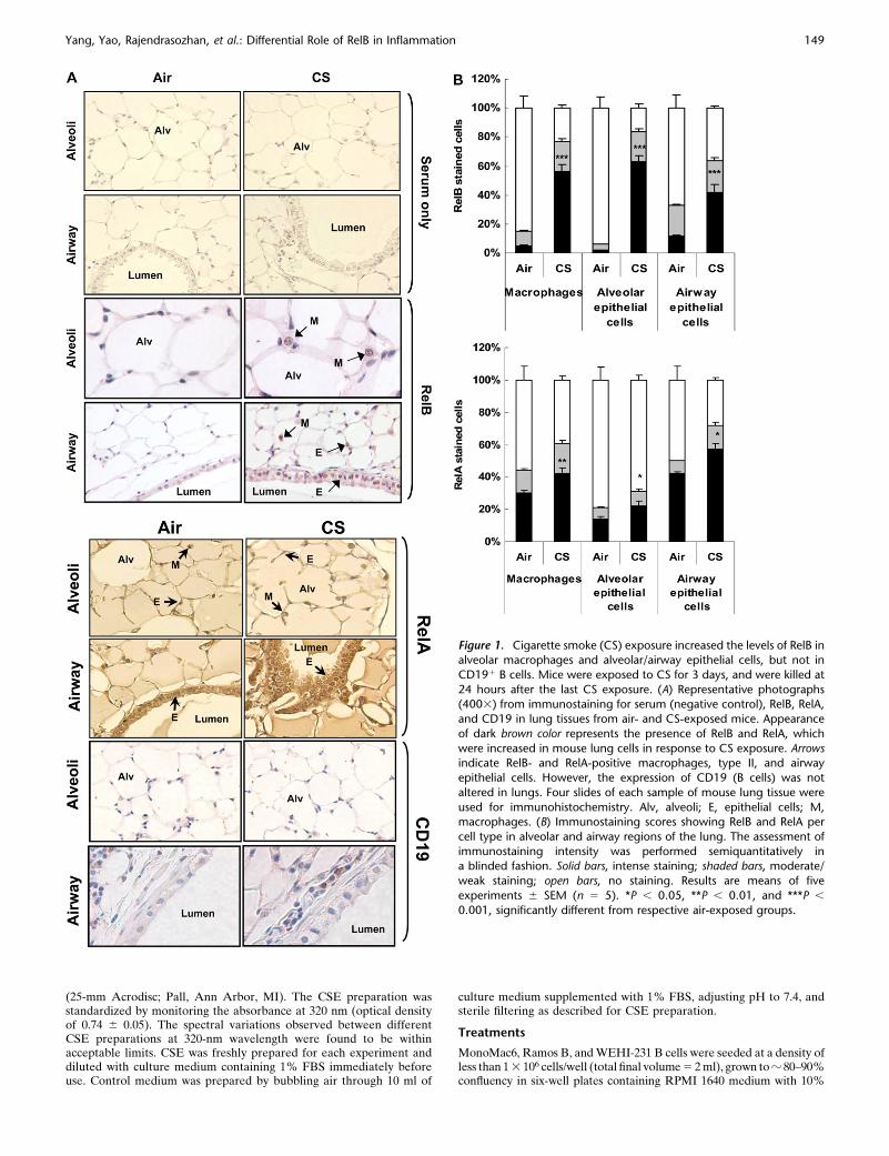
Figure 1. Cigarette smoke (CS) exposure increased the levels of RelB inalveolar macrophages and alveolar/airway epithelial cells, but not in
CD191 B cells. Mice were exposed to CS for 3 days, and were killed at
24 hours after the last CS exposure. (A) Representative photographs(4003) from immunostaining for serum (negative control), RelB, RelA,
and CD19 in lung tissues from air- and CS-exposed mice. Appearance
of dark brown color represents the presence of RelB and RelA, which
were increased in mouse lung cells in response to CS exposure. Arrowsindicate RelB- and RelA-positive macrophages, type II, and airway
epithelial cells. However, the expression of CD19 (B cells) was not
altered in lungs. Four slides of each sample of mouse lung tissue were
used for immunohistochemistry. Alv, alveoli; E, epithelial cells; M,macrophages. (B) Immunostaining scores showing RelB and RelA per
cell type in alveolar and airway regions of the lung. The assessment of
immunostaining intensity was performed semiquantitatively ina blinded fashion. Solid bars, intense staining; shaded bars, moderate/
weak staining; open bars, no staining. Results are means of five
experiments 6 SEM (n 5 5). *P , 0.05, **P , 0.01, and ***P ,
0.001, significantly different from respective air-exposed groups.
Yang, Yao, Rajendrasozhan, et al.: Differential Role of RelB in Inflammation 149
FBS, washed in Ca21- and Mg21-free PBS, and then exposed to varioustreatments in media containing 1% serum. The cells were treated withCSE (0.5, 1.0, or 2.5%) for 1 hour at 378C with 7.5% CO2. To investigatethe involvement of proteolysis of RelB, Ramos B cells were pretreatedwith 25 mM calpain inhibitor I (ALLN, product no. 208750; Calbiochem,San Diego, CA) for 20 minutes. The pretreated cells were washed twice inPBS and then treated with CSE (0.5 and 1.0%) for 1 hour at 378C with7.5% CO2. Primary spleen B cells (7 3 107) were seeded in 100-mm culturedishes containing RPMI 1640 with 1% FBS, 100 mg/ml penicillin, and 100U/ml streptomycin, and treated with CSE (0.1 and 0.5%) for 1 and 4 hoursat 378C with 7.5% CO2. All treatments were performed in duplicate. Atthe end of treatment, the cells were washed with cold, sterile Ca21- andMg21-free PBS and lysed in RIPA buffer as whole lysate (Westernblotting), and the lysates stored at 2808C.
Transfection
The plasmids for wild-type IKK-a, dominant negative IKK-a and NIKkinase mutant domain on lysine K429 and K430 (K429/A430) wereobtained as previously described (2, 42). Transient transfection wasperformed with 1 mg of plasmids in the presence of Lipofectamine-2000transfection reagent (product no. 11668–027; Invitrogen, Carlsbad, CA)in MonoMac6, Ramos B, and WEHI-231 cells. Transfection efficiencyin the case of both the plasmids was greater than 80%. Followingtransfection, MonoMac6, Ramos, and WEHI-231 B cells were treatedwith CSE (0.5, 1.0, and/or 2.5%). Whole lysate was used in Westernblotting analysis.
Cytoplasmic and Nuclear Protein Extraction
A total of 100 mg of lung tissue was mechanically homogenized in0.5 ml buffer A (10 mM HEPES, pH 7.8, 10 mM KCl, 2 mM MgCl2,1 mM DTT, 0.1 M EDTA, 0.2 mM NaF, 0.2 mM Na orthovandate, 1%[vol/vol] NP-40, 0.4 mM PMSF, and 1 mg/ml leupeptin) on ice. Thehomogenate was centrifuged at 2,000 3 g in a benchtop eppendorfcentrifuge for 30 s at 48C to remove cellular debris. The supernatantwas then transferred to a 1.7 ml ice-cold microtube and furthercentrifuged for 30 s at 13,000 3 g at 48C. The supernatant was collectedas a cytoplasmic extract. The pellet was resuspended in 200 ml of buffer
C (50 mM HEPES, pH 7.8, 50 mM KCl, 300 mM NaCl, 0.1 M EDTA,1 mM DTT, 10% [vol/vol] glycerol, 0.2 mM NaF, 0.2 mMNa orthovandate, and 0.6 mM PMSF) and placed on the rotor inthe cold room for 30 minutes. After centrifugation at 13,000 3 g in amicro-Eppendorf tube for 5 minutes, the supernatant was col-lected as the nuclear extract and kept frozen at 2808C for Westernblotting.
Western Blotting
Lung tissue homogenate samples (cytoplasmic and nuclear proteins)were separated on 7.5–12% SDS-PAGE. MonoMac6, Ramos, WEHI-231, and primary mouse spleen B cells were harvested and lysed with10% Igepal CA-630 lysis buffer supplemented with a protease inhibitorcocktail (leupeptin, aprotinin, pepstatin, and PMSF). Equal amounts ofprotein were subjected to electrophoresis on 7.5–12% PAGE gels,electroblotted onto nitrocellulose membranes (Amersham Bioscience,Piscataway, NJ), and then incubated overnight with primary antibodiesat 48C. The next day, membranes were washed and incubated for1 hour at room temperature with the appropriate secondary antibodylinked to horseradish peroxidase (Dako, Santa Barbara, CA). Boundcomplexes were detected with the use of the enhanced chemilumines-cence method (Jackson Immunology Research, West Grove, PA).
Immunoprecipitation
A total of 100 mg of proteins in mouse lung tissue homogenate wereimmunoprecipitated with 1 mg of specific antibodies and 20 ml ofprotein A/G agarose beads (product no. SC-2003; Santa Cruz Bio-technology) in RIPA buffer (50 mM Tris-HCl, pH 7.4, 150 mM NaCl,0.25 mM EDTA, 5 mM NaF, 0.1% sodium deoxycholate, 1% Triton X-100, in PBS) overnight at 48C. After immunoprecipitation, the precip-itates were washed three times with 10 mM Tris, 1 mM EDTA, 150 mMNaCl, 1 mg/ml BSA, 1% Triton X-100, and protease inhibitor in PBSwith spinning at 2,000 rpm for 1 minute at 48C. The precipitants wereresuspended in 50 ml of Laemmli sample buffer to a final concentrationof 13 sample buffer, and heated at 958C for 5 minutes. The collectedsupernatants (immunoprecipitants) were run on 7.5% SDS-PAGE.
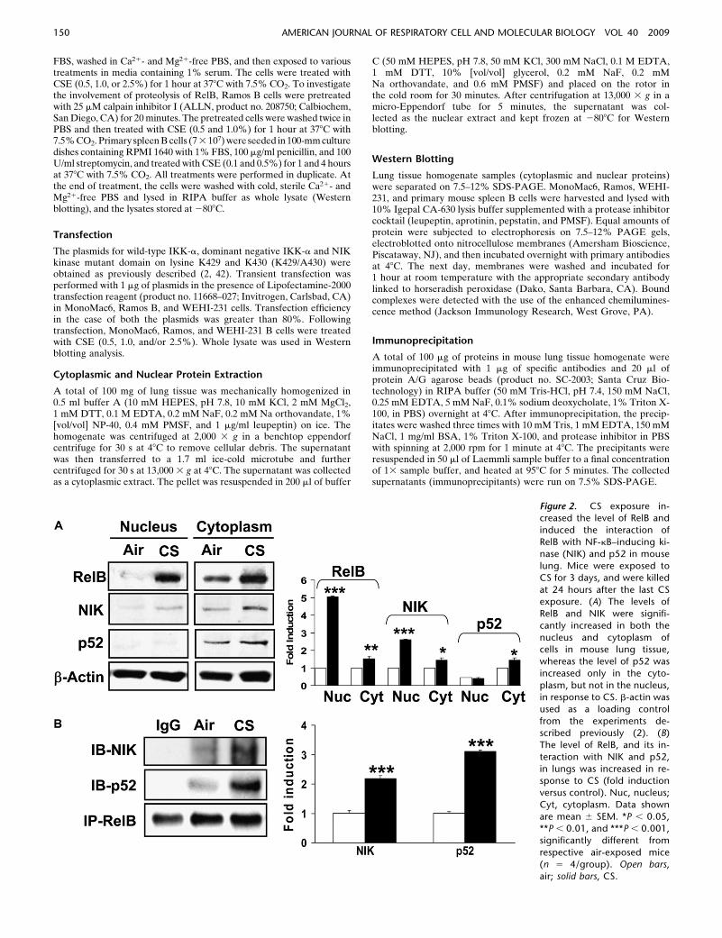
Figure 2. CS exposure in-creased the level of RelB and
induced the interaction of
RelB with NF-kB–inducing ki-nase (NIK) and p52 in mouse
lung. Mice were exposed to
CS for 3 days, and were killed
at 24 hours after the last CSexposure. (A) The levels of
RelB and NIK were signifi-
cantly increased in both the
nucleus and cytoplasm ofcells in mouse lung tissue,
whereas the level of p52 was
increased only in the cyto-
plasm, but not in the nucleus,in response to CS. b-actin was
used as a loading control
from the experiments de-scribed previously (2). (B)
The level of RelB, and its in-
teraction with NIK and p52,
in lungs was increased in re-sponse to CS (fold induction
versus control). Nuc, nucleus;
Cyt, cytoplasm. Data shown
are mean 6 SEM. *P , 0.05,**P , 0.01, and ***P , 0.001,
significantly different from
respective air-exposed mice(n 5 4/group). Open bars,
air; solid bars, CS.
150 AMERICAN JOURNAL OF RESPIRATORY CELL AND MOLECULAR BIOLOGY VOL 40 2009
Chromatin Immunoprecipitation
A total of 100 mg of lung tissue was homogenized in 1 mg/ml BSA withprotease inhibitor cocktail in PBS, and cross-linked with 1% formal-dehyde for 10 minutes, rinsed three times with PBS, and then 0.5 ml of2.5 M glycine was added. After a brief centrifugation, cell pellets wereresuspended in SDS-lysis buffer (50 mM Tris-HCl, 1% SDS, 5 mMEDTA, 5 mM Na-butyrate, protease inhibitors). Sonication of nuclearpellets containing chromatin was performed four times for 30 secondsand one time for 15 seconds, at a maximum speed, using Misonix-3000Sonicator (Misonix Inc., Farmingdale, NY). Supernatants were col-lected and diluted (1:10) with buffer (1% Triton X-100, 2 mM EDTA,150 mM NaCl, 20 mM Tris-HCl, pH 8.0, 5 mM NaButyrate, proteaseinhibitor), followed by preclearing the extract with 60 ml of protein Aagarose/salmon sperm DNA (no. 16-157; Upstate) for 3 hours at 48C(43). Immunoprecipitation was performed overnight at 48C with 1 mgof specific antibodies, as mentioned above. After immunoprecipitation,40 ml of protein A agarose/salmon sperm DNA was added andincubated for 2 hours and followed by brief centrifugation. Precipitateswere washed sequentially with Paro buffer I (0.1% SDS, 1% Triton X-100, 2 mM EDTA, 20 mM Tris-HCl, pH 8.1, 150 mM NaCl), Parobuffer II (0.1% SDS, 1% Triton X-100, 2 mM EDTA, 20 mM Tris-HCl,
pH 8.1, 500 mM NaCl), and Paro buffer III (0.25 M LiCl, 1% IgepalCA-630, 1% deoxycholate, 1 mM EDTA, 10 mM Tris-HCl, pH 8.1) for5 minutes at 48C. Precipitates were then washed again with Tris buffertwice for 5 minutes each. The antigen–antibody complexes wereextracted twice with 50 ml elution buffer (0.6 mg/ml proteinase K, 1%SDS, 0.1 M NaHCO3). The eluted samples were heated at 658Covernight to reverse formaldehyde crosslinking. The recovered DNAwas purified with a QIAquick PCR purification kit (product no., 28106;Qiagen, Valencia, CA) (43). Samples of input DNA were also preparedin the same way as described above. PCR amplification was performedusing a PTC-200 DNA engine (MJ Research, Waltham, MA) under thefollowing conditions: 948C for 180 seconds; 30–38 cycles at 948C for 45seconds, 608C for 60 seconds, and 728C for 60 seconds; and finalelongation at 728C for 10 minutes. PCR for the input reaction wasperformed using 100 ng of genomic DNA. Mouse primer sequences aredescribed in Table 1, and PCR products were analyzed on a 1.5–2.0%agarose gel.
Protein Assay
Protein levels were measured with a BCA kit (Pierce, Rockford, IL).Protein standards were obtained by diluting a stock solution of BSA.Linear regression was used to determine the actual protein concentra-tion of the samples.
Statistical Analysis
Results are shown as means 6 SEM. Statistical analysis of significancewas calculated by one-way ANOVA followed by Fisher’s protectedleast significant difference post hoc test for multigroup comparisons(StatView 5.0; SAS Institute, Cary, NC). Statistical significance isindicated in the figure legends.
RESULTS
CS Exposure Increased the Levels of RelB and RelA/p65 in
Alveolar and Airway Epithelial Cells in Mouse Lung
We studied the expression and localization of RelB and RelA/p65 in mouse lung sections by immunohistochemistry in mid-sagittal sections in response to air or CS exposure. RelB- andRelA/p65-positive cells with increased staining of RelB andRelA/p65 were detected in macrophages, type II alveolar, andairway epithelial cells in mouse lung tissue exposed to CS(Figures 1A and 1B). However, we were unable to detect B cellsusing the B-cell–specific surface marker CD191 in lung sectionsof mice exposed to CS for acute (3-d) exposure (Figure 1A).Likewise, the expression of CD45R/B220, another marker of Bcells, was also not detected in mouse lungs exposed to CS for3 days. However, CD19- and CD45R/B220-positive cells wereincreased in mouse lung after 8 weeks of CS exposure compared
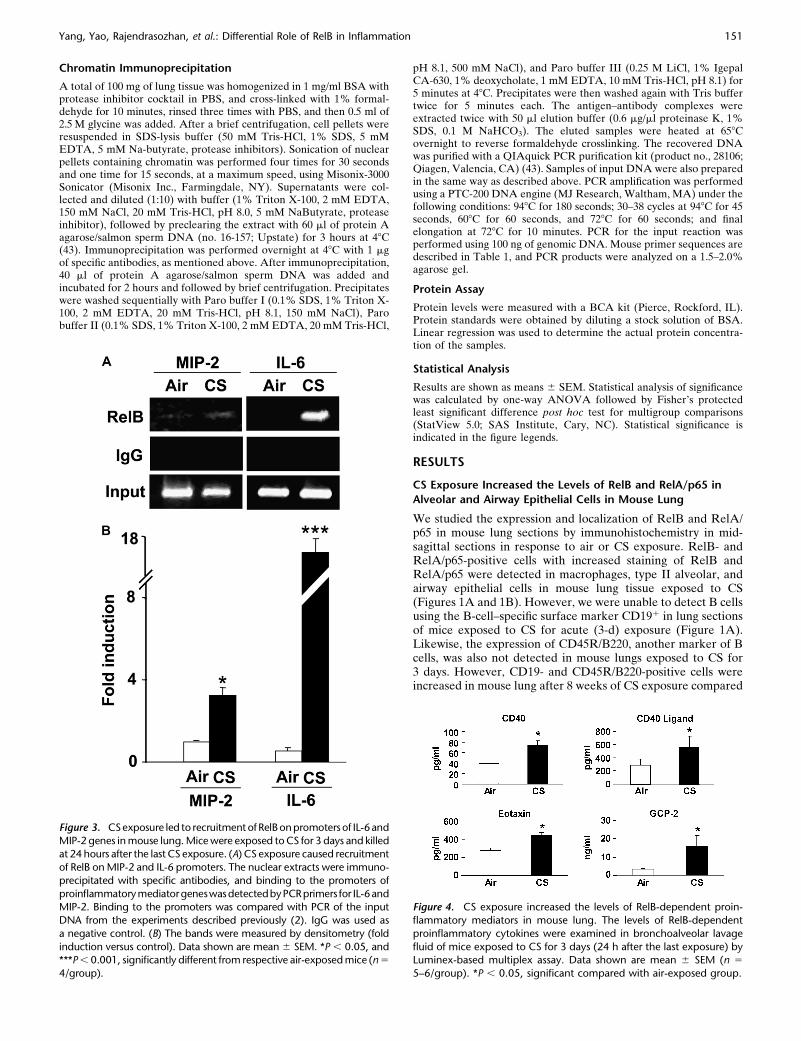
Figure 3. CSexposure led to recruitment ofRelBonpromoters of IL-6and
MIP-2 genes in mouse lung. Mice were exposed to CS for 3 days and killedat 24 hours after the last CS exposure. (A) CS exposure caused recruitment
of RelB on MIP-2 and IL-6 promoters. The nuclear extracts were immuno-
precipitated with specific antibodies, and binding to the promoters of
proinflammatorymediatorgeneswasdetectedbyPCRprimers for IL-6andMIP-2. Binding to the promoters was compared with PCR of the input
DNA from the experiments described previously (2). IgG was used as
a negative control. (B) The bands were measured by densitometry (fold
induction versus control). Data shown are mean 6 SEM. *P , 0.05, and***P , 0.001, significantly different from respective air-exposed mice (n 5
4/group).
Figure 4. CS exposure increased the levels of RelB-dependent proin-
flammatory mediators in mouse lung. The levels of RelB-dependent
proinflammatory cytokines were examined in bronchoalveolar lavage
fluid of mice exposed to CS for 3 days (24 h after the last exposure) byLuminex-based multiplex assay. Data shown are mean 6 SEM (n 5
5–6/group). *P , 0.05, significant compared with air-exposed group.
Yang, Yao, Rajendrasozhan, et al.: Differential Role of RelB in Inflammation 151
with air exposure (data not shown). These observations suggestthat CS exposure induced increased levels of RelB and RelA/p65 and increased numbers of B cells in mouse lungs.
CS Exposure Increased the Levels of RelB and Its Interaction
with NIK and p52 in Mouse Lung
We determined the levels of RelB and its interactions with NIKand p52 in lung tissue of mice exposed to CS for 3 days. Thelevels of RelB and NIK were significantly increased in both thenucleus and cytoplasm of cells in mouse lung tissue, whereas thelevel of p52 was increased only in the cytoplasm, but not in thenucleus in response to CS (Figures 2A and 2B). We nextdetermined the interaction of RelB with NIK and p52 in mousewhole lung homogenates by immunoprecipitation with relevantantibodies. CS increased the level of RelB interaction with bothNIK and p52 in mouse lungs (Figure 2B). These data showedthat CS exposure increased the levels of both RelB and NIK inthe nucleus, as well as the interactions of RelB with NIK andp52 in mouse lung.
CS Exposure Caused Recruitment of RelB on Proinflammatory
Gene Promoters in Mouse Lung
It has been shown that increased levels of RelB are a pre-requisite for the alternate pathway of NF-kB–dependent gene
transcription in response to proinflammatory stimuli (20, 21, 23,27, 44, 45). Therefore, we hypothesized that CS exposure wouldinduce proinflammatory mediators by recruiting RelB on thepromoters of proinflammatory genes. To test this hypothesis,chromatin immunoprecipitation (ChIP) assays were performedusing antibodies against RelB in lungs of mice exposed to CS.As expected, the levels of RelB on the proinflammatory pro-moter sites of macrophage inflammatory protein (MIP)-2 andIL-6 were increased (Figures 3A and 3B). However, the regionsthat were noncoding for IL-6 and MIP-2 showed no change ingene expression, validating the specificity of the ChIP assays(S.-R. Yang and coworkers, unpublished data). These resultsdemonstrate that RelB is recruited on the promoters of variousproinflammatory genes, thereby exerting its effect on proin-flammatory gene expression in mouse lung.
CS Increased the Levels of NF-kB–Dependent
Proinflammatory Mediators in Mouse Lung
We next determined whether or not CS induced the levels ofRelB-dependent proinflammatory cytokines in mouse BALfluid measured by Luminex-based multiplex assay. The proin-flammatory mediators, including CD40, CD40 ligand (which arepresent on antigen-presenting cells, and are costimulatorymolecules for proliferation and enhanced survival of T cells),eotaxin, and granulocyte chemotactic protein-2 (which are
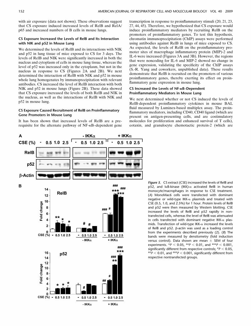
Figure 5. CS extract (CSE) increased the levels of RelB and
p52, and IkB-kinase (IKK)-a activated RelB in humanmonocyte/macrophages in response to CSE treatment.
(A) MonoMac6 cells were transfected with dominant
negative or wild-type IKK-a plasmids and treated with
CSE (0.5, 1.0, and 2.5%) for 1 hour. Protein levels of RelBand p52 were then measured by Western blotting. CSE
increased the levels of RelB and p52 rapidly in non-
transfected cells, whereas the level of RelB was attenuatedin cells transfected with dominant negative IKK-a plas-
mids. Transfection of wild-type IKK-a increased the levels
of RelB and p52. b-actin was used as a loading control
from the experiments described previously (2). (B) Thebands were measured by densitometry (fold induction
versus control). Data shown are mean 6 SEM of four
experiments. *P , 0.05, **P , 0.01, and ***P , 0.001,
significantly different from respective controls; #P , 0.05,##P , 0.01, and ###P , 0.001, significantly different from
respective nontransfected groups.
152 AMERICAN JOURNAL OF RESPIRATORY CELL AND MOLECULAR BIOLOGY VOL 40 2009
thought to be regulated by alternative NF-kB pathways) weresignificantly increased in BAL fluid in response to CS exposure(Figure 4). Previously, we have shown that the levels of MIP-2and IL-6 were increased in BAL fluid 3 days after CS exposure(2). Taken together, these data suggest that CS induces thealternative pathway of NF-kB–dependent proinflammatorymediators in mouse lung.
CSE Increased the Levels of RelB, and IKK-a Is a Critical
Regulator of RelB in MonoMac6
Macrophages play an important role in abnormal inflammatoryresponses observed in patients with COPD. Recently, we haveshown that CS induces proinflammatory mediators by an NF-kB–dependent mechanism in macrophages (2, 4). We thereforedetermined whether RelB was also activated in response toCSE in MonoMac6 cells, leading to RelB-dependent proin-flammatory cytokine release. Similar to the activation of RelA/p65 (2), we found that RelB was activated in response to CSEtreatment associated with increased levels of its partner, p52(Figures 5A and 5B). These data suggested that CS activatesboth the classical and the alternative NF-kB pathways inmacrophages, leading to proinflammatory mediator release.
It was recently shown that NIK activates the IKK-a homo-dimer in the IKK-s complex, which, in turn, results in activationof alternative NF-kB pathway, characterized by nuclear trans-location of RelB (22, 44). Therefore, we determined whether
NIK and IKK-a were involved in regulation of RelB in responseto CSE in MonoMac6 cells. Macrophages were transfected witha dominant negative IKK-a or a double mutant of NIK (K429/A430), and treated with CSE (0.5, 1.0, and 2.5%) for 1 hour. Thelevels of RelB and p52 were increased in response to CSE innontransfected cells, whereas the level of RelB was attenuated inboth dominant negative IKK-a–transfected and double mutantof NIK (K429/A430, S.-R. Yang and coworkers, unpublisheddata)-transfected MonoMac6 cells. Transfection of wild-typeIKK-a increased the levels of RelB and p52, suggesting thatIKK-a and NIK are required for CS-mediated activation ofalternative NF-kB pathway in macrophages (Figure 5A and 5B).
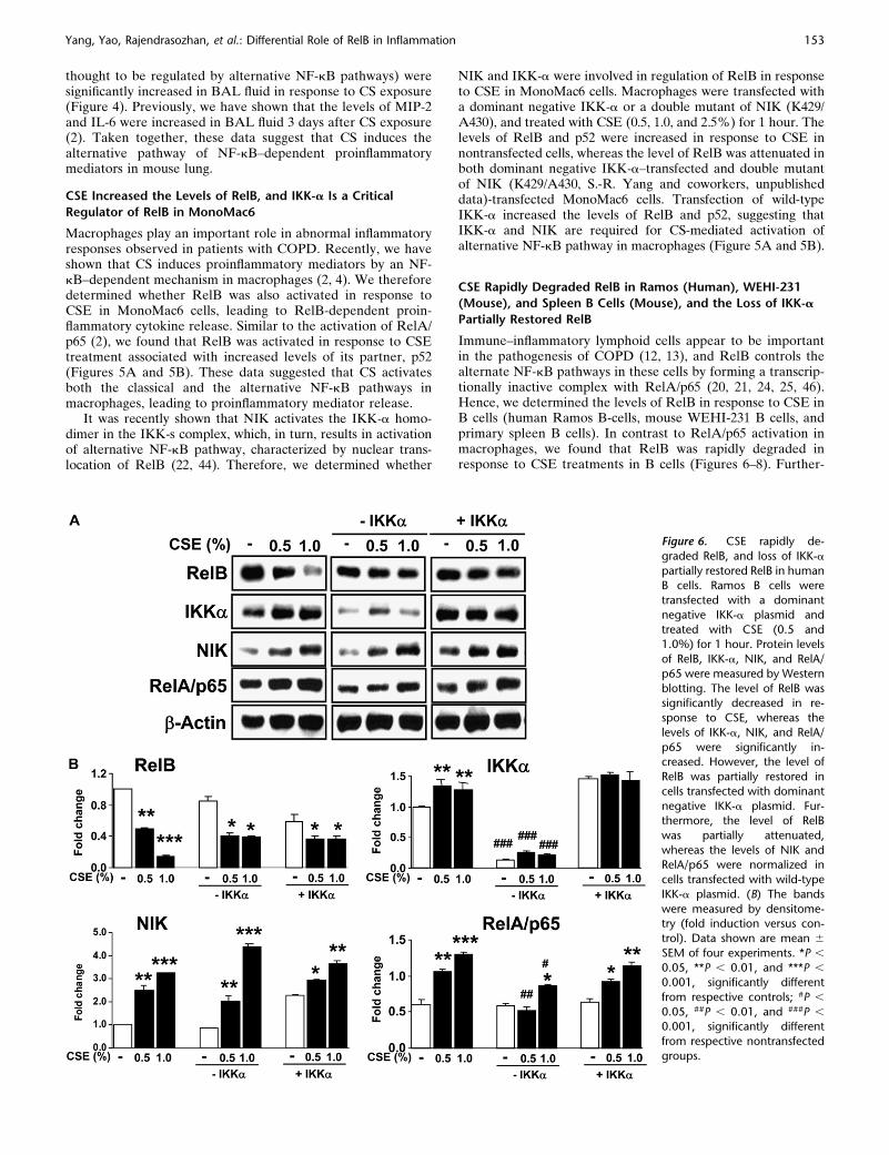
CSE Rapidly Degraded RelB in Ramos (Human), WEHI-231
(Mouse), and Spleen B Cells (Mouse), and the Loss of IKK-a
Partially Restored RelB
Immune–inflammatory lymphoid cells appear to be importantin the pathogenesis of COPD (12, 13), and RelB controls thealternate NF-kB pathways in these cells by forming a transcrip-tionally inactive complex with RelA/p65 (20, 21, 24, 25, 46).Hence, we determined the levels of RelB in response to CSE inB cells (human Ramos B-cells, mouse WEHI-231 B cells, andprimary spleen B cells). In contrast to RelA/p65 activation inmacrophages, we found that RelB was rapidly degraded inresponse to CSE treatments in B cells (Figures 6–8). Further-
Figure 6. CSE rapidly de-
graded RelB, and loss of IKK-a
partially restored RelB in human
B cells. Ramos B cells weretransfected with a dominant
negative IKK-a plasmid and
treated with CSE (0.5 and1.0%) for 1 hour. Protein levels
of RelB, IKK-a, NIK, and RelA/
p65 were measured by Western
blotting. The level of RelB wassignificantly decreased in re-
sponse to CSE, whereas the
levels of IKK-a, NIK, and RelA/
p65 were significantly in-creased. However, the level of
RelB was partially restored in
cells transfected with dominant
negative IKK-a plasmid. Fur-thermore, the level of RelB
was partially attenuated,
whereas the levels of NIK andRelA/p65 were normalized in
cells transfected with wild-type
IKK-a plasmid. (B) The bands
were measured by densitome-try (fold induction versus con-
trol). Data shown are mean 6
SEM of four experiments. *P ,
0.05, **P , 0.01, and ***P ,
0.001, significantly different
from respective controls; #P ,
0.05, ##P , 0.01, and ###P ,
0.001, significantly different
from respective nontransfected
groups.
Yang, Yao, Rajendrasozhan, et al.: Differential Role of RelB in Inflammation 153
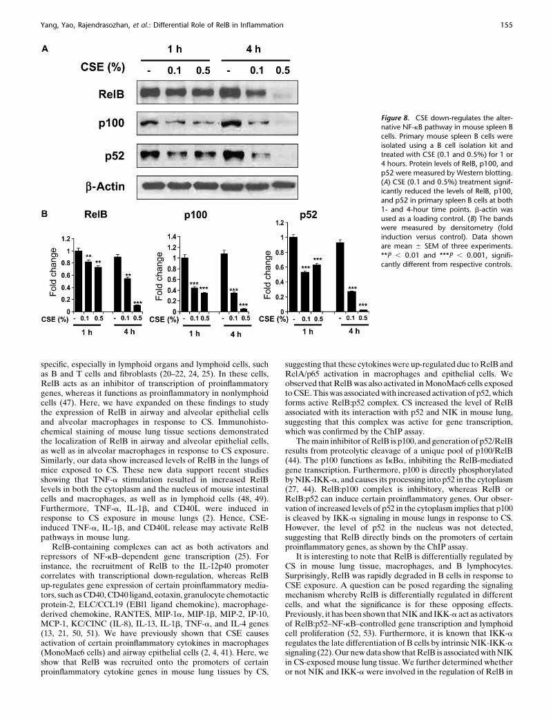
more, the levels of p100 and p52 were also decreased in primarysplenic B cells in response to CSE treatment, suggesting that CSexposure down-regulates the alternative NF-kB pathway in Bcells (Figure 8).
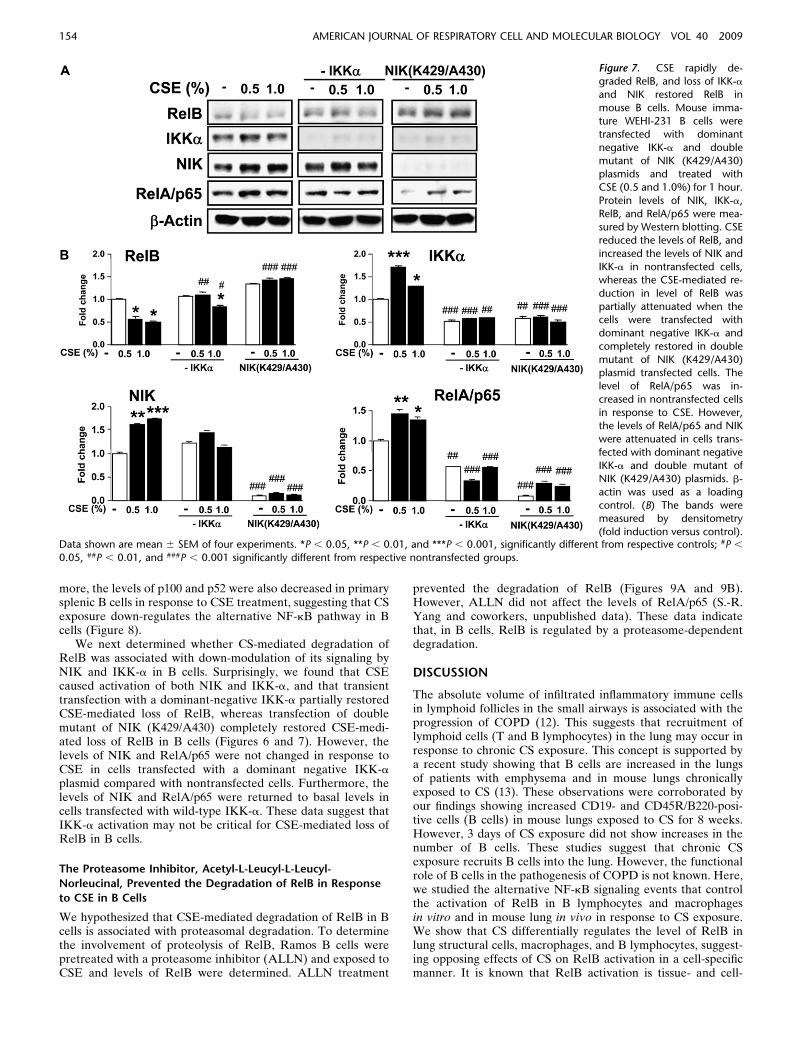
We next determined whether CS-mediated degradation ofRelB was associated with down-modulation of its signaling byNIK and IKK-a in B cells. Surprisingly, we found that CSEcaused activation of both NIK and IKK-a, and that transienttransfection with a dominant-negative IKK-a partially restoredCSE-mediated loss of RelB, whereas transfection of doublemutant of NIK (K429/A430) completely restored CSE-medi-ated loss of RelB in B cells (Figures 6 and 7). However, thelevels of NIK and RelA/p65 were not changed in response toCSE in cells transfected with a dominant negative IKK-aplasmid compared with nontransfected cells. Furthermore, thelevels of NIK and RelA/p65 were returned to basal levels incells transfected with wild-type IKK-a. These data suggest thatIKK-a activation may not be critical for CSE-mediated loss ofRelB in B cells.
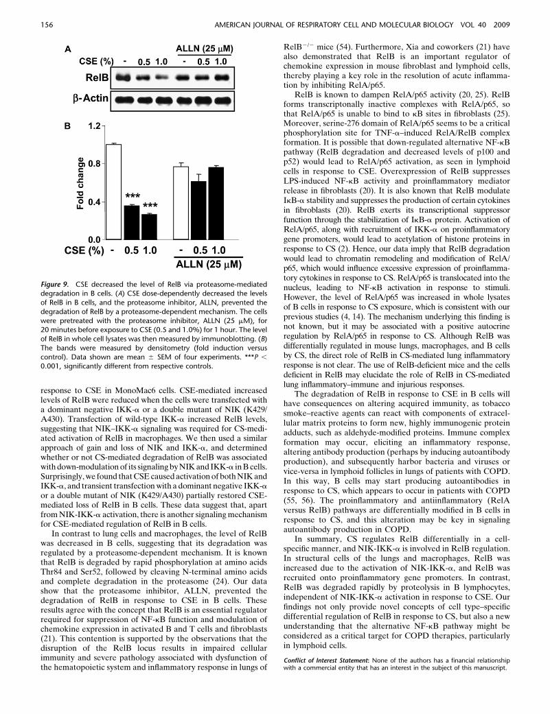
The Proteasome Inhibitor, Acetyl-L-Leucyl-L-Leucyl-
Norleucinal, Prevented the Degradation of RelB in Response
to CSE in B Cells
We hypothesized that CSE-mediated degradation of RelB in Bcells is associated with proteasomal degradation. To determinethe involvement of proteolysis of RelB, Ramos B cells werepretreated with a proteasome inhibitor (ALLN) and exposed toCSE and levels of RelB were determined. ALLN treatment
prevented the degradation of RelB (Figures 9A and 9B).However, ALLN did not affect the levels of RelA/p65 (S.-R.Yang and coworkers, unpublished data). These data indicatethat, in B cells, RelB is regulated by a proteasome-dependentdegradation.
DISCUSSION
The absolute volume of infiltrated inflammatory immune cellsin lymphoid follicles in the small airways is associated with theprogression of COPD (12). This suggests that recruitment oflymphoid cells (T and B lymphocytes) in the lung may occur inresponse to chronic CS exposure. This concept is supported bya recent study showing that B cells are increased in the lungsof patients with emphysema and in mouse lungs chronicallyexposed to CS (13). These observations were corroborated byour findings showing increased CD19- and CD45R/B220-posi-tive cells (B cells) in mouse lungs exposed to CS for 8 weeks.However, 3 days of CS exposure did not show increases in thenumber of B cells. These studies suggest that chronic CSexposure recruits B cells into the lung. However, the functionalrole of B cells in the pathogenesis of COPD is not known. Here,we studied the alternative NF-kB signaling events that controlthe activation of RelB in B lymphocytes and macrophagesin vitro and in mouse lung in vivo in response to CS exposure.We show that CS differentially regulates the level of RelB inlung structural cells, macrophages, and B lymphocytes, suggest-ing opposing effects of CS on RelB activation in a cell-specificmanner. It is known that RelB activation is tissue- and cell-
Figure 7. CSE rapidly de-graded RelB, and loss of IKK-a
and NIK restored RelB in
mouse B cells. Mouse imma-
ture WEHI-231 B cells weretransfected with dominant
negative IKK-a and double
mutant of NIK (K429/A430)plasmids and treated with
CSE (0.5 and 1.0%) for 1 hour.
Protein levels of NIK, IKK-a,
RelB, and RelA/p65 were mea-sured by Western blotting. CSE
reduced the levels of RelB, and
increased the levels of NIK and
IKK-a in nontransfected cells,whereas the CSE-mediated re-
duction in level of RelB was
partially attenuated when thecells were transfected with
dominant negative IKK-a and
completely restored in double
mutant of NIK (K429/A430)plasmid transfected cells. The
level of RelA/p65 was in-
creased in nontransfected cells
in response to CSE. However,the levels of RelA/p65 and NIK
were attenuated in cells trans-
fected with dominant negative
IKK-a and double mutant ofNIK (K429/A430) plasmids. b-
actin was used as a loading
control. (B) The bands weremeasured by densitometry
(fold induction versus control).Data shown are mean 6 SEM of four experiments. *P , 0.05, **P , 0.01, and ***P , 0.001, significantly different from respective controls; #P ,
0.05, ##P , 0.01, and ###P , 0.001 significantly different from respective nontransfected groups.
154 AMERICAN JOURNAL OF RESPIRATORY CELL AND MOLECULAR BIOLOGY VOL 40 2009
specific, especially in lymphoid organs and lymphoid cells, suchas B and T cells and fibroblasts (20–22, 24, 25). In these cells,RelB acts as an inhibitor of transcription of proinflammatorygenes, whereas it functions as proinflammatory in nonlymphoidcells (47). Here, we have expanded on these findings to studythe expression of RelB in airway and alveolar epithelial cellsand alveolar macrophages in response to CS. Immunohisto-chemical staining of mouse lung tissue sections demonstratedthe localization of RelB in airway and alveolar epithelial cells,as well as in alveolar macrophages in response to CS exposure.Similarly, our data show increased levels of RelB in the lungs ofmice exposed to CS. These new data support recent studiesshowing that TNF-a stimulation resulted in increased RelBlevels in both the cytoplasm and the nucleus of mouse intestinalcells and macrophages, as well as in lymphoid cells (48, 49).Furthermore, TNF-a, IL-1b, and CD40L were induced inresponse to CS exposure in mouse lungs (2). Hence, CSE-induced TNF-a, IL-1b, and CD40L release may activate RelBpathways in mouse lung.
RelB-containing complexes can act as both activators andrepressors of NF-kB–dependent gene transcription (25). Forinstance, the recruitment of RelB to the IL-12p40 promotercorrelates with transcriptional down-regulation, whereas RelBup-regulates gene expression of certain proinflammatory media-tors, such as CD40, CD40 ligand, eotaxin, granulocyte chemotacticprotein-2, ELC/CCL19 (EBl1 ligand chemokine), macrophage-derived chemokine, RANTES, MIP-1a, MIP-1b, MIP-2, IP-10,MCP-1, KC/CINC (IL-8), IL-13, IL-1b, TNF-a, and IL-4 genes(13, 21, 50, 51). We have previously shown that CSE causesactivation of certain proinflammatory cytokines in macrophages(MonoMac6 cells) and airway epithelial cells (2, 4, 41). Here, weshow that RelB was recruited onto the promoters of certainproinflammatory cytokine genes in mouse lung tissues by CS,
suggesting that these cytokines were up-regulated due to RelB andRelA/p65 activation in macrophages and epithelial cells. Weobserved that RelB was also activated in MonoMac6 cells exposedto CSE. This was associated with increased activation of p52, whichforms active RelB:p52 complex. CS increased the level of RelBassociated with its interaction with p52 and NIK in mouse lung,suggesting that this complex was active for gene transcription,which was confirmed by the ChIP assay.
The main inhibitor of RelB is p100, and generation of p52/RelBresults from proteolytic cleavage of a unique pool of p100/RelB(44). The p100 functions as IkBa, inhibiting the RelB-mediatedgene transcription. Furthermore, p100 is directly phosphorylatedby NIK-IKK-a, and causes its processing into p52 in the cytoplasm(27, 44). RelB:p100 complex is inhibitory, whereas RelB orRelB:p52 can induce certain proinflammatory genes. Our obser-vation of increased levels of p52 in the cytoplasm implies that p100is cleaved by IKK-a signaling in mouse lungs in response to CS.However, the level of p52 in the nucleus was not detected,suggesting that RelB directly binds on the promoters of certainproinflammatory genes, as shown by the ChIP assay.
It is interesting to note that RelB is differentially regulated byCS in mouse lung tissue, macrophages, and B lymphocytes.Surprisingly, RelB was rapidly degraded in B cells in response toCSE exposure. A question can be posed regarding the signalingmechanism whereby RelB is differentially regulated in differentcells, and what the significance is for these opposing effects.Previously, it has been shown that NIK and IKK-a act as activatorsof RelB:p52–NF-kB–controlled gene transcription and lymphoidcell proliferation (52, 53). Furthermore, it is known that IKK-aregulates the late differentiation of B cells by intrinsic NIK-IKK-asignaling (22). Our new data show that RelB is associated with NIKin CS-exposed mouse lung tissue. We further determined whetheror not NIK and IKK-a were involved in the regulation of RelB in
Figure 8. CSE down-regulates the alter-native NF-kB pathway in mouse spleen B
cells. Primary mouse spleen B cells were
isolated using a B cell isolation kit andtreated with CSE (0.1 and 0.5%) for 1 or
4 hours. Protein levels of RelB, p100, and
p52 were measured by Western blotting.
(A) CSE (0.1 and 0.5%) treatment signif-icantly reduced the levels of RelB, p100,
and p52 in primary spleen B cells at both
1- and 4-hour time points. b-actin was
used as a loading control. (B) The bandswere measured by densitometry (fold
induction versus control). Data shown
are mean 6 SEM of three experiments.
**P , 0.01 and ***P , 0.001, signifi-cantly different from respective controls.
Yang, Yao, Rajendrasozhan, et al.: Differential Role of RelB in Inflammation 155
response to CSE in MonoMac6 cells. CSE-mediated increasedlevels of RelB were reduced when the cells were transfected witha dominant negative IKK-a or a double mutant of NIK (K429/A430). Transfection of wild-type IKK-a increased RelB levels,suggesting that NIK–IKK-a signaling was required for CS-medi-ated activation of RelB in macrophages. We then used a similarapproach of gain and loss of NIK and IKK-a, and determinedwhether or not CS-mediated degradation of RelB was associatedwith down-modulation of its signaling by NIK and IKK-a in B cells.Surprisingly, we found that CSE caused activation of both NIK andIKK-a, and transient transfection with a dominant negative IKK-aor a double mutant of NIK (K429/A430) partially restored CSE-mediated loss of RelB in B cells. These data suggest that, apartfrom NIK-IKK-a activation, there is another signaling mechanismfor CSE-mediated regulation of RelB in B cells.
In contrast to lung cells and macrophages, the level of RelBwas decreased in B cells, suggesting that its degradation wasregulated by a proteasome-dependent mechanism. It is knownthat RelB is degraded by rapid phosphorylation at amino acidsThr84 and Ser52, followed by cleaving N-terminal amino acidsand complete degradation in the proteasome (24). Our datashow that the proteasome inhibitor, ALLN, prevented thedegradation of RelB in response to CSE in B cells. Theseresults agree with the concept that RelB is an essential regulatorrequired for suppression of NF-kB function and modulation ofchemokine expression in activated B and T cells and fibroblasts(21). This contention is supported by the observations that thedisruption of the RelB locus results in impaired cellularimmunity and severe pathology associated with dysfunction ofthe hematopoietic system and inflammatory response in lungs of
RelB2/2 mice (54). Furthermore, Xia and coworkers (21) havealso demonstrated that RelB is an important regulator ofchemokine expression in mouse fibroblast and lymphoid cells,thereby playing a key role in the resolution of acute inflamma-tion by inhibiting RelA/p65.
RelB is known to dampen RelA/p65 activity (20, 25). RelBforms transcriptonally inactive complexes with RelA/p65, sothat RelA/p65 is unable to bind to kB sites in fibroblasts (25).Moreover, serine-276 domain of RelA/p65 seems to be a criticalphosphorylation site for TNF-a–induced RelA/RelB complexformation. It is possible that down-regulated alternative NF-kBpathway (RelB degradation and decreased levels of p100 andp52) would lead to RelA/p65 activation, as seen in lymphoidcells in response to CSE. Overexpression of RelB suppressesLPS-induced NF-kB activity and proinflammatory mediatorrelease in fibroblasts (20). It is also known that RelB modulateIkB-a stability and suppresses the production of certain cytokinesin fibroblasts (20). RelB exerts its transcriptional suppressorfunction through the stabilization of IkB-a protein. Activation ofRelA/p65, along with recruitment of IKK-a on proinflammatorygene promoters, would lead to acetylation of histone proteins inresponse to CS (2). Hence, our data imply that RelB degradationwould lead to chromatin remodeling and modification of RelA/p65, which would influence excessive expression of proinflamma-tory cytokines in response to CS. RelA/p65 is translocated into thenucleus, leading to NF-kB activation in response to stimuli.However, the level of RelA/p65 was increased in whole lysatesof B cells in response to CS exposure, which is consistent with ourprevious studies (4, 14). The mechanism underlying this finding isnot known, but it may be associated with a positive autocrineregulation by RelA/p65 in response to CS. Although RelB wasdifferentially regulated in mouse lungs, macrophages, and B cellsby CS, the direct role of RelB in CS-mediated lung inflammatoryresponse is not clear. The use of RelB-deficient mice and the cellsdeficient in RelB may elucidate the role of RelB in CS-mediatedlung inflammatory–immune and injurious responses.
The degradation of RelB in response to CSE in B cells willhave consequences on altering acquired immunity, as tobaccosmoke–reactive agents can react with components of extracel-lular matrix proteins to form new, highly immunogenic proteinadducts, such as aldehyde-modified proteins. Immune complexformation may occur, eliciting an inflammatory response,altering antibody production (perhaps by inducing autoantibodyproduction), and subsequently harbor bacteria and viruses orvice-versa in lymphoid follicles in lungs of patients with COPD.In this way, B cells may start producing autoantibodies inresponse to CS, which appears to occur in patients with COPD(55, 56). The proinflammatory and antiinflammatory (RelAversus RelB) pathways are differentially modified in B cells inresponse to CS, and this alteration may be key in signalingautoantibody production in COPD.
In summary, CS regulates RelB differentially in a cell-specific manner, and NIK-IKK-a is involved in RelB regulation.In structural cells of the lungs and macrophages, RelB wasincreased due to the activation of NIK-IKK-a, and RelB wasrecruited onto proinflammatory gene promoters. In contrast,RelB was degraded rapidly by proteolysis in B lymphocytes,independent of NIK-IKK-a activation in response to CSE. Ourfindings not only provide novel concepts of cell type–specificdifferential regulation of RelB in response to CS, but also a newunderstanding that the alternative NF-kB pathway might beconsidered as a critical target for COPD therapies, particularlyin lymphoid cells.
Conflict of Interest Statement: None of the authors has a financial relationshipwith a commercial entity that has an interest in the subject of this manuscript.
Figure 9. CSE decreased the level of RelB via proteasome-mediateddegradation in B cells. (A) CSE dose-dependently decreased the levels
of RelB in B cells, and the proteasome inhibitor, ALLN, prevented the
degradation of RelB by a proteasome-dependent mechanism. The cells
were pretreated with the proteasome inhibitor, ALLN (25 mM), for20 minutes before exposure to CSE (0.5 and 1.0%) for 1 hour. The level
of RelB in whole cell lysates was then measured by immunoblotting. (B)
The bands were measured by densitometry (fold induction versuscontrol). Data shown are mean 6 SEM of four experiments. ***P ,
0.001, significantly different from respective controls.
156 AMERICAN JOURNAL OF RESPIRATORY CELL AND MOLECULAR BIOLOGY VOL 40 2009
Acknowledgments: The authors thank Dr. Aruna Kode, Samuel Caito, DavidAdenuga, and Ryan Henry for their technical assistance. They also thank Dr. PaulKirkham (Imperial College, UK) for useful discussion on B cell immunology inchronic obstructive pulmonary disease.
References
1. Burchfiel CM, Marcus EB, Curb JD, Maclean CJ, Vollmer WM,Johnson LR, Fong KO, Rodriguez BL, Masaki KH, Buist AS. Effectsof smoking and smoking cessation on longitudinal decline in pulmo-nary function. Am J Respir Crit Care Med 1995;151:1778–1785.
2. Yang SR, Valvo S, Yao H, Kode A, Rajendrasozhan S, Edrisinghe I,Caito S, Adenuga D, Henry R, Fromm G, et al. IKKa causeschromatin modification on pro-inflammatory genes by cigarettesmoke in mouse lung. Am J Respir Cell Mol Biol 2008;38:689–698.
3. Rahman I, Adcock IM. Oxidative stress and redox regulation of lunginflammation in COPD. Eur Respir J 2006;28:219–242.
4. Yang SR, Chida AS, Bauter MR, Shafiq N, Seweryniak K, MaggirwarSB, Kilty I, Rahman I. Cigarette smoke induces proinflammatorycytokine release by activation of NF-kB and posttranslational mod-ifications of histone deacetylase in macrophages. Am J Physiol LungCell Mol Physiol 2006;291:L46–L57.
5. Szulakowski P, Crowther AJ, Jimenez LA, Donaldson K, Mayer R,Leonard TB, MacNee W, Drost EM. The effect of smoking on thetranscriptional regulation of lung inflammation in patients withchronic obstructive pulmonary disease. Am J Respir Crit Care Med2006;174:41–50.
6. Ito K, Ito M, Elliott WM, Cosio B, Caramori G, Kon OM, Barczyk A,Hayashi S, Adcock IM, Hogg JC, et al. Decreased histone deacetylaseactivity in chronic obstructive pulmonary disease. N Engl J Med 2005;352:1967–1976.
7. Marwick JA, Kirkham PA, Stevenson CS, Danahay H, Giddings J,Butler K, Donaldson K, Macnee W, Rahman I. Cigarette smokealters chromatin remodeling and induces proinflammatory genes inrat lungs. Am J Respir Cell Mol Biol 2004;31:633–642.
8. Di Stefano A, Caramori G, Oates T, Capelli A, Lusuardi M, Gnemmi I,Ioli F, Chung KF, Donner CF, Barnes PJ, et al. Increased expressionof nuclear factor-kB in bronchial biopsies from smokers and patientswith COPD. Eur Respir J 2002;20:556–563.
9. Morrison D, Rahman I, Lannan S, MacNee W. Epithelial permeability,inflammation, and oxidant stress in the air spaces of smokers. Am JRespir Crit Care Med 1999;159:473–479.
10. Keatings VM, Collins PD, Scott DM, Barnes PJ. Differences in in-terleukin-8 and tumor necrosis factor-a in induced sputum frompatients with chronic obstructive pulmonary disease or asthma. AmJ Respir Crit Care Med 1996;153:530–534.
11. Puchelle E, Zahm JM, Tournier JM, Coraux C. Airway epithelial repair,regeneration, and remodeling after injury in chronic obstructivepulmonary disease. Proc Am Thorac Soc 2006;3:726–733.
12. Hogg JC, Chu F, Utokaparch S, Woods R, Elliott WM, Buzatu L,Cherniack RM, Rogers RM, Sciurba FC, Coxson HO, et al. Thenature of small-airway obstruction in chronic obstructive pulmonarydisease. N Engl J Med 2004;350:2645–2653.
13. van der Strate BW, Postma DS, Brandsma CA, Melgert BN, LuingeMA, Geerlings M, Hylkema MN, van den Berg A, Timens W,Kerstjens HA. Cigarette smoke–induced emphysema: a role for theB cell? Am J Respir Crit Care Med 2006;173:751–758.
14. Rajendrasozhan S, Yang SR, Kinnula VL, Rahman I. SIRT1, anantiinflammatory and antiaging protein, is decreased in lungs ofpatients with chronic obstructive pulmonary disease. Am J RespirCrit Care Med 2008;177:861–870.
15. Yang SR, Wright J, Bauter M, Seweryniak K, Kode A, Rahman I.Sirtuin regulates cigarette smoke–induced proinflammatory mediatorrelease via RelA/p65 NF-kB in macrophages in vitro and in rat lungsin vivo: implications for chronic inflammation and aging. Am JPhysiol Lung Cell Mol Physiol 2007;292:L567–L576.
16. Rahman I. Oxidative stress, transcription factors and chromatin remod-elling in lung inflammation. Biochem Pharmacol 2002;64:935–942.
17. Barnes PJ, Karin M. Nuclear factor-kB: a pivotal transcription factor inchronic inflammatory diseases. N Engl J Med 1997;336:1066–1071.
18. Murphy TF. The role of bacteria in airway inflammation in exacerba-tions of chronic obstructive pulmonary disease. Curr Opin Infect Dis2006;19:225–230.
19. Yagi O, Aoshiba K, Nagai A. Activation of nuclear factor-kB in airwayepithelial cells in patients with chronic obstructive pulmonary disease.Respiration 2006;73:610–616.
20. Xia Y, Chen S, Wang Y, Mackman N, Ku G, Lo D, Feng L. RelB
modulation of IkBa stability as a mechanism of transcription sup-pression of interleukin-1a (IL-1a), IL-1b, and tumor necrosis factor a
in fibroblasts. Mol Cell Biol 1999;19:7688–7696.21. Xia Y, Pauza ME, Feng L, Lo D. RelB regulation of chemokine expression
modulates local inflammation. Am J Pathol 1997;151:375–387.22. Mills DM, Bonizzi G, Karin M, Rickert RC. Regulation of late B cell
differentiation by intrinsic IKKa-dependent signals. Proc Natl AcadSci USA 2007;104:6359–6364.
23. Beinke S, Ley SC. Functions of NF-kB1 and NF-kB2 in immune cell
biology. Biochem J 2004;382:393–409.24. Marienfeld R, Berberich-Siebelt F, Berberich I, Denk A, Serfling E,
Neumann M. Signal-specific and phosphorylation-dependent RelBdegradation: a potential mechanism of NF-kB control. Oncogene2001;20:8142–8147.
25. Marienfeld R, May MJ, Berberich I, Serfling E, Ghosh S, Neumann M.
RelB forms transcriptionally inactive complexes with RelA/p65.J Biol Chem 2003;278:19852–19860.
26. Solan NJ, Miyoshi H, Carmona EM, Bren GD, Paya CV. RelB cellular
regulation and transcriptional activity are regulated by p100. J BiolChem 2002;277:1405–1418.
27. Bonizzi G, Bebien M, Otero DC, Johnson-Vroom KE, Cao Y, Vu D,
Jegga AG, Aronow BJ, Ghosh G, Rickert RC, et al. Activation ofIKKa target genes depends on recognition of specific kB binding sitesby RelB:p52 dimers. EMBO J 2004;23:4202–4210.
28. Fusco AJ, Savinova OV, Talwar R, Kearns JD, Hoffmann A, Ghosh G.
Stabilization of RelB requires multidomain interactions with p100/p52. J Biol Chem 2008;283:12324–12332.
29. Foronjy RF, Mirochnitchenko O, Propokenko O, Lemaitre V, Jia Y,
Inouye M, Okada Y, D’Armiento JM. Superoxide dismutase expres-sion attenuates cigarette smoke– or elastase-generated emphysema inmice. Am J Respir Crit Care Med 2006;173:623–631.
30. Thatcher TH, McHugh NA, Egan RW, Chapman RW, Hey JA, Turner
CK, Redonnet MR, Seweryniak KE, Sime PJ, Phipps RP. Role ofCXCR2 in cigarette smoke–induced lung inflammation. Am J PhysiolLung Cell Mol Physiol 2005;289:L322–L328.
31. Thatcher TH, Maggirwar SB, Baglole CJ, Lakatos HF, Gasiewicz TA,
Phipps RP, Sime PJ. Aryl hydrocarbon receptor–deficient micedevelop heightened inflammatory responses to cigarette smoke andendotoxin associated with rapid loss of the nuclear factor-kB com-ponent RelB. Am J Pathol 2007;170:855–864.
32. Yao H, Edirisinghe I, Yang SR, Rajendrasozhan S, Kode A, Caito S,
Adenuga D, Rahman I. Genetic ablation of NADPH oxidaseenhances susceptibility to cigarette smoke–induced lung inflammationand emphysema in mice. Am J Pathol 2008;172:1222–1237.
33. Yao H, Edirisinghe I, Rajendrasozhan S, Yang SR, Caito S, Adenuga D,
Rahman I. Cigarette smoke–mediated inflammatory and oxidativeresponses are strain dependent in mice. Am J Physiol Lung Cell MolPhysiol 2008;294:L1174–L1186.
34. Zhou LJ, Ord DC, Omori SA, Tedder TF. Structure of the genes
encoding the CD19 antigen of human and mouse B lymphocytes.Immunogenetics 1992;35:102–111.
35. Moesby L, Jensen S, Hansen EW, Christensen JD. A comparative study of
Mono Mac 6 cells, isolated mononuclear cells and Limulus amoebocytelysate assay in pyrogen testing. Int J Pharm 1999;191:141–149.
36. Ziegler-Heitbrock HW, Thiel E, Futterer A, Herzog V, Wirtz A,
Riethmuller G. Establishment of a human cell line (Mono Mac 6)with characteristics of mature monocytes. Int J Cancer 1988;41:456–461.
37. Klein G, Giovanella B, Westman A, Stehlin JS, Mumford D. An EBV-
genome–negative cell line established from an American Burkittlymphoma; receptor characteristics: EBV infectibility and permanentconversion into EBV-positive sublines by in vitro infection. Intervir-ology 1975;5:319–334.
38. McCabe MJ Jr, Laiosa MD, Li L, Menard SL, Mattingly RR, Rosenspire
AJ. Low and nontoxic inorganic mercury burdens attenuate BCR-mediated signal transduction. Toxicol Sci 2007;99:512–521.
39. Carp H, Janoff A. Possible mechanisms of emphysema in smokers:
in vitro suppression of serum elastase-inhibitory capacity by freshcigarette smoke and its prevention by antioxidants. Am Rev RespirDis 1978;118:617–621.
40. Moodie FM, Marwick JA, Anderson CS, Szulakowski P, Biswas SK,
Bauter MR, Kilty I, Rahman I. Oxidative stress and cigarette smokealter chromatin remodeling but differentially regulate NF-kB activa-tion and proinflammatory cytokine release in alveolar epithelial cells.FASEB J 2004;18:1897–1899.
Yang, Yao, Rajendrasozhan, et al.: Differential Role of RelB in Inflammation 157
41. Kode A, Yang SR, Rahman I. Differential effects of cigarette smoke onoxidative stress and proinflammatory cytokine release in primaryhuman airway epithelial cells and in a variety of transformed alveolarepithelial cells. Respir Res 2006;7:132–151.
42. Kweon SM, Wang B, Rixter D, Lim JH, Koga T, Ishinaga H, Chen LF,Jono H, Xu H, Li JD. Synergistic activation of NF-kB by nontypeableH. influenzae and S. pneumoniae is mediated by CK2, IKKb-IkBa,and p38 MAPK. Biochem Biophys Res Commun 2006;351:368–375.
43. Bulger M, Schubeler D, Bender MA, Hamilton J, Farrell CM, HardisonRC, Groudine M. A complex chromatin landscape revealed bypatterns of nuclease sensitivity and histone modification within themouse b-globin locus. Mol Cell Biol 2003;23:5234–5244.
44. Dejardin E. The alternative NF-kB pathway from biochemistry tobiology: pitfalls and promises for future drug development. BiochemPharmacol 2006;72:1161–1179.
45. Xiao G, Rabson AB, Young W, Qing G, Qu Z. Alternative pathways ofNF-kB activation: a double-edged sword in health and disease.Cytokine Growth Factor Rev 2006;17:281–293.
46. Yoza BK, Hu JY, Cousart SL, Forrest LM, McCall CE. Induction of RelBparticipates in endotoxin tolerance. J Immunol 2006;177:4080–4085.
47. Lernbecher T, Kistler B, Wirth T. Two distinct mechanisms contributeto the constitutive activation of RelB in lymphoid cells. EMBO J1994;13:4060–4069.
48. Yilmaz ZB, Weih DS, Sivakumar V, Weih F. RelB is required forPeyer’s patch development: differential regulation of p52-RelB bylymphotoxin and TNF. EMBO J 2003;22:121–130.
49. Bren GD, Solan NJ, Miyoshi H, Pennington KN, Pobst LJ, Paya CV.Transcription of the RelB gene is regulated by NF-kB. Oncogene2001;20:7722–7733.
50. Saccani S, Pantano S, Natoli G. Modulation of NF-kB activity byexchange of dimers. Mol Cell 2003;11:1563–1574.
51. Vogel CF, Sciullo E, Matsumura F. Involvement of RelB in arylhydrocarbon receptor–mediated induction of chemokines. BiochemBiophys Res Commun 2007;363:722–726.
52. Schneider G, Saur D, Siveke JT, Fritsch R, Greten FR, Schmid RM.IKKa controls p52/RelB at the skp2 gene promoter to regulate G1- toS-phase progression. EMBO J 2006;25:3801–3812.
53. Park GY, Wang X, Hu N, Pedchenko TV, Blackwell TS, Christman JW.NIK is involved in nucleosomal regulation by enhancing histone H3phosphorylation by IKKa. J Biol Chem 2006;281:18684–18690.
54. Weih F, Carrasco D, Durham SK, Barton DS, Rizzo CA, Ryseck RP,Lira SA, Bravo R. Multiorgan inflammation and hematopoieticabnormalities in mice with a targeted disruption of RelB, a memberof the NF-k B/Rel family. Cell 1995;80:331–340.
55. Lee SH, Goswami S, Grudo A, Song LZ, Bandi V, Goodnight-White S,Green L, Hacken-Bitar J, Huh J, Bakaeen F, et al. Antielastinautoimmunity in tobacco smoking-induced emphysema. Nat Med2007;13:567–569.
56. Feghali-Bostwick CA, Gadgil AS, Otterbein LE, Pilewski JM, StonerMW, Csizmadia E, Zhang Y, Sciurba FC, Duncan SR. Autoanti-bodies in patients with chronic obstructive pulmonary disease. Am JRespir Crit Care Med 2008;177:156–163.
158 AMERICAN JOURNAL OF RESPIRATORY CELL AND MOLECULAR BIOLOGY VOL 40 2009