Differential TCell Function and Fate in Lymph Node and Nonlymphoid Tissues
10
J. Exp. Med. The Rockefeller University Press • 0022-1007/2002/02/317/10 $5.00 Volume 195, Number 3, February 4, 2002 317–326 http://www.jem.org/cgi/content/full/195/3/317 317 Differential T Cell Function and Fate in Lymph Node and Nonlymphoid Tissues Nicola L. Harris, Victoria Watt, Franca Ronchese, and Graham Le Gros Malaghan Institute of Medical Research, Wellington School of Medicine, 6002 Wellington, New Zealand Abstract The functions and fate of antigen-experienced T cells isolated from lymph node or nonlym- phoid tissues were analyzed in a system involving adoptive transfer of in vitro–activated T cells into mice. Activated T cells present in the lymph nodes could be stimulated by antigen to di- vide, produce effector cytokines, and migrate to peripheral tissues. By contrast, activated T cells that had migrated into nonlymphoid tissues (lung and airway) produced substantial effector cy- tokines upon antigen challenge, but were completely unable to divide or migrate back to the lymph nodes. Therefore, activated T cells can undergo clonal expansion in the lymph node, but are recruited and retained as nondividing cells in nonlymphoid tissues. These distinct regu- latory events in lymph node and nonlymphoid tissues reveal simple key mechanisms for both inducing and limiting T cell immunity. Key words: T cell • lung • cell migration • cell division • cytokines Introduction The development of an effector T cell immune response involves a defined sequence of events. The first event in- volves the delivery of Ag from an exposed tissue site to the draining lymph node. This initiates the activation of naive T cells that constantly recirculate from blood to lymph node via specialized high-walled endothelium (1–3), and then back to the blood via lymphatic drainage. The final event that occurs is the differentiation and expansion of effector and memory T cells. These effector T cells express new cell surface molecules that allow them to recirculate back to nonlymphoid tissue sites (3). Here effector T cells influence with other leukocytes such as eosinophils, mast cells, neutrophils, and macrophages, to cause inflammation and to clear Ag. It is clear that some effector T cells must be retained in the lymphoid organs to contact B cells and orchestrate hu- moral responses. However, what has not been delineated is the response of effector T cells at nonlymphoid tissue sites when they reencounter Ag. Do they proliferate again, re- turn to the draining lymph node to proliferate, or simply carry out effector functions such as cytokine production? In this study we attempt to determine whether effector T cells present in nonlymphoid organs display similar functions to those effector T cells resident in lymph node, after contact with Ag. We also follow the fate of those cells that enter nonlymphoid tissues. We generated populations of in vitro–activated T cells that expressed CD62L med-lo and CD62L hi phenotypes, which, after adoptive transfer, localize to nonlymphoid tissue and lymph nodes, respectively. When adoptively transferred mice were challenged with Ag, the activated T cells in both the nonlymphoid tissues and lymph node pro- duced effector cytokines within 2 h. Activated T cells in the lymph node divided extensively after Ag challenge. By contrast, activated T cells in nonlymphoid tissues, such as the lung and airway, could not be induced to divide in vivo under any circumstance, despite being able to divide to the same antigenic stimulus in vitro. Those activated T cells resident in the airway remained fully competent with re- spect to effector cytokine production, but were apparently unable to return to the draining lymph nodes nor to the circulating pool of lymphocytes. Materials and Methods Mice. All mice were bred and maintained at the Animal Fa- cility of the Wellington School of Medicine (Wellington, New Zealand). B10.A mice were originally obtained from The Jackson Laboratory and maintained by brother x sister mating. The I line 5C.C7 transgenic mice (4, 5) were backcrossed to B10.A Address correspondence to Nicola L. Harris, Malaghan Institute of Medi- cal Research, PO Box 7060, Wellington South, New Zealand. Phone: 64 4 389 5096; Fax: 64 4 389 5095; E-mail: [email protected] on August 11, 2016 jem.rupress.org Downloaded from Published February 4, 2002
-
Upload
independent -
Category
Documents
-
view
1 -
download
0
Transcript of Differential TCell Function and Fate in Lymph Node and Nonlymphoid Tissues

J. Exp. Med.
The Rockefeller University Press • 0022-1007/2002/02/317/10 $5.00Volume 195, Number 3, February 4, 2002 317–326http://www.jem.org/cgi/content/full/195/3/317
317
Differential T Cell Function and Fate in Lymph Node and Nonlymphoid Tissues
Nicola L. Harris, Victoria Watt, Franca Ronchese,
and Graham Le Gros
Malaghan Institute of Medical Research, Wellington School of Medicine, 6002 Wellington,New Zealand
Abstract
The functions and fate of antigen-experienced T cells isolated from lymph node or nonlym-phoid tissues were analyzed in a system involving adoptive transfer of in vitro–activated T cellsinto mice. Activated T cells present in the lymph nodes could be stimulated by antigen to di-vide, produce effector cytokines, and migrate to peripheral tissues. By contrast, activated T cellsthat had migrated into nonlymphoid tissues (lung and airway) produced substantial effector cy-tokines upon antigen challenge, but were completely unable to divide or migrate back to thelymph nodes. Therefore, activated T cells can undergo clonal expansion in the lymph node,but are recruited and retained as nondividing cells in nonlymphoid tissues. These distinct regu-latory events in lymph node and nonlymphoid tissues reveal simple key mechanisms for bothinducing and limiting T cell immunity.
Key words: T cell • lung • cell migration • cell division • cytokines
Introduction
The development of an effector T cell immune responseinvolves a defined sequence of events. The first event in-volves the delivery of Ag from an exposed tissue site to thedraining lymph node. This initiates the activation of naiveT cells that constantly recirculate from blood to lymphnode via specialized high-walled endothelium (1–3), andthen back to the blood via lymphatic drainage. The finalevent that occurs is the differentiation and expansion ofeffector and memory T cells. These effector T cells expressnew cell surface molecules that allow them to recirculateback to nonlymphoid tissue sites (3). Here effector T cellsinfluence with other leukocytes such as eosinophils, mastcells, neutrophils, and macrophages, to cause inflammationand to clear Ag.
It is clear that some effector T cells must be retained inthe lymphoid organs to contact B cells and orchestrate hu-moral responses. However, what has not been delineated isthe response of effector T cells at nonlymphoid tissue siteswhen they reencounter Ag. Do they proliferate again, re-turn to the draining lymph node to proliferate, or simplycarry out effector functions such as cytokine production? Inthis study we attempt to determine whether effector T cellspresent in nonlymphoid organs display similar functions to
those effector T cells resident in lymph node, after contactwith Ag. We also follow the fate of those cells that enternonlymphoid tissues.
We generated populations of in vitro–activated T cells
that expressed CD62L
med-lo
and CD62L
hi
phenotypes,which, after adoptive transfer, localize to nonlymphoidtissue and lymph nodes, respectively. When adoptivelytransferred mice were challenged with Ag, the activated Tcells in both the nonlymphoid tissues and lymph node pro-duced effector cytokines within 2 h. Activated T cells inthe lymph node divided extensively after Ag challenge. Bycontrast, activated T cells in nonlymphoid tissues, such asthe lung and airway, could not be induced to divide in vivounder any circumstance, despite being able to divide to thesame antigenic stimulus in vitro. Those activated T cellsresident in the airway remained fully competent with re-spect to effector cytokine production, but were apparentlyunable to return to the draining lymph nodes nor to thecirculating pool of lymphocytes.
Materials and Methods
Mice.
All mice were bred and maintained at the Animal Fa-cility of the Wellington School of Medicine (Wellington, NewZealand). B10.A mice were originally obtained from The JacksonLaboratory and maintained by brother x sister mating. The
�
Iline 5C.C7 transgenic mice (4, 5) were backcrossed to B10.A
Address correspondence to Nicola L. Harris, Malaghan Institute of Medi-cal Research, PO Box 7060, Wellington South, New Zealand. Phone: 644 389 5096; Fax: 64 4 389 5095; E-mail: [email protected]
on August 11, 2016
jem.rupress.org
Dow
nloaded from
Published February 4, 2002

318
T Cell Function in Lymphoid and Nonlymphoid Tissues
mice and maintained by breeding transgenic males to B10.A fe-males. The TCR used to generate the 5C.C7 transgenic strainwas derived from the cytochrome c–specific T cell clone 5C.C7.All animal experimental procedures used in this study were ap-proved by the Wellington School of Medicine Animal EthicsCommittee, and performed in accordance with the guidelines setby the University of Otago (Dunedin, New Zealand).
T Cell Culture, Carboxyl Fluorescein Succinimidyl Ester Labeling,and Adoptive Transfer.
Naive T cells from 5C.C7 mice were pu-rified from the lymph nodes by gradient centrifugation over 60and 70% layered Percoll
®
(Amersham Pharmacia Biotech), fol-lowed by depletion of CD8
�
T cells and B cells using Dyna-beads
®
(Dynal). CD4
�
T cells were incubated on anti-CD3 mAb(2C11, 10
�
g/ml) coated six-well plates (Falcon; Becton Dickin-son) in complete IMDM plus 20 U/ml recombinant human IL-2(IL2L6 supernatant), 1,000 U/ml recombinant mouse IL-4 su-pernatant, 10 ng/ml recombinant human IL-6 and anti-CD28mAb (1:50 dilution of 37.51 supernatant). Complete IMDMconsisted of IMDM (Sigma-Aldrich) with 5% FCS (GIBCOBRL), 2 mM glutamine (Sigma-Aldrich), 1% penicillin-strepto-mycin (Sigma-Aldrich) and 5
�
10
�
5
M 2-ME (Sigma-Aldrich).Cells were cultured for 5–6 d with media. Floating cells were re-moved every 2–3 d and replaced with fresh cIMDM
�
cytokines.At the end of the anti-CD3 culture, plate-bound cells were re-moved, washed extensively, and then cultured for another 3–4 din cIMDM
�
20 U/ml IL-2. After this rest period, the percent-age of V
�
11
�
V
�
3
�
T cells was determined by FACS
®
analysis.Cells were labeled with carboxyl fluorescein succinimidyl ester(CFSE)
*
by incubation with 5
�
M CFSE (Molecular Probes) for8 min at room temperature in PBS, followed by quenching of theunlabeled CFSE with excess FCS and extensive washing. Be-tween 5
�
10
6
and 2
�
10
7
CFSE
�
V
�
11
�
V
�
3
�
T cells were in-jected into the tail vein of sex-matched B10.A mice in a totalvolume of 500
�
l IMDM, as indicated for each experiment. Al-ternatively 10
6
CFSE
�
V
�
11
�
V
�
3
�
T cells were administered in-tranasally to naive B10.A mice in a total volume of 30
�
l IMDM.
Peptide and Cytokine Challenge.
At the stated times afteradoptive transfer, recipient mice were anesthetized by injectionof a mixture of Ketamine and Xylazine (Phoenix), and then chal-lenged intranasally with 100
�
g moth cytochrome c (MCC)
88–103
in 50
�
l PBS, or with PBS alone. Alternatively, 100
�
g MCC
88–103
in 200
�
l alum adjuvant (SERVA) was injected into the perito-neum (i.p. challenge). For intranasal administration of IL-2, micewere given an inoculation of 1,000 U recombinant human IL-2in 50
�
l cIMDM, or 50
�
l cIMDM alone.
Bone Marrow Dendritic Cells (BMDC) Culture and Administra-tion.
Bone marrow cells from B10.A mice were cultured in six-well plates (Falcon) at 4
�
10
5
cells/ml in cIMDM containing 20ng/ml IL-4 and 20 ng/ml GM-CSF, as described previously (6).Cultures were provided with fresh cIMDM
�
cytokines every2 d and incubated at 37
�
C for a total of 7 d. BMDC (10
6
cells/ml)were loaded with MCC
88–103
by incubation for 2 h at 37
�
C incIMDM
�
16
�
g/ml MCC
88–103
. Cells were washed three timeswith IMDM to remove excess Ag, and then 10
6
BMDC
MCC
88–103
administered intranasally to anesthetized mice in a to-tal volume of 30
�
l IMDM.
Lymphocyte Isolation.
Cell suspensions were made fromlymph node or spleen, and RBC lysed by incubation for 5 min at
37
�
C in 3 ml ammonium-chloride-Tris buffer. Mice used for thepreparation of lung lymphocytes were killed and the lungs per-fused via the right ventricle of the heart with
�
5 ml PBS to re-move circulating blood. Minced lung tissue was incubated for1 h in cIMDM containing 2.4
�
g/ml collagenase type II(GIBCO BRL) and 0.1% DNase 1 (Sigma-Aldrich), and mono-nuclear cells purified by gradient centrifugation over 30% Per-coll
®
. When lung and airway cells were used for in vitro culture,macrophages were depleted by culturing cells on plastic dishes incIMDM for 1–2 h at 37
�
C. Alternatively, lung or liver tissue wasminced for 3 min in a Medimachine (Becton Dickinson) inIMDM containing 0.1% DNase 1, and mononuclear cells puri-fied by gradient centrifugation over 30% Percoll
®
. Lymphocyteswere isolated from peripheral blood by collecting 100
�
l of tailblood into 1 ml Alsever’s solution, followed by a 5–10-min incu-bation in 3 ml ammonium-chloride-Tris buffer to lyse RBC.Mononuclear cells were purified from airway by broncho-alveo-lar lavage (BAL). BAL was performed by flushing 3
�
1 ml PBSa total of five times each into the airway. Analysis of cellular infil-tration into the airway was performed by BAL and differentialcell counting as described previously (7). Intraperitoneal washmononuclear cells were isolated by flushing 8 ml PBS three timesinto the peritoneal cavity.
Flow Cytometric Analysis.
FACS
®
analysis of lymphocyteswas performed by staining as described (7). Anti-CD4 (GK1.5)was purified from hybridoma culture supernatant, and conjugatedto biotin. Anti–V
�
11-PE or Biotin, anti–V
�
3-Biotin, anti–CD44-PE, anti–3G-11-Biotin, anti–CD62L-PE, and Streptavi-din-PerCP or -APC were all obtained from PharMingen.
Intracellular Cytokine Detection.
Lymphocytes were culturedfor 2 h on uncoated, or anti-CD3 mAb (2C11, 10
�
g/ml)coated, 24-well plates (Falcon) in cIMDM and anti-CD28 mAb(1/50 37.51 supernatant) at a concentration of 10
6
cells/well.Monensin (Calbiochem-Novabiochem) was added to the cul-tures at a final concentration of 2
�
M and the incubation contin-ued for 4 h. Alternatively, lymphocytes were collected directlyinto cIMDM containing 2
�
M monensin, and then incubated at37
�
C for 20 min without stimulation. At the end of the culture,cells were harvested, washed once in PBS buffer, and fixed in 4%paraformaldehyde (Sigma-Aldrich) for 15 min at room tempera-ture. Fixed cells were washed and resuspended in FACS
®
buffercontaining anti-Fc receptor mAb (24.G2). Indicated cells werethen surface stained with V
�
11-Biotin mAb followed by SA-PerCP (PharMingen). After washing to remove unbound anti-body, cells were subjected to intracellular cytokine staining in sa-ponin buffer (PBS, 10 mM Hepes, 0.1% saponin, 0.1% BSA,0.02% Na azide) for 30 min at room temperature. Anti–IL-4–APC, –IL-5–PE, –IFN-
–APC, and –IL-2–PE were all obtainedfrom PharMingen and used at a 1:100 dilution.
Results
Lymph Node and Nonlymphoid Tissues Select Distinct T CellPopulations after Adoptive Transfer of In Vitro ActivatedV
�
11
�
V
�
3
�
T Cells.
Activated T cells were prepared foradoptive transfer to B10.A recipients according to the fol-lowing protocol. Naive CD4
�
T cells from 5C.C7 micewere purified by gradient centrifugation over Percoll
®
andnegative depletion using Dynabeads
®
. Between 50 to 70%of CD4
�
T cells from 5C.C7 mice expressed both trans-genic V
�
11 and V
�
3 TCR chains specific for either pi-geon cytochrome c fragment 81–104 and I-E
k
, or MCC (8,
*
Abbreviations used in this paper:
BAL, broncho-alveolar lavage; BMDC,bone marrow dendritic cells; CFSE, carboxyl fluorescein succinimidyl es-ter; DC, dentritic cells; MCC
88–103
, moth cytochrome c fragment 88–103;med LN, mediastinal lymph node.
on August 11, 2016
jem.rupress.org
Dow
nloaded from
Published February 4, 2002

319
Harris et al.
9). The remaining CD4
�
T cells expressed the transgenicV
�
3 TCR chain along with an endogenously rearrangedTCR
�
chain and did not respond to the MCC peptide(unpublished data).
The naive 5C.C7 CD4
�
T cells were activated withplate-bound anti-CD3 and anti-CD28 mAb in the presenceof IL-2, IL-6, and IL-4. Cells were cultured for 5–6 d, withfloating cells removed every 2 d and replaced with fresh me-dia plus additives. Cells were cultured for an additional 2–4 din IL-2 alone. At the end of this period the large majorityof harvested T cells were viable and expressed elevated levelsof CD44 (Pgp-1), and medium to low levels of CD62L (L-selectin, Mel-14) (Fig. 1 A). These cells completely lackedexpression of 3G-11 (Fig. 1 A), a marker that is lost after theactivation of naive cells (10). If restimulated with anti-CD3,these T cells were found to be predominantly IL-4 produc-ers (68%), whereas a smaller number produced IL-2 (33%),IL-5 (18%), and IFN-
(19%) (unpublished data).
Before adoptive transfer, the activated T cells were la-beled with CFSE (11) and injected intravenously into naivesyngenic B10.A hosts. 5 d later, cell suspensions were pre-pared from lymph nodes, lung, and airway tissue of adop-tive hosts. Airway cells were recovered by performing abroncho-alveolar lavage. Lung cells included all cells re-maining in the airways after lavage, as well as cells residentin the lung tissue itself. Reisolated CFSE
�
V
�
11
�
V
�
3
�
Tcells from each tissue were identified by fluorescent stainingand examined for the expression of CD44, CD62L, and3G-11 (Fig. 1 B). CFSE
�
V
�
11
�
V
�
3
�
T cells from thelymph nodes, lung, and airway of adoptive hosts all dis-played an activated phenotype (CD44
hi
, 3G-11
lo
). Cells re-covered from the lung contained two populations thatwere either CD62L
med
or CD62L
lo
, whereas the majorityof cells recovered from the airway were CD62Llo. In con-trast, CFSE�V�11�V�3� T cells recovered from thelymph nodes were almost all CD62Lhi.
Thus, adoptively transferred activated T cells appear toseparate into distinct subpopulations that migrate to eitherlymph nodes or nonlymphoid tissues according to theirexpression of high or low levels of CD62L, respectively.
In Vivo Ag Challenge Induces Clonal Expansion of Adop-tively Transferred V�11�V�3� T Cells Migrating to the LymphNode, but Not Those Migrating to Nonlymphoid Tissue. Wecompared the ability of previously activated V�11�V�3�
T cells to divide after contact with Ag in a lymph node,versus contact in a nonlymphoid tissue environment. Micewere injected intravenously with in vitro–activated CFSE�
V�11�V�3� T cells and then challenged with 100 �gMCC88–103 via the intranasal route. Intranasal challenge is areliable means of delivering either soluble Ag, infectiousagents, or cells to the lower airways, and results in a spe-cific T cell response in the airways, lung, and draining me-diastinal lymph node (med LN)(12, 13). Cell division ofthe adoptively transferred CFSE�V�11�V�3� T cells atvarious host tissue sites was followed by FACS® analysis.Although in vitro–activated T cells do not label withCFSE in a sufficiently narrow peak to enable accuratequantitation of cell division, we were able to demonstratethat V�11�V�3� T cells in the draining med LN dividedvigorously in response to MCC88–103 intranasal challenge(Fig. 2 A). Strikingly, V�11�V�3� T cells isolated fromthe airway had not divided, even by 90 h after intranasalAg challenge (Fig. 2 A). Similarly, no division of V�11�
V�3� T cells from lung tissue was detected (unpublisheddata). It is important to note that a small proportion(�0.8%) of T cells in naive B10.A hosts express the V�11and V�3 TCR chains. These cells are not specific forMCC and can be distinguished from the adoptively trans-ferred cells because they are CFSE negative in the FACS®
plots shown (Fig. 2 A).The Ag-induced accumulation of V�11�V�3� T cells in
the draining med LN and airway was also determined atvarious time points after intranasal challenge. A marked ac-cumulation of V�11�V�3� T cells was observed in boththe med LN and airway of MCC88–103 intranasally chal-lenged, but not PBS challenged, mice (Fig. 2 B). These
Figure 1. Activation marker expression by pre- and postadoptivelytransferred V�11�V�3� T cells. (A) Activated T cells were generated invitro, and then examined for expression of CD44, 3G-11 and CD62L.Profiles shown are for live V�11�V�3� T cells. Activation marker ex-pression on naive peripheral blood V�11�V�3� T cells from 5C.C7 miceis shown as a control. (B) In vitro–activated T cells were CFSE labeled,and 2 � 107 CFSE�V�11�V�3� T cells injected into the tail vein of naivesyngenic B10.A hosts. 2–5 d later, lymphocytes were purified from theperipheral lymph nodes, lung, and airway, and their expression of CD44,3G-11, and CD62L examined. Profiles shown are for live CFSE�
V�11�V�3� T cells. Numbers reflect the percentage of cells that fallwithin the shown boundaries. The data shown are from one representa-tive experiment using six to eight mice per group. Similar results wereobtained from three separate experiments.
on August 11, 2016
jem.rupress.org
Dow
nloaded from
Published February 4, 2002

320 T Cell Function in Lymphoid and Nonlymphoid Tissues
data indicate that the MCC88–103-induced V�11�V�3� Tcell accumulation in the airway occurs solely as a result ofcellular recruitment from the blood and lymph nodes, sinceT cells resident in the airway did not divide (Fig. 2 A).Analysis of the inflammatory exudate induced by intranasalMCC88–103 challenge also revealed that eosinophils hadbeen recruited to the airway (Fig. 2 C), an event dependenton local IL-5 production by CD4� T cells (7, 14, 15). Thisresult indicated that airway V�11�V�3� T cells had recog-nized Ag and were producing IL-5. No airway eosinophiliaor V�11�V�3� T cell accumulation was observed inB10.A mice that had been intranasally challenged withMCC88–103, and had not received in vitro–activatedV�11�V�3� T cells (unpublished data).
To further compare the function of T cells encounter-ing Ag in either the lymph nodes or lung and airway tis-sue, we analyzed in vivo cytokine production by adop-
tively transferred V�11�V�3� T cells after intranasalMCC88–103 challenge. Mice were given an intranasal chal-lenge with 100 �g MCC88–103 2 d after receiving an adop-tive transfer of in vitro–activated CFSE�V�11�V�3� Tcells. 2 h after the intranasal challenge, lymphocytes wereisolated from med LN, lung, and airway tissue. Once iso-lated, these cells were incubated for 20 min in monensin-containing medium (without stimulation) to inhibit theloss of any cytokines that were being synthesized in vivo atthe time of tissue removal. At the end of the incubationperiod, cells were fixed and cytokine production analyzedby intracellular cytokine staining.
The data in Fig. 3 shows the proportion of CFSE� Tcells that could produce IL-2, IFN-, or IL-4 after the in-tranasal Ag challenge. CFSE� T cells from either the medLN, or the lung and airway of MCC88–103 challenged mice,were able to produce IL-2, IFN-, and IL-4. Slightly moreCFSE� T cells from the lung and airways produced IL-2 ascompared to CFSE� T cells from the med LN after intra-nasal challenge with Ag (Fig. 3). In the experiment shown(Fig. 3), a greater proportion of CFSE� T cells in the medLN appeared to be producing IL-4, as compared with lungand airway T cells. However, in other experiments this sit-uation was reversed (unpublished data), indicating there issome background variation.
The percentage of CFSE� T cells found to produce ef-fector cytokines in these experiments was very low. Webelieve a much greater number of CFSE� T cells are capa-
Figure 2. In vivo accumulation and division of activated V�11�V�3�
T cells in the med LN and airway of mice challenged intranasally withMCC88–103. Activated V�11�V�3� T cells were generated in vitro, CFSElabeled, and 5 � 106 CFSE�V�11�V�3� T cells injected into the tailvein of naive syngenic B10.A hosts. On that day mice were challengedintranasally with PBS or MCC88–103. Responses were measured at indi-cated time points after intranasal challenge. (A) Cell division was exam-ined by FACS® analysis of CFSE intensity. Profiles shown are for liveV�11�V�3� T cells. (B) Accumulation of live V�11�V�3� T cells in themed LN and airway of intranasally challenged mice was examined byFACS®. (C) Eosinophil infiltration into the airway of intranasally chal-lenged mice was examined by BAL and differential cell counting of Gi-emsa-stained cytospins. The data shown represents mean SE of two tothree mice per group from one representative experiment. Similar resultswere obtained from three separate experiments.
Figure 3. In vivo cytokine production by activated V�11�V�3� T cellsin the med LN or lung and airway, after intranasal challenge with MCC88–103.Activated V�11�V�3� T cells were generated in vitro, CFSE labeled, and7.5 � 106 CFSE�V�11�V�3� T cells injected into the tail vein of naivesyngenic B10.A hosts. The next day mice were challenged intranasallywith MCC88–103 or PBS. 2 h later mice were killed and lymphocytes fromthe med LN, or lung and airway, were incubated for 20 min in cIMDMcontaining 2 �M monensin. T cells were then fixed and examined for ex-pression of IL-4, IL-2, or IFN- using intracellular cytokine staining. Pro-files shown are for CFSE� T cells. The data shown represents mean SEof three mice per group from one representative experiment. Similar re-sults were obtained from three separate experiments.
on August 11, 2016
jem.rupress.org
Dow
nloaded from
Published February 4, 2002

321 Harris et al.
ble of producing effector cytokines in the lung, airway, andmed LN, but that these are not detected by the intracellularcytokine staining protocol for ex vivo T cells. Indeed wefound no IL-5 production in any of the experiments per-formed. This likely reflects a poor sensitivity of the intra-cellular cytokine staining protocol used, as the Ag-depen-dent eosinophilia indicated that significant IL-5 productionwas occurring in the airways. Our results contrast withthose of Lee Reinhardt et al. (16) who reported that pro-duction of the effector cytokine IFN- is largely confinedto the nonlymphoid tissues. However, these authors wereinvestigating memory T cells, and the experimental proto-col used involved the coadministration of LPS, whichcould have significant effects on T cell activation and recir-culation patterns. Furthermore, our analysis of IL-4 expres-sion supports our finding that effector cytokines can beproduced in lymph node.
Taken together, our data demonstrates that previouslyactivated T cell populations resident in nonlymphoid tis-sues, such as the lung and airway, do not divide and do notclonally expand when activated by Ag (Fig. 2 A). How-ever, these T cells are not anergic as they are able to pro-duce cytokines (Fig. 3) and initiate profound inflammatorycell exudates in the airway (Fig. 2 C). By contrast, previ-ously activated T cells resident in the draining lymph nodeundergo rapid cell division after Ag challenge (Fig. 2 A).These lymph node–homing T cells also produce effectorcytokines (Fig. 3).
Reisolated Lung and Airway V�11�V�3� T Cells Can Di-vide In Vitro. We questioned whether the lack of detect-able division by Ag-challenged T cells in the airway wasan intrinsic property of those T cells. In vitro–activatedCFSE�V�11�V�3� T cells were adoptively transferredinto naive hosts, and then recovered from the peripherallymph nodes, lung, and airway, without additional in vivoAg challenge. These cells were then cultured with T cell–depleted splenocytes from naive B10.A mice 10 �g/mlMCC88–103. Cell division was detected by recording thechanges in CFSE intensity. Reisolated CFSE�V�11�V�3�
T cells from all tissues could divide in response to MCC88–103
stimulation in vitro (Fig. 4 A). In each of the experiments,previously activated T cells reisolated from the lung andairway were found to divide at a slightly faster rate thanthose reisolated from peripheral lymph nodes (Fig. 4 A andunpublished data). All the T cells from the lung and airwaysappeared to divide in vitro.
The ability of reisolated CFSE�V�11�V�3�T cells toproduce cytokines after reactivation in vitro was also exam-ined. Lymphocytes were isolated from lymph nodes, lung,and airway, and stimulated for 6 h on plate-bound anti-CD3 plus anti-CD28 mAb. Monensin was added for thelast 4 h of culture, after which cells were fixed in 4%paraformaldehyde. The production of IL-4, IL-5, IL-2, orIFN- by the reisolated CFSE�V�11�V�3� T cells wasexamined by intracellular cytokine staining. The data pre-sented in Fig. 4 B demonstrate that CFSE�V�11�V�3� Tcells from either the lymph nodes, or lung and airway, canproduce IL-4, IFN-, and IL-2. For each of these cyto-
kines a slightly larger proportion of CFSE�V�11�V�3� Tcells from the lung and airway were cytokine positive, ascompared with those from the lymph nodes (Fig. 4 B). Us-ing this technique, IL-5 producing CFSE�V�11�V�3� Tcells could only be detected in small numbers, and only inthe lung (Fig. 4 B).
In summary, our data indicate that activated T cellspresent in the lung and airway are more sensitive to Agstimulation in vitro (as measured by division and cytokineproduction) than those from the lymph nodes. Our find-ings also indicate that it is the microenvironment of thelung and airway that prevents T cell division in vivo (Fig. 2A), since the inability to divide is not an intrinsic propertyof the T cells present at these sites.
Immunization With Peptide-loaded BMDC Cannot InduceDivision of Airway Resident V�11�V�3� T Cells. Weconsidered it likely that the environmental factor limitingT cell division in the lung and airway was APC function.Although dendritic cells (DC) are considered to be themost potent lung APC (17, 18), numerous studies (19, 20)have shown that lung DC exhibit an immature phenotype
Figure 4. In vitro division and cytokine production by activatedV�11�V�3� T cells reisolated from the lymph nodes, or lung and airway.Activated V�11�V�3� T cells were generated in vitro, CFSE labeled, and2 � 107 CFSE�V�11�V�3� T cells injected into the tail vein of naivesyngenic B10.A hosts. 2–5 d later lymphocytes were purified from the pe-ripheral lymph nodes, and lung and airway. (A) 5 � 105 lymph node, orlung and airway, cells were cultured with 1.5 � 106 T cell–depleted sple-nocytes from naive B10.A mice 10 �g/ml MCC88–103. After 48 h ofculture, T cell division was examined by FACS® analysis of CFSE inten-sity. Profiles shown are for live V�11�V�3� T cells. (B) Peripheral lymphnode, or lung and airway, T cells were restimulated with anti-CD3 andanti-CD28 in the presence of 2 �M monensin for 6 h. T cells were thenexamined for expression of IL-4, IL-5, IL-2, or IFN- using intracellularcytokine staining. Profiles shown are for CFSE�V�11�V�3� T cells. Inboth A and B, lung represents lung and airway cells. The data shown arefrom representative experiments, using six to eight mice per group. Eachexperiment was repeated two to three times with similar results.
on August 11, 2016
jem.rupress.org
Dow
nloaded from
Published February 4, 2002

322 T Cell Function in Lymphoid and Nonlymphoid Tissues
with a low level of expression of MHC class II and costim-ulatory molecules. In our laboratory we routinely generatein vitro–cultured bone marrow DC (BMDC) that exhibit amature phenotype, and express high levels of MHC class IIand B7–1 (unpublished data). Lambrecht et al. and Ha-venith et al. have shown that DC delivered via the intratra-cheal route can activate naive T cells present in the medLN (13, 21). In addition, we found that CFSE-labeledBMDC delivered intranasally could later be isolated fromthe lung and med LN tissues, indicating their ability to es-tablish in the airway and to further migrate to local lymphnodes (unpublished data).
To test whether the lack of T cell division in the lungand airway was due to poor APC function by airway resi-dent DC, we challenged mice with in vitro–generatedBMDC presenting MCC88–103. As shown in Fig. 5 A, Ag-loaded BMDC induced vigorous division of CFSE�
V�11�V�3� T cells in the med LN. However, no divisionof airway CFSE�V�11�V�3� T cells was observed at 50
or 66 h after intranasal challenge. It is important to notethat at the 90-h time point we observed the appearance ofCSFEdull V�11�V�3� T cells in the airway (Fig. 5 A). Thelevel of CSFE staining in the 90-h airway population wascomparable to that seen in the med LN at 66 h (Fig. 5 A).This suggested that the CSFEdull T cells present in the air-way at 90 h had been recently recruited from the med LN,and were not resident airway T cells that had divided. In-terestingly, the adoptively transferred CFSE�V�11�V�3�
T cells had undergone several rounds of division in themed LN of the PBS-challenged mice at 66 h. Although thisphenomenon did not occur consistently throughout theexperiments, it suggests that in vitro–activated T cells maybe capable of dividing in vivo in the absence of Ag. Thismay reflect the activity of homeostatic mechanisms operat-ing to maintain memory T cells. Taken together, these dataindicate that the lack of division observed for airwayV�11�V�3� T cells in Fig. 2 A, cannot be overcome byAg presentation on BMDC. This suggests that lack of celldivision in the airway is not simply a result of reduced air-way APC function.
To evaluate whether donor DC transferred Ag to hostDC, a separate group of mice were challenged with etha-nol-killed BMDC � MCC88–103. Ethanol-fixed BMDCexpressed levels of MHC class II that were equivalent tothat seen for live BMDC. The conditions used to fix theBMDC were adjusted to avoid the loss of peptide fromMHC class II molecules. Intranasal delivery of live BMDC �MCC88–103 induced the accumulation of V�11�V�3� Tcells in both the med LN and the airway (Fig. 5 B). It alsoled to the development of a profound airway eosinophilia(Fig. 5 C). These responses were not observed after intra-nasal administration of ethanol-killed BMDC � MCC88–103
(Fig. 5, B and C), indicating that MCC88–103 presentationoccurs exclusively by the donor BMDCs.
Exogenous IL-2 Cannot Restore Division of Airway ResidentV�11�V�3� T Cells. The lack of division of T cells inthe airway did not appear to be due to poor airway APCfunction. We speculated that it could instead be caused bylimited local IL-2 production. We have shown in Fig. 3that CFSE�V�11�V�3� T cells in the lymph node, lung,and airway, all produced similar levels of IL-2 after intrana-sal Ag challenge. Nevertheless, it was possible that by-stander IL-2 production was confined to the lymph nodeenvironment. Therefore, to address the possibility of IL-2deficiency in the lung environment, we delivered largedoses of exogenous IL-2 to the airway of mice. The samemice were also given CFSE�V�11�V�3� T cells MCC88–103 via the intranasal route.
Intratracheal adoptive transfer of lung memory CD4� Tcells has already been shown by Hogan et al. (22) to resultin a functional population of cells that remain in the air-way for several weeks after transfer. In our experiments,intranasal adoptive transfer of in vitro–activated CFSE�
V�11�V�3� T cells resulted in a population of airway-resident T cells that were viable and could respond to in-tranasal-delivered Ag by producing IL-5 and generating anairway eosinophilia (Fig. 6 B). At day 4 after intranasal
Figure 5. Effect of intranasal challenge with BMDC � MCC88–103 ondivision of adoptively transferred V�11�V�3� T cells. ActivatedV�11�V�3� T cells were generated in vitro, CFSE labeled, and 5 � 106
CFSE�V�11�V�3� T cells injected into the tail vein of naive syngenicB10.A hosts. On that day mice were challenged intranasally with 106 eth-anol-killed or live BMDC MCC88–103. Responses were measured at in-dicated time points after intranasal challenge. (A) The extent of cell divi-sion was examined by FACS® analysis of CFSE intensity. Profiles shownare for live V�11�V�3� T cells (B) Accumulation of live V�11�V�3� Tcells in the med LN or airway of intranasally challenged mice was exam-ined by FACS®. (C) Eosinophil infiltration into the airway of intranasallychallenged mice was enumerated by BAL and differential cell counting ofGiemsa-stained cytospins. The data shown represent mean SE of threeto four mice per group from one representative experiment. Similar re-sults were obtained in two to four separate experiments.
on August 11, 2016
jem.rupress.org
Dow
nloaded from
Published February 4, 2002
323 Harris et al.
adoptive transfer, �20% of input CFSE� T cells could berecovered from the airway by broncho-alveolar lavage(BAL). In addition, a significant population of viableCFSE� T cells that were not isolated by BAL, could beidentified in lung and airway tissue that had been subjectedto collagenase digestion (Fig. 7 A).
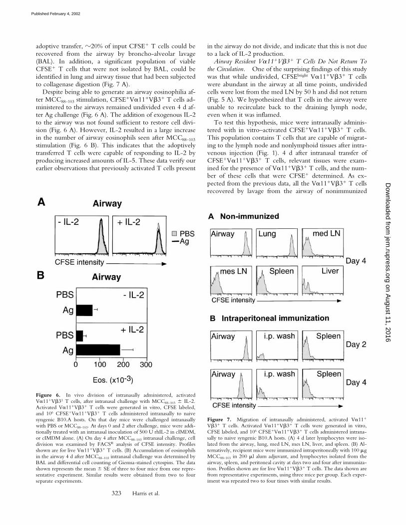
Despite being able to generate an airway eosinophilia af-ter MCC88–103 stimulation, CFSE�V�11�V�3� T cells ad-ministered to the airways remained undivided even 4 d af-ter Ag challenge (Fig. 6 A). The addition of exogenous IL-2to the airway was not found sufficient to restore cell divi-sion (Fig. 6 A). However, IL-2 resulted in a large increasein the number of airway eosinophils seen after MCC88–103
stimulation (Fig. 6 B). This indicates that the adoptivelytransferred T cells were capable of responding to IL-2 byproducing increased amounts of IL-5. These data verify ourearlier observations that previously activated T cells present
in the airway do not divide, and indicate that this is not dueto a lack of IL-2 production.
Airway Resident V�11�V�3� T Cells Do Not Return Tothe Circulation. One of the surprising findings of this studywas that while undivided, CFSEbright V�11�V�3� T cellswere abundant in the airway at all time points, undividedcells were lost from the med LN by 50 h and did not return(Fig. 5 A). We hypothesized that T cells in the airway wereunable to recirculate back to the draining lymph node,even when it was inflamed.
To test this hypothesis, mice were intranasally adminis-tered with in vitro–activated CFSE�V�11�V�3� T cells.This population contains T cells that are capable of migrat-ing to the lymph node and nonlymphoid tissues after intra-venous injection (Fig. 1). 4 d after intranasal transfer ofCFSE�V�11�V�3� T cells, relevant tissues were exam-ined for the presence of V�11�V�3� T cells, and the num-ber of these cells that were CFSE� determined. As ex-pected from the previous data, all the V�11�V�3� T cellsrecovered by lavage from the airway of nonimmunized
Figure 6. In vivo division of intranasally administered, activatedV�11�V�31 T cells, after intranasal challenge with MCC88–103 IL-2.Activated V�11�V�3� T cells were generated in vitro, CFSE labeled,and 106 CFSE�V�11�V�3� T cells administered intranasally to naivesyngenic B10.A hosts. On that day mice were challenged intranasallywith PBS or MCC88–103. At days 0 and 2 after challenge, mice were addi-tionally treated with an intranasal inoculation of 500 U rhIL-2 in cIMDM,or cIMDM alone. (A) On day 4 after MCC88–103 intranasal challenge, celldivision was examined by FACS® analysis of CFSE intensity. Profilesshown are for live V�11�V�3� T cells. (B) Accumulation of eosinophilsin the airway 4 d after MCC88–103 intranasal challenge was determined byBAL and differential cell counting of Giemsa-stained cytospins. The datashown represents the mean SE of three to four mice from one repre-sentative experiment. Similar results were obtained from two to fourseparate experiments.
Figure 7. Migration of intranasally administered, activated V�11�
V�3� T cells. Activated V�11�V�3� T cells were generated in vitro,CFSE labeled, and 106 CFSE�V�11�V�3� T cells administered intrana-sally to naive syngenic B10.A hosts. (A) 4 d later lymphocytes were iso-lated from the airway, lung, med LN, mes LN, liver, and spleen. (B) Al-ternatively, recipient mice were immunized intraperitoneally with 100 �gMCC88–103 in 200 �l alum adjuvant, and lymphocytes isolated from theairway, spleen, and peritoneal cavity at days two and four after immuniza-tion. Profiles shown are for live V�11�V�3� T cells. The data shown arefrom representative experiments, using three mice per group. Each exper-iment was repeated two to four times with similar results.
on August 11, 2016
jem.rupress.org
Dow
nloaded from
Published February 4, 2002

324 T Cell Function in Lymphoid and Nonlymphoid Tissues
mice were CFSE� (Fig. 7 A). The majority of V�11�V�3�
T cells found in the lung were also CFSE�. In contrast, noV�11�V�3� T cells recovered from the draining med LN,mesenteric LN, or spleen, were CFSE� (Fig. 7 A). Norwere any CFSE�V�11�V�3� T cells found in the liver, anorgan that has been suggested to act as a “graveyard” foractivated T cells (23).
We were concerned that CFSE�V�11�V�3� T cellsthat left the airway might have been distributed to systemictissues in numbers below our threshold of detection. Toaddress this possibility we challenged mice with MCC88–103
in alum adjuvant via the intraperitoneal route. Intraperito-neal immunization of naive or primed mice results in Agdrainage to the spleen (24), and an accumulation of Ag-specific T cells in both the peritoneum and spleen (unpub-lished data). Therefore, any CFSE�V�11�V�3� T cellspresent in the circulation after intranasal inoculation shouldbe recruited to the peritoneum and spleen after intraperito-neal MCC88–103–alum challenge. At days 2 and 4 after in-traperitoneal Ag challenge, we examined the spleen andperitoneal cavity of mice that had received an intranasaltransfer of CFSE�V�11�V�3� T cells. No CFSE�
V�11�V�3� T cells were detected in either the spleen orthe peritoneum at day 2, before any cell division would beexpected to occur, or day 4, after intraperitoneal challengewith MCC88–103 in alum (Fig. 7 B).
In summary, these data indicate that adoptively trans-ferred CFSE�V�11�V�3� T cells immigrating to the lungand airway are unable to recirculate to other tissue sites,even after specific Ag challenge. Together these data sup-port our hypothesis that previously activated T cells resi-dent in (or recruited to) the lung and airway are unable toreturn to the lymph node or recirculating pool.
DiscussionAdoptively transferred CFSE-labeled in vitro–activated
T cells were found to separate into distinct subpopulationsbased on their migration to lymph node, lung, or airwaytissue. Although we have no understanding of what in vitrofactors determine the commitment of naive T cells intoeach of the activated T cell subpopulations, their migrationinto lymph nodes correlated with higher levels of expres-sion of CD62L. CD62L is known to be important for lym-phocyte entry into lymph nodes (25), and acts as a ligandfor MAdCAM-1 that is expressed on high-walled endothe-lium of the lymph node (26). Our subpopulations of acti-vated T cells closely resemble the proposed TEM and TCM
subpopulations of memory T cells proposed by Sallusto etal. (27), with respect to their expression of CD62L. Thisresemblance in terms of CD62L expression may be ex-plained by the finding that TEM and TCM subpopulations ofmemory T cells also reside in nonlymphoid or lymphoidtissues, respectively (16, 28). CCR7, a chemokine receptorexpressed by naive and central memory (TCM) T cells (27),also controls homing to secondary lymphoid tissues andmay be expressed by T cells found to enter lymph node inour experiments.
We compared the functions of the adoptively transferredCFSE� T cells that migrate to the lymph node, withCSFE� T cells that migrate to nonlymphoid tissues such asthe lung and airway. CFSE� T cells present in either thelymph nodes, or the lung and airway, could all producesimilar levels of cytokines after encountering MCC88–103 invivo (Fig. 3). CFSE� T cells in the med LN also dividedvigorously in response to MCC88–103 (Fig. 2 A). In strikingcontrast, CFSE� T cells in the lung and airway could notbe induced to divide upon exposure to MCC88–103 (Fig. 2A and unpublished data). However, CFSE� T cells reiso-lated from either the lymph nodes, or the lung and airway,could divide extensively when restimulated with Ag duringin vitro culture (Fig. 4 A). Therefore, the inability to divideis not an intrinsic property of T cells that go to the lung,but is likely to be due to the microenvironment of the lungand airway not being supportive for T cell division.
We endeavored to determine the reason for the lack ofin vivo division observed for T cells in the lung and airway.To our surprise, MCC88–103 presentation by donor-matureBMDC could not induce CFSE� T cells in the airway todivide (Fig. 5 A), although an IL-5–dependent airway eosin-ophilia was observed (Fig. 5 C). These data strongly indi-cate that it is not the poor T cell costimulatory function,nor inadequate Ag processing and presentation, of lung andairway APC that is responsible for the absence of T cell di-vision and clonal expansion. Nor was this lack of divisionfound to be the result of limited IL-2 production in thelung environment, since the addition of large amounts ofIL-2 could not induce intranasally delivered CFSE� T cellsto divide (Fig. 6 A).
There are several reports that indicate that alveolar mac-rophages are able to suppress T cell division in vitro (forreview see 29). These findings were also extended to lungimmune responses (30). In two separate studies, it wasdemonstrated that alveolar macrophages could mediate thissuppressive effect on proliferation through modification ofthe signaling properties of the IL-2 receptor (31, 32). Thiseffect was found to be mediated by nitric oxide (33, 34).Our results extend these in vitro findings by reporting thekey observation that the airway environment does not af-fect the production of effector cytokines. Although wehave not resolved whether the absence of T cell division inthe airway is mediated through nitric oxide, there are sev-eral other candidate mediators and cytokines, such as IL-10and TGF-�, which could exert this effect. Alternatively,agents that induce production of pro-inflammatory cyto-kines, or that have mitogenic properties, may be requiredfor the proliferation of lung and airway T cells. Lastly, fac-tors (other than IL-2) that normally support T cell divisionin the lymph nodes may be absent from the lung and air-way. We are currently investigating all these possibilities inan attempt to further define the mechanism by which thesuppression of T cell proliferation in the lung and airwayoccurs. The question of the physiological significance ofthe inability of T cells to divide in the lung and airwayraises some intriguing possibilities. An important consider-ation is that suppression of T cell division in peripheral tis-
on August 11, 2016
jem.rupress.org
Dow
nloaded from
Published February 4, 2002

325 Harris et al.
sues may serve to prevent potentially pathological T cellactivation in response to harmless environmental Ag. Ad-ditionally, the development of cross-reactive autoimmu-nity by pathogen-activated T cells in response to seques-tered tissue Ag would be avoided in tissue environmentswhere division is prevented.
Our data indicates that activated T cells cannot divide inthe lung or airway, although cells that have divided can en-ter these tissues after exiting the lymph node. We posed thequestion as to whether these airway-homing T cells canmigrate from the airway back to the lymph node or generalcirculation. The data presented in Fig. 7 indicates thatCFSE� T cells delivered to the airway do not return to therecirculating pool. This failure to migrate out of the airwayand lung tissue is likely to be due to an inability of thesecells to move across epithelial barriers or into draining-afferent lymphatics. It is not due to the inability of the in-tranasally transferred cells to survive in the airway microen-vironment, since these cells were shown to respond to Agand induce an airway eosinophilia (Fig. 6 B). It is interest-ing to note that although intranasally administered, acti-vated T cells cannot leave the airway, BMDC administeredintranasally are capable of migrating to the draining medLN and inducing a response here (unpublished data and13). Our data appears to contrast that of Mackay et al. whoreported the presence of CD4� T cells (35) exhibiting amemory phenotype (3) in popliteal-afferent lymph. We donot rule out the possibility that activated T cells leave othernonlymphoid tissue sites to enter afferent lymphatics andreturn to the lymph node. However, it would appear thatthis is not the fate of the large majority of activated T cellsresident in the lung and airway.
In the context of devising vaccine strategies or immuno-therapeutic interventions, one key implication of our workis that the extent and degree of T cell accumulation at aninflammatory site is tightly regulated by two distinctevents. The first is the amplification of T cell number byexpansion in the lymph node. The second involves re-cruitment of T cells to the site of Ag exposure. There is noapparent on-site amplification of the T cell response. Inaddition, we found that the cells that migrate to the airwayare lost to this site, and fail to return to the recirculatingpool. Thus, previously activated T cells that have enteredthe airway appear to be trapped, and must either remain inthe airway as long-lived cells or be lost to the mucosal sur-face. This finding raises the possibility that resting memoryT cells may suffer the same fate. The question as towhether memory T cells can recirculate, or whether theyform two populations destined solely for lymphoid ornonlymphoid tissues, has important implications for ourunderstanding of secondary immune responses and re-quires further investigation.
Our studies have focused on the lung, airway and drain-ing mediastinal lymph node, and on an acute immune re-sponse. It remains to be determined whether the observa-tions we have made are relevant to other nonlymphoidtissues. It would also be of considerable interest to investi-gate whether chronic infections and/or autoimmune dis-
eases are subject to the same control mechanisms. Experi-mental evidence suggests that chronic inflammationinvolves ectopic formation of de novo lymphoid structures(for reviews see 36, 37) that may allow T cell division tooccur. Formation of de novo lymphoid structures occurs ininfectious diseases such as Helicobacter pylori–induced gastri-tis (38) and in autoimmune disorders including Hashi-moto’s thyroiditis (39), rheumatoid arthritis (40, 41), Sjo-gren’s syndrome (42), and experimental murine diabetes(43). It is also not clear what physiological controls existthat prevent activated T cells from dividing in the lung andairway tissues. Elucidation of such mechanisms would haveclear potential for the development of new therapeutics.
The authors thank the personnel of the animal facility of the Well-ington School of Medicine for animal husbandry.
This work was supported by grants from the Marsden Fund,Royal Society of New Zealand, Wellington Medical ResearchFoundation, Health Research Council of New Zealand, and theWellcome Trust.
Submitted: 7 September 2001Revised: 16 November 2001Accepted: 10 December 2001
References1. Gowans, J.L., and E.J. Knight. 1964. The route of recircula-
tion of lymphocytes in the rat. Proc. R. Soc. Lond. B. 159:257–282.
2. Picker, L.J., and E.C. Butcher. 1992. Physiological and mo-lecular mechanisms of lymphocyte homing. Annu. Rev. Im-munol. 10:561–581.
3. Mackay, C.R., W.L. Marston, and L. Dudler. 1990. Naiveand memory T cells show distinct pathways of lymphocyterecirculation. J. Exp. Med. 171:801–817.
4. Koh, W.P., E. Chan, K. Scott, G. McCaughan, M. France,and B. Fazekas de St Groth. 1999. TCR-mediated involve-ment of CD4� transgenic T cells in spontaneous inflamma-tory bowel disease in lymphopenic mice. J. Immunol. 162:7208–7216.
5. Seder, R.A., W.E. Paul, M.M. Davis, and B. Fazekas de StGroth. 1992. The presence of interleukin 4 during in vitropriming determines the lymphokine-producing potential ofCD4� T cells from T cell receptor transgenic mice. J. Exp.Med. 176:1091–1098.
6. Garrigan, K., P. Moroni-Rawson, C. McMurray, I. Her-mans, N. Abernethy, J. Watson, and F. Ronchese. 1996.Functional comparison of spleen dendritic cells and dendriticcells cultured in vitro from bone marrow precursors. Blood.88:3508–3512.
7. Harris, N., C. Campbell, G. Le Gros, and F. Ronchese.1997. Blockade of CD28/B7 co-stimulation by mCTLA4-Hgamma1 inhibits antigen-induced lung eosinophilia but notTh2 cell development or recruitment in the lung. Eur. J. Im-munol. 27:155–161.
8. Hansburg, D., T. Fairwell, R.H. Schwartz, and E. Appella.1983. The T lymphocyte response to cytochrome c. J. Immu-nol. 131:319–324.
9. Fink, P.J., L.A. Matis, D.L. McElligott, M. Bookman, andS.M. Hedrick. 1986. Correlations between T-cell specificityand the structure of the antigen receptor. Nature. 321:219–226.
on August 11, 2016
jem.rupress.org
Dow
nloaded from
Published February 4, 2002

326 T Cell Function in Lymphoid and Nonlymphoid Tissues
10. Ben-Sasson, S.Z., K. Makedonski, J. Hu-Li, and W.E. Paul.2000. Survival and cytokine polarization of naive CD4(�) Tcells in vitro is largely dependent on exogenous cytokines.Eur. J. Immunol. 30:1308–1317.
11. Lyons, A.B., and C.R. Parish. 1994. Determination of lym-phocyte division by flow cytometry. J. Immunol. Methods.171:131–137.
12. Harris, N.L., M. Prout, R.J. Peach, B. Fazekas de St. Groth, andF. Ronchese. 2001. CD80 costimulation is required for Th2cell cytokine production but not for antigen-specific accumula-tion and migration into the lung. J. Immunol. 166:4908–4914.
13. Lambrecht, B.N., R.A. Pauwels, and B. Fazekas de St.Groth. 2000. Induction of rapid T cell acitvation, division,and recirculation by intratracheal injection of dendritic cellsin a TCR transgenic model. J. Immunol. 164:2937–2946.
14. Coffman, R.L., B.W. Seymour, S. Hudak, J. Jackson, and D.Rennick. 1989. Antibody to interleukin-5 inhibits helminth-induced eosinophilia in mice. Science. 245:308–310.
15. Foster, P.S., S.P. Hogan, A.J. Ramsey, K.I. Matthaei, andI.G. Young. 1996. Interleukin 5 deficiency abolishes eosino-philia, airways hyperreactivity, and lung damage in a mouseasthma model. J. Exp. Med. 183:195–201.
16. Lee Reinhardt, R., A. Khoruts, R. Merica, T. Zell, andM.K. Jenkins. 2001. Visualizing the generation of memoryCD4 T cells in the whole body. Nature. 410:101–105.
17. Masten, B.J., J.L. Yates, A.M. Pollard Koga, and M.F. Lip-scomb. 1997. Characterization of accessory molecules in mu-rine lung dendritic cell function: roles for CD80, CD86,CD54, and CD40L. Am. J. Respir. Cell. Mol. Biol. 16:335–342.
18. Holt, P.G. 1993. Regulation of antigen-presenting cell func-tion(s) in lung and airway tissues. Eur. Respir. J. 6:120–129.
19. Cochand, L., P. Isler, F. Songeon, and L.P. Nicod. 1999.Human lung dendritic cells have an immature phenotypewith efficient mannose receptors. Am. J. Respir. Cell. Mol.Biol. 21:547–554.
20. Gonzalez-Juarrero, M., and I.M. Orme. 2001. Characteriza-tion of murine lung dendritic cells infected with Mycobacte-rium tuberculosis. Infect. Immun. 69:1127–1133.
21. Havenith, C.E., A.J. Breedijk, M.G. Betjes, W. Calame,R.H. Beelen, and E.C. Hoefsmit. 1993. T cell priming insitu by intratracheally instilled antigen-pulsed dendritic cells.Am. J. Respir. Cell. Mol. Biol. 8:319–324.
22. Hogan, R.J., W. Zhong, E.J. Usherwood, T. Cookenham,A.D. Roberts, and D.L. Woodland. 2001. Protection fromrespiratory virus infections can be mediated by antigen-spe-cific CD4� T cells that persist in the lungs. J. Exp. Med. 193:981–986.
23. Huang, L., G. Soldevila, M. Leeker, R. Flavell, and N.Crispe. 1994. The liver eliminates T cells undergoing anti-gen-triggered apoptosis in vivo. Immunity. 1:741–749.
24. Olin, T., and T. Saldeen. 1964. The lymphatic pathwaysfrom the peritoneal cavity: a lymphangiographic study in therat. Cancer Res. 24:1700–1711.
25. Gallatin, W.M., I.R. Weissman, and E.C. Butcher. 1983. Acell-surface molecule involved in organ-specific homing oflymphocytes. Nature. 304:30–34.
26. Streeter, P.R., B.T.N. Rouse, and E.C. Butcher. 1988. Im-munohistologic and functional characterization of a vascularaddressin involved in lymphocyte homing into peripherallymph nodes. J. Cell Biol. 107:1853–1862.
27. Sallusto, F., D. Leing, R. Forster, M. Lipp, and A. Lanzavec-chia. 1999. Two subsets of memory T lymphocytes with dis-tinct homing potentials and effector functions. Nature. 401:
708–712.28. Masopust, D., V. Vezys, A.L. Marzo, and L. Lefrancois.
2001. Preferential localization of effector memory cells innonlymphoid tissue. Science. 291:2413–2417.
29. Holt, P.G. 1986. Downregulation of immune responses inthe lower respiratory tract: the role of alveolar macrophages.Clin. Exp. Immunol. 63:261–270.
30. Thepen, T., C. McMenamin, B. Girn, G. Kraal, and P. Holt.1992. Regulation of IgE production in presensitised animals:in vivo elimination of alveolar macrophages preferentially in-creases IgE responses to inhaled allergen. Clin. Exp. Allergy.22:1107–1114.
31. Strickland, D., U.R. Kees, and P.G. Holt. 1996. Regulationof T-cell activation in the lung - alveolar macrophages inducereversible T-cell anergy in vitro associated with inhibition ofinterleukin-2 receptor signal transduction. Immunology. 87:250–258.
32. Upham, J.W., D.H. Strickland, B.W.S. Robinson, and P.G.Holt. 1997. Selective inhibition of T cell proliferation butnot expression of effector function by human alveolar macro-phages. Thorax. 52:786–795.
33. Bingisser, R.M., P.A. Tilbrook, P.G. Holt, and U.R. Kees.1998. Macrophage-derived nitric oxide regulates T cell acti-vation via reversible disruption of the Jak3/Stat5 signallingpathway. J. Immunol. 160:5729–5734.
34. Tulic, M.K., J.L. Wale, P.G. Holt, and P.D. Sly. 2000. Dif-ferential effects of nitric oxide synthase inhibitors in an invivo allergic rat model. Eur. Respir. J. 15:870–877.
35. Mackay, C.R., W.G. Kimpton, M.R. Brandon, and N.P.Cahill. 1988. Lymphocyte subsets show marked differences intheir distribution between blood, and afferent and efferentlymph draining peripheral lymph nodes. J. Exp. Med. 167:1755–1765.
36. Hjelmstrom, P. 2001. Lymphoid neogenesis: de novo forma-tion of lymphoid tissue in chronic inflammation through ex-pression of homing chemokines. J. Leukoc. Biol. 69:331–339.
37. Bachmann, M.F., and M. Kopf. 2001. On the role of the in-nate immunity in autoimmune disease. J. Exp. Med. 193:F47–F50.
38. Mazzucchelli, L., A. Blaser, A. Kappeler, P. Scharli, J.A. Lais-sue, M. Baggiolini, and M. Uguccioni. 1999. BCA-1 ishighly expressed in Helicobacter pylori-induced mucosa-associ-ated lymphoid tissue and gastric lymphoma. J. Clin. Invest.104:R49–R54.
39. Soderstrom, N., and A. Biorklund. 1974. Organization of theinvading lymphoid tissue in human lymphoid thyroiditis.Scand. J. Immunol. 3:295–301.
40. Iguchi, T., and M. Ziff. 1986. Electron microscopic study ofrheumatoid synovial vasculature: Intimate relationship be-tween tall endothelium and lymphoid aggregation. J. Clin.Invest. 77:355–361.
41. Wagner, U.G., P.J. Kurtin, A. Wahner, M. Bracketz, D.J.Berry, J.J. Goronzy, and C.M. Weyand. 1998. The role ofCD8�CD40L� T cells in the formation of germinal centre’sin rheumatoid synovitis. J. Immunol. 161:6390–6397.
42. Aziz, K.E., P.J. McCluskey, A. Montanaro, and D. Wake-field. 1992. Vascular endothelium and lymphocyte adhesionmolecules in minor salivary glands of patients with Sjogren’ssyndrome. J. Clin. Lab. Immunol. 37:39–49.
43. Ludewig, B., B. Odermatt, S. Landmann, H. Hengartner,and R. Zinkernagel. 1998. Dendritic cells induce autoim-mune diabetes and maintain disease via de novo formation oflocal lymphoid tissue. J. Exp. Med. 188:1493–1501.
on August 11, 2016
jem.rupress.org
Dow
nloaded from
Published February 4, 2002