Insect olfaction as an information filter for chemo-analytical ...

1
This document is a post-print version of a book chapter in Methods in molecular
biology (Clifton, N.J.). The final publication is available at link.springer.com:
http://link.springer.com/protocol/10.1007%2F978-1-62703-465-4_27
Glycosyltransferases in Chemo-enzymatic Synthesis of
Oligosaccharides.
Boris Tefsen1#
and Irma van Die1
1Department of Molecular Cell Biology and Immunology, VU University Medical
Center, van den Boechorststraat 7, 1081 BT, Amsterdam, The Netherlands
# Corresponding author: Department of Molecular Cell Biology and Immunology, VU
University Medical Center, van den Boechorststraat 7, 1081 BT, Amsterdam, The
Netherlands, +31204448150, [email protected].
Running title: Glycosyltransferases in chemo-enzymatic synthesis
Summary
Many oligosaccharides are not commercially available, which limits studies focused
on elucidation of glycan functions; therefore chemo-enzymatic methods to synthesize
them can be very useful. Here, we describe the procedure to synthesize the Gal1-
3GalNAc1-4GlcNAc-R (Gal-LDN) moiety, containing the Gal1-3GalNAc
epitope found on the parasitic helminth Haemonchus contortus. An acceptor substrate
providing a terminal N-acetylglucosamine was prepared by coupling the fluorescent

2
hydrophobic aglycon, 2,6-diaminopyridine (DAP), to N,N’-diacetylchitobiose. By the
subsequent action of recombinant Caenorhabditis elegans 1,4-N-
acetylgalactosaminyltransferase the substrate was efficiently converted to GalNAc1-
4GlcNAc-R (LDN-R). Since no recombinant 1,3-galactosyltransferase has been
described that acts on terminal βGalNAc, we used bovine 1,3-galactosyltransferase
to obtain a partial conversion of LDN-R to the Gal-LDN antigen. This method can be
applied to synthesize any oligosaccharide, provided that specific glycosyltransferases
are available, or related enzymes that can be pushed to elongate the selected acceptor.
Key Words: neoglycoconjugate; 2,6-Diaminopyridine; galactosyltransferase, chemo-
enzymatic synthesis, Gal-LDN, helminth, chitobiose
1. Introduction
Glycan molecules linked to proteins or lipids play important roles in cellular
communication, adhesion and signalling and are key molecules in regulation of
immune responses. To establish the role of individual glycans in diverse aspects of
biology, the availability of oligosaccharides or neoglycoconjugates that carry defined
glycan antigens is of crucial importance. Neoglycoconjugates are attractive tools to
define anti-glycan responses in infection or immunization (1,2), or to define specific
carbohydrate recognition by lectin receptors that occur on many immune cells (3).
Neoglycoconjugates are also used in vaccines to elicit carbohydrate-specific
antibodies that can confer protection to infection, for example to Neisseria
meningitides and Streptococcus pneumoniae (4,5).
Helminth parasites express a variety of unusual glycan antigens that are highly
antigenic in infection (6). The study of the biological properties of particular helminth

3
glycan antigens highly depends on the availability of neoglycoconjugates, which in
contrast to many mammalian-type glycans are not commercially available. A
drawback for the enzymatic synthesis of unusual glycans such as helminth glycans is
that not many recombinant parasite-type glycosyltransferases are available.
Previously, we have synthesized LDNF antigen using 1,4-N-
acetylgalactosaminyltransferase from the albumen gland of the snail Lymnaea
stagnalis and partially purified 1,3-FucT from human milk (7) and also using the
1,4-N-acetylgalactosaminyltransferase from C. elegans in combination with
recombinant human 1,3-fucosyltransferase VI (8). Neoglycoconjugates containing
the LDNF-moiety can be applied when antigens directly isolated from helminths are
hard to obtain, as has been shown in the case of serodiagnosis of trichinellosis (9).
Recently, we showed that the Gal1-3GalNAc epitope is present on the sheep
parasite Haemonchus contortus and that IgG antibodies against Gal1-3GalNAc were
elicited during a vaccination trial with ES antigens (10). In this chapter, we describe a
chemo-enzymatic method to synthesize the neoglycoconjugate Gal1-3GalNAc1-
4GlcNAc conjugated to DAP (Gal-LDN-DAP) in nanomolar amounts.
This synthesis starts with the chemical modification of the commercially available
N,N’-diacetylchitobiose (chitobiose) acceptor with 2,6-diaminopyridine (DAP) to
provide a fluorescent hydrophobic linker, facilitating detection and purification of the
oligosaccharide products (Figure 1), and allowing subsequent coupling to a protein
(11,12). In principle, each oligosaccharide acceptor structure can be derivatized with
DAP, and after elongation by the subsequent action of appropriate
glycosyltransferases, the final DAP-derivatized oligosaccharides can easily be used
for different downstream applications (8,9,11).

4
Subsequently, an N-acetylgalactosamine residue is coupled to the DAP-
derivatized chitobiose acceptor using recombinant C. elegans 1,4-N-
acetylgalactosaminyltransferase (13) as a catalyst, resulting in a terminal GalNAc1-
4GlcNAc moiety, which commonly is referred to as LacDiNAc (LDN). No enzymatic
activity has been reported before that can catalyze the transfer of a galactose residue
in 1-3 linkage to LDN. It has been described that some glycosyltransferases can be
forced to act on certain acceptors that they normally would not act on (14). Indeed,
when we used bovine 1,3-galactosyltransferase (15,16) as a catalyst on the LDN
acceptor, it proved to be successful, resulting in Gal-LDN-DAP. Notably, a maximum
of ten percent of LDN-DAP was transformed to Gal-LDN-DAP in a single reaction,
reflecting that this is a sub-optimal reaction. The progress in synthesis of intermediate
structures was monitored using normal-phase HPLC (Figure 2). Using preparative
HPLC, the synthesized oligosaccharide was purified and the structural identity of
Gal-LDN-DAP was verified by ESI-MS (Figure 3).
5
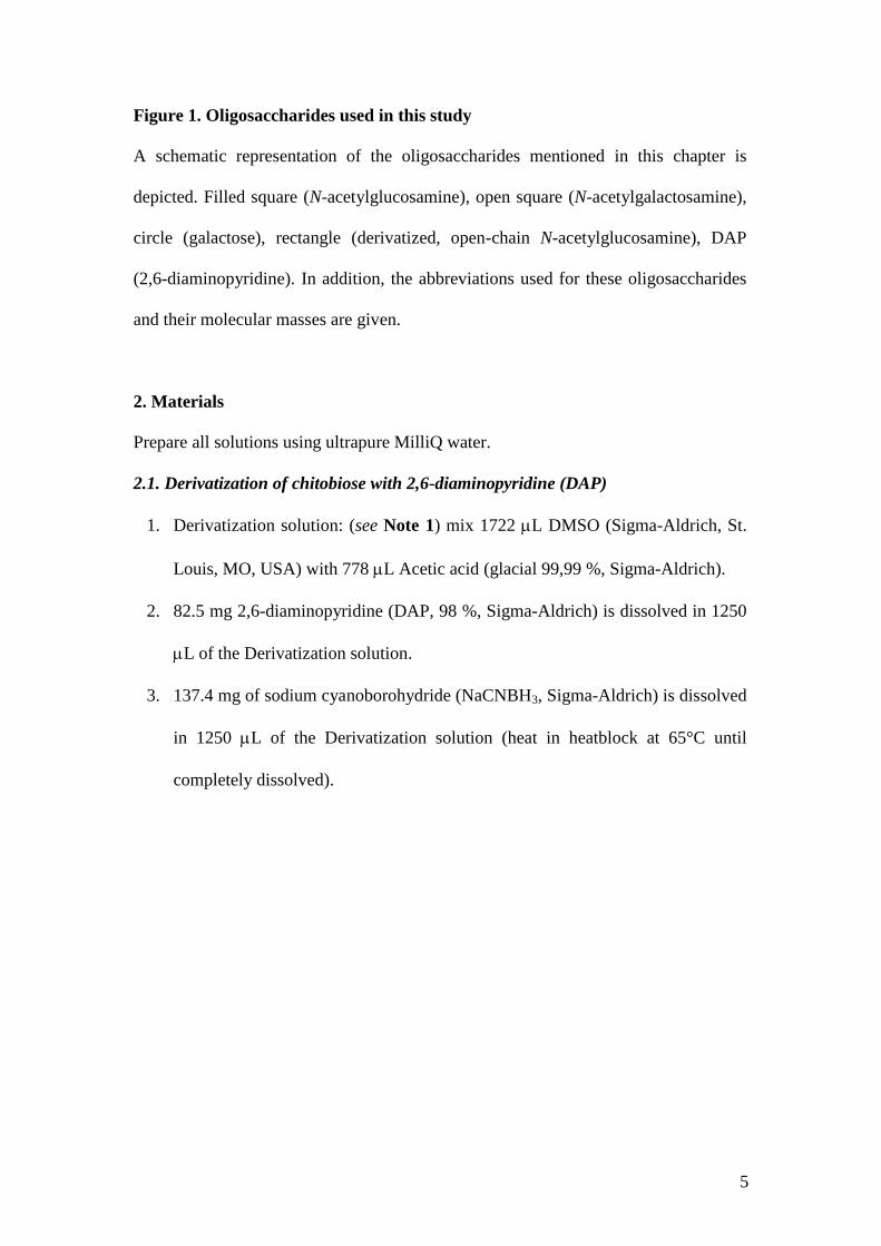
Figure 1. Oligosaccharides used in this study
A schematic representation of the oligosaccharides mentioned in this chapter is
depicted. Filled square (N-acetylglucosamine), open square (N-acetylgalactosamine),
circle (galactose), rectangle (derivatized, open-chain N-acetylglucosamine), DAP
(2,6-diaminopyridine). In addition, the abbreviations used for these oligosaccharides
and their molecular masses are given.
2. Materials
Prepare all solutions using ultrapure MilliQ water.
2.1. Derivatization of chitobiose with 2,6-diaminopyridine (DAP)
1. Derivatization solution: (see Note 1) mix 1722 L DMSO (Sigma-Aldrich, St.
Louis, MO, USA) with 778 L Acetic acid (glacial 99,99 %, Sigma-Aldrich).
2. 82.5 mg 2,6-diaminopyridine (DAP, 98 %, Sigma-Aldrich) is dissolved in 1250
L of the Derivatization solution.
3. 137.4 mg of sodium cyanoborohydride (NaCNBH3, Sigma-Aldrich) is dissolved
in 1250 L of the Derivatization solution (heat in heatblock at 65°C until
completely dissolved).

6
4. N,N′-Diacetylchitobiose (chitobiose, Sigma).
5. 12 cc reversed-phase C18 Sep-Pak columns (Waters, Milford, MA, USA).
6. Preparative normal-phase column (Zorbax NH2 PrepHT, 250 x 21.2 mm, 7 m,
Agilent Technologies, Santa Clara, CA, USA).
7. Buffer A contains acetonitrile (LC-MS, Chromasolv, Riedel-de Haen, Seelze,
Germany).
8. Buffer B contains 50 mM ammoniumformate pH 4.4: dissolve 5 ml 10 M
Ammonium formate solution and 200 l Formic acid in 1 L water.
9. Äkta Explorer HPLC apparatus (Amersham Pharmacia Biotech; now merged
with GE Healthcare Bio-Sciences AB, Uppsala, Sweden).
10. Speedvac centrifuge.
11. 1.5-mL reaction tubes.
12. 15-mL tubes.
2.2 Production of recombinant C. elegans UDP-GalNAc:GlcNAc 1,4-N-
acetylgalactosaminyltransferase
1. Human Embryonic Kidney 293T (HEK293T) (ATCC CRL 1573) cells.
2. Dulbecco’s modified Eagle’s medium (DMEM, Gibco, Life Technologies,
Paisley, UK).
3. 10 % Foetal Calf Serum (FCS, Gibco).
4. Pencillin-Streptomycin (Lonza, Basel, Switzerland).
5. HEPES (Gibco).
6. 10 mM MEM non essential amino acids (Gibco).
7. 100 mM MEM sodium pyruvate (Gibco).
8. Optimem I (Gibco).

7
9. Lipofectamin 2000 (Invitrogen, Life Technologies, Paisley, UK).
10. Incubator at 37°C/5 % CO2.
11. PBS.
12. Trypsinization solution: 0.025 % trypsin (Gibco) and 0.01 % EDTA in PBS.
13. T75 culture flasks.
2.3 β1-4GalNActransferase reaction
1. GalNActransferase reaction buffer: mix an equivalent of 4 µL 1 M MnCl2 (final
concentration is 40 mM), 2.5 L 4 M Sodium cacodylate (C2H12AsNaO5), pH
7.0 (final concentration is 100 mM).
2. 100 mM UDP-GalNAc (final concentration is 20 mM).
3. Culture medium containing recombinant C. elegans UDP-GalNAc:GlcNAc
1,4-N-acetylgalactosaminyltransferase (1,4GalNAcT) (section 3.2. item 11).
2.4. 1-3Galactosyltransferase reaction
1. Buffer for Galactosyltransferase reaction: mix an equivalent of 8 µL 1 M MnCl2
(final concentration is 20 mM), 5 L 4 M Sodium cacodylate (C2H12AsNaO5)
pH 7.0 (final concentration is 50 mM), 80 L 0.5 % BSA (final concentration is
0.1 %). Add water to an end-volume of (an equivalent of) 400 L.
2. 50 mM UDP-galactose.
3. Recombinant bovine 1,3-galactosyltransferase (see Note 2).

8
Figure 2. Monitoring product formation of intermediate structures by HPLC
Enzymatic synthesis was monitored by analytical normal-phase HPLC. The relative
intensity of DAP-derivatized glycans is plotted against time of elution from the
column. The starting compound Chi-2-DAP is shown (A), which is converted with C.
elegans 1,4-GalNAcT into LDN-DAP (B). After isolation of LDN-DAP with
reversed-phase cartridges, around 10 % of this compound is converted to Gal-LDN-
DAP with bovine 1,3-GalT (C).
2.5. Monitoring of the formation and purification of DAP-derivatized glycans
1. 6cc reversed-phase C18 Sep-Pak columns (Waters).
2. Methanol.
3. Normal-phase LudgerSep N1 Amide column (250 x 4.6 mm, Ludger,
Oxfordshire, UK).
4. Surveyor HPLC (Thermo Fischer Scientific, Waltham, MA, USA).
5. Fluorimeter 470 (Waters).
6. Buffer A: Acetonitrile.

9
7. Buffer B: 50 mM Ammonium formate pH 4.4: dissolve 5 ml 10 M Ammonium
formate solution (Sigma-Aldrich) and 200 l Formic acid (Sigma-Aldrich) in 1
L water.
2.6. Electrospray Injection Mass Spectrometry
1. LCQ DecaXP ion trap mass spectrometer equipped with a nano-ES ionization
source (Thermo Fischer Scientific).
2. 10 L syringe.
3. Medium NanoEs spray capillary (Proxeon, Thermo Fischer Scientific).
4. 50 % acetonitrile.
3. Methods
3.1. Derivatization of chitobiose with 2,6-diaminopyridine (DAP)
1. Mix the derivatization solutions containing DAP and cyanoborohydride in a 1:1
ratio (see Note 3).
2. Dissolve dry, water-free pellets of chitobiose (0.4 mg, 1 mol) (see Note 4) in
10 L of DMSO, and subsequently add 500 L of the mixture described in
section 3.1. item 1.
3. Incubate the reaction mixture for 18 h at 65°C in a heat block.
4. Directly after incubation, apply the content of 2 tubes (containing unreacted
chitobiose, Chi-2-DAP and free DAP) to one 12 cc Sep-Pak C18 column that
was pre-activated with 5 column volumes of methanol and washed with 10
column volumes of water. Subsequently wash (see Note 5) the column with 10
column volumes of water and elute the Chi-2-DAP (see Note 6) with 5 column
volumes of methanol.
10
5. Pool the eluted Chi-2-DAP coming from two C18 columns in a 15-mL tube and
dry in a speedvac; add 0.5 mL water to the resulting pellet (contains
approximately 4 mol Chi-2-DAP and excess free DAP) and let his stand for 10
h at 4°C (see Note 7).
6. Apply 0.5 mL from the dissolved material on the preparative normal-phase
column. (see Note 8) Use a gradient that starts with 81 % acetonitrile (buffer A)
and 29 % 50 mM ammonium formate, pH 4.4 (buffer B) and ends after 17 min
with 30 % buffer A and 70 % buffer B on an Äkta Explorer with a column flow
of 10 mL/min. DAP-containing fractions are detected by measuring absorbance
at 235 nm.
7. Collect the peak containing Chi-2-DAP (eluting at min 11.5) in a 15-mL tube
(see Note 9).
8. Freeze-dry the collected Chi-2-DAP and store at -20°C (see Note 10).
3.2. Production of recombinant C. elegans UDP-GalNAc:GlcNAc 1,4-N-
acetylgalactosaminyltransferase (1,4GalNAcT).
1. Culture adherent HEK293T cells in DMEM containing 10 mM HEPES, 100 M
MEM non essential amino acids, 1 mM MEM sodium pyruvate, 10 % FCS and
100 U/mL penicillin-streptomycin in a T75 flask at 37°C/5 % CO2 until the
bottom is completely covered by a monolayer of cells (~6x10⁶ cells/flask).
2. Prepare a transfection mixture by mixing 600 L Optimem I containing 54 L
Lipofectamin 2000 with 600 L Optimem I containing 9 g DNA of plasmid
pCMV-SH-Ceβ4GalNAcT (13).
3. Incubate the transfection mixture for 30 min at room temperature.
4. Wash the HEK293T cells twice with 9 mL Optimem I.
11
5. Mix 4.8 mL Optimem I with the transfection mixture.
6. Add this mixture to the cells and incubate the cells for 5-6 h at 37°C/5 % CO2.
7. Add 6 ml DMEM and culture the cells overnight at 37°C/5 % CO2.
8. Wash the cells with 2 mL PBS.
9. Treat the cells with 1 mL Trypsinization solution and transfer to a new T75
flask.
10. Culture the cells in 9 mL DMEM for another 3 days.
11. Harvest medium containing the active enzyme daily until 7 days after
transfection.
12. Determine the activity of the enzyme by monitoring the incorporation of
radiolabeled UDP-GalNAc in time (13) (see Note 11).
13. Store at -20°C. (see Note 12).
3.3 Addition of a β1-4GalNAc residue to Chi-2-DAP to create LDN-DAP
1. Dissolve 1 mol dried chitobiose-DAP (final concentration is 10 mM) in 6.5 L
GalNActransferase reaction buffer.
2. Add 20 L 100 mM UDP-GalNAc (final concentration is 20 mM) and 73.5 µL
medium containing 1,4GalNAcTransferase to a final volume of 100 L.
3. Incubate the reaction mixture at room temperature (20-24°C) until the Chi-2-
DAP has been completely converted to the desired end-product, LDN-DAP.
3.4. Monitoring the formation of LDN-DAP and subsequent purification
1. Position a LudgerSep N1 column in the HPLC.
2. Equilibrate the column with 100 % ammonium formate for 10 min, followed by
10 min of 70 % acetonitrile and 30 % 50 mM ammonium formate.

12
3. Dissolve 0.5 L of the 100 L reaction mixture of section 3.3. item 1 in 100 L
80 % acetonitrile.
4. Inject 25 L of this dilution onto the column.
5. Run a 30-min gradient starting with 70 % acetonitrile and 30 % 50 mM
ammonium formate, pH 4.4 and ending in a 50:50 ratio at 22°C.
6. Detect the glycan-DAP peaks by fluorescence in a fluorimeter by excitation at
345 nm and measuring the emission at 400 nm with a 10 times gain of signal.
The Chi-2-DAP peak elutes in this system around minute 17.8, whereas the
LDN-DAP elutes around minute 19.8 (see Fig. 2).
7. Purify LDN-DAP when conversion is complete from the reaction mixture from
section 3.3. item 2 using a 6 cc C18 reversed-phase Sep-Pak column as
described in section 3.1. item 4.
8. Dry the eluted LDN-DAP in the speedvac and store it at -20°C.
3.5. Addition of an α1-3Galactose residue to LDN-DAP to create Gal-LDN-DAP
1. Dissolve 1 mol dried LDN-DAP (final concentration is 1 mM) in 262.5 µL
Galactosyltransferase buffer.
2. Add 377.5 L water, 30 L 50 mM UDP-galactose (final concentration is 1.5
mM) and 360 L 1,3-galactosyltransferase to a final volume of 1 mL (see
Note 13).
3. Incubate the mixture at 37°C until no further enhancement of the desired end-
product, Gal-LDN-DAP, is observed (see Note 14).
3.6. Monitoring the formation of Gal-LDN-DAP and subsequent purification

13
1. The formation of Gal-LDN-DAP is monitored in time as described in section
3.4.
2. The LDN-DAP elutes around minute 19.8 and Gal-LDN-DAP around minute
22.8 (see Fig. 2).
3. When the reaction from section 3.5. item 3 is stopped, purify the mixture of
LDN-DAP and Gal-LDN-DAP with a 6 cc Sep-Pak as described in section 3.1.
item 4.
4. Dissolve the pellet containing this mixture in water and apply it to the
preparative column, similarly as described for the purification of Chi-2-DAP in
section 3.1. item 6.
5. Collect the fractions where Gal-LDN-DAP elutes (see Notes 15 and 16). LDN-
DAP starts to elute around minute 14.5 and Gal-LDN-DAP elutes around
minute 15.5.
6. Dry the eluted DAP-derivatized glycans in the speedvac and store at -20°C.
3.7. Confirmation of the formation of Gal-LDN-DAP by mass spectrometry
1. Characterize product formation by ESI–MS on an LCQ DecaXP ion trap mass
spectrometer equipped with a nano-ES ionization source.
2. Take 0.5 L of a collected fraction from the Surveyor run, containing Gal-LDN-
DAP, and dilute in 50 L 50 % acetonitrile.
3. Inject a few L of this sample into a medium NanoEs spray capillary (Proxeon)
using the 10 L syringe.
4. Mount the capillary in front of the ion-trap and set the capillary temperature to
200°C.

14
5. Take spectra in the positive ion mode with a spray voltage of 1.0 kV and a
capillary voltage of 40.1 V and subject to tandem MS to confirm the synthesis
of Gal-LDN-DAP (see Fig. 3).
Figure 3. Tandem mass spectrum of Gal-LDN-DAP
Characterization of Gal-LDN–DAP by tandem mass spectrometry with ESI-MS.
Oligosaccharide products were characterized in the positive-ion mode. The expected
molecular mass of the protonated Gal-LDN–DAP ion (m/z = 883.11) was found, and
by fragmentation using tandem MS/MS the structure was confirmed. Schematic
figures of the oligosaccharides representing the found fragments are depicted above
the peaks. The asterisks indicate ions that are similar to the structures shown, but that
lack a water molecule (-18 Da).
4. Notes
1. These solutions should be made fresh on the day they will be used.

15
2. Initially, we could obtain the recombinant bovine 1,3-galactosyltransferase
from Sigma, but this product was discontinued. At that point, we kindly
received the enzyme from Dr. M.M. Palcic (16).
3. The less-toxic compound picoline borate was tested instead of
cyanoborohydride for the coupling of DAP to chitobiose, but it inhibited
subsequent enzymatic reactions and was therefore discarded.
4. We originally started the whole procedure described here using N,N’,N’’-
triacetylchitotriose as the starting acceptor to have a greater distance between
the immunogenic epitope of the neoglycoconjugate and the carrier protein.
However, degradation of this compound by a -N-acetylhexosaminidase present
in Fetal Calf serum made this strategy impossible.
5. This step removes free chitobiose and salts.
6. Free DAP molecules will also be eluted from the column.
7. This step can be shortened, but then the amount of dissolved Chi-2-DAP will be
less.
8. This step separates free DAP molecules from DAP-derivatized glycans (see
figure 2 in (8)).
9. Characterization by ESI-MS of the intermediate DAP-derivatized glycans, Chi-
2-DAP and LDN-DAP is described in (8).
10. No loss of material was observed after 3 months of storage at -20°C.
11. The enzyme-containing medium showed an activity of approximately 200
nmol/mL/h using acceptor p-nitrophenyl-N-acetyl-1-thio--D-glucosaminide.
12. The enzyme was stored at -20°C for several months without loss of activity.
13. Galactosyltransferase activity of the bovine 1,3galactosyltransferase was
determined essentially as described previously (17).

16
14. This was after approximately 72 h. Longer incubations never yielded more than
approximately 10 % of LDN-DAP to be elongated with a galactose molecule.
15. Gal-LDN-DAP purified in this way, can still contain between 5 and 10 % of the
starting material LDN-DAP.
16. The elutions containing LDN-DAP can be collected, dried and re-used starting
from step 3.5. item 2.
Acknowledgements
This work was supported by the Dutch Technology Foundation (STW). We kindly
thank Dr. M.M. Palcic (Carlsberg Laboratory, Denmark) for providing the
recombinant bovine 1,3-galactosyltransferase.
References
1. Vervelde, L., Bakker, N., Kooyman,
F. N., Cornelissen, A. W., Bank, C.
M., Nyame, A. K., Cummings, R. D.,
and van Die, I. (2003) Vaccination-
induced protection of lambs against
the parasitic nematode Haemonchus
contortus correlates with high IgG
antibody responses to the LDNF
glycan antigen, Glycobiology 13,
795-804.
2. van Remoortere, A., van Dam, G. J.,
Hokke, C. H., van den Eijnden, D.
H., van Die, I., and Deelder, A. M.
(2001) Profiles of immunoglobulin
M (IgM) and IgG antibodies against
defined carbohydrate epitopes in
sera of Schistosoma-infected
individuals determined by surface
plasmon resonance, Infect Immun 69,
2396-2401.
3. van Vliet, S. J., Garcia-Vallejo, J. J.,
and van Kooyk, Y. (2008) Dendritic
cells and C-type lectin receptors:
coupling innate to adaptive immune
responses, Immunol Cell Biol 86,
580-587.
4. Cuello, M., Cabrera, O., Martinez, I.,
Del Campo, J. M., Camaraza, M. A.,
Sotolongo, F., Perez, O., and Sierra,
G. (2007) New meningococcal C
polysaccharide-tetanus toxoid
conjugate Physico-chemical and
immunological characterization,
Vaccine 25, 1798-1805.
5. Lee, L. H., Lee, C. J., and Frasch, C.
E. (2002) Development and
evaluation of pneumococcal
conjugate vaccines: clinical trials and
control tests, Crit Rev Microbiol 28,
27-41.
6. Nyame, A. K., Kawar, Z. S., and
Cummings, R. D. (2004) Antigenic
glycans in parasitic infections:
implications for vaccines and
diagnostics, Arch Biochem Biophys
426, 182-200.
7. van Remoortere, A., Hokke, C. H.,
van Dam, G. J., van Die, I., Deelder,

17
A. M., and van den Eijnden, D. H.
(2000) Various stages of schistosoma
express Lewis(x), LacdiNAc,
GalNAcβ1-4 (Fucα1-3)GlcNAc and
GalNAcβ1-4(Fucα1-2Fucα1-
3)GlcNAc carbohydrate epitopes:
detection with monoclonal
antibodies that are characterized by
enzymatically synthesized
neoglycoproteins, Glycobiology 10,
601-609.
8. Tefsen, B., van Stijn, C. M., van den
Broek, M., Kalay, H., Knol, J. C.,
Jimenez, C. R., and van Die, I. (2009)
Chemoenzymatic synthesis of
multivalent neoglycoconjugates
carrying the helminth glycan antigen
LDNF, Carbohydr Res 344, 1501-
1507.
9. Aranzamendi, C., Tefsen, B., Jansen,
M., Chiumiento, L., Bruschi, F.,
Kortbeek, T., Smith, D. F.,
Cummings, R. D., Pinelli, E., and
Van Die, I. (2011) Glycan
microarray profiling of parasite
infection sera identifies the LDNF
glycan as a potential antigen for
serodiagnosis of trichinellosis, Exp
Parasitol 129, 221-226.
10. van Stijn, C. M., van den Broek, M.,
Vervelde, L., Alvarez, R. A.,
Cummings, R. D., Tefsen, B., and
Die, I. V. (2010) Vaccination-
induced IgG response to Galα1-
3GalNAc glycan epitopes in lambs
protected against Haemonchus
contortus challenge infection, Int J
Parasitol 40, 215-222.
11.Xia, B., Kawar, Z. S., Ju, T., Alvarez,
R. A., Sachdev, G. P., and
Cummings, R. D. (2005) Versatile
fluorescent derivatization of glycans
for glycomic analysis, Nat Methods
2, 845-850.
12. Rothenberg, B. E., Hayes, B. K.,
Toomre, D., Manzi, A. E., and Varki,
A. (1993) Biotinylated
diaminopyridine: an approach to
tagging oligosaccharides and
exploring their biology, Proc Natl
Acad Sci USA 90, 11939-11943.
13. Kawar, Z. S., Van Die, I., and
Cummings, R. D. (2002) Molecular
cloning and enzymatic
characterization of a UDP-
GalNAc:GlcNAcβ-R β1,4-N-
acetylgalactosaminyltransferase from
Caenorhabditis elegans, J Biol Chem
277, 34924-34932.
14. Palcic, M. M. (2011)
Glycosyltransferases as biocatalysts,
Curr Opin Chem Biol 15, 226-233.
15. Joziasse, D. H., Shaper, J. H., Van
den Eijnden, D. H., Van Tunen, A. J.,
and Shaper, N. L. (1989) Bovine α1-
3-galactosyltransferase: isolation and
characterization of a cDNA clone.
Identification of homologous
sequences in human genomic DNA. J
Biol Chem 264, 14290-14297.
16. Sujino, K., Malet, C., Hindsgaul, O.,
and Palcic, M. M. (1997) Acceptor
hydroxyl group mapping for calf
thymus α1-3-galactosyltransferase
and enzymatic synthesis of α-D-
Galp-(1-3)-β-D-Galp-(1-4)-β D-
GlcpNAc analogs, Carbohydr Res
305, 483-489.
17.Joziasse, D. H., Shaper, N. L., Salyer,
L. S., Van den Eijnden, D. H., van
der Spoel, A. C., and Shaper, J. H.
(1990) α1-3-galactosyltransferase:
the use of recombinant enzyme for
the synthesis of α-galactosylated
glycoconjugates, Eur J Biochem 191,
75-83.
Copyright © 2022 FDOKUMEN