Atmospheric Pressure MALDI Fourier Transform Mass Spectrometry of Labile Oligosaccharides
10
Atmospheric Pressure MALDI Fourier Transform Mass Spectrometry of Labile Oligosaccharides Jinhua Zhang, LaTasha LaMotte, Eric D. Dodds, and Carlito B. Lebrilla* Department of Chemistry and School of Medicine, Biochemistry and Molecular Medicine, University of California Davis, Davis, California 95616 An atmospheric pressure matrix-assisted laser desorp- tion/ionization (AP MALDI) source coupled to Fourier transform ion cyclotron resonance mass spectrometry (FT ICR MS) under UV laser and solid matrix conditions has been demonstrated to analyze a variety of labile oligosac- charides including O-linked and N-linked complex glycans released from glycoproteins. Spectra were acquired by both AP MALDI and vacuum MALDI and directly com- pared. The results presented here confirm that AP MALDI can generate significantly less energetic ions than vacuum MALDI and is able to produce the intact molecular ions with little or no fragmentation in both positive and negative ion mode analyses. Under certain conditions, noncovalent complexes of sialylated oligosaccharides were observed. The sensitivity attainable by AP MALDI was found to be comparable to conventional MALDI, and tandem mass spectrometry of oligosaccharides ionized by AP MALDI was shown to allow detailed structural analysis. Analysis of N-glycan mixtures derived from human fibrinogen further demonstrated that AP MALDI-FT ICR MS is ideal for the study of complex glycan samples as it provides high-accuracy, high-resolution mass analysis with no difficulty in distinguishing sample constituents from frag- ment ions. Matrix-assisted laser desorption/ionization (MALDI) mass spectrometry has been extensively used for the rapid and sensitive analysis of oligosaccharides. MALDI has shown higher sensitivity than electrospray ionization (ESI) due to the hydrophilic character of underivatized oligosaccharides. 1,2 In addition, the analyte signals produced by ESI are distributed among singly and multiply charged ions as well as species with different associated cations, further reducing the sensitivity and complicating the mass spectral interpretation. MALDI yields predominant [M + Na] + ions for underivatized oligosaccharides. Usually, fragmentation of the sodiated species produces a higher percentage of cross-ring cleavage products than [M + H] + ions, thereby providing linkage information on the oligosaccharides. 3 The implementation of MALDI with Fourier transform ion cyclotron resonance mass spectrometry (FT ICR MS) has dem- onstrated high performance with regard to mass accuracy, sensitivity, resolution, and tandem MS capabilities for various oligosaccharides and glycoconjugates as reported by this la- boratory 4-19 and others. 20-22 However, the metastable decay of ions generated by conventional MALDI is sometimes problematic in the characterization of mixtures, as it is at times difficult to determine whether a particular ion is an independent species or merely a fragment. The metastable decay problem is more pronounced for carbohydrates and glycoconjugates containing labile groups, such as sialic acid, sulfate, fucose, and lipids, thus hindering the observation of the intact molecular ions in the mass spectrum. Extensive work has been conducted in the past several years on the development of MALDI methods that reduce the * Corresponding author. Fax: 1-530-754-5609. Tel: 1-530-752-6364. E-mail: [email protected]. (1) Burlingame, A. L.; Boyd, R. K.; Gaskell, S. J. Anal. Chem. 1994, 66, 634R- 683R. (2) Reinhold: V. N.; Reinhold: B. B.; Costello, C. E. Anal. Chem. 1995, 67, 1772-1784. (3) Harvey, D. J.; Bateman, R. H.; Green, M. R. J. Mass Spectrom. 1997, 32, 167-187. (4) Cancilla, M. T.; Penn, S. G.; Carroll, J. A.; Lebrilla, C. B. J. Am. Chem. Soc. 1996, 118, 6736-6745. (5) Penn, S. G.; Cancilla, M. T.; Lebrilla, C. B. Anal. Chem. 1996, 68, 2331- 2339. (6) Penn, S. G.; Cancilla, M. T.; Green, M. K.; Lebrilla, C. B. Eur. Mass Spectrom. 1997, 3, 67-79. (7) Cancilla, M. T.; Penn, S. G.; Lebrilla, C. B. Anal. Chem. 1998, 70, 663- 672. (8) Wong, A. W.; Cancilla, M. T.; Voss, L. R.; Lebrilla, C. B. Anal. Chem. 1999, 71, 205-211. (9) Cancilla, M. T.; Wang, A. W.; Voss, L. R.; Lebrilla, C. B. Anal. Chem. 1999, 71, 3206-3218. (10) Penn, S. G.; Cancilla, M. T.; Lebrilla, C. B. Int. J. Mass Spectrom. 2000, 196, 259-269. (11) Wong, A. W.; Wang, N.; Lebrilla, C. B. Anal. Chem. 2000, 72, 1419-1425. (12) Tseng, K.; Hedrick, J. L.; Lebrilla, C. B. Anal. Chem. 1999, 71, 3747- 3754. (13) Tseng, K.; Wang, H.; Lebrilla, C. B.; Bonnell, B.; Hedrick, J. L. Anal. Chem. 2001, 73, 3556-3561. (14) Franz, A. H.; Molinski, T. F.; Lebrilla, C. B. J. Am. Soc. Mass Spectrom. 2001, 12, 1254-1261. (15) Tseng, K.; Xie, Y.; Seeley, J.; Hedrick, J. L.; Lebrilla, C. B. Glycoconjugate J. 2001, 18, 309-320. (16) Xie, Y.; Lebrilla, C. B. Anal. Chem. 2003, 75, 1590-1598. (17) An, H. J.; Peavy, T. R.; Hedrick, J. L.; Lebrilla, C. B. Anal. Chem. 2003, 75, 5628-5637. (18) Xie, Y.; Liu, J.; Zhang, J.; Hedrick, J. L.; Lebrilla, C. B. Anal. Chem. 2004, 76, 5186-5197. (19) Zhang, J.; Lindsay, L. L.; Hedrick, J. L.; Lebrilla, C. B. Anal. Chem. 2004, 76, 5990-6001. (20) O’Connor, P. B.; Mirgorodskaya, E.; Costello, C. E. J. Am. Soc. Mass Spectrom. 2002, 13, 402-407. (21) Solouki, T.; Reinhold: B. B.; Costello, C. E.; O'Malley, M.; Guan, S.; Marshall, A. G. Anal. Chem. 1998, 70, 857-864. (22) Gaucher, S. P.; Cancilla, M. T.; Phillips, N. J.; Gibson, B. W.; Leary, J. A. Biochemistry 2000, 39, 12406-12414. Anal. Chem. 2005, 77, 4429-4438 10.1021/ac050010o CCC: $30.25 © 2005 American Chemical Society Analytical Chemistry, Vol. 77, No. 14, July 15, 2005 4429 Published on Web 05/24/2005
Transcript of Atmospheric Pressure MALDI Fourier Transform Mass Spectrometry of Labile Oligosaccharides
Atmospheric Pressure MALDI Fourier TransformMass Spectrometry of Labile Oligosaccharides
Jinhua Zhang, LaTasha LaMotte, Eric D. Dodds, and Carlito B. Lebrilla*
Department of Chemistry and School of Medicine, Biochemistry and Molecular Medicine, University of California Davis,Davis, California 95616
An atmospheric pressure matrix-assisted laser desorp-tion/ionization (AP MALDI) source coupled to Fouriertransform ion cyclotron resonance mass spectrometry (FTICR MS) under UV laser and solid matrix conditions hasbeen demonstrated to analyze a variety of labile oligosac-charides including O-linked and N-linked complex glycansreleased from glycoproteins. Spectra were acquired byboth AP MALDI and vacuum MALDI and directly com-pared. The results presented here confirm that AP MALDIcan generate significantly less energetic ions than vacuumMALDI and is able to produce the intact molecular ionswith little or no fragmentation in both positive and negativeion mode analyses. Under certain conditions, noncovalentcomplexes of sialylated oligosaccharides were observed.The sensitivity attainable by AP MALDI was found to becomparable to conventional MALDI, and tandem massspectrometry of oligosaccharides ionized by AP MALDIwas shown to allow detailed structural analysis. Analysisof N-glycan mixtures derived from human fibrinogenfurther demonstrated that AP MALDI-FT ICR MS is idealfor the study of complex glycan samples as it provideshigh-accuracy, high-resolution mass analysis with nodifficulty in distinguishing sample constituents from frag-ment ions.
Matrix-assisted laser desorption/ionization (MALDI) massspectrometry has been extensively used for the rapid and sensitiveanalysis of oligosaccharides. MALDI has shown higher sensitivitythan electrospray ionization (ESI) due to the hydrophilic characterof underivatized oligosaccharides.1,2 In addition, the analyte signalsproduced by ESI are distributed among singly and multiplycharged ions as well as species with different associated cations,further reducing the sensitivity and complicating the mass spectralinterpretation. MALDI yields predominant [M + Na]+ ions forunderivatized oligosaccharides. Usually, fragmentation of thesodiated species produces a higher percentage of cross-ringcleavage products than [M + H]+ ions, thereby providing linkageinformation on the oligosaccharides.3
The implementation of MALDI with Fourier transform ioncyclotron resonance mass spectrometry (FT ICR MS) has dem-onstrated high performance with regard to mass accuracy,sensitivity, resolution, and tandem MS capabilities for variousoligosaccharides and glycoconjugates as reported by this la-boratory4-19 and others.20-22 However, the metastable decay ofions generated by conventional MALDI is sometimes problematicin the characterization of mixtures, as it is at times difficult todetermine whether a particular ion is an independent species ormerely a fragment. The metastable decay problem is morepronounced for carbohydrates and glycoconjugates containinglabile groups, such as sialic acid, sulfate, fucose, and lipids, thushindering the observation of the intact molecular ions in the massspectrum. Extensive work has been conducted in the past severalyears on the development of MALDI methods that reduce the
* Corresponding author. Fax: 1-530-754-5609. Tel: 1-530-752-6364. E-mail:[email protected].(1) Burlingame, A. L.; Boyd, R. K.; Gaskell, S. J. Anal. Chem. 1994, 66, 634R-
683R.(2) Reinhold: V. N.; Reinhold: B. B.; Costello, C. E. Anal. Chem. 1995, 67,
1772-1784.
(3) Harvey, D. J.; Bateman, R. H.; Green, M. R. J. Mass Spectrom. 1997, 32,167-187.
(4) Cancilla, M. T.; Penn, S. G.; Carroll, J. A.; Lebrilla, C. B. J. Am. Chem. Soc.1996, 118, 6736-6745.
(5) Penn, S. G.; Cancilla, M. T.; Lebrilla, C. B. Anal. Chem. 1996, 68, 2331-2339.
(6) Penn, S. G.; Cancilla, M. T.; Green, M. K.; Lebrilla, C. B. Eur. Mass Spectrom.1997, 3, 67-79.
(7) Cancilla, M. T.; Penn, S. G.; Lebrilla, C. B. Anal. Chem. 1998, 70, 663-672.
(8) Wong, A. W.; Cancilla, M. T.; Voss, L. R.; Lebrilla, C. B. Anal. Chem. 1999,71, 205-211.
(9) Cancilla, M. T.; Wang, A. W.; Voss, L. R.; Lebrilla, C. B. Anal. Chem. 1999,71, 3206-3218.
(10) Penn, S. G.; Cancilla, M. T.; Lebrilla, C. B. Int. J. Mass Spectrom. 2000,196, 259-269.
(11) Wong, A. W.; Wang, N.; Lebrilla, C. B. Anal. Chem. 2000, 72, 1419-1425.(12) Tseng, K.; Hedrick, J. L.; Lebrilla, C. B. Anal. Chem. 1999, 71, 3747-
3754.(13) Tseng, K.; Wang, H.; Lebrilla, C. B.; Bonnell, B.; Hedrick, J. L. Anal. Chem.
2001, 73, 3556-3561.(14) Franz, A. H.; Molinski, T. F.; Lebrilla, C. B. J. Am. Soc. Mass Spectrom. 2001,
12, 1254-1261.(15) Tseng, K.; Xie, Y.; Seeley, J.; Hedrick, J. L.; Lebrilla, C. B. Glycoconjugate J.
2001, 18, 309-320.(16) Xie, Y.; Lebrilla, C. B. Anal. Chem. 2003, 75, 1590-1598.(17) An, H. J.; Peavy, T. R.; Hedrick, J. L.; Lebrilla, C. B. Anal. Chem. 2003, 75,
5628-5637.(18) Xie, Y.; Liu, J.; Zhang, J.; Hedrick, J. L.; Lebrilla, C. B. Anal. Chem. 2004,
76, 5186-5197.(19) Zhang, J.; Lindsay, L. L.; Hedrick, J. L.; Lebrilla, C. B. Anal. Chem. 2004,
76, 5990-6001.(20) O’Connor, P. B.; Mirgorodskaya, E.; Costello, C. E. J. Am. Soc. Mass
Spectrom. 2002, 13, 402-407.(21) Solouki, T.; Reinhold: B. B.; Costello, C. E.; O'Malley, M.; Guan, S.; Marshall,
A. G. Anal. Chem. 1998, 70, 857-864.(22) Gaucher, S. P.; Cancilla, M. T.; Phillips, N. J.; Gibson, B. W.; Leary, J. A.
Biochemistry 2000, 39, 12406-12414.
Anal. Chem. 2005, 77, 4429-4438
10.1021/ac050010o CCC: $30.25 © 2005 American Chemical Society Analytical Chemistry, Vol. 77, No. 14, July 15, 2005 4429Published on Web 05/24/2005
problem of metastable decay, particularly in FT ICR MS. Theseinclude the use of large alkali cations6 and collisional cooling withhigh pressure.20,23-25
Atmospheric pressure MALDI (AP MALDI) was first devel-oped by Laiko et al.26 to analyze peptide mixtures. During thepast several years, AP MALDI has been implemented with anumber of mass analyzers such as orthogonal acceleration time-of-flight,26 ion trap,27-36 and FT ICR.37 The AP MALDI interfacenot only allows the quick interchange with conventional atmo-spheric pressure ionization sources such as ESI and APCI butalso facilitates high-throughput analysis since the sample ismanipulated under atmospheric pressure. For AP MALDI, samplesare prepared in a manner similar to that for conventional MALDI,with solid matrix cocrystallized with analyte. There is considerableevidence that the ions generated by AP MALDI are less energeticthan those produced by vacuum MALDI and are thus subject tosignificantly less metastable decay.27-37 The advantages of highpressure and AP MALDI have been utilized to analyze a varietyof thermally labile oligosaccharides. Costello et al.20 detected themolecular ion species of gangliosides without loss of the sialicacid residue using a high-pressure MALDI source coupled withFT ICR MS. Recently, Cotter et al.27,33,34 and Doroshenko et al.32
have analyzed and fragmented the sialylated oligosaccharides andstandard N-linked oligosaccharides, respectively, using liquidmatrix infrared AP MALDI. AP MALDI has not been widelyimplemented with FT ICR MS. There has only been one exampleby Fabris et al.37 showing that complex peptide mixtures can beanalyzed by AP MALDI-FT ICR MS. There has been no exampleof AP MALDI coupled to FT ICR MS to analyze oligosaccharides;however, the ultrahigh mass resolution and mass accuracyprovided by FT ICR MS combined with the soft ionization featureoffered by AP MALDI would appear to be particularly well suitedto the analysis of complicated oligosaccharide mixtures.
In this work, we have applied UV laser AP MALDI-FT ICRMS to analyze various oligosaccharides, including sulfated, si-alylated, O-linked, and N-linked oligosaccharides as well asgangliosides. These mass spectra were compared with those
produced by vacuum MALDI-FT ICR MS. AP MALDI resulted indiminished fragmentation for all oligosaccharides studied, versusthe extensive fragmentation produced by vacuum MALDI. APMALDI interfaced with FT ICR MS has been shown to be apowerful tool for MS analyses of labile oligosaccharides andcomplex oligosaccharide mixtures with great sensitivity and speedas well as tandem MS capability.
EXPERIMENTAL SECTIONMaterials. Maltohexose and gangliosides (GM1 and GT1b)
were purchased from Sigma (St. Louis, MO). Neocarrahexose2,441,43,45-tetra-O-sulfate (Na+) was obtained from Dextra Labor-atories (Reading, U.K.). 6′-Sialyl-N-acetyllactosamine (6′-SLN), 6′-siallactose (6′-SL), and monosialylated, galactosylated biantennary(A1) N-glycan (Neu5Ac)(Gal)2(Man)3(GlcNAc)4 were purchasedfrom Oxford GlycoSciences (Abingdon, Oxfordshire, U.K.). Ma-trixes were obtained from Sigma and used without furtherpurification.
O-Linked neutral and anionic oligosaccharide alditols werereleased from the egg jelly of the amphibian Xenopus tropicalis.This procedure was described in detail in a previous publication.19
N-Linked oligosaccharides were released from human fibrino-gen (Calbiochem, San Diego, CA) by incubating 1 mg of theglycoprotein with 10 units of PNGase F (Calbiochem) in 100 mMNH4HCO3 buffer (pH 7.5) overnight at 37 °C. Protein wasprecipitated from the preparation by addition of 100% ethanol andchilling at -20 °C for 30 min. After centrifugation, the supernatantwas collected, dried in a vacuum centrifuge, and reconstituted inwater. The oligosaccharide solution was purified on a porousgraphitized carbon solid-phase extraction cartridge (PGC-SPE)(Alltech Associates Inc., Deerfield, IL) as described elsewhere.19
Sample Preparation. Samples for AP MALDI were preparedon the gold-plated target by mixing 1 µL of aqueous analytesolution with 1 µL of matrix solution. Unless otherwise stated,the amount of material for the analysis was in the range of a fewpicomoles. The matrix solution was composed of equal volumesof 0.4 M 2,5-dihydroxybenzoic acid in acetonitrile/water (50:50)and saturated 2,5-dihydroxyacetophenone in acetonitrile/water(50:50). For positive mode analyses, 1 µL of 0.01 M NaCl inacetonitrile/water solution (50:50) was added to enrich the Na+
concentration and produce primarily sodiated species. The sampleson the target were air-dried at room temperature.
AP MALDI-FT ICR MS. All experiments were performed ona commercial HiResESI Fourier transform mass spectrometer(IonSpec, Irvine, CA) using a 9.4-T actively shielded supercon-ducting magnet. This instrument, equipped with a Z-spray source,may be interfaced with interchangeable AP MALDI and ESIexternal ion sources. For this work, the MassTech AP MALDIsource (MassTech Inc., Columbia, MD) previously described byFabris et al. was used.37 Photons are produced by a nitrogen laser(337 nm) contained in the control unit and are transmitted to theion source via a fiber-optic cable. UV light pulses are focused bya quartz lens and directed onto the target surface with a mirror.The 96-spot sample plate is loaded magnetically onto an X, Ytranslational stage. The position of the sample plate can beautomatically controlled by the Target software (MassTech, Inc.).This software is also used to control the AP MALDI target motionand laser firing. The plate moves in a spiral pattern from the centerof the sample spot once the acquisition is begun.
(23) Baykut, G.; Jertz, R.; Witt, M. Rapid Commun. Mass Spectrom. 2000, 14(14), 1238-1247.
(24) O’Connor, P. B.; Costello, C. E. Rapid Commun. Mass Spectrom. 2001, 15(19), 1862-1868.
(25) Ivleva, V. B.; Elkin, Y. E.; Moyer, S. C.; O’Connor, P. E.; Costello, C. E.Anal. Chem. 2004, 76, 6484-6491
(26) Laiko, V. V.; Baldwin, M. A.; Burlingame, A. L. Anal. Chem. 2000, 72 (4),652-657.
(27) Von Seggern, C. E.; Zarek, P. E.; Cotter, R. J. Anal. Chem. 2003, 75 (23),6523-6530.
(28) Moyer, S. C.; Cotter, R. J. Anal. Chem. 2002, 74 (17), 468A-476A.(29) Laiko, V. V.; Moyer, S. C.; Cotter, R. J. Anal. Chem. 2000, 72 (21), 5239-
5243.(30) Moyer, S. C.; Marzilli, L. A.; Woods, A. S.; Laiko, V. V.; Doroshenko, V. M.;
Cotter, R. J. Int. J. Mass Spectrom. 2003, 226 (1), 133-150.(31) Creaser, C. S.; Reynolds, J. C.; Harvey, D. J. Rapid Commun. Mass Spectrom.
2002, 16, 176-184.(32) Tan, P. V.; Taranenko, N. I.; Laiko, V. V.; Yakshin, M. A., Prasad, C. R.;
Doroshenko, V. M. J. Mass Spectrom. 2004, 39, 913-921.(33) Von Seggern, C. E.; Moyer, S. C.; Cotter, R. J. Anal. Chem. 2003, 75 (23),
3212-3218.(34) Von Seggern, C. E.; Cotter, R. J. J. Am. Soc. Mass Spectrom. 2003, 14,
1158-1165.(35) Von Seggern, C. E.; Cotter, R. J. J. Mass Spectrom. 2004, 39, 736-742.(36) Mehl, J. T.; Cummings, J. J.; Rohde, E.; Yates, N. N. Rapid Commun. Mass
Spectrom. 2003, 17, 1600-1610.(37) Kellersberger, K. A.; Tan, P. V.; Laiko, V. V.; Doroshenko, V. M.; Fabris, D.
Anal. Chem. 2004, 76(14), 3930-3934.
4430 Analytical Chemistry, Vol. 77, No. 14, July 15, 2005
During typical operation, the MALDI sample plate was heldat high potential (3.5 kV) relative to the sampling cone to facilitatetransfer of the MALDI plume into the skimmer region. The sourcetemperature was maintained at 80 °C. Each spectrum was obtainedwith one scan and a hexapole accumulation time from 30 to 90 s,during which the laser was continuously fired using a 10-Hzrepetition rate.
For tandem MS experiments, sustained off-resonance irradia-tion collision-induced dissociation (SORI-CID) experiments wereperformed to obtain structural information.38 The desired ion was
isolated in the ICR cell with the use of an arbitrary waveformgenerator. Isolated ions were excited at +1000 Hz of theircyclotron frequencies for 1000 ms at an amplitude of 2-8 V (baseto peak), depending on the desired level of fragmentation andthe size of the oligosaccharide. Two argon pulses were usedduring the CID event to maintain a pressure of 10-6 Torr in theICR cell.
MALDI (Vacuum)-FT ICR MS. All vacuum MALDI-FT ICRexperiments were performed on a commercial HiResMALDIFourier transform mass spectrometer (IonSpec) with a 7.0-Tsuperconducting magnet. Sample preparation for the oligosac-charide analyses and instrumental conditions are described inearlier publications.16-19
(38) Gauthier, J. W.; Trautman, T. R.; Jacobson, D. B. Anal. Chim. Acta 1991,246, 211-225.
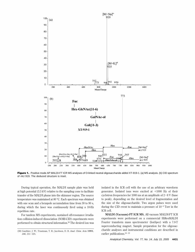
Figure 1. Positive mode AP MALDI-FT ICR MS analyses of O-linked neutral oligosaccharide alditol XT-919-1. (a) MS analysis. (b) CID spectrumof m/z 919. The deduced structure is inset.
Analytical Chemistry, Vol. 77, No. 14, July 15, 2005 4431
RESULTS AND DISCUSSIONAP MALDI-FT ICR MS of Labile Oligosaccharides. O-
Linked oligosaccharides. In general, the time scale between ionformation and ion detection in MALDI-FT ICR MS is relativelylong compared to MALDI-TOF, ranging from 0.3 to 30 s depend-ing on the desired resolution and the system’s pumping efficiency.The metastable ions formed in the MALDI process are subject toextensive fragmentation, particularly for labile compounds in FTICR MS. In oligosaccharide analysis, the fragment ions resultingfrom loss of specific residues and functional groups, such asfucose, sialic acid, sulfate, and phosphate groups, are commonlyobserved and can cause difficulty in assigning quasimolecular ions.
To investigate the application of AP MALDI-FT ICR MS to theanalysis of oligosaccharides, we tested the oligosaccharide alditolsreleased from the egg jelly coat of amphibians. Figure 1a showsthe AP MALDI-FT ICR MS of a neutral oligosaccharide alditolXT-919-1 (m/z 919.338) after HPLC separation as described in aprevious publication.19 The monosaccharide composition is cal-culated to be one fucose, two hexose, and two HexNAc based on
the theoretical mass (m/z 919.337). The base peak at m/z 919represents the sodium adduct while the peak at m/z 935 corre-sponds to the potassium adduct. The peaks at m/z 757 and 773are one hexose and one fucose less than the quasimolecular ions,respectively. One might assume that these ions are fragments dueto the decomposition of m/z 919; however, based on our experi-ence, the loss of fucose is always more abundant than the loss ofhexose due to the more labile nature of the fucose residuecompared to hexose. On the basis of this reasoning, we believethat the peaks at m/z 757 and 773 are not fragments of m/z 919,but minor components in this HPLC fraction. Therefore, APMALDI allows us to quickly assign the parent ions without theadditional step, for example, of doping with large alkali metals todecrease metastable decomposition.15
With the cooler ions produced by AP MALDI, there is aconcern that tandem MS, specifically CID, would not yieldabundant fragment ions. Figure 1b shows the CID mass spectrumof m/z 919. Fragment ions corresponding to cleavage of every
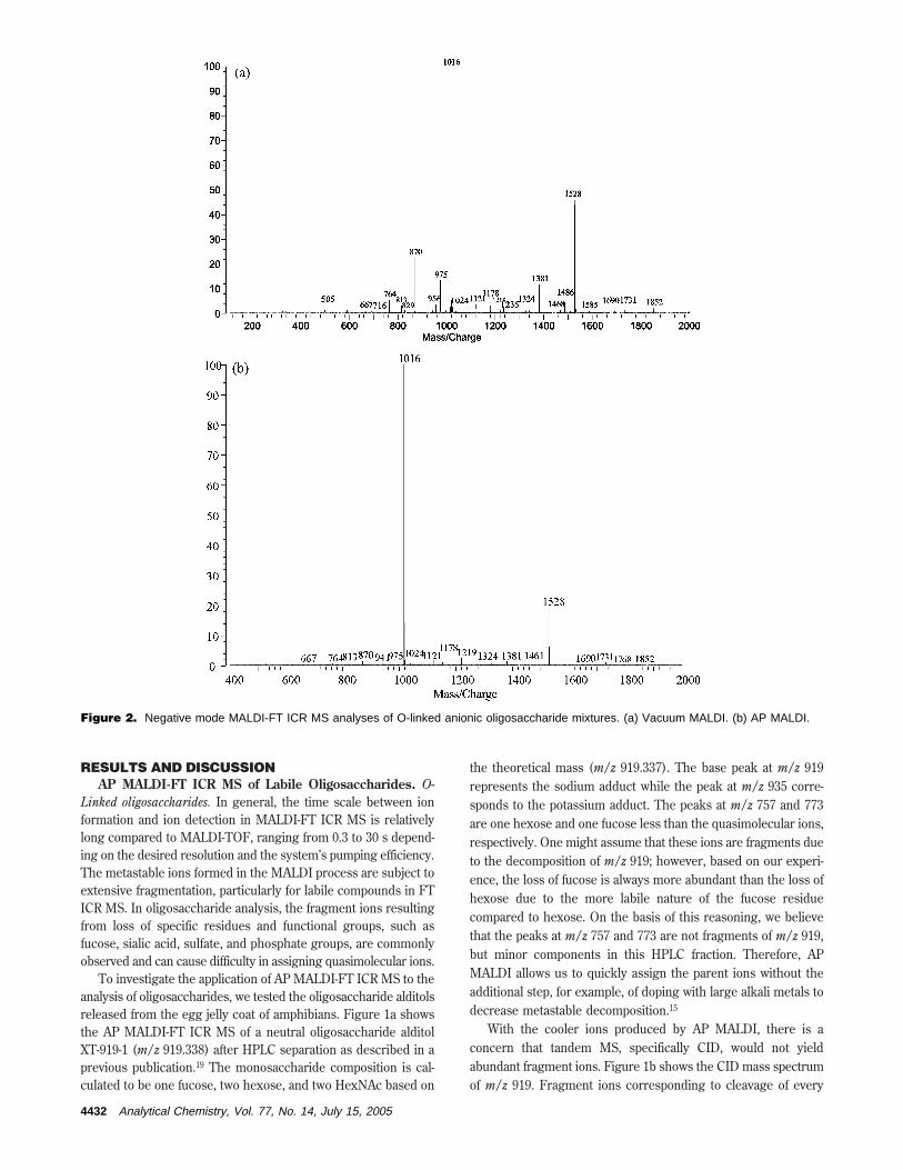
Figure 2. Negative mode MALDI-FT ICR MS analyses of O-linked anionic oligosaccharide mixtures. (a) Vacuum MALDI. (b) AP MALDI.
4432 Analytical Chemistry, Vol. 77, No. 14, July 15, 2005

glycosidic bond are observed. The losses of fucose (m/z 773) andhexose (m/z 757) from the quasimolecular ion indicate that fucoseand one of hexoses are present at the nonreducing ends. Thegroup of ions at m/z 611, 449, 431, 413, 408, and 390 arecharacteristic fragments of the known trisaccharide core,Gal(â1-3)[GlcNAc(â1-6)]GalNAc-ol.12,13 The ion at m/z 228is the terminal monosaccharide residue corresponding to[GalNAc-ol - H2O + Na]+. The inset structure is proposed basedon the CID fragmentation.
The lack of fragment ions in AP MALDI makes it ideal forrapid profiling of mixtures. Figure 2 shows the comparison ofvacuum MALDI and AP MALDI negative mode FT ICR MS of ananionic oligosaccharide mixture released from mucin-type glyco-proteins. The spectra are highly reproducible in both instruments.Repeat analyses of the same samples yield nearly identical spectra.Figure 2a is the mass spectrum produced through vacuum MALDIwith our best attempt at minimizing the fragmentation. Figure 2bis the mass spectrum of the same sample produced by AP MALDI.Both spectra show two major sulfated oligosaccharide componentspresent in the mixture corresponding to m/z 1016 and 1528, whosepresence are supported by HPLC.19 However, the fragment peaksat m/z 870 and 1381 are significantly reduced in AP MALDIspectra. These two peaks are fragments corresponding to the lossof fucose from each of the two species at m/z 1016 and 1528.Similarly, the peaks at m/z 505 and 1235, which are observed onlyin the vacuum MALDI spectra, are also fragments. This exampleillustrates the utility of AP MALDI in the analyses of anionic(sulfated) oligosaccharide mixtures.
Gangliosides. Gangliosides constitute a class of sialylatedglycosphingolipids that are particularly labile, often yielding theloss of sialic acid during ionization. They are often isolated asmixtures with minor structural variations. This class of compoundshas posed a significant challenge for mass spectrometry. Themetastable fragmentation is of particular concern and interfereswith the compositional determination of mixtures extracted frombiological samples. Costello et al. have employed “high-pressure”MALDI-FT ICR MS by introducing pulsed cooling gas into thesource during ionization to analyze the gangliosides.20,24 Theirresults nicely demonstrated minimal fragmentation as a result ofthe cooling effect of the collision gas during the ionization in avacuum MALDI. Figure 3a is a typical MALDI-FT ICR massspectrum of a monosialylated ganglioside (GM1) in the nega-tive mode. The base peak is the deprotonated sialic acid([Neu5Ac - H]-, m/z 290) produced by the loss of sialic acidfrom the quasimolecular ion. The quasimolecular ion [M - H]-
corresponds to m/z 1572.911 (theoretical m/z 1572.901) with thepeak at m/z 1545 corresponding to the low-mass homologue anddiffering from the high-mass homologue by a C2H4 unit due tothe ceramide moiety.24 Figure 3b shows the AP MALDI massspectra of GM1 in the negative mode. Only quasimolecular ionsare obtained with no evidence of fragmentation.
The vacuum MALDI-FT ICR MS of GT1b, a trisialylatedganglioside, in the negative mode (Figure 4a) does not yield thequasimolecular ions. Two sets of fragment ions are obtainedcorresponding to the loss of one and two sialic acids. The basepeak at m/z 563 corresponds to a species composed of two sialicacids while a lone sialic acid is observed at m/z 290. These smallion fragments point to the abundant loss of sialic acids during
the ionization process. Figure 4b shows the AP MALDI-FT ICRmass spectrum of GT1b. The base peaks are the intact molecularion [M - H]- (m/z 2155.091) and its lower homologue. Althoughthere are peaks that correspond to the sequential losses of sialicacids, m/z 1863.990 and 1572.891, it is more likely that they areimpurities. This notion is supported by the absence of m/z 290,which is the major fragmentation product of sialylated oligosac-charides in the negative mode.
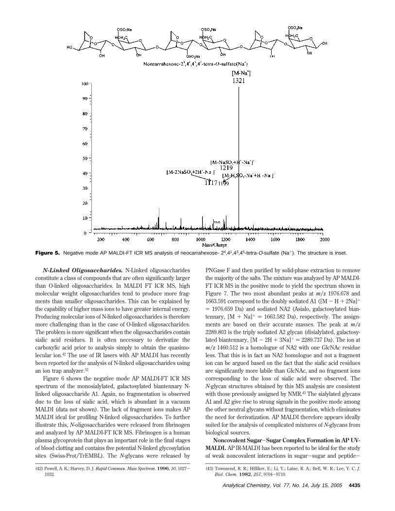
Carrageenans. Multiply sulfated oligosaccharides and polysac-charides are difficult to study by MALDI because of the labilenature of the sulfate groups.39 Furthermore, the tendency of themultiply sulfated groups to produce multiple anionic charges isnot favorable for analyses with MALDI, which has the tendencyof producing singly charged species. Carrageenans constitute aclass of oligosaccharides found in seaweed and consisting ofoligomers of sulfated residues. Neocarrahexose-2,441,43,45-tetra-O-sulfate (Na+) (monoisotopic mass 1344.051 Da), with four sulfategroups, does not yield the quasimolecular ion with vacuumMALDI (spectra not shown); however, AP MALDI yields thenegative mode mass spectrum of this compound with the quasi-molecular ion [M - Na+]- at m/z 1321.098 (Figure 5, structureinset).
(39) Harvey, D. J. Mass Spectrom. Rev. 1999, 18, 349-451.
Figure 3. Negative mode MALDI-FT ICR MS analyses of ganglio-side GM1. (a) Vacuum MALDI. (b) AP MALDI.
Analytical Chemistry, Vol. 77, No. 14, July 15, 2005 4433

The low-abundance ions at m/z 1219 and 1117 correspond tothe stepwise loss of 102 mass units from the molecular ions,possibly through fragmentation. These two peaks can be assignedas [M - NaSO3 + H+ - Na+]- and [M - 2NaSO3 + 2H+ - Na+]-,respectively. This fragmentation mechanism, proposed by Acklooet al.,40 is thought to occur first with the exchange of the Na+
with an H+ [X-OSO3Na f X-OSO3H] followed by the immediateloss of the SO3 group. According to those authors, ions such asm/z 1199 arise from a similar process whereby Na+/H+ exchangeis now followed by loss of H2SO4.
These results demonstrate the detection of the base peak ofmolecular ions with little fragmentation due to desulfation of this
tetrasulfated oligosaccharide. AP MALDI may provide even lessfragmentation than MALDI-TOF MS, where metastable decay ismitigated by the short detection time and the fact that thefragments retain the velocity of the precursor ions when frag-mentation occurs in the field-free region. Previous reports haveshown that the most intense peak in the MALDI-TOF-MSspectrum of this compound does not correspond to the molecularion, but to desulfated anions.41 An additional feature of the APMALDI FTICR MS is the lack of matrix clusters that tend topopulate the low-mass regions of nearly all MALDI-TOF massspectra.41
(40) Ackloo, S.; Terlouw, J. K.; Ruttink, P. A.; Burgers, P. C. Rapid Commun.Mass Spectrom. 2001, 15, 1152-1159.
(41) Fukuyama, Y.; Ciancia, M.; Nonami, H.; Cerezo, A. S.; Erra-Balsells, R.;Matulewicz, M. C. Carbohydr. Res. 2002, 337, 1553-1562.
Figure 4. Negative mode MALDI-FT ICR MS analyses of ganglioside GT1b (a) Vacuum MALDI. (b) AP MALDI.
4434 Analytical Chemistry, Vol. 77, No. 14, July 15, 2005
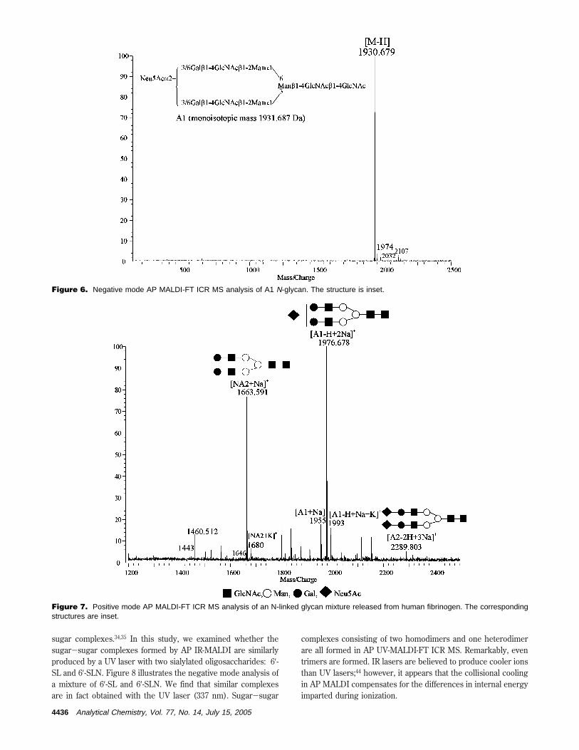
N-Linked Oligosaccharides. N-Linked oligosaccharidesconstitute a class of compounds that are often significantly largerthan O-linked oligosaccharides. In MALDI FT ICR MS, highmolecular weight oligosaccharides tend to produce more frag-ments than smaller oligosaccharides. This can be explained bythe capability of higher mass ions to have greater internal energy.Producing molecular ions of N-linked oligosaccharides is thereforemore challenging than in the case of O-linked oligosaccharides.The problem is more significant when the oligosaccharides containsialic acid residues. It is often necessary to derivatize thecarboxylic acid prior to analysis simply to obtain the quasimo-lecular ion.42 The use of IR lasers with AP MALDI has recentlybeen reported for the analysis of N-linked oligosaccharides usingan ion trap analyzer.32
Figure 6 shows the negative mode AP MALDI-FT ICR MSspectrum of the monosialylated, galactosylated biantennary N-linked oligosaccharide A1. Again, no fragmentation is observeddue to the loss of sialic acid, which is abundant in a vacuumMALDI (data not shown). The lack of fragment ions makes APMALDI ideal for profiling N-linked oligosaccharides. To furtherillustrate this, N-oligosaccharides were released from fibrinogenand analyzed by AP MALDI-FT ICR MS. Fibrinogen is a humanplasma glycoprotein that plays an important role in the final stagesof blood clotting and contains five potential N-linked glycosylationsites (Swiss-Prot/TrEMBL). The N-glycans were released by
PNGase F and then purified by solid-phase extraction to removethe majority of the salts. The mixture was analyzed by AP MALDI-FT ICR MS in the positive mode to yield the spectrum shown inFigure 7. The two most abundant peaks at m/z 1976.678 and1663.591 correspond to the doubly sodiated A1 ([M - H + 2Na]+
) 1976.659 Da) and sodiated NA2 (Asialo, galactosylated bian-tennary, [M + Na]+ ) 1663.582 Da), respectively. The assign-ments are based on their accurate masses. The peak at m/z2289.803 is the triply sodiated A2 glycan (disialylated, galactosy-lated biantennary, [M - 2H + 3Na]+ ) 2289.737 Da). The ion atm/z 1460.512 is a homologue of NA2 with one GlcNAc residueless. That this is in fact an NA2 homologue and not a fragmention can be argued based on the fact that the sialic acid residuesare significantly more labile than GlcNAc, and no fragment ionscorresponding to the loss of sialic acid were observed. TheN-glycan structures obtained by this MS analysis are consistentwith those previously assigned by NMR.43 The sialylated glycansA1 and A2 give rise to strong signals in the positive mode amongthe other neutral glycans without fragmentation, which eliminatesthe need for derivatization. AP MALDI therefore appears ideallysuited for the analysis of complicated mixtures of N-glycans frombiological sources.
Noncovalent Sugar-Sugar Complex Formation in AP UV-MALDI. AP IR-MALDI has been reported to be ideal for the studyof weak noncovalent interactions in sugar-sugar and peptide-
(42) Powell, A. K.; Harvey, D. J. Rapid Commun. Mass Spectrom. 1996, 10, 1027-1032.
(43) Townsend, R. R.; Hilliker, E.; Li, Y.; Laine, R. A.; Bell, W. R.; Lee, Y. C. J.Biol. Chem. 1982, 257, 9704-9710.
Figure 5. Negative mode AP MALDI-FT ICR MS analysis of neocarrahexose- 24,41,43,45-tetra-O-sulfate (Na+). The structure is inset.
Analytical Chemistry, Vol. 77, No. 14, July 15, 2005 4435
sugar complexes.34,35 In this study, we examined whether thesugar-sugar complexes formed by AP IR-MALDI are similarlyproduced by a UV laser with two sialylated oligosaccharides: 6′-SL and 6′-SLN. Figure 8 illustrates the negative mode analysis ofa mixture of 6′-SL and 6′-SLN. We find that similar complexesare in fact obtained with the UV laser (337 nm). Sugar-sugar
complexes consisting of two homodimers and one heterodimerare all formed in AP UV-MALDI-FT ICR MS. Remarkably, eventrimers are formed. IR lasers are believed to produce cooler ionsthan UV lasers;44 however, it appears that the collisional coolingin AP MALDI compensates for the differences in internal energyimparted during ionization.
Figure 6. Negative mode AP MALDI-FT ICR MS analysis of A1 N-glycan. The structure is inset.
Figure 7. Positive mode AP MALDI-FT ICR MS analysis of an N-linked glycan mixture released from human fibrinogen. The correspondingstructures are inset.
4436 Analytical Chemistry, Vol. 77, No. 14, July 15, 2005

Detection Sensitivity of Oligosaccharides by AP MALDI-FT ICR MS. Attomole detection limits have been reported forAP MALDI-ion trap MS of peptides using electrospray sampledeposition.45 For oligosaccharides, the analysis of branched andlabile sialylated oligosaccharides has been previously conductedwith nanomole analyte quantities by both AP UV-MALDI31 andAP IR-MALDI.27,33,34 Recently, the subpicomole MS analysis ofN-linked glycans by atmospheric pressure infrared laser ionizationfrom solution was performed on an ion trap mass spectrometer.32
To determine the detection limit of AP MALDI-FT ICR MS, astandard hexasaccharide, maltohexose, was used. Figure 9 showsthe AP MALDI-FT ICR mass spectrum resulting from analysis of20 fmol of maltohexose applied to the probe. The sodiated specieswas the only quasimolecular ion obtained. The expanded molec-ular ion peak clearly shows the isotope pattern with a signal-to-noise ratio of >10. Even at the threshold of detection, 2 ppm mass
accuracy was obtained with external calibration, and broadbandresolution of 30 800 (m/∆mfwhm) was attained.
CONCLUSIONAtmospheric pressure MALDI-FT ICR MS has been performed
on a number of oligosaccharides as well as O-linked and N-linkedglycan mixtures derived from biological samples. The oligosac-charides chosen for this study are particularly labile, containingfucoses, sialic acids, and sulfates. These oligosaccharides haveposed significant challenges to conventional MALDI mass spec-trometry due to their extensive fragmentation. The comparisonbetween AP MALDI and conventional MALDI reveals that APMALDI results in minimized fragmentation for both positive andnegative mode mass analysis of labile oligosaccharides. Thedifficulties in differentiating the molecular ions from the fragmentions often encountered in a vacuum MALDI-FT ICR MS areeliminated. In addition, no evidence of matrix cluster formationwas observed.
Due to the structural heterogeneity of glycans and glycocon-jugates in biological systems, samples purified for MS analysisfrequently represent complex mixtures, with components varying
(44) Berkenkamp, S.; Menzel, C.; Karas, M.; Hillenkamp, F. Rapid Commun.Mass Spectrom. 1997, 11, 1399-1406.
(45) Wei, H.; Nolkrantz, K.; Powell, D. H.; Woods, J. H.; Ko, M.-C.; Kennedy, R.T. Rapid Commun. Mass Spectrom. 2004, 18, 1193-1200.
Figure 8. Negative mode AP MALDI-FT ICR MS of 6′-SL and 6′-SLN mixture.
Figure 9. AP MALDI-FT ICR MS analysis of maltohexose (20 fmol deposited on the probe) with sodium dopant.
Analytical Chemistry, Vol. 77, No. 14, July 15, 2005 4437
in molecular weight by a few to several hundred mass units. Thecoupling of AP MALDI with FT ICR MS provides a powerful toolto obtain high mass accuracy and mass resolution analysis oncomplex carbohydrate analytes. The mass spectra yield readilyinterpretable molecular ion peaks with few or no fragment ionspresent. Therefore, AP MALDI-FT ICR MS has been demon-strated to be ideally suited for analysis of highly heterogeneousglycan and glycoconjugate mixtures.
ACKNOWLEDGMENTWe thank Dr. Jennifer Aguilan and Dr. Fabian Dayrit of Ateneo
de Manila University for providing the carrageenan samples.Funding provided by the National Science Foundation and theNational Institutes of Health is gratefully acknowledged.
Received for review January 3, 2005. Accepted April 21,2005.
AC050010O
4438 Analytical Chemistry, Vol. 77, No. 14, July 15, 2005