In-situ enrichment of phosphopeptides on MALDI plates modified by ambient ion landing
9
In-situ enrichment of phosphopeptides on MALDI plates modified by ambient ion landing Luká s Krásný, a,b Petr Pompach, a,c Martin Strohalm, a Veronika Obsilova, d Marcela Strnadová, a Petr Novák a,c and Michael Volný a,e * We report substantial in-situ enrichment of phosphopeptides in peptide mixtures using titanium and zirconium dioxide-coated matrix assisted laser desorption-ionization (MALDI) plates prepared by recently reported ambient ion landing deposition technique. The technique was able to modify four common materials currently used for MALDI targets (stainless steel, aluminum, indium-tin oxide glass and polymeric anchor chip). The structure of the deposited dioxide was investigated by electron micros- copy, and different surfaces were compared and discussed in this study. Two standard proteins were used to test the enrichment capabilities of modified MALDI plates: casein and in-vitro phosphorylated trehalase. The enrichment of casein tryptic digest resulted in identification of 20 phosphopeptides (including miscleavages). Trehalase was used as a suitable model of larger protein that provided more complex peptide mixture after the trypsin digestion. All four possible phosphorylation sites in trehalase were identified and up to seven phosphopetides were found (including methionine oxidations and miscleavages). Two different mass spectrometers, MALDI-Fourier transform ion cyclotron resonance (FTICR) and MALDI-time of flight, were used to detect the phosphopeptides from modified MALDI plates after the enrichment procedure. It was observed that the desorption- ionization phenomena on the modified surfaces are not critically influenced by the parameters of the different MALDI ion sources (e.g. different pressure, different extraction voltages), and thus the presence of dioxide layer on the standard MALDI plate does not significantly interfere with the main MALDI processes. The detection of phosphopeptides after the enrichment could be done by both instruments. Desorption electrospray ionization coupled to the FTICR was also tested, but, unlike MALDI, it did not provide satisfactory results. Copyright © 2012 John Wiley & Sons, Ltd. Supporting Information can be found in an online version of this article. Keywords: phosphopetides; MALDI; FTICR; enrichment; casein; trehalase Introduction Post-translational modifications of proteins are a late step in the protein biosynthesis and a key tool in the regulation of their func- tionality. [1] Protein phosphorylation, a covalent attachment of a phosphate group, was described more than 50 years ago [2] , and currently it is considered to be one of the most important signaling mechanisms in cells. Many cellular processes such as metabolism, transcription and differentiation are controlled by phosphorylation of serine, threonine, tyrosine (O-linked) or histidine (N-linked) residues. It is recognized that protein kinases and phosphatases mediate most of the signal transduction in eukaryotic cells [3] , and the misregulation of enzymes that control cellular phos- phorylation activities has been implicated in a wide range of diseases. [4–6] There are many, often complementary techniques, used in phosphoproteomics. [7–10] Mass spectrometry (MS), which is capable of fast protein sequencing, has also become a key method for investigation of protein phosphorylation. [8,9,11–14] Theoretical prediction of eukaryotic phosphorylation is still extremely difficult [15] , and its elucidation relies on experimental approaches. The detection of phosphorylated peptides is hin- dered by the fact that phosphorylation is a very dynamic event, and thus only a fraction of isolated protein molecules might be phosphorylated. [11] This results in low occurrence of phospho- peptides and in their low stoichiometry with respect to the corresponding non-modified analogs. Moreover, addition of a phosphate group, which increases the peptide acidity, depresses the ionization efficiency in positive ion mode due to the difficulty to form positive ions by protonation. This leads to phosphopeptides signal suppression in MS. [11] Harder ionization approaches can result in cleavage of the phosphate group bond from the peptide backbone and in the loss of the information about phosphorylation status. Isolation, extra chromatographic separation or prior enrichment of phosphopeptides from a pep- tide mixture after enzymatic digest of a protein are often used to overcome the above described difficulties. [8,11,16,17] Many techniques for phosphopeptide enrichment compatible with MS were reported and used. [16] They can be roughly divided into several groups, although the classification serves mainly a * Correspondence to: Michael Volny, Department of Chemistry, University of Washington, Seattle, Washington, 98195-1700, USA. E-mail: [email protected] The first two authors, LK and PP, contributed equally to this work. a Institute of Microbiology of the ASCR, v.v.i., Videnska 1083, Prague 4, 142 20, Czech Republic b Institute of Chemical Technology, Technická 5, Prague, 16628, Czech Republic c Department of Biochemistry, Faculty of Science, Charles University, Hlavova 8, 128 40 Prague 2, Czech Republic d Institute of Physiology of the ASCR, v.v.i., Videnska 1083, Prague 4, 142 20, Czech Republic e Department of Chemistry, University of Washington, Seattle, Washington, 98195-1700, USA J. Mass Spectrom. 2012, 47, 1294–1302 Copyright © 2012 John Wiley & Sons, Ltd. Research article Received: 15 June 2012 Revised: 25 July 2012 Accepted: 3 August 2012 Published online in Wiley Online Library (wileyonlinelibrary.com) DOI 10.1002/jms.3081 1294
-
Upload
washington -
Category
Documents
-
view
1 -
download
0
Transcript of In-situ enrichment of phosphopeptides on MALDI plates modified by ambient ion landing
Research article
Received: 15 June 2012 Revised: 25 July 2012 Accepted: 3 August 2012 Published online in Wiley Online Library
(wileyonlinelibrary.com) DOI 10.1002/jms.3081
1294
In-situ enrichment of phosphopeptides onMALDI plates modified by ambient ion landingLuká�s Krásný,a,b Petr Pompach,a,c Martin Strohalm,a Veronika Obsilova,d
Marcela Strnadová,a Petr Nováka,c and Michael Volnýa,e*
We report substantial in-situ enrichment of phosphopeptides in peptide mixtures using titanium and zirconium dioxide-coatedmatrix assisted laser desorption-ionization (MALDI) plates prepared by recently reported ambient ion landing deposition
technique. The technique was able to modify four commonmaterials currently used for MALDI targets (stainless steel, aluminum,indium-tin oxide glass and polymeric anchor chip). The structure of the deposited dioxide was investigated by electron micros-copy, and different surfaces were compared and discussed in this study. Two standard proteins were used to test the enrichmentcapabilities of modified MALDI plates: casein and in-vitro phosphorylated trehalase. The enrichment of casein tryptic digestresulted in identification of 20 phosphopeptides (including miscleavages). Trehalase was used as a suitable model of largerprotein that provided more complex peptide mixture after the trypsin digestion. All four possible phosphorylation sites intrehalase were identified and up to seven phosphopetides were found (including methionine oxidations and miscleavages).Two different mass spectrometers, MALDI-Fourier transform ion cyclotron resonance (FTICR) and MALDI-time of flight, were usedto detect the phosphopeptides frommodifiedMALDI plates after the enrichment procedure. It was observed that the desorption-ionization phenomena on the modified surfaces are not critically influenced by the parameters of the different MALDI ion sources(e.g. different pressure, different extraction voltages), and thus the presence of dioxide layer on the standard MALDI plate doesnot significantly interfere with the main MALDI processes. The detection of phosphopeptides after the enrichment could be doneby both instruments. Desorption electrospray ionization coupled to the FTICR was also tested, but, unlike MALDI, it did notprovide satisfactory results. Copyright © 2012 John Wiley & Sons, Ltd.Supporting Information can be found in an online version of this article.
Keywords: phosphopetides; MALDI; FTICR; enrichment; casein; trehalase
* Correspondence to: Michael Volny, Department of Chemistry, University ofWashington, Seattle, Washington, 98195-1700, USA. E-mail: [email protected]
The first two authors, LK and PP, contributed equally to this work.
a Institute of Microbiology of the ASCR, v.v.i., Videnska 1083, Prague 4, 142 20,Czech Republic
b Institute of Chemical Technology, Technická 5, Prague, 16628, Czech Republic
c Department of Biochemistry, Faculty of Science, Charles University, Hlavova 8,128 40 Prague 2, Czech Republic
d Institute of Physiology of the ASCR, v.v.i., Videnska 1083, Prague 4, 142 20,Czech Republic
e Department of Chemistry, University of Washington, Seattle, Washington,98195-1700, USA
Introduction
Post-translational modifications of proteins are a late step in theprotein biosynthesis and a key tool in the regulation of their func-tionality.[1] Protein phosphorylation, a covalent attachment of aphosphate group, was described more than 50 years ago[2], andcurrently it is considered to be one of the most important signalingmechanisms in cells. Many cellular processes such as metabolism,transcription and differentiation are controlled by phosphorylationof serine, threonine, tyrosine (O-linked) or histidine (N-linked)residues. It is recognized that protein kinases and phosphatasesmediate most of the signal transduction in eukaryotic cells[3],and the misregulation of enzymes that control cellular phos-phorylation activities has been implicated in a wide range ofdiseases.[4–6] There are many, often complementary techniques,used in phosphoproteomics.[7–10] Mass spectrometry (MS),which is capable of fast protein sequencing, has also become akey method for investigation of protein phosphorylation.[8,9,11–14]
Theoretical prediction of eukaryotic phosphorylation is stillextremely difficult[15], and its elucidation relies on experimentalapproaches. The detection of phosphorylated peptides is hin-dered by the fact that phosphorylation is a very dynamic event,and thus only a fraction of isolated protein molecules might bephosphorylated.[11] This results in low occurrence of phospho-peptides and in their low stoichiometry with respect to thecorresponding non-modified analogs. Moreover, addition of aphosphate group, which increases the peptide acidity,depresses the ionization efficiency in positive ion mode due to
J. Mass Spectrom. 2012, 47, 1294–1302
the difficulty to form positive ions by protonation. This leads tophosphopeptides signal suppression in MS.[11] Harder ionizationapproaches can result in cleavage of the phosphate group bondfrom the peptide backbone and in the loss of the informationabout phosphorylation status. Isolation, extra chromatographicseparation or prior enrichment of phosphopeptides from a pep-tide mixture after enzymatic digest of a protein are often used toovercome the above described difficulties.[8,11,16,17]
Many techniques for phosphopeptide enrichment compatiblewith MS were reported and used.[16] They can be roughly dividedinto several groups, although the classification serves mainly a
Copyright © 2012 John Wiley & Sons, Ltd.

MALDI plates modification by ion landing
129
practical purpose and the very basic principles behind theenrichment mechanisms often overlap. Based on a recentreview,[16] five main groups can be distinguished: immobilizedmetal affinity chromatography (IMAC),[18] reversible covalentbinding,[19] metal oxide affinity chromatography,[20,21] enrich-ment on magnetic beads[17] and in-situ enrichment on matrixassisted laser desorption-ionization (MALDI) plates.[22,23]
This report demonstrates advantages of a new way of surfacemodification for enrichment prior to MALDI ionization, which isbased on an ambient ion landing procedure.[24,25] In-situ enrich-ment on MALDI plates allows handling of very small volumes ofsamples with relatively smaller losses, because it only requiresminimal manipulation of the sample: after the small volume ofa sample is loaded on a MALDI plate, it is incubated, washedand the matrix is added.[16,26,27] More efficient sample handlingreduces the sample loss, which is crucial for analysis of low abun-dant phosphopeptides. The other advantage is the possibility tocouple the prespotted target with LC systems which allows highsample throughput due to automation of the process. The ideathat chemical modification and functionalization of a MALDIplate could be used to allow for affinity interaction between theanalyte and the plate appeared in the early 1990s.[22,23,28] Platesmodified by IMAC functional moieties were the first MALDI platesused for phosphopeptide enrichment (such plates are commer-cially available; the technology was reviewed for instance byTang[29]), and other concepts were later introduced. Blacken[30]
utilized a reactive landing of zirconium(IV)-n-propoxide solutionsin 1-propanol for modification of stainless steel surface by zirco-nium oxide. Reactive landing,[31,32] a modification of establishedsoft landing technology,[33–35] was found to produce durablefunctionalized surfaces for selective phosphopeptide captureand desorption-ionization by MALDI.[30] Enrichment factors onthe order of 20–90 were achieved for several monophosphory-lated peptides relative to abundant non-phosphorylated pep-tides in tryptic digests. In the follow-up report,[26] it was shownthat reactive landing can also produce modifications of MALDIplates by titanium and hafnium oxides. Reactively landed layersof titanium and zirconium dioxide were described to be mechan-ically and chemically very stable, but because reactive landingrequires specially designed vacuum instruments and suffers fromrelatively low efficiency,[36] the approach was not practical. How-ever, modification of MALDI plates for phosphopeptide enrichmenthas become a more common approach, and several differenttechniques for MALDI plate modification by metal oxides (mostlyby titanium dioxide) were reported.[27,37–48] A recent review,[49]
which describes how MALDI target plates can be used beyondsimple sample support as functional parts of novel analyticalprotocols, refers to this analytical approach as ‘lab-on-plate’.
Reactive landing seemed impractical for the purposes ofMALDI plate functionalization until the recent report from theCooks group[24] introduced a technique called ambient softlanding, which is a modification of vacuum soft and reactivelanding. Ambient ion soft landing is a process in which poly-atomic ions are deposited from air onto a surface underatmospheric pressure. Ions generated by electrospray ionizationare passed pneumatically through a heated metal drying tube(straight or coiled), their ion polarity can be selected using iondeflectors and the dry selected ions are soft-landed onto aselected surface. Unlike the corresponding vacuum soft landingexperiment, where ions are soft-landed within a mass spectrom-eter,[50–54] here, the ions to be deposited are selected throughthe choice of a compound that gives predominant ionic species
J. Mass Spectrom. 2012, 47, 1294–1302 Copyright © 2012 John
upon ambient ionization and drying in the tube. Dry ions pro-duced during the ambient soft landing experiment can bemanipulated by simple and relatively crude electrostatic opticsprior to surface deposition. This allows for the selective transferof ions, and not neutrals, to the surface24, although the precisionof the ion beam focusing and bending is far from the quality thatcan be obtained in low pressure environment where the ionoptics components can utilize longer mean free path of the ions.We now present an on-target phosphopeptide enrichmentmethod, which uses the ambient electrospray ion landing as asurface deposition and modification technique. It allows forspecific enrichment and direct analysis by MALDI-MS of phos-phorylated peptides. We show that ambient ion landing canproduce deposition of spots of thin film of titanium, zirconiumor hafnium dioxide on any material common for MALDI plates(stainless steel, aluminum, indium-tin oxide (ITO) glass, conduc-tive polymer used for the disposable polymeric anchor chips(PAC)). The method was tested using casein, which is the mostcommon model of phosphorylated proteins and in-vitro phos-phorylated trehalase, a glycoside hydrolase enzyme, whichserved for further demonstration of the technique capabilitiesbecause this relatively large protein provides a much morecomplex mixture of peptides after tryptic digestion than casein.
Experimental
MALDI plates
The four common types of MALDI plates, stainless steel, aluminum,ITO-coated glass and conductive polymer used for Bruker’sproprietary PAC, were selected for modification (all purchasedfrom Bruker Daltonics). Titanium(IV) n-propoxide, zirconium(IV)n-propoxide and hafnium(IV) n-butoxide used for surface modifica-tion were all purchased from Sigma-Aldrich.
Preparation of modified MALDI plates
The apparatus for ambient ion landing (Fig. 1) was built on-site; thelocal machine shop assembled the heating tube (id= 4mm) andthe moving stage. A modified electrosonic spray source was usedas the electrospray emitter. The sprayer was based on a standard1/16 stainless steel T element (Swagelok) and was assembled usingstandard Swagelok fittings, with stainless steel, graphite and PEEKferrules. Fused silica was used for tubing. Electrospray was estab-lished between the sprayer and the grounded heated tube byapplying a high voltage (3 kV) on the metallic part of the syringe.The syringe pump (KD Scientific) was used for pumping thesolutions into the sprayer. The stagewith vertically mountedMALDIplate was moved by two stepper electromotors controlled by aremote console. The high voltage (3–4 kV) of the opposite polaritywith respect to the spray voltage was connected by a simple clipto the MALDI plate. One high voltage power supply was purchasedfrom Spellman, the other was taken from disassembled ZAB EQsector mass spectrometer (VG Analytical) and controlled by a smallvoltage (0–10V) from a laboratory power supply.
Proteins
10 mg of casein (Sigma-Aldrich) was separated using SDS-PAGEon gradient gel (Invitrogen). After the separation, the gel wasstained using CBB-R250, and the gel part with visualized proteinspot was cut out and sliced to small cubic blocks (~1mm3).Individual fractions were then de-stained reduced (50mM
Wiley & Sons, Ltd. wileyonlinelibrary.com/journal/jms
5

Figure 1. Apparatus for surface modification by ambient ion landing.Panel A: Overall view of the apparatus with power supplies. Panel B: Schemaof the apparatus (voltage polarities are in positive ion mode). The heatedtube is kept on ground, while the slit mask electrode is on high negativepotential. The motion of the target is automated for array deposition.
L. Krásný et al.
1296
Tris(2-carboxyethyl)phosphine, pH 8), alkylated (55mM iodoacetamide, pH 8) and digested overnight by trypsin (Promega). Theresulting peptides and phosphopeptides were extracted fromgel, desalted on reversed phase material (microtrap, Michrom),dried and subsequently used for performance evaluation of themodified surface as a standard test mixture.The trehalase was recombinantly prepared in E. coli and in-vitro
phosphorylated by incubation with 80 units of PKA (Promega)per milligram of recombinant protein for 2 h at 30 �C and thenovernight at 4 �C in the presence of 20mM magnesium chlorideand 0.75mM ATP. Phosphorylated trehalase was reduced,alkylated and digested directly without SDS-PAGE separation.
In-situ incubation and washing protocol
The washing and incubation procedure was based on the onepreviously reported by Eriksson.[27] Briefly, MALDI plates withspots of deposited dioxides were rinsed by isopropanol and H2Oand let to dry out. Digested samples were mixed in 1:1 ratio withDHB buffer (20mg/ml DHB in 50% ACN/0.1% TFA), loaded ontospots (1 ml of mixture per spot) and let to dry out at room temper-ature. After the samples were dried, the MALDI plate was washedby DHB buffer and 50% ACN and bound phosphopeptides werereleased by 1.5 ml of 0.1% NH4OH, pH 11 and let to dry out. Spotswere overlaid by 0.5 ml of DHB/PA matrix (DHB 20mg/ml in 50%ACN/1% phosphoric acid), and mass spectra were acquired.
MS
MALDI-TOF-MS. Time-of-flight (TOF) mass spectra were acquiredin positive reflector mode using an Ultraflex III TOF mass
wileyonlinelibrary.com/journal/jms Copyright © 2012 Joh
spectrometer (Bruker Daltonics) with SmartBeam laser. MS datawere acquired in the m/z range between 500 and 4500 byaveraging signals from 2000 consecutive laser shots using targetrandom-walk movement. Prior to each data acquisition, externalcalibration was conducted using a peptide calibration standard(Bruker Daltonics). The FlexAnalysis 3.0 program (Bruker Daltonics)was used to process the data.
MALDI-Fourier transform ion cyclotron resonance (FTICR)-MS.High-resolution mass spectra were acquired using an APEX Ultra9.4 T mass spectrometer equipped with Apollo II ESI/MALDI ionsource (Bruker Daltonics). Mass spectra were acquired in a positiveion mode with 512 k data points using a SmartBeam 200Hz laser.Results were analyzed using Data Analysis 4.0 (Bruker Daltonics)and by open source program mMass.[55]
Electron microscopy
Dry samples with a deposited dioxide layer were sputter-coatedwith gold in a Polaron E5100 sputter-coater (Quorum TechnologiesLtd). The samples were analyzed by Aquasem scanning electronmicroscope (Tescan) at 15 kV using secondary electron detector.
Results and discussion
Modification of MALDI plates
Zirconium(IV)-n-propoxide was previously used for zirconiumdioxide thin film preparation by atmospheric-pressure electro-spray combined with thermal decomposition on a hot substratesurface[56–58] as reviewed.[59] The previous report,[26] as well asour own experiments, showed that simple electrospray deposi-tion of dissolved alkoxides had not resulted in a formation of sta-ble oxide spots on surfaces that would be usable as MALDI sub-strates for enrichment even if the surface was heated. Ambientlanding differs from the previous atmospheric-pressure techni-ques in that decomposition is achieved by collisions and heatingin the tube and subsequent deposition of dry ions on the surfaceat room temperature. Recently published work claimed that it ispossible to dry ions produced by electrospray by a rapid heatinginside an externally heated tube and manipulate the dry ions byelectrostatic ion optics at ambient conditions.[24] Our experi-ments (data not shown) as well as the previous report fromCooks lab24 demonstrated that the beam can be bent by theelectrostatic potential and thus that the species impacting thesurface are ions and not neutrals. Other reports confirmed suc-cessful fragmentation of ions at ambient conditions by externalheating.[60,61] We utilized the external heating tube not only todry charged droplets produced by electrospray but also to ther-mally decompose the titanium, zirconium and hafnium alkoxidesto their respective dioxides inside the heated tube. The chargeddioxide molecules were transported through a slit electrodeand attracted to the surface to be modified by an oppositeelectrostatic potential. Electrospray mass spectra of titaniumand zirconium alkoxides were previously reported.[62] It wasshown that moisture-sensitive alkoxides can be investigated byelectrospray MS and their electrospray-generated ions are stableat low cone voltages. However, fragmentation appeared withharder ionization conditions (high voltages on the ion optics ele-ments inside the ion source). Thus, we concluded that transitionfrom alkoxides to dioxides in our apparatus is due to the heatingand collisions in the tube and not a result of hydrolysis (same de-composition to dioxides was not observed if the tube was kept at
n Wiley & Sons, Ltd. J. Mass Spectrom. 2012, 47, 1294–1302

MALDI plates modification by ion landing
room temperature). This is important for the enrichment functionof the deposited dioxides because it is known that the method ofpreparation strongly influences their chromatographic proper-ties. For instance, hydrolytically precipitated zirconium dioxideis amorphous, and unlike its crystalline forms, it is not consideredto be a useful stationary phase in chromatography.[63] The appa-ratus used for the deposition of titanium, zirconium and hafniumdioxides by ambient ion landing (Fig. 1A and B) consisted of anelectrosonic spray ion source, a heated drying tube, mask andcollecting electrode (vertically mounted MALDI plate to be mod-ified) that can be positioned by an automated manipulator. It wasoperated as follows: Alkoxide solution (50mM in isopropanol)was pumped by a syringe pump (250 ml/h) and electrosprayedthrough the heated tube (120 �C) using nitrogen as a nebulizinggas (25 psi). The electrospray voltage was 3 kV. The distancebetween the collecting electrode and the end of the heated tubewas kept at 2–4mm. Spots of porous metal oxide with diameterof 3mm were deposited in less than 10min each, the collectingelectrode was kept at high negative potential of �3 kV. Auto-mated positioning of the collecting electrode could be used todeposit a pattern of multiple spots (Fig. 1C), and thus multiplesamples can be enriched and analyzed during a single MALDIanalysis. This procedure allowed deposition of a stable layer oftitanium, zirconium and hafnium dioxides. It was reportedthat phosphopeptide enrichment on hafnium dioxide wasachieved with monolytic material.[64] However, in accordancewith the report by Blacken,[26] hafnium dioxide deposited byion landing in our experiments showed very poor enrichmentproperties, and it was not used in further work. Interestingly, haf-nium dioxide, if prepared as a specific mesoporous nanomaterial,has been described to have better specificity for phosphopeptideenrichment than titanium dioxide.[65] This could indicate that theinability to prepare a hafnium-based material for phosphopep-tide enrichment by ion landing is due to the unsuitable structureof deposited hafnium dioxide and not because of the molecularor atomic properties of hafnium (such as larger atomic radiuscompared to titanium or zirconium). Titanium and zirconium
Figure 2. Spectra of tryptic digest of phosphorylated casein before (Paneldeposited by ambient ion landing on ITO glass. Spectra were acquired using
J. Mass Spectrom. 2012, 47, 1294–1302 Copyright © 2012 John
dioxides prepared by ambient ion landing did not show anydetectable differences in enrichment properties. The majorobserved difference was more frequent oxidation of methionineon zirconium dioxide surfaces.
Enrichment experiments
Casein is a very common and relatively hydrophobic phospho-protein with no disulfide bridges. It is often used as a proof ofprinciple analyte for testing new techniques for investigation ofphosphopeptides, and it serves as a benchmark in the literaturefor comparing MALDI-MS-based techniques for phosphopeptidedetermination. Casein consists of four main proteins: aS1, aS2,b and k; in the present work, we investigated phosphorylationsites of aS1 and b mixture.
Figure 2 compares a MALDI-FTICR-MS spectrum of an in-gel,tryptic digest of casein (Panel A) to a spectrum of the same pep-tide mixture after the enrichment procedure was performed onITO glass modified by titanium dioxide (Panel B). Twenty phos-phopeptides that were detected after enrichment are numberedin the spectrum. The accurate mass measurement is sufficient forpeptide identification in this case, especially because phosphogroup introduces a significant and characteristic mass defect.Thus, we included MSMS spectra in supporting material only(Fig. S7 and S8). Tables 1 and 2 show observed m/z values andprimary amino acid sequences of identified phosphopeptides.In addition, the associated supporting material contains TablesS1 and S2 that compare the results with selected literaturereports. All compared literature reports are based on theenrichment of casein tryptic peptides using metal oxide as theaffinity surface. However, they differ in the deposition techniqueused for modification of MALDI plate surface. Although thedifferences in MS performance (different mass spectrometerswere used in these reports) and in incubation and washingprotocols can play a role to some extent, it is clear that surfacesprepared by ambient ion landing performed very well comparedto other surfaces and present a new robust platform for
A) and after (Panel B) enrichment procedure on titanium dioxide spotsMALDI-FTICR mass spectrometer.
Wiley & Sons, Ltd. wileyonlinelibrary.com/journal/jms
1297

Table 1. Phosphopeptides identified in the tryptic digest of betacasein
Label inFig. 2B
m/z (charge) Sequence(number of phosphorylations)
1 1031.42440 (2+) FQSEEQQQTEDELQDK (1�p)
2 1561.63686 (2+) RELEELNVPGEIVESLSSSEESITR (4�p)
9 2061.82930 (1+) FQ-pS-EEQQQTEDELQDK (1�p)
10 2556.08588 (1+) FQ-pS-EEQQQTEDELQDKIHPF (1�p)
17 2966.19345 (1+) ELEELNVPGEIVESLSSSEESITR (4�p)
18 2962.37302 (1+) RELEELNVPGEIVELSSEESITR (2�p)
19 3042.27711 (1+) RELEELNVPGEIVESLSSSEESITR (3�p)
20 3122.26464 (1+) RELEELNVPGEIVESLSSSEESITR (4�p)
Table 2. Phosphopeptides identified in the tryptic digest of alpha S1casein
Label inFig. 2B
m/z (charge) Sequence(number of phosphorylations)
3 1660.79372 (1+) VPQLEIVPNSAEER (1�p)
4 1832.85326 (1+) YLGEYLIVPNSAEER (1�p)
5 1863.71919 (1+) DIGSESTEDQA-oxM-EDIK (1�p)
6 1927.69375 (1+) DIGSESTEDQAMEDIK (2�p)
7 1943.68659 (1+) DIGSESTEDQA-oxM-EDIK (2�p)
8 1951.95287 (1+) YKVPQLEIVPNSAEER (1�p)
11 2559.94371 (1+) Q(deamidated)-oxM-
EAESISSSEEIVPNSVEQK (3�p)
12 2576.99571 (1+) Q-oxM-EAESISSSEEIVPNSVEQK (3�p)
13 2639.93462 (1+) Q(deamidated)-oxM-
EAESISSSEEIVPNSVEQK (4�p)
14 2656.95603 (1+) Q-oxM-EAESISSSEEIVPNSVEQK (4�p)
15 2719.90729 (1+) Q(deamidated)-oxM-
EAESISSSEEIVPNSVEQK (5�p)
16 2736.92778 (1+) Q-oxM-EAESISSSEEIVPNSVEQK (5�p)
L. Krásný et al.
1298
phosphopeptide enrichment. The number of identified phospho-petides in casein digest is the same as in the previous study,[65]
which also utilized a high-resolution FTICR mass spectrometer,although with electrospray ionization. That study used morelaborious off-line phosphopeptide enrichment on mesoporouszirconium dioxide nanomaterial, and it concluded that properlyprepared zirconium dioxide exhibits better enrichment proper-ties than commercially available products.While testing the newly developed technique using casein is
advantageous because of the relative simplicity of the peptidemixture and because the results can be compared with previ-ously published studies, it is also necessary to test the techniqueusing a more complex model. Thus, trehalase, a glycoside hydro-lase enzyme that catalyzes the conversion of trehalose toglucose, was chosen as a model of a larger protein. Trehalase pro-vides a more complex mixture of peptides after trypsin digestthan casein. Prior to the experiment, trehalase was in-vitro phos-phorylated by proteinkinase A. Four serines (S154, S155, S194 andS217; the entire sequence is included as Fig. S3 in the supportingmaterial) were preferentially phosphorylated. The same washing/enrichment protocol used for casein was applied for trehalase.Figure 3 shows an example of MALDI mass spectra before(Panel A) and after the enrichment procedure (Panel B) measured
wileyonlinelibrary.com/journal/jms Copyright © 2012 Joh
by FTICR (MALDI-TOF spectra can be found in the supportingmaterial as Fig. S4). The spectra after enrichment procedure aresignificantly simpler while the intensity of the ion at nominalm/z = 3118 (base peak) corresponding to the doubly phosphory-lated peptide dropped by approximately 10%. This shows thatthere are no significant losses due to the in-situ enrichmentprocedure. Figure 4 summarizes observed results on differentsurfaces using MALDI-FTICR mass spectrometer (Panel A) andby MALDI-TOF instrument (Panel B). All m/z values in the bargraph correspond to phosphopeptides. Their sequences and therelative intensities, which were used to plot Fig. 4, are includedin the supporting material (Figs. S5 and S6). Relative intensity ofphosphopeptides (with respect to base peak) increased rapidlyafter the enrichment, and the ratio of identified phosphopeptidesto all peptides also increased significantly after the enrichment asshown also in Tables S5 and S6. Enrichment procedure resulted inlower complexity of MS spectra and simplification of datainterpretation, which should reduce the number of false positivehits. Interestingly, methionine oxidation is much more intense onzirconium dioxide than on titanium dioxide, which means thatthe previously described methionine oxidation[30] is catalyzedby the surface and both dioxides exhibit different catalyticactivities. The ratio of identified phosphopeptides to all peptidesalso increased significantly after enrichment (Tables S5 and S6),which again shows that it is easier to identify phosphopeptidesin the mass spectra after enrichment and also that spectra areeasier to interpret after the enrichment.
Ionization of enriched phosphopeptides by desorptionelectrospray
Using the previously described desorption electrospray (DESI)stage[66] compatible with FTICR-MS instrument, we also tested thepossibility of using DESI instead ofMALDI for detection of phospho-peptides after the in-situ enrichment. The main advantage of usingDESI instead of MALDI would be the ability to produce multiplycharged peptides that could be further fragmented by electroncapture or electron transfer dissociation. DESI was previously usedto detect peptides directly from TLC plates.[67] The titanium andzirconium oxides should theoretically represent good DESIsubstrates because previous reports focused on DESI improve-ments, and mechanisms showed that porous structure, lowconductivity and relative hydrophobicity are all advantageous inDESI.[68,69] However, no usable signal was obtained in DESI-MS ofphosphopeptides from ITO or metal substrates modified bytitanium or zirconium dioxide.
Surface properties
A more detailed characterization of the surfaces is currently inprogress, but electron microscopy images (Fig. 5) already indicatethat the ambient ion landing produced a porous ‘spongy’ layer ofdeposited dioxide that exhibits high surface area. Comparableenrichment results were obtained using the more traditionallow pressure MALDI source on the TOF instrument as in the highpressure MALDI source used on FTICR instrument, althoughbetter performance of FTICR instrument resulted in highernumber of identified phosphopeptides. The fact that the differentextraction voltages in both MALDI ion sources did not play a rolemeans that the dioxide layer does not dramatically influence theconductivity of the MALDI surface. The oxidation of methionine,especially on zirconium dioxide surfaces, must be considered forspectra interpretation.
n Wiley & Sons, Ltd. J. Mass Spectrom. 2012, 47, 1294–1302
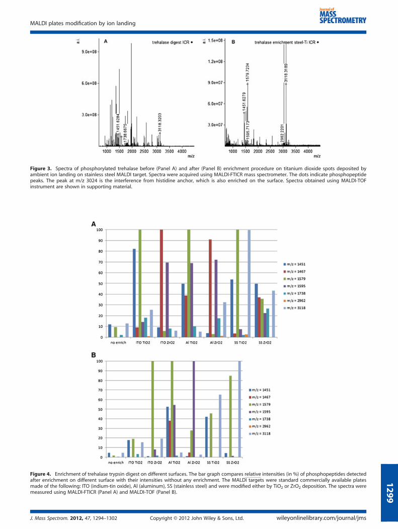
Figure 3. Spectra of phosphorylated trehalase before (Panel A) and after (Panel B) enrichment procedure on titanium dioxide spots deposited byambient ion landing on stainless steel MALDI target. Spectra were acquired using MALDI-FTICR mass spectrometer. The dots indicate phosphopeptidepeaks. The peak at m/z 3024 is the interference from histidine anchor, which is also enriched on the surface. Spectra obtained using MALDI-TOFinstrument are shown in supporting material.
Figure 4. Enrichment of trehalase trypsin digest on different surfaces. The bar graph compares relative intensities (in %) of phosphopeptides detectedafter enrichment on different surface with their intensities without any enrichment. The MALDI targets were standard commercially available platesmade of the following: ITO (indium-tin oxide), Al (aluminum), SS (stainless steel) and were modified either by TiO2 or ZrO2 deposition. The spectra weremeasured using MALDI-FTICR (Panel A) and MALDI-TOF (Panel B).
MALDI plates modification by ion landing
J. Mass Spectrom. 2012, 47, 1294–1302 Copyright © 2012 John Wiley & Sons, Ltd. wileyonlinelibrary.com/journal/jms
1299

Figure 5. Structure of the zirconium dioxide surface deposited using ambient ion landing visualized by electron microscopy at different magnifications.
L. Krásný et al.
1300
Conclusions
The presented procedure for deposition of titanium, zirconium andhafnium dioxides by ambient ion landing on MALDI plates is simple,inexpensive and reasonably fast. It allows modification of fourcommonmaterials used for manufacturingMALDI plates. TheMALDIsurfaces modified by titanium or zirconium dioxides were success-fully used for in-situ enrichment of phosphopeptides from a mixtureof tryptic peptides. The technique was tested on two standardproteins: casein and in-vitro phosphorylated trehalase. Significantenrichment was achieved for both tryptic digests. Electron micros-copy images showed that the ambient ion landing produces aporous layer of deposited dioxide that exhibits high surface area.The laser desorption-ionization process from the oxide-modifiedMALDI surfaces is not critically dependent on the type of the ionsource. Enrichment was achieved using the vacuum MALDI sourceon the TOF instrument as well as using the higher pressure MALDIion source used on FTICR instrument. Comparison with previouslypublished results shows that ambient ion landing is an advantageoustechnique for MALDI surface modification by metal oxides.
Acknowledgements
The work was supported mainly by the Czech Science Foundation(Projects P206/10/P018 and P207/11/0455). The authors thankDr. Oldrich Benada for electron microscopy measurements,
wileyonlinelibrary.com/journal/jms Copyright © 2012 Joh
Vaclav Kobliha for building the apparatus for automated ambiention deposition and Eva Macakova for the purification of trehalase(all from the Academy of Sciences of the CR). University ofWashington graduate student Joelle Rolfs is greatly acknowledgedfor proofreading the manuscript. M.V.’s research was supported bya Marie Curie International Reintegration Grant within the seventhEuropean Community Framework Program. Additional financingwas provided by the Ministry of Education, Youth and Sports ofthe Czech Republic (ME10013), by the Czech Science Foundation(204/09/H084 and P206/12/1150), by an Institutional researchconcept of the Institute of Microbiology (RVO61388971) and byan Institutional research concept of the Institute of Physiology(RVO:67985823).
Supporting information
Supporting Information can be found in an online version of thisarticle.
References[1] P. Cohen. The origins of protein phosphorylation. Nat. Cell Biol.
2002, 4, E127.[2] G. Burnett, E. P. Kennedy. The enzymatic phosphorylation of
proteins. J. Biol. Chem. 1954, 211, 969.[3] G. Manning, D. B. Whyte, R. Martinez, T. Hunter, S. Sudarsanam. The
protein kinase complement of the human genome. Science 2002,298, 1912.
n Wiley & Sons, Ltd. J. Mass Spectrom. 2012, 47, 1294–1302

MALDI plates modification by ion landing
13
[4] E. Lopez, I. Lopez, A. Ferreira, J. Sequi. Clinical and technical phos-phoproteomic research. Proteome Sci. 2011, 9, 20.
[5] M. F. Moran, J. F. Tong, P. Taylor, R. M. Ewing. Emerging applicationsfor phospho-proteomics in cancer molecular therapeutics. BBA-Rev.Cancer. 2006, 1766, 230.
[6] T. Hunter. Signaling - 2000 and beyond. Cell 2000, 100, 113.[7] M. Hjerrild, S. Gammeltoft. Phosphoproteomics toolbox: Computa-
tional biology, protein chemistry and mass spectrometry. FEBS Lett.2006, 580, 4764.
[8] M. J. Chalmers, W. Kolch, M. R. Emmett, A. G. Marshall, H. Mischak.Identification and analysis of phosphopeptides. J. Chromatogr. BAnalyt. Technol. Biomed. Life Sci. 2004, 803, 111.
[9] B. Bodenmiller, L. N. Mueller, M. Mueller, B. Domon, R. Aebersold.Reproducible isolation of distinct, overlapping segments of thephosphoproteome. Nat. Methods 2007, 4, 231.
[10] R. D. Tuerk, Y. Auchli, R. F. Thali, R. Scholz, T. Wallimann, R. A. Brunisholz,D. Neumann. Tracking and quantification of (32)p-labeled phospho-peptides in liquid chromatography matrix-assisted laser desorption/ionization mass spectrometry. Anal. Biochem. 2009, 390, 141.
[11] M. Mann, S. E. Ong, M. Gronborg, H. Steen, O. N. Jensen, A. Pandey.Analysis of protein phosphorylation using mass spectrometry: Deci-phering the phosphoproteome. Trends Biotechnol. 2002, 20, 261.
[12] G. L. Corthals, R. Aebersold, D. R. Goodlett. Identification ofphosphorylation sites using microimmobilized metal affinity chroma-tography. Methods Enzymol. 2005, 405, 66.
[13] W. D. Lehmann. Analysis of phosphopeptides by mass spectrometry.Protein Phosphorylation Analysis by Electrospray Mass Spectrometry: aGuide to Concepts and Practice, 2010, 18.
[14] F. J. Wang, C. X. Song, K. Cheng, X. N. Jiang, M. L. Ye, H. F. Zou.Perspectives of comprehensive phosphoproteome analysis usingshotgun strategy. Anal. Chem. 2011, 83, 8078.
[15] B. Trost, A. Kusalik. Computational prediction of eukaryotic phos-phorylation sites. Bioinformatics 2011, 27, 2927.
[16] J. D. Dunn, G. E. Reid, M. L. Bruening. Techniques for phosphopep-tide enrichment prior to analysis by mass spectrometry. MassSpectrom. Rev. 2010, 29, 29.
[17] G. H. Han, M. L. Ye, H. F. Zou. Development of phosphopeptideenrichment techniques for phosphoproteome analysis. Analyst2008, 133, 1128.
[18] J. Porath, J. Carlsson, I. Olsson, G. Belfrage. Metal chelate affinitychromatography, a new approach to protein fractionation. Nature1975, 258, 598.
[19] H. L. Zhou, J. D. Watts, R. Aebersold. A systematic approach to theanalysis of protein phosphorylation. Nat. Biotechnol. 2001, 19, 375.
[20] M. R. Larsen, T. E. Thingholm, O. N. Jensen, P. Roepstorff, T. J. D.Jorgensen. Highly selective enrichment of phosphorylated peptidesfrom peptide mixtures using titanium dioxide microcolumns. Mol.Cell. Proteomics 2005, 4, 873.
[21] A. Leitner. Phosphopeptide enrichment using metal oxide affinitychromatography. Trac-Trend. Anal. Chem. 2010, 29, 177.
[22] T. W. Hutchens, T. T. Yip. New desorption strategies for the mass-spectrometric analysis of macromolecules. Rapid Commun. MassSpectrom. 1993, 7, 576.
[23] D. I. Papac, J. Hoyes, K. B. Tomer. Direct analysis of affinity-boundanalytes by maldi/tof ms. Anal. Chem. 1994, 66, 2609.
[24] A. K. Badu-Tawiah, C. Wu, R. G. Cooks. Ambient ion soft landing. Anal.Chem. 2011, 83, 2648.
[25] R. D. Espy, A. Badu-Tawiah, R. G. Cooks. Analysis and modification ofsurfaces using molecular ions in the ambient environment. Curr.Opin. Chem. Biol. 2011, 15, 741.
[26] G. R. Blacken, M. Volny, M. Diener, K. E. Jackson, P. Ranjitkar,D. J. Maly, F. Turecek. Reactive landing of gas-phase ions as a toolfor the fabrication of metal oxide surfaces for in situ phosphopep-tide enrichment. J. Am. Soc. Mass Spectrom. 2009, 20, 915.
[27] A. Eriksson, J. Bergquist, K. Edwards, A. Hagfeldt, D. Malmstrom,V. A. Hernandez. Optimized protocol for on-target phosphopeptideenrichment prior to matrix-assisted laser desorption-ionization massspectrometry using mesoporous titanium dioxide. Anal. Chem. 2010,82, 4577.
[28] A. H. Brockman, R. Orlando. Probe immobilized affinity-chromatographymass-spectrometry. Anal. Chem. 1995, 67, 4581.
[29] N. Tang, P. Tornatore, S. R. Weinberger. Current developments inseldi affinity technology. Mass Spectrom. Rev. 2004, 23, 34.
[30] G. R. Blacken, M. Volny, T. Vaisar, M. Sadilek, F. Turecek. In situenrichment of phosphopeptides on maldi plates functionalized by
J. Mass Spectrom. 2012, 47, 1294–1302 Copyright © 2012 John
reactive landing of zirconium(iv)-n-propoxide ions. Anal. Chem.2007, 79, 5449.
[31] M. Volny, W. T. Elam, B. D. Ratner, F. Turecek. Preparative soft andreactive landing of gas-phase ions on plasma-treated metal surfaces.Anal. Chem. 2005, 77, 4846.
[32] M. Volny, W. T. Elam, B. D. Ratner, F. Turecek. Enhanced in-vitro bloodcompatibility of 316 l stainless steel surfaces by reactive landingof hyaluronan ions. J. Biomed. Mater. Res. B Appl. Biomater. 2007,80B, 505.
[33] R. G. Cooks, S. C. Jo, J. Green. Collisions of organic ions at surfaces.Appl. Surf. Sci. 2004, 231, 13.
[34] V. Franchetti, B. H. Solka, W. E. Baitinger, J. W. Amy, R. G. Cooks. Softlanding of ions as a means of surface modification. Int. J. MassSpectrom. Ion Processes 1977, 23, 29.
[35] B. Gologan, J. R. Green, J. Alvarez, J. Laskin, R. G. Cooks. Ion/surfacereactions and ion soft-landing. Phys. Chem. Chem. Phys. 2005, 7,1490.
[36] M. Volny, F. Turecek. High efficiency in soft landing of biomolecularions on a plasma-treated metal surface: Are double-digit yieldspossible? J. Mass Spectrom. 2006, 41, 124.
[37] H. Bi, L. Qiao, J.-M. Busnel, V. Devaud, B. Liu, H. H. Girault. Tio(2)printed aluminum foil: Single-use film for a laser desorption/ioniza-tion target plate. Anal. Chem. 2009, 81, 1177.
[38] A. Eriksson, J. Bergquist, K. Edwards, A. Hagfeldt, D. Malmstrom,V. A. Hernandez. Mesoporous tio(2)-based experimental layout foron-target enrichment and separation of multi- and monopho-sphorylated peptides prior to analysis with matrix-assisted laserdesorption-ionization mass spectrometry. Anal. Chem. 2011, 83, 761.
[39] T. Hoang, U. Roth, K. Kowalewski, C. Belisle, K. Steinert, M. Karas.Highly specific capture and direct maldi ms analysis of phosphopep-tides by zirconium phosphonate on self-assembled monolayers.Anal. Chem. 2010, 82, 219.
[40] P. Kouvonen, E.-M. Rainio, V. Suni, P. Koskinen, G. L. Corthals. Enrich-ment and sequencing of phosphopeptides on indium tin oxidecoated glass slides. Mol. Biosyst. 2011, 7, 1828.
[41] R. Liu, J. F. Liu, X. X. Zhou, G. B. Jiang. Cysteine modified smallligament au nanoporous film: An easy fabricating and highlyefficient surface-assisted laser desorption/ionization substrate. Anal.Chem. 2011, 83, 3668.
[42] Z. Lu, J. Duan, L. He, Y. Hu, Y. Yin. Mesoporous tio(2) nanocrystalclusters for selective enrichment of phosphopeptides. Anal. Chem.2010, 82, 7249.
[43] M.-L. Niklew, U. Hochkirch, A. Melikyan, T. Moritz, S. Kurzawski,H. Schlueter, I. Ebner, M. W. Linscheid. Phosphopeptide screeningusing nanocrystalline titanium dioxide films as affinity matrix-assisted laser desorption ionization targets in mass spectrometry.Anal. Chem. 2010, 82, 1047.
[44] L. Qiao, H. Bi, J.-M. Busnel, M. Hojeij, M. Mendez, B. Liu, H. H. Girault.Controlling the specific enrichment of multi-phosphorylated pep-tides on oxide materials: Aluminium foil as a target plate for laserdesorption ionization mass spectrometry. Chem. Sci. 2010, 1, 374.
[45] H. Sonderegger, C. Rameshan, H. Lorenz, F. Klauser, M. Klerks, M. Rainer,R. Bakry, C. W. Huck, G. K. Bonn. Surface-assisted laser desorption/ionization-mass spectrometry using tio(2)-coated steel targets for theanalysis of small molecules. Anal. Bioanal. Chem. 2011, 401, 1963.
[46] F. Torta, M. Fusi, C. S. Casari, C. E. Bottani, A. Bachi. Titanium dioxidecoated maldi plate for on target analysis of phosphopeptides.J. Proteome Res. 2009, 8, 1932.
[47] H. Wang, J. C. Duan, Q. Cheng. Photocatalytically patterned tio(2)arrays for on-plate selective enrichment of phosphopeptides anddirect maldi ms analysis. Anal. Chem. 2011, 83, 1624.
[48] X. Zou, D. Liu, L. Zhong, B. Yang, Y. Lou, B. Hu, Y. Yin. Highly specificcapture and direct maldi-ms analysis of phosphorylated peptidesusing novel multifunctional chitosan-gma-ida-fe (iii) nanosphere.Anal. Bioanal. Chem. 2011, 401, 1251.
[49] P. L. Urban, A. Amantonico, R. Zenobi. Lab-on-a-plate: Extending thefunctionality of maldi-ms and ldi-ms targets. Mass Spectrom. Rev.2011, 30, 435.
[50] O. Hadjar, P. Wang, J. H. Futrell, Y. Dessiaterik, Z. H. Zhu, J. P. Cowin,M. J. Iedema, J. Laskin. Design and performance of an instrument forsoft landing of biomolecular ions on surfaces. Anal. Chem. 2007, 79,6566.
[51] J. Laskin, P. Wang, O. Hadjar. Soft-landing of peptide ions ontoself-assembled monolayer surfaces: An overview. Phys. Chem. Chem.Phys. 2008, 10, 1079.
Wiley & Sons, Ltd. wileyonlinelibrary.com/journal/jms
01

L. Krásný et al.
1302
[52] W. Peng, M. Goodwin, Z. Nie, E. Al. Ion soft landing using a rectilinearion trap mass spectrometer. Anal. Chem. 2008, 80, 6640.
[53] Q. Song, S. Smith, L. Gao, E. Al. Mass selection of ions from beamsusing waveform isolation in radiofrequency quadrupoles. Anal.Chem. 2009, 81, 1833.
[54] M. Volny, W. Elam, A. Branca, E. Al. Preparative soft and reactivelanding of multiply charged protein ions on a plasma-treated metalsurface. Anal. Chem. 2005, 77, 4890.
[55] M. Strohalm, D. Kavan, P. Novak, M. Volny, V. Havlicek. Mmass 3: Across-platform software environment for precise analysis of massspectrometric data. Anal. Chem. 2010, 82, 4648.
[56] C. H. Chen, M. H. J. Emond, E. M. Kelder, B. Meester, J. Schoonman.Electrostatic sol-spray deposition of nanostructured ceramic thinfilms. J. Aerosol Sci. 1999, 30, 959.
[57] T. Nguyen, E. Djurado. Deposition and characterization of nanocrys-talline tetragonal zirconia films using electrostatic spray deposition.Solid State Ion. 2001, 138, 191.
[58] D. Perednis, O. Wilhelm, S. E. Pratsinis, L. J. Gauckler. Morphologyand deposition of thin yttria-stabilized zirconia films using spraypyrolysis. Thin Solid Films 2005, 474, 84.
[59] A. Jaworek. Electrospray droplet sources for thin film deposition.J. Mater. Sci. 2007, 42, 266.
[60] L. S. Eberlin, Y. Xia, H. Chen, R. G. Cooks. Atmospheric pressurethermal dissociation of phospho- and sulfopeptides. J. Am. Soc. MassSpectrom. 2008, 19, 1897.
[61] H. Chen, L. S. Eberlin, R. G. Cooks. Neutral fragment mass spectra viaambient thermal dissociation of peptide and protein ions. J. Am.Chem. Soc. 2007, 129, 5880.
wileyonlinelibrary.com/journal/jms Copyright © 2012 Joh
[62] T. Lover, W. Henderson, G. A. Bowmaker, J. M. Seakins, R. P. Cooney.Electrospray mass spectrometry of highly moisture-sensitive metalalkoxides. J. Mater. Chem. 1997, 7, 1553.
[63] J. Nawrocki, M. P. Rigney, A. Mccormick, P. W. Carr. Chemistry ofzirconia and its use in chromatography. J. Chromatogr. A 1993,657, 229.
[64] J. G. Rivera, Y. S. Choi, S. Vujcic, T. D. Wood, L. A. Colon. Enrichment/isolation of phosphorylated peptides on hafnium oxide prior to massspectrometric analysis. Analyst 2009, 134, 31.
[65] C. A. Nelson, J. R. Szczech, C. J. Dooley, Q. G. Xu, M. J. Lawrence,H. Y. Zhu, S. Jin, Y. Ge. Effective enrichment and mass spectrometryanalysis of phosphopeptides using mesoporous metal oxide nano-materials. Anal. Chem. 2010, 82, 7193.
[66] J. Pol, V. Vidova, G. Kruppa, V. Kobliha, P. Novak, K. Lemr, T. Kotiaho,R. Kostiainen, V. Havlicek, M. Volny. Automated ambientdesorption-ionization platform for surface imaging integrated witha commercial fourier transform ion cyclotron resonance mass spec-trometer. Anal. Chem. 2009, 81, 8479.
[67] S. P. Pasilis, V. Kertesz, G. J. Van Berkel, M. Schulz, S. Schorcht. Usinghptlc/desi-ms for peptide identification in 1d separations of trypticprotein digests. Anal. Bioanal. Chem. 2008, 391, 317.
[68] D. R. Ifa, N. E. Manicke, A. L. Rusine, R. G. Cooks. Quantitative analysisof small molecules by desorption electrospray ionization massspectrometry from polytetrafluoroethylene surfaces. Rapid Commun.Mass Spectrom. 2008, 22, 503.
[69] M. Volny, A. Venter, S. A. Smith, M. Pazzi, R. G. Cooks. Surface effectsand electrochemical cell capacitance in desorption electrosprayionization. Analyst 2008, 132, 525.
n Wiley & Sons, Ltd. J. Mass Spectrom. 2012, 47, 1294–1302