Site Directed Mutagenesis, Expression and Enzymatic Studies ...
70
Georgia State University Georgia State University ScholarWorks @ Georgia State University ScholarWorks @ Georgia State University Chemistry Theses Department of Chemistry 7-17-2009 Site Directed Mutagenesis, Expression and Enzymatic Studies of Site Directed Mutagenesis, Expression and Enzymatic Studies of the 60 kDa Human HIV-TAT 1 Interactive Protein, TIP60 the 60 kDa Human HIV-TAT 1 Interactive Protein, TIP60 Emilia N. Elangwe Follow this and additional works at: https://scholarworks.gsu.edu/chemistry_theses Recommended Citation Recommended Citation Elangwe, Emilia N., "Site Directed Mutagenesis, Expression and Enzymatic Studies of the 60 kDa Human HIV-TAT 1 Interactive Protein, TIP60." Thesis, Georgia State University, 2009. https://scholarworks.gsu.edu/chemistry_theses/21 This Thesis is brought to you for free and open access by the Department of Chemistry at ScholarWorks @ Georgia State University. It has been accepted for inclusion in Chemistry Theses by an authorized administrator of ScholarWorks @ Georgia State University. For more information, please contact [email protected].
-
Upload
khangminh22 -
Category
Documents
-
view
3 -
download
0
Transcript of Site Directed Mutagenesis, Expression and Enzymatic Studies ...

Georgia State University Georgia State University
ScholarWorks @ Georgia State University ScholarWorks @ Georgia State University
Chemistry Theses Department of Chemistry
7-17-2009
Site Directed Mutagenesis, Expression and Enzymatic Studies of Site Directed Mutagenesis, Expression and Enzymatic Studies of
the 60 kDa Human HIV-TAT 1 Interactive Protein, TIP60 the 60 kDa Human HIV-TAT 1 Interactive Protein, TIP60
Emilia N. Elangwe
Follow this and additional works at: https://scholarworks.gsu.edu/chemistry_theses
Recommended Citation Recommended Citation Elangwe, Emilia N., "Site Directed Mutagenesis, Expression and Enzymatic Studies of the 60 kDa Human HIV-TAT 1 Interactive Protein, TIP60." Thesis, Georgia State University, 2009. https://scholarworks.gsu.edu/chemistry_theses/21
This Thesis is brought to you for free and open access by the Department of Chemistry at ScholarWorks @ Georgia State University. It has been accepted for inclusion in Chemistry Theses by an authorized administrator of ScholarWorks @ Georgia State University. For more information, please contact [email protected].

SITE DIRECTED MUTAGENESIS, EXPRESSION AND ENZYMATIC STUDIES OF THE
60 kDa HUMAN HIV-TAT 1 INTERACTIVE PROTEIN, TIP60
by
EMILIA N. ELANGWE
Under the Direction of Dr. Yujun George Zheng Ph.D.
ABSTRACT
Tip60 is a 60 kDa nuclear protein which exists in three isoforms, belongs to the MYST/HAT
family of proteins and was discovered after its interaction with the Human HIV-1 Tat. As a
nuclear protein, Tip60 can act as a coactivator or repressor. To understand the HAT action of
Tip60, two possible catalytic models exist; the ping-pong and the ternary complex formation
models. In correlation with the exploration of HAT catalytic action, mutations of a Cys to Ala
and a Glu to Gln on Esa1 (yeast homolog of Tip60 and MYST/HAT prototype), was reported to
show wild type-like and decreased acetylating properties, respectively. In this work, Tip60 HAT
action was explored. In Tip60, the Cys in the active site is important for acetylation of the H4(1-
20) substrate and the Glu showed semi loss in acetylating the H4(1-20) peptide substrate. These
data highlight a unique mechanism of Tip60 catalysis.

INDEX WORDS: Tip60, Site-directed mutagenesis, HATs, MYST family HATs, Histones, Chromatin, Chromatin modification, Post-translational modification, Tip60 catalysis, HAT assay, His-tagged Protein Expression, Histone acetylation and HAT inhibitors
SITE DIRECTED MUTAGENESIS, EXPRESSION, AND STEADY-STATE KINETIC
STUDIES OF THE HUMAN HIV-TAT 1 INTERACTIVE PROTEIN, TIP60
by
EMILIA N. ELANGWE
A Thesis Submitted in Partial Fulfillment of the Requirements for the Degree of
Master of Science
in the College of Arts and Sciences
Georgia State University
2009

Copyright by Emilia N. Elangwe
2009

SITE DIRECTED MUTAGENESIS, EXPRESSION, AND STEADY-STATE KINETIC
STUDIES OF THE HUMAN HIV-TAT 1 INTERACTIVE PROTEIN, TIP60
by
EMILIA N. ELANGWE
Committee Chair: Dr. Yujun G. Zheng Committee: Dr. Giovanni Gadda Dr. Aimin Liu
Electronic Version Approved:
Office of Graduate Studies College of Arts and Sciences Georgia State University August 2009

iv
DEDICATION
I dedicate this thesis to my parents, my late mom, Christine Singui Elouti, who passed
away on June 21st, 2005 and to my father whom I’ll forever be thankful. They both instilled in
me the spirit of perseverance, positivity and hard work. To my sisters, Mary, Patricia, Susana and
Johana (AKA Joey) and my brother Irenous Elangwe, thank you for your encouragement and
support both physically and spiritually. To MohammedAli Khokhar, thank you for your support.

v
ACKNOWLEDGEMENTS
I want to first and foremost thank my advisor, Dr. George Zheng for giving me the
opportunity to join his research group. During this time, I was assigned projects which taught me
valuable lessons as well as gave me great experience. I would also like to thank my committee
members for accepting to take part in this wonderful step in my life especially, Dr. Giovanni
Gadda who directed me towards Dr. Zheng’s research group. Next, I would like to thank the
Georgia State University Department of Chemistry.
I would also like to thank members of the Zheng lab, past and present especially, Jiang
Wu who is my partner in Tip60 crime. I would also like to thank the NIH, Georgia Cancer
Coalition Distinguished Cancer Scholar program and the Georgia State University Research
Initiation program for funding this research.
Utmost, I would like to thank my family, my sisters, Mary, Patricia, Susana, and Johana
and my brother Irenous Elangwe for all their support and assistance. Special thanks and shout to
my father Romanus Elangwe to whom I am and will forever be grateful to for giving me the
opportunity to come here to have a better education.

vi
TABLE OF CONTENTS
ACKNOWLEDGEMENTS ............................................................................................................ v
LIST OF TABLES ......................................................................................................................... ix
LIST OF FIGURES ........................................................................................................................ x
CHAPTER 1: INTRODUCTION ................................................................................................... 1
1.0 Post-translational modification of histones by histone acetyltransferases (HATs) .................. 3
1.1 MYST family HATs ................................................................................................................. 6
1.2 Tip60 as a MYST Family HAT ................................................................................................ 8
1.3 Involvement of Tip60 in nuclear receptor activity and prostate cancer .................................. 10
1.4 Structure and catalytic activity of Tip60 ................................................................................. 13
1.5 Questions to be asked about this work .................................................................................... 15
CHAPTER 2: MATERIALS AND METHODS .......................................................................... 16
2.0 Chemicals and abbreviations .................................................................................................. 16
2.1 Enzymes .................................................................................................................................. 17
2.2 Kit ........................................................................................................................................... 17
2.3 Cell culture media and supplements ....................................................................................... 17
2.4 Bacteria cells ........................................................................................................................... 18
2.5 Oligonucleotides ..................................................................................................................... 18
2.6 Buffers..................................................................................................................................... 18
2.7 Instruments .............................................................................................................................. 20
2.8 Methods in molecular biology ................................................................................................ 21
2.8a Site-Directed Mutagenesis using polymerase chain reaction (PCR) .................................... 21
2.8b DNA amplification check in 1 % agarose gel Electrophoresis at 200 V .............................. 22

vii
2.8c Transformation of XL1-Blue competent cells ...................................................................... 22
2.8d Isolation of DNA plasmid ..................................................................................................... 23
2.8e Measurement of DNA concentration .................................................................................... 24
2.8f DNA sequencing .................................................................................................................... 24
2.8g Transformation of BL21(DE3) competent cells ................................................................... 24
2.8h Nickel-NTA Histidine-tagged protein expression & purification ........................................ 25
2.8i Radioactive HAT assay ......................................................................................................... 26
CHAPTER 3: SITE-DIRECTED MUTAGENESIS .................................................................... 28
3.0 Site-Directed Mutagenesis using polymerase chain reaction (PCR) ...................................... 28
3.1 Measurement of DNA concentration ...................................................................................... 30
3.2 DNA sequencing ..................................................................................................................... 31
CHAPTER 4: PROTEIN EXPRESSION ..................................................................................... 33
4.0 IPTG induction for protein expression……………………………………………………....33
4.1 Nickel-NTA Histidine-tagged protein expression assay......................................................... 34
CHAPTER 5: ENZYMATIC STUDY ......................................................................................... 36
5.0 Radioactive HAT assay........................................................................................................... 37
5.1 Initial enzymatic characterization …...………………………………………………………38
5.2 Future kinetic study of Tip60………………………………………………………………...40
CHAPTER 6: A DUAL-MODE FLUORESCENCE STRATEGY FOR SCREENING HAT
MODULATORS ........................................................................................................................... 42
6.0 Using the classical radioactive way to characterize HATs ..................................................... 42
6.1 Using immunoblotting to characterize HAT inhibitors .......................................................... 42
6.2 Using FRET and anisotropy measurements to characterize HAT inhibitors .......................... 43

viii
6.3 Expression of Dabcyl-labeled p300 protein …………………………………………………44
6.4 Fluorescence Measurement ………………………………………………………………….46
CHAPTER 7: CONCLUSION ..................................................................................................... 49
REFERENCES ............................................................................................................................. 51

ix
LIST OF TABLES
Table 1: PCR sample preparation for site directed mutagenesis………………………………...21
Table 2: PCR program setup for site directed mutagenesis……………………………………...22
Table 3: Sample preparation for radioactive HAT assay………………………………………...26
Table 4: HAT assay reaction time…………………………………………………………….....27
Table 5: Sample preparation for Tip60 autoacetylation…………………………………………38
Table 6: Tip60Cys369Ala enzyme kinetics reaction times……………………………………...39
Table 7: Tip60 wt. and Tip60Glu403Gln enzyme kinetics reaction times………………………39

x
LIST OF FIGURES
Figure 1: The epigenetic control of cell function............................................................................ 5
Figure 2: The reversible acetylation mechanism of the nucleosome .............................................. 6
Figure 3: The various isoforms of Tip60 that have been identified .............................................. 10
Figure 4: The anti-oncogenic (green) and pro-oncogenic (pink) connections of Tip60 ............... 12
Figure 5: The domain structure of Tip60 ...................................................................................... 13
Figure 6: Catalytic mechanism by ternary complex formation. ................................................... 14
Figure 7: Catalytic mechanism using the ping pong technique. ................................................... 14
Figure 8: Scheme of HAT radioactive assay ................................................................................ 27
Figure 9: pET15b vector map………………....…………...…………………………………….30 Figure 10: Agarose gel-electrophoresis to confirm success of mutation after PCR. .................... 30
Figure 11: Result of the NCBI Blast alignment of the sequencing results obtained after the
Tip60Glu403Gln and Tip60Cys369Ala mutations ....................................................................... 32
Figure 12: 12 % SDS-Page for wt, TipCys369Ala and TipGlu403Gln mutations ....................... 35
Figure 13: Phosphorimaging of Tip60 H4(1-20) histone acetylation and autoacetylation ........... 37
Figure 14: Plot of Product concentration, µM vs Time for Tip60……………………………….40
Figure 15: Schematic showing acetylation products via fluorescent intensity changes ............... 43
Figure 16: 12 % SDS-PAGE for p300 protein expression ……………………………………...44
Figure 17: 12 % SDS-PAGE for Dabcyl-labeled p300 …………………………………………44
Figure 18: FRET changes of LysCoAMca ……………………………………………………...46
Figure 19: Fluorescence anisotropy changes of LysCoAMca …………………………………..47

1
CHAPTER 1
INTRODUCTION
Site directed mutagenesis is a widely used scientific technique first described in 1978 by
Michael Smith (Hutschison, et al., 1978, Smith, 1993). This method allows for the introduction
or induction of point directed mutations to a known oligonucleotide (wild type) in order to
understand the effects of the mutation(s) on a particular biomolecule. Also, this point directed
mutation at a defined site of an oligonucleotide requires a circular biomolecule called a plasmid
inserted in a vector with the features of interest. The mutations could either be done by
insertion/substitution of an amino acid or by the deletion of a sequence of interest (Carter, P.,
1986). The most common technique for this mutation is the PCR based site-directed
mutagenesis. In this technique, an oligonucleotide complementary to part of a single stranded
DNA template, but containing an internal mismatch, is used to direct the mutation during the
thermal cycle (Carter, P, 1986). Before site directed mutagenesis becomes an option during
research, it is imperative to understand the DNA structure.
DNA is a 6 feet long polymer of repeating nucleic acid bases found in the nucleus of most
eukaryotes. This structure was referred to as a salt with ‘an angle of 36° between adjacent
residues in the same chain, so that the structure repeats after 10 residues on each chain, that is
after 34 Å (Watson and Crick, 1953). DNA is a double stranded biomolecule which contains the
genetic material of most organisms. It exists in many forms of which three are commonly
referred to (A-DNA, B-DNA and Z-DNA forms). The B-form DNA is the most common under
normal cellular conditions. Within cells, DNA is organized into structures known as
chromosomes. Chromosomes are nuclear structures that consist of DNA and proteins.
Chromosomes contain many genes; regulatory elements like the proteins bound to DNA that

2
control DNA function and other nucleotide sequences. In humans and other eukaryotes,
chromosomes are packaged by proteins into a structure known as chromatin.
Chromatin is a packaged structure of about 6 feet long of DNA material found in the nucleus
of most eukaryotic genomes. As mentioned earlier, it is composed of DNA and proteins in which
the proteins are mainly histones. Histones are small proteins that contain a high amount of basic
amino acids, mainly arginine and lysine, and facilitate the packaging of DNA in the nucleus
(Kuo and Allis, 1998; Wolffe, A.P., 2001). After numerous researches and studies, it is believed
that chromatin structure plays a role in most nuclear processes (gene regulation) which include
transcription, DNA replication and repair, mitosis and apoptosis (Cheung et al., 2000). An
understanding of the gene regulation role of chromatin could give major leads in therapeutic drug
targets against modern killers like cancer and HIV. Also, after intensive research, it was reported
that most of the proteins contained on chromatin are histones. These histones can be modified in
many different ways in order to suit the purpose of the research which leads to regulation of
chromatin activity. Taken together, chromatin bears pivotal epigenetic information for
development and maintenance of genomic integrity (Fischle, W., 2008).
So what are histones and what are they made of? Histones are the main protein components
of chromatin on which DNA is wrapped around that play major roles in chromatin functioning
and gene regulation. There are six different histones that have been identified: H1, H2A, H2B,
H3, H4 and H5, all bundled up into the histone core (H2A, H2B, H3 and H4, which are highly
conserved in most eukaryotes) and a linker (H1 and H5) (Jasencakova et al., 2000). Two of the
core histones, H2B and H4, assemble in the form of an octomer to constitute a main eukaryotic
nuclear structure known as a nucleosome. These histones are a main target for chromatin
regulation because their N-terminal is rich with lysines (K) and arginines (R) which are

3
positively charged amino acids and enable DNA to bind very tightly to it. Since DNA is wrapped
around these histones with their N-terminal (amino-terminal) tails hanging outside of the
structure, they make easy targets for post-translational modifications and provide easy access to
DNA (Silverman et al., 1973; Robin et al., 2007). These arginine or lysine side chains could be
modified in five different ways; acetylation, methylation, ubiquination, sumoylation and
phosphorylation. The most studied post-translational modifications of histones are acetylation of
the ε-lysine residue, methylation of both arginine and lysine residues and phosphorylation mostly
on serine and threonine residues (Robin et al., 2007). Amongst the three most studied histone
post-translational modifications, the best studied is acetylation. Acetylation is a reversible
process that neutralizes the positive charge of the lysine residue and can occur on all four core
histone tails (see figure 2). The reverse process of acetylation is known as deacetylation and is
done by histone deacetylases (HDACs). Understanding how these HDACs influence chromatin
regulation is an important issue towards understanding the effects of HATs on chromatin
regulation (Holbert and Marmorstein, 2005). Histone acetyltransferases (HATs) are an important
class of epigenetic enzymes involved in chromatin restructuring and transcriptional regulation.
Post-translational changes are best achieved using the concept of epigenetics. Epigenetics is
defined as the biology of heritable changes in gene expression without changes in the DNA
sequence. The beauty of this approach is the fact that the genes can be modified without
alterations of the coding sequence (Wu et al., 2008). Not surprisingly, the deregulation of
epigenetic mechanisms is linked to cancer, aging, hereditary and neurodegenerative diseases.
1.0 Post-translational modification of histones by histone acetylransferases (HATs)
After the RNA-polymerase involved in DNA transcription has completed making a copy
of one strand of the DNA is released, this strand (known as the mRNA strand) is released from

4
the nucleus through nuclear pores and into the cytoplasm. Once in the cytoplasm, this mRNA
strand binds to a ribosome to begin the process of translation. This translation process is the
decoding of the nuclear coding to the amino acid coding which constitute a protein. The protein
created through translation then undergoes post-translational modifications where the protein
folds into its functional conformation (Campbell, Neil and Reece, Jane. Biology, 7th edition).
The processes of transcription, mRNA processing and translation lead to the outward expression
of a gene which is the protein. The protein can then carry out its function whether it is as a
structural protein, a transport protein, or an enzyme; this completes the process of gene
expression (Campbell, Neil and Reece, Jane. Biology, 7th edition). One of these proteins could
be a histone. Histones, mostly found on chromatin, are structural modules to which DNA winds
on and therefore play an important role in gene regulation. Since histones are bound to DNA and
can be modified, it is possible that certain processes involving DNA can either be promoted or
inhibited by specialized enzymes in the body. As early as the 1950s, histones were reported by
Stedman and Stedman to have an inhibitory role on nucleic acid synthesis, particularly with
RNA (Stedman and Stedman, 1951). This inhibitory aspect raised an enormous interest amongst
scientists back then, so much so that, Huang and Bonner proved that histones, which protected
DNA from thermal denaturation, also blocked DNAs action as a sequencing initiator (Huang and
Bonner, 1962). All these histone modifications, particularly acetylation, are achieved with the
help of enzymes such as HATs.

5
Figure 1: The epigenetic control of cell function
HATs are essential chromatin modifying enzymes that participate in multiple cellular
processes which include but are not limited to transcription regulation through acetylation. HAT
is an important class of epigenetic enzymes involved in transcriptional regulation.
Transcriptional regulation, which is after the polypeptide has formed (post-translational) allows
for chromatin modification by acetylating the histone substrate or by self-acetylating (Allfrey et
al., 1964). Depending on the acetylating enzyme, certain lysine residues on the side chains of
core histones in the nucleosome could be acetylated using acetyl-coenzyme A (acetyl-CoA) for
this process. By acetylating the lysine residue (s), DNA would bind less to the histones thus
allowing for access to the chromatin and facilitating modifications and control. Since any nuclear
process is highly regulated, it was reported by Bird et al. that in order for DNA to be repaired, it
is imperative that the damaged DNA be acetylated (Bird et al., 2002). This aspect of histone
acetylation by HAT enzymes is of much interest that many HATs have been identified and

6
characterized. We are interested in studying and characterizing the MYST family of HATs.
Figure 2: The reversible acetylation mechanism of the nucleosome
1.1 MYST family HATs
The MYST domain of HATs is a 370 amino acid residues region which is highly
conserved in all MYST family member proteins. As can be seen in Figure 5, the highly
conserved MYST domain in all of its family members contains an acetyl-CoA binding motif,
zinc-binding domain within the HAT regions and an N-terminal chromodomains. The name
MYST derived from its four founding members from both yeast and mammals, MOZ,
Ybf2/Sas3, Sas2 and Tip60. Now, this family includes four human HATs (not counting Tip60
since it had been mentioned before); Tip60, MOZ, MORF, HBO and MOF (Kimura et al., 1998;
Goyon et al., 2007). Most of what is known today about MYST family functions is based on
studies of model organisms, for example yeast. Because MYST family proteins have certain
subunits in common with other HATs and nuclear proteins, they are capable of modifying
chromatin which in turn regulates cell growth (Lafon et al., 2007; Goyon et al., 2007). These
members possibly utilize a cysteine present in the active site for their catalytic activity.

7
Esa1 is a HAT catalytic subunit of the native multisubunit complex (NuA4; nucleosome
acetyltransferase 4). Esa1 stands for essential sas2-related acetyltransferase 1. As a result of the
resolved crystal structure of the yeast homolog of Tip60, Esa1 and other model MYST proteins
including mouse and Drosophila, most of the catalytic characteristics of MYST members are
known (Hodawadekar et al., 2007.). After the resolved structure of Esa1 and several experiments
carried out, it was established that the target of all H4 residues by Esa1 for acetylation was
necessary for yeast cell growth (Pillus et al., 2008). Because of this acetylating property and a
MYST/HAT family member, this mechanism was determined to be of commonality with the
other members thus relating it to DNA double-strand break repairs (Bird et al., 2002; Berndsen
et al., 2007).
MOZ (monocytic leukaemia zinc-finger protein also known as MYST3)/MORF (MOZ-
related factor also known as MYST4) are oncogene regulatory proteins with structural and
functional similarities (Doyon et al., 2006). MORF and MOZ function as co-activators of
Runx1- and 2-dependent transcription. Since MORF was discovered three years later after MOZ
was identified by Borrow et al., it was reported to be the first fusion associator of the CREB-
binding protein (CBP/p300 HATs) during a study on acute myeloid leukaemia (AML) (Borrow
et al., 1996; Utley et al., 2003, Thompson et al., 2004, Avvakumov et al., 2007). MOZ/MORF
was found to form a complex with ING5 for H3 targeting in cells. ING (INhibitor of Growth) is
a group of type II tumor suppressor proteins which are involved in many cellular processes such
as apoptosis, DNA repair and tumorigenesis. They are of five categories, ING1-ING5, and also
play an essential role in acetylation of chromatin substrates during DNA repair (Gordon et al.,
2008). The role of MOZ and MORF in the maintenance and development of hematopoietic stem
8
cells in humans and neutral stem cells in rat, respectively have been reported (Katumoto et al.,
2006; Thomas et al., 2006).
HBO1 (histone acetyltransferase binding to ORC1 (origin recognition complex), also
known as MYST2), is one of the recently discovered human MYST/HAT family proteins which
contribute to oncogenic transformation and is required by ING4 for the normal progression of
cell growth through the S phase and the majority of H4 histone acetylation in vivo (Doyon et al.,
2006). It has also been reported to positively regulate pre-RC assembly and initiate DNA
replication. Two hybrid screening by Georgiakaki et al. reported that a correlation between the
MYST protein, HBO1 with progesterone receptor (a member of the steroid receptors family)
required the steroid receptor co-activator SRC-1 to enhance PR-dependent transcription
(Georgiakaki et al., 2006). This is an interesting study because steroid receptors and their
coactivators play key regulatory roles in gene expression (Lonard and O’Malley, 2006) and
participate in signaling pathways which are susceptible to misregulation in cancer cells (Lafon et
al., 2007).
The MYST/HAT family member of interest in this research is the human interactive Hat-
1 protein, Tip60.
1.2 Tip60 as a MYST family HAT
Tip60 is a 60 kDa intrinsic nuclear protein which was first identified as an acetyltransferase
protein which interacts with HIV-1 Tat. It contains intrinsic acetyl transferase activity which
indicates that it has potential in chromatin remodeling and gene regulation. It is located at
chromosome 11q13.1 and consists of 14 exons which if spliced otherwise will lead to the
expression of its most common variants: isoform 1, isoform 2 and isoform 3 (Sapountzi et al.,
2006). Isoform 2, also referred to as Tip60α (domain structure shown in Figure 5; and referred to

9
as Tip60 in this entire paper unless otherwise stated), is the most studied and characterized full
length form of Tip60 which was used for this experiment (Sapountzi et al., 2006). As can be seen
in Figure 3, isoform 2 encodes 513 amino acids, an N-terminal chromo-domain, N-terminal zinc-
finger like region and a C-terminal conserved MYST domain (Red: alternatively spliced intron 1,
Yellow: exon 5, Green: other amino acid residues and Cyan: MYST domain
(http://atlasgeneticsoncology.org/Genes/HTATIPID40893ch11q13.html)).
The chromodomain is the chromatin modifier region, the zinc-finger like domain which
was not too long ago reported to be a target by HDAC7 for de-acetylation is essential for acetyl
transferase activity and for protein-protein interaction (Xiao et al., 2003; Sapountzi et al., 2006).
The MYST domain defines Tip60s membership to the MYST family of histone acetyl
transferases (HATs).
Although the HAT activity of Tip60 is well known, it is still unclear whether that activity
is necessary during the process of DNA transcription or is it before or after the process of
transcription. Knowing that Tip60 is capable of affecting DNA transcription, Tip60 was also
thought to affect that process by either acting as a co-regulator, a co-repressor, or as a co-
activator. However, whether its acetyl transferase activity is required in DNA transcription still
needs to be understood (Kim et al., 2006). This co-regulator and co-activator functions increase
the already diversified function of Tip60 to participate in many processes which include but not
limited to cellular signaling, DNA damage repair, cell cycle and with the introduction of
checkpoint control and apoptosis (Kusch et al., 2004, Latrasse et al., 2008).
To support the fact that Tip60 could act as a transcription enhancer and co-activator,
recent data by Beekum et al. suggested that Tip60 acts as a positive regulator of PPARγ
(peroxisome proliferator-activated receptor γ) (Beekum et al., 2008). This adipogenic

10
transcriptor (PPARγ) functions by recruiting Tip60 in the cell, which relies on the N-terminal
activation function 1 of PPARγ, and by using chimeric proteins Beekum et al. confirmed that
Tip60 acted as a co-activator. PPARγ is a family of transcriptional activators which regulate lipid
and glucose metabolism in adipocytes.
Figure 3: The various isoforms of Tip60 that have been identified
1.3 Involvement of Tip60 in nuclear receptor activity and prostate cancer
Depending on the cell type or cellular process, Tip60 forms stable complexes with the
appropriate binding partners and can either up-regulate or down-regulate the process of
transcription by its HAT activity (Dohmesen, 2006, Brady et al., 1999; Creaven et al., 1999;
Hlubek et al., 2001). Also, Tip60 has been reported to act as both an activator and an inhibitor of
certain cellular activities. Sun et al. reported that Tip60 acted as an activator by forming a
complex with ATM (ataxia telangiectasia mutant) thus activating it (Sun et al., 2005). However,
it is worth noting that ATM’s kinase activity is not initiated by the complex formation of Tip60-
ATM, but that DNA damage activates the complex formation. ATM is a protein kinase which
regulates cell response to DNA damage (Sun et al., 2005, Nunnari et al., 2008). Also, Tip60 uses
its extreme C-terminus (see figure 5; blue region from 390 – 513) which contains an NR box to
mediate interaction with androgen receptors (AR; estrogen and progesterone) (Gaughan et al.,

11
2001; Brady et al., 1999). Although Tip60 is reported to act as a mediator to enhance and up-
regulate the transactivation of these ARs, it is limited to class I NRs which include estrogen
receptors α and β (ERα and ERβ) and glucocorticoid receptor (GR).
Nuclear receptors (NRs) are ligand-activated transcription factors which are subdivided
into 7 subfamilies based on their amino acid sequence. Tip60 at low ligand concentration or in
the absence of the ligand influences the development of androgen escape by activating the AR
(Edwards and Bartlett, 2005). Since NRs are highly conserved across species, studying them
could facilitate research. So far, there are about 48 identified NRs in the human genome. In order
for these NRs to function, they must bind to receptor sites to elucidate their functions. Their
binding to receptors could either be inhibited or activated by certain enzymes. Earlier reports
suggested that for NRs to modulate their specific targets and perform their transcriptional
activation functions efficiently, help from certain factors was needed. This resulted in the
identification of common NR cofactors (Conneely et al., 1989; Tasset et al., 1990). One of the
NR co-activator enzymes that have been identified is Tip60. Brady et al. have reported that
Tip60 acts as a nuclear receptor co-activator for AR by direct interactions through the LBD
(Ligand binding domain) of the AR (Brady et al., 1999). In the same sense that Tip60 acts as an
NR co-activator, Frank et al. reported that Tip60 is recruited by the transcription factor MYC to
target genes for acetylation in response to mitogenic signals (Gorrini et al, 2007, Frank et al.,
2003, Taubert et al., 2004). MYC is a transcription factor which induces histone acetylation after
binding to DNA and is expressed in proliferating and mitogen-stimulated cells (Bouchard et al.,
2001; Fernandez et al., 2003; Frank et al., 2001). Figure 4 shows the oncogenic connections of
Tip60.

12
The AR signaling pathway is necessary for the development of prostate cancer and since
Tip60 has been reported to act as a co-activator of AR, Tip60 is being investigated for the
purpose of finding potential cures for prostate cancer. Using immunohistochemical evaluation, it
was determined by Ruizeveld De Winter et al. that AR protein is present in primary, metastatic
and hormone refractory prostate cancer (HRPC) regardless of the tumor stage. Despite the fact
that functional signaling of androgen receptor is necessary for the development of prostate
cancer, its exact role in this process is not well known (Ruizeveld De Winter et al., 1991; Taplin
et al., 2007). The development of new strategies and new drugs that more effectively terminate
AR signaling will probably result in important clinical benefits. Co-regulatory proteins, like
Tip60, can act as potential drug targets when an imbalance between AR co-activator and co-
repressor which result in the advantageous growth of prostate cancer cells occurs. Indeed,
Halkidou et al. studied Tip60 and reported that 87 % of HRPC specimens showed nuclear
accumulation of Tip60 compared to a more diffuse cellular distribution in noncancerous or
androgen-dependent cancer (Halkidou et al., 2003).
Figure 4: The anti-oncogenic (green) and pro-oncogenic (pink) connections of Tip60

13
1.4 Structure and catalytic activity of Tip60
As mentioned earlier, Tip60 is a member of the MYST family of HAT proteins in
which the MYST domain is well conserved from yeast to mammals. This short sequence binds to
acetyl coenzyme A and substrates (for example, H4(1-20) peptide) and allows for substrate
acetylation. Also, as described in the above section, Tip60 is capable of playing a role in
damaged DNA repair and apoptosis. The mechanism by which Tip60 does this is yet to be fully
understood. Although the catalytic mechanism of two prototypes of the MYST family (Gcn5 and
Esa1) has been studied and reported, the acetylating mechanism of Tip60 is still uncertain
because not to long ago, it was reported that Tip60 used the formation of a ternary complex to
catalyze its substrate(s) contradictory to an earlier report that Tip60 used a double displacement
mechanism for its catalytic activity. All these contradictory reports were based on an active
fragment on the crystal structure of the yeast homologue of Tip60, Esa1 (Essential sas2-related
acetyltransferase 1), which favored a ping pong mechanism involving residues Cys304 and
Glu338 for catalysis (Decker et al., 2008). Because of this contradiction, the catalytic activity of
Tip60 is uncertain (Yan et al., 2002; Berndsen et al., 2007).
Figure 5: The domain structure of Tip60
The catalytic mechanism that has been suggested by Berndsen et al., is the formation of a
ternary complex between the enzyme and the H4 histone substrate. In this mechanism, acetyl-
CoA binds first releasing CoA as a by-product. In a classical ternary complex, two substrates
bind to the enzyme at the same time forming the complex (Figure 6). In the scheme shown
Acetyl-CoA binding domain

14
below, whether the acetyl-CoA binds first or the histone binds first leaves room for further
investigation, if not the binding is referred to as random.
AcCoA Histone Ac-Histone CoA
E E*AcCoA E*AcCoA*Histone E*CoA E E*CoA*Ac-Histone Figure 6: Catalytic mechanism by ternary complex formation
The use of ping pong mechanism by MYST/HATs was reported by Yan et al. (Yan et al.,
2002). This mechanism is also referred to as a double displacement mechanism because after the
first substrate binds, the enzyme conformation is changed (Figure 7). This changed enzyme is
known as the intermediate. The first substrate has to leave before the second substrate can bind
and react with the altered enzyme while releasing the original enzyme. In the ping pong
mechanism reported by Yan et al., the first step in the Esa1 HAT reaction is the deprotonation of
Cys304 by Glu338 and nucleophilic attack transferring the acetyl moiety from acetyl-coenzyme
A (AcCoA) to Cys304, creating an acetyl-Cys304 enzyme intermediate. Subsequently, Glu338
deprotonates the histone lysine residue, mediating the transfer of the acetyl group from acetyl-
Cys304 to the ε-amino group of the lysine substrate (Yan et al., 2002; Decker et al., 2008). A
scheme of this process is demonstrated below.
AcCoA CoA Histone Ac-Histone
E E*AcCoA F-CoA F F*Histone E*Ac-Histone E Figure 7: Catalytic mechanism using the ping pong technique

15
Understanding how HATs perform their substrate acetylation properties is essential for
drug design and discovery. In this work, site-directed mutagenesis, protein expression and HAT
radioactive catalytic assays were performed in order to characterize the residues present in the
MYST/HAT Tip60 active site.
1.5 Questions to be asked about this work
The following questions should be answered in the course of this work:
1. What role does Tip60 play in post-translational modification?
2. Which domain(s) of Tip60 is required for catalysis?
3. What role does Cys play in the catalytic process?
4. What role does Glu play in the catalytic process?

16
CHAPTER 2
MATERIALS AND METHODS
The 513 amino acid Tip60 isoform 2 in a pET15b vector was used for this experiment.
2.0 Chemicals and abbreviations
Acetic acid (Glacial)
Acrylamide
Agarose electrophoresis grade
Ampicillin (AMP)
Ammonium persulfate (APS)
1,4-Dithiothreitol (DTT)
4-(2-hydroxyethyl)-1-piperazineethanesulfonic acid (HEPES)
β - mercaptoethanol
Bovine serum albumin (BSA)
Coomassie brilliant blue
DNA polymerase
dNTP-mix
Ethanol
Ethidium bromide (EB)
Ethylene diamine tetraacetic acid (EDTA)
Ethylene glycol
Glycerol
Imidazole
Isopropyl β-D-1-thiogalactopyranoside (IPTG)

17
Magnesium chloride
Magnesium sulfate
Ni-NTA magnetic agarose beads
Phenylmethanesulphonyl fluoride (PMSF)
Sodium chloride
Sodium dodecyl sulfate (SDS)
Trisacetate buffer (TAE)
N, N, N’, N’- tetramethylethylenediamine (TEMED)
Tris (hydroxyl methyl) aminomethane (Tris/HCl)
Triton-X 100
2.1 Enzymes
Native pfu DNA polymerase
2.2 Kit
A Wizard Plus SV Miniprep DNA purification system from Promega was used to extract
the mutated DNA plasmid from bacteria cells.
2.3 Cell culture media and supplements
LB- Media 10 g Tryptone
5 g Yeast extract
5 g NaCl
1 mL of 1 N NaOH
Fill up to 1000 mL
NZY Media 1 mL LB media

18
12.5 µL 1M MgSO4
12.5 µL 1M MgCl2
20 µL 20 % Glucose
2.4 Bacteria cells
XL1 Blue competent cells Stratagene
BL21(DE3) competent cells Stratagene
2.5 Oligonucleotides
After designing the targeted mutation oligonucleotide, using both the protein sequence
part to be mutated and the matching sequence on the plasmid, the 34-35 base pairs long
oligonucleotides were obtained from IDT website.
TipCys369Ala fwd: 5’- GACTACAATGTGGCCGCCATCCTAACCCTGCCTC-3’
TipCys369Ala rev: 5’- GAGGCAGGGTTAGGATGGCGGCCACAT TGTAGTC-3’
TipGlu403Gln fwd: 5’- CAAGAGTTATTCCCACAGCCTGTGTCTTTTGGCC-3’
TipGlu403Gln rev: 5’- GGCCAAAAGACACAGGCTGTGGGAATAACTCTTG-3’
2.6 Buffers
Cell Lysis buffer 25 mM Na-HEPES, pH 7.0
150 mM NaCl
1 mM MgSO4
5 % Ethylene glycol
5 % Glycerol
1 mM PMSF
Column buffer 25 mM HEPES, pH 8.0

19
500 mM NaCl
1 mM PMSF
30 mM Imidazole
10 % Glycerol
Wash buffer 25 mM HEPES, pH 8.0
300 mM NaCl
1 mM PMSF
70 mM Imidazole
10 % Glycerol
Elution buffer 25 mM HEPES, pH 7.0
300 mM NaCl
1 mM PMSF
10 % Glycerol
1 M Imidazole
Dialysis buffer 25 mM HEPES, pH 7.0
250 mM NaCl
1 mM EDTA
10 % Glycerol
10 mM DTT

20
12 % SDS-PAGE Stacking gel (10 mL total volume)
30 % acrylamide 1.7 mL
dd H2O 6.8 mL
1.5 M Tris-HCL pH 6.8 1.25 mL
10 % SDS 0.1 mL
10 % APS (ammonium persulfate) 0.1 mL
TEMED 0.01 mL
12 % SDS-PAGE Resolving gel (20 mL total volume)
30 % acrylamide 8.0 mL
dd H2O 1.7 mL
1.5 M Tris-HCL pH 8.8 1.25 mL
10 % SDS 0.2 mL
10 % APS (ammonium persulfate) 0.2 mL
TEMED 0.008 mL
2XRB 100 mM HEPES pH 8.0
200 mM EDTA
100 μg/ml BSA
1 mM DTT.
2.7 Instruments
An MJ Mini personal thermal cycle from BIORAD was used for the PCR reaction.
A PowerPac Basics from Biorad voltage provided was used for electrophoresis.

21
FRENCH® Press system from ThermoScientific was used.
Tj-25 Centrifuge from Beckman-Coulter and a 5415 D centrifuge from eppendorf.
UV-1700 PharmaSpec UV-Vis spectrophotometer from SHIMADZU.
An innova 2000 platform shaker from New Bunswick Scientific.
Typhoon 9400 Scanner from Amersham Biosciences.
2.8 Methods in molecular biology
Molecular biology is primarily involved in the understanding interactions as well as the
regulation of those interactions that take place between various systems in a cell. These
interactions include DNA, RNA and protein synthesis.
2.8a Site-directed mutagenesis using polymerase chain reaction (PCR)
The 34 (fwd) and 35 (rev) primer oligonucleotides shown below were designed by
selecting the 34 oligonucleotides containing the position of (three oligonucleotides) to be
mutated (CAG for Gln and GCC for Ala). These primers were then each used for the
mutagenesis using PCR.
Primers:
TipCys369Ala fwd: 5’- GACTACAATGTGGCCGCCATCCTAACCCTGCCTC-3’
TipCys369Ala rev: 5’- GAGGCAGGGTTAGGATGGCGGCCACAT TGTAGTC-3’
TipGlu403Gln fwd: 5’- CAAGAGTTATTCCCACAGCCTGTGTCTTTTGGCC-3’
TipGlu403Gln rev: 5’- GGCCAAAAGACACAGGCTGTGGGAATAACTCTTG-3’
Table 2: PCR sample preparation for site directed mutagenesis Samples 1 2 3 ddH2O 36 μL 35 μL 32 μL 10X Native pfu buffer
5 μL 5 μL 5 μL
Tip60 in pET15b (20 ng/μL)
1 μL 2 μL 5 μL
Primer fwd (40 3 μL 3 μL 3 μL

22
ng/μL) Primer rev (40 ng/μL)
3 μL 3 μL 3 μL
10 mM dNTP 1 μL 1 μL 1 μL Native pfu DNA polymerase
1 μL 1 μL 1 μL
Total volume 50 μL 50 μL 50 μL Table 3: PCR program setup for site directed mutagenesis Cycle T(°C) Time 1 95 30 sec 2 95 30sec 3 55 1 min 4 68 15 min 5 2-5 17 times 6 4 forever 7 end
2.8b DNA amplification check in 1 % agarose gel electrophoresis at 200 V
For this assay, 50 mL of 1X TAE buffer was measured, 0.500 g of agarose powder
weighed and microwaved for 2.5 minutes. The solution was then allowed to cool for a few
minutes before 2 µL of EB was added. This solution was then poured in a gel plate and an 8 lane
comb inserted in it to form the grooves/wells. The solution was then allowed to solidify at room
temperature. The samples were then loaded at ran at 200 V in TAE buffer.
2.8c Transformation of XL1-Blue competent cells
XL1-Blue cells were obtained from Stratagene, 0.85 μL of the β-mercaptoethanol
provided was then added to 50 μL of the cells, incubated on ice for 10 minutes, 3 μL of the DNA
plasmid obtained after PCR quick change added, the tube was incubated on ice for another 30
minutes, heat pulsed at 42 °C (water bath) for 45 seconds, incubated on ice for 2 minutes again,
0.5 mL NZY solution to the tube later added and the tube incubated at 37 °C for 1 h (rotation
speed 6). After the incubation, 50 μL of the solution was applied to one AMP (ampicillin) agar
plate, and the rest was centrifuged at 6 rpm for 1 minute, and applied to another AMP agar plate.

23
The plates were then incubated at 37 °C overnight. The next day, a single colony from an agar
plate was picked up, added to a culture tube containing 8 mL LB media, 8 µL of 250 mg/mL
AMP added and allowed to rotate at 6 rpm at 37 °C overnight. The next day, the amplified DNA
had to be extracted.
2.8d Isolation of DNA plasmid
The isolation of the Tip60 DNA plasmid in pET15b vector was performed using a
Promega miniprep DNA systems kit. The cells after 8 mL inoculation in LB media and 250
mg/mL AMP, the cells were centrifuged for 10 minutes, at 3000 rpm 4 ºC.
Cell lysis: the cell pellets were re-suspended with 250 μL of cell re-suspension solution and
transferred to a clean 1.5 mL eppendorf tube. 250 μL of cell lysis solution was then added to the
tube, inverted 4 times to mix, next 10 μL of alkaline protease solution was added and mixed by
inverting the tube 4 times. Next, the tube was incubated at room temperature for 5 minutes and
350 μL of neutralizing solution added. The tube and its contents were then centrifuged with a
bench top centrifuge at room temperature, top speed for 1 minute.
Binding of plasmid DNA: a spin column was inserted into a collection tube; the clear lysant from
the tube after centrifuge was poured into it and centrifuged at room temperature, at top speed for
1 minute. The clear lysate was discarded and the spin column re-inserted into the collection tube.
Washing: 750 μL of wash solution (with ethanol already added) was added to the spin column
and centrifuged at room temperature, at top speed for 1 minute. The flow through was discarded,
and another 250 μL of the wash solution added and centrifuged again this time for 2 minutes.
24
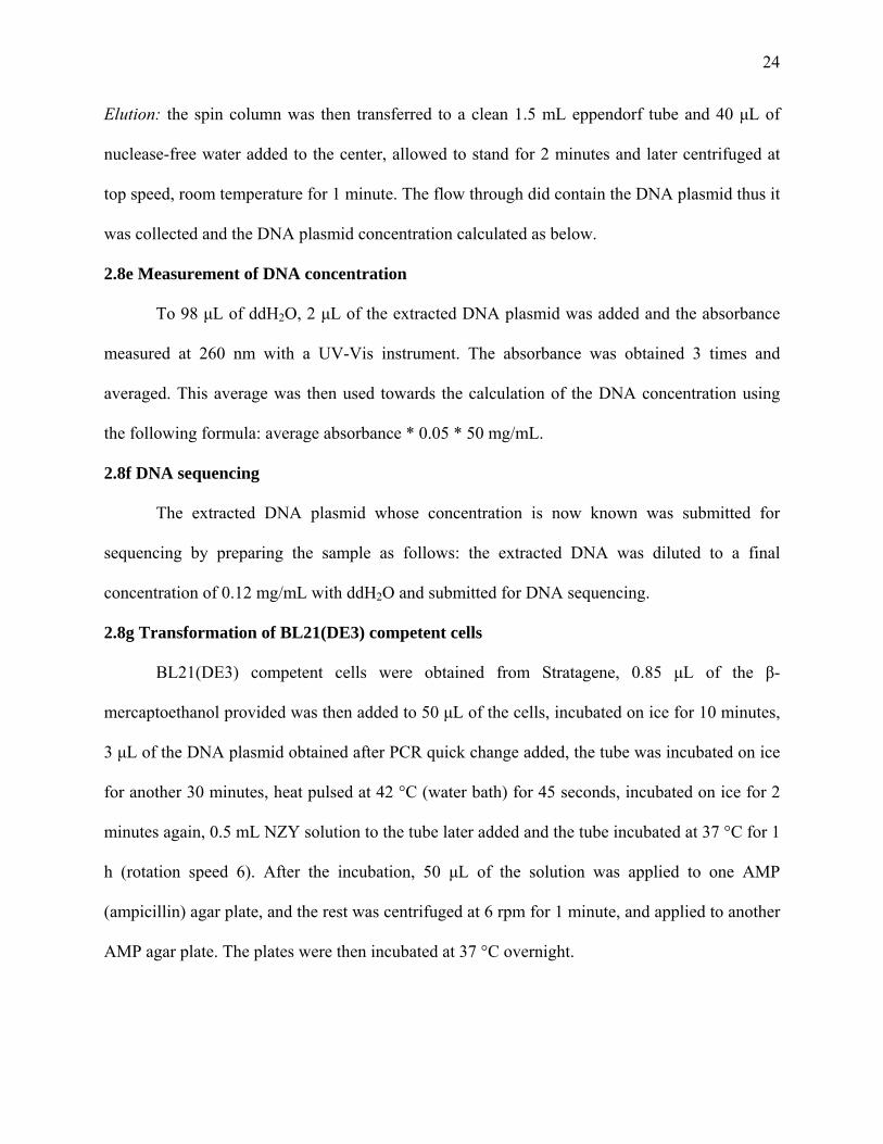
Elution: the spin column was then transferred to a clean 1.5 mL eppendorf tube and 40 μL of
nuclease-free water added to the center, allowed to stand for 2 minutes and later centrifuged at
top speed, room temperature for 1 minute. The flow through did contain the DNA plasmid thus it
was collected and the DNA plasmid concentration calculated as below.
2.8e Measurement of DNA concentration
To 98 μL of ddH2O, 2 μL of the extracted DNA plasmid was added and the absorbance
measured at 260 nm with a UV-Vis instrument. The absorbance was obtained 3 times and
averaged. This average was then used towards the calculation of the DNA concentration using
the following formula: average absorbance * 0.05 * 50 mg/mL.
2.8f DNA sequencing
The extracted DNA plasmid whose concentration is now known was submitted for
sequencing by preparing the sample as follows: the extracted DNA was diluted to a final
concentration of 0.12 mg/mL with ddH2O and submitted for DNA sequencing.
2.8g Transformation of BL21(DE3) competent cells
BL21(DE3) competent cells were obtained from Stratagene, 0.85 μL of the β-
mercaptoethanol provided was then added to 50 μL of the cells, incubated on ice for 10 minutes,
3 μL of the DNA plasmid obtained after PCR quick change added, the tube was incubated on ice
for another 30 minutes, heat pulsed at 42 °C (water bath) for 45 seconds, incubated on ice for 2
minutes again, 0.5 mL NZY solution to the tube later added and the tube incubated at 37 °C for 1
h (rotation speed 6). After the incubation, 50 μL of the solution was applied to one AMP
(ampicillin) agar plate, and the rest was centrifuged at 6 rpm for 1 minute, and applied to another
AMP agar plate. The plates were then incubated at 37 °C overnight.

25
2.8h Nickel-NTA Histidine-tagged protein expression & purification
8ml LB media inoculation: to 8 mL of LB media in culture tube(s), 4 μL of 2000x AMP
was added along with a colony from the agar plates, this culture was incubated at 37°C overnight
with 6rpm rotation speed.
IPTG induction: to 1 L of LB media, 0.5 mL of 250 mg/mL AMP was added, 1 culture tube from
the 8 mL inoculation step was added, and the culture incubated while shaking at 37 °C for
several hours until the O.D. value was in the range of 0.6 - 0.8. The culture was then cooled
down until the incubated equilibrated to 16 °C. 100 mM IPTG was then added and the culture
incubated overnight.
Cell harvesting and His.tagged protein expression using Ni-beads:
After IPTG induction, the cell culture was centrifuge for 10 minutes at 5000 rpm at 4°C. The
cultured cells were re-suspended in cell lysis buffer (see section 2.6). After re-suspending the
cells, they were French press twice and centrifuged immediately at 14,000 rpm, for 30 minutes at
4°C and the supernatant was collected.
Protein purification: 6 mL of Ni-beads was added to the column, washed two times with column
buffer (section 2.6 for preparation) to equilibrate the column, the supernatant loaded to the
column with the beads, shaken at 4°C for 1h. The supernatant was then allowed to flow through
the column into a tube. The column was washed again 10 times with 40 mL wash buffer (see
section 2.6 for preparation).

26
Protein elution and concentration: Elute protein from beads seven times with 7 mL each time
with elution buffer (see section 2.6). The eluants were then combined and dialyzed with dialysis
buffer for 48 hrs (see section 2.6). A 12 % SDS-PAGE gel (see preparation in section 2.6) was
ran at 120 V.
2.8i Radioactive HAT assay
This assay was divided into two, testing the autoacetylation of Tip60 wt and the mutants
and the other testing Tip60 acetylation of substrate H4(1-20) histone. The substrate and enzyme
concentrations were held constant throughout the experiment.
Sample preparation:
Dilutions to obtain final enzyme concentration of 0.6 µM, H4(1-20) substrate
concentration of 200 µM and 14AcCoA concentration of 20 µM were made. For the enzyme
dilutions, 2XRB was added for a final concentration of 1XRB and the appropriate amount of
water was added as well to make a total volume of 20 µL.
Original concentration of Tip60 wild type (wt) 7.1 µM
Original concentration of Tip60Cys369Ala 6.9 µM
Original concentration of Tip60Glu403Gln 4.6 µM
Mix solution: 100 µL 2XRB, 15.5 µL ddH2O and 4.5 µL 14AcCoA.
For 2XRB preparation see Chapter 2 section 2.6 Buffers.
Table 4: Sample preparation for radioactive HAT assay Sample 0 1 2 3 4 5 6
H4(1-20) - - - - 6 µL 6 µL 6 µL
Protein/enzyme wt wt Tip369A Tip403Q wt Tip369A Tip403Q
14AcCoA 2XRB * * * 18 µL 18 µL 18 µL
- were replaced with 6 µL ddH2O

27
* were replaced with 18 µL Mix solution
100 µL 2XRB 15.5 µL ddH2O 4.5 µL14AcCoA
at
Figure 8: Scheme of HAT radioactive assay
Table 5: HAT assay reaction time
Sample Add mixture at
30 ºC Add enzyme at
30 ºC Quench reaction
with 6x dye 0 0 5 15 1 0.5 5.5 15.5 2 1 6 16 3 1.5 6.5 16.5 4 2 7 17 5 2.5 7.5 17.5 6 3 8 18
120 µL 10 min at 30 ºC
Tip60 protein 6 µL
5 min at 30 ºC
6 µL 1000 µM H4(1-20)
18 µL
Quench reaction with 6 µL 6X quenching dye

28
CHAPTER 3
SITE-DIRECTED MUTAGENESIS
3.0 Site-directed mutagenesis using polymerase chain reaction (PCR)
This is a widely used technique which was first introduced in the 1970s to study and
localize the effects of mutations on genes (Smith, 1993). Before this process can be carried out,
an appropriate mutagenic primer has to be designed. This designed primer determines the
efficiency of the reaction because a 100 % base pairing at either ends of the target sequence is
necessary. The method of site-directed mutagenesis used for this experiment is a quick-change
PCR method using the quick-change site-directed mutagenesis kit from stratagene.
Many studies have been done on the MYST family prototype Esa1, so based on the
results of those studies and the close relation of Esa1 to Tip60, our point mutations were
determined to be at amino acids 369 and 403. Amino acid 369 which is a Cys was mutated to an
Ala. Ala because it eliminates the concerns of a similar side chain while not affecting the
conformation of the main-chain, i.e., we are interested in investigating the role the thiol in the
Cys side chain plays during catalysis (Lefèvre et al, 1997). Also, amino acid 403 was mutated
from a Glu to a Gln because Gln has a side chain that is structurally similar to glutamate but
lacks the acidic character required for it to function in a catalytic capacity (Trievel et al., 1999).
Also using the full length gene sequence of Tip60α inserted in the pET15b vector between NcoI
and BamH1 sites (map shown in figure 9); the point mutations were individually introduced
using thermal cycling quick change PCR base site-directed mutagenesis technique. The pET15b
vector was ideal for this experiment because it contains a His-tagged and a lac operon coding
sequence which helped enhance the concentration of the protein during expression of both the
Tip60 wild type and Tip60 mutants (Tip60Cys369Ala and Tip60Glu403Gln).

29
Quick change PCR based site-directed mutagenesis technique allows for DNA
amplification using thermostable polymerases for nucleotide incorporation. The thermal cycles
allow for the denaturation of the plasmid DNA to form single-stranded regions. The synthesized
mutant oligonucleotide is then annealed to the target strand at which point the pfu Turbo DNA
polymerase, which allows for high fidelity replication of the plasmid and extension of the mutant
strand and keeping the mutant oligonucleotide in place. In other words, during this thermal cycle,
the annealing of the oligonucleotides which carry the desired mutation (s) to one strand of the
DNA of interest which is accomplished (Cosby et al., 1997). The original strand of DNA serves
as a primer to initiate DNA synthesis in this process. In other words, the designed mutated
primer is introduced to the reaction, using the non-strand displacement characteristic of the pfu
Tubor DNA polymerase the oligonucleotide strands are incorporated into the site of mutation
and extended in a circular manner (Stratagene.com).
A digestive enzyme, Dpn I is then used to digest the parent DNA for 1 hour. This Dpn I
digestive enzyme targets the parent DNA because the parent DNA is methylated where as the
mutated DNA is not. The parent DNA is already methylated because it was obtained from the
cell, and a methylated DNA sequence is essential for gene expression control in a cell due the
fact that any alterations in methylation pattern could lead to serious forms of diseases such as
cancer (Richardson and Yung, 1999). After this step, the mutated DNA plasmid is not
completely circular, so it has to be transformed into an E coli. bacteria cell.
Following the protocol and thermal cycle described in section 3.1 of chapter 3, the results
shown in Figure 10 were obtained. The top gel is that for Tip60Cys369Ala mutation and the
bottom gel is for the Tip60Glu403Gln mutation. Using gel electrophoresis to confirm
30
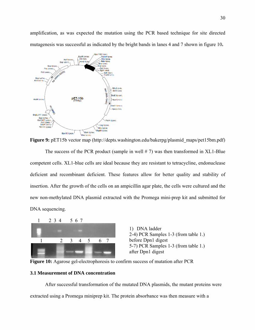
amplification, as was expected the mutation using the PCR based technique for site directed
mutagenesis was successful as indicated by the bright bands in lanes 4 and 7 shown in figure 10.
Figure 9: pET15b vector map (http://depts.washington.edu/bakerpg/plasmid_maps/pet15bm.pdf)
The success of the PCR product (sample in well # 7) was then transformed in XL1-Blue
competent cells. XL1-blue cells are ideal because they are resistant to tetracycline, endonuclease
deficient and recombinant deficient. These features allow for better quality and stability of
insertion. After the growth of the cells on an ampicillin agar plate, the cells were cultured and the
new non-methylated DNA plasmid extracted with the Promega mini-prep kit and submitted for
DNA sequencing.
1 2 3 4 5 6 7
1 2 3 4 5 6 7
Figure 10: Agarose gel-electrophoresis to confirm success of mutation after PCR
3.1 Measurement of DNA concentration
After successful transformation of the mutated DNA plasmids, the mutant proteins were
extracted using a Promega miniprep kit. The protein absorbance was then measure with a
1) DNA ladder 2-4) PCR Samples 1-3 (from table 1.) before Dpn1 digest 5-7) PCR Samples 1-3 (from table 1.) after Dpn1 digest
31
UV-Vis instrument at 260 nm and used towards the calculation of DNA concentration.
3.2 DNA sequencing
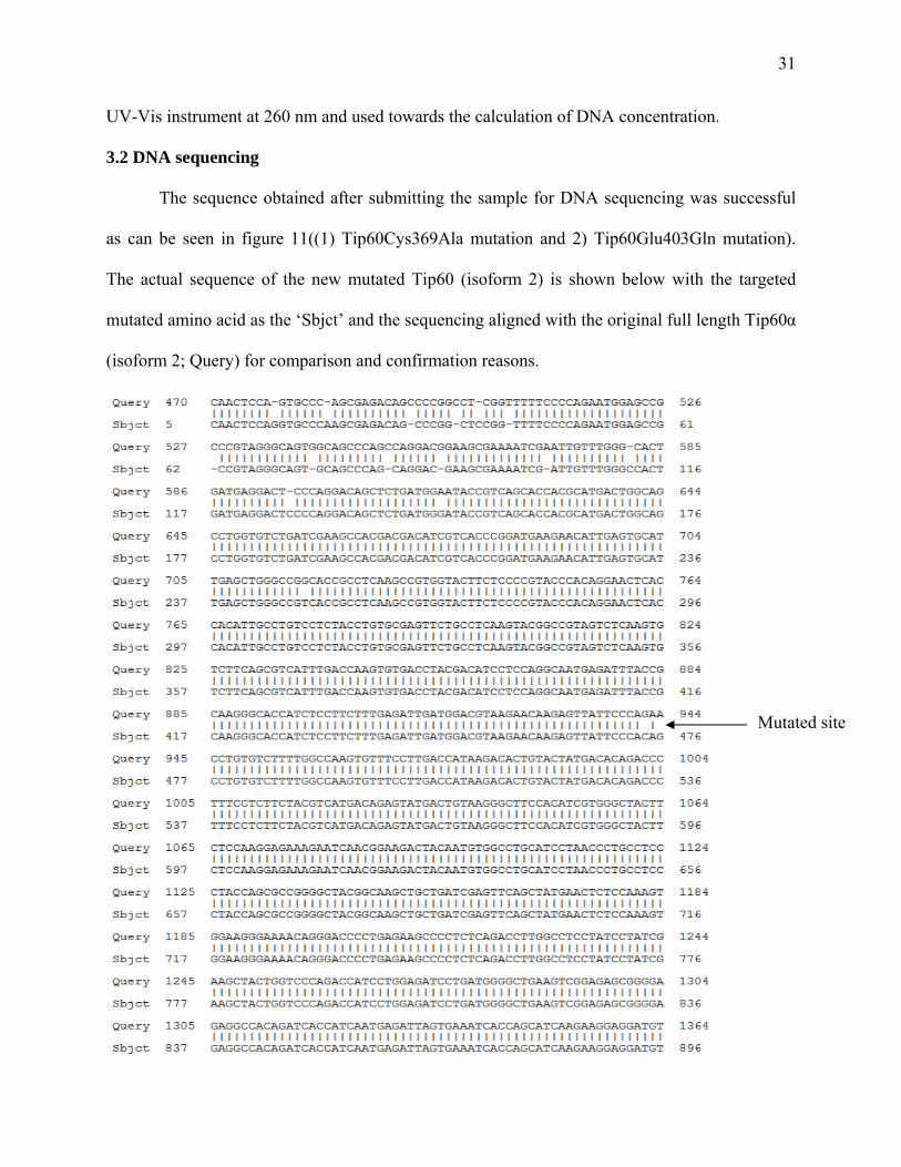
The sequence obtained after submitting the sample for DNA sequencing was successful
as can be seen in figure 11((1) Tip60Cys369Ala mutation and 2) Tip60Glu403Gln mutation).
The actual sequence of the new mutated Tip60 (isoform 2) is shown below with the targeted
mutated amino acid as the ‘Sbjct’ and the sequencing aligned with the original full length Tip60α
(isoform 2; Query) for comparison and confirmation reasons.
Mutated site

32
Figure 11: Result of the NCBI Blast alignment of the sequencing results obtained after the Tip60Glu403Gln and Tip60Cys369Ala mutations
Mutated site

33
CHAPTER 4
PROTEIN EXPRESSION
After DNA transcription in the nucleus, the synthesized mRNA migrates to the cytoplasm
where it binds to a ribosome in preparation for the process of translation. During translation, the
genetic information carried by the mRNA strand is translated into amino acids which later
encode for the appropriate protein. The study of proteins in a biomolecular system is known as
proteomics. Many methods have been used to isolate and characterize proteins after translation.
Some of these methods include mass spectroscopy, chromatography and gel analysis. For this
experiment, column chromatography using Nickel-NTA beads and later SDS-PAGE were used
to analyze and separate the target protein from others. Gene expression in cells under a given set
of experimental conditions could lead to the discovery of clues as to which proteins are involved
in certain pathways and disease states.
4.0 IPTG induction for protein expression
Using E. coli as a host to the Tip60 protein will allow control on the amount of protein is
obtained after protein expression. Through the use of AMP enhanced LB growth media, we were
able to amplify the successfully mutated Tip60 proteins through the methods described in
chapter 2. We were able to manipulate the expression of the target protein due to a host strain
condition introduced by Williams et al. in 1998. Williams et al. created an E. coli host containing
a conditionally essential gene under control of the lac operato/promoter region and a multicopy
plasmid containing the lac operator (Williams et al, 1998; Baneyx, F.; 1999). Based on the
results obtained by Williams et al. and others, Isopropyl β-D-1-thiogalactopyranoside (IPTG)
was found to be and ideal lactose analog along with an appropriate host to obtain better amounts
of the target protein. Isopropyl β-D-1-thiogalactopyranoside (IPTG) is the non-hydrolyzed and

34
commonly used lac-type operon (lactose metabolite reagent) that triggers/induces the
transcription of a lac operon present on the gene insertion of interest (Chen et al., 1997). The
presence of a lac repressor operator system is important for gene regulation in E. coli (Yansura
et al., 1984). Depending on the target protein, as little as 0.1-0.5 mM IPTG can be used for
induction during protein expression.
Using Bl21(DE3) as a host, Hur, et al. reported that produce a series of metabolic
changes over time (Hur et al., 2005). Also, according to their results, these metabolic changes
could be due to the presence of the T7/lac promoter which is triggered by IPTG to produce the
T7 RNA polymerase which subsequently clones the lacZ gene on the plasmid by the strong T7
polymerase (Studier et al.; 1986; Hur et al., 2005).
During this study, BL21(DE3) competent cells from stratagene were used for IPTG
induction for protein expression. This procedure is described in section 2.8h.
4.1 Nickel-NTA Histidine-tagged protein expression assay
This method of protein expression makes use of Ni2+ cations immobilized on the Ni-NTA
(Ni-nitriloacetic acid) His-Binding to resins forming a metal chelation complex in an affinity
chromatography. With this technique, the solid-phase NTA resin forms a four side chelating
bond with the Ni2+ metal cations. After the protein has been extracted from the cells, this lysate
is loaded to the chromatographic column where the protein binds to the Ni-resin. The unbound
proteins are washed away with a washing buffer (section 2.6 shows the buffer used in this
experiment). The target protein is then eluted with high concentrations of imidazole. Imidazole is
preferable because it is similar in structure to His, so during the washing process, it replaces the
His on the Ni2+ complex for the release of the protein.
35
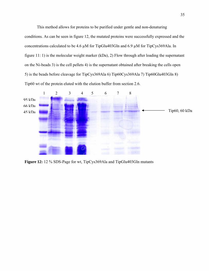
This method allows for proteins to be purified under gentle and non-denaturing
conditions. As can be seen in figure 12, the mutated proteins were successfully expressed and the
concentrations calculated to be 4.6 µM for TipGlu403Gln and 6.9 µM for TipCys369Ala. In
figure 11: 1) is the molecular weight marker (kDa), 2) Flow through after loading the supernatant
on the Ni-beads 3) is the cell pellets 4) is the supernatant obtained after breaking the cells open
5) is the beads before cleavage for TipCys369Ala 6) Tip60Cys369Ala 7) Tip60Glu403Gln 8)
Tip60 wt of the protein eluted with the elution buffer from section 2.6.
1 2 3 4 5 6 7 8
Figure 12: 12 % SDS-Page for wt, TipCys369Ala and TipGlu403Gln mutants
Tip60, 60 kDa
95 kDa66 kDa45 kDa

36
CHAPTER 5
ENZYMATIC STUDY
To understand how HATs function in regulating transcription/post-translation, it is
important to carry out certain assays. Acetylation of a lysine residue has been reported to affect
other post-translational modifications on a neighboring residue, particularly phosphorilation
(Yang and Seto, 2007). Histone acetyltransferases (HATs) have been reported to play crucial
roles in many cellular functions, such as gene transcription and cell proliferation. The method
used in the acetylating activity analysis of Tip60 is an assay which makes use of the
phosphoimaging technique where after the reaction of Tip60, Histone H4(1-20) and AcetylCoA
over time, the results could be quantified using phosphoimaging. AcetylCoA is ideal because
acetylCoA is independently produced and consumed by different organs and is a key
intermediate in a number of metabolic pathways (Oliver et al., 2009).
During this assay, Tip60, H4(1-20) and acetyl-CoA are incubated at 30 °C at different
time intervals (see section 28i). After the incubation, the expected outcome is the release of the
Tip60 (as is the case with enzymes), CoA and the acetylated H4(1-20).
With this easy phosphoimaging assay, whether the Tip60 mutants could acetylate the
H4(1-20) histone substrate or themselves with 14AcCoA was being investigated. As can be seen
in figure 13, Tip60 can acetylate the H4(1-20) histone substrate; 1 shows Tip60Glu403Gln
mutation, 2 shows Tip60Cys369Ala and 3 shows wild type Tip60. The wild-type showed more
activity, followed by TipGlu403Gln and lastly TipCys369Ala. The fact that TipCys369Ala did
not show much acetylating activity is indicative of the fact that the Cys present in its HAT active
is essential for catalysis contrary to the earlier report using the MYST prototype Esa1. The

Tip60Glu
play a ma
5.0 Radi
T
during ce
with eith
described
et al., 20
quick an
acetyl-Co
(H4(1-20
staining
phosphor
a typhoon
i) 0
ii) Figure 1
i) Phosph
2) TipCy
shown in
u403Gln on
ajor role in s
ioactive HA
The characte
ellular proce
her α-ketog
d by Kim et
000). Howev
nd simple. H
oA to the ε
0) in this cas
and destain
rimaging see
n phosphoim
1
1
3: Phosphor
horimage of
ys369Ala 3)
n Table 5 (0 i
the other ha
substrate ace
T assay
erization of
esses. Two c
glutarate deh
al. towards
ver, for our
Histone acet
ε-amino term
se). During t
ning and pla
e Poveda and
mager (show
2 3
2 3
rimaging of
substrate, H
wt. Tip60 (i
is the BSA s
and shows so
etylation.
HAT activi
continuous n
hydrogenase
minimizing
experiment
tyl transfera
minal of a s
this radioacti
aced in a ph
d Sendra, 20
wn in Figure
3 4
Tip60 H4(1-
H4(1-20), ace
i) Phosphori
standard).
ome activity
ity is crucia
non-radioact
e or pyruva
g hazards fac
t, the radioa
ases catalyz
specific lysin
ive assay, th
hotoimager
008). The res
13).
-20) histone
etylation by
image of aut
which is ind
al towards u
tive process
ate dehydro
ced when us
active assay
e the transf
ne residue w
he peptide ge
for 72 hour
sulting spot s
acetylation
Tip60 1) Tip
toacetylation
dicative that
understandin
es, coupled
genase with
ing a radioa
was suitabl
fer of an ac
within the c
el was dried
rs (for more
signals were
and autoace
pGlu403Gln
n by Tip60 p
t the Glu doe
ng their fun
enzyme sys
h CoASH,
active assay
e because it
cetyl-group
core of a hi
d for 2 hours
e on analys
e quantified u
etylation
n
prepared as
37
es not
nction
stems
were
(Kim
t was
from
stone
after
is by
using

38
Table 5: Sample preparation for Tip60 autoacetylation Sample 1 2 3 4 ddH2O + + + +
Mix + + + * Tip60 Tip60Glu403Gln Tip60Cys369Ala wt wt
* 2XRB added instead
5.1 Initial enzymatic characterization
Enzyme kinetics is a study of the time-dependent activity of enzymes with a goal of
attaining equilibrium (Hans Bisswanger, Enzyme Kinetics: principles and methods, 2nd edition,
Wiley VCH 2008). This study involves the behavior of an enzyme after binding with the substrate
to form the enzyme-substrate complex and its conversion into product(s) (Hans Bisswanger,
Enzyme Kinetics: principles and methods, 2nd edition, Wiley VCH 2008). In this study, the
acetylating activity of Tip60 wild type, Tip60Cys369Ala and Tip60Glu403Gln mutants on H4(1-
20) histone was individually evaluated over a period of time with a 3 minute interval between
reaction quenching.
In our assay, radioactive acetylCoA, H4(1-20) histone and the three various Tip60
expressed proteins were prepared separately at 30 ºC. The set up was as shown in figure 8 with a
3 minute interval between the quenching of each mixture in a tube. The reaction times are shown
in table 6 for both mutants. To show the steady state linearity and explore the acetylating activity
of the Tip60 wt. and Tip60Glu403Gln was performed and stopped at 12 minutes (see table 7) for
Tip60Cys369Ala, the reaction time was extended to 15 minutes. After the reactions for both the
wt. Tip60 and the two mutants, the final enzyme concentration was 200 nM and 380 nM
respectively. The H4(1-20) substrate concentration was 440 μM for both mutants and 400 μM for
the wt. The acetylCoA concentrations were 10 μM for the wt. reaction and 17.86 μM for the
mutants. As expected (shown in figure 14), the product formation increased with time and

39
leveled off after 12 minutes. This suggests that the enzyme has reached its maximum activity at
that time and the rate of product formation should remain the same.
Table 6: Tip60Cys369Ala enzyme kinetics reaction times Sample Add H4(1-20) Add enzyme Quench reaction on P81
paper 0 0 5 8 1 0.5 5.5 8.5 2 1 6 12 3 1.5 6.5 15.5 4 2 7 19 5 2.5 7.5 22.5
Table 7: Tip60 wt. and Tip60Glu403Gln enzyme kinetics reaction times
Sample Add H4(1-20) Add enzyme Quench reaction on P81 paper
0 0 5 8 1 0.5 5.5 8.5 2 1 6 11 3 1.5 6.5 13.5 4 2 7 17 5 2.5 7.5 19.5
0
0.5
1
1.5
2
2.5
3
0 2 4 6 8 10 12 14
Plot of Tip60 W.T vs time
[Pro
duct
], υΜ
Time, minutes

40
0
0.1
0.2
0.3
0.4
0.5
0 2 4 6 8 10 12 14
Plot of TipGlu403Gln vs time[P
rodu
ct], μΜ
Time, minutes
0
0.002
0.004
0.006
0.008
0.01
0.012
0.014
0 5 10 15 20
Plot of Tip60Cys369Ala vs time
[Pro
duct
], υΜ
Time, minutes
Figure 14: Plot of Product concentration, µM vs Time for Tip60 wild type, Tip60Cys369Ala and Tip60Glu403Gln 5.2 Future kinetic study of Tip60
Based on the results obtained above, we conclude that the enzyme is stable until 20
minutes also, the substrate/product yield is less than 10%. This value is within the acceptable

41
percent yield value. Our future work will involve an investigation towards understanding and
characterizing the enzyme mechanism of Tip60. With this mechanism investigation, it could lead
to a possible explanation as to what mechanism Tip60 uses during catalysis, i.e., whether Tip60
uses the ternary complex or the ping pong mechanism. Also, because Tip60 has been shown to
autoacetylate, the role this autoacetylaiton places during catalysis will be investigated.

42
CHAPTER 6
A DUAL-MODE FLUORESCENCE STRATEGY FOR SCREENING HAT MODULATORS
Many, but limited, approaches have been explored to characterize and identify HAT
inhibitors. An ideal method to characterize HAT catalytic activity which eventually leads to
HAT modulating inhibitor identification is the classical labeling of enzymes with a radioactive
element (Martinez et al., 2006; Hardcastle et al., 2005). This approach is a novel approach
reported by Xie et al. as a means to screen HAT small molecule inhibitors. This method is a
simple single-step assay that provides direct readout of acetylation products via fluorescent
intensity changes.
6.0 Using the classical radioactive way to characterize HATs
This is the commonly used assay by most scientists who have the goal of characterizing
HAT proteins/enzymes. In a radioactive method described by Turlais et al., a mixture of radio-
labeled acetylCoA and substrate (histone) were added together in one step followed by enzyme
in another step to initiate the acetylation reaction. The results were measured by means of
scintillating microtiter plates (Turlais, et al., 2001). This method allows for HTS and the accurate
identification of potential inhibitor leads for HATs.
6.1 Using immunoblotting to characterize HAT inhibitors
The immunoblotting technique involves the use of antibodies or other specific ligands to
identify target proteins in form of antigen-antibody (or protein-ligand) specific reactions. This
technique, when used in the study of HATs (acetylation or deacetylation), relies on the
recognition of acetyl-lysines by specific antibodies and has been reported to be of potential in
inhibitor screening studies (Hardcastle et al., 2005; Martinez et al., 2006; Stockwell et al., 1999;
Zhang et al., 1998). Although this method involves the use of original/specific antibodies,

43
antibodies are expensive, the washing step is time consuming and a robust deconvolution scheme
is needed to validate the action of hits identified in a cell-based screen. High throughput
screening (HTS) and automation are the features preferred by most scientists when using certain
assays in the quest to find the best inhibitor (s) or small molecule amongst a large pool of
potential molecules. The immunoblotting technique is not ideal for HTS because of the absence
of certain practical tools (Wegener et al., 2003). Since HATs have been reported to HTS is
beneficial in identifying small molecule inhibitors related to HATs functionality, fluorescence
spectroscopy has been reported as a potential technique for these purposes.
6.2 Using FRET and anisotropy measurements to characterize HAT inhibitors
Over expression of HATs has been related to several diseases states because they have
the capability to modify histones and if histones are modified, they grant easy access to
chromatin (Timmermann et al., 2001). The identification of small molecules that are involved in
epigenetic regulation is important in normal and disease cell processes (Johnson et al., 2008).
Anacardic acid, garcinol, isothiazolone, curcumin and cinnamoyl compounds are the most recent
small molecule HAT inhibitors. However, these small molecule inhibitors exhibit nonspecific
inhibition and have limited usage in pharmacological settings (Wynne et al., 2002, Wu et al.,
2008).
Fluorescence resonance energy transfer (FRET) is a distance-dependent technique. With
this technique, the donor and acceptor must be in close proximity, the absorption spectrum of the
acceptor must overlap with the emission spectrum of the donor and the donor acceptor transition
dipole orientation must be paralleled. This use of this strategy to characterize potential inhibitors
should provide renovating insights in the screening for new anticancer drugs that target the
substrate interfaces of HAT targets, as well as in the mechanistic characterization of HATs.

44
In this new approach we reported, FRET and fluorescence anisotropy were used to
identify and characterize HAT inhibitors (plots shown in Figures 18 & 19). We used Dabcyl
(Dab) to label the HATs p300 which acted as a FRET acceptor. Methoxycoumarin (Mca) in
conjugate with HAT substrate analogues functioned as fluorescent donors, specifically;
LysCoAMca was used to interact with p300. In this assay, when a protein binds the ligand to
form a complex, the fluorescence anisotropy increases and the fluorescence intensity of the
donor fluorophore decreases due to FRET quenching by the Dab acceptor. Figure 15 shows a
schematic diagram of the fluorescent reporter system (Xie et al., 2009).
Figure 15: Schematic showing acetylation products via fluorescent intensity changes.
6.3 Expression of Dabcyl-labeled p300 protein
p300 protein is part of the HAT domain family and has been reported to perform several
functions which include histone modification, regulation of cell growth, and gene expression.
Labeling of p300 protein with Dabcyl was achieved using the EPL protocol essentially the same
as in the literature (Thompson et al., 2004). First, the DNA sequence encoding for the p300 HAT
domain (aa 1287-1652) was subcloned into PTYB2 vector (a gift from Prof. Phillip Cote). BL-

45
21(DE3) Codonplus RIL competent cells were used for protein expression (Figure 16). The
expressed p300-intein-CBP fusion protein was loaded to chitin beads. After extensive washing, 6
mg of the ligation peptide (CMLVELHTQSQDRFK(Dabcyl)-NH2; Molecular weight; 2086 kD)
in 3 mL of ligation buffer (25 mM HEPES pH8.0, 250 mM NaCl, 1 mM EDTA, and 200 mM
MESNA solution) was added to the beads. The ligation reaction was kept at room temperature
for 48 h. The protein product was confirmed with 12% SDS-PAGE (Figure 16). The combined
eluants were dialyzed against the storage buffer and then concentrated in a centrifuge filter tube.
The concentration of labeled p300 protein (p300Dab) was determined by Bradford assay.
1 2 3 4 5 6 7 8 9
Figure 16: 12 % SDS-PAGE for p300 protein expression
In figure 15, (1) MW marker (kDa) (2) Eluant 1 (3) Beads before cleavage (4) Flow
through (5-8) eluants 2-5 (9) Beads after cleavage.
1 2 3 4 5 6 7 8 9 10
Figure 17: 12 % SDS-PAGE for Dabcyl-labeled p300
p300, 45 kDa
Ligated p300, 47 kDa
97 66 45 31 29
97 66 45 31 29

46
In figure 17, (1) MW marker (2) Flow Through (3) Protein supernatant (4) Beads before
cleavage (5-9) eluants 1-5 (10) Beads after cleavage (kDa).
6.4 Fluorescence measurement
The fluorescence experiments were performed on a Fluoromax-4 instrument (Horiba
Jobin Yvon, Edison, NJ) at 25 °C using a 0.5 mL cuvette in a buffer solution containing 50 mM
Hepes (pH 8.0), 0.1 mM EDTA, 150 mM NaCl, and 1 mM DTT. The dissociation constants (Kd)
were determined by measuring the fluorescence signals of Mca-labeled ligands (at fixed
concentration) with increasing Dab-labeled protein concentrations.
Fraction bound = (F - Fo)/(Ff - Fo)
={(Ff - Fo)/2[Pr]tot}{b(b2 - 4[L]tot[Pr]tot)1⁄2}+Fo (1)
b = Kd + [L]tot + [PCAF]tot
Where F0 and Ff are the initial and final fluorescence intensities, respectively, [Pr]tot is
the total protein concentration, and [L]tot is the total concentration of Mca-labeled ligand. The
inhibition constants (Ki) were determined by measuring the fluorescence signals of the binary
mixture containing Mca-labeled ligand and Dab-labeled protein (at fixed concentrations) with
increasing concentrations of respective inhibitors. Equation 2 was used for Ki value calculations
(values not shown) (Nikolovska-Coleska et al., 2004).
Ki = [I]50/ ([L]50/ Kd + [Pr]o/Kd + 1) (2)
Where [I]50 denotes the concentration of the free inhibitor at 50% inhibition, [L]50 is the
concentration of the free Mca-labeled ligand at 50 % inhibition, [Pr]0 is the concentration of the
free protein at 0 % inhibition, and Kd is the dissociation constant of the protein-ligand complex.
The results obtained from the both the FRET studies and anisotropy are shown in figures
18 and 19. As anticipated from these results, upon addition of p300Dab, the fluorescent intensity

47
of LysCoAMca declined in a dose-dependent manner owing to FRET quenching interaction
between Dab and Mca (Figure 18a,b). The fluorescence intensity was recovered when the
nonfluorescent ligand LysCoA was added to the solution, competitively displacing LysCoAMca
out of the binding pocket in p300 (Figure 18c,d). We also performed fluorescence anisotropy
measurements of LysCoAMca (Figure 19). In this regard, the performance of LysCoAMca
was analogous to that of H3CoA20Mca; the anisotropy value increased upon binding to
p300Dab protein, and the change was reversed by increasing concentrations of the competitive
inhibitor LysCoA. Therefore, both FRET and anisotropy signals were effective to probe and
characterize the p300-inhibitor interactions.
Figure 18: FRET changes of LysCoAMca (a) The emission spectral changes of LysCoAMca (0.2 µM) at different concentrations of p300-
Dabcyl (λex = 326 nm). (b) The titration curve of LysCoAMca (0.2 µM) emission at 396 nm vs
the concentrations of p300-Dabcyl. Kd is calculated to be 0.40 ± 0.06 µM. (c) The spectral
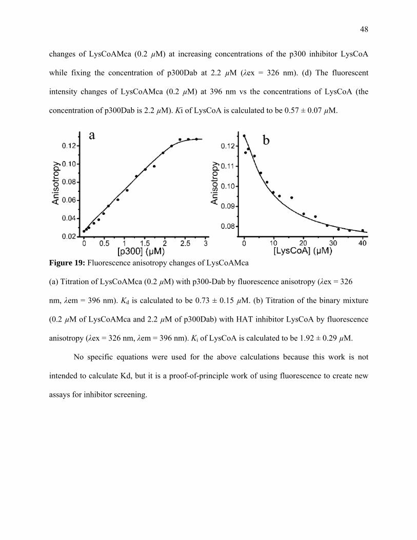
48
changes of LysCoAMca (0.2 µM) at increasing concentrations of the p300 inhibitor LysCoA
while fixing the concentration of p300Dab at 2.2 µM (λex = 326 nm). (d) The fluorescent
intensity changes of LysCoAMca (0.2 µM) at 396 nm vs the concentrations of LysCoA (the
concentration of p300Dab is 2.2 µM). Ki of LysCoA is calculated to be 0.57 ± 0.07 µM.
Figure 19: Fluorescence anisotropy changes of LysCoAMca (a) Titration of LysCoAMca (0.2 µM) with p300-Dab by fluorescence anisotropy (λex = 326
nm, λem = 396 nm). Kd is calculated to be 0.73 ± 0.15 µM. (b) Titration of the binary mixture
(0.2 µM of LysCoAMca and 2.2 µM of p300Dab) with HAT inhibitor LysCoA by fluorescence
anisotropy (λex = 326 nm, λem = 396 nm). Ki of LysCoA is calculated to be 1.92 ± 0.29 µM.
No specific equations were used for the above calculations because this work is not
intended to calculate Kd, but it is a proof-of-principle work of using fluorescence to create new
assays for inhibitor screening.

49
CHAPTER 7
CONCLUSION
Our results are different from that published for the yeast analog of Tip60, Esa1. The
results published stated that the Cys in the active site was not important for acetylating activity.
Cysteine is essential for acetylating substrate. Here, the effect of a mutation by substituting the
Cysteine on amino acid 369 with an Alanine and amino acid 403 a glutamate to glutamine is
explored. Kamine et al., reported that a mutation to the essential cysteine residue at the N-
terminal 31 of HIV-1 Tat terminates Tat’s transcription promoter and elongation activity
(Kamine et al., 1996). To evaluate the longevity of the enzyme activity, and kinetic assay was
carried out. This kinetic assay revealed that the enzyme activity is optimum up until 12 minutes
of reaction time with the substrate. With these kinetic studies and other further studies, better
understanding of the vast diversity of the MYST domain of Tip60 will be better obtained and
used towards to discovery of cures for many degenerative diseases. Tip60 may promote
acetylation of cellular proteins other than histones to regulate gene expression. For example,
Tip60 enhances the activity of wild-type but not an acetylation-deficient mutant of androgen
receptor, ‘demonstrating that Tip60 acts as a co-activator by promoting the acetylation of
androgen receptor but not histones’ (Xiao et al., 2003). Based on the kinetic studies results, we
conclude that Cys369 plays a more vital role in the enzyme activity of Tip60 compared to
Glu403.
The autoacetylation action of Tip60 by the wild-type and the acetylation of H4(1-20)
substrate were investigated using a radioactive assay. It can be concluded from this radioactive
study that both the Cys and the Glu in the active site are important for Tip60 to perform its post-
translational property of HATs have been reported to control chromatin structure allowing or

50
blocking gene transcription. Because of this evidence, suggestions have been made that
inhibition of HATs could result in antitumor effects (Turlais et al., 2001). In correlation with this
suggestion, we used both FRET and fluorescence anisotropy to characterize and identify HAT
inhibitors. From this experiment, both FRET and anisotropy signals were effective to probe and
characterize the p300-inhibitor interactions. Also, a displacement strategy for the study of HAT-
inhibitor interactions was demonstrated. The developed strategy should be useful in the search of
new anticancer drugs that target the substrate interfaces of the HAT targets, as well as find
values in mechanistic study of HATs.

51
REFERENCES
Allfrey, V. G., Faulkner, R. and Mirsky, A. E Acetylation and methylation of histones and possible role in the regulation of RNA synthesis (1964), Biochemistry 51, 786-793.
Avvakumov, N. and Cote, J. (2007) The MYST family of histone acetyltransferases and their
intimate links to cancer, Oncogene 26, 5395-5407. Baneyx, François (1999) Recombinant protein expression in Escherichia coli, Current Opinion
in Biotechnology 10, 411-421. Beekum, V. O., Brenkman, B. A., Grontved, L., Hamers, N., van den Broek,
J. F. N., Berger, R., Mandrup, S., and Kalkhoven, E. (2008) The adipogenic acetyltransferase Tip60 targets activation function 1 of peroxisome proliferator-activated receptor, Endocrinology 149, 1840-1849.
Berndsen C.E., Selleck, W., McBryant, S. J., Hansen, J. C., Tan, S. and Denu, J. M. (2007)
Nucleosome recognition by the piccolo NuA4 histone acetyltransferase complex, Biochemistry 46, 2091-2099.
Bird, W. A., Yu, Y. D., Pray-Grant, G. M., Qui, Q., Harmon, E. K., Megee, C. P., Grant, A. P.,
Smith, M. and Christman, F. M. (2002) Acetylation of histone H4 by Esa1 is required for DNA double-strand break repair, Nature 419, 411-415.
Bouchard, C., Dittrich, O., Klermaler, A., Dohmann, K., Menkel, A., Ellers, M. and Luscher, B.
(2001) Regulation of cyclin D2 gene expression by the MYc/Max/Mad network: Myc-dependent TRRAP recruitment and histone acetylation at the cyclin D2 promoter. Genes Dev. 15, 2042-2047.
Brady, E. M., Ozanne, M. D., Gaughan, L., Waite, I., Cook, S., Neal, E. D. and Robson, N. C.
(1999) Tip60 is a nuclear hormone receptor coactivator, Journal of Biological Chemistry 274, 17599-17604.
Carter, P. (1986) Site-directed mutagenesis. Journal of Biochemistry 237, 1-7. Cheung, W.L., Briggs, S.D. and Allis, D.C (2000) Acetyltion and chromosomal functions,
Current opinion in cell biology 12, 326-333. Conneely, O. M., Kettelberger, D. M., Tsai, M.- J. and O'Malley, B. W. (1989) The chicken
progesterone receptor A and B isoforms are products of an alternate translation initiation event., J. Biol Chem. 264, 14062-14064.
Cosby, N. and Lesley, S. (1997) Site-directed mutagenesis, Promega notes magazine 61, 12-18.

52
Creaven, M., Hans, F., Mutskov, V., Col, E., Caron, C., Dimitrov, S. and Khochbin, S. (1999) Control of the histone-acetytransferase activity of Tip60 by the HIV-1 transactivator protein Tat, Biochemistry 38, 8826-8830.
Decker, V. P., Yu, Y. D., Iizuka, M., Qiu, Q. and Smith, M. M. (2008) Catalytic-site mutations
in the MYST family histone acetyltransferase Esa1, Genetics 178, 1209-1220. Dohmesen, C. (2006) The histone acetyl transferase Tip60 as a regulator of tumor suppression. Philipps-University Marburg, Marburg. Doyon, Y., Cayrou, C., Ullah, M., Landry, A., Cote, V., Shelleck, W., Lane, S. W., Tan, S.,
Yang, X. and Cote, J. (2006) ING tumor suppressor proteins are critical regulators of chromatin acetylation required for genome expression and perpetuation, Molecular Cell 21, 51-64.
Edwards, J. and Bartlett, M.S. (2005) The androgen receptor and signal-transduction pathways in
hormone-refractory prostate cancer. Part 2: androgen-receptor cofactors and bypass pathways, BJU International 95, 1327-1335.
Fernandez, P., Frank, S.R., Wang, L., Schroeder, M., Liu, S., Greene, J., Coclto, A. and Amati,
B. (2003) Genomic targets of the human c-Myc protein. Genes Dev. 17, 1115-1129. Fischle, Wolfgang (2008) Talk is cheap- cross-talk in estrablishment, maintenance, and readout
of chromatin modifications, Genes Dev 22, 3375-3382. Frank, R. S., Parisi, T., Taubert, S., Fernandez, P., Fuchs, M., Chan, H., Livingston, D. M. and
Amati, B. (2003) MYC recruits the Tip60 histone acetyltransferase complex to chromatin, EMBO 4, 575-580.
Gaughan, L., Brady, E. M., Cook, S., Neal, E. D. and Robson, N. C. (2007) Tip60 is a co-
activator for class I nuclear hormone receptors, J. Biol. Chem. 276, 46841-46848. Gorrini, C., Squatrito, M., Luise, C., Syed, N., Perna, D., Wark, L., Martinato, F., Sardella, D.,
Verrechia, A., Bennett, S., Confalonieri, S., Cesaron, M., Marchesi, F., Gasco, M., Scanziani, E., Capra, M., Mai, S., Nuceforo, P., Crook, T., Lough, J. and Amati, B. (2007) Tip60 is a haplo-insufficient tumour suppressor required for an oncogene-induced DNA damage response, Nature 448, 1063-1069.
Halkidou K, Gnanapragasam, V.J., Mehta, P.B., Logan, I.R., Brady, M.E., Cook, S., Leung,
H.Y., Neal, D.E. and Robson, C.N. (2003) Expression of Tip60, an androgen receptor coactivator, and its role in prostate cancer development. Oncogene 22, 2466–2477.
Hansen, L.H, Knudsen, S. and Sorensen, S.J (1998) The effect of the lacY gene on the induction
of IPTG inducible promoters, studied in Escherichia coli and Pseudomonas fluorescens. Current Microbiology 36, 341-347.

53
Hardcastle, A., Boxall, K., Richards, J., Tomlin, P., Sharp, S., Clarke, P., Workman, P., and Aherne, W. (2005) Solid-phase immunoassays in mechanism-based drug discovery: their application in the development of inhibitors of the molecular chaperone heatshock protein 90. Assay Drug Dev. Technol. 3, 273–85.
Hlubek, F., Lohberg, C., Meiler, J., Jung, A., Kirchner, T. and Brabletz, T. (2001) Tip60 is a
cell-type-specific transcriptional regulator, J. Biochem 129, 635-641. Hodawadekar S. C. and Marmorstein R. (2007) Chemistry of acetyl transfer by histone
modifying enzymes: structure, mechanism and implications for effector design. Oncogene 26, 5528- 5540.
Holbert, M. A. and Marmorstein, R. (2005), Structure and activity of enzymes that remove
histone modifications. Curr. Opin. Struct. Biol. 15, 673-680. Hur, Won and Chung, Yoon-Keun (2005), On-line monitoring of IPTG induction for
recombinant protein production using an automatic pH control signal. Biotechnology and Bioprocess Engineering 10, 304-308.
Hutschison, C.A., Phillips, S., Edgell, M.H., Gillham, S., Jahnke, P. and Smith, M. (1978(,
Mutagenesis at a specific position in a DNA sequence. J. Biol. Chem. 253, 6551-6560. Huang, R. C. and Bonner, J. Histone, a suppressor of chromosomal RNA synthesis (1962),
Biochemistry 48, 1216-1222. Jasencakova, Z., Meister, A., Walter, J., Turner, M. B. and Schubert, I. (2000) Histone H4
acetylation of euchromatin and heterochromatin is cell cycle dependent and correlated with replication rather than with transcription, The Plant Cell 12, 2087-2100.
Johnson, R. L., Huang, W., Jadhav, A., Austin, C. P., Inglese, J. and Martinez, E. D. (2008) A
quantitative high-throughput screen identifies potential epigenetic modulators of gene expression, Analytical Biochemistry 375, 237-248.
Kamine, J., Elangovan, B., Subramanian, T. Coleman, D. and Chinnadurai, G. (1996)
Identification of a cellular protein that specifically interacts with the essential cysteine region of the HIV-1 Tat transactivator. Virology 216, 357-366.
Kim, M.-S., Merlo, X., Wilson, C. and Lough, J. (2006) Co-activation of atrial natriuretic factor promoter by Tip60 and serum response factor. The Journal of Biological Chemistry 281, 15082-15089.
Kim, Y., Tanner, G. K. and Denu, M. J. (2000) A continuous, nonradioactive assay for histone
acetyltransferases, Analytical Biochemistry 280, 308-314. Kuo, Min-Hao and Allis, David C. (1998) Roles of histones acetyltransferases and deacetylases
in gene regulation, BioEssays 20, 615-626.

54
Kusch, T., Florens, L., Macdonald, W.H., Swanson, S.K., Glaser, R.L., Yates III, J.R., Abmayr,
S.M., Washburn, M.P and Workman, J.L. (2004) Acetylation by Tip60 is required for selective histone variant exchange at DNA lesions, Science 306, 2084-2087.
Lafon, A. Chang, C. S., Scott, E. M., Jacobson, S. J. And Pillus L. (2007) MYST opportunities
for growth control: yeast genes illuminate human cancer gene functions, Oncogene, 5373-5384.
Latrasse, D., Benhamed, M., Henry, Y., Domenichini, S., Kim, W., Zhou, D. and Delarue, M.
(2008) The MYST histone acetyltransferases are essential for gametophyte development in Arabidopsis, BMC Plant Biology 8, 121-137.
Lefèvre, F., Rémy, M-H. and Masson, J-M (1997) Alanine-stretch scanning mutagenesis: a
simple and efficient method to probe protein structure and function, Nucleic Acids Research 25, 447-448.
Martinez, E. D., Dull, A. B., Beutler, J. A., and Hager, G. L. (2006) High-content fluorescence-
based screening for epigenetic modulators. Methods Enzymol. 414, 21–36. Nikolovska-Coleska, Z., Wang, R., Fang, X., Pan, H., Tomita, Y., Li, P., Roller, P. P.,
Krajewski, K., Saito, N. G., Stuckey, J. A., and Wang, S. (2004) Development and optimization of a binding assay for the XIAP BIR3 domain using fluorescence polarization. Anal. Biochem. 332, 261–73.
Nunnari, G., Smith, A. J. and Daniel, R. (2008) HIV-I Tat and AIDS-associated cancer: targeting
the cellular anti-cancer barrier?, Journal of Experimental & Clinical Cancer Research, 27, doi:10.1186/1756-9966-27-3
Oliver, D.J., Nikolau, B.J. and Wurtele, E.S. (2009) Acetyl-CoA-life at the metabolic nexus,
Plant Science 176, 597-601. Patterson, D.S. and Aebersold, H.R.(2003), Proteomics: the first decade and beyond, Nature
Genetics Supplement 33, 311-323. Pillus, Lorraine (2008), MYSTs mark chromatin for chromosomal functions, Current Opinion in
Cell Biology 20, 326-333. Poveda, Ana and Sendra, Ramon (2008) An easy assay for histone acetyltransferase activity
using a phosphoimager, Analytical Biochemistry 383, 296-300. Richardson, Bruce and Yung, Raymond (1999) Role of DNA methylation in the regulation of
cell function, J. Lab. Clin. Med., 333-340.

55
Robin, P., Fritsch, L., Philipot, O., Svinarchuk, F. and Ait-Si-Ali, S. (2007) Post-translational modification of histones H3 and H4 associated with the histone methyltransferases Suv39h I and G9a, Genome Biology 8, R270.1-R270.10.
Ruizeveld de Winter, J.A., Trapman, J., Vermey, M., Mulder, E., Zegers, N.D. and Van Der
Kwast, T.H. (1991) Androgen receptor expression in human tissues: an immunohistochemical study. J Histochem Cytochem 39, 927-936.
Sapountzi, V., Logan, R. I. and Robson, N. C. (2006) Cellular functions of Tip60. The
International Journal of Biochemistry & Cell Biology 38, 1496-1509. Silverman, B. and Mirsky, E. A. (1973) Addition of histones to histone-depleted nuclei: effect on
template activity toward DNA and RNA polymerase, PNAS 70, 2637-2641. Smith, Michael. (1993) Synthetic DNA and biology, Chemistry Nobel lecture, 119-136. Stedman, E. and E. Stedman, The basic proteins of cell nuclei (1951), Phil. Trans. Roy. Soc.
London, Ser. B 235, 565-596. Stockwell, B. R., Haggarty, S. J., and Schreiber, S. L. (1999) High-throughput screening of small
molecules in miniaturized mammalian cell-based assays involving post-translational modifications. Chem. Biol. 6, 71–83.
Studier, F. W. and Moffatt, B.A (1986) Use of bacteriophage T7 RNA polymerase to direct
selective high-level expression of cloned genes. J. Mol. Biol. 189, 113-130. Sun, Y., Jiang, X., Chen, S., Fernandes, N. and Price, D. B. (2005) A role for the Tip60 histone
acetyltransferase in the acetylation and activation of ATM, PNAS 102, 13182-13187. Taplin, Mary-Ellen (2007) Drug insight: role of the androgen receptor in the development and
progression of prostate cancer, nature 4, 236-244. Tasset, D., Tora, L., Fromental, c., Scheer, E. and Chambon, P. (1990) Distinct classes of
transcriptional activating domains function by different mechanisms, Cell 62, 1177-1187. Taubert, S., Gorrini, C, Frank, S.R., Parisi, t., Fuchs, M., Chan, H-M., Livingston, D.M. and
Amati, B. (2004) E2F-Dependent histone acetylation and recruitment of the Tip60 acetyltransferase complex to chromatin in late G1, Mol. and Cel Biol. 24, 4546-4556.
Thompson, P. R., Wang, D., Wang, L., Fulco, M., Pediconi, N., Zhang, D., An, W., Ge, Q.,
Roeder, R. G., Wong, J., Levrero, M., Sartorelli, V., Cotter, R. J., and Cole, P. A. (2004) Regulation of the p300 HAT domain via a novel activation loop. Nat. Struct. Mol. Biol. 11, 308–15.
Timmermann, S., Lehrmann, H., Polesskaya, A. and Harel-Bellan, A. (2001) Histone acetylation
and diseases, Cell. Mol. Life Sci. 58, 728-736.

56
Trievel, R.C, Rojas, J.R., Sterner, D.E., Venkataramani, R.N., Wang, L., Zhou, J., Allis, C.D.,
Berger, S.L. and Marmostein, R. (1999) Crystal structure and mechanism of histone acetylation of the yeast GCN5 transcriptional coactivator. Proc. Natl. Acad. Sci. USA 96, 8931-8936.
Turlais, F., Hardcastle, A., Rowlands, M., Newbatt, Y., Bannister, A., Kouzarides, T., Workman,
P. and Aherne, G. W. (2001) High-throughput screening for identification of small molecule inhibitors of histone acetyltransferases using scintillation microplates (FlashPlate), Analytical Biochemistry 298, 62-68.
Utley, R. T. and Cote, J. (2003) The MYST family of histone acetyltransferases, Curr. Top.
Microbiol. Immunol. 274, 203-236. Watson, J. D. and Crick, F. H. C. (1953) Molecular structure of nucleic acids, Nature 171, 737-
738. Wegener, D., Hildmann, C. and Schwienhorst, A. (2003) Recent progress in the development of
assays suited for histone deacetylase inhibitor screening. Mol. Genet. Metab. 80, 138-47. Williams, S.G., Cranenburgh, R.M., Weiss, A.M.E, Wrighton, C.J., Sherratt, D.J. and Hanak,
J.A.J (1998) Repressor tritration: a novel system for selection and stable maintenance of recombinant plasmids, Nucleic Acids Res 26, 2120-2124.
Wolffe, Alan P. (2001) Chromatin remodeling: why it is important in cancer. Oncogene 20,
2988-2990. Wu, J., and Zheng, G. (2008) Fluorescent reporters of the histone acetyltransferase.
Analytical Biochemistry 380, 106-110. Wynne Aherne, G., Rowlands, M. G., Stimson, L., and Workman, P. (2002) Assays for the
identification and evaluation of histone acetyltransferase inhibitors. Methods 26, 245–53. Xiao, H., Chung, J., K., H., and Yang, Y. (2003) Tip60 Is a Co-repressor for STAT3. The
Journal of Biological Chemistry 278, 11197-11204. Xie, N., Elangwe, E. N., Asher, S. and Zheng, Y. G. (2009) A dual-mode fluorescence strategy
for screening HAT modulators, Bioconjugate Chemistry 20, 360-366. Yan, Y., Barlev, A. N., Haley, H. R., Berger, L. S. and Marmostein, R. (2000) Crystal structure
of yeast Esa1 suggests a unified mechanism for catalysis and substrate binding by histone acetyltransferases, Molecular Cell 6, 1195-1205.
Yang, X-J. and Seto, E. (2007) HATs and HDACs: from structure, function and regulation to
novel strategies for therapy and prevention, Oncogene 26, 5310-5318.
57
Yansura, D.G. and Henner, D. J (1984) Use of Escherichia coli lac repressor and operator to control gene expression in Bacillus subtilis. Proc. Natl. Acad. Sci 81, 439-443.
Zhang, Y., LeRoy, G., Seelig, H. P., Lane, W. S., and Reinberg, D. (1998) The dermatomyositis-
specific autoantigen Mi2 is a component of a complex containing histone deacetylase and nucleosome remodeling activities. Cell 95, 279–89.
Zheng, G. Y., Wu, J., Chen, Z. and Goodman, M. (2008) Chemical regulation of epigenetic
modifications: opportunities for new cancer therapy, Med. Res. Rew. 28, 645-687. www.stratagene.com/manuals/200518.pdf http://atlasgeneticsoncology.org/Genes/HTATIPID40893ch11q13.html Campbell, Neil and Reece, Jane. Biology, 7th edition. Hans Bisswanger, Enzyme Kinetics: principles and methods, 2nd edition, Wiley VCH 2008