Catalytic surface radical in dye-decolorizing peroxidase: A computational, spectroscopic and...
21
1 Revised version for the Biochemical Journal Catalytic surface radical in dye-decolorizing peroxidase: A computational, spectroscopic and site-directed mutagenesis study Dolores LINDE * 1 , Rebecca POGNI † 1 , Marina CAÑELLAS ‡§ 1 , Fátima LUCAS ‡ , Victor GUALLAR ‡¶ , Maria Camilla BARATTO † , Adalgisa SINICROPI † , Verónica SÁEZ-JIMÉNEZ * , Cristina COSCOLÍN * , Antonio ROMERO * , Francisco Javier MEDRANO * , Francisco J. RUIZ-DUEÑAS * 2 , and Angel T. MARTÍNEZ * 2 * Centro de Investigaciones Biológicas, CSIC, Ramiro de Maeztu 9, E-28040 Madrid, Spain, † Department of Biotechnology, Chemistry and Pharmacy, University of Siena, I-53100, Siena, Italy, ‡ Joint BSC-CRG-IRB Research Program in Computational Biology, Barcelona Supercomputing Center, Jordi Girona 29, E-08034 Barcelona, Spain, § Anaxomics Biotech, Balmes 89, E-08008 Barcelona, Spain and ¶ ICREA, Passeig Lluís Companys 23, E-08010 Barcelona, Spain Short Title: Dye-decolorizing peroxidase catalytic radical Summary statement: We demonstrate that an exposed tryptophan is responsible for high-turnover oxidation by DyP, a representative for a new proteins superfamily. Long-range electron transfer from surface tryptophan residues forming radicals appears as a general mechanism for peroxidase oxidation of bulky substrates. Keywords: dye-decolorizing peroxidase, site-directed mutagenesis, EPR spectroscopy, molecular docking, QM/MM, catalytic protein radicals Dye-decolorizing peroxidase (DyP) of Auricularia auricula-judae has been expressed in E. coli, as a representative for new DyP family, and subjected to mutagenesis, spectroscopic, crystallographic and computational studies. The crystal structure of DyP shows a buried haem cofactor, and surface tryptophan and tyrosine residues potentially involved in long-range electron transfer from bulky dyes. Simulations using PELE software provided several binding-energy optima for the anthraquinone-type Reactive Blue 19 (RB19) near the above aromatic residues, and the haem access-channel. Subsequent QM/MM calculations showed a higher tendency of Trp-377 than other exposed haem-neighbour residues to harbour a catalytic protein radical, and identified the electron- transfer pathway. The existence of such a radical in H 2 O 2 -activated DyP was shown by low temperature EPR, being identified as a mixed tryptophanyl/tyrosyl radical in multifrequency experiments. The signal was dominated by the Trp-377 neutral radical contribution, which disappeared in the W377S variant, and included a tyrosyl contribution assigned to Tyr-337 after analysing the W377S spectra. Kinetics of substrate oxidation by DyP suggests the existence of high and low turnover sites. The high-turnover site for oxidation of RB19 (k cat > 200 s -1 ) and other DyP substrates was assigned to Trp-377 since it was absent from the W377S variant. The low-turnover site/s (RB19 k cat ~20 s -1 ) could correspond to the haem access-channel, since activity was decreased when the haem channel was occluded by the G169L mutation. If a tyrosine residue is also involved, it will be different from Tyr-337 since all activities are largely unaffected in the Y337S variant. 1 These authors contributed equally to this work 2 To whom correspondence may be addressed: CIB, CSIC, Ramiro de Maeztu 9, E-28040 Madrid, Spain. Tel.: +34 918373112, Fax: +34 915360432 E-mail: [email protected] and [email protected] . Abbreviations: ABTS, 2,2'-azino-bis(3-ethylbenzothiazoline-6-sulfonic acid); CDE, chlorite dismutase, DyP and Escherichia coli EfeB proteins (superfamily); DMP, 2,6-dimethoxyphenol; DyP, dye-decolorizing peroxidase; LiP, lignin peroxidase; LRET, long-range electron transfer; NBS, N-bromosuccinimide; RB19, Reactive Blue 19; RB5, Reactive Black 5; TNM, tetranitromethane; VA, veratryl alcohol; VP, versatile peroxidase Biochemical Journal Immediate Publication. Published on 11 Dec 2014 as manuscript BJ20141211 THIS IS NOT THE VERSION OF RECORD - see doi:10.1042/BJ20141211 Accepted Manuscript Licenced copy. Copying is not permitted, except with prior permission and as allowed by law. © 2014 The Authors Journal compilation © 2014 Biochemical Society
-
Upload
independent -
Category
Documents
-
view
1 -
download
0
Transcript of Catalytic surface radical in dye-decolorizing peroxidase: A computational, spectroscopic and...

1
Revised version for the Biochemical Journal
Catalytic surface radical in dye-decolorizing peroxidase: A computational, spectroscopic and site-directed mutagenesis study Dolores LINDE*1
, Rebecca POGNI†1, Marina CAÑELLAS‡§1, Fátima LUCAS‡, Victor GUALLAR‡¶, Maria Camilla BARATTO†, Adalgisa SINICROPI†, Verónica SÁEZ-JIMÉNEZ*, Cristina COSCOLÍN*, Antonio ROMERO*, Francisco Javier MEDRANO*, Francisco J. RUIZ-DUEÑAS*2, and Angel T. MARTÍNEZ*2 *Centro de Investigaciones Biológicas, CSIC, Ramiro de Maeztu 9, E-28040 Madrid, Spain, †Department of Biotechnology, Chemistry and Pharmacy, University of Siena, I-53100, Siena, Italy, ‡Joint BSC-CRG-IRB Research Program in Computational Biology, Barcelona Supercomputing Center, Jordi Girona 29, E-08034 Barcelona, Spain, §Anaxomics Biotech, Balmes 89, E-08008 Barcelona, Spain and ¶ICREA, Passeig Lluís Companys 23, E-08010 Barcelona, Spain
Short Title: Dye-decolorizing peroxidase catalytic radical Summary statement: We demonstrate that an exposed tryptophan is responsible for high-turnover oxidation by DyP, a representative for a new proteins superfamily. Long-range electron transfer from surface tryptophan residues forming radicals appears as a general mechanism for peroxidase oxidation of bulky substrates. Keywords: dye-decolorizing peroxidase, site-directed mutagenesis, EPR spectroscopy, molecular docking, QM/MM, catalytic protein radicals Dye-decolorizing peroxidase (DyP) of Auricularia auricula-judae has been expressed in E. coli, as a representative for new DyP family, and subjected to mutagenesis, spectroscopic, crystallographic and computational studies. The crystal structure of DyP shows a buried haem cofactor, and surface tryptophan and tyrosine residues potentially involved in long-range electron transfer from bulky dyes. Simulations using PELE software provided several binding-energy optima for the anthraquinone-type Reactive Blue 19 (RB19) near the above aromatic residues, and the haem access-channel. Subsequent QM/MM calculations showed a higher tendency of Trp-377 than other exposed haem-neighbour residues to harbour a catalytic protein radical, and identified the electron-transfer pathway. The existence of such a radical in H2O2-activated DyP was shown by low temperature EPR, being identified as a mixed tryptophanyl/tyrosyl radical in multifrequency experiments. The signal was dominated by the Trp-377 neutral radical contribution, which disappeared in the W377S variant, and included a tyrosyl contribution assigned to Tyr-337 after analysing the W377S spectra. Kinetics of substrate oxidation by DyP suggests the existence of high and low turnover sites. The high-turnover site for oxidation of RB19 (kcat> 200 s-1) and other DyP substrates was assigned to Trp-377 since it was absent from the W377S variant. The low-turnover site/s (RB19 kcat ~20 s-1) could correspond to the haem access-channel, since activity was decreased when the haem channel was occluded by the G169L mutation. If a tyrosine residue is also involved, it will be different from Tyr-337 since all activities are largely unaffected in the Y337S variant.
1These authors contributed equally to this work 2To whom correspondence may be addressed: CIB, CSIC, Ramiro de Maeztu 9, E-28040 Madrid, Spain.
Tel.: +34 918373112, Fax: +34 915360432 E-mail: [email protected] and [email protected]. Abbreviations: ABTS, 2,2'-azino-bis(3-ethylbenzothiazoline-6-sulfonic acid); CDE, chlorite dismutase, DyP and Escherichia coli EfeB proteins (superfamily); DMP, 2,6-dimethoxyphenol; DyP, dye-decolorizing peroxidase; LiP, lignin peroxidase; LRET, long-range electron transfer; NBS, N-bromosuccinimide; RB19, Reactive Blue 19; RB5, Reactive Black 5; TNM, tetranitromethane; VA, veratryl alcohol; VP, versatile peroxidase
Biochemical Journal Immediate Publication. Published on 11 Dec 2014 as manuscript BJ20141211T
HIS
IS N
OT
TH
E V
ER
SIO
N O
F R
EC
OR
D -
see
doi
:10.
1042
/BJ2
0141
211
Acce
pted
Man
uscr
ipt
Licenced copy. Copying is not permitted, except with prior permission and as allowed by law.
© 2014 The Authors Journal compilation © 2014 Biochemical Society

2
INTRODUCTION Dye-decolorizing peroxidases (DyPs, EC 1.11.1.19) represent a new family of haem peroxidases widespread in bacteria, archaea, fungi and other microorganisms [1-4]. Among those of fungal origin, the enzymes from Bjerkandera adusta [5-7] and Auricularia auricula-judae [8-10] have been crystallized and biochemically characterized as representative DyPs from two phylogenetically different basidiomycetes (in orders Polyporales and Agaricales, respectively). The structures of bacterial DyPs were simultaneously solved [11-15]. B. adusta DyP was largely characterized as a recombinant protein [16;17], while A. auricula-judae DyP was isolated from fungal cultures [18]. The latter enzyme has been recently overexpressed in Escherichia coli as inclusion bodies, and a refolding protocol was optimized yielding a recombinant DyP with basically the same properties of wild DyP [19].
Xenobiotic anthraquinone-type dyes are the best-known substrates for DyPs. Among wood-rotting basidiomycetes, DyP genes are significantly more frequent in the sequenced genomes of white-rot (ligninolytic) than brown-rot species [20]. This fact and their reported capability to degrade non-phenolic lignin model dimers, although with much lower efficiency than white-rot fungal lignin peroxidases (LiPs) [8], suggest a possible contribution of fungal DyPs to lignin biodegradation. Similarly, lignin-degrading capabilities have been recently claimed for bacterial DyPs [21;22].
Both lignin polymer and substituted anthraquinone dyes, such as Reactive Blue 19 (RB19; Supplementary Fig. S1A), cannot easily access the buried haem cofactor in DyPs and other haem peroxidases. As an alternative for oxidation of these bulky substrates, long-range electron transfer (LRET) from radical-forming aromatic residues at the DyP surface has been suggested [9;23]. Surface residues at the origin of LRET routes were first reported in Phanerochaete chrysosporium LiP [24] and Pleurotus eryngii versatile peroxidase (VP) [25], and later identified in the sequences of many putative LiPs and VPs from genomes of lignin-degrading white-rot basidiomycetes [20].
Computational analyses can help to understand these LRET processes, requiring, however, the combination of different levels of theory [26]. Long time scale processes, like substrate binding, can only be accomplished through molecular mechanics (MM) methods while electron transfer requires quantum mechanics (QM) based methods, such as QM/MM [27]. The combination of these techniques was shown to be a successful approach in the study of oxidation and electron transfer processes in haem proteins [28].
In the present study, we expressed A. auricula-judae DyP in E. coli, solved the crystal structure of the recombinant enzyme and several site-directed variants, and used PELE (Protein Energy Landscape Exploration) [29] to describe the binding of its typical substrate RB19. Subsequent QM/MM analyses of binding sites indicate a preference for substrate oxidation at an exposed tryptophan residue, and identified the LRET pathway to haem. Simultaneously, a mixed tryptophanyl/tyrosyl radical was detected by EPR spectroscopy of the H2O2-activated WT DyP. A combined multifrequency EPR and computational approach, together with site-directed mutagenesis studies, enabled the identification of both protein radical contributions. Moreover, we associated a high-turnover site in DyP to the presence of a tryptophanyl radical, in agreement with the QM/MM predictions. In this way, a multidisciplinary evaluation on the role of protein radicals in DyP catalysis is provided. MATERIALS AND METHODS Chemicals Among DyP substrates, RB19, 2,6-dimethoxyphenol (DMP), Reactive Black 5 (RB5) and veratryl alcohol (VA) were from Sigma-Aldrich, and 2,2'-azino-bis(3-ethylbenzothiazoline-6-sulfonic acid) (ABTS) was from Boehringer Mannheim (Fig. S1A-E, respectively) (see Supplementary methods for other chemicals). DyP production, activation and purification
Biochemical Journal Immediate Publication. Published on 11 Dec 2014 as manuscript BJ20141211T
HIS
IS N
OT
TH
E V
ER
SIO
N O
F R
EC
OR
D -
see
doi
:10.
1042
/BJ2
0141
211
Acce
pted
Man
uscr
ipt
Licenced copy. Copying is not permitted, except with prior permission and as allowed by law.
© 2014 The Authors Journal compilation © 2014 Biochemical Society

3
The DNA sequence coding mature DyP-I from A. auricula-judae (GenBank JQ650250) [18] was synthesized (ATG:biosynthetics, Merzhausen, Germany), expressed in E. coli, in vitro activated, and purified as described elsewhere [19] (see Supplementary methods for details). Site-directed mutagenesis and chemical modification of DyP Simple DyP variants were produced by PCR using the pET23a-DyPI vector harbouring the mature protein-coding sequence of A. auricula-judae DyP as a template. For each mutation, direct and reverse primers were designed. For double (or triple) mutations the mutated vector for the first (or second) mutation was used as template. The pET23a-DyPI plasmids containing the mutations were digested with endonuclease DpnI and transformed into E. coli DH5α for propagation.
Tryptophan and tyrosine residues in 3 μM wild-type (WT) DyP and the W377S variant were also chemically-modified using up to 0.3 mM N-bromosuccinimide (NBS) and up to 40 mM tetranitromethane (TNM, including 2.6% ethanol), respectively [30]. Chemically-modified enzymes were used for estimation of residual activity on RB19 (180 μM), DMP (7.5 mM), RB5 (15 μM) and ABTS (1.25 mM) (see Supplementary methods for PCR primers and conditions, and details on chemical modification).
Crystallization, data collection, and refinement Crystallization of WT DyP and five site-directed variants was optimized by the sitting-drop vapour diffusion method. Crystals of WT DyP were obtained in 32.5% PEG 4000, and those of all the variants were obtained in PEG 2000 MME (30-35%). X-ray diffraction intensities were collected at the SOLEIL (Gyf-sur-Yvette, France) and ALBA (Barcelona, Spain) synchrotrons. The structure of WT DyP and its variants were solved by molecular replacement (see Supplementary methods for details; collection, refinement and final statistics are in Table S1). Some of the structures did not show electron density for the first 2-3 residues at the N-terminus, but the whole sequence could be solved for two of them (4W7K and 4W7L). In contrast, the C-terminal region showed good electron density for all the structures. The coordinates and structure factors have been deposited in the Protein Data Bank.
Enzyme kinetics Steady-state kinetic constants were determined from absorbance increases during oxidation of DMP, ABTS and VA at pH 3 (pH 2.5 for VA) measured using a Thermo Spectronic UV-visible spectrophotometer. Absorbance decreases were followed for RB5 and RB19 oxidation (assayed at pH 3 and pH 3.5, respectively) using the same equipment. Eventual changes of enzyme molecular mass after turnover were investigated by MALDI-TOF (see Supplementary methods for details). Plotting and analysis of kinetic curves were carried out with SigmaPlot (version 11.0). Apparent affinity, turnover number and catalytic efficiency were estimated by non-linear least-squares fitting to the Michaelis-Menten model. The catalytic efficiency for VA was estimated by linear regression, since no saturation was attained. Calculation of two sets of kinetic constants was performed by adjusting to the Michaelis-Menten model the data from 0.2 to 10 μM RB19, 4 to 60 μM DMP, and 0.2 to 7 μM ABTS, separately from those of 50 to 270 μM RB19, 200 to 8000 μM DMP, and 30 to 5000 μM ABTS. Computational analyses: PELE, MD and QM/MM calculations The starting structure (based on 4W7J) was prepared at pH 3.5, the optimal pH for RB19 oxidation, by adjusting the protonation state of ionizable residues. Histidines were double-protonated, except His-304 (δ-protonated) and His-115 (ε-protonated), and several aspartic acids (residues 8, 12, 84, 129, 189, 246 and 270) and glutamic acids (residues 158, 220, 225 and 432) were kept in their acidic form. The RB19 atomic charges were derived from QM calculations (see Supplementary methods for details on system preparation). Then, RB19 was placed manually in 20 initial random positions on the protein surface and the protein-ligand conformational space was explored with PELE [29]. Results shown are based on 160 independent 48-h PELE simulations. Enhanced local sampling on Trp-377 was obtained with a 5 ns MD simulation allowing us to investigate the effect of solvent and charge fluctuations on the oxidative tendency of Trp-377 and RB19. QM/MM calculations were performed with Qsite [31]. Trp-377 LRET pathway calculations were performed with the QM/MM e-pathway approach [32] with His304-Arg311, Leu323-Ala325, Leu373-Gln375 and Asp395 in the quantum region. EPR spectroscopy and parameter calculations CW (continuous wave) X-band (9.8 GHz) and W-band (94.17 GHz) experiments were recorded on Bruker Elexsys spectrometers E500 and E600, respectively (see Supplementary methods for details). DyP (0.1 μM)
Biochemical Journal Immediate Publication. Published on 11 Dec 2014 as manuscript BJ20141211T
HIS
IS N
OT
TH
E V
ER
SIO
N O
F R
EC
OR
D -
see
doi
:10.
1042
/BJ2
0141
211
Acce
pted
Man
uscr
ipt
Licenced copy. Copying is not permitted, except with prior permission and as allowed by law.
© 2014 The Authors Journal compilation © 2014 Biochemical Society

4
activation was carried out using an enzyme/H2O2 molar ratio of 1:10 in tartrate, pH 3. H2O2 addition was done directly in the EPR tube for the X-band measurements, and the reaction time before freezing was less than 10 s. For the W-band measurements the H2O2 addition was done before filling the EPR tube determining a longer freezing time of the sample. Spectra simulations were performed by the Easyspin 4.5.5 package using the “Pepper” function [33]. Preparatory force field calculations were performed before QM/MM estimation of EPR magnetic parameters. The QM/MM calculations were performed with the MOLCAS 7.4 package [34] coupled with a modified version of the MM package Tinker 4.2. EPR magnetic parameters (g-tensors, hfcc values, and Mulliken spin densities) were computed via single-point calculations on the optimized structures using the ORCA2.9 package [35]. Details of the protocols used to compute the EPR parameters are reported by Bernini et al. [36;37] RESULTS AND DISCUSSION Molecular structure: General fold and exposed aromatic residues
The crystal structure of A. auricula-judae WT DyP expressed in E. coli was solved at 1.79 Å resolution (PDB 4W7J), together with those of the Y147S, D168N, W377S, Y147S/W377S and Y147S/G169L/W377S variants (PDB 4W7K, 4W7L, 4W7M, 4W7N and 4W7O) solved at 1.05-1.40 Å resolution (Table S1). The recombinant DyP is similar (0.48 Å rms deviation, 1776 atoms) to the enzyme isolated from a fungal culture (PDB 4AU9). Moreover, most of the variants show crystal structures largely superimposable with that of WT DyP, except for the mutated residues.
The DyP structure is formed by two domains, each of them including an antiparallel four-stranded large β-sheet and 2-3 helices resulting in a ferredoxin-like fold (plus two additional β-strands) (Fig. 1A). The C-terminal region also includes two small additional helices extending into the N-terminal domain. In spite of the obvious similarity between the two domains, only the C-terminal domain harbours a haem cofactor. His-304 (Nε) acts as the fifth ligand of the haem iron, with Asp-395 at 2.66 Å. At the opposite side of the haem, Asp-168 and Arg-332 occupy neighbouring positions, suggesting a contribution to the haem reaction with H2O2. A single cysteine residue (Cys-299) is present in the C-terminal domain of DyP. The above structural characteristics of the A. auricula-judae and other DyPs indicate a common origin with the other members of the CDE superfamily [38] comprising DyP, chlorite dismutase [39] and E. coli EfeB proteins [13]. Therefore, similarities in the haem pocket architecture with the superfamily of classical plant-fungal-prokaryotic peroxidases [40] result from adaptative convergence to provide similar reactivity properties to the haem cofactor (see Supplementary results and discussion).
Near the confluence of the two domains, a channel provides access to the haem cofactor that, due to its location in DyPs, connects to the top of the haem (Fig. S2). H2O2 will enter through this channel to activate the enzyme forming compound I. However, direct oxidation of typical DyP substrates, such as RB19 and other bulky dyes, by the activated haem is not possible due to the narrow opening of the channel. Therefore, LRET appears as a feasible alternative. This is in agreement with the high number of aromatic residues in the A. auricula-judae DyP sequence, including seven tyrosines and four tryptophans. The exposed nature of six of them (Fig. 1B and C) suggests participation in LRET oxidation by forming reactive radicals at the protein surface. This is reminiscent to that found in ligninolytic peroxidases (LiPs and VPs) where the bulky lignin polymer is oxidized by LRET from an exposed protein radical [41-45]. Interestingly, ligninolytic peroxidases have none or only a few tyrosines in their sequences, a fact that has been considered as a protection against oxidative inactivation [46]. One remarkable exception is the Trametopsis cervina LiP that has a tyrosine residue involved in catalysis [41]. In the molecular models of other LiPs and VPs isolated from fungi or identified from genomes [20], an exposed tryptophan acts as the oxidation site for high redox-potential aromatics, dyes and polymeric lignin via LRET [47].
Computational simulations: Substrate binding (PELE) and LRET pathways (QM/MM) To identify the possible substrate binding site/s in A. auricula-judae DyP, we performed 160 PELE [29] non-biased simulations, where the typical DyP substrate RB19 was free to explore the structure of the recombinant enzyme. These simulations (Fig. 2A) show that RB19 encounters several favourable docking positions on the protein surface, with local minima close to Trp-105, Tyr-
Biochemical Journal Immediate Publication. Published on 11 Dec 2014 as manuscript BJ20141211T
HIS
IS N
OT
TH
E V
ER
SIO
N O
F R
EC
OR
D -
see
doi
:10.
1042
/BJ2
0141
211
Acce
pted
Man
uscr
ipt
Licenced copy. Copying is not permitted, except with prior permission and as allowed by law.
© 2014 The Authors Journal compilation © 2014 Biochemical Society

5
147/Tyr-337, Trp-207, Tyr-285 and Trp-377 sites, as well as in the haem channel entrance. Among them, Tyr-147, Tyr337 and Trp-377 are solvent exposed, but Trp-105 and Tyr-285 are buried into the protein (Fig. S3) making their interaction with the dye substrate more difficult. It is important to keep in mind that, while simulations were performed with only one substrate molecule (see Movie S1 for an example of the exploration), under in vitro reaction conditions (with a large excess of substrate) multiple local minima might be populated at different extents. To address computationally which minimum will oxidize the substrate, we turn into QM/MM studies.
First, we performed a simple QM/MM pairwise comparisons between Trp-377 and the other residues identified in the protein exploration with PELE, by including only the two selected residues in the quantum region, subtracting one electron and computing the spin density. Trp-377 has a clear preference to be oxidized over Trp-105, Tyr-285 and Tyr-337 (Table S2). A comparison of Trp-207 and Trp-377 suggests that both residues could be oxidized. However, electron coupling exponentially decays with donor-acceptor distance and, therefore, we can exclude Trp-207 due to its large distance to the haem iron (Fig. S3). To further investigate the oxidation of Trp-377 and Tyr-337 by compound I, we performed new calculations where, in addition to these two residues, the haem was modelled as compound I and included in the quantum region (Fig. 3A). The total spin density at Trp-377 shows its preferential oxidation by compound I, validating the previous pairwise analysis, although some density was also observed at Tyr-337.
As a final step, substrate oxidation was investigated in new calculations where we added to the quantum region RB19 at the best PELE position for each of the two residues. Fig. 3B shows the total spin density for a structure including the dye, the surface Trp-377 and the haem cofactor. Spin density depends on the local electrostatic environment (it was previously found that Trp-377 oxidation was 40% improved in the presence of a neighbour Cl- ion). In agreement with these results, the presence of anionic RB19 enhances Trp-377 oxidation. Similarly, we investigated the possibility of RB19 oxidation in the Tyr-337 site (Fig. 3C), where spin density in the tyrosine and substrate are observed. Nevertheless, the most favourable residue for substrate oxidation on the protein surface is Trp-377 and so even though other surface residues may act as potential oxidizing sites, these would have a minor participation in catalysis.
Finally, using QM/MM methods we were able to map the important residues along the LRET pathway from WT DyP Trp-377 to the haem (Fig. 4). This pathway would include a first 3.0 Å electron transfer between the Trp-377 side-chain and the Pro-310/Arg-309 backbone, and a second one (2.9 Å) between the Arg-309 and Arg-306 carbonyls, followed by the Arg-306 to His-304 backbone to reach the haem iron (at only 2.2 Å from the His-304 side-chain). The path depicted by the QM/MM e-pathway approach [32] used here is more precise than that previously predicted for the same residue using simpler geometric methods [23].
Catalytic protein radica/s: EPR detection in WT DyP and mutated variants The EPR spectrum of the WT DyP resting state (Fig. 5A, top) shows a ferric species prevalently in its axial high spin state (g┴≈ 6 and g||= 2.0). After adding 10 eq of H2O2, a strong decrease in the ferric signal, and appearance of an intense protein radical signal are evident (Fig. 5A, bottom) (the electronic absorption spectra of WT DyP, and the EPR and electronic absorption spectra of the D168N and R332L variants are described in Supplementary results and Figs. S4 and S5, respectively). The yield of the radical observed in the spectrum of the H2O2-activated WT DyP is estimated as 0.58 spin/haem. Expansion of the EPR spectrum (Fig. 5B) shows a protein radical signal centred at g = 2.0041(1) with two low and high field components, and several sub-splittings. The overall lineshape suggests the presence of two different radical contributions, with the low field side of the spectrum less structured than the high field side, and an intense central line. To identify the radical contributions based on their different anisotropy [48-50], high frequency EPR spectra were recorded. The narrow scan of the 94 GHz EPR spectrum of H2O2-activated WT DyP shows the contributions from two radicals with different g-tensor anisotropy (Fig. 5C). For a tryptophanyl radical, 94 GHz EPR (3.3 T) still does not represent the high-field limit where the three g-tensor
Biochemical Journal Immediate Publication. Published on 11 Dec 2014 as manuscript BJ20141211T
HIS
IS N
OT
TH
E V
ER
SIO
N O
F R
EC
OR
D -
see
doi
:10.
1042
/BJ2
0141
211
Acce
pted
Man
uscr
ipt
Licenced copy. Copying is not permitted, except with prior permission and as allowed by law.
© 2014 The Authors Journal compilation © 2014 Biochemical Society

6
components are separated, while for tyrosyl radicals g-tensor components are well separated at 94 GHz (Fig. 5C) and enabled site assignment of the protein radical.
The proof for Trp-377 being involved in the mixed protein radical was provided by the W377S variant. In the high resolution narrow scan of its 9 GHz EPR spectrum (Fig. 5D, top), the tryptophanyl contribution observed for WT DyP (Fig. 5B) completely disappeared (the spectrum of the W377S/Y147S variant, not shown, being nearly superimposable). The W377S spectrum (Fig. 5D, top) shows a single line with hyperfine resolution at g = 2.0046(2). Simulation of this spectrum at X-band (Fig. 5D, bottom) confirmed that it corresponds to a tyrosine phenoxyl radical. This identification was obtained by taking into account the experimental g-tensor component, gxx = 2.0075 from the high-field EPR spectrum, and the hf-tensor data from the simulated 9 GHz EPR spectrum. The β-protons hyperfine coupling constants agree with the computed constants for Tyr-337 (Table S3). Therefore, this residue would be responsible for the tyrosyl contribution observed in WT DyP, mixed with the main Trp-377 radical contribution.
It had been claimed that Tyr-337 was responsible for substrate oxidation by A. auricula-judae DyP based on spin trapping and TNM modification (see below) results [9]. Although their redox potential is affected by pH and residue environment [51] tyrosyl radicals are less reactive than tryptophanyl radicals, as shown for P. eryngii VP whose W164Y variant lost activity on RB5 and VA [42]. Here we confirm that Tyr-337 forms a radical during H2O2 activation of A. auricula-judae DyP, but this radical represents a relatively minor contribution of the mixed tryptophanyl/tyrosyl radical signal detected (and Tyr-337 is not catalytically relevant, as discussed below) in agreement with QM/MM spin calculations. Formation of a protein radical was also suggested for R. jostii DyP [14]. Tryptophanyl and tyrosyl radicals have been identified in different redox enzymes [41;47;52], and it has been suggested that both could be involved in substrate oxidation by DyPs [9;10]. Here, we directly detect for the first time a protein radical in a DyP, whose tryptophanyl and tyrosyl contributions were identified by a combined EPR multifrequency and computational approach.
Catalytic properties after chemical modification and site-directed mutagenesis First, the effect of pH on DyP activity was analyzed (Fig. S6) and the optimal values (pH 3.5 for RB19, pH 2.5 for VA, and pH 3.0 for DMP, ABTS and RB5) were used in subsequent studies. Acidic pH optima were already reported for A. auricula-judae DyP [19], and are also typical of lignin-degrading peroxidases (LiP and VP) [46;53]. It is interesting that a delay period was not observed in oxidation reactions with WT DyP, and its W377S variant described below, which showed identical reaction traces and MALDI-TOF molecular masses with/without treatment with VA and H2O2 (Fig. S7A and B). This permits us to rule out in DyP an activation mechanism similar to that of T. cervina LiP, which enabled a tyrosine residue to oxidize high redox-potential substrates after forming a reactive adduct with VA [54].
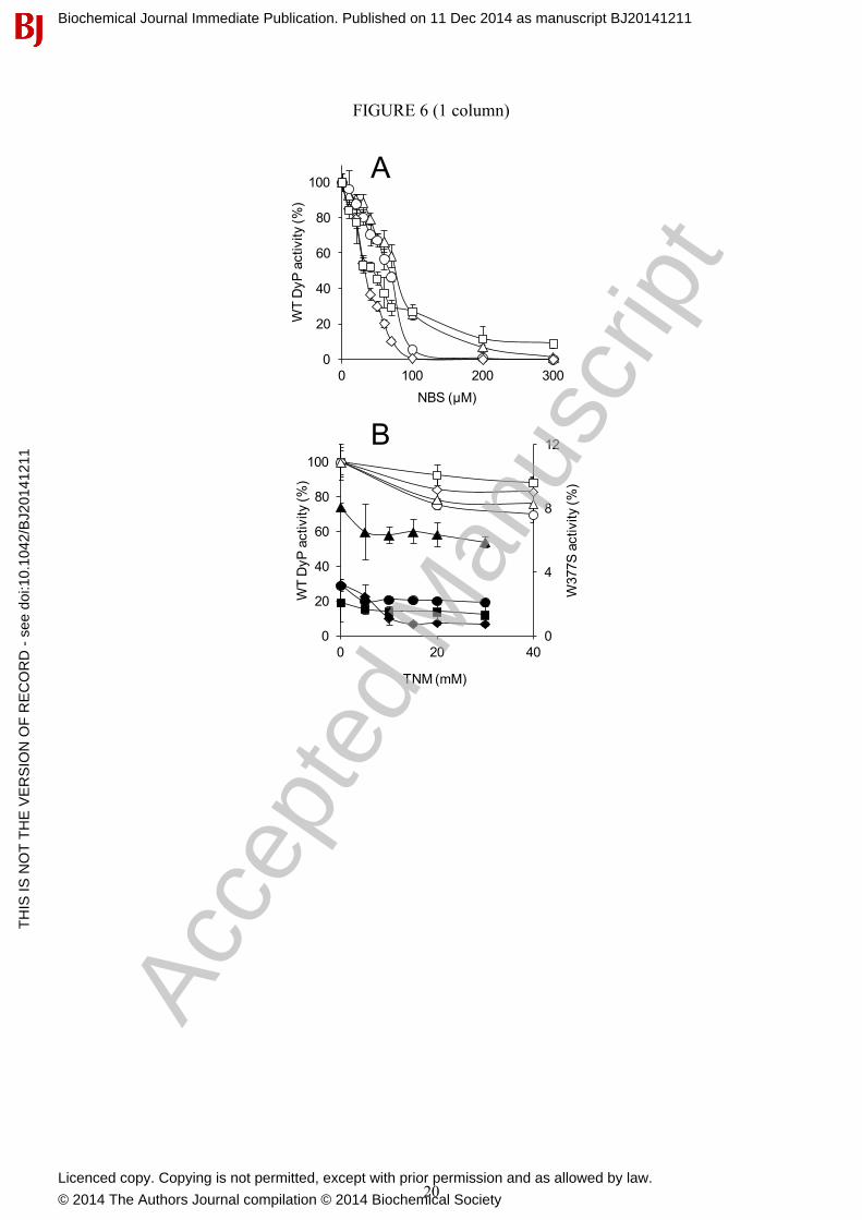
In a first approach for residue modification, A. auricula-judae DyP (3 µM) was treated with NBS, which oxidizes the tryptophan ring to oxindole [55], and TNM, which nitrates the phenolic ring of tyrosine [56]. Near 90% activity on the four substrates assayed was removed when NBS (up to 300 µM) was used (Fig. 6A). This confirms that most DyP activity is associated with tryptophan residue/s, while less than 15% would be due to a different site/s. Similar TNM concentrations did not affect DyP activity, but a partial decrease was observed when a 200-fold higher concentration was used (Fig. 6B, white symbols). This indicates that some tyrosine residue/s contribute (directly or indirectly) to substrate oxidation by DyP, although to a lower extent than the tryptophan residue/s. Additional experiments analysed the effect of chemically modifying tyrosines on the very low activity W377S variant described below (Fig. 6B, black symbols). Although activity decrease with some substrates (such as RB19) was observed, the activity with others (such as ABTS) was only slightly reduced indicating that, in these cases, residues other than tyrosines (or maybe the haem cofactor) are involved.
An interesting initial observation, when analysing the kinetics of DyP oxidations, was the bimodal curves obtained for most substrates (Fig. 7A, regions a and b). Similar sigmoidal kinetic curves were recently reported for DyP [19], and previously for P. eryngii VP [57]. Such curves
Biochemical Journal Immediate Publication. Published on 11 Dec 2014 as manuscript BJ20141211T
HIS
IS N
OT
TH
E V
ER
SIO
N O
F R
EC
OR
D -
see
doi
:10.
1042
/BJ2
0141
211
Acce
pted
Man
uscr
ipt
Licenced copy. Copying is not permitted, except with prior permission and as allowed by law.
© 2014 The Authors Journal compilation © 2014 Biochemical Society

7
enable calculation of two set of constants (Table 2) and reveal the existence of, at least, two oxidation sites for the same substrate. The a and b sites in A. auricula-judae DyP are characterized by a high turnover (kcat) with low apparent affinity (as shown by the Km values), and a low turnover with high apparent affinity, respectively. The kinetic constants obtained show that the A. auricula-judae DyP is very efficient in oxidizing RB19 and ABTS dyes. However, it has lower catalytic efficiencies on DMP and RB5, and extremely low activity on VA. Finally, no Mn2+ oxidation activity was observed, as reported for bacterial DyPs [14;15].
Then, the exposed aromatic residues identified in PELE and QM/MM simulations were substituted to verify their eventual involvement in catalysis (information on the effect of mutating haem pocket residues is provided in Supplementary results). Aromatic residues were changed by serines in the simple W377S, Y147S and Y337S variants (the latter with low refolding yield). The double variant combining the last two mutations could not be refolded, but Y147F/Y337F could be obtained. Since the haem access channel was also identified as a possible RB19 binding site (Fig. 2A), the G169L variant was also obtained, whose leucine side-chain completely blocks the haem access channel, as shown in the crystal structure of a variant including the G169L mutation, compared with the WT DyP (Fig. S2B and A, respectively).
The two sets of constants for oxidation of RB19, DMP and ABTS, and a single set for RB5 and VA, by the above five variants are shown in Table 2. The Y285F variant was also analysed but no significant modification of the RB19 constants was observed. As a main conclusion, Trp-377 appears responsible for the high-turnover catalytic site, since its substitution (W377S variant) completely prevented high-turnover oxidation of RB19 and DMP (and decreased over 40 fold the catalytic efficiency of high-turnover ABTS oxidation). In contrast, similar Tyr-337 and Tyr-147 variants (and the double Y147F/Y337F variant) only slightly affected substrate oxidation, and did not remove the high-turnover site.
The above results are illustrated in Fig. 7A-C, where kinetic plots for RB19 oxidation by WT DyP, and several tryptophan (W377S) and tyrosine (Y147S, Y337S and Y147F/Y337F) variants are, respectively, shown. It is observed that the DyP low-turnover site (Fig. 7A, region b), remaining after Trp-377 removal (Fig. 7B), does not correspond to Tyr-147 or Tyr-337 since changing these residues does not significantly affect the enzyme kinetics (Fig. 7C). However, the disappearance of the low-turnover site in the G169L variant (Fig. 7D inset) suggests that this site involves the haem access channel (although a decrease of the maximal turnover was also observed). Similar results were obtained for ABTS oxidation (Table 2) but the situation could be more complicated for DMP suggesting that a third, still unidentified, site participates in oxidation of phenols by A. auricula-judae DyP. Finally, none of the mutations cause noticeable changes in oxidation of VA, which was highly inefficient in all the cases, although Trp-377 appears to be the main residue involved.
A global view on substrate oxidation sites in the A. auricula-judae and other basidiomycete DyPs The enzyme kinetics reveals the existence of at least one high-turnover and one low-turnover oxidation sites in A. auricula-judae DyP. Site-directed mutagenesis showed that high-turnover oxidation of RB19 and other substrates by DyP takes place at the same tryptophan residue (Trp-377), previously identified as forming a protein radical. This agrees with 85-100% decrease of enzyme activity after modifying tryptophan residues with 0.2 mM NBS, and contrasts with the absence of a significant effect reported previously [9]. This discrepancy is probably due to the low NBS concentration used in the latter study. Modification of tyrosine residues with comparatively high TNM concentrations (40 mM) partially decreased (10-25%) the activity of DyP, as previously reported for the enzyme purified from a fungal culture [23]. However, this does not seem to be due to modification of Tyr-337 (or contiguous Tyr-147) since the enzyme activity was practically unaffected after site-directed mutagenesis of these two residues. A possibility suggested by favourable RB19 docking by PELE, is low-turnover oxidation at the DyP haem channel. Interestingly, the B. adusta DyP has been crystallized with DMP occupying the entrance of a second channel near the haem propionates [7]. This channel does not exist in the A. auricula-judae
Biochemical Journal Immediate Publication. Published on 11 Dec 2014 as manuscript BJ20141211T
HIS
IS N
OT
TH
E V
ER
SIO
N O
F R
EC
OR
D -
see
doi
:10.
1042
/BJ2
0141
211
Acce
pted
Man
uscr
ipt
Licenced copy. Copying is not permitted, except with prior permission and as allowed by law.
© 2014 The Authors Journal compilation © 2014 Biochemical Society

8
DyP, where it is covered with a loop (residues 316-324). Therefore, direct substrate oxidation by the haem cofactor in the latter DyP is only possible at the main haem access channel, in agreement with results from the G169L mutation that blocked the channel. A tyrosine residue could be also responsible for the second/third catalytic sites, in agreement with the partial activity loss after TNM modification. Nevertheless, once demonstrated that Trp-377 is the main substrate oxidation site in A. auricula-judae DyP, it is difficult to unambiguously identify these second/third catalytic sites due to the variety of exposed aromatic residues in this haem peroxidase.
The two aromatic residues forming the mixed tryptophanyl/tyrosyl radical in the H2O2-activated DyP of A. auricula-judae (Trp-377 and Tyr-337) are not a peculiarity of this enzyme, but they are significantly conserved among basidiomycete DyPs (Fig. S8). Among the 65 DyP sequences compared, 58 include a tryptophan homologous to Trp-377, and 48 a tyrosine homologous to Tyr-337 (while the neighbour Tyr-147 is only conserved in 14 sequences). Therefore, the presence of exposed aromatic residues able to form catalytic radicals appears as a common characteristic of basidiomycete, and probably other, DyPs. Although involvement of tyrosyl radicals in oxidation of bulky dyes by DyP has been recently compared to oxidation of recalcitrant aromatics by tryptophanyl radicals in ligninolytic classical peroxidases [4], the present results suggest that tryptophanyl radicals are mainly responsible for substrate oxidation by these two phylogenetically unrelated peroxidase types. AUTHOR CONTRIBUTIONS D. Linde and C. Coscolín performed most of the experimental biochemical work. R. Pogni, V. Sáez-Jiménez, M.C. Baratto and A. Sinicropi contributed EPR experiments and parameter calculations, simulations and interpretations, M. Canyellas, F. Lucas and V. Guallar contributed molecular docking, QM/MM and other computations. F.J. Medrano and A. Romero contributed crystallographic structures. All authors participated in the interpretation and discussion of results. A.T. Martínez, F.J. Ruiz-Dueñas, V. Guallar and R. Pogni contributed data integration and writing. ACKNOWLEDGEMENTS The authors thank the staff of the SOLEIL (Gyf-sur-Yvette, France) and ALBA (Barcelona, Spain) synchrotrons, and the BSC (Barcelona, Spain) computational facilities. The MALDI-TOF analyses were carried out at the CIB Proteomics facility, a member of the Spanish ProteoRed-ISCIII network. D.L. and F.J.R.-D. thank the financial support of an EU project contract, and a Ramón y Cajal contract of the Spanish Ministry of Economy and Competitiveness (MINECO), respectively. FUNDING This work was supported by the INDOX (KBBE-2013-7-613549) and PELE (ERC-2009-Adg 25027) EU projects, by the BIO2011-26694, CTQ2013-48287 and BFU2011-24615 projects of the Spanish MINECO, and by the PRIN 2009-STNWX3 project of the Italian Ministry of Education, Universities and Research (MIUR). REFERENCES 1 Sugano, Y. (2009) DyP-type peroxidases comprise a novel heme peroxidase family. Cell Mol. Life Sci., 66, 1387-
1403 2 Hofrichter, M., Ullrich, R., Pecyna, M. J., Liers, C. and Lundell, T. (2010) New and classic families of secreted fungal
heme peroxidases. Appl. Microbiol. Biotechnol., 87, 871-897 3 Ruiz-Dueñas, F. J. and Martínez, A. T. (2010) Structural and functional features of peroxidases with a potential as
industrial biocatalysts. In Biocatalysts based on heme peroxidases (Torres,E. and Ayala,M. Eds.), pp. 37-59. Springer-Verlag, Berlin
4 Colpa, D. I., Fraaije, M. W. and van Bloois, E. (2014) DyP-type peroxidases: a promising and versatile class of enzymes. J. Ind. Microbiol. Biotechnol., 41, 1-7
5 Kim, S. J. and Shoda, M. (1999) Purification and characterization of a novel peroxidase from Geotrichum candidum Dec 1 involved in decolorization of dyes. Appl. Environ. Microbiol., 65, 1029-1035
6 Sugano, Y., Muramatsu, R., Ichiyanagi, A., Sato, T. and Shoda, M. (2007) DyP, a unique dye-decolorizing peroxidase, represents a novel heme peroxidase family. Asp171 replaces the distal histidine of classical peroxidases. J. Biol. Chem., 282, 36652-36658
Biochemical Journal Immediate Publication. Published on 11 Dec 2014 as manuscript BJ20141211T
HIS
IS N
OT
TH
E V
ER
SIO
N O
F R
EC
OR
D -
see
doi
:10.
1042
/BJ2
0141
211
Acce
pted
Man
uscr
ipt
Licenced copy. Copying is not permitted, except with prior permission and as allowed by law.
© 2014 The Authors Journal compilation © 2014 Biochemical Society

9
7 Yoshida, T., Tsuge, H., Hisabori, T. and Sugano, Y. (2012) Crystal structures of dye-decolorizing peroxidase with ascorbic acid and 2,6-dimethoxyphenol. FEBS Lett., 586, 4351-4356
8 Liers, C., Pecyna, M. J., Kellner, H., Worrich, A., Zorn, H., Steffen, K. T., Hofrichter, M. and Ullrich, R. (2013) Substrate oxidation by dye-decolorizing peroxidases (DyPs) from wood- and litter-degrading agaricomycetes compared to other fungal and plant heme-peroxidases. Appl Microbiol Biotechnol, 87, 5839-5849
9 Strittmatter, E., Liers, C., Ullrich, R., Wachter, S., Hofrichter, M., Plattner, D. A. and Piontek, K. (2013) First crystal structure of a fungal high-redox potential dye-decolorizing peroxidase: Substrate interaction sites and long-range electron transfer. J. Biol. Chem., 288, 4095-4102
10 Liers, C., Aranda, E., Strittmatter, E., Piontek, K., Plattner, D. A., Zorn, H., Ullrich, R. and Hofrichter, M. (2014) Phenol oxidation by DyP-type peroxidases in comparison to fungal and plant peroxidases. J. Mol. Catal. B-Enzym., 103, 41-46
11 Zubieta, C., Joseph, R., Krishna, S. S., McMullan, D., Kapoor, M., Axelrod, H. L., Miller, M. D., Abdubek, P., Acosta, C., Astakhova, T., Carlton, D., Chiu, H. J., Clayton, T., Deller, M. C., Duan, L., Elias, Y., Elsliger, M. A., Feuerhelm, J., Grzechnik, S. K., Hale, J., Han, G. W., Jaroszewski, L., Jin, K. K., Klock, H. E., Knuth, M. W., Kozbial, P., Kumar, A., Marciano, D., Morse, A. T., Murphy, K. D., Nigoghossian, E., Okach, L., Oommachen, S., Reyes, R., Rife, C. L., Schimmel, P., Trout, C. V., van den Bedem, H., Weekes, D., White, A., Xu, Q. P., Hodgson, K. O., Wooley, J., Deacon, A. M., Godzik, A., Lesley, S. A. and Wilson, I. A. (2007) Identification and structural characterization of heme binding in a novel dye-decolorizing peroxidase, TyrA. Proteins, 69, 234-243
12 Zubieta, C., Krishna, S. S., Kapoor, M., Kozbial, P., McMullan, D., Axelrod, H. L., Miller, M. D., Abdubek, P., Ambing, E., Astakhova, T., Carlton, D., Chiu, H. J., Clayton, T., Deller, M. C., Duan, L., Elsliger, M. A., Feuerhelm, J., Grzechnik, S. K., Hale, J., Hampton, E., Han, G. W., Jaroszewski, L., Jin, K. K., Klock, H. E., Knuth, M. W., Kumar, A., Marciano, D., Morse, A. T., Nigoghossian, E., Okach, L., Oommachen, S., Reyes, R., Rife, C. L., Schimmel, P., van den, B. H., Weekes, D., White, A., Xu, Q., Hodgson, K. O., Wooley, J., Deacon, A. M., Godzik, A., Lesley, S. A. and Wilson, I. A. (2007) Crystal structures of two novel dye-decolorizing peroxidases reveal a beta-barrel fold with a conserved heme-binding motif. Proteins, 69, 223-233
13 Liu, X. H., Du, Q., Wang, Z., Zhu, D. Y., Huang, Y., Li, N., Wei, T. D., Xu, S. J. and Gu, L. C. (2011) Crystal structure and biochemical features of EfeB/YcdB from Escherichia coli O157. Asp235 plays divergent roles in different enzyme-cayalyzed processes. J. Biol. Chem., 286, 14922-14931
14 Roberts, J. N., Singh, R., Grigg, J. C., Murphy, M. E. P., Bugg, T. D. H. and Eltis, L. D. (2011) Characterization of dye-decolorizing peroxidases from Rhodococcus jostii RHA1. Biochemistry, 50, 5108-5119
15 Brown, M. E., Barros, T. and Chang, M. C. Y. (2012) Identification and characterization of a multifunctional dye peroxidase from a lignin-reactive bacterium. ACS Chemical Biology, 7, 2074-2081
16 Sugano, Y., Nakano, R., Sasaki, K. and Shoda, M. (2000) Efficient heterologous expression in Aspergillus oryzae of a unique dye decoloryzing peroxidase, DyP, of Geotrichum candidum Dec 1. Appl. Environ. Microbiol., 66, 1754-1758
17 Sugano, Y., Ishii, Y. and Shoda, M. (2004) Role of H164 in a unique dye-decolorizing heme peroxidase DyP. Biochem. Biophys. Res. Commun., 322, 126-132
18 Liers, C., Bobeth, C., Pecyna, M., Ullrich, R. and Hofrichter, M. (2010) DyP-like peroxidases of the jelly fungus Auricularia auricula-judae oxidize nonphenolic lignin model compounds and high-redox potential dyes. Appl. Microbiol. Biotechnol., 85, 1869-1879
19 Linde, D., Coscolín, C., Liers, C., Hofrichter, M., Martínez, A. T. and Ruiz-Dueñas, F. J. (2014) Heterologous expression and physicochemical characterization of a fungal dye-decolorizing peroxidase from Auricularia auricula-judae. Protein Express. Purif., 103, 28-37
20 Floudas, D., Binder, M., Riley, R., Barry, K., Blanchette, R. A., Henrissat, B., Martínez, A. T., Otillar, R., Spatafora, J. W., Yadav, J. S., Aerts, A., Benoit, I., Boyd, A., Carlson, A., Copeland, A., Coutinho, P. M., de Vries, R. P., Ferreira, P., Findley, K., Foster, B., Gaskell, J., Glotzer, D., Górecki, P., Heitman, J., Hesse, C., Hori, C., Igarashi, K., Jurgens, J. A., Kallen, N., Kersten, P., Kohler, A., Kües, U., Kumar, T. K. A., Kuo, A., LaButti, K., Larrondo, L. F., Lindquist, E., Ling, A., Lombard, V., Lucas, S., Lundell, T., Martin, R., McLaughlin, D. J., Morgenstern, I., Morin, E., Murat, C., Nolan, M., Ohm, R. A., Patyshakuliyeva, A., Rokas, A., Ruiz-Dueñas, F. J., Sabat, G., Salamov, A., Samejima, M., Schmutz, J., Slot, J. C., St.John, F., Stenlid, J., Sun, H., Sun, S., Syed, K., Tsang, A., Wiebenga, A., Young, D., Pisabarro, A., Eastwood, D. C., Martin, F., Cullen, D., Grigoriev, I. V. and Hibbett, D. S. (2012) The Paleozoic origin of enzymatic lignin decomposition reconstructed from 31 fungal genomes. Science, 336, 1715-1719
21 Ahmad, M., Roberts, J. N., Hardiman, E. M., Singh, R., Eltis, L. D. and Bugg, T. D. H. (2011) Identification of DypB from Rhodococcus jostii RHA1 as a lignin peroxidase. Biochemistry, 50, 5096-5107
22 Brown, M. E. and Chang, M. C. Y. (2014) Exploring bacterial lignin degradation. Curr. Opin. Chem. Biol., 19, 1-7 23 Strittmatter, E., Wachter, S., Liers, C., Ullrich, R., Hofrichter, M., Plattner, D. A. and Piontek, K. (2013) Radical
formation on a conserved tyrosine residue is crucial for DyP activity. Arch. Biochem. Biophys., 537, 161-167
Biochemical Journal Immediate Publication. Published on 11 Dec 2014 as manuscript BJ20141211T
HIS
IS N
OT
TH
E V
ER
SIO
N O
F R
EC
OR
D -
see
doi
:10.
1042
/BJ2
0141
211
Acce
pted
Man
uscr
ipt
Licenced copy. Copying is not permitted, except with prior permission and as allowed by law.
© 2014 The Authors Journal compilation © 2014 Biochemical Society

10
24 Doyle, W. A., Blodig, W., Veitch, N. C., Piontek, K. and Smith, A. T. (1998) Two substrate interaction sites in lignin peroxidase revealed by site-directed mutagenesis. Biochemistry, 37, 15097-15105
25 Pérez-Boada, M., Ruiz-Dueñas, F. J., Pogni, R., Basosi, R., Choinowski, T., Martínez, M. J., Piontek, K. and Martínez, A. T. (2005) Versatile peroxidase oxidation of high redox potential aromatic compounds: Site-directed mutagenesis, spectroscopic and crystallographic investigations of three long-range electron transfer pathways. J. Mol. Biol., 354, 385-402
26 Wallrapp, F. H., Voityuk, A. A. and Guallar, V. (2013) In-silico assessment of protein-protein electron transfer. A case study: Cytochrome c peroxidase - cytochrome c. PLoS Comput. Biol., 9, 3, e1002990
27 van der Kamp, M. W. and Mulholland, A. J. (2013) Combined quantum mechanics/molecular mechanics (QM/MM) methods in computational enzymology. Biochemistry, 52, 2708-2728
28 Guallar, V. and Wallrapp, F. H. (2010) QM/MM methods: Looking inside heme proteins biochemisty. Biophys. Chem., 149, 1-11
29 Borrelli, K. W., Vitalis, A., Alcantara, R. and Guallar, V. (2005) PELE: Protein energy landscape exploration. A novel Monte Carlo based technique. J. Chem. Theory Comput., 1, 1304-1311
30 Miki, Y., Ichinose, H. and Wariishi, H. (2011) Determination of a catalytic tyrosine in Trametes cervina lignin peroxidase with chemical modification techniques. Biotechnol. Lett., 33, 1423-1427
31 Schrödinger. (2011) QSite 5.7, LCC, New York 32 Guallar, V. and Wallrapp, F. (2008) Mapping protein electron transfer pathways with QM/MM methods. J. R. Soc.
Interface, 5, S233-S239 33 Stoll, S. and Schweiger, A. (2006) EasySpin, a comprehensive software package for spectral simulation and
analysis in EPR. Journal of Magnetic Resonance, 178, 42-55 34 Aquilante, F., De Vico, L., Ferre, N., Ghigo, G., Malmqvist, P. A., Neogrady, P., Pedersen, T. B., Pitonak, M., Reiher,
M., Roos, B. O., Serrano-Andres, L., Urban, M., Veryazov, V. and Lindh, R. (2010) Software news and update MOLCAS 7: The next generation. J. Comp. Chem., 31, 224-247
35 Neese, F. (2012) ORCA: An, ab initio, density functional and semi-empirical program package. Version 2.9, University of Bonn (Germany),
36 Bernini, C., Pogni, R., Ruiz-Dueñas, F. J., Martínez, A. T., Basosi, R. and Sinicropi, A. (2011) EPR parameters of amino acid radicals in P. eryngii versatile peroxidase and its W164Y variant computed at the QM/MM level. Phys. Chem. Chem. Phys., 13, 5078-5098
37 Bernini, C., Andruniow, T., Olivucci, M., Pogni, R., Basosi, R. and Sinicropi, A. (2013) Effects of the protein environment on the spectral properties of tryptophan radicals in Pseudomonas aeruginosa azurin. J. Am. Chem. Soc., 135, 4822-4833
38 Goblirsch, B., Kurker, R. C., Streit, B. R., Wilmot, C. M. and Dubois, J. L. (2011) Chlorite dismutases, DyPs, and EfeB: 3 microbial heme enzyme families comprise the CDE structural superfamily. J. Mol. Biol., 408, 379-398
39 Hofbauer, S., Schaffner, I., Furtmuller, P. G. and Obinger, C. (2014) Chlorite dismutases - a heme enzyme family for use in bioremediation and generation of molecular oxygen. Biotechnol. J., 9, 461-473
40 Welinder, K. G. (1992) Superfamily of plant, fungal and bacterial peroxidases. Curr. Opin. Struct. Biol., 2, 388-393 41 Miki, Y., Calviño, F. R., Pogni, R., Giansanti, S., Ruiz-Dueñas, F. J., Martínez, M. J., Basosi, R., Romero, A. and
Martínez, A. T. (2011) Crystallographic, kinetic, and spectroscopic study of the first ligninolytic peroxidase presenting a catalytic tyrosine. J. Biol. Chem., 286, 15525-15534
42 Ruiz-Dueñas, F. J., Pogni, R., Morales, M., Giansanti, S., Mate, M. J., Romero, A., Martínez, M. J., Basosi, R. and Martínez, A. T. (2009) Protein radicals in fungal versatile peroxidase: Catalytic tryptophan radical in both Compound I and Compound II and studies on W164Y, W164H and W164S variants. J. Biol. Chem., 284, 7986-7994
43 Smith, A. T., Doyle, W. A., Dorlet, P. and Ivancich, A. (2009) Spectroscopic evidence for an engineered, catalytically active Trp radical that creates the unique reactivity of lignin peroxidase. Proc. Natl. Acad. Sci. USA, 106, 16084-16089
44 Pogni, R., Baratto, M. C., Teutloff, C., Giansanti, S., Ruiz-Dueñas, F. J., Choinowski, T., Piontek, K., Martínez, A. T., Lendzian, F. and Basosi, R. (2006) A tryptophan neutral radical in the oxidized state of versatile peroxidase from Pleurotus eryngii: a combined multi-frequency EPR and DFT study. J. Biol. Chem., 281, 9517-9526
45 Pogni, R., Baratto, M. C., Giansanti, S., Teutloff, C., Verdín, J., Valderrama, B., Lendzian, F., Lubitz, W., Vázquez-Duhalt, R. and Basosi, R. (2005) Tryptophan-based radical in the catalytic mechanism of versatile peroxidase from Bjerkandera adusta. Biochemistry, 44, 4267-4274
46 Martínez, A. T. (2002) Molecular biology and structure-function of lignin-degrading heme peroxidases. Enzyme Microb. Technol., 30, 425-444
47 Ruiz-Dueñas, F. J., Morales, M., García, E., Miki, Y., Martínez, M. J. and Martínez, A. T. (2009) Substrate oxidation sites in versatile peroxidase and other basidiomycete peroxidases. J. Exp. Bot., 60, 441-452
48 Jeschke, G. (2005) EPR techniques for studying radical enzymes. Biochim. Biophys. Acta-Bioenerg., 1707, 91-102
Biochemical Journal Immediate Publication. Published on 11 Dec 2014 as manuscript BJ20141211T
HIS
IS N
OT
TH
E V
ER
SIO
N O
F R
EC
OR
D -
see
doi
:10.
1042
/BJ2
0141
211
Acce
pted
Man
uscr
ipt
Licenced copy. Copying is not permitted, except with prior permission and as allowed by law.
© 2014 The Authors Journal compilation © 2014 Biochemical Society

11
49 Bleifuss, G., Kolberg, M., Potsch, S., Hofbauer, W., Bittl, R., Lubitz, W., Graslund, A., Lassmann, G. and Lendzian, F. (2001) Tryptophan and tyrosine radicals in ribonucleotide reductase: a comparative high-field EPR study at 94 GHz. Biochemistry, 40, 15362-15368
50 Svistunenko, D. A., Dunne, J., Fryer, M., Nicholls, P., Reeder, B. J., Wilson, M. T., Bigotti, M. G., Cutruzzola, F. and Cooper, C. E. (2002) Comparative study of tyrosine radicals in hemoglobin and myoglobins treated with hydrogen peroxide. Biophys. J., 83, 2845-2855
51 Warren, J. J., Winkler, J. R. and Gray, H. B. (2012) Redox properties of tyrosine and related molecules. FEBS Lett., 586, 596-602
52 Stubbe, J. and Der Donk, W. A. (1998) Protein radicals in enzyme catalysis. Chem. Rev., 98, 705-762 53 Fernández-Fueyo, E., Ruiz-Dueñas, F. J. and Martínez, A. T. (2014) Engineering a fungal peroxidase that degrades
lignin at very acidic pH. Biotechnol. Biofuels, 7, 114 (published 24 July 2014) 54 Miki, Y., Pogni, R., Acebes, S., Lucas, F., Fernández-Fueyo, E., Baratto, M. C., Fernández, M. I., de los Ríos, V.,
Ruiz-Dueñas, F. J., Sinicropi, A., Basosi, R., Hammel, K. E., Guallar, V. and Martínez, A. T. (2013) Formation of a tyrosine adduct involved in lignin degradation by Trametopsis cervina lignin peroxidase: A novel peroxidase activation mechanism. Biochem. J., 452, 575-584
55 Spande, T. F., Green, N. M. and Witkop, B. (1966) Reactivity toward N-bromosuccinimide of tryptophan in enzymes zymogens and inhibited enzymes. Biochemistry, 5, 1926-1933
56 Sokolovsky, M., Riordan, J. F. and Vallee, B. L. (1966) Tetranitromethane. A reagent for nitration of tyrosyl residues in proteins. Biochemistry, 5, 3582-3589
57 Morales, M., Mate, M. J., Romero, A., Martínez, M. J., Martínez, A. T. and Ruiz-Dueñas, F. J. (2012) Two oxidation sites for low redox-potential substrates: A directed mutagenesis, kinetic and crystallographic study on Pleurotus eryngii versatile peroxidase. J. Biol. Chem., 287, 41053-41067
Biochemical Journal Immediate Publication. Published on 11 Dec 2014 as manuscript BJ20141211T
HIS
IS N
OT
TH
E V
ER
SIO
N O
F R
EC
OR
D -
see
doi
:10.
1042
/BJ2
0141
211
Acce
pted
Man
uscr
ipt
Licenced copy. Copying is not permitted, except with prior permission and as allowed by law.
© 2014 The Authors Journal compilation © 2014 Biochemical Society

12
Table 1 Steady-state kinetic constants of WT DyP and five site-directed variants Km (µM), kcat (s-1), and kcat/Km (s-1·mM-1) of WT DyP and its Trp-377, Tyr-147, Tyr-337 and Gly-169 variants oxidizing RB19, DMP, ABTS, RB5 and VA (including two sets of constants for the first four substrates corresponding to a high-turnover and a low-turnover site). Means and 95% confidence limits from reactions at 25 °C in 0.1 M tartrate, pH 3 (pH 3.5 for RB19 and pH 2.5 for VA) using 0.1 mM H2O2 and 10 nM enzyme (100 nM for VA oxidation), are shown (when 1 mM H2O2 concentration was used, lower catalytic efficiencies were obtained, although the kcat values often increased).
WT DyP W377S Y147S Y337S Y147F/Y337F G169L RB19 (high turnover)
Km 90 ± 10 - 95 ± 24 83 ± 3 130 ± 30 10 ± 3 kcat 224 ± 10 0 175 ± 24 240 ± 47 220 ± 16 106 ± 9 kcat/Km 2460 ± 180 - 1860 ± 300 2700 ± 400 1680 ± 170 12400 ± 3000
RB19 (low turnover)
Km 14.0 ± 2.0 3.9 ± 0.6 6.4 ± 0.8 7.4 ± 2.3 5.8 ± 0.8 - kcat 32.0 ± 3.0 8.9 ± 0.6 26.0 ± 6.1 26.0 ± 1.9 18.0 ± 1.9 0 kcat/Km 2230 ± 200 2240 ± 240 4070 ± 640 3370 ± 350 3200 ± 200 -
DMP (high turnover)
Km 703 ± 61 - 763 ± 70 2840 ± 300 4720 ± 460 353 ± 41 kcat 120 ± 3 0 71 ± 2 228 ± 18 216 ± 7 88 ± 3 kcat/Km 200 ± 18 - 93 ± 7 80 ± 1.1 46 ± 3 200 ± 21
DMP (low turnover)
Km 6.0 ± 0.5 3560 ± 250 38.0 ± 0.4 3.1 ± 0.6 0.7 ± 0.1 6.0 ± 0.4 kcat 8.0 ± 0.2 6.4 ± 0.1 15.0 ± 0.9 8.4 ± 0.3 3.9 ± 0.1 9.9 ± 0.2 kcat/Km 1350 ± 100 1.8 ± 0.1 397 ± 41 2730 ± 520 5900 ± 1000 1600 ± 70
ABTS (high turnover)
Km 121 ± 7 2750 ± 470 366 ± 30 239 ± 29 173 ± 21 25 ± 2 kcat 224 ± 3 171 ± 15 311 ± 13 288 ± 10 286 ± 7.2 96 ± 2 kcat/Km 1850 ± 94 62 ± 1 850± 80 1200 ± 70 1650 ± 180 3770 ± 270
ABTS (low turnover)
Km 3.1 ± 1.1 30.0 ± 0.5 20.0 ± 6.0 2.0 ± 0.2 17.0 ± 3.6 - kcat 7.4 ± 1.4 14.0 ± 1.0 21.0 ± 4.0 2.3 ± 0.3 21.0 ± 6.0 0 kcat/Km 2370 ± 420 472 ± 61 1010 ± 100 1100 ± 100 1260 ± 120 -
RB5 Km 15.6 ± 2.0 10.6 ± 1.0 15.9 ± 1.7 13.4 ± 1.7 39.0 ± 6.9 5.8 ± 0.6 kcat 4.8 ± 0.2 0.70 ± 0.02 8.3 ± 0.6 5.2 ± 0.3 9.1 ± 2.6 1.2 ± 0.1 kcat/Km 310 ± 20 68 ± 4 525 ± 230 400 ± 29 233 ± 20 217 ± 13
VA Km nsa 7360 ± 1270 ns ns ns Ns kcat - 0.23 ± 0.01 - - - - kcat/Km 0.096 ± 0.002 0.032 ± 0.003 0.101 ± 0.005 0.079 ± 0.011 0.095 ± 0.002 0.130 ± 0.006
a ns, no saturation preventing kinetic constant estimation
Biochemical Journal Immediate Publication. Published on 11 Dec 2014 as manuscript BJ20141211T
HIS
IS N
OT
TH
E V
ER
SIO
N O
F R
EC
OR
D -
see
doi
:10.
1042
/BJ2
0141
211
Acce
pted
Man
uscr
ipt
Licenced copy. Copying is not permitted, except with prior permission and as allowed by law.
© 2014 The Authors Journal compilation © 2014 Biochemical Society

13
Figure 1 Folding of A. auricula-judae DyP and location of exposed aromatic residues (A) General folding constituted by two domains, each of them including two large β-sheets and 2-3 helices, with the haem cofactor in the upper part of the lower domain (cartoon coloured from the N to the C termini, with the haem as CPK sticks). (B and C) Location of exposed Trp105, Tyr147, Trp207, Tyr285, Tyr337 and Trp377 (as CPK spheres) in two different orientations of the DyP molecule (cartoon with the haem as CPK sticks). From PDB 4W7J. Figure 2 Substrate exploration on the DyP surface (A) Local minima identified in the PELE [29] simulations of RB19 diffusion on the recombinant DyP crystal structure (PDB 4W7J) showing interaction energy vs distance to Tyr-147 (taken as a reference residue). The presence of RB19 in the vicinity of different surface residues and the haem access channel is indicated. (B) Distances between the closest positions of RB19 (magenta sticks) with respect to haem (1), Tyr-147/Tyr-337 (2), and Trp-377 (3) shown by PELE (A), and between the above residues and the haem cofactor (the distances are measured including hydrogen atoms). RB19 as CPK sticks, Tyr-147/Tyr-337 as magenta sticks and Trp-377 as gray sticks. Figure 3 QM/MM electron spin distribution on Trp377 and Tyr337 residues and RB19 substrate (A) Total spin density when including Trp-377, Tyr-337 and haem compound I in the quantum region (in absence of RB19). (B and C) Total spin density when, in addition to RB19 and compound I, the quantum region includes Trp377 or Tyr337, respectively. From PDB 4W7J after 5 ns MD (A) and selected snapshots from two energy minima showing RB19 near Trp377 and Tyr337, during PELE [29] diffusion in Fig. 2 (B and C, respectively). Figure 4 Electron transfer pathway from DyP Trp377 to haem The electron transfer pathway was obtained after three iterations of the QM/MM e-pathway approach [32] with a total of fifteen residues (His-304 to Arg-311, Leu-323 to Ala-325, Leu-373 to Gln-375 and Asp-395) included in the quantum region. Each iteration identifies the residue/s with the highest affinity for the electron, and is shown in a different colour. The mapped route includes Pro-310 and Arg-309, followed by Arg-306, Ile-305 and His-304, as shown by the electron spin distribution. Figure 5 EPR spectra of WT DyP and its W377S variant (A) X-band EPR spectra of WT DyP at pH 3 before (top) and after (bottom) the addition of H2O2 (and rapid freezing). Experimental conditions: ν= 9.39 GHz, 0.2 mW microwave power, 0.4 mT modulation amplitude). (B and C) Narrow scan X-band (ν= 9.39 GHz) and W-band (ν= 94.29 GHz), respectively, of the radical species. The positions of the three g-tensor components of the tyrosyl contribution are indicated. X-band experimental conditions: ν= 9.38 GHz, 1 mW microwave power, and 0.05 mT modulation amplitude; W-band experimental conditions: ν= 94.29 GHz, 0.05 mW microwave power, and 0.1 mT modulation amplitude. (D) X-band EPR spectrum of the radical intermediate formed in the W377S variant paired with its better simulation (see magnetic parameters in Table S3). Experimental conditions: v= 9.39 GHz, 1 mW microwave power, 0.2 mT modulation amplitude. Figure 6 Chemical modification of tryptophan and tyrosine residues in WT DyP and its W377S variant (A) Residual activities of 3 μM WT DyP treated with increasing NBS concentrations (in 50 mM acetate, pH 4) for modification of tryptophan residues. (B) Residual activities of 3 μM WT DyP and W377S variant (white and black symbols, respectively) treated with increasing TNM concentrations (in 50 mM Tris-HCl, pH 7, with 2.6% ethanol) for modification of tyrosine residues. The residual activities of the WT DyP and the W377S variant in A and B were monitored for oxidation of 180 μM RB19 (diamonds), 7.5 mM DMP (circles), 15 µM RB5 (squares) and 1.25 mM ABTS (triangles), and referred to activities of untreated WT DyP (taken as 100%).
Biochemical Journal Immediate Publication. Published on 11 Dec 2014 as manuscript BJ20141211T
HIS
IS N
OT
TH
E V
ER
SIO
N O
F R
EC
OR
D -
see
doi
:10.
1042
/BJ2
0141
211
Acce
pted
Man
uscr
ipt
Licenced copy. Copying is not permitted, except with prior permission and as allowed by law.
© 2014 The Authors Journal compilation © 2014 Biochemical Society

14
Figure 7 Kinetics for RB19 oxidation by WT DyP and different variants (A) WT DyP biphasic kinetics enabling calculation of two sets of constants in the 0.2-10 μM (b) and 50-270 μM (a) ranges (inset with Lineweaver-Burk inverse representation for the high, a, and low, b, turnover sites). (B) Simple kinetics yielding a single set of constants (inset, inverse representation) for the W377S variant. (C) Results from Y147S (squares), Y337S (diamonds) and Y147F/Y337F (triangles) variants yielding kinetic curves superimposable with that of WT DyP (circles). (D) Simple kinetics yielding a single set of constants (inset, inverse representation) for the G169L variant. A substrate concentration logarithmic scale is used in the main plots.
Biochemical Journal Immediate Publication. Published on 11 Dec 2014 as manuscript BJ20141211T
HIS
IS N
OT
TH
E V
ER
SIO
N O
F R
EC
OR
D -
see
doi
:10.
1042
/BJ2
0141
211
Acce
pted
Man
uscr
ipt
Licenced copy. Copying is not permitted, except with prior permission and as allowed by law.
© 2014 The Authors Journal compilation © 2014 Biochemical Society

15
FIGURE 1 (2 columns)
A B C
Biochemical Journal Immediate Publication. Published on 11 Dec 2014 as manuscript BJ20141211T
HIS
IS N
OT
TH
E V
ER
SIO
N O
F R
EC
OR
D -
see
doi
:10.
1042
/BJ2
0141
211
Acce
pted
Man
uscr
ipt
Licenced copy. Copying is not permitted, except with prior permission and as allowed by law.
© 2014 The Authors Journal compilation © 2014 Biochemical Society

16
FIGURE 2 (1.5 columns)
Tyr147
Trp377
Tyr337
His304
2.8 Å
6.4 Å6.1 Å
6.4 Å
3.0 Å
1.6 Å
B
21
3
2
1
3
A
Biochemical Journal Immediate Publication. Published on 11 Dec 2014 as manuscript BJ20141211T
HIS
IS N
OT
TH
E V
ER
SIO
N O
F R
EC
OR
D -
see
doi
:10.
1042
/BJ2
0141
211
Acce
pted
Man
uscr
ipt
Licenced copy. Copying is not permitted, except with prior permission and as allowed by law.
© 2014 The Authors Journal compilation © 2014 Biochemical Society
17
FIGURE 3 (1.5 columns)
A
B
C
Trp377
Heme Tyr337
Biochemical Journal Immediate Publication. Published on 11 Dec 2014 as manuscript BJ20141211T
HIS
IS N
OT
TH
E V
ER
SIO
N O
F R
EC
OR
D -
see
doi
:10.
1042
/BJ2
0141
211
Acce
pted
Man
uscr
ipt
Licenced copy. Copying is not permitted, except with prior permission and as allowed by law.
© 2014 The Authors Journal compilation © 2014 Biochemical Society
18
FIGURE 4 (1 column)
Biochemical Journal Immediate Publication. Published on 11 Dec 2014 as manuscript BJ20141211T
HIS
IS N
OT
TH
E V
ER
SIO
N O
F R
EC
OR
D -
see
doi
:10.
1042
/BJ2
0141
211
Acce
pted
Man
uscr
ipt
Licenced copy. Copying is not permitted, except with prior permission and as allowed by law.
© 2014 The Authors Journal compilation © 2014 Biochemical Society

19
FIGURE 5 (1.5 columns)
Biochemical Journal Immediate Publication. Published on 11 Dec 2014 as manuscript BJ20141211T
HIS
IS N
OT
TH
E V
ER
SIO
N O
F R
EC
OR
D -
see
doi
:10.
1042
/BJ2
0141
211
Acce
pted
Man
uscr
ipt
Licenced copy. Copying is not permitted, except with prior permission and as allowed by law.
© 2014 The Authors Journal compilation © 2014 Biochemical Society
20
FIGURE 6 (1 column)
0
20
40
60
80
100
0 100 200 300
WT
DyP
act
ivity
(%)
NBS (µM)
A
B
0
4
8
12
0
20
40
60
80
100
0 20 40
W37
7S a
ctiv
ity (%
)
WT
DyP
act
ivity
(%)
TNM (mM)
Biochemical Journal Immediate Publication. Published on 11 Dec 2014 as manuscript BJ20141211T
HIS
IS N
OT
TH
E V
ER
SIO
N O
F R
EC
OR
D -
see
doi
:10.
1042
/BJ2
0141
211
Acce
pted
Man
uscr
ipt
Licenced copy. Copying is not permitted, except with prior permission and as allowed by law.
© 2014 The Authors Journal compilation © 2014 Biochemical Society

21
FIGURE 7 (1.5 columns)
0
50
100
150
0 1 10 100
k obs
(s-1
)
RB19 (μΜ)
0
50
100
150
0 1 10 100
k obs
(s-1
)
RB19 (μΜ)
A
0
50
100
150
1 10 100
k obs
(s-1
)
RB19 (μΜ)
B
C
0
0,1
0,2
0,3
0,000
0,005
0,010
0,015
0,020
0 0,25 0,5 0,75
1/V
(mg/
U) (
regi
on b
)
1/V
(mg/
U) (
regi
on a
)
1/S (µM-1)
ab
ab
0,0
0,1
0,2
0,3
0 0,25 0,5 0,75
1/V
(mg/
U)
1/S (µM-1)
D
0
50
100
150
0 1 10 100
k obs
(s-1
)
RB19 (μΜ)
0,000
0,005
0,010
0,015
0,020
0 0,25 0,5 0,75
1/V
(mg/
U)
1/S (µM-1)
Biochemical Journal Immediate Publication. Published on 11 Dec 2014 as manuscript BJ20141211T
HIS
IS N
OT
TH
E V
ER
SIO
N O
F R
EC
OR
D -
see
doi
:10.
1042
/BJ2
0141
211
Acce
pted
Man
uscr
ipt
Licenced copy. Copying is not permitted, except with prior permission and as allowed by law.
© 2014 The Authors Journal compilation © 2014 Biochemical Society