
DISTANCE-DEPENDENT FLUORESCENCE QUENCHING OF TRYPTOPHAN BY ACRYLAMIDE
Upload
independentCategory
view
0download
0
Domain folding and flexibility of Escherichia coli FtsZdetermined by tryptophan site-directed mutagenesis
RODRIGO DIAZ-ESPINOZA,1,3 ANDREA P. GARCES,1,3 JOSE J. ARBILDUA,1,3
FELIPE MONTECINOS,1 JUAN E. BRUNET,2 ROSALBA LAGOS,1
AND OCTAVIO MONASTERIO1
1Laboratorio de Biologıa Estructural y Molecular, Departamento de Biologıa, Facultad de Ciencias, Universidad deChile, Casilla 653, Santiago, Chile2Instituto de Quımica, Facultad de Ciencias Basicas y Matematicas, Pontificia Universidad Catolica de Valparaıso,Casilla 4059, Valparaıso, Chile
(RECEIVED February 5, 2007; FINAL REVISION April 26, 2007; ACCEPTED May 21, 2007)
Abstract
FtsZ has two domains, the amino GTPase domain with a Rossmann fold, and the carboxyl domain thatresembles the chorismate mutase fold. Bioinformatics analyses suggest that the interdomain interactionis stronger than the interaction of the protofilament longitudinal interfaces. Crystal B factor analysis ofFtsZ and detected conformational changes suggest a connection between these domains. The unfolding/folding characteristics of each domain of FtsZ were tested by introducing tryptophans into the flexibleregion of the amino (F135W) and the carboxyl (F275W and I294W) domains. As a control, the mutationF40W was introduced in a more rigid part of the amino domain. These mutants showed a native-likestructure with denaturation and renaturation curves similar to wild type. However, the I294W mutantshowed a strong loss of functionality, both in vivo and in vitro when compared to the other mutants. Thefunctionality was recovered with the double mutant I294W/F275A, which showed full in vivocomplementation with a slight increment of in vitro GTPase activity with respect to the single mutant.The formation of a stabilizing aromatic interaction involving a stacking between the tryptophanintroduced at position 294 and phenylalanine 275 could account for these results. Folding/unfolding ofthese mutants induced by guanidinium chloride was compatible with a mechanism in which bothdomains within the protein show the same stability during FtsZ denaturation and renaturation, probablybecause of strong interface interactions.
Keywords: FtsZ; folding; protein flexibility; GTPase
FtsZ is a GTPase that forms the Z-ring, the first step inbacterial cell division. This ring is located around themiddle of the cell on the cytosolic side of the innermembrane and is essential for recruiting the rest of theproteins that form the divisome (Errington et al. 2003;Vicente et al. 2006). The 3D crystal structure of FtsZ hasbeen resolved for four microorganisms: the extremophilicarchaebacteria Methanococcus jannaschii (MjFtsZ) inGDP and GTP conformations and an engineered con-formation of Thermotoga maritima (TmFtsZ); and themesophilic Pseudomonas aeruginosa (PaFtsZ) in GDPstate and Mycobacterium tuberculosis (MtbFtsZ) in bothforms (Lowe 1998; Cordell et al. 2003; Leung et al. 2004;Oliva et al. 2004). In all cases, the 3D structures show the
3These authors contributed equally to this work.Reprint requests to: Octavio Monasterio, Laboratorio de Biologıa
Estructural y Molecular, Departamento de Biologıa, Facultad deCiencias, Universidad de Chile, Las Palmeras 3425, Nunoa, Casilla653, Santiago, Chile; e-mail: [email protected]; fax: 56-2-276-3870.
Abbreviations: DASA, change in the accessible solvent area; GdmCl,guanidinium chloride; EcFtsZ, Escherichia coli FtsZ; MjFtsZ, Methano-coccus jannaschii FtsZ; TmFtsZ, Thermotoga maritima FtsZ; PaFtsZ,Pseudomona aeruginosa FtsZ; MtbFtsZ, Mycobacterium tuberculosisFtsZ; Mes, 2-Morpholinoethanesulfonic acid; IPTG, isopropyl-1-thio-b-D-galactopyranoside; CD, circular dichroism; TBAP, tetrabutylammonium dihydrogen phosphate; RMS, root mean square deviation;ANS, 8-anilinonaphthalene-1- sulphonate; FITC, fluorescein isothiocya-nate; HPLC, high-performance liquid chromatography; Ap, ampicillin;Kn, kanamycin; Cm, chloramphenicol; Cr, critical concentration.
Article and publication are at http://www.proteinscience.org/cgi/doi/10.1110/ps.072807607.
Protein Science (2007), 16:1543–1556. Published by Cold Spring Harbor Laboratory Press. Copyright � 2007 The Protein Society 1543

presence of an N-terminal domain, with a Rossmann foldand a GTP-binding pocket, and a C-terminal domain witha chorismate mutase fold that interacts with other proteinsof the divisome.
FtsZ folding is a spontaneous and reversible processthat occurs in the absence of chaperones, suggestingcooperative folding between the two domains (Andreuet al. 2002; Santra and Panda 2003). Recently, the aminoand carboxyl domains of TmFtsZ were expressed sepa-rately, and it was determined that each domain foldedindependently, and that the GTPase activity was recov-ered only on mixing both domains (Oliva et al. 2004). Thecrystal structure highlighted that both FtsZ domains havea well-defined surface of interaction. However, the func-tional consequences of this interaction have not beenanalyzed. Protein–protein interactions have been charac-terized from crystallized complexes with respect to thenature of the contacts between each protein, such as theformation of interprotein hydrogen bonds, salt bridges,and the area involved (DASA), identifying real protein–protein interfaces (Jones and Thornton 1996; Henrick andThornton 1998; Nooren and Thornton 2003). Proteininteractions can be classified in two types according tothe association constant values: ‘‘permanent,’’ which onlydissociated by denaturation, and ‘‘non-obligate,’’ whichcan dissociate (Jones and Thornton 1996). From now on,this nomenclature will be used throughout this study.These approaches provide tools to analyze the interac-tions between the FtsZ domains, using their crystallo-graphic structural features.
Two conformations, straight (GTP) and curved (GDP),have been postulated for FtsZ in relation to its dynamicassembly (Mukherjee and Lutkenhaus 1998; Lu et al.2000), where the switch between these conformationswould be given principally by the movement of the T3loop (Diaz et al. 2001; Leung et al. 2004). Other move-ments associated with the nucleotide-binding site havebeen described for the calcium-induced association offilaments in bundling (Marrington et al. 2004). Inspectionof the crystal structures in GTP and GDP conformationsindicate minor displacements between the two states(Oliva et al. 2004). The free energy for the transitionfrom the inactive state (GDP) to active (straight con-formation) is lowered by GTP, allowing polymerization(Buey et al. 2006). Two models have been proposed toexplain how the polymer remains stable after GTPhydrolysis. In the first model, GDP is exchanged forGTP in the polymer (Mingorance et al. 2001; Oliva et al.2004), whereas in the second model, the monomers arereleased from the filaments, prior to the exchange of thenucleotides (Chen and Erickson 2005). The latter modelhas been supported by kinetic evidence that showed thatthe rate-limiting step for GTP hydrolysis was nucleotideturnover, similar to the exchange rate of FtsZ monomers
from the polymers (Romberg and Mitchison 2004; Chenet al. 2005). Once hydrolysis has occurred, conforma-tional changes should occur, weakening the longitudinalinteractions. Interestingly, GTPase activity located inthe amino domain, and polymerization, are affected byseveral mutations within the carboxyl domain of FtsZ(Lutkenhaus 1990; Lu et al. 2001; Huecas and Andreu2004; Chen et al. 2005). To date, the only study todetermine the effect of a mutation on the secondarystructure of FtsZ, using UV circular dichroism, analyzedthe importance of a residue located in the carboxyl zone(Y319W) of MjFtsZ (Oliva et al. 2003).
The aim of this study was to determine the unfolding/folding characteristics and functional relationship be-tween the amino and carboxyl domains of Escherichiacoli FtsZ (EcFtsZ). To follow the structural changes, onetryptophan was introduced in the flexible zones, eitherinto the amino or the carboxyl domains. Selected on thebasis of bioinformatics, F40W, F135W, F275W, and I294Wmutants and the double mutant F275A/I294W were con-structed. The structural properties of these mutants weredetermined following the equilibrium unfolding usingcircular dichroism and the intrinsic fluorescence of trypto-phan. The effect of these mutations on the function ofEcFtsZ was evaluated by in vivo complementation, GTPaseactivity, and in vitro polymerization. Using a bioinformaticsapproach that involved structural evaluation of GDP andGTP conformations, the results were analyzed consider-ing the flexibility of FtsZ, the interactions betweenboth domains, and the longitudinal interaction in theprotofilament.
Results
In silico characterization of the interface betweenthe amino and carboxyl domains of FtsZ
To determine the nature of the interaction between thesedomains, a 3D model of EcFtsZ was constructed byhomology using FtsZ from P. aeruginosa, which shares58% sequence identity with EcFtsZ, as a structuraltemplate (1OFU.pdb) (Cordell et al. 2003). Analysis ofthe model showed a DASA value for the interdomaininterface of 1547 A2, similar to the characteristic valuesfor interfaces of permanent interactions (Jones andThornton 1996), where 75% of the residues involved inthe interaction surface correspond to hydrophobic resi-dues (Table 1). EcFtsZ possessed eight interdomainH-bonds within the interface, with 0.5 H-bonds per 100 A2
of DASA, a typical value for the permanent complexinterface (Table 1). As shown in Figure 1A, secondarystructural elements belonging to the interaction surface ofthe carboxyl domain were mainly the central a-helix (H7),which interacts with the H1-helix; and the b-sheet formed
Dıaz-Espinoza et al.
1544 Protein Science, vol. 16
by the S4, S5, and S6 strands within the Rossmann fold ofthe amino domain. These results suggest the existence of astrong interaction between the amino and carboxyl domainsof EcFtsZ, indicating a permanent association of bothdomains, which would maintain a global structural com-munication within the protein. Table 1 shows the sameanalysis for the crystal structure of FtsZ from P. aeruginosa(1OFU.pdb), M. tuberculosis FtsZ (1RQ7.pdb), and M.jannaschii (1FSZ.pdb), confirming the permanent characterof the interaction. This behavior is conserved in all the FtsZstructures analyzed, from both extremophilic and meso-philic organisms. The longitudinal interaction surface of aMjFtsZ homodimer is 1038 A2 for DASA, close to thevalue reported by Oliva et al. (2004) and typical of a non-obligate interaction, and values of 0.2 for H-bonds and62% of hydrophobic amino acid residues, which arefrequent in permanent interactions (Table 1).
Thermal factor analysis
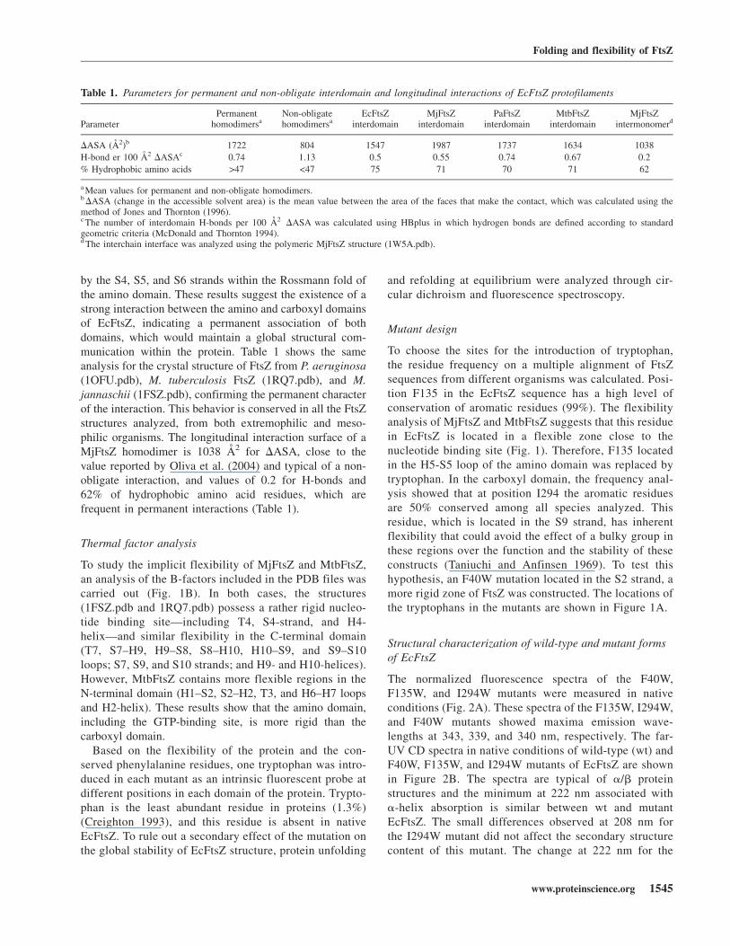
To study the implicit flexibility of MjFtsZ and MtbFtsZ,an analysis of the B-factors included in the PDB files wascarried out (Fig. 1B). In both cases, the structures(1FSZ.pdb and 1RQ7.pdb) possess a rather rigid nucleo-tide binding site—including T4, S4-strand, and H4-helix—and similar flexibility in the C-terminal domain(T7, S7–H9, H9–S8, S8–H10, H10–S9, and S9–S10loops; S7, S9, and S10 strands; and H9- and H10-helices).However, MtbFtsZ contains more flexible regions in theN-terminal domain (H1–S2, S2–H2, T3, and H6–H7 loopsand H2-helix). These results show that the amino domain,including the GTP-binding site, is more rigid than thecarboxyl domain.
Based on the flexibility of the protein and the con-served phenylalanine residues, one tryptophan was intro-duced in each mutant as an intrinsic fluorescent probe atdifferent positions in each domain of the protein. Trypto-phan is the least abundant residue in proteins (1.3%)(Creighton 1993), and this residue is absent in nativeEcFtsZ. To rule out a secondary effect of the mutation onthe global stability of EcFtsZ structure, protein unfolding
and refolding at equilibrium were analyzed through cir-cular dichroism and fluorescence spectroscopy.
Mutant design
To choose the sites for the introduction of tryptophan,the residue frequency on a multiple alignment of FtsZsequences from different organisms was calculated. Posi-tion F135 in the EcFtsZ sequence has a high level ofconservation of aromatic residues (99%). The flexibilityanalysis of MjFtsZ and MtbFtsZ suggests that this residuein EcFtsZ is located in a flexible zone close to thenucleotide binding site (Fig. 1). Therefore, F135 locatedin the H5-S5 loop of the amino domain was replaced bytryptophan. In the carboxyl domain, the frequency anal-ysis showed that at position I294 the aromatic residuesare 50% conserved among all species analyzed. Thisresidue, which is located in the S9 strand, has inherentflexibility that could avoid the effect of a bulky group inthese regions over the function and the stability of theseconstructs (Taniuchi and Anfinsen 1969). To test thishypothesis, an F40W mutation located in the S2 strand, amore rigid zone of FtsZ was constructed. The locations ofthe tryptophans in the mutants are shown in Figure 1A.
Structural characterization of wild-type and mutant formsof EcFtsZ
The normalized fluorescence spectra of the F40W,F135W, and I294W mutants were measured in nativeconditions (Fig. 2A). These spectra of the F135W, I294W,and F40W mutants showed maxima emission wave-lengths at 343, 339, and 340 nm, respectively. The far-UV CD spectra in native conditions of wild-type (wt) andF40W, F135W, and I294W mutants of EcFtsZ are shownin Figure 2B. The spectra are typical of a/b proteinstructures and the minimum at 222 nm associated witha-helix absorption is similar between wt and mutantEcFtsZ. The small differences observed at 208 nm forthe I294W mutant did not affect the secondary structurecontent of this mutant. The change at 222 nm for the
Table 1. Parameters for permanent and non-obligate interdomain and longitudinal interactions of EcFtsZ protofilaments
ParameterPermanent
homodimersaNon-obligatehomodimersa
EcFtsZinterdomain
MjFtsZinterdomain
PaFtsZinterdomain
MtbFtsZinterdomain
MjFtsZintermonomerd
DASA (A2)b 1722 804 1547 1987 1737 1634 1038
H-bond er 100 A2 DASAc 0.74 1.13 0.5 0.55 0.74 0.67 0.2
% Hydrophobic amino acids >47 <47 75 71 70 71 62
a Mean values for permanent and non-obligate homodimers.b DASA (change in the accessible solvent area) is the mean value between the area of the faces that make the contact, which was calculated using themethod of Jones and Thornton (1996).c The number of interdomain H-bonds per 100 A2 DASA was calculated using HBplus in which hydrogen bonds are defined according to standardgeometric criteria (McDonald and Thornton 1994).d The interchain interface was analyzed using the polymeric MjFtsZ structure (1W5A.pdb).
Folding and flexibility of FtsZ
www.proteinscience.org 1545
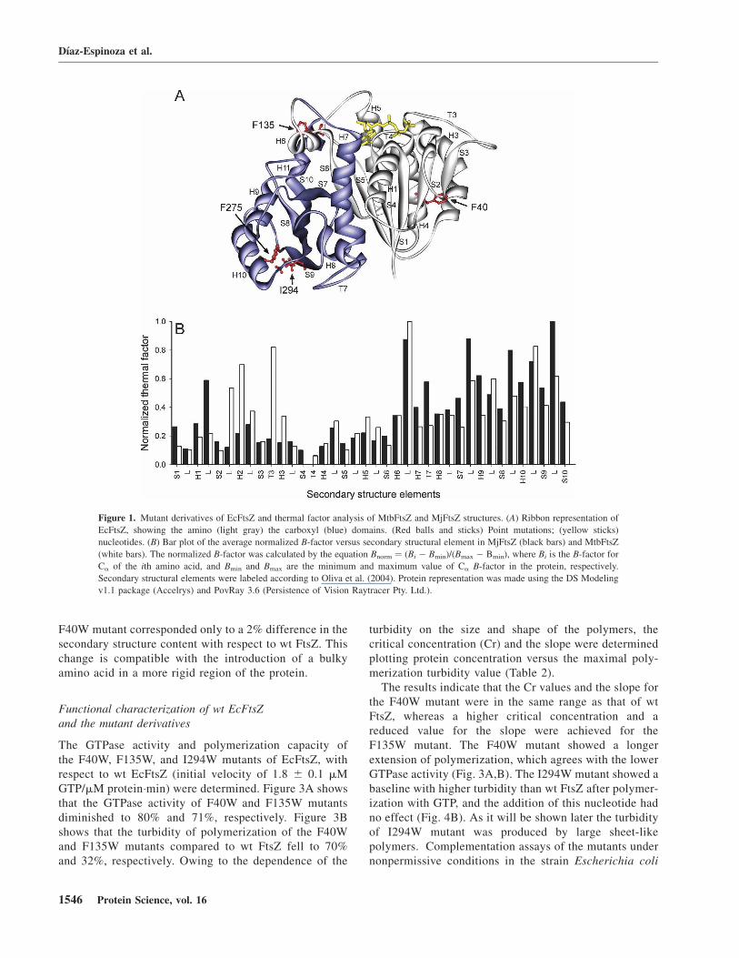
F40W mutant corresponded only to a 2% difference in thesecondary structure content with respect to wt FtsZ. Thischange is compatible with the introduction of a bulkyamino acid in a more rigid region of the protein.
Functional characterization of wt EcFtsZand the mutant derivatives
The GTPase activity and polymerization capacity ofthe F40W, F135W, and I294W mutants of EcFtsZ, withrespect to wt EcFtsZ (initial velocity of 1.8 6 0.1 mMGTP/mM protein�min) were determined. Figure 3A showsthat the GTPase activity of F40W and F135W mutantsdiminished to 80% and 71%, respectively. Figure 3Bshows that the turbidity of polymerization of the F40Wand F135W mutants compared to wt FtsZ fell to 70%and 32%, respectively. Owing to the dependence of the
turbidity on the size and shape of the polymers, thecritical concentration (Cr) and the slope were determinedplotting protein concentration versus the maximal poly-merization turbidity value (Table 2).
The results indicate that the Cr values and the slope forthe F40W mutant were in the same range as that of wtFtsZ, whereas a higher critical concentration and areduced value for the slope were achieved for theF135W mutant. The F40W mutant showed a longerextension of polymerization, which agrees with the lowerGTPase activity (Fig. 3A,B). The I294W mutant showed abaseline with higher turbidity than wt FtsZ after polymer-ization with GTP, and the addition of this nucleotide hadno effect (Fig. 4B). As it will be shown later the turbidityof I294W mutant was produced by large sheet-likepolymers. Complementation assays of the mutants undernonpermissive conditions in the strain Escherichia coli
Figure 1. Mutant derivatives of EcFtsZ and thermal factor analysis of MtbFtsZ and MjFtsZ structures. (A) Ribbon representation of
EcFtsZ, showing the amino (light gray) the carboxyl (blue) domains. (Red balls and sticks) Point mutations; (yellow sticks)
nucleotides. (B) Bar plot of the average normalized B-factor versus secondary structural element in MjFtsZ (black bars) and MtbFtsZ
(white bars). The normalized B-factor was calculated by the equation Bnorm ¼ (Bi � Bmin)/(Bmax � Bmin), where Bi is the B-factor for
Ca of the ith amino acid, and Bmin and Bmax are the minimum and maximum value of Ca B-factor in the protein, respectively.
Secondary structural elements were labeled according to Oliva et al. (2004). Protein representation was made using the DS Modeling
v1.1 package (Accelrys) and PovRay 3.6 (Persistence of Vision Raytracer Pty. Ltd.).
Dıaz-Espinoza et al.
1546 Protein Science, vol. 16
VIP2(DE3), which lacks chromosomal ftsZ, were carriedout in order to determine in vivo functionality. F135Wand F40W mutants presented similar complementation tothat of wt EcFtsZ within the experimental error (Fig. 3C).Interestingly, Figure 4C shows that the I294W mutant wasunable to complement the defect of VIP2(DE3) undernonpermissive conditions.
Given that the in vitro polymerization and GTPaseactivity of F135W and I294W mutants were altered, theprotein stability of the mutants was analyzed throughunfolding/folding curves using GdmCl.
Stability of wt FtsZ and mutant derivatives followedby GdmCl unfolding/folding assays
The GdmCl equilibrium unfolding curves of wt andF135W and I294W mutants of EcFtsZ, as determined
by CD, showed that raising the concentration of thechaotropic agent to 0.6 M released the nucleotide com-pletely. The apo-protein denaturation at concentrations>0.6 M GdmCl showed a cooperative behavior. Theinserted table (Fig. 5A) shows that the transition midpoint(GdmCl50%) for the mutants and wt present similar valuesaround 1.2 M GdmCl. These values were obtained adjust-ing the experimental points to a cooperative denaturationcurve using Equation 1 as indicated in Materials andMethods. It has been described that this parameter isaltered when the protein stability is affected by structuralperturbations, such as point mutations (Khorasanizadehet al. 1996; Hannemann et al. 2002). To follow thestability of each domain and the unfolding/folding behav-ior of the mutants, the fluorescence properties of trypto-phan were characterized. The equilibrium unfoldingprocess of FtsZ was not affected by the tryptophansubstitutions at F135 or I294, and the global stability ofthe secondary structure remained similar to that of wtFtsZ. Figure 5, B and C, shows that the unfolding curvesof F135W and I294W mutants as tracked by fluorescencemeasurements were similar and that the refolding curvesshowed the same pattern observed in the denaturationcurves reaching the same fluorescence intensity andmaximum emission wavelength compared with the nativestate. Interestingly, the F135W mutant showed a smallhyperfluorescence state that was more prominent in theF275W mutant (Fig. 5D). Figure 5, B and D, shows thatthe maximum of the hyperfluorescence effect is producedat the same concentration of GdmCl at which thenucleotide is released. This result indicates that trypto-phan senses the structural changes induced by the apo-protein conformation. The shape and the overlap of thedenaturation/renaturation curves are indicative of chemi-cally reversible unfolding/folding processes. In addition,the average transition midpoint for the F135W, F275W,and I294W mutants occurs at a similar concentration ofGdmCl as for wt FtsZ (1.2 M). However, the stability ofF40W was reduced, as shown by a lower value for thetransition midpoint (1.0 M GdmCl) (data not shown). It isimportant to point out that the same unfolding behaviorobserved from the CD experiments was detected follow-ing the fluorescence of a tryptophan residue located in theamino or in the carboxyl domain. Taken together, theresults suggest that the stability of both domains in theprotein is determined by a common structural feature thatcould be the stronger interdomain interface interaction.
The effect of the point mutation I294W on the functionalproperties of FtsZ was reverted by the doublemutation I294W/F275A
To determine the nature of the inhibitory effect of thepoint mutation I294W on the functional properties of
Figure 2. Fluorescence normalized spectra and circular dichroism (CD)
of tryptophan mutants of EcFtsZ. (A) Uncorrected fluorescence emission
spectra of F135W (dashed line), I294W (solid line), and F40W (dotted
line) mutants, recorded in the same day in buffer 50 mM Tris-HCl (pH 7.5)
at 25°C, at the same protein concentration (12 mM). lexcitation ¼ 295 nm.
Maximum emission fluorescence occurred at 343, 340, and 339 nm for
F135W, F40W, and I294W mutants, respectively. (B) Far-UV circular
dichroism spectra of (r) EcFtsZ wt, (j) F40W, (s) F135W, and (n)
I294W mutants (7.5 mM), recorded under the same experimental con-
ditions.
Folding and flexibility of FtsZ
www.proteinscience.org 1547

FtsZ, GTPase activity (13% of wt) and polymerization(permanently polymerized) were analyzed (Fig. 4A,B).The interaction of tryptophan at position 294 with thesurrounding amino acids was evaluated in the 3D struc-ture of the model of EcFtsZ. Figure 6A shows that thetryptophan in the I294W mutant was very close to F275located in the H10-helix, with both aromatic rings sep-arated by a distance of 4.5 A, probably forming anaromatic stack. This interaction is located between theH10-helix and S9-strand, a flexible region of FtsZ (Fig.6A). It seems reasonable to postulate that the inhibitoryeffect produced by this mutation could be in partattributed to this aromatic interaction. If this hypothesisis correct, the simultaneous presence of aromatic residuesat positions 275 and 294 should not be found in FtsZfrom different species. A multiple alignment of 474 FtsZsequences from different organisms, which is partlypresented in Figure 6B, demonstrates that aromatic
residues can be found at either positions 275 or 294,but interestingly, aromatic residues are never present inboth positions.
To examine whether phenylalanine 275 is involved inthe effect produced by the I294W mutation, this phenyl-alanine was mutated to either tryptophan or alanine.F275W mutants fully complement in vivo, although themodified FtsZ polymerizes in vitro with reduced effi-ciency (Fig. 4C,B). The Cr of the F275W mutant wasapproximately one order of magnitude higher than that ofwt FtsZ, and the slope was similar (Table 2). These dataindicate that this mutation diminished the polymerizationcapacity but had no effect on the shape of the polymers,discarding a specific effect of tryptophan in the environ-ment of I294. To test the hypothetical interaction betweenF275 and W294 in the mutant, a double mutant wasdesigned in which F275 was changed to alanine (smallside-chain residue), which is found at position 275 in
Figure 3. Functional characterization of wt and F40W and F135W mutants of EcFtsZ. (A) GTP hydrolysis activity was initiated by the
addition of 1 mM GTP and stopped by adding 10% perchloric acid. Each protein (25 mM) sample was incubated in buffer containing
50 mM Mes (pH 6.5), 10 mM MgCl2, and 50 mM KCl for 5 min at 30°C. Percentages of activity are with respect to wt EcFtsZ. The
errors were obtained by linear regression analysis. (B) Polymerization of F40W and F135W mutants and wt EcFtsZ (25 mM) in a
solution containing 50 mM Mes (pH 6.5), 50 mM KCl, and 10 mM MgCl2, followed by turbidity changes at 30°C. The addition of GTP
is indicated by the arrow. (C) In vivo complementation was estimated from the number of colonies obtained after titration at the
restrictive temperature (42°C) compared with the number of colonies obtained at the permissive temperature (30°C). C+ corresponds to
the transformation of E. coli VIP-2(DE3) strain with wt EcFtsZ, and C� with the vector alone (see Materials and Methods).
Dıaz-Espinoza et al.
1548 Protein Science, vol. 16

several sequences of FtsZ. One example is MjFtsZ, whichhas tryptophan and alanine located in the equivalentpositions of I294 and F275 in EcFtsZ. Figure 4A showsthat the GTP hydrolytic activity of the double mutantincreased to 21% of wt levels, instead of just 13%achieved for the I294W mutant. The double mutantpresented lower turbidity than the I294W mutant (Fig.4B). Figure 7A shows that the mutant I294W producedlarge sheet-like polymers. Glutamate (1 M) increased theturbidity of both the I294W mutant and the double mutant(data not shown). Figure 7B shows that in the presence ofglutamate, I294W polymerizes, forming large sheets thatare not affected by GTP or GDP (Fig. 7C). In the doublemutant, glutamate induced a small amount of amorphousaggregates (Fig. 7E), and the addition of GTP inducedsingle and double filaments and sheets (Fig. 7F). Surpris-ingly, the double mutant fully reestablished cell divisionby in vivo complementation (Fig. 4C).
These results show that the flexibility of S9 where I294is located has a dramatic influence on the functionality ofthe protein. In order to analyze the relationship of theGTPase activity with the flexibility of the protein, astructural comparison between GTP/GDP forms of FtsZwas carried out.
Structural changes of GTP/GDP forms
To compare two crystallized conformations of the sameprotein, it is important to have the same space group be-cause protein contacts belonging to different space groupsmay affect the comparison between conformations. Forthis reason, the two crystal structures of FtsZ chosen forthe comparison of GTP and GDP forms had the samespace group in the crystals. Another important point isthat these proteins were crystallized in the GDP form,and GTP was exchanged into the crystal. Under theserestrictions, the structures of MtbFtsZ bound to GTP(1RLU.pdb) and GDP (1RQ7.pdb) (Leung et al. 2004)and MjFtsZ crystallized in GTP (1W58.pdb) and GDP(1FSZ.pdb) conformation (Lowe 1998) were analyzed.
The sequence identity among these proteins is 48%, witha RMSD (root mean square deviation) for the structuralsuperposition of 1.45 A between the 1W58.pdb and1RLU.pdb. Figure 8 shows the secondary structuralchanges given by the difference in position of Ca of thepolypeptide chain in the GTP and GDP states. Analysis ofthese data shows that GTP hydrolysis induces movementsthat are greater than the noise (RMSD shown in Table 3)in the polypeptide chain, mainly in the external regions ofFtsZ. The amino acids involved in movements >0.5 A areshown in Table 4; specifically, they are located in theamino domain near the GTP-binding site in the T2 and T3loops and in the H6–H7 loop. More remarkable are themotions of other residues of the carboxyl domain asso-ciated with the phosphorylation state of the nucleotide.According to the nomenclature given by Oliva et al.(2004), these movements are located in the H8–S7, S8–H10, and S9–S10 loops. These results indicate that thecarboxyl domain also shows flexibility, a finding that hasnot been reported previously.
Discussion
In this study, using single tryptophan fluorescence, bothamino and carboxyl domains of EcFtsZ showed similarunfolding/folding characteristics, and the interactionbetween these domains was determined to be permanent.GTPase activity and polymerization of FtsZ were foundto be dependent on the structural flexibility of thecarboxyl domain of the protein. Thus, the I294W muta-tion located in the flexible carboxyl domain appears toform an aromatic interaction with phenylalanine 275,affecting both the functionality in vitro and in vivo. Onthe other hand, the double mutant F275A/I294W partiallyrecovers both the GTPase activity and the polymerizationcapacity in the presence of glutamate, and furthermore, itfully recovers in vivo functionality.
Structural stability and folding of EcFtsZ
Whereas the structural stability of proteins is generallynot affected when mutations are introduced in flexiblezones, their functionality can be altered (Hilser et al.1998; Freire 1999). The results presented in this studyindicate that the content of secondary structures of theF135W and I294W mutants was similar, suggesting thatno significant secondary structure rearrangements werecaused by these substitutions. In addition, the unfoldingof wt FtsZ followed by changes in the secondary structurewas the same as that of the mutants, as analyzed by thefluorescence of a single tryptophan located in each do-main, showing that the structural stability of FtsZ re-mained unaltered in the tryptophan substitution mutants.
Using the intrinsic properties of tryptophan, the envi-ronmental characteristics of the tertiary structure of FtsZ
Table 2. Critical concentration and slope valuesfor polymerization of wt and mutant derivatives of EcFtsZ
Protein Cr (mM) Slopea
FtsZ wt 1.25 57
F40W 0.25 50
F135W 4.0 23
F275W 16.0 49
Cr (the critical concentration, which corresponds to the minimal concen-tration required for polymerization) and the slope were obtained from thestraight lines of the plot of protein concentration versus the maximalpolymerization turbidity at 350 nm.a The slope depends on the shape and length of the polymers (Gaskin et al.1974; Mukherjee and Lutkenhaus 1999).
Folding and flexibility of FtsZ
www.proteinscience.org 1549

around these residues introduced into the amino (F135W)or carboxyl (I294W) domains of the mutants allowed thestability and tracking of the unfolding/folding processto be determined. The unfolding and refolding of themutants with tryptophan in the amino or in the carboxyldomain showed similar fluorescence curves, indicatingthe same global stability for the whole protein. Theseresults are compatible with a cooperative folding mech-anism where both domains fold with similar stabilitiesdepending on the global structure of the protein, althoughthey may have different stabilities separately.
The unfolding/folding processes in the F135W andI294W mutants did not show the presence of intermedi-ates. In contrast, other studies on EcFtsZ monitoring thefluorescence of ANS (8-anilinonaphthalene-1-sulphonate)and FITC (fluorescein isothiocyanate) probes report the
presence of intermediates (Santra and Panda 2003). How-ever, this apparent contradiction could be explained by theCD results (Fig. 4B) of the mutants, confirming thepresence of the apo-protein at 0.6 M GdmCl (Andreuet al. 2002). This value is coincident with the maximumvalue for the minor hyperfluorescence shown by theF135W mutant and for the more pronounced hyperfluo-rescence of the F275W mutant that could correspond to theintermediates that appear upon denaturation with urea(Santra and Panda 2003). The possibility of local arrange-ments taking place during the initial steps of FtsZ unfoldingin regions close to the nucleotide binding site that arefar from the interdomain zone would give greater accessi-bility to the action of denaturant agents. These changescan be attributed to local unfolding of regions that aremore susceptible to denaturant agents than the interdomain
Figure 4. Functional characterization of wt EcFtsZ and the F275W, I294W, and F275A/I294W mutants. (A) GTP hydrolysis activity
was started by the addition of 1 mM GTP and stopped by adding 10% perchloric acid. Each protein (25 mM) sample was incubated in
buffer containing 50 mM Mes (pH 6.5), 10 mM MgCl2, and 50 mM KCl for 5 min at 30°C. Percentages of activity are respect to
EcFtsZ wt. The errors were obtained by linear regression analysis. (B) Polymerization of I294W and F275A/I294W mutants and wt
EcFtsZ at 12 mM, and F275W (22 mM) in a solution containing 50 mM Mes (pH 6.5), 50 mM KCl, and 10 mM MgCl2, followed by
turbidity changes at 30°C. The addition of GTP is indicated by the arrow. (C) In vivo complementation was estimated from the number
of colonies obtained after titration at the restrictive temperature (42°C) compared with the number of colonies obtained at the
permissive temperature (30°C). C+ corresponds to the transformation of E. coli VIP-2(DE3) strain with wt EcFtsZ, and C� with the
vector alone (see Materials and Methods).
Dıaz-Espinoza et al.
1550 Protein Science, vol. 16

region, generating the intermediates seen by Santra andPanda (2003), and by the CD unfolding curves obtained inthis study. This kind of behavior has been observed inother proteins and has been defined as local cooperativityrelated to conformational changes (Hilser et al. 1998;Freire 1999).
Influence of the interdomain interaction and flexibilityon FtsZ function
The thermal factor analysis of the implicit flexibility andthe exchange of GDP by GTP showed minor displace-ments (Tables 3 and 4; Fig. 8) mainly in specific external
regions of the protein including those close to the GTP-binding site, and interestingly, in zones of the carboxyldomain far from the GTP-binding site.
A hypothetical model for polymerization and depoly-merization of FtsZ correlating the flexible zones ofEcFtsZ will be discussed based on a double filament(Oliva et al. 2003). It is known that the hydrolysis of GTPis induced by the interaction of the amino longitudinalface of one monomer with the carboxyl longitudinal faceof the other (Sossong et al. 1999). The surface of thisinteraction, which falls closer to the non-obligate clas-sification, allows a conformational change from GTP toGDP that weakens the structure of the polymer (Table 1).
Figure 5. Secondary structure and stability analysis of wt and mutant derivatives of EcFtsZ. (A) Equilibrium unfolding curves
followed by circular dichroism of (r) wt EcFtsZ and (s) F135W and (D) I294W mutants (7.5 mM) at 25°C in 50 mM Tris-HCl (pH
7.5). Each point corresponds to the unfolded protein fraction calculated from experimental values of ellipticity at 222 nm using GdmCl
as denaturant agent. (+) mole of nucleotide released per mole of FtsZ. The inserted tables show the transition midpoints [GdmCl]50%
(GdmCl concentration value for 50% of denatured protein) of wt and mutant FtsZ estimated from the unfolding curves. (B) Equilibrium
(s) unfolding and (d) refolding curves determined by tryptophan fluorescence of the F135W mutant (12 mM) in 50 mM Tris-HCl (pH
7.5) at 25°C. (C) Equilibrium (n) unfolding and (m) refolding curves followed by tryptophan fluorescence of the I294W (mutant
12 mM) in 50 mM Tris-HCl (pH 7.5) at 25°C. (D) Equilibrium (u) unfolding and (j) refolding curves followed by tryptophan
fluorescence of the F275W mutant (12 mM) in 50 mM Tris-HCl (pH 7.5) at 25°C. Each folded and refolded protein fraction was
calculated from the normalized fluorescence intensity at maximum emission wavelength of 343, 340, and 339 nm for F135W, F275W,
and I294W mutants, respectively. Equilibrium refolding curves were under the same experimental conditions used for unfolding, where
GdmCl was diminished by fast dilution. The inserted tables in B and C show the transition midpoint ([GdmCl]50%) values determined
from the unfolding and refolding curves as tracked by tryptophan fluorescence.
Folding and flexibility of FtsZ
www.proteinscience.org 1551

It is assumed that this conformational change spreadsthrough the flexible zones of the molecule during depo-lymerization. Thus, GTP stabilizes the straight conforma-tion in the polymer, while GDP destabilizes it. Therefore,a mutation such as I294W located in a flexible region ofthe carboxyl domain should alter the equilibrium betweenthese conformations, affecting polymerization or depoly-merization, depending on the conformation favored.Consistent with our hypothesis, the functionality of theI294 mutant was almost totally abolished; this mutantremained permanently polymerized, had very low GTPaseactivity, and showed residual in vivo complementation.Also the point mutation F135W, located in a flexible zoneof the amino domain, reduced the functionality of theprotein (diminished polymerization, reduced GTPase
to 70%, and showed poor in vivo complementation). Inagreement with our results, it was reported that the replace-ment of amino acid T92 by tryptophan in the flexible loopT3 in MjFtsZ diminished the functionality of the protein(Diaz et al. 2001). On the other hand, the F40W mutant,with a tryptophan located in the rigid zone of the aminodomain, was almost totally functional (polymerization invitro was almost identical to wt FtsZ, the GTPase activitywas 80%, and it fully complemented in vivo), where thesmall differences could be due to the small change insecondary structure induced by a steric effect of tryptophan(Fig. 2B). These results support the hypothesis that asmall modification in the flexible zone affects the functionof FtsZ. Previous work reported a significant alteration inGTPase activity of MjFtsZ when the W319 residue, located
Figure 6. Ca ribbon representation of the I294W mutant in the structural model of EcFtsZ. (A) (light gray) The amino domain; (blue)
the carboxyl domain. The residues F275 and W294 are presented in detail showing the stacking formed according to our model. GTP is
projected in yellow sticks. The image was made using DS Modeling v1.1 and PovRay 3.6 (as in Fig. 1). (B) A multiple alignment of
FtsZ sequence fragments where EcFtsZ (red) and MjFtsZ (violet) are highlighted. Positions 275 and 294 are highlighted in blue.
Dıaz-Espinoza et al.
1552 Protein Science, vol. 16

in a structurally equivalent position of the I294 residue inthe 3D structure of EcFtsZ, was changed to tyrosine (Olivaet al. 2003). These results and the fact that the I294Wmutation did not affect the structural stability of FtsZ, asdemonstrated by the CD and fluorescence experiments,suggest that the strong effect on functionality caused by theI294W mutation could be the result of an alteration in theflexibility. This effect could be explained through anaromatic stacking interaction between tryptophan 294 andphenylalanine 275 in the mutant, which could stabilize theGTP conformation. This is confirmed by the full recoveryof the in vivo complementation and the increment in the
rate of GTP hydrolysis in the F275A/I294W double mutant,in which the stacking would disappear.
A rigid C-terminal region in the ‘‘GTP conformation’’could stabilize the protofilaments as occurs with thestabilization of the polymers in the I294W mutant (Fig.7A). The fine regulation of the longitudinal interactionsshould be carried out by specific amino acid residues atthe interface between the monomers and the state ofphosphorylation of the nucleotide. This model requiresflexible zones in the amino and carboxyl domains. Theimportance of the interdomain interaction for a functionalconformation was demonstrated in TmFtsZ, in which the
Figure 7. Polymerization products of EcFtsZ mutant I294W and double mutant F275A/I294W. Electron micrograph of the
polymerization products of the I294W and F275A/I294W mutants (7.5 mM). (A,D) I294W and I294W/F275A polymerization controls
in buffer (50 mM Mes at pH 6.5, 50 mM KCl, 10 mM MgCl2) without GTP or sodium glutamate at 30°C. The polymerization reaction
was initiated with GTP at a final concentration of 1 mM. (B,E) I294W and F275A/I294W mutants before the addition of GTP,
respectively, in the presence of 1 M sodium glutamate. (C,F) I294W and F275A/I294W mutants 5 min after the addition of GTP,
respectively, in the presence of 1 M sodium glutamate. Bars, 100 nm.
Figure 8. Comparison of MjFtsZ and MtbFtsZ structures in GTP and GDP states. A bar plot of the average Ca–Ca distance between
structural equivalent amino acids versus secondary structural element in MjFtsZ (black bars) and MtbFtsZ (white bars). The symbol (*)
indicates secondary structural elements that have been reported to move between GTP and GDP conformations (Leung et al. 2004;
Oliva et al. 2004).
Folding and flexibility of FtsZ
www.proteinscience.org 1553
GTPase activity was recovered in the presence of bothdomains that were purified separately (Oliva et al. 2004).These results indicate a strong interaction between themthat could induce a conformational change that, in turn,would be responsible for the correct flexibility of func-tional FtsZ.
Materials and Methods
Reagents
Guanidinium chloride (GdmCl), MgCl2, glycerol, and EDTAwere purchased from Winkler Ltda. Tris Base ultrapure wassupplied by US Biological and Mes (2-morpholinoethanesulfonicacid) from AppliChem. KCl and ammonium sulfate were pur-chased from Merck. Monosodium glutamate was acquired fromFluka Chemie AG, and IPTG (isopropyl-1-thio-b-D-galactopy-ranoside) was supplied by Sigma Chemical Co.
Model building
The FtsZ structure from P. aeruginosa (1OFU.pdb chain A)(Cordell et al. 2003), sharing 58% sequence identity with thesequence of EcFtsZ, was obtained from the Protein Data Bankand used as the template structure to construct a 3D model ofEcFtsZ, with the program Modeller v6.2 (Sanchez and Sali1997). The model was constructed using the default ‘‘model’’routine, and the reliability was evaluated with PROSAII (Sippl1993) and Verify3D software.
Characterization of the FtsZ interdomain interface
The interdomain interfaces of PaFtsZ (1OFU.pdb chain A),MtbFtsZ (1RQ7.pdb), MjFtsZ (1FSZ.pdb), and the model ofEcFtsZ 3D structures were analyzed (Lowe 1998; Cordell et al.2003; Leung et al. 2004; Oliva et al. 2004). Ser177 in EcFtsZand the analog residues for the other structures were used as thedomain limit. The number of interdomain H-bonds per 100 A2
of DASA (change in the accessible solvent area) was calculatedusing HBPLUS in which H-bonds are defined according tostandard geometric criteria (McDonald and Thornton 1994). Thedetermination of the DASA value and the percentage of hydro-phobic residues in the interdomain interface were obtained via
the Protein–Protein Interaction Server v1.5 (Jones and Thornton1996), where neither water molecules nor GTP was included.
Conformational changes related to GTP hydrolysis
The putative amino acids involved in conformational changeswere obtained from the structural comparison of MtbFtsZ(1RLU.pdb and 1RQ7.pdb) in GTP and GDP conformationsand MjFtsZ (1W58.pdb and 1FSZ.pdb) in both conformations.Tables of Ca distances were obtained from the structural com-parison made with Align3d routine, from Modeller v6.2.
Mutant construction and protein purification
pMFV57 was constructed starting from pMFV56 (Rivas et al.2000) in which the gene b-lactamase was cloned into the EcoRVsite interrupting the gene encoding for Kn resistance. EcFtsZmutants were constructed by site-directed mutagenesis using theQuick Change Mutagenesis Kit from Stratagene. All plasmidDNAs were purified with E.Z.N.A Plasmid Miniprep Kit I fromOmega Bio-tek. Each ftsZ gene with the corresponding pointmutation was completely sequenced. Recombinant EcFtsZ andFtsZ mutants were overexpressed in the E. coli BL21(DE3)strain by induction with IPTG. The cells were grown at 37°Cwith agitation in 2 L of LB broth containing 100 mg/mL Ap untilan OD600 of 0.7 was reached; then 0.4 mM IPTG was added,and the cells were grown for an additional 4 h. The proteinswere purified as described in Beuria et al. (2003) with somemodifications. Briefly, the cells were centrifuged at 7140g in aSORVALL RC-3B centrifuge with an H6000A rotor for 45 min,and the pellet was suspended in buffer A (50 mM Tris-HCl at pH8.0, 50 mM KCl, 1 mM EDTA, and 10% glycerol) and lysedby sonication with five pulses of 45 W and two pulses of 75 W.The lysed cells were centrifuged at 92,000g for 90 min, and thesupernatant was saturated with 25% ammonium sulfate. Thesolution was centrifuged at 9391g at 4°C, and the pellet wasdissolved in buffer A then dialyzed against buffer A to removethe salt excess. Mono Q anion-exchange chromatography wasperformed, and the protein eluted with a salt gradient (Buffer B:50 mM Tris-HCl at pH 8.0, 50 mM KCl, 1 mM EDTA, 1 M KCl,and 10% glycerol). A monosodium glutamate polymerizationand depolymerization cycle was performed using a solution of50 mM PIPES (pH 6.5), 1 M glutamate, 10 mM CaCl2, 10 mMMgCl2, and 2 mM GTP for 30 min at 37°C, and the polymersobtained were centrifuged at 9391g for 30 min at 27°C (Beuriaet al. 2003). The protein was suspended in 5 mL of buffer A,washed twice with the same volume of buffer, and concentrated
Table 3. Parameters from the crystal structure of FtsZ in GTPand GDP conformations
PDBGTP/GDP Source
Space groupGTP/GDPa
RMS AGTP/GDPb
GTP/GDPconformations
1W58/1FSZ M. jannaschii I213/I213 0.3 Substitution
on the crystal
Amino domain 0.3
Carboxyl domain 0.4
1RLU/1RQ7 M. tuberculosis P65/P65 0.2 Substitution
on the crystal
Amino domain 0.2
Carboxyl domain 0.2
a Crystallization space groups for both conformations.b RMSD between the Ca of the GTP and GDP conformation structures.
Table 4. Principal changes in Ca position betweenGTP/GDP conformations
Secondarystructure
ResidueMtbFtsZ
Distance(A)
Secondarystructure
ResidueMjFtsZ
Distance(A)a
T2 D43 0.5 H6–H7 L206 0.7
T2 A44 0.5 H8–S7 N245 0.7
T3 G69 0.9 S9–S10 E328 1.3
S8-H10 D266 0.6 S9–S10 N329 0.8
Structural change analysis of FtsZ related to the GTP/GDP conformationon MtbFtsZ (1RLU.pdb and 1RQ7.pdb) and MjFtsZ (1FSZ.pdb and1W58.pdb).a Corresponds to the Ca–Ca Euclidian distance (A) between the sameamino acids in both conformations (GTP and GDP).
Dıaz-Espinoza et al.
1554 Protein Science, vol. 16

in a Centripep system and stored at �80°C. The protein concen-tration and the nucleotide content were determined by absorp-tion at 254 and 280 nm in 6 M GdmCl with phosphate buffer(pH 6.5) (Rivas et al. 2000). The Bradford method wascalibrated for FtsZ. The content of GDP or GTP was determinedby HPLC (high-performance liquid chromatography) in aSUPELCOSIL LC-18-D13 column.
Polymerization turbidity
The assay was performed in a solution at 30°C containing50 mM Mes (pH 6.5), 50 mM KCl, and 10 mM MgCl2 using theprotein concentration indicated in the figures. The samples wereincubated for 3 min at 30°C, and the reaction was started byadding 1 mM GTP. The turbidity of the polymerization reactionswas monitored at 90° by illuminating the sample at 350 nm in aPerkin-Elmer LS-50 spectrofluorimeter with a 2% transmissionfilter in the emission beam.
GTPase activity
The protein was incubated for 3 min at 30°C in a solutioncontaining 50 mM Mes (pH 6.5), 50 mM KCl, and 10 mMMgCl2; 1 mM GTP was added to initiate the reaction. Thereactions were stopped at different times by adding 10%perchloric acid and neutralized with 1 M KHCO3; the bubblesof CO2 were removed by centrifugation in a Speed-Vac for5 min. The denatured protein and the insoluble KClO4 salt wereremoved by centrifugation at 17,530g for 10 min, and thesupernatants were analyzed by HPLC in a SUPELCOSIL LC-18-D13 column. The separation of GDP from GTP was under-taken using a mobile phase containing 4 mM TBAP (tetrabutylammonium dihydrogen phosphate), 0.2 M K2HPO4, and 0.1 Mglacial acetic acid. The nucleotide content was calculated fromthe area of the peak associated with GDP and GTP, and GDPwas normalized with respect to the total nucleotide content(GDP/[GDP + GTP]).
In vivo assay
To evaluate the functionality in vivo of the F40W, F135W,F275W, I294W, and I294W/F275A mutants of EcFtsZ, an assaymeasuring the viability of cells carrying these mutations withrespect to the wild type was performed using as a host the strainE. coli VIP2(DE3)/pLAR9. VIP2(DE3)/pLAR9 is VIP2/pLAR9(Pla et al. 1991) lysogenized by the lDE3 Lysogenization Kit(Novagen). In this strain, the ftsZ chromosomal copy is inter-rupted by an insertion, and it is complemented with a copyencoded in the thermosensitive plasmid pLAR9. Overnightcultures of VIP2(DE3)/pLAR9 grown at the permissive temper-ature (30°C) were transformed with plasmid pMFV57 contain-ing ftsZ wt or the point-mutated ftsZ genes. The transformedcells were grown at 30°C in LB broth containing 100 mg/mL Ap,50 mg/mL Kn, and 50 mg/mL Cm until an OD600 of 0.6 wasattained. Serial dilutions were plated at both the permissive tem-perature (30°C) in LB-Ap/Kn/Cm and nonpermissive temperature(42°C) in LB-Ap/Kn, and the number of colonies was counted.
Circular dichroism and fluorescence spectroscopies
CD (circular dichroism) spectra were recorded in a Jasco 600spectropolarimeter using cells of 1 mm pathlength. Each sampleand buffer were measured with four scans, using a 20-nm/min
scan speed, 1-nm bandwidth, and 2-sec time constant. Emissionfluorescence spectra were obtained with a Perkin Elmer LS-50spectrofluorimeter. Three scans were recorded for each mea-surement, using a 295-nm excitation wavelength and 140-nm/minscan speed. CD and fluorescence emission spectra were analyzedwith GRAMS software and plotted with SigmaPlot of SystatSoftware Inc.
Protein unfolding and refolding
Unfolding and refolding experiments were measured by CD(EcFtsZ 7.4 mM) and fluorescence (EcFtsZ 12 mM) in solutionscontaining 50 mM Tris-HCl (pH 7.5) with different GdmClconcentrations. The samples were left on ice for 1 h, to reach theequilibrium avoiding thermal denaturation, then incubated for15 min at 25°C and carefully filtrated through a 0.2-mm filterbefore the measurement. A control for each sample (solutionwithout protein) was treated in the same way. The native spectrawere obtained from the sample without GdmCl. For the refold-ing experiments, the protein was previously denatured in 6 MGdmCl and equilibrated for 30 min at 4°C. An aliquot of thisdenatured sample was rapidly diluted (100 times) in the samebuffer used for denaturation with the corresponding GdmClconcentrations and kept on ice for 1 h; after 15 min at 25°C, CDand fluorescence were measured. The emission spectra weremeasured observing the change in fluorescence intensity and inthe red shift when the GdmCl concentration was increased. Tocalculate the value of [GdmCl]50%, which corresponds to theconcentration of GdmCl at 50% of unfolding or refolding, a fitof the experimental data to the following equation was under-taken using SigmaPlot software:
S = ððaN + bN ½GdmCl�+ ðaD + bD½GdmCl�ÞÞefðmD�Nð½GdmCl�� ½D�50%Þ=RTÞgÞ=ð1 + efðmD�Nð½GdmCl� � ½D�50%Þ=RTÞgÞ
ð1Þ
where S is the observed spectroscopic signal; aN is the signal ofthe native state without GdmCl, and bN ¼ daN/d[GdmCl](the slope); aD and bD are the corresponding quantities for thedenatured state; mD�N is a constant of proportionality; and[D]50% is the GdmCl concentration in which the protein is 50%unfolded (Staniforth et al. 1993).
Electron microscopy
FtsZ samples were adsorbed to carbon-coated grids, negativelystained with 1% uranyl acetate, and observed in a Zeiss EM-109model electron microscope.
Correlated mutations analysis
The 1fsz.hssp alignment obtained from the HSSP database,available in May 2005, was analyzed by PLOTCORR software(Pazos et al. 1997).
Acknowledgments
We thank Jose Manuel Andreu, David Jameson, and MichaelHandford for critically reading the manuscript. This work wassupported by FONDECYT Grants 1050677 and 7060162.
Folding and flexibility of FtsZ
www.proteinscience.org 1555

References
Andreu, J.M., Oliva, M.A., and Monasterio, O. 2002. Reversible unfolding ofFtsZ cell division proteins from archaea and bacteria. Comparison witheukaryotic tubulin folding and assembly. J. Biol. Chem. 277: 43262–43270.
Beuria, T.K., Krishnakumar, S.S., Sahar, S., Singh, N., Gupta, K.,Meshram, M., and Panda, D. 2003. Glutamate-induced assembly ofbacterial cell division protein FtsZ. J. Biol. Chem. 278: 3735–3741.
Buey, R.M., Diaz, J.F., and Andreu, J.M. 2006. The nucleotide switch of tubulinand microtubule assembly: A polymerization-driven structural change.Biochemistry 45: 5933–5938.
Chen, Y. and Erickson, H.P. 2005. Rapid in vitro assembly dynamics andsubunit turnover of FtsZ demonstrated by fluorescence resonance energytransfer. J. Biol. Chem. 280: 22549–22554.
Chen, Y., Bjornson, K., Redick, S.D., and Erickson, H.P. 2005. A rapidfluorescence assay for FtsZ assembly indicates cooperative assembly witha dimer nucleus. Biophys. J. 88: 505–514.
Cordell, S.C., Robinson, E.J., and Lowe, J. 2003. Crystal structure of the SOScell division inhibitor SulA and in complex with FtsZ. Proc. Natl. Acad.Sci. 100: 7889–7894.
Creighton, T.E. 1993. Proteins: Structures and molecular properties, 2nd ed.,pp. 1–47. Freeman, New York.
Diaz, J.F., Kralicek, A., Mingorance, J., Palacios, J.M., Vicente, M., andAndreu, J.M. 2001. Activation of cell division protein FtsZ. Control ofswitch loop T3 conformation by the nucleotide g-phosphate. J. Biol. Chem.276: 17307–17315.
Errington, J., Daniel, R.A., and Scheffers, D.J. 2003. Cytokinesis in bacteria.Microbiol. Mol. Biol. Rev. 67: 52–65.
Freire, E. 1999. The propagation of binding interactions to remote sites inproteins: Analysis of the binding of the monoclonal antibody D1.3 tolysozyme. Proc. Natl. Acad. Sci. 96: 10118–10122.
Gaskin, F., Cantor, C.R., and Shelanski, M.L. 1974. Turbidimetric studies ofthe in vitro assembly and disassembly of porcine neurotubules. J. Mol. Biol.89: 737–755.
Hannemann, F., Bera, A.K., Fischer, B., Lisurek, M., Teuchner, K., andBernhardt, R. 2002. Unfolding and conformational studies on bovineadrenodoxin probed by engineered intrinsic tryptophan fluorescence.Biochemistry 41: 11008–11016.
Henrick, K. and Thornton, J.M. 1998. PQS: A protein quaternary structure fileserver. Trends Biochem. Sci. 23: 358–361.
Hilser, V.J., Dowdy, D., Oas, T.G., and Freire, E. 1998. The structuraldistribution of cooperative interactions in proteins: Analysis of the nativestate ensemble. Proc. Natl. Acad. Sci. 95: 9903–9908.
Huecas, S. and Andreu, J.M. 2004. Polymerization of nucleotide-free, GDP-and GTP-bound cell division protein FtsZ: GDP makes the difference.FEBS Lett. 569: 43–48.
Jones, S. and Thornton, J.M. 1996. Principles of protein–protein interactions.Proc. Natl. Acad. Sci. 93: 13–20.
Khorasanizadeh, S., Peters, I.D., and Roder, H. 1996. Evidence for a three-statemodel of protein folding from kinetic analysis of ubiquitin variants withaltered core residues. Nat. Struct. Biol. 3: 193–205.
Leung, A.K., White, E.L., Ross, L.J., Reynolds, R.C., DeVito, J.A., andBorhani, D.W. 2004. Structure of Mycobacterium tuberculosis FtsZ revealsunexpected, G protein-like conformational switches. J. Mol. Biol. 342:953–970.
Lowe, J. 1998. Crystal structure determination of FtsZ from Methanococcusjannaschii. J. Struct. Biol. 124: 235–243.
Lu, C., Reedy, M., and Erickson, H.P. 2000. Straight and curved conformationsof FtsZ are regulated by GTP hydrolysis. J. Bacteriol. 182: 164–170.
Lu, C., Stricker, J., and Erickson, H.P. 2001. Site-specific mutations ofFtsZ—Effects on GTPase and in vitro assembly. BMC Microbiol. 1: 7.doi: 10.1186/1471-2180-1-7.
Lutkenhaus, J. 1990. Regulation of cell division in E. coli. Trends Genet. 6: 22–25.Marrington, R., Small, E., Rodger, A., Dafforn, T.R., and Addinall, S.G. 2004.
FtsZ fiber bundling is triggered by a conformational change in bound GTP.J. Biol. Chem. 279: 48821–48829.
McDonald, I.K. and Thornton, J.M. 1994. Satisfying hydrogen bondingpotential in proteins. J. Mol. Biol. 238: 777–793.
Mingorance, J., Rueda, S., Gomez-Puertas, P., Valencia, A., and Vicente, M.2001. Escherichia coli FtsZ polymers contain mostly GTP and have a highnucleotide turnover. Mol. Microbiol. 41: 83–91.
Mukherjee, A. and Lutkenhaus, J. 1998. Dynamic assembly of FtsZ regulatedby GTP hydrolysis. EMBO J. 17: 462–469.
Mukherjee, A. and Lutkenhaus, J. 1999. Analysis of FtsZ assembly by lightscattering and determination of the role of divalent metal cations. J.Bacteriol. 181: 823–832.
Nooren, I.M. and Thornton, J.M. 2003. Structural characterisation and func-tional significance of transient protein–protein interactions. J. Mol. Biol.325: 991–1018.
Oliva, M.A., Huecas, S., Palacios, J.M., Martin-Benito, J., Valpuesta, J.M., andAndreu, J.M. 2003. Assembly of archaeal cell division protein FtsZ and aGTPase-inactive mutant into double-stranded filaments. J. Biol. Chem. 278:33562–33570.
Oliva, M.A., Cordell, S.C., and Lowe, J. 2004. Structural insights into FtsZprotofilament formation. Nat. Struct. Mol. Biol. 11: 1243–1250.
Pazos, F., Helmer-Citterich, M., Ausiello, G., and Valencia, A. 1997. Correlatedmutations contain information about protein–protein interaction. J. Mol.Biol. 271: 511–523.
Pla, J., Sanchez, M., Palacios, P., Vicente, M., and Aldea, M. 1991. Preferentialcytoplasmic location of FtsZ, a protein essential for Escherichia coliseptation. Mol. Microbiol. 5: 1681–1686.
Rivas, G., Lopez, A., Mingorance, J., Ferrandiz, M.J., Zorrilla, S., Minton, A.P.,Vicente, M., and Andreu, J.M. 2000. Magnesium-induced linear self-association of the FtsZ bacterial cell division protein monomer. Theprimary steps for FtsZ assembly. J. Biol. Chem. 275: 11740–11749.
Romberg, L. and Mitchison, T.J. 2004. Rate-limiting guanosine 59-triphosphatehydrolysis during nucleotide turnover by FtsZ, a prokaryotic tubulinhomologue involved in bacterial cell division. Biochemistry 43: 282–288.
Sanchez, R. and Sali, A. 1997. Evaluation of comparative protein structuremodeling by MODELLER-3. Proteins (Suppl.) 1: 50–58.
Santra, M.K. and Panda, D. 2003. Detection of an intermediate during unfold-ing of bacterial cell division protein FtsZ: Loss of functional propertiesprecedes the global unfolding of FtsZ. J. Biol. Chem. 278: 21336–21343.
Sippl, M.J. 1993. Recognition of errors in three-dimensional structures ofproteins. Proteins 17: 355–362.
Sossong Jr., T.M., Brigham-Burke, M.R., Hensley, P., and Pearce Jr., K.H. 1999.Self-activation of guanosine triphosphatase activity by oligomerization ofthe bacterial cell division protein FtsZ. Biochemistry 38: 14843–14850.
Staniforth, R.A., Burston, S.G., Smith, C.J., Jackson, G.S., Badcoe, I.G.,Atkinson, T., Holbrook, J.J., and Clarke, A.R. 1993. The energetics andcooperativity of protein folding: A simple experimental analysis basedupon the solvation of internal residues. Biochemistry 32: 3842–3851.
Taniuchi, H. and Anfinsen, C.B. 1969. An experimental approach to thestudy of the folding of staphylococcal nuclease. J. Biol. Chem. 244: 3864–3875.
Vicente, M., Rico, A.I., Martinez-Arteaga, R., and Mingorance, J. 2006.Septum enlightenment: Assembly of bacterial division proteins. J. Bacte-riol. 188: 19–27.
Dıaz-Espinoza et al.
1556 Protein Science, vol. 16
Copyright © 2022 FDOKUMEN