FLP/FRT-mediated conditional mutagenesis in pre-erythrocytic stages of Plasmodium berghei
Upload
independentCategory
view
4download
0
©20
11 N
atu
re A
mer
ica,
Inc.
All
rig
hts
res
erve
d.
protocol
1412 | VOL.6 NO.9 | 2011 | nature protocols
IntroDuctIonThe first reports of successful selection of homologous recombi-nation events in Plasmodium were published 15 years ago in the human-infecting P. falciparum1 and rodent-infecting P. berghei2 species. Gene targeting—the use of homologous recombination for genome manipulation—is now a common practice in many malaria laboratories around the world. The numerous studies performed have amply demonstrated the exclusive integration of targeting plasmids via homologous recombination in Plasmodium, and have allowed the refinement of gene targeting schemes for protein deletion or modification in both P. falciparum and P. berghei3.
Plasmodium parasites undergo a complex life cycle and transform into numerous stages in two hosts, a mosquito and a mammal. Only the blood stages in the mammal, which undergo iterative cycles of multiplication inside erythrocytes and cause symptoms in the host, can be genetically modified. They are the only parasite stages that are produced in the large numbers required for electroporation techniques, and that undergo the continuous multiplication nec-essary for efficient recombinant selection. The primary limitation of the system is that the parasite genes that have a key role in the erythrocytic cycle cannot be characterized, as a loss-of-function mutation in such genes of the (haploid) blood stages is lethal or impairs growth, preventing clone selection4.
The pre-erythrocytic stages of the parasite include the sporozoite stage, which is injected by the mosquito into the skin of the host, and the liver stages, which develop inside hepatocytes and gener-ate the first merozoites. Rodent Plasmodium species, in particular P. berghei, which allow analysis of parasite behavior in situ, have traditionally been used to study these stages. Similarly to the blood stages, however, molecular genetic studies of the pre-erythrocytic stages have important limitations. The functions of many of the genes of interest that are not specific to pre-erythrocytic stages cannot be addressed by simple gene inactivation because they have
essential roles in the parasite life cycle before sporozoites are formed in the mosquito. For example, many components of the motor of the sporozoite, which powers motility and host cell invasion, are also essential to the preceding merozoite and ookinete stages, which invade erythrocytes in the mammal and gut cells in the mosquito, respectively5. Similarly, many parasite products involved in para-site multiplication in hepatocytes are presumably also involved in parasite multiplication in erythrocytes and in mosquito oocysts6. Even the genetic analysis of the pre-erythrocytic stage-specific circumsporozoite (CS) protein, the major sporozoite surface protein and a primary vaccine target7, is complicated by its essential role in sporozoite morphogenesis inside oocysts8. Clearly, temporal gene inactivation is needed in functional studies of the Plasmodium parasite to test protein function at various points in the parasite life cycle.
In eukaryotes, many approaches exist for generating mutants that conditionally lack a protein of interest. One approach is to control gene expression through transcriptional regulators. The most popular transcriptional control systems are derivatives of the tetracycline-resistance operon of the bacterial transposon Tn 10. In these systems, the Tet repressor TetR prevents transcription by bind-ing to repeated 19-bp tetO operator sequences positioned around the transcriptional start site, whereas tetracycline dissociates the TetR-tetO interaction and activates target gene expression9,10. In blood stages of P. falciparum, expression of a tet-transactivator, consisting of a fusion of TetR to an ‘artificial’ sequence transform-ing TetR into a transcriptional activator, activates transcription from a tetO-minimal promoter and is repressed to 1–10% on addi-tion of anhydrotetracycline11. Although this ‘Tet-Off ’ system can regulate stage-specific expression of transgenes in blood stages of P. falciparum and may prove useful for expressing dominant-negative constructs, it does not currently permit conditional repression
FLP/FRT-mediated conditional mutagenesis in pre-erythrocytic stages of Plasmodium bergheiCéline Lacroix1, Donatella Giovannini1, Audrey Combe1, Daniel Y Bargieri1, Stephan Späth1, Dhruv Panchal2, Lina Tawk3, Sabine Thiberge1, Teresa Gil Carvalho1, Jean-Christophe Barale3, Purnima Bhanot2 & Robert Ménard1
1Institut Pasteur, Unité de Biologie et Génétique du Paludisme, Paris, France. 2Department of Microbiology and Molecular Genetics, University of Medicine and Dentistry of New Jersey–New Jersey Medical School, Newark, New Jersey, USA. 3Institut Pasteur, Unité d’Immunologie Moléculaire des Parasites, CNRS URA2581, Paris, France. Correspondence should be addressed to C.L. ([email protected]) or R.M. ([email protected]).
Published online 25 August 2011; doi:10.1038/nprot.2011.363
We describe here a highly efficient procedure for conditional mutagenesis in Plasmodium. the procedure uses the site-specific recombination Flp-FRT system of yeast and targets the pre-erythrocytic stages of the rodent Plasmodium parasite P. berghei, including the sporozoite stage and the subsequent liver stage. the technique consists of replacing the gene under study by an Frted copy (i.e., flanked by FRT sites) in the erythrocytic stages of a parasite clone that expresses the flip (Flp) recombinase stage-specifically—called the ‘deleter’ clone. We present the available deleter clones, which express Flp at different times of the parasite life cycle, as well as the schemes and tools for constructing new deleter parasites. We also outline and discuss the various strategies for exchanging a wild-type gene with an FRTed copy and for generating conditional gene knockout or knockdown parasite clones. Finally, we detail the protocol for obtaining sporozoites that lack a protein of interest and for monitoring sporozoite-specific Dna excision and depletion of the target protein. the protocol should allow the functional analysis of any essential protein in the sporozoite, liver stage or hepatic merozoite stages of rodent Plasmodium parasites.

©20
11 N
atu
re A
mer
ica,
Inc.
All
rig
hts
res
erve
d.
protocol
nature protocols | VOL.6 NO.9 | 2011 | 1413
of parasite endogenous genes. In contrast, in Toxoplasma gondii, a related and genetically more tractable apicomplexan parasite, con-ditional mutants of endogenous genes can be obtained because transactivation of an anhydrotetracycline-responsive copy of a gene can be coupled to the disruption of the endogenous copy12–14. However, stably expressing both the transactivator and a second copy of a target gene via a tetO-minimal promoter—before inacti-vating the endogenous copy—is still too complex to be transposed to P. falciparum.
A second conditional approach is to control stability of the target protein. Recently, in mammalian cells, a destabilization domain was engineered (ddFKBP, a mutant of the human FKBP12 rapamycin-binding protein) that is rapidly and constitutively degraded but is stabilized by the reversible binding of a cell-permeable synthetic ligand called Shield-1 (ref. 15). The ddFKBP protein thus provides the means to conditionally deplete proteins of interest fused to ddFKBP after Shield-1 removal. The approach was used in blood stages of P. falciparum, for the conditional rescue of a cysteine pro-tease falcipain-2 knockout parasite line16, as well as in T. gondii tachyzoites for the conditional stabilization of dominant-negative versions of Rab proteins17,18. The technique has clear potential to permit conditional destabilization of endogenous proteins through a simple exchange of a target gene by a copy expressing the con-trollable fusion. However, it has potential drawbacks: ddFKBP, a protein of 107 residues, might alter the function of the fused target protein, and the knockdown achieved might not be sufficient to generate a phenotype.
A third conditional approach is to control excision of the endo-genous target DNA. Site-specific recombination (SSR), which involves a recombinase and its short DNA recognition site, has been used in many model organisms. The site-specific recombinases Cre (of bacteriophage P1) and FLP (of the yeast Saccharomyces cerevisiae), which recognize 34-bp target sites called loxP and FRT, respectively, are most commonly used19. They catalyze a reciprocal conservative recombination between two identical target sequences and, depending on the position and relative orientation of the lat-ter, the recombination will excise, invert, insert or exchange DNA (Fig. 1). Although they have been used for many purposes, includ-ing chromosome engineering and recycling of selectable markers19,
spatial or temporal gene inactivation via DNA excision remains their primary application (Fig. 1b).
Because SSR does not need any exogenously added cofactor, it is well suited for conditional manipulation of the Plasmodium stages that reside in the mosquito. We have adapted the FLP/FRT system to studies of P. berghei pre-erythrocytic stages, as FLP (flippase; named for its ability to invert, or ‘flip’ DNA segment in S. cerevi-siae) has optimum activity at lower temperatures compared with Cre—compatible with the growth of mosquito stages20. We have recently developed a procedure for exchanging a gene of interest with an FRTed copy (flanked by FRT-FLP recognition target sites) in parasites expressing FLP stage-specifically21. This procedure, which creates clonal populations of conditional blood stages that undergo SSR with maximal efficiency only at the stage of interest, was used for conditional inactivation of MSP1 (ref. 21) in P. berghei liver stages and PKG in sporozoites22, as well as a number of other sporozoite or liver-stage genes in our and other laboratories.
Here we discuss strategies for conditional mutagenesis in P. berghei pre-erythrocytic stages, which all consist of replacing the wild-type (WT) copy of a gene by an FRTed copy in blood stages of a parasite clone that expresses the recombinase stage-specifically; this is called a deleter clone. We briefly discuss below the char-acteristics of FLP/FRT system, the P. berghei deleter clones that are available and the strategies for constructing additional deleter clones with distinct stage-specificity. We then outline the various gene-FRTing schemes that can be envisaged for target knockout or knockdown. Finally, in the PROCEDURE, we detail the protocol for obtaining a P. berghei conditional clone, i.e., harboring the FRTed target and the stage-specific FLP (Steps 1–22), and monitoring stage-specific DNA excision and protein depletion in sporozoites (Steps 23–29). With the available deleter clones, the protocol can be adapted to investigate protein function at the liver and hepatic merozoite stages of P. berghei.
FLP recombinases and FRT sitesThe two site-specific recombinases FLP and Cre have important differ-ences. First, FLP has a lower affinity for its target site, FRT (8.92 × 10⁸ M − 1), compared with Cre for its target, loxP (7.4 × 10¹⁰ M − 1; ref. 23). Consequently, approximately tenfold more FLP than Cre is required
Figure 1 | The FLP/FRT site-specific recombination system. (a) Nucleotide sequence of the 34-bp-long FRT site (red arrow). The site contains two palindromic sequences (except for a single bp, underlined) of 13 bp each (thin lines), separated by a central and asymmetric sequence of 8 bp, or spacer (open arrowhead, bold), which defines the orientation of the site. The palindromic sequences bind the recombinase, whereas the spacer is the site of DNA break, exchange and ligation. (b–d) SSR reactions catalyzed by FLP (blue tetramer). The position and relative orientation of the two FRT sites determine the outcome of the FLP-mediated recombination reaction. (b) FLP excises DNA (as a circular molecule) located between two FRT sites having the same orientation. Although the excision reaction is reversible, the reintegration of the excised circle into the linear product (intermolecular reaction) is kinetically less favorable than excision of the circle (intramolecular reaction), and excision products accumulate with time. (c) FLP inverts DNA located between two FRT sites in opposite orientation. (d) FLP mediates recombination between two FRT sites located on different linear DNA molecules (chromosomes); when FRT sites are located in two non-homologous chromosomes, FLP exchanges the chromosomal sequences located downstream of the sites (reciprocal translocation). Note that during plasmid insertion into a chromosome (b) or DNA inversion (c), the recombination product contains two sites in cis, which are substrates of further recombination reactions (excision or new inversion, respectively). To insert a circle into a linear chromosome or to ‘lock’ an inversion event, FRT variants can be engineered to allow a single and irreversible SSR event.
c
d
–10
= 5′ 3′
a +1 +10
b+

©20
11 N
atu
re A
mer
ica,
Inc.
All
rig
hts
res
erve
d.
protocol
1414 | VOL.6 NO.9 | 2011 | nature protocols
to yield maximum recombination of a given quantity of substrate in vitro23. However, under optimum conditions, FLP is able to reach maximum excision levels approaching 100%, whereas Cre-mediated excision does not exceed 75%, even when the protein is in excess23,24.
FLP and Cre also differ in their optimum temperature of activ-ity19. Although Cre recombines optimally at 37 °C and above, the activity of FLP is maximal in the 23–30 °C range. FLP activity is optimal near 30 °C (the optimal growth temperature of yeast), drops with increasing temperature and is undetectable above 39 °C. Two FLP variants with altered characteristics, FLPe (FLP enhanced) and FLP-L (FLP low), are available. FLPe, which harbors four amino acid changes, is fourfold more active at 37 °C and ten-fold more active at 40 °C than the WT FLP25,26. In contrast, FLP-L, which harbors a point mutation (F70L), is less active compared with WT FLP at temperatures above 30 °C, and is virtually inac-tive at 37 °C, but it retains an activity similar to that of WT FLP at temperatures below 30 °C (ref. 27). Therefore, by being virtually inactive at 37 °C and optimally active at temperatures < 30 °C, FLP-L seems to be ideal for ensuring non-excision in parasite blood stages, which multiply at 37 °C, and for mediating maximum excision levels in mosquito stages, which grow in a temperature range of 21–25 °C.
FRT mutant sites have also been generated to add versatility to the system. An FRT site has an overall structure that includes two 13-bp palindromic sequences, or inverted repeats, separated by an 8-bp asymmetric core, called a spacer sequence, which defines the orientation of the site (Fig. 1a). FRT variant sites, such as F
3 and F
5,
which contain nucleotide substitutions within the spacer, allow efficient recombination when pairs of homotypic sites (e.g., F
3/F
3),
but not heterotypic (e.g., FRT/F3), are used28. The F
3 and F
5 variant
sites are thus useful, in combination with WT FRT sites, in schemes in which independent excision events are sought.
Available P. berghei deleter clones: the UIS4/FLP and TRAP/FLPL systemsWe have generated deleter parasite clones that harbor the recom-binase gene under the control of the promoters of UIS4 or the
TRAP genes21. The UIS4 gene is expressed in salivary gland (SG) sporozoites29, whereas TRAP starts being expressed in midgut (MG) sporozoites20, including those forming inside oocysts. Reverse transcription–PCR analysis indicated that both UIS4 and TRAP are induced ~10,000-fold in SG sporozoites compared with the blood stages of P. berghei30. We constructed four deleter clones: UIS4/FLP and TRAP/FLPL, and their fluorescent derivatives UIS4/FLP-F and TRAP/FLPL-F. These clones can be used to insert conditional allel-les of the target gene with an hDHFR marker, which confers resist-ance to both pyrimethamine and the compound WR99210.
The construction of the UIS4/FLP deleter clone, which has FLP under the control of the UIS4 promoter, is depicted in Figure 2. A plasmid bearing FLP and an FRTed marker was targeted at the UIS4 locus in the WT P. berghei NK65 and a recombinant parasite clone was selected (Fig. 2a, top line). The clone was passaged to mosquitoes and PCR analysis of parasite genomic DNA showed that efficient SSR occurred, and virtually only in SG sporozoites (Fig. 2b), as predicted by the UIS4 expression profile. After para-site transfer to mice, a parasite clone lacking the marker, UIS4/FLP deleter, was obtained (Fig. 2a, bottom line). A similar strategy was used to construct the TRAP/FLPL deleter clone (Fig. 3). To assess SSR efficiency in the latter, we integrated at its CS locus a reporter system that would signal SSR by green fluorescence in the sporo-zoite (Fig. 3a). Both PCR (Fig. 3b,c) and fluorescence (data not shown) analysis indicate that a majority of the SG sporozoites have undergone SSR, which starts in MG sporozoites, in agreement with the expression profile of TRAP.
Both the UIS4/FLP and TRAP/FLPL clones, which ensure maxi-mal SSR rates, can thus be used as deleter clones. Integration of the conditional allele of the target gene with an hDHFR marker in these deleter parasites, which are devoid of any selectable marker, can be selected using either pyrimethamine or WR99210. The UIS4/FLP and TRAP/FLPL clones differ by the time of onset of recombinase production (SG versus MG sporozoite stage) and by the nature of the recombinase (FLP versus FLPL). In the 20–25 °C temperature range, compatible with P. berghei growth in A. stephensi mosqui-toes, FLP is slightly more efficient than FLPL and FLPL is more
UIS4FLP
Pa Pb
TRAP UIS4UIS4
2.2
a b
2.2
RB
C-1
MG
SP
Z
SG
SP
Z
RB
C-2
0.6
MUIS4
UIS4FLP
Pa Pb
TRAP UIS4UIS4
UIS4
0.6
Figure 2 | Construction of the UIS4/FLP P. berghei deleter clone and SSR efficiency at the sporozoite stage. (a) The top line shows the recombinant UIS4 locus obtained after SCO integration of a plasmid (shown by a thin line above the locus) at the UIS4 locus of the WT P. berghei line. The plasmid contains the FLP gene under the control of 1.5 kb of the UIS4 5′ regulatory sequence and 0.6 kb of 3′ regulatory sequence of TRAP, along with an FRTed hDHFR selectable marker, and was linearized in the UIS4 5′ sequence. A parasite clone with the expected recombinant UIS4 locus, named UIS4/FLP( + ), was selected. Bottom line, after transfer of the clone from the mouse blood to mosquitoes, the marker is excised in sporozoites, in which the UIS4 promoter is active. Symbols: arrow, 5′ regulatory sequence of UIS4; black lollipop, 3′ regulatory sequence of TRAP; open lollipop, 3′ regulatory sequence of UIS4; thick line, bacterial plasmid; red arrows, FRT sites; M-marked black box, hDHFR expression cassette. The coding sequences of genes indicated are shown by open (UIS4) and blue (FLP) arrows. Pa, forward primer; Pb, reverse primer. Together, these amplify a 2.2-kb band from a non-excised locus and a 0.6-kb band from an excised locus. The experiment was approved by the committee of Institut Pasteur and was performed in accordance with the applicable guidelines and regulations. (b) PCR analysis of genomic DNA of various stages of the UIS4/FLP( + ) parasite clone. The clone was cycled from a first to a second mouse through mosquitoes and SSR was investigated in blood stages in the first mouse (RBC-1), midgut (MG) and salivary gland (SG) sporozoites (SPZ) in mosquitoes, and blood stages in the second mouse (RBC-2). PCR was done using a forward primer (Pa) hybridizing to the plasmid and a reverse primer (Pb) hybridizing to the UIS4 gene, which amplify a 2.2-kb band from a non-excised locus and a 0.6-kb band from an excised locus. The UIS4/FLP deleter parasite, which lacks the hDHFR cassette, was obtained after cloning from the RBC-2 population of erythrocytic parasites.

©20
11 N
atu
re A
mer
ica,
Inc.
All
rig
hts
res
erve
d.
protocol
nature protocols | VOL.6 NO.9 | 2011 | 1415
active at 25 °C than at 21 °C. In fact, SSR efficiency in TRAP/FLPL parasites is higher when harboring mosquitoes are kept at 25 °C rather than 21 °C (Fig. 3b).
The UIS4/FLP-F and TRAP/FLPL-F fluorescent deleter clones were derived from the UIS4/FLP and TRAP/FLPL clones, respec-tively, after double-crossover (DCO) integration of a fluorescence cassette31 in which GFP is flanked by the regulatory sequences of the constitutively expressed HSP70 gene. The fluorescence cassette was integrated at the DHFR-TS locus using the pyrimeth-amine-resistance DHFR-TS mutant gene of P. berghei (ref. 2), as previously described32. As the UIS4/FLP-F and TRAP/FLPL-F deleter clones contain a pyrimethamine-resistance gene, integra-tion of the conditional allele of the target gene with an hDHFR marker can only be selected using WR99210. All four deleter clones, deposited at MR4, have a normal phenotype throughout the parasite life cycle.
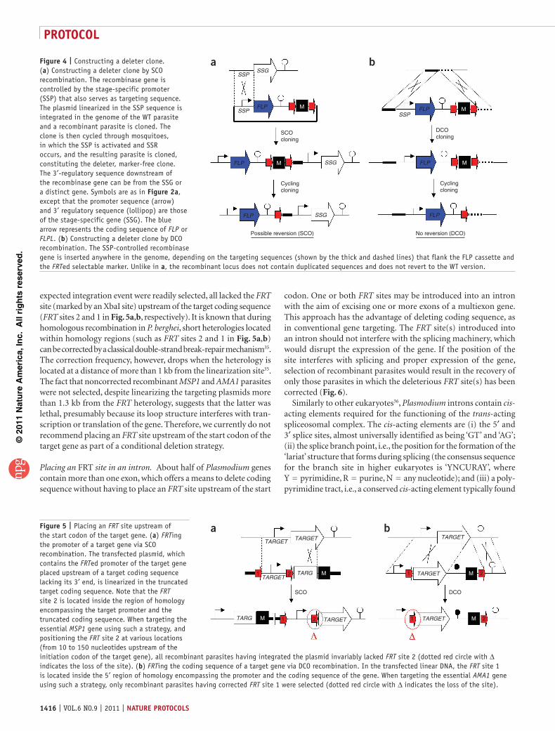
Constructing a new deleter cloneTo construct a deleter clone of distinct stage-specificity, FLP or FLPL should be expressed via the promoter of a parasite stage-specific gene (SSG), the activation profile of which mimics the desired onset of SSR. The SSG should be silent or minimally expressed before SSR is required to avoid premature SSR (although the use of the FLP/FRT system tolerates some leakiness, given that high amounts of FLP are required for efficient SSR), and expressed at high levels thereafter. Ideally, SSR should be achieved before the target gene is normally expressed to avoid any accumulation of the target product before SSR occurs. However, it is important to keep in mind that high levels of DNA excision in the parasite population are only slowly achieved, requiring, for example, days of high-level FLP pro-duction in sporozoites. Moreover, when conditionally inactivating a protein that is normally constitutively produced, the stability of the mRNA and of the protein are crucial considerations: if either one has a long half-life, then DNA excision might not lead immediately to a substantial decrease in target concentration.
The recombinase gene controlled by the SSG promoter, along with an FRTed marker, can be integrated into the genome of the
WT parasite by single-crossover (SCO; Fig. 4a) or DCO (Fig. 4b) recombination. Importantly, a locus constructed by SCO is prone to further recombination between the duplicated homology regions and to reversion to the WT locus. Therefore, using a deleter con-structed by SCO requires the verification of a lack of marked rever-sion at the recombinase locus before inserting the conditional allele or when characterizing the phenotype of the conditional clone. In contrast, a locus obtained by DCO cannot revert to the WT configuration and remains stable with time. However, although inserting the recombinase by DCO has the advantage of yielding a stable locus, it might be harmful at the chosen recombination site in the genome, and it involves more complex constructs than those necessary for the SCO strategy.
Devising a conditional allele of the target geneTo obtain a conditional clone, the WT copy of the target gene must be replaced in the deleter clone by an FRTed copy. The two FRT sites should allow normal expression of the target gene before SSR occurs, and efficient target knockout or knockdown after SSR. The schemes proposed below ensure expression of the target gene from its natural promoter prior to SSR, avoiding misexpression of the target product. Theoretically, the 5′ regulatory sequence, the coding sequence, or the 3′ regulatory sequence can be FRTed to achieve conditional gene inactivation or silencing. So far, only exon or 3′ regulatory sequence excision schemes have been used successfully. It is also possible to envisage the conditional deletion of the entire target locus.
Placing an FRT site upstream of the start codon. FRTing the pro-moter (Fig. 5a, by SCO recombination) or the coding sequence (Fig. 5b, by DCO recombination) of the target gene requires plac-ing one site upstream of the initiation codon of the gene. So far, however, all our attempts to place an FRT site upstream of the start codon of an essential gene have failed. Specifically, our attempts to FRT the promoter of MSP1 and the coding sequence of AMA1 (Fig. 5a,b, respectively), which are both essential for merozoite invasion of erythrocytes33,34, failed in repeated experiments. In both cases, although recombinant parasites having undergone the
Figure 3 | Testing SSR efficiency mediated by the TRAP/FLPL SSR system using a GFP reporter at the CS locus. (a) Cycling of a P. berghei parasite clone, derivative of the TRAP/FLPL deleter (top line), which bears an SSR GFP reporter at the CS locus. Top line, the TRAP/FLPL deleter, which is devoid of hDHFR marker, contains the FLPL gene integrated at the TRAP locus and controlled by TRAP regulatory sequences. Middle line, in this background, SCO integration of a plasmid at the CS locus separates the GFP-coding sequence from the CS promoter by an FRTed hDHFR marker, generating a TRAP/FLPL reporter clone. Bottom line, in the TRAP/FLPL reporter clone, the GFP gene is expressed via the CS promoter after SSR in the sporozoite stage in the mosquito. Symbols are as in Figure 2a, except for the 5′ (arrows, as indicated) and 3′ (open lollipop, 3′ regulatory sequence of TRAP; black lollipop, 3′ regulatory sequence of CS) regulatory sequences. The experiment was approved by the committee of Institut Pasteur and was performed in accordance with the applicable guidelines and regulations. (b,c) SSR efficiency in the TRAP/FLPL reporter clone measured by PCR analysis of genomic DNA of various stages of the clone. PCR was performed using a forward primer (Pa) hybridizing to 5′ CS regulatory sequences and a reverse primer (Pb) hybridizing to the GFP gene (a), which amplify a 2.2-kb band from a non-excised locus and a 0.6-kb band from an excised locus. (b) SSR efficiency MG and SG sporozoites (SPZ) at day 21 post-transmission maintained at 21 or 25 °C from day 16 onward. (c) SSR efficiency in blood stages in a first mouse (RBC-1), MG and SG sporozoites collected from mosquitoes at day 21 after a temperature switch at 25 °C from day 16, and blood stages in a sporozoite-infected mouse (RBC-2). SSR mediated by the TRAP/FLPL system starts in MG sporozoites; its efficiency remains limited at 21 °C but is increased at 25 °C, and it reaches high levels of excision in SG sporozoites kept at 25 °C from day 16.
MG SPZ
21° 25°
2.22.2
0.60.6
21° 25°
2.2
a
b c
CS CSGFP
GFP
CSCS
CS
CS CS CSCS
0.6Pa Pb
SG SPZ
CS
RBC-1
TRAP
M
FLPL TRAP
RBC-2
2.2
0.6
MG
SP
Z
SG
SP
Z
©20
11 N
atu
re A
mer
ica,
Inc.
All
rig
hts
res
erve
d.
protocol
1416 | VOL.6 NO.9 | 2011 | nature protocols
expected integration event were readily selected, all lacked the FRT site (marked by an XbaI site) upstream of the target coding sequence (FRT sites 2 and 1 in Fig. 5a,b, respectively). It is known that during homologous recombination in P. berghei, short heterologies located within homology regions (such as FRT sites 2 and 1 in Fig. 5a,b) can be corrected by a classical double-strand break-repair mechanism35. The correction frequency, however, drops when the heterology is located at a distance of more than 1 kb from the linearization site35. The fact that noncorrected recombinant MSP1 and AMA1 parasites were not selected, despite linearizing the targeting plasmids more than 1.3 kb from the FRT heterology, suggests that the latter was lethal, presumably because its loop structure interferes with tran-scription or translation of the gene. Therefore, we currently do not recommend placing an FRT site upstream of the start codon of the target gene as part of a conditional deletion strategy.
Placing an FRT site in an intron. About half of Plasmodium genes contain more than one exon, which offers a means to delete coding sequence without having to place an FRT site upstream of the start
codon. One or both FRT sites may be introduced into an intron with the aim of excising one or more exons of a multiexon gene. This approach has the advantage of deleting coding sequence, as in conventional gene targeting. The FRT site(s) introduced into an intron should not interfere with the splicing machinery, which would disrupt the expression of the gene. If the position of the site interferes with splicing and proper expression of the gene, selection of recombinant parasites would result in the recovery of only those parasites in which the deleterious FRT site(s) has been corrected (Fig. 6).
Similarly to other eukaryotes36, Plasmodium introns contain cis-acting elements required for the functioning of the trans-acting spliceosomal complex. The cis-acting elements are (i) the 5′ and 3′ splice sites, almost universally identified as being ‘GT’ and ‘AG’; (ii) the splice branch point, i.e., the position for the formation of the ‘lariat’ structure that forms during splicing (the consensus sequence for the branch site in higher eukaryotes is ‘YNCURAY’, where Y = pyrimidine, R = purine, N = any nucleotide); and (iii) a poly-pyrimidine tract, i.e., a conserved cis-acting element typically found
Figure 4 | Constructing a deleter clone. (a) Constructing a deleter clone by SCO recombination. The recombinase gene is controlled by the stage-specific promoter (SSP) that also serves as targeting sequence. The plasmid linearized in the SSP sequence is integrated in the genome of the WT parasite and a recombinant parasite is cloned. The clone is then cycled through mosquitoes, in which the SSP is activated and SSR occurs, and the resulting parasite is cloned, constituting the deleter, marker-free clone. The 3′-regulatory sequence downstream of the recombinase gene can be from the SSG or a distinct gene. Symbols are as in Figure 2a, except that the promoter sequence (arrow) and 3′ regulatory sequence (lollipop) are those of the stage-specific gene (SSG). The blue arrow represents the coding sequence of FLP or FLPL. (b) Constructing a deleter clone by DCO recombination. The SSP-controlled recombinase gene is inserted anywhere in the genome, depending on the targeting sequences (shown by the thick and dashed lines) that flank the FLP cassette and the FRTed selectable marker. Unlike in a, the recombinant locus does not contain duplicated sequences and does not revert to the WT version.
SSG
FLP M
FLP
FLP
FLP
M
M
FLP M
FLP
SCOcloning
Cyclingcloning
DCOcloning
Cyclingcloning
Possible reversion (SCO) No reversion (DCO)
SSG
SSG
SSP
SSPSSP
a b
Figure 5 | Placing an FRT site upstream of the start codon of the target gene. (a) FRTing the promoter of a target gene via SCO recombination. The transfected plasmid, which contains the FRTed promoter of the target gene placed upstream of a target coding sequence lacking its 3′ end, is linearized in the truncated target coding sequence. Note that the FRT site 2 is located inside the region of homology encompassing the target promoter and the truncated coding sequence. When targeting the essential MSP1 gene using such a strategy, and positioning the FRT site 2 at various locations (from 10 to 150 nucleotides upstream of the initiation codon of the target gene), all recombinant parasites having integrated the plasmid invariably lacked FRT site 2 (dotted red circle with ∆ indicates the loss of the site). (b) FRTing the coding sequence of a target gene via DCO recombination. In the transfected linear DNA, the FRT site 1 is located inside the 5′ region of homology encompassing the promoter and the coding sequence of the gene. When targeting the essential AMA1 gene using such a strategy, only recombinant parasites having corrected FRT site 1 were selected (dotted red circle with ∆ indicates the loss of the site).
SCO DCO
M
M M
M1 2 1 2
1 21 2 TARG
TARG
TARGET
TARGET
TARGETTARGET
TARGET
TARGET
TARGET
a b

©20
11 N
atu
re A
mer
ica,
Inc.
All
rig
hts
res
erve
d.
protocol
nature protocols | VOL.6 NO.9 | 2011 | 1417
between the branch point and the 3′ splice site. The FRT site should not disrupt these sequences. In general, placing it closer to the 5′ splice site should minimize the risks of disrupting these con-served cis-acting elements.
Obviously, although both FRT sites can be incorporated within introns, only one site can be inserted within an intron, whereas the other is placed upstream of the 5′ regulatory sequence or downstream of the 3′ regulatory sequence of the gene (Fig. 6). A DCO recombination strategy for placing an FRT site in an intron and the other downstream of the 3′ regulatory sequence was successfully used for conditionally inactivating the PKG gene in the P. berghei sporozoites22. In this case, placing FRT site 1 between the ‘branch point’ sequence and the 3′ splice site resulted in the exclusive recovery of integration events in which FRT site 1 was corrected (Fig. 6a). Modification of the targeting plasmid such that the FRT site was situated upstream of the ‘branchpoint’ sequence was successful in recovering integration events with intact FRT sites22 (Fig. 6b).
FRTing the 3′ regulatory sequence. We also attempted to condi-tionally deplete a protein of interest by deleting the 3′ regulatory sequence of the gene (Fig. 7). Using both AMA1 and MSP1 as tar-gets, we used DCO recombination to select recombinant parasites having an FRT site immediately after the stop codon of the target gene. Both AMA1 and MSP1 recombinant parasites having the 3′ FRT site (upstream of the heterologous 3′ regulatory sequence of TRAP) were repeatedly selected, showing that the FRT downstream of the stop codon did not interfere with normal gene expression. Stage-specific SSR caused efficient target protein depletion, typi-cally a 95% decrease of the target protein in the conditional popu-lation compared with the controls (i.e., having only the deleter locus or only the conditional target allele) (Fig. 7b). As AMA1 is a sporozoite protein, the conditional AMA1 allele was introduced in the TRAP/FLPL-F background, which led to AMA1 depletion in SG sporozoites. As MSP1 is a liver-stage protein, the MSP1 allele was introduced in the UIS4/FLP-F background, which led to MSP1 depletion in liver stages.
Notably, when using the strategy depicted in Figure 7a, we found that the second FRT site must be placed immediately upstream of the plasmid, not of the selectable marker. Successful protein depletion was only achieved in the former case, thereby suggesting that, in the latter case, parasites used sequences in the promoter region of the selectable marker to stabilize target gene transcripts. The starting plasmid for using the strategy of 3′ regulatory sequence excision is depicted in Figure 8a, and the cloning scheme to insert the homology regions of the target gene in this plasmid is shown in Figure 8b. Figure 8c shows the linker sequence between the stop codon of the target gene and the first
Figure 6 | Placing an FRT site in an intron of a multiexon target gene. The FRT site 1 is placed in an intron, whereas the selectable marker and the FRT site 2 are placed in the 3′ regulatory sequence of the target gene. Symbols are as in Figure 2. Blue line, intron; blue arrowhead, ‘branch point’ sequence and the polypyrimidine tract. (a) Placement of the FRT site 1 between the branch point sequence and the polypyrimidine tract can interfere with correct splicing of the target locus. Recombinant parasites obtained after DCO lack the intronic FRT site and thus cannot undergo SSR. (b) Placement of the FRT site 1 to avoid the branch point sequence and the polypyrimidine tract that lie upstream of the right splicing junction. SSR in the resulting parasites leads to the excision of the targeted exon and the selectable marker.
Exon 1 Exon 2 Exon 2
DCO
AGGT
AGGT
AGGT
GT
Exon 1
Exon 2Exon 1
Exon 1
SSR
2
21
1 M
M
2
Exon 1a b
Exon 2
DCO
GT AG
GT AG
GT AG
1 2M
M
Figure 7 | Placing an FRT site downstream of the stop codon of the target gene. (a) Strategy used to FRT the 3′ regulatory sequence of a target gene via DCO recombination. Top line, the 5′ and 3′ segments of homology in the transfected linear DNA are the 3′ end of the coding sequence and 3′ regulatory sequence of the target gene, respectively. Note that the FRTed 3′ regulatory sequence driving target gene expression before SSR is not the natural 3′ untranslated region of the target gene. Middle line, expected recombinant parasite before SSR. Bottom line, recombinant parasite after SSR, in which the plasmid sequence is placed downstream of the coding sequence of the target gene. (b) Western blot analysis assessing MSP1 production in blood stages (left) and liver stages (right) of MSP1 conditional mutants (COND), constructed as in a in the UIS4/FLP deleter, compared with the control UIS4/FLP deleter parasites (CONT). AMA1-specific antibodies were used as a control in both blots.
TARGET
TARGET
TARGET
RBC stages
COND
AMA1
MSP1
AMA1
MSP1
CONDCONT CONT
Liver stages
M
DCO
SSR
GET
M
a
b

©20
11 N
atu
re A
mer
ica,
Inc.
All
rig
hts
res
erve
d.
protocol
1418 | VOL.6 NO.9 | 2011 | nature protocols
FRT site that was designed for MSP1 mutagenesis and that led to efficient gene silencing after SSR.
FRTing the entire locus. FRTing an entire locus might also be achieved in a single DCO recombination step (Fig. 9) if the target gene is small enough. However, we do not recommend constructing targeting plasmids larger than 11–12 kb, as cloning and replicating large fragments of A/T-rich Plasmodium DNA is notoriously inefficient. It should also be possible to devise two-step strategies to FRT and conditionally excise the entire target locus, by first placing an FRT site upstream of the promoter region of the target gene, and—after cycling through mosquitoes and marker recycling—then inserting a second FRT site downstream of the target gene.
Protocol overviewHere we detail the construction of a conditional parasite clone. This is achieved by exchanging, in a deleter clone that con-tains the stage-specifically expressed FLP, the WT target gene with an FRTed copy of the gene. The erythrocytic stages of the
conditional clone are then transferred to mosquitoes and SSR efficiency is monitored at the sporozoite stage. SSR efficiency and the phenotype of the conditional clone are then compared with those of control clones, which have either the FRTed allele or the stage-specifically expressed FLP only. The protocol requires an appropriate deleter clone and a targeting plasmid; these depend on the chosen strategy, and, therefore, on the struc-ture and length of the target gene, the amounts of the target proteins and the desired timing of protein depletion. Both the deleter clone and the targeting plasmid must be validated before starting the procedure. The deleter clone, if constructed by SCO recombination, should not have substantial amounts of rever-sion at the recombinase locus (see Step 5). The functionality of the FRT sites in the targeting plasmid should also be verified. This can be done by sequencing or by transforming FLP-express-ing 294-FLP E. coli27 with the plasmid and checking the SSR- competence of the FRT sites by restriction digestion analysis of plasmid DNA. Fluorescent parasites are useful for expediting the PROCEDURE as well for characterizing the phenotypes of conditional sporozoites.
Figure 8 | The p3′regFRT plasmid. (a) The plasmid p3′regFRT for FRTing the 3′ regulatory sequence of the target gene (Fig. 7a). Plasmid p3′regFRT, a derivative of pUC18 (thick line), contains the 3′ regulatory sequence of TRAP (black lollipop) and the hDHFR marker (1.6 kb; M-marked black box), flanked by two FRT sites (red arrows) and preceded by a HindIII-NotI linker (H-S-N). Note that the hDHFR marker contains 0.6 kb of the P. berghei eef1α promoter, 0.6 kb of the coding sequence of hDHFR and 0.4 kb of the 3′ regulatory sequence of P. berghei DHFR-TS. The restriction map of plasmid p3′regFRT and 4,992 bp of its sequence (2,711 bp of insert and 2,281 of pUC backbone) are available from MR4 (see MATERIALS). (b) Strategy for constructing the targeting plasmid from plasmid p3′regFRT. The 3′ end of the coding sequence of the target gene (fragment 1; 0.5–1 kb) is amplified with a forward and a reverse primer incorporating a SphI and a NotI site, respectively. The 3′ regulatory sequence of the target gene (fragment 2; 0.5–1 kb) is amplified with a forward and a reverse primer incorporating a HindIII and a SphI site, respectively. Fragments 1 and 2 are then cloned in the respective sites in the linker of plasmid p3′regFRT (see a). The resulting plasmid should be linearized in the Sph1 site to promote the desired DCO recombination event, as depicted in Figure 7a. (b,c) Note that the reverse primer used to amplify fragment 1 (blue asterisk) needs to incorporate additional sequence ensuring a correct linker between the end of coding sequence of the target gene and the first FRT site, as shown in c. (c) DNA sequence surrounding the first FRT site in the recombinant locus of the MSP1 conditional clone obtained after DCO recombination, as depicted in Figure 7a. Two stop codons are followed by the first 12 nucleotides of the TRAP 3′-regulatory sequence, the NotI site, seven nucleotides from the plasmid, the FRT site, eight nucleotides from the plasmid, and the rest of the 3′ regulatory sequence of TRAP, starting at nucleotide 13. The 15 nucleotides between the stop codon of the target gene and the NotI site, or a related sequence, should thus be incorporated in the reverse primer used to amplify fragment 1. H, HindIII; S, SphI; N, NotI.
TA
Target 3 ′ TRAP UTR(1–12)
3 ′ Trap
TARGET
TARGET
1 2
SHNS
M
M
2
H-S-N
1
3 ′ TRAP UTR(13–...)
Notl
Reverse primer fragment 1 *
*
Plasmid p3 ′regFRT
Plasmidp3′regFRT
a b
c
FRT
A AAT TTTT TA AAA AC G GG G A AAGG G GTT T TT T TA AAA T T TTA A AAGG C CC CGT T T T TA A A ATATCGC C GCC AAACTC CC CA
Figure 9 | Conditional deletion of the target locus. A one-step, DCO-based procedure to conditionally delete the target locus. The targeting plasmid contains, from 5′ to 3′: a 5′ region of homology corresponding to a region upstream of the promoter of the target gene (thick line), an FRT site, the promoter and the coding sequence of the target gene, a second FRT site, the 3′ regulatory sequence of the target gene, the selectable marker, and a 3′ region of homology corresponding to a region downstream of the 3′ regulatory sequence of the target gene (dashed line). Note that the promoter and 3′ regulatory sequences driving target gene expression can be heterologous; a heterologous promoter region would avoid the risk of correction of the first FRT site during recombination. Also note that, after SSR, the excised DNA contains a copy of the target gene that lacks a 3′ regulatory sequence. The numbers below the plasmid indicate the sizes of the respective DNA fragments.
TARGET
TARGETTARGET
>0.5 1 X 0.7
M
M
M
1.6
DCO
SSR
>0.5

©20
11 N
atu
re A
mer
ica,
Inc.
All
rig
hts
res
erve
d.
protocol
nature protocols | VOL.6 NO.9 | 2011 | 1419
MaterIalsREAGENTS
Clones and plasmids. The deleter clones, the p3′regFRT plasmid and its complete sequence are available at MR4 (http://www.mr4.org/).Laboratory animals. 3-week-old female Wistar rats and 3-week-old female Swiss mice (Janvier or equivalent source) ! cautIon All experiments in-volving rodents must conform to governmental and institutional guidelines and regulations.Parasites. In our laboratory, we use the NK65 P. berghei line. The UIS4-FLP( − ) and TRAP-FLPL( − ) deleter clones, as well as their fluorescent counterparts, are derivatives of NK65 (MR4, cat. no. MRA-268). Mosquitoes Anopheles stephensi Sda500 mosquitoes (Centre de Production et Infection des Anophèles (CEPIA), Institut Pasteur)
General reagentsHiSpeed Plasmid Midi Kit (Qiagen, cat. no. 12643)iQ SYBR Green Supermix (Bio-Rad, cat. no. 170-8880)NucleoSpin Extract II (Macherey-Nagel, cat. no. 740588.50)NucleoSpin Plasmid kit (Macherey-Nagel, cat. no. 740609.50)QIAamp DNA Blood Mini Kit (Qiagen, cat. no. 51306)SuperSignal West Pico Chemiluminescent Substrate (Pierce, cat. no. 34080)Hybond-XL membrane (GE Healthcare, RPN 303D)PCR DIG probe synthesis kit (Roche, cat. no. 11636090910)DIG Easy Hyb (Roche, cat. no. 11603558001)DIG Wash and Block Buffer set (Roche, cat. no. 11585762001)DIG luminescence detection kit (Roche, cat. no. 11363514910)Insulin syringes (1 ml; Terumo, cat. no. 304000)WR99210 (Jacobus Pharmaceutical Company)Pyrimethamine (Sigma, cat. no. P-7771)Glycerol (Sigma, cat. no. G5516)Alsever’s solution (Sigma, cat. no. A3551)Ethanol (Sigma, cat. no. 02860)Benzyl alcohol (Sigma, cat. no. 305197)LB medium (Sigma, cat. no. L2542)Ampicillin (Sigma, cat. no. A9518)Sodium acetate (pH 5.5, 3M; Ambion, cat. no. AM9740)Agarose (Invitrogen, cat. no. 15510019)TaKaRa Ex Taq polymerase (Takara, cat. no. RR001A)dNTP set, 100 mM solution (Invitrogen, cat. no. 10297018)SSC 20× (Sigma, cat. no. S6639)HCl (Fluka, cat. no. 84419)NaOH (Fluka, cat. no. 30531)Tris-HCl (20 mM, pH 8; Sigma, cat. no. T5941)EDTA (Sigma, cat. no. 431788)Triton X-100 (Euromedex, cat. no. 2000)
•
•
•
•
••••••
••••••••••••••••••••••••
Plasmodipur (Euro-diagnostic, cat. no. 8011)Saponin (Sigma, cat. no. S7900)Ethidium bromide (EUROBIO, cat. no. GEPBET02.AF)Southern blot Nylon membrane (Amersham hybond-XL, GE Healthcare, cat. no. RPN82S)Western blot membrane (Amersham hybond-ECL, GE Healthcare, cat. no. 9007590)DMSO (Sigma, cat. no. D5879)Phenol (Sigma, cat. no. P1037)Chloroform (Sigma, cat. no. C2432)PBS (Gibco, cat. no. 14190-169)Bromofluorescein (Sigma, cat. no. E4009)Tween-20 (Sigma, cat. no. P9416)SDS-PAGE reagents (Bio-Rad)β-Mercaptoethanol (Sigma, cat. no. M7154)Sucrose (white sugar)
EQUIPMENTNet-covered cages for mosquitoesAspirator for collecting mosquitoesPCR instrument (GeneAmp, Applied Biosystems)SDS-PAGE dual casting unit (Bio-Rad)Infrared heat lampNucleofector device (Amaxa; http://www.amaxa.com/)Orbital shakerMicroscope (fluorescence microscope for fluorescent parasite strains)Microscope slides and cover slipsTweezersQuantity One software (Bio-Rad)
REAGENT SETUPParasite freezing solution To prepare parasite freezing solution, dissolve 10% (vol/vol) glycerol in Alsever’s solution. It can be stored for 6 months at room temperature (around 22 °C).Pyrimethamine solution To prepare pyrimethamine solution, dissolve pyrimethamine powder in DMSO to a final concentration of 7 mg ml − 1 and dilute 100× with water at a pH of 3.5–5. Store at 4 °C and use the solution as drinking water for mice for a maximum of 7 d.WR99210 solution To prepare WR99210 solution, add 16 mg of WR99210 powder to 2 ml of ethanol and 0.15 ml of benzyl alcohol. Mix by stirring on a vortexer to dissolve the powder. Make up to 5 ml with 2.85 ml of dH
2O. Store
at room temperature and use the solution for a maximum of 7 d.Sporozoite lysis buffer (10×) Mix 20 mM Tris-HCl (pH 8), 1 mM EDTA, 0.5% (vol/vol) Triton and 0.5% (wt/vol) SDS. Store at room temperature for up to 6 months.
••••
•
•••••••••
•••••••••••
proceDurepreparation of the targeting plasmid ● tIMInG 2 d1| At least 2 d before electroporation, inoculate 80–100 ml of LB medium containing ampicillin (0.1 mg ml − 1) with bacteria containing the targeting plasmid and culture for 12–16 h (usually overnight) at 37 °C and 200 r.p.m. agitation.
2| After overnight culture, isolate the plasmid from bacteria using the HiSpeed Plasmid Midi kit according to the manufacturer’s instructions, and digest the DNA (15 µg in 100 µl) overnight with the appropriate restriction enzymes and at the appropriate temperature (add to DNA 1× final concentration buffer and 1 unit of enzyme per µg of DNA) to linearize the plasmid.
3| To purify the DNA, precipitate with two volumes of absolute ethanol and one-tenth volume of sodium acetate 3 M (pH 5.5) overnight at − 20 °C. Next, centrifuge at 16,000g for 15 min at 4 °C, discard the supernatant, wash the pellet by adding 800 µl of 70% (vol/vol) ethanol, and centrifuge again for 10 min at 16,000g. Finally, discard the supernatant, air-dry the pellet at room temperature and resuspend the DNA with sterile water at a final concentration of 1 µg µl − 1. crItIcal step 5 µg of linear DNA is required for one transfection experiment. crItIcal step Because transfection of undigested DNA leads to the selection of parasites carrying episomes, the quality of linearization of the targeting plasmid after overnight digestion must be checked before transfection by running 300 ng of the purified digested plasmid on 0.8% (wt/vol) agarose gel with ethidium bromide (one drop for 60 ml) for 40 min at 100 V.

©20
11 N
atu
re A
mer
ica,
Inc.
All
rig
hts
res
erve
d.
protocol
1420 | VOL.6 NO.9 | 2011 | nature protocols
Linearization of the plasmid DNA should be >95%, corresponding to only one detectable band in the gel, or linear DNA should be purified using the Nucleospin Extract II kit according to the manufacturer’s instructions. It is preferable, although not mandatory, to gel-purify the linear DNA.
transfection of schizonts of the deleter clone ● tIMInG 10 d4| By intraperitoneal (i.p.) injection with a 1-ml syringe, infect one rat with 450 µl of cryopreserved blood stages of the deleter clone immediately after thawing. crItIcal step It is preferable to inject young (3–4 weeks old) rats, which are easier to manipulate and more sensitive to the infection.! cautIon All experiments involving rodents must conform to governmental and institutional guidelines and regulations.
5| Check parasitemia by blood smears daily (see ref. 37), and when the parasitemia of the rat is 2–5% (normally 3 d after injection) collect the blood by cardiac puncture (between 3 and 5 ml) under anesthesia and isolate parasite genomic DNA (as described in Box 1) for genetic analysis of the recombinase locus by Southern blotting (as described in Box 2; Figure 10 shows one restriction digestion that differentiates the two loci in the two available deleter clones). crItIcal step Deleter parasites that have been constructed by SCO recombination (Fig. 4a) can undergo reversion to the WT locus, which may occur in parasites that have been cultured and cycled extensively. The lack of substantial reversion at the recombinase locus in the deleter population can also be verified at various steps of the procedure (Steps 15 and 20), preferably before transferring a conditional clone to mosquitoes. Southern blot analysis (Box 2), by providing a semiquantitative estimation of the proportion of recombinant versus reverted parasites, is preferable to simple PCR analysis.
6| Transfer blood from Step 5 to two naive rats by intravenous injection, so as to obtain animals with 2–5% parasitemia the day prior to electroporation. From these rats, prepare schizonts for electroporation (detailed in ref. 37). crItIcal step On the basis of a tenfold increase in parasitemia per day, and the fact that rats have 6 × 10⁹ red blood cells (RBC) per ml of blood, the number (n) of infected red blood cells (iRBCs) to be injected to obtain a 5% parasitemia can be calculated using the formula n = ((3 × 10⁸) / 10a)V, where ‘a’ is the number of days to wait until the day prior to electroporation and ‘V ’ the volume of blood of the receiver rat (considering the fact that a rat has 7 ml of blood per 100 g weight). The number of iRBCs per ml of blood from the donor rat from Step 5 can be calculated using the formula iRBCs per ml = (parasitemia × 6 × 10⁹)/100. Ideally, 200 µl of diluted blood from the donor rat (corresponding to
Box 1 | ExTRACTIoN oF GENoMIC DNA (a) From parasite blood stages1. Collect 700 µl of blood at 2–5% parasitemia from the infected animal.2. Dilute the blood with 5 ml of 1× PBS.3. Remove leukocytes from the infected blood by passing the blood through a leukocyte filter (Plasmodipur).4. Add 0.15% (wt/vol) saponin, final concentration (from a stock solution of 10% (wt/vol) saponin in water).5. Wait for 5 min.6. Centrifuge for 20 min at 3,200g.7. Discard the supernatant and wash the parasite pellet twice with 1× PBS (add 1 ml of 1× PBS and centrifuge at 12,000g for 5 min at room temperature and discard the supernatant).8. Extract the genomic DNA of the parasite pellet using the QIAamp DNA Blood Mini Kit following the manufacturer’s instructions.(B) From sporozoites1. Collect 106 sporozoites from mosquito salivary glands (yielding 200 ng to 1 µg of genomic DNA) in 1× PBS (50 µl final).2. Resuspend with 20 volumes of 10× sporozoite lysing buffer diluted to 1× final concentration with water and add proteinase K (1 mg ml − 1 final).4. Incubate for 1 h at 37 °C.5. Add 800 µl chloroform/phenol (vol/vol).6. Centrifuge at 10,000g for 10 min at 4 °C.7. Collect the upper phase (800 µl), add 800 µl of chloroform and centrifuge at 10,000g for 10 min at 4 °C.8. Collect the upper phase (800 µl).9. Precipitate with absolute ethanol (2 volumes) + 1/10 sodium acetate 3 M (pH 5.5), overnight at − 20 °C.10. Centrifuge at 13,000g for 15 min at 4 °C.11. Discard the supernatant.12. Wash the pellet by adding 800 µl of 70% (vol/vol) ethanol, centrifuge for 10 min at 16,000g, discard the supernatant, air-dry at room temperature and resuspend in 50 µl of water.

©20
11 N
atu
re A
mer
ica,
Inc.
All
rig
hts
res
erve
d.
protocol
nature protocols | VOL.6 NO.9 | 2011 | 1421
the volume in which there are the desired number of iRBCs as calculated by the formula above) in 1× PBS is injected intravenously. crItIcal step We normally transfer the parasites to naive rats 4 d before the parasitized blood of these rats can be cul-tured in vitro overnight for schizont production. Typically, if Step 5 is performed on day 0 (D0), schizonts are available for electroporation on D5.
7| Electroporate the purified schizonts/merozoites from Step 6 with the DNA from Step 5 using the AMAXA Nucleofector electroporator (see ref. 37). Inject one electroporation mix, which contains 107–108 merozoites and 5 µl (5 µg) of linear DNA, intravenously into two 3-week-old female Swiss mice. Mice injected with an electroporation mix are called ‘parental’.
8| Prepare a blood smear from the animals 24 h after electroporation. Parasitemia should be between 0.1 and 3%.
selection of drug-resistant parasite populations ● tIMInG 2 weeks9| If the transfected parasites do not contain any selectable marker, e.g., the UIS4-FLP and TRAP-FLPL deleters, and the transfected DNA contains the hDHFR marker, treat parental mice from Step 7 with pyrimethamine (option A). If they contain the DHFR-TS marker, e.g., fluorescent derivatives of the UIS4-FLP( − ) and TRAP-FLPL( − ) deleters, treat parental mice from Step 7 with WR99210 (option B).(a) pyrimethamine treatment (i) Provide mice with drinking water containing pyrimethamine (see REAGENT SETUP) starting 1 d after injection of the
transfected parasites and continue for 4 consecutive days.(B) Wr99210 treatment (i) Inject, subcutaneously, a single dose of 0.1 ml WR99210 (equal to 6 mg kg − 1 weight in mice of 20 g) 1 d after
injection of the transfected parasites. (ii) Repeat the same treatment on the following 3 d (a total of four injections).
crItIcal step Typically, recombinant parasites grow as efficiently as WT parasites (i.e., a tenfold increase in parasitemia per day) and a pure parental population of recombinant parasites should be detectable (parasitemia > 0.01%) 6–7 d after electroporation. A slower growth rate of the parental population, or its delayed emergence, might be due to different reasons as discussed in TROUBLESHOOTING. ? trouBlesHootInG
10| Prepare a blood smear from the animals 24 h after initiation of the treatment and verify that parasitemia has lowered or is undetectable.
11| After D4 of treatment, check daily by blood smear until parasitemia is > 0.01% in the treated animals. When the parasitemia re-emerges, usually around D6, collect 50 µl of blood, mix with 150 µl of PBS and inject 100 µl of the mixture into each of two naive ‘transfer’ mice.
Box 2 | SoUTHERN BLoT ANALYSIS oF PARASITE GENoMIC DNA Southern blot analysis allows a semiquantitative assessment of the proportions of recombinant versus WT target loci by comparing band intensities, especially if bands are of similar sizes. We usually validate a transfer population for parasite cloning only when the proportion of parasites having the desired recombinant locus is > 10%. If neither of the two transfer populations contains this minimal ratio of desired recombinants, a new transfection is recommended.1. Digest 2–5 µg of parasite genomic DNA using appropriate restriction endonucleases.2. Precipitate with ethanol (2 volumes) + 1/10 sodium acetate 3 M, pH5.5, discard the supernatant, wash twice in 70% (vol/vol) ethanol, centrifuge at 13,000g for 15 min at 4 °C, discard the supernatant and resuspend in 25 µl of water.3. Run the total (2–5 µg) digested DNA on a 0.8% (wt/vol) agarose gel with ethidium bromide overnight at 40 volts.4. Depurinate the DNA in the gel in 250 mM HCl for 15 min.5. Neutralize the gel in 0.5 M NaOH for 15 min.6. Transfer the DNA for 4 h to a positively charged nylon membrane using an alkaline transfer buffer in 0.5 M NaOH.7. Label the DNA probe with digoxigenin using the PCR DIG Probe synthesis kit according to the manufacturer’s instructions.8. Immerse the membrane in 2× SSC for 10 min.9. Incubate the membrane with DIG Easy Hyb for 30 min.10. Denature DIG-labeled DNA probe by boiling for 10 min and rapidly cooling on ice.11. Hybridize at the appropriate hybridization temperature (calculated according to GC content) using the PCR DIG Probe synthesis kit and DIG luminescence detection kit according to the manufacturer’s instructions.12. Wash, block and detect signals by immunological detection of DIG-labeled probes according to the manufacturer’s instructions.

©20
11 N
atu
re A
mer
ica,
Inc.
All
rig
hts
res
erve
d.
protocol
1422 | VOL.6 NO.9 | 2011 | nature protocols
12| Treat transfer mice with the appropriate drug for 3 d starting 24 h after parasite transfer, as indicated in Step 9.
13| When the parasitemia of the transfer mice is > 1% (check every day after injection by blood smear), collect blood by cardiac puncture (~1 ml). crItIcal step Typically, a transfer population rises to a parasitemia of 2–5% between 3 and 5 d after parasite injection.
14| Prepare two stabilates from each transfer mouse by mixing 300 µl of blood and 600 µl of parasite freezing solution, and immediately store as two stabilates of 450 µl each in liquid nitrogen for up to 10 years. crItIcal step One such frozen stabilate can be used to infect three mice (each injected i.p. with 150 µl of the stabilate thawed at room temperature), in which the parasitemia usually rises to >1% within 3–4 d.
analyzing transfer population ● tIMInG 1 week15| Use the remaining 700 µl of the blood from Step 13 for preparing genomic DNA of transfer parasites (Box 1) and for characterizing the target locus by PCR (Box 3) and/or Southern blot (Box 2) analysis.
cloning conditional parasite(s) ● tIMInG 2 weeks16| Infect a Swiss mouse (donor) with a frozen stabilate of the transfer parasite population (from Step 14) that contains the highest proportion of the desired recombinant parasites (as determined in Step 15).
17| When the parasitemia in the donor mouse is between 0.1 and 1% (check parasitemia every day after injection by preparing blood smears and precisely counting 20–30 fields of 300–400 erythrocytes), clone parasites by limiting dilution into Swiss mice. To do this, calculate the number of iRBCs per ml in the blood of the donor mouse as described in Step 6 and dilute the blood in 1× PBS to prepare a suspension (200 µl) of 5 × 10⁶ iRBCs per ml. Prepare serial dilutions of 1/10 in 1× PBS to obtain a final dilution of 5 iRBCs per ml and inject recipient mice i.v. with 100 µl of the final dilution each. crItIcal step Mice infected with a single parasite usually become patent (parasitemia > 0.01%) 7–9 d after injection. crItIcal step The parasitemia of the donor mouse should be < 1% to avoid infection of individual erythrocytes with multiple parasites, which might occur at high parasitemia. crItIcal step The number of recipient mice depends on the proportion of the desired recombinant in the donor mouse assessed by Southern blotting in Step 15. We typically clone in 30 mice after WR99210 selection and in 15 mice after pyrimethamine selection, as the former selection yields a greater proportion of nonrecombinant but resistant parasites. crItIcal step Successful cloning implies that no more than 40% of the recipient mice become patent.
18| Ten days after cloning, when parasitemia in patent animals is >1%, store the blood of patent animals in liquid nitrogen as described in Step 14.
19| Using > 700 µl of the fresh infected blood from Step 18, prepare parasite genomic DNA (Box 1) and perform PCR analysis (Box 3) using primers specific for the recombinant and the WT loci (Fig. 11). Retain for further analysis the clone(s) in which the recombinant but not the WT locus is amplified.
TRAPTRAP TRAP
TRAPFLPL
TRAP TRAP TRAPTRAP
UIS4UIS4UIS4
UIS4 UIS4 UIS4UIS4FLP
E E
B B5.3
B B9
EE 5.1
11
TRAP
a
b
Figure 10 | Wild-type and recombinant TRAP and UIS4 loci in the deleter clones. (a) TRAP locus in the WT (top) and in the TRAP-FLPL( − ) deleter clone (bottom). The reversion of the TRAP locus from its recombinant, FLPL-containing form to the WT form can be assessed by Southern blot analysis using a BamHI (B) digestion. In the TRAP-FLPL( − ) clone, the proximal BamHI site is located 23-bp downstream of the 5′ end of the FRT site (in opposite orientation). The numbers above the diagrams indicate DNA fragment sizes in kb. (b) UIS4 locus in the WT (top) and in the UIS4-FLP( − ) deleter clone (bottom). The reversion of the UIS4 locus from its recombinant, FLP-containing form to the WT form can be assessed by Southern blot analysis using EcoRV (E) digestion. In the UIS4-FLP( − ) clone, the proximal EcoRV site is located 22 bp upstream of the 3′ end of the FRT site (in opposite orientation). The numbers above the diagrams indicate DNA fragment sizes in kb.

©20
11 N
atu
re A
mer
ica,
Inc.
All
rig
hts
res
erve
d.
protocol
nature protocols | VOL.6 NO.9 | 2011 | 1423
analyzing clones ● tIMInG 1 week20| Perform Southern blot analysis (Box 2) on the clone(s) validated by PCR in Step 19, and confirm the presence of the expected recombinant locus and the absence of significant reversion at the recombinase locus.
preparing stabilates of selected conditional clones ● tIMInG 1 week21| By i.p. injection, infect one rat with 450 µl of cryopreserved infected blood from Step 18 of the clone(s) validated in Steps 19 and 20. When the parasitemia of the rat is between 1 and 5% (usually 3–4 d after injection), collect the blood by cardiac puncture and inject 100 µl i.v. into four receiver rats.
22| When parasitemia in the receiver rats is 3–5% (usually 3 d after injection), store as many blood stabilates as possible in liquid nitrogen (as described in Step 14). crItIcal step The number of stabilates will correspond to the number of mosquito infections and sporozoite characterization experiments that can be performed. A complete characterization of a clone (SSR efficiency, protein depletion, phenotype) requires large numbers (millions) of sporozoites and repeated experiments.
scoring ssr efficiency in sporozoites of a conditional clone ● tIMInG 5–6 weeks23| By i.p. injection, infect two Swiss mice with 200 µl of a clone stabilate (from Step 22) 3 d before feeding the mosquitoes.
24| To transfer the parasite clone to mosquitoes, feed mosquitoes on P. berghei gametocyte-harboring mice from Step 23 (parasitemia 1–5%) 3–5 d after emergence (pupae to adults). Mosquitoes should be maintained at elevated humidity (70%) in dedicated incubators or in a room at 21 °C and fed on 1% (wt/vol) sucrose solution. The quality of mosquito infection can be checked 5–7 d post-transmission by dissecting a few mosquitoes and counting the proportion of mosquitoes with oocysts: aspirate ten mosquitoes from the cage and anesthetize by placing them at 4 °C or on ice. Place the mosquitoes onto a microscope slide and pinch the tip of the abdomen of the mosquito with tweezers and, using a needle, gently pull the other end of the abdomen to remove the MG. Place the MG in a few drops of PBS (for fluorescent parasites) or bromofluorescein (5%, wt/vol) and mount between microscope slide and cover slip. Observe with a microscope at low magnification to count the number of oocyst (fluorescent or stained with bromofluorescein)-positive MGs. At least 60% of
the mosquitoes should have oocysts, characterizing a good quality of infection that will allow the recovery of a sufficient number of sporozoites for phenotypic analysis.
Box 3 | PCR ANALYSIS oF PARASITE GENoMIC DNA PCR analysis is useful for detecting the presence of each of the potential resistant parasite populations (i.e., containing a WT target locus with or without an episome, or a recombinant locus) (Fig. 11). It is also useful for assessing the proportion of recombinant parasites that, during homologous recombination, have corrected an FRT heterology potentially located within a region of homology in the targeting plasmid, as in Figure 5. In this case, the proportion of recombinant parasites having or lacking an FRT site, which contains an XbaI site, can be assessed by PCR amplification of a fragment that encompasses the FRT site, digesting with the XbaI enzyme, and by agarose gel analysis of the digestion products.1. Design PCR primers (Tm 60 °C, 25–35 bp long).2. Prepare PCR reaction mixture (50 µl) as follows: PCR mix: 5 µl buffer 10×; 4 µl dNTP (2.5 mM each); 4 µl primer (10 µM each) TaKaRa ex Taq polymerase: 1.25 units Parasite genomic DNA (40–90 ng)3. Use the following program: initial denaturation (95 °C, 5 min) followed by 30 cycles of denaturation (95 °C, 45 s), annealing (55 °C, 45 s) and extension (68 °C, 1 min kb − 1) before the final elongation (68 °C, 7 min).
21
43 3
2
4
SCO1+42+3
1
WT1+2
EPI3+4
DCO1+3
WT
SCO DCO
1+2EPI3+4
Figure 11 | PCR primers for assessing the outcome of gene targeting experiments. Top, schematics of homologous recombinations between homology regions in the genome (dashed lines) and the targeting DNA. The primers are shown as red arrows above their hybridization sites, and immediately precede or follow one (blue labeled) homology region. Bottom, transfection outcomes and the primer pairs that amplify a fragment. Symbols: blue and red rectangles, homology regions; thick line, plasmid backbone; double arrows, linearization sites in the targeting plasmids. EPI, episome.
©20
11 N
atu
re A
mer
ica,
Inc.
All
rig
hts
res
erve
d.
protocol
1424 | VOL.6 NO.9 | 2011 | nature protocols
This is essential because mosquito mortality increases with time, particularly if mosquitoes are kept at 25 °C from D16 after infection. crItIcal step Keep mosquitoes in the absence of sucrose for 1 d before the infective blood meal. Many mosquitoes should be infected, ideally from 600 to 800 mosquitoes divided into two cages, for SSR evaluation and phenotype characterization.
25| Temperature switch. For conditional parasites containing FLPL, place infected mosquitoes at 25 °C, a temperature at which FLPL shows better efficiency, from D16 onwards. crItIcal step Do not place infected mosquitoes at 25 °C before D16, as this decreases sporozoite invasion of SGs and SG loads. However, placing mosquitoes at 25 °C from D16 does not alter sporozoite infectivity to mice at least until D28.
26| Collect mosquito SGs between days 24 and 28 after infection, as previously described38, to obtain sporozoites, and keep them on ice in PBS, or in tissue culture medium with or without FCS (depending on the purpose). crItIcal step Note that FCS activates the sporozoites (gliding and infectivity), so this should be taken into consideration when deciding whether or not to dissect them in FCS-containing medium.
27| Prepare genomic DNA from SG sporozoites (Box 1).
28| Evaluate SSR efficiency in SG sporozoites. SSR efficiency can be monitored by conventional (option A) or quantitative PCR (option B) at the target locus.(a) conventional pcr evaluation of ssr (i) Use a set of primers that amplifies both the excised and non-excised versions of the locus (a 5′ forward primer hybridizing
upstream of the 5′ FRT site and a 3′ reverse primer hybridizing downstream of the 3′ FRT site), and/or a set of primers that only amplifies the non-excised locus (hybridizing on either side of one FRT site). Carry out PCR as described in Box 3.
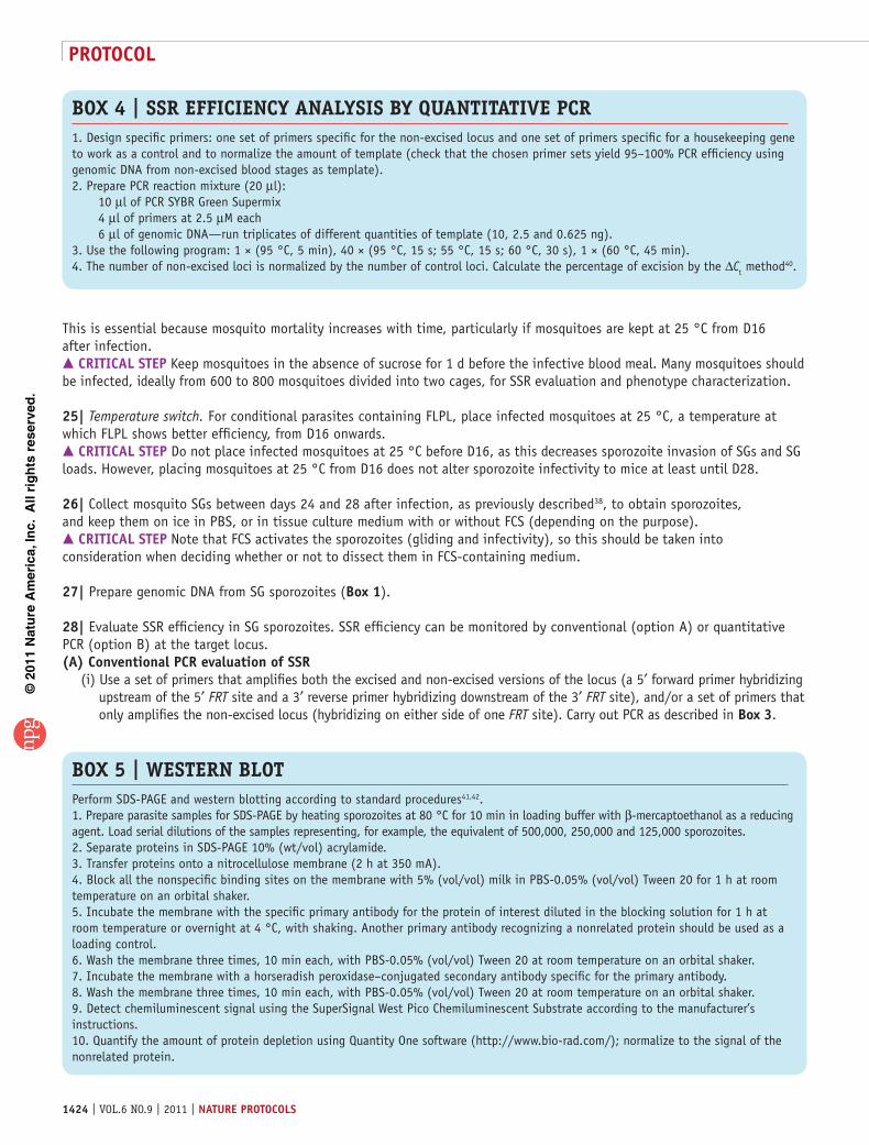
Box 4 | SSR EFFICIENCY ANALYSIS BY QUANTITATIVE PCR 1. Design specific primers: one set of primers specific for the non-excised locus and one set of primers specific for a housekeeping gene to work as a control and to normalize the amount of template (check that the chosen primer sets yield 95–100% PCR efficiency using genomic DNA from non-excised blood stages as template).2. Prepare PCR reaction mixture (20 µl): 10 µl of PCR SYBR Green Supermix 4 µl of primers at 2.5 µM each 6 µl of genomic DNA—run triplicates of different quantities of template (10, 2.5 and 0.625 ng).3. Use the following program: 1 × (95 °C, 5 min), 40 × (95 °C, 15 s; 55 °C, 15 s; 60 °C, 30 s), 1 × (60 °C, 45 min).4. The number of non-excised loci is normalized by the number of control loci. Calculate the percentage of excision by the ∆Ct method40.
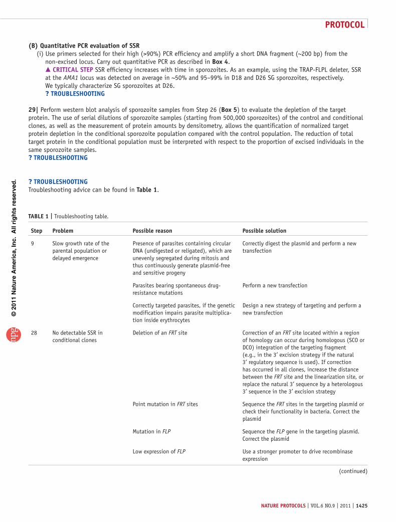
Box 5 | WESTERN BLoT Perform SDS-PAGE and western blotting according to standard procedures41,42.1. Prepare parasite samples for SDS-PAGE by heating sporozoites at 80 °C for 10 min in loading buffer with β-mercaptoethanol as a reducing agent. Load serial dilutions of the samples representing, for example, the equivalent of 500,000, 250,000 and 125,000 sporozoites.2. Separate proteins in SDS-PAGE 10% (wt/vol) acrylamide.3. Transfer proteins onto a nitrocellulose membrane (2 h at 350 mA).4. Block all the nonspecific binding sites on the membrane with 5% (vol/vol) milk in PBS-0.05% (vol/vol) Tween 20 for 1 h at room temperature on an orbital shaker.5. Incubate the membrane with the specific primary antibody for the protein of interest diluted in the blocking solution for 1 h at room temperature or overnight at 4 °C, with shaking. Another primary antibody recognizing a nonrelated protein should be used as a loading control.6. Wash the membrane three times, 10 min each, with PBS-0.05% (vol/vol) Tween 20 at room temperature on an orbital shaker.7. Incubate the membrane with a horseradish peroxidase–conjugated secondary antibody specific for the primary antibody.8. Wash the membrane three times, 10 min each, with PBS-0.05% (vol/vol) Tween 20 at room temperature on an orbital shaker.9. Detect chemiluminescent signal using the SuperSignal West Pico Chemiluminescent Substrate according to the manufacturer’s instructions.10. Quantify the amount of protein depletion using Quantity One software (http://www.bio-rad.com/); normalize to the signal of the nonrelated protein.
©20
11 N
atu
re A
mer
ica,
Inc.
All
rig
hts
res
erve
d.
protocol
nature protocols | VOL.6 NO.9 | 2011 | 1425
(B) Quantitative pcr evaluation of ssr (i) Use primers selected for their high (>90%) PCR efficiency and amplify a short DNA fragment (~200 bp) from the
non-excised locus. Carry out quantitative PCR as described in Box 4. crItIcal step SSR efficiency increases with time in sporozoites. As an example, using the TRAP-FLPL deleter, SSR at the AMA1 locus was detected on average in ~50% and 95–99% in D18 and D26 SG sporozoites, respectively. We typically characterize SG sporozoites at D26. ? trouBlesHootInG
29| Perform western blot analysis of sporozoite samples from Step 26 (Box 5) to evaluate the depletion of the target protein. The use of serial dilutions of sporozoite samples (starting from 500,000 sporozoites) of the control and conditional clones, as well as the measurement of protein amounts by densitometry, allows the quantification of normalized target protein depletion in the conditional sporozoite population compared with the control population. The reduction of total target protein in the conditional population must be interpreted with respect to the proportion of excised individuals in the same sporozoite samples.? trouBlesHootInG
? trouBlesHootInGTroubleshooting advice can be found in table 1.
taBle 1 | Troubleshooting table.
step problem possible reason possible solution
9 Slow growth rate of the parental population or delayed emergence
Presence of parasites containing circular DNA (undigested or religated), which are unevenly segregated during mitosis and thus continuously generate plasmid-free and sensitive progeny
Correctly digest the plasmid and perform a new transfection
Parasites bearing spontaneous drug- resistance mutations
Perform a new transfection
Correctly targeted parasites, if the genetic modification impairs parasite multiplica-tion inside erythrocytes
Design a new strategy of targeting and perform a new transfection
28 No detectable SSR in conditional clones
Deletion of an FRT site Correction of an FRT site located within a region of homology can occur during homologous (SCO or DCO) integration of the targeting fragment (e.g., in the 3′ excision strategy if the natural 3′ regulatory sequence is used). If correction has occurred in all clones, increase the distance between the FRT site and the linearization site, or replace the natural 3′ sequence by a heterologous 3′ sequence in the 3′ excision strategy
Point mutation in FRT sites Sequence the FRT sites in the targeting plasmid or check their functionality in bacteria. Correct the plasmid
Mutation in FLP Sequence the FLP gene in the targeting plasmid. Correct the plasmid
Low expression of FLP Use a stronger promoter to drive recombinase expression
(continued)

©20
11 N
atu
re A
mer
ica,
Inc.
All
rig
hts
res
erve
d.
protocol
1426 | VOL.6 NO.9 | 2011 | nature protocols
● tIMInGPlasmid construction: 2–4 weeksSteps 1–3, Preparation of the plasmid: 2 dSteps 4–8, Transfection of schizonts of the deleter clone: 10 dSteps 9–14, Selection of drug-resistant parasite populations: 2 weeksStep 15, Analysis of transfer population: PCR, 2 d; Southern blotting, 1 weekSteps 16–19, Cloning conditional parasite(s): 2 weeksStep 20, Analysis of cloning populations: 1 weekSteps 21–22, Preparation of stabilates of validated clones: 1 weekSteps 23–29, Scoring SSR efficiency in sporozoites of a conditional clone: 5–6 weeks
antIcIpateD resultsThe procedure for conditional mutagenesis in P. berghei presented here, along with the available deleter clones UIS4/FLP and TRAP/FLPL and fluorescent derivatives, should be useful for testing the function of any essential gene in pre-erythrocytic stages of the parasite. Typically, the procedure yields high percentages (>95%) of sporozoites having undergone SSR in D26 sporozoites. Low amounts of SSR can arise if the promoter-driving FLP expression is not strong enough, or if the recombinase locus constructed by SCO recombination has undergone reversion in a substantial proportion of the parasites. Low deple-tion of the target protein despite high SSR might also occur. For example, when a 3′ untranslated region excision strategy is used, the sequence downstream of the target gene may still allow transcript stabilization in some cases, or when a coding sequence excision strategy is used, low levels of depletion of the target protein may be as a result of the late timing of SSR occurrence (i.e., after the target product has been produced).
These conditional techniques should be useful for molecular studies on invasive stages (zoites). The two Plasmodium zoites that enter host cells inside a parasitophorous vacuole, the merozoite and the SG sporozoite, are both targets of the technique. Until now, in studies on merozoite invasion of erythrocytes, transfection has been helpful in identifying the important parasite genes, the inactivation of which was lethal, but not for characterizing their function, because the mutants cannot be selected. The conditional approach allows investigators to obtain hepatic merozoite mutants. If the merozoite gene of interest is not required for sporozoite invasion of hepatocytes, it can then be deleted using the available deleter parasites at the sporozoite stage. This is the case for AMA1, for which the same scheme of conditional excision of the
taBle 1 | Troubleshooting table (continued).
step problem possible reason possible solution
Low SSR rates ( < 95%) in conditional clones
Reversion at the FLP locus Verify the locus by Southern blotting or PCR analysis of RBC stages and sporozoites of the clone. If marked reversion occurred (traces of reversion are systematically detected), retransfect the targeting fragment into a new, verified (no reversion) vial of the deleter clone
29 Low levels of protein reduction despite substantial SSR (with SSR strategy causing excision of the coding sequence)
SSR occurs too late, allowing the target product to accumulate before SSR
Use sporozoites at a later time point or transfect the targeting plasmid into a distinct deleter clone, in which the recombinase promoter is active earlier in the life cycle. Take into account the time of initiation of gene expression, protein stability and turnover
Low levels of protein reduction despite substantial SSR (with SSR strategy causing excision of the 3′ regulatory sequence)
SSR does not lead to transcript reduction Check target transcripts by reverse transcription- PCR. If RNA transcripts are present, verify the sequence between the stop codon and the first FRT site, as deviations from the sequence shown in Figure 7 might allow transcript stability (or generation of a hybrid transcript spanning the plasmid backbone and presumably using the downstream natural 3′ expression sequence located downstream the plasmid, as already seen)
SSR occurs too late See TROUBLESHOOTING for Step 29 above

©20
11 N
atu
re A
mer
ica,
Inc.
All
rig
hts
res
erve
d.
protocol
nature protocols | VOL.6 NO.9 | 2011 | 1427
3′ regulatory sequence has no effect on sporozoite invasion but blocks merozoite invasion (D.G., S.S., C.L., A. Perazzi, D.Y.B. et al., unpublished data). Alternatively, if the gene of interest is also important for sporozoite invasion, obtaining deficient merozoites requires deleting the gene only when the sporozoite is inside the hepatocyte. Obtaining merozoite mutants of such genes, in turn, requires a deleter in which the recombinase is produced early in the liver stage so as to ensure the generation of deficient merozoites. To maximize SSR efficiency in liver stages, the time of liver stage maturation could be extended by placing infected HepG2 hepatoma cells at 25–30 °C. These temperatures, which are still compatible with full maturation of the parasite into merozoites but extend its duration, would thus maximize FLPL activity while providing more time to achieve high percentages of SSR. Therefore, the conditional technique, coupled to quantitative assays of merozoite invasion events, should help considerably in dissecting the molecular basis of this most crucial step of the parasite life cycle.
It should be stressed that even if the FLP/FRT approach to conditional mutagenesis allows large amounts of SSR and target protein depletion, it does not necessarily require high efficiency to be informative. Clearly, obtaining conditional parasite populations in which most individuals are excised and devoid of the target protein is important for demonstrating the fact that the protein has no role in a phenotype of interest. However, even a partially excised population and/or a partial reduction of the target protein can be informative if it is sufficient to cause a phenotype. For example, while conditionally inactivating RON4, we obtained SSR in only 50% of the sporozoites, because of substantial reversion at the TRAP locus and FLPL loss in the TRAP/FLPL-F deleter (D.G., S.S., C.L., A. Perazzi, D.Y.B. et al., unpublished data). Nevertheless, invasion assays showed that only non-excised sporozoites entered hepatocytes, indicating the essential role of the gene in sporozoite invasion.
As new deleter clones with distinct stage-specificity are constructed, new stages will become targets of the conditional approach. For example, the ookinete and oocyst mosquito stages could be targeted using deleters having the recombinase gene expressed by a gametocyte, gamete or zygote-specific promoter, depending on the expression profile of the gene under study. The sexual stages and the zygote, however, might not be suitable targets for the conditional approach. Their genes of particular interest involved in host-parasite and parasite-parasite interactions are frequently transcribed during the erythro-cytic cycle and translationally repressed39. In this case, SSR-mediated DNA excision would not prevent protein production.
Finally, an important advantage of SSR over other means of conditional manipulation of the genome is that SSR functions at the DNA level and can do more than just inactivating/silencing a gene. The versatility of SSR, coupled to multiple rounds of parasite genome modification, allows one to consider complex schemes. For example, it should be possible to condition-ally modify a gene of interest, which would be useful when investigating multifunctional proteins and inactivating specific protein domains.
acknoWleDGMents We thank C. Bourgouin and I. Thiéry and the other members of the Center for Production and Infection of Anopheles of Institut Pasteur for rearing the mosquitoes. This work was performed with the financial help of the American Heart Association (to P.B.); of the ‘Institut Carnot-Pasteur Maladies Infectieuses’ (to J.-C.B.); and of the Institut Pasteur, the ‘Agence Nationale pour la Recherche’ and the Howard Hughes Medical Institute International Program (to R.M.).
autHor contrIButIons C.L. performed all transfections, contributed to the characterization of conditional clones and is the protocol expert; D.G. and A.C. constructed the deleters, and inactivated and characterized MSP1, AMA1 and RON4; S.S. characterized the AMA1 and RON4 mutant; D.P. and P.B. inactivated PKG and provided Figure 6; D.B., L.T. and J.-C.B. helped to refine the 3′ excision strategy; S.T. helped with transfections; T.G.C. demonstrated the feasibility of the clonal conditional approach; and R.M. wrote the paper.
coMpetInG FInancIal Interests The authors declare no competing financial interests.
Published online at http://www.natureprotocols.com/. Reprints and permissions information is available online at http://www.nature.com/reprints/index.html.
1. Wu, Y., Kirkman, L.A. & Wellems, T.E. Transformation of Plasmodium falciparum malaria parasites by homologous integration of plasmids that confer resistance to pyrimethamine. Proc. Natl. Acad. Sci. USA 93, 1130–1134 (1996).
2. van Dijk, M.R., Janse, C.J. & Waters, A.P. Expression of a Plasmodium gene introduced into subtelomeric regions of Plasmodium berghei chromosomes. Science 271, 662–665 (1996).
3. Carvalho, T.G. & Ménard, R. Manipulating the Plasmodium genome. Curr. Issues Mol. Biol. 7, 39–55 (2005).
4. Cowman, A.F. et al. Functional analysis of proteins involved in Plasmodium falciparum merozoite invasion of red blood cells. FEBS Lett. 476, 84–88 (2000).
5. Ménard, R., Heussler, V., Yuda, M. & Nussenzweig, V. Plasmodium pre-erythrocytic stages: what’s new? Trends Parasitol. 24, 564–569 (2008).
6. Vaughan, A.M., Aly, A.S. & Kappe, S.H. Malaria parasite pre-erythrocytic stage infection: gliding and hiding. Cell Host & Microbe 4, 209–218 (2008).
7. Casares, S., Brumeanu, T.D. & Richie, T.L. The RTS,S malaria vaccine. Vaccine 12, 4880–4894 (2010).
8. Ménard, R. et al. Circumsporozoite protein is required for development of malaria sporozoites in mosquitoes. Nature 385, 336–340 (1997).
9. Gossen, M. & Bujard, H. Tight control of gene expression in mammalian cells by tetracycline-responsive promoters. Proc. Natl. Acad. Sci. USA 89, 5547–5551 (1992).
10. Gossen, M. & Bujard, H. Studying gene function in eukaryotes by conditional gene inactivation. Annu. Rev. Genet. 36, 153–173 (2002).
11. Meissner, M. et al. Tetracycline analogue-regulated transgene expression in Plasmodium falciparum blood stages using Toxoplasma gondii transactivators. Proc. Natl. Acad. Sci. USA 102, 2980–2985 (2005).
12. Meissner, M., Schlüter, D. & Soldati, D. Role of Toxoplasma gondii myosin A in powering parasite gliding and host cell invasion. Science 298, 837–840 (2002).
13. Mital, J., Meissner, M., Soldati, D. & Ward, G.E. Conditional expression of Toxoplasma gondii apical membrane antigen-1 (TgAMA1) demonstrates that TgAMA1 plays a critical role in host cell invasion. Mol. Biol. Cell 16, 4341–4349 (2005).
14. Huynh, M.H. & Carruthers, V.B. Toxoplasma MIC2 is a major determinant of invasion and virulence. PLoS Pathog. 2, e84 (2006).
15. Banaszynski, L.A., Chen, L.-C., Maynard-Smith, L.A., Ooi, A.G. & Wandless, T.J. A rapid, reversible, and tunable method to regulate protein function in living cells using synthetic small molecules. Cell 126, 995–1004 (2006).
16. Armstrong, C.M. & Goldberg, D.E. An FKBP destabilization domain modulates protein levels in Plasmodium falciparum. Nat. Methods 4, 1007–1009 (2007).
17. Herm-Götz, A. et al. Rapid control of protein level in the apicomplexan Toxoplasma gondii. Nat. Methods 4, 1003–1005 (2007).
©20
11 N
atu
re A
mer
ica,
Inc.
All
rig
hts
res
erve
d.
protocol
1428 | VOL.6 NO.9 | 2011 | nature protocols
18. Agop-Nersesian, C. et al. Biogenesis of the inner membrane complex is dependent on vesicular transport by the alveolate specific GTPase Rab11B. PLoS Pathog. 6, e1001029 (2010).
19. Branda, C.S. & Dymecki, S.M. Talking about a revolution: the impact of site-specific recombinases on genetic analyses in mice. Dev. Cell 6, 7–28 (2004).
20. Gil Carvalho, T., Thiberge, S., Sakamoto, H. & Ménard, R. Conditional mutagenesis using site-specific recombination in Plasmodium berghei. Proc. Natl. Acad. Sci. USA 101, 14931–14936 (2004).
21. Combe, A. et al. Clonal conditional mutagenesis in malaria parasites. Cell Host & Microbe 5, 386–396 (2009).
22. Falae, A. et al. Role of Plasmodium berghei cGMP-dependent protein kinase in late liver stage development. J. Biol. Chem. 285, 3282–3288 (2010).
23. Ringrose, L. et al. Comparative kinetic analysis of FLP and Cre recombinases: mathematical models for DNA binding and recombination. J. Mol. Biol. 284, 363–384 (1998).
24. Abremski, K. & Hoess, R. Bacteriophage P1 site-specific recombination. Purification and properties of the Cre recombinase protein. J. Biol. Chem. 259, 1509–1514 (1984).
25. Buchholz, F., Angrand, P.-O. & Stewart, A.F. Improved properties of FLP recombinase evolved by cycling mutagenesis. Nat. Biotech. 16, 657–662 (1998).
26. Rodriguez, C.I. et al. High-efficiency deleter mice show that FLPe is an alternative to Cre-loxP. Nat. Gen. 25, 139–140 (2000).
27. Buchholz, F., Ringrose, L., Angrand, P.-O., Rossi, F. & Stewart, A.F. Different thermostabilities of FLP and Cre recombinases: implications for applied site-specific recombination. Nucleic Acids Res. 24, 4256–4262 (1996).
28. Senecoff, J.F., Rossmeissl, P.J. & Cox, M.M. DNA recognition by the FLP recombinase of the yeast 2m plasmid: a mutational analysis of the FLP binding site. J. Mol. Biol. 201, 405–421 (1988).
29. Matuschewski, K. et al. Infectivity-associated changes in the transcriptional repertoire of the malaria parasite sporozoite stage. J. Biol. Chem. 277, 41948–41953 (2002).
30. Rosinski-Chupin, I. et al. Serial analysis of gene expression in Plasmodium berghei salivary gland sporozoites. BMC Genomics 8, 466 (2007).
31. Ishino, T., Orito, Y., Chinzei, Y. & Yuda, M. A calcium-dependent protein kinase regulates Plasmodium ookinetes access to the midgut epithelial cell. Mol. Microbiol. 59, 1175–1184 (2006).
32. Amino, R. et al. Host cell traversal is important for progression of the malaria parasite through the dermis to the liver. Cell Host & Microbe 3, 88–96 (2008).
33. O’Donnell, R.A., Saul, A., Cowman, A.F. & Crabb, B.S. Functional conservation of the malaria vaccine antigen MSP1-19 across distantly related Plasmodium species. Nat. Med. 6, 91–95 (2000).
34. Triglia, T. et al. Apical membrane antigen 1 plays a central role in erythrocyte invasion by Plasmodium species. Mol. Microbiol. 38, 706–718 (2000).
35. Nunes, A. et al. Subtle mutagenesis by ends-in recombination in malaria parasites. Mol. Cell Biol. 19, 2895–2902 (1999).
36. Tycowski, K.T., Kolev, N.G., Conrad, N.K., Fok, V. & Steitz, J.A. The ever-growing world of small nuclear ribonucleoproteins in the RNA world. (eds. Gesteland, R.F., Cech, T. & Atkins, J.F.) 331 (Cold Spring Harbor Laboratory Press, 2006).
37. Janse, C.J., Ramesar, J. & Waters, A.P. High-efficiency transfection and drug selection of genetically transformed blood stages of the rodent malarial parasite Plasmodium berghei. Nat. Protoc. 1, 346–356 (2006).
38. Thiberge, S. et al. In vivo imaging of malaria parasites in the murine liver. Nat. Protoc. 2, 1811–1818 (2007).
39. Mair, G.R. et al. Regulation of sexual development of Plasmodium by translational repression. Science 313, 667–669 (2006).
40. Applied Biosystems. Real-time PCR Systems: Chemistry Guide, Part no. 4348358, Rev. E http://www3.appliedbiosystems.com/cms/groups/mcb_marketing/documents/generaldocuments/cms_041440.pdf (2005).
41. Laemmli, U.K. Cleavage of structural proteins during the assembly of the head of bacteriophage T4. Nature 227, 680–685 (1970).
42. Towbin, H., Staehelin, T. & Gordon, J. Electrophoretic transfer of proteins from polyacrylamide gels to nitrocellulose sheets: procedure and some applications. Proc. Natl. Acad. Sci. USA 76, 4350–4354 (1979).
Copyright © 2022 FDOKUMEN