Investigation of the molecular events in Plasmodium berghei ...
235
HAL Id: tel-01542791 https://tel.archives-ouvertes.fr/tel-01542791 Submitted on 20 Jun 2017 HAL is a multi-disciplinary open access archive for the deposit and dissemination of sci- entific research documents, whether they are pub- lished or not. The documents may come from teaching and research institutions in France or abroad, or from public or private research centers. L’archive ouverte pluridisciplinaire HAL, est destinée au dépôt et à la diffusion de documents scientifiques de niveau recherche, publiés ou non, émanant des établissements d’enseignement et de recherche français ou étrangers, des laboratoires publics ou privés. Investigation of the molecular events in Plasmodium berghei gametogenesis through proteomic approaches Carlos Henrique Saraiva Garcia To cite this version: Carlos Henrique Saraiva Garcia. Investigation of the molecular events in Plasmodium berghei game- togenesis through proteomic approaches. Parasitology. Université Pierre et Marie Curie - Paris VI; Universidade de Brasília, 2016. English. NNT : 2016PA066582. tel-01542791
-
Upload
khangminh22 -
Category
Documents
-
view
0 -
download
0
Transcript of Investigation of the molecular events in Plasmodium berghei ...

HAL Id: tel-01542791https://tel.archives-ouvertes.fr/tel-01542791
Submitted on 20 Jun 2017
HAL is a multi-disciplinary open accessarchive for the deposit and dissemination of sci-entific research documents, whether they are pub-lished or not. The documents may come fromteaching and research institutions in France orabroad, or from public or private research centers.
L’archive ouverte pluridisciplinaire HAL, estdestinée au dépôt et à la diffusion de documentsscientifiques de niveau recherche, publiés ou non,émanant des établissements d’enseignement et derecherche français ou étrangers, des laboratoirespublics ou privés.
Investigation of the molecular events in Plasmodiumberghei gametogenesis through proteomic approaches
Carlos Henrique Saraiva Garcia
To cite this version:Carlos Henrique Saraiva Garcia. Investigation of the molecular events in Plasmodium berghei game-togenesis through proteomic approaches. Parasitology. Université Pierre et Marie Curie - Paris VI;Universidade de Brasília, 2016. English. �NNT : 2016PA066582�. �tel-01542791�


1
"Les mots prennent toujours la couleur des actions ou des sacrifices qu'ils suscitent."
Lettres à un ami allemand (1945), Albert Camus

2
Agradecimentos
Agradeço primeiramente a meus orientadores, Prof. Sebastien Olivier Charneau e
Prof. Philippe Grellier por todo suporte ao longo destes anos de trabalho para a realização
deste projeto.
Sou grato também àquelas pessoas que não participaram deste projeto mas que
estiveram comigo nesta jornada e de alguma forma estiveram aqui presentes nestas palavras
que se seguem: Sônia Nogueira da Gama Gondim, Elinda Saraiva Garcia, Marina Saraiva
Garcia, Ana Paula Saraiva Garcia, Maria Luiza Saraiva Calgaro, Maria Eduarda Saraiva
Soares, Giovana Saraiva Calgaro, Leda Saraiva Viana, José Leitão Viana, Rafael Saraiva
Viana, Márcia Saraiva Viana, Murilo Rabelo, Letícia Saraiva Rabelo, Beatriz Saraiva Rabelo
e Ana Júlia Vieira de Ribeiro.
Meus agradecimentos a meus colegas e professores do Laboratório de Bioquímica e
Química de Proteínas e aos colegas do Laboratoire de Biodiversité et Adaptation des
Microorganismes Eucaryotes à leur Environnement.

3
Agradecimentos as intituições e fomentadores
Aknowledgements
This work was supported by CAPES/COFECUB programme [723/11],
MCTI/CNPq/FNDCT/PRO-CENTRO-OESTE [407730/2013-3 and 407855/2013-0],
CAPES/Incentivo a Pesquisa em Parasitologia Básica [23038.005298/2011-83], FAPDF
[193.000.987], CNPq, Finep, Fundação Hemocentro de Brasília, École Doctorale Complexité
du Vivant à l’Université Pierre et Marie Curie (UPMC), Muséum National d'Histoire
Naturelle na França and Programa de Pós-graduação em Patologia Molecular, Faculdade de
Medicina, Universidade de Brasília (UnB).
Funding sources had no involvement in study design, data collection or interpretation
or decision on publication submission.

4
Sumário
Agradecimentos ......................................................................................................................... 2 Agradecimentos as intituições e fomentadores .......................................................................... 3 Sumário ...................................................................................................................................... 4 1. Introdução .......................................................................................................................... 6 1.1. Breve história da malária ................................................................................................ 8 1.2. História da malária no Brasil ........................................................................................ 13 1.3. Aspectos epidemiológicos da malária ........................................................................... 18
1.3.1. Epidemiologia da malária no Brasil e aspectos socioeconômicos ........................ 21 1.3.2. Malária na França .................................................................................................. 22
1.4. Estratégias científicas para erradicação ........................................................................ 24 1.5. Condutas para prevenção e tratamento ......................................................................... 27 1.6. Agentes etiológicos e modo de transmissão ................................................................. 30 1.7. Vetores transmissores e hospedeiros ............................................................................ 33 1.8. A biologia reprodutiva dos plasmódios ........................................................................ 38
1.8.1. Fase reprodutiva assexuada do Plasmodium ......................................................... 39 1.8.2. Fase reprodutiva sexuada do Plasmodium ............................................................. 41
1.8.2.1. Fertilização e proliferação no mosquito ......................................................... 43 1.9. Morfologia dos gametócitos ......................................................................................... 45 1.10. Diferenciação em gametas e exflagelação ................................................................ 47 1.11. Malária de roedores – Plasmodium berghei .............................................................. 51 1.12. Cinesinas ................................................................................................................... 55
1.12.2. A motilidade das cinesinas ................................................................................. 57 1.12.3. Regulação de cinesinas ...................................................................................... 58 1.12.4. A cinesina 8 em Plasmodium ............................................................................. 58 1.12.5. A cinesina 8 em Plasmodium berghei ................................................................ 61
1.13. Proteômica e proteoma de Plasmodium .................................................................... 64 1.14. Fosfoproteoma de Plasmodium ................................................................................. 67 2. Justificativa científica ...................................................................................................... 73 3. Objetivos gerais ............................................................................................................... 74 4. Materiais e métodos ......................................................................................................... 75
4.1. Produção de gametócitos ........................................................................................... 75 4.2. Separação das células ................................................................................................ 76 4.3. Obtenção das triplicatas biológicas ........................................................................... 76 4.4. Lise celular e extração de proteínas .......................................................................... 77 4.5. Precipitação de proteínas ........................................................................................... 78 4.6. Hidrólise enzimática de proteínas ............................................................................. 79 4.7. Dessalinização de amostra ........................................................................................ 80 4.8. Quantificação de peptídeos ....................................................................................... 81 4.9. Marcação de peptídeos por iTRAQ ........................................................................... 81 4.10. Preparação de amostras multiplexadas .................................................................. 82 4.11. Enriquecimento de fosfopeptídeos por TiO2 ......................................................... 85 4.12. Dessalinização de peptídeos por STAGETIP para espectrometria de massas ...... 85 4.13. Espectrometria de massas ...................................................................................... 86 4.14. Processamento de dados ........................................................................................ 87 4.15. Identificação de proteínas ...................................................................................... 87 4.16. Ontologia de genes ................................................................................................ 89
5. Resultados ........................................................................................................................ 90

5
5.1. Parte I - Dynamic molecular events associated to Plasmodium berghei gametogenesis through proteomic and phosphoproteomic approaches ............................... 91 5.2. Parte II - Deciphering the kinesis 8 role during Plasmodium berghei gametogenesis by proteomic and phosphoproteomic approaches .............................................................. 152
6. Conclusão ....................................................................................................................... 208 7. Bibliografia .................................................................................................................... 211 8. Índice de figuras ............................................................................................................. 228 9. Índice de tabelas ............................................................................................................. 230 10. Resumos ...................................................................................................................... 231
6
1. Introdução
A malária é uma patologia provocada no homem por organismos unicelulares
pertencentes ao gênero Plasmodium. No ano de 2015, a malária ganhou notoriedade na
impressa internacional após a outorga do Premio Nobel de Medicina à professora chinesa
Youyou Tu pela descoberta da artemisina, um composto utilizado no tratamento de pacientes
infectados e que engendrou drástica redução do número de mortes pela doença (Assembly,
2015).
A importância do tema remonta de mais de 200 anos e já rendeu outros 4 Prêmio
Nobel a Sir Ronald Ross (Assembly, 1902), Charles Louis Alphonse Laveran (Assembly,
1907), Julius Wagner-Jauregg (Assembly, 1927) e Paul Hermann Müller (Assembly, 1948).
Dentre esses, Alphonse Laveran foi o primeiro Prêmio Nobel de medicina da França.
Os vários prêmios Nobel outorgados refletem a dimensão e relevância do tema para a
saúde pública e para o desenvolvimento econômico dos Estados. Incentivos e campanhas
propiciaram o desenvolvimento de políticas públicas de combate, prevenção e formação de
redes de pesquisas (Keating, 2014).
Apesar da enormidade de trabalhos científicos e da difusão de informações sobre a
malária nos recentes anos, a doença ainda provoca onerosos gastos em saúde pública e em
pesquisa científica (Ricci, 2012;WHO, 2015c) e permanece como epidemia em diversos
países, enquanto, em outros países, ela acontece por casos de importação e transmissão
congênita. Contudo, ainda não foram desenvolvidas uma vacina ou estratégias de combates
que eliminem o fardo. O tratamento utilizado para curar pacientes acometidos pela malária
tem tomado a maior parte dos recursos dos programas de combate a essa doença. Sua possível
erradicação requer o investimento de recursos para prevenção da transmissão do parasita para
o homem como também o bloqueio da transmissão do parasita do homem para o mosquito.
Para a prevenção da doença, há uma demanda pela formulação de um corpo de
conhecimento que possibilite um melhor entendimento da biologia do parasita e, desta forma,
amplie a gama de alvos terapêuticos, permitindo, portanto, um melhor enfrentamento da
transmissão, da infecção e da reprodução do parasita. Os estágios de desenvolvimento sexuais
são essenciais para a continuidade da proliferação do parasita e, por esta razão, são
considerados um alvo capital para a construção de vacinas e fármacos que interrompam o

7
ciclo reprodutivo (Guttery et al., 2012b). Portanto, este trabalho procura contribuir na
formação de um corpo de conhecimento do estágio gametocítico do parasita a fim de
propiciar a atenuação ou o bloqueio definitivo à etiologia.

8
1.1. Breve história da malária
A malária possivelmente é a doença de maior importância, a mais antiga, a mais
amplamente distribuída e a que mais assolou dentre todas as doenças a que o ser humano
esteve sujeito que se tem conhecimento na história da medicina (North, 1896). Talvez não
tenha existido uma doença em que tão grande número de teorias de contágio tenham sido
formuladas, em que tão grande número de organismos tenham sidos investigados e nem que
tão grande número deles tenham sido atestado como, comprovadamente, o verdadeiro agente
causador da malária.
De maneira similar, são inúmeros os termos utilizados para designar a doença. Temos,
no princípio do século XIX, nos anos de 1842, quando Jean-Christian-Marc Boudin publicou
seu livro intitulado Traité des fièvres intermittentes, rémittentes et continues des pays chauds
et des contrées marécageuses, a patologia nominada como febre intermitente, febre rémitente,
febre pseudo-continuada, febre latente, febre perniciosa e febre de países quentes (Boudin,
1842). Ao final do século XIX, na medida em que o conhecimento sobre a doença se
ampliava, também era conhecida por febre palustre, febre telúrica, febre miasmática, febre
fito-hémica, febre marematica, febre intermitente (Moreira, 1890). Na atualidade, final de
2015, a malária é reputada como uma doença endêmica de países tropicais provocada em
homens, répteis, aves e outros mamíferos por organismos do gênero Plasmodium spp. e
transmitida por insetos anofelinos, do gênero Anopheles. Com maior rigor de diagnóstico,
sintomas e diagnósticos da malária são atribuídos com maior acerto e referidos na atualidade
pelos nomes de impaludismo, paludismo, febre palustre, febre intermitente, febre terçã
benigna, febre terçã maligna, cachexia palustre, além de popularmente conhecida por maleita,
sezão, tremedeira, batedeira, febre e febre de São Paulo.
Os primeiros registros que se tem notícia de seres humanos com o que supõe-se ser a
malária datam de mais de 4700 anos na China, porém diversos outros já foram encontrados,
como os da Mesopotâmia em 2000 A.C., do Egito em 1570 A.C., de Homer em 850 A.C., de
Hindus de 600 A.C. e de Empédocles de Agrigentum em 550 A.C. (Cox, 2010b). Todavia, foi
em 400 anos A.C. que o pai da medicina, Hipócrates, consagrou um capítulo especial para
discutir sobre a patologia: De natura palustrium et lacustrium aquarum (Boudin, 1842).
9
Desde Hipócrates já existia a ideia de que o pântano tem papel importante na etiologia
da malária, como se pode observar pela etimologia de um dos nomes dados à doença: o
paludismo. Esse termo foi comumente empregado para referir-se à essa patologia na Europa a
partir do século XVIII e teve como origem o vocábulo palus do latim, que literalmente
significa “pântano” (Sampaio, 1872b;CNRpalu, 2015b). O termo malária também foi
atribuído segundo a crença de que "ares ruins" contribuiriam no seu desenvolvimento.
Portanto, apesar de controvérsias, a origem mais aceita desse termo é a partir da expressão
mal’aire, do italiano antigo, que significa “ar estragado” ou “ar ruim”, que adoeceria as
pessoas que a ele estivessem sujeitos (Cox, 2010b).
Portanto segundo as definições da época, doenças eram ocasionadas por miasmas e
eflúvios (Boudin, 1842;Sampaio, 1872a;Castanheira, 1885). A teoria miasmática foi a
principal linha teórica confiada por diversos estudiosos para explicar o contagio pela malária
até o final do século XIX. No entanto, a teoria parasitária não era nova nas cercanias da
formulação da teoria dos germes. Ela é antiga e surgiu ainda na Idade Antiga. Pela hipótese
parasitária, as doenças do pântano eram causadas por pequenos insetos que penetrariam no
corpo (Houzeau, 1874-1889 ). Em seguida, a teoria estendeu-se na Idade Moderna para
infecção provocada por seres microscópicos capazes de penetrar epitélios, chamados na Itália
de serafici ou “causadora de morte” (Kreier, 2012).
Ainda como complicador para a investigação da doença, havia alguma confusão
quanto ao diagnóstico de algumas doenças com alguns sintomas semelhantes, como a febre
tifoide e a febre amarela, com a malária (Boudin, 1842). Os sintomas da doença começam a
ser melhor diferenciados a partir de 1868 (Pearce, 2002) com o aprimoramento da
termometria e definição de temperatura corporal basal (Gorzoni et al., 2010) por Karl August
Wunderlich (1868).
Em 1876, Matteo Lanzi e Guglielmo Terrigi também realizaram experimento com
infecção de cobaias com orvalho coletado de pântanos endêmicos e encontraram, nestes
animais, pigmentos no baço e no fígado destes animais que são análogos àqueles que existem
no sangue das pessoas falecidas pela forma mais grave da malária. Eles cultivaram os
elementos pigmentados e acreditaram ter conseguido isolar o patógeno, uma bactéria
acastanhada, o que explicaria a coloração dos pigmentos, e a qual deram o nome de
Bacteridium brunneum (Laveran, 1898).

10
Em 1879, Theodor Albrecht Edwin Klebs e Corrado Tommasi Crudeli retomaram as
experiências de Lanzi e Terrigi de 1876, mas com o intuito de melhor controlar as condições
experimentais a fim de reproduzir a descoberta dos germes causadores da malária. Os
pesquisadores observaram que um destes organismos estava presente no ar, na água e na lama
dos pântanos como também no sangue dos animais inoculados. O bacilo com aspecto de
esporo móvel, ovalado e reflexivo de luz solar, quando inoculado em animais, causava febre
intermitente. Foi, portanto, chamado de Bacillus malariae (Laveran, 1898).
Em 1878, no departamento de Bône na Argélia, enquanto autopsiava óbitos por
malária, Laveran se atentou quanto à pigmentação decorrente da doença como um sinal
bastante específico da malária (Laveran, 1898). A pigmentação estava presente no sangue,
que é um tecido ostensivamente afetado pela doença em aparência e composição (Sampaio,
1872a) , como uma substância espalhada no sangue sob forma de finas granulações negras,
produto da patologia, chamada de pigmentos da malária ou melanina, conforme já havia sido
indicado por Mickel, Virchouw e Freirich (Pinto, 1904). Ao estudar o sangue humano de
doentes, Laveran observou, no final de 1880, em Constantina, que estes corpos esféricos
pigmentados eram corpos celulares e flagelados. Isto lhe forneceu indícios de que a doença
era causada por um parasita que estava presente no sangue. Estes parasitas foram chamados
por ele de Oscillaria malárias, depois de hematozoário do paludismo e, pela denominação de
Marchiafava e Celli, de Plasmodium malariae por ser um organismo protoplasmático, de
movimentos ameboides, que é encontrado dentro do plasma de glóbulos vermelhos (North,
1896;Laveran, 1898).
Ao final de 1880, Laveran publicou acerca da presença do parasita no sangue (Roche
et al., 1880) e, ao final de 1881, o descreveu no trabalho Nature parasitaire des accidents de
l'impaludisme, description d'un nouveau parasite trouvé dans le sang des malades atteints de
fièvre palustre (Laveran, 1881a). Após a descoberta do hematozoário causador da malária, a
próxima pergunta a se elucidar era qual o vetor do parasita. Em 1896, já se conhecia 5
espécies de vetores da malária humana (Silva, 1896). Com quase 30 anos, em 1909, se tinha
conhecimento de mais de 20 espécies mosquitos transmissores do gênero Anopheles (Alvares,
1909). No ano de 1948, Shortt e Garnham descreveram o ciclo hepático da malária a partir de
P. cynomolgi (Shortt and Garnham, 1948).
Ainda com o transcorrer dos anos, após a descoberta do parasita e de seus vetores,
diversos estudos sobre malária tiveram curso por grupos de pesquisa isolados e em diversas

11
partes do mundo. Em Dezembro de 1977, uma iniciativa importante teve curso por parte de
Ken Warren através da Rockefeller Foundation. A malária, juntamente com outras patologias,
passou a fazer parte do grupo de doenças da nova disciplina que se iniciava, a das doenças
negligenciáveis, que seriam as patologias de grande prevalência e de pouco incentivo de
estudo. Portanto, a finalidade do novo empreendimento foi de construir a nível global a
primeira coordenação de grupos de pesquisas cientificas, com fomento financeiro específico,
formação de pesquisadores de excelência e com trabalho de estudantes brilhantes (Keating,
2014).
O pontapé inicial para o projeto foi dado com o programa The Great Neglected
Disease of Mandkind da Fundação Rockefeller. Ele permitiria a projeção de unidades de
pesquisas em modernos institutos, universidades e escolas médicas em países desenvolvidos e
em desenvolvimento. A partir de 1977, o número de publicações sobre malária teve aumento
relevante e progressivo até o ano de 2016. O projeto foi uma influência importante para a
ciência biomédica moderna, e a iniciativa configurou um importante episódio para a
transformação da medicina em uma prática globalmente integrada e com métricas para
avaliação dos impactos e progressos das políticas de combate por parte de financiadores e
governo (Keating, 2014).
Em 2001, engajou-se no combate às doenças negligenciáveis a organização não-
governamental Médicos Sem Fronteiras em uma campanha chamada: Fatal Imbalance - The
Crisis in Research and Development for Drugs for Neglected Diseases (Frontières, 2001). Em
meados de 2010, a doença já era excluída do corpo de doenças negligenciável (Souza,
2010;FAPESP, 2014). Persistentes e volumosos esforços de pesquisa e investimentos
financeiros no tema ao longo dos anos permitiram a remoção da malária do grupo de doença
negligenciável no Brasil na acepção empregada por Warren e Médicos Sem Fronteiras. Em
2015, puderam ser contabilizados mais de 1.330.000 trabalhos sobre a malária indexados pela
ferramenta Google Scholar quando buscado o termo "Malaria” em todos os documentos nele
registrados, sem nenhuma restrição de período de tempo na busca. Um volume crescente de
trabalhos é observado ao longo dos anos também por outras indexações como pela PubMed,
U.S. National Library of Medicine (Figura 1).


13
1.2. História da malária no Brasil
A história da malária no restante do globo tem algumas similaridades com a história da
malária no Brasil no aspecto da investigação da patologia e suas descobertas (Torres-Homem,
1877) como também nas interpretações dos quadros clínicos (Torres-Homem, 1877).
No Rio de Janeiro, ao mesmo tempo em que tinha curso a reforma de Haussmann em
Paris (1853-1870), possivelmente os componentes do plano de remodelação urbana carioca já
eram projetados (Benchimol, 2000) e teriam seus efeitos nas medidas dos prefeitos da
Guanabara, ao final do século XIX e início do XX, para atenuar os impactos econômicos e
sociais causados pela malária. O fardo se refletia nos países endêmicos em ditos populares,
como o de que “Todo o branco que trabalha na terra, cava nela também a sua sepultura“
(Acabado, 1900), pelas questões de suscetibilidades relacionadas à etnia e à frequência de
ocorrência de casos.
Durante o governo dos ex-prefeitos Candido Barata Ribeiro e Pereira Passos, no Rio
de Janeiro, teriam ocorrido desapropriações de cortiços e regiões com população de baixo
poder econômico com intuito de sanitização em vista das frequentes epidemias por que a
cidade passava, além da remodelação da malha viária municipal. Portanto, a malária seria
possivelmente uma das influências para o surgimento das primeiras favelas no Brasil.
O Congresso Nacional de Medicina e Cirurgia se reuniria em 1889 com o objetivo de
tomar decisões que pudessem mitigar os frequentes eventos epidemiológicos por que passava
a cidade (Benchimol, 2000). As resoluções projetadas eram tomadas aà luz da teoria
sustentada por Pettenkofers, que atribuía a sazonalidade da malária a pântanos subterrâneos
que precisavam ser combatidos. De acordo com a teoria, com as elevações do nível da água
da zona freática do solo do Rio de Janeiro, em decorrência das chuvas do verão até o mês de
Março, os germes alcançavam as condições propícias do solo para se desenvolver e produzir
miasmas (Benchimol and Sá, 2005) em condição análoga a dos pântanos descobertos
(Moreira, 1890). Os lençóis freáticos seriam mais importantes para o curso das doenças do
solo do que cursos d’água próximos (Weber, 1865).
As modelações das ações do homem sob influência dos episódios de epidemia da
malária também poderiam ser observadas na construção das estradas de ferro brasileiras como

14
também em óbitos e recuo de tropas na Guerra do Paraguai. Nessa guerra, no período entre
1864 a 1870, mais gente teria morrido em consequência das enfermidades do que de ações
bélicas (Marinha, 1870). A batalha da Retirada da Laguna teria trazido uma baixa de cerca de
400 soldados por dia depois que esses atravessavam áreas alagadiças de reprodução do
mosquito, sendo essas baixas a possível causa para retirada das tropas (de Castro Souza,
1972).
A construção das estradas de ferro, como a ferrovia do Estado de São Paulo, na região
da Serra do Mar, para ligação São Paulo–Santos no período entre 1897-1898, precisou ser
interrompida por consequência da epidemia que se abateu sobre os trabalhadores do
empreendimento (Benchimol and Sá, 2005). O mesmo aconteceu na construção da ferrovia
Madeira e Mamoré em Rondônia entre 1907-1912, com a diferença na gravidade dos casos
em Rondônia ser maior (Benchimol and Sá, 2005), ao "risco, para não dizer com todas as
probabilidades, de sacrificar a própria vida” (Mamoré, 1885). Na Serra do Mar, entre Santa
Catarina e Paraná no período entre 1880-1885, a malária surgiu com grande intensidade
durante os trabalhos de construção da malha ferroviária Paranaguá–Curitiba (Costa, 1885).
Além desses casps, a malária causava perdas em todas regiões de Norte a Sul do
Brasil. No entanto, Laveran publicou, em 1898, que febres intermitentes no Brasil eram mais
comuns nas costas, junto ao mar. A partir dos vales, elas se tornavam cada vez mais raras na
medida que se alcançava maiores altitudes (Laveran, 1898). Os rios brasileiros produzem
mangues e emanações deletérias no curso dos rios e igarapés devido a inundações anuais ou
na região de sua foz com o mar. Portanto, as formas mais graves da doença estariam nas
proximidade do Rio Amazonas e seus afluentes, do Rio Paranaíba, do Rio São Francisco, e de
outros (Corre, 1887).
No alto Paraguai e Paraná, os casos também eram igualmente frequentes em várias
localidades na costa do Atlântico, como no Rio de Janeiro, onde mais de 8.000 casos de febre
intermitente foram tratados em um mesmo hospital durante em seis anos (Corre, 1887). Na
região de Florianópolis, em Santa Catarina, H. Rey, em 1877, reporta nos Arquivos de
Medicina Naval francês que febres intermitentes estariam longe de serem raras (Rey, 1877). A
incidência dos casos de malária era elevada nos arredores de alagados e terrenos pantanosos,
nos arredores das Lagoa de Nossa Senhora da Conceição e eram mais observados no verão
(Rey, 1877;Costa, 1885). Desde as regiões mais ao norte, mas também ao sul do Brasil, na
região do Prata, as embarcações já eram fortemente afetadas pela malária (Bourel-Roncière,

15
1872). Colin registra, em 1870, que grave epidemia assolava a cidade do Rio de Janeiro e
diversas outras cidades e teve princípio com as grandes migrações ao Brasil em 1835 (Colin,
1870).
Contudo, a documentação sobre a malária no Brasil e sua influência em seus
habitantes antecede o século XIX, apesar de mais detalhada para o período. Jacques Lind
registrou, em 1777, que o ar no Brasil era bom e, portanto, era estimado pelos portugueses
como um paraíso (Lind and de La Chaume, 1777). No mesmo período, no entanto, temos o
registro do óbito de dois engenheiros militares italianos, Enrico Antonio Galluzzi de Mantova
e Domenico Sambuceti, pela causa da malária, em 1769, na Amazônia, e 1780, no Mato
Grosso, respectivamente. Eles estavam encarregados da construção de fortalezas da coroa
portuguesa (Fontana, 2005). A malária neste período não parecia ser uma novidade nem
apresentar ocorrência isolada, como constatado pela familiaridade do diagnóstico pela
medicina colonial da época.
Os registros sobre casos de malária no Brasil antes de XIX trazem evidências de que
os casos de malária não tiveram início com as grandes imigrações colonizadoras do princípio
do século XIX. O Brasil suscitava horror aos europeus por ser tido como uma das regiões
mais insalubres dos trópicos ao longo de todo o século XIX (Figueiredo et al., 1996). Nessa
época, a malária afetava principalmente o litoral brasileiro, a região mais densamente
colonizada e, portanto, em que se tem a maior riqueza de documentação nos registros.
A confirmação da existência do parasita da malária no Brasil foi feita por Francisco de
Paula Fajardo Júnior, em 1893, e por Miranda Azevedo (Laveran, 1898). Fajardo foi
considerado pela imprensa médica carioca como o ‘descobridor’ do hematozoário de Laveran
no Brasil. No mesmo ano de 1893, o médico sanitarista Adolpho Lutz iniciou um dos
primeiros trabalhos de investigação sobre a malária que se tem registro. Lutz demonstrou a
natureza não paludica das febres paulistas; identificou e estudou aspectos morfológicos de
plasmódios presentes no Brasil; determinou quais eram as espécies de vetores (Figura 2); e
estabeleceu medidas de saneamento para combate (Corrêa, 1955).

16
Figura 2 : Anopheles (Kerteszia) cruzii, 1903. Primeiro vetor descrito por Aldopho Lutz como provável causador da malária no Brasil na região do interior do Estado de São Paulo (Lutz).
Antes de 1877, data em que foi comprovada a transmissibilidade de filarias por
mosquitos, a suspeita de mosquitos como vetores de doenças era uma hipótese debatida pela
comunidade científica. (Manson, 1878;King, 1883) Esse debate se estendeu à malária a partir
de 1883, devido a evidências colecionadas por Albert King (King, 1883).Em 1899, foi
confirmado que a malária era transmitida ao homem por mosquito. Em 1898, Lutz suspeitava
que um vetor da malária seria um anofelino descoberto durante suas incursões de investigação
sobre a febre paulista, Anopheles cruzii. Contudo, ele não dispunha de aparelhagem de
microscopia que o permitisse fazer a comprovação causal com evidências científicas (Lutz,
1936).
As investigações de Lutz sobre a doença concentraram-se de início na barra de Santos
e nas áreas de pântano e ele deparou-se por duas vezes com doentes pelo Plasmodium
malariae em 1893. Em relatório de 1895, ele descreveria mais três casos da doença
(Benchimol and Sá, 2005). Em seguida aos trabalhos de Lutz, somou-se os trabalhos do
médico sanitarista Carlos Chagas, que relatou surto de malária a 700 metros de altitude em
1906 (Benchimol and Sá, 2005), e outros pesquisadores que se seguiram desde então.
17
De acordo com consulta realizada na ferramenta Web of Science, de 1945 a 2015,
trabalhos de brasileiros sobre a malária constituíram cerca de 3% das publicações mundiais,
atrás de EUA com 30%, além de UK, FR e outros. O número de trabalhos brasileiros foi de
2.259 publicações científicas em periódicos internacionais e livros.

18
1.3. Aspectos epidemiológicos da malária
A OMS reporta que o número de acometidos pela malária de 2000 a 2015 é de cerca
de 1,2 bilhões. Deste total, 6,2 milhões evoluíram para o óbito. Para 2015, a estimativa de
ocorrências ficou entre 149-303 milhões de novos casos e 236-635 mil óbitos em todo o
mundo (WHO, 2015c).
Quando comparados 2000 e 2015, houve redução da incidência da malária em 37% e
60% de redução dos casos de óbito, o que pode ser uma estimativa otimista se levadas em
conta outras projeções como a do Seattle Institute for Health Metrics, que apontou duas
vezes mais casos de morbidade e de mortalidade para o ano de 2010 (Murray et al., 2012).
Entre 2010, 2013 e 2015 houve pouca variação no número de países com a malária
endêmica, com 106, 99 e 96 países acometidos, respectivamente, nesses anos. A maioria
deles são países em desenvolvimento e na zona intertropical, como representado na Figura 3
(WHO, 2010;Heppner, 2013;WHO, 2014;2015c). Mesmo com cerca de 2,5 bilhões de
dólares gastos anualmente no combate e prevenção da doença por fundos nacionais e
mundiais, como o PMI, o Programa Nacional de Controle da Malária, o Fundo Global e o
Banco Mundial, quase metade da população mundial, cerca de 3,2 bilhões de pessoas,
estão expostas ao risco de adquirir a doença (WHO, 2015c).
Figura 3 : Distribuição geográfica da malária no Mundo. A malária é transmitida em áreas tropicais e subtropicais. O mapa tem representado o número de mortes evitadas no período entre 2001 a 2013. Modificada de Relatório sobre Malária de 2014 (WHO, 2014).

19
Na atualidade, a maior parte dos óbitos pela malária ocorrem na África (87,9%),
seguido pelo sudeste da Ásia (9,3%), pela região oriental do Mediterrâneo (1,8%), pela
região das Américas (0,3%) e pela região ocidental do Pacífico (0,7%), segundo estimativa
de 2015 (WHO, 2015c). Em alguns países, o alto risco de infecção alcança mais de 90% da
população, como no caso de Angola, Congo, Gabão e Senegal, dentre outros em 2011
(WHO, 2011).
A região das Américas (Figura 4) é composta por 21 países com a malária endêmica.
Três destes países foram responsáveis por 77% dos casos de malária em 2013: Brasil (37%),
Venezuela (23%) e Colômbia (17%) (WHO, 2015c). Para a região, em 2015, 121 milhões de
pessoas (20%) estiveram em risco de infecção dos cerca de 600 milhões de indivíduos dos 21
países. Destes, 20 milhões (3,3%) foram categorizados como sujeitos a elevado risco de
contraírem (WHO, 2015c). A Venezuela tem apresentado um aumento dos índices desde
2008 e em 2014 atingiu o maior número de casos de incidência dos últimos 50 anos (WHO,
2015c). Acredita-se que a mineração de ouro seja uma das principais causas do crescente
número de acometidos pela malária no país (Moreno et al., 2009;BBC, 2014).
Contudo, no cálculo geral dos anos de 2000 a 2015, a região das Américas reduziu a
morbidade em 74% e a mortalidade em quase 72% (WHO, 2015c). A expectativa é que a
região alcance o quadro de 12,1 milhões de casos e 24 mil óbitos até 2030.

20
Figura 4 : Mapa das Américas Centrais e do Sul com status percentual de risco populacional e fases de programa estratégico: fases de controle, pré-eliminação, eliminação e prevenção de reintrodução da malária.
Quanto ao financiamento para prevenção e combate da malária por fundos
internacionais, de 2005 a 2015 foram investidos cerca de 20 bilhões de dólares em todo o
mundo. A previsão é de que será investido cerca de 100 bilhões de dólares de 2015 a 2030,
dos quais 24,2 bilhões serão distribuídos no intervalo de 2015-2020; US$35,95 bilhões de
2020-2025; US$ 41,5 bilhões de 2025-2030 (WHO, 2015c). Outros 10 bilhões adicionais,
US$ 673 milhões anuais, devem ser implementados para finalidade de pesquisa e
desenvolvimento até 2030 a fim de eliminar a malária em ao menos 35 dos países endêmicos
e reduzir morbidade e mortalidade em 90% em comparação com dados de 2015 (WHO,
2015a).


23
ocorrem e suas ocorrências são registradas pelo Centro Nacional de Referência de Malária da
França (CNRpalu).
Na França, o número de casos de malária de importação esteve em queda desde 2000
até 2007, mesmo sob pressão de um crescente número de viajantes. Para o ano de 2014,
houve 2.299 casos de malária declarados ao CNRpalu, distribuídos por todo o país. Dois dos
casos foram presumidos como autóctones aeroportuários. No entanto, o número de casos de
paludismos estimados foram de 4.370 casos (CNRpalu, 2015a). Assim como em anos
anteriores, os casos foram em sua maioria, 96%, de origem Africana, da região subsaariana.
Os casos de malária causada por Plasmodium falciparum foram de 86%, próximo da
porcentagem reportada em 2013. O número de formas graves foi de 311 casos, o que
correspondeu a 13,5% dos casos. Um total de 11 casos evoluíram para óbito, resultando em
uma letalidade de 0,48% (CNRpalu, 2015a).
A malária é transmitida atualmente na França principalmente por casos de contágio
sanguíneo como transfusionais, congenitais e aeroportuários. Na França metropolitana
existem 13 espécies de anofelinos, contudo somente duas espécies são consideradas vetores
primários: Anopheles atroparvus, na França continental, e Anopheles labranchiae, na Córsega
(Poncon et al., 2007).

24
1.4. Estratégias científicas para erradicação
O tratamento para curar pacientes acometidos pela malária permanece o objetivo
primário dos programas de combate a malária. Contudo, a eliminação da malária requer a
redução da transmissão do parasita para o homem e a eliminação da infecção latente no
fígado.
A pesquisa por medicamentos antimaláricos geralmente focam nos estágios
sanguíneos porque eles são responsáveis pelos sintomas da doença e porque as técnicas de
manipulação deles foram bem estabelecidas em laboratório a fim de estagnar a evolução
clínica da doença. Contudo, o controle e a erradicação da malária requerem o
desenvolvimento de medicamentos contra estágios responsáveis pela transmissão e para
aqueles que permanecem latentes no fígado (Flannery et al., 2013).
Pesquisas acadêmicas, de institutos de pesquisa e da indústria farmacêutica concebem
empreendimentos que ocupam-se do delineamento de abordagens que permitam reduzir o
fardo da doença para indivíduos e economias de governos, revertendo o quadro
epidemiológico da malária.
As estratégias de intervenção no ciclo reprodutivo do parasito podem ser categorizada
em 2 formas gerais: intervenção baseada no parasita e intervenção baseada no hospedeiro.
Intervenções baseadas no parasita podem compreender: 1) estágio sanguíneo na forma sexual;
2) estágio sanguíneo na forma assexuada; 3) estágio infecioso no fígado; 4) estágio latente no
fígado; 5) estágios no inseto. Intervenções baseadas no hospedeiro tratam-se de estudos
relacionados a respostas metabólicas e imunológicas do homem ao parasita, de roedores e
outros mamíferos; e estudos das respostas da entidade dos insetos vetores.
Algumas abordagens estratégicas de estudo são, em relação ao hospedeiro
intermediário: no sangue, quanto à invasão parasitária e a replicação e desenvolvimento em
eritrócitos infectados; no fígado, quanto à invasão parasitária; quanto à replicação,
crescimento e diferenciação de estágio celular, quanto aos mecanismos de evasão
imunológicas dos parasitas e quanto aos mecanismos de evasão do parasita aos tratamentos
medicamentosos. De um modo geral, temos como possibilidades o estudo da resposta
metabólica e imunológica às várias etapas da invasão e estudos sobre o ciclo reprodutivo dos
parasitas (Figura 7).

25
Figura 7 : Diagrama gráfico com principais estratégia para desenvolvimento de fármacos antimaláricos com uso de experimentação animal em estudos de campo (Flannery et al., 2013).
Em relação aos vetores, existem também diversas abordagens possíveis. Nos insetos,
hospedeiros definitivos, podem ser realizados estudos desde a formação dos gametócitos até a
fecundação dos gametas; quanto à translocação no trato digestório; quanto à diferenciação de
oocinetos; quanto à formação de esporócitos; quanto à alocação nas células salivares; quanto
à resposta metabólica e imunológica nas várias etapas da invasão e ciclo reprodutivo dos
parasitas; resposta e resistência a inseticidas; estudos comportamentais dos mosquitos e
desenvolvimento de organismos transgênicos.

26
Embora diversos fármacos sejam funcionais para o tratamento da malária em seu ciclo
eritrocitário, derradeiro objetivo é o a descoberta de um composto capaz de suprimir todos os
estágios dos ciclos de vida do parasita (Flannery et al., 2013).

27
1.5. Condutas para prevenção e tratamento
Nos países com doença endêmica, e em países com elevado número de casos de
importação, consta a malária na lista de agravos de notificação compulsória. No Brasil, todos
os casos de malária devem ser notificado às autoridades de saúde através de ficha de
notificação de caso de malária tanto em área endêmica quanto em área não endêmica,
permitindo o monitoramento da doença e o delineamento de estratégias de prevenção e de
combate.
Os programas de prevenção da malária visam a interrupção do ciclo de transmissão
entre o homem e o mosquito através, principalmente, do controle vetorial. Isso leva a algumas
ações estratégicas como instalação de redes impregnadas com inseticidas, aplicação
intradomiciliar de inseticida de efeito residual, pulverização espacial de inseticida, prevenção
de depósitos de água parada para controle larvário do mosquito, controle biológico do
mosquito e modificações ambientais (drenagem de área, aterros, modificação de cursos de
água, controle da vegetação aquática e limpeza das margens). Há registro de que somente o
uso de redes impregnadas reduziram a mortalidade numa taxa aproximada de 55% em
crianças abaixo de 5 anos na região subsaariana (WHO, 2015c).
Também são empregadas ações de educação em saúde visando informar sobre a
doença, sobre os sintomas e sobre os cuidados do tratamento. Estas são principalmente
estratégias que buscam evitar a picada do mosquito e, portanto, impedir que o parasita seja
transmitido para o homem, mas também do homem para o mosquito e, portanto, impedir o
ciclo de transmissão da malária (Figura 8).

Mosquito vector
Human host
1. Vector controlPrevent mosquito from acquiring or passing on an infection (ITN or IRS)
2. ChemopreventionSuppress and prevent infections establishing
themselves in human beings
3. Case managementDetect, diagnose,
treat and cureinfections


30
1.6. Agentes etiológicos e modo de transmissão
A malária epidêmica é uma doença de transmissão vetorial causada por agestes
etiológicos do gênero Plasmodium, pertencente à seguinte taxonomia:
-Eukaryota -SAR
-Alveolata; Filo -Apicomplexa;
Classe -Aconoidasida; Ordem -Haemosporida;
Família -Plasmodiidae; Gênero -Plasmodium.
De acordo com a origem do plastídeo, o ancestral de todos Apicomplexa eram células
fotossintetizantes (Cavalier-Smith, 1999;Keeling, 2009;Cavalier-Smith, 2010;Kalanon and
McFadden, 2010). Importantes patógenos como Eimeria spp., Toxoplasma gondii,
Cyclospora cayetanensis, Cystoisospora belli, Babesia bovis e Theileria annulata
(McFadden, 2014;Bartosova-Sojkova et al., 2015) estão dentre os organismos que possuem
apicoplasto.
O gênero Plasmodium apresenta componentes celulares comuns a outros eucariotos
como mitocôndria, núcleo, ribossomos, retículo endosplamático e aparelho de Golgi.
Estruturas celulares particulares são o apicoplasto seguido do complexo apical, além de
também apresentarem conoide, anel polar apical, micronema, exonemas, róptrias, grânulos
densos e microtúbulos subpeliculares (Baum et al., 2006). O complexo apical, em particular, é
que confere ao grupo atributos para o agrupamento Apicomplexa (Lim and McFadden, 2010).
O apicoplasto tem parte de seu genoma codificado no genoma nuclear (Foth and
McFadden, 2003). Sua relevância se dá pelo seu papel na motilidade, na invasão do
hospedeiro, no sequestro de grupo heme e na formação do vacúolo parasitóforo não
fagossômico (Ralph et al., 2004;Baum et al., 2008;Lim and McFadden, 2010;van Dooren et
al., 2012). Tem função de biossíntese de centros de ferro-enxofre, biossíntese do grupamento
heme, biossíntese de ácidos graxos e biossíntese de terpenos (Foth and McFadden, 2003). Sua
atividade de biossíntese é similar a de plastídeos, com processos do metabolismo constitutivo
e vias metabólicas similares a de procariotos (Kalanon and McFadden, 2010).

31
Existem mais de 100 espécies no gênero Plasmodium, contudo, as espécies conhecidas
como causadoras da malária no homem são restritas a cinco agentes etiológicos. Eles são: P.
malariae, P. vivax, P. falciparum, P. ovale e P. knowlesi. Destes, o P. vivax (84%) e P.
falciparum (15%) são os principais causadores da moléstia no Brasil (WHO, 2015b). P.
malariae foi descoberto por Laveran em 1881 e teve seu meio de transmissão revelado por
Grassi e Faletti em 1890, assim como o P. vivax (Laveran, 1881b); P. falciparum foi
reportado por Welch em 1897; P. ovale foi reportado por Stephens em 1922; e P. knowlesi foi
reportado por Franchini em 1927 e nomeado por Sinton e Mulligan em 1932 (Sinton and
Mulligan, 1932).
O mecanismo de infecção é similar entre as diferentes espécies de vetores e de agentes
etiológicos. De forma geral, a transmissão pode ser subdividida em transmissão direta e
transmissão indireta.
A transmissão indireta, ou induzida, pode ser decorrente de contaminações sanguíneas,
como acidentes de manipulação de sangue contaminado ou seringas, ou por meio de
transfusões, transplante de órgãos e transmissões congenitais. Contaminações indiretas
acontecem em caráter excepcional e constituem pequena parcela dos casos de transmissão,
não representando um perigo epidemiológico.
Na transmissão direta, a picada da fêmea do anófeles é responsável pelo desdobrar
epidemiológico. Ela transmite para o homem a forma esporozóide do Plasmodium depositado
em suas glândulas salivares. Quando injetados na pele pelo mosquito, esporozoítos tornam-se
móveis e penetram em vasos sanguíneos, dos quais são transportados passivamente até sua
destinação final: o fígado, onde iniciarão o ciclo hepático da doença. Uma parcela das células
infectantes entra em vasos linfáticos e são drenados por linfonodos, onde são eliminadas pelas
células imunitárias, e a grande maioria dos esporozoítos injetados permanece na pele. Estes
são eliminados pelas células dendríticas ou penetram em células epiteliais e se diferenciam em
merozóides (Figura 11) (Gueirard et al., 2010;Graewe et al., 2012).

32
Figura 11 : Representação esquemática do transcurso da forma esporozoíto do parasita após transmissão pelo mosquito (Graewe et al., 2012).
A malária só pode ser transmitida ao homem por algumas espécies de mosquitos
anofelinos que picaram um ser humano infectado, ingeriu gametócitos das espécies que tem o
homem como hospedeiro e, depois que a forma esporozoíto encontra-se presente em suas
glândulas salivares. No entanto, alguns casos onde há transmissão vetorial sem que tenha
ocorrido o ciclo sexual tem sido estudados, a exemplo da transmissão já confirmada de
macacos para homens através da forma assexuada do parasita transmitido pela picada do
mosquito.
Além das espécies etiológicas já confirmadas como causadoras da malária humana, há
a suspeita de contágio natural ou por zoonose por P. brasilianum (Contacos et al.,
1963;Lalremruata et al., 2015;Rayner, 2015), por P. inui (Coatney et al., 1966), P. cynomolgi,
P. eylesi, P. schwetzi, e P. simium (Ta et al., 2014).

33
1.7. Vetores transmissores e hospedeiros
Os vetores transmissores da malária são conhecidos popularmente no Brasil por
“carapanã”, “muriçoca”, “sovela”, “mosquito-prego” e “bicuda”. A maioria das espécies tem
dispersão nos entorno de seus sítios larvários (SUCEN, 2016).
Os transmissores são insetos Diptera, Culicomorpha, Culicidae e Anopheles. São mais
de 430 espécies anofelinas conhecidas em todo o mundo, das quais 40 espécies são dotadas da
capacidade vetorial em humanos.
De forma geral, os vetores são dependentes de fatores microclimáticos para
sobrevivência como também no desenvolvimento de esporogonia em mosquitos infectados.
Cada espécie tem suas preferências de criadores e podem responder de forma diversa da
habitual quando sob pressão populacional. De forma generalizada, as diversas espécies
utilizam-se de amplo espectro de criadouros que podem variar entre regiões e entre as
estações climáticas e terem comportamentos distintos.
Para regiões de risco de malária, ao longo das diferentes estações do ano, podemos
constatar como fatores microclimáticos e de pressão populacional dos mosquitos podem
influenciar na probabilidade de contágio ao longo de tempo de permanência (Figura 12).

34
Figura 12 : Relação da probabilidade de contrair malária pela duração da exposição ao longo das diversas condições microclimáticas e variáveis populacionais dos mosquitos em cada estação do ano (Massad et al., 2009).
Quanto aos criadouros, os mosquitos podem ser fitotelmatas ou ocupar corpos d’água
de grande volume ou pequenas depressões naturais e artificiais. Podem se desenvolver em
diferente tipos de solos: criadouros permanentes, semipermanentes ou temporários; expostos
ao sol ou sobreados; com água salobra, semisalobra, ou pouco salobra; água turva ou água
límpida; com vegetação emergente ou flutuante; ou com matéria orgânica abundante ou pouca
matéria orgânica.
O conhecimento dos sítios larvários são importantes para medidas de controle e
prevenção da doença. O controle do mosquito pode ser mediante a combinação do combate ao
vetor adulto, pela borrifação da parede dos domicílios com inseticida de depósito; combate às
larvas, por meio do uso de larvicidas, controle biológico por bacilos (Bacillus turigiensis e
Bacillus sphericus) ou peixes larvófagos, e mosquitos transgênicos (McArthur et al., 2014);
saneamento básico; melhoria das condições de habitação; e campanhas educativas para
conhecimento de como a malária é transmitida, os meios de proteção e dos hábitos do
mosquito transmissor (SUCEN, 2015).

35
No Brasil, ao menos 60 espécies anofelinas estão presentes. Contudo, a capacidade e
competência vetorial, de importância epidemiológica para malária humana no Brasil, é restrita
a 11 espécies:
Anopheles (Nyssorhynchus) darlingi. Root, 1926; Transmissor do Plasmodium
falciparum, P. vivax e P. malariae, é o principal vetor de malária no Brasil. Hábito
alimentar predominantemente antropofílico e endofágico. Estão presentes nas áreas
com altitude abaixo de 1.000 metros. É capaz de manter a transmissão mesmo quando
em baixa densidade populacional de mosquitos.
Anopheles (Nyssorhynchus) aquasalis. Curry, 1932; é um anofelino pouco endofílico e
as fêmeas costumam ser mais zoófilas que antropófilas.
Anopheles (Nyssorhynchus) albitarsis s. l.; registrou o encontro de espécimes
naturalmente infectados tanto para P. vivax quanto para P. falciparum Klein et al.
(1991 a, b). dotado de certa endofilia, ou atração pelo domicílio humano.
Anopheles (Nyssorhynchus) marajoara. Galvão & Damasceno, 1942;
Anopheles (Nyssorhynchus) janconnae. Wilkerson & Sallum, 2009;
Anopheles (Nyssorhynchus) albitarsis s. s. Rosa-Freitas & Deane, 1989;
Anopheles (Nyssorhynchus) deaneorum. Rosa-Freitas, 1989;
Anopheles (Nyssorhynchus) oswaldoi;
Anopheles (Kerteszia) cruzii. Dyar & Knab, 1908;
Anopheles (Kerteszia) bellator. Dyar & Knab, 1906
Anopheles (Kerteszia) homunculus. Komp, 1937.
Dentre essas 11 espécies, podemos considerar cinco delas como as principais
responsáveis pelos casos da doença: Anopheles (Nyssorhynchus) darlingi, A. (Nyssorhynchus)
albitarsis lato senso, A. (Nyssorhynchus) aquasalis, A. (Kertezsia) cruzii e A. (Kertezsia)
bellator.

36
Acredita-se que muitas espécies de anófeles sejam originárias da África e
atravessaram o Oceano Atlântico por meio das rotas transatlânticas do comércio de escravos
(Figura 13).
Figura 13 : Mapa das prováveis rotas transatlânticas da África para as América (Sinka et al., 2012).
A resistência a inseticidas tem se tornado uma barreira para o combate que pode
ameaçar avanços obtidos na redução da incidência de casos da doença. Relatos de populações
que se tornam resistentes a inseticidas vem aumentando a partir de 2010. Segundo a OMS
(WHO, 2015c), de 78 países que fornecem dados de monitoramento das populações de
mosquitos, 60 deles observaram resistência da população de vetores a ao menos um
inseticida. Em 2014, a porcentagem de países que reportavam resistência a piretroide nos
principais vetores alcançava cerca de 75% (Figura 14).

37
Figura 14 : Distribuição global dos casos de resistência a piretroide reportados até 2014 (WHO, 2015c).
É de conhecimento que a transmissão vetorial da malária decorre estritamente pela
atividade de mosquitos Anopheles. Apesar disto, mosquitos da subfamília Culicinae como
Aedes spp. e Culex spp. são objetos de estudo relacionados à malária, tendo em vista tanto o
interesse na compreensão do desenvolvimento parcial de Plasmodium de mamíferos nesses
mosquitos e na debelação da infecção destas formas infectantes pelo sistema imunitário destes
mosquitos, como também por eles serem capazes de transmitir a malária aviaria.
A plasticidade adaptativa de Plasmodium a novos vetores associado ao possível
impacto da atividade antropogênica na natureza na promoção da adaptação de Plasmodium a
Culicinae podem vir a implicar em novos fatores de risco epidemiológico (Molina-Cruz et al.,
2013).

38
1.8. A biologia reprodutiva dos plasmódios
A biologia reprodutiva de Plasmodium spp é complexa, sendo composta por 3 etapas
de vida haploides. Os ciclos envolvem interações moleculares tanto com o hospedeiro
intermediário vertebrado como com o hospedeiro definitivo invertebrado.
De forma geral, as etapas do ciclo reprodutivo são diferenciadas em: sexuado,
assexuado hepático e assexuado eritrocitário (Figura 15). O foco deste trabalho está na
diferenciação celular de gametócitos do parasita, processo que normalmente ocorre dentro do
mosquito, hospedeiro definitivo, e faz parte do ciclo sexuado. Por esta razão, discorreremos
de forma mais superficial sobre os ciclos assexuados e com mais profundidade sobre o ciclo
sexuado e a gametogênese.
Os ciclos de vida abarcam os estágios de vida do tipo esporozoíto (forma infecciosa
invasiva dos hepatócitos), merozoíto (forma invasiva de eritrócitos), trofozoíto (estágios de
diferenciação intra-eritrocitários) e gametócitos (estágio sexual).
Figura 15 : Representação geral com os três ciclos de vida do gênero Plasmodium e a representação de dois de seus hospedeiros (Bousema and Drakeley, 2011).

39
1.8.1. Fase reprodutiva assexuada do Plasmodium
No ser humano, o parasita pode ser observado em duas fases: pré-eritrocitário
transitório, ou hepático, e no ciclo intra-eritrocitário (Bannister and Mitchell, 2003). O último
tem por finalidade a morfogênese da forma infectante: o merozoíto que se propaga na
circulação sanguínea. É o ciclo intra-eritrocitário o responsável pelas manifestações clínicas
mais importantes e pela evolução do quadro a morbimortalidade e ao óbito.
Com uma picada do mosquito, são inoculados no hospedeiro em média uma
quantidade de 50 a 100 esporozoítos, uma forma haploide do parasita responsável pela
infecção do hospedeiro vertebrado (Frischknecht et al., 2004;Medica and Sinnis, 2005). Cerca
de 35% do total de esporozoítos inoculados conseguem adentrar no sistema circulatório do
hospedeiro vertebrado (Amino et al., 2006;Graewe et al., 2012).
Até o final da década de 40, acreditava-se que esporozoítos principiavam a infecção
malárica no sistema circulatório na invasão de eritrócitos. Sabe-se hoje que o ciclo de vida do
parasita é mais complexo. Foi observado que a infecção inicia-se a partir da infecção de
hepatócitos em um ciclo de vida anterior à infecção e ciclo eritrocitário (Fonseca et al.,
1946;Bastianelli, 1948;Shortt and Garnham, 1948).
Adicionalmente, uma descoberta recente é de que as primeiras infecções maláricas
acontecem ainda no epitélio do hospedeiro vertebrado após a picada do mosquito.
Esporozoítos de P. berghei inoculados pelo mosquito adentram em células epiteliais e
diferenciam-se em merozoítos (Gueirard et al., 2010). Contudo, o papel destes merozoítos no
processo de infecção da doença ainda não foi compreendido e tampouco demonstrado para
malárias humanas. A conclusão atual é de que, apesar de os esporozoítos infectarem outros
tipos celulares, hepatócitos continuam a ser o principal tipo celular onde acontece o
desenvolvimento inicial do parasita (Graewe et al., 2012;Sinnis et al., 2013).
Os esporozoítos que conseguem chegam ao fígado pela corrente sanguínea, ligam-se a
proteoglicanos de sulfato de heparano e iniciam uma cascata de sinalização com proteína
cinase 6 para que atravessem a barreira endotelial e alcancem os hepatócitos (Coppi et al.,
2007). O mecanismo de invasão não é totalmente esclarecido e dois principais mecanismos
propostos podem ser revistos em (Mota et al., 2001;Pradel and Frevert, 2001).

40
Nos hepatócitos, após a infecção pelos esporozoítos circulantes na corrente sanguínea,
eles diferenciam-se em uma forma trofozoíto e em seguida realizam 14 sequências de
multiplicação do DNA que produzirão sincícios, uma forma poliploide do parasita chamado
de forma esquizonte (Jayabalasingham et al., 2010).
No esquizonte, ocorrerão as divisões mitóticas que darão origem a milhares de células
haploides, os merozoítos, completamente funcionais, que iniciarão um outro ciclo de
reprodução assexuado em eritrócitos e formarão uma massa crítica de células infectadas. Em
humanos, esta diferenciação ocorre com cerca de 4 ou 5 dias, enquanto que em roedores ela se
processa em 2 dias.
O ciclo reprodutivo em eritrócitos acontece com a invasão de eritrócitos por
merozoítos de origem hepática ou de origem eritrocitária que encontram-se circulantes no
sistema sanguíneo. Merozoítos invadem eritrócitos e diferenciam-se nos “estágio de anel” até
o estágio trofozoítico, formas também haplóides (Figura 16). A citocinese e a síntese do DNA
plastidial se completa com a divisão do esquizonte em diversos merozoítos que são liberados
na circulação sanguínea com a ruptura da hemácia (van Dijk et al., 1997;Guttery et al.,
2012b).
Ainda no ciclo eritrocitário, ocorre a gametocitogênese para a perpetuação do parasita
em novo hospedeiro invertebrado através do ciclo reprodutivo sexuado. Alguns merozoítos se
diferenciarão dentro de glóbulos vermelhos para dar origem a células gametocíticas.

41
Figura 16 : A) Ciclo assexuado eritrocitário. B) Representação gráfica das etapas infeciosas do gênero Plasmodium no ser humano em sequência da picadura do mosquito. 1) infecção; 2) fase pré-erotrocitário; 3) formação de merozoítos no ciclo eritrocitário; e 4) gametocitogênese a partir do ciclo eritrocitário. Imagem modificada de (Guttery et al., 2012b).
1.8.2. Fase reprodutiva sexuada do Plasmodium
Após mais de 100 anos da descoberta do parasita, muitas etapas críticas dos
mecanismos de infecção e do ciclo de vida sexuado do parasita estão sem respostas. Os
mecanismos que desencadeiam a diferenciação de merozoítos em gametócitos não foram
completamente elucidados (Bruce et al., 1990;Alano, 2014); tampouco foram os principais
reguladores que levam à designação sexual dos gametócitos (Miao et al., 2010;Boisson et al.,
2011).
Gametócitos são formas de vida haploides produzidas ao termo da gametocitogênese,
pelo ciclo assexuado eritrocitário; são precursores dos gametas masculinos e femininos para a
reprodução sexuada.

42
Fatores responsáveis pela decisão do parasita em produzir ou não gametócitos são
mais conhecidos in vitro do que in vivo e não são necessariamente os mesmos fatores para
ambos. Para experimentação in vitro, estresses ambientais como alta parasitemia, presença de
sérum e linfócitos, elevado número de reticulócitos, inibidores de síntese nucleica, hormônio
de mamíferos e medicamento antimalárico servem de gatilho para a diferenciação de
merozoítos em gametócitos ao longo do ciclo reprodutivo (Baker, 2010). Porém, para
infecções in vivo, estes fatores não são obrigatoriamente os responsáveis pelo aumento da
gametocitemia (Dunyo et al., 2006).
Acerca do gatilho para gametocitogênese e determinação de sexo, são merozoítos
originados de uma mesmo esquizonte que, após nova infecção eritrocitária, darão origem a
gametócitos de um mesmo sexo, e jamais uma mistura de sexos, ou então continuarão o
desenvolvimento assexuado (Guttery et al., 2012b). O sexo dos gametócitos é determinado
para todos os merozoítos de um esquizonte parental específico, ainda em uma rodada
reprodutiva precedente à formação dos gametas no ciclo assexuado eritrocitário (Alano,
2007). Portanto, o sinal que determina o tipo de célula a ser formada reside na gêneses do
merozoíto no ciclo anterior (Bruce et al., 1990;Alano, 2014;Josling and Llinas, 2015).
O gênero parasitário não apresenta cromossomo sexual, mas genes sexuais (Janse and
Water, 2004;Josling and Llinas, 2015). Para o P. berghei, por exemplo, cerca de 60-65% dos
merozoítos darão origem a gametócitos femininos e 35-40% formarão gametócitos
masculinos (van Dijk et al., 2001). Contudo, isto não significa que o número de gametas
masculinos gerados sejam em menor número que gametas femininos. Com a replicação do
DNA durante a exflagelação, o número de gametas masculinos pode potencialmente aumentar
em 8 vezes, o que nos leva a um computo final da proporção sexual de cerca de 16-19% de
gametas femininos e 84-81% de gametas masculinos.
Os mecanismos que levam os gametócitos a diferenciarem-se em gametas masculino e
feminino in vivo ainda não são suficientemente claros, mas se é conhecido que fatores
ambientais in vitro e fatores in vivo são gatilhos da diferenciação a gametas.
Gametócitos, em resposta a mudanças ambientais associadas à passagem do parasita
do hospedeiro mamífero para o trato digestório do hospedeiro invertebrado, diferenciam-se
em gametas (Billker et al., 1997;Billker et al., 1998).

43
1.8.2.1. Fertilização e proliferação no mosquito
Os gametócitos produzidos devem passar por etapas finais de desenvolvimento dentro
dos eritrócitos na circulação sanguínea até tornarem-se competentes para a formação do
zigoto quando ingeridos pelo mosquito (Figura 17).
Figura 17 : Cinco etapas de desenvolvimento de gametócitos de P. falciparum até a competência para fertilização (Josling and Llinas, 2015).
Ao picar um vertebrado, o mosquito ingere cerca de 2 µL de sangue (Vaughan et al.,
1994), que contém milhares de formas assexuais e de gametócitos. As formas assexuadas são
digeridas enquanto os gametócitos precisam se diferenciar para formação dos gametas,
consumar a fecundação entre gametas para formar o zigoto, replicar o DNA para diferenciar-
se em oocineto e evadir do lúmen do trato digestório do mosquito antes que sejam digeridos
(Figura 18). Portanto, esse deve ser um processo rápido com vários ciclos de replicação de
DNA, de meioses, de diferenciação celular e de migração no período do repasto do mosquito
(Janse et al., 1986)
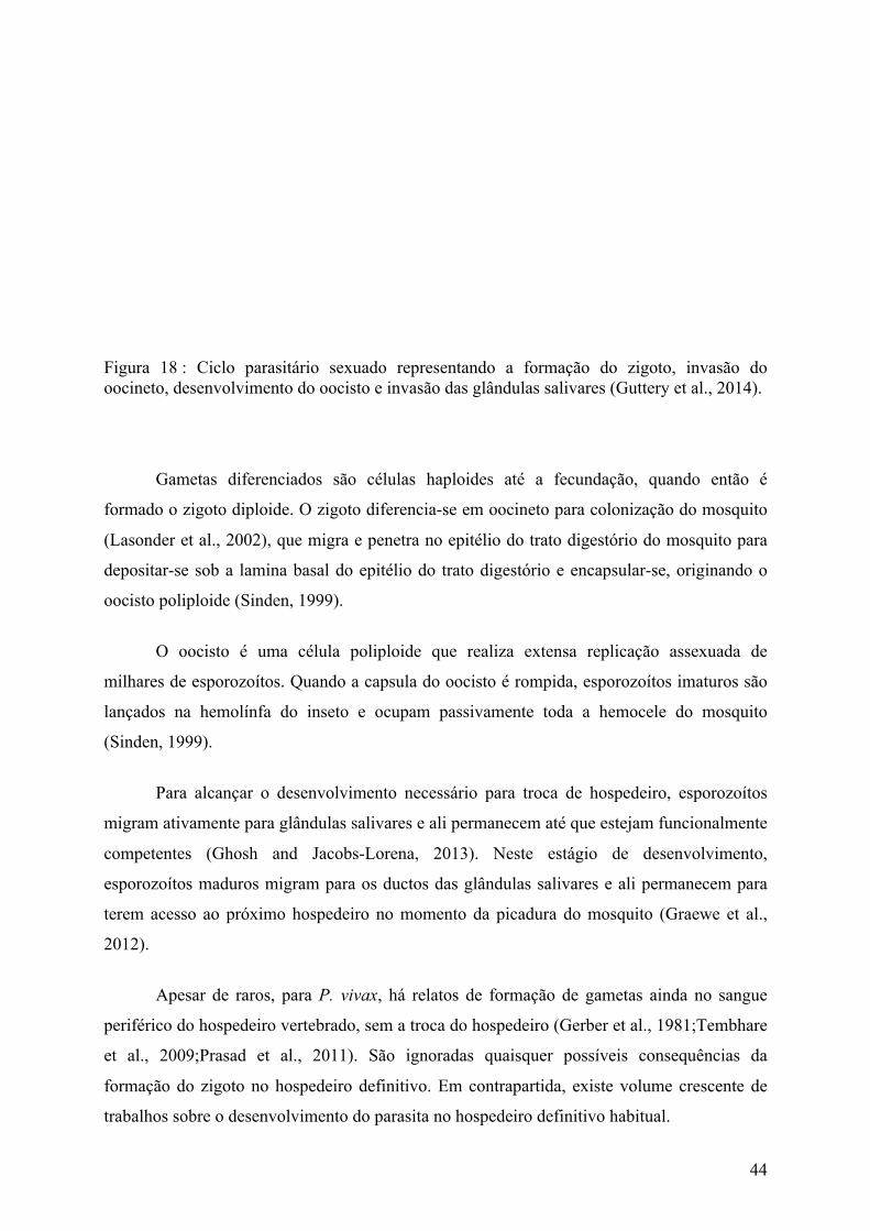
44
Figura 18 : Ciclo parasitário sexuado representando a formação do zigoto, invasão do oocineto, desenvolvimento do oocisto e invasão das glândulas salivares (Guttery et al., 2014).
Gametas diferenciados são células haploides até a fecundação, quando então é
formado o zigoto diploide. O zigoto diferencia-se em oocineto para colonização do mosquito
(Lasonder et al., 2002), que migra e penetra no epitélio do trato digestório do mosquito para
depositar-se sob a lamina basal do epitélio do trato digestório e encapsular-se, originando o
oocisto poliploide (Sinden, 1999).
O oocisto é uma célula poliploide que realiza extensa replicação assexuada de
milhares de esporozoítos. Quando a capsula do oocisto é rompida, esporozoítos imaturos são
lançados na hemolínfa do inseto e ocupam passivamente toda a hemocele do mosquito
(Sinden, 1999).
Para alcançar o desenvolvimento necessário para troca de hospedeiro, esporozoítos
migram ativamente para glândulas salivares e ali permanecem até que estejam funcionalmente
competentes (Ghosh and Jacobs-Lorena, 2013). Neste estágio de desenvolvimento,
esporozoítos maduros migram para os ductos das glândulas salivares e ali permanecem para
terem acesso ao próximo hospedeiro no momento da picadura do mosquito (Graewe et al.,
2012).
Apesar de raros, para P. vivax, há relatos de formação de gametas ainda no sangue
periférico do hospedeiro vertebrado, sem a troca do hospedeiro (Gerber et al., 1981;Tembhare
et al., 2009;Prasad et al., 2011). São ignoradas quaisquer possíveis consequências da
formação do zigoto no hospedeiro definitivo. Em contrapartida, existe volume crescente de
trabalhos sobre o desenvolvimento do parasita no hospedeiro definitivo habitual.

45
1.9. Morfologia dos gametócitos
Os gametócitos iniciam sua maturação dentro de hemácias no sistema circulatório do
hospedeiro vertebrado até alcançar a maturação final. A partir deste ajuste das competências,
as células permanecem na circulação em um estado de inibição da diferenciação até serem
ativados por estímulos específicos para tornarem-se gametas (Khan et al., 2005).
Em P. falciparum, esta maturação gametocítica acontece na maior parte do tempo na
medula óssea (Aguilar et al., 2014) e pode ser dividida em cinco fases, as quais podem ser
identificadas morfologicamente (Carter and Miller, 1979;Talman et al., 2004) como também
por marcadores moleculares (Pradel, 2007).
A despeito das similaridades, há vantagens e desvantagens dos modelos animais para
compreensão da malária. Por exemplo, quando comparamos o desenvolvimento de
gametócitos de P. falciparum com o de P. berghei, constatamos diferenças tanto de
morfologia, quanto de tempo de desenvolvimento e local de produção. Para fins
comparativos, as constatações das semelhanças e dessemelhanças nos levam a concluir que P.
berghei tem mais similaridades com outras malárias humanas e de primatas do que com o
agente causador da malária falciforme.
Em P. berghei, as fases de amadurecimento gametocítico não são de fácil distinção
por microscopia óptica e eletrônica. O gametócito não pode ser diferenciado de parasita na
fase assexuada de trofozoítos por nenhuma técnica de microscopia até suas primeiras 18
horas. A partir deste momento, podem ser observadas estruturas sexo-específicas como
distribuição pigmentar granulada no citoplasma, tamanho de célula e de núcleo aumentado, e
de corpos osmiofílicos por microscopia eletrônica. Não é possível fazer qualquer distinção
entre sexos por microscopia antes de 24 horas de desenvolvimento. Até o final do
desenvolvimento, com cerca de 30 horas, é possível a observação de maior quantidade de
corpos osmiofílicos em fêmeas (de Koning-Ward et al., 2008), quando comparadas com
machos; o macho apresenta núcleo maior e irregular e menor contraste de coloração pela
desmontagem de ribossomos (Figura 19).

46
Figura 19 : Representação esquemática das estruturas celulares do macrogametócito e do microgametócito de Plasmodium infectante de aves com diferença do tamanho do núcleo e quantidade de ribossomos dentro de eritrócitos de aviários, de acordo com Carter, R. et al (Carter and Graves, 1989).
Ao término de cerca de 30 horas de desenvolvimento, gametócitos apresentam
morfologia arredondada a ovalada com cerca de 15 µm de tamanho em P. berghei. Em P.
falciparum, o tempo de desenvolvimento pode ser de 8-16 dias com microgametócitos com
cerca de 16-25 µm (Janse and Water, 2004).

47
1.10. Diferenciação em gametas e exflagelação
É através da gametogênese que tanto gametócitos machos e fêmeas tornam-se células
competentes, os gametas, para a fecundação, a formação de zigoto e a perpetuação
reprodutiva do parasita por ciclo reprodutivo sexuado. Para Plasmodium, gametas masculinos
e femininos são também chamados de microgametas e macrogametas.
Os microgametócitos são células mais simples que sua contraparte feminina (Janse
and Water, 2004), e é por meio delas que o evento da gametogênese é mais facilmente
observável até a formação dos gametas, tanto pelas profundas transformações morfológicas e
moleculares como também como marcador de início dos estímulos ambientais que induzem a
gametogênese. Trata-se de um desenvolvimento celular de breve duração, que se processa em
cerca de 20 minutos após a ativação dos gametócitos no mosquito (Janse and Water, 2004).
A diferenciação dos gametócitos masculinos em microgametas é comumente referida
por exflagelação. Este processo inicia-se pela percepção pelas células de abruptas
modificações no ambiente circundante, desencadeando sinais para que a diferenciação se
inicie.
Uma vez ativados, os gametócitos masculinos realizam rapidamente três rodadas de
replicação do DNA, formando um núcleo octaplóide. Cada célula com núcleo poliplóide
passa pela individualização do núcleo juntamente com a montagem intracelular de oito
flagelos que, ao fim do processo de diferenciação, ou seja, após exflagelação dos mesmos,
darão origem a oito gametas masculinos sexualmente competentes que fertilizarão gametas
femininos (Figura 20).

48
Figura 20 : Representação das etapas de ativação de gametócitos masculino e feminino de P. falciparum, seguido da formação de gametas (modificado de Trustees of the Wellcome Trust).
A atividade destes microgametas pode ser associada a dois momentos patentes:
intenso movimento flagelar após formação dos mesmos, que irão realizar a etapa final da
citocinese; e intenso movimento flagelar para fusão dos gametas masculino e feminino
(Talman et al., 2014). Os eventos moleculares atrelados à ativação dos microgametócitos, ao
irromper da exflagelação e à culminância de microgametas são quase que totalmente
desconhecidos (Talman et al., 2014).
Sabe-se que existe um papel para o ácido xanturênico na ativação da gametogênese
(Figura 21), apesar de não se ter conhecimento completo sobre o receptor deste estímulo. Foi
observado que proteínas integrais de membrana, guanilil ciclases, poderiam ter envolvimento
na ativação, contudo P. berghei nocauteado para estas proteínas foi capaze de produzir
gametas funcionais (Hirai et al., 2006).
De estudos anteriores (Kawamoto et al., 1990;Kawamoto et al., 1993), existiam dados
de que, com a ativação celular para gametogênese, existe aumento significativo do cálcio
intracelular como também do cGMP. O aumento de cGMP recruta proteínas cinases
dependentes de cGMP (McRobert et al., 2008), que, por sua vez, atuam sobre os mensageiros
secundários diacilglicerol e inositol trifosfato (Martin et al., 1994) e levam à liberação e ao
influxo de cálcio pelos retículos endoplasmáticos para o citosol celular.Esse, por sua vez,
atuaria na ativação de enzimas e vias de sinalização celular, dentre elas as proteínas motoras

49
como as cinesinas para atuarem na reorganização celular e montagem do axonema neste novo
contexto metabólico da celula.
Figura 21 : Visão global das vias de sinalização relacionadas à ativação da gametogênese em Plasmodium onde cálcio de retículo endoplasmático (ER) seria liberado após a atuação dos mensageiros secundários diacilglicerol e inositol 1,4,5-trifosfato (IP3) e desencadeariam cascatas de sinalização celular para ativação de vias metabólicas importantes para a gametogênese (Kuehn and Pradel, 2010).
Sem embargo, os elementos ambientais percebidos pelos gametócitos que são
associados a sua ativação são conhecidos pela experimentação prática de culturas celulares.
Uma abrupta mudança no ambiente, similar às mudanças presentes na troca de hospedeiros,
servem como estímulo para ativação de cascatas de sinalização. Foram elencados um conjunto
de variações que podem ser: 1) alterações de temperatura ambiental; 2) aumento de pH; 3)
presença do ácido xanturênico, encontrado no trato digestório do mosquito; 4) concentração
de sais divalentes; 5) outros compostos orgânicos (Gerber et al., 1981;Billker et al.,

50
1998;Billker et al., 2004;Tembhare et al., 2009;Baker, 2010). Contudo, para que haja
eficiência de estimulação, a combinação de dois fatores são desejáveis: queda da temperatura
e presença de ácido xanturênico a pH entre 7,8 e 8,2 in vitro (Billker et al., 1998).
Tanto o microgametócito como o microgameta precisam responder rapidamente às
necessidades funcionais enquanto encontram-se no lúmen do trato digestório do mosquito
antes que sejam digeridos. Por esta razão, o gametócito masculino maduro é uma célula com
reduzida quantidade de ribossomos e de retículos endoplasmáticos, quando comparado com o
gametócito feminino, para transcrição e tradução reduzida ao limiar que permita a sua
sobrevivência. A célula concentra sua maquinaria para realizar sua função de gerar oito
gametas masculinos. O evento de replicação de DNA na exflagelação é considerado um dos
mais rápidos conhecido entre eucariotas (Janse et al., 1988).
Assim como microgametócitos apresentam maquinaria celular mínima suficiente para
executar sua função, gametas masculinos também são células mais simples. De fato,
constituem umas das células mais simples dentre os eucariotos (Talman et al., 2014). Em
termos de dimensões, um microgametócito tem cerca de 15 µm (Janse and Water, 2004) e um
microgameta apresenta cerca de 0,28 µm (Talman et al., 2014). Nos microgametas, são
reconhecidas poucas estruturas celulares como núcleo haploide, um axonema ligado a um
corpo basal modificado e uma membrana plasmática (Sinden et al., 1978). Ele não possui
mitocôndria (Okamoto et al., 2009) e a forma que ele produz suficiente ATP para prover seus
batimentos flagelares é desconhecida (Talman et al., 2014).

51
1.11. Malária de roedores – Plasmodium berghei
Há 40 anos, em 1976, foi estabelecida pela primeira vez uma cultura de células
contínua de Plasmodium de humanos (Trager and Jensen, 2005) e de P. berghei de roedores
em 1985 (Mons et al., 1985;Ramaiya et al., 1987). Até então, as culturas eram de curta
duração ou em modelos biológicos animais (Freire, 1892). A otimização das técnicas de
cultivo permitiu que grupos de pesquisa lançassem mão de estudos que ampliassem o
conhecimento sobre a biologia da malária.
De forma similar, o desenvolvimento de tecnologia de cultivo celular em modelos
animais avança o conhecimento da biologia dos parasitas humanos em perspectivas de
estudos que ainda não podem ser endereçadas a técnicas in vitro.
Não obstante, o estudo dos parasitas da malária humana pelo expediente de parasitas
de roedores são oportunos para avançar o conhecimento sobre biologia do desenvolvimento
de parasitas da malária, interações parasito-hospedeiro, desenvolvimento de vacinas e testes
de drogas. Cuidadosa avaliação dos resultados de modelos de roedores é essencial para avaliar
a sua relevância para a doença humana.
Na malária humana, causada por P. falciparum, P. vivax, P. ovale, P. malariae e P.
knowlesi, frequentemente se é empregado Plasmodium berghei como modelo biológico. As
similaridades estão em nível fenotípico e na biologia (Sanni et al., 2002), como na
gametocitogênese, ativação de gameta, exflagelação, fertilização, formação do zigoto,
formação de oocineto (Guttery et al., 2012b), sincronização, taxa de síntese do DNA e
momento de replicação do material genético extra-nuclear (van Dijk et al., 1997).
P. berghei foi provavelmente visualizado pela primeira vez há pouco mais de 70 anos,
em 1943, por Vincke, pela observação de esporozoítos em glândulas salivares (Landau and
Boulard, 2012;Vanderberg and Gwadz, 2014) e, em 1946, por esfregaços de sangue do
estômago de mosquitos Anopheles dureni millecamps, seu hospedeiro invertebrado na
natureza (Sinden et al., 2002). Grammomys surdaster, uma espécie de roedor encontrado na
floresta próxima à cidade de Kisanga, no país de Catanga, é seu hospedeiro vertebrado (Bafort
et al., 1968;Sinden et al., 2002).

52
Esse organismo propicia um modelo atraente para a pesquisa sobre malária, pois é
capaz de produzir em laboratório todas as formas sexuadas e assexuadas, o que permite uma
infinidade de estudos e a comparação dos dados in vivo e in vitro (Sanni et al., 2002;Briquet
et al., 2015). O organismo é utilizado em rotina por laboratórios e projetos de pesquisa, o que
aproxima alguns padrões de comparação entre estudos. Podem ser usados vetores particulares
de diversas regiões do mundo (Tabela 1). Seu genoma, mitocondrial e do apicoplasto, já foi
sequenciado.
Tabela 1: Relação de mosquitos com capacidade vetorial e mosquitos sem capacidade para transmissão de P. berghei (Sinden et al., 2002).
Com Capacidade Vetorial Sem Capacidade Vetorial
An. dureni Culex bitaeneorhynchus
An. stephensi mysorensis Ae. aegypti*
An. gambiae An. aztecus
An. atroparvus An. concolor
An. quadrumaculotus An. coustani var ziemanni
An. maculipennis An. fluviatilis
An. albimanus An. funestus
An. hyrcanus
An. jamesi
An. pulcherrimus
An. splendidus
An. subpictus
* – produz até o oocisto da linhagem SP28 S. Keiberg.
Não obstante as possibilidades de abordagens de pesquisa e a base de conhecimento já
construída sobre a doença em humanos e sobre Plasmodium berghei, o emprego do modelo
para estudo das malárias é oportuno pela existência e disponibilidade de linhagens de
camundongos suceptíveis, geneticamente definidos e nocauteados (Janse et al., 2006).
Existem clones de P. berghei bem caracterizados, com fenótipos particulares como
para expressão de proteínas fluorescentes (Natarajan et al., 2001;Franke-Fayard et al., 2004)
ou pela incapacidade de gerar estágios sexuais.

53
Algumas linhagens de P. berghei isoladas desconhecidas são K173, ANKA, LUKA,
SP11 e NK65 (Sinden et al., 2002). Este trabalho utiliza-se da linhagem P. berghei NK-65 e
do banco de dados da linhagem ANKA.
A designação NK-65 tem origem de “New York-Kisanga 1965”. Este é o nome em
que consta local do isolamento, local de origem e data do isolamento. A linhagem foi isolada
em 1965 a partir de mosquitos Anopheles dureni, infectados por P. berghei, capturados na
floresta de Kisanga, em Catanga, e enviados pelo pesquisador belga, Josef Maria Bafort. O
plasmódio foi isolado e replicado na Escola Medica da Universidade de Nova Iorque
(Vanderberg and Gwadz, 2014).
P. berghei ANKA foram também capturados por Bafort e Vincke na floresta de
Kasapa, próxima de Lubumbashi, na data de Março de 1965. A designação advem de obtidos
de mosquitos coletados na floresta de Antwerp-Kasapa. O plasmódio foi isolado e replicado
no Prince Leopold Institute of Tropical Medicine Antwerp em Antwerp (Vincke and Bafort,
1968;Vanderberg and Gwadz, 2014).
P. berghei tem por ponto forte a plasticidade para manipulações genéticas para estudo
da malária humana e um rápido ciclo de vida para maturação e diferenciação das formas
sexuais (Guttery et al., 2012b). Ciclos reprodutivos do P. berghei são de curta duração quando
comparados com de outras espécies plasmodiais, incluindo o P. falciparum. Todas fases de
desenvolvimento no roedor hospedeiro permanecem circulantes no sangue.
Em laboratórios, a fase sexual de P. berghei é com frequência realizada em mosquitos
An. stephensi. Esporozoítos tornam-se maduros na glândula dos mosquitos com cerca de 13 a
14 dias após a infecção do mosquito e tem tamanho médio de 11 a 12 µm.
A fase do fígado é de curta duração. Ela se inicia com no mínimo 3 h após infeção
(Frevert et al., 2005), para que suceda a infecção de hepatócitos, e transcorre por cerca de 50
horas para que conclua o ciclo assexuado hepático. Este ciclo culmina com a formação de
formas esquizontes com o máximo de 30 µm que liberarão merozoítos para a próxima fase
reprodutiva (Killick-Kendrick, 2012).
Ao fim do ciclo hepático, merozoítos são liberados no sistema circulatório e infectarão
hemácias e, preferencialmente, reticulócitos de roedores hospedeiros (Rénia et al., 2012). Os

54
ciclos assexuados eritrocitários têm duração aproximada de 24 horas sem sequestração
tecidual. Os gametócitos são formados a partir de ciclos assexuados eritrocitários.
Algo próximo de 20% de merozoítos de cada ciclo assexuado eritrocitário dão origem
a gametócitos (Jambou et al., 2011). O tempo de desenvolvimento dos gametócitos até se
tornarem maduros é de cerca de 30 horas. Ao término de seu desenvolvimento, as células
apresentarão um tamanho de 15 µm (Garnham, 1965) e fase de crescimento G0 até serem
ingeridas ou ocorrer a morte do hospedeiro (Sinden, 2012).
Após ingestão e ativação dos gametócitos por mosquitos, a diferenciação celular em
gametas finda em aproximadamente 15 minutos e, portanto, o plasmódio encontra-se
competente para a fertilização do zigoto. O zigoto, com cerca de 12 µm, replica seu DNA e
diferencia-se em uma célula móvel e tetraploide de cerca de 12 µm (Garnham, 1965).
Para que a infecção tenha êxito com ao menos um oocisto, deverão ser formados entre
40-140 oocinetos (Sinden et al., 2007). A transição de oocineto para oocisto depende da
densidade do primeiro, da resposta imunológica do mosquito (Sinden et al., 2007), de
variáveis fisiológicas, bióticas e genotípicas dos organismos (Christophides et al.,
2002;Lambrechts et al., 2005;Dong et al., 2006). Contudo, uma vez que o desenvolvimento de
oocineto para oocisto tenha sucesso, ele acontecerá em média dentro de 18 a 24 horas.
Um oocisto maduro tem média de tamanho de 45µm (Killick-Kendrick, 2012) e libera
uma média de 2000 esporozoítos imaturos por oocisto na hemocele do mosquito (Stone et al.,
2013) em cerca de 18 dias após a infecção (Stone et al., 2013). Dentre os esporozoítos
liberados em An. gambiae e An. stephensi, cerca de 20-60% (Hillyer et al., 2007;Stone et al.,
2013) são capazes de invadir glândulas salivares após 8 horas de sua liberação pelo oocisto
(Hillyer et al., 2007). O paradeiro dos esporozoítos que não conseguiram invadir é
desconhecido; sabe-se apenas que 2% sofrem fagocitose, morte e degradação celular (Hillyer
et al., 2007).

55
1.12. Cinesinas
1.12.1. As cinesinas em eucariotos
A literatura sobre a malária traz informações de que um grupo de proteínas, dentre elas
as proteínas motoras, desempenham papel crítico em todos os passos de desenvolvimento do
parasita (Gerald et al., 2011). O estudo de proteínas motoras relacionadas ao processo de
formação do flagelo e exflagelação é desejável para conhecimento mais pleno sobre o
desenvolvimento dos estágios sexuados para transmissão da doença (Arevalo-Herrera et al.,
2011;Guttery et al., 2012b;Sinden et al., 2012;Josling and Llinas, 2015).
As proteínas motoras formam 3 grandes superfamílias, que são: miosinas, cinesinas e
dineínas (Wickstead and Gull, 2006;Kardon and Vale, 2009). Destas, a superfamília das
cinesinassão codificadas pelo genoma de todos os eucariotas (Wickstead and Gull, 2006) e,
para gametócitos de P. berghei, há indícios de que elas podem desempenhar um papel
essencial na regulação do flagelo (Talman et al., 2014). Cinesinas são hierarquizadas
atualmente em 14 famílias, cinesina 1 a cinesina 14, de acordo com características do domínio
motor da enzima (Lawrence et al., 2004) (Figura 22). Quando fazem parte da mesma família,
normalmente apresentam funções similares no reino animal, apesar de nem todas enzimas de
mesma família apresentarem funções análogas (Tomei and Wolniak, 2016). Somente as
proteínas da família das cinesina 5 tem papel conservado na formação do fuso mitótico tanto
em plantas como em animais (Tomei and Wolniak, 2016).
As cinesinas estão relacionadas a uma variedade de processos em cílios e flagelos
(Fox et al., 1994). Elas desempenham papéis estruturais e funcionais (Fox et al., 1994)
durante suas atuações na mitose, na ciliogênese (Yokoyama et al., 2004;Tomei and Wolniak,
2016) e na atividade ordenada de flagelos de Chlamydomonas (Fox et al., 1994). A
ciliogênese é sujeita a regulação pelas cinesinas em organismos do reino animal como vegetal
(Tomei and Wolniak, 2016) e sua ausência tem sido associada a defeitos na montagem do
axonema tanto em cílios sensoriais como em cílios de locomoção (Yokoyama et al., 2004).
São também importantes na regulação de processos essenciais da gametogênese masculina em
gametófitos (Tomei and Wolniak, 2016) como também para a fecundação dos gametas em
Chlamydomonas (Fox et al., 1994).

56
Figura 22 : Representação esquemática das cinesinas de algumas famílias onde pode-se observar domínio motor conservado e diversidade de domínio variável entre famílias de cinesina (Verhey and Hammond, 2009).
Cinesinas fazem o transporte de cargas (Yokoyama et al., 2004;Kevenaar et al., 2016),
como vesículas, cromossomos ou outras partículas (Hirokawa and Noda, 2008), a organização
espacial de organelas e controlam a dinâmica de microtúbulos tanto em axônios como em
dendritos de células nervosas (Kevenaar et al., 2016). Assim, cinesinas fazem o transporte
intraflagelar dirigido ao longo de axonemas de microtúbulos, responsável pelo movimento de
componentes axonemais para a montagem da extremidade distal de flagelos (Fox et al., 1994).
Além disto, em flagelos de Chlamydomonas reinhardtii, o par central de microtúbulos
é revestido por um conjunto de proteínas associadas e, possivelmente, não deixam qualquer
superfície livre no axonema para um movimento progressivo de cinesina, o que indica que
cinesinas no par central de microtúbulos de flagelos não atuam propriamente como proteínas
motoras típicas (Yokoyama et al., 2004).
De forma geral, cinesinas podem ser empregadas de diversas formas pelas células
eucarióticas, como na organização de microtúbulos em geral, nas interações de microtúbulos

57
corticais, na transferência de sub-compartimentos celulares, na distribuição mitocondrial, na
dinâmica do cinetócoro, na morfogênese de fuso e no alinhamento cromossômico (Gupta et
al., 2006;Messin and Millar, 2014;Shin et al., 2015;Kevenaar et al., 2016), no transporte
intracelular, na segregação de organelas, desestabilizando microtúbulos, na alteração da
morfologia, ou mesmo no movimento de toda a célula (Wickstead and Gull, 2006).
1.12.2. A motilidade das cinesinas
Cinesinas usam a energia proveniente da hidrólise do ATP para se deslocarem ao
longo do microtúbulo no sentido da extremidade positiva. O domínio motor é altamente
conservado e compreende um núcleo catalítico de ATP e um sítio de ligação de microtúbulos
(Tomei and Wolniak, 2016).
Ademais do domínio motor, há um domínio variável em cauda que é importante para a
ligação de vesículas de transporte (Tomei and Wolniak, 2016) (Figura 23). As regiões de
domínio variável são resposáveis pela interação específica da proteína, conferindo diferentes
funções às cinesinas (Tomei and Wolniak, 2016).
A maioria das cinesinas atuam como homodímeros, embora existam heterodímeros,
heterotrímeros e oligomerizados que se conectam através do motivo hélice-hélice das
proteínas (Tomei and Wolniak, 2016).
Figura 23 : Representação esquemática da estrutura da cinesina 8 com seus domínios motores e variável e sua forma dimérica. Modificado de (Roostalu and Surrey, 2013).
58
1.12.3. Regulação de cinesinas
Cinesinas são reguladas por 3 mecanismos: 1) interações cauda-cargas de transporte,
2) interações motor-microtúbulos e 3) autoinibitórios pela interação motor-cauda (Kevenaar et
al., 2016). Outros mecanismos relacionados à regulação das cinesinas ainda são
desconhecidos.
A ausência de cargas de transporte ligada à cinesina faz com que ela assuma uma
conformação enovelada (Kevenaar et al., 2016). Com o enovelamento, o domínio variável da
cauda interage com o domínio motor e inibe sua atividade motora (Kevenaar et al., 2016). Por
outro lado, quando há ligação de carga de transporte ao domínio variável, a autoinibição é
interrompida pela alteração conformacional da enzima e, como consequência, o movimento
torna-se possível pelas atividades do domínio motor (Kevenaar et al., 2016).
A atividade motora das cinesinas também são inibidas quando proteínas ligadoras de
cinesinas se associam ao domínio motor. Elas impedem a ligação das cinesinas com os
microtúbulos e, desta forma, modulam as atividades de motilidade e despolimerização de
cinesinas em microtúbulos (Kevenaar et al., 2016). De outro modo, microtúbulos e tubulina
não polimerizada podem ativar a atividade motora de cinesina 8 (Gupta et al., 2006).
1.12.4. A cinesina 8 em Plasmodium
Neste trabalho, empregamos uma linhagem de P. berghei mutante para cinesina 8 com
o objetivo de estudar a diferenciação do gametócito macho ao microgameta, único estágio do
gênero Plasmodium em que os flagelos estão presentes. São investigados mecanismos
compensatórios ou regulatórios relacionados à mutação da cinesina 8 que justifiquem as
alterações fenotípicas observadas e os mecanismos chave de atuação da cinesina 8 no
desenvolvimento gametocítico normal. O propósito do uso do mutante foi, portanto, de
ampliar a compreensão e o discurso sobre mecanismos críticos que permitam interferências e
interrupção da gametogênese.
A Cinesina 8 é uma família de enzimas motoras ditas ultraprocessivas que regulam a
atuação de microtúbulos em seu deslocamento em direção à “extremidade mais” (plus end)

59
para atuar na desorganização, despolimerização ou recuperação da extremidade do
microtúbulo (Messin and Millar, 2014;Gergely et al., 2016). Desta forma, elas regulam o
comprimento de microtúbulos em diferentes compartimentos celulares e ao longo de
diferentes etapas do ciclo biológico (Messin and Millar, 2014;Gergely et al., 2016).
Sua dissociação da extremidade positiva dos microtúbulos é pouco frequente, o que
faz com que elas sejam enriquecidas nestas extremidades positivas de forma diretamente
relacionada ao comprimento dos microtúbulos e, desta forma, promovam a pausa ou ruína dos
arranjos de tubulinas alfa e beta na extremidade mais de microtúbulos (Gupta et al., 2006;Shin
et al., 2015).
Microtúbulos apresentam polaridade e sentido de crescimento de acordo com a
disposição dos monômeros de tubulina alfa e beta. Alfa tubulinas dão a orientação “minus
end” e beta tubulinas dão a orientação “plus end”, onde as tubulinas alfa e beta são
adicionadas para o crescimento de microtúbulos (Messin and Millar, 2014) (Figura 24).
Figura 24 : Imagem modificada de Messin et. al. (Messin and Millar, 2014). A seta representa o sentido de deslocamento da cinesina 8, que se concentra na extremidade positiva (plus end), sobre as unidades alfa e beta de tubulina, que formam o microtúbulo. Representados em 1) unidade de tubulina alfa (em azul); 2) unidade de tubulina beta (em vermelho); 3) unidades de cinesina 8 (em verde).
A família cinesina-8 tem sido reportada em atividades implicadas na dinâmica de
microtúbulos nas múltiplas respostas celulares como: no aumento ou redução da velocidade
de crescimento celular, na frequência de recuperação, na parada de ciclo celular, na
nucleação, na dimerização de tubulina, na formação de anel tubulina, na ligação cruzada de
microtúbulos e nodeslizamento de microtúbulos (Gergely et al., 2016).

60
São ditas ultraprocessivas devido, possivelmente, à atuação de um segundo sítio de
ligação de microtúbulos que conecta domínios motores ao carril de microtúbulos, apesar de
haver contestação quanto à eficiência de processividade (Shin et al., 2015).
A grande diversidade de atuação das cinesinas 8 na dinâmica de microtúbulos trouxe
algumas explanações sobre os distintas atuações desta família para diferentes organismos
eucarióticos (Gupta et al., 2006;Gergely et al., 2016). Contudo, ainda não está totalmente
esclarecido como cinesina 8 contribui para movimentos organizados de cromossomos na
performance do fuso mitótico no alinhamento com o plano de divisão celular (Gupta et al.,
2006;Stumpff et al., 2008;Gergely et al., 2016), na dinâmica dos cinetócoros (Gupta et al.,
2006;Stumpff et al., 2008), no controle das oscilações cromossômicas (Stumpff et al., 2008),
na regulação do comprimento de microtúbulos ligados ao córtex celular, para interações
microtubulocortical, na distribuição mitocondrial (Gupta et al., 2006) ou na formação dos
flagelos em gametócitos e subsequente exflagelação.
Em humanos, observou-se que a cinesina 8 é acumulada em um gradiente de
concentração (Messin and Millar, 2014) ao longo de microtúbulos cinetócoros de uma
maneira dependente da sua atividade motora (Stumpff et al., 2008). Em leveduras, a atividade
ATPásica indispensável para função motora é ativada por dímeros de tubulina (Gupta et al.,
2006). Na regulação de microtúbulos, quando ligada a ATP, a cinesina 8 forma um complexo
com a tubulina não polimerizada que pode provocar a intermitência ou a despolimerização de
microtúbulos (Gupta et al., 2006).
Observou-se que a ausência de cinesina 8 em organismos mutantes é a causa de
defeitos na polaridade celular e de fusos mitóticos mais longos (Gupta et al., 2006;Messin and
Millar, 2014), mas que isto não é um padrão obedecido em todos organismos (Messin and
Millar, 2014;Gergely et al., 2016).
Neste trabalho, aspiramos enriquecer o conhecimento sobre as relações dos
componentes proteicos e mecanismos moleculares relacionados ao desenvolvimento
ontogenético de gametócitos na formação de gametas e o papel que cabe à cinesina 8 no
desempenho de sua função molecular.

61
1.12.5. A cinesina 8 em Plasmodium berghei
O genoma do P. berghei codifica duas proteínas cinesina 8. Uma delas é codificada no
cromossomo 8 do P. berghei, gene PbANKA_08_v3, para proteína de massa molecular de
161.603 Da, ponto isoelétrico de 9,38, sob número de registro no banco de dados GeneDB:
PBANKA_0805900 (banco de dados NCBI PB000348.03.0). A outra cinesina 8, alvo deste
estudo e enunciada ao longo deste trabalho, está codificada no cromossomo 2, gene
PbANKA_02_v3, sob número de registro no GeneDB como PBANKA_0202700 (banco de
dados NCBI PB001397.02.0) com massa molecular de 168.815 Da e ponto isoelétrico de
6,52.
Até o presente, não houve detecção de cinesina 8 em fêmeas da linhagem NK65, tanto
nos estágios do ciclo assexuados como em macrogametócitos e macrogametas. De acordo
com comunicação pessoal dos professores Philippe Grellier e Delphine Depoix, a partir de
trabalhos em desenvolvimento pelo grupo de pesquisa, a cinesina 8, que faz parte do âmago
deste trabalho (PBANKA_0202700), não é encontrada expressa em fêmeas de P. berghei. Se
expressa em macrogametócitos, não pode ser detectada pelas técnicas pelas quais foi
investigada, como por imunocitolocalização, por anticorpos para microscopia eletrônica, por
microscopia de fluorescência confocal, ou mesmo pela espectrometria de massas.
Ainda de acordo com trabalhos do Prof. Grellier, por microscopia de fluorescência foi
observada uma construção transgênica do plasmódio em que a cinesina 8 foi expressa
acoplada à sequência de Proteína Verde Fluorescente, conhecida por GFP (green fluorescent
protein). A fluorescência de kin8-GFP ocorreu somente em algumas células, as quais eram
coincidentes com células masculinas que fluoresciam pela marcação de alfa tubulina II,
utilizada como marcador especifico do sexo masculino em gametócitos de P. berghei (Khan
et al., 2005).
Após 15 minutos da indução da exflagelação, possivelmente o microgameta realiza
todos os três ciclos de replicação de DNA e em seguida o condensa, mas não é capaz de
cumprir com a separação dos cromossomos e formação de gametas individualizados. As
microscopias eletrônica (Figura 25) e de fluorescência revelam que, em organismos mutantes,
os núcleos não se individualizam e as duplas de microtúbulos dos axonemas se forma de
forma desorganizada, não chegando, portanto, a constituir a estrutura flagelar do tipo 9 + 2
normal do selvagem (Figura 25-D).
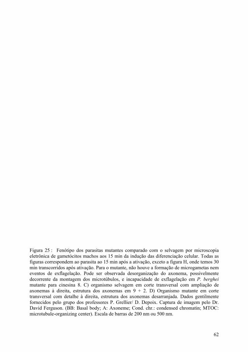
62
Figura 25 : Fenótipo dos parasitas mutantes comparado com o selvagem por microscopia eletrônica de gametócitos machos aos 15 min da indução das diferenciação celular. Todas as figuras correspondem ao parasita ao 15 min após a ativação, exceto a figura H, onde temos 30 min transcorridos após ativação. Para o mutante, não houve a formação de microgametas nem eventos de exflagelação. Pode ser observada desorganização do axonema, possivelmente decorrente da montagem dos microtúbulos, e incapacidade de exflagelação em P. berghei mutante para cinesina 8. C) organismo selvagem em corte transversal com ampliação de axonemas à direita, estrutura dos axonemas em 9 + 2. D) Organismo mutante em corte transversal com detalhe à direita, estrutura dos axonemas desarranjada. Dados gentilmente fornecidos pelo grupo dos professores P. Grellier/ D. Depoix. Captura de imagem pelo Dr. David Ferguson. (BB: Basal body; A: Axoneme; Cond. chr.: condensed chromatin; MTOC: microtubule-organizing center). Escala de barras de 200 nm ou 500 nm.

63
Nos gametócitos machos, a cinesina 8 pode ser encontrada nos flagelos e de forma
difusa no citoplasma celular, o que é um indicativo de que também esteja associada a outros
processos celulares não relacionados a formação do flagelo e exflagelação, como tráfego
intracelular e outras funções das cinesinas descritas acima.
Se associada a tubulinas e formação de microtúbulos, conforme discutido ao longo
deste trabalho, há a possibilidade de que a ausência da cinesina 8 poderia afetar, nas fêmeas,
somente a classe funcional de microtúbulos subpeliculares, enquanto que, no macho,
possivelmente afetaria as três principais classes funcionais em que os microtúbulos estão
associados em plasmódio (Sinden, 1982;Schwank et al., 2010):
• microtúbulos de axonemas, importantes para locomoção e fecundação;
• microtúbulos de fusos mitóticos, que atuam na segregação cromossômica e divisão
celular;
• microtúbulos subpeliculares, importantes para o arranjo das estrutural da célula
(Bell, 1998).
Devido ao pequeno número de cinesinas em plasmódio quando comparado a outros
organismos eucariotos (Chung et al., 2009), hipotetiza-se que cinesinas de Plasmodium
podem ter atividade ampliada a variados processos dentro da célula.

64
1.13. Proteômica e proteoma de Plasmodium
A proteômica é uma área da ciência que, em muitas das vezes, utiliza-se de abordagem
científica não clássica (Kell and Oliver, 2004). Ou seja, a metodologia científica pode ser
dirigida pela descoberta, ou a formulação de hipóteses pode ser feita ao final da observação
indutiva dos fenômenos da natureza. Apesar de esta metodologia apresentar-se como uma
nova abordagem, ela também é utilizada pelas ciências “ômicas”, como genômica,
transcriptômica, metabolômica. Por vezes, essa metodologia tem seu valor para a
biotecnologia e para a ciência reivindicado no meio científico (Marincola, 2007). Contudo,
essa metodologia empregada pela proteômica (Aebersold et al., 2000) não é uma novidade
das “ômicas” e remonta de tempos remotos da ciência para fornecer arcabouço de
informações para importantes hipóteses e teorias (Casadevall and Fang, 2008). A proteômica
pode auxiliar a elucidar mecanismos moleculares dos sistemas biológicos e, portanto, o
método científico é muitas vezes apenas um método de fazer descobertas (Feynman, 2005).
No presente momento, a proteômica compreende o conjunto de técnicas utilizadas
para investigar misturas complexas de proteínas e determinar suas relações em um contexto
funcional e de expressão gênica em determinada condição fisiológica de interesse. O uso de
técnicas diversas nos estudos das proteínas permite a determinação da identidade de proteínas,
quantificação, localização, caracterização e quantificação de modificações pré-, co- e pós-
traducionais e determinação de interações e mecanismos de sinalização celular do maior
número possível de proteínas, o que torna esse campo interdisciplinar e versátil de forma a
abarcar questões complexas características dos sistemas biológicos e para a formulação de
hipóteses.
A descoberta e descrição do conjunto de proteínas presentes em determinada célula,
tecido ou fluido corporal em dado momento fisiológico, para um genoma designado, o
proteoma (Olsen et al., 2006), e de importância para o funcionamento saudável de um ente
vivo faz das proteínas de longe a maior classe de alvos medicamentosos (Kumar and Mann,
2009). A identificação de moléculas axiais do parasita e do hospedeiro nas etapas da infecção
são capitais para compreensão e intervenção nos mecanismos que acarretam a patogenicidade.
Ao final da década de 90, iniciou-se o sequenciamento genômico dos primeiros
organismos responsáveis pela malária (Carlton et al., 2002;Gardner et al., 2002). Portanto,

65
subsequentes investigações proteômicas sobre a malária só viriam a se acentuar no princípio
da década seguinte. Trabalhos pioneiros para elucidar o proteoma tiveram início em 1987
através de 2D-SDS-PAGE (Pinswasdi et al., 1987).
Os trabalhos de proteômica, subsequentes ao sequenciamento genômico dos
organismos, permitiram vislumbrar mecanismos de regulação da expressão proteica e de
evasão ao sistema imune do hospedeiro (Florens et al., 2002a;Lasonder et al., 2002). A
maioria destes estudos proteômicos tratam da compreensão de mecanismos do ciclo
intraerocitário, tanto pelo provimento de amostra como por esta ser a fase mais patogênica
para o ser humano.
Os trabalhos de proteômica com Plasmodium se concentram principalmente nas
formas dos estágios assexuados, nos quais é apresentado o quadro sintomático da doença e
em que estão concentrados a maior parte dos estudos sobre a biologia do parasita. Contudo,
diferentes estágios de gametócitos foram caracterizados por análises de proteômica, que
detectaram aumento de proteínas específicas de gametócitos ao longo do desenvolvimento
(Florens et al., 2002a;Lasonder et al., 2002), assim como também estudos em P. berghei no
desenvolvimento de gametas (Khan et al., 2005;Talman et al., 2014;Alonso-Morales et al.,
2015).
Talman et al. aponta que o metabolismo energético de gametas masculinos é
sustentado fundamentalmente pela glicólise devido à ausência de mitocôndrias como
também pela detecção reprodutível de todas as enzimas da via glicolítica e do transportador
de hexose. Neste estudo, foram identificadas 615 proteínas por espectrometria de massas
acoplada à técnica de fracionamento de proteínas por MudPit (Talman et al., 2014).
Foi investigado em Khan et al. a composição de cinases e fosfatases no proteoma de
gametócitos e as implicações de proteínas sexo-específicas como mitogen-activated protein
kinase 2 (MAP2) e NIMA-related kinase (NEK4) ao separar gametócitos machos e fêmeas
por citometria de fluxo. As cinases MAP são frequentemente relacionadas à proliferação e
diferenciação de células. Neste estudo, foram encontradas 779 proteínas em P. berghei
(Khan et al., 2005).
Em Tao et al., foram identificadas 1703 proteínas em gametócitos de P. falciparum e,
por reanálise de dados obtidos em trabalho de Khan et al. (Khan et al., 2005) com P.

66
berghei, foram identificadas 1128 e 1180 proteínas de gametócitos masculinos e femininos,
respectivamente (Tao et al., 2014a).
Mais recentemente, Alonso-Morales e colaboradores (Alonso-Morales et al., 2015),
ao trabalhar com técnica de eletroforese bidimensional para fracionamento de proteínas e
identificação por espectrometria de massas, detectou um somatório aproximado de 710 spots
na faixa de massa de 200-20 kDa e pI de 3-10 a partir de todos géis produzidos. Suas
identificações foram restritas a 23 fosfoproteínas a partir de P. berghei no marco de 15
minutos de diferenciação do gametócito e em comparação com gametócito não estimulado.

67
1.14. Fosfoproteoma de Plasmodium
Os mecanismos de sinalização e regulação em processos celulares, como o da
gametogênese em plasmódio, são dirigidos por “núcleos executivos” (Kumar and Mann,
2009) formados por proteínas, os principais efetores das funções biológicas (Cox and Mann,
2007).
Os níveis de expressão de todas as proteínas expressas por um sistema podem fornecer
dados relevantes para a caracterização de um sistema biológico e seu funcionamento. A
atuação concertada das proteínas em determinado organismo vivo depende não somente da
composição deste conjunto de proteínas, mas também de outras variáveis como os
mecanismos de regulação por modificações pós-traducionais (Cox and Mann, 2007).
As fosforilações figuram entre os tipos de modificações pós-traducionais mais comuns
(Pandey et al., 2014) nas regulações dos mecanismos celulares em eucariotos (Alonso-
Morales et al., 2015;Panichakul et al., 2015). Estima-se que de 10 a 30% de todas proteínas de
um organismo eucariótico sejam reguladas por fosforilação (Weise and Lenz, 2008;Wu et al.,
2009).
Este tipo de regulação celular confere a um organismo a capacidade de respostas
rápidas e reversíveis ao nível molecular frente às condições ambientais cambiantes (Chung et
al., 2009), e de regular temporal e espacialmente algumas proteínas (Dastidar et al., 2013).
Desta forma, permite ao parasita uma pronta alteração de sua biologia (Bautista et al.,
2014;Lasonder et al., 2015) para uma melhor adaptabilidade às condições ambientas
circundantes, como para escapar da resposta imunológica do hospedeiro, se desenvolver e,
portanto, perpetuar-se (Chung et al., 2009;Bautista et al., 2014).
O balanço de atividades antagonistas relacionadas às fosforilações são controladas por
duas classes de proteínas: proteínas cinases e proteínas fosfatases, que realizam
respectivamente a fosforilação e desfosforilação de proteínas (Pandey et al., 2014).
Para a atividade de proteínas cinases, as fosforilações envolvem esterificação
reversível do grupo fosfato em determinados resíduos de aminoácidos. Reações catalisadas
por proteínas cinases acontecem com a transferência de grupo fosforil ao custo de um ATP
para um hidroxiaminoácido (Chung et al., 2009).

68
Para atividade de fosfatases, o grupo fosfato é hidrolisado da ligação fosfoéster do
resíduo de aminoácido modificado. O resultado é a liberação de um íon fosfato livre,
deixando somente o grupo hidroxila do resíduo previamente fosforilado (Sickmann and
Meyer, 2001).
As fosforilações e desfosforilações acontecem predominantemente em resíduos de
serina e treonina, mas também podem acontecer com menor frequência em resíduos de
tirosina e histidina (Hanks, 2003). As fosforilações modificam as propriedades fisioquímicas
das proteínas e, desta forma, podem resultar em alterações moleculares particulares, como
dobramentos proteicos próprios, atividade de microtúbulos, atividade de histonas, regulação
de fatores de transcrição, alteração de atividade enzimática, afinidade de interações com
outras proteínas, alocação subcelular de proteínas ou tempo de residência (Wu et al.,
2009;Eyrich et al., 2011).
A consequência global proporcionada por um conjunto de fosforilações e
desfosforilações para os organismos está muita veze na regulação direta de processos do ciclo
celular (Solyakov et al., 2011) e na transdução de sinais. A transdução de sinal em
metazoários regula praticamente todos os aspectos da função biológica celular, como
comunicação intercelular, replicação de DNA, transcrição, metabolismo e crescimento,
diferenciação celular, ativação de receptores, organização citoesquelética, remodelação da
cromatina e apoptose (Doerig et al., 2008;Chung et al., 2009;Hilger et al., 2009;Thingholm et
al., 2009;Solyakov et al., 2011;Pandey et al., 2014), como também processos especializados e
críticos do parasita como na invasão de eritrócitos e citoaderência (Wu et al., 2009).
Os efeitos das fosforilações diferenciais são observados nas mudanças funcionais na
biologia do parasita (Lasonder et al., 2015) e podem ocorrer em múltiplos sítios de uma
mesma proteína. A consequência de um sistema de múltiplos níveis de regulação é um
controle ajustado de funções celulares e respostas a estímulos (Wu et al., 2009).
Proteínas cinases, fosfatases e vias de transdução de sinais são as responsáveis pela
orquestração regulatória por fosforilações em diversas proteínas (Solyakov et al.,
2011;Lasonder et al., 2015), como nas proteínas chaperonas, que são reguladas por
fosforilação para o dobramento orientado de proteínas ou desdobramento para translocação,
complexação ou degradação de proteínas (Wu et al., 2009).

69
Estima-se que mais de 50% das proteínas do proteoma do plasmódio sejam proteínas
estágio específicas (Bautista et al., 2014). Isto indica substancial grau de especialização
molecular do parasita a cada estágio de seu ciclo celular (Bautista et al., 2014), mas também a
necessidade de mecanismos regulatórios eficazes especializados para cada circunstância
metabólica celular. Além da fosforilação do parasita, temos que a infecção por P. falciparum
acarreta em um aumento drástico das fosforilações dos eritrócitos (Wu et al., 2009), o que é
uma evidência quanto à relevância das fosforilações também na regulação do hospedeiro
como também em uma possível implicação nas interações parasita-hospedeiro.
Nos plasmódios, as proteínas fosforiladas são predominantemente intracelulares e
poucas delas localizam-se na superfície membranar do parasita (Lasonder et al., 2012).
Análises por interrupção gênica apontam que metade das proteínas fosfatases de P. berghei
são críticas para o correto funcionamento celular (Pandey et al., 2014).
Em eucariotos, o número de proteínas fosfatases é relativamente pequeno quando
comparado ao de proteínas cinases. Apesar disto, proteínas reguladoras de fosfatases atuam na
mediação da atividade de fosforilase e, desta forma, trazem à tona a hipótese de que a
quantidade de complexos de fosfatases e proteínas mediadoras associadas implicadas na
regulação de vias metabólicas pode exceder o repertório de proteínas cinases (Chung et al.,
2009).
As proteínas cinases também podem atuar na inibição do desenvolvimento de
diferentes estágios do ciclo de vida de plasmódio (Le Roch et al., 2008;McRobert et al.,
2008;Kato, 2016). Inibidores de proteínas cinases podem interferir no crescimento do parasita
e, portanto, serem utilizados para o desenvolvimento de terapias farmacoterápicas para o
combate da malária (Wu et al., 2009).
A exemplo do potencial do emprego de terapias farmacológicas, temos as proteínas
implicadas na regulação cromossômica como as histonas, que apresentam amplo número de
sítios de fosforilação (Bautista et al., 2014). Em histonas, as fosforilações são mais frequentes
na região N-terminal e podem controlar diversos processos nucleares do parasita (Dastidar et
al., 2013). Elas podem estar associadas de forma reversível a mitose, condensação
cromossômica, replicação do DNA, ativação de transcrição, apoptose, resposta ao estresse e
ao dano do DNA, a depender do contexto e sítio da fosforilação (Berger, 2010). Tratamentos

70
que afetem a atividade reguladora das mitocôndrias podem afetar criticamente a estratégia
energética e sobrevivência do parasita.
Apesar de ser um tema de relevância, existe pouca informação produzida que permita
a predição da respostas celulares aos tratamentos dirigidos a proteínas cinases e fosfatases
(Solyakov et al., 2011).
Alguns estudos sobre o fosfoproteoma da malária têm sido conduzidos com o estágio
sexuado de Plasmodium berghei (Bautista et al., 2014;Alonso-Morales et al., 2015) e
principalmente com os diversos estágios do ciclo reprodutivo de Plasmodium falciparum
(Lasonder et al., 2012;Pease et al., 2013;Lasonder et al., 2015), Plasmodium vivax
(Panichakul et al., 2015), mas também de forma comparativa com outros organismos
próximos, como Toxoplasma gondii (Treeck et al., 2011;Mitcheson et al., 2015), e com
organismos distantes, como leveduras e seres humanos (Wu et al., 2009;Talevich et al., 2012).
O genoma do plasmódio abarca de 84 a 99 genes codificando proteínas cinases
(Talevich et al., 2012). Isto representa de 1,1 a 1,6% dos genes codificados em P. falciparum
(Solyakov et al., 2011) e reflete a importância que os eventos de fosforilação tem para as
células (Chung et al., 2009).
Muitas das proteínas cinases ortólogas essenciais em P. berghei são também essenciais
em P. falciparum (Lasonder et al., 2012). O cinoma é pouco redundante (Lasonder et al.,
2012) e apresenta um número de 85 cinases (Solyakov et al., 2011;Guttery et al., 2014) em
contrapartida com a estimativa de 67 fosfatases para P. falciparum (Pandey et al., 2014). Em
P. berghei, a estimativa é de 60 cinases e 16 fosfatases, das quais 10 cinases e 10 fosfatases
são específicas de gametócitos e sexo-específicas (Khan et al., 2005).
Assim como em eucariotos em geral, as principais fosforilações nos plasmódios
acontecem também em resíduos de aminoácidos do tipo serina e treonina (Chung et al.,
2009;Talevich et al., 2012) e, com menor frequência, em resíduos de histidina (Hanks,
2003;Pandey et al., 2014) e tirosina (Solyakov et al., 2011).
Dentre estas categorias, as tirosinas são alvo de interesse particular, apesar de ser uma
das fosforilações menos frequentes, posto que parasitas do gênero Plasmodium não
codificam tirosina cinase em seu genoma (Talevich et al., 2012;Lasonder et al., 2015) e estas
fosforilações observadas seriam resultado da atuação de tirosinas cinases do hospedeiro

71
quando secretadas pelo parasita (Chung et al., 2009;Solyakov et al., 2011;Lasonder et al.,
2012;Talevich et al., 2012;Lasonder et al., 2015).
Há evidências de que as fosforilações em tirosina são importantes para a viabilidade
do parasita no estágio eritrocitário (Solyakov et al., 2011). Experimentos com aminoácido
marcados teriam trazido à tona traços de fosfotirosina em sequências peptídicas no vacúolo
parasitóforo do plasmódio. As fosforilações em resíduos de tirosina seriam produzidas não
exclusivamente no eritrócito do hospedeiro, mas também pelo parasita e contabilizariam
menos de 1% das fosforilações celulares (Solyakov et al., 2011).
Ainda não está totalmente esclarecido se a fosforilação de tirosinas ocorre apenas no
vacúolo parasitóforo do parasita ou se são também fosforiladas quando alcançam o citoplasma
do eritrócito do hospedeiro (Talevich et al., 2012). A ampliar a complexa conjuntura da
atuação e regulação enzimática de cinase e fosfosfatase, há registro de proteínas cinases e
fosfatases que são secretadas pelo parasita (Hiller et al., 2004) e que podem atuar dentro do
hospedeiro nos mecanismos de fosforilação. Portanto, o quadro pode ser mais complexo onde
a fosforegulação do parasita e do hospedeiro estejam mais estreitamente relacionada ou
interdependente.
Proteínas fosforilases frequentemente procedem a um nível sub-estequiométrico e
coordenado por, ao menos, 4 tipos de fosfatases: O-fosfatases, N-fosfatases, S-fosfatases e
aciclofosfatases (Eyrich et al., 2011). As proteínas cinases podem ser divididas em alguns
grupos como AGC, CMGC, CK1, TKL e CaMK (Talevich et al., 2012).
Proteínas fosfatases são classificadas em quatro grandes famílias (Chung et al.,
2009;Pandey et al., 2014):
1) PPP – Fosfoproteínas fosfatases (Phosphoprotein phosphatases);
2) PPM – Metalofosfatases (Metallophosphatases);
3) PPT – Proteínas tirosina fosfatases (Protein Tyrosine Phosphatases);
4) NLF – Fatores de interação do tipo fosfatase (Interacting Factor Like
phosphatases).
Porém, há também a descrição de outros dois grupos:
5) RHOD – Domínio de homologia Rhodanese (Rhodanese Homology Domain);

72
6) PTPLA – Proteínas do tipo Proteínas tirosina fosfatases (Protein-Tyrosine
Phosphatases-Like Proteins).
Os estudos com plasmódio não apontam que existam proteínas glicosiladas contendo
ácido siálico intracelular no repertório proteico do plasmódio e, consequentemente, há a
dispensa do uso de enzimas glicolíticas como a PNGase-F (Larsen et al., 2007;Wu et al.,
2009;Orsburn et al., 2011;Solyakov et al., 2011;Lasonder et al., 2012;Dastidar et al.,
2013;Alonso-Morales et al., 2015;Lasonder et al., 2015;Panichakul et al., 2015) para um
proteoma mais preciso após enriquecimento por técnicas de fracionamento/enriquecimento de
proteínas e peptídeos, como o enriquecimento por microcolunas de dióxido de titânio (Larsen
et al., 2007). Desta forma, o racional dos trabalhos de fosfoproteômica tem seguido um
racional geral que contempla seis etapas principais: 1) extração das proteínas; 2) hidrólise
enzimática; 3) fracionamento proteico; 4) enriquecimento de fosfopeptídeos; 5)
espectrometria de massas; e 6) análise bioinformática (Orsburn et al., 2011;Lasonder et al.,
2012;Mitcheson et al., 2015).
Ao longo das rápidas etapas do complexo ciclo de vida do gênero Plasmodium,
investigações inquirem quanto aos mecanismos de regulação que sejam eficientes (Talevich et
al., 2012) para atender a gama de transformações por que passa o parasita ao longo de cada
estágio de vida.
O proteoma tem sido estudado em busca dos mecanismo de regulações e
transformações celulares (Doerig et al., 2009). A fosfoproteômica se encarrega de sobrelevar
os eventos de fosforilação e desfosforilação e as redes de sinalização celular em células e
tecidos através de espectrômetros de massas de elevada sensibilidade (Olsen et al.,
2006;Rosenqvist et al., 2011) e em larga escala.
O papel das fosforilações na regulação de processos essenciais do parasita a partir da
descrição de proteínas fosforiladas pode ampliar o conhecimento sobre o fosfoproteoma
global e as variações quantitativas após tratamento medicamentoso ou manipulação genética
(Mitcheson et al., 2015).
Uma das vantagens das análises pós enriquecimento subproteômico é a redução da
complexidade proteica da amostra e a possibilidade de aferir com maior robustez
experimental as detecções.

73
2. Justificativa científica
A malária continua sem tratamento eficaz que elimine o seu fardo para a humanidade.
A reprodução sexual é uma etapa essencial do ciclo reprodutivo e informações críticas sobre
os mecanismos moleculares cruciais para a biologia dos organismos do gênero ainda não
foram completamente elucidadas de maneira que permita a produção de drogas eficazes para
prevenção, produção de vacinas ou ampliação de alternativas de tratamento. A gametogênese
é um evento biológico determinante para a perpetuação do parasita em que procedem
drásticas alterações morfológicas e de metabolismo, resultando, em um curto intervalo de
tempo de 15 minutos, na produção de 8 gametas masculinos a partir de um gametócito
masculino. A compreensão mais completa da biologia do parasita na gametogênese se faz
necessária.
Work Justificative
Malaria still constitutes a major burden for humankind. The increasing emergence of
resistance of the parasite Plasmodium to the available antimalarial drugs and the absence of
efficient vaccines required an urgent need of development of new therapeutic strategies to
fight against that disease. The sexual reproduction is an essential step of Plasmodium life-
cycle and constitutes an attractive target for the development of new therapeutic approaches
since blockage of this essential step would led to new strategies to control malaria based on an
inhibition of the parasite transmission to Anopheles, and then to humans. Male and female
gametes formation (gametogenesis) is thus a biological event that is determinant for the
perpetuation of the parasite in which drastic morphological and metabolic changes occur in a
short time interval, resulting in the production of 8 male gametes from a male gametocyte,
and fertilization of the female gamete. Development of such transmission-blocking strategies
required a deep understanding of the molecular and cellular events associated to
gametogenesis. Despite several studies done on malaria, our understanding on gametogenesis
is still incomplete and requires further investigations.

74
3. Objetivos gerais
Investigar os eventos moleculares associados à formação de gametas de Plasmodium
berghei em linhagem selvagem NK65 (New York – Katanga, 1965) com a utilização de
linhagem “wild type” e de linhagem mutante com interrupção do gene cinesina 8 (Δkin8),
incapaz de produzir gametas, a fim de identificar os mecanismos cruciais no transcurso da
gametogênese de forma a guiar o desenvovimento de vacinas e agentes imunoterápicos.
A análise visa contribuição descritiva do proteoma e fosfoproteoma da gametogênese
de P. berghei tomando-se três momentos específicos da diferenciação: gametócitos
quiescentes (T0), 7 minutos após indução da gametogênese pelo uso do ácido xanturênico
(T7) e com 15 minutos (T15).
Também objetivada por este estudo é uma análise quantitativa e comparativa das
proteínas do proteoma e do fosfoproteoma durante o desenvolvimento do selvagem, do
mutante e entre as duas linhagens em cada um dos tempos específicos da diferenciação
escolhidos neste estudo.
General objectives
- To investigate the molecular events associated to the formation of Plasmodium
berghei gametes in lineage NK65 (New York - Katanga, 1965) using wild type and mutant
lines with disruption for kinesin 8 gene (Δkin8), unable to produce gametes, in order to
identify the crucial mechanisms involved during gametogenesis.
- To provide new insights on molecular processes involved in gametogenesis, we
undertook here the first quantitative and comparative proteomic and phosphoproteomic time-
course analysis of the xanthurenic acid-induced gamete development in P. berghei wild type
and kinesin 8-deleted mutants. Protein and phosphoprotein relative quantitative comparisons
over 7 and 15 min of gametogenesis induction were performed through iTRAQ labelled
peptides.

75
4. Materiais e métodos
4.1. Produção de gametócitos
A etapa de manipulação animal foi realizada no Museum National d´Histoire
Naturelle, aprovado e seguindo as normas do comité de ética Comité Cuvier d’Ethique en
Matière d’Expérimentation Animale (COMETH).
A linhagem de P. berghei utilizado foi a NK65 (New York, Katanga). Os parasitos
estudados são os selvagens e os parasitos mutante para o gene da cinesina 8.
O ciclo eritrocitário dos parasitos foram mantido em camundongos fêmeas não
consanguíneas, Swiss OF1, com 4 a 6 semanas de idade. A infecção é por via intraperitoneal a
partir de uma ampola de 1/3 de sangue parasitado e 2/3 de solução de congelação (9 volumes
de solução de Alsever [NaCl 4,2 g/L, ácido cítrico 0,55 g/L, citrato de sódio 8 g/L et D-
glicose 20,5 g/L, 1 volume de glicerol] crioconservado em nitrogênio líquido. A parasitemia
dos camundongos foi verificada por esfregaço sanguíneo corado May-Grünwald Giemsa e
observada ao microscópio óptico.
O inicio do tratamento dos camundongos foi com injeção intraperitoneal de 100 µL de
fenilidrazina 25 mg/mL (Acros Organics, cat# 151551000); 0,9% NaCl a fim de provocar
condição anêmica nos camundongos e assim estimular a hiper-reticulogênese (Flanagan J. P.,
1970) e, consequentemente, a presença de reticulócitos circulantes no sistema sanguíneo dos
animais. No 2o dia, os roedores foram infectados intraperitonealmente com 1 × 10⁷ de
parasitos de camundongo doador infectado, com parasitas em sua 4a passagem e deixados em
repouso para desenvolvimento da infecção até o quinto dia. No 5o dia, os animais foram
tratados oralmente com solução a 15 mg/mL sulfadiazina (Silgma-Aldrich, Cat# S8626) na
água de beber. Sulfadiazina é um antibiótico que mata especificamente, estágios jovens
eritrocitários de Plasmodium. Dia 7, os camundongos foram sacrificados, sague coletado com
heparina e gametócitos foram obtidos por centrifugação em gradiente de densidade (Beetsma
et al., 1998;Sinden et al., 2002). Parasitemia e gametocitemia foram monitorados diariamente
durante o tratamento com esfregaço de sangue.

76
4.2. Separação das células
O enriquecimento de gametócitos a partir do sangue dos murinos coletados com
heparina foi obtido por gradiente de densidade e 48% NycoDenz em meio Coelenterazine
Loading Buffer (CLB) (PBS, 20 mM HEPES, 20 mM Glicose, 4 mM bicarbonato de sodium,
1 mM EDTA, e pH 7,25). O protocolo de enriquecimento foi baseado em Baetsman et al.
(Beetsma et al., 1998;Sinden et al., 2002) com algumas modificações.
O sangue coletado foi misturado com meio CLB a temperatura ambiente para remoção
do soro e de proteínas dispersas nele. A proporção foi de 5 ml de meio CLB para o sangue
cada dois camundongos, aproximadamente 2 ml. O sangue misturado com meio CLB foi
centrifugado por 5 min a 600 x g. O sobrenadante foi removido e 2 ml de meio CLB
adicionado a amostra e suavemente homogeneizado por pipetagem. A amostra foi em seguida
depositada na superfície de 5 ml de solução de Nycodenz a 48% e 52% meio CLB a
temperatura ambiente e centrifugado por 10 min a 1000 x g. Os gametócitos depositados no
gradiente foram coletados com uma pipeta de 1 ml e transferidos para um novo tubo. A
amostra foi lavada com cerca de 500 µL de meio CLB a temperatura ambiente, proporção
aproximada de 1:1 volume de gametócitos e meio CLB, para remoção da solução de
Nycodenz. A amostra foi novamente centrifugada, a 800 x g. O sobrenadante foi descartado e
os gametócitos resuspendidos em meio CLB a temperatura ambiente de forma a obter volume
variável de 30 a 70 µL, a depender da quantidade de gametócitos obtidos.
4.3. Obtenção das triplicatas biológicas
A exflagelação foi obtida in vitro a partir do enriquecimento de gametócito pela adição
de ácido xanturénico (XA), o indutor natural da gametogênese no mosquito (Billker et al.,
1998) combinado com diminuição da temperatura. A contagem dos parasitos com
exflagelação é feito com câmara de Malassez.
A solução de ativação da gametogênese, composta de meio CLB suplementado por
200 µm de XA a fim de atingir concentração final de 100 µM XA quando homogeneizada
com os gametócitos foi condicionada em gelo após sua preparação imediatamente antes do
uso.

77
Após o enriquecimento celular, uma alíquota de gametócitos, células não estimuladas,
foi separado em novo tubo e congelada em nitrogênio liquido. Outros dois terços das células
foram estimulados para diferenciação pela variação da temperatura e presença de XA, com a
solução de ativação, de acordo com (Beetsma et al., 1998). Uma alíquota de células
estimuladas foi congelada em nitrogênio líquido após 7 minutos do estímulo a diferenciação.
Uma última alíquota foi congelada após 15 minutos do estímulo.
O processo de diferenciação foi observado por microscopia ótica, em câmara de
Malassez, a partir de uma pequena alíquota das células estimuladas.
Portanto, temos que:
Tempo zero (T0): Alíquota de replicata biológica não estimulada para diferenciação;
Tempo sete (T7): Alíquota de replicata biológica estimulada para diferenciação e
congelada após 7 minutos de estímulo;
Tempo quinze (T15): Alíquota de replicata biológica estimulada para diferenciação e
congelada após 15 minutos de estímulo.
Foram realizados ao total 5 experimentos de enriquecimento do selvagem e 7
experimentos de enriquecimento do mutante. Destes, foram utilizados para experimentos
subsequentes apenas 3 replicatas biológicas da condição selvagem como também da condição
mutante para cinesina 8.
4.4. Lise celular e extração de proteínas
As células foram lisadas pela combinação de duas técnicas de lise: técnica de
cisalhamento mecânico por congelamento e descongelamento; técnica química por agente
caotrópico, pela incubação com uréia.
O extrato celular foi obtido a partir de três ciclos de congelamento/descongelamento
das células na presença de solução de lise, composta na concentração final de lise por 8M de
uréia e 2X do inibidor Halt Protease and Phosphatase Inhibitor Cocktail 100X (Thermo
Scientific cat.# 78441).

78
Os ciclos de congelamento e descongelamento foram realizados pela imersão de cada
tudo de amostra em nitrogênio líquido por 1 minuto e descongelado por imersão em água
próxima de zero grau Celsius, com alguns intervalos para agitação das amostras por vortex
para descongelamento homogêneo, até descongelamento total da amostra.
Para prevenção da hidrólise enzimática das proteínas e da atuação de fosforilases,
além da atuação caotrópica da uréia, atuou na amostra o coquetel de inibidores de
proteases/fosfatases para aminopeptidases, cisteíno e serino proteases, além de fosfatases de
resíduos de aminoácidos de fosfoserina/fosfotreonina e fosfotirosina.
O lisado total de células foi congelado em nitrogênio líquido, transferido para freezer a
-80 ºC e liofilizado.
4.5. Precipitação de proteínas
O lisado total de células liofilizado foi transportado da França para o Brasil, onde foi
resuspendido por 100mM TEAB (Fluka, Cat# 17902) para mesmo volume e concentração
prévia a liofilização e, em seguida, precipitado por acetona/etanol para remoção de sais e
contaminantes incompatíveis com procedimentos de quantificação e espectrometria de massas
subsequentes como também para concentração da amostra.
A precipitação por etanol/acetona foi realizada conforme (Thingholm, 2008). Foram
adicionados em tubos de propileno 4 volumes de etanol (J.T.Baker, Cat# B5924-03) 99,5%
resfriado a -20 ºC para cada volume de amostra e rapidamente homogeneizado. Após 5
minutos, 4 volumes de acetona (Silgma-Aldrich, Cat# 32201-L) 99,5% resfriado a -20 ºC foi
homogeneizado para cada volume inicial da amostra e deixada para precipitação por toda a
noite a -20 ºC. Em continuação, as proteínas foram centrifugadas por 15 minutos a 4 ºC e
13.000 x g. O sobrenadante foi descartado e o precipitado lavado por solução de
etanol+acetona+água (4:4:2) resfriada por três vezes.
Ao final do procedimento de precipitação, as proteínas foram resolubilizadas em
solução com 8M de uréia (GE-Healthcare, Cat# 17–131901) em 100mM TEAB e
centrifugada por 15 minutos a 4 ºC e 13.000 x g. Ao final da centrifugação, o sobrenadante foi
transferido para outro tubo para digestão enzimática.

79
4.6. Hidrólise enzimática de proteínas
A hidrólise enzimática das proteínas dos extratos celulares foi efetuada com a
combinação da ação de duas enzimas de elevada especificidade: Tripsina, para sítios de
clivagem C terminal de KR não sucedido de P; e Endoproteinase Lys-C, para clivagem C
terminal de K não sucedido de P.
A combinação das enzimas permite a digestão de proteínas resistente a proteólise
(Saveliev et al., 2013). Em condições desnaturantes por uréia resulta no aumento da eficiência
da hidrólise enzimática e maior cobertura de identificações.
Previamente a ação das enzimas hidrolíticas, as proteínas foram reduzidas e
alquiladas. A reação para redução das pontes dissulfeto das proteínas ocorreu em condição
altamente desnaturante, na presença de 8M de uréia, e teve duração de uma hora a
temperatura ambiente em solução a concentração de 20mM de DTT (Sigma-Aldrich, cat# 17-
1318-02).
Em sequencia da redução por DTT, as proteínas foram alquiladas por reação em
solução a concentração final de 40mM de Iodoacetamide (Sigma-Aldrich, cat# i1149, lote
29H5004) por uma hora a temperatura ambiente.
As proteínas foram digeridas inicialmente por Endoproteinase Lys-C from Lysobacter
enzymogenes (Sigma-Aldrich, cat# p2289, lote SLBJ1672V) a 8M uréia por 12 horas e 25°C.
Foi utilizada a proporção de enzima de 0,01 AU para cada 100µg de extrato proteico. Em
seguida, o tubo de reação enzimática foi diluído por solução de 100mM TEAB e 10% ACN
(Mallinckrodt Baker, cat# 0043) até a concentração de uréia de 1M, quando a ação enzimática
da Lys-C ocorreu por outras 4 horas a 25°C concomitantemente a ação de hidrólise
enzimática da tripsina suplementada ao tubo.
Para a digestão por tripsina foi utilizado o produto Sequencing Grade Modified
Trypsin (Promega, cat#V5111, lote 0000106205) após a diluição da solução com os extratos
proteicos até a concentração final de 1M de uréia. Foi utilizada a proporção de 1µg de tripsina
para cada 100µg de extrato proteico.
Ao final de 4 horas da ultima etapa de hidrólise enzimática do extrato proteico por
Lys-C e tripsina, as amostras foram acidificadas por TFA, ácido trifluoracético (Sigma-

80
Aldrich, cat# T6508), à concentração final de 0,1% ácido trifluoracético e
desalinizadas/concentrada para quantificação de peptídeos e marcação por iTRAQ.
4.7. Dessalinização de amostra
Após a digestão enzimática as amostras foram desalinizadas, liofilizadas para
eliminação de ACN, resuspendida em TEAB e quantificadas. Após a quantificação, a amostra
foi resuspendida em 300mM TEAB para marcação por iTRAQ, fosfoenriquecimento, e
análise por espectrometria de massas.
A dessalinização das amostras foram perfeitas por meio de microcolunas
manufaturadas por um tampão de 3m C8 Empore Disk em uma extremidade seguida de 5,6
mg de resina de fase reversa POROS 20 R2 (Applied Biosystems, cat# 1-1129-06) e 5,6 mg
de resina Oligo R3 (Applied Biosystems, cat# 1-1339-06) na outra extremidade da
microcoluna. A capacidade de cada coluna foi projetada para concentração e dessalinização
de aproximadamente 400 µg de proteínas, tendo em vista que a capacidade das resinas é de ao
menos 35,7ng de proteína por grama de resina. O tampão de C8 foi engendrado para captura
de peptídeos mais hidrofílicos, de menor retenção pelas resinas POROS utilizadas.
Na montagem das colunas, uma solução estoque com cada matriz de resina foi
preparada a partir de sua suspensão em solução de 50% de acetonitrila a concentração final de
14µg/µL de resina. Para cada coluna, foram utilizadas 400 µL de cada solução estoque bem
homogeneizada, correspondente a 5,6 mg de resina.
As colunas montadas foram lavadas por 100% ACN e em seguida equilibradas
por 2 volumes da coluna (aproximadamente 300 µL) de solução de TFA 0,1%. Em seguida, o
sobrenadante da amostra previamente centrifugada a 15.000 x g por 5 min e separados em um
novos tubos, foram acidificados para pH entre 2-3 e percolados través das microcolunas com
o uso de seringa. Posteriormente, a coluna foi lavada por cerca de 5 volumes de coluna de
solução de TFA 0,1%, 1000 µL, e a amostra eluida por 3 lavagens de 200 µL de solução a
70% ACN e 0,1% TFA.
O eluído foi concentrado por speed-vac e em seguida resuspendido em TEAB
300mM a concentração esperada para 8µg/µL, tendo por base a contagem do número de

81
células da preparação da amostra e estimativa da quantidade de proteínas para quantificação
por Qubit.
4.8. Quantificação de peptídeos
Os peptídeos digeridos e dessalinizados foram quantificados no Qubit 2.0
Fluorometers (Invitrogen , cat# Q32866), firmware v3.1, de acordo com instrução do manual
do fabricante após resolubilização em 300mM TEAB.
Brevemente, os reagentes foram preparados no momento de uso e sob proteção contra
luminosidade após atingirem temperatura ambiente. A incubação da amostra e dos padrões de
referencia foram incubados em tubos também a temperatura controlada.
A incubação das amostras foram feitas em duplicatas e dentro do intervalo de 10
minutos da incubação dos padrões de referencia em triplicata. Faixa de leitura de absorbância
por fluorescência utilizada foi de 0,001 µg/µL - 0,00125 µg/µL, correspondente de 0,2 µg a
0,25 µg de peptídeos. As amostras e padrões foram incubados por 15 minutos até a leitura na
leitura de absorbância.
4.9. Marcação de peptídeos por iTRAQ
Amostra quantificada por Qubit tiveram seu volume ajustado por 300mM TEAB a fim
de alcançar a concentração de 4µg/µL de peptídeos. Desta forma, cada 25 µL de amostra
submetida a reação de marcação por iTRAQ continha 100µg de peptídeos.
Cada amostra utilizou um marcador do 4-plex iTRAQ Reagent Multi-plex Kit
(Applied Biosystems, cat# 4352135) de acordo com procedimento recomendado pelo manual
do fabricante para marcação da cadeia lateral em resíduos de aminoácidos de lisina dos
digestos proteicos e grupos N-terminais.
82
Em resumo, foi produzida uma reação com cada 2 5µL de amostra contendo 100 µg de
proteínas com 70 µL da solução correspondente do isótopo marcador e incubado a
temperatura ambiente por 2 horas, com passos de homogeneização a cada 30 minuto.
Ao final da marcação isotópica, previamente a montagem definitiva dos
multiplex, as proporções de marcadores foram testados via espectrometria de massas. Um
micro-ensaio de multiplex foi construído a partir da combinação de volumes de 1 µL de cada
amostra marcada, dessalinizada pelo protocolo StageTip, que segue a seguir, e submetida a
espectrometria de massas.
As configurações teste dos multiplex tiveram o mesmo desenho experimental dos
multiplex definitivos, reportados a seguir (Figura 28). Alíquota das amostras na proporção de
1:1 e 1:1:1, totalizando respectivamente 2µg, 3µg e 4µg de proteínas foi utilizada para
formação dos micro-multiplex teste de número 1, 2, 3, 4, 5, 6, 10 e 12.
A proporção dos isótopos marcadores foi verificada de antemão a montagem definitiva
dos multiplex com base na razão da intensidade de cada ion assinatura detectado em toda a
analise de espectrometria de massas em Orbitrap. O protocolo da analise espectrometria é
diferencia-se da análise dos multiplex descrita a subsequentemente a este tópico apenas no
tempo de análise, 110 minutos.
A análise dos ions assinatura dos micro-ensaios de multiplex foi realizada pela
conversão de arquivos .raw em .mgf na faixa e leitura de valores de intensidade dos ions pelo
script programado em R desenvolvido in home por Alan Ribeiro Mól e Prof. Wagner Fontes.
4.10. Preparação de amostras multiplexadas
Para análise em curso de tempo, amostras com diferentes marcações por iTRAQ foram
combinadas para redução de variabilidades técnicas e para quantificação relativa das proteínas
expressas antes e durante a gametogênese. A combinação de amostras foi projetada de forma
a permitir análise em separado da linhagem selvagens em suas replicatas biológicas e
experimentais (Figura 26), da linhagem mutante e, finalmente da combinação entre selvagem
e mutante.

83
Figura 26 : Desenho experimental da analise intra-classe dos experimentos dos selvagens como também para experimento com os mutantes.
Todas as amostras, com diferentes marcadores, foram combinadas em proporções
próximas de 1:1. Amostras da linhagem selvagem receberam pela reação química do iTRAQ
rótulos de marcação 114, 115 e 116. Amostras da linhagem mutante receberam rótulos 115,
116 e 117. Foram produzidos 3 replicatas biológicas para cada tipo de marcação com uma
quantidade total de cerca de 300µg de peptídeo em cada amostra. Amostras das quais a
combinação de rótulos foi planejado para comparação de selvagem e mutante receberam
rótulos 114, 115, 116 e 117, e alcançaram a quantidade final de aproximadamente 400 µg de
peptídeo por amostra.
Cerca de 20 µg de peptídeos de cada combinação de amostra foram empregados para
análise do proteoma total. O restante dos peptídeos foram utilizados no enriquecimento de
fosfopeptídeos em colunas de dióxido de titânio. Portanto, em resumo, tivemos as
combinações de rótulos a seguir.
Combinação de amostra de linhagem selvagem, totalizando 3 amostras biológicas com
300 µg de peptídeos cada:

84
Rótulo 114 => Amostra Selvagem T0 (100 µg);
Rótulo 115 => Amostra Selvagem T7 (100 µg);
Rótulo 116 => Amostra Selvagem T15 (100 µg);
Combinação de amostra de linhagem mutante, totalizando 3 amostras biológicas com
300 µg de peptídeos cada:
Rótulo 117 => Mutante K8 T0 (100 µg);
Rótulo 114 => Mutante K8 T7 (100 µg);
Rótulo 115 => Mutante K8 T15 (100 µg);
Combinação de amostra selvagem e mutante, totalizando 3 amostras biológicas com
400 µg de peptídeos cada:
Amostras selvagem T0 (100 µg) => Rótulo 114
Amostras selvagem T15 (100 µg) => Rótulo 116
Amostras mutantes T0 (100 µg) => Rótulo 117
Amostras mutantes T15 (100 µg) => Rótulo 115
Combinação de amostra selvagem e mutante, totalizando 3 amostras biológicas com
200 µg de peptídeos cada:
Amostras selvagem T7 (100 µg) => Rótulo 116
Amostras mutantes T7 (100 µg) => Rótulo 117

85
4.11. Enriquecimento de fosfopeptídeos por TiO2
Para o enriquecimento de fosfopeptídeos, aproximadamente 280 a 380 µg de peptídeos
de cada multiplex foi utilizado resina de cromatografia de afinidade por TiO2, como descrito
previamente (Thingholm et al., 2006;Jensen and Larsen, 2007), com modificações. A resina
de TiO2 foi resuspendida em 100% ACN e utilizados para incubação da amostra na proporção
de 0,6mg de resina para cada 100µg de peptídeo. Previamente a incubação, a amostra foi
equilibrada com loading solution na proporção de 1:5 (V:V, amostra: loading solution). A
resina equilibradas por loading solution (1M acido glicólico em 80% ACN em 5% TFA);
Foram realizadas três etapas de incubação da amostra com a resina de TiO2, por cerca de 10-
15 minutos seguidos de lavagem da resina com a amostra por 80% ACN em 1% TFA, uma
segunda lavagem da resina 80% ACN em 0,1% TFA e, finalmente, uma primeira eluição da
amostra por solução de 0,28% de amônia, pH 11. Ao final, uma segunda eluição por 30%
ACN.
Amostras eluídas foram dessecadas por speed-vac. Em e desalinizadas por StageTip
para espectrometria de massas, de acordo com o procedimento descrito a seguir.
4.12. Dessalinização de peptídeos por STAGETIP para
espectrometria de massas
Para concentração e dessalinização de peptídeos marcados por iTRAQ antes da
espectrometria de massas, foi utilizado protocolo modificado de Ishihama para micro-coluna
do tipo StageTip (Rappsilber et al., 2007). A coluna foi montada a partir de um plug de C8 e 6
discos de C18 em ponteira de p200 feitos a partir de agulha de HPLC de calibre 20. A coluna
montada foi lavada por metanol e 100% ACN. Em seguida, foi equilibrada por solução de
equilíbrio (TFA 1%). A amostra diluída em 5 vezes na solução de equilíbrio foi injetada na
micro-coluna e em seguida, lavada por solução de 0,5% ácido acético. Para cada amostras,
foram realizadas eluições em novo tubo por um gradiente formado por 3 soluções de ACN em
0,5% ácido acético. Foi utilizado cerca de 30 µL para cada solução. A primeira de 25% ACN,
a segunda de 50% ACN e a terceira de 80% ACN. Uma quarta eluição foi realizada por 100%
ACN. Após procedimento por StageTip, amostras foram secadas em speedvac e,
imediatamente antes da espectrometria de massas, foram resuspendidas em 5 µL de ácido
fórmico a 0,1%.
86
4.13. Espectrometria de massas
Amostras multiplexadas enriquecidas e não enriquecidas foram aplicadas para análise
em triplicatas biológicas e uma replicata técnica de cada.
Os dados foram adquiridos por sistema de LC-MS/MS com colunas capilares em
Dionex UltimanteTM 3000 RSLCnano system (Thermo Fisher Scientific, Waltham, MA)
diretamente acoplada à espectrômetro de massas híbrido do tipo iontrap-orbitrap, Orbitrap
Elite™ ETD (Thermo Fisher Scientific, San Jose, CA).
As amostras foram carregadas primeiramente em pré-coluna de tubo capilar de sílica
fundida de dimensões 200 µm x 100 µm, manualmente empacotadas com partículas esféricas
de sílica revestida por resina Reprosil-Pur 120 Å C18-AQ 5 µm (Dr. Maisch GmbH,
Ammerbuch, Germany, cat. n# r13.aq.) e então fracionada por coluna analítica fase reversa de
35 centímetros e 75µm de diâmetro, montada manualmente a partir de tubo capilar de sílica
fundida preenchido por resina Reprosil-Pur 120 Å C18-AQ 3 µm (Dr. Maisch GmbH,
Ammerbuch, Germany, cat. n# r15.aq.).
Os peptídeos solubilizados em 0,1% de ácido fórmico em água foram injetados na pré-
coluna pela técnica de microliter pick-up injection e lavados a fluxo de 3 µL/min com a
solução de 98% do solvente A (0,1% de ácido fórmico em água) e 2% solvente B (0,1% de
ácido fórmico em acetonitrila).
A cromatografia foi realizada por 210 minutos a fluxo de 230 nL/min divididos em
etapas de gradiente de 2-40% do solvente B por 170 minutos; 40-85% até 185 min; 85% por
10 min, até 190 min; e finalmente, 2% até 210 min.
A amostra fracionada foi ionizada a 2,5 kV pela fonte de ionização Nanospray FlexTM
(Thermo Fisher Scientific, Waltham, MA, USA) e temperatura de capilar a 275 °C.

87
4.14. Processamento de dados
Dados foram adquiridos por espectrometria de massas em modo positivo por Data-
Dependent Acquisition, operado pelo software Xcalibur 2.2 SP 1.78 (Thermo Fisher
Scientific, CA, USA).
Escâner de varreduras foram ajustadas para utilizar resolução de 120.000 FWHM, m/z
de 400, valor alvo de controle de ganho automático de 1x10⁶ íons para todas leituras no
FTMS e tempo máximo de preenchimento de 200 ms, faixa de m/z de 300 a 1800 e seleção
dos 15 picos mais intensos com carga diferente de +1 a cada ciclo de trabalho para
fragmentação por MS2 com uso de elevada energia de colisão, por higher-energy collisional
dissociation, e energia de colisão normalizada de 50% para uma replicata técnica e 35% para
a segunda replicata técnica, e valor alvo de íons igual a 1E04 no íon trap linear.
Os eventos de leitura para isolamento do pico monoisotópico tiveram janela de 2 Th e
carga padrão de 2. Não foi habilitada a função de massas fixas. Tempo de ativação de 0,1ms.
Filtragem de estado de carga foi habilitado com as configurações de repetição de contagem =
1; duração da repetição = 90 s; exclusão de tamanho da lista de exclusão = 500. Parâmetro de
ativação q = 0,25.
4.15. Identificação de proteínas
O programa PatternLab for Proteomics v.4.0.27 (Carvalho et al., 2016) foi utilizado
para identificação e análise quantitativa das proteínas. As buscas para identificações utilizou a
comparação dos dados experimentais da espectrometria de massas com espectros teóricos
produzidos a partir de sequencias proteínas anotadas em bancos de dados. A comparação dos
dados teóricos contra os experimentais foi realizada através da ferramenta Comet MS/MS
search engine v.2015.020 implementada no software.
O banco de dado com target-decoys utilizado na busca por PSM foi elaborado
a partir da combinação de dados de Plasmodium berghei ANKA com Mus músculos.
Sequencias de proteínas armazenadas em GeneDB, banco de dados , release 26 do PlasmoDB,
para Plasmodium berghei ANKA (5.143 sequencias). Para Mus músculos (50.814

88
sequencias), as sequencias proteicas foram obtidos em formato fasta depositada em The
National Center for Biotechnology Information, NCBInr. O banco de dados utilizado
totalizou 55.957 sequencias, incluindo padrão de 127 sequencias de contaminantes frequentes
oferecida pelo Patternlab. Decoys foram obtidos a partir de sequencias reversas do banco de
dados construido.
Os arquivos de dados no formato .raw foram submetidos a busca para
identificação de proteínas utilizando como parâmetros especificidade enzimática na hidrólise
proteica como semi-tríptica, com tolerância para até 2 clivagens perdidas, modificação fixa de
resíduos de cisteínas carbamidometilados, iTRAQ-4plex variável em resíduos de tirosina, e
fixa e N-terminal para resíduos de lisina, modificação variável para fosforilação de resíduos
de Serina, Tirosina e Treonina em amostras enriquecidas por dióxido de titânio, Fragment bin
tolerance = 1,0005, Fragment bin offset = 0,40, Theoretical frag ions = M peak only, busca na
faixa de massa entre 550 e 5500.
Resultados da busca foram pós-processado e filtrados estatisticamente via Search
Engine Processor (SEPro). O resultado de Peptide Sequence Matches obtidos a partir do
processamento estatístico em SEPro levou em conta somente peptídeos com False Discovery
Rate (FDR) de 0,15% e proteínas com FDR 0,57%. Os valores de FDR foram calculados
automaticamente pelo programa ao se definir como aceitável somente FDR < 2% para
peptídeo e FDR <1% para proteínas. Para cada grupo de proteínas, foi usado valores de
Primary Score, Secondary Score e DeltaCN para gerar automaticamente uma função de
discriminador Bayesiano.
Na analise diferencial das proteínas no proteoma total, atendiam aos critérios aquelas
proteínas que tinham sua identidade confirmada por ao menos dois peptídeos que haviam
passado pelos filtros estatísticos. Para o fosfoproteoma, um único peptídeo por proteína foi
tolerado. O modulo utilizou o teste stouffer, p-valor e q-valor de Benjamin-Hochberg na
analise das proteínas de abundancia diferencial.

89
4.16. Ontologia de genes
A categorização funcional das proteínas foi avaliada através do programa Blast2GO,
versão 3.3.0, build 201605091812. A sequencia de cada proteína identificada por PSM
utilizada no programa Blast2GO foi obtida no formato .fasta pelo programa PatternLab. O
alinhamento de sequencias através do alinhamento BlastP, versão 2.2.31+, usou os parâmetros
default do programa com exceção do Word Size = 3 e do número de hits, que foram
aumentados de 20 para 30 hits. Blast Expectation Value, também default, foi de 0,001 e filtro
“F”. Os alinhamentos utilizaram todos organismos no banco de dados NCBInr e o serviço
público de alinhamento CloundBlast (BioBam Bioinformatics S.L. Valencia, Spain).

90
5. Resultados
A apresentação dos resultados foi dividido em duas sessões na qual podemos observar
na Parte I a análise centrada no desenvolvimento do gameta selvagem ao longo do tempo e na
Parte II a análise centrada no mutante durante 15 minutos de desenvolvimento e comparado
com o desenvolvimento do selvagem.

91
5.1. Parte I - Dynamic molecular events associated to Plasmodium berghei
gametogenesis through proteomic and phosphoproteomic approaches
Carlos H. S. Garcia1,2 Delphine Depoix2, Rayner M. L. Queiroz1, Jaques M.F. Souza1, Wagner Fontes1, Marcelo V. de Sousa1, Marlon D. M. Santos3, Paulo C. Carvalho3, Philippe Grellier 2* and Sébastien Charneau1,*
1 Laboratory of Biochemistry and Protein Chemistry, Department of Cell Biology, Institute of Biology, University of Brasilia, Brasilia, 70910–900 Brazil.
2 UMR 7245 CNRS Molécules de Communication et Adaptation des Micro-organismes,
Muséum National d’Histoire Naturelle, Sorbonne Universités, CP52, 61 rue Buffon,
75231 Paris Cedex 05, France 3 Computational Mass Spectrometry Group, Carlos Chagas Institute, Fiocruz, Parana, Brazil.
CORRESPONDING AUTHORS:
e-mails: [email protected] (Sébastien Charneau, PhD), [email protected] (Philippe Grellier, PhD)
KEYWORDS:
Plasmodium, gametogenesis, exflagellation, quantitative proteomics, phosphopeptide, proteome, phosphoproteome
This manuscript section is shown as paper format in English, at its final development step,
and as part of the first thesis’ publication.

92
Abstract
Plasmodium mature sexual cycle occurs in the vector mosquitoes and ensures the
transmission to a new host. Gametogenesis is a rapid process that takes place within minutes
in the vector midgut. Gametocytes have to complete a deep nuclear reorganization, quick
differentiation, and in the case of male gametocytes, intracytoplasmic flagellum assembly that
results in the formation of eight flagellar and free-swimming microgametes required for
macrogamete fertilization. In efforts to improve our knowledge of molecular mechanisms
involved in gamete morphogenesis, we carried out a quantitative proteomic and
phosphoproteomic analysis throughout the xanthurenic acid-induced gametogenesis of rodent
P. berghei. Time-course analysis were performed 7 and 15 min after gametogenesis
induction. The high-resolution nano-LC/MS-MS analysis resulted in the identification of 443
proteins and 206 phosphoproteins from 2,617 iTRAQ-labelled peptides. Fifty-two proteins
were found differentially expressed and 39 phosphoproteins showed modulation of their
phosphorylation status within the 15 min of gamete formations. Proteins related to RNA
translation, DNA, and protein biosynthesis were most prominent and strongly regulated
throughout the gamete maturation, and phosphorylated proteins are mainly RNA, ATP or
protein binding proteins. The energetic metabolic pathway, glycolysis, environmental stress
response, RNA/protein biosynthesis and mitosis and axoneme formation, both related to
tubulin-associated cytoskeleton dynamic, were predominant regulated cell processes at
protein and phosphoprotein level during the differentiation. This is the first high-throughput
proteomic and phosphoproteomic time course analysis of Plasmodium gamete development
and sheds light on the biological protein orchestration within gametogenesis.

93
1. INTRODUCTION
Despite the decline of malaria deaths by 48% in the last 15 years among populations at
risk (438,000 in 2015), malaria remains a major cause of death for children, particularly in
sub-Saharan Africa, taking the life of a child every 2 min. Moreover, there are no licensed
vaccines against malaria and in recent years, parasite resistance to the best available
treatment, artemisinin-based combination therapy (known as ACT), has emerged (WHO,
2015c).
Malaria is due to the development in vertebrates of a protozoan parasite belonging to
the Plasmodium genus. Uptake of sexual precursor cells, the male and female gametocytes,
from vertebrate blood during the Anopheles vector meal leads to the mosquito infection and to
the genesis of sporozoites that accumulate into the salivary glands. Vertebrate infection
occurs by injection of sporozoites from salivary glands to vertebrates during mosquito bite.
Establishment of mosquito infection requires a differentiation of the haploid gametocytes into
male and female gametes, a process called gametogenesis, for fertilization and formation of a
diploid zygote in the gut lumen. The zygote differentiates into a mobile ookinete that crosses
the midgut epithelium and settles to differentiate, after meiosis, into oocyst. The later, after
multiple mitosis, produces sporozoites that migrate to the salivary glands.
Gametocyte maturation is blocked in G0 phase in vertebrate blood. Gametogenesis
induction requires several changes of the parasite environmental conditions that are met in
mosquito gut viz.: drop of temperature from 37 °C to around 20 °C, increase of pH from 7.4 to
7.8-8.0, O2 change, and the presence of the inducer xanthurenic acid, a mosquito intermediate
metabolite of tryptophan metabolism (Billker et al., 1998). Gametogenesis of Plasmodium
berghei, a rodent parasite, is a quick process occurring within 15 to 20 min and involving 1)
xanturenic acid-triggered calcium signalling to activate Ca2+-dependent protein kinases; 2)
egress of the parasite from the red blood cells (RBC) by lysis of the parasitophorous vacuole
and the erythrocyte membranes; 3) in male gametocyte, completion of three rounds of rapid
mitotic DNA replication associated to the intracytoplasmic axoneme assembly that will
produce 8 highly motile male gametes by a process called exflagellation (Florens et al.,
2002a;Sinden, 2015b).
Reversible protein phosphorylation with protein kinases and protein phosphatases,
rapid mitosis, and translational repression are major cell processes that have been highlighted

94
to orchestrate the Plasmodium sexual development (reviewed in (Guttery et al., 2015;Sinden,
2015a) as examples). Although several proteomic studies of Plasmodium gametocytes and
gametes have been described (Florens et al., 2002b;Hall et al., 2005;Khan et al., 2005;Wass et
al., 2012;Talman et al., 2014;Tao et al., 2014b), molecular and cellular events associated to
gametogenesis are still incompletely characterized. To provide new insights on molecular
processes involved in gametogenesis, we here disclose the first quantitative proteomic and
phosphoproteomic time-course analysis of the xanthurenic acid-induced gamete development
in P. berghei. Front to the quick transformation, the necessity to obtain high quantity of
proteins and phosphoproteins from a reproducible triplicate and to minimize extensive
manipulation period, we performed protein and phosphoprotein relative quantitative
comparisons over 7 and 15 min of gametogenesis induction through iTRAQ labelled peptides.
2. MATERIALS AND METHODS
2.1. In vivo P. berghei gametocytes production and in vitro gamete exflagellation
process.
A pyrimethamine-sensitive clone of Plasmodium berghei NK65 strain (kindly
provided by R. Ménard) was used throughout the study. P. berghei was maintained by cyclic
passage in 4-6 weeks old female Swiss OF1 (Janvier, Le Genest Saint Isle, France).
Enrichment and purification of gametocytes were achieved using a modified protocol from
Beetsma et al. (Beetsma et al., 1998), as described in Guttery et al. (Guttery et al., 2012a).
Briefly, mice were treated intraperitoneally (ip) with 100 µL of 25 mg/ml phenylhydrazine in
saline solution to induce hyper-reticulocytosis (Flanagan J. P., 1970). Two to three days later,
mice were infected ip with 107 infected RBC from a donor mouse. At day 3 post-infection,
mice were treated with sulfadiazine (Sigma) at 15 mg/L in drinking water for two days to kill
asexual blood stages. On day five after infection, parasites were mainly constituted of
gametocyte stages. Mice were euthanized; blood harvested with heparin and kept on ice.
Gametocytes-infected RBC were separated from uninfected erythrocytes by a 48% NycoDenz
gradient (27.6% w/v Nycodenz in 5 mM Tris-HCl, pH 7.2, 3 mM KCl, 0.3 mM EDTA) in
coelenterazine loading buffer (CLB: containing PBS, 20 mM HEPES, 20 mM Glucose, 4 mM
sodium bicarbonate, 1 mM EDTA, 0.1% w/v bovine serum albumin, pH 7.25) during 10 min
at 1000 x g. Gametocyte-infected RBC were collected at the interphase, transferred into CLB-


96
enrichment through TiO2 micro-chromatography and both total and TiO2 enriched peptide fraction were subjected to LC-MS/MS analysis.
2.2. Sample preparation for LC-MS/MS
Each sample was lysed in 10 volumes of 8 M urea containing a complete mixture of
protease and phosphatase inhibitor, 2X Halt Protease and Phosphatase Inhibitor Cocktail
100X (Thermo Scientific), through three rapid cycles of freezing in nitrogen liquid and
thawing on ice. Lysates were lyophilized and stored at -80°C until use. Lyophilized samples
were resuspended in 100 mM TEAB (Fluka), vigorously vortexed and proteins were
precipitated by ethanol/acetone. Briefly, each sample was vigorously vortexed and incubated
overnight at -20 °C for protein precipitation. Sample was then centrifuged at 13,000 x g, 15
min, at 4 °C and the supernatant was discarded. The pellet was washed three times with ice-
cold ethanol/acetone/water solution (4/4/2). Proteins were resolubilized in 8 M urea in 100
mM TEAB, centrifuged at 13,000 x g, 15 min, at 4 °C and the supernatant transferred into a
new tube. Proteins were reduced by 20 mM DTT at RT for one hour and alkylated with 40
mM iodoacetamide for another hour. Proteins were first digested at RT by endoproteinase
Lys-C (Sigma-Aldrich) for 12 hours with 0.01 AU for 100 µg of proteins. The solution was
then diluted with 100 mM TEAB and 10% acetonitrile to a final urea concentration of 1M,
supplemented with 1 µg of trypsin per 100 µg of proteins, Sequencing Grade Modified
Trypsin (Promega), for 4 h, at RT. The peptide sample was acidified with TFA 0,1% (v/v)
final concentration and desalted using homemade 2 cm long microcolumns of Poros 20 R2
and Oligo R3 packed resins (Applied Biosystems) in p200 tips (Gobom et al., 1999). Peptides
were eluted with 70% acetonitrile/0.1% TFA. Dried up peptides were solubilized in 300 mM
TEAB and the concentration of the peptide solution was determined by Qubit® quantification
assay (Thermo Fischer).
2.3. iTRAQ labelling
Peptides were labelled using 4-plex iTRAQ Reagent Multiplex Kit (Applied
Biosystems) according to the manufacturer’s instructions. Briefly, 100 µg of peptides of each
sample were resuspended in 25 µL of 300 mM TEAB; then, they were added to an iTRAQ
label vial already prepared in 70 µL ethanol and incubated 2 h, at RT. Labelling configuration

97
was label 114: time 0, label 115: time 7 min, label 116: time 15 min. Labelling efficiency was
controlled by micro-analysis by mass spectrometry for each labelled sample (1 µL). After
normalization, labelled samples were mixed in equal proportion.
2.4. Phosphopeptide enrichment
iTRAQ-labelled phosphopeptides were enriched in batch via TiO2 affinity
chromatography with minor modifications (Thingholm et al., 2006;Thingholm and Larsen,
2016). Briefly, iTRAQ-labelled multiplexed samples (around 280 µg per biological replicate)
were resuspended in 1 M glycolic acid in 80% acetonitrile/5% TFA (v/v), and 0.6 mg of TiO2
beads per 100 µg of peptide sample then incubated under vigorous shaking for 10 to 15 min.
Beads were spun down and the supernatant was transferred to new microtubes. Additionally,
a new bunch of TiO2 beads was added to the supernatants (using 0.3 mg TiO2 per 100 µg of
peptide); this procedure was done twice. The TiO2 beads from the three rounds of enrichment
were combined and washed twice with 80% acetonitrile/1% TFA (v/v) and then
phosphopeptides were eluted by 0.28% ammonia solution, pH 11, and dried out.
2.5. LC-MS/MS
The enriched and non-enriched phosphopeptide fractions were analysed in biological
triplicate with two technical replicates each, using a Dionex UltimanteTM 3000 RSLCnano
system (Thermo Fisher Scientific, Waltham, MA) coupled online with an LTQ-Orbitrap
Elite™ mass spectrometer (Thermo Scientific, San Jose, CA). Samples were first loaded onto
a 200 µm x 100 µm fused silica emitter trap column in-house packed with Reprosil-Pur 120 Å
C18-AQ 5 µm resine (Dr. Maisch GmbH, Ammerbuch, Germany) and then fractionated by 35
cm reverse phase capillary column made by fused silica emitter (75 µm inner diameter)
packed in-house with Reprosil-Pur 120 Å C18-AQ 3 µm resine (Dr. Maisch GmbH,
Ammerbuch, Germany). In brief, peptides in 0.1% (v/v) formic acid were injected onto the
trap column with 98% solvent A (0.1% formic acid in water) and 2% solvent B (0.1% formic
acid in acetonitrile) under flow rate of 3 µL/min. After washing, peptides were resolved over
210 minutes with 230 nL/min flow rate in analytical column through use of organic gradients:
2-40% solvent B until 170 min; and then 40-85% until 185 min; 85% until 190 min; and at

98
least 2% until 210 min. Peptides were ionized by Nanospray FlexTM ion source (Thermo
Fisher Scientific, Waltham, MA, USA) at 2.5 kV and a capillary temperature of 275 °C.
2.6. Data processing
Data were acquired in a positive mode by Data-Dependent Acquisition (DDA)
operated by the software Xcalibur 2.2 SP 1.78 (Thermo Fisher Scientific, CA, USA). DDA
survey scans were set to 120.000 FWHM, m/z of 400, in the m/z range of 300 to 1800 and the
top 15 multiply charged ions in each duty cycle selected for MS/MS fragmentation using
higher-energy collisional dissociation (HCD) with a normalized collision energy of 50%, and
35% in technical replicate.
2.7. Data analysis
The PatternLab v.4.0.0.27 was used for identification and quantitative comparison of
proteins as following procedures described in its bioinformatics protocol (Carvalho et al.,
2016). The search used to compare experimental spectra against those theoretically generated
from a sequence database using the embedded Comet MS/MS search engine v. 2015.020.
The database included 5,143 protein sequences of P. berghei ANKA downloaded from
PlasmoDB, 55,957 sequences of Mus musculus downloaded from non-redundant NCBI, and
127 common contaminant sequences to proteomic experiments automatically included by
PatternLab 4.0. The false discovery rate (FDR) was then estimated from reverse target-decoy
sequence generated.
Peptide search parameters were as in accordance to PatternLab’s protocol (Carvalho et
al., 2016); the mass spectrometer raw files search considered all tryptic and semi-tryptic
peptides candidates, a tolerance of two missed cleavages, cabamidomethylation of cysteines
and N-terminus iTRAQ-4plex tagging of lysine residues as fixed modification, iTRAQ-4plex
in tyrosine as variable modification, fragment bin tolerance of 1.0005 with an offset of 0.4
Da, M peak only as theoretical fragment ion and search range comprising the mass range of
550 to 5,500 Da. Phosphorylation of serine, threonine, and tyrosine amino acid residues as

99
variable modification were also considered in Peptide Spectrum Matches (PSMs) searches of
phospho-enriched peptide datasets.
A statistical filtering and validation of peptide spectrum matches was performed using
PatternLab Search Engine Processor in order to meet a subset of protein sequences that
satisfies a FDR < 1%. Quantitation only considered proteins with at least two peptides and
peptide with p-value < 0.05%. For phosphoproteome, one peptide was accepted as long as it
had an XCorr > 2.5.
2.8. Gene Ontology
The functional categorization of proteins was assessed using Blast2GO software
v.3.3.0 (Oracle Corporation). The primary sequence of each protein identified by PSM was
exported from PatternLab in FASTA format and blasted by Blast2GO using default
parameters with exception of Word Size of 3 and number of hits increased to 30. All
alignments were done using BlastP v2.2.31+ service from CloundBlast (BioBam
Bioinformatics S.L. Valencia, Spain) and all organisms in non-redundant NCBI database.
2.9. Ethical compliance
All the experiments using rodents in this study were carried out according to European
regulations in compliance with the French guidelines and regulations, and approved by the
Ethic Committee CUVIER (authorization n°68-007).
3. RESULTS AND DISCUSSION
3.1. Proteome and phosphoproteome during gametogenesis
In order to perform a large-scale proteome and phosphoproteome analysis of the P.
berghei sexual cells, strain NK65, during gametogenesis, we enriched gametocyte-infected
RBC by NycoDenz gradient from blood of infected mice for each biological replicate, and

100
maintained in vitro in glucose-containing medium (CLB buffer) (Figure 1). Each enrichment
had about 3 × 10⁸ cells. Gamete differentiation (gametogenesis) was triggered by xanthurenic
acid (Billker et al., 1998). Since gametogenesis is a rapid process occurring within minutes,
parasite samples, once purified, were divided into 3 equal aliquots. One aliquot was
immediately frozen in liquid nitrogen (time 0) and xanthurenic acid was added to the two
others for 7 and 15 min and frozen. In our hands, no free male gametes or gametocytes under
process of exflagellation were observed at time 0. Fifteen minutes after xanthurenic acid
induction, budding male gametes from gametocytes with vigorous flagellar beating,
associated with formation of characteristic nucleation centres with the surrounding cells, as
well as free male gametes, were reproducibly observed.
Relative quantitative comparison overtime was performed by tryptic peptide iTRAQ
labelling for the independent biological triplicates. Each replicate was analysed twice by a
high-resolution nanoLC/MS-MS to increase the number of protein groups identified. The
identification lists of high-confidence peptides and TiO2-enriched phosphopeptides, with their
respective iTRAQ reporter-ion intensities revealed modulations in protein and
phosphorylation levels during the xanthurenic acid-induced gametogenesis. We identified
3,402 iTRAQ-labelled peptides mapping to 693 proteins assigned to P. berghei and Mus
musculus, among which 2,617 iTRAQ-labelled peptides were assigned to 443 P. berghei
proteins, according to FDR criterion of 0.15% for peptides and 0.57% for proteins (Suppl.
Table S1).
All proteins identified from the parasite were found in all steps of the time course,
probably due to the short interval between time points. Among them, 51 P. berghei proteins
presented a statistically significant change in the modulation of abundance during
gametogenesis (Table 1). Moreover, 566 P. berghei phosphopeptides were identified
corresponding to 206 phosphorylated proteins (Suppl. Table S2), whose 39 proteins show a
modulation of their phosphorylation status during gamete formation (Table 2). 92.7% of
potential phosphoproteins identified after TiO2-affinity enrichment were recovered in our
proteome analysis (443 proteins) suggesting that more than 46.5% of gametocyte proteins
were phosphorylated. This level of phosphorylated proteins is in accordance with that it is
estimated that 1/10 to 1/3 of proteins are phosphorylated depending upon the cellular state
(Weise and Lenz, 2008;Wu et al., 2009).

101
Table 1. P. berghei proteins with modulated abundance during xanthurenic acid-induced gametogenesis.
Accession number (Plasmodium berghei ANKA) /protein Sequence
Count
Spectrum
Count
Avg Log
Fold
Fold
Change
Host cell hemoglobin degradation and proteolysis
PBANKA_141030 | M1-family alanyl aminopeptidase
T0vsT15 2 8 0.425 1.53
PBANKA_103440 | Plasmepsin IV
T7vsT15 2 6 0.282 1.326
PBANKA_061030 | Polyubiquitin
T0vsT7 2 21 0.246 1.279
T0vsT15 2 13 0.34 1.405
PBANKA_144900 | Aspartyl protease
T0vsT7 8 88 -0.391 0.676
T0vsT15 9 85 -0.441 0.643
T7vsT15 2 9 -0.255 0.775
Energy metabolism
PBANKA_130860 | Fructose-bisphosphate aldolase 2 (ALDO2)
T0vsT7 7 38 0.306 1.358
T0vsT15 8 56 0.176 1.192
PBANKA_130380 | Triosephosphate isomerase (TIM)
T0vsT15 2 10 0.111 1.117
PBANKA_132640 | Glyceraldehyde-3-phosphate dehydrogenase (GAPDH)
T0vsT7 8 50 0.208 1.231
T0vsT15 9 69 0.05 1.051
PBANKA_092810 | Phosphoglycerate mutase (PGM1)
T0vsT15 2 9 0.657 1.929
PBANKA_082340 | Phosphoglycerate kinase (PGK)
T0vsT15 2 16 0.033 1.034
PBANKA_121430 | Enolase (ENO)
T0vsT7 15 148 -0.167 0.846
T0vsT15 15 133 -0.116 0.89
T7vsT15 6 56 -0.029 0.971
PBANKA_112560 | Pyruvate kinase (PK)
T0vsT7 5 39 -0.202 0.817
T0vsT15 3 23 -0.466 0.628
T7vsT15 2 18 -0.297 0.743
PBANKA_134010 | L-lactate dehydrogenase (LDH)
T0vsT7 5 15 0.397 1.487
T0vsT15 3 14 -0.321 0.725
T7vsT15 2 11 -0.238 0.788
PBANKA_134040 | Oxidoreductase
T0vsT7 8 70 -0.339 0.712
T0vsT15 8 62 -0.281 0.755
PBANKA_145030 | Mitochondrial ATP synthase subunit beta
T0vsT7 3 12 -0.054 0.947
T0vsT15 3 14 -0.349 0.705
PBANKA_100380 | Vacuolar ATP synthase subunit b
T0vsT15 2 11 -0.328 0.72

102
Cytoskeleton and cell motillity
PBANKA_120690 | Tubulin beta chain
T0vsT7 13 102 0.117 1.124
T0vsT15 18 151 -0.014 0.986
T7vsT15 5 32 -0.249 0.78
PBANKA_041770 | Alpha tubulin 1
T0vsT7 5 36 -0.162 0.85
T0vsT15 6 42 -0.148 0.862
T7vsT15 2 12 -0.385 0.68
PBANKA_052270 | Alpha tubulin 2
T0vsT7 6 41 -0.165 0.848
T0vsT15 9 78 -0.196 0.822
T7vsT15 2 12 -0.385 0.68
PBANKA_083300 | Profilin
T0vsT15 2 19 -0.292 0.747
PBANKA_145930 | Actin I
T0vsT15 2 12 0.071 1.074
Nucleic acid and protein synthesis
PBANKA_133190 | Eukaryotic initiation factor 4a
(eIF4A)
T0vsT7 2 12 -0.206 0.814
T0vsT15 3 14 -0.259 0.772
PBANKA_113330/113340| Elongation factor 1-alpha (EF-1alpha)
T0vsT7 10 105 -0.134 0.875
T0vsT15 12 127 -0.126 0.882
T7vsT15 2 10 0.04 1.041
PBANKA_131480 | Elongation factor 2 (EF2)
T0vsT7 5 31 -0.357 0.7
T0vsT15 5 30 -0.47 0.625
PBANKA_071260 | 14-3-3I protein
T0vsT7 3 19 0.19 1.209
T0vsT15 4 22 0.037 1.038
T7vsT15 2 7 -0.306 0.736
PBANKA_120440 | DNA/RNA-binding protein Alba 3
T0vsT7 2 8 -0.302 0.739
PBANKA_142330 | DNA/RNA-binding protein Alba 1
T0vsT15 4 29 0.201 1.223
PBANKA_130130 | Trailer hitch homolog (CITH)
T0vsT15 2 16 0.059 1.061
T7vsT15 2 18 0.216 1.241
PBANKA_142690 | RNA helicase
T0vsT15 2 8 0.482 1.619 PBANKA_114170 | Ubiquitin-60S ribosomal protein
L40
T0vsT7 3 24 0.025 1.025
T0vsT15 2 13 0.34 1.405 PBANKA_081990 | nucleosome assembly protein
(NAPS)
T0vsT7 2 16 0.093 1.097
T0vsT15 3 21 0.202 1.224

103
PBANKA_101670 | Conserved Plasmodium protein
T0vsT7 2 14 0.505 1.657
T0vsT15 2 20 0.605 1.831
PBANKA_103390 | 40S ribosomal protein S8e
T0vsT7 2 5 0.594 1.811
T0vsT15 2 5 0.632 1.881
PBANKA_135510 | 40S ribosomal protein S6
T0vsT7 4 23 -0.304 0.738
T0vsT15 4 23 -0.352 0.703
PBANKA_140130 | 40S ribosomal protein S7
T0vsT15 2 12 -0.42 0.657
PBANKA_141630 | 40S ribosomal protein S19
T0vsT7 2 12 -0.362 0.696
PBANKA_123170 | 60S ribosomal protein L2
T0vsT7 2 8 -0.298 0.742
T0vsT15 2 8 -0.369 0.691 PBANKA_020300 | Chromatin assembly factor 1 protein WD40
domain
T0vsT7 7 40 -0.153 0.858
T0vsT15 8 52 -0.166 0.847
T7vsT15 4 23 -0.135 0.874
PBANKA_094180 | Histone H2B
T0vsT15 2 8 -0.431 0.65 PBANKA_102230 | mannose-1-phosphate
guanyltransferase
T0vsT7 2 15 -0.231 0.794
T0vsT15 2 15 -0.315 0.73
Membrane protein
PBANKA_111340 | Secreted ookinete protein (PSOP12)
T0vsT7 2 22 0.071 1.074
T0vsT15 5 37 -0.042 0.959
Stress and protein folding
PBANKA_121400 | Heat shock protein 60 (HSP60)
T0vsT7 2 7 -0.298 0.742
T0vsT15 2 9 0.012 1.012
PBANKA_071190 | Heat shock protein 70 (HSP70)
T0vsT7 10 122 -0.102 0.903
T0vsT15 13 151 0.036 1.037
T7vsT15 4 22 0.27 1.31
PBANKA_081890| Heat shock protein 70
T0vsT7 8 62 -0.026 0.974
T0vsT15 7 63 0.027 1.027
T7vsT15 2 14 0.427 1.533
PBANKA_121930 | Heat shock protein 110 (HSP110c)
T0vsT7 4 24 -0.022 0.978
T0vsT15 3 23 -0.41 0.664
PBANKA_121650 | Peptidyl-prolyl cis-trans isomerase (CYP19A)
T0vsT7 7 98 -0.396 0.673
T0vsT15 9 110 -0.451 0.637
T7vsT15 2 10 -0.265 0.767

104
PBANKA_143730 | Endoplasmin (GRP94)
T0vsT15 4 17 -0.46 0.631
PBANKA_070280| Protein disulfide isomerase (PDI)
T0vsT7 8 44 -0.395 0.674
T0vsT15 6 59 -0.082 0.921
T7vsT15 2 5 -0.03 0.97
PBANKA_132090 | Thioredoxin 1 (TRX1)
T0vsT7 3 12 -0.008 0.992
T0vsT15 2 9 -0.236 0.79
PBANKA_112070 | Nascent polypeptide associated complex alpha chain (NAC)
T0vsT15 2 12 -0.373 0.689
PBANKA_112620 | Protein DJ-1
T0vsT15 2 6 0.339 1.404 PBANKA_111860 | Cell division cycle protein 48
homologue
T0vsT7 3 32 -0.244 0.783
T0vsT15 5 42 -0.313 0.731
Protein sequences satisfied a FDR < 1%. Quantitation only considered proteins with at least two peptides and peptide with p-value < 0.05%. Up regulated proteins are indicated in green. Down regulated proteins are indicated in red.

105
Table 2. P. berghei phosphoproteins with modulated phosphorylation during xanthurenic acid-induced gametogenesis.
Accession number (Plasmodium berghei ANKA)/protein Sequence
Count
Spectrum
Count
Avg Log
Fold
Fold
Change
Host cell hemoglobin degradation and proteolysis
PBANKA_144900 | Aspartyl protease
T0_T7 1 2 -0.295 0.745
Energy metabolism
PBANKA_082340 | Phosphoglycerate kinase (PGK)
T7_T15 1 4 -0.498 0.608
PBANKA_092810 | Phosphoglycerate mutase (PGM1)
T0_T7 2 4 0.671 1.956
T7_T15 1 2 -0.529 0.589 PBANKA_130860 | Fructose-bisphosphate aldolase 2
(ALDO2)
T0_T7 1 4 0.236 1.266 PBANKA_132640 | Glyceraldehyde-3-phosphate dehydrogenase
(GAPDH)
T0_T7 1 2 0.736 2.088
T7_T15 1 3 -0.23 0.795
PBANKA_134010 | L-lactate dehydrogenase (LDH)
T0_T7 1 6 -0.202 0.817
T0_T15 1 6 -0.302 0.739
PBANKA_111770 | Malate dehydrogenase (MDH)
T0_T7 1 4 0.224 1.251
T7_T15 1 4 -0.349 0.705
PBANKA_112560 | Pyruvate kinase (PK)
T7_T15 1 2 0.296 1.344
PBANKA_121430 | Enolase (ENO)
T0_T15 1 3 0.394 1.483
T7_T15 2 8 -0.026 0.974
Cytoskeleton and cell motillity
PBANKA_041770 | Alpha tubulin 1
T0_T7 3 9 0.365 1.441
T0_T15 3 9 0.418 1.519
T7_T15 1 3 0.207 1.230
PBANKA_052270 | Alpha tubulin 2
T0_T7 3 9 0.365 1.441
T0_T15 3 9 0.418 1.519
T7_T15 1 3 0.207 1.230
PBANKA_120690 | Tubulin beta chain
T0_T7 2 11 0.42 1.522
T0_T15 2 12 -0.379 0.685
T7_T15 3 14 -0.438 0.645
Nucleic acid and protein synthesis
PBANKA_051700 | Early transcribed membrane protein
(ETRAMP10.2)
T7_T15 1 5 0.234 1.264 PBANKA_020300 | Chromatin assembly factor 1 protein WD40
domain
T0_T7 1 3 1.04 2.829

106
T0_T15 2 11 0.253 1.288
PBANKA_101670 | Conserved Plasmodium protein
T0_T7 1 4 0.441 1.554
T0_T15 1 2 0.64 1.896 PBANKA_113330/113340 | Elongation factor 1-alpha
(EF-1alpha)
T0_T7 3 18 -0.497 0.608
T0_T15 4 22 -0.314 0.731
T7_T15 2 13 0.226 1.254
PBANKA_113570 | RNA-binding protein
T0_T15 1 5 0.32 1.377
PBANKA_142330 | DNA/RNA-binding protein Alba 1
T0_T7 1 4 -0.448 0.639
T0_T15 1 4 -0.817 0.442
T7_T15 2 8 -0.345 0.708
PBANKA_142690 | RNA helicase
T0_T7 1 3 0.732 2.079
T0_T15 1 3 0.595 1.813
PBANKA_130130 | Trailer hitch homolog (CITH)
T0_T15 1 6 0.229 1.257
T7_T15 2 17 0.222 1.249 PBANKA_111300 | Purine nucleoside phosphorylase
(PNP)
T0_T7 1 4 0.932 2.540
T7_T15 1 4 -0.96 0.383 PBANKA_093960 | Polyadenylate-binding protein-interacting protein
1 (PAIP1)
T0_T7 1 4 -0.963 0.382
T7_T15 1 4 0.269 1.309 PBANKA_082170 | Inosine-5'-monophosphate
dehydrogenase
T0_T7 1 3 -0.321 0.725
PBANKA_071260 | 14-3-3I protein
T0_T15 2 6 -0.098 0.907
T7_T15 1 2 0.206 1.229
Exit from host cell
PBANKA_143220 | Male development gene 1 (MDV1)
T0_T7 2 6 -0.215 0.807
T0_T15 2 5 -0.058 0.944
T7_T15 4 13 0.095 1.100
Stress and protein folding
PBANKA_071190 | Heat shock protein 70 (HSP70)
T0_T7 1 3 0.843 2.323
T0_T15 2 13 0.287 1.332
PBANKA_081890 | Heat shock protein 70
T7_T15 1 7 -0.203 0.816
PBANKA_101950 | 60S ribosomal protein L5
T0_T7 1 2 0.271 1.311
PBANKA_122120 | 60S ribosomal protein L34
T0_T15 1 2 -0.602 0.548
PBANKA_135510 | 40S ribosomal protein S6

107
T0_T7 1 3 0.242 1.274
PBANKA_140760 | 60S ribosomal protein L24
T0_T7 1 2 -0.536 0.585 PBANKA_121650 | Peptidyl-prolyl cis-trans isomerase
(CYP19A)
T0_T15 1 3 -1.254 0.285
PBANKA_070280 | Protein disulfide isomerase (PDI)
T0_T15 1 4 0.391 1.478 PBANKA_111860 | Cell division cycle protein 48
homologue
T0_T7 1 2 -0.64 0.527
T0_T15 1 2 -0.547 0.579
Conserved Plasmodium protein with unknown function
PBANKA_060430
T7_T15 1 4 -0.217 0.805
PBANKA_093110
T0_T15 1 2 -0.62 0.538
PBANKA_101670
T0_T7 1 4 0.441 1.554
T0_T15 1 2 0.64 1.896
PBANKA_124440
T0_T7 1 5 -0.277 0.758
T0_T15 1 5 -0.263 0.769
PBANKA_135630
T0_T7 1 3 -0.248 0.780
Protein sequences satisfied a FDR < 1%. Quantitation only considered proteins with at least two peptides and peptide with p-value < 0.05%. For phosphoproteome, one peptide was accepted as long as it had an XCorr > 2.5. Up regulated proteins are indicated in green. Down regulated proteins are indicated in red.

108
3.2. GO analysis of proteome and phosphoproteome
A GO analysis according to the cellular component term revealed that 24% of proteins
are assigned to integral membrane component and 24% of protein to ribosomal subunit or
cytosolic ribosomal components. Others are related to organelles or vesicle compartments
(mitochondria, nucleus, lysosome, plastid phagocytic vesicle and bounding organellar), and to
cytoskeletal components (Figure 2A). The biological processes GO analysis revealed proteins
related to RNA translation and protein biosynthesis to be the most prominent. Combined, they
represent more than 50% of biological processes possibilities (Figure 2B). With term
molecular function, proteins were associated within the cellular differentiation with binding
function being the most recurrent: ATP, RNA, protein and metal ion binding proteins
represent 61% (Figure 2C).
Figure 2. Gene Ontology terms from all identified proteins of P. berghei blasted in
Blast2GO. Bars charts represent in percentage A) Cellular component; B) Biological process; C) Molecular function.
GO analysis of phophoproteome reveals that phosphorylated proteins are mainly RNA
or ATP or protein binding proteins or enzymes (Figure 3A). The vast majority of

109
phosphoproteins identified are integral component of membrane (Figure 3B). The biological
processes related to RNA translation, protein biosynthesis, DNA biosynthesis, were clearly
most prominent (Figure 3C).
Figure 3. Gene Ontology terms from all phosphorylated proteins of P. berghei blasted in
Blast2GO. Bars charts represent in percentage A) Molecular function; B) Cellular component; C) Biological process.
GO term cellular component analysis assessed a significant proportion of proteins
related to biomembranes, showing that our sample preparation method was efficient in
recovering hydrophobic proteins as previously described (Queiroz et al., 2014). Moreover the
process and molecular function analysis correlates with a high involvement of parasite in
metabolic activity, DNA translation, RNA/protein biosynthesis and cell reorganization.

110
3.3. Proteome and phosphoproteome modulations during the gametogenesis
Our results presented a relatively low number of proteins (51), corresponding to 11,7%
of total identified proteins, showing a significant change in their abundance after xanthurenic
acid-induction of the gametogenesis (Table 1). These proteins are mostly related to energy
metabolism (11), response to stress or protein folding (11), proteolysis (4), constituents of
ribosome/translation machinery (7) and cytoskeleton (5), have nucleic acid binding properties
or participate to nucleosome assembly (8). Their fold change magnitudes are reflected in
Table 1 and Suppl. Fig. 1-2.
The majority of the differentially abundant proteins (45) presented no major changes
in abundance over the gametogenesis induction as observed for the increased content of alpha
tubulins, proteins involved in protein folding and response to stress (e.g. CYP19A, PDI,
HSP60, GRP94, HSP110c, TRX1), and structural constituents of ribosomes (e.g. EF2, 40S
ribosomal proteins S6, S7 and S19, 60S ribosomal protein L2), or the decreased content of
proteolytic activities (alanyl aminopeptidase, plasmepsin IV), part of the glycolysis enzymes
(ALDO2, TIM, GAPDH, PGM1,PGK), and nucleic acid binding proteins (ALBA1, CITH,
RNA helicase, NAPS). Furthermore, notable variations in abundance along the 15 minutes of
induction were observed for LDH and beta tubulin (down expression during the first 7 min
followed by an up expression) or heat shock protein 70 (up expression followed by down
expression). Intriguingly, modulated proteins involved in a same biological pathway did not
systematically show similar expression trends as for glycolysis enzymes or ribosomal
proteins.
The 39 up and down phosphorylated proteins categorized mainly to the same
biological functions reported for proteins with modulated abundance. However, most of the
phosphopeptide modulations were independent of the corresponding peptide abundance
profile. A linear regression analysis comparing the levels of modulation of phosphorylation to
the whole proteome showed no correlation (r = 0.185) between them, leading us to
hypothesize the phosphorylation patterns to be mostly related to functional processes rather
than to the protein abundance. As exemplified by a decrease of phosphorylation of alpha
tubulins, PK, PDI and 40S ribosomal protein S6 associated to an up expression, or the up
111
phosphorylation of HSP 70, ALBA1, PGM1 associated to a down expression. Furthermore,
13 of these 39 phosphoproteins showed no modulation of their abundance.
3.4. Host cell haemoglobin degradation and proteolysis:
There are 2 proteases involved in both ‘haemoglobin catabolic process’ and
‘proteolysis’ of which expression is decreased during gametogenesis (Table 1). The aspartic
protease Plasmepsin IV (PBANKA_103440) is well known to be involved in the initial step
of hemoglobin degradation in the specialized digestive vacuole taking place during the
asexual intra-erythrocytic stages of the Plasmodium parasite (Jones et al., 2011). In addition,
Plasmepsin IV is expressed in the sexual stages (Hall et al., 2005;Young et al., 2005) and
synergizes with the Plasmodium chitinase to facilitate ookinete invasion of the mosquito
midgut and/or may be involved in the development of ookinete to oocyst (Li et al., 2010). The
exo-aminopeptidase M1 Alanyl Aminopeptidase (PBANKA_141030) is essential for the
release of amino acids from hemoglobin-derived peptides (Jones et al., 2011). The free amino
acids are used by the parasite for protein synthesis and serves as a rich nutrient source for
parasite metabolism. During gametocyte development, haemoglobin digestion occurs with
production of hemozoin (Sinden, 1982). Our observation suggests that the parasite slows
down degradation of haemoglobin once it initiates gametogenesis. That is supported by the
reduced availability of haemoglobin when parasite egresses from RBC. Interestingly, it can be
notified that the post-translational modification polyubiquitin (PBANKA_061030) is also
down regulated during the gametogenesis. Lys48- and Lys63-linked polyubiquitins
predominantly are involved in proteasomal degradation and cellular signalling, respectively
(Kulathu and Komander, 2012).
3.5. Energy metabolism:
Kyoto Encyclopedia of Genes and Genomes (KEGG) analysis (Kanehisa and Goto,
2000) generated from BLAST2GO identified the main metabolic pathways during
gametogenesis as: purine metabolism, pyrimidine metabolism, glycolysis/gluconeogenesis,
pentose phosphate pathway (PPP), glutathione metabolism, amino sugar and nucleotide sugar
metabolism, and fructose and mannose metabolism. Such information shows the abundance
112
of proteins connected to energetic metabolisms in sexual stages as previously mentioned
(Wass et al., 2012;Talman et al., 2014).
Glycolysis - Plasmodium parasites rely on the glycolytic pathway to fulfil its energy
requirements that makes the glucose consume increases in two magnitude orders when
compared to uninfected RBCs (Mehta et al., 2005). Lactate, the ended metabolite, is excreted
from the infected-RBC through parasite transporters and an enhanced RBC membrane
permeability (Kanaani and Ginsburg, 1991;Marchetti et al., 2015;Wu et al., 2015). For sexual
stages, glycolysis is the exclusive source of energy for motility of male gametes, which are
devoid of mitochondria and apicoplast (Slavic et al., 2011;Talman et al., 2014). Importance of
this pathway for sexual stages is supported by the identification of all 10 glycolytic enzymes
with a high abundance in the present work (Table 1). Glycolysis is the main energy source for
parasite rapid growth and reproduction (MacRae et al., 2013;Marchetti et al., 2015;Wu et al.,
2015).
Pentose phosphate pathway - PPP is a key route for another energy generating
pathway through recruitment of intermediates derived from glucose-6-phosphate and
glyceraldehyde-3-phosphate, (Stincone et al., 2014). PPP is the major pathway for the
recycling of NADP+ to NADPH, what has some importance on host-parasite relationship and
rapidly proliferation since it keeps a pool of NADPH that serves as reducing power to
protection against oxidant stress (Barrett, 1997). Indeed, reducing intracellular environment is
of greater importance for Plasmodium subjected to reactive oxygen species (ROS). Besides
redox balance, ROS are important for anabolism, cellular carbon homeostasis by generation
of precursors for amino acid biosynthesis as well as for nucleotide synthesis, Pyrimidine and
Purine metabolism (Barrett, 1997;Kruger and von Schaewen, 2003;Kovarova and Barrett,
2016), also reported here as pathways most present in sexual stages.
Oxidative phosphorylation - As observed in the intraerythrocytic asexual stages, the
oxidative phosphorylation in sexual stages should be attenuated by high concentration of
glucose (Seeber et al., 2008). That is supported by the identification of only 5 enzymes
involved in oxidative phosphorylation in the present study. MacRae, et al provide insights that
besides glycolysis and PPP, tricarboxylic acid pathway (TCA) can metabolise pyruvate but
only contribute to a small fraction of the total cell energy production, the main part of
pyruvate being converted to lactate and disregarded (MacRae et al., 2013). Beside ATP
production, electron transport chain (mETC) and additional enzymes (glycerol-3-phosphate

113
dehydrogenase, type-II alternative NAD(P)H dehydrogenase, dihydroorotate dehydrogenase
and malate:quinone oxidoreductase) serve other purposes, such as balancing the
NAD+/NADH ratio or reducing intermediates for the pyrimidine synthesis (Zikova et al.,
2016). mETC regenerates ubiquinone and thus is essential for the activity of dihydroorotate
dehydrogenase (DHODH), a key step in de novo pyrimidine biosynthesis (Jacot et al., 2016).
mETC enzymes, FAD-dependent glycerol-3-phosphate dehydrogenase (PBANKA_040480)
and malate dehydrogenase (PBANKA_111770), were identified in the present study (Suppl.
Table S1) suggesting that these mETC complementary functions are effective in sexual
stages.
Tricarboxylic acid cycle - TCA cycle enzymes were identified in the present study.
TCA cycle is essential for sexual stages since its inhibition led to arrested development and
death (MacRae et al., 2013). A conventional TCA cycle must thus operate in the insect stages
despite lacking a recognizable mitochondrial pyruvate dehydrogenase complex that converts
pyruvate into acetyl-CoA used by TCA cycle to carry out cellular respiration. That complex
links glycolysis to TCA. However, we identified in the present study a second P. berghei
mitochondrial dehydrogenase complex, the branched chain ketoacid dehydrogenase
(BCKDH) complex composed of BCKDHB (PBANKA_110420), BCKDH-E2
(PBANKA_040230) and -E3 (PBANKA_144690). Normally involved in branched amino
acid catabolism in Plasmodium, BCKDH is required to convert pyruvate to acetyl-CoA and
further catabolize glucose in the TCA cycle (Oppenheim et al., 2014). While aconitase
activity is essential during gametocytogenesis of P. falciparum (Ke et al., 2015), this protein
was not identified in our study. However, we detected the alpha and beta subunits of the
mitochondrial ATP-specific succinyl-CoA synthetase (PBANKA_093850 and
PBANKA_101320). Since TCA cycle is intimately linked to mETC, we can suppose that P.
berghei sexual stages possess TCA cycle and mETC linked to glycolysis by pyruvate
oxidation. Consequently, complete oxidation of glucose could then happen in the
mitochondrion allowing higher ATP production through the mitochondrial ATP synthase
(PBANKA_142180 and PBANKA_145030) than just by aerobic fermentative glycolysis.
These data are in consonance with Sturm et al.’s work (Sturm et al., 2015) showing that
mitochondrial ATP synthase is dispensable in asexual blood stages of P. berghei but essential
for transmission to the mosquito vector.

114
Modulation of expression and phosphorylation during gametogenesis. – Significant
modulations of 8 over the 10 glycolytic enzymes were observed during all the gametogenesis
induction (Table 1). Intriguingly, there is a decreased expression of 5 enzymes of the 8 first
steps of glycolysis (ALDO2, TIM, GAPDH, PGK, PGM1) and an increased expression of the
ENO and PK, the two last enzymes of the pathway. LDH shows down expression within the 7
first minutes followed by an up expression. Furthermore, excepted for TIM, modulations of
their phosphorylation levels were observed (Table 2). Phosphorylation can be transitory as
observed for PGM1 (PBANKA_092810) where a rapid global down phosphorylation is
measured within the first minutes followed an increase of the phosphorylation level,
indicating that gametogenesis induction does not only imply an intriguing modulation of
expression of glycolysis enzymes but also a highly dynamic phosphorylation pattern. The
overall consequences of such a differential expression on the glycolysis metabolism and the
importance of phosphorylations in the regulation of the enzyme activities need to be
deciphered.
It is reasonable to argue that in the face of a high demand for ATP for gamete
differentiation and due to a decrease of available glucose content in the surrounding
environment in vivo, the parasites increase the lactate and pyruvate metabolism for conversion
of lactate into acetyl CoA until ATP production by the ATP synthase. Interestingly, an up
expression of the oxidoreductase (PBANKA_134040), a putative LDH-like enzyme, was
observed during gametogenesis and could amplify the LDH activity for converting lactate into
pyruvate and its mobilisation into TCA cycle. This enzyme is absent in the asexual blood
stage and mainly present in male gametocytes according to plasmoDB.
3.6 Cytoskeleton and cell motility:
There is an increase of proteins associated to the GO term ‘organelle organization’
during gametogenesis (Figure 3). While actin I (PBANKA_145930) was slightly down
regulated according to the Patternlab software, profiling (PBANKA_083300), an actin-
binding protein participating in the actin sequestration and the incorporation of monomers
into filamentous actin polymers (Kursula et al., 2008), is up regulated. It indicates a dynamic
turnover and restructuring of the actin cytoskeleton during gametogenesis. Actins plays

115
essential roles during gametocyte maturation (Hliscs et al., 2015) and gametogenesis,
particularly for gamete male exflagellation (Deligianni et al., 2011).
During male gametogenesis, tubulins are essential compounds for mitotic spindles
required for the three rounds of DNA replication and endomitosis, and for the de novo
assembly of the 8 male gamete axonemes. Alpha-tubulin 1 and beta tubulin are expressed in
all blood and mosquito stages, but alpha-tubulin 2 is highly expressed in male gametocytes
(Rawlings et al., 1992). A global increase of abundance of tubulins is observed during
gametogenesis with a consistent increase of alpha tubulin 1 and 2 expression and a transient
beta tubulin expression with a down regulation in the first minutes of induction followed by
an up regulation. Endomitosis and axoneme assembly are rapid processes taking place in
minutes, which required important amounts of tubulins. Our observations suggest that such a
rapid tubulin mobilisation required for those events is not due, at least not only, to the
presence of a pre-synthetized pool of tubulins, but is dependant of a rapid tubulin synthesis
once gametogenesis is induced.
The heterodimer of alpha and beta tubulins have abundant and unique post-
translational modifications (PTM), such as detyronisation, polyglutamylation,
polyglycylation, acetylation, ubiquitylation, O-GlcNAcylation and phosphorylation, which
play important roles in the regulation of various microtubule functions and associated proteins
and motors. Phosphorylation is involved in the regulation of microtubule stability, dynamic
and interaction with microtubule-associated proteins (Liu et al., 2015). Tubulin PTMs and
their functional insights have been poorly investigated in Plasmodium (Read et al.,
1993;Kooij et al., 2005;Fennell et al., 2008). Tubulins are highly conserved amongst
Eukaryotes. In silico analysis, 19 of the 22 reported phosphorylation sites of human alpha
tubulin (Liu et al., 2015) are identified in P. berghei alpha tubulins. In the present study, nine
phosphorylation sites were recovered in the purified Plasmodium alpha tubulin
phosphopeptides (Suppl. Fig. 3). Interestingly, potential new sites of phosphorylation of alpha
tubulin 1 and/or 2 (phosphopeptide 2), alpha 1 (phosphopeptide 6) and alpha tubulin 2
(phosphopeptide 8, phosphopeptide 9) were identified (Suppl. Fig. 3). Twenty one of the 24
phosphorylation sites reported in human beta tubulin (Liu et al., 2015) are identified in P.
berghei beta tubulin. Seventeen phosphorylation sites were recovered in the purified
Plasmodium beta tubulin phosphopeptides and potential new sites of phosphorylation were
identified (phosphopeptide 1; phosphopeptide 10) (Suppl. Fig. 4).

116
A global decrease of phosphorylation content of alpha tubulins is observed during
gametogenesis whereas their abundance increases (Table 2). These changes are rapid
occurring within the first minutes of induction, but also can be transient as observed for
phosphopeptide 1 (Suppl. Fig. 3), indicating a highly dynamic phosphorylation process
through gametogenesis. It is however not possible to assign them to a particular isotype since
phosphopeptides concerned are conserved between alpha tubulins 1 and 2. It is to notice that
no modulation of phosphorylation was observed for the specific alpha tubulin 2
phosphopeptides identified. This overall decrease in phosphorylation is intriguing since
phosphorylation is generally associated to an increase stability of microtubules (Liu et al.,
2015) that was expected for the assembly of male gamete axonemes.
The abundance variation of beta tubulin phosphopeptides follows the same variation
for the whole protein abundance profile (Table 2). However, a different timing for its
phosphorylation events is observed with a decrease phosphorylation of phosphopeptides 1 and
2 within the first 7 min followed by an increase phosphorylation of phosphopeptides 2 and 3,
and an overall decrease of phosphorylation of phosphopeptide 4 (Suppl. Fig 4). From these
observations emerge that complex and dynamic patterns of tubulin phosphorylation and
certainly other PTMs occur during gametogenesis. Phosphorylated amino acids within
phosphopeptides need to be identified and functionally analysed, as well as for other PTMs.
Alpha and beta tubulins were shown to be phosphorylated by serine/threonine kinases (Wloga
and Gaertig, 2010), but our understanding of their functional relevance stays limited excepted
for specific sites such as the Cdk1 phosphorylation of S172 of beta tubulin, which inhibits
polymerization (Fourest-Lieuvin, 2005). A recent study mapped conventional PTMs
(phosphorylation, acetylation, ubiquitylation, and O-GlcNAcylation) on tubulin purified from
HeLa cells and functionally characterized PTM sites (Liu et al., 2015). It enlarges the view
how distinct biochemical activities and cellular functions of microtubules can be encoded by
PTMs.
3.7 Nucleic acid-binding proteins and protein synthesis
Protein synthesis - A marked increase of the abundance of initiation and elongation
factors (eIF4A, EF-1 alpha, EF2), as well as numerous ribosomal proteins, excepted for the
40S ribosomal protein S8e, is observed within the first minutes of gametogenesis induction

117
(Table 1), indicating that entry in gametogenesis is accompanied by an increase of protein
synthesis. However, this increase is compelled to a limited set of proteins such as tubulins.
Female gametocyte are known to store repressed mRNAs that may be quickly activated in an
ordered progression upon activation of gamete formation (Khan et al., 2005). Such a
regulation may explain the rapid cell differentiation (within 15 min) observed in female and
male gametocytes when gametogenesis is engaged. Interestingly, modulation of the level of
phosphorylation of ribosomal proteins is also observed during gametogenesis. However,
excepted for EF-1 alpha, ribosomal proteins concerned (40S ribosomal protein S6, 60S
ribosomal protein L5, L24, L34) do not show modulation of their abundance (Table 2). Are
these modulations of phosphorylation important in the renewal of protein synthesis stay to be
deciphered?
DNA/RNA-binding proteins - Thirteen different gene families of RNA-binding
proteins (RBPs) are reported in Plasmodium, corresponding to 3.5% of all annotated genes
(Reddy et al., 2015). Interestingly we identified expression level modulation of RBPs
belonging to the prominent classes, DEAD/H-box RNA helicase, acetylation lowers binding
affinity proteins (Alba) and trailer hitch homolog protein (CITH). A decrease expression of
RNA helicase (PBANKA_142690), Alba 1 (PBANKA_142330) and CITH
(PBANKA_130130), and an increase expression of Alba 3 (PBANKA_120440) (Table 1)
were measured during gametogenesis concomitant to a global dephosphorylation of RNA
Helicase and CITH and an important increase of phosphorylation of Alba 1 (Table 2).
RNA helicases of P. falciparum were assigned to ribosome biogenesis and related,
pre-mRNA processing, RNA degradation, mRNA turnover, genome repair and maintenance,
and post- transcriptional regulation (Reddy et al., 2015). Alba proteins act as non-histone
substrates for DNA regulation (Jelinska et al., 2005;Goyal et al., 2012), and for RNA
regulation (Bunnik and Le Roch, 2015;Vembar et al., 2015). When acetylated, Alba became
unable to inhibit transcription or translation (Goyal et al., 2012;Goyal et al., 2016). Alba
proteins seem to occupy major chore because their function on translation and transcription
regulation for differentiation and development (Goyal et al., 2016). Alba proteins were
described as evenly expressed in all intraerytrocytic stages of P. falciparum (Goyal et al.,
2012) and in zygote messenger ribonucleoprotein particles (mRNP) complex (Mair et al.,
2010). CITH is a Plasmodium protein containing domain LSM14 and extended FDF motif
known to compete with the enhancer of mRNA decapping EDC3 for binding to DDX6

118
helicases. Phenotype analysis of mutants lacking expression of CITH indicates that CITH
plays an important role in translational repression of mRNAs in female gametocytes (Mair
G.R., Janse C.J., Waters A.P, Leiden Malaria Research Group, Leiden University Medical
Center, http://pberghei.eu/index.php?rmgm=358). Interestingly, RNA helicase DOZI and
CITH are both essential for ookinete formation by stabilising translationally repressed
mRNAs stored into quiescent mRNPs for translation at a later time in the female gametocyte
(Mair et al., 2010). Furthermore, P. berghei Alba1-4 were associated with the DOZI and
CITH translational repression complexes, confirming their roles in Plasmodium RNA biology
(Mair et al., 2010). Our findings support that Helicase, CITH, Alba belong to translational
repression complexes that stabilize mRNA until parasite is engaged in gametogenesis. Their
down expression, certainly associated to the change of their phosphorylation status,
participates to the renewal of protein synthesis observed when gametogenesis is induced.
Chromatin assembly factors- Histone H2B (PBANKA_094180) and chromatin
assembly factor 1 protein WD640 domain (PBANKA_020300) show an increase expression
during gametogenesis. In contrary, Nucleosome Assembly Protein (NAPS)
(PBANKA_081990) is down expressed (Table 1). Only chromatin assembly factor 1 show a
significant phosphorylation modulation (decrease). H2B is known to be ubiquitylated,
methylated, acetylated and phosphorylated in the asexual intraerythrocytic parasite (Saraf et
al., 2016) and is described as important for host-interaction, growth and virulence in P.
falciparum asexual stages (Luah et al., 2010). Its abundance raised until the mature gamete
stages and would be tied up with post-replication DNA repair (Martini et al., 2002).
According to Khan et al. (Khan et al., 2005), NAPS, also with function associated to Alba,
FACT (Facilitates Chromatin Transcription) and H2B, is highly expressed in mature male
gametocytes. Our observation states that NAPS has a relative declined abundance along all
gametogenesis steps. Since gametocytes are under replication and differentiation processes, it
makes sense that NAPS would be repressed and DNA freely available for replication and
transcription. NAPS interact with H2B histone for DNA assembly and disassembly (Pace et
al., 2006;Gill et al., 2010;Ransom et al., 2010;Laurentino et al., 2011). Nevertheless, the
Chromatin assembly factor protein WD40 observed herein and that probably has coordinated
function associated to NAPS, was observed with abundance broadened.
14-3-3 Proteins – They are a family of conserved regulatory proteins expressed in all
eukaryotic cells. They interact with proteins displaying recognition motifs containing

119
phosphorylated serine or threonine and play a major role in cellular regulation networks. Two
putative 14-3-3 proteins are highly expressed during the asexual Plasmodium cycle. P.
falciparum 14-3-3I is present in both cytoplasmic and nuclear compartments of the
intraerythrocytic parasite and has been identified as a histone H3 phosphorylation reader
(Dastidar et al., 2013). Here, the observation of a transient down and up expression of P.
berghei 14-3-3I protein (Table 1) associated to an inverse up and down phosphorylation status
(Table 2) suggests a prevalent histone phosphorylation in the control of nuclear processes
during the gametogenesis.
3.8 Stress and protein folding
Heat shock proteins and other chaperones are abundant and ubiquitous throughout the
Plasmodium entire life cycle (Feder and Hofmann, 1999) and constitute almost 2% of P.
falciparum genome (Acharya et al., 2007). It performs housekeeping functions and a broad
range of cellular processes ranging from the help to newly synthesized proteins to fold or on
protein refolding, protein complex-assembling and disassembling, protein trafficking, signal
transduction and degradation (Acharya et al., 2007;Shonhai, 2010). That allows the parasite to
adapt itself to temperature changes, growth and differentiation. Futhermore, chaperones
participates on motor proteins regulation as kinesins (Tsai et al., 2000;Gindhart, 2006) that are
crucial for male differentiation development (Talman et al., 2014).
Our findings show that entry in gametogenesis of P. berghei is accompanied by an
increase expression of stress and folding proteins (Table 1) as exemplified by the increase
expression of the oxidative stress proteins, thioredoxin 1 (PBANKA_132090) and DJ-1
(PBANKA_112620) to minimize the oxidative damage. Plasmodium DJ-1 has been suggested
important in parasite development (Singhal et al., 2014). DJ-1 functions are still poorly
understood but certainly acts as redox sensor and regulates the process of microneme
secretion in Toxoplasma (Hall et al., 2011). Interestingly, we also observe the increasing of
the alpha chain of the nascent polypeptide associated complex NAC (PBANKA_112070),
which is a conserved eukaryote heterodimeric and multifunctional protein, involved in stress
response (chaperone activity), assembly and transport of proteins, ribosome biogenesis, and
apoptosis regulation. Moreover, the homodimeric alpha NAC could act as a transcription

120
factor (Kogan and Gvozdev, 2014). According to plasmoDB, this protein is present in female
gametocyte, suggesting that it could play a major role in macrogametogenesis.
The chaperone Hsp110c (PBANKA_121930) relative abundance was increased
through gametogenesis whereas Hsp60 (PBANKA_121400) and Hsp70s (PBANKA_071190
and PBANKA_081890) exhibit a transient expression with an up expression within minutes
of gametogenesis induction followed by an ending down expression. Hsp110c is localized in
Plasmodium cytoplasm and probably acts as nucleotide exchange factor for cytosolic Hsp70
and performs chaperone activity to maintain substrate in folding competent states as for P.
falciparum Hsp110 (Pallavi et al., 2010;Zininga et al., 2016) to prevent asparagine repeats
proteins aggregation (Muralidharan et al., 2012). Increase expression of GRP94 endoplasmin
(PBANKA_143730), peptidyl-prolyl cis-trans isomerase (PBANKA_121650) and protein
disulphide isomerase PDI (PBANKA_070280) was also measured that reflects an increased
protein folding activity associated to the rapid increase of protein biosynthesis activity and
probably related to cellular need for keeping proteins in competent state (Pallavi et al., 2010).
Phosphorylation modulation was observed for PDI, Hsp70s and peptidyl-prolyl cis-trans
isomerase (Table 2). Endoplasmin with other chaperones including the CDC48 homologue
protein (PBANKA_111860), a subunit of polyubiquitin-selective segregase complex, with an
up regulated expression (Table 1) and which is phosphorylated (Table 2), are involved in the
proteasome-mediated process known as ER-associated degradation (ERAD) of misfolded
proteins. ERAD could compensate for Plasmodium a lack of conventional transcriptional
regulation and the little coordinated response to internal or external perturbations (Bozdech et
al., 2003;Harbut et al., 2012).
3.9 Exit from host cell
Gametogenesis is associated to the egress of both differentiated male and female
gametocytes outside RBC requiring lysis of the parasitophorous vacuole membrane and the
erythrocyte membrane. This sequential membrane lysis requires the exocytosis of specialized
secretory vesicles called osmophilic bodies. In P. berghei, this egress process occurs in few
minutes and is important for male gamete exflagellation, fertilization and parasite
transmission to mosquito. Egress involved different proteins that begin to be identified
(Kehrer et al., 2016) including for the best characterized: the male development gene 1

121
(MDV1/PEG3), the G337 protein, the perforin-like protein PPL2 and the gamete egress and
sporozoite traversal protein GEST. In our study, MDV1 phosphorylation status was found
highly modulated during the 15 min-induced gametogenesis with an increase phosphorylation
of the peptide 132AADLLDKNLESALKPLIENKSFDK155 within the 7 min of induction
followed by a decrease phosphorylation of the peptide 74NKDLDDIKDDLASELATK91 and
an increase of phosphorylation of the peptide 116NLKIYVKEISGYVGLK131. MDV1
encodes for a 24 kDa protein colocalized with G337 and GEST in osmophilic bodies of male
and female gametocytes. MDV1 is conserved in Plasmodium spp. MDV1-deleted parasites
show no or few osmolitic bodies and defect in erythrocyte and parasitophorus membrane
lysis, leading to low parasite transmission to mosquito (Ponzi et al., 2009). Our observations
support that phosphorylation processes are likely involved in the egress process.
Interestingly, the aspartyl protease PBANKA_144900 is found up regulated along the
gametogenesis. This uncharacterized protease was recently identified associated to osmolytic
bodies, but not essential for parasite egress and ookinete formation (Kehrer et al., 2016). Its
function in the egress process is unknown. It must be notified that a set of chaperones
(endoplasmin, Hsps70, Hsp110) that we observed up regulated during gametogenesis, were
also identified in the osmophilic body proteome. The authors suggested that these chaperones
should be likely involved in trafficking and folding of MDV1 (Kehrer et al., 2016).
4. CONCLUDING REMARKS
P. berghei gametogenesis is a rapid and complex process that results in a drastic
transformation from quiescent gametocytes whose maturation is blocked in blood vertebrates
to differentiated male and female gametes, a process taking place within 15-20 min in
mosquito gut once sexual stages were ingested during the blood meal. In this short period of
time, gametogenesis involved egress of parasites from RBC and for male gametocytes, a
spectacular differentiation process requiring three rounds of DNA replication,
intracytoplasmic assembly of axonemes and exflagellation of male gametes. Although
extensively studied, our knowledge of molecular events associated to gametogenesis is still
superficial. Here, we induced gametogenesis using xanthurenic acid and performed a
differential quantitative proteomic and phosphoproteomic analysis, using isobaric labels, of
different time points of the gametogenesis.

122
We were able to identify a significant modulation of the expression of 51 proteins
during gametogenesis, corresponding to 11.7% of total identified proteins. Function analysis
of these proteins suggested the following biological processes associated to the course of the
gametogenesis: 1) a marked reduced expression of the first enzymes of glycolysis associated
to an up expression of the last enzymes of this pathways, LDH-like proteins and
mitochondrial ATP synthase, suggesting an increase of the pyruvate and lactate metabolism
for ATP production through TCA cycle and mETC; 2) an important increase of tubulin
abundance that is required for the mitotic spindle and axoneme assembly, as well a possible
restructuring of the actin cytoskeleton; 3) a slow-down haemoglobin digestion; 4) an increase
of protein synthesis reflected by the increase abundance of initiation and elongation factors
and ribosomal proteins; 5) a decrease expression of proteins potentially involved in
translationally repressed mRNAs; 6) a modulation of the expression of chromatin interacting
factors of which roles stay to be deciphered; 7) an involvement of proteins associated to stress
response, chaperone activity and protein folding/refolding.
Thirty-nine proteins showed phosphorylation modifications during gametogenesis.
Deeper investigations indicated that some proteins have highly dynamic and changing
patterns of phosphorylation during the course of the gametogenesis.
Phosphorylation/dephosphorylation process is known to be important to regulate protein
functions. It is thus tempting to hypothesize that these post-translational modification
modulations have important roles in the biological processes observed associated to
gametogenesis. This proteomic study supports that P. berghei gametogenesis would be
controlled by epigenetic modulations and the progressive breaking down of the translational
repression of mRNAs allowing a quiescent and pre-prepared gametocytes to respond to
environmental conditions and entry in gamete differentiation.

123
SUPPLEMENTARY DATA
Suppl. Table S1. P. berghei and proteins identified during gametogenesis. The lengths are indicated by number of amino acid residues.
Accession number
#Unique
Peptides MW
Sequence
Count
Spectrum
Count Coverage Description
PBANKA_071190 64 75155.0 70 525 0.665 | Plasmodium berghei ANKA | heat shock protein 70 (HSP70) | protein | length=693
PBANKA_120690 54 49739.9 57 410 0.852 | Plasmodium berghei ANKA | tubulin beta chain, putative | protein | length=445
PBANKA_121430 49 48977.4 52 305 0.818 | Plasmodium berghei ANKA | enolase, putative (ENO) | protein | length=446
PBANKA_132640 47 36541.0 47 312 0.875 | Plasmodium berghei ANKA | glyceraldehyde-3-phosphate dehydrogenase, putative (GAPDH) | protein | length=337
PBANKA_113330 6 48909.9 44 327 0.731 | Plasmodium berghei ANKA | elongation factor 1-alpha (EF-1alpha) | protein | length=443
PBANKA_081890 41 72084.9 41 205 0.492 | Plasmodium berghei ANKA | heat shock protein 70, putative | protein | length=651
PBANKA_070280 40 55358.1 40 220 0.598 | Plasmodium berghei ANKA | protein disulfide isomerase | protein | length=482
PBANKA_130860 40 39808.9 40 168 0.930 | Plasmodium berghei ANKA | fructose-bisphosphate aldolase 2 (ALDO2) | protein | length=369
PBANKA_144900 39 65322.5 39 165 0.504 | Plasmodium berghei ANKA | aspartyl protease, putative | protein | length=577
PBANKA_111860 36 90764.2 38 172 0.498 | Plasmodium berghei ANKA | cell division cycle protein 48 homologue, putative | protein | length=815
PBANKA_113340 0 48909.9 38 317 0.731 | Plasmodium berghei ANKA | elongation factor 1-alpha (EF-1alpha) | protein | length=443
PBANKA_131480 36 93297.2 38 100 0.414 | Plasmodium berghei ANKA | elongation factor 2, putative | protein | length=832
PBANKA_020300 31 50481.8 31 143 0.601 | Plasmodium berghei ANKA | chromatin assembly factor 1 protein WD40 domain, putative | protein | length=446
PBANKA_052270 15 49669.3 31 216 0.567 | Plasmodium berghei ANKA | alpha tubulin 2 | protein | length=450
PBANKA_143920 29 93239.1 29 66 0.368 | Plasmodium berghei ANKA | polyadenylate-binding protein, putative (PABP) | protein | length=834
PBANKA_071260 27 30166.7 28 105 0.771 | Plasmodium berghei ANKA | 14-3-3 protein (14-3-3I) | protein | length=262
PBANKA_134010 27 34384.1 27 119 0.760 | Plasmodium berghei ANKA | L-lactate dehydrogenase (LDH) | protein | length=316
PBANKA_080570 26 83143.2 26 63 0.383 | Plasmodium berghei ANKA | heat shock protein 90, putative (HSP90) | protein | length=720
PBANKA_112560 26 55493.8 26 109 0.536 | Plasmodium berghei ANKA | pyruvate kinase, putative | protein | length=511
PBANKA_134040 25 36916.3 25 128 0.566 | Plasmodium berghei ANKA | oxidoreductase, putative | protein | length=334
PBANKA_121400 24 62189.5 24 56 0.544 | Plasmodium berghei ANKA | heat shock protein 60, putative | protein | length=579
PBANKA_145030 21 57830.3 24 77 0.580 | Plasmodium berghei ANKA | ATP synthase subunit beta, mitochondrial, putative | protein | length=533
PBANKA_130130 23 39026.9 23 128 0.623 | Plasmodium berghei ANKA | trailer hitch homolog, putative (CITH) | protein | length=337
PBANKA_142330 23 26845.5 23 123 0.496 | Plasmodium berghei ANKA | DNA/RNA-binding protein Alba 1, putative (ALBA1) | protein | length=242
PBANKA_040520 22 59033.4 22 46 0.372 | Plasmodium berghei ANKA | T-complex protein beta subunit, putative | protein | length=533
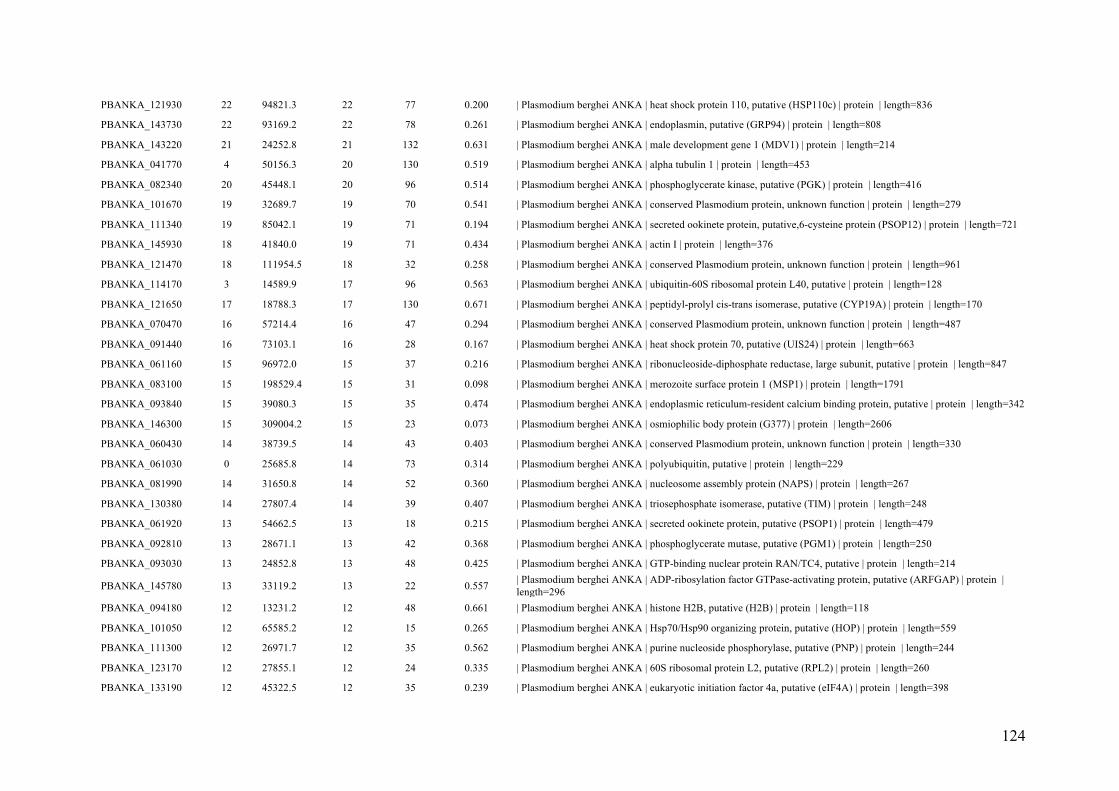
124
PBANKA_121930 22 94821.3 22 77 0.200 | Plasmodium berghei ANKA | heat shock protein 110, putative (HSP110c) | protein | length=836
PBANKA_143730 22 93169.2 22 78 0.261 | Plasmodium berghei ANKA | endoplasmin, putative (GRP94) | protein | length=808
PBANKA_143220 21 24252.8 21 132 0.631 | Plasmodium berghei ANKA | male development gene 1 (MDV1) | protein | length=214
PBANKA_041770 4 50156.3 20 130 0.519 | Plasmodium berghei ANKA | alpha tubulin 1 | protein | length=453
PBANKA_082340 20 45448.1 20 96 0.514 | Plasmodium berghei ANKA | phosphoglycerate kinase, putative (PGK) | protein | length=416
PBANKA_101670 19 32689.7 19 70 0.541 | Plasmodium berghei ANKA | conserved Plasmodium protein, unknown function | protein | length=279
PBANKA_111340 19 85042.1 19 71 0.194 | Plasmodium berghei ANKA | secreted ookinete protein, putative,6-cysteine protein (PSOP12) | protein | length=721
PBANKA_145930 18 41840.0 19 71 0.434 | Plasmodium berghei ANKA | actin I | protein | length=376
PBANKA_121470 18 111954.5 18 32 0.258 | Plasmodium berghei ANKA | conserved Plasmodium protein, unknown function | protein | length=961
PBANKA_114170 3 14589.9 17 96 0.563 | Plasmodium berghei ANKA | ubiquitin-60S ribosomal protein L40, putative | protein | length=128
PBANKA_121650 17 18788.3 17 130 0.671 | Plasmodium berghei ANKA | peptidyl-prolyl cis-trans isomerase, putative (CYP19A) | protein | length=170
PBANKA_070470 16 57214.4 16 47 0.294 | Plasmodium berghei ANKA | conserved Plasmodium protein, unknown function | protein | length=487
PBANKA_091440 16 73103.1 16 28 0.167 | Plasmodium berghei ANKA | heat shock protein 70, putative (UIS24) | protein | length=663
PBANKA_061160 15 96972.0 15 37 0.216 | Plasmodium berghei ANKA | ribonucleoside-diphosphate reductase, large subunit, putative | protein | length=847
PBANKA_083100 15 198529.4 15 31 0.098 | Plasmodium berghei ANKA | merozoite surface protein 1 (MSP1) | protein | length=1791
PBANKA_093840 15 39080.3 15 35 0.474 | Plasmodium berghei ANKA | endoplasmic reticulum-resident calcium binding protein, putative | protein | length=342
PBANKA_146300 15 309004.2 15 23 0.073 | Plasmodium berghei ANKA | osmiophilic body protein (G377) | protein | length=2606
PBANKA_060430 14 38739.5 14 43 0.403 | Plasmodium berghei ANKA | conserved Plasmodium protein, unknown function | protein | length=330
PBANKA_061030 0 25685.8 14 73 0.314 | Plasmodium berghei ANKA | polyubiquitin, putative | protein | length=229
PBANKA_081990 14 31650.8 14 52 0.360 | Plasmodium berghei ANKA | nucleosome assembly protein (NAPS) | protein | length=267
PBANKA_130380 14 27807.4 14 39 0.407 | Plasmodium berghei ANKA | triosephosphate isomerase, putative (TIM) | protein | length=248
PBANKA_061920 13 54662.5 13 18 0.215 | Plasmodium berghei ANKA | secreted ookinete protein, putative (PSOP1) | protein | length=479
PBANKA_092810 13 28671.1 13 42 0.368 | Plasmodium berghei ANKA | phosphoglycerate mutase, putative (PGM1) | protein | length=250
PBANKA_093030 13 24852.8 13 48 0.425 | Plasmodium berghei ANKA | GTP-binding nuclear protein RAN/TC4, putative | protein | length=214
PBANKA_145780 13 33119.2 13 22 0.557 | Plasmodium berghei ANKA | ADP-ribosylation factor GTPase-activating protein, putative (ARFGAP) | protein |
length=296
PBANKA_094180 12 13231.2 12 48 0.661 | Plasmodium berghei ANKA | histone H2B, putative (H2B) | protein | length=118
PBANKA_101050 12 65585.2 12 15 0.265 | Plasmodium berghei ANKA | Hsp70/Hsp90 organizing protein, putative (HOP) | protein | length=559
PBANKA_111300 12 26971.7 12 35 0.562 | Plasmodium berghei ANKA | purine nucleoside phosphorylase, putative (PNP) | protein | length=244
PBANKA_123170 12 27855.1 12 24 0.335 | Plasmodium berghei ANKA | 60S ribosomal protein L2, putative (RPL2) | protein | length=260
PBANKA_133190 12 45322.5 12 35 0.239 | Plasmodium berghei ANKA | eukaryotic initiation factor 4a, putative (eIF4A) | protein | length=398

125
PBANKA_142690 12 104135.3 12 29 0.127 | Plasmodium berghei ANKA | RNA helicase, putative | protein | length=909
PBANKA_100380 11 55775.4 11 23 0.338 | Plasmodium berghei ANKA | vacuolar ATP synthase subunit b, putative | protein | length=494
PBANKA_111770 11 34273.8 11 34 0.489 | Plasmodium berghei ANKA | malate dehydrogenase, putative (MDH) | protein | length=313
PBANKA_130280 11 21622.3 11 33 0.421 | Plasmodium berghei ANKA | thioredoxin peroxidase 1 (TPx1) | protein | length=195
PBANKA_135510 11 35429.9 11 57 0.326 | Plasmodium berghei ANKA | 40S ribosomal protein S6, putative | protein | length=310
PBANKA_030510 10 154804.4 10 16 0.076 | Plasmodium berghei ANKA | serine repeat antigen 1 (SERA1) | protein | length=1406
PBANKA_081420 10 29995.1 10 27 0.327 | Plasmodium berghei ANKA | elongation factor 1-beta, putative | protein | length=257
PBANKA_091790 9 50927.3 10 22 0.279 | Plasmodium berghei ANKA | 26S protease regulatory subunit 6a, putative (RPT5) | protein | length=451
PBANKA_091800 10 34820.2 10 18 0.378 | Plasmodium berghei ANKA | 60S acidic ribosomal protein P0, putative | protein | length=315
PBANKA_103440 10 50432.6 10 36 0.247 | Plasmodium berghei ANKA | plasmepsin IV (PM4) | protein | length=450
PBANKA_121770 10 49325.6 10 30 0.122 | Plasmodium berghei ANKA | ATP-dependent RNA helicase DDX6 (DOZI) | protein | length=433
PBANKA_132090 10 11612.9 10 26 0.548 | Plasmodium berghei ANKA | thioredoxin 1, putative (TRX1) | protein | length=104
PBANKA_140260 10 102620.0 10 13 0.071 | Plasmodium berghei ANKA | DNA ligase I, putative | protein | length=898
PBANKA_141040 10 68289.2 10 17 0.188 | Plasmodium berghei ANKA | vacuolar ATP synthase subunit a, putative | protein | length=611
PBANKA_144410 10 60779.0 10 17 0.164 | Plasmodium berghei ANKA | t-complex protein 1 gamma subunit, putative | protein | length=542
PBANKA_021150 9 28311.4 9 25 0.441 | Plasmodium berghei ANKA | proteasome subunit alpha type-5, putative | protein | length=256
PBANKA_051280 9 66720.1 9 14 0.094 | Plasmodium berghei ANKA | merozoite TRAP-like protein, putative (MTRAP) | protein | length=618
PBANKA_070390 9 35542.9 9 24 0.183 | Plasmodium berghei ANKA | receptor for activated c kinase, putative (RACK) | protein | length=322
PBANKA_093220 9 21179.1 9 27 0.438 | Plasmodium berghei ANKA | peptidyl-prolyl cis-trans isomerase, putative | protein | length=192
PBANKA_111520 9 132736.1 9 19 0.077 | Plasmodium berghei ANKA | conserved Plasmodium protein, unknown function | protein | length=1138
PBANKA_111700 8 14194.8 9 54 0.707 | Plasmodium berghei ANKA | histone H2A, putative (H2A) | protein | length=133
PBANKA_120060 9 250881.9 9 13 0.015 | Plasmodium berghei ANKA | Plasmodium exported protein, unknown function | protein | length=2206
PBANKA_142430 9 32548.1 9 29 0.387 | Plasmodium berghei ANKA | conserved Plasmodium protein, unknown function | protein | length=284
PBANKA_040540 8 15434.0 8 17 0.394 | Plasmodium berghei ANKA | 40S ribosomal protein S12, putative | protein | length=142
PBANKA_050580 8 29223.5 8 19 0.328 | Plasmodium berghei ANKA | deoxyribose-phosphate aldolase, putative | protein | length=262
PBANKA_090360 8 93443.4 8 13 0.098 | Plasmodium berghei ANKA | dynamin-like protein, putative (DYN1) | protein | length=814
PBANKA_093420 8 41217.1 8 28 0.440 | Plasmodium berghei ANKA | ubiquitin domain-containing protein DSK2, putative (DSK2) | protein | length=375
PBANKA_103270 8 58837.1 8 10 0.217 | Plasmodium berghei ANKA | RNA binding protein Bruno, putative (HoBo) | protein | length=507
PBANKA_103520 8 148090.1 8 10 0.049 | Plasmodium berghei ANKA | LCCL domain-containing protein (CCp3) | protein | length=1304
PBANKA_110670 8 46085.3 8 14 0.190 | Plasmodium berghei ANKA | 60S ribosomal protein L4, putative (RPL4) | protein | length=411

126
PBANKA_120440 8 11965.3 8 35 0.523 | Plasmodium berghei ANKA | DNA/RNA-binding protein Alba 3, putative (ALBA3) | protein | length=107
PBANKA_122900 8 126199.2 8 9 0.057 | Plasmodium berghei ANKA | Plasmodium exported protein, unknown function | protein | length=1095
PBANKA_123100 8 16046.7 8 15 0.523 | Plasmodium berghei ANKA | 40S ribosomal protein S11, putative | protein | length=151
PBANKA_136030 8 42121.4 8 49 0.174 | Plasmodium berghei ANKA | DNA/RNA-binding protein Alba 4, putative (ALBA4) | protein | length=374
PBANKA_145070 8 45627.5 8 13 0.171 | Plasmodium berghei ANKA | clustered-asparagine-rich protein, putative | protein | length=397
PBANKA_030490 7 123106.3 7 13 0.056 | Plasmodium berghei ANKA | serine repeat antigen 3 (SERA3) | protein | length=1096
PBANKA_030500 7 123889.8 7 12 0.078 | Plasmodium berghei ANKA | serine repeat antigen 2 (SERA2) | protein | length=1122
PBANKA_040470 7 12361.4 7 25 0.514 | Plasmodium berghei ANKA | glutaredoxin 1, putative (GRX1) | protein | length=109
PBANKA_060320 7 17524.7 7 36 0.398 | Plasmodium berghei ANKA | eukaryotic translation initiation factor 5A, putative (EIF5A) | protein | length=161
PBANKA_082170 7 55844.6 7 13 0.172 | Plasmodium berghei ANKA | inosine-5'-monophosphate dehydrogenase, putative | protein | length=507
PBANKA_091620 7 60078.6 7 18 0.147 | Plasmodium berghei ANKA | TCP-1/cpn60 chaperonin family, putative | protein | length=543
PBANKA_100950 7 336064.3 7 10 0.030 | Plasmodium berghei ANKA | NAD(P)H-dependent glutamate synthase, putative | protein | length=2962
PBANKA_103390 7 24959.3 7 18 0.211 | Plasmodium berghei ANKA | 40S ribosomal protein S8e, putative | protein | length=218
PBANKA_120720 7 27364.5 7 17 0.289 | Plasmodium berghei ANKA | adenylate kinase, putative (AK1) | protein | length=242
PBANKA_123210 7 36233.1 7 11 0.137 | Plasmodium berghei ANKA | pre-mRNA-splicing factor, putative (SR1) | protein | length=315
PBANKA_124180 7 107214.3 7 14 0.098 | Plasmodium berghei ANKA | DNA replication licensing factor MCM3, putative (MCM3) | protein | length=945
PBANKA_141030 7 123256.3 7 23 0.072 | Plasmodium berghei ANKA | M1-family alanyl aminopeptidase, putative | protein | length=1064
PBANKA_143105 7 10972.9 7 30 0.373 | Plasmodium berghei ANKA | 10 kDa chaperonin, putative (CPN10) | protein | length=102
PBANKA_145020 7 49575.4 7 19 0.122 | Plasmodium berghei ANKA | serine hydroxymethyltransferase, putative (SHMT) | protein | length=442
PBANKA_020160 6 18642.4 6 33 0.254 | Plasmodium berghei ANKA | early transcribed membrane protein (ETRAMP) | protein | length=177
PBANKA_030130 6 9784.8 6 30 0.582 | Plasmodium berghei ANKA | conserved Plasmodium protein, unknown function | protein | length=91
PBANKA_051190 6 44319.8 6 9 0.122 | Plasmodium berghei ANKA | 60S ribosomal protein L3, putative (RPL3) | protein | length=386
PBANKA_082670 6 25218.7 6 26 0.230 | Plasmodium berghei ANKA | conserved Plasmodium protein, unknown function | protein | length=222
PBANKA_093770 6 12700.3 6 26 0.504 | Plasmodium berghei ANKA | 60S ribosomal protein L36, putative (RPL36) | protein | length=113
PBANKA_093960 6 368279.4 6 14 0.015 | Plasmodium berghei ANKA | polyadenylate-binding protein-interacting protein 1, putative (PAIP1) | protein |
length=3296
PBANKA_101850 6 19905.6 6 17 0.339 | Plasmodium berghei ANKA | transcription factor 3b, putative | protein | length=174
PBANKA_101950 6 33843.2 6 26 0.215 | Plasmodium berghei ANKA | 60S ribosomal protein L5, putative | protein | length=293
PBANKA_103010 5 42609.2 6 10 0.098 | Plasmodium berghei ANKA | actin II | protein | length=376
PBANKA_111710 1 15413.6 6 14 0.279 | Plasmodium berghei ANKA | histone H3 variant, putative (H3.3) | protein | length=136
PBANKA_124440 6 9931.8 6 15 0.233 | Plasmodium berghei ANKA | conserved Plasmodium protein, unknown function | protein | length=86

127
PBANKA_130070 6 183560.8 6 10 0.051 | Plasmodium berghei ANKA | LCCL domain-containing protein (CCp1) | protein | length=1615
PBANKA_131080 6 29901.8 6 15 0.292 | Plasmodium berghei ANKA | 40S ribosomal protein S5, putative | protein | length=271
PBANKA_132930 6 24702.5 6 10 0.131 | Plasmodium berghei ANKA | 40S ribosomal protein S3, putative | protein | length=222
PBANKA_135440 6 21668.5 6 12 0.179 | Plasmodium berghei ANKA | 60S ribosomal protein L18, putative | protein | length=184
PBANKA_140960 6 59450.3 6 12 0.105 | Plasmodium berghei ANKA | conserved Plasmodium protein, unknown function | protein | length=484
PBANKA_141910 6 47783.5 6 13 0.135 | Plasmodium berghei ANKA | dihydrolipoyllysine-residue succinyltransferase component of 2-oxoglutarate
dehydrogenase complex, putative | protein | length=423
PBANKA_144680 6 79764.8 6 6 0.031 | Plasmodium berghei ANKA | 60 kDa chaperonin, putative (CPN60) | protein | length=707
PBANKA_010880 0 15418.5 5 13 0.279 | Plasmodium berghei ANKA | histone H3, putative | protein | length=136
PBANKA_020450 5 121466.2 5 12 0.025 | Plasmodium berghei ANKA | LCCL domain-containing protein (CCp5) | protein | length=1049
PBANKA_031000 5 38079.2 5 15 0.084 | Plasmodium berghei ANKA | heat shock 40 kDa protein, putative | protein | length=334
PBANKA_031100 5 156519.8 5 8 0.027 | Plasmodium berghei ANKA | protein transport protein SEC31, putative (SEC31) | protein | length=1388
PBANKA_031600 5 99152.9 5 9 0.040 | Plasmodium berghei ANKA | replication factor C subunit 1, putative | protein | length=861
PBANKA_040760 5 66318.9 5 6 0.095 | Plasmodium berghei ANKA | asparagine synthetase, putative | protein | length=581
PBANKA_050110 5 16759.0 5 8 0.156 | Plasmodium berghei ANKA | early transcribed membrane protein,small exported protein (SEP3) | protein | length=154
PBANKA_050360 5 11787.1 5 20 0.444 | Plasmodium berghei ANKA | 60S ribosomal protein L30e, putative | protein | length=108
PBANKA_052280 5 19994.8 5 11 0.244 | Plasmodium berghei ANKA | 40S ribosomal protein S19, putative (RPS19) | protein | length=172
PBANKA_060270 5 40275.8 5 12 0.197 | Plasmodium berghei ANKA | nucleosome assembly protein, putative (NAPL) | protein | length=345
PBANKA_082300 5 161341.7 5 8 0.044 | Plasmodium berghei ANKA | ubiquitin-like protein, putative | protein | length=1384
PBANKA_083300 5 19236.1 5 28 0.379 | Plasmodium berghei ANKA | profilin, putative (PFN) | protein | length=174
PBANKA_090500 5 9166.6 5 13 0.537 | Plasmodium berghei ANKA | 40S ribosomal protein S21, putative (RPS21) | protein | length=82
PBANKA_091910 5 44649.8 5 8 0.191 | Plasmodium berghei ANKA | parasitophorous vacuolar protein 1, putative (PV1) | protein | length=388
PBANKA_092130 5 19697.1 5 29 0.531 | Plasmodium berghei ANKA | deoxyuridine 5'-triphosphate nucleotidohydrolase, putative | protein | length=175
PBANKA_093110 5 24745.5 5 11 0.269 | Plasmodium berghei ANKA | conserved Plasmodium protein, unknown function | protein | length=219
PBANKA_094150 5 29807.4 5 8 0.176 | Plasmodium berghei ANKA | 40S ribosomal protein S4, putative | protein | length=261
PBANKA_100880 5 68014.1 5 6 0.084 | Plasmodium berghei ANKA | glucose-6-phosphate isomerase, putative | protein | length=586
PBANKA_110220 5 46689.2 5 10 0.181 | Plasmodium berghei ANKA | merozoite surface protein 8 (MSP8) | protein | length=409
PBANKA_112620 5 20675.9 5 10 0.185 | Plasmodium berghei ANKA | protein DJ-1, putative (DJ1) | protein | length=189
PBANKA_113570 5 57133.8 5 16 0.221 | Plasmodium berghei ANKA | RNA-binding protein, putative | protein | length=511
PBANKA_113780 5 291206.2 5 6 0.010 | Plasmodium berghei ANKA | conserved Plasmodium protein, unknown function | protein | length=2605
PBANKA_114280 5 72969.3 5 15 0.126 | Plasmodium berghei ANKA | conserved Plasmodium protein, unknown function | protein | length=634

128
PBANKA_121080 5 26490.3 5 13 0.117 | Plasmodium berghei ANKA | hypoxanthine-guanine phosphoribosyltransferase, putative (HGPRT) | protein |
length=231
PBANKA_121850 5 16636.2 5 18 0.215 | Plasmodium berghei ANKA | protein phosphatase inhibitor 2, putative | protein | length=144
PBANKA_122800 5 25359.9 5 8 0.168 | Plasmodium berghei ANKA | 1-cys peroxiredoxin, putative | protein | length=220
PBANKA_123560 5 53623.2 5 7 0.081 | Plasmodium berghei ANKA | adenosylhomocysteinase, putative (SAHH) | protein | length=479
PBANKA_124230 5 47637.9 5 20 0.109 | Plasmodium berghei ANKA | Hsc70-interacting protein,58 kDa phosphoprotein (HIP) | protein | length=423
PBANKA_131740 5 28079.7 5 5 0.082 | Plasmodium berghei ANKA | co-chaperone p23, putative | protein | length=255
PBANKA_135860 5 53688.7 5 10 0.075 | Plasmodium berghei ANKA | isocitrate dehydrogenase (NADP), mitochondrial precursor, putative (IDH) | protein |
length=469
PBANKA_136290 5 67091.5 5 10 0.038 | Plasmodium berghei ANKA | lysine--tRNA ligase, putative (KRS1) | protein | length=578
PBANKA_142350 5 25385.0 5 12 0.214 | Plasmodium berghei ANKA | 60S ribosomal protein L13-2, putative | protein | length=215
PBANKA_143080 5 24693.7 5 17 0.391 | Plasmodium berghei ANKA | thioredoxin peroxidase 2, putative (Trx-Px2) | protein | length=215
PBANKA_143800 5 49622.8 5 18 0.110 | Plasmodium berghei ANKA | CAMP-dependent protein kinase regulatory subunit, putative (PKAr) | protein |
length=436
PBANKA_010330 4 234947.2 4 5 0.023 | Plasmodium berghei ANKA | conserved Plasmodium protein, unknown function | protein | length=2033
PBANKA_010740 4 46275.8 4 7 0.099 | Plasmodium berghei ANKA | ornithine aminotransferase, putative (OAT) | protein | length=414
PBANKA_010910 4 75446.0 4 5 0.054 | Plasmodium berghei ANKA | transketolase, putative (TK) | protein | length=670
PBANKA_030680 4 52978.9 4 5 0.137 | Plasmodium berghei ANKA | ATP-dependent RNA helicase UAP56, putative (UAP56) | protein | length=466
PBANKA_031090 4 60751.7 4 4 0.030 | Plasmodium berghei ANKA | T-complex protein 1, putative | protein | length=543
PBANKA_040480 4 71075.5 4 9 0.065 | Plasmodium berghei ANKA | FAD-dependent glycerol-3-phosphate dehydrogenase, putative | protein | length=627
PBANKA_040770 4 11778.8 4 9 0.523 | Plasmodium berghei ANKA | 60S acidic ribosomal protein P2, putative | protein | length=111
PBANKA_041460 4 14900.1 4 4 0.215 | Plasmodium berghei ANKA | 40S ribosomal protein S15A, putative | protein | length=130
PBANKA_041610 4 684398.2 4 5 0.009 | Plasmodium berghei ANKA | dynein heavy chain, putative | protein | length=5834
PBANKA_051990 4 180209.2 4 4 0.043 | Plasmodium berghei ANKA | conserved Plasmodium protein, unknown function | protein | length=1591
PBANKA_052420 4 16719.9 4 7 0.155 | Plasmodium berghei ANKA | early transcribed membrane protein,small exported protein (SEP2) | protein | length=155
PBANKA_061090 4 48368.5 4 7 0.146 | Plasmodium berghei ANKA | HSP40, subfamily A, putative | protein | length=424
PBANKA_061520 4 60533.3 4 4 0.063 | Plasmodium berghei ANKA | calcium-dependent protein kinase 4 (CDPK4) | protein | length=528
PBANKA_061990 4 45274.4 4 4 0.069 | Plasmodium berghei ANKA | GTP-binding protein, putative | protein | length=393
PBANKA_081130 4 134587.0 4 4 0.009 | Plasmodium berghei ANKA | Ran-binding protein, putative | protein | length=1160
PBANKA_090640 4 16455.0 4 6 0.189 | Plasmodium berghei ANKA | 60S ribosomal protein L35ae, putative | protein | length=143
PBANKA_092990 4 92140.7 4 4 0.045 | Plasmodium berghei ANKA | heat shock protein 90, putative | protein | length=802
PBANKA_093720.1 3 144166.2 4 10 0.015 | Plasmodium berghei ANKA | asparagine-rich antigen, putative | protein | length=1242

129
PBANKA_093850 4 35487.3 4 10 0.137 | Plasmodium berghei ANKA | succinyl-CoA synthetase alpha subunit, putative | protein | length=329
PBANKA_101320 4 51484.5 4 11 0.094 | Plasmodium berghei ANKA | ATP-specific succinyl-CoA synthetase beta subunit, putative | protein | length=458
PBANKA_102230 4 47030.7 4 19 0.116 | Plasmodium berghei ANKA | mannose-1-phosphate guanyltransferase, putative | protein | length=414
PBANKA_103880 4 24598.7 4 9 0.052 | Plasmodium berghei ANKA | conserved Plasmodium protein, unknown function | protein | length=210
PBANKA_110420 4 41566.4 4 4 0.194 | Plasmodium berghei ANKA | 2-oxoisovalerate dehydrogenase subunit beta, mitochondrial, putative (BCKDHB) |
protein | length=372
PBANKA_111750 4 16808.1 4 5 0.189 | Plasmodium berghei ANKA | ribosomal protein L27a, putative | protein | length=148
PBANKA_112070 4 20789.3 4 16 0.157 | Plasmodium berghei ANKA | nascent polypeptide associated complex alpha chain, putative | protein | length=185
PBANKA_113790 4 30637.6 4 16 0.215 | Plasmodium berghei ANKA | proliferating cell nuclear antigen, putative (PCNA) | protein | length=274
PBANKA_114580 4 20338.1 4 9 0.251 | Plasmodium berghei ANKA | conserved rodent malaria protein, unknown function | protein | length=179
PBANKA_120500 4 27807.9 4 8 0.185 | Plasmodium berghei ANKA | RNA-binding protein, putative | protein | length=233
PBANKA_120610 4 62149.9 4 7 0.116 | Plasmodium berghei ANKA | eukaryotic translation initiation factor 3 subunit 7, putative | protein | length=537
PBANKA_121090 4 68099.0 4 7 0.084 | Plasmodium berghei ANKA | phosphoglucomutase, putative | protein | length=593
PBANKA_121560 4 29947.3 4 18 0.046 | Plasmodium berghei ANKA | 40S ribosomal protein S3A, putative | protein | length=262
PBANKA_122120 4 18839.7 4 7 0.152 | Plasmodium berghei ANKA | 60S ribosomal protein L34, putative (RPL34) | protein | length=164
PBANKA_123880 4 127725.3 4 8 0.060 | Plasmodium berghei ANKA | karyopherin beta, putative | protein | length=1126
PBANKA_132500 4 7487.1 4 23 0.627 | Plasmodium berghei ANKA | 40S ribosomal protein S28e, putative | protein | length=67
PBANKA_132970 4 137982.7 4 7 0.040 | Plasmodium berghei ANKA | 26S proteasome regulatory subunit RPN2, putative (RPN2) | protein | length=1219
PBANKA_133830 4 21614.9 4 8 0.290 | Plasmodium berghei ANKA | 60S ribosomal protein L6, putative | protein | length=190
PBANKA_133840 4 14023.5 4 13 0.422 | Plasmodium berghei ANKA | vacuolar ATP synthase subunit g, putative | protein | length=121
PBANKA_133860 4 20660.5 4 8 0.182 | Plasmodium berghei ANKA | 60S ribosomal protein L23, putative (RPL23) | protein | length=181
PBANKA_134560 4 53679.0 4 4 0.023 | Plasmodium berghei ANKA | elongation factor Tu, putative | protein | length=477
PBANKA_134670 4 14947.0 4 4 0.281 | Plasmodium berghei ANKA | 60S ribosomal protein L23, putative | protein | length=139
PBANKA_135720 4 106076.6 4 6 0.029 | Plasmodium berghei ANKA | heat shock protein 110, putative (HSP110) | protein | length=917
PBANKA_010070 3 36115.2 3 11 0.190 | Plasmodium berghei ANKA | Plasmodium exported protein, unknown function | protein | length=321
PBANKA_010080 3 157332.9 3 7 0.016 | Plasmodium berghei ANKA | conserved Plasmodium protein, unknown function | protein | length=1382
PBANKA_010390 3 81987.8 3 4 0.029 | Plasmodium berghei ANKA | RNA-binding protein, putative | protein | length=735
PBANKA_010710 3 26410.5 3 5 0.089 | Plasmodium berghei ANKA | proteasome subunit alpha type-2, putative | protein | length=235
PBANKA_021550 1 45147.6 3 6 0.031 | Plasmodium berghei ANKA | erythrocyte membrane associated protein 2 (EMAP2) | protein | length=387
PBANKA_030860 3 66393.6 3 5 0.064 | Plasmodium berghei ANKA | asparagine--tRNA ligase, putative | protein | length=575
PBANKA_031380 3 61305.4 3 5 0.055 | Plasmodium berghei ANKA | ATP synthase F1, alpha subunit, putative | protein | length=548

130
PBANKA_040180 3 126401.7 3 3 0.037 | Plasmodium berghei ANKA | HAD superfamily protein, putative | protein | length=1093
PBANKA_040230 3 50061.9 3 5 0.029 | Plasmodium berghei ANKA | lipoamide acyltransferase component of branched-chain alpha-keto acid dehydrogenase
complex, putative (BCKDH-E2) | protein | length=443
PBANKA_040530 3 16087.9 3 5 0.248 | Plasmodium berghei ANKA | 40S ribosomal protein S23, putative | protein | length=145
PBANKA_041600 3 104988.7 3 3 0.021 | Plasmodium berghei ANKA | high molecular weight rhoptry protein 3, putative (RhopH3) | protein | length=908
PBANKA_041820 3 23684.9 3 11 0.121 | Plasmodium berghei ANKA | ras-related protein RAB7, putative (RAB7) | protein | length=206
PBANKA_060160 3 201781.0 3 4 0.009 | Plasmodium berghei ANKA | conserved protein, unknown function | protein | length=1738
PBANKA_060190 3 12379.5 3 12 0.238 | Plasmodium berghei ANKA | high mobility group protein B1, putative (HMGB1) | protein | length=105
PBANKA_060760 3 12067.3 3 15 0.324 | Plasmodium berghei ANKA | U6 snRNA-associated Sm-like protein LSm7, putative (LSM7) | protein | length=108
PBANKA_061020 3 85475.5 3 3 0.041 | Plasmodium berghei ANKA | DNA replication licensing factor MCM5, putative (MCM5) | protein | length=758
PBANKA_061040 3 165465.4 3 3 0.016 | Plasmodium berghei ANKA | ATPase, putative | protein | length=1472
PBANKA_071910 3 33470.0 3 4 0.203 | Plasmodium berghei ANKA | conserved Plasmodium protein, unknown function | protein | length=281
PBANKA_081050 3 15508.3 3 4 0.130 | Plasmodium berghei ANKA | apoptosis-related protein, putative (ARP) | protein | length=131
PBANKA_081300 3 39931.8 3 5 0.124 | Plasmodium berghei ANKA | inhibitor of cysteine proteases (ICP) | protein | length=354
PBANKA_081640 3 146654.8 3 4 0.040 | Plasmodium berghei ANKA | 6-phosphofructokinase, putative (PFK9) | protein | length=1299
PBANKA_083000 3 32349.5 3 5 0.097 | Plasmodium berghei ANKA | RNA-binding protein, putative | protein | length=279
PBANKA_090270 3 35928.2 3 3 0.095 | Plasmodium berghei ANKA | leucine-rich repeat protein (LRR10) | protein | length=304
PBANKA_090650 3 14555.0 3 7 0.198 | Plasmodium berghei ANKA | 60S ribosomal protein L28, putative | protein | length=126
PBANKA_090880 3 36997.4 3 8 0.070 | Plasmodium berghei ANKA | conserved Plasmodium protein, unknown function | protein | length=315
PBANKA_091020 3 89049.9 3 3 0.058 | Plasmodium berghei ANKA | protein phosphatase 2C, putative | protein | length=787
PBANKA_091230 3 117761.9 3 4 0.048 | Plasmodium berghei ANKA | tudor staphylococcal nuclease, putative (TSN) | protein | length=1013
PBANKA_092000 3 15597.6 3 3 0.243 | Plasmodium berghei ANKA | multiprotein bridging factor type 1, putative | protein | length=136
PBANKA_092080 3 35198.9 3 4 0.100 | Plasmodium berghei ANKA | CRAL/TRIO domain-containing protein, putative | protein | length=299
PBANKA_092210 3 17852.7 3 5 0.173 | Plasmodium berghei ANKA | 40S ribosomal protein S18, putative | protein | length=156
PBANKA_092540 3 604152.0 3 3 0.002 | Plasmodium berghei ANKA | dynein heavy chain, putative | protein | length=5176
PBANKA_092670 3 17914.5 3 13 0.223 | Plasmodium berghei ANKA | exported protein 1, putative,circumsporozoite-related antigen (EXP1) | protein |
length=166
PBANKA_093130 3 78627.4 3 10 0.073 | Plasmodium berghei ANKA | dipeptidyl aminopeptidase 1, putative (DPAP1) | protein | length=681
PBANKA_094250 3 49016.5 3 6 0.043 | Plasmodium berghei ANKA | conserved Plasmodium protein, unknown function | protein | length=420
PBANKA_100230 3 471911.6 3 3 0.003 | Plasmodium berghei ANKA | conserved Plasmodium protein, unknown function | protein | length=3974
PBANKA_100850 3 101867.1 3 3 0.039 | Plasmodium berghei ANKA | translocon component PTEX150 (PTEX150) | protein | length=919

131
PBANKA_101940 3 33244.8 3 6 0.078 | Plasmodium berghei ANKA | 60S ribosomal protein L7-3, putative | protein | length=284
PBANKA_102200 3 11770.6 3 9 0.267 | Plasmodium berghei ANKA | 40S ribosomal protein S25, putative | protein | length=105
PBANKA_102840 3 25212.2 3 3 0.142 | Plasmodium berghei ANKA | 60S ribosomal protein L10, putative | protein | length=219
PBANKA_103210 3 70624.1 3 4 0.084 | Plasmodium berghei ANKA | rhoptry-associated protein 1, putative (RAP1) | protein | length=604
PBANKA_103230 3 85799.8 3 5 0.036 | Plasmodium berghei ANKA | cytidine triphosphate synthetase, putative | protein | length=756
PBANKA_103800 3 12998.6 3 4 0.259 | Plasmodium berghei ANKA | cytochrome c, putative | protein | length=116
PBANKA_110290 3 101475.1 3 6 0.037 | Plasmodium berghei ANKA | conserved Plasmodium protein, unknown function | protein | length=869
PBANKA_110310 3 13937.0 3 6 0.180 | Plasmodium berghei ANKA | actin-depolymerizing factor 1 (ADF1) | protein | length=122
PBANKA_111540 3 9689.2 3 9 0.417 | Plasmodium berghei ANKA | BolA-like protein, putative | protein | length=84
PBANKA_112290 3 55405.6 3 7 0.113 | Plasmodium berghei ANKA | hexokinase, putative (HK) | protein | length=494
PBANKA_112430 3 43531.9 3 3 0.043 | Plasmodium berghei ANKA | conserved Plasmodium protein, unknown function | protein | length=372
PBANKA_113410 3 57883.4 3 7 0.047 | Plasmodium berghei ANKA | TCP-1/cpn60 chaperonin, putative | protein | length=529
PBANKA_113700 3 133431.5 3 5 0.022 | Plasmodium berghei ANKA | bergheilysin (BLN) | protein | length=1149
PBANKA_114240 3 17094.9 3 3 0.262 | Plasmodium berghei ANKA | nucleoside diphosphate kinase b, putative | protein | length=149
PBANKA_114480 3 42374.0 3 8 0.141 | Plasmodium berghei ANKA | conserved Plasmodium protein, unknown function | protein | length=361
PBANKA_114590 3 31722.2 3 6 0.119 | Plasmodium berghei ANKA | conserved rodent malaria protein, unknown function | protein | length=278
PBANKA_120080 3 30158.9 3 8 0.076 | Plasmodium berghei ANKA | RNA-binding protein, putative | protein | length=249
PBANKA_120220 3 138038.1 3 4 0.018 | Plasmodium berghei ANKA | U5 small nuclear ribonuclear protein, putative | protein | length=1193
PBANKA_120660 3 49660.3 3 3 0.101 | Plasmodium berghei ANKA | 26S protease regulatory subunit 4, putative | protein | length=447
PBANKA_121820 3 59128.2 3 7 0.049 | Plasmodium berghei ANKA | T-complex protein 1 epsilon subunit, putative | protein | length=535
PBANKA_123180 3 18184.7 3 8 0.170 | Plasmodium berghei ANKA | 60S ribosomal protein L12, putative | protein | length=165
PBANKA_123220 3 127467.2 3 7 0.035 | Plasmodium berghei ANKA | FACT complex subunit SPT16 (FACT-L) | protein | length=1096
PBANKA_123780 3 160818.7 3 5 0.017 | Plasmodium berghei ANKA | multidrug resistance protein, putative (MDR1) | protein | length=1419
PBANKA_130990 3 72029.8 3 5 0.027 | Plasmodium berghei ANKA | M17 leucyl aminopeptidase, putative | protein | length=635
PBANKA_131040 3 19338.5 3 8 0.316 | Plasmodium berghei ANKA | centrin-2, putative (CEN2) | protein | length=168
PBANKA_131270 3 28760.4 3 8 0.080 | Plasmodium berghei ANKA | gamete egress and sporozoite traversal protein (GEST) | protein | length=249
PBANKA_131810 3 89819.7 3 4 0.070 | Plasmodium berghei ANKA | aminopeptidase P, putative (APP) | protein | length=776
PBANKA_131840 3 53305.2 3 3 0.030 | Plasmodium berghei ANKA | 6-phosphogluconate dehydrogenase, decarboxylating, putative | protein | length=472
PBANKA_132010 3 93268.5 3 4 0.075 | Plasmodium berghei ANKA | conserved Plasmodium protein, unknown function (UIS25) | protein | length=789
PBANKA_134580 3 55898.3 3 3 0.020 | Plasmodium berghei ANKA | conserved Plasmodium protein, unknown function | protein | length=499

132
PBANKA_135920 3 23532.0 3 4 0.178 | Plasmodium berghei ANKA | DNA/RNA-binding protein Alba 2, putative (ALBA2) | protein | length=202
PBANKA_135970 3 48879.1 3 8 0.037 | Plasmodium berghei ANKA | 6-cysteine protein (P47) | protein | length=431
PBANKA_136550 3 37365.3 3 16 0.083 | Plasmodium berghei ANKA | intra-erythrocytic P. berghei-induced structures protein 1 (IBIS1) | protein | length=327
PBANKA_140490 3 44674.5 3 6 0.109 | Plasmodium berghei ANKA | 26S protease regulatory subunit 10B, putative (RPT4) | protein | length=393
PBANKA_141130 3 117937.0 3 3 0.043 | Plasmodium berghei ANKA | eukaryotic translation initation factor 4 gamma, putative (EIF4G) | protein | length=1013
PBANKA_141630 3 16847.3 3 16 0.283 | Plasmodium berghei ANKA | 40S ribosomal protein S19, putative (RPS19) | protein | length=145
PBANKA_142270 3 22536.2 3 7 0.293 | Plasmodium berghei ANKA | superoxide dismutase [Fe], putative (FeSOD) | protein | length=198
PBANKA_144690 3 57729.4 3 5 0.021 | Plasmodium berghei ANKA | dihydrolipoyl dehydrogenase, mitochondrial (LPD1) | protein | length=516
PBANKA_145610 3 16049.7 3 13 0.190 | Plasmodium berghei ANKA | 40S ribosomal protein S17, putative | protein | length=137
PBANKA_145690 3 12813.7 3 5 0.391 | Plasmodium berghei ANKA | translation initiation factor SUI1, putative | protein | length=115
PBANKA_020270 2 168693.5 2 3 0.023 | Plasmodium berghei ANKA | kinesin-8, putative | protein | length=1460
PBANKA_020630 2 19572.8 2 2 0.202 | Plasmodium berghei ANKA | centrin-1, putative (CEN1) | protein | length=168
PBANKA_030360 2 110614.3 2 2 0.017 | Plasmodium berghei ANKA | 26S proteasome regulatory subunit RPN1, putative (RPN1) | protein | length=993
PBANKA_030780 2 32091.5 2 2 0.060 | Plasmodium berghei ANKA | conserved Plasmodium protein, unknown function | protein | length=282
PBANKA_031680 0 45147.6 2 5 0.031 | Plasmodium berghei ANKA | Plasmodium exported protein, unknown function | protein | length=387
PBANKA_040110 2 153538.2 2 4 0.006 | Plasmodium berghei ANKA | serine/threonine protein kinase, putative (SRPK1) | protein | length=1306
PBANKA_040190 2 17727.4 2 9 0.178 | Plasmodium berghei ANKA | DNA-directed RNA polymerase subunit I, putative | protein | length=152
PBANKA_040420 2 15584.7 2 2 0.158 | Plasmodium berghei ANKA | ubiquitin-conjugating enzyme E2, putative | protein | length=139
PBANKA_040620 2 430381.7 2 3 0.004 | Plasmodium berghei ANKA | conserved Plasmodium protein, unknown function | protein | length=3610
PBANKA_040650 2 59569.9 2 3 0.048 | Plasmodium berghei ANKA | TCP-1/cpn60 chaperonin, putative | protein | length=539
PBANKA_041750 2 15495.6 2 2 0.221 | Plasmodium berghei ANKA | 60S ribosomal protein L32, putative (RPL32) | protein | length=131
PBANKA_050320 2 246699.5 2 3 0.010 | Plasmodium berghei ANKA | eukaryotic translation initiation factor subunit eIF2A, putative | protein | length=2110
PBANKA_050510 2 20830.8 2 2 0.105 | Plasmodium berghei ANKA | ADP-ribosylation factor, putative (ARF1) | protein | length=181
PBANKA_050690 2 50258.7 2 2 0.079 | Plasmodium berghei ANKA | phospholipid scramblase, putative | protein | length=431
PBANKA_051270 2 16985.7 2 2 0.146 | Plasmodium berghei ANKA | conserved Plasmodium protein, unknown function | protein | length=151
PBANKA_051360 2 41944.6 2 9 0.074 | Plasmodium berghei ANKA | adenosine deaminase, putative (ADA) | protein | length=363
PBANKA_051720 2 33509.2 2 9 0.068 | Plasmodium berghei ANKA | haloacid dehalogenase-like hydrolase, putative (HAD1) | protein | length=296
PBANKA_051870 2 104617.0 2 4 0.020 | Plasmodium berghei ANKA | methionine--tRNA ligase, putative | protein | length=898
PBANKA_060420 2 133016.3 2 2 0.028 | Plasmodium berghei ANKA | zinc finger protein, putative | protein | length=1165
PBANKA_061400 2 55657.5 2 2 0.052 | Plasmodium berghei ANKA | conserved Plasmodium protein, unknown function | protein | length=478
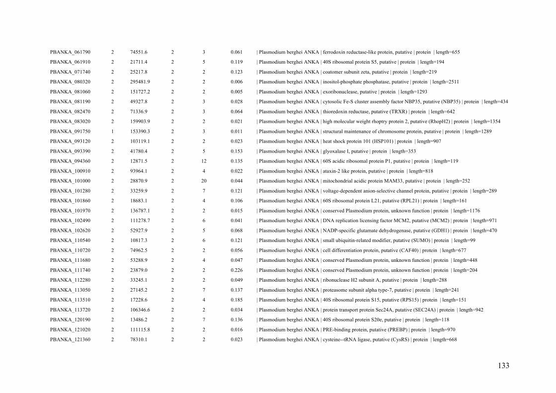
133
PBANKA_061790 2 74551.6 2 3 0.061 | Plasmodium berghei ANKA | ferrodoxin reductase-like protein, putative | protein | length=655
PBANKA_061910 2 21711.4 2 5 0.119 | Plasmodium berghei ANKA | 40S ribosomal protein S5, putative | protein | length=194
PBANKA_071740 2 25217.8 2 2 0.123 | Plasmodium berghei ANKA | coatomer subunit zeta, putative | protein | length=219
PBANKA_080320 2 295481.9 2 2 0.006 | Plasmodium berghei ANKA | inositol-phosphate phosphatase, putative | protein | length=2511
PBANKA_081060 2 151727.2 2 2 0.005 | Plasmodium berghei ANKA | exoribonuclease, putative | protein | length=1293
PBANKA_081190 2 49327.8 2 3 0.028 | Plasmodium berghei ANKA | cytosolic Fe-S cluster assembly factor NBP35, putative (NBP35) | protein | length=434
PBANKA_082470 2 71336.9 2 3 0.064 | Plasmodium berghei ANKA | thioredoxin reductase, putative (TRXR) | protein | length=642
PBANKA_083020 2 159903.9 2 2 0.021 | Plasmodium berghei ANKA | high molecular weight rhoptry protein 2, putative (RhopH2) | protein | length=1354
PBANKA_091750 1 153390.3 2 3 0.011 | Plasmodium berghei ANKA | structural maintenance of chromosome protein, putative | protein | length=1289
PBANKA_093120 2 103119.1 2 2 0.023 | Plasmodium berghei ANKA | heat shock protein 101 (HSP101) | protein | length=907
PBANKA_093390 2 41780.4 2 5 0.153 | Plasmodium berghei ANKA | glyoxalase I, putative | protein | length=353
PBANKA_094360 2 12871.5 2 12 0.135 | Plasmodium berghei ANKA | 60S acidic ribosomal protein P1, putative | protein | length=119
PBANKA_100910 2 93964.1 2 4 0.022 | Plasmodium berghei ANKA | ataxin-2 like protein, putative | protein | length=818
PBANKA_101000 2 28870.9 2 20 0.044 | Plasmodium berghei ANKA | mitochondrial acidic protein MAM33, putative | protein | length=252
PBANKA_101280 2 33259.9 2 7 0.121 | Plasmodium berghei ANKA | voltage-dependent anion-selective channel protein, putative | protein | length=289
PBANKA_101860 2 18683.1 2 4 0.106 | Plasmodium berghei ANKA | 60S ribosomal protein L21, putative (RPL21) | protein | length=161
PBANKA_101970 2 136787.1 2 2 0.015 | Plasmodium berghei ANKA | conserved Plasmodium protein, unknown function | protein | length=1176
PBANKA_102490 2 111278.7 2 6 0.041 | Plasmodium berghei ANKA | DNA replication licensing factor MCM2, putative (MCM2) | protein | length=971
PBANKA_102620 2 52927.9 2 5 0.068 | Plasmodium berghei ANKA | NADP-specific glutamate dehydrogenase, putative (GDH1) | protein | length=470
PBANKA_110540 2 10817.3 2 6 0.121 | Plasmodium berghei ANKA | small ubiquitin-related modifier, putative (SUMO) | protein | length=99
PBANKA_110720 2 74962.5 2 2 0.056 | Plasmodium berghei ANKA | cell differentiation protein, putative (CAF40) | protein | length=677
PBANKA_111680 2 53288.9 2 4 0.047 | Plasmodium berghei ANKA | conserved Plasmodium protein, unknown function | protein | length=448
PBANKA_111740 2 23879.0 2 2 0.226 | Plasmodium berghei ANKA | conserved Plasmodium protein, unknown function | protein | length=204
PBANKA_112280 2 33245.1 2 2 0.049 | Plasmodium berghei ANKA | ribonuclease H2 subunit A, putative | protein | length=288
PBANKA_113050 2 27145.2 2 7 0.137 | Plasmodium berghei ANKA | proteasome subunit alpha type-7, putative | protein | length=241
PBANKA_113510 2 17228.6 2 4 0.185 | Plasmodium berghei ANKA | 40S ribosomal protein S15, putative (RPS15) | protein | length=151
PBANKA_113720 2 106346.6 2 2 0.034 | Plasmodium berghei ANKA | protein transport protein Sec24A, putative (SEC24A) | protein | length=942
PBANKA_120190 2 13486.2 2 7 0.136 | Plasmodium berghei ANKA | 40S ribosomal protein S20e, putative | protein | length=118
PBANKA_121020 2 111115.8 2 2 0.016 | Plasmodium berghei ANKA | PRE-binding protein, putative (PREBP) | protein | length=970
PBANKA_121360 2 78310.1 2 2 0.023 | Plasmodium berghei ANKA | cysteine--tRNA ligase, putative (CysRS) | protein | length=668

134
PBANKA_121760 0 16465.4 2 22 0.146 | Plasmodium berghei ANKA | histone H2A variant, putative (H2A.Z) | protein | length=158
PBANKA_123420 2 15244.7 2 2 0.068 | Plasmodium berghei ANKA | 40S ribosomal protein S24, putative (RPS24) | protein | length=133
PBANKA_123480 2 22114.2 2 3 0.159 | Plasmodium berghei ANKA | 40S ribosomal protein S9, putative | protein | length=189
PBANKA_124390 2 176304.0 2 2 0.010 | Plasmodium berghei ANKA | conserved Plasmodium protein, unknown function | protein | length=1522
PBANKA_130140 2 28858.4 2 2 0.134 | Plasmodium berghei ANKA | proteasome subunit alpha type-1, putative | protein | length=253
PBANKA_130510 2 24785.8 2 2 0.124 | Plasmodium berghei ANKA | 60S ribosomal protein L10a, putative | protein | length=217
PBANKA_131750 2 109618.9 2 2 0.013 | Plasmodium berghei ANKA | glucose-6-phosphate dehydrogenase-6-phosphogluconolactonase (G6PD-6PGL) | protein
| length=950
PBANKA_132530 2 77547.0 2 4 0.031 | Plasmodium berghei ANKA | splicing factor 3B subunit 2, putative (SF3B2) | protein | length=671
PBANKA_132690 2 63597.6 2 5 0.019 | Plasmodium berghei ANKA | DNA polymerase alpha subunit, putative | protein | length=541
PBANKA_132810 2 44527.4 2 3 0.042 | Plasmodium berghei ANKA | ATP synthase (C/AC39) subunit, putative | protein | length=382
PBANKA_134030 2 48167.0 2 2 0.042 | Plasmodium berghei ANKA | phosphoribosylpyrophosphate synthetase, putative | protein | length=430
PBANKA_134430 2 58820.6 2 3 0.039 | Plasmodium berghei ANKA | chromatin assembly factor 1 subunit, putative | protein | length=513
PBANKA_134910 2 37664.1 2 2 0.048 | Plasmodium berghei ANKA | merozoite surface protein 7 (MSP7) | protein | length=335
PBANKA_135630 2 110869.0 2 4 0.024 | Plasmodium berghei ANKA | conserved Plasmodium protein, unknown function | protein | length=955
PBANKA_135890 2 85165.0 2 2 0.019 | Plasmodium berghei ANKA | dynactin subunit 2, putative | protein | length=728
PBANKA_140130 2 22449.6 2 12 0.129 | Plasmodium berghei ANKA | 40S ribosomal protein S7, putative | protein | length=194
PBANKA_140680 2 9176.6 2 4 0.171 | Plasmodium berghei ANKA | 40S ribosomal protein S27, putative (RPS27) | protein | length=82
PBANKA_140760 2 18920.4 2 3 0.086 | Plasmodium berghei ANKA | 60S ribosomal protein L24, putative | protein | length=163
PBANKA_142060 2 13722.5 2 9 0.179 | Plasmodium berghei ANKA | histone H2B variant, putative (H2B.Z) | protein | length=123
PBANKA_142420 2 77278.8 2 7 0.021 | Plasmodium berghei ANKA | conserved Plasmodium protein, unknown function | protein | length=661
PBANKA_143470 2 229999.6 2 3 0.011 | Plasmodium berghei ANKA | clathrin heavy chain, putative | protein | length=1986
PBANKA_144110 2 32735.1 2 8 0.052 | Plasmodium berghei ANKA | haloacid dehalogenase-like hydrolase, putative (HAD2) | protein | length=288
PBANKA_145330 2 86929.9 2 3 0.024 | Plasmodium berghei ANKA | acyl-CoA synthetase, putative | protein | length=754
PBANKA_145480 2 212593.3 2 2 0.012 | Plasmodium berghei ANKA | RNA-binding protein, putative | protein | length=1854
PBANKA_145510 2 37629.5 2 4 0.060 | Plasmodium berghei ANKA | replication factor C subunit 4, putative | protein | length=336
PBANKA_145830 2 117090.9 2 2 0.026 | Plasmodium berghei ANKA | kinesin-13, putative | protein | length=1025
PBANKA_000220 0 119648.9 1 2 0.018 | Plasmodium berghei ANKA | reticulocyte binding protein, putative (Pb235) | protein | length=1025
PBANKA_010100 1 30700.2 1 1 0.053 | Plasmodium berghei ANKA | MYND finger protein, putative | protein | length=262
PBANKA_020134 1 12965.4 1 7 0.110 | Plasmodium berghei ANKA | fam-c protein | protein | length=109
PBANKA_020500 1 19873.8 1 1 0.116 | Plasmodium berghei ANKA | conserved Plasmodium protein, unknown function | protein | length=172

135
PBANKA_020930 1 42993.9 1 3 0.055 | Plasmodium berghei ANKA | actin-related protein (ARP1) | protein | length=384
PBANKA_030060 1 72409.4 1 1 0.035 | Plasmodium berghei ANKA | Plasmodium exported protein, unknown function | protein | length=650
PBANKA_030280 1 57058.8 1 1 0.030 | Plasmodium berghei ANKA | conserved Plasmodium protein, unknown function | protein | length=495
PBANKA_030650 1 55048.9 1 1 0.020 | Plasmodium berghei ANKA | conserved protein, unknown function | protein | length=454
PBANKA_040840 1 126079.8 1 1 0.012 | Plasmodium berghei ANKA | phosphoglycerate mutase, putative | protein | length=1069
PBANKA_041040 1 49688.1 1 2 0.056 | Plasmodium berghei ANKA | glycogen synthase kinase 3 (GSK3) | protein | length=430
PBANKA_041060 1 14763.4 1 1 0.110 | Plasmodium berghei ANKA | 60S ribosomal protein L26, putative | protein | length=127
PBANKA_041410 1 43425.4 1 1 0.074 | Plasmodium berghei ANKA | inorganic pyrophosphatase, putative | protein | length=367
PBANKA_050130 1 126455.3 1 7 0.016 | Plasmodium berghei ANKA | DNA polymerase delta catalytic subunit, putative | protein | length=1097
PBANKA_050170 1 28275.3 1 1 0.057 | Plasmodium berghei ANKA | phosphomannomutase, putative | protein | length=245
PBANKA_050430 1 11559.1 1 6 0.178 | Plasmodium berghei ANKA | flagellar outer arm dynein-associated protein, putative | protein | length=101
PBANKA_051020 1 36734.5 1 1 0.026 | Plasmodium berghei ANKA | conserved Plasmodium protein, unknown function | protein | length=310
PBANKA_051400 1 14892.4 1 1 0.168 | Plasmodium berghei ANKA | transcription elongation factor SPT4, putative (SPT4) | protein | length=131
PBANKA_051700 1 32820.7 1 5 0.099 | Plasmodium berghei ANKA | early transcribed membrane protein (ETRAMP10.2) | protein | length=313
PBANKA_052480 1 12208.7 1 1 0.139 | Plasmodium berghei ANKA | early transcribed membrane protein,small exported protein (SEP1) | protein | length=115
PBANKA_061320 1 198795.0 1 1 0.014 | Plasmodium berghei ANKA | DNA polymerase alpha, putative | protein | length=1709
PBANKA_061720 1 16598.8 1 2 0.139 | Plasmodium berghei ANKA | 40S ribosomal protein S10, putative | protein | length=137
PBANKA_061860 1 27530.6 1 1 0.098 | Plasmodium berghei ANKA | conserved Plasmodium protein, unknown function | protein | length=235
PBANKA_062080 1 63674.0 1 1 0.029 | Plasmodium berghei ANKA | conserved Plasmodium protein, unknown function | protein | length=559
PBANKA_062330 1 41866.7 1 2 0.061 | Plasmodium berghei ANKA | tryptophan-rich antigen, putative | protein | length=342
PBANKA_070940 1 13232.0 1 1 0.112 | Plasmodium berghei ANKA | conserved Plasmodium protein, unknown function | protein | length=116
PBANKA_071090 1 10482.4 1 2 0.198 | Plasmodium berghei ANKA | U6 snRNA-associated Sm-like protein LSm3, putative (LSM3) | protein | length=91
PBANKA_071520 1 31517.1 1 4 0.071 | Plasmodium berghei ANKA | eukaryotic translation initiation factor, putative | protein | length=269
PBANKA_080150 1 88645.7 1 1 0.031 | Plasmodium berghei ANKA | conserved Plasmodium protein, unknown function | protein | length=745
PBANKA_080760 1 18689.9 1 1 0.087 | Plasmodium berghei ANKA | 40S ribosomal protein S11, putative (RPS11) | protein | length=161
PBANKA_080820 1 28692.5 1 1 0.036 | Plasmodium berghei ANKA | proteasome subunit alpha type-3, putative | protein | length=250
PBANKA_081610 1 18156.3 1 2 0.226 | Plasmodium berghei ANKA | SUMO-conjugating enzyme UBC9, putative (UBC9) | protein | length=159
PBANKA_081670 1 22342.9 1 1 0.142 | Plasmodium berghei ANKA | conserved Plasmodium protein, unknown function | protein | length=190
PBANKA_081900 1 44260.6 1 1 0.035 | Plasmodium berghei ANKA | secreted acid phosphatase, putative,glideosome-associated protein 50, putative (GAP50) |
protein | length=395
PBANKA_082800 1 142083.8 1 1 0.011 | Plasmodium berghei ANKA | zinc finger protein, putative | protein | length=1233

136
PBANKA_090080 0 325970.2 1 2 0.007 | Plasmodium berghei ANKA | reticulocyte binding protein, putative (Pb235) | protein | length=2772
PBANKA_090540 1 27863.0 1 1 0.059 | Plasmodium berghei ANKA | conserved Plasmodium protein, unknown function | protein | length=238
PBANKA_090560 1 17816.8 1 1 0.103 | Plasmodium berghei ANKA | translation initiation factor eIF-1A, putative | protein | length=155
PBANKA_091760 0 115942.0 1 1 0.014 | Plasmodium berghei ANKA | structural maintenance of chromosome protein, putative, fragment | protein | length=986
PBANKA_092340 1 14817.8 1 1 0.073 | Plasmodium berghei ANKA | 60S ribosomal protein L35, putative | protein | length=124
PBANKA_092360 1 32847.4 1 1 0.049 | Plasmodium berghei ANKA | GrpE protein homolog, mitochondrial, putative (MGE1) | protein | length=285
PBANKA_092440 1 15371.1 1 3 0.135 | Plasmodium berghei ANKA | splicing factor, putative | protein | length=141
PBANKA_092480 1 135471.8 1 1 0.020 | Plasmodium berghei ANKA | conserved Plasmodium protein, unknown function | protein | length=1192
PBANKA_093610 1 15129.9 1 1 0.147 | Plasmodium berghei ANKA | acylphosphatase, putative | protein | length=129
PBANKA_093650 1 40473.2 1 1 0.040 | Plasmodium berghei ANKA | replication factor C subunit 5, putative (RFC5) | protein | length=349
PBANKA_093720.2 0 148412.1 1 7 0.014 | Plasmodium berghei ANKA | asparagine-rich antigen, putative | protein | length=1279
PBANKA_093860 0 39928.8 1 1 0.057 | Plasmodium berghei ANKA | casein kinase 2, alpha subunit (CK2alpha) | protein | length=335
PBANKA_094280 1 41878.8 1 2 0.071 | Plasmodium berghei ANKA | syntaxin, putative | protein | length=351
PBANKA_100060 1 38018.4 1 1 0.047 | Plasmodium berghei ANKA | erythrocyte membrane antigen 1 | protein | length=339
PBANKA_101310 1 17895.1 1 1 0.103 | Plasmodium berghei ANKA | 60S ribosomal protein L14, putative | protein | length=156
PBANKA_101490 1 145511.3 1 2 0.016 | Plasmodium berghei ANKA | coatamer protein, beta subunit, putative | protein | length=1256
PBANKA_101630 1 42719.1 1 3 0.027 | Plasmodium berghei ANKA | proliferation-associated protein 2g4, putative | protein | length=377
PBANKA_102280 1 94962.8 1 1 0.014 | Plasmodium berghei ANKA | glycine--tRNA ligase, putative (GlyRS) | protein | length=812
PBANKA_102340 1 56460.7 1 1 0.020 | Plasmodium berghei ANKA | glutathione reductase, putative (GR) | protein | length=497
PBANKA_103370 1 35862.0 1 2 0.101 | Plasmodium berghei ANKA | conserved Plasmodium protein, unknown function | protein | length=307
PBANKA_103660 1 40441.5 1 1 0.046 | Plasmodium berghei ANKA | ribonucleotide reductase small subunit, putative | protein | length=349
PBANKA_110300 1 38099.9 1 4 0.060 | Plasmodium berghei ANKA | serine/arginine-rich splicing factor 12, putative (SRSF12) | protein | length=317
PBANKA_111140 1 69610.4 1 2 0.041 | Plasmodium berghei ANKA | inositol-3-phosphate synthase, putative (INO1) | protein | length=607
PBANKA_112350 1 292422.5 1 1 0.006 | Plasmodium berghei ANKA | SNF2 helicase, putative (ISWI) | protein | length=2523
PBANKA_112530 1 106772.4 1 1 0.016 | Plasmodium berghei ANKA | conserved Plasmodium protein, unknown function | protein | length=906
PBANKA_112770 1 79447.3 1 5 0.031 | Plasmodium berghei ANKA | nicotinate phosphoribosyltransferase, putative (NAPRT) | protein | length=689
PBANKA_113420 1 28292.4 1 4 0.050 | Plasmodium berghei ANKA | pyrroline-5-carboxylate reductase, putative | protein | length=260
PBANKA_113500 1 18334.7 1 2 0.065 | Plasmodium berghei ANKA | HVA22-like protein, putative | protein | length=153
PBANKA_113550 1 29794.4 1 1 0.064 | Plasmodium berghei ANKA | high mobility group protein B4, putative (HMGB4) | protein | length=252
PBANKA_120900 1 25459.3 1 1 0.094 | Plasmodium berghei ANKA | eukaryotic translation initiation factor 2 beta subunit, putative | protein | length=224

137
PBANKA_121070 1 22853.9 1 1 0.177 | Plasmodium berghei ANKA | ubiquinol-cytochrome c reductase complex subunit, putative | protein | length=192
PBANKA_121890 1 17629.9 1 2 0.057 | Plasmodium berghei ANKA | elongation factor 1 (EF-1), putative | protein | length=157
PBANKA_122210 1 74190.4 1 2 0.015 | Plasmodium berghei ANKA | regulator of chromosome condensation, putative | protein | length=650
PBANKA_122580 1 23866.0 1 2 0.057 | Plasmodium berghei ANKA | peptidyl-prolyl cis-trans isomerase, putative (CYP24) | protein | length=210
PBANKA_122660 1 51558.9 1 3 0.020 | Plasmodium berghei ANKA | tubulin gamma chain, putative | protein | length=452
PBANKA_124190 0 17176.0 1 2 0.113 | Plasmodium berghei ANKA | ubiquitin-conjugating enzyme E2 N, putative (UBC13) | protein | length=151
PBANKA_130220 1 44468.6 1 1 0.032 | Plasmodium berghei ANKA | translocation protein sec62, putative | protein | length=380
PBANKA_130620 1 46126.4 1 5 0.050 | Plasmodium berghei ANKA | tRNA binding protein, putative | protein | length=403
PBANKA_131030 1 163877.0 1 3 0.012 | Plasmodium berghei ANKA | conserved Plasmodium protein, unknown function, fragment | protein | length=1395
PBANKA_131820 1 18136.3 1 1 0.062 | Plasmodium berghei ANKA | nifU protein, putative | protein | length=161
PBANKA_133110 1 10660.2 1 1 0.120 | Plasmodium berghei ANKA | conserved Plasmodium protein, unknown function | protein | length=92
PBANKA_133690 1 60071.0 1 1 0.037 | Plasmodium berghei ANKA | conserved Plasmodium protein, unknown function | protein | length=508
PBANKA_133710 1 82473.2 1 1 0.030 | Plasmodium berghei ANKA | splicing factor 1, putative (SF1) | protein | length=710
PBANKA_134660 1 100797.0 1 1 0.015 | Plasmodium berghei ANKA | glutamine--tRNA ligase, putative | protein | length=852
PBANKA_134890 1 60945.2 1 4 0.028 | Plasmodium berghei ANKA | chaperone binding protein, putative | protein | length=509
PBANKA_135060 1 41730.9 1 1 0.048 | Plasmodium berghei ANKA | nuclear movement protein, putative (NUDC) | protein | length=355
PBANKA_135190 1 26708.1 1 2 0.121 | Plasmodium berghei ANKA | 60S ribosomal protein L6-2, putative | protein | length=231
PBANKA_135310 1 193602.8 1 1 0.007 | Plasmodium berghei ANKA | conserved Plasmodium protein, unknown function | protein | length=1637
PBANKA_135880 1 25703.4 1 1 0.060 | Plasmodium berghei ANKA | conserved Plasmodium protein, unknown function | protein | length=216
PBANKA_136520 1 23599.2 1 3 0.063 | Plasmodium berghei ANKA | thioredoxin-related protein, putative | protein | length=206
PBANKA_140060 1 152048.9 1 2 0.020 | Plasmodium berghei ANKA | cytoadherence linked asexual protein, putative | protein | length=1296
PBANKA_141560 1 107417.0 1 2 0.012 | Plasmodium berghei ANKA | DNA replication licensing factor MCM4, putative (MCM4) | protein | length=944
PBANKA_142180 1 8320.3 1 1 0.186 | Plasmodium berghei ANKA | mitochondrial ATP synthase F1, epsilon subunit, putative | protein | length=70
PBANKA_142700 1 34622.8 1 1 0.095 | Plasmodium berghei ANKA | protein phosphatase, putative | protein | length=305
PBANKA_142750 1 10275.1 1 7 0.182 | Plasmodium berghei ANKA | acyl-CoA binding protein, putative | protein | length=88
PBANKA_144140 1 30280.0 1 1 0.072 | Plasmodium berghei ANKA | proliferating cell nuclear antigen 2, putative (PCNA2) | protein | length=264
PBANKA_144510 1 22856.8 1 3 0.077 | Plasmodium berghei ANKA | ATP-dependent protease subunit ClpQ, putative (ClpQ) | protein | length=207
PBANKA_145530 1 47687.3 1 1 0.047 | Plasmodium berghei ANKA | conserved Plasmodium protein, unknown function | protein | length=408
PBANKA_145630 1 10633.3 1 1 0.140 | Plasmodium berghei ANKA | mitochondrial import inner membrane translocase subunit TIM13, putative (TIM13) |
protein | length=93
PBANKA_146180 1 47958.7 1 1 0.045 | Plasmodium berghei ANKA | 26S protease regulatory subunit 8, putative (RPT6) | protein | length=422

138
Suppl. Table S2. P. berghei phosphopeptides and assigned phosphorylated proteins identified during gametogenesis. The lengths are indicated
by number of amino acid residues.
Accession number
#Unique
Peptides MW
Sequence
Count
Spectrum
Count Coverage Description
PBANKA_071190 24 75155.0 26 67 0.4531 | Plasmodium berghei ANKA | heat shock protein 70 (HSP70) | protein | length=693
PBANKA_113340 0 48909.9 17 59 0.4853 | Plasmodium berghei ANKA | elongation factor 1-alpha (EF-1alpha) | protein | length=443
PBANKA_113330 0 48909.9 17 59 0.4853 | Plasmodium berghei ANKA | elongation factor 1-alpha (EF-1alpha) | protein | length=443
PBANKA_132640 16 36541.0 16 50 0.6766 | Plasmodium berghei ANKA | glyceraldehyde-3-phosphate dehydrogenase, putative (GAPDH) | protein | length=337
PBANKA_120690 16 49739.9 16 42 0.4090 | Plasmodium berghei ANKA | tubulin beta chain, putative | protein | length=445
PBANKA_143220 13 24252.8 13 37 0.6308 | Plasmodium berghei ANKA | male development gene 1 (MDV1) | protein | length=214
PBANKA_130860 12 39808.9 12 33 0.3848 | Plasmodium berghei ANKA | fructose-bisphosphate aldolase 2 (ALDO2) | protein | length=369
PBANKA_071260 13 30166.7 13 30 0.5840 | Plasmodium berghei ANKA | 14-3-3 protein (14-3-3I) | protein | length=262
PBANKA_052270 4 49669.3 9 28 0.3311 | Plasmodium berghei ANKA | alpha tubulin 2 | protein | length=450
PBANKA_020300 10 50481.8 10 27 0.2399 | Plasmodium berghei ANKA | chromatin assembly factor 1 protein WD40 domain, putative | protein | length=446
PBANKA_130130 6 39026.9 6 26 0.3650 | Plasmodium berghei ANKA | trailer hitch homolog, putative (CITH) | protein | length=337
PBANKA_142330 11 26845.5 11 25 0.4298 | Plasmodium berghei ANKA | DNA/RNA-binding protein Alba 1, putative (ALBA1) | protein | length=242
PBANKA_070280 13 55358.1 13 24 0.2718 | Plasmodium berghei ANKA | protein disulfide isomerase | protein | length=482
PBANKA_081890 12 72084.9 12 22 0.2396 | Plasmodium berghei ANKA | heat shock protein 70, putative | protein | length=651
PBANKA_121430 5 48977.4 6 20 0.2309 | Plasmodium berghei ANKA | enolase, putative (ENO) | protein | length=446
PBANKA_101670 8 32689.7 8 19 0.2796 | Plasmodium berghei ANKA | conserved Plasmodium protein, unknown function | protein | length=279
PBANKA_041770 1 50156.3 6 19 0.2384 | Plasmodium berghei ANKA | alpha tubulin 1 | protein | length=453
PBANKA_142690 9 104135.3 9 18 0.1111 | Plasmodium berghei ANKA | RNA helicase, putative | protein | length=909
PBANKA_134010 5 34384.1 5 18 0.2278 | Plasmodium berghei ANKA | L-lactate dehydrogenase (LDH) | protein | length=316
PBANKA_121470 10 111954.5 10 15 0.2029 | Plasmodium berghei ANKA | conserved Plasmodium protein, unknown function | protein | length=961
PBANKA_112560 9 55493.8 9 15 0.2544 | Plasmodium berghei ANKA | pyruvate kinase, putative | protein | length=511
PBANKA_111860 6 90764.2 6 15 0.1153 | Plasmodium berghei ANKA | cell division cycle protein 48 homologue, putative | protein | length=815
PBANKA_020160 2 18642.4 2 15 0.2034 | Plasmodium berghei ANKA | early transcribed membrane protein (ETRAMP) | protein | length=177
PBANKA_143920 8 93239.1 8 14 0.1403 | Plasmodium berghei ANKA | polyadenylate-binding protein, putative (PABP) | protein | length=834
PBANKA_134040 5 36916.3 5 13 0.1707 | Plasmodium berghei ANKA | oxidoreductase, putative | protein | length=334
PBANKA_144900 6 65322.5 6 12 0.0971 | Plasmodium berghei ANKA | aspartyl protease, putative | protein | length=577

139
PBANKA_114170 0 14589.9 5 12 0.4688 | Plasmodium berghei ANKA | ubiquitin-60S ribosomal protein L40, putative | protein | length=128
PBANKA_061030 0 25685.8 5 12 0.2620 | Plasmodium berghei ANKA | polyubiquitin, putative | protein | length=229
PBANKA_121650 7 18788.3 7 11 0.2118 | Plasmodium berghei ANKA | peptidyl-prolyl cis-trans isomerase, putative (CYP19A) | protein | length=170
PBANKA_113570 3 57133.8 3 11 0.1585 | Plasmodium berghei ANKA | RNA-binding protein, putative | protein | length=511
PBANKA_092810 5 28671.1 5 10 0.2440 | Plasmodium berghei ANKA | phosphoglycerate mutase, putative (PGM1) | protein | length=250
PBANKA_082340 5 45448.1 5 10 0.1899 | Plasmodium berghei ANKA | phosphoglycerate kinase, putative (PGK) | protein | length=416
PBANKA_030130 3 9784.8 3 10 0.5165 | Plasmodium berghei ANKA | conserved Plasmodium protein, unknown function | protein | length=91
PBANKA_124440 5 9931.8 5 9 0.2209 | Plasmodium berghei ANKA | conserved Plasmodium protein, unknown function | protein | length=86
PBANKA_121930 6 94821.3 6 9 0.0646 | Plasmodium berghei ANKA | heat shock protein 110, putative (HSP110c) | protein | length=836
PBANKA_114480 2 42374.0 2 9 0.1413 | Plasmodium berghei ANKA | conserved Plasmodium protein, unknown function | protein | length=361
PBANKA_060430 4 38739.5 4 9 0.1939 | Plasmodium berghei ANKA | conserved Plasmodium protein, unknown function | protein | length=330
PBANKA_093960 4 368279.4 4 8 0.0064 | Plasmodium berghei ANKA | polyadenylate-binding protein-interacting protein 1, putative (PAIP1) | protein |
length=3296
PBANKA_061160 4 96972.0 4 8 0.0614 | Plasmodium berghei ANKA | ribonucleoside-diphosphate reductase, large subunit, putative | protein | length=847
PBANKA_131480 7 93297.2 7 7 0.1130 | Plasmodium berghei ANKA | elongation factor 2, putative | protein | length=832
PBANKA_093420 3 41217.1 3 7 0.1573 | Plasmodium berghei ANKA | ubiquitin domain-containing protein DSK2, putative (DSK2) | protein | length=375
PBANKA_143105 2 10972.9 2 6 0.2451 | Plasmodium berghei ANKA | 10 kDa chaperonin, putative (CPN10) | protein | length=102
PBANKA_136030 3 42121.4 3 6 0.1070 | Plasmodium berghei ANKA | DNA/RNA-binding protein Alba 4, putative (ALBA4) | protein | length=374
PBANKA_135510 3 35429.9 3 6 0.1613 | Plasmodium berghei ANKA | 40S ribosomal protein S6, putative | protein | length=310
PBANKA_121850 3 16636.2 3 6 0.2153 | Plasmodium berghei ANKA | protein phosphatase inhibitor 2, putative | protein | length=144
PBANKA_091910 2 44649.8 2 6 0.0515 | Plasmodium berghei ANKA | parasitophorous vacuolar protein 1, putative (PV1) | protein | length=388
PBANKA_145930 4 41840.0 4 5 0.1702 | Plasmodium berghei ANKA | actin I | protein | length=376
PBANKA_145780 3 33119.2 3 5 0.2027 | Plasmodium berghei ANKA | ADP-ribosylation factor GTPase-activating protein, putative (ARFGAP) | protein |
length=296
PBANKA_142430 4 32548.1 4 5 0.2077 | Plasmodium berghei ANKA | conserved Plasmodium protein, unknown function | protein | length=284
PBANKA_130280 2 21622.3 2 5 0.1897 | Plasmodium berghei ANKA | thioredoxin peroxidase 1 (TPx1) | protein | length=195
PBANKA_114280 2 72969.3 2 5 0.0584 | Plasmodium berghei ANKA | conserved Plasmodium protein, unknown function | protein | length=634
PBANKA_111300 2 26971.7 2 5 0.1393 | Plasmodium berghei ANKA | purine nucleoside phosphorylase, putative (PNP) | protein | length=244
PBANKA_101000 1 28870.9 1 5 0.0794 | Plasmodium berghei ANKA | mitochondrial acidic protein MAM33, putative | protein | length=252
PBANKA_093770 2 12700.3 2 5 0.3274 | Plasmodium berghei ANKA | 60S ribosomal protein L36, putative (RPL36) | protein | length=113
PBANKA_083300 3 19236.1 3 5 0.2241 | Plasmodium berghei ANKA | profilin, putative (PFN) | protein | length=174

140
PBANKA_083100 2 198529.4 2 5 0.0201 | Plasmodium berghei ANKA | merozoite surface protein 1 (MSP1) | protein | length=1791
PBANKA_081990 4 31650.8 4 5 0.1423 | Plasmodium berghei ANKA | nucleosome assembly protein (NAPS) | protein | length=267
PBANKA_070470 5 57214.4 5 5 0.1232 | Plasmodium berghei ANKA | conserved Plasmodium protein, unknown function | protein | length=487
PBANKA_051700 1 32820.7 1 5 0.0990 | Plasmodium berghei ANKA | early transcribed membrane protein (ETRAMP10.2) | protein | length=313
PBANKA_040470 4 12361.4 4 5 0.5138 | Plasmodium berghei ANKA | glutaredoxin 1, putative (GRX1) | protein | length=109
PBANKA_143800 1 49622.8 1 4 0.0390 | Plasmodium berghei ANKA | CAMP-dependent protein kinase regulatory subunit, putative (PKAr) | protein |
length=436
PBANKA_132970 2 137982.7 2 4 0.0345 | Plasmodium berghei ANKA | 26S proteasome regulatory subunit RPN2, putative (RPN2) | protein | length=1219
PBANKA_132500 3 7487.1 3 4 0.5672 | Plasmodium berghei ANKA | 40S ribosomal protein S28e, putative | protein | length=67
PBANKA_111770 1 34273.8 1 4 0.0479 | Plasmodium berghei ANKA | malate dehydrogenase, putative (MDH) | protein | length=313
PBANKA_101940 2 33244.8 2 4 0.0775 | Plasmodium berghei ANKA | 60S ribosomal protein L7-3, putative | protein | length=284
PBANKA_092130 2 19697.1 2 4 0.2629 | Plasmodium berghei ANKA | deoxyuridine 5'-triphosphate nucleotidohydrolase, putative | protein | length=175
PBANKA_081420 2 29995.1 2 4 0.1556 | Plasmodium berghei ANKA | elongation factor 1-beta, putative | protein | length=257
PBANKA_051280 4 66720.1 4 4 0.0744 | Plasmodium berghei ANKA | merozoite TRAP-like protein, putative (MTRAP) | protein | length=618
PBANKA_031100 1 156519.8 1 4 0.0122 | Plasmodium berghei ANKA | protein transport protein SEC31, putative (SEC31) | protein | length=1388
PBANKA_030500 3 123889.8 3 4 0.0731 | Plasmodium berghei ANKA | serine repeat antigen 2 (SERA2) | protein | length=1122
PBANKA_010330 3 234947.2 3 4 0.0212 | Plasmodium berghei ANKA | conserved Plasmodium protein, unknown function | protein | length=2033
PBANKA_010080 1 157332.9 1 4 0.0159 | Plasmodium berghei ANKA | conserved Plasmodium protein, unknown function | protein | length=1382
PBANKA_145070 2 45627.5 2 3 0.0856 | Plasmodium berghei ANKA | clustered-asparagine-rich protein, putative | protein | length=397
PBANKA_141910 2 47783.5 2 3 0.0662 | Plasmodium berghei ANKA | dihydrolipoyllysine-residue succinyltransferase component of 2-oxoglutarate
dehydrogenase complex, putative | protein | length=423
PBANKA_135630 1 110869.0 1 3 0.0241 | Plasmodium berghei ANKA | conserved Plasmodium protein, unknown function | protein | length=955
PBANKA_133190 1 45322.5 1 3 0.0402 | Plasmodium berghei ANKA | eukaryotic initiation factor 4a, putative (eIF4A) | protein | length=398
PBANKA_122120 2 18839.7 2 3 0.1524 | Plasmodium berghei ANKA | 60S ribosomal protein L34, putative (RPL34) | protein | length=164
PBANKA_121760 0 16465.4 1 3 0.0570 | Plasmodium berghei ANKA | histone H2A variant, putative (H2A.Z) | protein | length=158
PBANKA_121400 3 62189.5 3 3 0.1192 | Plasmodium berghei ANKA | heat shock protein 60, putative | protein | length=579
PBANKA_120610 3 62149.9 3 3 0.1043 | Plasmodium berghei ANKA | eukaryotic translation initiation factor 3 subunit 7, putative | protein | length=537
PBANKA_111700 0 14194.8 1 3 0.0677 | Plasmodium berghei ANKA | histone H2A, putative (H2A) | protein | length=133
PBANKA_110670 3 46085.3 3 3 0.0803 | Plasmodium berghei ANKA | 60S ribosomal protein L4, putative (RPL4) | protein | length=411
PBANKA_110220 1 46689.2 1 3 0.0416 | Plasmodium berghei ANKA | merozoite surface protein 8 (MSP8) | protein | length=409
PBANKA_103270 2 58837.1 2 3 0.0907 | Plasmodium berghei ANKA | RNA binding protein Bruno, putative (HoBo) | protein | length=507

141
PBANKA_102620 1 52927.9 1 3 0.0404 | Plasmodium berghei ANKA | NADP-specific glutamate dehydrogenase, putative (GDH1) | protein | length=470
PBANKA_101950 2 33843.2 2 3 0.1092 | Plasmodium berghei ANKA | 60S ribosomal protein L5, putative | protein | length=293
PBANKA_101850 2 19905.6 2 3 0.1379 | Plasmodium berghei ANKA | transcription factor 3b, putative | protein | length=174
PBANKA_100950 2 336064.3 2 3 0.0125 | Plasmodium berghei ANKA | NAD(P)H-dependent glutamate synthase, putative | protein | length=2962
PBANKA_093110 2 24745.5 2 3 0.1142 | Plasmodium berghei ANKA | conserved Plasmodium protein, unknown function | protein | length=219
PBANKA_093030 0 24852.8 1 3 0.0654 | Plasmodium berghei ANKA | GTP-binding nuclear protein RAN/TC4, putative | protein | length=214
PBANKA_091440 3 73103.1 3 3 0.0407 | Plasmodium berghei ANKA | heat shock protein 70, putative (UIS24) | protein | length=663
PBANKA_090650 1 14555.0 1 3 0.1111 | Plasmodium berghei ANKA | 60S ribosomal protein L28, putative | protein | length=126
PBANKA_082300 2 161341.7 2 3 0.0210 | Plasmodium berghei ANKA | ubiquitin-like protein, putative | protein | length=1384
PBANKA_082170 1 55844.6 1 3 0.0217 | Plasmodium berghei ANKA | inosine-5'-monophosphate dehydrogenase, putative | protein | length=507
PBANKA_061920 3 54662.5 3 3 0.0710 | Plasmodium berghei ANKA | secreted ookinete protein, putative (PSOP1) | protein | length=479
PBANKA_051990 3 180209.2 3 3 0.0339 | Plasmodium berghei ANKA | conserved Plasmodium protein, unknown function | protein | length=1591
PBANKA_051190 2 44319.8 2 3 0.0415 | Plasmodium berghei ANKA | 60S ribosomal protein L3, putative (RPL3) | protein | length=386
PBANKA_040110 1 153538.2 1 3 0.0061 | Plasmodium berghei ANKA | serine/threonine protein kinase, putative (SRPK1) | protein | length=1306
PBANKA_030490 2 123106.3 2 3 0.0374 | Plasmodium berghei ANKA | serine repeat antigen 3 (SERA3) | protein | length=1096
PBANKA_145020 2 49575.4 2 2 0.0769 | Plasmodium berghei ANKA | serine hydroxymethyltransferase, putative (SHMT) | protein | length=442
PBANKA_143730 2 93169.2 2 2 0.0483 | Plasmodium berghei ANKA | endoplasmin, putative (GRP94) | protein | length=808
PBANKA_142270 1 22536.2 1 2 0.1061 | Plasmodium berghei ANKA | superoxide dismutase [Fe], putative (FeSOD) | protein | length=198
PBANKA_141130 2 117937.0 2 2 0.0434 | Plasmodium berghei ANKA | eukaryotic translation initation factor 4 gamma, putative (EIF4G) | protein | length=1013
PBANKA_140760 1 18920.4 1 2 0.0798 | Plasmodium berghei ANKA | 60S ribosomal protein L24, putative | protein | length=163
PBANKA_135920 2 23532.0 2 2 0.1782 | Plasmodium berghei ANKA | DNA/RNA-binding protein Alba 2, putative (ALBA2) | protein | length=202
PBANKA_133840 2 14023.5 2 2 0.2810 | Plasmodium berghei ANKA | vacuolar ATP synthase subunit g, putative | protein | length=121
PBANKA_132530 1 77547.0 1 2 0.0313 | Plasmodium berghei ANKA | splicing factor 3B subunit 2, putative (SF3B2) | protein | length=671
PBANKA_132090 1 11612.9 1 2 0.1346 | Plasmodium berghei ANKA | thioredoxin 1, putative (TRX1) | protein | length=104
PBANKA_130070 2 183560.8 2 2 0.0198 | Plasmodium berghei ANKA | LCCL domain-containing protein (CCp1) | protein | length=1615
PBANKA_124390 1 176304.0 1 2 0.0099 | Plasmodium berghei ANKA | conserved Plasmodium protein, unknown function | protein | length=1522
PBANKA_124230 1 47637.9 1 2 0.0544 | Plasmodium berghei ANKA | Hsc70-interacting protein,58 kDa phosphoprotein (HIP) | protein | length=423
PBANKA_123220 1 127467.2 1 2 0.0201 | Plasmodium berghei ANKA | FACT complex subunit SPT16 (FACT-L) | protein | length=1096
PBANKA_123210 2 36233.1 2 2 0.0635 | Plasmodium berghei ANKA | pre-mRNA-splicing factor, putative (SR1) | protein | length=315
PBANKA_123100 2 16046.7 2 2 0.1325 | Plasmodium berghei ANKA | 40S ribosomal protein S11, putative | protein | length=151

142
PBANKA_122210 1 74190.4 1 2 0.0154 | Plasmodium berghei ANKA | regulator of chromosome condensation, putative | protein | length=650
PBANKA_121560 1 29947.3 1 2 0.0420 | Plasmodium berghei ANKA | 40S ribosomal protein S3A, putative | protein | length=262
PBANKA_113510 2 17228.6 2 2 0.1854 | Plasmodium berghei ANKA | 40S ribosomal protein S15, putative (RPS15) | protein | length=151
PBANKA_111520 1 132736.1 1 2 0.0185 | Plasmodium berghei ANKA | conserved Plasmodium protein, unknown function | protein | length=1138
PBANKA_111340 2 85042.1 2 2 0.0388 | Plasmodium berghei ANKA | secreted ookinete protein, putative,6-cysteine protein (PSOP12) | protein | length=721
PBANKA_111140 1 69610.4 1 2 0.0412 | Plasmodium berghei ANKA | inositol-3-phosphate synthase, putative (INO1) | protein | length=607
PBANKA_103440 2 50432.6 2 2 0.1111 | Plasmodium berghei ANKA | plasmepsin IV (PM4) | protein | length=450
PBANKA_103390 1 24959.3 1 2 0.0734 | Plasmodium berghei ANKA | 40S ribosomal protein S8e, putative | protein | length=218
PBANKA_103230 1 85799.8 1 2 0.0172 | Plasmodium berghei ANKA | cytidine triphosphate synthetase, putative | protein | length=756
PBANKA_102200 1 11770.6 1 2 0.1429 | Plasmodium berghei ANKA | 40S ribosomal protein S25, putative | protein | length=105
PBANKA_101050 2 65585.2 2 2 0.0555 | Plasmodium berghei ANKA | Hsp70/Hsp90 organizing protein, putative (HOP) | protein | length=559
PBANKA_100850 1 101867.1 1 2 0.0109 | Plasmodium berghei ANKA | translocon component PTEX150 (PTEX150) | protein | length=919
PBANKA_094150 2 29807.4 2 2 0.0920 | Plasmodium berghei ANKA | 40S ribosomal protein S4, putative | protein | length=261
PBANKA_093840 2 39080.3 2 2 0.1023 | Plasmodium berghei ANKA | endoplasmic reticulum-resident calcium binding protein, putative | protein | length=342
PBANKA_093130 1 78627.4 1 2 0.0352 | Plasmodium berghei ANKA | dipeptidyl aminopeptidase 1, putative (DPAP1) | protein | length=681
PBANKA_092670 1 17914.5 1 2 0.2048 | Plasmodium berghei ANKA | exported protein 1, putative,circumsporozoite-related antigen (EXP1) | protein |
length=166
PBANKA_091790 2 50927.3 2 2 0.0754 | Plasmodium berghei ANKA | 26S protease regulatory subunit 6a, putative (RPT5) | protein | length=451
PBANKA_090880 2 36997.4 2 2 0.0698 | Plasmodium berghei ANKA | conserved Plasmodium protein, unknown function | protein | length=315
PBANKA_090690 2 272989.3 2 2 0.0090 | Plasmodium berghei ANKA | conserved Plasmodium protein, unknown function | protein | length=2336
PBANKA_081640 1 146654.8 1 2 0.0239 | Plasmodium berghei ANKA | 6-phosphofructokinase, putative (PFK9) | protein | length=1299
PBANKA_081050 1 15508.3 1 2 0.1298 | Plasmodium berghei ANKA | apoptosis-related protein, putative (ARP) | protein | length=131
PBANKA_080570 2 83143.2 2 2 0.0444 | Plasmodium berghei ANKA | heat shock protein 90, putative (HSP90) | protein | length=720
PBANKA_061020 2 85475.5 2 2 0.0303 | Plasmodium berghei ANKA | DNA replication licensing factor MCM5, putative (MCM5) | protein | length=758
PBANKA_060420 2 133016.3 2 2 0.0275 | Plasmodium berghei ANKA | zinc finger protein, putative | protein | length=1165
PBANKA_060320 1 17524.7 1 2 0.0870 | Plasmodium berghei ANKA | eukaryotic translation initiation factor 5A, putative (EIF5A) | protein | length=161
PBANKA_052420 1 16719.9 1 2 0.1548 | Plasmodium berghei ANKA | early transcribed membrane protein,small exported protein (SEP2) | protein | length=155
PBANKA_030510 2 154804.4 2 2 0.0349 | Plasmodium berghei ANKA | serine repeat antigen 1 (SERA1) | protein | length=1406
PBANKA_020500 1 19873.8 1 2 0.1163 | Plasmodium berghei ANKA | conserved Plasmodium protein, unknown function | protein | length=172
PBANKA_020270 1 168693.5 1 2 0.0075 | Plasmodium berghei ANKA | kinesin-8, putative | protein | length=1460
PBANKA_020134 1 12965.4 1 2 0.1101 | Plasmodium berghei ANKA | fam-c protein | protein | length=109

143
PBANKA_010070 1 36115.2 1 2 0.1308 | Plasmodium berghei ANKA | Plasmodium exported protein, unknown function | protein | length=321
PBANKA_145690 1 12813.7 1 1 0.1391 | Plasmodium berghei ANKA | translation initiation factor SUI1, putative | protein | length=115
PBANKA_144110 1 32735.1 1 1 0.0521 | Plasmodium berghei ANKA | haloacid dehalogenase-like hydrolase, putative (HAD2) | protein | length=288
PBANKA_143150 1 42125.5 1 1 0.0390 | Plasmodium berghei ANKA | conserved Plasmodium protein, unknown function | protein | length=359
PBANKA_143080 1 24693.7 1 1 0.0884 | Plasmodium berghei ANKA | thioredoxin peroxidase 2, putative (Trx-Px2) | protein | length=215
PBANKA_142420 1 77278.8 1 1 0.0287 | Plasmodium berghei ANKA | conserved Plasmodium protein, unknown function | protein | length=661
PBANKA_142180 1 8320.3 1 1 0.1857 | Plasmodium berghei ANKA | mitochondrial ATP synthase F1, epsilon subunit, putative | protein | length=70
PBANKA_141630 1 16847.3 1 1 0.1310 | Plasmodium berghei ANKA | 40S ribosomal protein S19, putative (RPS19) | protein | length=145
PBANKA_141030 1 123256.3 1 1 0.0141 | Plasmodium berghei ANKA | M1-family alanyl aminopeptidase, putative | protein | length=1064
PBANKA_136550 1 37365.3 1 1 0.0428 | Plasmodium berghei ANKA | intra-erythrocytic P. berghei-induced structures protein 1 (IBIS1) | protein | length=327
PBANKA_135890 1 85165.0 1 1 0.0192 | Plasmodium berghei ANKA | dynactin subunit 2, putative | protein | length=728
PBANKA_135130 1 213504.3 1 1 0.0107 | Plasmodium berghei ANKA | conserved Plasmodium protein, unknown function | protein | length=1864
PBANKA_134580 1 55898.3 1 1 0.0200 | Plasmodium berghei ANKA | conserved Plasmodium protein, unknown function | protein | length=499
PBANKA_133110 1 10660.2 1 1 0.1196 | Plasmodium berghei ANKA | conserved Plasmodium protein, unknown function | protein | length=92
PBANKA_133000 1 31903.4 1 1 0.0623 | Plasmodium berghei ANKA | conserved Plasmodium protein, unknown function | protein | length=273
PBANKA_132440 1 16835.5 1 1 0.0616 | Plasmodium berghei ANKA | 60S ribosomal protein L27, putative (RPL27) | protein | length=146
PBANKA_132020 1 267298.3 1 1 0.0079 | Plasmodium berghei ANKA | conserved Plasmodium protein, unknown function | protein | length=2275
PBANKA_132010 1 93268.5 1 1 0.0304 | Plasmodium berghei ANKA | conserved Plasmodium protein, unknown function (UIS25) | protein | length=789
PBANKA_131030 1 163877.0 1 1 0.0115 | Plasmodium berghei ANKA | conserved Plasmodium protein, unknown function, fragment | protein | length=1395
PBANKA_130380 1 27807.4 1 1 0.0927 | Plasmodium berghei ANKA | triosephosphate isomerase, putative (TIM) | protein | length=248
PBANKA_124180 1 107214.3 1 1 0.0159 | Plasmodium berghei ANKA | DNA replication licensing factor MCM3, putative (MCM3) | protein | length=945
PBANKA_123450 1 22469.7 1 1 0.0628 | Plasmodium berghei ANKA | conserved Plasmodium protein, unknown function | protein | length=191
PBANKA_123170 1 27855.1 1 1 0.0538 | Plasmodium berghei ANKA | 60S ribosomal protein L2, putative (RPL2) | protein | length=260
PBANKA_122900 1 126199.2 1 1 0.0183 | Plasmodium berghei ANKA | Plasmodium exported protein, unknown function | protein | length=1095
PBANKA_121020 1 111115.8 1 1 0.0155 | Plasmodium berghei ANKA | PRE-binding protein, putative (PREBP) | protein | length=970
PBANKA_120720 1 27364.5 1 1 0.0579 | Plasmodium berghei ANKA | adenylate kinase, putative (AK1) | protein | length=242
PBANKA_112350 1 292422.5 1 1 0.0055 | Plasmodium berghei ANKA | SNF2 helicase, putative (ISWI) | protein | length=2523
PBANKA_111740 1 23879.0 1 1 0.0539 | Plasmodium berghei ANKA | conserved Plasmodium protein, unknown function | protein | length=204
PBANKA_110720 1 74962.5 1 1 0.0561 | Plasmodium berghei ANKA | cell differentiation protein, putative (CAF40) | protein | length=677
PBANKA_110310 1 13937.0 1 1 0.1803 | Plasmodium berghei ANKA | actin-depolymerizing factor 1 (ADF1) | protein | length=122

144
PBANKA_103660 1 40441.5 1 1 0.0458 | Plasmodium berghei ANKA | ribonucleotide reductase small subunit, putative | protein | length=349
PBANKA_103520 1 148090.1 1 1 0.0123 | Plasmodium berghei ANKA | LCCL domain-containing protein (CCp3) | protein | length=1304
PBANKA_101970 1 136787.1 1 1 0.0145 | Plasmodium berghei ANKA | conserved Plasmodium protein, unknown function | protein | length=1176
PBANKA_101320 1 51484.5 1 1 0.0284 | Plasmodium berghei ANKA | ATP-specific succinyl-CoA synthetase beta subunit, putative | protein | length=458
PBANKA_100610 1 54490.2 1 1 0.0522 | Plasmodium berghei ANKA | flap endonuclease 1, putative (FEN1) | protein | length=479
PBANKA_094180 1 13231.2 1 1 0.1271 | Plasmodium berghei ANKA | histone H2B, putative (H2B) | protein | length=118
PBANKA_092990 1 92140.7 1 1 0.0224 | Plasmodium berghei ANKA | heat shock protein 90, putative | protein | length=802
PBANKA_092540 1 604152.0 1 1 0.0023 | Plasmodium berghei ANKA | dynein heavy chain, putative | protein | length=5176
PBANKA_092340 1 14817.8 1 1 0.0726 | Plasmodium berghei ANKA | 60S ribosomal protein L35, putative | protein | length=124
PBANKA_091230 1 117761.9 1 1 0.0138 | Plasmodium berghei ANKA | tudor staphylococcal nuclease, putative (TSN) | protein | length=1013
PBANKA_090500 1 9166.6 1 1 0.1098 | Plasmodium berghei ANKA | 40S ribosomal protein S21, putative (RPS21) | protein | length=82
PBANKA_082800 1 142083.8 1 1 0.0105 | Plasmodium berghei ANKA | zinc finger protein, putative | protein | length=1233
PBANKA_080800 1 157950.9 1 1 0.0060 | Plasmodium berghei ANKA | cdc2-related protein kinase 4 (CRK4) | protein | length=1342
PBANKA_080760 1 18689.9 1 1 0.0870 | Plasmodium berghei ANKA | 40S ribosomal protein S11, putative (RPS11) | protein | length=161
PBANKA_071660 1 29402.7 1 1 0.1725 | Plasmodium berghei ANKA | conserved Plasmodium protein, unknown function | protein | length=255
PBANKA_071090 1 10482.4 1 1 0.1978 | Plasmodium berghei ANKA | U6 snRNA-associated Sm-like protein LSm3, putative (LSM3) | protein | length=91
PBANKA_061720 1 16598.8 1 1 0.1387 | Plasmodium berghei ANKA | 40S ribosomal protein S10, putative | protein | length=137
PBANKA_061320 1 198795.0 1 1 0.0135 | Plasmodium berghei ANKA | DNA polymerase alpha, putative | protein | length=1709
PBANKA_060160 1 201781.0 1 1 0.0092 | Plasmodium berghei ANKA | conserved protein, unknown function | protein | length=1738
PBANKA_051020 1 36734.5 1 1 0.0258 | Plasmodium berghei ANKA | conserved Plasmodium protein, unknown function | protein | length=310
PBANKA_050810 1 304513.5 1 1 0.0086 | Plasmodium berghei ANKA | chromodomain-helicase-DNA-binding protein 1, putative (CHD1) | protein |
length=2673
PBANKA_050690 1 50258.7 1 1 0.0510 | Plasmodium berghei ANKA | phospholipid scramblase, putative | protein | length=431
PBANKA_050660 1 55599.8 1 1 0.0291 | Plasmodium berghei ANKA | serine/arginine-rich splicing factor 4, putative (SRSF4) | protein | length=481
PBANKA_050360 1 11787.1 1 1 0.2037 | Plasmodium berghei ANKA | 60S ribosomal protein L30e, putative | protein | length=108
PBANKA_050320 1 246699.5 1 1 0.0095 | Plasmodium berghei ANKA | eukaryotic translation initiation factor subunit eIF2A, putative | protein | length=2110
PBANKA_050110 1 16759.0 1 1 0.1104 | Plasmodium berghei ANKA | early transcribed membrane protein,small exported protein (SEP3) | protein | length=154
PBANKA_041820 1 23684.9 1 1 0.0680 | Plasmodium berghei ANKA | ras-related protein RAB7, putative (RAB7) | protein | length=206
PBANKA_040620 1 430381.7 1 1 0.0036 | Plasmodium berghei ANKA | conserved Plasmodium protein, unknown function | protein | length=3610
PBANKA_040520 1 59033.4 1 1 0.0281 | Plasmodium berghei ANKA | T-complex protein beta subunit, putative | protein | length=533
PBANKA_040300 1 12039.7 1 1 0.1359 | Plasmodium berghei ANKA | 60S ribosomal protein L44, putative (RPL44) | protein | length=103

145
PBANKA_040180 1 126401.7 1 1 0.0156 | Plasmodium berghei ANKA | HAD superfamily protein, putative | protein | length=1093
PBANKA_030280 1 57058.8 1 1 0.0303 | Plasmodium berghei ANKA | conserved Plasmodium protein, unknown function | protein | length=495

146
Suppl. Figure 1. Representation of the biological process categories obtained from Blast2GO of all regulated proteins after pairwise comparison of each gametocyte differentiation step through PatternLab. Proteins that had statistically significant variation in their volume abundance between times were used in this analysis. The variation in percentage by category of proteins from each GO-term between two times: T0<T7, T0>T7, T7<T15 and T7>T15.

147
Suppl. Figure 2. Representation of the biological process categories obtained from Blast2GO of all phospho-regulated proteins after pairwise comparison of each gametocyte differentiation step through PatternLab. Proteins that had statistically significant variation in their volume abundance between times were used in this analysis. The variation in percentage by category of proteins from each GO-term between two times: T0<T7, T0>T7, T7<T15 and T7>T15.

148
A) Multiple alignment of human alpha tubulin 1 (A23035) and Plasmodium berghei
alpha tubulin 1 and 2 isotypes (PBANKA_041770 and PBANKA_052270).
Amino acids in red correspond to the phosphorylation sites of the human alpha tubulin 1 (HSATub) reported in the literature (Liu N, Xiong Y, Ren Y et al. J. Proteome Res 2015 14: 3292-3304.) and the corresponding phosphorylation sites in P. berghei alpha tubulin 1 isotypes (PfATub1 and PfATub2). Bold and blue amino acids correspond to potential new sites of phosphorylation. Underlined and colored sequences correspond to phosphopeptides recovered in this study. HSATub MRECISIHVGQAGVQIGNACWELYCLEHGIQPDGQMPSDKTIGGGDDSFNTFFSETGAGK 60
PfATub1 MREVISIHVGQAGIQVGNACWELFCLEHGIQPDGQMPPDQAGRANDDAFNTFFSETGAGK
PfATub2 MREVISIHVGQAGIQIGNACWELFCLEHGIQPDGQMPSDQVVAGGDDAFNTFFSETGAGK
*** *********:*:*******:*************.*:. ..**:************
HSATub HVPRAVFVDLEPTVIDEVRTGTYRQLFHPEQLITGKEDAANNYARGHYTIGKEIIDLVLD120
PfATub1 HVPRCVFVDLEPTVVDEVRTGTYRQLFHPEQLISGKEDAANNFARGHYTIGKEVIDVCLD
PfATub2 HVPRCVFVDLEPTVVDEVRTGTYRQLFHPEQLISGKEDAANNFARGHYTIGKEIVDICLD
****.*********:******************:********:**********::*: **
HSATub RIRKLADQCTRLQGFLVFHSFGGGTGSGFTSLLMERLSVDYGKKSKLEFSIYPAPQVSTA180
PfATub1 RIRKLADNCTGLQGFLMFSAVGGGTGSGFGCLMLERLSVDYGKKSKLNFCCWPSPQVSTA
PfATub2 RVRKLADNCTGLQGFLMFNAVGGGTGSGLGCLLLERLAIDYGKKSKLNFCSWPSPQVSTA
*:*****:** *****:* :.*******: .*::***::********:*. :*:******
HSATub VVEPYNSILTTHTTLEHSDCAFMVDNEAIYDICRRNLDIERPTYTNLNRLISQIVSSITA240
PfATub1 VVEPYNSVLSTHSLLEHTDVAIMLDNEAIYDICKKNLDIERPTYTNLNRLIAQVISSLTA
PfATub2 VVEPYNSVLSTHSLLEHTDVAIMLDNEAIYDICKKNLDIERPTYTNLNRLIAQVISSLTA
*******:*:**: ***:* *:*:*********..****************:*::**:**
HSATub SLRFDGALNVDLTEFQTNLVPYPRIHFPLATYAPVISAEKAYHEQLSVADITNACFEPAN300
PfATub1 SLRFDGALNVDVTEFQTNLVPYPRIHFMLSSYAPVVSAEKAYHEQLSVSEITNSAFEPAN
PfATub2 SLRFDGALNVDVTEFQTNLVPYPRIHFMLSSYAPIISAEKAYHEQLSVSEITNSAFEPAS
***********:*************** *::***::************::***:.****.
HSATub QMVKCDPGHGKYMACCLLYRGDVVPKDVNAAIATIKTKRTIQFVDWCPTGFKVGINYQPP360
ATub1 MMAKCDPRHGKYMACCLMYRGDVVPKDVNAAVATIKTKRTIQFVDWCPTGFKCGINYQPP
ATub2 MMAKCDPRHGKYMACCLMYRGDVVPKDVNAAVATIKTKRSIQFVDWCPTGFKCGINYQPP
*.**** *********:*************:*******:************ *******
HSATub TVVPGGDLAKVQRAVCMLSNTTAIAEAWARLDHKFDLMYAKRAFVHWYVGEGMEEGEFSE420
PfATub1 TVVPGGDLAKVMRAVCMISNSTAIAEVFSRMDQKFDLMYAKRAFVHWYVGEGMEEGEFSE
PfATub2 TVVPGGDLAKVMRAVCMISNSTAIAEVFSRMDQKFDLMYAKRAFVHWYVGEGMEEGEFSE
*********** *****:**:*****.::*:*:***************************
HSATub AREDMAALEKDYEEVGVDSVEGEGEEEGEE--Y
PfATub1 AREDLAALEKDYEEVGIETNDGEGEDEGYEADY
PfATub2 AREDLAALEKDYEEVGIETNDGEGEDEGYE---
****:***********::: :****:** *

149
B) Modulation of phosphorylation of P. berghei alpha tubulin 1 and 2 isotypes within the
15 min of xanthurenic acid-induced gametogenesis.
Differential levels of phosphorylation are expressed as average log fold increase between two time points (T0 vs T7 min; T7 vs T15 min; T0 vs T15 min). Positive value indicates a decrease of phosphorylation, negative value an increase of phosphorylation. Peptide p-value Cutoff = 0.05. a : Peptide Log Fold Change Cutoff = 0.20. b: Peptide Log Fold Change Cutoff = 0.05. ND: no significant variation of phosphorylation detected. Amino acids in red correspond to the phosphorylation sites of the human alpha tubulin reported in the literature and the corresponding phosphorylation sites in P. berghei alpha tubulin 1 isotypes. Blue amino acids correspond to potential new sites of phosphorylation. AVG Log Fold
Alpha tubulin 1/2 phosphopeptides T0‐T7 T7‐T15 T0‐T15
1 : CVFVDLEPTVVDEVRTGTYR 0.436a ‐0.1758
b 0.2602
a
2 : NLDIERPTYTNLNR 0.3539a 0.207
a 0.561
a
3 : QLFHPEQLISGKEDAANNFAR 0.3062a ND 0.4335
a
4 : AVCMISNSTAIAEVFSR ND ‐0.1665b ND
5 : CGINYQPPTVVPGGDLAK ND ND ND
Alpha tubulin 1 phosphopeptide
6 : GHYTIGKEVIDVCLDRIR ND ND ND
Alpha tubulin 2 phosphopeptides
7 : SIQFVDWCPTGFK ND ND ND
8 : GHYTIGKEIVDICLDRVR ND ND ND
9 :AYHEQLSVSEITNSAFEPASMMAKCDPR ND ND ND
Suppl. Figure 3. A) Multiple alignment of human alpha tubulin 1 (A23035) and P.berghei alpha tubulin 1 and 2 isotypes (PBANKA_041770 and PBANKA_052270), and B) modulation of phosphorylation of P. berghei alpha tubulin 1 and 2 isotypes within the 15 min of xanthurenic acid-induced gametogenesis.

150
A) Alignment of human beta tubulin (AAB59507) and Plasmodium berghei beta tubulin
(PBANKA_120690).
Amino acids in red correspond to the phosphorylation sites of the human beta tubulin (HSbTub) reported in the literature (Liu N, Xiong Y, Ren Y et al. J. Proteome Res 2015 14: 3292-3304) and the corresponding phosphorylation sites in P. berghei beta tubulin (PbBtub). Bold and blue amino acids correspond to potential new sites of phosphorylation. Underlined and colored sequences correspond to phosphopeptides recovered in this study.
HSbTub 1 MREIVHIQAGQCGNQIGAKFWEVISDEHGIDPTGTYHGDSDLQLDRISVY 50
||||||:|||||||||||||||||||||||||:|||.|||||||:|:.|:
PbBTub 1 MREIVHVQAGQCGNQIGAKFWEVISDEHGIDPSGTYRGDSDLQLERVDVF 50
HSbTub 51 YNEATGGKYVPRAILVDLEPGTMDSVRSGPFGQIFRPDNFVFGQSGAGNN 100
|||||||:|||||||:|||||||||||:|||||:||||||||||:|||||
PbBTub 51 YNEATGGRYVPRAILMDLEPGTMDSVRAGPFGQLFRPDNFVFGQTGAGNN 100
HSbTub 101 WAKGHYTEGAELVDSVLDVVRKEAESCDCLQGFQLTHSLGGGTGSGMGTL 150
||||||||||||:|:||||||||||.||||||||:|||||||||||||||
PbBTub 101 WAKGHYTEGAELIDAVLDVVRKEAEGCDCLQGFQITHSLGGGTGSGMGTL 150
HSbTub 151 LISKIREEYPDRIMNTFSVVPSPKVSDTVVEPYNATLSVHQLVENTDETY 200
||||||||||||||.||||.|||||||||||||||||||||||||.||..
PbBTub 151 LISKIREEYPDRIMETFSVFPSPKVSDTVVEPYNATLSVHQLVENADEVQ 200
HSbTub 201 CIDNEALYDICFRTLRLTTPTYGDLNHLVSGTMECVTTCLRFPGQLNADL 250
.||||||||||||||:||||||||||||||..|..||..||||||||:||
PbBTub 201 VIDNEALYDICFRTLKLTTPTYGDLNHLVSAAMSGVTCSLRFPGQLNSDL 250
HSbTub 251 RKLAVNMVPFPRLHFFMPGFAPLTSRGSQQYRALTVPDLTQQVFDAKNMM 300
||||||::|||||||||.|||||||||||||||||||:||||:|||||||
PbBTub 251 RKLAVNLIPFPRLHFFMIGFAPLTSRGSQQYRALTVPELTQQMFDAKNMM 300
HSbTub 301 AACDPRHGRYLTVAAVFRGRMSMKEVDEQMLNVQNKNSSYFVEWIPNNVK 350
.|.|||||||||..|:||||||.|||||||||||||||||||||||:|.|
PbBTub 301 CASDPRHGRYLTACAMFRGRMSTKEVDEQMLNVQNKNSSYFVEWIPHNTK 350
HSbTub 351 TAVCDIPPRGLKMAVTFIGNSTAIQELFKRISEQFTAMFRRKAFLHWYTG 400
::||||||:||||||||:||||||||:|||:|:|||||||||||||||||
PbBTub 351 SSVCDIPPKGLKMAVTFVGNSTAIQEMFKRVSDQFTAMFRRKAFLHWYTG 400
HSbTub 401 EGMDEMEFTEAESNMNDLVSEYQQYQDATAEEEEDF-GEEAEEEA 444
|||||||||||||||||||||||||||||||||.:| .||.:.||
PbBTub 401 EGMDEMEFTEAESNMNDLVSEYQQYQDATAEEEGEFEEEEGDVEA 445

151
B) Modulation of phosphorylation of P. berghei beta tubulin within the 15 min of
xanthurenic acid-induced gametogenesis.
Differential levels of phosphorylation are expressed as average log fold increase between two time points (T0 vs T7 min; T7 vs T15 min; T0 vs T15 min). Positive value indicates a decrease of phosphorylation, negative value an increase of phosphorylation. Peptide p-value Cutoff = 0.05. a : Peptide Log Fold Change Cutoff = 0.20. b: Peptide Log Fold Change Cutoff = 0.05. ND: no significant variation of phosphorylation detected. Amino acids in red correspond to the phosphorylation sites of the human beta tubulin reported in the literature and the corresponding phosphorylation sites in P. berghei beta tubulin. Blue amino acids correspond to potential new sites of phosphorylation. AVGLogFold
Beta tubulin phosphopeptides T0‐T7 T7‐T15 T0‐T15
1 : GHYTEGAELIDAVLDVVRK 0.6009a ND ND
2 : MAVTFVGNSTAIQEMFKR 0.2389a ‐0.4873
a ‐0.2484
a
3 : VSDQFTAMFR ND ‐0.4283a ‐0.5095
a
4 :
AGPFGQLFRPDNFVFGQTGAGNNWAK
ND ND 0.1996b
5 : VDVFYNEATGGR ND ND ND
6 : FMIGFAPLTSR ND ND ND
7 : MSTKEVDEQMLNVQNK ND ND ND
8 : AILMDLEPGTMDSVR ND ND ND
9 : ALTVPELTQQMFDAK ND ND ND
10 : NSSYFVEWIPHNTK ND ND ND
11 : YLTACAMFR ND ND ND
Suppl. Figure 4. A) Alignment of human beta tubulin (AAB59507) and P. berghei beta tubulin (PBANKA_120690), and B) modulation of phosphorylation of P. berghei beta tubulin within the 15 min of xanthurenic acid-induced gametogenesis.

152
5.2. Parte II - Deciphering the kinesis 8 role during Plasmodium berghei
gametogenesis by proteomic and phosphoproteomic approaches
KEYWORDS:
Plasmodium, gametogenesis, exflagellation, quantitative proteomics, phosphopeptide, proteome, phosphoproteome
This manuscript section is shown as paper format in English, at its final development step,
and as part of the second thesis’ publication.

153
Abstract
Malaria is one of the most serious and widespread human parasitic diseases that have burden
humankind for centuries mainly in subtropical and tropical areas. To reduce its effects to
heath systems and populations, researches have been focused to the sexual development
stages, besides the symptomatic asexual stages, to shed light on cellular processes and
regulatory mechanisms underneath the parasite reproductive success. The sexual stages, as the
gametogenesis, can work as a bottleneck point for the life cycle development. The
malfunction of proteins and regulatory mechanisms in gametogenesis might disturb the life
cycle propagation and reduce number of morbid. With the aim to better understand the
gametogenesis mechanisms based the gametes production from the point of view of proteins,
we have outlined P. berghei proteomic and phosphoproteomic comparison approach of
normal and mutant microgamete cell line for kinesin 8 motor protein. The gametes were
xanthurenic acid produced from gametocytes harvest from mice blood were conduced in
independent biological triplicate sample. For this investigation were used iTRAQ labelled
peptides, TiO2 enrichment technique for posphopeptides, nanoLC/MS-MS Orbitrap Elite
Mass Spectrometer analysis and post-processed by PatternLab and Blast2GO bioinformatics
for data interpretation. Along the 15 min of differentiation, male gametocytes wild type and
mutant were investigated concerning to nuclear reorganization and intracytoplasmic flagellum
assembly and regulation. The disrupted organism was metabolically affected in its energy
metabolism, response to stress proteins and synthesis of proteins. For the mutant, we have
observed functional defects of microtubule assembly that affect chromatin organisation,
nuclei individualisation and axoneme assembly.

154
1. INTRODUCTION
Malaria is one of the most serious and widespread human parasitic disease according
to World Health Organization (WHO, 2015c) and possibly the one that has affected humans
for longer in medicine history (North, 1896). It has been reported to burden humankind
probably before 2700 BC (Cox, 2010a) and worked as a force to recent human evolutionary
selection (Kwiatkowski, 2005;Livnat, 2013) in endemic subtropical and tropical areas. Much
on pharmacotherapy strategies and vaccine approaches have been done focused on the
parasite stages affecting humans, the asexual stages, to mitigate the disease symptomatic
condition in most of cases (Flannery et al., 2013). To dip disease burden one must also have
called to the importance that Plasmodium sexual stage has in mosquitos for parasite
propagation and, consequently, morbidly (Arevalo-Herrera et al., 2011;Guttery et al.,
2012b;Sinden et al., 2012;Josling and Llinas, 2015).
The sexual development is dependent from function and regulation of a set of proteins
as motor ones (Gerald et al., 2011) that if not correctly applied, might disturb the parasite life
cycle and the parasite perpetuation (Guttery et al., 2012b). Particularly, for the male gamete of
Plasmodium that is the only developmental stage that possesses a flagellum. There exist three
core motor proteins superfamily encoded by most of organisms that are myosins, kinesins and
dyneins (Wickstead and Gull, 2006;Kardon and Vale, 2009). Among them, kinesins are
largely held in eukaryotes (Wickstead and Gull, 2006) and play important structural and
functional processes in cilia and flagella (Fox et al., 1994) as for mitosis (Gerald et al., 2011).
Kinesins are motor enzymes that interact with microtubules and use ATP-hydrolysis energy to
move along microtubules (Messin and Millar, 2014) bringing with it cellular vesicles
(Yokoyama et al., 2004;Kevenaar et al., 2016), organelles or chromosomes (Hirokawa and
Noda, 2008). It has further cellular tasks in morphology shaping, organelle segregation and
microtubule regulation (Messin and Millar, 2014;Gergely et al., 2016) as a probable role in
distal flagella assembling in Eukaryotes (Johnson and Rosenbaum, 1992;Fox et al., 1994).
Currently, kinesins are classified in 14 families, called kinesin-1 to 14, according to
the conserved motor domain (Lawrence et al., 2004) that do not have compulsorily the same
function between different organisms (Tomei and Wolniak, 2016) with the exception of the
kinesin-5 family involved in the activity of mitotic spindles in plants and animals (Tomei and

155
Wolniak, 2016). Furthermore, kinesin variants can participate in manifoldness of responses
and have various end uses by the eukaryotic cells (Gergely et al., 2016) (Gupta et al.,
2006;Messin and Millar, 2014;Shin et al., 2015;Kevenaar et al., 2016) (Wickstead and Gull,
2006). Notwithstanding, it is known that kinesins are important for essential processes within
the spermatogenesis of Marsilea vestita (Tomei and Wolniak, 2016), for normal flagella
function in Chlamydomonas (Fox et al., 1994).
The genome of Plasmodium. berghei (plasmodb.org), a rodent parasite, show that at
least 6 conserved kinesin sequences are annotated but to date, little is known about them:
kinesin-5, -7, -13, -19, and two kinesin-8. In efforts to improve our knowledge about mal
gamete morphogenesis and exflagellation, we focused on the study of kinesin 8 (kin8;
PBANKA_0202700), initially identified as male gamete-specific in P. falciparum (Khan et
al., 2005). Nonetheless, there is no proteome data proving the expression of the second
kinesin 8 of P. berghei (PBANKA_0805900) and P. falciparum (Khan et al., 2005), in
gametocyte and gamete. In eukaryote cells, kin8 migrates towards the microtubule plus end
terminus, according to its length (Shin et al., 2015), and accumulates there (Gupta et al.,
2006;Shin et al., 2015). It might work on microtubules as rescue, pausing or catastrophic
events that regulates the length in the course of different biological cycle stages (Messin and
Millar, 2014;Gergely et al., 2016). In P. berghei gametocytes, kin8 may play role on
structural and functional tasks in the cell as flagella regulation (Talman et al., 2014). Since
kin8 family members have been implicated in microtubule depolymerisation, notably in the
mitotic spindle, Talman et al. suggest that in Plasmodium kin8 could therefore be contained in
the nucleus (Talman et al., 2014).
Gametogenesis of Plasmodium berghei, is a rapid process occurring in about 15-20
min and involving 1) xanturenic acid-triggered calcium signalling to activate Ca2+-dependent
protein kinases; 2) egress of the parasites from the red blood cells (RBC) by lysis of the
parasitophorous vacuole and the erythrocyte membranes; 3) three rounds of rapid mitotic
DNA replication in male gametocyte associated to the intracytoplasmic axoneme assembly
that will produce by an exflagellation process 8 highly motile male gametes from one parental
gametocyte (Sinden, 2015b). To improve our knowledge about kin8 functions during male
gametogenesis, Depoix et al. (manuscript in preparation) produced kin8-disrupted parasites,
named Δkin8, and studied their phenotype. Male and female gametocytes of mutants were
similar to wild-type (WT) parasites. Female gametes could develop normally that is consistent

156
with the exclusive identification of the protein within the male gamete proteome (Khan et al.,
2005). Male gametogenesis was however severely impaired. Genome replication occurs but
nuclear division, axoneme formation and male gamete exflagellation abort. The Δkin8
ultrastucture analysis revealed a default in axoneme assembly: elongated microtubules were
seen in longitudinal sections but the classical 9+2 axoneme organisation was never observed.
To better understand the molecular mechanisms underlying the ontogeneic development of
Plasmodium gametes and the role of kin8 in this process, we undertook a quantitative and
comparative proteomic and phosphoproteomic analysis during the course of the xanthurenic
acid-induced gamete development in Δkin8 and WT P. berghei.
2. MATERIALS AND METHODS
2.10. Parasites
A pyrimethamine-sensitive clone of P. berghei NK65 strain (kindly provided by R.
Ménard) was used throughout this study to infect mice as described by Janse and
collaborators (de Koning-Ward et al., 1998).
2.11. Generation of transgenic disrupted kinesin 8 locus P. berghei (Δkin8
mutant)
Construction of transgenic disruption plasmids
For disruption of kine8 gene (PBANKA_0202700) by insertion, internal gene
sequence was amplified by polymerase chain reaction (PCR) using genomic DNA from the P.
berghei NK65 strain and oligonucleotides forward 109 (5’-
GGAATTCGAAGGAACCGAACTATTAAATG -3’) and reverse 110 (5’-
TCCCCGCGGCTGCACCAGTAGCTCCATATG-3’) (EcoRI and SacII sites respectively are
underlined) and cloned into the vector b3D.DT^H.^D(pl001) (Janse and collaborators)
supplied by the Malaria Research and Reference Reagent Ressource Center; MR4/ATCC
(Manassas, VA, USA). Recombinant plasmid was linearized by XbaI digestion (nt 544 of the
1165bp insert) (Suppl. Fig. 1).

157
Parasite transfections and selection of stable transformants
P. berghei transfection has been performed as described in (Janse et al., 2006). Briefly,
after overnight culture (37°C, 5%CO2) of infected blood, mature schizonts were purified
using a 55% Nycodenz gradient and collected at room temperature. The mix for
electroporation was composed of 10 µL of parasites (containing about 107 mature schizonts),
100 µL of 88A6 Nucleofector solution (Lonza, Europe) and 10 µL DNA (containing 8 µg of
digested DNA in TE buffer). Electroporation was performed using the U33 program of the
nucleofectorTM electroporator (Lonza, Europe), and electroporated parasites were resuspended
in RPMI and injected immediately intravenously into 3 weeks old female Swiss mice.
Recipient mice were treated with pyrimethamine in drinking water (0.07 mg/mL), starting
24 h post-electroporation. Pyrimethamine-resistant parasites were subsequently cloned by
limiting dilution. Transfection experiments for gene disruption strategy were done twice on
different days using different batches of material. Following drug selection two independent
clonal populations of Δkin8 mutant were selected by limiting dilution. Their genotype was
validated by PCR (Suppl. Fig. 1) and their phenotype showing an aborting male gamete
exflagellation was confirmed (data not shown).
2.12. In vivo P. berghei gametocytes production and in vitro gamete
exflagellation process.
P. berghei was maintained by cyclic passage in 4-6 weeks old female Swiss OF1
(Janvier, Le Genest Saint Isle, France). Enrichment and purification of gametocytes were
achieved using a modified protocol from (Beetsma et al., 1998), as described (Guttery et al.,
2012a). Briefly, mice were treated intraperitoneally (ip) with 100 µL of 25 mg/mL
phenylhydrazine in saline solution to induce hyper-reticulocytosis (Flanagan J. P., 1970). Two
to three days later, mice were infected ip with 107 infected red blood cells (RBC) from a
donor mouse. At day 3 post-infection, mice were treated with sulphadiazine (Sigma) at 15
mg/L in drinking water for two days to kill asexual blood stages. On day five post-infection,
parasites were mainly constituted of gametocyte stages. Mice were euthanized; blood
harvested with heparin and kept on ice to avoid premature activation. Gametocytes-infected

158
RBC were separated from uninfected erythrocytes by a 48% NycoDenz gradient (27.6% w/v
Nycodenz in 5 mM Tris-HCl, pH 7.2, 3 mM KCl, 0.3 mM EDTA) in coelenterazine loading
buffer (CLB: containing PBS, 20 mM HEPES, 20 mM Glucose, 4 mM sodium bicarbonate, 1
mM EDTA, 0.1% w/v bovine serum albumin, pH 7.25) during 10 min at 1000 x g.
Gametocyte-infected RBC were collected at the interphase, transferred into CLB-containing
tubes, centrifuged 1 min at 800xg and the pellet resuspended in CLB, and Giemsa-stained
smears realized. One third of the sample was immediately frozen in nitrogen liquid (Time 0)
and xanthurenic acid (100 µM final concentration) was immediately added to the remaining
two thirds to activate male gamete exflagellation. A small amount of cell suspension was
mounted onto a Malassez chamber to count the RBC number and to follow the exflagellation
process under microscope. Half of the sample was frozen in nitrogen liquid after 7 min of
incubation at room temperature (RT) and the remainder after 15 min of incubation. Samples
were stored at −80 °C. All samples were prepared in biological triplicate. Purity was
determined on Giemsa-stained smears.
2.13. Sample preparation for LC-MS/MS
Each sample was lysed in 10 volumes µL of 8 M urea containing a complete mixture
of protease and phosphatase inhibitor, 2X Halt Protease and Phosphatase Inhibitor Cocktail
100X (Thermo Scientific), through three rapid cycles of freezing in nitrogen liquid and
thawing on ice. Lysates were lyophilized and stored at -80°C until use. Lyophilized samples
were resuspended in 100 mM TEAB (Fluka), vigorously vortexed and diluted with 4 volumes
of ice-cold ethanol. After homogenization, 4 volumes of ice-cold acetone were added. Sample
was vigorously vortexed and incubated overnight at -20°C for protein precipitation. Sample
was then centrifuged at 13,000 x g, 15 min, at 4°C and the supernatant was discarded. The
pellet was washed three times with ice-cold ethanol/acetone/water solution (4/4/2). Proteins
were resolubilized in 8M urea in 100 mM TEAB, centrifuged at 13,000 x g, 15 min, at 4°C
and the supernatant transferred into a new tube. Proteins were reduced by 20 mM DTT at RT
for one hour and alkylated with 40 mM iodoacetamide for another hour. Proteins were first
digested at RT by Endoproteinase Lys-C (Sigma-Aldrich) for 12 hours with 0.01AU for
100µg of proteins. The solution was then diluted with 100 mM TEAB and 10% acetonitrile to
a final urea concentration of 1M, supplemented with 1 µg of tripsin per 100 µg of proteins,
Sequencing Grade Modified Trypsin (Promega), for 4 h, at RT. The peptide sample was

159
acidified with TFA 0,1% (v/v) final concentration and desalted using homemade 2 cm long
microcolumns of Poros 20 R2 and Oligo R3 packed resins (Applied Biosystems) in p200 tips
(Gobom et al., 1999). Peptides were eluted with 70% acetonitrile/0.1% TFA. Dried up
peptides were solubilized in 300 mM TEAB and its concentration was determined by Qubit®
quantification assay (Thermo Fischer).
2.14. iTRAQ labelling
Samples were labelled by 4-plex iTRAQ Reagent Multi-plex Kit (Applied
Biosystems) according to the manufacturer’s recommendation. Briefly, 100 µg of peptides of
each sample, resuspended in 25 µL of 300 mM TEAB, were added to an iTRAQ label vial
already prepared in 70 µL ethanol and incubated 2 h, at RT. Labelling configuration is shown
in (label 114: time 0, label 115: time 7 min, label 116: time 15 min). Labelling efficiency was
controlled by micro-analysis by mass spectrometry for each labelled sample (1 µL). After
normalization, labelled samples were mixed in equal proportion.
2.15. Phosphopeptide enrichment
iTRAQ-labelled phosphopeptides were enriched in batch via TiO2 affinity
chromatography with minor modifications from Thingholm et al. (Thingholm et al.,
2006;Thingholm and Larsen, 2016). In briefly, iTRAQ-labeled multiplexed samples (around
280 µg per biological replicate) were resuspended in 1 M glycolic acid in 80% acetonitrile/5%
TFA (v/v), and 0.6 mg of TiO2 beads per 100 µg of peptide sample then incubated under
vigorous shaking for 10 to 15 min. Beads were spun down and the supernatant was transferred
to new microtubes. Additionally, new bunch of TiO2 beads was added to the supernatants
(using 0.3 mg TiO2 per 100 µg of peptide). That procedure was repeated two times. The TiO2
beads from the three rounds of enrichment were combined and washed twice with 80%
acetonitrile/1% TFA (v/v) and then phosphopeptides were eluted by 0.28% ammonia
solution, pH 11, and dried out.

160
2.16. LC-MS/MS
Phosphopeptide enriched fraction and not enriched ones were analyzed in biological
triplicate with two technical repetitions each using a Dionex UltimanteTM 3000 RSLCnano
system (Thermo Fisher Scientific, Waltham, MA) coupled online with an LTQ-Orbitrap mass
spectrometer, Orbitrap Elite™ (Thermo Scientific, San Jose, CA). Sample were first loaded
onto a 200 µm x 100 µm fused silica emitter trap column in-house packed with Reprosil-Pur
120 Å C18-AQ 5 µm resine (Dr. Maisch GmbH, Ammerbuch, Germany) and then
fractionated by 35 cm reverse phase capillary column made by fused silica emitter (75 µm
inner diameter) packed in-house with Reprosil-Pur 120 Å C18-AQ 3 µm resine (Dr. Maisch
GmbH, Ammerbuch, Germany). In brief, peptides in 0.1% (v/v) formic acid were injected
onto the trap column with 98% solvent A (0.1% formic acid in water) and 2% solvent B
(0.1% formic acid in acetonitrile) under flow rate of 3µL/min. After washing, peptides were
resolved over 210 minutes with 230 nL/min flow rate in analytical column through use of
organic gradients: 2-40% solvent B until 170 min; and then 40-85% until 185 min; 85% until
190 min; and at least 2% until 210 min. Peptides were ionized by Nanospray FlexTM ion
source (Thermo Fisher Scientific, Waltham, MA, USA) ionization at 2.5 kV and a capillary
temperature of 275 °C.
2.17. Data processing
Data were acquired in a positive mode by Data-Dependent Acquisition (DDA)
operated by the software Xcalibur 2.2 SP 1.78 (Thermo Fisher Scientific, CA, USA). DDA
survey scans were set to 120.000 FWHM, m/z of 400, in the m/z range of 300 to 1800 and the
top 15 multiply charged ions in each duty cycle selected for MS/MS fragmentation using
higher-energy collisional dissociation (HCD) with a normalized collision energy of 50%, and
35% in technical replicate.

161
2.18. Data analysis
The PatternLab v.4.0.0.27 was used for identification and quantitative comparison of
proteins. The search used to compare experimental spectra against theoretical generated ones
from database was carried out through Comet MS/MS search engine v. 2015.020
implemented in PatternLab.
The database used was a combination of 5,143 protein sequence of Plasmodium
berghei ANKA downloaded from PlasmoDB, 55,957 sequences of Mus musculus
downloaded from NCBInr and 127 common contaminants to proteomic experiments proposed
by Patternlab’s FastaDBXtractor v.1.0. The false discovery rate was then estimated from
reverse target-decoy sequence generated.
In accordance to parameters suggested by Carvalho et al. (Carvalho et al., 2016), all
files in .raw format were subjected for PSM and search parameters setted up were semi-
tryptic and tryptic peptides hydrolytic enzyme specificity, tolerance of two missed cleavages,
cabamidomethylation of cysteines and N-terminus iTRAQ-4plex tagging of lysine residues as
fixed modification, iTRAQ-4plex in tyrosine as variable modification, Fragment bin tolerance
of 1.0005, MS/MS accuracy of 0.4 Da, M peak only as Theoretical fragment ion and search
range comprising the mass range of 550 to 5,500. Phosphorylation of serine, threonine and
tyrosine amino acid residues as variable modification was used additionally in PSMs searches
of phospho-enriched peptides.
A statistical filtering and validation of peptide spectrum matches was performed using
PatternLab in-house Search Engine Processor in order to meet a subset of protein sequences
that satisfies a FDR < 1%.
The PSM results from all replicates were tied in in protein groups. For quantitative
analysis, only proteins with a minimum of two peptides per protein and peptide with average
log fold change of 0.2 and p-value of 5% was regarded for whole proteome differential
analysis. For phosphoproteome quantitative analysis and general list of proteins identification
reckoned, one peptide was accepted as enough with caution and manual assessment.
2.19. Gene Ontology

162
The functional categorization of proteins was assessed using Blast2GO software
v.3.3.0 (Oracle Corporation). The primary sequence of each protein identified by PSM was
exported from PatternLab in fasta format and blasted by Blast2GO using default parameters
with exception of Word Size of 3 and number of hits increased to 30. All alignments were
done using BlastP v2.2.31+ service from CloundBlast (BioBam Bioinformatics S.L. Valencia,
Spain) and all organisms in NCBInr database.
2.20. Ethical compliance
All the experiments using rodents in this study were carried out according to European
regulations in compliance with the French guidelines and regulations, and approved by the
Ethic Committee CUVIER (authorization n°68-007).
3. RESULTS
3.1. Proteomic and phosphoproteomic description of Δkin8 mutant line
The methodology for enrichment of gametocyte-infected RBC followed by
gametogenesis induction by xanthurenic acid was successfully employed to monitor
biological processes affected in Δkin8 parasites when compared to the normal WT parasites.
As previously described (Garcia et al., Submitted), the experiment was performed at time 0
corresponding to gametocyte-infected RBC; time 7 min after induction corresponding to
intermediate forms and time 15 min after induction corresponding to mature gametes. The
relative comparative proteomic analysis was performed between the 2 lines during the gamete
morphogenesis and at each time point of this cell development process (Figure 1).


164
phosphoproteins from 735 phosphopeptides (FDR = 0%) were identified in Δkin8 mutants
(Suppl. Table S2). In WT parasites, 206 phosphoproteins over a total of 443 proteins were
identified (Garcia et al., Submitted). The ratio of phosphoproteins identified over total
proteins is close between Δkin8 (41%) and WT (46%) parasites demonstrating a high
association of phosphorylation/dephosphorylation processes in sexual stages of both parasite
lines. Furthermore, 462 mouse blood proteins were identified from 1,344 peptides, whose 176
phosphoproteins from 243 phosphorylated peptides.
3.2. Global relative quantitative comparative analysis between Δkin8
and WT parasites through gametogenesis
All protein sequences identified from the Δkin8 parasites were retrieved by Blast2GO
software. In order to have an overall view we performed an in-depth GO analysis and not GO
slim, which explains why proteins are listed in several categories. As depicted in Figure 2A-
C, Δkin8 parasites shared the majority of the GO categories observed with WT parasites
(Garcia et al., Submitted). The majority of proteins are integral components of membrane
showing that our sample preparation method was efficient in recovering hydrophobic proteins
as previously described (Garcia et al., Submitted). The most recurrent molecular functions of
proteins are molecule-binding functions. The most abundant biological processes for both
lines are related to RNA translation and protein synthesis, and metabolic activity.

165
Figure 2. Comparison of regulated proteins and phospho-regulated proteins displayed in
WT and Δkin8 parasites during gametogenesis. Venn set of proteins regulated (A) and phosphoregulated (B) and GO categorization of the proteins regulated (C) and phospho-regulated (D) in each line.
The two lines shared 382 proteins (Fig. 3B) corresponding to 86% and 72% of the
total of WT and Δkin8 parasite proteins, respectively and 149 phosphoproteins corresponding
to 72% and 68% of the total of WT and Δkin8 parasite proteins, respectively (Figure 3D).
In our experimental conditions, proteins and phosphoproteins only found in one
parasite line, Δkin8 or WT, respectively named here Δkin8-detected and WT-detected proteins
and phosphoproteins, through the gametogenesis were GO-categorized. These proteins were
not found in the other parasite line because either the expression level is too low to be
detected by our approach or the protein is unexpressed. No protein and phosphoprotein just
found in one parasite line was classified in energy-producing metabolism process, in
particular glycolysis. While a majority of Δkin8-detected proteins and WT-detected proteins
and phosphoproteins are listed in several processes related to biosynthesis of DNA, RNA and
proteins, showing an important differential regulation of acid nucleic and protein synthesis
(Fig. 3 A-C).


167
Proteins and phosphoproteins not detected in Δkin8 parasite line
Remarkably, several WT-detected proteins were assigned as microtubule dynamic-
dependant processes: microtubule-based movement, cellular component and chromatin
assembly/disassembly, and as DNA packaging (Fig. 3 A-C).
The three proteins belonging to Microtubule-based movement were kin8
(PBANKA_020270), the object of our study, confirming the gene disruption, kinesin 13
(PBANKA_145830) and one dynein heavy chain (PBANKA_092540) of the axoneme outer
dynein arm (Talman et al., 2014). Dyneins are widely conserved and identified in the
eukaryotic flagellum. Kinesins 8 and 13 were found previously in male gametocytes and
gametes, and may play a role in mitosis and/or in flagellar regulation (Talman et al., 2014).
Nucleosome proteins, histone H2B (H2B.Z; PBANKA_142060) and two histone 3
variants (PBANKA_010880 and H3.3; PBANKA_111710), and the unknown protein
PBANKA_103880 with a EGF-like domain, clustered in chromosome assembly/DNA
packaging and have not been found in Δkin8 parasites. The histone binding protein N1/N2
(“conserved Plasmodium protein”, PBANKA_093030) was enriched in WT parasites
compared to Δkin8 parasites by dioxide titanium affinity underlying the importance of histone
phosphoregulation in WT parasites.
Besides chromatin regulation, the abundance of motility microtubule-based and
structural-based proteins were affected in Δkin8 parasites as well as at the proteome than the
phosphoproteome level. Alpha tubulin 2, which is used as male-specific marker for P. berghei
gametocytes, has a differential abundance variation incremented of 90% of phosphopeptides
in Δkin8 parasites compared to WT while the phosphorylation level of beta tubulin is reduced
of about 22% in the mutant when compared to the wild type. We were also able to identify
phosphopeptides from kin8 (PBANKA_020270) as 40S ribosomal protein S28e
(PBANKA_132500), 60S ribosomal protein L3 (RPL3) (PBANKA_051190) serine repeat
antigen 3 (SERA3) (PBANKA_030490) in WT parasites, absent in Δkin8 parasites. It brings
evidences that shed light on the involvement of phosphoregulations in the abnormal Δkin8
mutant phenotype.

168
Proteins and phosphoproteins found just in Δkin8 parasite line
In addition to molecule biosynthesis processes, several Δkin8-detected proteins were
assigned in two specific processes, ubiquitin-dependent protein catabolic process (some of
them repeated in cellular protein modification process as well) and organelle organisation
process (Fig. 3 A-C).
Abnormal or short-lived proteins are targeted for degradation by the proteasome via
the posttranslational modification ubiquitination. P. falciparum 26S proteasome components
and a bacteria-like proteasome are involved in protein degradation (Chung et al., 2012). 26S
proteasome AAA-ATPase subunit RPT3 (PBANKA_071560), 26S protease regulatory
subunit 7 RPT1 (PBANKA_141000), 26S proteasome regulatory subunit RPN7
(PBANKA_091900) and 26S proteasome regulatory subunit RPN11 (PBANKA_114400)
belong to the 26S proteasome in which the majority of selective cellular protein proteolysis
takes place. The ubiquitin carboxyl-terminal hydrolase 2 (PBANKA_123150), up-regulated in
Δkin8 parasite, might deubiquitinates polyubiquitinated target proteins. The two proteasome
subunits alpha type-6 (PBANKA_122310) and beta type-7 (PBANKA_134330) contribute for
a complete assembly of 20S proteasome subunit of the 26S proteasome. In addition, the
ubiquitin-activating enzyme E1 (UBA1; PBANKA_144070) was classified in this process.
Some proteins in the organelle organisation process are microtubule-associated
proteins, like dynein light chain 1 (PBANKA_142940) of the axoneme inner dynein arm
(Talman et al., 2014) and the unknown protein (PBANKA_143150), that the sequence reveals
to be a radial spoke head protein 9 homolog. In human patients with primary ciliary
dyskinesia, a common mutation in the radial spoke head protein-encoding gene RSPH9 gives
rise to cilia dysmotility associated with central-microtubular-pair abnormalities (Reish et al.,
2010). In Chlamydomonas, flagella of mutants deficient in entire radial spokes or spoke heads
are paralyzed (White et al., 2005). Dynein light chain 1 and RSPH9 are structural proteins
found inside the axoneme, known to play a role in the mechanical movement of the
flagellum/cilium organisms. Regulation of motile 9+2 cilia and flagella depends on
interactions between radial spokes and a central pair apparatus. When having improperly
inner arm and radial spoke organization, flagellum/cilium organisms have high risks of
abnormalities of motile flagella/cilia.
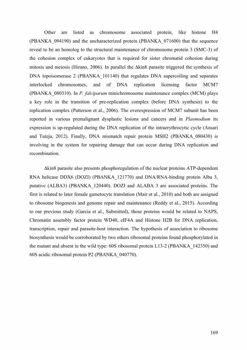
169
Other are listed as chromosome associated protein, like histone H4
(PBANKA_094190) and the uncharacterized protein (PBANKA_071600) that the sequence
reveal to be an homolog to the structural maintenance of chromosome protein 3 (SMC-3) of
the cohesion complex of eukaryotes that is required for sister chromatid cohesion during
mitosis and meiosis (Hirano, 2006). In parallel the Δkin8 parasite triggered the synthesis of
DNA topoisomerase 2 (PBANKA_101140) that regulates DNA supercoiling and separates
interlocked chromosomes, and of DNA replication licensing factor MCM7
(PBANKA_080310). In P. falciparum minichromosome maintenance complex (MCM) plays
a key role in the transition of pre-replication complex (before DNA synthesis) to the
replication complex (Patterson et al., 2006). The overexpression of MCM7 subunit has been
reported in various premalignant dysplastic lesions and cancers and in Plasmodium its
expression is up-regulated during the DNA replication of the intraerythrocytic cycle (Ansari
and Tuteja, 2012). Finally, DNA mismatch repair protein MSH2 (PBANKA_080430) is
involving in the system for repairing damage that can occur during DNA replication and
recombination.
Δkin8 parasite also presents phosphoregulation of the nuclear proteins ATP-dependent
RNA helicase DDX6 (DOZI) (PBANKA_121770) and DNA/RNA-binding protein Alba 3,
putative (ALBA3) (PBANKA_120440). DOZI and ALABA 3 are associated proteins. The
first is related to later female gametocyte translation (Mair et al., 2010) and both are assigned
to ribosome biogenesis and genome repair and maintenance (Reddy et al., 2015). According
to our previous study (Garcia et al., Submitted), those proteins would be related to NAPS,
Chromatin assembly factor protein WD40, eIF4A and Histone H2B for DNA replication,
transcription, repair and parasite-host interaction. The hypothesis of association to ribosome
biosynthesis would be corroborated by two others ribosomal proteins found phosphorylated in
the mutant and absent in the wild type: 60S ribosomal protein L13-2 (PBANKA_142350) and
60S acidic ribosomal protein P2 (PBANKA_040770).

170
3.3. Global description Δkin8 regulated proteins and phosphoproteins
compared to WT in time-course experiment
Protein and phosphoprotein relative quantification was obtained through iTRAQ
labelled peptides for each cell line, and samples were analysed in biological triplicate. There
were 59 regulated proteins and 37 phospho-regulated proteins from Δkin8 parasites in three
time intervals of differentiation (T0-T7; T7-T15; T0-T15). Compared to the 52 regulated and
39 phospho-regulated proteins from WT parasites (Garcia et al., Submitted), both lines shared
36 regulated proteins and 17 phospho-regulated proteins during gametogenesis (Fig. 4 A-B).
Those shared proteins in common between WT and Kin8 proteome and phosphoprome are
related mainly in RNA translation and protein biosynthesis, cellular homeostasis and
energetic metabolism as reported for WT parasites (Garcia et al., Submitted).
Here we would like to highlight the 23 and 19 proteins, and 20 and 22
phosphoproteins that were restrictedly modulated in Δkin8 and WT parasites, respectively,
when no modulation was observed in the other line (Fig. 4 A-D). The majority of these line-
specific modulated proteins and phosphoproteins were classified in the same main cell
processes, RNA translation and protein biosynthesis, cellular homeostasis and energetic
metabolism (Fig. 4 C-D). Nonetheless, some line-specific modulated proteins and
phosphoproteins are categorised to cell processes related to the mutant phenotype (Fig. 4 C-
D). For example, WT-specific modulated proteins are related to cell motility/ cytoskeleton
organization (profilin, PBANKA_083300), cell differentiation/ reproduction (nascent
polypeptide associated complex alpha chain, PBANKA_112070), and one WT-specific
modulated phosphoprotein is related to cell division (cell division cycle protein 48
homologue, PBANKA_111860). One Δkin8-specific modulated protein is related to
chromosome organization (histone H2A, PBANKA_111700). Consequently these differences
in the expression and phosphorylation modulations throughout the cell differentiation reflect
the global disorganisation produced in the mutants as an indirect effect.


172
3.4. Time-specific comparison between Δkin8 and WT parasite lines
In order to compared the protein and phosphoprotein abundance differences between
Δkin8 and WT within gametogenesis, we performed a time-dependant pairwise quantitative
and comparative analysis at each time T0, and T7 and T15 post induction.
The 37 proteins that have displayed an abundance difference between Δkin8 and WT
parasites along time points of the differentiation can be clustered in 6 main important cell
processes: host cell hemoglobin degradation and proteolysis, energy metabolism, cytoskeleton
and cell motility, nucleic acid and protein synthesis, host interaction and egress, stress and
protein folding and one unknown conserved Plasmodium protein (Table 1). All categories
have large variation between cell conditions subjected in the current study.
The most severely affected processes were nucleic acid and protein synthesis
and stress and protein folding. The large HSP abundance fluctuation observed among
development times evidences that Δkin8 parasites are responding to unfavourable/abnormal
developmental conditions.
Concerning the energy metabolism process, globally the Δkin8 parasites
produced less glycolysis enzymes than the WT parasites. This could reflect that the WT
parasites need more quantity of energy per cell when compared to Δkin8 parasites to
differentiate and exflagellate, and that the Δkin8 parasite has a slower metabolism activity
(Table 1).
Among the modulated processes, cytoskeleton and cell motility would be directly
affected by kin8 disruption since it is about motor protein interference involved to manifold
related cellular processes. Δkin8 parasite has strong deregulation of the most dynamic
component of the cytoskeleton and axoneme, the microtubules. Moreover the microtubules
are dynamically phospho-regulated by serine and threonine phosphorylation (Fourest-Lieuvin
et al., 2006;Wang et al., 2014;Garcia et al., Submitted). The phosphor-regulation is abnormal
in mutant since we can observe remarkable variation in phosphoprotein abundance between
both groups (Table 2).

173
Wherefore, we have found alpha tubulin I (PBANKA_041770) and tubulin II
(PBANKA_052270) as very up-regulated in Δkin8 parasites and on contrary, we have found
in our experiment that tubulin beta chain (PBANKA_120690) was down-regulated in Δkin8
parasites. The modulation of the 3 proteins was striking at T7. Alpha tubulin 1 and 2, and
tubulin beta were found to show increased expression during the gametogenesis of WT
parasites (Garcia et al., Submitted).
Moreover two proteins, male development gene 1 (MDV1) and osmiophilic
body protein (G377) important for the egress were found up-regulated in the Δkin8 parasites
through gametogenesis at each time point. To egress, the gametocytes must lyse the two
external membrane barriers, the parasitophorous vacuole membrane produced by the parasite
and the red blood cell membrane. The sequential inside-out lysis occurs in response to the
exocytosis of specialized secretory vesicles only present in the gametocyte including the
osmiophilic bodies (Kehrer et al., 2016). In fact, in WT parasites G377 was rapidly down
regulated suggesting that the egress of female gametocyte happens earlier during the
gametogenesis (Garcia et al., Submitted).

174
Table 1: Abundance variation of proteins through time point comparison. According to colour scale, abundance variations were represented
in a heatmap representation for easy visualization of time point abundance differential variation.
Cell process Plasmodium berghei ANKA accession number WT / Δkin8
T0 T7 T15
Host cell hemoglobin
degradation and proteolysis
PBANKA_144900 | aspartyl protease, putative | protein | length=577 -69% -101% 114%
PBANKA_061030 | polyubiquitin, putative | protein | length=229 - 7% -23%
Energy metabolism
PBANKA_111770 | malate dehydrogenase, putative (MDH) | protein | length=313 63% -22% 68%
PBANKA_112560 | pyruvate kinase, putative | protein | length=511 3% 5% 62%
PBANKA_130860 | fructose-bisphosphate aldolase 2 (ALDO2) | protein | length=369 73% 10% 21%
PBANKA_132640 | glyceraldehyde-3-phosphate dehydrogenase, putative (GAPDH) | protein | length=337 39% -35% 1%
PBANKA_121430 | enolase, putative (ENO) | protein | length=446 -14% 1% 66%
PBANKA_134010 | L-lactate dehydrogenase (LDH) | protein | length=316 64% 6% 29%
PBANKA_134040 | oxidoreductase, putative | protein | length=334 -28% 21% 21%
Cytoskeleton and cell motillity
PBANKA_120690 | tubulin beta chain, putative | protein | length=445 -7% 52% 8%
PBANKA_041770 | alpha tubulin 1 | protein | length=453 - -15% -35%
PBANKA_052270 | alpha tubulin 2 | protein | length=450 5% -30% 9%
Nucleic acid and protein
synthesis
PBANKA_121770 | ATP-dependent RNA helicase DDX6 (DOZI) | protein | length=433 -90% - -62%
PBANKA_020300 | chromatin assembly factor 1 protein WD40 domain, putative | protein | length=446 37% -3% 28%
PBANKA_113330 | elongation factor 1-alpha (EF-1alpha) | protein | length=443 43% -5% -9%
PBANKA_113340 | elongation factor 1-alpha (EF-1alpha) | protein | length=443 43% -5% -9%
PBANKA_131480 | elongation factor 2, putative | protein | length=832 39% 93% 52%
PBANKA_136030 | DNA/RNA-binding protein Alba 4, putative (ALBA4) | protein | length=374 -74% -1% -
PBANKA_091800 | 60S acidic ribosomal protein P0, putative | protein | length=315 - -117% 2%
PBANKA_114170 | ubiquitin-60S ribosomal protein L40, putative | protein | length=128 - 7% -23%
PBANKA_111300 | purine nucleoside phosphorylase, putative (PNP) | protein | length=244 43% 14% -
PBANKA_143920 | polyadenylate-binding protein, putative (PABP) | protein | length=834 -108% -12% 6%
PBANKA_061160 | ribonucleoside-diphosphate reductase, large subunit, putative | protein | length=847 - 20% 4%
PBANKA_101670 | conserved Plasmodium protein, unknown function | protein | length=279 - -19% 3%

175
Host interaction and egress PBANKA_146300 | osmiophilic body protein (G377) | protein | length=2606 - 79% -107%
PBANKA_143220 | male development gene 1 (MDV1) | protein | length=214 -32% -59% -82%
Stress and protein folding
PBANKA_071190 | heat shock protein 70 (HSP70) | protein | length=693 18% 0% 7%
PBANKA_081890 | heat shock protein 70, putative | protein | length=651 -33% 54% -80%
PBANKA_091440 | heat shock protein 70, putative (UIS24) | protein | length=663 - 30% -16%
PBANKA_080570 | heat shock protein 90, putative (HSP90) | protein | length=720 57% -26% 72%
PBANKA_111860 | cell division cycle protein 48 homologue, putative | protein | length=815 42% -36% 101%
PBANKA_070280 | protein disulfide isomerase | protein | length=482 - 39% -6%
PBANKA_143730 | endoplasmin, putative (GRP94) | protein | length=808 - 17% 0%
PBANKA_093840 | endoplasmic reticulum-resident calcium binding protein, putative | protein | length=342 47% - 135%
PBANKA_121650 | peptidyl-prolyl cis-trans isomerase, putative (CYP19A) | protein | length=170 113% -47% 34%
PBANKA_093220 | peptidyl-prolyl cis-trans isomerase, putative | protein | length=192 -74% -49% -
Unknown PBANKA_082670 | conserved Plasmodium protein, unknown function | protein | length=222 - -92% 14%
Colour scale
-50
0 50

176
Table 2: Abundance variation of phosphoproteins through time point comparison. According to colour scale, abundance variation were
represented in a heatmap representation for easy visualization of time point abundance differential variation.
Cell process Plasmodium berghei ANKA accession number WT / Δkin8
T0 T7 T15
Host cell hemoglobin
degradation and
proteolysis
PBANKA_093420 | ubiquitin domain-containing protein DSK2, putative (DSK2) | protein | length=375 - 204% 183%
PBANKA_100610 | flap endonuclease 1, putative (FEN1) | protein | length=479 - - -54%
Energy metabolism
PBANKA_112560 | pyruvate kinase, putative | protein | length=511 - 94% 104%
PBANKA_092810 | phosphoglycerate mutase, putative (PGM1) | protein | length=250 - - 194%
PBANKA_121430 | enolase, putative (ENO) | protein | length=446 - 44% -
PBANKA_130380 | triosephosphate isomerase, putative (TIM) | protein | length=248 72% - -
Cytoskeleton and cell
motillity
PBANKA_120690 | tubulin beta chain, putative | protein | length=445 - - 22%
PBANKA_052270 | alpha tubulin 2 | protein | length=450 - - -90%
Nucleic acid and protein
synthesis
PBANKA_142330 | DNA/RNA-binding protein Alba 1, putative (ALBA1) | protein | length=242 - - 104%
PBANKA_135920 | DNA/RNA-binding protein Alba 2, putative (ALBA2) | protein | length=202 72% - 265%
PBANKA_113340 | elongation factor 1-alpha (EF-1alpha) | protein | length=443 44% 57% 13%
PBANKA_113330 | elongation factor 1-alpha (EF-1alpha) | protein | length=443 44% 57% 13%
PBANKA_093770 | 60S ribosomal protein L36, putative (RPL36) | protein | length=113 - - 164%
PBANKA_110670 | 60S ribosomal protein L4, putative (RPL4) | protein | length=411 - - 298%
PBANKA_093030 | GTP-binding nuclear protein RAN/TC4, putative | protein | length=214 - -69% -
PBANKA_130130 | trailer hitch homolog, putative (CITH) | protein | length=337 - - -89%
PBANKA_113570 | RNA-binding protein, putative | protein | length=511 - - -69%
PBANKA_143920 | polyadenylate-binding protein, putative (PABP) | protein | length=834 -37% - -59%
PBANKA_060430 | conserved Plasmodium protein, unknown function | protein | length=330 - - 26%
Host interaction and egress PBANKA_143220 | male development gene 1 (MDV1) | protein | length=214 - -126% -146%
Stress and protein folding
PBANKA_121650 | peptidyl-prolyl cis-trans isomerase, putative (CYP19A) | protein | length=170 - - 34%
PBANKA_081890 | heat shock protein 70, putative | protein | length=651 79% 6% -
PBANKA_071190 | heat shock protein 70 (HSP70) | protein | length=693 305% - -97%

177
PBANKA_080570 | heat shock protein 90, putative (HSP90) | protein | length=720 - - -106%
PBANKA_111860 | cell division cycle protein 48 homologue, putative | protein | length=815 - - -24%
Unknown PBANKA_030130 | conserved Plasmodium protein, unknown function | protein | length=91 - 74% -
Colour scale
-50
0 50

178
4. DISCUSSION
The kinesin-8 family of motors is thought to mediate chromosome movements
through a combination of translocation and microtubule depolymerisation activities in
budding yeast (Kip3 protein) (Gupta et al., 2006;Varga et al., 2006) and in human cell (Kif18
protein) (Zhu et al., 2005;Stumpff and Wordeman, 2007). E. g. the human Kif18 is involved
in regulating the process of chromosome alignment, referred to chromosome congression and
considered to be essential for cell viability. Depletion of Kif18A affects kinetochore
movements and results in mitotic cells with abnormally long mitotic spindles that fail to
congress chromosomes at the metaphase plate. Kif18 is not essential for the establishment
and/or maintenance of kinetochore-microtubule attachment, but is required for judging
tension across kinetochore pairs. Recently, the two kinesin 8, Klp5 and Klp6, in the fission
yeast were shown to contribute on microtubule dynamics, kinetochore attachment stability
and sliding force in the mitotic spindle (Gergely et al., 2016). In addition to aberrant
chromosome pushing movements, occasional cells lacking Klp5 contained three spindle pole
bodies and tripolar mitotic spindles. It was observed that the absence of kinesin-8 in
organisms causes cellular polarity defect and longer mitotic spindles but that is not always the
case (Messin and Millar, 2014;Gergely et al., 2016). In the amoeba Dictyostelium discoideum,
for exemple, the kinesin 8 kif10 is not essential for development or viability and appears to
cooperate with dynein to organize the interphase radial microtubule array (Gergely et al.,
2016). This is relevant because D. discoideum possesses 13 kinesin-8 isoforms that suggests a
functional redundancy of motor isoforms in this organism. Moreover, the variability in
reported roles of kinesin 8 members suggest that their effects may be different in different
organisms (Gergely et al., 2016).
In P. berghei, gene disruption for one of the two kinesin 8 members, the one
that known to be expressed during gametogenesis by proteome data (Khan et al., 2005),
induces defects of microtubule assembly affecting the male gamete morphogenesis and the
exflagellation process and consequently reproduction (Depoix et al. manuscript in
preparation). Kinesin-8 is thus important for normal parasite DNA replication and gamete
differentiation. Our study shows that Δkin8 gametocyte is metabolically active until the last
time lapse observed, 15 min post induction for gamete production, even if very distinct
metabolic conditions and proteome are observed compared to WT parasites. Specifically,

179
Δkin8 gametocytes performed all the three DNA replication cycles but daughter nuclei did not
individualize (Depoix et al., manuscript in preparation). In other cellular models kin8
depletion disturbed the mitotic spindle length and the alignment of chromosomes in the
metaphase and induced default in chromosome congression. This phenomena could be
involved in the phenotype observed in P. berghei. However, the way chromatin is organized
inside the nucleus of eukaryotic cells is a key factor in gene control and chromosomal
organization and dynamic as well as gene positioning could influence spatiotemporally gene
expression in bacteria (Sobetzko et al., 2012) and in Eukaryotes (Spector, 2003;Schneider and
Grosschedl, 2007) and in Plasmodium (Duffy et al., 2012). Such an implication could explain
the high number of modulated proteins related to the gene expression in Δkin8 parasites
compared to WT parasites. This is well illustrated by the higher quantity of proteins and
proteins modified posttranslationally by phosphorylation clustered in the acid nucleic and
protein processes (Fig. 2-4, Table 1).
Concomitant to the three rounds of DNA replication, WT male cells
differentiate with an intracytoplasmic axoneme formation and exflagellate, releasing eight
male gametes. While Δkin8 parasites shows assembly of single or doublet of microtubules but
axonemes were not formed or highly disorganised. Plasmodium encode one beta-tubulin and
two alpha tubulin isotypes (I and II) (Kooij et al., 2005). Alpha tubulin I is expressed by every
Plasmodium stage, except in sporozoite stage, and has higher abundance in female than male
gametocytes (Kooij et al., 2005). It has great sequence similarity to alpha tubulin II 95%
(Schwank et al., 2010) and its most notable difference is the three well conserved C-terminal
amino acids and the terminal tyrosine residue lacking in alpha tubulin II (Kooij et al., 2005).
Researchers have reported that alpha tubulin II is expressed in Plasmodium as male-specific
gametocytes protein (Schwank et al., 2010;Alonso-Morales et al., 2015). Nevertheless, alpha
tubulin II is also expressed by P. berghei female gametocyte at low expression level in a
ration 6:1 (Kooij et al., 2005) despite it still can be used as sex-specific marker (Khan et al.,
2005). In P. berghei, alpha tubulin II would be a critical component for exflagellation and
cellular motility as component of the axonemal microtubules (Rawlings et al., 1992;Kooij et
al., 2005). The high abundance variation observed in Δkin8 parasites compared to WT
parasites could have an explanation on the assorted activities disciplined by microtubules for
cellular organization, gene expression and flagellar formation. Besides the post-translational
assumptive importance to alpha and beta tubulins for microtubule stability and interactions to

180
others proteins (Kooij et al., 2005), the dynamic of tubulin isoforms might have an affect on
cellular adaptation, stages development and chromatin condensing.
In P. berghei, actin-1 and actin-2 are expressed in gametocytes but actin-2 is
solely expressed in male gametocytes (Wesseling et al., 1989;Alonso-Morales et al., 2015)
although its function needs to be further investigated for a better understanding of its role in
male gamete development (Deligianni et al., 2011;Alonso-Morales et al., 2015). From our
findings (shown in Table 1, Table 2), actin 2 does not have significant abundance variation
between WT parasites and Δkin8 parasites, and that could be an indicative that actin 2 has no
relationship to effects and co-effects trigged by kin8.
The fact that disruption of kin8 affects directly two microtubule-related
gametogenesis processes: nuclei individualization and axoneme formation, could be explain
in part by the small number of kinesins present in Plasmodium genome suggesting that one
kinesin protein could participate in more than one process within the parasite. Deficiency in
kin8 does not seem to represent a great interference to P. berghei morphology at the
beginning of the gametogenesis process and DNA replication occurs. However, both parasite
lines are likely to be departing from gametocyte physiologic condition under different
metabolically state and protein content that end to a dissimilar structural organization. The
differential phosphorylation of several proteins in both parasite lines during the
gametogenesis have certainly also importance in regulating their functions during the cell
process, but further studies are required to demonstrate their role in this process.
In summary, with kin8 disruption, we observed 2 main phenotypic and
functional defects in chromatin organisation/nuclei individualisation and axoneme assembly
(Figure 5). At the proteotype level, we observed important synthesis and phosphorylation
modulations of proteins. The nuclear division disorganisation could be related to the down-
expression of nucleosome unit, up-expression of protein involved in the replication. In
parallel, we observed clearly a high expression of the ubiquitin-dependant proteasome
complex and proteins of stress response and folding and low expression of some proteins
involved in energy metabolism and egress from RBC. In the other side, the axoneme
disorganisation and mitotic spindle microtubules probably correspond to the down-regulation
of motor proteins like kinesin 13, dynein heavy chain and obviously the disrupted kin8 and
the up-regulation of proteins involved in the improperly inner arm and radial spoke
organization, and the tubulins as well. This overview could explain the slower growth of the


182
SUPPLEMENTARY DATA
Suppl. Table S1. Δkin8 P. berghei proteins proteins identified during gametogenesis.
Accession number
#Unique
Peptides MW
Sequence
Count
Spectrum
Count Coverage Description
PBANKA_071190 68 75155 81 597 0.6724 | Plasmodium berghei ANKA | heat shock protein 70 (HSP70) | protein | length=693
PBANKA_120690 53 49739.9 55 539 0.8539 | Plasmodium berghei ANKA | tubulin beta chain, putative | protein | length=445
PBANKA_121430 56 48977.4 58 447 0.8879 | Plasmodium berghei ANKA | enolase, putative (ENO) | protein | length=446
PBANKA_132640 50 36541 50 355 0.9585 | Plasmodium berghei ANKA | glyceraldehyde-3-phosphate dehydrogenase, putative (GAPDH) | protein |
length=337
PBANKA_113330 5 48909.9 44 331 0.6749 | Plasmodium berghei ANKA | elongation factor 1-alpha (EF-1alpha) | protein | length=443
PBANKA_081890 26 72084.9 52 329 0.5561 | Plasmodium berghei ANKA | heat shock protein 70, putative | protein | length=651
PBANKA_113340 0 48909.9 39 325 0.6749 | Plasmodium berghei ANKA | elongation factor 1-alpha (EF-1alpha) | protein | length=443
PBANKA_070280 50 55358.1 50 297 0.6432 | Plasmodium berghei ANKA | protein disulfide isomerase | protein | length=482
PBANKA_052270 12 49669.3 30 225 0.5911 | Plasmodium berghei ANKA | alpha tubulin 2 | protein | length=450
PBANKA_111860 37 90764.2 38 212 0.4945 | Plasmodium berghei ANKA | cell division cycle protein 48 homologue, putative | protein | length=815
PBANKA_020300 31 50481.8 31 194 0.6256 | Plasmodium berghei ANKA | chromatin assembly factor 1 protein WD40 domain, putative | protein | length=446
PBANKA_131480 48 93297.2 51 186 0.5192 | Plasmodium berghei ANKA | elongation factor 2, putative | protein | length=832
PBANKA_130860 38 39808.9 38 173 0.8672 | Plasmodium berghei ANKA | fructose-bisphosphate aldolase 2 (ALDO2) | protein | length=369
PBANKA_130130 27 39026.9 27 164 0.7329 | Plasmodium berghei ANKA | trailer hitch homolog, putative (CITH) | protein | length=337
PBANKA_041770 9 50156.3 26 149 0.5453 | Plasmodium berghei ANKA | alpha tubulin 1 | protein | length=453
PBANKA_112560 34 55493.8 34 142 0.7280 | Plasmodium berghei ANKA | pyruvate kinase, putative | protein | length=511
PBANKA_134040 26 36916.3 26 140 0.6048 | Plasmodium berghei ANKA | oxidoreductase, putative | protein | length=334
PBANKA_080570 42 83143.2 43 136 0.4986 | Plasmodium berghei ANKA | heat shock protein 90, putative (HSP90) | protein | length=720
PBANKA_082340 27 45448.1 27 136 0.6274 | Plasmodium berghei ANKA | phosphoglycerate kinase, putative (PGK) | protein | length=416
PBANKA_134010 29 34384.1 29 129 0.7690 | Plasmodium berghei ANKA | L-lactate dehydrogenase (LDH) | protein | length=316
PBANKA_145930 24 41840 28 126 0.6755 | Plasmodium berghei ANKA | actin I | protein | length=376
PBANKA_142330 20 26845.5 20 121 0.4876 | Plasmodium berghei ANKA | DNA/RNA-binding protein Alba 1, putative (ALBA1) | protein | length=242
PBANKA_143220 22 24252.8 22 116 0.6308 | Plasmodium berghei ANKA | male development gene 1 (MDV1) | protein | length=214
PBANKA_071260 28 30166.7 31 115 0.7748 | Plasmodium berghei ANKA | 14-3-3 protein (14-3-3I) | protein | length=262
PBANKA_143920 30 93239.1 30 115 0.4568 | Plasmodium berghei ANKA | polyadenylate-binding protein, putative (PABP) | protein | length=834
PBANKA_144900 31 65322.5 31 115 0.5390 | Plasmodium berghei ANKA | aspartyl protease, putative | protein | length=577

183
PBANKA_101670 20 32689.7 20 110 0.5090 | Plasmodium berghei ANKA | conserved Plasmodium protein, unknown function | protein | length=279
PBANKA_145030 16 57830.3 19 110 0.5441 | Plasmodium berghei ANKA | ATP synthase subunit beta, mitochondrial, putative | protein | length=533
PBANKA_121650 13 18788.3 14 104 0.5176 | Plasmodium berghei ANKA | peptidyl-prolyl cis-trans isomerase, putative (CYP19A) | protein | length=170
PBANKA_143730 25 93169.2 25 102 0.3453 | Plasmodium berghei ANKA | endoplasmin, putative (GRP94) | protein | length=808
PBANKA_121930 28 94821.3 28 93 0.2931 | Plasmodium berghei ANKA | heat shock protein 110, putative (HSP110c) | protein | length=836
PBANKA_146300 31 309004.2 31 89 0.1474 | Plasmodium berghei ANKA | osmiophilic body protein (G377) | protein | length=2606
PBANKA_111340 21 85042.1 21 88 0.2899 | Plasmodium berghei ANKA | secreted ookinete protein, putative,6-cysteine protein (PSOP12) | protein |
length=721
PBANKA_103440 14 50432.6 14 84 0.2978 | Plasmodium berghei ANKA | plasmepsin IV (PM4) | protein | length=450
PBANKA_121400 25 62189.5 25 76 0.4421 | Plasmodium berghei ANKA | heat shock protein 60, putative | protein | length=579
PBANKA_081990 18 31650.8 18 70 0.4532 | Plasmodium berghei ANKA | nucleosome assembly protein (NAPS) | protein | length=267
PBANKA_121470 27 111954.5 27 70 0.2976 | Plasmodium berghei ANKA | conserved Plasmodium protein, unknown function | protein | length=961
PBANKA_040520 15 59033.4 15 69 0.3640 | Plasmodium berghei ANKA | T-complex protein beta subunit, putative | protein | length=533
PBANKA_135510 14 35429.9 14 69 0.3194 | Plasmodium berghei ANKA | 40S ribosomal protein S6, putative | protein | length=310
PBANKA_136030 13 42121.4 13 64 0.2380 | Plasmodium berghei ANKA | DNA/RNA-binding protein Alba 4, putative (ALBA4) | protein | length=374
PBANKA_092810 12 28671.1 12 62 0.3040 | Plasmodium berghei ANKA | phosphoglycerate mutase, putative (PGM1) | protein | length=250
PBANKA_114170 3 14589.9 12 62 0.4688 | Plasmodium berghei ANKA | ubiquitin-60S ribosomal protein L40, putative | protein | length=128
PBANKA_111700 7 14194.8 8 61 0.6316 | Plasmodium berghei ANKA | histone H2A, putative (H2A) | protein | length=133
PBANKA_070470 17 57214.4 17 60 0.3368 | Plasmodium berghei ANKA | conserved Plasmodium protein, unknown function | protein | length=487
PBANKA_093420 9 41217.1 9 59 0.5867 | Plasmodium berghei ANKA | ubiquitin domain-containing protein DSK2, putative (DSK2) | protein | length=375
PBANKA_061030 0 25685.8 9 57 0.2620 | Plasmodium berghei ANKA | polyubiquitin, putative | protein | length=229
PBANKA_093840 18 39080.3 18 57 0.6199 | Plasmodium berghei ANKA | endoplasmic reticulum-resident calcium binding protein, putative | protein |
length=342
PBANKA_061920 14 54662.5 14 56 0.2839 | Plasmodium berghei ANKA | secreted ookinete protein, putative (PSOP1) | protein | length=479
PBANKA_093030 12 24852.8 12 56 0.5327 | Plasmodium berghei ANKA | GTP-binding nuclear protein RAN/TC4, putative | protein | length=214
PBANKA_133190 14 45322.5 14 56 0.3141 | Plasmodium berghei ANKA | eukaryotic initiation factor 4a, putative (eIF4A) | protein | length=398
PBANKA_061160 22 96972 22 55 0.2963 | Plasmodium berghei ANKA | ribonucleoside-diphosphate reductase, large subunit, putative | protein | length=847
PBANKA_111770 15 34273.8 15 52 0.5495 | Plasmodium berghei ANKA | malate dehydrogenase, putative (MDH) | protein | length=313
PBANKA_050580 9 29223.5 9 51 0.4962 | Plasmodium berghei ANKA | deoxyribose-phosphate aldolase, putative | protein | length=262
PBANKA_093220 12 21179.1 12 51 0.5677 | Plasmodium berghei ANKA | peptidyl-prolyl cis-trans isomerase, putative | protein | length=192
PBANKA_121770 11 49325.6 11 51 0.2771 | Plasmodium berghei ANKA | ATP-dependent RNA helicase DDX6 (DOZI) | protein | length=433

184
PBANKA_142690 20 104135.3 20 48 0.2585 | Plasmodium berghei ANKA | RNA helicase, putative | protein | length=909
PBANKA_083300 8 19236.1 8 47 0.4713 | Plasmodium berghei ANKA | profilin, putative (PFN) | protein | length=174
PBANKA_060430 16 38739.5 16 46 0.5000 | Plasmodium berghei ANKA | conserved Plasmodium protein, unknown function | protein | length=330
PBANKA_130070 19 183560.8 19 46 0.1802 | Plasmodium berghei ANKA | LCCL domain-containing protein (CCp1) | protein | length=1615
PBANKA_110670 18 46085.3 18 45 0.3528 | Plasmodium berghei ANKA | 60S ribosomal protein L4, putative (RPL4) | protein | length=411
PBANKA_091440 15 73103.1 15 44 0.1795 | Plasmodium berghei ANKA | heat shock protein 70, putative (UIS24) | protein | length=663
PBANKA_091620 11 60078.6 11 44 0.3039 | Plasmodium berghei ANKA | TCP-1/cpn60 chaperonin family, putative | protein | length=543
PBANKA_130280 12 21622.3 12 44 0.4769 | Plasmodium berghei ANKA | thioredoxin peroxidase 1 (TPx1) | protein | length=195
PBANKA_111300 15 26971.7 15 43 0.7008 | Plasmodium berghei ANKA | purine nucleoside phosphorylase, putative (PNP) | protein | length=244
PBANKA_100380 14 55775.4 14 42 0.4494 | Plasmodium berghei ANKA | vacuolar ATP synthase subunit b, putative | protein | length=494
PBANKA_111520 17 132736.1 17 42 0.1722 | Plasmodium berghei ANKA | conserved Plasmodium protein, unknown function | protein | length=1138
PBANKA_142430 12 32548.1 12 41 0.3521 | Plasmodium berghei ANKA | conserved Plasmodium protein, unknown function | protein | length=284
PBANKA_082670 6 25218.7 6 39 0.2748 | Plasmodium berghei ANKA | conserved Plasmodium protein, unknown function | protein | length=222
PBANKA_141030 14 123256.3 14 39 0.1814 | Plasmodium berghei ANKA | M1-family alanyl aminopeptidase, putative | protein | length=1064
PBANKA_120440 8 11965.3 8 38 0.6916 | Plasmodium berghei ANKA | DNA/RNA-binding protein Alba 3, putative (ALBA3) | protein | length=107
PBANKA_094180 9 13231.2 9 37 0.5254 | Plasmodium berghei ANKA | histone H2B, putative (H2B) | protein | length=118
PBANKA_101950 9 33843.2 9 36 0.3208 | Plasmodium berghei ANKA | 60S ribosomal protein L5, putative | protein | length=293
PBANKA_130380 13 27807.4 13 36 0.4032 | Plasmodium berghei ANKA | triosephosphate isomerase, putative (TIM) | protein | length=248
PBANKA_091790 8 50927.3 10 35 0.3370 | Plasmodium berghei ANKA | 26S protease regulatory subunit 6a, putative (RPT5) | protein | length=451
PBANKA_103390 10 24959.3 10 35 0.3349 | Plasmodium berghei ANKA | 40S ribosomal protein S8e, putative | protein | length=218
PBANKA_141040 11 68289.2 11 34 0.2930 | Plasmodium berghei ANKA | vacuolar ATP synthase subunit a, putative | protein | length=611
PBANKA_092130 8 19697.1 8 33 0.5429 | Plasmodium berghei ANKA | deoxyuridine 5'-triphosphate nucleotidohydrolase, putative | protein | length=175
PBANKA_103270 14 58837.1 14 33 0.3629 | Plasmodium berghei ANKA | RNA binding protein Bruno, putative (HoBo) | protein | length=507
PBANKA_123100 6 16046.7 6 33 0.3046 | Plasmodium berghei ANKA | 40S ribosomal protein S11, putative | protein | length=151
PBANKA_021150 8 28311.4 8 32 0.4531 | Plasmodium berghei ANKA | proteasome subunit alpha type-5, putative | protein | length=256
PBANKA_060320 10 17524.7 10 31 0.6522 | Plasmodium berghei ANKA | eukaryotic translation initiation factor 5A, putative (EIF5A) | protein | length=161
PBANKA_113790 8 30637.6 9 31 0.3796 | Plasmodium berghei ANKA | proliferating cell nuclear antigen, putative (PCNA) | protein | length=274
PBANKA_131080 10 29901.8 10 31 0.5314 | Plasmodium berghei ANKA | 40S ribosomal protein S5, putative | protein | length=271
PBANKA_145780 14 33119.2 14 31 0.5574 | Plasmodium berghei ANKA | ADP-ribosylation factor GTPase-activating protein, putative (ARFGAP) | protein |
length=296
PBANKA_020450 9 121466.2 9 30 0.1087 | Plasmodium berghei ANKA | LCCL domain-containing protein (CCp5) | protein | length=1049

185
PBANKA_142350 12 25385 12 30 0.3721 | Plasmodium berghei ANKA | 60S ribosomal protein L13-2, putative | protein | length=215
PBANKA_145610 5 16049.7 5 30 0.3212 | Plasmodium berghei ANKA | 40S ribosomal protein S17, putative | protein | length=137
PBANKA_093110 8 24745.5 8 29 0.5160 | Plasmodium berghei ANKA | conserved Plasmodium protein, unknown function | protein | length=219
PBANKA_103520 13 148090.1 13 29 0.1281 | Plasmodium berghei ANKA | LCCL domain-containing protein (CCp3) | protein | length=1304
PBANKA_113570 6 57133.8 6 29 0.2603 | Plasmodium berghei ANKA | RNA-binding protein, putative | protein | length=511
PBANKA_121560 9 29947.3 9 29 0.1527 | Plasmodium berghei ANKA | 40S ribosomal protein S3A, putative | protein | length=262
PBANKA_030130 8 9784.8 8 28 0.3407 | Plasmodium berghei ANKA | conserved Plasmodium protein, unknown function | protein | length=91
PBANKA_040540 10 15434 10 28 0.4859 | Plasmodium berghei ANKA | 40S ribosomal protein S12, putative | protein | length=142
PBANKA_052280 5 19994.8 5 28 0.2442 | Plasmodium berghei ANKA | 40S ribosomal protein S19, putative (RPS19) | protein | length=172
PBANKA_090360 8 93443.4 8 28 0.1511 | Plasmodium berghei ANKA | dynamin-like protein, putative (DYN1) | protein | length=814
PBANKA_141910 8 47783.5 8 28 0.2861 | Plasmodium berghei ANKA | dihydrolipoyllysine-residue succinyltransferase component of 2-oxoglutarate
dehydrogenase complex, putative | protein | length=423
PBANKA_143105 6 10972.9 6 28 0.4510 | Plasmodium berghei ANKA | 10 kDa chaperonin, putative (CPN10) | protein | length=102
PBANKA_081420 10 29995.1 10 27 0.3424 | Plasmodium berghei ANKA | elongation factor 1-beta, putative | protein | length=257
PBANKA_132090 7 11612.9 7 27 0.5385 | Plasmodium berghei ANKA | thioredoxin 1, putative (TRX1) | protein | length=104
PBANKA_132500 6 7487.1 6 27 0.5075 | Plasmodium berghei ANKA | 40S ribosomal protein S28e, putative | protein | length=67
PBANKA_140130 4 22449.6 4 27 0.2835 | Plasmodium berghei ANKA | 40S ribosomal protein S7, putative | protein | length=194
PBANKA_120720 8 27364.5 8 26 0.3802 | Plasmodium berghei ANKA | adenylate kinase, putative (AK1) | protein | length=242
PBANKA_101940 6 33244.8 6 25 0.2148 | Plasmodium berghei ANKA | 60S ribosomal protein L7-3, putative | protein | length=284
PBANKA_123880 8 127725.3 8 25 0.1083 | Plasmodium berghei ANKA | karyopherin beta, putative | protein | length=1126
PBANKA_135440 7 21668.5 7 25 0.1957 | Plasmodium berghei ANKA | 60S ribosomal protein L18, putative | protein | length=184
PBANKA_144410 10 60779 10 25 0.1993 | Plasmodium berghei ANKA | t-complex protein 1 gamma subunit, putative | protein | length=542
PBANKA_121760 0 16465.4 2 24 0.1456 | Plasmodium berghei ANKA | histone H2A variant, putative (H2A.Z) | protein | length=158
PBANKA_120500 7 27807.9 7 23 0.1845 | Plasmodium berghei ANKA | RNA-binding protein, putative | protein | length=233
PBANKA_121090 5 68099 5 23 0.0776 | Plasmodium berghei ANKA | phosphoglucomutase, putative | protein | length=593
PBANKA_135970 6 48879.1 6 23 0.1323 | Plasmodium berghei ANKA | 6-cysteine protein (P47) | protein | length=431
PBANKA_070390 7 35542.9 7 22 0.1925 | Plasmodium berghei ANKA | receptor for activated c kinase, putative (RACK) | protein | length=322
PBANKA_083100 11 198529.4 11 22 0.1072 | Plasmodium berghei ANKA | merozoite surface protein 1 (MSP1) | protein | length=1791
PBANKA_093770 7 12700.3 7 22 0.3805 | Plasmodium berghei ANKA | 60S ribosomal protein L36, putative (RPL36) | protein | length=113
PBANKA_101850 8 19905.6 8 22 0.3966 | Plasmodium berghei ANKA | transcription factor 3b, putative | protein | length=174
PBANKA_124230 7 47637.9 7 22 0.1797 | Plasmodium berghei ANKA | Hsc70-interacting protein,58 kDa phosphoprotein (HIP) | protein | length=423

186
PBANKA_135860 7 53688.7 7 22 0.1450 | Plasmodium berghei ANKA | isocitrate dehydrogenase (NADP), mitochondrial precursor, putative (IDH) | protein
| length=469
PBANKA_145020 8 49575.4 8 22 0.2104 | Plasmodium berghei ANKA | serine hydroxymethyltransferase, putative (SHMT) | protein | length=442
PBANKA_020160 5 18642.4 5 21 0.4463 | Plasmodium berghei ANKA | early transcribed membrane protein (ETRAMP) | protein | length=177
PBANKA_030680 7 52978.9 7 21 0.1974 | Plasmodium berghei ANKA | ATP-dependent RNA helicase UAP56, putative (UAP56) | protein | length=466
PBANKA_040760 9 66318.9 9 21 0.1670 | Plasmodium berghei ANKA | asparagine synthetase, putative | protein | length=581
PBANKA_051190 8 44319.8 8 21 0.2383 | Plasmodium berghei ANKA | 60S ribosomal protein L3, putative (RPL3) | protein | length=386
PBANKA_101050 10 65585.2 10 21 0.1878 | Plasmodium berghei ANKA | Hsp70/Hsp90 organizing protein, putative (HOP) | protein | length=559
PBANKA_094360 4 12871.5 4 20 0.5042 | Plasmodium berghei ANKA | 60S acidic ribosomal protein P1, putative | protein | length=119
PBANKA_100950 10 336064.3 10 20 0.0655 | Plasmodium berghei ANKA | NAD(P)H-dependent glutamate synthase, putative | protein | length=2962
PBANKA_123210 6 36233.1 6 20 0.1365 | Plasmodium berghei ANKA | pre-mRNA-splicing factor, putative (SR1) | protein | length=315
PBANKA_133830 3 21614.9 3 20 0.2895 | Plasmodium berghei ANKA | 60S ribosomal protein L6, putative | protein | length=190
PBANKA_040650 8 59569.9 8 19 0.2282 | Plasmodium berghei ANKA | TCP-1/cpn60 chaperonin, putative | protein | length=539
PBANKA_040770 8 11778.8 8 19 0.5045 | Plasmodium berghei ANKA | 60S acidic ribosomal protein P2, putative | protein | length=111
PBANKA_091800 11 34820.2 11 19 0.4952 | Plasmodium berghei ANKA | 60S acidic ribosomal protein P0, putative | protein | length=315
PBANKA_051280 9 66720.1 9 18 0.0631 | Plasmodium berghei ANKA | merozoite TRAP-like protein, putative (MTRAP) | protein | length=618
PBANKA_060270 6 40275.8 6 18 0.1710 | Plasmodium berghei ANKA | nucleosome assembly protein, putative (NAPL) | protein | length=345
PBANKA_093850 7 35487.3 7 18 0.2705 | Plasmodium berghei ANKA | succinyl-CoA synthetase alpha subunit, putative | protein | length=329
PBANKA_111750 6 16808.1 6 18 0.4054 | Plasmodium berghei ANKA | ribosomal protein L27a, putative | protein | length=148
PBANKA_113780 6 291206.2 6 18 0.0330 | Plasmodium berghei ANKA | conserved Plasmodium protein, unknown function | protein | length=2605
PBANKA_123170 10 27855.1 10 18 0.2692 | Plasmodium berghei ANKA | 60S ribosomal protein L2, putative (RPL2) | protein | length=260
PBANKA_132930 6 24702.5 6 18 0.2838 | Plasmodium berghei ANKA | 40S ribosomal protein S3, putative | protein | length=222
PBANKA_040470 6 12361.4 6 17 0.6514 | Plasmodium berghei ANKA | glutaredoxin 1, putative (GRX1) | protein | length=109
PBANKA_040840 8 126079.8 8 17 0.0889 | Plasmodium berghei ANKA | phosphoglycerate mutase, putative | protein | length=1069
PBANKA_060760 3 12067.3 3 17 0.6574 | Plasmodium berghei ANKA | U6 snRNA-associated Sm-like protein LSm7, putative (LSM7) | protein |
length=108
PBANKA_090500 3 9166.6 3 17 0.1220 | Plasmodium berghei ANKA | 40S ribosomal protein S21, putative (RPS21) | protein | length=82
PBANKA_091910 8 44649.8 8 17 0.2577 | Plasmodium berghei ANKA | parasitophorous vacuolar protein 1, putative (PV1) | protein | length=388
PBANKA_123560 5 53623.2 5 17 0.1420 | Plasmodium berghei ANKA | adenosylhomocysteinase, putative (SAHH) | protein | length=479
PBANKA_133840 5 14023.5 5 17 0.4380 | Plasmodium berghei ANKA | vacuolar ATP synthase subunit g, putative | protein | length=121
PBANKA_143080 5 24693.7 5 17 0.3907 | Plasmodium berghei ANKA | thioredoxin peroxidase 2, putative (Trx-Px2) | protein | length=215
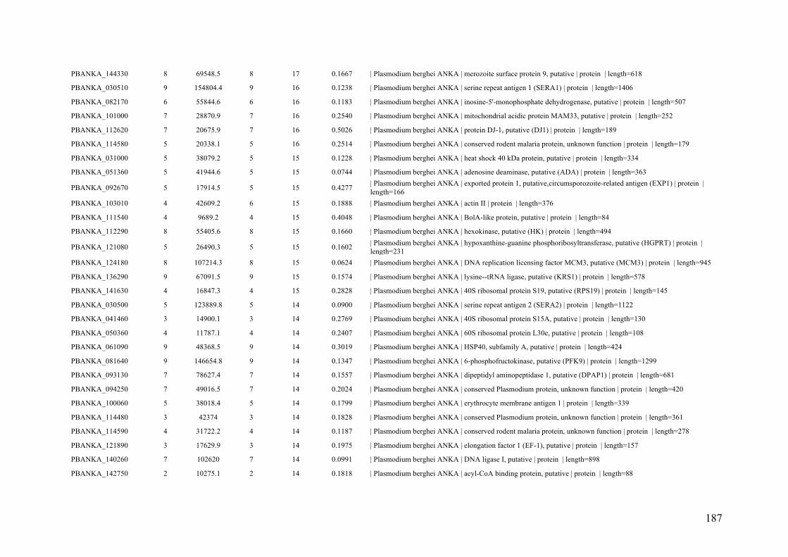
187
PBANKA_144330 8 69548.5 8 17 0.1667 | Plasmodium berghei ANKA | merozoite surface protein 9, putative | protein | length=618
PBANKA_030510 9 154804.4 9 16 0.1238 | Plasmodium berghei ANKA | serine repeat antigen 1 (SERA1) | protein | length=1406
PBANKA_082170 6 55844.6 6 16 0.1183 | Plasmodium berghei ANKA | inosine-5'-monophosphate dehydrogenase, putative | protein | length=507
PBANKA_101000 7 28870.9 7 16 0.2540 | Plasmodium berghei ANKA | mitochondrial acidic protein MAM33, putative | protein | length=252
PBANKA_112620 7 20675.9 7 16 0.5026 | Plasmodium berghei ANKA | protein DJ-1, putative (DJ1) | protein | length=189
PBANKA_114580 5 20338.1 5 16 0.2514 | Plasmodium berghei ANKA | conserved rodent malaria protein, unknown function | protein | length=179
PBANKA_031000 5 38079.2 5 15 0.1228 | Plasmodium berghei ANKA | heat shock 40 kDa protein, putative | protein | length=334
PBANKA_051360 5 41944.6 5 15 0.0744 | Plasmodium berghei ANKA | adenosine deaminase, putative (ADA) | protein | length=363
PBANKA_092670 5 17914.5 5 15 0.4277 | Plasmodium berghei ANKA | exported protein 1, putative,circumsporozoite-related antigen (EXP1) | protein |
length=166
PBANKA_103010 4 42609.2 6 15 0.1888 | Plasmodium berghei ANKA | actin II | protein | length=376
PBANKA_111540 4 9689.2 4 15 0.4048 | Plasmodium berghei ANKA | BolA-like protein, putative | protein | length=84
PBANKA_112290 8 55405.6 8 15 0.1660 | Plasmodium berghei ANKA | hexokinase, putative (HK) | protein | length=494
PBANKA_121080 5 26490.3 5 15 0.1602 | Plasmodium berghei ANKA | hypoxanthine-guanine phosphoribosyltransferase, putative (HGPRT) | protein |
length=231
PBANKA_124180 8 107214.3 8 15 0.0624 | Plasmodium berghei ANKA | DNA replication licensing factor MCM3, putative (MCM3) | protein | length=945
PBANKA_136290 9 67091.5 9 15 0.1574 | Plasmodium berghei ANKA | lysine--tRNA ligase, putative (KRS1) | protein | length=578
PBANKA_141630 4 16847.3 4 15 0.2828 | Plasmodium berghei ANKA | 40S ribosomal protein S19, putative (RPS19) | protein | length=145
PBANKA_030500 5 123889.8 5 14 0.0900 | Plasmodium berghei ANKA | serine repeat antigen 2 (SERA2) | protein | length=1122
PBANKA_041460 3 14900.1 3 14 0.2769 | Plasmodium berghei ANKA | 40S ribosomal protein S15A, putative | protein | length=130
PBANKA_050360 4 11787.1 4 14 0.2407 | Plasmodium berghei ANKA | 60S ribosomal protein L30e, putative | protein | length=108
PBANKA_061090 9 48368.5 9 14 0.3019 | Plasmodium berghei ANKA | HSP40, subfamily A, putative | protein | length=424
PBANKA_081640 9 146654.8 9 14 0.1347 | Plasmodium berghei ANKA | 6-phosphofructokinase, putative (PFK9) | protein | length=1299
PBANKA_093130 7 78627.4 7 14 0.1557 | Plasmodium berghei ANKA | dipeptidyl aminopeptidase 1, putative (DPAP1) | protein | length=681
PBANKA_094250 7 49016.5 7 14 0.2024 | Plasmodium berghei ANKA | conserved Plasmodium protein, unknown function | protein | length=420
PBANKA_100060 5 38018.4 5 14 0.1799 | Plasmodium berghei ANKA | erythrocyte membrane antigen 1 | protein | length=339
PBANKA_114480 3 42374 3 14 0.1828 | Plasmodium berghei ANKA | conserved Plasmodium protein, unknown function | protein | length=361
PBANKA_114590 4 31722.2 4 14 0.1187 | Plasmodium berghei ANKA | conserved rodent malaria protein, unknown function | protein | length=278
PBANKA_121890 3 17629.9 3 14 0.1975 | Plasmodium berghei ANKA | elongation factor 1 (EF-1), putative | protein | length=157
PBANKA_140260 7 102620 7 14 0.0991 | Plasmodium berghei ANKA | DNA ligase I, putative | protein | length=898
PBANKA_142750 2 10275.1 2 14 0.1818 | Plasmodium berghei ANKA | acyl-CoA binding protein, putative | protein | length=88

188
PBANKA_145070 7 45627.5 7 14 0.3174 | Plasmodium berghei ANKA | clustered-asparagine-rich protein, putative | protein | length=397
PBANKA_020930 2 42993.9 2 13 0.1094 | Plasmodium berghei ANKA | actin-related protein (ARP1) | protein | length=384
PBANKA_060190 4 12379.5 4 13 0.2381 | Plasmodium berghei ANKA | high mobility group protein B1, putative (HMGB1) | protein | length=105
PBANKA_091230 6 117761.9 6 13 0.0800 | Plasmodium berghei ANKA | tudor staphylococcal nuclease, putative (TSN) | protein | length=1013
PBANKA_092440 2 15371.1 2 13 0.3901 | Plasmodium berghei ANKA | splicing factor, putative | protein | length=141
PBANKA_094150 3 29807.4 3 13 0.1111 | Plasmodium berghei ANKA | 40S ribosomal protein S4, putative | protein | length=261
PBANKA_113700 9 133431.5 9 13 0.0975 | Plasmodium berghei ANKA | bergheilysin (BLN) | protein | length=1149
PBANKA_120190 2 13486.2 2 13 0.1356 | Plasmodium berghei ANKA | 40S ribosomal protein S20e, putative | protein | length=118
PBANKA_121820 6 59128.2 6 13 0.0953 | Plasmodium berghei ANKA | T-complex protein 1 epsilon subunit, putative | protein | length=535
PBANKA_130620 2 46126.4 2 13 0.0844 | Plasmodium berghei ANKA | tRNA binding protein, putative | protein | length=403
PBANKA_131810 6 89819.7 6 13 0.0992 | Plasmodium berghei ANKA | aminopeptidase P, putative (APP) | protein | length=776
PBANKA_142270 4 22536.2 4 13 0.3586 | Plasmodium berghei ANKA | superoxide dismutase [Fe], putative (FeSOD) | protein | length=198
PBANKA_040530 3 16087.9 3 12 0.2483 | Plasmodium berghei ANKA | 40S ribosomal protein S23, putative | protein | length=145
PBANKA_041820 5 23684.9 5 12 0.1699 | Plasmodium berghei ANKA | ras-related protein RAB7, putative (RAB7) | protein | length=206
PBANKA_092210 5 17852.7 6 12 0.4487 | Plasmodium berghei ANKA | 40S ribosomal protein S18, putative | protein | length=156
PBANKA_093390 2 41780.4 2 12 0.1530 | Plasmodium berghei ANKA | glyoxalase I, putative | protein | length=353
PBANKA_100880 7 68014.1 7 12 0.1297 | Plasmodium berghei ANKA | glucose-6-phosphate isomerase, putative | protein | length=586
PBANKA_113510 7 17228.6 7 12 0.4305 | Plasmodium berghei ANKA | 40S ribosomal protein S15, putative (RPS15) | protein | length=151
PBANKA_121850 5 16636.2 5 12 0.3194 | Plasmodium berghei ANKA | protein phosphatase inhibitor 2, putative | protein | length=144
PBANKA_123220 3 127467.2 3 12 0.0511 | Plasmodium berghei ANKA | FACT complex subunit SPT16 (FACT-L) | protein | length=1096
PBANKA_136420 5 22820.7 5 12 0.2475 | Plasmodium berghei ANKA | 60S ribosomal protein L17, putative | protein | length=198
PBANKA_140490 4 44674.5 4 12 0.1527 | Plasmodium berghei ANKA | 26S protease regulatory subunit 10B, putative (RPT4) | protein | length=393
PBANKA_143520 3 55729.8 3 12 0.1104 | Plasmodium berghei ANKA | conserved Plasmodium protein, unknown function | protein | length=480
PBANKA_010080 6 157332.9 6 11 0.0695 | Plasmodium berghei ANKA | conserved Plasmodium protein, unknown function | protein | length=1382
PBANKA_010710 4 26410.5 4 11 0.0894 | Plasmodium berghei ANKA | proteasome subunit alpha type-2, putative | protein | length=235
PBANKA_030490 5 123106.3 5 11 0.0794 | Plasmodium berghei ANKA | serine repeat antigen 3 (SERA3) | protein | length=1096
PBANKA_071550 2 34928.2 2 11 0.1007 | Plasmodium berghei ANKA | phosphoglucomutase-2 (PGM2) | protein | length=298
PBANKA_082300 5 161341.7 5 11 0.0831 | Plasmodium berghei ANKA | ubiquitin-like protein, putative | protein | length=1384
PBANKA_090640 2 16455 2 11 0.1748 | Plasmodium berghei ANKA | 60S ribosomal protein L35ae, putative | protein | length=143
PBANKA_093960 6 368279.4 6 11 0.0264 | Plasmodium berghei ANKA | polyadenylate-binding protein-interacting protein 1, putative (PAIP1) | protein |
length=3296

189
PBANKA_110220 5 46689.2 5 11 0.1394 | Plasmodium berghei ANKA | merozoite surface protein 8 (MSP8) | protein | length=409
PBANKA_110540 3 10817.3 3 11 0.1212 | Plasmodium berghei ANKA | small ubiquitin-related modifier, putative (SUMO) | protein | length=99
PBANKA_120660 3 49660.3 3 11 0.0895 | Plasmodium berghei ANKA | 26S protease regulatory subunit 4, putative | protein | length=447
PBANKA_122800 3 25359.9 3 11 0.1682 | Plasmodium berghei ANKA | 1-cys peroxiredoxin, putative | protein | length=220
PBANKA_123180 5 18184.7 5 11 0.3455 | Plasmodium berghei ANKA | 60S ribosomal protein L12, putative | protein | length=165
PBANKA_123480 5 22114.2 5 11 0.2857 | Plasmodium berghei ANKA | 40S ribosomal protein S9, putative | protein | length=189
PBANKA_136550 4 37365.3 4 11 0.0887 | Plasmodium berghei ANKA | intra-erythrocytic P. berghei-induced structures protein 1 (IBIS1) | protein |
length=327
PBANKA_140960 5 59450.3 5 11 0.0971 | Plasmodium berghei ANKA | conserved Plasmodium protein, unknown function | protein | length=484
PBANKA_010070 3 36115.2 3 10 0.1900 | Plasmodium berghei ANKA | Plasmodium exported protein, unknown function | protein | length=321
PBANKA_031380 6 61305.4 6 10 0.1259 | Plasmodium berghei ANKA | ATP synthase F1, alpha subunit, putative | protein | length=548
PBANKA_040480 6 71075.5 6 10 0.1260 | Plasmodium berghei ANKA | FAD-dependent glycerol-3-phosphate dehydrogenase, putative | protein |
length=627
PBANKA_052420 3 16719.9 3 10 0.1613 | Plasmodium berghei ANKA | early transcribed membrane protein,small exported protein (SEP2) | protein |
length=155
PBANKA_061910 3 21711.4 3 10 0.1959 | Plasmodium berghei ANKA | 40S ribosomal protein S5, putative | protein | length=194
PBANKA_094190 4 11430.4 4 10 0.4078 | Plasmodium berghei ANKA | histone H4, putative | protein | length=103
PBANKA_102840 5 25212.2 5 10 0.2603 | Plasmodium berghei ANKA | 60S ribosomal protein L10, putative | protein | length=219
PBANKA_124190 5 17176 5 10 0.5099 | Plasmodium berghei ANKA | ubiquitin-conjugating enzyme E2 N, putative (UBC13) | protein | length=151
PBANKA_131040 3 19338.5 3 10 0.3155 | Plasmodium berghei ANKA | centrin-2, putative (CEN2) | protein | length=168
PBANKA_131270 3 28760.4 3 10 0.1205 | Plasmodium berghei ANKA | gamete egress and sporozoite traversal protein (GEST) | protein | length=249
PBANKA_133860 4 20660.5 4 10 0.1602 | Plasmodium berghei ANKA | 60S ribosomal protein L23, putative (RPL23) | protein | length=181
PBANKA_135190 3 26708.1 3 10 0.2381 | Plasmodium berghei ANKA | 60S ribosomal protein L6-2, putative | protein | length=231
PBANKA_031100 3 156519.8 3 9 0.0252 | Plasmodium berghei ANKA | protein transport protein SEC31, putative (SEC31) | protein | length=1388
PBANKA_050510 4 20830.8 4 9 0.2652 | Plasmodium berghei ANKA | ADP-ribosylation factor, putative (ARF1) | protein | length=181
PBANKA_061020 5 85475.5 5 9 0.1029 | Plasmodium berghei ANKA | DNA replication licensing factor MCM5, putative (MCM5) | protein | length=758
PBANKA_071740 1 25217.8 1 9 0.1233 | Plasmodium berghei ANKA | coatomer subunit zeta, putative | protein | length=219
PBANKA_072170 3 32042.1 3 9 0.1617 | Plasmodium berghei ANKA | ran binding protein 1, putative | protein | length=266
PBANKA_082470 3 71336.9 3 9 0.0639 | Plasmodium berghei ANKA | thioredoxin reductase, putative (TRXR) | protein | length=642
PBANKA_090650 5 14555 5 9 0.3095 | Plasmodium berghei ANKA | 60S ribosomal protein L28, putative | protein | length=126
PBANKA_090880 3 36997.4 3 9 0.1238 | Plasmodium berghei ANKA | conserved Plasmodium protein, unknown function | protein | length=315
PBANKA_101320 6 51484.5 6 9 0.1681 | Plasmodium berghei ANKA | ATP-specific succinyl-CoA synthetase beta subunit, putative | protein | length=458

190
PBANKA_111050 3 19843.7 3 9 0.2573 | Plasmodium berghei ANKA | translationally-controlled tumor protein homolog, putative (TCTP) | protein |
length=171
PBANKA_113720 3 106346.6 3 9 0.0594 | Plasmodium berghei ANKA | protein transport protein Sec24A, putative (SEC24A) | protein | length=942
PBANKA_122900 7 126199.2 7 9 0.0804 | Plasmodium berghei ANKA | Plasmodium exported protein, unknown function | protein | length=1095
PBANKA_123450 2 22469.7 2 9 0.1047 | Plasmodium berghei ANKA | conserved Plasmodium protein, unknown function | protein | length=191
PBANKA_130990 5 72029.8 5 9 0.0598 | Plasmodium berghei ANKA | M17 leucyl aminopeptidase, putative | protein | length=635
PBANKA_134030 4 48167 4 9 0.1023 | Plasmodium berghei ANKA | phosphoribosylpyrophosphate synthetase, putative | protein | length=430
PBANKA_134330 4 29774.2 4 9 0.0926 | Plasmodium berghei ANKA | proteasome subunit beta type-7, putative | protein | length=270
PBANKA_135720 5 106076.6 5 9 0.0632 | Plasmodium berghei ANKA | heat shock protein 110, putative (HSP110) | protein | length=917
PBANKA_020630 4 19572.8 4 8 0.4405 | Plasmodium berghei ANKA | centrin-1, putative (CEN1) | protein | length=168
PBANKA_051400 1 14892.4 1 8 0.1679 | Plasmodium berghei ANKA | transcription elongation factor SPT4, putative (SPT4) | protein | length=131
PBANKA_061540 7 61773.3 7 8 0.1082 | Plasmodium berghei ANKA | serine--tRNA ligase, putative | protein | length=536
PBANKA_061990 3 45274.4 3 8 0.1349 | Plasmodium berghei ANKA | GTP-binding protein, putative | protein | length=393
PBANKA_081050 3 15508.3 3 8 0.1908 | Plasmodium berghei ANKA | apoptosis-related protein, putative (ARP) | protein | length=131
PBANKA_092000 3 15597.6 3 8 0.2426 | Plasmodium berghei ANKA | multiprotein bridging factor type 1, putative | protein | length=136
PBANKA_101860 3 18683.1 3 8 0.1677 | Plasmodium berghei ANKA | 60S ribosomal protein L21, putative (RPL21) | protein | length=161
PBANKA_120240 4 23734.3 4 8 0.1782 | Plasmodium berghei ANKA | 60S ribosomal protein L13, putative | protein | length=202
PBANKA_121360 4 78310.1 4 8 0.0554 | Plasmodium berghei ANKA | cysteine--tRNA ligase, putative (CysRS) | protein | length=668
PBANKA_132810 3 44527.4 3 8 0.0654 | Plasmodium berghei ANKA | ATP synthase (C/AC39) subunit, putative | protein | length=382
PBANKA_135920 5 23532 5 8 0.2921 | Plasmodium berghei ANKA | DNA/RNA-binding protein Alba 2, putative (ALBA2) | protein | length=202
PBANKA_140680 2 9176.6 2 8 0.1585 | Plasmodium berghei ANKA | 40S ribosomal protein S27, putative (RPS27) | protein | length=82
PBANKA_141000 6 46769.1 6 8 0.1762 | Plasmodium berghei ANKA | 26S protease regulatory subunit 7, putative (RPT1) | protein | length=420
PBANKA_142420 1 77278.8 1 8 0.0212 | Plasmodium berghei ANKA | conserved Plasmodium protein, unknown function | protein | length=661
PBANKA_145510 2 37629.5 2 8 0.1101 | Plasmodium berghei ANKA | replication factor C subunit 4, putative | protein | length=336
PBANKA_010330 5 234947.2 5 7 0.0482 | Plasmodium berghei ANKA | conserved Plasmodium protein, unknown function | protein | length=2033
PBANKA_010740 4 46275.8 4 7 0.1208 | Plasmodium berghei ANKA | ornithine aminotransferase, putative (OAT) | protein | length=414
PBANKA_020134 2 12965.4 2 7 0.1101 | Plasmodium berghei ANKA | fam-c protein | protein | length=109
PBANKA_020700 2 127254 3 7 0.0330 | Plasmodium berghei ANKA | calcium-transporting ATPase, putative (SERCA) | protein | length=1120
PBANKA_031090 4 60751.7 4 7 0.1031 | Plasmodium berghei ANKA | T-complex protein 1, putative | protein | length=543
PBANKA_051870 5 104617 5 7 0.0601 | Plasmodium berghei ANKA | methionine--tRNA ligase, putative | protein | length=898
PBANKA_051990 5 180209.2 5 7 0.0999 | Plasmodium berghei ANKA | conserved Plasmodium protein, unknown function | protein | length=1591

191
PBANKA_061040 5 165465.4 5 7 0.0306 | Plasmodium berghei ANKA | ATPase, putative | protein | length=1472
PBANKA_061450 4 37171.6 4 7 0.1376 | Plasmodium berghei ANKA | eukaryotic translation initiation factor subunit, putative | protein | length=327
PBANKA_081300 2 39931.8 2 7 0.1243 | Plasmodium berghei ANKA | inhibitor of cysteine proteases (ICP) | protein | length=354
PBANKA_092080 4 35198.9 4 7 0.1438 | Plasmodium berghei ANKA | CRAL/TRIO domain-containing protein, putative | protein | length=299
PBANKA_092490 2 59918.8 2 7 0.0940 | Plasmodium berghei ANKA | translation elongation factor EF-1, subunit alpha, putative | protein | length=532
PBANKA_102200 2 11770.6 2 7 0.2286 | Plasmodium berghei ANKA | 40S ribosomal protein S25, putative | protein | length=105
PBANKA_102230 2 47030.7 2 7 0.0725 | Plasmodium berghei ANKA | mannose-1-phosphate guanyltransferase, putative | protein | length=414
PBANKA_103190 2 51212.1 2 7 0.0860 | Plasmodium berghei ANKA | eukaryotic translation initiation factor 2 gamma subunit, putative | protein |
length=465
PBANKA_110310 2 13937 2 7 0.1803 | Plasmodium berghei ANKA | actin-depolymerizing factor 1 (ADF1) | protein | length=122
PBANKA_112770 2 79447.3 2 7 0.0552 | Plasmodium berghei ANKA | nicotinate phosphoribosyltransferase, putative (NAPRT) | protein | length=689
PBANKA_114240 4 17094.9 4 7 0.1678 | Plasmodium berghei ANKA | nucleoside diphosphate kinase b, putative | protein | length=149
PBANKA_114400 2 35112.1 2 7 0.0868 | Plasmodium berghei ANKA | 26S proteasome regulatory subunit RPN11, putative (RPN11) | protein | length=311
PBANKA_124440 5 9931.8 5 7 0.1512 | Plasmodium berghei ANKA | conserved Plasmodium protein, unknown function | protein | length=86
PBANKA_130510 1 24785.8 1 7 0.0783 | Plasmodium berghei ANKA | 60S ribosomal protein L10a, putative | protein | length=217
PBANKA_135450 4 21492.1 4 7 0.2246 | Plasmodium berghei ANKA | 60S ribosomal protein L18-2, putative | protein | length=187
PBANKA_143800 4 49622.8 4 7 0.1101 | Plasmodium berghei ANKA | CAMP-dependent protein kinase regulatory subunit, putative (PKAr) | protein |
length=436
PBANKA_144140 1 30280 2 7 0.0720 | Plasmodium berghei ANKA | proliferating cell nuclear antigen 2, putative (PCNA2) | protein | length=264
PBANKA_146180 2 47958.7 2 7 0.1090 | Plasmodium berghei ANKA | 26S protease regulatory subunit 8, putative (RPT6) | protein | length=422
PBANKA_031600 2 99152.9 2 6 0.0395 | Plasmodium berghei ANKA | replication factor C subunit 1, putative | protein | length=861
PBANKA_050660 2 55599.8 2 6 0.0457 | Plasmodium berghei ANKA | serine/arginine-rich splicing factor 4, putative (SRSF4) | protein | length=481
PBANKA_060280 2 16510.5 2 6 0.2041 | Plasmodium berghei ANKA | ubiquitin-conjugating enzyme E2, putative (UBC) | protein | length=147
PBANKA_091760 2 115942 2 6 0.0304 | Plasmodium berghei ANKA | structural maintenance of chromosome protein, putative, fragment | protein |
length=986
PBANKA_092820 4 93642.7 4 6 0.0508 | Plasmodium berghei ANKA | conserved Plasmodium protein, unknown function | protein | length=788
PBANKA_093720.1 1 144166.2 2 6 0.0145 | Plasmodium berghei ANKA | asparagine-rich antigen, putative | protein | length=1242
PBANKA_102490 3 111278.7 3 6 0.0608 | Plasmodium berghei ANKA | DNA replication licensing factor MCM2, putative (MCM2) | protein | length=971
PBANKA_103800 4 12998.6 4 6 0.2931 | Plasmodium berghei ANKA | cytochrome c, putative | protein | length=116
PBANKA_111140 4 69610.4 4 6 0.0972 | Plasmodium berghei ANKA | inositol-3-phosphate synthase, putative (INO1) | protein | length=607
PBANKA_111680 4 53288.9 4 6 0.0647 | Plasmodium berghei ANKA | conserved Plasmodium protein, unknown function | protein | length=448
PBANKA_113050 3 27145.2 3 6 0.2033 | Plasmodium berghei ANKA | proteasome subunit alpha type-7, putative | protein | length=241

192
PBANKA_114280 4 72969.3 4 6 0.1215 | Plasmodium berghei ANKA | conserved Plasmodium protein, unknown function | protein | length=634
PBANKA_122120 3 18839.7 3 6 0.1524 | Plasmodium berghei ANKA | 60S ribosomal protein L34, putative (RPL34) | protein | length=164
PBANKA_142870 5 152338.9 5 6 0.0230 | Plasmodium berghei ANKA | eukaryotic translation initiation factor 3 subunit 10, putative | protein | length=1263
PBANKA_145690 3 12813.7 3 6 0.2522 | Plasmodium berghei ANKA | translation initiation factor SUI1, putative | protein | length=115
PBANKA_010730 3 61375.9 3 5 0.1639 | Plasmodium berghei ANKA | chaperone, putative | protein | length=543
PBANKA_010910 3 75446 3 5 0.0716 | Plasmodium berghei ANKA | transketolase, putative (TK) | protein | length=670
PBANKA_030360 1 110614.3 1 5 0.0171 | Plasmodium berghei ANKA | 26S proteasome regulatory subunit RPN1, putative (RPN1) | protein | length=993
PBANKA_040230 4 50061.9 4 5 0.0880 | Plasmodium berghei ANKA | lipoamide acyltransferase component of branched-chain alpha-keto acid
dehydrogenase complex, putative (BCKDH-E2) | protein | length=443
PBANKA_041750 3 15495.6 3 5 0.1298 | Plasmodium berghei ANKA | 60S ribosomal protein L32, putative (RPL32) | protein | length=131
PBANKA_050130 2 126455.3 2 5 0.0264 | Plasmodium berghei ANKA | DNA polymerase delta catalytic subunit, putative | protein | length=1097
PBANKA_052020 3 33765.7 3 5 0.0432 | Plasmodium berghei ANKA | ADP/ATP transporter on adenylate translocase, putative | protein | length=301
PBANKA_060710 2 8521.2 2 5 0.2267 | Plasmodium berghei ANKA | mitochondrial import inner membrane translocase subunit TIM10, putative (TIM10)
| protein | length=75
PBANKA_071290 3 11453.4 3 5 0.4082 | Plasmodium berghei ANKA | high mobility group protein B2, putative (HMGB2) | protein | length=98
PBANKA_071910 2 33470 2 5 0.1210 | Plasmodium berghei ANKA | conserved Plasmodium protein, unknown function | protein | length=281
PBANKA_090560 3 17816.8 3 5 0.2710 | Plasmodium berghei ANKA | translation initiation factor eIF-1A, putative | protein | length=155
PBANKA_092230 2 103580.7 2 5 0.0124 | Plasmodium berghei ANKA | threonine--tRNA ligase, putative (ThrRS) | protein | length=890
PBANKA_093720.2 0 148412.1 1 5 0.0141 | Plasmodium berghei ANKA | asparagine-rich antigen, putative | protein | length=1279
PBANKA_093860 2 39928.8 2 5 0.1045 | Plasmodium berghei ANKA | casein kinase 2, alpha subunit (CK2alpha) | protein | length=335
PBANKA_101630 2 42719.1 2 5 0.0477 | Plasmodium berghei ANKA | proliferation-associated protein 2g4, putative | protein | length=377
PBANKA_103230 2 85799.8 2 5 0.0384 | Plasmodium berghei ANKA | cytidine triphosphate synthetase, putative | protein | length=756
PBANKA_110300 2 38099.9 2 5 0.0599 | Plasmodium berghei ANKA | serine/arginine-rich splicing factor 12, putative (SRSF12) | protein | length=317
PBANKA_112430 2 43531.9 2 5 0.1210 | Plasmodium berghei ANKA | conserved Plasmodium protein, unknown function | protein | length=372
PBANKA_114360 2 141239 2 5 0.0320 | Plasmodium berghei ANKA | alanine--tRNA ligase, putative (AlaRS) | protein | length=1218
PBANKA_120900 1 25459.3 1 5 0.0938 | Plasmodium berghei ANKA | eukaryotic translation initiation factor 2 beta subunit, putative | protein | length=224
PBANKA_130140 1 28858.4 1 5 0.0593 | Plasmodium berghei ANKA | proteasome subunit alpha type-1, putative | protein | length=253
PBANKA_131840 4 53305.2 4 5 0.0805 | Plasmodium berghei ANKA | 6-phosphogluconate dehydrogenase, decarboxylating, putative | protein |
length=472
PBANKA_132440 5 16835.5 5 5 0.2466 | Plasmodium berghei ANKA | 60S ribosomal protein L27, putative (RPL27) | protein | length=146
PBANKA_132970 2 137982.7 2 5 0.0254 | Plasmodium berghei ANKA | 26S proteasome regulatory subunit RPN2, putative (RPN2) | protein | length=1219
PBANKA_134430 1 58820.6 1 5 0.0175 | Plasmodium berghei ANKA | chromatin assembly factor 1 subunit, putative | protein | length=513

193
PBANKA_134670 4 14947 4 5 0.2806 | Plasmodium berghei ANKA | 60S ribosomal protein L23, putative | protein | length=139
PBANKA_135200 3 46737.5 3 5 0.1092 | Plasmodium berghei ANKA | elongation factor 1-gamma, putative | protein | length=403
PBANKA_136200 4 94055.3 4 5 0.0286 | Plasmodium berghei ANKA | glutamate--tRNA ligase, putative | protein | length=803
PBANKA_136520 2 23599.2 2 5 0.0631 | Plasmodium berghei ANKA | thioredoxin-related protein, putative | protein | length=206
PBANKA_140060 2 152048.9 2 5 0.0316 | Plasmodium berghei ANKA | cytoadherence linked asexual protein, putative | protein | length=1296
PBANKA_010100 2 30700.2 2 4 0.1107 | Plasmodium berghei ANKA | MYND finger protein, putative | protein | length=262
PBANKA_010390 3 81987.8 3 4 0.0653 | Plasmodium berghei ANKA | RNA-binding protein, putative | protein | length=735
PBANKA_030860 2 66393.6 2 4 0.0330 | Plasmodium berghei ANKA | asparagine--tRNA ligase, putative | protein | length=575
PBANKA_040550 1 30452.7 1 4 0.0934 | Plasmodium berghei ANKA | 60S ribosomal protein L7, putative | protein | length=257
PBANKA_041600 3 104988.7 3 4 0.0374 | Plasmodium berghei ANKA | high molecular weight rhoptry protein 3, putative (RhopH3) | protein | length=908
PBANKA_050430 2 11559.1 2 4 0.1782 | Plasmodium berghei ANKA | flagellar outer arm dynein-associated protein, putative | protein | length=101
PBANKA_061710 2 20221.8 2 4 0.1214 | Plasmodium berghei ANKA | 60S ribosomal protein L11a, putative | protein | length=173
PBANKA_061790 3 74551.6 3 4 0.0519 | Plasmodium berghei ANKA | ferrodoxin reductase-like protein, putative | protein | length=655
PBANKA_071520 2 31517.1 2 4 0.0706 | Plasmodium berghei ANKA | eukaryotic translation initiation factor, putative | protein | length=269
PBANKA_071560 3 45146 3 4 0.1013 | Plasmodium berghei ANKA | 26S proteasome AAA-ATPase subunit RPT3, putative | protein | length=395
PBANKA_071660 2 29402.7 2 4 0.1725 | Plasmodium berghei ANKA | conserved Plasmodium protein, unknown function | protein | length=255
PBANKA_083000 2 32349.5 2 4 0.1577 | Plasmodium berghei ANKA | RNA-binding protein, putative | protein | length=279
PBANKA_083020 4 159903.9 4 4 0.0281 | Plasmodium berghei ANKA | high molecular weight rhoptry protein 2, putative (RhopH2) | protein | length=1354
PBANKA_091810 1 9649.7 1 4 0.1358 | Plasmodium berghei ANKA | 60S ribosomal protein L38, putative (RPL38) | protein | length=81
PBANKA_092990 3 92140.7 3 4 0.0374 | Plasmodium berghei ANKA | heat shock protein 90, putative | protein | length=802
PBANKA_100200 3 71228.6 3 4 0.0881 | Plasmodium berghei ANKA | conserved Plasmodium protein, unknown function | protein | length=613
PBANKA_100850 2 101867.1 2 4 0.0392 | Plasmodium berghei ANKA | translocon component PTEX150 (PTEX150) | protein | length=919
PBANKA_100910 4 93964.1 4 4 0.0403 | Plasmodium berghei ANKA | ataxin-2 like protein, putative | protein | length=818
PBANKA_101280 2 33259.9 2 4 0.1211 | Plasmodium berghei ANKA | voltage-dependent anion-selective channel protein, putative | protein | length=289
PBANKA_101970 2 136787.1 2 4 0.0281 | Plasmodium berghei ANKA | conserved Plasmodium protein, unknown function | protein | length=1176
PBANKA_102340 4 56460.7 4 4 0.0704 | Plasmodium berghei ANKA | glutathione reductase, putative (GR) | protein | length=497
PBANKA_103060 4 38734 4 4 0.0518 | Plasmodium berghei ANKA | p1/s1 nuclease, putative | protein | length=328
PBANKA_103210 3 70624.1 3 4 0.0613 | Plasmodium berghei ANKA | rhoptry-associated protein 1, putative (RAP1) | protein | length=604
PBANKA_120610 3 62149.9 3 4 0.0931 | Plasmodium berghei ANKA | eukaryotic translation initiation factor 3 subunit 7, putative | protein | length=537
PBANKA_121020 4 111115.8 4 4 0.0577 | Plasmodium berghei ANKA | PRE-binding protein, putative (PREBP) | protein | length=970

194
PBANKA_121100 2 62632.9 2 4 0.0294 | Plasmodium berghei ANKA | GMP synthetase, putative (GMPS) | protein | length=544
PBANKA_122250 3 62683.1 3 4 0.0884 | Plasmodium berghei ANKA | plasmepsin X | protein | length=554
PBANKA_122280 3 43300.8 3 4 0.1210 | Plasmodium berghei ANKA | tyrosine--tRNA ligase, putative | protein | length=372
PBANKA_122580 2 23866 2 4 0.1095 | Plasmodium berghei ANKA | peptidyl-prolyl cis-trans isomerase, putative (CYP24) | protein | length=210
PBANKA_122660 1 51558.9 1 4 0.0199 | Plasmodium berghei ANKA | tubulin gamma chain, putative | protein | length=452
PBANKA_123350 3 137143 3 4 0.0397 | Plasmodium berghei ANKA | mRNA-binding protein PUF1 (PUF1) | protein | length=1183
PBANKA_130500 1 26486.7 1 4 0.1217 | Plasmodium berghei ANKA | conserved Plasmodium protein, unknown function | protein | length=230
PBANKA_136430 3 56501.5 3 4 0.0898 | Plasmodium berghei ANKA | glycerol kinase, putative | protein | length=501
PBANKA_141110 3 49809.8 3 4 0.0557 | Plasmodium berghei ANKA | 2-oxoisovalerate dehydrogenase subunit alpha, mitochondrial (BCKDHA) | protein
| length=431
PBANKA_142360 2 16332.1 2 4 0.1736 | Plasmodium berghei ANKA | 40S ribosomal protein S16, putative | protein | length=144
PBANKA_144070 3 128855.4 3 4 0.0259 | Plasmodium berghei ANKA | ubiquitin-activating enzyme E1, putative (UBA1) | protein | length=1120
PBANKA_144680 2 79764.8 2 4 0.0523 | Plasmodium berghei ANKA | 60 kDa chaperonin, putative (CPN60) | protein | length=707
PBANKA_145330 1 86929.9 1 4 0.0239 | Plasmodium berghei ANKA | acyl-CoA synthetase, putative | protein | length=754
PBANKA_010550 1 25259.5 1 3 0.0602 | Plasmodium berghei ANKA | glutaredoxin-like protein (GLP2) | protein | length=216
PBANKA_030060 3 72409.4 3 3 0.0354 | Plasmodium berghei ANKA | Plasmodium exported protein, unknown function | protein | length=650
PBANKA_031060 1 17709.4 1 3 0.1948 | Plasmodium berghei ANKA | conserved protein, unknown function | protein | length=154
PBANKA_041610 3 684398.2 3 3 0.0024 | Plasmodium berghei ANKA | dynein heavy chain, putative | protein | length=5834
PBANKA_050110 3 16759 3 3 0.1429 | Plasmodium berghei ANKA | early transcribed membrane protein,small exported protein (SEP3) | protein |
length=154
PBANKA_061520 3 60533.3 3 3 0.0606 | Plasmodium berghei ANKA | calcium-dependent protein kinase 4 (CDPK4) | protein | length=528
PBANKA_080310 2 94594.3 2 3 0.0508 | Plasmodium berghei ANKA | DNA replication licensing factor MCM7, putative (MCM7) | protein | length=827
PBANKA_080430 2 98031.6 2 3 0.0152 | Plasmodium berghei ANKA | DNA mismatch repair protein MSH2 (MSH2-2) | protein | length=853
PBANKA_081610 2 18156.3 2 3 0.2264 | Plasmodium berghei ANKA | SUMO-conjugating enzyme UBC9, putative (UBC9) | protein | length=159
PBANKA_083400 2 25558.2 2 3 0.1806 | Plasmodium berghei ANKA | calcyclin binding protein, putative | protein | length=216
PBANKA_091020 2 89049.9 2 3 0.0864 | Plasmodium berghei ANKA | protein phosphatase 2C, putative | protein | length=787
PBANKA_091430 3 49164.5 3 3 0.0757 | Plasmodium berghei ANKA | protein disulfide isomerase related protein, putative | protein | length=423
PBANKA_092480 2 135471.8 2 3 0.0143 | Plasmodium berghei ANKA | conserved Plasmodium protein, unknown function | protein | length=1192
PBANKA_100610 1 54490.2 1 3 0.0522 | Plasmodium berghei ANKA | flap endonuclease 1, putative (FEN1) | protein | length=479
PBANKA_101490 2 145511.3 2 3 0.0382 | Plasmodium berghei ANKA | coatamer protein, beta subunit, putative | protein | length=1256
PBANKA_102280 2 94962.8 2 3 0.0345 | Plasmodium berghei ANKA | glycine--tRNA ligase, putative (GlyRS) | protein | length=812

195
PBANKA_110290 1 101475.1 1 3 0.0196 | Plasmodium berghei ANKA | conserved Plasmodium protein, unknown function | protein | length=869
PBANKA_110420 3 41566.4 3 3 0.1532 | Plasmodium berghei ANKA | 2-oxoisovalerate dehydrogenase subunit beta, mitochondrial, putative (BCKDHB) |
protein | length=372
PBANKA_110710 2 67253.3 2 3 0.0301 | Plasmodium berghei ANKA | subtilisin-like protease 1 (SUB1) | protein | length=599
PBANKA_110800 1 45570.8 1 3 0.0436 | Plasmodium berghei ANKA | transcription factor IIb, putative | protein | length=390
PBANKA_112020 2 32649.9 2 3 0.0909 | Plasmodium berghei ANKA | pyridoxine biosynthesis protein PDX1, putative (PDX1) | protein | length=297
PBANKA_113260 2 22756.6 2 3 0.1238 | Plasmodium berghei ANKA | ubiquitin-conjugating enzyme, putative | protein | length=202
PBANKA_120080 1 30158.9 1 3 0.0763 | Plasmodium berghei ANKA | RNA-binding protein, putative | protein | length=249
PBANKA_121070 1 22853.9 1 3 0.1771 | Plasmodium berghei ANKA | ubiquinol-cytochrome c reductase complex subunit, putative | protein | length=192
PBANKA_122310 2 29581.8 2 3 0.1385 | Plasmodium berghei ANKA | proteasome subunit alpha type-6, putative | protein | length=260
PBANKA_122920 2 21564.4 2 3 0.1889 | Plasmodium berghei ANKA | 60S ribosomal protein L19, putative | protein | length=180
PBANKA_123780 1 160818.7 1 3 0.0169 | Plasmodium berghei ANKA | multidrug resistance protein, putative (MDR1) | protein | length=1419
PBANKA_123790 2 61206.1 2 3 0.0524 | Plasmodium berghei ANKA | mitochondrial processing peptidase alpha subunit, putative | protein | length=534
PBANKA_131020 1 139639.9 1 3 0.0331 | Plasmodium berghei ANKA | conserved Plasmodium protein, unknown function, fragment | protein | length=1238
PBANKA_132690 2 63597.6 2 3 0.0166 | Plasmodium berghei ANKA | DNA polymerase alpha subunit, putative | protein | length=541
PBANKA_134890 1 60945.2 1 3 0.0275 | Plasmodium berghei ANKA | chaperone binding protein, putative | protein | length=509
PBANKA_141130 2 117937 2 3 0.0188 | Plasmodium berghei ANKA | eukaryotic translation initation factor 4 gamma, putative (EIF4G) | protein |
length=1013
PBANKA_144110 2 32735.1 2 3 0.1146 | Plasmodium berghei ANKA | haloacid dehalogenase-like hydrolase, putative (HAD2) | protein | length=288
PBANKA_030650 1 55048.9 1 2 0.0198 | Plasmodium berghei ANKA | conserved protein, unknown function | protein | length=454
PBANKA_030690 1 9371.9 1 2 0.3373 | Plasmodium berghei ANKA | Sec61-gamma subunit of protein translocation complex, putative | protein |
length=83
PBANKA_031450 2 12508.6 2 2 0.1215 | Plasmodium berghei ANKA | 40S ribosomal protein S26, putative (RPS26) | protein | length=107
PBANKA_031460 2 59542.3 2 2 0.0655 | Plasmodium berghei ANKA | conserved Plasmodium protein, unknown function | protein | length=504
PBANKA_040420 1 15584.7 1 2 0.1583 | Plasmodium berghei ANKA | ubiquitin-conjugating enzyme E2, putative | protein | length=139
PBANKA_041040 2 49688.1 2 2 0.0907 | Plasmodium berghei ANKA | glycogen synthase kinase 3 (GSK3) | protein | length=430
PBANKA_041280 1 26870.7 1 2 0.0793 | Plasmodium berghei ANKA | translation initiation factor 4E, putative (eIF4E) | protein | length=227
PBANKA_041660 2 56485.5 2 2 0.0473 | Plasmodium berghei ANKA | replication protein A1, small fragment | protein | length=486
PBANKA_050170 1 28275.3 1 2 0.0571 | Plasmodium berghei ANKA | phosphomannomutase, putative | protein | length=245
PBANKA_050690 1 50258.7 1 2 0.0278 | Plasmodium berghei ANKA | phospholipid scramblase, putative | protein | length=431
PBANKA_051720 2 33509.2 2 2 0.0676 | Plasmodium berghei ANKA | haloacid dehalogenase-like hydrolase, putative (HAD1) | protein | length=296
PBANKA_051820 2 70383.7 2 2 0.0523 | Plasmodium berghei ANKA | flavoprotein subunit of succinate dehydrogenase, putative (SDHA) | protein |
length=631

196
PBANKA_061400 2 55657.5 2 2 0.0251 | Plasmodium berghei ANKA | conserved Plasmodium protein, unknown function | protein | length=478
PBANKA_061720 2 16598.8 2 2 0.1387 | Plasmodium berghei ANKA | 40S ribosomal protein S10, putative | protein | length=137
PBANKA_062080 2 63674 2 2 0.0626 | Plasmodium berghei ANKA | conserved Plasmodium protein, unknown function | protein | length=559
PBANKA_071600 2 141223.4 2 2 0.0201 | Plasmodium berghei ANKA | chromosome associated protein, putative | protein | length=1194
PBANKA_080060 2 12437.1 2 2 0.0935 | Plasmodium berghei ANKA | centrin, putative | protein | length=107
PBANKA_080760 2 18689.9 2 2 0.0932 | Plasmodium berghei ANKA | 40S ribosomal protein S11, putative (RPS11) | protein | length=161
PBANKA_081900 1 44260.6 1 2 0.0354 | Plasmodium berghei ANKA | secreted acid phosphatase, putative,glideosome-associated protein 50, putative
(GAP50) | protein | length=395
PBANKA_082480 1 22881.3 1 2 0.1407 | Plasmodium berghei ANKA | RNA-binding protein, putative | protein | length=199
PBANKA_082800 2 142083.8 2 2 0.0178 | Plasmodium berghei ANKA | zinc finger protein, putative | protein | length=1233
PBANKA_083310 1 56314.3 1 2 0.0325 | Plasmodium berghei ANKA | M18 aspartyl aminopeptidase, putative (M18AAP) | protein | length=493
PBANKA_083530 1 27227.4 1 2 0.0468 | Plasmodium berghei ANKA | vacuolar ATP synthase subunit e, putative | protein | length=235
PBANKA_083620 1 79426 1 2 0.0201 | Plasmodium berghei ANKA | conserved Plasmodium protein, unknown function | protein | length=698
PBANKA_090690 2 272989.3 2 2 0.0116 | Plasmodium berghei ANKA | conserved Plasmodium protein, unknown function | protein | length=2336
PBANKA_092250 1 63382.2 1 2 0.0180 | Plasmodium berghei ANKA | conserved Plasmodium protein, unknown function | protein | length=555
PBANKA_093370 2 61065.6 2 2 0.0727 | Plasmodium berghei ANKA | mitogen-activated protein kinase 2 (MAPK2) | protein | length=523
PBANKA_093650 1 40473.2 1 2 0.0401 | Plasmodium berghei ANKA | replication factor C subunit 5, putative (RFC5) | protein | length=349
PBANKA_101060 2 16906 2 2 0.3557 | Plasmodium berghei ANKA | calmodulin, putative (CAM) | protein | length=149
PBANKA_101310 2 17895.1 2 2 0.1026 | Plasmodium berghei ANKA | 60S ribosomal protein L14, putative | protein | length=156
PBANKA_102350 1 55025.7 1 2 0.0723 | Plasmodium berghei ANKA | conserved Plasmodium protein, unknown function | protein | length=470
PBANKA_102830 1 34922.6 2 2 0.0493 | Plasmodium berghei ANKA | serine/threonine protein phosphatase PP1 (PP1) | protein | length=304
PBANKA_103370 1 35862 1 2 0.1010 | Plasmodium berghei ANKA | conserved Plasmodium protein, unknown function | protein | length=307
PBANKA_103730 2 34045.4 2 2 0.1027 | Plasmodium berghei ANKA | conserved Plasmodium protein, unknown function | protein | length=292
PBANKA_103790 2 18080.9 2 2 0.3290 | Plasmodium berghei ANKA | conserved Plasmodium protein, unknown function | protein | length=155
PBANKA_111270 2 30548.6 2 2 0.0784 | Plasmodium berghei ANKA | conserved Plasmodium protein, unknown function | protein | length=268
PBANKA_111740 1 23879 1 2 0.1029 | Plasmodium berghei ANKA | conserved Plasmodium protein, unknown function | protein | length=204
PBANKA_112070 1 20789.3 1 2 0.0919 | Plasmodium berghei ANKA | nascent polypeptide associated complex alpha chain, putative | protein | length=185
PBANKA_112280 1 33245.1 1 2 0.0486 | Plasmodium berghei ANKA | ribonuclease H2 subunit A, putative | protein | length=288
PBANKA_113410 2 57883.4 2 2 0.0870 | Plasmodium berghei ANKA | TCP-1/cpn60 chaperonin, putative | protein | length=529
PBANKA_113800 1 23678.8 1 2 0.0758 | Plasmodium berghei ANKA | conserved Plasmodium protein, unknown function | protein | length=198
PBANKA_114060 1 24008.8 1 2 0.1122 | Plasmodium berghei ANKA | DNA-directed RNA polymerase II, putative | protein | length=205

197
PBANKA_120060 2 250881.9 2 2 0.0095 | Plasmodium berghei ANKA | Plasmodium exported protein, unknown function | protein | length=2206
PBANKA_120940 2 16389.3 2 2 0.0559 | Plasmodium berghei ANKA | inner membrane complex sub-compartment protein 1 (ISP1) | protein | length=143
PBANKA_121900 1 93929.2 1 2 0.0204 | Plasmodium berghei ANKA | heat shock protein 90, putative | protein | length=785
PBANKA_130220 2 44468.6 2 2 0.1105 | Plasmodium berghei ANKA | translocation protein sec62, putative | protein | length=380
PBANKA_130330 1 41039.4 1 2 0.0616 | Plasmodium berghei ANKA | ubiquinol-cytochrome c reductase iron-sulfur subunit, putative | protein |
length=357
PBANKA_131750 2 109618.9 2 2 0.0305 | Plasmodium berghei ANKA | glucose-6-phosphate dehydrogenase-6-phosphogluconolactonase (G6PD-6PGL) |
protein | length=950
PBANKA_131920 1 121418 1 2 0.0216 | Plasmodium berghei ANKA | AP-1 complex subunit gamma, putative | protein | length=1067
PBANKA_132010 2 93268.5 2 2 0.0127 | Plasmodium berghei ANKA | conserved Plasmodium protein, unknown function (UIS25) | protein | length=789
PBANKA_132630 1 46007.9 1 2 0.0279 | Plasmodium berghei ANKA | cytochrome c1 precursor, putative | protein | length=394
PBANKA_133000 1 31903.4 1 2 0.0623 | Plasmodium berghei ANKA | conserved Plasmodium protein, unknown function | protein | length=273
PBANKA_133710 1 82473.2 1 2 0.0296 | Plasmodium berghei ANKA | splicing factor 1, putative (SF1) | protein | length=710
PBANKA_134580 2 55898.3 2 2 0.0581 | Plasmodium berghei ANKA | conserved Plasmodium protein, unknown function | protein | length=499
PBANKA_135550 2 40488.7 2 2 0.0610 | Plasmodium berghei ANKA | casein kinase II beta chain, putative (CK2beta2) | protein | length=344
PBANKA_140670 1 247471.5 1 2 0.0088 | Plasmodium berghei ANKA | carbamoyl phosphate synthetase, putative | protein | length=2153
PBANKA_140700 2 53561.1 2 2 0.0811 | Plasmodium berghei ANKA | conserved Plasmodium protein, unknown function | protein | length=456
PBANKA_140760 1 18920.4 1 2 0.0675 | Plasmodium berghei ANKA | 60S ribosomal protein L24, putative | protein | length=163
PBANKA_141290 2 13865.3 2 2 0.1382 | Plasmodium berghei ANKA | conserved Plasmodium protein, unknown function | protein | length=123
PBANKA_141560 2 107417 2 2 0.0350 | Plasmodium berghei ANKA | DNA replication licensing factor MCM4, putative (MCM4) | protein | length=944
PBANKA_143150 1 42125.5 1 2 0.0390 | Plasmodium berghei ANKA | conserved Plasmodium protein, unknown function | protein | length=359
PBANKA_143470 2 229999.6 2 2 0.0176 | Plasmodium berghei ANKA | clathrin heavy chain, putative | protein | length=1986
PBANKA_144970 1 7601.6 1 2 0.2353 | Plasmodium berghei ANKA | conserved Plasmodium protein, unknown function | protein | length=68
PBANKA_145250 1 80964.5 1 2 0.0332 | Plasmodium berghei ANKA | conserved Plasmodium protein, unknown function | protein | length=722
PBANKA_145260 1 26567.8 1 2 0.0487 | Plasmodium berghei ANKA | calcyclin binding protein, putative | protein | length=226
PBANKA_146040 1 34983.8 1 2 0.0490 | Plasmodium berghei ANKA | FK506-binding protein (FKBP)-type peptidyl-prolyl isomerase, putative (FKBP35) |
protein | length=306
PBANKA_010500 1 17016.6 1 1 0.1611 | Plasmodium berghei ANKA | ubiquitin-conjugating enzyme E2, putative | protein | length=149
PBANKA_010890 1 14298.6 1 1 0.1626 | Plasmodium berghei ANKA | conserved Plasmodium protein, unknown function | protein | length=123
PBANKA_011040 1 67608.3 1 1 0.0546 | Plasmodium berghei ANKA | eukaryotic translation initiation factor 3 subunit L, putative | protein | length=568
PBANKA_020720 1 45226.1 1 1 0.0392 | Plasmodium berghei ANKA | vacuolar ATP synthase subunit c, putative | protein | length=383
PBANKA_021550 0 45147.6 1 1 0.0284 | Plasmodium berghei ANKA | erythrocyte membrane associated protein 2 (EMAP2) | protein | length=387

198
PBANKA_031470 1 37628.6 1 1 0.0455 | Plasmodium berghei ANKA | replication factor C subunit 2, putative | protein | length=330
PBANKA_031680 0 45147.6 1 1 0.0284 | Plasmodium berghei ANKA | Plasmodium exported protein, unknown function | protein | length=387
PBANKA_040110 1 153538.2 1 1 0.0061 | Plasmodium berghei ANKA | serine/threonine protein kinase, putative (SRPK1) | protein | length=1306
PBANKA_041740 1 40469.7 1 1 0.0407 | Plasmodium berghei ANKA | GTPase-activating protein, putative | protein | length=344
PBANKA_060420 1 133016.3 1 1 0.0215 | Plasmodium berghei ANKA | zinc finger protein, putative | protein | length=1165
PBANKA_060680 1 18629.5 1 1 0.0949 | Plasmodium berghei ANKA | mitochondrial ACP precursor, putative | protein | length=158
PBANKA_061220 1 143590 1 1 0.0136 | Plasmodium berghei ANKA | conserved Plasmodium protein, unknown function | protein | length=1251
PBANKA_061320 1 198795 1 1 0.0135 | Plasmodium berghei ANKA | DNA polymerase alpha, putative | protein | length=1709
PBANKA_061860 1 27530.6 1 1 0.0979 | Plasmodium berghei ANKA | conserved Plasmodium protein, unknown function | protein | length=235
PBANKA_062330 1 41866.7 1 1 0.0614 | Plasmodium berghei ANKA | tryptophan-rich antigen, putative | protein | length=342
PBANKA_071090 1 10482.4 1 1 0.1319 | Plasmodium berghei ANKA | U6 snRNA-associated Sm-like protein LSm3, putative (LSM3) | protein | length=91
PBANKA_071330 1 21076.2 1 1 0.1559 | Plasmodium berghei ANKA | protein kinase c inhibitor-like protein, putative | protein | length=186
PBANKA_071930 1 68821.6 1 1 0.0375 | Plasmodium berghei ANKA | bifunctional dihydrofolate reductase-thymidylate synthase, putative (DHFR-TS) |
protein | length=587
PBANKA_071970 1 88550.5 1 1 0.0172 | Plasmodium berghei ANKA | conserved Plasmodium protein, unknown function | protein | length=755
PBANKA_080150 1 88645.7 1 1 0.0309 | Plasmodium berghei ANKA | conserved Plasmodium protein, unknown function | protein | length=745
PBANKA_080320 1 295481.9 1 1 0.0060 | Plasmodium berghei ANKA | inositol-phosphate phosphatase, putative | protein | length=2511
PBANKA_080910 1 32773.9 1 1 0.0755 | Plasmodium berghei ANKA | subunit of proteaseome activator complex, putative | protein | length=278
PBANKA_080940 1 47544.7 1 1 0.0347 | Plasmodium berghei ANKA | P1 nuclease, putative | protein | length=404
PBANKA_081190 1 49327.8 1 1 0.0276 | Plasmodium berghei ANKA | cytosolic Fe-S cluster assembly factor NBP35, putative (NBP35) | protein |
length=434
PBANKA_081670 1 22342.9 1 1 0.1421 | Plasmodium berghei ANKA | conserved Plasmodium protein, unknown function | protein | length=190
PBANKA_081910 1 17348 1 1 0.0811 | Plasmodium berghei ANKA | cytochrome b5, putative | protein | length=148
PBANKA_082290 1 93454.5 1 1 0.0213 | Plasmodium berghei ANKA | conserved Plasmodium protein, unknown function | protein | length=797
PBANKA_082860 1 32169.1 1 1 0.0693 | Plasmodium berghei ANKA | cytochrome c oxidase subunit 5B, putative (COX5B) | protein | length=274
PBANKA_083580 1 22166.4 1 1 0.0885 | Plasmodium berghei ANKA | U2 small nuclear ribonucleoprotein B'', putative | protein | length=192
PBANKA_090140 1 17515.1 1 1 0.1558 | Plasmodium berghei ANKA | mitochondrial ATP synthase delta subunit, putative | protein | length=154
PBANKA_090540 1 27863 1 1 0.0588 | Plasmodium berghei ANKA | conserved Plasmodium protein, unknown function | protein | length=238
PBANKA_091370 1 57443.5 1 1 0.0202 | Plasmodium berghei ANKA | coatomer delta subunit, putative | protein | length=494
PBANKA_091500 1 63691.2 1 1 0.0414 | Plasmodium berghei ANKA | apical membrane antigen 1 (AMA1) | protein | length=556
PBANKA_091740 1 57694.3 1 1 0.0373 | Plasmodium berghei ANKA | armadillo repeat protein PF16 (PF16) | protein | length=509

199
PBANKA_091900 1 46524.6 1 1 0.0433 | Plasmodium berghei ANKA | 26S proteasome regulatory subunit RPN7, putative (RPN7) | protein | length=393
PBANKA_092360 1 32847.4 1 1 0.0491 | Plasmodium berghei ANKA | GrpE protein homolog, mitochondrial, putative (MGE1) | protein | length=285
PBANKA_093120 1 103119.1 1 1 0.0232 | Plasmodium berghei ANKA | heat shock protein 101 (HSP101) | protein | length=907
PBANKA_094000 1 20256.7 1 1 0.0833 | Plasmodium berghei ANKA | conserved Plasmodium protein, unknown function | protein | length=168
PBANKA_100310 1 344141.6 1 1 0.0081 | Plasmodium berghei ANKA | pre-mRNA-processing-splicing factor 8, putative (PRPF8) | protein | length=2949
PBANKA_100720 1 135490.3 1 1 0.0205 | Plasmodium berghei ANKA | replication protein A1, large subunit, putative (RPA1) | protein | length=1170
PBANKA_101140 1 168494.4 1 1 0.0177 | Plasmodium berghei ANKA | DNA topoisomerase II, putative | protein | length=1472
PBANKA_102550 1 481536.3 1 1 0.0084 | Plasmodium berghei ANKA | NOT family protein, putative | protein | length=4169
PBANKA_102660 1 106757.7 1 1 0.0219 | Plasmodium berghei ANKA | GTP-binding protein, putative | protein | length=913
PBANKA_103660 1 40441.5 1 1 0.0458 | Plasmodium berghei ANKA | ribonucleotide reductase small subunit, putative | protein | length=349
PBANKA_110720 1 74962.5 1 1 0.0561 | Plasmodium berghei ANKA | cell differentiation protein, putative (CAF40) | protein | length=677
PBANKA_111230 1 22873.7 1 1 0.1200 | Plasmodium berghei ANKA | ras-related protein Rab-1B, putative (RAB1b) | protein | length=200
PBANKA_113620 1 21513.1 1 1 0.0621 | Plasmodium berghei ANKA | conserved Plasmodium protein, unknown function | protein | length=177
PBANKA_113820 1 53794.5 1 1 0.0358 | Plasmodium berghei ANKA | RuvB-like helicase 3, putative (RUVB3) | protein | length=475
PBANKA_114410 1 70079.4 1 1 0.0275 | Plasmodium berghei ANKA | RNAse L inhibitor protein, putative | protein | length=619
PBANKA_120210 1 133217.2 1 1 0.0087 | Plasmodium berghei ANKA | conserved Plasmodium protein, unknown function | protein | length=1146
PBANKA_120270 1 86602.9 1 1 0.0140 | Plasmodium berghei ANKA | RNA-binding protein, putative | protein | length=715
PBANKA_120850 1 33791 1 1 0.0629 | Plasmodium berghei ANKA | conserved Plasmodium protein, unknown function | protein | length=286
PBANKA_122210 1 74190.4 1 1 0.0185 | Plasmodium berghei ANKA | regulator of chromosome condensation, putative | protein | length=650
PBANKA_123150 1 142292.7 1 1 0.0115 | Plasmodium berghei ANKA | ubiquitin carboxyl-terminal hydrolase 2, putative | protein | length=1218
PBANKA_123680 1 80002.5 1 1 0.0159 | Plasmodium berghei ANKA | conserved Plasmodium protein, unknown function | protein | length=690
PBANKA_124280 1 62502 1 1 0.0190 | Plasmodium berghei ANKA | eukaryotic translation initiation factor 3, subunit 6, putative | protein | length=527
PBANKA_130350 1 22270.7 1 1 0.1167 | Plasmodium berghei ANKA | conserved Plasmodium protein, unknown function | protein | length=180
PBANKA_130450 1 55254.9 1 1 0.0260 | Plasmodium berghei ANKA | conserved Plasmodium protein, unknown function | protein | length=461
PBANKA_134810 1 27065.8 1 1 0.0649 | Plasmodium berghei ANKA | conserved Plasmodium protein, unknown function | protein | length=231
PBANKA_135630 1 110869 1 1 0.0251 | Plasmodium berghei ANKA | conserved Plasmodium protein, unknown function | protein | length=955
PBANKA_135960 1 52854.5 1 1 0.0681 | Plasmodium berghei ANKA | 6-cysteine protein (P48/45) | protein | length=455
PBANKA_142070 1 73694.8 1 1 0.0162 | Plasmodium berghei ANKA | conserved Plasmodium protein, unknown function | protein | length=618
PBANKA_142940 1 10749.4 1 1 0.2366 | Plasmodium berghei ANKA | dynein light chain 1, putative | protein | length=93
PBANKA_142960 1 88080.9 1 1 0.0146 | Plasmodium berghei ANKA | proline--tRNA ligase, putative | protein | length=755

200
PBANKA_143240 1 114257.2 1 1 0.0340 | Plasmodium berghei ANKA | perforin-like protein 2 (PLP2) | protein | length=999
PBANKA_144510 1 22856.8 1 1 0.0773 | Plasmodium berghei ANKA | ATP-dependent protease subunit ClpQ, putative (ClpQ) | protein | length=207
PBANKA_144950 1 15669.3 1 1 0.0612 | Plasmodium berghei ANKA | conserved Plasmodium protein, unknown function | protein | length=147
PBANKA_146390 1 21562.3 1 1 0.0492 | Plasmodium berghei ANKA | conserved Plasmodium protein, unknown function | protein | length=183
Suppl. Table S2. Δkin8 P. berghei phosphorylated proteins identified during gametogenesis.
Accession number
#Unique
Peptides MW
Sequence
Count
Spectrum
Count Coverage Description
PBANKA_071190 31 75155 33 88 0.5310 | Plasmodium berghei ANKA | heat shock protein 70 (HSP70) | protein | length=693
PBANKA_132640 26 36541 26 88 0.8160 | Plasmodium berghei ANKA | glyceraldehyde-3-phosphate dehydrogenase, putative (GAPDH) | protein | length=337
PBANKA_120690 27 49739.9 28 80 0.7236 | Plasmodium berghei ANKA | tubulin beta chain, putative | protein | length=445
PBANKA_113330 0 48909.9 20 71 0.5508 | Plasmodium berghei ANKA | elongation factor 1-alpha (EF-1alpha) | protein | length=443
PBANKA_113340 0 48909.9 20 71 0.5508 | Plasmodium berghei ANKA | elongation factor 1-alpha (EF-1alpha) | protein | length=443
PBANKA_143220 17 24252.8 17 60 0.6589 | Plasmodium berghei ANKA | male development gene 1 (MDV1) | protein | length=214
PBANKA_081890 25 72084.9 25 55 0.4777 | Plasmodium berghei ANKA | heat shock protein 70, putative | protein | length=651
PBANKA_070280 20 55358.1 20 47 0.4564 | Plasmodium berghei ANKA | protein disulfide isomerase | protein | length=482
PBANKA_130130 12 39026.9 12 44 0.5401 | Plasmodium berghei ANKA | trailer hitch homolog, putative (CITH) | protein | length=337
PBANKA_052270 4 49669.3 12 38 0.3800 | Plasmodium berghei ANKA | alpha tubulin 2 | protein | length=450
PBANKA_071260 14 30166.7 14 33 0.5992 | Plasmodium berghei ANKA | 14-3-3 protein (14-3-3I) | protein | length=262
PBANKA_121430 15 48977.4 15 31 0.4529 | Plasmodium berghei ANKA | enolase, putative (ENO) | protein | length=446
PBANKA_112560 8 55493.8 8 29 0.3170 | Plasmodium berghei ANKA | pyruvate kinase, putative | protein | length=511
PBANKA_041770 2 50156.3 10 27 0.3223 | Plasmodium berghei ANKA | alpha tubulin 1 | protein | length=453
PBANKA_121470 13 111954.5 13 27 0.2383 | Plasmodium berghei ANKA | conserved Plasmodium protein, unknown function | protein | length=961
PBANKA_143920 11 93239.1 11 22 0.2158 | Plasmodium berghei ANKA | polyadenylate-binding protein, putative (PABP) | protein | length=834
PBANKA_101670 9 32689.7 9 21 0.3513 | Plasmodium berghei ANKA | conserved Plasmodium protein, unknown function | protein | length=279
PBANKA_142330 7 26845.5 7 20 0.3182 | Plasmodium berghei ANKA | DNA/RNA-binding protein Alba 1, putative (ALBA1) | protein | length=242

201
PBANKA_020300 9 50481.8 9 19 0.2354 | Plasmodium berghei ANKA | chromatin assembly factor 1 protein WD40 domain, putative | protein | length=446
PBANKA_082340 8 45448.1 8 19 0.2837 | Plasmodium berghei ANKA | phosphoglycerate kinase, putative (PGK) | protein | length=416
PBANKA_093420 5 41217.1 5 19 0.3360 | Plasmodium berghei ANKA | ubiquitin domain-containing protein DSK2, putative (DSK2) | protein | length=375
PBANKA_111860 4 90764.2 4 19 0.0798 | Plasmodium berghei ANKA | cell division cycle protein 48 homologue, putative | protein | length=815
PBANKA_113570 3 57133.8 3 19 0.1585 | Plasmodium berghei ANKA | RNA-binding protein, putative | protein | length=511
PBANKA_020160 5 18642.4 5 18 0.4915 | Plasmodium berghei ANKA | early transcribed membrane protein (ETRAMP) | protein | length=177
PBANKA_136030 3 42121.4 3 18 0.1872 | Plasmodium berghei ANKA | DNA/RNA-binding protein Alba 4, putative (ALBA4) | protein | length=374
PBANKA_114170 2 14589.9 7 17 0.4766 | Plasmodium berghei ANKA | ubiquitin-60S ribosomal protein L40, putative | protein | length=128
PBANKA_121930 6 94821.3 6 17 0.1100 | Plasmodium berghei ANKA | heat shock protein 110, putative (HSP110c) | protein | length=836
PBANKA_134040 8 36916.3 8 17 0.3413 | Plasmodium berghei ANKA | oxidoreductase, putative | protein | length=334
PBANKA_030130 4 9784.8 4 16 0.2967 | Plasmodium berghei ANKA | conserved Plasmodium protein, unknown function | protein | length=91
PBANKA_130860 9 39808.9 9 16 0.3442 | Plasmodium berghei ANKA | fructose-bisphosphate aldolase 2 (ALDO2) | protein | length=369
PBANKA_142690 8 104135.3 8 14 0.1265 | Plasmodium berghei ANKA | RNA helicase, putative | protein | length=909
PBANKA_080570 7 83143.2 7 13 0.1458 | Plasmodium berghei ANKA | heat shock protein 90, putative (HSP90) | protein | length=720
PBANKA_114480 3 42374 3 13 0.1828 | Plasmodium berghei ANKA | conserved Plasmodium protein, unknown function | protein | length=361
PBANKA_083300 4 19236.1 4 12 0.5345 | Plasmodium berghei ANKA | profilin, putative (PFN) | protein | length=174
PBANKA_131480 7 93297.2 7 12 0.1382 | Plasmodium berghei ANKA | elongation factor 2, putative | protein | length=832
PBANKA_145780 5 33119.2 5 12 0.2703 | Plasmodium berghei ANKA | ADP-ribosylation factor GTPase-activating protein, putative (ARFGAP) | protein |
length=296
PBANKA_091910 6 44649.8 6 11 0.1572 | Plasmodium berghei ANKA | parasitophorous vacuolar protein 1, putative (PV1) | protein | length=388
PBANKA_121850 5 16636.2 5 11 0.3194 | Plasmodium berghei ANKA | protein phosphatase inhibitor 2, putative | protein | length=144
PBANKA_141910 2 47783.5 2 11 0.0662 | Plasmodium berghei ANKA | dihydrolipoyllysine-residue succinyltransferase component of 2-oxoglutarate
dehydrogenase complex, putative | protein | length=423
PBANKA_143730 4 93169.2 4 11 0.0842 | Plasmodium berghei ANKA | endoplasmin, putative (GRP94) | protein | length=808
PBANKA_144900 6 65322.5 6 11 0.1664 | Plasmodium berghei ANKA | aspartyl protease, putative | protein | length=577
PBANKA_081420 4 29995.1 4 10 0.2140 | Plasmodium berghei ANKA | elongation factor 1-beta, putative | protein | length=257
PBANKA_103270 6 58837.1 6 10 0.1696 | Plasmodium berghei ANKA | RNA binding protein Bruno, putative (HoBo) | protein | length=507
PBANKA_051990 7 180209.2 7 9 0.1295 | Plasmodium berghei ANKA | conserved Plasmodium protein, unknown function | protein | length=1591
PBANKA_061920 2 54662.5 2 9 0.0710 | Plasmodium berghei ANKA | secreted ookinete protein, putative (PSOP1) | protein | length=479
PBANKA_070470 4 57214.4 4 9 0.0986 | Plasmodium berghei ANKA | conserved Plasmodium protein, unknown function | protein | length=487
PBANKA_111300 3 26971.7 3 9 0.1926 | Plasmodium berghei ANKA | purine nucleoside phosphorylase, putative (PNP) | protein | length=244

202
PBANKA_093960 5 368279.4 5 8 0.0264 | Plasmodium berghei ANKA | polyadenylate-binding protein-interacting protein 1, putative (PAIP1) | protein |
length=3296
PBANKA_101050 4 65585.2 4 8 0.1020 | Plasmodium berghei ANKA | Hsp70/Hsp90 organizing protein, putative (HOP) | protein | length=559
PBANKA_121770 5 49325.6 5 8 0.1755 | Plasmodium berghei ANKA | ATP-dependent RNA helicase DDX6 (DOZI) | protein | length=433
PBANKA_124440 5 9931.8 5 8 0.2209 | Plasmodium berghei ANKA | conserved Plasmodium protein, unknown function | protein | length=86
PBANKA_134010 2 34384.1 2 8 0.1203 | Plasmodium berghei ANKA | L-lactate dehydrogenase (LDH) | protein | length=316
PBANKA_145930 4 41840 5 8 0.1197 | Plasmodium berghei ANKA | actin I | protein | length=376
PBANKA_031100 2 156519.8 2 7 0.0418 | Plasmodium berghei ANKA | protein transport protein SEC31, putative (SEC31) | protein | length=1388
PBANKA_061160 3 96972 3 7 0.0779 | Plasmodium berghei ANKA | ribonucleoside-diphosphate reductase, large subunit, putative | protein | length=847
PBANKA_083100 2 198529.4 2 7 0.0235 | Plasmodium berghei ANKA | merozoite surface protein 1 (MSP1) | protein | length=1791
PBANKA_093030 3 24852.8 3 7 0.2617 | Plasmodium berghei ANKA | GTP-binding nuclear protein RAN/TC4, putative | protein | length=214
PBANKA_093770 2 12700.3 2 7 0.1947 | Plasmodium berghei ANKA | 60S ribosomal protein L36, putative (RPL36) | protein | length=113
PBANKA_093840 3 39080.3 3 7 0.2339 | Plasmodium berghei ANKA | endoplasmic reticulum-resident calcium binding protein, putative | protein | length=342
PBANKA_111340 5 85042.1 5 7 0.1290 | Plasmodium berghei ANKA | secreted ookinete protein, putative,6-cysteine protein (PSOP12) | protein | length=721
PBANKA_111520 6 132736.1 6 7 0.0518 | Plasmodium berghei ANKA | conserved Plasmodium protein, unknown function | protein | length=1138
PBANKA_113510 4 17228.6 4 7 0.3179 | Plasmodium berghei ANKA | 40S ribosomal protein S15, putative (RPS15) | protein | length=151
PBANKA_121650 3 18788.3 3 7 0.2059 | Plasmodium berghei ANKA | peptidyl-prolyl cis-trans isomerase, putative (CYP19A) | protein | length=170
PBANKA_142350 5 25385 5 7 0.2837 | Plasmodium berghei ANKA | 60S ribosomal protein L13-2, putative | protein | length=215
PBANKA_145070 4 45627.5 4 7 0.2217 | Plasmodium berghei ANKA | clustered-asparagine-rich protein, putative | protein | length=397
PBANKA_146300 6 309004.2 6 7 0.0315 | Plasmodium berghei ANKA | osmiophilic body protein (G377) | protein | length=2606
PBANKA_050660 2 55599.8 2 6 0.0457 | Plasmodium berghei ANKA | serine/arginine-rich splicing factor 4, putative (SRSF4) | protein | length=481
PBANKA_052420 3 16719.9 3 6 0.1613 | Plasmodium berghei ANKA | early transcribed membrane protein,small exported protein (SEP2) | protein |
length=155
PBANKA_100610 1 54490.2 1 6 0.0522 | Plasmodium berghei ANKA | flap endonuclease 1, putative (FEN1) | protein | length=479
PBANKA_101000 1 28870.9 1 6 0.0794 | Plasmodium berghei ANKA | mitochondrial acidic protein MAM33, putative | protein | length=252
PBANKA_120440 4 11965.3 4 6 0.5607 | Plasmodium berghei ANKA | DNA/RNA-binding protein Alba 3, putative (ALBA3) | protein | length=107
PBANKA_130070 3 183560.8 3 6 0.0310 | Plasmodium berghei ANKA | LCCL domain-containing protein (CCp1) | protein | length=1615
PBANKA_132970 2 137982.7 2 6 0.0254 | Plasmodium berghei ANKA | 26S proteasome regulatory subunit RPN2, putative (RPN2) | protein | length=1219
PBANKA_135510 4 35429.9 4 6 0.1677 | Plasmodium berghei ANKA | 40S ribosomal protein S6, putative | protein | length=310
PBANKA_141630 2 16847.3 2 6 0.1310 | Plasmodium berghei ANKA | 40S ribosomal protein S19, putative (RPS19) | protein | length=145
PBANKA_010080 2 157332.9 2 5 0.0326 | Plasmodium berghei ANKA | conserved Plasmodium protein, unknown function | protein | length=1382

203
PBANKA_031060 1 17709.4 1 5 0.1948 | Plasmodium berghei ANKA | conserved protein, unknown function | protein | length=154
PBANKA_040470 2 12361.4 2 5 0.3486 | Plasmodium berghei ANKA | glutaredoxin 1, putative (GRX1) | protein | length=109
PBANKA_040760 1 66318.9 1 5 0.0396 | Plasmodium berghei ANKA | asparagine synthetase, putative | protein | length=581
PBANKA_040770 3 11778.8 3 5 0.1261 | Plasmodium berghei ANKA | 60S acidic ribosomal protein P2, putative | protein | length=111
PBANKA_050580 3 29223.5 3 5 0.1756 | Plasmodium berghei ANKA | deoxyribose-phosphate aldolase, putative | protein | length=262
PBANKA_101850 3 19905.6 3 5 0.1897 | Plasmodium berghei ANKA | transcription factor 3b, putative | protein | length=174
PBANKA_103440 3 50432.6 3 5 0.1400 | Plasmodium berghei ANKA | plasmepsin IV (PM4) | protein | length=450
PBANKA_130380 4 27807.4 4 5 0.2339 | Plasmodium berghei ANKA | triosephosphate isomerase, putative (TIM) | protein | length=248
PBANKA_142430 5 32548.1 5 5 0.2077 | Plasmodium berghei ANKA | conserved Plasmodium protein, unknown function | protein | length=284
PBANKA_143105 3 10972.9 3 5 0.3235 | Plasmodium berghei ANKA | 10 kDa chaperonin, putative (CPN10) | protein | length=102
PBANKA_040110 2 153538.2 2 4 0.0061 | Plasmodium berghei ANKA | serine/threonine protein kinase, putative (SRPK1) | protein | length=1306
PBANKA_060430 2 38739.5 2 4 0.1152 | Plasmodium berghei ANKA | conserved Plasmodium protein, unknown function | protein | length=330
PBANKA_091620 2 60078.6 2 4 0.0276 | Plasmodium berghei ANKA | TCP-1/cpn60 chaperonin family, putative | protein | length=543
PBANKA_093110 3 24745.5 3 4 0.2192 | Plasmodium berghei ANKA | conserved Plasmodium protein, unknown function | protein | length=219
PBANKA_094360 3 12871.5 3 4 0.4370 | Plasmodium berghei ANKA | 60S acidic ribosomal protein P1, putative | protein | length=119
PBANKA_101940 4 33244.8 4 4 0.1408 | Plasmodium berghei ANKA | 60S ribosomal protein L7-3, putative | protein | length=284
PBANKA_103390 2 24959.3 2 4 0.0734 | Plasmodium berghei ANKA | 40S ribosomal protein S8e, putative | protein | length=218
PBANKA_111700 0 14194.8 1 4 0.0677 | Plasmodium berghei ANKA | histone H2A, putative (H2A) | protein | length=133
PBANKA_120610 2 62149.9 2 4 0.0670 | Plasmodium berghei ANKA | eukaryotic translation initiation factor 3 subunit 7, putative | protein | length=537
PBANKA_121400 2 62189.5 2 4 0.0846 | Plasmodium berghei ANKA | heat shock protein 60, putative | protein | length=579
PBANKA_121760 0 16465.4 1 4 0.0570 | Plasmodium berghei ANKA | histone H2A variant, putative (H2A.Z) | protein | length=158
PBANKA_130280 2 21622.3 2 4 0.1897 | Plasmodium berghei ANKA | thioredoxin peroxidase 1 (TPx1) | protein | length=195
PBANKA_133840 1 14023.5 1 4 0.1818 | Plasmodium berghei ANKA | vacuolar ATP synthase subunit g, putative | protein | length=121
PBANKA_000510 1 94098.1 1 3 0.0353 | Plasmodium berghei ANKA | reticulocyte binding protein, putative (Pb235) | protein | length=793
PBANKA_030510 2 154804.4 2 3 0.0505 | Plasmodium berghei ANKA | serine repeat antigen 1 (SERA1) | protein | length=1406
PBANKA_040520 2 59033.4 2 3 0.0600 | Plasmodium berghei ANKA | T-complex protein beta subunit, putative | protein | length=533
PBANKA_061540 2 61773.3 2 3 0.0653 | Plasmodium berghei ANKA | serine--tRNA ligase, putative | protein | length=536
PBANKA_081990 3 31650.8 3 3 0.2060 | Plasmodium berghei ANKA | nucleosome assembly protein (NAPS) | protein | length=267
PBANKA_082300 2 161341.7 2 3 0.0210 | Plasmodium berghei ANKA | ubiquitin-like protein, putative | protein | length=1384
PBANKA_092130 2 19697.1 2 3 0.2629 | Plasmodium berghei ANKA | deoxyuridine 5'-triphosphate nucleotidohydrolase, putative | protein | length=175

204
PBANKA_092670 2 17914.5 2 3 0.3554 | Plasmodium berghei ANKA | exported protein 1, putative,circumsporozoite-related antigen (EXP1) | protein |
length=166
PBANKA_092820 2 93642.7 2 3 0.0355 | Plasmodium berghei ANKA | conserved Plasmodium protein, unknown function | protein | length=788
PBANKA_100950 2 336064.3 2 3 0.0074 | Plasmodium berghei ANKA | NAD(P)H-dependent glutamate synthase, putative | protein | length=2962
PBANKA_110670 2 46085.3 2 3 0.0584 | Plasmodium berghei ANKA | 60S ribosomal protein L4, putative (RPL4) | protein | length=411
PBANKA_111140 1 69610.4 1 3 0.0412 | Plasmodium berghei ANKA | inositol-3-phosphate synthase, putative (INO1) | protein | length=607
PBANKA_113700 2 133431.5 2 3 0.0235 | Plasmodium berghei ANKA | bergheilysin (BLN) | protein | length=1149
PBANKA_121560 3 29947.3 3 3 0.1603 | Plasmodium berghei ANKA | 40S ribosomal protein S3A, putative | protein | length=262
PBANKA_123210 2 36233.1 2 3 0.0667 | Plasmodium berghei ANKA | pre-mRNA-splicing factor, putative (SR1) | protein | length=315
PBANKA_131020 1 139639.9 1 3 0.0331 | Plasmodium berghei ANKA | conserved Plasmodium protein, unknown function, fragment | protein | length=1238
PBANKA_133190 2 45322.5 2 3 0.1055 | Plasmodium berghei ANKA | eukaryotic initiation factor 4a, putative (eIF4A) | protein | length=398
PBANKA_135920 1 23532 1 3 0.1089 | Plasmodium berghei ANKA | DNA/RNA-binding protein Alba 2, putative (ALBA2) | protein | length=202
PBANKA_136550 3 37365.3 3 3 0.0979 | Plasmodium berghei ANKA | intra-erythrocytic P. berghei-induced structures protein 1 (IBIS1) | protein | length=327
PBANKA_141130 2 117937 2 3 0.0484 | Plasmodium berghei ANKA | eukaryotic translation initation factor 4 gamma, putative (EIF4G) | protein |
length=1013
PBANKA_010330 1 234947.2 1 2 0.0084 | Plasmodium berghei ANKA | conserved Plasmodium protein, unknown function | protein | length=2033
PBANKA_020134 1 12965.4 1 2 0.1101 | Plasmodium berghei ANKA | fam-c protein | protein | length=109
PBANKA_030500 2 123889.8 2 2 0.0419 | Plasmodium berghei ANKA | serine repeat antigen 2 (SERA2) | protein | length=1122
PBANKA_040180 2 126401.7 2 2 0.0293 | Plasmodium berghei ANKA | HAD superfamily protein, putative | protein | length=1093
PBANKA_050810 2 304513.5 2 2 0.0086 | Plasmodium berghei ANKA | chromodomain-helicase-DNA-binding protein 1, putative (CHD1) | protein |
length=2673
PBANKA_060320 1 17524.7 1 2 0.0870 | Plasmodium berghei ANKA | eukaryotic translation initiation factor 5A, putative (EIF5A) | protein | length=161
PBANKA_060420 1 133016.3 1 2 0.0086 | Plasmodium berghei ANKA | zinc finger protein, putative | protein | length=1165
PBANKA_071660 1 29402.7 1 2 0.1725 | Plasmodium berghei ANKA | conserved Plasmodium protein, unknown function | protein | length=255
PBANKA_080760 2 18689.9 2 2 0.0932 | Plasmodium berghei ANKA | 40S ribosomal protein S11, putative (RPS11) | protein | length=161
PBANKA_081640 1 146654.8 1 2 0.0239 | Plasmodium berghei ANKA | 6-phosphofructokinase, putative (PFK9) | protein | length=1299
PBANKA_082670 1 25218.7 1 2 0.1081 | Plasmodium berghei ANKA | conserved Plasmodium protein, unknown function | protein | length=222
PBANKA_082800 2 142083.8 2 2 0.0178 | Plasmodium berghei ANKA | zinc finger protein, putative | protein | length=1233
PBANKA_083530 1 27227.4 1 2 0.0979 | Plasmodium berghei ANKA | vacuolar ATP synthase subunit e, putative | protein | length=235
PBANKA_090360 2 93443.4 2 2 0.0504 | Plasmodium berghei ANKA | dynamin-like protein, putative (DYN1) | protein | length=814
PBANKA_090500 1 9166.6 1 2 0.1098 | Plasmodium berghei ANKA | 40S ribosomal protein S21, putative (RPS21) | protein | length=82
PBANKA_090880 2 36997.4 2 2 0.0698 | Plasmodium berghei ANKA | conserved Plasmodium protein, unknown function | protein | length=315

205
PBANKA_091440 2 73103.1 2 2 0.0528 | Plasmodium berghei ANKA | heat shock protein 70, putative (UIS24) | protein | length=663
PBANKA_092810 2 28671.1 2 2 0.1160 | Plasmodium berghei ANKA | phosphoglycerate mutase, putative (PGM1) | protein | length=250
PBANKA_093220 1 21179.1 1 2 0.0729 | Plasmodium berghei ANKA | peptidyl-prolyl cis-trans isomerase, putative | protein | length=192
PBANKA_094000 1 20256.7 1 2 0.0833 | Plasmodium berghei ANKA | conserved Plasmodium protein, unknown function | protein | length=168
PBANKA_100910 2 93964.1 2 2 0.0391 | Plasmodium berghei ANKA | ataxin-2 like protein, putative | protein | length=818
PBANKA_101320 1 51484.5 1 2 0.0808 | Plasmodium berghei ANKA | ATP-specific succinyl-CoA synthetase beta subunit, putative | protein | length=458
PBANKA_101950 2 33843.2 2 2 0.1536 | Plasmodium berghei ANKA | 60S ribosomal protein L5, putative | protein | length=293
PBANKA_101970 2 136787.1 2 2 0.0400 | Plasmodium berghei ANKA | conserved Plasmodium protein, unknown function | protein | length=1176
PBANKA_103660 1 40441.5 1 2 0.0458 | Plasmodium berghei ANKA | ribonucleotide reductase small subunit, putative | protein | length=349
PBANKA_110220 1 46689.2 1 2 0.0733 | Plasmodium berghei ANKA | merozoite surface protein 8 (MSP8) | protein | length=409
PBANKA_111050 1 19843.7 1 2 0.0819 | Plasmodium berghei ANKA | translationally-controlled tumor protein homolog, putative (TCTP) | protein |
length=171
PBANKA_111770 2 34273.8 2 2 0.0479 | Plasmodium berghei ANKA | malate dehydrogenase, putative (MDH) | protein | length=313
PBANKA_121090 1 68099 1 2 0.0202 | Plasmodium berghei ANKA | phosphoglucomutase, putative | protein | length=593
PBANKA_123100 2 16046.7 2 2 0.1060 | Plasmodium berghei ANKA | 40S ribosomal protein S11, putative | protein | length=151
PBANKA_123170 2 27855.1 2 2 0.0962 | Plasmodium berghei ANKA | 60S ribosomal protein L2, putative (RPL2) | protein | length=260
PBANKA_123220 2 127467.2 2 2 0.0219 | Plasmodium berghei ANKA | FACT complex subunit SPT16 (FACT-L) | protein | length=1096
PBANKA_124180 1 107214.3 1 2 0.0243 | Plasmodium berghei ANKA | DNA replication licensing factor MCM3, putative (MCM3) | protein | length=945
PBANKA_124190 1 17176 1 2 0.1126 | Plasmodium berghei ANKA | ubiquitin-conjugating enzyme E2 N, putative (UBC13) | protein | length=151
PBANKA_130500 1 26486.7 1 2 0.1217 | Plasmodium berghei ANKA | conserved Plasmodium protein, unknown function | protein | length=230
PBANKA_131270 1 28760.4 1 2 0.0843 | Plasmodium berghei ANKA | gamete egress and sporozoite traversal protein (GEST) | protein | length=249
PBANKA_132010 2 93268.5 2 2 0.0304 | Plasmodium berghei ANKA | conserved Plasmodium protein, unknown function (UIS25) | protein | length=789
PBANKA_133000 1 31903.4 1 2 0.0623 | Plasmodium berghei ANKA | conserved Plasmodium protein, unknown function | protein | length=273
PBANKA_135630 1 110869 1 2 0.0251 | Plasmodium berghei ANKA | conserved Plasmodium protein, unknown function | protein | length=955
PBANKA_142870 2 152338.9 2 2 0.0253 | Plasmodium berghei ANKA | eukaryotic translation initiation factor 3 subunit 10, putative | protein | length=1263
PBANKA_144950 1 15669.3 1 2 0.0612 | Plasmodium berghei ANKA | conserved Plasmodium protein, unknown function | protein | length=147
PBANKA_144970 1 7601.6 1 2 0.2353 | Plasmodium berghei ANKA | conserved Plasmodium protein, unknown function | protein | length=68
PBANKA_145020 1 49575.4 1 2 0.0407 | Plasmodium berghei ANKA | serine hydroxymethyltransferase, putative (SHMT) | protein | length=442
PBANKA_010390 1 81987.8 1 1 0.0259 | Plasmodium berghei ANKA | RNA-binding protein, putative | protein | length=735
PBANKA_010730 1 61375.9 1 1 0.0460 | Plasmodium berghei ANKA | chaperone, putative | protein | length=543
PBANKA_020450 1 121466.2 1 1 0.0191 | Plasmodium berghei ANKA | LCCL domain-containing protein (CCp5) | protein | length=1049

206
PBANKA_030860 1 66393.6 1 1 0.0313 | Plasmodium berghei ANKA | asparagine--tRNA ligase, putative | protein | length=575
PBANKA_040860 1 27965 1 1 0.0936 | Plasmodium berghei ANKA | SAC3/GNAP family-related protein, putative | protein | length=235
PBANKA_041750 1 15495.6 1 1 0.1298 | Plasmodium berghei ANKA | 60S ribosomal protein L32, putative (RPL32) | protein | length=131
PBANKA_050360 1 11787.1 1 1 0.1759 | Plasmodium berghei ANKA | 60S ribosomal protein L30e, putative | protein | length=108
PBANKA_051280 1 66720.1 1 1 0.0518 | Plasmodium berghei ANKA | merozoite TRAP-like protein, putative (MTRAP) | protein | length=618
PBANKA_060160 1 201781 1 1 0.0092 | Plasmodium berghei ANKA | conserved protein, unknown function | protein | length=1738
PBANKA_060270 1 40275.8 1 1 0.0377 | Plasmodium berghei ANKA | nucleosome assembly protein, putative (NAPL) | protein | length=345
PBANKA_061220 1 143590 1 1 0.0136 | Plasmodium berghei ANKA | conserved Plasmodium protein, unknown function | protein | length=1251
PBANKA_061320 1 198795 1 1 0.0135 | Plasmodium berghei ANKA | DNA polymerase alpha, putative | protein | length=1709
PBANKA_061720 1 16598.8 1 1 0.1387 | Plasmodium berghei ANKA | 40S ribosomal protein S10, putative | protein | length=137
PBANKA_062080 1 63674 1 1 0.0465 | Plasmodium berghei ANKA | conserved Plasmodium protein, unknown function | protein | length=559
PBANKA_062130 1 204990.7 1 1 0.0058 | Plasmodium berghei ANKA | conserved Plasmodium protein, unknown function | protein | length=1737
PBANKA_070390 1 35542.9 1 1 0.0652 | Plasmodium berghei ANKA | receptor for activated c kinase, putative (RACK) | protein | length=322
PBANKA_071930 1 68821.6 1 1 0.0375 | Plasmodium berghei ANKA | bifunctional dihydrofolate reductase-thymidylate synthase, putative (DHFR-TS) |
protein | length=587
PBANKA_072020 1 125020.1 1 1 0.0218 | Plasmodium berghei ANKA | conserved Plasmodium protein, unknown function | protein | length=1057
PBANKA_080800 1 157950.9 1 1 0.0060 | Plasmodium berghei ANKA | cdc2-related protein kinase 4 (CRK4) | protein | length=1342
PBANKA_081300 1 39931.8 1 1 0.0876 | Plasmodium berghei ANKA | inhibitor of cysteine proteases (ICP) | protein | length=354
PBANKA_082470 1 71336.9 1 1 0.0343 | Plasmodium berghei ANKA | thioredoxin reductase, putative (TRXR) | protein | length=642
PBANKA_090650 1 14555 1 1 0.1111 | Plasmodium berghei ANKA | 60S ribosomal protein L28, putative | protein | length=126
PBANKA_091760 1 115942 1 1 0.0162 | Plasmodium berghei ANKA | structural maintenance of chromosome protein, putative, fragment | protein |
length=986
PBANKA_100310 1 344141.6 1 1 0.0081 | Plasmodium berghei ANKA | pre-mRNA-processing-splicing factor 8, putative (PRPF8) | protein | length=2949
PBANKA_100720 1 135490.3 1 1 0.0205 | Plasmodium berghei ANKA | replication protein A1, large subunit, putative (RPA1) | protein | length=1170
PBANKA_100850 1 101867.1 1 1 0.0109 | Plasmodium berghei ANKA | translocon component PTEX150 (PTEX150) | protein | length=919
PBANKA_100880 1 68014.1 1 1 0.0222 | Plasmodium berghei ANKA | glucose-6-phosphate isomerase, putative | protein | length=586
PBANKA_101440 1 243062.6 1 1 0.0048 | Plasmodium berghei ANKA | acid phosphatase, putative | protein | length=2092
PBANKA_101490 1 145511.3 1 1 0.0223 | Plasmodium berghei ANKA | coatamer protein, beta subunit, putative | protein | length=1256
PBANKA_101860 1 18683.1 1 1 0.1056 | Plasmodium berghei ANKA | 60S ribosomal protein L21, putative (RPL21) | protein | length=161
PBANKA_102550 1 481536.3 1 1 0.0084 | Plasmodium berghei ANKA | NOT family protein, putative | protein | length=4169
PBANKA_102620 1 52927.9 1 1 0.0404 | Plasmodium berghei ANKA | NADP-specific glutamate dehydrogenase, putative (GDH1) | protein | length=470

207
PBANKA_102840 1 25212.2 1 1 0.1416 | Plasmodium berghei ANKA | 60S ribosomal protein L10, putative | protein | length=219
PBANKA_103230 1 85799.8 1 1 0.0185 | Plasmodium berghei ANKA | cytidine triphosphate synthetase, putative | protein | length=756
PBANKA_103520 1 148090.1 1 1 0.0184 | Plasmodium berghei ANKA | LCCL domain-containing protein (CCp3) | protein | length=1304
PBANKA_103790 1 18080.9 1 1 0.1806 | Plasmodium berghei ANKA | conserved Plasmodium protein, unknown function | protein | length=155
PBANKA_110720 1 74962.5 1 1 0.0561 | Plasmodium berghei ANKA | cell differentiation protein, putative (CAF40) | protein | length=677
PBANKA_112430 1 43531.9 1 1 0.0780 | Plasmodium berghei ANKA | conserved Plasmodium protein, unknown function | protein | length=372
PBANKA_114280 1 72969.3 1 1 0.0394 | Plasmodium berghei ANKA | conserved Plasmodium protein, unknown function | protein | length=634
PBANKA_114580 1 20338.1 1 1 0.0950 | Plasmodium berghei ANKA | conserved rodent malaria protein, unknown function | protein | length=179
PBANKA_120060 1 250881.9 1 1 0.0095 | Plasmodium berghei ANKA | Plasmodium exported protein, unknown function | protein | length=2206
PBANKA_121100 1 62632.9 1 1 0.0423 | Plasmodium berghei ANKA | GMP synthetase, putative (GMPS) | protein | length=544
PBANKA_121360 1 78310.1 1 1 0.0225 | Plasmodium berghei ANKA | cysteine--tRNA ligase, putative (CysRS) | protein | length=668
PBANKA_122900 1 126199.2 1 1 0.0183 | Plasmodium berghei ANKA | Plasmodium exported protein, unknown function | protein | length=1095
PBANKA_123180 1 18184.7 1 1 0.0848 | Plasmodium berghei ANKA | 60S ribosomal protein L12, putative | protein | length=165
PBANKA_123780 1 160818.7 1 1 0.0113 | Plasmodium berghei ANKA | multidrug resistance protein, putative (MDR1) | protein | length=1419
PBANKA_123990 1 76774.7 1 1 0.0193 | Plasmodium berghei ANKA | acyl-CoA synthetase, putative | protein | length=674
PBANKA_131080 1 29901.8 1 1 0.0627 | Plasmodium berghei ANKA | 40S ribosomal protein S5, putative | protein | length=271
PBANKA_131920 1 121418 1 1 0.0216 | Plasmodium berghei ANKA | AP-1 complex subunit gamma, putative | protein | length=1067
PBANKA_135190 1 26708.1 1 1 0.0823 | Plasmodium berghei ANKA | 60S ribosomal protein L6-2, putative | protein | length=231
PBANKA_135860 1 53688.7 1 1 0.0554 | Plasmodium berghei ANKA | isocitrate dehydrogenase (NADP), mitochondrial precursor, putative (IDH) | protein |
length=469
PBANKA_140060 1 152048.9 1 1 0.0201 | Plasmodium berghei ANKA | cytoadherence linked asexual protein, putative | protein | length=1296
PBANKA_141030 1 123256.3 1 1 0.0141 | Plasmodium berghei ANKA | M1-family alanyl aminopeptidase, putative | protein | length=1064
PBANKA_142270 1 22536.2 1 1 0.1061 | Plasmodium berghei ANKA | superoxide dismutase [Fe], putative (FeSOD) | protein | length=198
PBANKA_143240 1 114257.2 1 1 0.0340 | Plasmodium berghei ANKA | perforin-like protein 2 (PLP2) | protein | length=999
PBANKA_145030 0 57830.3 1 1 0.0263 | Plasmodium berghei ANKA | ATP synthase subunit beta, mitochondrial, putative | protein | length=533

208
6. Conclusão
O presente trabalho é o primeiro estudo proteomico e fosfoproteomico em curso de
tempo, em highthrouput, para estudar o desenvolvimento de gametas. Neste trabalho podemos
observar a diversidade de processos celulares por que passa tanto o Plasmodium selvagem
como o mutante em seguida do estímulo a produção de gametas.
Trazemos informações inéditas quanto aos efeitos fisiológicos da interrupção do gene
da cinesina 8 e, portanto, a importância desta proteína motora para os dois processos por que
passa o microgametócito para produção de gametas masculinos: diferenciação celular e
replicação celular.
Temos que, em conseguinte a ingestão de sangue pelo mosquito, microgametócito tem
15-20 minutos para promover rápida transformação que produz 8 células microgâmeticas a
partir de cada microgametócito maduro para a fecundação do macrogameta e dar continuidade
ao desenvolvimento sexuado e proliferação do parasita. Não obstante, a partir do presente
estudo, podemos observar que a drástica transformação celular e condição fisiológica
provocada por conjunto de proteínas que tiveram sua variação abundância inferior 90%.
Certamente, a principal hipótese para rápidas mudanças de função e atividade de proteínas,
quando não apoiada em diferença de abundancia das proteinas e expressão de proteínas
superior a 1,5 vezes, são as modificações pós-traducionais. Contudo, o parasita não apresenta
proporção de proteínas fosforiladas muito diferente do que podemos encontrar em outros
organismos como, a exemplo, humanos ou camundongos (Wu et al., 2009).
Outros estudos de proteômica usualmente seguem recomendações para que seja
considerado diferenças de abundancia de 1,5 ou 2 vezes para proteínas em análises de
expressão diferencial entre condições metabólicas. Contudo, essa adopção poderia ser
considerada arbitrária, sem muitos embasamentos objetivos, se não for levado em conta a o
tempo disponível para a síntese proteica para o plasmódio, como também a sua capacidade de
replicação do material gentico em elevada velocidade. Mesmo a variação de volume de 30%
proteínas transcricionais podem acarretar em alterações funcionais e fenotípicas relevantes, a
exemplo das drásticas alterações morfológicas e fisiológicas por que passam o Plasmodium.
Importantes alterações fenotípicas e funcionais são observáveis em P. berghei ao longo da
replicação e diferenciação celular apesar de número reduzido de proteínas ter variação de

209
abundancia inferior a 50% na e nenhuma alcançar os valores de corte sugeridos por trabalhos
de proteômica de 1,5 ou 2 vezes.
O presente estudo com P. berghei selvagem aponta que cerca de 12% de todas as
proteínas identificadas nas três etapas de diferenciação estão associados à resposta ao estresse,
metabolismo energético, biossíntese de macromoléculas e dobramento (anabolismo), e de
defesa contra o hospedeiro. Eles representam os mecanismos mais ativos utilizados por P.
berghei na sua diferenciação exigidos para a rápida alternância do ciclo de vida do parasita da
malária, multiplicação rápida e diferenciação celular (Kooij et al., 2006), com reorganização
celular e controle da expressão gênica. A produçao de gametas férteis é uma etapa crítica para
a continuidade dos ciclos de vida do parasita da malária.
Confirmamos que a glicólise é a principal via energética do desenvolvimento sexuado
e a resposta ao estresse ambiental e proeminente principalmente no inicio da transformação.
Gametócitos são como atletas em bloco de largada, em que todos os atores como mRNA,
proteínas, material para trocas energéticas estão prontos para permitir um desempenho
excepcional pelo organismo a fim de se transformar em gametas em curto intervalo de tempo.
A ausência na cinesina 8 torna as células sexuais com larga diferença em sua
composição proteômica já a partir do estágio de gametócito e, em seguida ao estímulo para
produção de gametas, a desorganização de microtúbulos. A estrutura citoesquelética
desorganizada provoca alterações de diversos processos moleculares no metabolismo
energético, empacotamento de DNA e síntese proteica, o que torna a célula mutante
fisiologicamente distinta da célula selvagem a cada etapa analisada e incompetente para
fecundação do zigoto.
Em síntese, apesar do curto período de tempo para a biossíntese de proteínas e de
DNA, e a variação relativa perceptual estreita de proteínas, gametócitos foram sujeitos a uma
transformação radical do microgametócito e menos radical do macrogametócitos quanto a
morfologia. Todas as proteínas com abundância diferencial relativa aqui detectadas durante
todo o processo de diferenciação convergem para uma condição fisiológica particular que
permitam a replicação celular, além das possiveis três resposas celulares a distintos estados
fisiologicos observados em transcriptoma de parasitas que infectam humanos: resposta
estarvação, glicolíticas como via energética principal e resposta ao estresse ambiental (Pallavi
et al., 2010).

210
Proteínas com elevada taxa de reciclagem mas também de síntese não seria detectada
e, portanto, considerada como de primeira importância para o processo de diferenciação por
nossa abordagem. Experiências adicionais teriam de ser levada a cabo para explorar melhor a
relação entre expressão de proteínas e reciclagem para o presente sistema biológico.
Perspectivas e sugestão para novos trabalhos
Embora um progresso considerável tem sido feito na compreensão da gametogênese,
abordagens proteômicas e phosphoproteômicas fornecem informações que as tornam
complementares a outros estudos e, portanto adequadas para investigar os eventos celulares
durante este curto transformação. Estas abordagens podem ser combinadas com abordagens
de metabolômica e lipidômica para estudo dos mensageiros de vias combinados a abordagens
de manipulação genética para exclusão de genes envolvidos na organização flagelar afim de
melhor estudar a exflagelação.
Passo importante para a ampliação do conhecimento sobre a biologia do parasita está
no desenvolvimento de técnica que permita a separação das células sexuais masculinas das
femininas. A superação do desafio separação das populações permitiria tanto reduzir a
complexidade da amostra a ser estudada como na redução de variáveis experimentais e de
bystander effect nos estímulos celulares.
Experimentos de imunocitolocalização das proteínas motoras ao longo da análise em
curso de tempo como microscopia de fluorescência confocal, microscopia eletrônica, assim
como aplicação de técnicas de enriquecimento de complexos podem trazer informações
importantes para a formação do corpo de conhecimento da biologia reprodutiva do parasita da
malária.

211
7. Bibliografia
Acabado, A.M. (1900). Breve estudo sobre o Paludismo. Escola Medico-Cirurgica do Porto. Acharya, P., Kumar, R., and Tatu, U. (2007). Chaperoning a cellular upheaval in malaria: heat shock
proteins in Plasmodium falciparum. Mol Biochem Parasitol 153, 85-94. Aebersold, R., Hood, L.E., and Watts, J.D. (2000). Equipping scientists for the new biology. Nature
Biotechnology 18, 359-359. Aguilar, R., Magallon-Tejada, A., Achtman, A.H., Moraleda, C., Joice, R., Cistero, P., Li Wai Suen,
C.S., Nhabomba, A., Macete, E., Mueller, I., Marti, M., Alonso, P.L., Menendez, C., Schofield, L., and Mayor, A. (2014). Molecular evidence for the localization of Plasmodium falciparum immature gametocytes in bone marrow. Blood 123, 959-966.
Alano, P. (2007). Plasmodium falciparum gametocytes: still many secrets of a hidden life. Mol Microbiol 66, 291-302.
Alano, P. (2014). The sound of sexual commitment breaks the silencing of malaria parasites. Trends Parasitol 30, 509-510.
Alonso-Morales, A., Gonzalez-Lopez, L., Cazares-Raga, F.E., Cortes-Martinez, L., Torres-Monzon, J.A., Gallegos-Perez, J.L., Rodriguez, M.H., James, A.A., and Hernandez-Hernandez Fde, L. (2015). Protein phosphorylation during Plasmodium berghei gametogenesis. Exp Parasitol 156, 49-60.
Alvares, M.G.D.A. (1909). A Esplenomegalia Palustre. Escola Medico-Cirugica do Porto. Amino, R., Thiberge, S., Martin, B., Celli, S., Shorte, S., Frischknecht, F., and Menard, R. (2006).
Quantitative imaging of Plasmodium transmission from mosquito to mammal. Nat Med 12, 220-224.
Ansari, A., and Tuteja, R. (2012). Genome wide comparative comprehensive analysis of Plasmodium falciparum MCM family with human host. Commun Integr Biol 5, 607-615.
Arevalo-Herrera, M., Solarte, Y., Marin, C., Santos, M., Castellanos, J., Beier, J.C., and Valencia, S.H. (2011). Malaria transmission blocking immunity and sexual stage vaccines for interrupting malaria transmission in Latin America. Mem Inst Oswaldo Cruz 106 Suppl 1, 202-211.
Assembly, N.P. (1902). The Nobel Prize in Physiology or Medicine 1902 - Ronald Ross [Online]. Available: http://www.nobelprize.org/nobel_prizes/medicine/laureates/1902/ [Accessed December 2015].
Assembly, N.P. (1907). The Nobel Prize in Physiology or Medicine 1907 - Charles Louis Alphonse Laveran [Online]. Available: http://www.nobelprize.org/nobel_prizes/medicine/laureates/1907/ [Accessed December 2015].
Assembly, N.P. (1927). The Nobel Prize in Physiology or Medicine 1927 - Julius Wagner-Jauregg [Online]. Available: http://www.nobelprize.org/nobel_prizes/medicine/laureates/1927/ [Accessed December 2015].
Assembly, N.P. (1948). The Nobel Prize in Physiology or Medicine 1948 - Paul Hermann Müller [Online]. Available: http://www.nobelprize.org/nobel_prizes/medicine/laureates/1948/ [Accessed].
Assembly, N.P. (2015). "The Nobel Prize in Physiology or Medicine 2015".). Bafort, J., Timperman, G., and Delbar, T. (1968). Observations on tissue schizogony and sporogony of
rodent malaria. Ann Soc Belges Med Trop Parasitol Mycol 48, 535-540. Baker, D.A. (2010). Malaria gametocytogenesis. Mol Biochem Parasitol 172, 57-65. Bannister, L., and Mitchell, G. (2003). The ins, outs and roundabouts of malaria. Trends Parasitol 19,
209-213. Barrett, M.P. (1997). The pentose phosphate pathway and parasitic protozoa. Parasitol Today 13, 11-
16. Bartosova-Sojkova, P., Oppenheim, R.D., Soldati-Favre, D., and Lukes, J. (2015). Epicellular
Apicomplexans: Parasites "On the Way In". PLoS Pathog 11, e1005080. Bastianelli, G. (1948). Exo-erythrocytic forms of malaria parasite. Br Med J 1, 520.

212
Baum, J., Gilberger, T.W., Frischknecht, F., and Meissner, M. (2008). Host-cell invasion by malaria parasites: insights from Plasmodium and Toxoplasma. Trends Parasitol 24, 557-563.
Baum, J., Papenfuss, A.T., Baum, B., Speed, T.P., and Cowman, A.F. (2006). Regulation of apicomplexan actin-based motility. Nat Rev Microbiol 4, 621-628.
Bautista, J.M., Marin-Garcia, P., Diez, A., Azcarate, I.G., and Puyet, A. (2014). Malaria proteomics: insights into the parasite-host interactions in the pathogenic space. J Proteomics 97, 107-125.
Bbc (2014). "The malaria mines of Venezuela".). Beauchamp, C. (1988). Fièvres d'hier, paludisme d'aujourd'hui. Vie et mort d'une maladie. Annales.
Économies, Sociétés, Civilisations 43ᵉ année, 6. Beetsma, A.L., Van De Wiel, T.J., Sauerwein, R.W., and Eling, W.M. (1998). Plasmodium berghei
ANKA: purification of large numbers of infectious gametocytes. Exp Parasitol 88, 69-72. Behrens, R.H., Neave, P.E., and Jones, C.O. (2015). Imported malaria among people who travel to
visit friends and relatives: is current UK policy effective or does it need a strategic change? Malar J 14, 149.
Bell, A. (1998). Microtubule inhibitors as potential antimalarial agents. Parasitol Today 14, 234-240. Benchimol, J.L. (2000). A instituição da microbiologia e a história da saúde pública no Brasil. Ciência
& Saúde Coletiva 5, 265-292. Benchimol, J.L., and Sá, M.R. (2005). Adolpho Lutz - Febre amarela, malária e protozoologia.
Editora FIOCRUZ. Berger, S.L. (2010). Cell signaling and transcriptional regulation via histone phosphorylation. Cold
Spring Harb Symp Quant Biol 75, 23-26. Billker, O., Dechamps, S., Tewari, R., Wenig, G., Franke-Fayard, B., and Brinkmann, V. (2004).
Calcium and a calcium-dependent protein kinase regulate gamete formation and mosquito transmission in a malaria parasite. Cell 117, 503-514.
Billker, O., Lindo, V., Panico, M., Etienne, A.E., Paxton, T., Dell, A., Rogers, M., Sinden, R.E., and Morris, H.R. (1998). Identification of xanthurenic acid as the putative inducer of malaria development in the mosquito. Nature 392, 289-292.
Billker, O., Shaw, M.K., Margos, G., and Sinden, R.E. (1997). The roles of temperature, pH and mosquito factors as triggers of male and female gametogenesis of Plasmodium berghei in vitro. Parasitology 115 ( Pt 1), 1-7.
Boisson, B., Lacroix, C., Bischoff, E., Gueirard, P., Bargieri, D.Y., Franke-Fayard, B., Janse, C.J., Menard, R., and Baldacci, P. (2011). The novel putative transporter NPT1 plays a critical role in early stages of Plasmodium berghei sexual development. Mol Microbiol 81, 1343-1357.
Boudin, J.C.M.F.J. (1842). Traité des fièvres intermittentes, rémittentes et continues, des pays chauds et des contrées marécageuses; suivi de Recherches sur l'emploi thérapeutique des préparations arsénicales. Paris: Germer-Baillière.
Bourel-Roncière (1872). La station navale du Bresil et de la plata. Paris: J.-B. Baillière. Bousema, T., and Drakeley, C. (2011). Epidemiology and infectivity of Plasmodium falciparum and
Plasmodium vivax gametocytes in relation to malaria control and elimination. Clin Microbiol Rev 24, 377-410.
Bozdech, Z., Llinas, M., Pulliam, B.L., Wong, E.D., Zhu, J., and Derisi, J.L. (2003). The transcriptome of the intraerythrocytic developmental cycle of Plasmodium falciparum. PLoS Biol 1, E5.
Briquet, S., Lawson-Hogban, N., Boisson, B., Soares, M.P., Péronet, R., Smith, L., Ménard, R., Huerre, M., Mécheri, S., Vaquero, C., and Adams, J.H. (2015). Disruption of Parasitehmgb2Gene Attenuates Plasmodium berghei ANKA Pathogenicity. Infection and Immunity 83, 2771-2784.
Bruce, M.C., Alano, P., Duthie, S., and Carter, R. (1990). Commitment of the malaria parasite Plasmodium falciparum to sexual and asexual development. Parasitology 100 Pt 2, 191-200.
Bunnik, E.M., and Le Roch, K.G. (2015). PfAlba1: master regulator of translation in the malaria parasite. Genome Biol 16, 221.
Carlton, J.M., Angiuoli, S.V., Suh, B.B., Kooij, T.W., Pertea, M., Silva, J.C., Ermolaeva, M.D., Allen, J.E., Selengut, J.D., Koo, H.L., Peterson, J.D., Pop, M., Kosack, D.S., Shumway, M.F., Bidwell, S.L., Shallom, S.J., Van Aken, S.E., Riedmuller, S.B., Feldblyum, T.V., Cho, J.K., Quackenbush, J., Sedegah, M., Shoaibi, A., Cummings, L.M., Florens, L., Yates, J.R., Raine,

213
J.D., Sinden, R.E., Harris, M.A., Cunningham, D.A., Preiser, P.R., Bergman, L.W., Vaidya, A.B., Van Lin, L.H., Janse, C.J., Waters, A.P., Smith, H.O., White, O.R., Salzberg, S.L., Venter, J.C., Fraser, C.M., Hoffman, S.L., Gardner, M.J., and Carucci, D.J. (2002). Genome sequence and comparative analysis of the model rodent malaria parasite Plasmodium yoelii yoelii. Nature 419, 512-519.
Carter, R., and Graves, P. (1989). "Gametocytes," in Malaria: Principles and Practice of Malariology, eds. W.H. Wernsdorfer & I.A. Mcgregor. Churchill Livinstone), 2092.
Carter, R., and Miller, L.H. (1979). Evidence for environmental modulation of gametocytogenesis in Plasmodium falciparum in continuous culture. Bull World Health Organ 57 Suppl 1, 37-52.
Carvalho, P.C., Lima, D.B., Leprevost, F.V., Santos, M.D., Fischer, J.S., Aquino, P.F., Moresco, J.J., Yates, J.R., 3rd, and Barbosa, V.C. (2016). Integrated analysis of shotgun proteomic data with PatternLab for proteomics 4.0. Nat Protoc 11, 102-117.
Casadevall, A., and Fang, F.C. (2008). Descriptive Science. Infection and Immunity 76, 3835-3836. Castanheira, H.G. (1885). Do Diagnóstico e Tratamento da Pyrexias Palustres. Faculdade de
Medicina do Rio de Janeiro. Cavalier-Smith, T. (1999). Principles of protein and lipid targeting in secondary symbiogenesis:
euglenoid, dinoflagellate, and sporozoan plastid origins and the eukaryote family tree. J Eukaryot Microbiol 46, 347-366.
Cavalier-Smith, T. (2010). Kingdoms Protozoa and Chromista and the eozoan root of the eukaryotic tree. Biology Letters 6, 342-345.
Christophides, G.K., Zdobnov, E., Barillas-Mury, C., Birney, E., Blandin, S., Blass, C., Brey, P.T., Collins, F.H., Danielli, A., Dimopoulos, G., Hetru, C., Hoa, N.T., Hoffmann, J.A., Kanzok, S.M., Letunic, I., Levashina, E.A., Loukeris, T.G., Lycett, G., Meister, S., Michel, K., Moita, L.F., Muller, H.M., Osta, M.A., Paskewitz, S.M., Reichhart, J.M., Rzhetsky, A., Troxler, L., Vernick, K.D., Vlachou, D., Volz, J., Von Mering, C., Xu, J., Zheng, L., Bork, P., and Kafatos, F.C. (2002). Immunity-related genes and gene families in Anopheles gambiae. Science 298, 159-165.
Chung, D.W., Ponts, N., Cervantes, S., and Le Roch, K.G. (2009). Post-translational modifications in Plasmodium: more than you think! Mol Biochem Parasitol 168, 123-134.
Chung, D.W., Ponts, N., Prudhomme, J., Rodrigues, E.M., and Le Roch, K.G. (2012). Characterization of the ubiquitylating components of the human malaria parasite's protein degradation pathway. PLoS One 7, e43477.
Cnrpalu (2010). "Rapport annuel d'activité 2010". (Centre de national de référence du Paludisme). Cnrpalu (2011). "Rapport annuel d'activité 2011". (Centre de national de référence du Paludisme). Cnrpalu (2012). "Rapport annuel d'activité 2012". (Centre de national de référence du Paludisme). Cnrpalu (2013). "Rapport annuel d'activité 2013". (Centre de national de référence du Paludisme). Cnrpalu (2014). "Rapport annuel d'activité 2014". (Centre de national de référence du Paludisme). Cnrpalu (2015a). "Rapport annuel d’activité 2015". (Centre de national de référence du Paludisme). Cnrpalu, C.N.D.R.D.P. (2015b). Paludisme [Online]. Available: http://www.cnrpalu-france.org
[Accessed Decembre 2015]. Coatney, G.R., Chin, W., Contacos, P.G., and King, H.K. (1966). Plasmodium inui, a quartan-type
malaria parasite of Old World monkeys transmissible to man. J Parasitol 52, 660-663. Colin, L. (1870). Traité des fièvres intermittentes, par Léon Colin. Paris: J.-B. Baillière. Contacos, P.G., Lunn, J.S., Coatney, G.R., Kilpatrick, J.W., and Jones, F.E. (1963). Quartan-Type
Malaria Parasite of New World Monkeys Transmissible to Man. Science 142, 676. Coppi, A., Tewari, R., Bishop, J.R., Bennett, B.L., Lawrence, R., Esko, J.D., Billker, O., and Sinnis, P.
(2007). Heparan sulfate proteoglycans provide a signal to Plasmodium sporozoites to stop migrating and productively invade host cells. Cell Host Microbe 2, 316-327.
Corre, A. (1887). Traité clinique des maladies des pays chauds. Paris: Octave Doin. Corrêa, M.O.A. (1955). Adolfo Lutz, protozoologista. revistas bvs 15, 39-56. Costa, D.D.a.M. (1885). A malaria e suas diversas modalidades clínicas. Lombaerts. Cottet, R. (1962). "La lutte contre le paludisme". (Paris: La Poste). Cox, F.E. (2010a). History of the discovery of the malaria parasites and their vectors. Parasit Vectors
3, 5.
214
Cox, F.E.G. (2010b). History of the discovery of the malaria parasites and their vectors. Parasites & Vectors 3, 5-5.
Cox, J., and Mann, M. (2007). Is proteomics the new genomics? Cell 130, 395-398. Dastidar, E.G., Dzeyk, K., Krijgsveld, J., Malmquist, N.A., Doerig, C., Scherf, A., and Lopez-Rubio,
J.J. (2013). Comprehensive histone phosphorylation analysis and identification of Pf14-3-3 protein as a histone H3 phosphorylation reader in malaria parasites. PLoS One 8, e53179.
De Castro Souza, L.U. (1972). A medicina na guerra do Paraguai. Revista de História. De Koning-Ward, T.F., Olivieri, A., Bertuccini, L., Hood, A., Silvestrini, F., Charvalias, K., Berzosa
Diaz, P., Camarda, G., Mcelwain, T.F., Papenfuss, T., Healer, J., Baldassarri, L., Crabb, B.S., Alano, P., and Ranford-Cartwright, L.C. (2008). The role of osmiophilic bodies and Pfg377 expression in female gametocyte emergence and mosquito infectivity in the human malaria parasite Plasmodium falciparum. Mol Microbiol 67, 278-290.
De Koning-Ward, T.F., Thomas, A.W., Waters, A.P., and Janse, C.J. (1998). Stable expression of green fluorescent protein in blood and mosquito stages of Plasmodium berghei. Mol Biochem Parasitol 97, 247-252.
Deligianni, E., Morgan, R.N., Bertuccini, L., Kooij, T.W., Laforge, A., Nahar, C., Poulakakis, N., Schuler, H., Louis, C., Matuschewski, K., and Siden-Kiamos, I. (2011). Critical role for a stage-specific actin in male exflagellation of the malaria parasite. Cell Microbiol 13, 1714-1730.
Doerig, C., Baker, D., Billker, O., Blackman, M.J., Chitnis, C., Dhar Kumar, S., Heussler, V., Holder, A.A., Kocken, C., Krishna, S., Langsley, G., Lasonder, E., Menard, R., Meissner, M., Pradel, G., Ranford-Cartwright, L., Sharma, A., Sharma, P., Tardieux, T., Tatu, U., and Alano, P. (2009). Signalling in malaria parasites. The MALSIG consortium. Parasite 16, 169-182.
Doerig, C., Billker, O., Haystead, T., Sharma, P., Tobin, A.B., and Waters, N.C. (2008). Protein kinases of malaria parasites: an update. Trends Parasitol 24, 570-577.
Dong, Y., Aguilar, R., Xi, Z., Warr, E., Mongin, E., and Dimopoulos, G. (2006). Anopheles gambiae immune responses to human and rodent Plasmodium parasite species. PLoS Pathog 2, e52.
Duffy, M.F., Selvarajah, S.A., Josling, G.A., and Petter, M. (2012). The role of chromatin in Plasmodium gene expression. Cell Microbiol 14, 819-828.
Dunyo, S., Milligan, P., Edwards, T., Sutherland, C., Targett, G., and Pinder, M. (2006). Gametocytaemia after drug treatment of asymptomatic Plasmodium falciparum. PLoS Clin Trials 1, e20.
Eyrich, B., Sickmann, A., and Zahedi, R.P. (2011). Catch me if you can: mass spectrometry-based phosphoproteomics and quantification strategies. Proteomics 11, 554-570.
Fanica, P.-O. (2006). La disparition du paludisme dans la France rurale et la régression des terres humides : exemple de la Sologne. Etude et Gestion des Sols 13, 8.
Fapesp (2014). A malária já não é uma doença negligenciada [Online]. Available: http://agencia.fapesp.br/a_malaria_ja_nao_e_uma_doenca_negligenciada/19241 [Accessed].
Feder, M.E., and Hofmann, G.E. (1999). Heat-shock proteins, molecular chaperones, and the stress response: evolutionary and ecological physiology. Annu Rev Physiol 61, 243-282.
Fennell, B.J., Al-Shatr, Z.A., and Bell, A. (2008). Isotype expression, post-translational modification and stage-dependent production of tubulins in erythrocytic Plasmodium falciparum. Int J Parasitol 38, 527-539.
Feynman, R.P. (2005). The Meaning of it All: Thoughts of a Citizen Scientist. Basic Books. Figueiredo, L.T.M.U.H.W.S.B.S.P.S.S.A., Pid=S, and Nrm=Iso (1996). A febre amarela na região de
Ribeirão Preto durante a virada do século XIX: importância científica e repercussões econômicas. Revista da Sociedade Brasileira de Medicina Tropical 29, 63-76.
Flanagan J. P., L.M.A. (1970). Controlled phenylhydrazine-induced reticulocytosis in the rat. The Ohio journal of science 70, 5.
Flannery, E.L., Chatterjee, A.K., and Winzeler, E.A. (2013). Antimalarial drug discovery - approaches and progress towards new medicines. Nat Rev Microbiol 11, 849-862.
Florens, L., Washburn, M.P., Raine, J.D., Anthony, R.M., Grainger, M., Haynes, J.D., Moch, J.K., Muster, N., Sacci, J.B., Tabb, D.L., Witney, A.A., Wolters, D., Wu, Y., Gardner, M.J., Holder,

215
A.A., Sinden, R.E., Yates, J.R., and Carucci, D.J. (2002a). A proteomic view of the Plasmodium falciparum life cycle. Nature 419, 520-526.
Florens, L., Washburn, M.P., Raine, J.D., Anthony, R.M., Grainger, M., Haynes, J.D., Moch, J.K., Muster, N., Sacci, J.B., Tabb, D.L., Witney, A.A., Wolters, D., Wu, Y., Gardner, M.J., Holder, A.A., Sinden, R.E., Yates, J.R., and Carucci, D.J. (2002b). A proteomic view of the Plasmodium falciparum life cycle. Nature 419, 520-526.
Fonseca, F., Cambournac, J.C., and Et Al. (1946). Studies on the exo-erythrocytic cycle of malaria. Parasitology 37, 113-117.
Fontana, R. (2005). As obras dos engenheiros militares Galluzzi e Sambuceti e do arquiteto Landi no Brasil colonial do século XVIII. Brasilia: Senado Federal.
Foth, B.J., and Mcfadden, G.I. (2003). The apicoplast: a plastid in Plasmodium falciparum and other Apicomplexan parasites. Int Rev Cytol 224, 57-110.
Fourest-Lieuvin, A. (2005). Microtubule Regulation in Mitosis: Tubulin Phosphorylation by the Cyclin-dependent Kinase Cdk1. Molecular Biology of the Cell 17, 1041-1050.
Fourest-Lieuvin, A., Peris, L., Gache, V., Garcia-Saez, I., Juillan-Binard, C., Lantez, V., and Job, D. (2006). Microtubule regulation in mitosis: tubulin phosphorylation by the cyclin-dependent kinase Cdk1. Mol Biol Cell 17, 1041-1050.
Fox, L.A., Sawin, K.E., and Sale, W.S. (1994). Kinesin-related proteins in eukaryotic flagella. J Cell Sci 107 ( Pt 6), 1545-1550.
Franke-Fayard, B., Trueman, H., Ramesar, J., Mendoza, J., Van Der Keur, M., Van Der Linden, R., Sinden, R.E., Waters, A.P., and Janse, C.J. (2004). A Plasmodium berghei reference line that constitutively expresses GFP at a high level throughout the complete life cycle. Mol Biochem Parasitol 137, 23-33.
Freire, D. (1892). Physiologie Pathologique sur l'origine bacterienne de la fievre bilieuse des pays chaud. Gazette Medicale de Paris 42, 2.
Frevert, U., Engelmann, S., Zougbede, S., Stange, J., Ng, B., Matuschewski, K., Liebes, L., and Yee, H. (2005). Intravital observation of Plasmodium berghei sporozoite infection of the liver. PLoS Biol 3, e192.
Frischknecht, F., Baldacci, P., Martin, B., Zimmer, C., Thiberge, S., Olivo-Marin, J.C., Shorte, S.L., and Menard, R. (2004). Imaging movement of malaria parasites during transmission by Anopheles mosquitoes. Cell Microbiol 6, 687-694.
FrontièRes, M.D.S. (2001). Fatal Imbalance - The Crisis in Research and Development for Drugs for Neglected Diseases. Essential Medicines Campaign and the Drugs for Neglected Diseases.
Garcia, C.H.S., Depoix, D., Queiroz, R.M.L., Souza, J.M.F., Fontes, W., Sousa, M.V.D., Santos, M.D.M., Carvalho, P.C., Grellier, P., and Charneau, S. (Submitted). Quantitative Proteomic and Phosphoproteomic Analysis within Plasmodium berghei gametogenesis.
Gardner, M.J., Hall, N., Fung, E., White, O., Berriman, M., Hyman, R.W., Carlton, J.M., Pain, A., Nelson, K.E., Bowman, S., Paulsen, I.T., James, K., Eisen, J.A., Rutherford, K., Salzberg, S.L., Craig, A., Kyes, S., Chan, M.-S., Nene, V., Shallom, S.J., Suh, B., Peterson, J., Angiuoli, S., Pertea, M., Allen, J., Selengut, J., Haft, D., Mather, M.W., Vaidya, A.B., Martin, D.M.A., Fairlamb, A.H., Fraunholz, M.J., Roos, D.S., Ralph, S.A., Mcfadden, G.I., Cummings, L.M., Subramanian, G.M., Mungall, C., Venter, J.C., Carucci, D.J., Hoffman, S.L., Newbold, C., Davis, R.W., Fraser, C.M., and Barrell, B. (2002). Genome sequence of the human malaria parasite Plasmodium falciparum. Nature 419, 498-511.
Garnham, P.C. (1965). The structure of early sporogonic stages of Plasmodium berghei. Ann Soc Belges Med Trop Parasitol Mycol 45, 259-264.
Gerald, N., Mahajan, B., and Kumar, S. (2011). Mitosis in the human malaria parasite Plasmodium falciparum. Eukaryot Cell 10, 474-482.
Gerber, J.E., Ukena, T.E., Cote, L., Wyllie, J.M., and Winn, W.C., Jr. (1981). Exflagellation of malarial parasites in human peripheral blood. J Clin Microbiol 13, 236-237.
Gergely, Z.R., Crapo, A., Hough, L.E., Mcintosh, J.R., and Betterton, M.D. (2016). Kinesin-8 effects on mitotic microtubule dynamics contribute to spindle function in fission yeast. Mol Biol Cell.
Ghosh, A.K., and Jacobs-Lorena, M. (2013). In Vitro Differentiation of Plasmodium falciparum Gametocytes into Ookinetes. Methods Mol Biol 923, 27-33.

216
Gill, J., Kumar, A., Yogavel, M., Belrhali, H., Jain, S.K., Rug, M., Brown, M., Maier, A.G., and Sharma, A. (2010). Structure, localization and histone binding properties of nuclear-associated nucleosome assembly protein from Plasmodium falciparum. Malar J 9, 90.
Gindhart, J.G. (2006). Towards an understanding of kinesin-1 dependent transport pathways through the study of protein-protein interactions. Brief Funct Genomic Proteomic 5, 74-86.
Gobom, J., Nordhoff, E., Mirgorodskaya, E., Ekman, R., and Roepstorff, P. (1999). Sample purification and preparation technique based on nano-scale reversed-phase columns for the sensitive analysis of complex peptide mixtures by matrix-assisted laser desorption/ionization mass spectrometry. J Mass Spectrom 34, 105-116.
Gorzoni, M.L., Pires, S.L., and Faria, L.D.F.C. (2010). Temperatura basal em idosos asilados. Revista Brasileira de Geriatria e Gerontologia 13, 173-178.
Goyal, M., Alam, A., Iqbal, M.S., Dey, S., Bindu, S., Pal, C., Banerjee, A., Chakrabarti, S., and Bandyopadhyay, U. (2012). Identification and molecular characterization of an Alba-family protein from human malaria parasite Plasmodium falciparum. Nucleic Acids Res 40, 1174-1190.
Goyal, M., Banerjee, C., Nag, S., and Bandyopadhyay, U. (2016). The Alba protein family: Structure and function. Biochim Biophys Acta 1864, 570-583.
Graewe, S., Stanway, R.R., Rennenberg, A., and Heussler, V.T. (2012). Chronicle of a death foretold: Plasmodium liver stage parasites decide on the fate of the host cell. FEMS Microbiol Rev 36, 111-130.
Gueirard, P., Tavares, J., Thiberge, S., Bernex, F., Ishino, T., Milon, G., Franke-Fayard, B., Janse, C.J., Menard, R., and Amino, R. (2010). Development of the malaria parasite in the skin of the mammalian host. Proc Natl Acad Sci U S A 107, 18640-18645.
Gupta, M.L., Jr., Carvalho, P., Roof, D.M., and Pellman, D. (2006). Plus end-specific depolymerase activity of Kip3, a kinesin-8 protein, explains its role in positioning the yeast mitotic spindle. Nat Cell Biol 8, 913-923.
Guttery, D.S., Ferguson, D.J., Poulin, B., Xu, Z., Straschil, U., Klop, O., Solyakov, L., Sandrini, S.M., Brady, D., Nieduszynski, C.A., Janse, C.J., Holder, A.A., Tobin, A.B., and Tewari, R. (2012a). A putative homologue of CDC20/CDH1 in the malaria parasite is essential for male gamete development. PLoS Pathog 8, e1002554.
Guttery, D.S., Holder, A.A., and Tewari, R. (2012b). Sexual development in Plasmodium: lessons from functional analyses. PLoS Pathog 8, e1002404.
Guttery, D.S., Poulin, B., Ramaprasad, A., Wall, R.J., Ferguson, D.J., Brady, D., Patzewitz, E.M., Whipple, S., Straschil, U., Wright, M.H., Mohamed, A.M., Radhakrishnan, A., Arold, S.T., Tate, E.W., Holder, A.A., Wickstead, B., Pain, A., and Tewari, R. (2014). Genome-wide functional analysis of Plasmodium protein phosphatases reveals key regulators of parasite development and differentiation. Cell Host Microbe 16, 128-140.
Guttery, D.S., Roques, M., Holder, A.A., and Tewari, R. (2015). Commit and Transmit: Molecular Players in Plasmodium Sexual Development and Zygote Differentiation. Trends Parasitol 31, 676-685.
Hall, C.I., Reese, M.L., Weerapana, E., Child, M.A., Bowyer, P.W., Albrow, V.E., Haraldsen, J.D., Phillips, M.R., Sandoval, E.D., Ward, G.E., Cravatt, B.F., Boothroyd, J.C., and Bogyo, M. (2011). Chemical genetic screen identifies Toxoplasma DJ-1 as a regulator of parasite secretion, attachment, and invasion. Proc Natl Acad Sci U S A 108, 10568-10573.
Hall, N., Karras, M., Raine, J.D., Carlton, J.M., Kooij, T.W., Berriman, M., Florens, L., Janssen, C.S., Pain, A., Christophides, G.K., James, K., Rutherford, K., Harris, B., Harris, D., Churcher, C., Quail, M.A., Ormond, D., Doggett, J., Trueman, H.E., Mendoza, J., Bidwell, S.L., Rajandream, M.A., Carucci, D.J., Yates, J.R., 3rd, Kafatos, F.C., Janse, C.J., Barrell, B., Turner, C.M., Waters, A.P., and Sinden, R.E. (2005). A comprehensive survey of the Plasmodium life cycle by genomic, transcriptomic, and proteomic analyses. Science 307, 82-86.
Hanks, S.K. (2003). Genomic analysis of the eukaryotic protein kinase superfamily: a perspective. Genome Biol 4, 111.
Harbut, M.B., Patel, B.A., Yeung, B.K., Mcnamara, C.W., Bright, A.T., Ballard, J., Supek, F., Golde, T.E., Winzeler, E.A., Diagana, T.T., and Greenbaum, D.C. (2012). Targeting the ERAD

217
pathway via inhibition of signal peptide peptidase for antiparasitic therapeutic design. Proc Natl Acad Sci U S A 109, 21486-21491.
Heppner, D.G. (2013). The malaria vaccine – Status quo 2013. Travel Medicine and Infectious Disease 11, 2-7.
Hilger, M., Bonaldi, T., Gnad, F., and Mann, M. (2009). Systems-wide analysis of a phosphatase knock-down by quantitative proteomics and phosphoproteomics. Mol Cell Proteomics 8, 1908-1920.
Hiller, N.L., Bhattacharjee, S., Van Ooij, C., Liolios, K., Harrison, T., Lopez-Estrano, C., and Haldar, K. (2004). A host-targeting signal in virulence proteins reveals a secretome in malarial infection. Science 306.
Hillyer, J.F., Barreau, C., and Vernick, K.D. (2007). Efficiency of salivary gland invasion by malaria sporozoites is controlled by rapid sporozoite destruction in the mosquito haemocoel. Int J Parasitol 37, 673-681.
Hirai, M., Arai, M., Kawai, S., and Matsuoka, H. (2006). PbGCbeta is essential for Plasmodium ookinete motility to invade midgut cell and for successful completion of parasite life cycle in mosquitoes. J Biochem 140, 747-757.
Hirano, T. (2006). At the heart of the chromosome: SMC proteins in action. Nat Rev Mol Cell Biol 7, 311-322.
Hirokawa, N., and Noda, Y. (2008). Intracellular transport and kinesin superfamily proteins, KIFs: structure, function, and dynamics. Physiol Rev 88, 1089-1118.
Hliscs, M., Millet, C., Dixon, M.W., Siden-Kiamos, I., Mcmillan, P., and Tilley, L. (2015). Organization and function of an actin cytoskeleton inPlasmodium falciparumgametocytes. Cellular Microbiology 17, 207-225.
Houzeau, A.E. (1874-1889 ). Dictionnaire encyclopédique des sciences médicales. Paris: Masson. Jacot, D., Waller, R.F., Soldati-Favre, D., Macpherson, D.A., and Macrae, J.I. (2016). Apicomplexan
Energy Metabolism: Carbon Source Promiscuity and the Quiescence Hyperbole. Trends Parasitol 32, 56-70.
Jambou, R., El-Assaad, F., Combes, V., and Grau, G.E. (2011). In vitro culture of Plasmodium berghei-ANKA maintains infectivity of mouse erythrocytes inducing cerebral malaria. Malar J 10, 346.
Janse, C.J., Ponnudurai, T., Lensen, A.H., Meuwissen, J.H., Ramesar, J., Van Der Ploeg, M., and Overdulve, J.P. (1988). DNA synthesis in gametocytes of Plasmodium falciparum. Parasitology 96 ( Pt 1), 1-7.
Janse, C.J., Ramesar, J., and Waters, A.P. (2006). High-efficiency transfection and drug selection of genetically transformed blood stages of the rodent malaria parasite Plasmodium berghei. Nat Protoc 1, 346-356.
Janse, C.J., Van Der Klooster, P.F., Van Der Kaay, H.J., Van Der Ploeg, M., and Overdulve, J.P. (1986). Rapid repeated DNA replication during microgametogenesis and DNA synthesis in young zygotes of Plasmodium berghei. Trans R Soc Trop Med Hyg 80, 154-157.
Janse, C.J., and Water, A.P. (2004). "Sexual development of malaria parasites," in Malaria parasites: genomes and molecular biology, eds. A.P. Water & C.J. Janse. (Leiden: Caister Academic Press).
Jayabalasingham, B., Bano, N., and Coppens, I. (2010). Metamorphosis of the malaria parasite in the liver is associated with organelle clearance. Cell Res 20, 1043-1059.
Jelinska, C., Conroy, M.J., Craven, C.J., Hounslow, A.M., Bullough, P.A., Waltho, J.P., Taylor, G.L., and White, M.F. (2005). Obligate heterodimerization of the archaeal Alba2 protein with Alba1 provides a mechanism for control of DNA packaging. Structure 13, 963-971.
Jensen, S.S., and Larsen, M.R. (2007). Evaluation of the impact of some experimental procedures on different phosphopeptide enrichment techniques. Rapid Commun Mass Spectrom 21, 3635-3645.
Johnson, K.A., and Rosenbaum, J.L. (1992). Polarity of flagellar assembly in Chlamydomonas. J Cell Biol 119, 1605-1611.
Jones, P.M., Robinson, M.W., Dalton, J.P., and George, A.M. (2011). The Plasmodium falciparum malaria M1 alanyl aminopeptidase (PfA-M1): insights of catalytic mechanism and function from MD simulations. PLoS One 6, e28589.

218
Josling, G.A., and Llinas, M. (2015). Sexual development in Plasmodium parasites: knowing when it's time to commit. Nat Rev Microbiol 13, 573-587.
Kalanon, M., and Mcfadden, G.I. (2010). Malaria, Plasmodium falciparum and its apicoplast. Biochem Soc Trans 38, 775-782.
Kanaani, J., and Ginsburg, H. (1991). Transport of lactate in Plasmodium falciparum-infected human erythrocytes. J Cell Physiol 149, 469-476.
Kanehisa, M., and Goto, S. (2000). KEGG: kyoto encyclopedia of genes and genomes. Nucleic Acids Res 28, 27-30.
Kardon, J.R., and Vale, R.D. (2009). Regulators of the cytoplasmic dynein motor. Nat Rev Mol Cell Biol 10, 854-865.
Kato, K. (2016). Evaluation of the enzyme activity of protozoan protein kinases by using an in vitro kinase assay. Parasitol Int 65, 510-513.
Kawamoto, F., Alejo-Blanco, R., Fleck, S.L., Kawamoto, Y., and Sinden, R.E. (1990). Possible roles of Ca2+ and cGMP as mediators of the exflagellation of Plasmodium berghei and Plasmodium falciparum. Mol Biochem Parasitol 42, 101-108.
Kawamoto, F., Fujioka, H., Murakami, R., Syafruddin, Hagiwara, M., Ishikawa, T., and Hidaka, H. (1993). The roles of Ca2+/calmodulin- and cGMP-dependent pathways in gametogenesis of a rodent malaria parasite, Plasmodium berghei. Eur J Cell Biol 60, 101-107.
Ke, H., Lewis, I.A., Morrisey, J.M., Mclean, K.J., Ganesan, S.M., Painter, H.J., Mather, M.W., Jacobs-Lorena, M., Llinas, M., and Vaidya, A.B. (2015). Genetic investigation of tricarboxylic acid metabolism during the Plasmodium falciparum life cycle. Cell Rep 11, 164-174.
Keating, C. (2014). Ken Warren and the Rockefeller Foundation's great neglected diseases network, 1978-1988: the transformation of tropical and global medicine. Mol Med 20 Suppl 1, S24-30.
Keeling, P.J. (2009). Chromalveolates and the evolution of plastids by secondary endosymbiosis. J Eukaryot Microbiol 56, 1-8.
Kehrer, J., Frischknecht, F., and Mair, G.R. (2016). Proteomic Analysis of the Plasmodium berghei Gametocyte Egressome and Vesicular bioID of Osmiophilic Body Proteins Identifies Merozoite TRAP-like Protein (MTRAP) as an Essential Factor for Parasite Transmission. Mol Cell Proteomics 15, 2852-2862.
Kell, D.B., and Oliver, S.G. (2004). Here is the evidence, now what is the hypothesis? The complementary roles of inductive and hypothesis-driven science in the post-genomic era. BioEssays 26, 99-105.
Kevenaar, J.T., Bianchi, S., Van Spronsen, M., Olieric, N., Lipka, J., Frias, C.P., Mikhaylova, M., Harterink, M., Keijzer, N., Wulf, P.S., Hilbert, M., Kapitein, L.C., De Graaff, E., Ahkmanova, A., Steinmetz, M.O., and Hoogenraad, C.C. (2016). Kinesin-Binding Protein Controls Microtubule Dynamics and Cargo Trafficking by Regulating Kinesin Motor Activity. Curr Biol 26, 849-861.
Khan, S.M., Franke-Fayard, B., Mair, G.R., Lasonder, E., Janse, C.J., Mann, M., and Waters, A.P. (2005). Proteome analysis of separated male and female gametocytes reveals novel sex-specific Plasmodium biology. Cell 121, 675-687.
Killick-Kendrick, R. (2012). "Taxonomy, zoogeography and evolution " in Rodent Malaria, ed. Killick-Kendrick. Elsevier), 432.
King, A.F.A. (1883). Insects and Disease - Mosquitoes and Malaria. Popular Science Monthly 23, 644–658.
Kogan, G.L., and Gvozdev, V.A. (2014). Multifunctional nascent polypeptide-associated complex (NAC). Molecular Biology 48, 189-196.
Kooij, T.W., Franke-Fayard, B., Renz, J., Kroeze, H., Van Dooren, M.W., Ramesar, J., Augustijn, K.D., Janse, C.J., and Waters, A.P. (2005). Plasmodium berghei alpha-tubulin II: a role in both male gamete formation and asexual blood stages. Mol Biochem Parasitol 144, 16-26.
Kooij, T.W., Janse, C.J., and Waters, A.P. (2006). Plasmodium post-genomics: better the bug you know? Nat Rev Microbiol 4, 344-357.
Kovarova, J., and Barrett, M.P. (2016). The Pentose Phosphate Pathway in Parasitic Trypanosomatids. Trends Parasitol.
Kreier, J.P.U. (2012). Parasitic Protozoa: Babesia and Plasmodia. Elsevier Science.

219
Kruger, N.J., and Von Schaewen, A. (2003). The oxidative pentose phosphate pathway: structure and organisation. Curr Opin Plant Biol 6, 236-246.
Kuehn, A., and Pradel, G. (2010). The coming-out of malaria gametocytes. J Biomed Biotechnol 2010, 976827.
Kulathu, Y., and Komander, D. (2012). Atypical ubiquitylation - the unexplored world of polyubiquitin beyond Lys48 and Lys63 linkages. Nat Rev Mol Cell Biol 13, 508-523.
Kumar, C., and Mann, M. (2009). Bioinformatics analysis of mass spectrometry-based proteomics data sets. FEBS Lett 583, 1703-1712.
Kursula, I., Kursula, P., Ganter, M., Panjikar, S., Matuschewski, K., and Schuler, H. (2008). Structural basis for parasite-specific functions of the divergent profilin of Plasmodium falciparum. Structure 16, 1638-1648.
Kwiatkowski, D.P. (2005). How malaria has affected the human genome and what human genetics can teach us about malaria. Am J Hum Genet 77, 171-192.
Lalremruata, A., Magris, M., Vivas-Martinez, S., Koehler, M., Esen, M., Kempaiah, P., Jeyaraj, S., Perkins, D.J., Mordmuller, B., and Metzger, W.G. (2015). Natural infection of Plasmodium brasilianum in humans: Man and monkey share quartan malaria parasites in the Venezuelan Amazon. EBioMedicine 2, 1186-1192.
Lambrechts, L., Halbert, J., Durand, P., Gouagna, L.C., and Koella, J.C. (2005). Host genotype by parasite genotype interactions underlying the resistance of anopheline mosquitoes to Plasmodium falciparum. Malar J 4, 3.
Landau, I., and Boulard, Y. (2012). "Life Cycles and Morphology in Rodent Malaria " in Rodent Malaria, ed. R. Killick-Kendrick. Elsevier), 432.
Larsen, M.R., Jensen, S.S., Jakobsen, L.A., and Heegaard, N.H. (2007). Exploring the sialiome using titanium dioxide chromatography and mass spectrometry. Mol Cell Proteomics 6, 1778-1787.
Lasonder, E., Green, J.L., Grainger, M., Langsley, G., and Holder, A.A. (2015). Extensive differential protein phosphorylation as intraerythrocytic Plasmodium falciparum schizonts develop into extracellular invasive merozoites. Proteomics 15, 2716-2729.
Lasonder, E., Ishihama, Y., Andersen, J.S., Vermunt, A.M., Pain, A., Sauerwein, R.W., Eling, W.M., Hall, N., Waters, A.P., Stunnenberg, H.G., and Mann, M. (2002). Analysis of the Plasmodium falciparum proteome by high-accuracy mass spectrometry. Nature 419, 537-542.
Lasonder, E., Treeck, M., Alam, M., and Tobin, A.B. (2012). Insights into the Plasmodium falciparum schizont phospho-proteome. Microbes Infect 14, 811-819.
Laurentino, E.C., Taylor, S., Mair, G.R., Lasonder, E., Bartfai, R., Stunnenberg, H.G., Kroeze, H., Ramesar, J., Franke-Fayard, B., Khan, S.M., Janse, C.J., and Waters, A.P. (2011). Experimentally controlled downregulation of the histone chaperone FACT in Plasmodium berghei reveals that it is critical to male gamete fertility. Cell Microbiol 13, 1956-1974.
Laveran, A. (1881a). Nature parasitaire des accidents de l'impaludisme. Description d'un nouveau parasite trouvé dans le sang des malades atteints de fièvre palustre. Par A. Laveran. Paris: J.-B. Baillière et fils.
Laveran, C.L.A. (1881b). "Nature parasitaire des accidents de l'impaludisme, description d'un nouveau parasite trouvé dans le sung des malades atteints de fièvre palustre". (Paris: J.-B. Baillière).
Laveran, C.L.A. (1898). Traité du paludisme. Libraires de L'Académie de Médecine. Lawrence, C.J., Dawe, R.K., Christie, K.R., Cleveland, D.W., Dawson, S.C., Endow, S.A., Goldstein,
L.S., Goodson, H.V., Hirokawa, N., Howard, J., Malmberg, R.L., Mcintosh, J.R., Miki, H., Mitchison, T.J., Okada, Y., Reddy, A.S., Saxton, W.M., Schliwa, M., Scholey, J.M., Vale, R.D., Walczak, C.E., and Wordeman, L. (2004). A standardized kinesin nomenclature. J Cell Biol 167, 19-22.
Le Roch, K.G., Johnson, J.R., Ahiboh, H., Chung, D.W., Prudhomme, J., Plouffe, D., Henson, K., Zhou, Y., Witola, W., Yates, J.R., Mamoun, C.B., Winzeler, E.A., and Vial, H. (2008). A systematic approach to understand the mechanism of action of the bisthiazolium compound T4 on the human malaria parasite, Plasmodium falciparum. BMC Genomics 9, 513.
Li, F., Patra, K.P., Yowell, C.A., Dame, J.B., Chin, K., and Vinetz, J.M. (2010). Apical Surface Expression of Aspartic Protease Plasmepsin 4, a Potential Transmission-blocking Target of the Plasmodium Ookinete. Journal of Biological Chemistry 285, 8076-8083.
220
Lim, L., and Mcfadden, G.I. (2010). The evolution, metabolism and functions of the apicoplast. Philos Trans R Soc Lond B Biol Sci 365, 749-763.
Lind, J., and De La Chaume, T. (1777). Essai sur les maladies des Européens dans les pays chauds et
les moyens d'en prévenir les suites; suivi d'un appendice sur les fièvres intermittentes et d'un
Mémoire qui fait connaître une méthode simple pour dessaler l'eau de mer et prévenir la disette des comestibles dans les navigations de long cours. T. Barrois le jeune.
Liu, N., Xiong, Y., Ren, Y., Zhang, L., He, X., Wang, X., Liu, M., Li, D., Shui, W., and Zhou, J. (2015). Proteomic Profiling and Functional Characterization of Multiple Post-Translational Modifications of Tubulin. Journal of Proteome Research 14, 3292-3304.
Livnat, A. (2013). Interaction-based evolution: how natural selection and nonrandom mutation work together. Biol Direct 8, 24.
Luah, Y.H., Chaal, B.K., Ong, E.Z., and Bozdech, Z. (2010). A moonlighting function of Plasmodium falciparum histone 3, mono-methylated at lysine 9? PLoS One 5, e10252.
Lutz, A. Anopheles (Kerteszia) cruzii [Online]. Biblioteca Virtual em Saúde: Fundação Oswaldo Cruz. Available: http://www.bvsalutz.coc.fiocruz.br [Accessed Dezembro 2015].
Lutz, A. (1936). Reminiscencias da Febre Typhoide. Memórias do Instituto Oswaldo Cruz 31, 15. Macrae, J.I., Dixon, M.W., Dearnley, M.K., Chua, H.H., Chambers, J.M., Kenny, S., Bottova, I.,
Tilley, L., and Mcconville, M.J. (2013). Mitochondrial metabolism of sexual and asexual blood stages of the malaria parasite Plasmodium falciparum. BMC Biol 11, 67.
Mair, G.R., Lasonder, E., Garver, L.S., Franke-Fayard, B.M., Carret, C.K., Wiegant, J.C., Dirks, R.W., Dimopoulos, G., Janse, C.J., and Waters, A.P. (2010). Universal features of post-transcriptional gene regulation are critical for Plasmodium zygote development. PLoS Pathog 6, e1000767.
Mamoré, C.D.E.D.E.D.F.D.M.E. (1885). Do Rio de Janeiro ao Amazonas e Alto Madeira : itinerario e trabalhos da Comissão de Estudos da Estrada de Ferro do Madeira e Mamoré : impressões de viagem por um dos membros da mesma comissão. Rio de Janeiro: Soares & Niemeyer.
Manson, P. (1878). On the Development of Filaria sanguinis hominis and on the Mosquito Considered as a Nurse. Journal of Linnean Society of London 14, 304-311.
Marchetti, R.V., Lehane, A.M., Shafik, S.H., Winterberg, M., Martin, R.E., and Kirk, K. (2015). A lactate and formate transporter in the intraerythrocytic malaria parasite, Plasmodium falciparum. Nat Commun 6, 6721.
Marincola, F.M. (2007). In support of descriptive studies; relevance to translational research. Journal of Translational Medicine 5, 21.
Marinha (1870). Relatório do Ministério e Secretaria de Estado da Marinha do Brasil. Arquivos da Marinha.
Martin, S.K., Jett, M., and Schneider, I. (1994). Correlation of phosphoinositide hydrolysis with exflagellation in the malaria microgametocyte. J Parasitol 80, 371-378.
Martini, E.M., Keeney, S., and Osley, M.A. (2002). A role for histone H2B during repair of UV-induced DNA damage in Saccharomyces cerevisiae. Genetics 160, 1375-1387.
Massad, E., Behrens, R.H., Burattini, M.N., and Coutinho, F.A. (2009). Modeling the risk of malaria for travelers to areas with stable malaria transmission. Malar J 8, 296.
Mcarthur, C.C., Meredith, J.M., and Eggleston, P. (2014). Transgenic Anopheles gambiae expressing an antimalarial peptide suffer no significant fitness cost. PLoS One 9, e88625.
Mcfadden, G.I. (2014). Apicoplast. Curr Biol 24, R262-263. Mcrobert, L., Taylor, C.J., Deng, W., Fivelman, Q.L., Cummings, R.M., Polley, S.D., Billker, O., and
Baker, D.A. (2008). Gametogenesis in malaria parasites is mediated by the cGMP-dependent protein kinase. PLoS Biol 6, e139.
Medica, D.L., and Sinnis, P. (2005). Quantitative dynamics of Plasmodium yoelii sporozoite transmission by infected anopheline mosquitoes. Infect Immun 73, 4363-4369.
Mehta, M., Sonawat, H.M., and Sharma, S. (2005). Malaria parasite-infected erythrocytes inhibit glucose utilization in uninfected red cells. FEBS Lett 579, 6151-6158.
Messin, L.J., and Millar, J.B. (2014). Role and regulation of kinesin-8 motors through the cell cycle. Syst Synth Biol 8, 205-213.

221
Miao, J., Li, J., Fan, Q., Li, X., Li, X., and Cui, L. (2010). The Puf-family RNA-binding protein PfPuf2 regulates sexual development and sex differentiation in the malaria parasite Plasmodium falciparum. J Cell Sci 123, 1039-1049.
Mitcheson, D.F., Tobin, A.B., and Alam, M.M. (2015). Applying chemical genetic tools to the study of phospho-signalling pathways in malaria parasites. Biochim Biophys Acta 1854, 1650-1656.
Molina-Cruz, A., Lehmann, T., and Knockel, J. (2013). Could culicine mosquitoes transmit human malaria? Trends Parasitol 29, 530-537.
Mons, B., Janse, C.J., Boorsma, E.G., and Van Der Kaay, H.J. (1985). Synchronized erythrocytic schizogony and gametocytogenesis of Plasmodium berghei in vivo and in vitro. Parasitology 91 ( Pt 3), 423-430.
Moreira, A.J. (1890). Breve estudo sobre a etiologia da malaria. Doctoral, Universidade do Porto. Moreno, J.E., Rubio-Palis, Y., Paez, E., Perez, E., Sanchez, V., and Vaccari, E. (2009). Malaria
entomological inoculation rates in gold mining areas of Southern Venezuela. Mem Inst Oswaldo Cruz 104, 764-768.
Mota, M.M., Pradel, G., Vanderberg, J.P., Hafalla, J.C., Frevert, U., Nussenzweig, R.S., Nussenzweig, V., and Rodriguez, A. (2001). Migration of Plasmodium sporozoites through cells before infection. Science 291, 141-144.
Ms (2014a). Guia de Vigilancia em Saúde Brasília: Ministério da Saúde. Ms (2014b). Mapa de risco da malária por município de infecção em 2014 [Online]. Available:
http://portalsaude.saude.gov.br/index.php/o-ministerio/principal/leia-mais-o-ministerio/662-secretaria-svs/vigilancia-de-a-a-z/malaria/11346-situacao-epidemiologica-dados [Accessed Dez 2015].
Muralidharan, V., Oksman, A., Pal, P., Lindquist, S., and Goldberg, D.E. (2012). Plasmodium falciparum heat shock protein 110 stabilizes the asparagine repeat-rich parasite proteome during malarial fevers. Nat Commun 3, 1310.
Murray, C.J., Rosenfeld, L.C., Lim, S.S., Andrews, K.G., Foreman, K.J., Haring, D., Fullman, N., Naghavi, M., Lozano, R., and Lopez, A.D. (2012). Global malaria mortality between 1980 and 2010: a systematic analysis. Lancet 379, 413-431.
Natarajan, R., Thathy, V., Mota, M.M., Hafalla, J.C., Menard, R., and Vernick, K.D. (2001). Fluorescent Plasmodium berghei sporozoites and pre-erythrocytic stages: a new tool to study mosquito and mammalian host interactions with malaria parasites. Cell Microbiol 3, 371-379.
North, W. (1896). Roman fever : the results of an enquiry during three years' residence on the spot into the origin, history, distribution and nature of the malarial fever of the Roman Campagna, with especial reference to their supposed connection with pathogenic organisms. London: Sampson Low, Marston & Company.
Okamoto, N., Spurck, T.P., Goodman, C.D., and Mcfadden, G.I. (2009). Apicoplast and mitochondrion in gametocytogenesis of Plasmodium falciparum. Eukaryot Cell 8, 128-132.
Olsen, J.V., Blagoev, B., Gnad, F., Macek, B., Kumar, C., Mortensen, P., and Mann, M. (2006). Global, in vivo, and site-specific phosphorylation dynamics in signaling networks. Cell 127, 635-648.
Opas (2015). Malaria Champions 2015 [Online]. Available: http://www.paho.org/campeonesmalaria [Accessed Dez 2015].
Oppenheim, R.D., Creek, D.J., Macrae, J.I., Modrzynska, K.K., Pino, P., Limenitakis, J., Polonais, V., Seeber, F., Barrett, M.P., Billker, O., Mcconville, M.J., and Soldati-Favre, D. (2014). BCKDH: the missing link in apicomplexan mitochondrial metabolism is required for full virulence of Toxoplasma gondii and Plasmodium berghei. PLoS Pathog 10, e1004263.
Orsburn, B.C., Stockwin, L.H., and Newton, D.L. (2011). Challenges in plasma membrane phosphoproteomics. Expert Rev Proteomics 8, 483-494.
Pace, T., Olivieri, A., Sanchez, M., Albanesi, V., Picci, L., Siden Kiamos, I., Janse, C.J., Waters, A.P., Pizzi, E., and Ponzi, M. (2006). Set regulation in asexual and sexual Plasmodium parasites reveals a novel mechanism of stage-specific expression. Mol Microbiol 60, 870-882.
Pallavi, R., Acharya, P., Chandran, S., Daily, J.P., and Tatu, U. (2010). Chaperone expression profiles correlate with distinct physiological states of Plasmodium falciparum in malaria patients. Malaria Journal 9, 1-12.

222
Pandey, R., Mohmmed, A., Pierrot, C., Khalife, J., Malhotra, P., and Gupta, D. (2014). Genome wide in silico analysis of Plasmodium falciparum phosphatome. BMC Genomics 15, 1024.
Panichakul, T., Ponnikorn, S., Roytrakul, S., Paemanee, A., Kittisenachai, S., Hongeng, S., and Udomsangpetch, R. (2015). Plasmodium vivax inhibits erythroid cell growth through altered phosphorylation of the cytoskeletal protein ezrin. Malar J 14, 138.
Patterson, S., Robert, C., Whittle, C., Chakrabarti, R., Doerig, C., and Chakrabarti, D. (2006). Pre-replication complex organization in the atypical DNA replication cycle of Plasmodium falciparum: characterization of the mini-chromosome maintenance (MCM) complex formation. Mol Biochem Parasitol 145, 50-59.
Pearce, J.M. (2002). A brief history of the clinical thermometer. QJM 95, 251-252. Pease, B.N., Huttlin, E.L., Jedrychowski, M.P., Talevich, E., Harmon, J., Dillman, T., Kannan, N.,
Doerig, C., Chakrabarti, R., Gygi, S.P., and Chakrabarti, D. (2013). Global analysis of protein expression and phosphorylation of three stages of Plasmodium falciparum intraerythrocytic development. J Proteome Res 12, 4028-4045.
Pinswasdi, C., Thaithong, S., Beale, G.H., Fenton, B., Webster, H.K., and Pavanand, K. (1987). Polymorphism of proteins in malaria parasites following mefloquine treatment. Mol Biochem Parasitol 23, 159-164.
Pinto, J.C. (1904). Estudo sobre a etiologia e a prophylaxia da Malaria. Escola Medico-Cirurgica do Porto.
Poncon, N., Toty, C., L'ambert, G., Le Goff, G., Brengues, C., Schaffner, F., and Fontenille, D. (2007). Biology and dynamics of potential malaria vectors in Southern France. Malar J 6, 18.
Ponzi, M., Siden-Kiamos, I., Bertuccini, L., Curra, C., Kroeze, H., Camarda, G., Pace, T., Franke-Fayard, B., Laurentino, E.C., Louis, C., Waters, A.P., Janse, C.J., and Alano, P. (2009). Egress of Plasmodium berghei gametes from their host erythrocyte is mediated by the MDV-1/PEG3 protein. Cell Microbiol 11, 1272-1288.
Pradel, G. (2007). Proteins of the malaria parasite sexual stages: expression, function and potential for transmission blocking strategies. Parasitology 134, 1911-1929.
Pradel, G., and Frevert, U. (2001). Malaria sporozoites actively enter and pass through rat Kupffer cells prior to hepatocyte invasion. Hepatology 33, 1154-1165.
Prasad, C.S., Aparna, N., and Harendra Kumar, M.L. (2011). Exflagellated microgametes of Plasmodium vivax in human peripheral blood: an uncommon feature of malaria. Indian J Hematol Blood Transfus 27, 104-106.
Queiroz, R.M., Charneau, S., Mandacaru, S.C., Schwammle, V., Lima, B.D., Roepstorff, P., and Ricart, C.A. (2014). Quantitative proteomic and phosphoproteomic analysis of Trypanosoma cruzi amastigogenesis. Mol Cell Proteomics 13, 3457-3472.
Ralph, S.A., Van Dooren, G.G., Waller, R.F., Crawford, M.J., Fraunholz, M.J., Foth, B.J., Tonkin, C.J., Roos, D.S., and Mcfadden, G.I. (2004). Tropical infectious diseases: metabolic maps and functions of the Plasmodium falciparum apicoplast. Nat Rev Microbiol 2, 203-216.
Ramaiya, M.L., Kamath, V.R., and Renapurkar, D.M. (1987). Long-term in vitro cultivation of Plasmodium berghei. Int J Parasitol 17, 1329-1331.
Ransom, M., Dennehey, B.K., and Tyler, J.K. (2010). Chaperoning histones during DNA replication and repair. Cell 140, 183-195.
Rappsilber, J., Mann, M., and Ishihama, Y. (2007). Protocol for micro-purification, enrichment, pre-fractionation and storage of peptides for proteomics using StageTips. Nat Protoc 2, 1896-1906.
Rawlings, D.J., Fujioka, H., Fried, M., Keister, D.B., Aikawa, M., and Kaslow, D.C. (1992). Alpha-tubulin II is a male-specific protein in Plasmodium falciparum. Mol Biochem Parasitol 56, 239-250.
Rayner, J.C. (2015). Plasmodium malariae Malaria: From Monkey to Man? EBioMedicine 2, 1023-1024.
Read, M., Sherwin, T., Holloway, S.P., Gull, K., and Hyde, J.E. (1993). Microtubular organization visualized by immunofluorescence microscopy during erythrocytic schizogony in Plasmodium falciparum and investigation of post-translational modifications of parasite tubulin. Parasitology 106 ( Pt 3), 223-232.

223
Reddy, B.P., Shrestha, S., Hart, K.J., Liang, X., Kemirembe, K., Cui, L., and Lindner, S.E. (2015). A bioinformatic survey of RNA-binding proteins in Plasmodium. BMC Genomics 16, 890.
Reish, O., Slatkin, M., Chapman-Shimshoni, D., Elizur, A., Chioza, B., Castleman, V., and Mitchison, H.M. (2010). Founder mutation(s) in the RSPH9 gene leading to primary ciliary dyskinesia in two inbred Bedouin families. Ann Hum Genet 74, 117-125.
Rénia, L., Belnoue, E., and Landau, I. (2012). "Mouse Models for Pre-Erythrocytic–Stage Malaria," in Malaria Methods and Protocols Vol. 72, ed. D.L. Doolan. Humana Press).
Rey, H. (1877). L’Ile de Sainte-Catherine. Paris: J.-B. Baillière. Ricci, F. (2012). Social implications of malaria and their relationships with poverty. Mediterr J
Hematol Infect Dis 4, e2012048. Roche, L.-C., Blanchard, R., Pariset, É., Bousquet, J.B.É., Gérardin, A., Dubois, F., Mêlier, F., Gibert,
C.-M., Depaul, J.a.H., Devergie, A., Robin, C., Béclard, J., Roger, H., Bergeron, J., Proust, A., Féréol, F., Cadet De Gassicourt, E., Vallin, É., Jaccoud, S., Weiss, G., and Académie Nationale De Médecine (1880). "Bulletin de l'Académie nationale de médecine". (Paris: J.-B. Baillière Masson Académie nationale de médecine).
Rodhain, F., and Charmot, G. (1982). Evaluation des risques de reprise de transmission du paludisme en France. Médecine et Maladies Infectieuses 12, 231-236.
Roostalu, J., and Surrey, T. (2013). The multiple talents of kinesin-8. Nat Cell Biol 15, 889-891. Rosenqvist, H., Ye, J., and Jensen, O.N. (2011). Analytical strategies in mass spectrometry-based
phosphoproteomics. Methods Mol Biol 753, 183-213. Sampaio, M.a.R.D. (1872a). Prophylaxia da infecção palustre. Imprensa Portugueza: Porto. Sampaio, M.a.R.D. (1872b). Prophylaxia da Infecção Palustre. Doctoral, Universidade do Porto. Sanni, L.A., Fonseca, L.F., and Langhorne, J. (2002). "Mouse Models for Erythrocytic-Stage
Malaria," in Malaria Methods and Protocols Vol. 72, ed. D.L. Doolan. Humana Press). Saraf, A., Cervantes, S., Bunnik, E.M., Ponts, N., Sardiu, M.E., Chung, D.D., Prudhomme, J.,
Varberg, J.M., Wen, Z., Washburn, M.P., Florens, L., and Le Roch, K.G. (2016). Dynamic and Combinatorial Landscape of Histone Modifications during the Intraerythrocytic Developmental Cycle of the Malaria Parasite. J Proteome Res.
Saveliev, S., Bratz, M., Zubarev, R., Szapacs, M., Budamgunta, H., and Urh, M. (2013). Trypsin/Lys-C protease mix for enhanced protein mass spectrometry analysis. Nat Meth 10.
Schneider, R., and Grosschedl, R. (2007). Dynamics and interplay of nuclear architecture, genome organization, and gene expression. Genes Dev 21, 3027-3043.
Schwank, S., Sutherland, C.J., and Drakeley, C.J. (2010). Promiscuous expression of alpha-tubulin II in maturing male and female Plasmodium falciparum gametocytes. PLoS One 5, e14470.
Seeber, F., Limenitakis, J., and Soldati-Favre, D. (2008). Apicomplexan mitochondrial metabolism: a story of gains, losses and retentions. Trends Parasitol 24, 468-478.
Shin, Y., Du, Y., Collier, S.E., Ohi, M.D., Lang, M.J., and Ohi, R. (2015). Biased Brownian motion as a mechanism to facilitate nanometer-scale exploration of the microtubule plus end by a kinesin-8. Proc Natl Acad Sci U S A 112, E3826-3835.
Shonhai, A. (2010). Plasmodial heat shock proteins: targets for chemotherapy. FEMS Immunol Med Microbiol 58, 61-74.
Shortt, H.E., and Garnham, P.C. (1948). Pre-erythrocytic stage in mammalian malaria parasites. Nature 161, 126.
Sickmann, A., and Meyer, H.E. (2001). Phosphoamino acid analysis. Proteomics 1, 200-206. Silva, J.P.B.D. (1896). Necessidade do Diagnóstico Bacteriológico na Clínica. Faculdade de Medicina
e de Pharmacia do Rio de Janeiro. Sinden, R., Butcher, G., and Beetsma, A.L. (2002). "Maintenance of the Plasmodium berghei Life
Cycle," in Malaria Methods and Protocols, ed. D. Doolan. Humana Press), 25-40. Sinden, R.E. (1982). Gametocytogenesis of Plasmodium falciparum in vitro: an electron microscopic
study. Parasitology 84, 1-11. Sinden, R.E. (1999). Plasmodium differentiation in the mosquito. Parassitologia 41, 139-148. Sinden, R.E. (2012). "Infection of mosquitoes with rodent malaria," in The Molecular Biology of
Insect Disease Vectors: A Methods Manual, eds. J. Crampton, C. Beard & C. Louis. (Chapman and Hall).

224
Sinden, R.E. (2015a). The cell biology of malaria infection of mosquito: advances and opportunities. Cell Microbiol 17, 451-466.
Sinden, R.E. (2015b). The cell biology of malaria infection of mosquito: advances and opportunities. Cellular Microbiology 17, 451-466.
Sinden, R.E., Canning, E.U., Bray, R.S., and Smalley, M.E. (1978). Gametocyte and gamete development in Plasmodium falciparum. Proc R Soc Lond B Biol Sci 201, 375-399.
Sinden, R.E., Carter, R., Drakeley, C., and Leroy, D. (2012). The biology of sexual development of Plasmodium: the design and implementation of transmission-blocking strategies. Malar J 11, 70.
Sinden, R.E., Dawes, E.J., Alavi, Y., Waldock, J., Finney, O., Mendoza, J., Butcher, G.A., Andrews, L., Hill, A.V., Gilbert, S.C., and Basanez, M.G. (2007). Progression of Plasmodium berghei through Anopheles stephensi is density-dependent. PLoS Pathog 3, e195.
Singhal, N., Atul, Mastan, B.S., Kumar, K.A., and Sijwali, P.S. (2014). Genetic ablation of plasmoDJ1, a multi-activity enzyme, attenuates parasite virulence and reduces oocyst production. Biochem J 461, 189-203.
Sinka, M.E., Bangs, M.J., Manguin, S., Rubio-Palis, Y., Chareonviriyaphap, T., Coetzee, M., Mbogo, C.M., Hemingway, J., Patil, A.P., Temperley, W.H., Gething, P.W., Kabaria, C.W., Burkot, T.R., Harbach, R.E., and Hay, S.I. (2012). A global map of dominant malaria vectors. Parasit Vectors 5, 69.
Sinnis, P., De La Vega, P., Coppi, A., Krzych, U., and Mota, M.M. (2013). Quantification of sporozoite invasion, migration, and development by microscopy and flow cytometry. Methods Mol Biol 923, 385-400.
Sinton, J.A., and Mulligan, H.W. (1932). A Critical Review of the Literature Relating to the Identification of the Malarial Parasites Recorded from Monkeys of the Families Circopithecidæ and Colobidæ. Thacker's Press & Directories.
Slavic, K., Delves, M.J., Prudencio, M., Talman, A.M., Straschil, U., Derbyshire, E.T., Xu, Z., Sinden, R.E., Mota, M.M., Morin, C., Tewari, R., Krishna, S., and Staines, H.M. (2011). Use of a Selective Inhibitor To Define the Chemotherapeutic Potential of the Plasmodial Hexose Transporter in Different Stages of the Parasite's Life Cycle. Antimicrobial Agents and Chemotherapy 55, 2824-2830.
Sobetzko, P., Travers, A., and Muskhelishvili, G. (2012). Gene order and chromosome dynamics coordinate spatiotemporal gene expression during the bacterial growth cycle. Proc Natl Acad Sci U S A 109, E42-50.
Solyakov, L., Halbert, J., Alam, M.M., Semblat, J.P., Dorin-Semblat, D., Reininger, L., Bottrill, A.R., Mistry, S., Abdi, A., Fennell, C., Holland, Z., Demarta, C., Bouza, Y., Sicard, A., Nivez, M.P., Eschenlauer, S., Lama, T., Thomas, D.C., Sharma, P., Agarwal, S., Kern, S., Pradel, G., Graciotti, M., Tobin, A.B., and Doerig, C. (2011). Global kinomic and phospho-proteomic analyses of the human malaria parasite Plasmodium falciparum. Nat Commun 2, 565.
Souza, W.D. (2010). Doenças negligenciadas. Academia Brasileira de Ciências. Spector, D.L. (2003). The dynamics of chromosome organization and gene regulation. Annu Rev
Biochem 72, 573-608. Stincone, A., Prigione, A., Cramer, T., Wamelink, M.M., Campbell, K., Cheung, E., Olin-Sandoval,
V., Gruning, N., Kruger, A., Tauqeer Alam, M., Keller, M.A., Breitenbach, M., Brindle, K.M., Rabinowitz, J.D., and Ralser, M. (2014). The return of metabolism: biochemistry and physiology of the pentose phosphate pathway. Biol Rev Camb Philos Soc.
Stone, W.J., Eldering, M., Van Gemert, G.J., Lanke, K.H., Grignard, L., Van De Vegte-Bolmer, M.G., Siebelink-Stoter, R., Graumans, W., Roeffen, W.F., Drakeley, C.J., Sauerwein, R.W., and Bousema, T. (2013). The relevance and applicability of oocyst prevalence as a read-out for mosquito feeding assays. Sci Rep 3, 3418.
Stumpff, J., Von Dassow, G., Wagenbach, M., Asbury, C., and Wordeman, L. (2008). The kinesin-8 motor Kif18A suppresses kinetochore movements to control mitotic chromosome alignment. Dev Cell 14, 252-262.
Stumpff, J., and Wordeman, L. (2007). Chromosome congression: the kinesin-8-step path to alignment. Curr Biol 17, R326-328.

225
Sturm, A., Mollard, V., Cozijnsen, A., Goodman, C.D., and Mcfadden, G.I. (2015). Mitochondrial ATP synthase is dispensable in blood-stage Plasmodium berghei rodent malaria but essential in the mosquito phase. Proc Natl Acad Sci U S A 112, 10216-10223.
Sucen (2015). Malária: Vetores [Online]. SUCEN - Superintendência de Controle de Endemias: Governo do Estado de São Paulo - Secretaria de Saúde. Available: http://www.saude.sp.gov.br/sucen-superintendencia-de-controle-de-endemias/programas/malaria/vetores [Accessed Dezembro 2015].
Sucen (2016). Secretaria de Saúde do Governo do Estado de São Paulo. Available: http://www.saude.sp.gov.br/sucen-superintendencia-de-controle-de-endemias [Accessed Jan 2016].
Ta, T.H., Hisam, S., Lanza, M., Jiram, A.I., Ismail, N., and Rubio, J.M. (2014). First case of a naturally acquired human infection with Plasmodium cynomolgi. Malar J 13, 68.
Talevich, E., Tobin, A.B., Kannan, N., and Doerig, C. (2012). An evolutionary perspective on the kinome of malaria parasites. Philos Trans R Soc Lond B Biol Sci 367, 2607-2618.
Talman, A.M., Domarle, O., Mckenzie, F.E., Ariey, F., and Robert, V. (2004). Gametocytogenesis: the puberty of Plasmodium falciparum. Malar J 3, 24.
Talman, A.M., Prieto, J.H., Marques, S., Ubaida-Mohien, C., Lawniczak, M., Wass, M.N., Xu, T., Frank, R., Ecker, A., Stanway, R.S., Krishna, S., Sternberg, M.J., Christophides, G.K., Graham, D.R., Dinglasan, R.R., Yates, J.R., 3rd, and Sinden, R.E. (2014). Proteomic analysis of the Plasmodium male gamete reveals the key role for glycolysis in flagellar motility. Malar J 13, 315.
Tao, D., Ubaida-Mohien, C., Mathias, D.K., King, J.G., Pastrana-Mena, R., Tripathi, A., Goldowitz, I., Graham, D.R., Moss, E., Marti, M., and Dinglasan, R.R. (2014a). Sex-partitioning of the Plasmodium falciparum Stage V Gametocyte Proteome Provides Insight into falciparum-specific Cell Biology. Molecular & Cellular Proteomics 13, 2705-2724.
Tao, D., Ubaida-Mohien, C., Mathias, D.K., King, J.G., Pastrana-Mena, R., Tripathi, A., Goldowitz, I., Graham, D.R., Moss, E., Marti, M., and Dinglasan, R.R. (2014b). Sex-partitioning of the Plasmodium falciparum stage V gametocyte proteome provides insight into falciparum-specific cell biology. Mol Cell Proteomics 13, 2705-2724.
Tembhare, P., Shirke, S., Subramanian, P.G., Sehgal, K., and Gujral, S. (2009). Exflagellated microgametes of Plasmodium vivax in human peripheral blood: a case report and review of the literature. Indian J Pathol Microbiol 52, 252-254.
Thingholm, T.E. (2008). "Test of 5 protocols for protein precipitation". (Odense: Univeristy of Southern Denmark).
Thingholm, T.E., Jensen, O.N., and Larsen, M.R. (2009). Analytical strategies for phosphoproteomics. Proteomics 9, 1451-1468.
Thingholm, T.E., Jorgensen, T.J., Jensen, O.N., and Larsen, M.R. (2006). Highly selective enrichment of phosphorylated peptides using titanium dioxide. Nat Protoc 1, 1929-1935.
Thingholm, T.E., and Larsen, M.R. (2016). The Use of Titanium Dioxide for Selective Enrichment of Phosphorylated Peptides. Methods Mol Biol 1355, 135-146.
Tomei, E.J., and Wolniak, S.M. (2016). Transcriptome analysis reveals a diverse family of kinesins essential for spermatogenesis in the fern Marsilea. Cytoskeleton (Hoboken) 73, 145-159.
Torres-Homem, J.V. (1877). Estudo clínico sobre as febres do Rio de Janeiro. Rio de Janeiro: Imprensa Nacional.
Trager, W., and Jensen, J.B. (2005). Human malaria parasites in continuous culture. 1976. J Parasitol 91, 484-486.
Treeck, M., Sanders, J.L., Elias, J.E., and Boothroyd, J.C. (2011). The phosphoproteomes of Plasmodium falciparum and Toxoplasma gondii reveal unusual adaptations within and beyond the parasites' boundaries. Cell Host Microbe 10, 410-419.
Tsai, M.Y., Morfini, G., Szebenyi, G., and Brady, S.T. (2000). Release of kinesin from vesicles by hsc70 and regulation of fast axonal transport. Mol Biol Cell 11, 2161-2173.
Van Dijk, M.R., Janse, C.J., Thompson, J., Waters, A.P., Braks, J.A., Dodemont, H.J., Stunnenberg, H.G., Van Gemert, G.J., Sauerwein, R.W., and Eling, W. (2001). A central role for P48/45 in malaria parasite male gamete fertility. Cell 104, 153-164.

226
Van Dijk, M.R., Vinkenoog, R., Ramesar, J., Vervenne, R.A., Waters, A.P., and Janse, C.J. (1997). Replication, expression and segregation of plasmid-borne DNA in genetically transformed malaria parasites. Mol Biochem Parasitol 86, 155-162.
Van Dooren, G.G., Kennedy, A.T., and Mcfadden, G.I. (2012). The use and abuse of heme in apicomplexan parasites. Antioxid Redox Signal 17, 634-656.
Vanderberg, J.P., and Gwadz, R. (2014). "The Transmission by Mosquitoes of Plasmodia in the Laboratory," in Pathology, Vector Studies, and Culture, ed. J.P. Kreier. Academic Press).
Varga, V., Helenius, J., Tanaka, K., Hyman, A.A., Tanaka, T.U., and Howard, J. (2006). Yeast kinesin-8 depolymerizes microtubules in a length-dependent manner. Nat Cell Biol 8, 957-962.
Vaughan, J.A., Noden, B.H., and Beier, J.C. (1994). Sporogonic development of cultured Plasmodium falciparum in six species of laboratory-reared Anopheles mosquitoes. Am J Trop Med Hyg 51, 233-243.
Vembar, S.S., Macpherson, C.R., Sismeiro, O., Coppee, J.Y., and Scherf, A. (2015). The PfAlba1 RNA-binding protein is an important regulator of translational timing in Plasmodium falciparum blood stages. Genome Biol 16, 212.
Verhey, K.J., and Hammond, J.W. (2009). Traffic control: regulation of kinesin motors. Nat Rev Mol Cell Biol 10, 765-777.
Vincke, L.H., and Bafort, J. (1968). Résultats de deux ans d'observation sur la transmission cyclique
de Plasmodium berghei Ann Soc Belges Med Trop Parasitol Mycol 48, 439-454. Wang, K., Zhao, Y., Li, M., Gao, F., Yang, M.K., Wang, X., Li, S., and Yang, P. (2014). Analysis of
phosphoproteome in rice pistil. Proteomics 14, 2319-2334. Wass, M.N., Stanway, R., Blagborough, A.M., Lal, K., Prieto, J.H., Raine, D., Sternberg, M.J.,
Talman, A.M., Tomley, F., Yates, J., 3rd, and Sinden, R.E. (2012). Proteomic analysis of Plasmodium in the mosquito: progress and pitfalls. Parasitology 139, 1131-1145.
Weber, H. (1865). "On Pettenkofer's Theory of the Mode of the Propagation of Cholera," in Transactions of the Epidemiological Society of London. (London: John W. Davies), 404-413.
Weise, C., and Lenz, C. (2008). "Identification of Protein Phosphorylation Sites by Advanced LC-ESI-MS/MS Methods," in Post-translational Modifications of Proteins: Tools for Functional Proteomics, ed. C. Kannicht. (Totowa, NJ %@ 978-1-60327-084-7: Humana Press), 33-46.
Wesseling, J.G., Snijders, P.J., Van Someren, P., Jansen, J., Smits, M.A., and Schoenmakers, J.G. (1989). Stage-specific expression and genomic organization of the actin genes of the malaria parasite Plasmodium falciparum. Mol Biochem Parasitol 35, 167-176.
White, D., Aghigh, S., Magder, I., Cosson, J., Huitorel, P., and Gagnon, C. (2005). Two anti-radial spoke monoclonal antibodies inhibit Chlamydomonas axonemal motility by different mechanisms. J Biol Chem 280, 14803-14810.
Who (2010). World Malaria Report 2010. Who (2011). World Malaria Report 2011. Who (2014). World Malaria Report 2014. Who (2015a). Global technical strategy for malaria 2016-2030. Who (2015b). International travel and health 2015. Who (2015c). World Malaria Report 2015. Wickstead, B., and Gull, K. (2006). A "holistic" kinesin phylogeny reveals new kinesin families and
predicts protein functions. Mol Biol Cell 17, 1734-1743. Wloga, D., and Gaertig, J. (2010). Post-translational modifications of microtubules. Journal of Cell
Science 123, 3447-3455. Wu, B., Rambow, J., Bock, S., Holm-Bertelsen, J., Wiechert, M., Soares, A.B., Spielmann, T., and
Beitz, E. (2015). Identity of a Plasmodium lactate/H(+) symporter structurally unrelated to human transporters. Nat Commun 6, 6284.
Wu, Y., Nelson, M.M., Quaile, A., Xia, D., Wastling, J.M., and Craig, A. (2009). Identification of phosphorylated proteins in erythrocytes infected by the human malaria parasite Plasmodium falciparum. Malar J 8, 105.
Yokoyama, R., O'toole, E., Ghosh, S., and Mitchell, D.R. (2004). Regulation of flagellar dynein activity by a central pair kinesin. Proc Natl Acad Sci U S A 101, 17398-17403.

227
Young, J.A., Fivelman, Q.L., Blair, P.L., De La Vega, P., Le Roch, K.G., Zhou, Y., Carucci, D.J., Baker, D.A., and Winzeler, E.A. (2005). The Plasmodium falciparum sexual development transcriptome: A microarray analysis using ontology-based pattern identification. Molecular and Biochemical Parasitology 143, 67-79.
Zhu, C., Zhao, J., Bibikova, M., Leverson, J.D., Bossy-Wetzel, E., Fan, J.B., Abraham, R.T., and Jiang, W. (2005). Functional analysis of human microtubule-based motor proteins, the kinesins and dyneins, in mitosis/cytokinesis using RNA interference. Mol Biol Cell 16, 3187-3199.
Zikova, A., Hampl, V., Paris, Z., Tyc, J., and Lukes, J. (2016). Aerobic mitochondria of parasitic protists: Diverse genomes and complex functions. Mol Biochem Parasitol.
Zininga, T., Achilonu, I., Hoppe, H., Prinsloo, E., Dirr, H.W., and Shonhai, A. (2016). Plasmodium falciparum Hsp70-z, an Hsp110 homologue, exhibits independent chaperone activity and interacts with Hsp70-1 in a nucleotide-dependent fashion. Cell Stress Chaperones 21, 499-513.

228
8. Índice de figuras
Figura 1 : Número de publicações sobre malária indexados no PubMed de 1845 a 2016. Dados de 1845 a 1945 foram somados e representados em uma única barra no gráfico. ....... 12 Figura 2 : Anopheles (Kerteszia) cruzii, 1903. Primeiro vetor descrito por Aldopho Lutz como provável causador da malária no Brasil na região do interior do Estado de São Paulo (Lutz). 16 Figura 3 : Distribuição geográfica da malária no Mundo. A malária é transmitida em áreas tropicais e subtropicais. O mapa tem representado o número de mortes evitadas no período entre 2001 a 2013. Modificada de Relatório sobre Malária de 2014 (WHO, 2014). ............... 18 Figura 4 : Mapa das Américas Centrais e do Sul com status percentual de risco populacional e fases de programa estratégico: fases de controle, pré-eliminação, eliminação e prevenção de reintrodução da malária. ........................................................................................................... 20 Figura 5 : Mapa de risco da malária no Brasil de acordo com o Ministério da Saúde para 2014 (MS, 2014b) (A) e áreas de maior incidência de casos de malária no Brasil em 2015 (B), pelo relatório da OMS de 2015. Distribuição de casos confirmados da malária (por 1000 habitantes). ............................................................................................................................... 21 Figura 6 : Selo intitulado La lutte contre le paludisme de autoria de René Cottet. Foi emitida em 1962 pela La Poste, inicialmente em Paris, em comemoração a luta pela erradicação da malária (Cottet, 1962). ............................................................................................................. 22 Figura 7 : Diagrama gráfico com principais estratégia para desenvolvimento de fármacos antimaláricas com uso de experimentação animal em estudos de campo (Flannery et al., 2013). ....................................................................................................................................... 25 Figura 8 : Representação do racional das estratégias para prevenção e tratamento da malária (WHO, 2015c). ......................................................................................................................... 28 Figura 9 : Algoritmo utilizado por equipes de saúde médica para investigação de suspeito de malária (MS, 2014a). ............................................................................................................... 28 Figura 10 : Algoritmo recomendado pelo Guia de Vigilância em Saúde 2014 (MS, 2014a) para decisão após diagnóstico em malária de pacientes. ......................................................... 29 Figura 11 : Representação esquemática do transcurso da forma esporozoíto do parasita após transmissão pelo mosquito (Graewe et al., 2012). ................................................................... 32 Figura 12 : Relação da probabilidade de contrair malária pela duração da exposição ao longo das diversas condições microclimáticas e variáveis populacionais dos mosquitos em cada estação do ano (Massad et al., 2009). ...................................................................................... 34 Figura 13 : Mapa das prováveis rotas transatlânticas da África para as América (Sinka et al., 2012). ....................................................................................................................................... 36 Figura 14 : Distribuição global dos casos de resistência a piretroide reportados até 2014 (WHO, 2015c). ......................................................................................................................... 37 Figura 15 : Representação geral com os três ciclos de vida do gênero Plasmodium e a representação de dois de seus hospedeiros (Bousema and Drakeley, 2011). .......................... 38 Figura 16 : A) Ciclo assexuado eritrocitário. B) Representação gráfica das etapas infeciosas do gênero Plasmodium no ser humano em sequencia da picadura do mosquito. 1) infecção; 2) fase pré-erotrocitário; 3) formação de merozoítos no ciclo eritrocitário; e 4) gametocitogênese a partir do ciclo eritrocitário. Imagem modificada de (Guttery et al., 2012b). ........................ 41 Figura 17 : Cinco etapas de desenvolvimento de gametócitos de P. falciparum até a competência para fertilização (Josling and Llinas, 2015). ....................................................... 43 Figura 18 : Ciclo parasitário sexuado representando a formação do zigoto, invasão do oocineto, desenvolvimento do oocisto e invasão das glândulas salivares (Guttery et al., 2014). .................................................................................................................................................. 44

229
Figura 19 : Representação esquemática das estruturas celulares do macrogametócito e do microgametócito de Plasmodium infectante de aves com diferença do tamanho do núcleo e quantidade de ribossomos dentro de eritrócitos de aviários, de acordo com Carter, R. et al (Carter and Graves, 1989). ....................................................................................................... 46 Figura 20 : Representação das etapas de ativação de gametócitos masculino e feminino de P. falciparum, seguido da formação de gametas (modificado de Trustees of the Wellcome Trust). .................................................................................................................................................. 48 Figura 21 : Visão global das vias de sinalização relacionadas a ativação da gametogênese em Plasmodium onde cálcio de retículo endoplasmático (ER) seria liberado após a atuação de mensageiros segundarios diacilglicerol e inositol trifosfato (IP3) e desencadeariam cascatas de sinalização celular para ativação de vias metabólicas importantes para a gametogenese (Kuehn and Pradel, 2010). ....................................................................................................... 49 Figura 22 : Representação esquemática das cinesinas de algumas famílias onde pode-se observar domínio mortor conservado e diversidade de domínio variável entre famílias de cinesina (Verhey and Hammond, 2009). ................................................................................. 56 Figura 23 : Representação esquemática da estrutura da cinesina 8 com seus domínios motores e variável e sua forma dimérica. Modificado de (Roostalu and Surrey, 2013). ...................... 57 Figura 24 : Imagem modificada de Messin et. al. (Messin and Millar, 2014). A seta representa o sentido de deslocamento da cinesina 8 sobre as unidades alfa e beta de tubulina, que formam o microtúbulo, e se concentram na exterminada positiva (plus end). Representados em 1) unidade de tubulina alfa (em azul); 2) unidade de tubulina (em vermelho) beta; 3) unidades de cinesina 8 (em verde). ........................................................... 59 Figura 25 : Fenótipo do parasitas mutantes comparado com o selvagem por microscopia eletrônica de gametócitos macho ao 15 min da indução das diferenciação celular. Todas figuras correspondem ao parasita ao 15o min apos a ativação, exceto a figura H onde temos 30 min transcorrido após ativação. Para o mutante, não houve a formação de microgametas nem eventos de exflagelação. Pode ser observada desorganização do axonema, possivelmente decorrente da montagem dos microtúbulos e incapacidade de exflagelação em P. berghei mutante para cinesina 8. C) organismo selvagem em corte transversal com ampliação de axonemas a direita, estrutura do axonemas em 9 + 2. D) Organismo mutante em corte transversal com detalhe a direita, estrutura do axonemas desarranjada. Dados gentilmente fornecidos pelo grupo do professor P. Grellier/ Delphine Depoix. Captura de imagem pelo Dr. David Ferguson. (BB: Basal body; A: Axoneme; Cond. chr.: condensed chromatin; MTOC: microtubule-organizing center). Escala de barras de 200 nm ou 500 nm. ............................... 62 Figura 26 : Desenho experimental da analise intra-classe dos experimentos dos selvagens como também para experimento com os mutantes. ................................................................. 83

230
9. Índice de tabelas
Tabela 1: Relação de mosquitos com capacidade vetorial e mosquitos sem capacidade para transmissão de P. berghei (Sinden et al., 2002). ...................................................................... 52

SARAIVA GARCIA Carlos Henrique – Thèse de doctorat - 2016
231
10. Resumos
Investigation of the molecular events in Plasmodium berghei gametogenesis through proteomic approaches Abstract: Malaria is one of the most serious and widespread parasitic diseases that affected humans in medicine history. The disease is caused by parasites from Plasmodium genus. Rodent malaria parasites are commonly used as model since it permits the combination of in vivo and in vitro experiments and reverse genetic studies. Although Plasmodium sexual stages gametogenesis is an essential step to ensure the malaria transmission, molecular data on nuclear reorganization and intracytoplasmic flagellum assembly proteins and their regulating in gametogenesis is still missing.
The P. berghei motor ATPase kinesin 8 is proposed as important component for male gametogenesis. Kin8-disrupted gametocytes (Δkin8) were morphologically similar to wild-type (WT) parasites, while male gametogenesis is not functional and able to exflagellate.
In efforts to improve the knowledge on gamete exflagellation, we did quantitative proteomic and phosphoproteomic experiments with WT and Δkin8 gametocytes. Gametocytes-enriched sample from mice blood at time T0 were xanthurenic acid-induced to exflagellate and harvested at time T7 and T15 min. The relative quantitative comparisons overtime were performed through iTRAQ labelled peptides from independent biological triplicate sample analysed by nanoLC/MS-MS Orbitrap Elite Mass Spectrometer after TiO2 enrichment. Data was processed through Patternlab for Proteomics software and Blast2GO.
In the WT parasites, 443 proteins and 206 phosphoproteins were identified from 2,617 peptides. In Δkin8 parasites, 530 proteins and 218 phosphoproteins were identified from 3,198 peptides. Gamete formation is highly regulated by phosphorylation in both cell conditions. Among GO biological processes, those related to RNA translation, DNA and protein biosynthesis were most prominent and strongly regulated throughout the gametogenesis, and phosphorylated proteins are mainly RNA, ATP or protein binding proteins. We observed that glycolysis and environmental stress response are predominant, mainly at the beginning of the differentiation.
For Δkin8 parasites, axoneme and mitotic spindle microtubules disorganization is likely to be caused by motor proteins down-regulation (kinesin-8, -13, and dynein) as well as by up-regulated proteins from inner arm and radial spoke organization. We noticed important down-expression of nucleosome units, up-expression of proteins involved in the replication that could be related to the nuclear division disorganisation. A higher expression of proteins involved in the ubiquitin-dependant proteasome complex and stress response and folding, and lower expression of some proteins that interferes in energy metabolism and egress in the mutant compared to the WT. The Δkin8 proteomic approach brings new clues on the exflagellation and the mutant microgamete impairment observed.
In conclusion, gametogenesis clearly requires a high protein expression and phosphorylation modulation to produce energy for gamete differentiation. Kinesin 8 is essential for male gametogenesis and normal axoneme assembly in P. berghei, providing new insights into Plasmodium flagellar organization and biology. Keywords: Plasmodium, gametogenesis, exflagellation, quantitative proteomics, phosphopeptide, proteome, phosphoproteome, kinesin, mutant.

SARAIVA GARCIA Carlos Henrique – Thèse de doctorat - 2016
232
Estudo dos eventos moleculares da gametogênese de Plasmodium berghei por abordagens proteômicas Resumo:
Na história da medicina, a malária é uma das doenças parasitárias mais graves e distribuídas que afetam seres humanos. A doença é causada por parasitas do gênero Plasmodium. A malária de roedores é utilizada comumente como modelo por permitir a combinação de experimentos in vivo e in vitro, além de estudos por genética reversa. Embora a gametogênese dos estágios sexuais de Plasmodium seja um passo essencial da transmissão da malária, dados moleculares sobre a reorganização nuclear, montagem do flagelo e sua regulação na gametogênese estão incompletas.
A ATPase cinesina-8 de P. berghei é uma proteína motora sugerida como um importante componente para a gametogênese macho. Os gametócitos com o gene kin8 interrompido (Δkin8) são semelhantes morfologicamente aos tipos selvagens (WT), enquanto que na gametogênese existe disfunção que tornam as células Δkin8 incompetentes para gametogênese completa e incapazes de exflagelar.
Nos esforços para melhorar o conhecimento sobre a exflagelação de gametas, fizemos experimentos quantitativos de proteômica e fosfoproteômica com gametócitos WT e Δkin8. A amostra enriquecida de gametócitos do sangue murinho no tempo T0 foi induzida a exflagelação por ácido xanturênico e colhida no tempo T7 e T15 min. A quantificação relativa entre amostras foi realizada com uso de peptídeos marcados por iTRAQ na triplicata biológica independente e analisados por nanoLC/MS-MS Orbitrap Elite Mass Spectrometer após enriquecimento TiO2. Os dados foram processados através do softwares Patternlab for Proteomics e Blast2GO.
Em parasitas WT, 443 proteínas e 206 fosfoproteínas foram identificados a partir de 2.617 peptídeos. Em parasitas Δkin8, 530 proteínas e 218 fosfoproteínas proteínas foram identificadas a partir de 3.198 peptídeos. A produção de gametas é regulada por fosforilações nas duas condições celulares. Entre os processos biológicos da Ontologia de Genes, aqueles relacionados a tradução de RNA, e biossíntese de DNA e proteínas foram as que mais se destacaram e mais regulados na gametogênese. Foi observado também que a glicólise e a resposta ao estresse ambiental foram mais notável principalmente no início da diferenciação celular.
Para Δkin8, a desorganização de microtúbulos dos axonemas e fusos mitóticos é causa provável da supressão de proteínas motoras (cinesina -8, -13 e dineína) como também pela superexpressão de proteínas de organização do braço interno e braço radial. Observamos importante diminuição de unidades de nucleossomos e aumento de proteínas envolvidas com a desorganização da divisão nuclear. Quando comparado com o WT, no mutante houve maior expressão de proteínas envolvidas no complexo proteassoma dependente de ubiquitina e menor quantidade de algumas proteínas interferentes no metabolismo energético e egresso. A análise do Δkin8 traz novas informações sobre a exflagelação e mal funcionamento do microgameta mutante.
Em conclusão, a gametogênese exige manifestamente a expressão de proteínas e sua regulação por fosforilações para a produção de energia na diferenciação dos gametas. A Cinesina 8 é essencial para a gametogênese masculina normal como também para a montagem de axonemas em P. berghei, e seu estudo fornece informações quanto a organização celular e biologia de Plasmodium. Palavras-chave: Plasmodium, gametogênese, exflagelação, proteômica quantitativa, fosfopeptídeo, proteoma, fosfoproteoma, cinesina, mutante.

SARAIVA GARCIA Carlos Henrique – Thèse de doctorat - 2016
233
Etude les événements moléculaires de la gamétogenèse de Plasmodium berghei par des approches protéomiques Résumé:
Dans l'histoire de la médecine, le paludisme est l'une des maladies parasitaires les plus graves et répandues. Elle est causée par des parasites du genre Plasmodium. Les parasites du paludisme de rongeurs sont couramment utilisés comme modèles pour étudier le paludisme humain permettant des études de génétique inverse in vivo. Bien que la gamétogenèse des stades sexués de Plasmodium soit une étape essentielle pour la transmission du paludisme, les données moléculaires sur la réorganisation nucléaire, l'assemblage du flagelle et de leurs régulations pendant la gamétogenèse, sont toujours incomplètes.
La kinésine-8 de P. berghei est une protéine motrice qui jouerait un rôle important lors de la formation des gamètes mâles. Les gamétocytes avec le gène kin8 interrompu (Δkin8) sont morphologiquement semblables au type sauvage (WT) mais la gametogenèse mâle n’est pas fonctionnelle et les cellules mutantes sont incapables de produire des gamètes mâles.
Afin d’avoir une meilleure connaissance des mécanismes moléculaires mis en œuvre au cours de la gamétogenese, nous avons entrepris des études protéomiques et phosphoprotéomiques comparatives et quantitatives chez le parasite de type sauvage et chez le mutant Δkin8. Les gamétocytes enrichis à partir du sang de souris infectées ont été récoltés au temps 0, 7 et 15 min après induction de la gamétogenese par l'acide xanthurénique. Des comparaisons quantitatives relatives ont été effectuées par nanoLC/MS-MS du type Orbitrap Elite à partir des peptides et phosphopeptides préalablement enrichis par TiO2, marqués par iTRAQ, et de triplicats biologiques. Les données ont été traitées par les logiciels Patternlab for Proteomics et Blast2GO.
Chez WT, 443 protéines et 206 phosphoprotéines ont été identifiées à partir de 2.617 peptides. Chez Δkin8, 530 protéines et 218 phosphoprotéines ont été identifiées à partir de 3.198 peptides. La formation de gamètes est fortement régulée par phosphorylation dans les deux conditions cellulaires. Parmi les processus biologiques (GO term), ceux liés à la traduction de l’ARN, la biosynthèse de l’ADN et de protéines sont les plus marquants et les plus régulés. La glycolyse et la réponse aux stress environnementaux sont également prédominantes, surtout au début de la différenciation des gamètes.
Chez Δkin8, une forte sous-expression des protéines motrices kinésine-13 et dynéine, et une surexpression de protéines de l'organisation des bras intérieur et radiaire des axonèmes sont observées. La modulation de l’expression de ces protéines participe certainement, en plus de l’absence de la kinésine-8, au phénotype observé chez Δkin8: l’absence d’assemblage des axonèmes des gamètes mâles. Nous avons constaté également une sous-expression significative d’unités des nucléosomes et la surexpression de protéines impliquées dans la réplication. Ceci pourrait être relié à la perturbation de la division nucléaire observée chez Δkin8. Par rapport au WT, Δkin8 montre une expression élevée de protéines du complexe de protéasome dépendant de l’ubiquitine et de protéines impliquées dans la réponse au stress et le repliement des protéines, ainsi qu’une faible expression de certaines protéines qui interviennent dans le métabolisme énergétique et la sortie de l’hématie.
Ces approches de protéomique et phosphoprotéomique comparatives permettent pour la première fois d’identifier les évènements moléculaires, leur dynamique et leur modulation, mis en jeux lors de la gamétogenèse de P. berghei.
Mots-clés: Plasmodium, gamétogenèse, exflagellation, protéomique quantitative, phosphopeptide, protéome, phosphoprotéome, kinésine, mutant.