Histopathological studies in two strains of semi-immune mice infected with Plasmodium berghei ANKA...
8
ORIGINAL PAPER Histopathological studies in two strains of semi-immune mice infected with Plasmodium berghei ANKA after chronic exposure Gideon Kofi Helegbe & Tetsuo Yanagi & Masachika Senba & Nguyen Tien Huy & Mohammed Nasir Shuaibu & Akiko Yamazaki & Mihoko Kikuchi & Michio Yasunami & Kenji Hirayama Received: 19 July 2010 / Accepted: 7 October 2010 / Published online: 27 October 2010 # Springer-Verlag 2010 Abstract To mimic a human malaria infection in the endemic condition, two strains of mice (Balb/c and CBA) were infected and treated several times to generate so-called semi-immune status. As previously reported, neither mice (Balb/c and CBA) strain showed cerebral malaria, even in the susceptible C57BL/6 (B6). The significant difference between the mice strains in our previous study was the rate of destruction of uninfected red blood cells (uRBCs) at infection. After the established repeated cycles of infection and treatment and the final challenge with 10 4 Plasmodium berghei ANKA until minimum Hb, Balb/c and CBA mice were sacrificed. The spleen, liver, brain, kidney, lung, heart, and muscle were removed, stained with hematoxylin–eosin and analyzed with light microscopy. Previous observation suggested that Balb/c destroyed uRBC at much higher rate than the other strains although the parasitemia was very low. Pathological investigation carried out in this study revealed that this destruction was mainly contributed by the uRBCs as no parasite sequestration was observed in any of the organs. However, malaria pigment deposition was observed in spleen and liver of all the semi-immune mice strains. This histopathological study in the severe malaria anemia model, which is difficult to conduct in humans, will be helpful in taking into account different responses to malaria infection when designing therapeutic interventions and vaccine studies. Background Immunity plays a critical role in helping individuals fight infection. Thus, immune responses to blood-stage malaria infection are in general deficient, with the need for long- term exposure to the parasite to achieve immunity. This results in the development of immunopathological states in some individuals such as cerebral malaria (CM) and severe malaria anemia (SMA) in most cases. However, with increase in age, the frequency of clinical attacks reduces, and after puberty most individuals except for pregnant women present some amount of immunity against clinical stages of malaria (Baird 1998; Druilhe and Perignon 1997). In the holo-endemic area in Ghana, for example, the major G. K. Helegbe (*) : N. T. Huy : M. N. Shuaibu : A. Yamazaki : M. Kikuchi : M. Yasunami : K. Hirayama (*) Department of Immunogenetics, Institute of Tropical Medicine (NEKKEN), Nagasaki University, 1-12-4 Sakamoto, Nagasaki 852-8523, Japan e-mail: [email protected] e-mail: [email protected] G. K. Helegbe Department of Biochemistry and Molecular Medicine, SMHS, UDS, Tamale, Ghana T. Yanagi Animal Research Centre for Tropical Infections, Institute of Tropical Medicine (NEKKEN), Nagasaki University, 1-12-4 Sakamoto, Nagasaki 852-8523, Japan M. Senba Department of Pathology, Institute of Tropical Medicine (NEKKEN), Nagasaki University, 1-12-4 Sakamoto, Nagasaki 852-8523, Japan M. Kikuchi : M. Yasunami : K. Hirayama Center for International Collaborative Research, Institute of Tropical Medicine (NEKKEN), Nagasaki University, 1-12-4 Sakamoto, Nagasaki 852-8523, Japan Parasitol Res (2011) 108:807–814 DOI 10.1007/s00436-010-2121-6
-
Upload
independent -
Category
Documents
-
view
4 -
download
0
Transcript of Histopathological studies in two strains of semi-immune mice infected with Plasmodium berghei ANKA...

ORIGINAL PAPER
Histopathological studies in two strains of semi-immune miceinfected with Plasmodium berghei ANKA after chronicexposure
Gideon Kofi Helegbe & Tetsuo Yanagi & Masachika Senba & Nguyen Tien Huy &
Mohammed Nasir Shuaibu & Akiko Yamazaki & Mihoko Kikuchi & Michio Yasunami &Kenji Hirayama
Received: 19 July 2010 /Accepted: 7 October 2010 /Published online: 27 October 2010# Springer-Verlag 2010
Abstract To mimic a human malaria infection in theendemic condition, two strains of mice (Balb/c and CBA)were infected and treated several times to generate so-calledsemi-immune status. As previously reported, neither mice(Balb/c and CBA) strain showed cerebral malaria, even inthe susceptible C57BL/6 (B6). The significant differencebetween the mice strains in our previous study was the rateof destruction of uninfected red blood cells (uRBCs) at
infection. After the established repeated cycles of infectionand treatment and the final challenge with 104 Plasmodiumberghei ANKA until minimum Hb, Balb/c and CBA micewere sacrificed. The spleen, liver, brain, kidney, lung, heart,and muscle were removed, stained with hematoxylin–eosinand analyzed with light microscopy. Previous observationsuggested that Balb/c destroyed uRBC at much higher ratethan the other strains although the parasitemia was verylow. Pathological investigation carried out in this studyrevealed that this destruction was mainly contributed by theuRBCs as no parasite sequestration was observed in any ofthe organs. However, malaria pigment deposition wasobserved in spleen and liver of all the semi-immune micestrains. This histopathological study in the severe malariaanemia model, which is difficult to conduct in humans, willbe helpful in taking into account different responses tomalaria infection when designing therapeutic interventionsand vaccine studies.
Background
Immunity plays a critical role in helping individuals fightinfection. Thus, immune responses to blood-stage malariainfection are in general deficient, with the need for long-term exposure to the parasite to achieve immunity. Thisresults in the development of immunopathological states insome individuals such as cerebral malaria (CM) and severemalaria anemia (SMA) in most cases. However, withincrease in age, the frequency of clinical attacks reduces,and after puberty most individuals except for pregnantwomen present some amount of immunity against clinicalstages of malaria (Baird 1998; Druilhe and Perignon 1997).In the holo-endemic area in Ghana, for example, the major
G. K. Helegbe (*) :N. T. Huy :M. N. Shuaibu :A. Yamazaki :M. Kikuchi :M. Yasunami :K. Hirayama (*)Department of Immunogenetics, Institute of Tropical Medicine(NEKKEN), Nagasaki University,1-12-4 Sakamoto,Nagasaki 852-8523, Japane-mail: [email protected]: [email protected]
G. K. HelegbeDepartment of Biochemistry and Molecular Medicine,SMHS, UDS,Tamale, Ghana
T. YanagiAnimal Research Centre for Tropical Infections,Institute of Tropical Medicine (NEKKEN), Nagasaki University,1-12-4 Sakamoto,Nagasaki 852-8523, Japan
M. SenbaDepartment of Pathology,Institute of Tropical Medicine (NEKKEN), Nagasaki University,1-12-4 Sakamoto,Nagasaki 852-8523, Japan
M. Kikuchi :M. Yasunami :K. HirayamaCenter for International Collaborative Research,Institute of Tropical Medicine (NEKKEN), Nagasaki University,1-12-4 Sakamoto,Nagasaki 852-8523, Japan
Parasitol Res (2011) 108:807–814DOI 10.1007/s00436-010-2121-6

cause of malaria death is severe anemia, whereas it is CMin the meso-endemic area. The reason why such differenceoccurred is not clear; however, it is highly possible that thefrequent infection can enhance the destruction of RBC,during infection. This motivated us and others to use asemi-immune malaria model mouse (Evans et al. 2006;Helegbe et al. 2009) for the analysis of the pathogenesis ofsevere malaria anemia.
Hb loss in the semi-immune mice occurred at relativelylow parasitemia and was speculated to be generated by thedestruction of normal uninfected red blood cell (uRBC;Evans et al. 2006; Helegbe et al. 2009). Meanwhile, somestudies observed that parasitemia level may confoundanemia and thus actual parasite numbers may be related toanemia (Amante et al. 2007; Lamb and Langhorne 2008),suggesting that some parasites may be sequestered in someorgans and not available in the peripheral blood asdetermined by percent parasitemia. Histopathological studyon these organs will therefore be helpful to elucidate ifthese organs do sequester iRBC in the SMA model of thesemi-immune individual. In most reports, histopathologicalstudies have been carried in naïve animal models (Adun etal. 1965) and recently by Martins et al. (2009). However,not much characterization has been carried out in the SMAmodel of semi-immune mice strains.
The main advantage of the rodent model of SMA asdeveloped by Evans et al. (2006) and used successfully inanother study (Helegbe et al. 2009) is that they areuncomplicated by excessive parasite burdens, which isreflective of the associated hemolytic anemia of SMA in thehuman populations. Thus, with the variation in rates ofRBC destruction observed in the different semi-immunemice strains and for the fact that actual parasite numbershave been implicated in the development of anemia (Lamband Langhorne 2008), the histopathological studies ofmajor organs responsible for RBC circulation, storage,and clearance has been carried out in two semi-immunemice strains in this study.
Methods
Mice, parasites, and infection
With our previous studies of high Hb loss at low para-sitemia and high antibody titer in Balb/c, and low antibodytiter with low Hb loss at relatively high parasitemia in CBA(Helegbe et al. 2009), we chose these two strains for thehistopathology study to help understand further the mech-anism of uRBC destruction in Balb/c. Thus, the two strainsof mice Balb/c and CBA, aged 8 weeks supplied by SLClaboratories, Fukuoka, Japan, were injected intraperito-neally with 104 Plasmodium berghei ANKA (PbANKA)-
infected RBCs. Parasitemia and reticulocyte levels weremonitored every 2 days by Giemsa-stained thin blood filmand are expressed as a percentage of more than 500 RBCs.Hemoglobin (Hb) was measured in a 96-well plate at570 nm on Bio-Rad Model 3550 Micro plate Reader aspreviously described (Lamb and Langhorne 2008). Fourmicroliter of tail-vein blood was suspended in 1 mLDrabkin reagent (Sigma, St Louis, MO) and absorbancewas measured and expressed as a percentage of baselinelevels. Laboratory and animal practices of the AnimalCenter of Institute of Tropical Medicine (NEKKEN),Nagasaki were adhered to after the approval from the localethics committee for animal care and research wasobtained.
Generation of semi-immune status in the mice strains
This was a modified method as described elsewhere (Evanset al. 2006). Two strains (Balb/c and CBA) of mice infectedwith 104 P. berghei ANKA, were treated at day 6 afterinfection with chloroquine (10 mg/kg intraperitoneally) andpyrimethamine (10 mg/kg intraperitoneally) daily for6 days. During subsequent rounds of infection, mice wererested for 2 weeks before being rechallenged with 104
P. berghei ANKA, then monitored and drug cured prior toparasitemias reaching 5%. To evaluate if mice will becompletely immunized, they underwent 14 cycles of drug-cured infection before finally being challenged with 104
P. berghei parasites without treatment. After the final cycleof infection, mice were monitored every other day and ondays in which minimum Hb level (Fig. 1) was observed atparasitemia not exceeding 20%; mice were sacrificed andorgans taken for histopathological study. Parasitemiasexceeding 20% have been shown to complicate malariaanemia (Langhorne et al. 2002) and were excluded fromanalyses.
Histopathological study
The spleen, brain, liver, kidney, lung, heart, and musclewere fixed in 10% formalin until ready to be used. Afterfixation, the spleen, liver, and brain were cut in transversalsections, while kidney, lung, heart, and muscle were cut inlongitudinal sections. These specimens were suspended inabsolute alcohol, absolute xylene for 4 days, and embeddedin paraffin. Sections were cut at 3.5 μm, stained withhematoxylin–eosin (HE) and analyzed by light microscopy.Same organs were obtained from uninfected mice and takenthrough the HE staining to serve as control. Results ofhistological findings were confirmed by at least anadditional person. To further ensure the validity of theresults, not less than ten fields per organ with similarmicroscopic field were observed carefully for histopatho-
808 Parasitol Res (2011) 108:807–814
logical signs of malaria pigment and sequestered iRBCs.Semi-qualitative approach was used to analyze and com-pare the variation in malaria pigment deposition among thesemi-immune mice strains. For each histological section peranimal not less than ten microscopic fields at 400× wererandomly selected and examined for any histopathologicalchanges, such as malaria pigment deposition and seques-tered iRBCs.
Statistical analysis
Data analysis was done using the GraphPad Prism Version5.00 for Windows, GraphPad Software, San Diego, CA,USA, www.graphpad.com. Data are expressed as the meanunless otherwise stated. Data were log transformed to ensurenormal distribution before t test analysis was performed.Values were considered significant when p<0.05.
Results
Profile of parasitemia and hematological parametersin the semi-immune mice strains
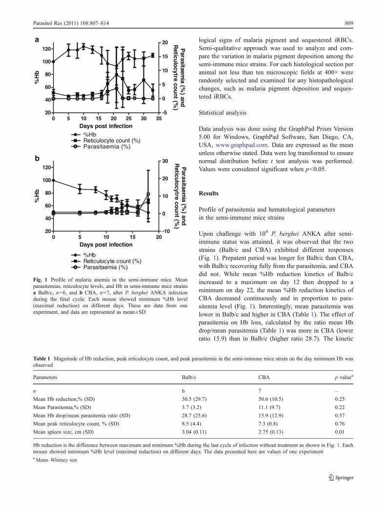
Upon challenge with 104 P. berghei ANKA after semi-immune status was attained, it was observed that the twostrains (Balb/c and CBA) exhibited different responses(Fig. 1). Prepatent period was longer for Balb/c than CBA,with Balb/c recovering fully from the parasitemia, and CBAdid not. While mean %Hb reduction kinetics of Balb/cincreased to a maximum on day 12 then dropped to aminimum on day 22, the mean %Hb reduction kinetics ofCBA decreased continuously and in proportion to para-sitemia level (Fig. 1). Interestingly, mean parasitemia waslower in Balb/c and higher in CBA (Table 1). The effect ofparasitemia on Hb loss, calculated by the ratio mean Hbdrop/mean parasitemia (Table 1) was more in CBA (lowerratio 15.9) than in Balb/c (higher ratio 28.7). The kinetic
0 5 10 15 2020
40
60
80
100
120
-10
0
10
20
30
%HbReticulocyte count (%)Parasitaemia (%)
b
Days post infection
%H
b
Parasitaem
ia (%) an
dR
eticulo
cytre cou
nt (%
)
0 5 10 15 20 25 30 3520
40
60
80
100
120
-5
0
5
10
15
20
%HbReticulocyte count (%)Parasitaemia (%)
a
Days post infection
%H
b
Parasitaem
ia (%) an
dR
eticulo
cytre cou
nt (%
)
Fig. 1 Profile of malaria anemia in the semi-immune mice. Meanparasitemias, reticulocyte levels, and Hb in semi-immune mice strainsa Balb/c, n=6, and b CBA, n=7, after P. berghei ANKA infectionduring the final cycle. Each mouse showed minimum %Hb level(maximal reduction) on different days. These are data from oneexperiment, and data are represented as mean±SD
Table 1 Magnitude of Hb reduction, peak reticulocyte count, and peak parasitemia in the semi-immune mice strain on the day minimum Hb wasobserved
Parameters Balb/c CBA p valuea
n 6 7 –
Mean Hb reduction,% (SD) 30.5 (29.7) 50.6 (10.5) 0.25
Mean Parasitemia,% (SD) 3.7 (3.2) 11.1 (9.7) 0.22
Mean Hb drop/mean parasitemia ratio (SD) 28.7 (25.6) 15.9 (12.9) 0.57
Mean peak reticulocyte count, % (SD) 8.5 (4.4) 7.3 (0.8) 0.76
Mean spleen size, cm (SD) 3.04 (0.11) 2.75 (0.13) 0.01
Hb reduction is the difference between maximum and minimum %Hb during the last cycle of infection without treatment as shown in Fig. 1. Eachmouse showed minimum %Hb level (maximal reduction) on different days. The data presented here are values of one experimentaMann–Whitney test
Parasitol Res (2011) 108:807–814 809

profile of reticulocyte count (Fig. 1) revealed erythropoieticresponse to be slightly better in Balb/c with higher meanpeak reticulocyte count of 8.5% and 7.3% in CBA, p=0.76(Table 1). Splenomegaly was observed in both semi-immune mice, increasing in size between two and threetimes, than the uninfected mice. Increase in spleen size wassignificantly higher in Balb/c, p=0.01 (Table 1).
Histopathological study
Spleen Clear distinction between red and white pulp,resting follicles, and marginal zone was evident in thespleen of normal uninfected control mice (Fig. 2). Thesplenomegaly observed in all the infected semi-immunemice might be as a result of an overall enlargement of thered and white pulp (Martins et al. 2009) as shown in Fig. 2.In addition, the germinal centers in the spleen of semi-immunemice lost their typical structure. Malaria pigment deposition(indicated arrows) was observed in both strains. However, thatof Balb/c was fine while that of CBA were aggregates asshown in Fig. 2. By means of the semi-qualitative approach,it was observed that between two and three thick/clumps of
malaria pigments were seen at 400× magnification of theCBA spleen (Fig. 2), while smaller sizes of the malariapigments were seen in that of Balb/c. Hypertrophy of the redpulp and intense proliferation of the cells in the restingfollicles were also observed in the semi-immune mice.
Liver The liver sections of the normal uninfected controlmice revealed normal, clear architecture, and color, such assinusoids and Kupffer cells, as shown in Fig. 3. Hypertro-phy of Kupffer cells are seen in all the semi-immune micestrains in addition to congested sinusoids (Fig. 3). InfectedRBCs were also not observed, except for some few uRBCs.Meanwhile that of the infected immune mice showed someamount of malaria pigment deposition (indicated arrows) inthe sinusoids. Semi-qualitative approach revealed five toeight malaria pigments thicker/clump in CBA. Those of Balb/c were few in number between 3 and 4, as shown in Fig. 3.
Brain It was also observed that the brain tissues of thenormal uninfected control mice did not show any sign ofRBC sequestration or leukocytes infiltration. Abnormalfeatures, such as hemorrhages and edema, were not
200X
Uninfected Control Balb/c CBA
400X 400X
Fig. 2 Hematoxylin–eosin (HE) staining of spleen of uninfectedcontrol (n=3), semi-immune Balb/c (n=3), and CBA (n=7). Theorgans were removed when similar minimum %Hb reduction wasobserved as shown in Fig. 1 (Balb/c, days 18–22, and CBA, days 16–
18). Sections of these organs were stained with HE and analyzed withlight microscopy. The blue arrows indicate malaria pigment deposi-tion in the spleen
400X
Uninfected Control Balb/c
400X
CBA
400X
Fig. 3 Hematoxylin–eosin (HE) staining of liver of uninfected control(n=3), semi-immune Balb/c (n=3), and CBA (n=7). The organs wereremoved when similar minimum %Hb reduction was observed asshown in Fig. 1 (Balb/c, days 18–22, and CBA, days 16–18). Sections
of these organs were stained with HE and analyzed with lightmicroscopy. The blue arrows indicate malaria pigment deposition inthe liver
810 Parasitol Res (2011) 108:807–814
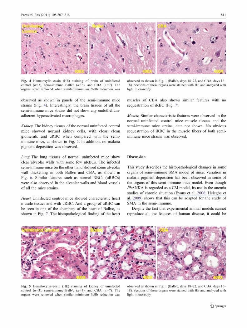
observed as shown in panels of the semi-immune micestrains (Fig. 4). Interestingly, the brain tissues of all thesemi-immune mice strains did not show any endothelium-adherent hyperactivated macrophages.
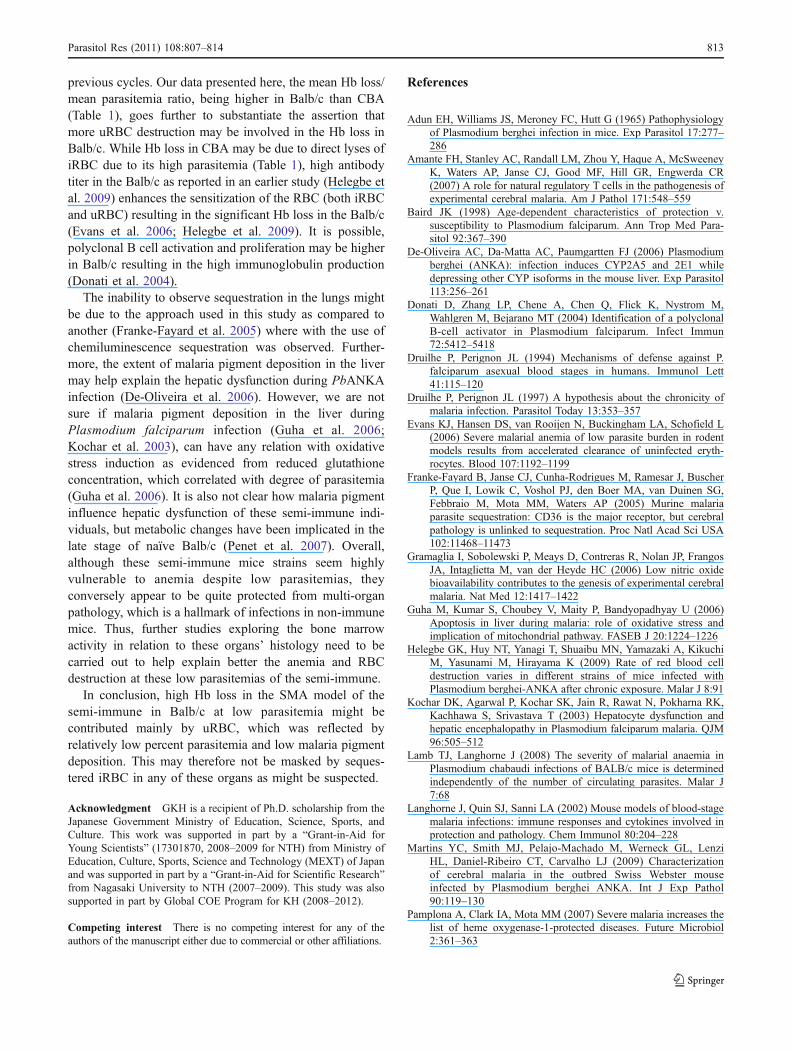
Kidney The kidney tissues of the normal uninfected controlmice showed normal kidney cells, with clear, cleanglomeruli, and uRBC when compared with the semi-immune mice, as shown in Fig. 5. In addition, no malariapigment deposition was observed.
Lung The lung tissues of normal uninfected mice showclear alveolar walls with some few uRBCs. The infectedsemi-immune mice on the other hand showed some alveolarwall thickening in both Balb/c and CBA, as shown inFig. 6. Similar features such as normal RBCs (uRBCs)were also observed in the alveolar walls and blood vesselsof all the mice strains.
Heart Uninfected control mice showed characteristic heartmuscle tissues and with uRBC. And a group of uRBC canbe seen in one of the chambers of the heart of Balb/c, asshown in Fig. 7. The histopathological finding of the heart
muscles of CBA also shows similar features with nosequestration of iRBC (Fig. 7).
Muscle Similar characteristic features were observed in thenormal uninfected control mice muscle tissues and thesemi-immune mice strains, data not shown. No obvioussequestration of iRBC in the muscle fibers of both semi-immune mice strains was observed.
Discussion
This study describes the histopathological changes in someorgans of semi-immune SMA model of mice. Variation inmalaria pigment deposition has been observed in some ofthe organs of this semi-immune mice model. Even thoughPbANKA is regarded as a CM model, its use in the anemiastudies of chronic situation (Evans et al. 2006; Helegbe etal. 2009) shows that this can be adapted for the study ofSMA in the semi-immune.
Despite the fact that experimental animal models cannotreproduce all the features of human disease, it could be
Uninfected Control
Balbc CBA
400X 400X400X
Fig. 4 Hematoxylin–eosin (HE) staining of brain of uninfectedcontrol (n=3), semi-immune Balb/c (n=3), and CBA (n=7). Theorgans were removed when similar minimum %Hb reduction was
observed as shown in Fig. 1 (Balb/c, days 18–22, and CBA, days 16–18). Sections of these organs were stained with HE and analyzed withlight microscopy
Balb/c CBA Uninfected Control
400X 400X 400X
Fig. 5 Hematoxylin–eosin (HE) staining of kidney of uninfectedcontrol (n=3), semi-immune Balb/c (n=3), and CBA (n=7). Theorgans were removed when similar minimum %Hb reduction was
observed as shown in Fig. 1 (Balb/c, days 18–22, and CBA, days 16–18). Sections of these organs were stained with HE and analyzed withlight microscopy
Parasitol Res (2011) 108:807–814 811

explored to explain some phenomena in human situations.Also animal studies have been useful in understanding themechanisms of pathogenetic basis of various diseaseconditions to help in prophylactic or therapeutic interven-tions. Repeated infection plays a role in providing protec-tion to the semi-immune mice strains where antibody titerwas observed to be high (Helegbe et al. 2009). One of theroles of antibody during malaria infection is that it preventsthe cytoadherence of iRBC onto the endothelial walls of theblood vessels (Druilhe and Perignon 1994). Since antibodytiter was observed to increase in semi-immune individuals,it could be an explanation why sequestration was notobserved in the brains of the semi-immune but occurred inthe naïve. This might contribute to less CM incidence inadults (who have high antibody titer due to long exposureto plasmodium infections) than children in endemic areas.While sequestration in the adipose tissue, lung and spleen(Franke-Fayard et al. 2005), and brain hemorrhages(Amante et al. 2007) have been associated with severemalaria in naïve mice, none is reported in the histopatho-logical sections of the organs of each semi-immune micestrain. Our study, however, did not evaluate any study onthe naïve. In addition, the lack of endothelium-adherent
hyperactivated macrophages in the brain, a hall mark ofacute infection by PbANKA, could help explain theabsence of systemic damage.
Splenomegaly, which was observed in all the infectedsemi-immune mice, might be a result of overall enlarge-ment of the red and white pulp (Martins et al. 2009),suggesting hyperactivity of the macrophages. The signifi-cant difference in spleen size of the semi-immune micestrains might suggest differential immune response. Closelyrelated to the high parasitemia in CBA is the high malariapigmentation in the spleen and liver when compared withthe other strain. This is consistent with other studies wherea higher level of parasitemia results in higher amounts offree hemoglobin leading to free heme released (Gramagliaet al. 2006; Pamplona et al. 2007; Penet et al. 2006).Malaria pigment does not only stimulate TNF secretion, butalso impair macrophage function (Turrini et al. 1993).Thus, this relatively higher malaria pigmentation in CBAcould result in higher impairment of macrophage functionthan Balb/c. Hemozoin do persist in macrophages for somemonths. Thus, we do not exclude the fact that aconsiderable amount of the malaria pigment in thehistological sections might have been released during
400X
Uninfected Control CBABalb/c
400X400X
Fig. 6 Hematoxylin–eosin (HE) staining of lung of uninfected control(n=3), semi-immune Balb/c (n=3), and CBA (n=7). The organs wereremoved when similar minimum %Hb reduction was observed as
shown in Fig. 1 (Balb/c, days 18–22, and CBA, days 16–18). Sectionsof these organs were stained with HE and analyzed with lightmicroscopy
CBA Uninfected control Balb/c
400X 400X 400X
Fig. 7 Hematoxylin–eosin (HE) staining of heart of uninfectedcontrol (n=3), semi-immune Balb/c (n=3), and CBA (n=7). Theorgans were removed when similar minimum %Hb reduction was
observed as shown in Fig. 1 (Balb/c, days 18–22, and CBA, days 16–18). Sections of these organs were stained with HE and analyzed withlight microscopy
812 Parasitol Res (2011) 108:807–814
previous cycles. Our data presented here, the mean Hb loss/mean parasitemia ratio, being higher in Balb/c than CBA(Table 1), goes further to substantiate the assertion thatmore uRBC destruction may be involved in the Hb loss inBalb/c. While Hb loss in CBA may be due to direct lyses ofiRBC due to its high parasitemia (Table 1), high antibodytiter in the Balb/c as reported in an earlier study (Helegbe etal. 2009) enhances the sensitization of the RBC (both iRBCand uRBC) resulting in the significant Hb loss in the Balb/c(Evans et al. 2006; Helegbe et al. 2009). It is possible,polyclonal B cell activation and proliferation may be higherin Balb/c resulting in the high immunoglobulin production(Donati et al. 2004).
The inability to observe sequestration in the lungs mightbe due to the approach used in this study as compared toanother (Franke-Fayard et al. 2005) where with the use ofchemiluminescence sequestration was observed. Further-more, the extent of malaria pigment deposition in the livermay help explain the hepatic dysfunction during PbANKAinfection (De-Oliveira et al. 2006). However, we are notsure if malaria pigment deposition in the liver duringPlasmodium falciparum infection (Guha et al. 2006;Kochar et al. 2003), can have any relation with oxidativestress induction as evidenced from reduced glutathioneconcentration, which correlated with degree of parasitemia(Guha et al. 2006). It is also not clear how malaria pigmentinfluence hepatic dysfunction of these semi-immune indi-viduals, but metabolic changes have been implicated in thelate stage of naïve Balb/c (Penet et al. 2007). Overall,although these semi-immune mice strains seem highlyvulnerable to anemia despite low parasitemias, theyconversely appear to be quite protected from multi-organpathology, which is a hallmark of infections in non-immunemice. Thus, further studies exploring the bone marrowactivity in relation to these organs’ histology need to becarried out to help explain better the anemia and RBCdestruction at these low parasitemias of the semi-immune.
In conclusion, high Hb loss in the SMA model of thesemi-immune in Balb/c at low parasitemia might becontributed mainly by uRBC, which was reflected byrelatively low percent parasitemia and low malaria pigmentdeposition. This may therefore not be masked by seques-tered iRBC in any of these organs as might be suspected.
Acknowledgment GKH is a recipient of Ph.D. scholarship from theJapanese Government Ministry of Education, Science, Sports, andCulture. This work was supported in part by a “Grant-in-Aid forYoung Scientists” (17301870, 2008–2009 for NTH) from Ministry ofEducation, Culture, Sports, Science and Technology (MEXT) of Japanand was supported in part by a “Grant-in-Aid for Scientific Research”from Nagasaki University to NTH (2007–2009). This study was alsosupported in part by Global COE Program for KH (2008–2012).
Competing interest There is no competing interest for any of theauthors of the manuscript either due to commercial or other affiliations.
References
Adun EH, Williams JS, Meroney FC, Hutt G (1965) Pathophysiologyof Plasmodium berghei infection in mice. Exp Parasitol 17:277–286
Amante FH, Stanley AC, Randall LM, Zhou Y, Haque A, McSweeneyK, Waters AP, Janse CJ, Good MF, Hill GR, Engwerda CR(2007) A role for natural regulatory T cells in the pathogenesis ofexperimental cerebral malaria. Am J Pathol 171:548–559
Baird JK (1998) Age-dependent characteristics of protection v.susceptibility to Plasmodium falciparum. Ann Trop Med Para-sitol 92:367–390
De-Oliveira AC, Da-Matta AC, Paumgartten FJ (2006) Plasmodiumberghei (ANKA): infection induces CYP2A5 and 2E1 whiledepressing other CYP isoforms in the mouse liver. Exp Parasitol113:256–261
Donati D, Zhang LP, Chene A, Chen Q, Flick K, Nystrom M,Wahlgren M, Bejarano MT (2004) Identification of a polyclonalB-cell activator in Plasmodium falciparum. Infect Immun72:5412–5418
Druilhe P, Perignon JL (1994) Mechanisms of defense against P.falciparum asexual blood stages in humans. Immunol Lett41:115–120
Druilhe P, Perignon JL (1997) A hypothesis about the chronicity ofmalaria infection. Parasitol Today 13:353–357
Evans KJ, Hansen DS, van Rooijen N, Buckingham LA, Schofield L(2006) Severe malarial anemia of low parasite burden in rodentmodels results from accelerated clearance of uninfected eryth-rocytes. Blood 107:1192–1199
Franke-Fayard B, Janse CJ, Cunha-Rodrigues M, Ramesar J, BuscherP, Que I, Lowik C, Voshol PJ, den Boer MA, van Duinen SG,Febbraio M, Mota MM, Waters AP (2005) Murine malariaparasite sequestration: CD36 is the major receptor, but cerebralpathology is unlinked to sequestration. Proc Natl Acad Sci USA102:11468–11473
Gramaglia I, Sobolewski P, Meays D, Contreras R, Nolan JP, FrangosJA, Intaglietta M, van der Heyde HC (2006) Low nitric oxidebioavailability contributes to the genesis of experimental cerebralmalaria. Nat Med 12:1417–1422
Guha M, Kumar S, Choubey V, Maity P, Bandyopadhyay U (2006)Apoptosis in liver during malaria: role of oxidative stress andimplication of mitochondrial pathway. FASEB J 20:1224–1226
Helegbe GK, Huy NT, Yanagi T, Shuaibu MN, Yamazaki A, KikuchiM, Yasunami M, Hirayama K (2009) Rate of red blood celldestruction varies in different strains of mice infected withPlasmodium berghei-ANKA after chronic exposure. Malar J 8:91
Kochar DK, Agarwal P, Kochar SK, Jain R, Rawat N, Pokharna RK,Kachhawa S, Srivastava T (2003) Hepatocyte dysfunction andhepatic encephalopathy in Plasmodium falciparum malaria. QJM96:505–512
Lamb TJ, Langhorne J (2008) The severity of malarial anaemia inPlasmodium chabaudi infections of BALB/c mice is determinedindependently of the number of circulating parasites. Malar J7:68
Langhorne J, Quin SJ, Sanni LA (2002) Mouse models of blood-stagemalaria infections: immune responses and cytokines involved inprotection and pathology. Chem Immunol 80:204–228
Martins YC, Smith MJ, Pelajo-Machado M, Werneck GL, LenziHL, Daniel-Ribeiro CT, Carvalho LJ (2009) Characterizationof cerebral malaria in the outbred Swiss Webster mouseinfected by Plasmodium berghei ANKA. Int J Exp Pathol90:119–130
Pamplona A, Clark IA, Mota MM (2007) Severe malaria increases thelist of heme oxygenase-1-protected diseases. Future Microbiol2:361–363
Parasitol Res (2011) 108:807–814 813

Penet MF, Laigle C, Fur YL, Confort-Gouny S, Heurteaux C,Cozzone PJ, Viola A (2006) In vivo characterization of brainmorphometric and metabolic endophenotypes in three inbredstrains of mice using magnetic resonance techniques. BehavGenet 36:732–744
Penet MF, Kober F, Confort-Gouny S, Le Fur Y, Dalmasso C, ColtelN, Liprandi A, Gulian JM, Grau GE, Cozzone PJ, Viola A (2007)
Magnetic resonance spectroscopy reveals an impaired brainmetabolic profile in mice resistant to cerebral malaria infectedwith Plasmodium berghei ANKA. J Biol Chem 282:14505–14514
Turrini F, Schwarzer E, Arese P (1993) The involvement of hemozointoxicity in depression of cellular immunity. Parasitol Today9:297–300
814 Parasitol Res (2011) 108:807–814