Histopathological Markers for Copper Toxicity in Rainbow Trout Fry (Oncorhynchus mykiss
14
Basrah J. Agric. Sci. 25 (special issued 2), 2012 26 Histopathological Markers for Copper Toxicity in Rainbow Trout Fry (Oncorhynchus mykiss) Nader A. Salman 1 , Jeffery L. Ullman 2 , Kevin Snekvik 3 , Xiao Q. Lu 2 1 Department of Fisheries, College of Agriculture, University of Basrah, Iraq; 2 Department of biological Systems Engineering, Washington State university, USA; 3 College of Veterinary Medicine, Washington State University, Pullman, USA. Abstract. One month old fry of rainbow trout (Oncorhynchus mykiss) of 0.5 g weight were exposed to three levels of CuSO 4 .5H 2 0 at 100, 200 and 400 ppb Cu for 24, 48 , 72 and 96 h periods. Samples for histological sections were taken and prepared for microscopic examination using the standard methods. Due to the small size of the fry, the whole fish was sectioned longitudinally. Four selected organs were examined including gills, liver, kidney and alimentary canal. Gills showed mark changes of lamellar hypertrophy appeared as fusion of secondary lamella and formation of bud-like structures filled with epithelial cells. Hyperplasia and lifting of epithelial cells were also noticed in the high dose media. Signs of necrosis appeared in fish kept at 200 and 400 ppb Cu exposures. Hepatocytes showed less alteration represented by morphological deformities especially in the nuclei which has no crisp shape. Granular cytoplasm was commonly seen. Its relationship with toxicity needs to be confirmed. Up- normal red blood cells and signs of autolysis was also noticed. Mild changes in both tubules and glomeruli of the head and rear kidneys were noticed. Evidence of pykcnotic and degeneration of the tubule epithelial cells was seen in fish exposed to the highest copper concentration. Macrophages along the capillary were also noticed. Some alteration in tubule structure is mostly related to hypoxia. The less affected organ was the alimentary canal which showed minor changes mostly in the upper and middle part. Degeneration of epithelial cells which appeared as vacuoles in the stomach proximal region was noticed. Pyknosis due to starvation was apparent as well. It is concluded that histopathological signs should be considered among other biomarkers as far as Cu-toxicity is concerned. Key Words: Histopathology. Copper toxicity, rainbow trout fry Corresponding author: [email protected] Introduction The contamination of fresh waters with a wide range of pollutants has become a matter of concern over the last few decades (39). Increased human activities especially with rapid development of agriculture and industry has resulted in a considerable increase in levels of pollutant such as heavy metals ( 25). Heavy metals occur naturally in the environment and are found in varying levels in the ground and surface water. Industrial pollution and anthropogenic activities do, however, cause an increased discharge of these metals into natural aquatic ecosystems. Sometimes, aquatic organisms are exposed to unnaturally high levels of these metals. Fish are relatively sensitive to changes in their surroundings environment. Therefore, heavy metals, have been recognized as strong biological poisons because of their persistent nature, toxicity ,tendency to accumulate in organisms and undergo food chain amplification (15, 10). Fish health may therefore reflect and give a good indication of the health status of a specific aquatic ecosystem. Copper can exert adverse toxicological effects, when present in high concentrations in water (32). In fact, it is potentially toxic when the internal available concentration exceeds the
Transcript of Histopathological Markers for Copper Toxicity in Rainbow Trout Fry (Oncorhynchus mykiss

Basrah J. Agric. Sci. 25 (special issued 2), 2012
26
Histopathological Markers for Copper Toxicity in Rainbow Trout
Fry (Oncorhynchus mykiss)
Nader A. Salman
1, Jeffery L. Ullman
2, Kevin Snekvik
3, Xiao Q. Lu
2
1Department of Fisheries, College of Agriculture, University of Basrah, Iraq; 2Department of biological Systems Engineering, Washington State university, USA; 3College of Veterinary
Medicine, Washington State University, Pullman, USA.
Abstract. One month old fry of rainbow trout (Oncorhynchus mykiss) of 0.5 g weight were exposed to three levels
of CuSO4.5H20 at 100, 200 and 400 ppb Cu for 24, 48 , 72 and 96 h periods. Samples for histological sections were
taken and prepared for microscopic examination using the standard methods. Due to the small size of the fry, the
whole fish was sectioned longitudinally. Four selected organs were examined including gills, liver, kidney and
alimentary canal. Gills showed mark changes of lamellar hypertrophy appeared as fusion of secondary lamella and
formation of bud-like structures filled with epithelial cells. Hyperplasia and lifting of epithelial cells were also
noticed in the high dose media. Signs of necrosis appeared in fish kept at 200 and 400 ppb Cu exposures.
Hepatocytes showed less alteration represented by morphological deformities especially in the nuclei which has no
crisp shape. Granular cytoplasm was commonly seen. Its relationship with toxicity needs to be confirmed. Up-
normal red blood cells and signs of autolysis was also noticed. Mild changes in both tubules and glomeruli of the
head and rear kidneys were noticed. Evidence of pykcnotic and degeneration of the tubule epithelial cells was seen
in fish exposed to the highest copper concentration. Macrophages along the capillary were also noticed. Some
alteration in tubule structure is mostly related to hypoxia. The less affected organ was the alimentary canal which
showed minor changes mostly in the upper and middle part. Degeneration of epithelial cells which appeared as
vacuoles in the stomach proximal region was noticed. Pyknosis due to starvation was apparent as well. It is
concluded that histopathological signs should be considered among other biomarkers as far as Cu-toxicity is
concerned.
Key Words: Histopathology. Copper toxicity, rainbow trout fry
Corresponding author: [email protected]
Introduction
The contamination of fresh waters with a wide range of pollutants has become a matter of
concern over the last few decades (39). Increased human activities especially with rapid
development of agriculture and industry has resulted in a considerable increase in levels of
pollutant such as heavy metals ( 25). Heavy metals occur naturally in the environment and are
found in varying levels in the ground and surface water. Industrial pollution and anthropogenic
activities do, however, cause an increased discharge of these metals into natural aquatic
ecosystems. Sometimes, aquatic organisms are exposed to unnaturally high levels of these
metals. Fish are relatively sensitive to changes in their surroundings environment. Therefore,
heavy metals, have been recognized as strong biological poisons because of their persistent
nature, toxicity ,tendency to accumulate in organisms and undergo food chain amplification (15,
10). Fish health may therefore reflect and give a good indication of the health status of a specific
aquatic ecosystem.
Copper can exert adverse toxicological effects, when present in high concentrations in
water (32). In fact, it is potentially toxic when the internal available concentration exceeds the
Basrah J. Agric. Sci. 25 (special issued 2), 2012
27
capacity of physiological detoxification processes. High concentrations of this heavy metal were
detected in some aquatic ecosystems collecting vineyard runoff water and it is also highly
concentrated in ground water (12) (36). There are also anthropogenic sources of environmental
contamination by copper including mining, smelting, foundries, municipal waste incinerators,
burning of coal for power generation and a variety of copper-based products used in building and
construction (26)(43).
Fishes exposed to toxicants undergo stress, which is a state of re-established
homeostasis. Fish are responding to various stressors by a series of biochemical and
physiological stress reactions, so called secondary stress responses comparable to those of higher
vertebrates (22). Biochemical and physiological biomarkers are frequently used for detecting or
diagnosing sublethal effects in fish exposed to different toxic substances (37). The toxicity of
Cu2 is a stress factor for fish, but the physiological responses to stress can be alleviated by a
variety of acclimation mechanisms (23). Usually, the process of Cu2 accumulation is
characterized as a damage-repair model (19), and it includes three phases: an initial shock phase,
a recovery phase, and then acclimation itself. The last phase includes increased tolerance. The
initial shock phase corresponds to physiological regulation. The damage phase is usually short
lived (a few days) and consists of assorted disturbances to internal physiological homeostasis.
The toxicity tests are, therefore, necessary in water pollution evaluation because chemical and
physical measurements alone are not sufficient to assess potential effects on aquatic biota (35).
It has been noted that heavy metals had a negative impact on all relevant parameters and
caused histo-pathological changes in fish. After conducting a survey on Poecilia reticulata
(guppy) and Oryzias latipes (medaka) to examine the usefulness of histopathology for the
identification of toxic effects of environmental contaminants in fish (42), they concluded that
histopathology provides useful data in characterizing toxic effects in fish. Histological study
appears to be a very sensitive parameter and is crucial in determining cellular changes that may
occur in target organs, such as the gills, liver, kidney and gut (1). Sub-lethal copper exposure
may cause damage to numerous tissues and organs, including liver, gills, kidneys. If any of these
changes can be related to exposure concentration or time and show enough specificity it can be
used as a biomarker (33). The aim of this research is to evaluate the usefulness of histological
biomarkers in gills, kidney and gut for toxicity of acute exposure of rainbow trout fry to copper.
Materials and Methods
Copper exposure trials
Copper stock solution used in the toxicity tests was prepared by dissolving Cu2SO4.5H2O into a
desirable concentration using Milli Q water. Exposure of rainbow trout fry was conducted in
glass aquaria (28 x 72 x 42 cm) under controlled temperature (13 ± 1 oC) and artificial aeration.
Forty randomly chosen rainbow trout fry raised in our fish laboratory were acclimatized to each
test aquarium for 24h with aeration. Afterwards, the fish were exposed to copper by spiking the
metal with different concentrations (0, 100, 200 and 400µg/L) into the aquarium.
Food was not provided during the acclimation and in the course of exposures. Copper
concentration in water was analysed after copper stock solution was spiked into the aquarium
and was then analysed after the exposure experiment was completed. At the end of the exposure
(24, 48, 72 and 96 h), the whole fish were anaesthetised using a solution containing 0.1% of
Tricaine anesthetics and then fixed in 10% phosphate buffered formalin for histological
examination.
Basrah J. Agric. Sci. 25 (special issued 2), 2012
28
Preparation of histological sections
The whole larva were fixed in Bouin's fluid for 24 h, dehydrated in graded ethanol
concentrations, cleared in xylene and infiltrated with paraffin using Tissue Tek II automatic
tissue processor. They were embedded in paraffin using Shandon Histocentre embedding
machine. Sections (5m) were cut using a Microm microtome.
The sections were attached to slides and stained with haematoxylin and eosin (H&E)
using Shandon Linistain GLX automatic staining machine. The slides were examined using
Leica DMLS microscope and photographs were taken using Wild Leitz MPS46/52 photoautomat
using magnification x 100 for scanning all the slides and x 400 for examination of any
pathological changes found. Presence of histological changes and their severity were determined
for each fish. All histological processing and examination of slides were performed at the
College of Veterinary Medicine, Washington State University.
Results
1. Gills
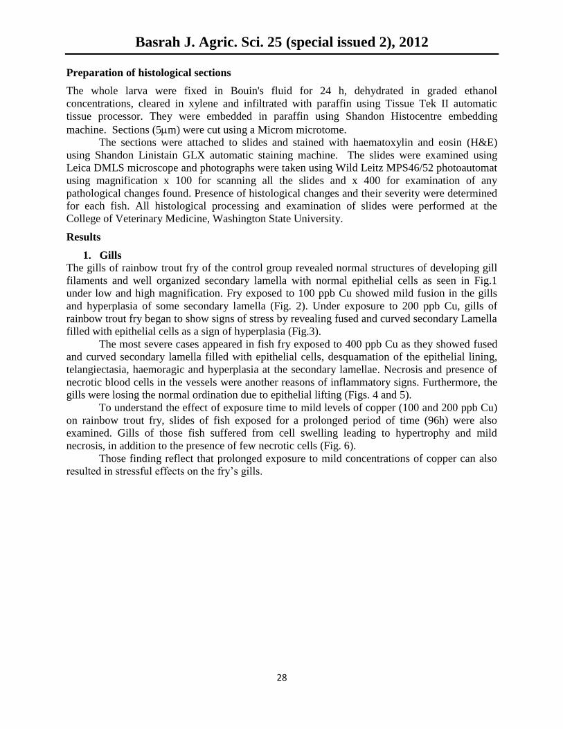
The gills of rainbow trout fry of the control group revealed normal structures of developing gill
filaments and well organized secondary lamella with normal epithelial cells as seen in Fig.1
under low and high magnification. Fry exposed to 100 ppb Cu showed mild fusion in the gills
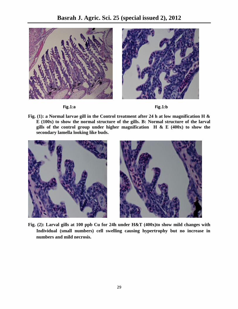
and hyperplasia of some secondary lamella (Fig. 2). Under exposure to 200 ppb Cu, gills of
rainbow trout fry began to show signs of stress by revealing fused and curved secondary Lamella
filled with epithelial cells as a sign of hyperplasia (Fig.3).
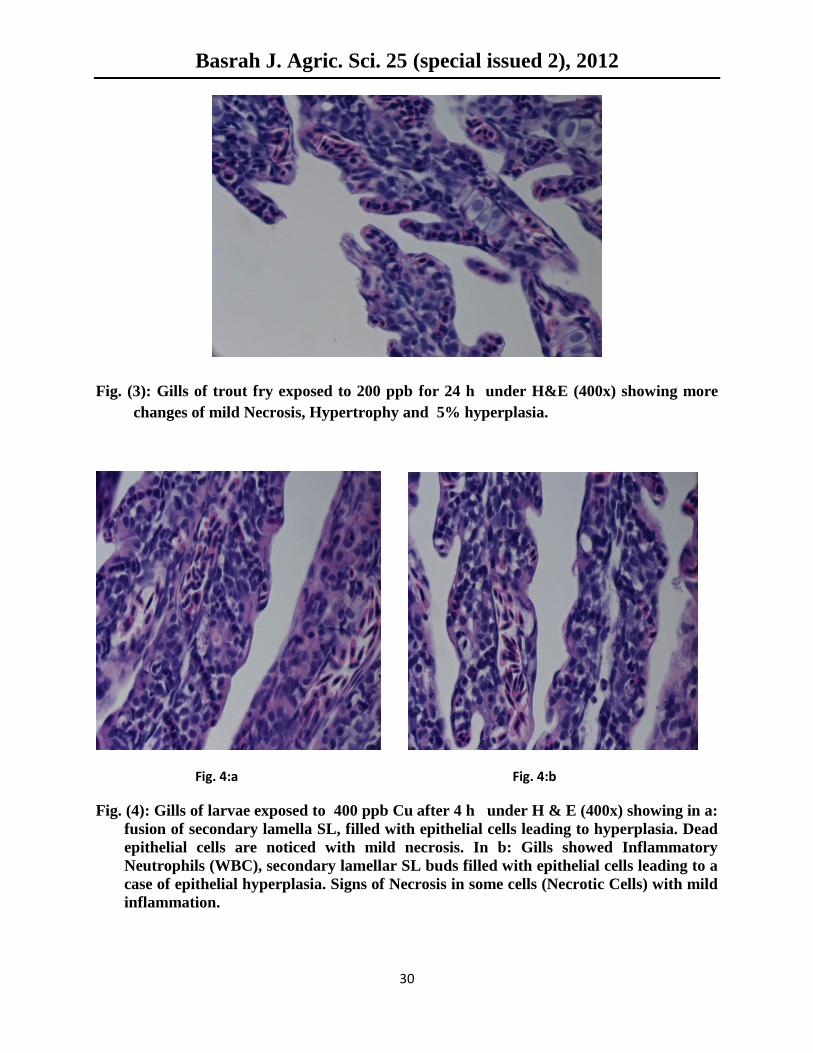
The most severe cases appeared in fish fry exposed to 400 ppb Cu as they showed fused
and curved secondary lamella filled with epithelial cells, desquamation of the epithelial lining,
telangiectasia, haemoragic and hyperplasia at the secondary lamellae. Necrosis and presence of
necrotic blood cells in the vessels were another reasons of inflammatory signs. Furthermore, the
gills were losing the normal ordination due to epithelial lifting (Figs. 4 and 5).
To understand the effect of exposure time to mild levels of copper (100 and 200 ppb Cu)
on rainbow trout fry, slides of fish exposed for a prolonged period of time (96h) were also
examined. Gills of those fish suffered from cell swelling leading to hypertrophy and mild
necrosis, in addition to the presence of few necrotic cells (Fig. 6).
Those finding reflect that prolonged exposure to mild concentrations of copper can also
resulted in stressful effects on the fry’s gills.
Basrah J. Agric. Sci. 25 (special issued 2), 2012
29
Fig.1:a Fig.1:b
Fig. (1): a Normal larvae gill in the Control treatment after 24 h at low magnification H &
E (100x) to show the normal structure of the gills. B: Normal structure of the larval
gills of the control group under higher magnification H & E (400x) to show the
secondary lamella looking like buds.
Fig. (2): Larval gills at 100 ppb Cu for 24h under H&T (400x)to show mild changes with
Individual (small numbers) cell swelling causing hypertrophy but no increase in
numbers and mild necrosis.
Basrah J. Agric. Sci. 25 (special issued 2), 2012
30
Fig. (3): Gills of trout fry exposed to 200 ppb for 24 h under H&E (400x) showing more
changes of mild Necrosis, Hypertrophy and 5% hyperplasia.
Fig. 4:a Fig. 4:b
Fig. (4): Gills of larvae exposed to 400 ppb Cu after 4 h under H & E (400x) showing in a:
fusion of secondary lamella SL, filled with epithelial cells leading to hyperplasia. Dead
epithelial cells are noticed with mild necrosis. In b: Gills showed Inflammatory
Neutrophils (WBC), secondary lamellar SL buds filled with epithelial cells leading to a
case of epithelial hyperplasia. Signs of Necrosis in some cells (Necrotic Cells) with mild
inflammation.
Basrah J. Agric. Sci. 25 (special issued 2), 2012
31
Fig. (5): Gills of Larvae exposed to 400 ppb CU after 28h under H&E (400x) showing
severe malfunctioning signs such as: Stuffing of epithelial cells, lifting, hyperplasia,
necrosis is spreading, dilating causing epithelium take off, deterioration leading to loss
of the normal ordination, blood cells RBC coming out of vessels, and finally dead cells
covering SL.
Fig. (6): (left) Gills of larvae exposed to 100 ppb Cu for 96h under H&E (400x) showing
hypertrophy, little hyperplasia and few necrotic cells. (right) Gills of larvae exposed to
200 ppb for 96h H&E (400x) showing hyperplasia , slight centric necrosis, hypertrophy
(puncture) and fusion of secondary lamella.
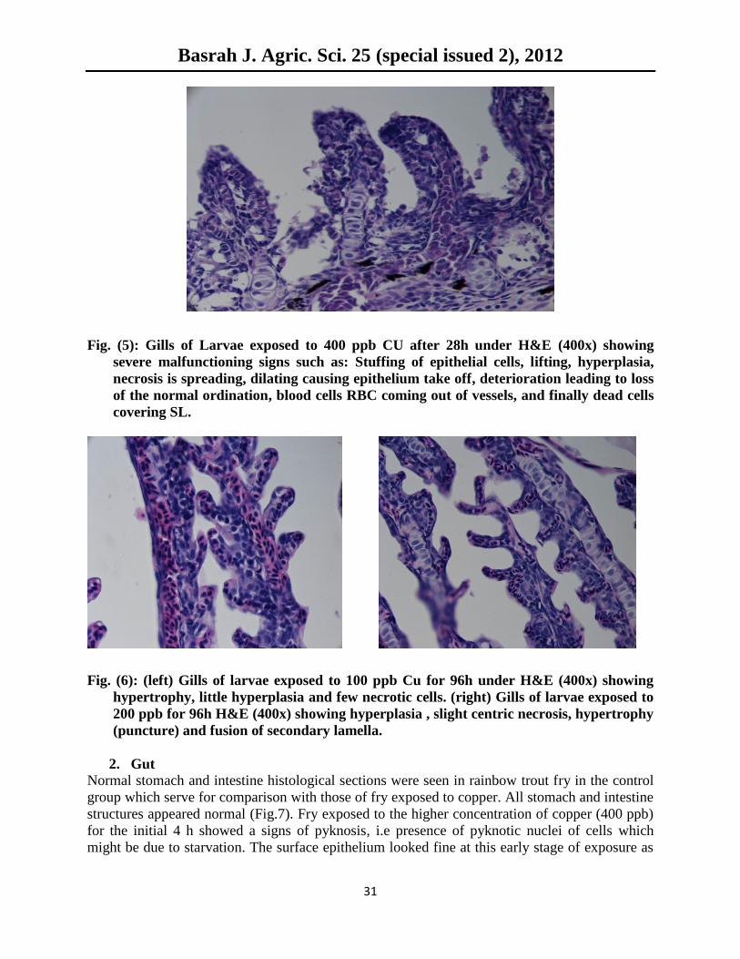
2. Gut
Normal stomach and intestine histological sections were seen in rainbow trout fry in the control
group which serve for comparison with those of fry exposed to copper. All stomach and intestine
structures appeared normal (Fig.7). Fry exposed to the higher concentration of copper (400 ppb)
for the initial 4 h showed a signs of pyknosis, i.e presence of pyknotic nuclei of cells which
might be due to starvation. The surface epithelium looked fine at this early stage of exposure as
Basrah J. Agric. Sci. 25 (special issued 2), 2012
32
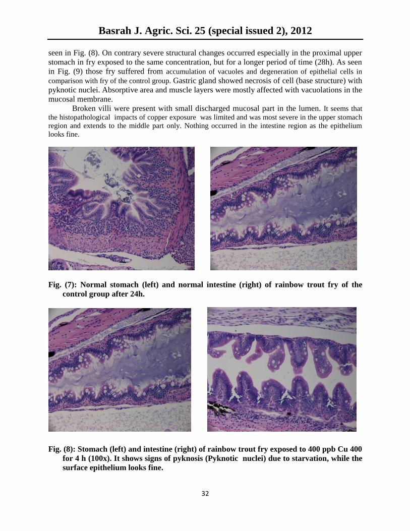
seen in Fig. (8). On contrary severe structural changes occurred especially in the proximal upper
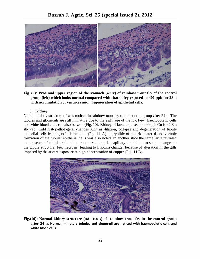
stomach in fry exposed to the same concentration, but for a longer period of time (28h). As seen
in Fig. (9) those fry suffered from accumulation of vacuoles and degeneration of epithelial cells in
comparison with fry of the control group. Gastric gland showed necrosis of cell (base structure) with
pyknotic nuclei. Absorptive area and muscle layers were mostly affected with vacuolations in the
mucosal membrane.
Broken villi were present with small discharged mucosal part in the lumen. It seems that
the histopathological impacts of copper exposure was limited and was most severe in the upper stomach
region and extends to the middle part only. Nothing occurred in the intestine region as the epithelium
looks fine.
Fig. (7): Normal stomach (left) and normal intestine (right) of rainbow trout fry of the
control group after 24h.
Fig. (8): Stomach (left) and intestine (right) of rainbow trout fry exposed to 400 ppb Cu 400
for 4 h (100x). It shows signs of pyknosis (Pyknotic nuclei) due to starvation, while the
surface epithelium looks fine.
Basrah J. Agric. Sci. 25 (special issued 2), 2012
33
Fig. (9): Proximal upper region of the stomach (400x) of rainbow trout fry of the control
group (left) which looks normal compared with that of fry exposed to 400 ppb for 28 h
with accumulation of vacuoles and degeneration of epithelial cells.
3. Kidney
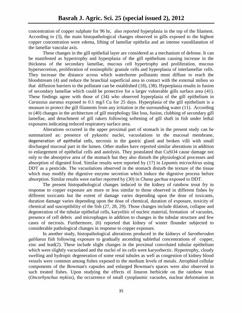
Normal kidney structure of was noticed in rainbow trout fry of the control group after 24 h. The
tubules and glomeruli are still immature due to the early age of the fry. Few haemopoietic cells
and white blood cells can also be seen (Fig. 10). Kidney of larva exposed to 400 ppb Cu for 4-8 h
showed mild histopathological changes such as dilation, collapse and degeneration of tubule
epithelial cells leading to Inflammation (Fig. 11 A). karyolitic of nucleic material and vacuole
formation of the tubular epithelial cells was also noted. In another slide the same larva revealed
the presence of cell debris and microphages along the capillary in addition to some changes in
the tubule structure. Few necrosis leading to hypoxia changes because of alteration in the gills
imposed by the severe exposure to high concentration of copper (Fig. 11 B).
Fig.(10): Normal kidney structure (H&E 100 x) of rainbow trout fry in the control group
after 24 h. Normal immature tubules and glomeruli are noticed with haemopoietic cells and white blood cells.

Basrah J. Agric. Sci. 25 (special issued 2), 2012
34
A B
Fig. 11: Kidney of larvae (H&E 400x) exposed to 400 ppb Cu for 4 h.
A : Dilated and collapse of tubules, degenerating of epithelial cells and Inflammation.
B: Cell debris – microphages along the capillary, few necrosis from the gills, changes in
tubule structure, hypoxia changes because of alteration in gills.
Discussion
Findings of the present study indicated clearly that histopathological markers can be used as
clear evident of copper toxicity in rainbow trout fry. As seen in the results, the three studied
organs showed marked alterations in fish exposed to the highest copper concentration of 400
ppb for 34,48 and 96h. Mild changes were also noticed among fish fry exposed to the medium
(200 ppb) and low (100 ppb) copper concentrations. There was an evident of severe changes with
increasing the copper concentration and with prolonging the exposure time intervals. Findings of
the present study agreed with other studies investigating the histopathological changes in various
tissues in response to copper toxicity in many other fish species (42, 33, 1).
In aquatic environments with elevated levels of copper the primary method of uptake of
the metal is via the gills (13). Gills are the first target of waterborne pollutants due to the
constant contact with the external environment, as well as the main place for copper uptake (7)
(31). Copper diffuses passively across the gill epithelia into the bloodstream, although active
transport may also occur (44). Once copper has entered the bloodstream it is transported to the
liver which is the primary site of accumulation of the metal within fish (33). It is well known that
changes in fish gill are among the most commonly recognized responses to environmental
pollutants (21, 20, 5). Histological study of the gills shows a typical structural organization of the lamella in the
untreated fish. However, fish exposed to copper shows several histological alterations, namely
lamellar epithelium lifting, epithelium proliferation, lamellar axis vasodilation, fusion of
lamellae and lamellar aneurisms. These gill histological alterations has been observed by several
authors in fish submitted to copper (16, 8, 9). Changes in the gills of the juvenile common carp,
Cyprinus carpio (L.) exposed to copper have been reported by (14). The exposure has resulted in
separation of epithelial secondary gill lamellae, hyperplasia, fusion of secondary lamellae and
necrosis. The results of (24) on gold fish Carassius auratus which was subjected to sub-lethal
Basrah J. Agric. Sci. 25 (special issued 2), 2012
35
concentration of copper sulphate for 96 hr, also reported hyperplasia in the top of the filament.
According to (3), the main histopathological changes observed in gills exposed to the highest
copper concentration were edema, lifting of lamellar epithelia and an intense vasodilatation of
the lamellar vascular axis.
These changes in the gill epithelial layer are considered as a mechanism of defense. It can
be manifested as hypertrophy and hyperplasia of the gill epithelium causing increase in the
thickness of the secondary lamellae, mucous cell hypertrophy and proliferation, mucous
hypersecretion, proliferation of eosinophilic granule cells and hyperplasia of interlamellar cells.
They increase the distance across which waterborne pollutants must diffuse to reach the
bloodstream (4) and reduce the branchial superficial area in contact with the external milieu so
that diffusion barriers to the pollutant can be established (18), (38). Hyperplasia results in fusion
of secondary lamellae which could be protective for a larger vulnerable gills surface area (41).
These findings agree with those of (34) who observed hyperplasia of the gill epithelium in
Carassius auratus exposed to 0.1 mg/l Cu for 25 days. Hyperplasia of the gill epithelium is a
measure to protect the gill filaments from any irritation in the surrounding water (11). According
to (40) changes in the architecture of gill morphology like loss, fusion, clubbing of secondary gill
lamellae, and detachment of gill rakers following softening of gill shaft in fish under lethal
exposures indicating reduced respiratory surface area.
Alterations occurred in the upper proximal part of stomach in the present study can be
summarized as: presence of pyknotic nuclei, vacuolations in the mucosal membrane, degeneration of epithelial cells, necrosis in the gastric gland and broken villi with small
discharged mucosal part in the lumen. Other studies have reported similar alterations in addition
to enlargement of epithelial cells and autolysis. They postulated that CuSO4 cause damage not
only to the absorptive area of the stomach but they also disturb the physiological processes and
absorption of digested food. Similar results were reported by (17) in Lepomis microchirus using
DDT as a pesticide. The vacuolations observed in the stomach disturb the texture of the tissue
which may modify the digestive enzyme secretion which induce the digestive process before
absorption. Similar results were earlier reported by (30) in Chana gachua exposed to DDT.
The present histopathological changes induced in the kidney of rainbow trout fry in
response to copper exposure are more or less similar to those observed in different fishes by
different toxicants but the extent of damage varies depending upon the dose of toxicants,
duration damage varies depending upon the dose of chemical, duration of exposure, toxicity of
chemical and susceptibility of the fish (27, 28, 29). Those changes include dilation, collapse and
degeneration of the tubular epithelial cells, karyolitic of nucleic material, formation of vacuoles,
presence of cell debris and microphages in addition to changes in the tubular structure and few
cases of necrosis. Furthermore, (6) reported that kidney of winter flounder subjected to
considerable pathological changes in response to copper exposure.
In another study, histopathological alterations produced in the kidneys of Sarotherodon
galilaeus fish following exposure to gradually ascending sublethal concentrations of copper,
zinc and lead(2). These include slight changes in the proximal convoluted tubular epithelium
which were slightly vacuolated and the nuclei of its cells were karyorhectic. Hypertrophy, cloudy
swelling and hydropic degeneration of some renal tubules as well as congestion of kidney blood
vessels were common among fishes exposed to the medium levels of metals. Atrophied cellular
components of the Bowman's capsules and enlarged Bowman's spaces were also observed in
such treated fishes. Upon studying the effects of linuron herbicide on the rainbow trout
(Oncorhynchus mykiss), the occurrence of small cytoplasmic vacuoles, nuclear deformation in
Basrah J. Agric. Sci. 25 (special issued 2), 2012
36
the epithelium of the first and second segments of the proximal tubule was reported (29). The
kidney cells were observed to have been massively destroyed, the renal corpuscles of the kidney
were scattered resulting in their disorganization and consequently obstruction to their
physiological functions. Such alterations were not seen in the present study.
Acknowledgement
The sponsorship of International Institute of Education (IIE) and SRF-Iraq program to the
fellowship of the first author in Washington State University, Pullman is highly appreciated.
References
1. Abdel-Warith, A. A., Younis, E. M., Al-Asgah, N. A., and Wahbi, O. M. 2011. Effect of
zinc toxicity on liver histology of Nile tilapia, Oreochromis niloticus. Scientific Research
and Essays Vol. 6(17), pp. 3760-3769.
2. Al-Zahaby, A,S,;Hemmaid, K.Z.; Carnal, A.M, and Ghoncmy, 0.1. 1998. The pollutant
effect of copper, zinc and lead on the histological patterns of fish kidney. Egypt J. Aquat
Eioi & Fish., Vol. 2, No. 3 :15-41.
3. António Figueiredo-Fernandes; Jorge V. Ferreira-Cardoso; Sofia Garcia-Santos; Sandra
M. Monteiro; João Carrola; Pedro Matos; António Fontaínhas-Fernandes. 2007
Histopathological changes in liver and gill epithelium of Nile tilapia, Oreochromis
niloticus, exposed to waterborne copper. Pesq. Vet. Bras. vol.27 no.3.
4. Arellano J.M., Storch V. & Sarasquete C. 1999. Histological changes and copper
accumulation in liver and gills of the Senegales sole, Solea senegalensis. Ecotoxicol.
Environ. Saf. 44:62-72.
5. Au D.W.T. 2004. The application of histo-cytopathological biomarkers in marine
pollution monitoring: a review. Mar. Poll. Bull. 48:817-834.
6. Baker, J.T.P.(1969): Histological and electron microscopical observations on copper
poisoning in the winter flounder (Pseudopleuranectes americanus). J.Fish. Res. Bd.
Canada, 26:2785-2739.
7. Campbell H.A., Handy R.D. & Nimmo M. 1979. Copper uptake kinetics across the gills
of rainbow trout (Oncorhyncus mykiss) measured using an improved isolated perfused
head technique. Aquat. Toxicol. 46:177-190.
8. Chen J.C. & Lin C.H. 2001. Toxicity of copper sulfate for survival, growth, molting and
feeding of juveniles of the tiger shrimp, Penaeus monodon. Aquaculture 192:55-65.
9. De Boeck G., Vlaeminck A., Balm P.H., Lock R.A., De Wachter B. & Blust R. 2001.
Morphological and metabolic changes in common carp, Cyprinus carpio, during short-
term copper exposure: interactions between Cu2+
and plasma cortisol elevation. Environ.
Toxicol. Chem. 20:374-381.
10. Dinodia, G.S., Gupta, R. K., Jain K.L. 2002. Effect of cadmium toxicity on liver
glycogen in some fresh water fishes. Proc. XI Natl., Symp. Environ., 236-238.
11. Eller, L.L. 1975. Gill lesions in freshwater teleosts. pp 305 – 330. In: W.E. Ribelin and
G. Migaki (eds.) The pathology of fishes. University of Wisconsin Press, Madison,
Wisconsin.
Basrah J. Agric. Sci. 25 (special issued 2), 2012
37
12. Gerbe 1996. "Toxicologie, Ecotoxicologie des pesticides et des métaux lourds", p.39. In:
Premier rapport d'activité, Programme de Research Europol'Agro (ed.), Faculté de
Sciences, Université de Reims Champagne-Ardenne, Reims, France.
13. Grosell, M., Wood, C.M. 2002. Copper uptake across rainbow trout gills: mechanisms of
apical entry. J. Exp. Biol. 205: 1179–1188.
14. Hassan, B.K. 2011. The effect of copper and cadmium on oxygen consumption of the
juvenile common carp, Cyprinus carpio (L.) Mesopot. J. Mar. Sci., 26 (1): 25 - 34.
15. Kamble G.B., Muley D.V.2000. Effect of acute exposure of endosulfan and
chlorpyriphos on the biochemical composition of the freshwater fish, Sarotherodon
mossambicus. Ind. J. Environ. Sci., 4(1): 97-102.
16. Karan V., Vitorovic S., Tutundzic V. & Poleksic V. 1998. Functional enzymes activity
and gill histology of carp after copper sulfate exposure and recovery. Ecotoxicol.
Environ. Safety 40:49-55. Karan V., Vitorovic S., Tutundzic V. & Poleksic V. 1998.
Functional enzymes activity and gill histology of carp after copper sulfate exposure and
recovery. Ecotoxicol. Environ. Safety 40:49-55.
17. King F. 1962. Some effects of DDT on blue gill Lepomis microchirus- Trans. Am. Fish.
Soc. 92 : 372-378.
18. Laurèn D.J. & McDonald D.G. 1985. Effects of copper on branchial ionoregulation in the
rainbow trout, Salmo gairdneri Richardson: modulation by water hardness and pH. J.
Comp. Physiol. B 155:635-644.
19. Laurén, D.J., and McDonald, D.G. 1987. Acclimation to copper by rainbow trout, Salmo
gairdneri: physiology. Can. J. Fish. Aquat. Sci. 44: 99–104.
20. Laurent P. & Perry S.F. 1991. Environmental effects on fish gill morphology. Physiol.
Zool. 53:4-25.
21. Mallatt J. 1985. Fish gill structural changes induced by toxicants and other irritants: a
statistical review. Can. J. Fish. Aquat. Sci. 42:630-648.
22. Mazeaud, M. and Mazeaud, F. 1981. Adrenergic responses to stress in fish. Pages 49-57
In A. D. Pickring, editor. In Stress and Fish. Academic press New York & London.
23. McGeer, J.C., Szebedinszky, C., McDonald, D.G., and Wood, C.M. 2000. Effects of
sublethal exposure to waterborne Cu, Cd or Zn in rainbow trout. 1: Ionoregulatory
disturbance and metabolic costs. Aquat. Toxicol. 50: 231–243.
24. Muhvich, A.G., Jones, R.T., Kane, A.S., Anderson, R.S. and Reimscheussel, R. 1995.
Effects of chronic copper exposure on the macrophage chemiluminescent response and
gill histology in gold fish Carassius auratus (L.). Fish and shell fish Immun., 5: 251-264.
25. Murugan, S.S., Karuppasamy, R., Poongodi, K. and Puvaneswari, S. 2008.
Bioaccumulation pattern of zinc in fresh water fish Channa punctatus (Bloch) after
chronic exposure. Turk. J. Fish. Aqua. Sci., 8: 55-59.
26. Nor Y.M. 1987. Ecotoxicology of copper to aquatic biota: A review. Environ. Res.
43:274-282.
Basrah J. Agric. Sci. 25 (special issued 2), 2012
38
27. Olufayo M.O. 2009. Haematological Characteristics of C. gariepinus juveniles exposed
to Derris root powder. Afr. J. Food Agric. Nutr. Dev. 9(3):921-933.
28. Olufayo, M. O. and Alade, O. H. 2012. .Acute toxicity and histological changes in gills,
liver and kidney of catfish, Heterobranchus bidorsalis exposed to cypermethrin
concentration, African Journal of Agricultural Research Vol. 7(31), pp. 4453-4459.
29. Oulmi Y, Negele RD, Braunbeck T (1995). Cytopathology of liver and kidney in rainbow
trout (Oncorhynchus mykiss) after long-term exposure to sub lethal concentrations of
linuron. Dis. Aqua. Org. 21:35-52.
30. Paraveen D. 1980. Effect of DDT on digestive tract of Channa gachua. IV All India
Science Congress, Bhopal University, Bhopal. Abstr. No. TC 18.
31. Perry S.F. & Laurent P. 1993. Environmental effects on fish gill structure and function,
p.231-264. In: Rankin J.C. & Jensen F.B. (ed.), Fish Ecophysiology. Chapman and Hall,
London.
32. Pelgrom S., Lamers L., Lock R., Balm P. & Wendelaar Bonga S. 1995. Integrated
physiological response of tilapia, Orechromis mossambicus, to sublethal copper
exposure. Aquat. Toxicol. 32:303-320.
33. Sorensen E.M. 1991. Cadmium. In: Metal poisoning in fish. CRC Press, Boca Raton,
Florida, pp 175–234
34. Sultan, S. and Khan, S.M. 1983. Histopathological studies on the liver and gills in
Carassius auratus exposed to copper sulphate. Indian Journal of Fisheries 30 (1): 96 –98.
35. Tarzwell, C. M. (1971): Bioassays to determine allowable waste concentrations in the
aquatic environment. I.Measurement of pollution effects on living organisms. Proc.
Research Society
36. Teisseire H. 1999. Toxicologie et écotoxicologie des pesticides et des métaux lourds
susceptibles d'être présents dans le vignoble champenois: étude de leur impact
physiologique et biochimique sur Lemna minor. Université de Reims Champagne-
Ardenne, Reims, France. 272 p.
37. Theodorakis, C. W.; D'Surney, S. J.; Bickham, J. W.; Lyne, T. B.; Bardley, B. P.;
Hawkins, W. E.; Farkas, W. L.; Mc.Carthy, J. F. and Shugart, L. R. 1992. Ecotoxicology
1: 45-73.
38. Van Heerden D., Vosloo A. & Nikinmaa M. 2004. Effects of short-term copper exposure
on gill structure, methallothionein and hypoxia-inducible factor-1á (HIF-1á) levels in
rainbow trout (Oncorhynchus mykiss). Aquat. Toxicol. 69:271-280.
39. Vinodhini, R. and Narayanan, M. 2008. Bioaccumulation of heavy metals inorgans of
fresh water fish Cyprinius carpio. Int. J. Environ. Sci. Tech., 5(2):179-182.
40. Vutukuru SS, Suma C, Radha Madhavi K, Juveria, Smitha Pauleena J, Venkateswara Rao
J, Anjaneyulu Y. 2005. Studies on the development of potential biomarkers for rapid
assessment of copper toxicity to freshwater fish using Esomus danricus as model. Int J
Environ Res Public Health. 2(1):63-73.
Basrah J. Agric. Sci. 25 (special issued 2), 2012
39
41. Wangsongsak, A. 2003. Toxicity of cadmium on histology of gill, liver and kidney in
Puntius gonionotus (Bleeker). Thesis, Mahidol University. India. 92pp.
42. Wester PW, Canton JH. 1991 The usefulness of histopathology in aquatic toxicity
studies. Comp Biochem Physiol C.100(1-2):115-7.
43. WHO. 1998. Copper. Environmental Health Criteria 200. IPCS-International Programme
on Chemical Safety, World Health Organization, Geneva.
44. Wilson R.W., Taylor E.W. 1993. The physiological response of freshwater rainbow trout,
Oncorhynchus mykiss, during acutely lethal copper exposure. J Comp Physiol B 163:38–
47.