Identification of a Novel Streptococcal Gene Cassette Mediating SOS Mutagenesis in Streptococcus...
14
Published Ahead of Print 18 May 2007. 2007, 189(14):5210. DOI: 10.1128/JB.00473-07. J. Bacteriol. and Pekka Varmanen Emilia Varhimo, Kirsi Savijoki, Jari Jalava, Oscar P. Kuipers Streptococcus uberis in Gene Cassette Mediating SOS Mutagenesis Identification of a Novel Streptococcal http://jb.asm.org/content/189/14/5210 Updated information and services can be found at: These include: REFERENCES http://jb.asm.org/content/189/14/5210#ref-list-1 at: This article cites 67 articles, 36 of which can be accessed free CONTENT ALERTS more» articles cite this article), Receive: RSS Feeds, eTOCs, free email alerts (when new http://journals.asm.org/site/misc/reprints.xhtml Information about commercial reprint orders: http://journals.asm.org/site/subscriptions/ To subscribe to to another ASM Journal go to: on September 25, 2014 by guest http://jb.asm.org/ Downloaded from on September 25, 2014 by guest http://jb.asm.org/ Downloaded from
-
Upload
independent -
Category
Documents
-
view
3 -
download
0
Transcript of Identification of a Novel Streptococcal Gene Cassette Mediating SOS Mutagenesis in Streptococcus...
Published Ahead of Print 18 May 2007. 2007, 189(14):5210. DOI: 10.1128/JB.00473-07. J. Bacteriol.
and Pekka VarmanenEmilia Varhimo, Kirsi Savijoki, Jari Jalava, Oscar P. Kuipers
Streptococcus uberisin Gene Cassette Mediating SOS Mutagenesis Identification of a Novel Streptococcal
http://jb.asm.org/content/189/14/5210Updated information and services can be found at:
These include:
REFERENCEShttp://jb.asm.org/content/189/14/5210#ref-list-1at:
This article cites 67 articles, 36 of which can be accessed free
CONTENT ALERTS more»articles cite this article),
Receive: RSS Feeds, eTOCs, free email alerts (when new
http://journals.asm.org/site/misc/reprints.xhtmlInformation about commercial reprint orders: http://journals.asm.org/site/subscriptions/To subscribe to to another ASM Journal go to:
on Septem
ber 25, 2014 by guesthttp://jb.asm
.org/D
ownloaded from
on S
eptember 25, 2014 by guest
http://jb.asm.org/
Dow
nloaded from
JOURNAL OF BACTERIOLOGY, July 2007, p. 5210–5222 Vol. 189, No. 140021-9193/07/$08.00�0 doi:10.1128/JB.00473-07Copyright © 2007, American Society for Microbiology. All Rights Reserved.
Identification of a Novel Streptococcal Gene Cassette Mediating SOSMutagenesis in Streptococcus uberis�
Emilia Varhimo,1 Kirsi Savijoki,2 Jari Jalava,3 Oscar P. Kuipers,4 and Pekka Varmanen1*University of Helsinki, Department of Basic Veterinary Sciences, P.O. Box 66, FIN-00014 Helsinki, Finland1; University of Helsinki,
Institute of Biotechnology, P.O. Box 56, FIN-00014 Helsinki, Finland2; National Public Health Institute, P.O. Box 57,20521 Turku, Finland3; and University of Groningen, Department of Molecular Genetics, P.O. Box 14,
9750 AA Haren, The Netherlands4
Received 29 March 2007/Accepted 8 May 2007
Streptococci have been considered to lack the classical SOS response, defined by increased mutation afterUV exposure and regulation by LexA. Here we report the identification of a potential self-regulated SOSmutagenesis gene cassette in the Streptococcaceae family. Exposure to UV light was found to increase mutationsto antibiotic resistance in Streptococcus uberis cultures. The mutational spectra revealed mainly G:C3A:Ttransitions, and Northern analyses demonstrated increased expression of a Y-family DNA polymerase resem-bling UmuC under DNA-damaging conditions. In the absence of the Y-family polymerase, S. uberis cells weresensitive to UV light and to mitomycin C. Furthermore, the UV-induced mutagenesis was almost completelyabolished in cells deficient in the Y-family polymerase. The gene encoding the Y-family polymerase waslocalized in a four-gene operon including two hypothetical genes and a gene encoding a HdiR homolog.Electrophoretic mobility shift assays demonstrated that S. uberis HdiR binds specifically to an inverted repeatsequence in the promoter region of the four-gene operon. Database searches revealed conservation of the genecassette in several Streptococcus species, including at least one genome each of Streptococcus pyogenes, Strepto-coccus agalactiae, Streptococcus mitis, Streptococcus sanguinis, and Streptococcus thermophilus strains. In addition,the umuC operon was localized in several mobile DNA elements of Streptococcus and Lactococcus species. Weconclude that the hdiR-umuC-ORF3-ORF4 operon represents a novel gene cassette capable of mediating SOSmutagenesis among members of the Streptococcaceae.
Organisms from all kingdoms of life avail of complex re-sponses to protect cells from deleterious consequences ofDNA damage caused by, e.g., UV radiation as well as someclasses of antibiotics (23). The cellular physiological responseto insult genetic integrity, i.e., SOS response, was first recog-nized in Escherichia coli more than three decades ago (52). InE. coli, the expression of SOS genes is controlled by a repressorprotein, LexA, as well as by RecA via its inducible co-proteaseactivity. In response to DNA damage, activated RecA inducesautocatalytic cleavage of the Ala84-Gly85 bond of LexA, whichdisrupts its ability to dimerize (35, 57). Self-cleavage of LexAinactivates LexA as a repressor, which induces the expressionof SOS genes. SOS mutagenesis is mainly mediated by error-prone polymerases and takes place under severe conditionswhere nonmutagenic repair processes are insufficient to ensureDNA replication over lesions (23). In the process of transle-sion synthesis (TLS), a DNA polymerase incorporates nucle-otides opposite noninstructional or misinstructional DNA le-sions. In E. coli, three SOS-induced genes encode DNApolymerases that perform TLS (42). The Y-family polymerasesPolIV (DinB), encoded by dinB, and PolV (UmuD2�C), en-coded by the umuDC operon, and the B-family polymerase,encoded by the polB gene, improve the ability of cells to sur-
vive DNA damage by synthesizing through DNA lesions thatblock replication forks (63).
In E. coli, almost all SOS-targeted UV mutagenesis resultsfrom the activity of PolV (53, 64), and the umuDC operon isthe only SOS locus that must be induced for SOS mutagenesis(61). PolV consists of one molecule of UmuC together withtwo molecules of activated UmuD (UmuD�) that arise fromRecA-mediated self-cleavage of native UmuD in a reactionresembling that of LexA (12). According to phylogenetic anal-ysis, the UmuC subfamily of Y polymerases can be furthersubdivided into gram-negative and gram-positive branches(45). The umuC-like genes of gram-negative bacteria are char-acteristically regulated by LexA and always expressed in anoperon with a umuD-like gene that has not been identified ingram-positive bacteria. With the notable exceptions of YqjWand YqjH of Bacillus subtilis (19, 62) and SACOL1400 ofStaphylococcus aureus (16), there is little functional data onUmuC-like proteins from gram-positive bacteria. Thus, it re-mains to be determined which of the gram-positive orthologsidentified in silico (45) are indeed functional homologs of E.coli UmuC.
Notably, TLS not only provides the cell with an immediateescape from DNA replication blocks but also is a major mech-anism in adaptive mutation. Bacteria appear to be able toadjust their mutation rates depending on environmental con-ditions and, consequently, also speed up their adaptation tohostile conditions (6, 54). One outcome of genetic variation inbacterial populations is the evolution of resistance to antibiot-ics. SOS-driven adaptive mutation and the spread of antibioticresistance through SOS-triggered induction of mobile DNA
* Corresponding author. Mailing address: University of Helsinki,Department of Basic Veterinary Sciences, P.O. Box 66, FIN-00014,Finland. Phone: 358919157057. Fax: 358919157033. E-mail: [email protected].
� Published ahead of print on 18 May 2007.
5210
on Septem
ber 25, 2014 by guesthttp://jb.asm
.org/D
ownloaded from
elements pose a major threat to public health and motivatestudies of SOS-like mechanisms in pathogenic bacteria (28).Several antibiotics, such as ciprofloxacin (50), trimethoprim(32), rifamycins (14), and �-lactams (39), are known to inducean SOS response that increases mutations and accelerates theevolution of antibiotic resistance in E. coli (14). Furthermore,it has been shown that the prevention of induction of theLexA-regulated SOS response inhibits the evolution of antibi-otic resistance in E. coli (14). Thus, inhibitors of SOS-inducedpolymerases present an appealing strategy to combat the evo-lution of antibiotic resistance (14). Understanding of themutagenic responses in diverse pathogens is crucial for suchpharmacological approaches. In Vibrio cholerae, the ciprofloxa-cin-induced SOS response increased the expression of genesnecessary for transfer of the integrating conjugative elementSXT (3). SXT carries several antibiotic resistance genes, andthe transfer of this element to other strains of V. choleraepromotes the spread of antibiotic resistance. In S. aureus, an-tibiotic-induced SOS responses affect virulence by modulatingmobile genetic elements and affecting chromosomal virulencegene expression (4, 25, 37, 67). Remarkably, SOS-induced er-ror-prone DnaE2 polymerase, by its mutagenic activity, con-tributes to the ability of Mycobacterium tuberculosis to persistand develop antibiotic resistance during infection (9).
The genus Streptococcus includes species that are part of thenormal microbiota of animals and humans, although severalmembers of this genus are important pathogens. Streptococcusuberis is an environmental mastitis pathogen and a member ofthe pyogenic group of streptococci. In a recent study, 51% of S.uberis infections failed to respond to conventional antibiotictreatment and most of these infections were found to be per-sistent (40), suggesting the adaptability of this pathogen. Atpresent, the mechanisms of adaptive mutagenesis in S. uberis,as well as in other Streptococcus species, are largely unex-plored. Streptococcal species lack LexA, and even the exis-tence of a classical SOS response, defined by increased muta-tion after UV exposure, has remained questionable (24).However, streptococci are equipped with polymerases capableof mutagenic DNA repair. For example, in Streptococcus pyo-genes, essential DnaE has been characterized as a highly error-prone DNA polymerase capable of TLS in vitro (11). Recently,Streptococcus pneumoniae cells were shown to respond, uponexposure to the DNA-damaging agent mitomycin C, by RecA-dependent induction of the com regulon and genetic trans-formability (51). It was demonstrated that in this bacteriumcompetence could play a role similar to that of the SOS re-sponse in E. coli (51). However, as noted by Cirz and cowork-ers (15), genomes of S. pneumoniae do encode Y-family poly-merases, which could contribute to induced mutagenesis andgenetic diversification. Previously, Tn5252, a streptococcalconjugative transposon carrying genes capable of encodingcoding proteins resembling UmuC and UmuD, has been re-ported to confer a mutagenic response in recipient cells (41).In our previous study, the genome of a nonpathogenic memberof the Streptococcaceae, Lactococcus lactis, was searched forthe presence of UmuD-like proteins (58). Functional analysisof the closest homolog of the UmuD-like protein of Tn5252,encoded by ynaB in L. lactis, revealed that YnaB carries anN-terminal helix-turn-helix (HTH) motif for DNA binding(58). Furthermore, it was found that YnaB is in fact a LexA-
like regulator that was named HdiR (heat and DNA damageinducible regulator) (58). Unlike LexA, the N-terminal cleav-age product of HdiR still binds to its target sequence and losesits repressor activity only after ClpP-dependent degradation.Thus, the activity of HdiR is controlled by both RecA and ClpP(22, 58).
In the present study, we investigated S. uberis for the pres-ence of mechanisms enabling adaptive mutagenesis and evo-lution of antibiotic resistance. We found a small DNA damage-inducible cassette including a gene encoding an error-proneDNA polymerase. Inactivation of the gene encoding the DNApolymerase revealed that this gene cassette promotes UV-inducible mutations to antibiotic resistance. The gene cassettewas localized in several Streptococcus genomes and mobileelements. We propose that the novel mutagenesis gene cas-sette identified in this study represents a streptococcal SOSresponse unit.
MATERIALS AND METHODS
Bacterial strains, plasmids, oligonucleotides, and culture growth conditions.The strains and plasmids used in this study are listed in Table 1, and theoligonucleotides used are provided in Table 2. S. uberis ATCC BAA-854 (0140J)is a well-characterized bovine mastitis strain (26, 31, 60). The S. uberis strainswere grown routinely at 37°C using TSYE agar (3% [wt/vol] Trypticase soy broth,0.3% [wt/vol] yeast extract, 1.5% [wt/vol] Bacto agar) or THY broth (Todd-Hewitt broth with 1% [wt/vol] yeast extract), without shaking, supplemented with1 �g/ml tetracycline when needed. E. coli strains were cultivated at 37°C inLuria-Bertani (LB) medium (55) supplemented with 5 �g/ml tetracycline whenneeded. The antibiotic concentrations used for the screening of S. uberis-resistantmutants were 2 �g/ml of ciprofloxacin (MP Biomedicals LLC, Germany) and 2�g/ml of rifampin (Sigma).
MIC tests were performed using Etest strips (AB Biodisk, Sweden). S. uberiscells grown overnight on a TSYE agar plate and suspended in THY broth wereused to seed fresh TSYE agar plates prior to placing Etest strips in the middleof the plate. Plates were incubated overnight, and results were interpretedaccording to the instructions provided by the manufacturer (AB Biodisk, Swe-den). The MIC was defined as the lowest concentration of the antibiotic thatinhibited growth.
UV-induced mutagenesis. The appropriate UV dose for S. uberis cells wasdetermined as follows. Overnight cultures were diluted 1:500 in THY broth andgrown at 37°C until an optical density at 600 nm (OD600) of 0.2. Then, 2-mlaliquots were centrifuged (4,500 � g, 10 min, room temperature), and pelletedcells were resuspended in 10 ml of 0.9% NaCl. Cell suspensions were transferredto petri dishes and subjected to UV light dosages of 0, 25, or 50 J/m2 using aSpectrolinker XL-1000 UV cross-linker (Spectronics Co.). Cells were platedimmediately after UV treatment on TSYE agar and incubated overnight at 37°C.Cell viability was determined by counting the number of colonies. Using thisprotocol for the S. uberis ATCC BAA-854, the highest UV dose used (50 J/m2)was found to result in 20% (standard deviation, �6.7%) cell survival, calculatedfrom three independent experiments. To determine UV-induced antibiotic re-sistance, UV-irradiated cells were harvested as described above, resuspended in15 ml of THY broth, and allowed to grow overnight at 37°C. Overnight cultureswere plated onto TSYE agar (cell viability) and agar supplemented with 2 �g/mlrifampin or 2 �g/ml ciprofloxacin. Plates were grown overnight at 37°C, and thenumbers of colonies were counted. The proportion of mutations was the ratio ofresistant cells/viable cells. Determination of the proportions of rifampin- andciprofloxacin-resistant cells was conducted from at least three independent cul-tures.
Pyrosequencing. For pyrosequencing of ciprofloxacin resistance-determiningmutations, S. uberis cells were grown and UV induction was performed asdescribed above. Following UV induction, the cultures were divided into 0.5-mlaliquots and grown overnight. Each aliquot was plated onto TSYE agar supple-mented with 2 �g/ml ciprofloxacin. After incubation for 2 days at 37°C, a singleciprofloxacin-resistant colony from each plate was picked for sequencing analysisto ensure independent mutational events. DNA regions covering bases 228 to255 and 293 to 314 from the parC gene were analyzed by pyrosequencing ofciprofloxacin-resistant wild-type (wt) and EH58 clones to determine the muta-tion frequency and mutation types present in the selected DNA region.
VOL. 189, 2007 STREPTOCOCCAL SOS MUTAGENESIS CASSETTE 5211
on Septem
ber 25, 2014 by guesthttp://jb.asm
.org/D
ownloaded from
PCR amplification of part of the parC gene was performed with primerpair p1/p2, in which p2 was biotinylated. Pyrosequencing was performedusing streptavidin-coated Sepharose beads (Amersham Biosciences), aPSQ 96 MA instrument, a vacuum prep workstation, and Pyro Gold SQAreagents (Biotage AB, Uppsala, Sweden) according to the instructions of the
manufacturer. Twenty microliters of the PCR product and 15 pmol of se-quencing primer p3 or p4 were used in sequencing reactions. The dispensa-tion order was TGAGATCTAACGTACGTACGTACGTACGTACGTACGTACGT for the p3 sequencing reaction and TAGATCATCGATCGATCGfor the p4 sequencing reaction.
TABLE 1. Strains and plasmids used in this work
Strain or plasmid Relevant phenotype(s) or genotype(s) Source or reference
StrainsS. uberis
ATCC BAA-854(0140J)
Genome sequencing strain (sequence available in http://www.sanger.ac.uk/Projects/S_uberis/) ATCC
EH 78 ATCC BAA-854 derivative containing pEH55, Tetr This workEH 58 ATCC BAA-854 derivative �umuC (1.4-kb deletion) Tets This workEH 79 EH58 containing pEH79, Tetr This workEH 80 EH58 derivative, Tets, with umuC complement This work
E. coliJM109 endA1 recA1 gyrA96 thi hsdR17(rK
� mK�) relA1 supE44 �(lac-proAB) F� traD36 proAB
laqIqZ�M15Promega
EH55 JM109 containing pEH55 This workXL1-Blue recA1 endA1 gyrA96 thi-1 hsdR17 supE44 relA1 lac F� proAB lacIqZ�M15 Tn10(Tetr)c Stratagene
PlasmidspGhost8 Tetr, a thermosensitive derivative of pGK12 Maguin et al. (36)pEH55 Tetr, pGhost with 1.0-kb XbaI-PstI-SalI fragment containing 1.4-kb in-frame deletion of
umuCThis work
pEH79 Tetr, pGhost with 3.0-kb XbaI-EcoRI fragment containing hdiR-umuC-ORF3-ORF4 operonwithout the upstream region
This work
pBluescript-II SK� Amr, cloning vector StratagenepQE-30 Amr, vector for overexpression of proteins with N-terminal His6 tag QIAGENpQE-hdiR Amr, pQE derivative for overexpression of His-tagged HdiR This workpBluescript-IR Amr, E. coli vector carrying 26-bp fragment containing the IR preceding the hdiR This workpBluescript-ctrl Amr, E. coli vector carrying 26-bp fragment from the region preceding the hdiR (not
containing the IR)This work
TABLE 2. Oligonucleotides used in this work
Name Sequence (5�33�) Purpose
p1 GCTAAATCCGTTGGTAATATCATGG parC amplificationp2 CCAGATATTTCCGATAAACGGGC parC amplificationp3 GGTAATTTCCATCCTCA parC pyrosequencingp4 GGAAAAATCGTGAAATTT parC pyrosequencingp5 ATCGCGGCTTAAGCCCCTTA umuC probe DNAp6 CTTTTCGCGTAAGCTCATTACTTG umuC probe DNAp7 AGGCGAAAGAATTGGGCTAT dnaE probe DNAp8 GCTCTTTTGCTGGAACTTCG dnaE probe DNAp9 GGCTGCAGGGGAAAAATGTTCCAAATCAGAAG hdiR probe DNAp10 CCGTCGACCTCGACAGCATAGATTGCTCC hdiR probe DNAp11 CCTCTAGACAATGATTTATCACGAAAAATTATTC dinP probe DNAp12 CGGTCGACGTTCAACTAAATCTGTATAACG dinP probe DNAp13 AGTTTGATCCTGGCTCAGGA 16S rRNA probe DNAp14 GGTGTTACAAACTCTCGTGGT 16S rRNA probe DNAp15 CGTCTAGAGCTTCAATCCTTATCAAGC umuC cloningp16 TACTGCAGGTAAGACATTTTGCCGTTTAG umuC cloningp17 GCCTGCAGGAACGCAGCCAATTAATTG umuC cloningp18 AAGTCGACAATTGAGATTGGCCCATTG umuC cloningp19 GGAATTCAGATACCAAGTTGGTCGTTTTTC umuC complementationp20 AATCTAGATAAAAGGAGTCACTACTATGTTC umuC complementationp21 CTGGATCCTTCTCAGGAAAACAATTAAAAACGATTC HdiR purificationp22 CAATGTCGACCCTTGGTTCACGTGAATAATC HdiR purificationp23 TAAATGGATCCCTTCACAAAATGAACTAGT hdiR gel mobility shiftp24 HEX-CTGAGAACATAGTAGTGACTCC hdiR gel mobility shiftp25 HEX-TCTCTAATAGCTGATTAAGATGC hdiR gel mobility shiftp26 CTAGTTTAGTAAAGTTATAACTTTACTAAA hdiR gel mobility shiftp27 AATTTTTAGTAAAGTTATAACTTTACTAAA hdiR gel mobility shiftp28 CTAGAATATACTCATTTTTATCCATCTTTA hdiR gel mobility shiftp29 AATTTAAAGATGGATAAAAATGAGTATATT hdiR gel mobility shift
5212 VARHIMO ET AL. J. BACTERIOL.
on Septem
ber 25, 2014 by guesthttp://jb.asm
.org/D
ownloaded from
Database and bioinformatic tools used for identification of S. uberis genes andprotein sequence comparisons. The sequence data used were obtained fromthe S. uberis sequencing group at the Sanger Centre (ftp://ftp.sanger.ac.uk/pub/pathogens/su/). Protein and DNA sequences for putative umuC, hdiR,dinP, and dnaE were downloaded from the Sanger Centre after identificationof homologs by on-site BLAST (http://www.sanger.ac.uk/cgi-bin/BLAST/submitblast/s_uberis). The sequences for the UmuC-like protein encoded byTn5252 (accession no. AAC98439), putative DinP of S. pyogenes (accessionno. AAM80202), S. pyogenes DnaE (accession no. P0C0F2) (10), and HdiR ofL. lactis (accession no. CAD89881) (58) were used as query sequences.
Annotations of S. uberis proteins were retrieved from an FTP site at ftp://ftp.sanger.ac.uk/pub/pathogens/bf/. The search of UmuC orthologs in different ge-nomes was performed using the BLAST program at the National Center for Bio-technology Information (NCBI) website (http://www.ncbi.nlm.nih.gov) and theComprehensive Microbial Resource at The Institute for Genomic Research (TIGR)website (http://www.tigr.org/tigr-scripts/CMR2/CMRHomePage.spl). The gene an-notation of Streptococcus mitis NCTC12661 was taken from the TIGR Comprehen-sive Microbial Resource (http://www.tigr.org/tigr-scripts/CMR2/CMRHomePage.spl).
RNA extraction and Northern blotting. S. uberis overnight cultures were di-luted 1:500 in THY broth and grown at 37°C to an OD600 of 0.2 for the UV stressassay and an OD600 of 0.4 for the ciprofloxacin stress assay. Samples werewithdrawn at 0, 30, and 60 min after UV treatment with 50 J/m2 or 0, 30, 60, and180 min after the addition of 0.5 �g/ml (0.5� MIC) ciprofloxacin. Cells weredisrupted with 250 mg of 0.1-mm-diameter glass beads (Sigma) and a FastprepFP120 homogenizer (ThermoSavant). RNA extraction was performed using aQIAGEN RNeasy Mini kit. DNA probes specific for umuC (encodingSUB0898), dnaE (encoding SUB1002), hdiR (encoding SUB0899), and dinP(encoding SUB1567) were amplified from strain ATCC BAA-854 by PCR usingprimer pairs p5/p6, p7/p8, p9/p10, and p11/p12, respectively. For Northern anal-ysis, separation and transfer of RNA were carried out using a Latitude precastgel (1.25% Seakem Gold gel; Cambrex, CA) and a Hybond-XL uncharged nylonmembrane (Amersham Pharmacia Biotech, CA) following the instructions pro-vided by the manufacturer. Probe DNAs were labeled with [�-33P]ATP (�92.5TBq mmol�1) using a DNA Megaprime labeling kit (Amersham Biosciences)following the manufacturer’s instructions. Northern hybridization was carriedout as described elsewhere (68).
The membrane was scanned and transcripts were quantified using a FujifilmFLA-5100 scanner and AIDA software version 4.03.031 (Raytest Isotopenmess-geraete Gmbh, Germany). Differences in the amounts of RNA were corrected byquantifying the 16S rRNA amounts in each lane (primers p13/p14 for the probe).
Strain construction. Molecular cloning techniques were performed essentiallyas described by Sambrook and Russell (55). Restriction enzymes, DNA poly-merase, and T4 DNA ligase were obtained from Promega (Madison, WI),Finnzymes (Espoo, Finland), and Roche (Mannheim, Germany), respectively.To create an in-frame deletion of the umuC gene in S. uberis ATCC BAA-854,a replacement recombination technique was used. The UmuC encoding region inATCC BAA-854 was identified with the S. uberis BLAST server (http://www.sanger.ac.uk/cgi-bin/BLAST/submitblast/s_uberis) using the UmuC encoded bystreptococcal conjugative transposon Tn5252 (accession no. AAC98439) as thequery sequence.
PCR products were amplified from DNA regions flanking umuC with primersp15/p16 and p17/p18, digested with XbaI/PstI and PstI/SalI, respectively, andligated to a XbaI/SalI-digested thermosensitive pGhost8 plasmid (36). The re-sulting plasmid, pEH55, was transferred to E. coli JM109 (Promega, WI), ac-cording to the manufacturer’s instructions. Plasmid pEH55 was used to trans-form S. uberis cells in 10% glycerol with electroporation at 600 and 25 kV/cmusing a Bio-Rad Genepulser (29). The transformed cells were allowed to recoverin THY broth supplemented with 6% sucrose at 28°C for 2 h followed by platingonto TSYE agar supplemented with 1 �g/ml tetracycline. Plasmid integrationand excision were carried out as described by Biswas and coworkers (5), withintegration and excision temperatures of 37.5°C and 28°C, respectively. Mutantswere screened using colony PCR with primer pair p15/p18. The mutant straincarrying an in-frame deletion of umuC was assigned as EH58.
Complementation of the umuC deletion in strain EH58 was performed bypGhost8-mediated replacement of the hdiR-�umuC-ORF3-ORF4 operon withan intact operon. To accomplish this, a DNA fragment containing a hdiR-umuC-ORF3-ORF4 operon without its putative promoter region was amplified with thep19 and p20 oligonucleotides and cloned into pGhost8 as an EcoRI-XbaI frag-ment. Transformation, plasmid integration, and excision were performed asdescribed above. The complemented EH58 derivative was assigned as EH80.
UV survival and mitomycin C sensitivity assays. UV sensitivity testing of S.uberis cells was performed on agar plates using spotting protocol adapted from
Frees et al. (21). Briefly, overnight cultures were diluted 1:500 in fresh, preheatedTHY broth and incubated at 37°C to a final OD600 of 0.1. At this point, dilutionswere made in phosphate-buffered saline, and 10 �l of the different dilutions werespotted on TSYE agar plates. Plates were exposed to UV light with an irradianceof 0 J/m2 and 50 J/m2 as described above. Colony-forming abilities of the wt andmutant strains were assessed after 1 day of incubation at 37°C.
Mitomycin C sensitivity testing was performed by spotting 10 �l of overnightcultures and appropriate dilutions made in phosphate-buffered saline on TSYEagar and TSYE agar supplemented with 25 ng/ml mitomycin C (Sigma). Colony-forming abilities of the wt and mutant strains were assessed after 1 day ofincubation at 37°C.
Overexpression and purification of HdiR and analysis of self-cleavage. ThehdiR coding region was amplified using primer pair p21/p22, digested withBamHI and SalI, and cloned into respective sites in pQE30 (QIAGEN). His6-HdiR was purified from E. coli XL1-Blue carrying pQE30-6His-hdiR using theHisTrap HP 1-ml column according to the standard procedure recommended byAmersham Biosciences. The purified His6-HdiR was used for DNA gel mobilityshift experiments.
Self-cleavage of the His6-HdiR was studied in the pH range of 6.0 to 10.0 usinga buffer system containing either 50 mM Bis-Tris, Tris-HCl, or glycine. Briefly,each reaction containing 1 �g (20 �l) of purified HdiR was incubated at roomtemperature for 18 h. Reaction products were separated in a 12% Bis-TrisNuPage gel (Invitrogen) followed by staining with Coomassie brilliant blueR-250.
EMSA. A DNA fragment (167 bp) covering �157 to �10 relative to the hdiRstart codon from S. uberis was generated by PCR using primer pair p23/p24. Asa control DNA, an internal 154-bp fragment of hdiR (from �4 to �157) wasamplified by PCR using primers p21/p25. The primers p24 and p25 carried HEX(4,7,2�,4�,5�,7�-hexachloro-6-carboxyfluorescein) fluorescence label at their 5�end. Electromobility shift assay (EMSA) reactions (15 �l) were assembled bymixing the PCR-amplified fragment (35 ng) and the His6-HdiR (0 to 80 ng) in gelshift buffer [20 mM Tris-HCl, pH 8.0, 60 mM KCl, 1 mM dithiothreitol, 10%glycerol, 0.1 mg/ml bovine serum albumin, and 2 �g poly(dI-dC)]. Gel shiftreactions were incubated at 25°C for 15 min followed by electrophoresis on a 5%polyacrylamide gel with 0.5� TBE (0.0445 M Tris, 0.0455 M borate, 1.25 MEDTA) at room temperature. Following electrophoresis, gels were scanned witha Fuji FLA-5100 scanner (Fuji Photo Film Co., Ltd., Japan) using an excitationlaser at 532 nm, an output voltage of 400 V, and an LPG emission filter. Imageswere analyzed using the Aida Image Analyzer software v. 4.03 (Raytest GmbH,Straubenhardt, Germany).
Oligonucleotide pairs p26/p27 and p28/p29 with overhangs creating EcoRI andXbaI compatible ends, respectively, were annealed, treated with T4 polynucle-otide kinase (MBI Fermentas), and ligated with T4 ligase (Roche) to XbaI-EcoRI cut pBluescript-II SK� to obtain pBluescript-IR and pBluescript-ctrl,respectively. The M13 rev and M13 uni primers were used to amplify 230-bpfragments from pBluescript-IR and pBluescript-ctrl for the gel mobility assay.Reactions (15 �l) were assembled by mixing the PCR-amplified fragment (35 ng)and His6-HdiR (0 to 80 ng) in gel shift buffer (20 mM Tris-HCl, pH 8.0, 60 mMKCl, 1 mM dithiothreitol, 10% glycerol, 0.1 mg/ml bovine serum albumin, and0.5 �g sonicated salmon sperm DNA). Gel shift reactions were incubated at 25°Cfor 15 min, followed by electrophoresis on a 5% polyacrylamide gel. Gels werestained with ethidium bromide, scanned with a Fuji FLA-5100 scanner (FujiPhoto Film Co., Ltd., Japan) using an excitation laser at 532 nm, an outputvoltage of 400 V, and an LPG emission filter. Images were analyzed using theAida Image Analyzer software v. 4.03 (Raytest GmbH, Straubenhardt, Ger-many).
RESULTS
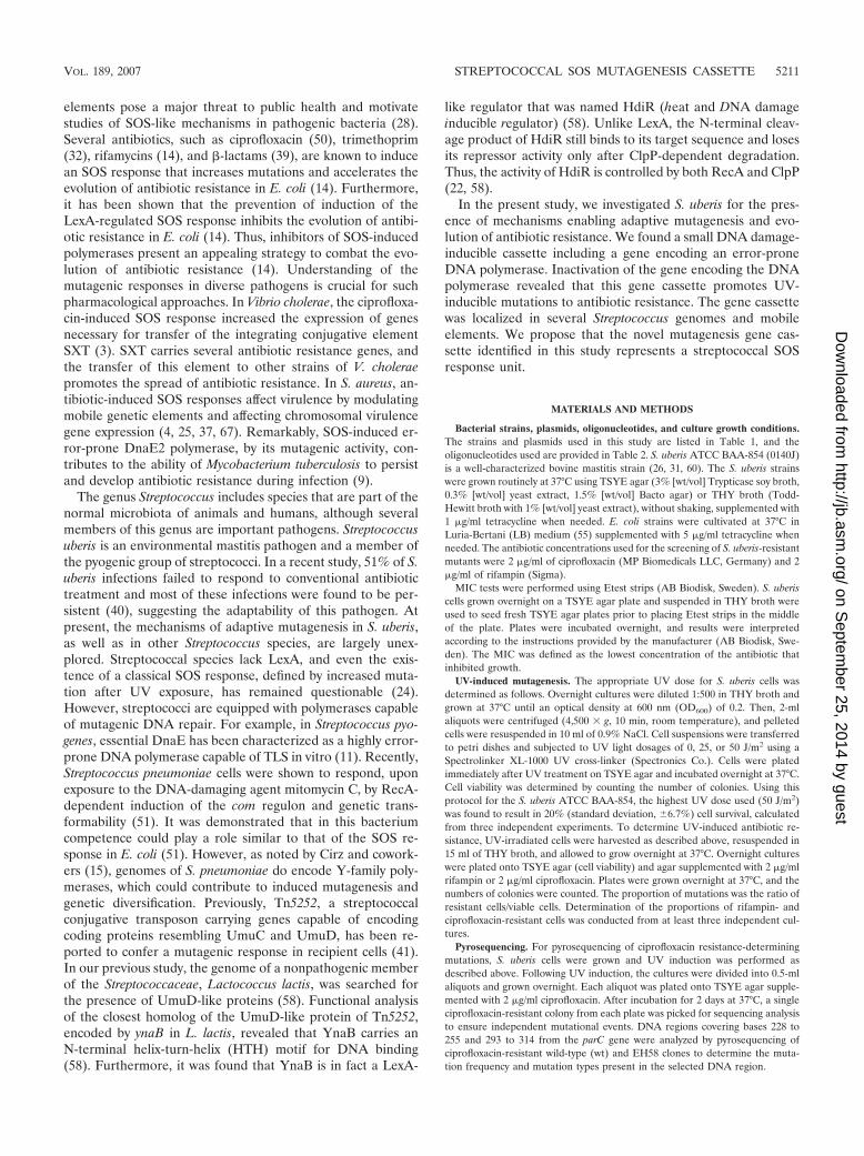
Induction of antibiotic resistance in S. uberis by UV light.Mutations leading to rifampin or ciprofloxacin resistance wereused as an assay to determine whether S. uberis possesses aclassical SOS response, as defined by increased mutation inresponse to UV light exposure. Rifampin resistance resultsfrom mutations in the � subunit of RNA polymerase (rpoB)that allow the polymerase to overcome the block (13). Cipro-floxacin resistance has been characterized from mutations intwo genes, the topoisomerase gene parC and the gyrase genegyrA (27).
UV-induced mutagenesis was examined by measuring the
VOL. 189, 2007 STREPTOCOCCAL SOS MUTAGENESIS CASSETTE 5213
on Septem
ber 25, 2014 by guesthttp://jb.asm
.org/D
ownloaded from
proportion of rifampin-resistant (Rifr) or ciprofloxacin-resis-tant (Cfxr) cells in the population. After UV induction at 25J/m2 and 50 J/m2, the number of rifampin-resistant clones inovernight cultures increased 145- and 209-fold, respectively(Fig. 1A), and the number of ciprofloxacin-resistant clones inovernight cultures increased 31-fold and 48-fold, respectively(Fig. 1B). We determined the MIC of rifampin for nine indi-vidual UV-induced rifampin-resistant S. uberis clones by Etest.All rifampin-resistant clones were found to exhibit over 500-fold higher MICs than the wt strain (data not shown).
The nature of UV-induced mutations was analyzed in the
parC gene by sequencing and pyrosequencing. First, we se-quenced a fragment covering nucleotides �88 to �570 of theparC gene in 12 ciprofloxacin-resistant clones (data notshown). This area is part of the quinolone resistance-determin-ing region, a 699-bp region of parC where ciprofloxacin resis-tance-promoting mutations have been characterized in S.pneumoniae and S. pyogenes parC (27, 46, 47). Based on se-quencing results (data not shown) and previous work on otherStreptococcus species (27, 46, 47), a 71-bp region (bases 228 to299 of parC) that contains three frequently occurring muta-tions in streptococci was chosen as the template for pyrose-quencing. DNA samples from 28 randomly selected individualciprofloxacin-resistant clones were extracted and subjected topyrosequencing. In 18/28 (64%) ciprofloxacin-resistant mu-tants, a mutation was observed in the 71-bp region responsiblefor previously described fluoroquinolone resistance-conferringamino acid substitutions in ParC (33). The mutation spectrumis shown in Fig. 1C. Most of the mutations observed wereG:C3A:T transitions, constituting 67% of base changes ob-served. In most clones, the base substitution occurred in posi-tion 235 where 11 G:C3A:T transitions and 3 G:C3T:Atransversions were detected (Fig. 1C). In one clone a transitionwas observed in position 246, whereas in three clones a trans-version was observed in this position (Fig. 1C). In E. coli,G:C3A:T transitions represent the hallmark of UV mutagen-esis (23).
From these results we conclude that S. uberis is capable ofSOS mutagenesis.
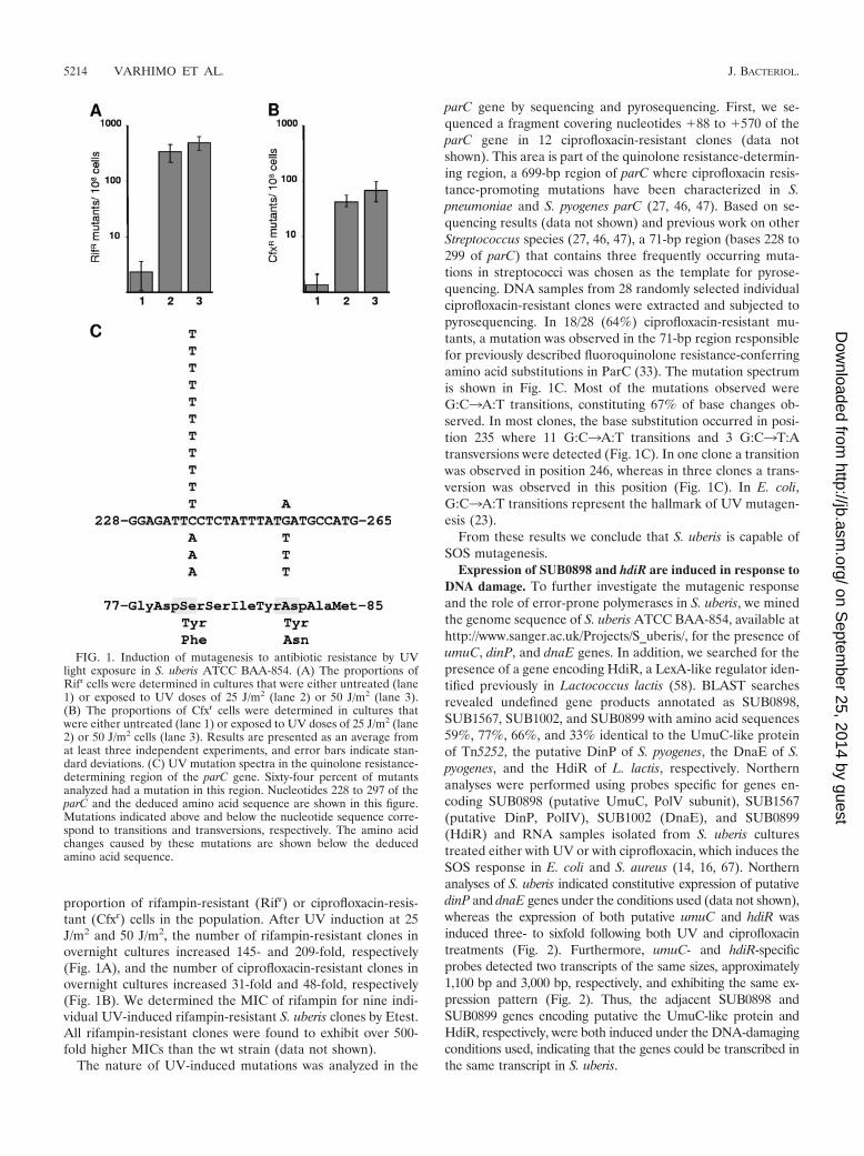
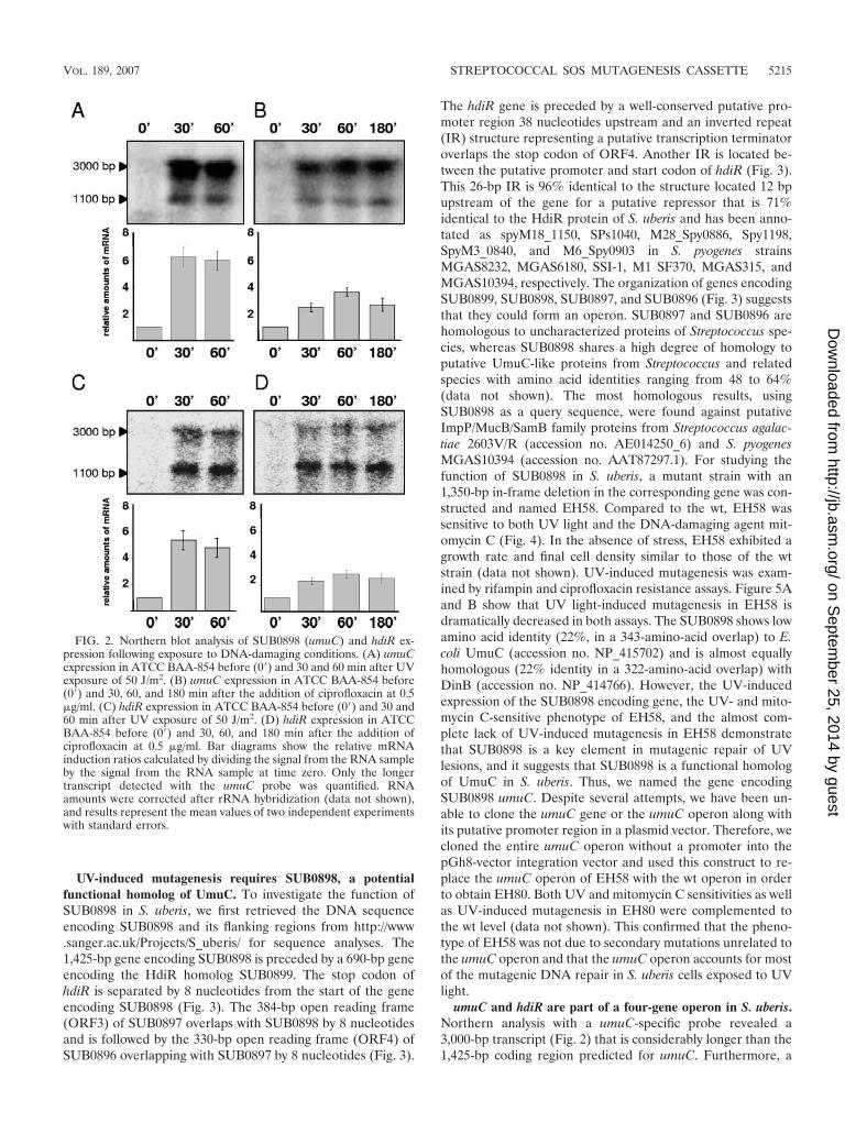
Expression of SUB0898 and hdiR are induced in response toDNA damage. To further investigate the mutagenic responseand the role of error-prone polymerases in S. uberis, we minedthe genome sequence of S. uberis ATCC BAA-854, available athttp://www.sanger.ac.uk/Projects/S_uberis/, for the presence ofumuC, dinP, and dnaE genes. In addition, we searched for thepresence of a gene encoding HdiR, a LexA-like regulator iden-tified previously in Lactococcus lactis (58). BLAST searchesrevealed undefined gene products annotated as SUB0898,SUB1567, SUB1002, and SUB0899 with amino acid sequences59%, 77%, 66%, and 33% identical to the UmuC-like proteinof Tn5252, the putative DinP of S. pyogenes, the DnaE of S.pyogenes, and the HdiR of L. lactis, respectively. Northernanalyses were performed using probes specific for genes en-coding SUB0898 (putative UmuC, PolV subunit), SUB1567(putative DinP, PolIV), SUB1002 (DnaE), and SUB0899(HdiR) and RNA samples isolated from S. uberis culturestreated either with UV or with ciprofloxacin, which induces theSOS response in E. coli and S. aureus (14, 16, 67). Northernanalyses of S. uberis indicated constitutive expression of putativedinP and dnaE genes under the conditions used (data not shown),whereas the expression of both putative umuC and hdiR wasinduced three- to sixfold following both UV and ciprofloxacintreatments (Fig. 2). Furthermore, umuC- and hdiR-specificprobes detected two transcripts of the same sizes, approximately1,100 bp and 3,000 bp, respectively, and exhibiting the same ex-pression pattern (Fig. 2). Thus, the adjacent SUB0898 andSUB0899 genes encoding putative the UmuC-like protein andHdiR, respectively, were both induced under the DNA-damagingconditions used, indicating that the genes could be transcribed inthe same transcript in S. uberis.
FIG. 1. Induction of mutagenesis to antibiotic resistance by UVlight exposure in S. uberis ATCC BAA-854. (A) The proportions ofRifr cells were determined in cultures that were either untreated (lane1) or exposed to UV doses of 25 J/m2 (lane 2) or 50 J/m2 (lane 3).(B) The proportions of Cfxr cells were determined in cultures thatwere either untreated (lane 1) or exposed to UV doses of 25 J/m2 (lane2) or 50 J/m2 cells (lane 3). Results are presented as an average fromat least three independent experiments, and error bars indicate stan-dard deviations. (C) UV mutation spectra in the quinolone resistance-determining region of the parC gene. Sixty-four percent of mutantsanalyzed had a mutation in this region. Nucleotides 228 to 297 of theparC and the deduced amino acid sequence are shown in this figure.Mutations indicated above and below the nucleotide sequence corre-spond to transitions and transversions, respectively. The amino acidchanges caused by these mutations are shown below the deducedamino acid sequence.
5214 VARHIMO ET AL. J. BACTERIOL.
on Septem
ber 25, 2014 by guesthttp://jb.asm
.org/D
ownloaded from
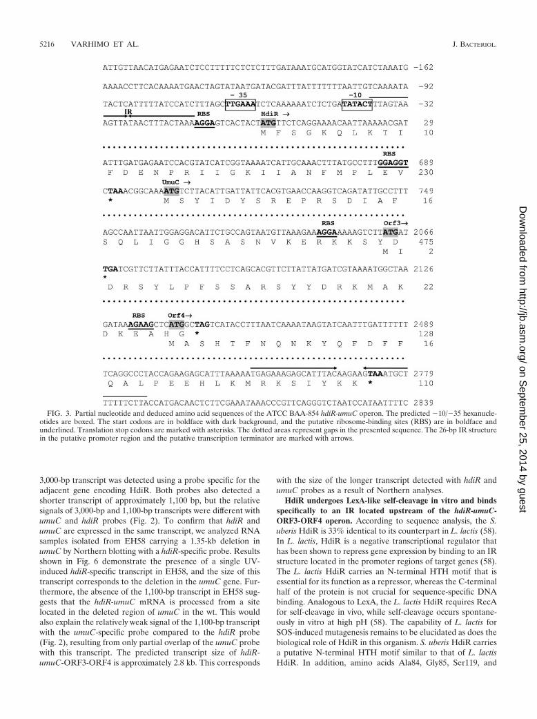
UV-induced mutagenesis requires SUB0898, a potentialfunctional homolog of UmuC. To investigate the function ofSUB0898 in S. uberis, we first retrieved the DNA sequenceencoding SUB0898 and its flanking regions from http://www.sanger.ac.uk/Projects/S_uberis/ for sequence analyses. The1,425-bp gene encoding SUB0898 is preceded by a 690-bp geneencoding the HdiR homolog SUB0899. The stop codon ofhdiR is separated by 8 nucleotides from the start of the geneencoding SUB0898 (Fig. 3). The 384-bp open reading frame(ORF3) of SUB0897 overlaps with SUB0898 by 8 nucleotidesand is followed by the 330-bp open reading frame (ORF4) ofSUB0896 overlapping with SUB0897 by 8 nucleotides (Fig. 3).
The hdiR gene is preceded by a well-conserved putative pro-moter region 38 nucleotides upstream and an inverted repeat(IR) structure representing a putative transcription terminatoroverlaps the stop codon of ORF4. Another IR is located be-tween the putative promoter and start codon of hdiR (Fig. 3).This 26-bp IR is 96% identical to the structure located 12 bpupstream of the gene for a putative repressor that is 71%identical to the HdiR protein of S. uberis and has been anno-tated as spyM18_1150, SPs1040, M28_Spy0886, Spy1198,SpyM3_0840, and M6_Spy0903 in S. pyogenes strainsMGAS8232, MGAS6180, SSI-1, M1 SF370, MGAS315, andMGAS10394, respectively. The organization of genes encodingSUB0899, SUB0898, SUB0897, and SUB0896 (Fig. 3) suggeststhat they could form an operon. SUB0897 and SUB0896 arehomologous to uncharacterized proteins of Streptococcus spe-cies, whereas SUB0898 shares a high degree of homology toputative UmuC-like proteins from Streptococcus and relatedspecies with amino acid identities ranging from 48 to 64%(data not shown). The most homologous results, usingSUB0898 as a query sequence, were found against putativeImpP/MucB/SamB family proteins from Streptococcus agalac-tiae 2603V/R (accession no. AE014250_6) and S. pyogenesMGAS10394 (accession no. AAT87297.1). For studying thefunction of SUB0898 in S. uberis, a mutant strain with an1,350-bp in-frame deletion in the corresponding gene was con-structed and named EH58. Compared to the wt, EH58 wassensitive to both UV light and the DNA-damaging agent mit-omycin C (Fig. 4). In the absence of stress, EH58 exhibited agrowth rate and final cell density similar to those of the wtstrain (data not shown). UV-induced mutagenesis was exam-ined by rifampin and ciprofloxacin resistance assays. Figure 5Aand B show that UV light-induced mutagenesis in EH58 isdramatically decreased in both assays. The SUB0898 shows lowamino acid identity (22%, in a 343-amino-acid overlap) to E.coli UmuC (accession no. NP_415702) and is almost equallyhomologous (22% identity in a 322-amino-acid overlap) withDinB (accession no. NP_414766). However, the UV-inducedexpression of the SUB0898 encoding gene, the UV- and mito-mycin C-sensitive phenotype of EH58, and the almost com-plete lack of UV-induced mutagenesis in EH58 demonstratethat SUB0898 is a key element in mutagenic repair of UVlesions, and it suggests that SUB0898 is a functional homologof UmuC in S. uberis. Thus, we named the gene encodingSUB0898 umuC. Despite several attempts, we have been un-able to clone the umuC gene or the umuC operon along withits putative promoter region in a plasmid vector. Therefore, wecloned the entire umuC operon without a promoter into thepGh8-vector integration vector and used this construct to re-place the umuC operon of EH58 with the wt operon in orderto obtain EH80. Both UV and mitomycin C sensitivities as wellas UV-induced mutagenesis in EH80 were complemented tothe wt level (data not shown). This confirmed that the pheno-type of EH58 was not due to secondary mutations unrelated tothe umuC operon and that the umuC operon accounts for mostof the mutagenic DNA repair in S. uberis cells exposed to UVlight.
umuC and hdiR are part of a four-gene operon in S. uberis.Northern analysis with a umuC-specific probe revealed a3,000-bp transcript (Fig. 2) that is considerably longer than the1,425-bp coding region predicted for umuC. Furthermore, a
FIG. 2. Northern blot analysis of SUB0898 (umuC) and hdiR ex-pression following exposure to DNA-damaging conditions. (A) umuCexpression in ATCC BAA-854 before (0�) and 30 and 60 min after UVexposure of 50 J/m2. (B) umuC expression in ATCC BAA-854 before(0�) and 30, 60, and 180 min after the addition of ciprofloxacin at 0.5�g/ml. (C) hdiR expression in ATCC BAA-854 before (0�) and 30 and60 min after UV exposure of 50 J/m2. (D) hdiR expression in ATCCBAA-854 before (0�) and 30, 60, and 180 min after the addition ofciprofloxacin at 0.5 �g/ml. Bar diagrams show the relative mRNAinduction ratios calculated by dividing the signal from the RNA sampleby the signal from the RNA sample at time zero. Only the longertranscript detected with the umuC probe was quantified. RNAamounts were corrected after rRNA hybridization (data not shown),and results represent the mean values of two independent experimentswith standard errors.
VOL. 189, 2007 STREPTOCOCCAL SOS MUTAGENESIS CASSETTE 5215
on Septem
ber 25, 2014 by guesthttp://jb.asm
.org/D
ownloaded from
3,000-bp transcript was detected using a probe specific for theadjacent gene encoding HdiR. Both probes also detected ashorter transcript of approximately 1,100 bp, but the relativesignals of 3,000-bp and 1,100-bp transcripts were different withumuC and hdiR probes (Fig. 2). To confirm that hdiR andumuC are expressed in the same transcript, we analyzed RNAsamples isolated from EH58 carrying a 1.35-kb deletion inumuC by Northern blotting with a hdiR-specific probe. Resultsshown in Fig. 6 demonstrate the presence of a single UV-induced hdiR-specific transcript in EH58, and the size of thistranscript corresponds to the deletion in the umuC gene. Fur-thermore, the absence of the 1,100-bp transcript in EH58 sug-gests that the hdiR-umuC mRNA is processed from a sitelocated in the deleted region of umuC in the wt. This wouldalso explain the relatively weak signal of the 1,100-bp transcriptwith the umuC-specific probe compared to the hdiR probe(Fig. 2), resulting from only partial overlap of the umuC probewith this transcript. The predicted transcript size of hdiR-umuC-ORF3-ORF4 is approximately 2.8 kb. This corresponds
with the size of the longer transcript detected with hdiR andumuC probes as a result of Northern analyses.
HdiR undergoes LexA-like self-cleavage in vitro and bindsspecifically to an IR located upstream of the hdiR-umuC-ORF3-ORF4 operon. According to sequence analysis, the S.uberis HdiR is 33% identical to its counterpart in L. lactis (58).In L. lactis, HdiR is a negative transcriptional regulator thathas been shown to repress gene expression by binding to an IRstructure located in the promoter regions of target genes (58).The L. lactis HdiR carries an N-terminal HTH motif that isessential for its function as a repressor, whereas the C-terminalhalf of the protein is not crucial for sequence-specific DNAbinding. Analogous to LexA, the L. lactis HdiR requires RecAfor self-cleavage in vivo, while self-cleavage occurs spontane-ously in vitro at high pH (58). The capability of L. lactis forSOS-induced mutagenesis remains to be elucidated as does thebiological role of HdiR in this organism. S. uberis HdiR carriesa putative N-terminal HTH motif similar to that of L. lactisHdiR. In addition, amino acids Ala84, Gly85, Ser119, and
FIG. 3. Partial nucleotide and deduced amino acid sequences of the ATCC BAA-854 hdiR-umuC operon. The predicted �10/�35 hexanucle-otides are boxed. The start codons are in boldface with dark background, and the putative ribosome-binding sites (RBS) are in boldface andunderlined. Translation stop codons are marked with asterisks. The dotted areas represent gaps in the presented sequence. The 26-bp IR structurein the putative promoter region and the putative transcription terminator are marked with arrows.
5216 VARHIMO ET AL. J. BACTERIOL.
on Septem
ber 25, 2014 by guesthttp://jb.asm
.org/D
ownloaded from

Lys156, involved in the RecA-dependent self-cleavage of E.coli LexA (34, 59) and L. lactis HdiR (58), are also conservedin the HdiR of S. uberis (Ala117, Gly118, Ser150, and Lys187)(data not shown). We have not been able to inactivate the recAgene in S. uberis or to replace the wt hdiR gene with geneencoding uncleavable G118D HdiR (data not shown), whichhas prevented studies of HdiR self-cleavage in vivo. To studythe function of S. uberis HdiR in vitro, we overproduced andpurified HdiR as a His-tagged protein. The autodigestion atalkaline pH is a characteristic feature of LexA-like proteinsundergoing RecA-mediated cleavage in vivo and has been doc-umented for every LexA-like protein tested (23). For investi-gation of LexA-like self-cleavage, we incubated purified HdiRprotein at various pHs and observed that self-cleavage of HdiRproceeded spontaneously at high pH, resulting in two productsof �14.6 and �13.1 kDa (Fig. 7A). Next, we used EMSA tostudy the binding of HdiR to the upstream region of the hdiR-umuC-ORF3-ORF4 operon. To this end, the gel shift reactionscontaining purified HdiR and the DNA fragment spanning theregion from �157 to �10 (promoter probe) or �4 to �157(control probe) were assembled in binding buffer and the DNA
complexes were separated in TBE gel, as described above.When scanning with an excitation wavelength specific forHEX-labeled DNA complexes, we observed that HdiR bindsin a concentration-dependent manner to the promoter probebut not to the control probe (Fig. 7B). The additional bandobserved by EMSA is a single-stranded PCR product as dem-onstrated by denaturating the probe with formamide prior toelectrophoresis (data not shown).
Next, we asked whether the 26-bp IR sequence inside thepromoter probe is the target of HdiR binding. To examine this,the IR was cloned into pBluescript-II SK� and used as a
FIG. 4. Effect of absence of UmuC-like protein on DNA damagetolerance of S. uberis. (A) Survival of S. uberis ATCC BAA-854 andEH58 after UV exposure. ATCC BAA-854 (1 and 3) and EH58 (2 and4) cultures were grown exponentially in THY and at an OD600 of 0.1,cultures were diluted, and 10 �l of each dilution was spotted on twoTSYE agar plates. Plates either were not exposed (�UV) or wereexposed to UV light of 50 J/m2 (�UV) prior to incubation overnightat 37°C. The results presented are representative of several indepen-dent experiments. (B) Sensitivity of S. uberis ATCC BAA-854 andEH58 to mitomycin C (MMC). ATCC BAA-854 (1 and 3) and EH58(2 and 4) cultures were grown overnight in THY, and 10 �l of over-night cultures and appropriate dilutions were spotted on TSYE agarplates without and with 25 ng/ml of MMC. Plates were grown over-night at 37°C. The results presented are representative of severalindependent experiments.
FIG. 5. Effects of absence of UmuC-like protein on UV-targetedmutagenesis. The proportions of (A) rifampin- and (B) ciprofloxacin-resistant cells in ATCC BAA-854 (dark gray bars) and EH58 (lightgray bars) were determined in cultures that were either untreated (1and 2) or were exposed to UV doses of (3 and 4) 25 J/m2 and (5 and6) 50 J/m2.
FIG. 6. Northern analysis of hdiR transcription in umuC deletionstrain EH58. The hdiR expression in ATCC BAA-854 and EH58 be-fore (0�) and 30 and 60 min after UV exposure of 50 J/m2 is shown.
VOL. 189, 2007 STREPTOCOCCAL SOS MUTAGENESIS CASSETTE 5217
on Septem
ber 25, 2014 by guesthttp://jb.asm
.org/D
ownloaded from
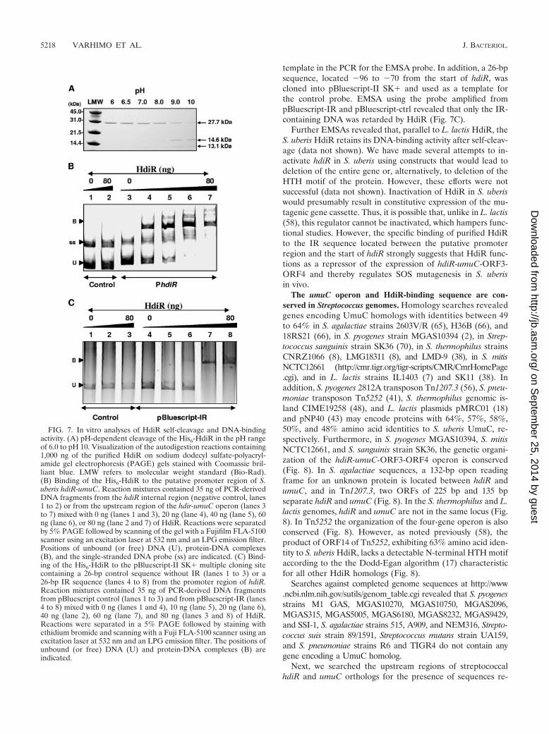
template in the PCR for the EMSA probe. In addition, a 26-bpsequence, located �96 to �70 from the start of hdiR, wascloned into pBluescript-II SK� and used as a template forthe control probe. EMSA using the probe amplified frompBluescript-IR and pBluescript-ctrl revealed that only the IR-containing DNA was retarded by HdiR (Fig. 7C).
Further EMSAs revealed that, parallel to L. lactis HdiR, theS. uberis HdiR retains its DNA-binding activity after self-cleav-age (data not shown). We have made several attempts to in-activate hdiR in S. uberis using constructs that would lead todeletion of the entire gene or, alternatively, to deletion of theHTH motif of the protein. However, these efforts were notsuccessful (data not shown). Inactivation of HdiR in S. uberiswould presumably result in constitutive expression of the mu-tagenic gene cassette. Thus, it is possible that, unlike in L. lactis(58), this regulator cannot be inactivated, which hampers func-tional studies. However, the specific binding of purified HdiRto the IR sequence located between the putative promoterregion and the start of hdiR strongly suggests that HdiR func-tions as a repressor of the expression of hdiR-umuC-ORF3-ORF4 and thereby regulates SOS mutagenesis in S. uberisin vivo.
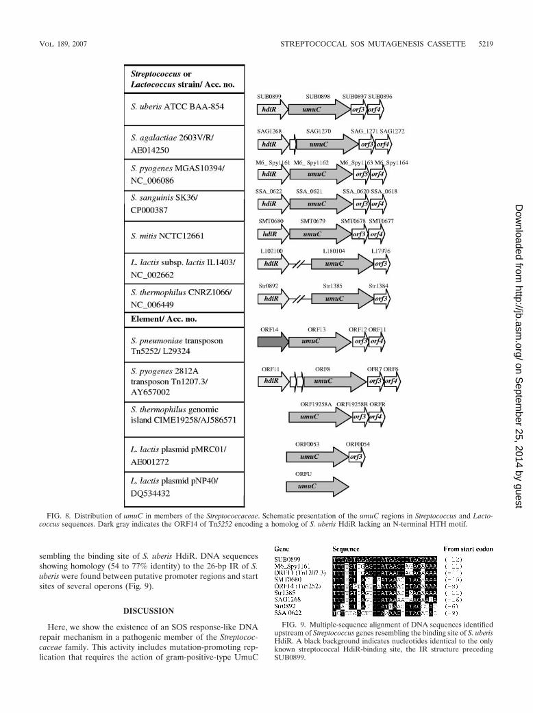
The umuC operon and HdiR-binding sequence are con-served in Streptococcus genomes. Homology searches revealedgenes encoding UmuC homologs with identities between 49to 64% in S. agalactiae strains 2603V/R (65), H36B (66), and18RS21 (66), in S. pyogenes strain MGAS10394 (2), in Strep-tococcus sanguinis strain SK36 (70), in S. thermophilus strainsCNRZ1066 (8), LMG18311 (8), and LMD-9 (38), in S. mitisNCTC12661 (http://cmr.tigr.org/tigr-scripts/CMR/CmrHomePage.cgi), and in L. lactis strains IL1403 (7) and SK11 (38). Inaddition, S. pyogenes 2812A transposon Tn1207.3 (56), S. pneu-moniae transposon Tn5252 (41), S. thermophilus genomic is-land CIME19258 (48), and L. lactis plasmids pMRC01 (18)and pNP40 (43) may encode proteins with 64%, 57%, 58%,50%, and 48% amino acid identities to S. uberis UmuC, re-spectively. Furthermore, in S. pyogenes MGAS10394, S. mitisNCTC12661, and S. sanguinis strain SK36, the genetic organi-zation of the hdiR-umuC-ORF3-ORF4 operon is conserved(Fig. 8). In S. agalactiae sequences, a 132-bp open readingframe for an unknown protein is located between hdiR andumuC, and in Tn1207.3, two ORFs of 225 bp and 135 bpseparate hdiR and umuC (Fig. 8). In the S. thermophilus and L.lactis genomes, hdiR and umuC are not in the same locus (Fig.8). In Tn5252 the organization of the four-gene operon is alsoconserved (Fig. 8). However, as noted previously (58), theproduct of ORF14 of Tn5252, exhibiting 63% amino acid iden-tity to S. uberis HdiR, lacks a detectable N-terminal HTH motifaccording to the the Dodd-Egan algorithm (17) characteristicfor all other HdiR homologs (Fig. 8).
Searches against completed genome sequences at http://www.ncbi.nlm.nih.gov/sutils/genom_table.cgi revealed that S. pyogenesstrains M1 GAS, MGAS10270, MGAS10750, MGAS2096,MGAS315, MGAS5005, MGAS6180, MGAS8232, MGAS9429,and SSI-1, S. agalactiae strains 515, A909, and NEM316, Strepto-coccus suis strain 89/1591, Streptococcus mutans strain UA159,and S. pneumoniae strains R6 and TIGR4 do not contain anygene encoding a UmuC homolog.
Next, we searched the upstream regions of streptococcalhdiR and umuC orthologs for the presence of sequences re-
FIG. 7. In vitro analyses of HdiR self-cleavage and DNA-bindingactivity. (A) pH-dependent cleavage of the His6-HdiR in the pH rangeof 6.0 to pH 10. Visualization of the autodigestion reactions containing1,000 ng of the purified HdiR on sodium dodecyl sulfate-polyacryl-amide gel electrophoresis (PAGE) gels stained with Coomassie bril-liant blue. LMW refers to molecular weight standard (Bio-Rad).(B) Binding of the His6-HdiR to the putative promoter region of S.uberis hdiR-umuC. Reaction mixtures contained 35 ng of PCR-derivedDNA fragments from the hdiR internal region (negative control, lanes1 to 2) or from the upstream region of the hdir-umuC operon (lanes 3to 7) mixed with 0 ng (lanes 1 and 3), 20 ng (lane 4), 40 ng (lane 5), 60ng (lane 6), or 80 ng (lane 2 and 7) of HdiR. Reactions were separatedby 5% PAGE followed by scanning of the gel with a Fujifilm FLA-5100scanner using an excitation laser at 532 nm and an LPG emission filter.Positions of unbound (or free) DNA (U), protein-DNA complexes(B), and the single-stranded DNA probe (ss) are indicated. (C) Bind-ing of the His6-HdiR to the pBluescript-II SK� multiple cloning sitecontaining a 26-bp control sequence without IR (lanes 1 to 3) or a26-bp IR sequence (lanes 4 to 8) from the promoter region of hdiR.Reaction mixtures contained 35 ng of PCR-derived DNA fragmentsfrom pBluescript control (lanes 1 to 3) and from pBluescript-IR (lanes4 to 8) mixed with 0 ng (lanes 1 and 4), 10 ng (lane 5), 20 ng (lane 6),40 ng (lane 2), 60 ng (lane 7), and 80 ng (lanes 3 and 8) of HdiR.Reactions were separated in a 5% PAGE followed by staining withethidium bromide and scanning with a Fuji FLA-5100 scanner using anexcitation laser at 532 nm and an LPG emission filter. The positions ofunbound (or free) DNA (U) and protein-DNA complexes (B) areindicated.
5218 VARHIMO ET AL. J. BACTERIOL.
on Septem
ber 25, 2014 by guesthttp://jb.asm
.org/D
ownloaded from
sembling the binding site of S. uberis HdiR. DNA sequencesshowing homology (54 to 77% identity) to the 26-bp IR of S.uberis were found between putative promoter regions and startsites of several operons (Fig. 9).
DISCUSSION
Here, we show the existence of an SOS response-like DNArepair mechanism in a pathogenic member of the Streptococ-caceae family. This activity includes mutation-promoting rep-lication that requires the action of gram-positive-type UmuC
FIG. 8. Distribution of umuC in members of the Streptococcaceae. Schematic presentation of the umuC regions in Streptococcus and Lacto-coccus sequences. Dark gray indicates the ORF14 of Tn5252 encoding a homolog of S. uberis HdiR lacking an N-terminal HTH motif.
FIG. 9. Multiple-sequence alignment of DNA sequences identifiedupstream of Streptococcus genes resembling the binding site of S. uberisHdiR. A black background indicates nucleotides identical to the onlyknown streptococcal HdiR-binding site, the IR structure precedingSUB0899.
VOL. 189, 2007 STREPTOCOCCAL SOS MUTAGENESIS CASSETTE 5219
on Septem
ber 25, 2014 by guesthttp://jb.asm
.org/D
ownloaded from
after induction with UV light. Furthermore, this study adds toprevious work on HdiR (58), and we propose that this DNA-binding protein is a regulator of SOS mutagenesis as LexA is inother bacteria.
It was shown that UV light exposure induces mutations thatconfer antibiotic resistance in S. uberis ATCC BAA-854. Un-der DNA-damaging conditions, increased expression of thehdiR-umuC-ORF3-ORF4 operon was detected by Northernanalyses, whereas no effect on dinP or dnaE expressions wasobserved. In addition to UV light, exposure to fluoroquinoloneantibiotic ciprofloxacin also induced the expression of thehdiR-umuC-ORF3-ORF4 operon. A previous study revealedthat the DNA polymerase encoded by dnaE in S. pyogenes ishighly error prone, producing frameshift and point mutationsin undamaged DNA as well as during TLS in vitro (11). In M.tuberculosis the SOS-induced DnaE2 was shown to be essentialfor evolution of antibiotic resistance in vivo (9). The functionalstudy of DnaE proteins in gram-positive bacteria equippedwith a single dnaE is hampered by the essentiality of the geneproduct (11). Our results demonstrate that the intact umuCgene is essential for UV-induced mutagenesis in S. uberis invivo under the growth conditions used. The mutation typesdetected in the wt S. uberis strain after UV exposure weremainly transition-type base substitutions. This is in accordancewith the transition mutations caused by PolV after UV induc-tion in E. coli (30, 64). PolIV mainly produces �1 frameshiftdeletions in E. coli (69).
It was shown that the umuC gene is cotranscribed with agene encoding the HdiR homolog as well as two unknowngenes in S. uberis. Northern analyses suggest that the mRNAcontaining the hdiR-umuC-ORF3-ORF4 operon is processedwithin the umuC gene. The processing of the hdiR-umuC-ORF3-ORF4 transcript and possible differential stability ofprocessing products could be a tool used by S. uberis to ensurethat the amount of HdiR regulator exceeds other gene prod-ucts of the operon and, possibly, results in more-efficient re-pression of the mutagenic operon. The novel regulator HdiRwas previously characterized from L. lactis and was shown tofunction as a repressor that binds to an IR sequence located inthe promoter regions of target genes (58). One target gene ofHdiR was a umuC-like gene, present in L. lactis IL1403 butabsent in another L. lactis strain, MG1363 (58). O’Driscoll andcoworkers (44) recently reported the presence of a umuC or-tholog in the Lactococcus plasmid pNP40 preceded by a repeatstructure highly similar to the HdiR-binding sequence. EMSArevealed that the target sequence of S. uberis HdiR binding islocated within an IR sequence that differs from the target of L.lactis HdiR. This is not surprising, since the two DNA-bindingproteins are only 33% identical and the putative DNA-bindingdomains are not particularly well conserved (data not shown).DNA elements resembling the binding site of S. uberis HdiRwere located upstream of hdiR and umuC genes in severalStreptococcus species. However, it remains to be experimen-tally determined whether these DNA sequences representfunctional operator sites of HdiR proteins in these organismsand whether the HdiR-binding site is conserved among strep-tococci. Database searches revealed that many, but not all,Streptococcus genomes encode proteins highly homologous toS. uberis UmuC. On the other hand, with the exception of S.pneumoniae genomes, all publicly available genome sequences
from Streptococcaceae indicate the presence of an HdiR ho-molog (data not shown). However, the functions of all theseHdiR-like proteins remain to be elucidated. Interestingly,while pathogenic and commensal members of the Streptococ-caceae equipped with UmuC have conserved the organizationof hdiR and umuC in a single operon, in the nonpathogenicdairy bacteria L. lactis and S. thermophilus, the operon orga-nization has been split during evolution (Fig. 8.). It could bespeculated that the presence of positive pressure in L. lactisand S. thermophilus, adapted to dairy environments, resulted ina separate regulator that gives the ability, e.g., for improvedrepression of mutagenic genes. umuDC homologs are oftenassociated with mobile genetic elements of bacteria (49). InStreptococcus and Lactococcus, database searches revealed thepresence of umuC homologs in plasmids, transposons, andgenomic islands. Munoz-Najar and Vijayakumar (41) previ-ously reported the presence of genes encoding UmuDC-likeproteins in the pneumococcal conjugative transposon Tn5252.It was shown that plasmids carrying fragments of Tn5252 re-stored UV-inducible mutagenic repair in S. pneumoniae (41).The UmuD-like protein encoded by Tn5252 is highly homol-ogous to HdiR; however, the lack of a HTH motif in theUmuD-like protein indicates functional diversification fromHdiR. Small DNA damage-inducible and self-regulated genecassettes capable of mediating adaptive mutagenesis havepreviously been identified from other bacteria including Pro-teobacteria and Actinobacteria (1, 20). However, to our knowl-edge the hdiR-umuC-ORF3-ORF4 operon enabling UV-in-duced adaptive mutagenesis in S. uberis, and potentially inother streptococci, represents the first SOS mutagenesis sys-tem that is not regulated by LexA.
In E. coli the function and activity of PolV is regulated at thetranscriptional and posttranscriptional level, and it needs sev-eral other proteins to be capable of TLS. Despite the fact thatUmuC is the catalytically active subunit, PolV requires twotruncated UmuD subunits in order to be active (23). In gram-positive organisms, the functional homolog of UmuD remainsto be identified despite the identification of candidates co-occurring with gram-positive UmuC-like proteins, like YqjXwith YqjW in B. subtilis (19, 49). One of our future goals is tostudy the function encoded by the two uncharacterized genespresent in hdiR-umuC-ORF3-ORF4 and address the questionof why these genes co-occur with umuC among streptococci.
ACKNOWLEDGMENT
This study received financial support from the Academy of Finland(decision no. 211165 and 209136).
REFERENCES
1. Abella, M., I. Erill, M. Jara, G. Mazon, S. Campoy, and J. Barbe. 2004.Widespread distribution of a lexA-regulated DNA damage-inducible multi-ple gene cassette in the Proteobacterium phylum. Mol. Microbiol. 54:212–222.
2. Banks, D. J., S. F. Porcella, K. D. Barbian, S. B. Beres, L. E. Philips, J. M.Voyich, F. R. DeLeo, J. M. Martin, G. A. Somerville, and J. M. Musser. 2004.Progress toward characterization of the group a Streptococcus metagenome:complete genome sequence of a macrolide-resistant serotype M6 strain.J. Infect. Dis. 190:727–738.
3. Beaber, J. W., B. Hochhut, and M. K. Waldor. 2004. SOS response promoteshorizontal dissemination of antibiotic resistance genes. Nature 427:72–74.
4. Bisognano, C., W. L. Kelley, T. Estoppey, P. Francois, J. Schrenzel, D. Li,D. P. Lew, D. C. Hooper, A. L. Cheung, and P. Vaudaux. 2004. A RecA-LexA-dependent pathway mediates ciprofloxacin-induced fibronectin bind-ing in Staphylococcus aureus. J. Biol. Chem. 279:9064–9071.
5220 VARHIMO ET AL. J. BACTERIOL.
on Septem
ber 25, 2014 by guesthttp://jb.asm
.org/D
ownloaded from
5. Biswas, I., A. Gruss, S. D. Ehrlich, and E. Maguin. 1993. High-efficiencygene inactivation and replacement system for gram-positive bacteria. J. Bac-teriol. 175:3628–3635.
6. Bjedov, I., O. Tenaillon, B. Gerard, V. Souza, E. Denamur, M. Radman, F.Taddei, and I. Matic. 2003. Stress-induced mutagenesis in bacteria. Science300:1404–1409.
7. Bolotin, A., P. Wincker, S. Mauger, O. Jaillon, K. Malarme, J. Weissenbach,S. D. Ehrlich, and A. Sorokin. 2001. The complete genome sequence of thelactic acid bacterium Lactococcus lactis ssp. lactis IL1403. Genome Res.11:731–753.
8. Bolotin, A., B. Quinquis, P. Renault, A. Sorokin, S. D. Ehrlich, S. Kulakauskas,A. Lapidus, E. Goltsman, M. Mazur, G. D. Pusch, M. Fonstein, R. Overbeek, N.Kyprides, B. Purnelle, D. Prozzi, K. Ngui, D. Masuy, F. Hancy, S. Burteau, M.Boutry, J. Delcour, A. Goffeau, and P. Hols. 2004. Complete sequence andcomparative genome analysis of the dairy bacterium Streptococcus thermophilus.Nat. Biotechnol. 22:1554–1558.
9. Boshoff, H. I., M. B. Reed, C. E. Barry III, and V. Mizrahi. 2003. DnaE2polymerase contributes to in vivo survival and the emergence of drug resis-tance in Mycobacterium tuberculosis. Cell 113:183–193.
10. Bruck, I., and M. O’Donnell. 2000. The DNA replication machine of agram-positive organism. J. Biol. Chem. 275:28971–28983.
11. Bruck, I., M. F. Goodman, and M. O’Donnell. 2003. The essential C familyDnaE polymerase is error-prone and efficient at lesion bypass. J. Biol. Chem.278:44361–44368.
12. Burckhardt, S. E., R. Woodgate, R. J. Scheuermann, and H. Echols. 1988.UmuD mutagenesis protein of Escherichia coli, overproduction, purification,and cleavage by RecA. Proc. Natl. Acad. Sci. USA 85:1811–1815.
13. Campbell, E. A., N. Korzheva, A. Mustaev, K. Murakami, S. Nair, A. Gold-farb, and S. A. Darst. 2001. Structural mechanism for rifampicin inhibition ofbacterial RNA polymerase. Cell 104:901–912.
14. Cirz, R. T., J. K. Chin, D. R. Andes, V. D. Crecy-Lagard, W. A. Craig, andF. E. Romesberg. 2005. Inhibition of mutation and combating the evolutionof antibiotic resistance. PLoS Biol. 3:e176.
15. Cirz, R. T., N. Gingles, and F. E. Romesberg. 2006. Side effects may includeevolution. Nat. Med. 12:890–891.
16. Cirz, R. T., M. B. Jones, N. A. Gingles, T. D. Minogue, B. Jarrahi, S. N.Peterson, and F. E. Romesberg. 2007. The complete and SOS-mediatedresponse of Staphylococcus aureus to the antibiotic ciprofloxacin. J. Bacteriol.189:531–539.
17. Dodd, I. B., and J. B. Egan. 1990. Improved detection of helix-turn-helixDNA binding motifs in protein sequences. Nucleic Acids Res. 18:5019–5026.
18. Dougherty, B. A., C. Hill, J. F. Weidman, D. R. Richardson, J. C. Venter, andR. P. Ross. 1998. Sequence and analysis of the 60 kb conjugative, bacteriocin-producing plasmid pMRC01 from Lactococcus lactis DPC3147. Mol. Micro-biol. 29:1029–1038.
19. Duigou, S., S. D. Ehrlich, P. Noirot, and M. F. Noirot-Gros. 2004. Distinctivegenetic features exhibited by the Y-family DNA polymerases in Bacillussubtilis. Mol. Microbiol. 54:439–451.
20. Erill, I., S. Campoy, G. Mazon, and J. Barbe. 2006. Dispersal and regulationof an adaptive mutagenesis cassette in the Bacteria domain. Nucleic AcidsRes. 34:66–77.
21. Frees, D., P. Varmanen, and H. Ingmer. 2001. Inactivation of a gene that ishighly conserved in gram-positive bacteria stimulates degradation of non-native proteins and concomitantly increases stress tolerance in Lactococcuslactis. Mol. Microbiol. 41:93–103.
22. Frees, D., K. Savijoki, P. Varmanen, and H. Ingmer. 2007. Clp ATPases andClpP proteolytic complexes regulate vital biological processes in low GC,gram-positive bacteria. Mol. Microbiol. 63:1285–1295.
23. Friedberg, E. C., G. C. Walker, W. Seide, R. D. Wood, R. A. Schultz, and T.Ellenberger. 2006. DNA repair and mutagenesis, 2nd ed. American Societyfor Microbiology, Washington, DC.
24. Gasc, A. M., N. Sicard, J. P. Claverys, and A. M. Sicard. 1980. Lack of SOSrepair in Streptococcus pneumoniae. Mutat. Res. 70:157–165.
25. Goerke, C., J. Koller, and C. Wolz. 2006. Ciprofloxacin and trimethoprimcause phage induction and virulence modulation in Staphylococcus aureus.Antimicrob. Agents Chemother. 50:171–177.
26. Hill, A. W. 1988. Pathogenicity of two strains of Streptococcus uberis infusedinto lactating and non-lactating bovine mammary glands. Res. Vet. Sci.45:400–404.
27. Janoir, C., V. Zeller, M. D. Kitziz, N. J. Moreau, and L. Gutmann. 1996.High-level fluoroquinolone resistance in Streptococcus pneumoniae requiresmutations in parC and gyrA. Antimicrob. Agents Chemother. 40:2760–2764.
28. Kelley, W. L. 2006. LexA marks the spot: the virulent side of SOS and acloser look at the LexA regulon. Mol. Microbiol. 62:1228–1238.
29. Kimoto, H., and A. Taketo. 2003. Efficient electrotransformation system andgene targeting in pyogenic streptococci. Biosci. Biotechnol. Biochem. 67:2203–2209.
30. LeClerc, J. E., A. Borden, and C. W. Lawrence. 1991. The thymine-thyminepyrimidine-pyrimidinone (6-4) ultraviolet light photoproduct is highly mu-tagenic and specifically induces 3� thymine-to-cytosine transitions in Esche-richia coli. Proc. Natl. Acad. Sci. USA 88:9685–9689.
31. Leigh, J. A., T. R. Field, and M. R. Williams. 1990. Two strains of Strepto-
coccus uberis, of differing ability to cause clinical mastitis, differ in theirability to resist some host defence factors. Res. Vet. Sci. 49:85–87.
32. Lewin, C. S., and S. G. Amyes. 1991. The role of the SOS response in bacteriaexposed to zidovudine or trimethoprim. J. Med. Microbiol. 34:329.
33. Lim, S., D. Bast, A. McGeer, J. de Azavedo, and D. E. Low. 2003. Antimi-crobial susceptibility breakpoints and first-step parC mutations in Strepto-coccus pneumoniae: redefining fluoroquinolone resistance. Emerg. Infect.Dis. 9:833–837.
34. Little, J. W. 1984. Autodigestion of LexA and phage lambda repressors.Proc. Natl. Acad. Sci. USA 81:1375–1379.
35. Little, J. W. 1991. Mechanism of specific LexA cleavage: autodigestion andthe role of RecA coprotease. Biochimie 73:411–422.
36. Maguin, E., H. Prevost, S. D. Ehrlich, and A. Gruss. 1996. Efficient inser-tional mutagenesis in lactococci and other gram-positive bacteria. J. Bacte-riol. 178:931–935.
37. Maiques, E., C. Ubeda, S. Campoy, S. Salvador, I. Lasa, R. P. Novick, J.Barbe, and J. R. Penades. 2006. �-Lactam antibiotics induce the SOS re-sponse and horizontal transfer of virulence factors in Staphylococcus aureus.J. Bacteriol. 188:2726–2729.
38. Makarova, K., A. Slesarev, Y. Wolf, A. Sorokin, B. Mirkin, E. Koonin, A. Pavlov,N. Pavlova, V. Karamychev, N. Polouchine, V. Shakhova, I. Grigoriev, Y. Lou, D.Rohksar, S. Lucas, K. Huang, D. M. Goodstein, T. Hawkins, V. Plengvidhya, D.Welker, J. Hughes, Y. Goh, A. Benson, K. Baldwin, J. H. Lee, I. Diaz-Muniz,B. Dosti, V. Smeianov, W. Wechter, R. Barabote, G. Lorca, E. Altermann, R.Barrangou, B. Ganesan, Y. Xie, H. Rawsthorne, D. Tamir, C. Parker, F. Breidt,J. Broadbent, R. Hutkins, D. O’sullivan, J. Steele, G. Unlu, M. Saier, T.Klaenhammer, P. Richardson, S. Kozyavkin, B. Weimer, and D. Mills. 2006.Comparative genomics of the lactic acid bacteria. Proc. Natl. Acad. Sci. USA103:15611–15616.
39. Miller, C., L. E. Thomsen, C. Gaggero, R. Mosseri, H. Ingmer, and S. N.Cohen. 2004. SOS response induction by �-lactams and bacterial defenseagainst antibiotic lethality. Science 305:1629–1631.
40. Milne, M. H., A. M. Biggs, D. C. Barrett, F. J. Young, S. Doherty, G. T.Innocent, and J. L. Fitzpatrick. 2005. Treatment of persistent intramammaryinfections with Streptococcus uberis in dairy cows. Vet. Rec. 27:245–250.
41. Munoz-Najar, U., and M. N. Vijayakumar. 1999. An operon that confers UVresistance by evoking the SOS mutagenic response in streptococcal conju-gative transposon Tn5252. J. Bacteriol. 181:2782–2788.
42. Nohmi, T. 2006. Environmental stress and lesion-bypass DNA polymerases.Annu. Rev. Microbiol. 60:231–253.
43. O’Driscoll, J., F. Glynn, O. Cahalane, M. O’Connell-Motherway, G. F.Fitzgerald, and D. Van Sinderen. 2004. Lactococcal plasmid pNP40 encodesa novel, temperature-sensitive restriction-modification system. Appl. Envi-ron. Microbiol. 70:5546–5556.
44. O’Driscoll, J., F. Glynn, G. F. Fitzgerald, and D. van Sinderen. 2006. Se-quence analysis of the lactococcal plasmid pNP40: a mobile replicon forcoping with environmental hazards. J. Bacteriol. 188:6629–6639.
45. Ohmori, H., E. C. Friedberg, R. P. Fuchs, M. F. Goodman, F. Hanaoka, D.Hinkle, T. A. Kunkel, C. W. Lawrence, Z. Livneh, T. Nohmi, L. Prakash, S.Prakash, T. Todo, G. C. Walker, Z. Wang, and R. Woodgate. 2001. TheY-family of DNA polymerases. Mol. Cell 8:7–8.
46. Orscheln, R. C., D. R. Johnson, S. M. Olson, R. M. Presti, J. M. Martin,E. L. Kaplan, and G. A. Storch. 2005. Intrinsic reduced susceptibility ofserotype 6 Streptococcus pyogenes to fluoroquinolone antibiotics. J. Infect.Dis. 191:1272–1279.
47. Pan, X. S., and L. M. Fisher. 1996. Cloning and characterization of the parCand parE genes of Streptococcus pneumoniae encoding DNA topoisomeraseIV: role in fluoroquinolone resistance. J. Bacteriol. 178:4060–4069.
48. Pavlovic, G., V. Burrus, B. Gintz, B. Decaris, and G. Guedon. 2004. Evolu-tion of genomic islands by deletion and tandem accretion by site-specificrecombination: ICESt1-related elements from Streptococcus thermophilus.Microbiology 150:759–774.
49. Permina, E. A., A. A. Mironov, and M. S. Gelfand. 2002. Damage-repairerror-prone polymerases of eubacteria: association with mobile genome el-ements. Gene 293:133–140.
50. Power, E. G., and I. Phillips. 1992. Induction of the SOS gene (umuC) by4-quinolone antibacterial drugs. J. Med. Microbiol. 36:78–82.
51. Prudhomme, M., L. Attaiech, G. Sanchez, B. Martin, and J. P. Claverys.2006. Antibiotic stress induces genetic transformability in the human patho-gen Streptococcus pneumoniae. Science 313:89–92.
52. Radman, M. 1974. Phenomenology of an inducible mutagenic DNA repairpathway in Escherichia coli: SOS repair hypothesis, p. 128–142. In L.Prakash, F. Sherman, M. Miller, C. W. Lawrence, and H. W. Tabor (ed.),Molecular and environmental aspects of mutagenesis. Charles C. Thomas,Springfield, IL.
53. Reuven, N. B., G. Arad, A. Maor-Shoshani, and Z. Livneh. 1999. The mu-tagenesis protein UmuC is a DNA polymerase activated by UmuD’, RecA,and SSB and is specialized for translesion replication. J. Biol. Chem. 274:31763–31766.
54. Rosenberg, S. M. 2001. Evolving responsively: adaptive mutation. Nat. Rev.Genet. 2:504–515.
55. Sambrook, J., and D. W. Russell. 2001. Molecular cloning. A laboratory
VOL. 189, 2007 STREPTOCOCCAL SOS MUTAGENESIS CASSETTE 5221
on Septem
ber 25, 2014 by guesthttp://jb.asm
.org/D
ownloaded from
manual, 3rd ed. Cold Spring Harbor Laboratory Press, Cold Spring Harbor,NY.
56. Santagati, M., F. Iannelli, C. Cascone, F. Campanile, M. R. Oggioni, S.Stefani, and G. Pozzi. 2003. The novel conjugative transposon Tn1207.3carries the macrolide efflux gene mef(A) in Streptococcus pyogenes. Microb.Drug Resist. 9:243–247.
57. Sassanfar, M., and J. W. Roberts. 1990. Nature of the SOS-inducing signalin Escherichia coli. The involvement of DNA replication. J. Mol. Biol. 212:79–96.
58. Savijoki, K., H. Ingmer, D. Frees, F. K. Vogensen, A. Palva, and P.Varmanen. 2003. Heat and DNA damage induction of the LexA-like regu-lator HdiR from Lactococcus lactis is mediated by RecA and ClpP. Mol.Microbiol. 50:609–621.
59. Slilaty, S. N., and J. W. Little. 1987. Lysine-156 and serine-119 are requiredfor LexA repressor cleavage: a possible mechanism. Proc. Natl. Acad. Sci.USA 84:3987–3991.
60. Smith, A. J., P. N. Ward, T. R. Field, C. L. Jones, R. A. Lincoln, and J. A.Leigh. 2003. MtuA, a lipoprotein receptor antigen from Streptococcus uberis,is responsible for acquisition of manganese during growth in milk and isessential for infection of the lactating bovine mammary gland. Infect. Im-mun. 71:4842–4849.
61. Sommer, S., J. Knezevic, A. Bailone, and R. Devoret. 1993. Induction of onlyone SOS operon, umuDC, is required for SOS mutagenesis in Escherichiacoli. Mol. Gen. Genet. 239:137–144.
62. Sung, H. M., G. Yeamans, C. A. Ross, and R. E. Yasbin. 2003. Roles of YqjHand YqjW, homologs of the Escherichia coli UmuC/DinB or Y superfamilyof DNA polymerases, in stationary-phase mutagenesis and UV-induced mu-tagenesis of Bacillus subtilis. J. Bacteriol. 185:2153–2160.
63. Sutton, M. D., B. T. Smith, V. G. Godoy, and G. C. Walker. 2000. The SOSresponse: recent insights into umuDC-dependent DNA damage tolerance.Annu. Rev. Genet. 34:479–497.
64. Tang, M., P. Pham, X. Shen, J.-S. Taylor, M. O’Donnell, R. Woodgate, andM. F. Goodman. 2000. Roles of E. coli polymerases IV and V in lesion-targeted and untargeted SOS mutagenesis. Nature 404:1014–1018.
65. Tettelin, H., V. Masignani, M. J. Cieslewicz, J. A. Eisen, S. Peterson, M. R.
Wessels, I. T. Paulsen, K. E. Nelson, I. Margarit, T. D. Read, L. C. Madoff,A. M. Wolf, M. J. Beanan, L. M. Brinkac, S. C. Daugherty, R. T. DeBoy, A. S.Durkin, J. F. Kolonay, R. Madupu, M. R. Lewis, D. Radune, N. B. Fedorova,D. Scanlan, H. Khouri, S. Mulligan, H. A. Carty, R. T. Cline, S. E. Van Aken,J. Gill, M. Scarselli, M. Mora, E. T. Iacobini, C. Brettoni, G. Galli, M.Mariani, F. Vegni, D. Maione, D. Rinaudo, R. Rappuoli, J. L. Telford, D. L.Kasper, G. Grandi, and C. M. Fraser. 2002. Complete genome sequence andcomparative genomic analysis of an emerging human pathogen, serotype VStreptococcus agalactiae. Proc. Natl. Acad. Sci. USA 99:12391–12396.
66. Tettelin, H., V. Masignani, M. J. Cieslewicz, C. Donati, D. Medini, N. L.Ward, S. V. Angiuoli, J. Crabtree, A. L. Jones, A. S. Durkin, R. T. Deboy,T. M. Davidsen, M. Mora, M. Scarselli, I. Ros, J. D. Peterson, C. R. Hauser,J. P. Sundaram, W. C. Nelson, R. Madupu, L. M. Brinkac, R. J. Dodson,M. J. Rosovitz, S. A. Sullivan, S. C. Daugherty, D. H. Haft, J. Selengut, M. L.Gwinn, L. Zhou, N. Zafar, H. Khouri, D. Radune, G. Dimitrov, K. Watkins,K. J. O’Connor, S. Smith, T. R. Utterback, O. White, C. E. Rubens, G.Grandi, L. C. Madoff, D. L. Kasper, J. L. Telford, M. R. Wessels, R.Rappuoli, and C. M. Fraser. 2005. Genome analysis of multiple pathogenicisolates of Streptococcus agalactiae: implications for the microbial “pan-genome”. Proc. Natl. Acad. Sci. USA 102:13950–13955.
67. Ubeda, C., E. Maiques, E. Knecht, I. Lasa, R. P. Novick, and J. R. Penades.2005. Antibiotic-induced SOS response promotes horizontal disseminationof pathogenicity island-encoded virulence factors in staphylococci. Mol. Mi-crobiol. 56:836–844.
68. Varmanen, P., H. Ingmer, and F. K. Vogensen. 2000. ctsR of Lactococcuslactis encodes a negative regulator of clp gene expression. Microbiology146:1447–1455.
69. Wagner, J., P. Gruz, S. R. Kim, M. Yamada, K. Matsui, R. P. Fuchs, and T.Nohmi. 1999. The dinB gene encodes a novel E. coli DNA polymerase, DNAPol IV, involved in mutagenesis. Mol. Cell 4:281–286.
70. Xu, P., J. M. Alves, T. Kitten, A. Brown, Z. Chen, L. S. Ozaki, P. Manque,X. Ge, M. G. Serrano, D. Puiu, S. Hendricks, Y. Wang, M. D. Chaplin, D.Akan, S. Paik, D. L. Peterson, F. L. Macrina, and G. A. Buck. 2007. Genomeof the opportunistic pathogen Streptococcus sanguinis. J. Bacteriol. 189:3166–3175.
5222 VARHIMO ET AL. J. BACTERIOL.
on Septem
ber 25, 2014 by guesthttp://jb.asm
.org/D
ownloaded from