Single-Molecule Imaging of Transcriptionally Coupled and Uncoupled Splicing
24
Single-Molecule Imaging of Transcriptionally Coupled and Uncoupled Splicing Diana Y. Vargas 1 , Khyati Shah 1,2 , Mona Batish 1,3 , Michael Levandoski 1,3 , Sourav Sinha 1 , Salvatore A.E. Marras 1,3 , Paul Schedl 4 , and Sanjay Tyagi 1,* 1 Public Health Research Institute, University of Medicine and Dentistry of New Jersey, Newark, NJ 07103 2 New Jersey Institute of Technology, Newark, NJ 07102 3 Department of Microbiology and Molecular Genetics, New Jersey Medical School, University of Medicine and Dentistry of New Jersey, Newark, NJ 07103 4 Department of Molecular Biology, Princeton University, Princeton, NJ 08544 Summary It is believed that the introns are removed from pre-mRNAs during transcription, while the pre- mRNA is still tethered to the gene locus via RNA polymerase. However, during alternative splicing it is important that splicing be deferred until all of the exons and introns involved in the choice have been synthesized. We have developed an in situ RNA imaging method with single- molecule sensitivity to define the intracellular sites of splicing. Using this approach, we found that the normally tight coupling between transcription and splicing is broken in situations where the intron’s polypyrimidine tract is sequestered within strong secondary structures. We also found that in two cases of alternative splicing, in which certain exons are skipped due to the activity of the RNA binding proteins Sxl and PTB, splicing is uncoupled from transcription. This uncoupling occurs only on the perturbed introns, while the preceding and succeeding introns are removed co- transcriptionally. Introduction Where in the nucleus, and at what point during mRNA biogenesis, does splicing occur, are questions that have held the attention of cell biologists for many years (Han et al., 2011; Moore and Proudfoot, 2009; Neugebauer, 2002; Oesterreich et al., 2011; Perales and Bentley, 2009). When splicing factors were first found concentrated in subnuclear structures called “speckles” (Lamond and Spector, 2003), it was suggested that these might correspond to splicing centers (Fu and Maniatis, 1990). However, subsequent studies indicated that splicing generally does not occur in speckles (Huang et al., 1994; Zhang et al., 1994). Instead it proceeds while nascent mRNAs are still tethered to the DNA via RNA polymerase © 2011 Elsevier Inc. All rights reserved. * To whom correspondence should be addressed. [email protected]. Publisher's Disclaimer: This is a PDF file of an unedited manuscript that has been accepted for publication. As a service to our customers we are providing this early version of the manuscript. The manuscript will undergo copyediting, typesetting, and review of the resulting proof before it is published in its final citable form. Please note that during the production process errors may be discovered which could affect the content, and all legal disclaimers that apply to the journal pertain. Supplemental Information The details of experimental procedures, including a review of the evidence for single-molecule sensitivity, the sequences of repeats and their probes, structures of constructs, cloning procedures, cell culture conditions, and a description of the imaging set up are provided in the supplementary information. NIH Public Access Author Manuscript Cell. Author manuscript; available in PMC 2012 November 23. Published in final edited form as: Cell. 2011 November 23; 147(5): 1054–1065. doi:10.1016/j.cell.2011.10.024. NIH-PA Author Manuscript NIH-PA Author Manuscript NIH-PA Author Manuscript
Transcript of Single-Molecule Imaging of Transcriptionally Coupled and Uncoupled Splicing
Single-Molecule Imaging of Transcriptionally Coupled andUncoupled Splicing
Diana Y. Vargas1, Khyati Shah1,2, Mona Batish1,3, Michael Levandoski1,3, Sourav Sinha1,Salvatore A.E. Marras1,3, Paul Schedl4, and Sanjay Tyagi1,*
1Public Health Research Institute, University of Medicine and Dentistry of New Jersey, Newark,NJ 071032New Jersey Institute of Technology, Newark, NJ 071023Department of Microbiology and Molecular Genetics, New Jersey Medical School, University ofMedicine and Dentistry of New Jersey, Newark, NJ 071034Department of Molecular Biology, Princeton University, Princeton, NJ 08544
SummaryIt is believed that the introns are removed from pre-mRNAs during transcription, while the pre-mRNA is still tethered to the gene locus via RNA polymerase. However, during alternativesplicing it is important that splicing be deferred until all of the exons and introns involved in thechoice have been synthesized. We have developed an in situ RNA imaging method with single-molecule sensitivity to define the intracellular sites of splicing. Using this approach, we found thatthe normally tight coupling between transcription and splicing is broken in situations where theintron’s polypyrimidine tract is sequestered within strong secondary structures. We also found thatin two cases of alternative splicing, in which certain exons are skipped due to the activity of theRNA binding proteins Sxl and PTB, splicing is uncoupled from transcription. This uncouplingoccurs only on the perturbed introns, while the preceding and succeeding introns are removed co-transcriptionally.
IntroductionWhere in the nucleus, and at what point during mRNA biogenesis, does splicing occur, arequestions that have held the attention of cell biologists for many years (Han et al., 2011;Moore and Proudfoot, 2009; Neugebauer, 2002; Oesterreich et al., 2011; Perales andBentley, 2009). When splicing factors were first found concentrated in subnuclear structurescalled “speckles” (Lamond and Spector, 2003), it was suggested that these might correspondto splicing centers (Fu and Maniatis, 1990). However, subsequent studies indicated thatsplicing generally does not occur in speckles (Huang et al., 1994; Zhang et al., 1994).Instead it proceeds while nascent mRNAs are still tethered to the DNA via RNA polymerase
© 2011 Elsevier Inc. All rights reserved.*To whom correspondence should be addressed. [email protected]'s Disclaimer: This is a PDF file of an unedited manuscript that has been accepted for publication. As a service to ourcustomers we are providing this early version of the manuscript. The manuscript will undergo copyediting, typesetting, and review ofthe resulting proof before it is published in its final citable form. Please note that during the production process errors may bediscovered which could affect the content, and all legal disclaimers that apply to the journal pertain.Supplemental InformationThe details of experimental procedures, including a review of the evidence for single-molecule sensitivity, the sequences of repeatsand their probes, structures of constructs, cloning procedures, cell culture conditions, and a description of the imaging set up areprovided in the supplementary information.
NIH Public AccessAuthor ManuscriptCell. Author manuscript; available in PMC 2012 November 23.
Published in final edited form as:Cell. 2011 November 23; 147(5): 1054–1065. doi:10.1016/j.cell.2011.10.024.
NIH
-PA Author Manuscript
NIH
-PA Author Manuscript
NIH
-PA Author Manuscript
II (Bauren and Wieslander, 1994; Beyer and Osheim, 1988; Listerman et al., 2006; Pandya-Jones and Black, 2009; Singh and Padgett, 2009; Zhang et al., 1994).
While it is nearly universally accepted that transcription and splicing are coupled, two viewsconcerning the mechanism of coupling prevail: structural coupling and kinetic coupling.According to the structural coupling model, splicing factors are pre-positioned on the RNApolymerase II C-terminal domain and hop on to the introns as they emerge from polymerase(Das et al., 2007; Fong and Bentley, 2001; Yuryev et al., 1996). The kinetic coupling model,on the other hand, suggests that, owing to their high concentration and mobility (Phair andMisteli, 2000), splicing factors directly assemble on the nascent introns into productivespliceosomes, as fast as the RNA polymerase can synthesize them (Neugebauer, 2002;Oesterreich et al., 2011). The rate-limiting step is not splicing, but rather it is the completionof mRNA synthesis, 3’-end processing and release. Support for the kinetic coupling modelcomes from the finding that exon inclusion is promoted by an intrinsically slow RNApolymerase, or by the nucleosomes impending the progress of the polymerase (Batsche etal., 2006; de la Mata et al., 2003). Furthermore, there is evidence that the rates of the twoprocesses are sometimes coordinated to ensure that only fully spliced mRNAs are released(Alexander et al., 2010; Carrillo Oesterreich et al., 2010; Custodio et al., 1999).
The processive removal of introns immediately after their synthesis provides an attractivemechanism for ensuring fidelity in joining constitutively spliced exons in the propersequential order. However, during alternative splicing (Black, 2003; Chen and Manley,2009; Han et al., 2011), the splicing must be slowed down until all of the splice sitesinvolved in the choice have been synthesized. Is processing just delayed briefly untilalternative splice sites are generated, or does alternative splicing result instead in theuncoupling of splicing from transcription, so that it is concluded post-transcriptionally? Theformer has been found to be the case for several alternatively spliced transcripts (Dutertre etal., 2010; Pandya-Jones and Black, 2009; Waks et al., 2011). However, how RNA bindingsplicing regulators impact the splicing-transcription coupling when they impose strict tissueand developmental stage specific alternative splicing patterns remains to be explored.
We have developed and used a single-molecule imaging approach to explore how splicing ofconstitutively or alternatively spliced introns-exons is coupled to transcription. Our resultsshow that the processing of constitutively spliced introns-exons is generally tightly coupledto transcription. However, it is possible to uncouple the transcription and processing of aconstitutively spliced intron by introducing mutations that interfere with the recognition ofkey splice signals by the splicing machinery. Using the same approach, we also examinedtwo well studied examples of regulated splicing: the sex-specific splicing of Sxl pre-mRNAsin Drosophila and polypyrimidine tract binding (PTB) protein mediated exclusion of exon10 during alternative splicing of neuronal PTB pre-mRNA. Significantly, transcriptionalcoupling depends upon whether the pre-mRNA is spliced in the default or regulated pattern.When the alternatively spliced intron-exon cassette is spliced in the default pattern by thebasal splicing machinery, splicing is co-transcriptional. By contrast, when the appropriateregulatory factors are present and impose the regulated pattern of splicing, processing isdelayed until after transcription has been completed. The uncoupling of processing andtranscription appears to be an intrinsic property of the regulated cassette, as the splicing ofother, constitutively spliced introns in the same pre-mRNA remains co-transcriptional.
ResultsImaging individual molecules of pre-mRNA, mRNA and introns
Our approach involves the attachment of multiple fluorophores to target mRNAs viahybridization probes (depositing 32–384 fluorescent moieties depending upon the construct
Vargas et al. Page 2
Cell. Author manuscript; available in PMC 2012 November 23.
NIH
-PA Author Manuscript
NIH
-PA Author Manuscript
NIH
-PA Author Manuscript

and the probe), in order to render them so intensely fluorescent that each molecule can beseen as a diffraction-limited spot with a wide-field microscope (Raj et al., 2006; Raj et al.,2008; Vargas et al., 2005). When a pair of distinctly labeled probe sets, one against thecoding sequence and the other against an intron, is used in combination, spliced mRNAmolecules become visible in one channel and the free introns in the other, whereas pre-mRNA molecules are visible in both channels, allowing us to image the precursor andproducts of splicing with single-molecule sensitivity. Evidence for single-moleculesensitivity and high specificity of this method is reviewed in the supplementary material.
We first examined the coupling of splicing to transcription utilizing a pair of GFP mRNAreporters that have a tandemly repeated sequence, array 3, inserted in their 3’-UTR and oneof two tandem arrays (1 or 2) inserted into an artificial intron (with canonical splice sites)placed in the middle of the GFP coding sequence (Fig.1A). The tandem arrays of arbitrarilyselected sequences (Supplementary Online Material) were used to achieve single-moleculesensitivity with just one oligonucleotide probe and to permit imaging in live cells.Doxycycline-controlled versions of the two genes were stably integrated into the genome ofChinese hamster ovary cell lines. We isolated clones that integrated 8 and 19 copies of thesereporter genes at one locus in each cell (Fig. S1A). Upon induction by removingdoxycycline from the culture medium, both cell lines produced appropriately splicedmRNAs and exhibited GFP fluorescence.
The pre-mRNAs and their spliced products were imaged by fixing the cells after hours ofinduction, followed by in situ hybridization with fluorescently labeled probes against array 3and either array 1, or array 2 repeats. We observed three classes of diffraction-limited spots.Two of these corresponded to single-molecules containing either the intron array or the 3’-UTR array (Fig. 1B). The third class consisted of unspliced molecules containing both theintron and the 3’-UTR arrays (Fig. 1B). When the center of a spot seen in one channel waslocated within 0.25 µm of the center of a spot seen in the other channel, we considered thespots to be colocalized and to be representing an unspliced pre-mRNA (Fig. 1C and D). Thiscriterion for identifying single-molecules was obtained empirically by measuring thedistances between spots produced by two sets of probes bound to two halves of the samemRNA (Fig. S1B).
Divergent modes of splicing depending on whether array 1 and array 2 is present withinthe intron
Transcripts expressed from the two reporter genes exhibited striking differences in how theycoordinate transcription and splicing. In the case of array 1, high levels of pre-mRNAaccumulated at the gene locus while pre-mRNA was rarely seen elsewhere in thenucleoplasm. While splicing and transcription were tightly coupled for array 1 transcripts,this was not true of array 2 transcripts. Most array 2 pre-mRNA molecules were scatteredthroughout the nucleoplasm with little retention at the transcription site. In addition, thespliced introns from array 1 and 2 transcripts degraded differently. Only a few spliced array1 intron molecules diffuse away from the gene locus, whereas, a large number of array 2intron molecules were found scattered in the nucleoplasm. For both constructs, the splicedmRNAs were exported efficiently into the cytoplasm, while the pre-mRNAs and the intronwere retained within the nucleus (Fig. 1B–D).
To show that the array 2 pre-mRNAs that are dispersed into the nucleoplasm are substratesfor splicing, we induced the reporter for a short period (2 hours) and then after turning offthe reporter we monitored the fate of the previously synthesized pre-mRNAs. In cells fixedimmediately after 2 hours of induction, many pre-mRNA molecules were seen scatteredwithin the nucleoplasm with little accumulation of spliced mRNA molecules in thecytoplasm (Fig. 2A). As predicted if the dispersed array 2 pre-mRNAs are splicing
Vargas et al. Page 3
Cell. Author manuscript; available in PMC 2012 November 23.
NIH
-PA Author Manuscript
NIH
-PA Author Manuscript
NIH
-PA Author Manuscript
competent, there was a decrease in the proportion of pre-mRNA molecules in cells fixedafter the chase period, with a concomitant increase in spliced mRNA molecules in thecytoplasm (Fig. 2B). While there were almost no spliced mRNA molecules in the cytoplasmin the beginning, they came to dominate the RNA population after chasing for 2 hour.
The splicing behavior of array 1 and array 2 remained the same irrespective of the chromatincontext in which the gene was integrated or the location of the intron within the GFP codingsequence (Fig. S2).
Visualization of splicing in live cellsTo explore where in the nucleus the pre-mRNAs of the array 2 construct are spliced, weimaged it in live cells using a pair of molecular beacons against array 2 and array 3 in livecells. Molecular beacons are probes that become fluorescent upon hybridization (Bratu et al.,2003) and allow detection and tracking of individual RNA molecules containing tandemlyrepeated sequences (Vargas et al., 2005). We found that the pre-mRNA molecules exhibitedthree distinct behaviors: freely diffusing as individual molecules; diffusing as clusters of afew molecules; and congregating around, but never entering, relatively stationary circularbodies in the nucleus (Movie S1). Suspecting that these structures correspond to speckles,the nuclear bodies with a high concentration of splicing factors, we stained fixed cells withantibodies raised against SC35, a key marker for speckles (Lamond and Spector, 2003),while simultaneously probing them with probes specific for array 2. The resulting images,presented as z-sections through a nucleus, confirm that RNAs containing the intron tend toencircle the speckles without entering them (Movie S2). As the splicing factors rapidlyshuttle in and out of speckles (Phair and Misteli, 2000), it is possible that this clusteringreflects splicing factors in the process of entering the speckles while being bound to the pre-mRNAs. A similar tendency was observed in the case of a β-globin pre-mRNA expressedfrom a plasmid that was microinjected into a nucleus (Dias et al., 2010).
Two introns are removed in their characteristic manner irrespective of their order in thegene
The striking differences between array 1 and array 2 introns in how their splicing andtranscription are coordinated caused us to wonder what would happened if both introns wereincluded in the same pre-mRNA. Would array 1 impose co-transcriptional splicing on thechimeric pre-mRNA, or would array 2 delay the splicing of the array 1 intron untiltranscription is finished? To explore this issue we constructed a pair of reporter genes,“array1–array2” and “array2-array1”, in which the two arrays are present in the same pre-mRNA but in different order (surrounded by the same splice sites as before) (Fig. 3A). Thefirst intron was placed towards the 5’ end and the second towards the middle of the GFPcoding sequence. Since it is difficult to construct genes with three arrays, we did not insertan array in the 3’-UTR and the detection of spliced mRNA molecules was accomplishedusing 48-labelled oligonucleotides complementary to the GFP coding sequence. The twomethods of visualizing the RNA yield equivalent results (Fig. S1B–E). Cells expressingthese reporters were imaged for the two intronic arrays and the GFP coding sequence inthree different fluorescence channels and molecules corresponding to each of the sevenpossible permutations were computationally identified (Table S1).
For both constructs, the partially spliced array2-GFP pre-mRNA was one of the mostabundant species, and was found scattered throughout the nucleus (Fig. 3 and Table S1). Bycontrast, the other partially spliced product, array1-GFP, and the unspliced array1-array2-GFP or array2-array1-GFP pre-mRNAs were rarely detected except at the gene locus. Theseobservations indicate that, irrespective of their order in the transcript, array 1 is spliced co-transcriptionally and array 2 post-transcriptionally. Significantly, in the case of the array2-
Vargas et al. Page 4
Cell. Author manuscript; available in PMC 2012 November 23.
NIH
-PA Author Manuscript
NIH
-PA Author Manuscript
NIH
-PA Author Manuscript
array1 transcript, array 2 was not spliced at the gene locus, even though the splicingapparatus assembled on the downstream array 1 intron and spliced it co-transcriptionally.
Sequestration of intronic polypyrimidine tract within a secondary structure promotes post-transcriptional splicing
The post-transcriptional splicing of array 2 is either an intrinsic property of the arraysequence or it arises from interactions between array 2 and the splice sites we used in ourGFP reporter construct. To explore the former possibility, we tested whether array 2 canconfer post-transcriptional splicing on an otherwise co-transcriptionally spliced naturalintron. Before selecting a host gene, we studied the splicing pattern of two differentendogenous genes, c-Fos and FKBP5, using sets of 48-probes each for one of their intronsand the coding sequences. Consistent with prevailing understanding, we found that intron 1of the c-Fos pre-mRNA and intron 4 of the FKBP5 pre-mRNA were spliced co-transcriptionally at their respective gene loci (Fig. S3A).
Focusing on c-Fos, we placed the human c-Fos gene under a doxycycline controlledpromoter, inserted either array 1 or array 2 into the first intron and then integrated therecombinant genes into the HeLa cell genome (Fig. S3B). Since the endogenous gene isexpressed only transiently after the addition of serum, we could keep it silent by growing thecells continuously in the presence of serum, and turn on the reporter genes by removingdoxycycline from the culture medium. When we examined the transcripts produced by thetwo recombinant transcripts, we found that neither array 2, nor array 1, had any influence onthe splicing behavior of the c-Fos intron. For both pre-mRNAs, the intron was spliced co-transcriptionally, just like the pre-mRNA of the endogenous gene (Fig. S3C).
As this result suggests that post-transcriptional splicing is not an intrinsic property of array2, we reasoned that array 2 must specifically interfere with one of the splice signals in ourGFP reporter. Modeling of the folding patterns of the full length GFP-array2 pre-mRNApredicted that the polypyrimidine tract (a key intron recognition element that is situatedtowards the 3’ end of introns) is sequestered in a double stranded region (Fig. S4A). Thishypothesis was validated by ribonuclease T1 and ribonuclease V1 mapping of an RNAsegment containing a 3’ proximal repeat from array 2 and the 3’ intron recognition elements(Fig. S4B).
Mutations that impair the function of splice site recognition elements uncouple splicingfrom transcription
If sequestration of the polypyrimidine tract causes post-transcriptional splicing in transcriptscontaining array 2, then would sequestration of the polypyrimidine tract in the array 1reporter cause its transcripts to behave in a similar manner? To test this possibility wemodified the array 1 intron sequence upstream of the polypyrimidine tract so that it wouldbe occupied in a strong stem (Fig. 4A and S4B fourth panel, S5A). The cell line expressingthis construct exhibited an increased number of unspliced pre-mRNAs in the nucleoplasmcompared to the parent construct (Fig. 4B and Fig. S5B).
The uncoupling of transcription and splicing likely arises because the splicing factor U2AF,has reduced or slower access to the polypyrimidine tract. This hypothesis suggests that othermeans of reducing the U2AF polypyrimidine interaction may result in the same effect. Wetested that proposition by weakening the polypyrimidine tract by converting two pyrimidinesresidues into purines (Fig. S5). This perturbation resulted in the release of unspliced pre-mRNA molecules in the nucleoplasm (Fig. 4B and S5B).
We also explored if sequestering the polypyrimidne tract within a strong hairpin stem resultsin the post-transcriptional splicing of intron 1 in c-Fos. We modified a portion of the
Vargas et al. Page 5
Cell. Author manuscript; available in PMC 2012 November 23.
NIH
-PA Author Manuscript
NIH
-PA Author Manuscript
NIH
-PA Author Manuscript
sequence upstream of the polypyrimidine tract in intron 1 so that it would form a 13-basepair stem that includes a portion of it’s polypyrimidine tract (Fig. 4A and S4B). Aspredicted, masking the c-Fos polypyrimidine tract within a hairpin stem resulted in a strikingincrease in the number of pre-mRNAs that were dispersed away from the gene locus (Fig.4C and D). When splicing was allowed to proceed without the synthesis of new RNA by theaddition of doxycycline, no pre-mRNA remained in the nucleus, indicating that thedispersed pre-mRNA molecules are precursors to mRNAs (data not shown). These resultsindicate that when the polypyrimidine tract of a natural intron is masked, it tends to bespliced post-transcriptionally.
Regulated splicing in Sxl and nPTB pre-mRNAs occurs post-transcriptionallyIn alternative splicing, spliceosomal complexes that are assembled on newly synthesizedsplice sites must remain inactive until the transcription of all of the introns and exonsinvolved in the choice has been completed. Moreover, the recognition signals associatedwith alternatively spliced introns-exons are often suboptimal. These features of alternativesplicing led us to wonder whether the processing of regulated alternatively spliced introns-exons is also uncoupled from transcription? To address this question we turned to twocanonical examples of regulated splicing: sex-specific splicing of the pre-mRNA ofDrosophila gene Sex-lethal (Sxl) (Bell et al., 1988) and polypyrimidine tract binding protein(PTB) modulated splicing of the neuronal PTB pre-mRNA (Boutz et al., 2007; Spellman etal., 2007).
Default and regulated Sxl pre-mRNA splicing—Drosophila gene Sxl controls sexdetermination by regulating the splicing of several pre-mRNAs including its own. In males,where Sxl is off and there is no Sxl protein, the pre-mRNAs are spliced in the default patternto include a translation-terminating male-specific exon, exon 3 (Fig. 5). In females, Sxlprotein binds to multiple polyuridine tracts in introns 2 and 3, forces the splicing machineryto skip exon 3, thereby linking exon 2 directly to exon 4 (Black, 2003; Sakamoto et al.,1992; Samuels et al., 1991)(Fig.5). Translation of the resulting mRNAs into Sxl proteinestablishes a positive feedback loop that serves to maintain female identity.
To examine the coordination between transcription and splicing, we took advantage ofDrosophila male and female cell lines. The sex-specific patterns of Sxl pre-mRNA splicingare faithfully recapitulated in these lines, and they have been used to study the mechanismsof Sxl-dependent splicing regulation (Penalva et al., 2001; Sakamoto et al., 1992). In the firstexperiment we examined the coupling of transcription and splicing of the first Sxl intron,using sets of distinctly labeled fluorescent probes for this intron and for the downstreamexon 8. This intron is about the same size as the regulated intron2-exon3-intron3 cassette,but is spliced in the same pattern in both sexes. There was, on average, about one moleculeof pre-mRNA containing intron 1 and exon 8 in both male (1.12 ±0.09) and female(1.27±0.09) cells. This frequency suggests that this constitutively spliced intron is generallyprocessed co-transcriptionally in both sexes. Moreover, though both cell lines are reported tohave extra X and other chromosomes, the number of Sxl genes being transcribed at any onetime is about the same whether the cell is male or female.
We next examined the transcriptional coupling of the regulated intron2-exon3-intron3cassette in male cells using distinctly labeled probes specific to intron2, intron3 and exon8.As was seen for the constitutively spliced intron 1, only about one pre-mRNA molecule percell containing the intron 2, intron 3 and exon 8 sequences was detected in nuclei from themale cell line (Fig. 5B and C). Thus, in spite of the fact that the splice sites of the male exonare sub-optimal (Horabin and Schedl, 1993; Penalva et al., 2001), the default splicingmachinery joins the regulated cassette exons 2, 3, and 4 together co-transcriptionally.
Vargas et al. Page 6
Cell. Author manuscript; available in PMC 2012 November 23.
NIH
-PA Author Manuscript
NIH
-PA Author Manuscript
NIH
-PA Author Manuscript
Strikingly, a quite different result is obtained in female cells. We found that pre-mRNAscontaining intron 2, intron 3, and exon 8 sequences are dispersed throughout the nuclei offemale cells (Fig. 5B). On average, there were about 3 pre-mRNA molecules containingboth of the introns and exon 8 in female nuclei, with many nuclei having 5–7 molecules ofthese incompletely spliced pre-mRNAs (Fig. 5C). Thus, unlike the processing of theconstitutively spliced intron 1 which is co-transcriptional, the splicing of the regulated Sxlintron2-exon3-intron3 cassette is uncoupled from transcription in female cells. Moreover,since the presence of suboptimal splice signals is not sufficient to impose post-transcriptional splicing, it would appear that uncoupling requires the active participation ofSxl, and may be important for this regulatory mechanism. The presence of incompletelyspliced pre-mRNAs in female cells also explains previous biochemical studies, whichshowed that polyadenylated transcripts containing intron 2 and 3 sequences, but not theother intron sequences, can be readily detected in RNA isolated from a mixed sex population(Samuels et al., 1991). Presumably, these partially spliced pre-mRNAs are derived fromfemale and not male flies.
PTB controlled alternative splicing in nPTB pre-mRNA also occurs post-transcriptionally—Similar to Sxl, PTB also promotes exon skipping events by binding topyrimidine-rich sequences associated with the alternatively spliced exons in many pre-mRNAs (Boutz et al., 2007; Spellman et al., 2007; Xue et al., 2009). This results in aparticularly interesting splicing regulation in its neuronal paralog, nPTB (Boutz et al., 2007;Spellman et al., 2007). In non-neuronal cells, where PTB is abundantly expressed and islocalized in the nucleus, PTB binds to exon 10 of nPTB and to surrounding introns andpromotes its exclusion from the final mRNA. The resulting mRNA harbors a prematuretermination codon and is degraded by the non sense mediated decay (NMD) machinery. Inneuronal cells, however, where PTB expression is lower, constitutive splicing producesfunctional nPTB mRNAs containing exon 10 with the normal stop codon (Fig. 6A).
We examined the splicing of nPTB introns 1, 8, 9, and 11 in pair wise combinations with the3’ terminal exon 13 in HeLa cells. The results indicate that, while there was only a lowfrequency of pre-mRNAs containing introns 1 or 11 paired with exon13, there were a largenumber of pre-mRNAs containing intron 9 and exon 13 dispersed throughout thenucleoplasm (Fig. 6B, Table 1 and Movie S3). The average numbers of intron 8 and exon13-containing pre-mRNAs were at an intermediate level, perhaps reflecting the facts thatthis intron is associated with another alternative splicing event in which the 3’ splice site isselected from two different alternatives (Rahman et al., 2002) and that the PTB binds to aregion near its 3’ splice site (Xue et al., 2009).
To confirm that the dispersed intron 9 pre-mRNAs are processed, we treated the cells withactinomycin D. In short incubations with this drug, we found that there was a decrease in thenumber of pre-mRNA molecules per cell and an accompanying increase in spliced mRNAs.After longer periods of incubation with actinomycin D, the overall population of all threeRNA species declined due to decay (Fig. 6C). We fitted the data to a simple modelconsisting of a pair of first order sequential reactions, in which pre-mRNA first converts intomRNA, which then decays over time. The results show that these pre-mRNA molecules areconverted into mRNA with a half-life of about 24 minutes, whereas, the half-life of theprocessed mRNA is 230 minutes.
To show that dispersed pre-mRNAs containing intron 9 arise from the regulatory activity ofPTB, we decreased the expression of PTB by RNAi. After checking that PTB waseffectively knocked down (Fig. S6A), we counted the number of molecules of each RNAspecies. We found that the number of pre-mRNAs decreased from 9.3 to 3.8 molecules percell while the number of spliced mRNAs increased from 26 to 111 molecules per cell (Fig.
Vargas et al. Page 7
Cell. Author manuscript; available in PMC 2012 November 23.
NIH
-PA Author Manuscript
NIH
-PA Author Manuscript
NIH
-PA Author Manuscript

S6B). This confirms that PTB represses the expression of nPTB. In order to show that therepression of nPTB mRNA stems from NMD, we treated the cells with the protein synthesisinhibitor cyclohexamide, which prevents the NMD. This treatment also resulted in anincrease in spliced mRNA (to 100 molecules per cell); however, it did not result in adecrease in pre-mRNAs (8.0 molecules per cell). These observations indicate that like Sxl,PTB action is necessary for the release of unspliced pre-mRNA from gene locus.
DiscussionThe experiments reported here support the idea that co-transcriptional splicing is the defaultmechanism. In all of the constitutively spliced introns we examined using single-moleculeimaging, splicing was completed prior to transcription termination and transcript release.One likely mechanism for coordinating transcription and splicing is suggested by recentstudies in yeast. These studies showed that there are regulated pauses in polymeraseelongation at each 3’ splice site and in the 3’ most exon, which function to ensure thatsplicing is completed before the transcript is released (Alexander et al., 2010; CarrilloOesterreich et al., 2010). Equivalent checkpoints that prevent the release of unsplicedtranscripts are also likely to exist in higher eukaryotes (Custodio et al., 1999; Rigo andMartinson, 2009), and would account for the co-transcriptional splicing of constitutivelyspliced introns that we observed.
However, there are circumstances in which it is possible to overcome or escape whatevercheckpoints exist to ensure that transcripts are fully processed before RNA polymeraseterminates transcription and releases the transcript. One of these is in artificial and naturallyoccurring introns that have functionally impaired splice signals. For example, interferencewith polypyrimidine tract recognition in our reporter transcripts delayed splicing until aftertranscription is finished, and high levels of unprocessed pre-mRNAs accumulated in thenucleoplasm. In these transcripts, the binding of U1 snRNP to the 5’ splice site will benormal, but the binding of U2AF, U2 snRNP, and other factors to the 3’ splice site will berate limiting and may not occur co-transcriptionally. In this situation, the signals thatnormally trigger pausing might not be properly activated, and instead of pausing, thepolymerase would transcribe through the termination signals and release the incompletelyprocessed transcript. Once a functional complex is assembled on the defective 3’ splice site,the remaining processing steps should proceed unimpeded. In these instances, functionallycompromised splicing signals are, by themselves, sufficient to uncouple splicing fromtranscription.
The other circumstances in which splicing is uncoupled from transcription occur during thealternative splicing of Sxl and nPTB pre-mRNAs. However, the uncoupling seen in theseregulated events is different from that observed when the 3’ splice site is functionallycompromised. When the alternatively spliced Sxl cassette is spliced in the default pattern, asit occurs in male flies, splicing is co–transcriptional, just like the constitutively splicedintrons in the same transcript. Thus, even though the splicing signals in the regulated Sxlcassette are suboptimal, this is not sufficient to uncouple default splicing and transcription inmales. A plausible explanation is that these suboptimal sites differ from the functionallycompromised signals in that they are capable of directing the association of the neededsplicing factors on at least a subset of the regulated splice sites while the mRNA is beingactively transcribed. This allows them to signal to the polymerase to pause until the defaultsplicing of the regulated cassette is complete. On the other hand, in female flies, Sxl boundat the regulated cassette is somehow able to disrupt this signaling and cause the release ofthe partially processed transcript.
Vargas et al. Page 8
Cell. Author manuscript; available in PMC 2012 November 23.
NIH
-PA Author Manuscript
NIH
-PA Author Manuscript
NIH
-PA Author Manuscript
How does splicing become uncoupled from transcription during regulated splicing? In thecase of Sxl, the female splicing pattern is absolutely dependent upon Sxl protein, thus Sxlmust be responsible, either directly or indirectly. Since Sxl binds to its own pre-mRNAs co-transcriptionally (Samuels et al., 1994), it could potentially block the association of U2 andU1 snRNPs with the male exon 3’ and 5’ splice sites. However, this model for uncouplingseems unlikely, since the key Sxl target sites are located more than 200 nucleotides from themale exon splice sites. Therefore, Sxl probably can’t prevent U2 and U1 snRNPs frominteracting with these splice sites (Horabin and Schedl, 1993; Sakamoto et al., 1992).Moreover, even if Sxl could block these sites, functional U1 and U2 complexes should stillbe able to assemble at the flanking female-specific splice sites (the exon 2 5’ site and theexon 4 3’ site) (see figure 5A) by the time the polymerase has reached the end of the Sxlgene. Thus, Sxl would have to act on transcripts that have, at the minimum, U1/U2spliceosomal complexes associated with the incompletely spliced cassette. While it remainsto be determined how Sxl promotes the uncoupling of splicing and transcription, the fact thatan analogous switch also occurs in the case of PTB, suggests that the mechanism may beprevalent.
A second question is whether uncoupling is important for changing the splicing pattern. Inthe case of splicing regulators, such as Sxl and PTB, which use a blockage mechanism, therapid release of transcripts from the gene locus where spliceosomes are being assembled denovo would help them prevent functional spliceosomal complexes from forming on splicesites that should not have such complexes. In fact, the longer the transcript release isdelayed, the greater the chance that components of the default splicing machinery couldcircumvent the regulatory effects of these factors. Further support for the idea thatuncoupling plays an important role in alternative splicing comes from recent genetic studies,which indicate that a translation factor, eIF4E (the cap binding protein), functions as a co-factor in Sxl regulation of Sxl and male specific lethal-2 pre-mRNA splicing in females(Graham et al., 2011). eIF4E is normally thought to associate with mRNAs only when theyare ready for export; however, these studies showed that in nuclear extracts eIF4E is boundto Sxl pre-mRNAs that haven’t yet spliced the regulated cassette and is present in complexeswith Sxl, a Sxl co-factor, Fl(2)d, U2AF and components of the U1 and U2 snRNPs.
One of the key principles that emerge from this study is that when transcription and splicingare uncoupled, uncoupling is restricted to the affected intron and the preceding andsucceeding introns continue to be removed co-transcriptionally. We found that no matterhow introns containing array 1 and array 2 sequences are arranged within our GFP splicingreporter, splicing of the array 1 intron is co-transcriptional while splicing of the array 2intron remains post-transcriptional. Likewise, the processing of intron 1 and the regulatedcassette in Sxl pre-mRNA and the processing of the three constitutive introns and theregulated cassette in nPTB pre-mRNA were independent of each other. This indicates thatspliceosomes assemble at each intron independently of the surrounding introns, and theycatalyze the splicing reaction of each intron with its own unique kinetics.
Does post-transcriptional splicing have other biologically useful roles? A number ofalternative splicing events occur in response to external stimuli, such as electric stimuli inneurons and signal transduction cascades in other cells (Nilsen and Graveley, 2010). A poolof unspliced mRNAs in the nucleus could provide a mechanism for readily producing theneeded isoforms upon receipt of the stimuli. Furthermore, cells may be able to exertadditional controls on splicing by coupling it with downstream processes such as RNAexport (Han et al., 2011).
While this study demonstrates that splicing and transcription are uncoupled in two canonicalcases of regulated alternative splicing, many questions remain. Do Sxl and PTB act directly
Vargas et al. Page 9
Cell. Author manuscript; available in PMC 2012 November 23.
NIH
-PA Author Manuscript
NIH
-PA Author Manuscript
NIH
-PA Author Manuscript

on the transcriptional machinery to cause the uncoupling or is the uncoupling simply aconsequence of interfering with the functioning of splice sites in the regulated cassettes? Isthe uncoupling of splicing and transcription a general feature of splicing regulators thatfunction by a blockage mechanism, or is it true for only a special subset? Do splicing factorsthat activate rather than block splice sites have the opposite effect on coupling, promoting adelay in transcript release until splicing is complete? Our results suggest that constitutivelyspliced introns are processed co-transcriptionally; however, we only examined a limitedsample and it is possible that there is a group of constitutively spliced introns that areprocessed only after transcription termination. If so, what distinguishes these introns fromconstitutive introns that are processed co-transcriptionally?
Although this study clearly demonstrates several cases of post-transcriptional splicing, giventhe diversity of mechanisms regulating alternative splicing (Nilsen and Graveley, 2010),there are likely to be instances in which splicing is not only uncoupled from transcription,but also where this uncoupling is important for facilitating the regulatory activities of tissue,developmental, or sex-specific factors that orchestrate alternative splicing. Genome-widestudies are needed to uncover such examples, and indeed, to understand the full extent of thepost-transcriptional splicing.
Experimental ProceduresProbes and in situ hybridization
We achieve single-molecule sensitivity by either having tandemly repeated sequences in thetarget to which a single probe binds multiple times (as for constructs with arrays), or bytargeting unique sequences in the RNA with multiple probes (for all natural genes). Arrayrepeats were detected using probes that either had a single fluorophore (molecular beacons)or had five fluorophores that were attached to their thymidine residues. The naturalunrepeated targets were detected with sets of about 50 different oligonucleotides, each about20 nucleotides long, and each labeled with a single fluorescent moiety. Thus, dependingupon the target and the probe, 32 to 384 fluorophore moieties were bound to each targetmolecule, which creates sufficient fluorescence for each molecule to become visible as adiffraction-limited spot in a fluorescence microscope. The length and number of probesrequired necessitates that the target be at least 800 to 1000 nunleotides long. The sequencesof the probes that we used are compiled in a supplementary spread sheet (Table S2).
For in situ hybridization, cells were attached to thin coverslips which were fixed with 4%formaldehyde, permeabilized with 70% alcohol and hybridized overnight with the probe setsin 2X SSC supplemented with 10% formamide. The coverslips were washed and mounted ina special deoxygenated medium that limits photobleaching, and then imaged in a wide-fieldmicroscope.
Image analysisFor each image, 10 to 30 optical slices, with 0.2 µm separation between them were acquiredin each fluorescence channel with 1 second exposure. These stacks were analyzed usingcustom computer programs written in the Matlab programming environment. Theseprograms enhance the stack of images using a Laplacian filter optimized for the size of spotsthat we expect, permit users to select a threshold based on a 3-dimentional display ofintensity in spots, segment the image based on the provided threshold, and produce a list ofcoordinates of the centers of all spots in 3-dimentions in each channel. The programs canalso determine the distances between spots in two or three fluorescence channels, identifyco-localized spots based on provided distance limits, draw circles to produce overlays on the
Vargas et al. Page 10
Cell. Author manuscript; available in PMC 2012 November 23.
NIH
-PA Author Manuscript
NIH
-PA Author Manuscript
NIH
-PA Author Manuscript
raw images, and count the number of spots in a user-defined region. The scripts of theprograms are available upon request.
The spot-detection algorithm works well for spots that do not overlap with each other.However, in some regions of a cell, such as at the gene locus, where RNA molecules arecrowded together, the algorithm performs poorly. For this reason, when we observed anRNA cluster at the gene locus, we restricted the analysis to regions that exclude the cluster.The accuracy of detection of co-localized spots is demonstrated in Movie S3.
Supplementary MaterialRefer to Web version on PubMed Central for supplementary material.
AcknowledgmentsWe thank Ben Gold and Fred Russell Kramer for their contributions. This work was supported by NationalInstitutes of Health grants MH 079197 (ST) and GM 043432 (PS).
ReferencesAlexander RD, Innocente SA, Barrass JD, Beggs JD. Splicing-dependent RNA polymerase pausing in
yeast. Mol Cell. 2010; 40:582–593. [PubMed: 21095588]Batsche E, Yaniv M, Muchardt C. The human SWI/SNF subunit Brm is a regulator of alternative
splicing. Nat Struct Mol Biol. 2006; 13:22–29. [PubMed: 16341228]Bauren G, Wieslander L. Splicing of Balbiani ring 1 gene pre-mRNA occurs simultaneously with
transcription. Cell. 1994; 76:183–192. [PubMed: 8287477]Bell LR, Maine EM, Schedl P, Cline TW. Sex-lethal, a Drosophila sex determination switch gene,
exhibits sex-specific RNA splicing and sequence similarity to RNA binding proteins. Cell. 1988;55:1037–1046. [PubMed: 3144435]
Beyer AL, Osheim YN. Splice site selection, rate of splicing, and alternative splicing on nascenttranscripts. Genes Dev. 1988; 2:754–765. [PubMed: 3138163]
Black DL. Mechanisms of alternative pre-messenger RNA splicing. Annu Rev Biochem. 2003;72:291–336. [PubMed: 12626338]
Boutz PL, Stoilov P, Li Q, Lin CH, Chawla G, Ostrow K, Shiue L, Ares M Jr, Black DL. A post-transcriptional regulatory switch in polypyrimidine tract-binding proteins reprograms alternativesplicing in developing neurons. Genes Dev. 2007; 21:1636–1652. [PubMed: 17606642]
Bratu DP, Cha BJ, Mhlanga MM, Kramer FR, Tyagi S. Visualizing the distribution and transport ofmRNAs in living cells. Proc Natl Acad Sci U S A. 2003; 100:13308–13313. [PubMed: 14583593]
Carrillo Oesterreich F, Preibisch S, Neugebauer KM. Global analysis of nascent RNA revealstranscriptional pausing in terminal exons. Mol Cell. 2010; 40:571–581. [PubMed: 21095587]
Chen M, Manley JL. Mechanisms of alternative splicing regulation: insights from molecular andgenomics approaches. Nat Rev Mol Cell Biol. 2009; 10:741–754. [PubMed: 19773805]
Custodio N, Carmo-Fonseca M, Geraghty F, Pereira HS, Grosveld F, Antoniou M. Inefficientprocessing impairs release of RNA from the site of transcription. Embo J. 1999; 18:2855–2866.[PubMed: 10329631]
Das R, Yu J, Zhang Z, Gygi MP, Krainer AR, Gygi SP, Reed R. SR proteins function in couplingRNAP II transcription to pre-mRNA splicing. Mol Cell. 2007; 26:867–881. [PubMed: 17588520]
de la Mata M, Alonso CR, Kadener S, Fededa JP, Blaustein M, Pelisch F, Cramer P, Bentley D,Kornblihtt AR. A slow RNA polymerase II affects alternative splicing in vivo. Mol Cell. 2003;12:525–532. [PubMed: 14536091]
Dias AP, Dufu K, Lei H, Reed R. A role for TREX components in the release of spliced mRNA fromnuclear speckle domains. Nat Commun. 2010; 1:97. [PubMed: 20981025]
Vargas et al. Page 11
Cell. Author manuscript; available in PMC 2012 November 23.
NIH
-PA Author Manuscript
NIH
-PA Author Manuscript
NIH
-PA Author Manuscript
Dutertre M, Sanchez G, De Cian MC, Barbier J, Dardenne E, Gratadou L, Dujardin G, Le Jossic-Corcos C, Corcos L, Auboeuf D. Cotranscriptional exon skipping in the genotoxic stress response.Nat Struct Mol Biol. 2010; 17:1358–1366. [PubMed: 20972445]
Fong N, Bentley DL. Capping, splicing, and 3' processing are independently stimulated by RNApolymerase II: different functions for different segments of the CTD. Genes Dev. 2001; 15:1783–1795. [PubMed: 11459828]
Fu XD, Maniatis T. Factor required for mammalian spliceosome assembly is localized to discreteregions in the nucleus. Nature. 1990; 343:437–441. [PubMed: 2137203]
Graham PL, Yanowitz JL, Penn JKM, Deshpande G, Schedl P. The Translation Initiation Factor eIF4ERegulates the Sex-Specific Expression of the Master Switch Gene Sxl in Drosophila melanogaster.PLoS Genetics. 2011; 7:e1002185. [PubMed: 21829374]
Han J, Xiong J, Wang D, Fu XD. Pre-mRNA splicing: where and when in the nucleus. Trends CellBiol. 2011; 21:336–343. [PubMed: 21514162]
Horabin JI, Schedl P. Sex-lethal autoregulation requires multiple cis-acting elements upstream anddownstream of the male exon and appears to depend largely on controlling the use of the maleexon 5' splice site. Mol Cell Biol. 1993; 13:7734–7746. [PubMed: 8246990]
Huang S, Deerinck TJ, Ellisman MH, Spector DL. In vivo analysis of the stability and transport ofnuclear poly(A)+ RNA. J Cell Biol. 1994; 126:877–899. [PubMed: 7519622]
Lamond AI, Spector DL. Nuclear speckles: a model for nuclear organelles. Nat Rev Mol Cell Biol.2003; 4:605–612. [PubMed: 12923522]
Listerman I, Sapra AK, Neugebauer KM. Cotranscriptional coupling of splicing factor recruitment andprecursor messenger RNA splicing in mammalian cells. Nat Struct Mol Biol. 2006; 13:815–822.[PubMed: 16921380]
Moore MJ, Proudfoot NJ. Pre-mRNA processing reaches back to transcription and ahead totranslation. Cell. 2009; 136:688–700. [PubMed: 19239889]
Neugebauer KM. On the importance of being co-transcriptional. J Cell Sci. 2002; 115:3865–3871.[PubMed: 12244124]
Nilsen TW, Graveley BR. Expansion of the eukaryotic proteome by alternative splicing. Nature. 2010;463:457–463. [PubMed: 20110989]
Oesterreich FC, Bieberstein N, Neugebauer KM. Pause locally, splice globally. Trends Cell Biol.2011; 21:328–335. [PubMed: 21530266]
Pandya-Jones A, Black DL. Co-transcriptional splicing of constitutive and alternative exons. Rna.2009; 15:1896–1908. [PubMed: 19656867]
Penalva LO, Lallena MJ, Valcarcel J. Switch in 3' splice site recognition between exon definition andsplicing catalysis is important for sex-lethal autoregulation. Mol Cell Biol. 2001; 21:1986–1996.[PubMed: 11238934]
Perales R, Bentley D. "Cotranscriptionality": the transcription elongation complex as a nexus fornuclear transactions. Mol Cell. 2009; 36:178–191. [PubMed: 19854129]
Phair RD, Misteli T. High mobility of proteins in the mammalian cell nucleus. Nature. 2000; 404:604–609. [PubMed: 10766243]
Rahman L, Bliskovski V, Reinhold W, Zajac-Kaye M. Alternative splicing of brain-specific PTBdefines a tissue-specific isoform pattern that predicts distinct functional roles. Genomics. 2002;80:245–249. [PubMed: 12213192]
Raj A, Peskin CS, Tranchina D, Vargas DY, Tyagi S. Stochastic mRNA synthesis in mammalian cells.PLoS Biol. 2006; 4:e309. [PubMed: 17048983]
Raj A, van den Bogaard P, Rifkin SA, van Oudenaarden A, Tyagi S. Imaging individual mRNAmolecules using multiple singly labeled probes. Nat Methods. 2008; 5:877–879. [PubMed:18806792]
Rigo F, Martinson HG. Polyadenylation releases mRNA from RNA polymerase II in a process that islicensed by splicing. RNA. 2009; 15:823–836. [PubMed: 19304926]
Sakamoto H, Inoue K, Higuchi I, Ono Y, Shimura Y. Control of Drosophila Sex-lethal pre-mRNAsplicing by its own female-specific product. Nucleic Acids Res. 1992; 20:5533–5540. [PubMed:1454517]
Vargas et al. Page 12
Cell. Author manuscript; available in PMC 2012 November 23.
NIH
-PA Author Manuscript
NIH
-PA Author Manuscript
NIH
-PA Author Manuscript
Samuels ME, Bopp D, Colvin RA, Roscigno RF, Garcia-Blanco MA, Schedl P. RNA binding by Sxlproteins in vitro and in vivo. Mol Cell Biol. 1994; 14:4975–4990. [PubMed: 7516476]
Samuels ME, Schedl P, Cline TW. The complex set of late transcripts from the Drosophila sexdetermination gene sex-lethal encodes multiple related polypeptides. Mol Cell Biol. 1991;11:3584–3602. [PubMed: 1710769]
Singh J, Padgett RA. Rates of in situ transcription and splicing in large human genes. Nat Struct MolBiol. 2009; 16:1128–1133. [PubMed: 19820712]
Spellman R, Llorian M, Smith CW. Crossregulation and functional redundancy between the splicingregulator PTB and its paralogs nPTB and ROD1. Mol Cell. 2007; 27:420–434. [PubMed:17679092]
Vargas DY, Raj A, Marras SA, Kramer FR, Tyagi S. Mechanism of mRNA transport in the nucleus.Proc Natl Acad Sci U S A. 2005; 102:17008–17013. [PubMed: 16284251]
Waks Z, Klein AM, Silver PA. Cell-to-cell variability of alternative RNA splicing. Mol Syst Biol.2011; 7:506. [PubMed: 21734645]
Xue Y, Zhou Y, Wu T, Zhu T, Ji X, Kwon YS, Zhang C, Yeo G, Black DL, Sun H, et al. Genome-wide analysis of PTB-RNA interactions reveals a strategy used by the general splicing repressor tomodulate exon inclusion or skipping. Mol Cell. 2009; 36:996–1006. [PubMed: 20064465]
Yuryev A, Patturajan M, Litingtung Y, Joshi RV, Gentile C, Gebara M, Corden JL. The C-terminaldomain of the largest subunit of RNA polymerase II interacts with a novel set of serine/arginine-rich proteins. Proc Natl Acad Sci U S A. 1996; 93:6975–6980. [PubMed: 8692929]
Zhang G, Taneja KL, Singer RH, Green MR. Localization of pre-mRNA splicing in mammaliannuclei. Nature. 1994; 372:809–812. [PubMed: 7997273]
Vargas et al. Page 13
Cell. Author manuscript; available in PMC 2012 November 23.
NIH
-PA Author Manuscript
NIH
-PA Author Manuscript
NIH
-PA Author Manuscript
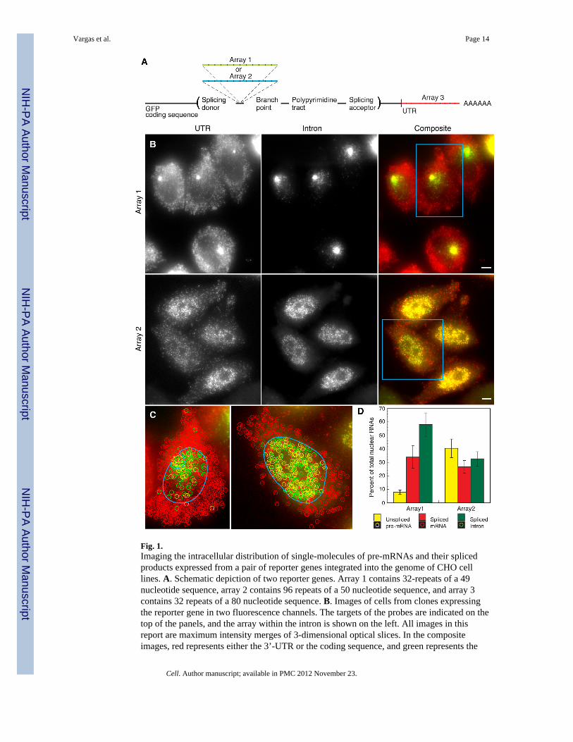
Fig. 1.Imaging the intracellular distribution of single-molecules of pre-mRNAs and their splicedproducts expressed from a pair of reporter genes integrated into the genome of CHO celllines. A. Schematic depiction of two reporter genes. Array 1 contains 32-repeats of a 49nucleotide sequence, array 2 contains 96 repeats of a 50 nucleotide sequence, and array 3contains 32 repeats of a 80 nucleotide sequence. B. Images of cells from clones expressingthe reporter gene in two fluorescence channels. The targets of the probes are indicated on thetop of the panels, and the array within the intron is shown on the left. All images in thisreport are maximum intensity merges of 3-dimensional optical slices. In the compositeimages, red represents either the 3’-UTR or the coding sequence, and green represents the
Vargas et al. Page 14
Cell. Author manuscript; available in PMC 2012 November 23.
NIH
-PA Author Manuscript
NIH
-PA Author Manuscript
NIH
-PA Author Manuscript
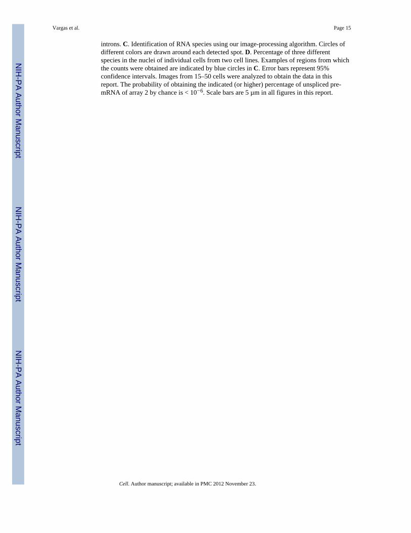
introns. C. Identification of RNA species using our image-processing algorithm. Circles ofdifferent colors are drawn around each detected spot. D. Percentage of three differentspecies in the nuclei of individual cells from two cell lines. Examples of regions from whichthe counts were obtained are indicated by blue circles in C. Error bars represent 95%confidence intervals. Images from 15–50 cells were analyzed to obtain the data in thisreport. The probability of obtaining the indicated (or higher) percentage of unspliced pre-mRNA of array 2 by chance is < 10−6. Scale bars are 5 µm in all figures in this report.
Vargas et al. Page 15
Cell. Author manuscript; available in PMC 2012 November 23.
NIH
-PA Author Manuscript
NIH
-PA Author Manuscript
NIH
-PA Author Manuscript
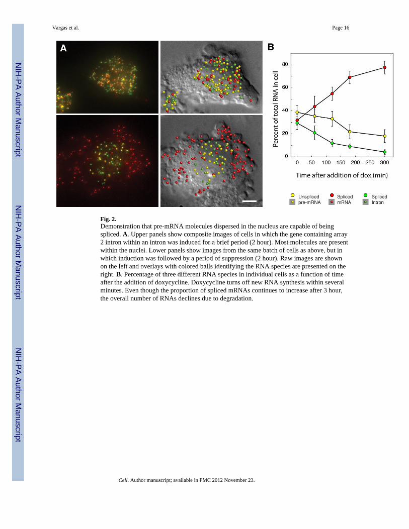
Fig. 2.Demonstration that pre-mRNA molecules dispersed in the nucleus are capable of beingspliced. A. Upper panels show composite images of cells in which the gene containing array2 intron within an intron was induced for a brief period (2 hour). Most molecules are presentwithin the nuclei. Lower panels show images from the same batch of cells as above, but inwhich induction was followed by a period of suppression (2 hour). Raw images are shownon the left and overlays with colored balls identifying the RNA species are presented on theright. B. Percentage of three different RNA species in individual cells as a function of timeafter the addition of doxycycline. Doxycycline turns off new RNA synthesis within severalminutes. Even though the proportion of spliced mRNAs continues to increase after 3 hour,the overall number of RNAs declines due to degradation.
Vargas et al. Page 16
Cell. Author manuscript; available in PMC 2012 November 23.
NIH
-PA Author Manuscript
NIH
-PA Author Manuscript
NIH
-PA Author Manuscript
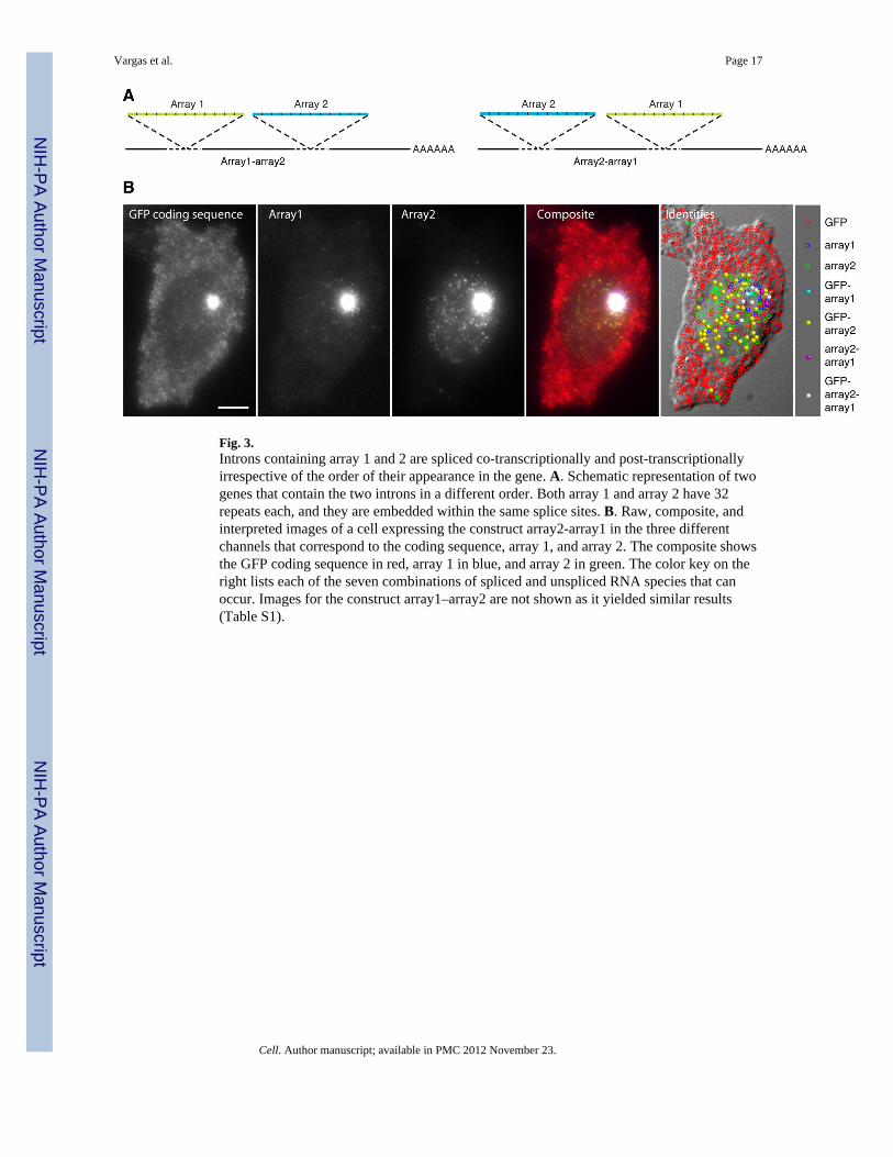
Fig. 3.Introns containing array 1 and 2 are spliced co-transcriptionally and post-transcriptionallyirrespective of the order of their appearance in the gene. A. Schematic representation of twogenes that contain the two introns in a different order. Both array 1 and array 2 have 32repeats each, and they are embedded within the same splice sites. B. Raw, composite, andinterpreted images of a cell expressing the construct array2-array1 in the three differentchannels that correspond to the coding sequence, array 1, and array 2. The composite showsthe GFP coding sequence in red, array 1 in blue, and array 2 in green. The color key on theright lists each of the seven combinations of spliced and unspliced RNA species that canoccur. Images for the construct array1–array2 are not shown as it yielded similar results(Table S1).
Vargas et al. Page 17
Cell. Author manuscript; available in PMC 2012 November 23.
NIH
-PA Author Manuscript
NIH
-PA Author Manuscript
NIH
-PA Author Manuscript
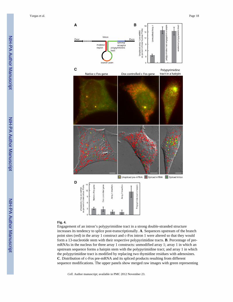
Fig. 4.Engagement of an intron’s polypyrimidine tract in a strong double-stranded structureincreases its tendency to splice post-transcriptionally. A. Sequences upstream of the branchpoint sites (red) in the array 1 construct and c-Fos intron 1 were altered so that they wouldform a 13-nucleotide stem with their respective polypyrimidine tracts. B. Percentage of pre-mRNAs in the nucleus for three array 1 constructs: unmodified array 1; array 1 in which anupstream sequence forms a hairpin stem with the polypyrimidine tract; and array 1 in whichthe polypyrimidine tract is modified by replacing two thymidine residues with adenosines.C. Distribution of c-Fos pre-mRNA and its spliced products resulting from differentsequence modifications. The upper panels show merged raw images with green representing
Vargas et al. Page 18
Cell. Author manuscript; available in PMC 2012 November 23.
NIH
-PA Author Manuscript
NIH
-PA Author Manuscript
NIH
-PA Author Manuscript
the signal from intron probes, and red representing the signal from exon probes. The lowerpanels identify the molecules on DIC images. D. Number of pre-mRNA (excluding theclusters at the gene loci) as a percentage of total cellular RNA in cell lines of differentconstructs. Representative images corresponding to the insertions of the arrays into intron 1are shown in Fig. S3.
Vargas et al. Page 19
Cell. Author manuscript; available in PMC 2012 November 23.
NIH
-PA Author Manuscript
NIH
-PA Author Manuscript
NIH
-PA Author Manuscript
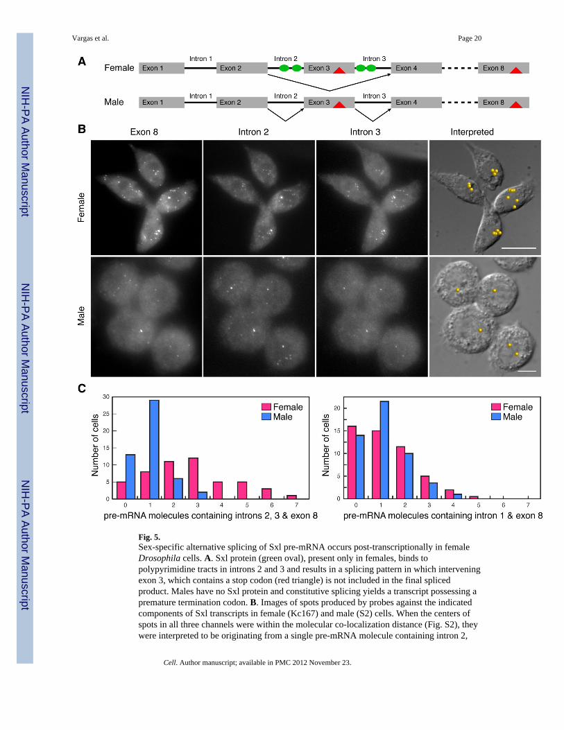
Fig. 5.Sex-specific alternative splicing of Sxl pre-mRNA occurs post-transcriptionally in femaleDrosophila cells. A. Sxl protein (green oval), present only in females, binds topolypyrimidine tracts in introns 2 and 3 and results in a splicing pattern in which interveningexon 3, which contains a stop codon (red triangle) is not included in the final splicedproduct. Males have no Sxl protein and constitutive splicing yields a transcript possessing apremature termination codon. B. Images of spots produced by probes against the indicatedcomponents of Sxl transcripts in female (Kc167) and male (S2) cells. When the centers ofspots in all three channels were within the molecular co-localization distance (Fig. S2), theywere interpreted to be originating from a single pre-mRNA molecule containing intron 2,
Vargas et al. Page 20
Cell. Author manuscript; available in PMC 2012 November 23.
NIH
-PA Author Manuscript
NIH
-PA Author Manuscript
NIH
-PA Author Manuscript

intron 3 and exon 8. The locations of the identified molecules are shown on DIC images. C.Frequency with which the given numbers of pre-mRNA molecules were found in a group of50 cells. After a 30 minute exposure to actinomycin D no pre-mRNAs could be detected inthe cells.
Vargas et al. Page 21
Cell. Author manuscript; available in PMC 2012 November 23.
NIH
-PA Author Manuscript
NIH
-PA Author Manuscript
NIH
-PA Author Manuscript
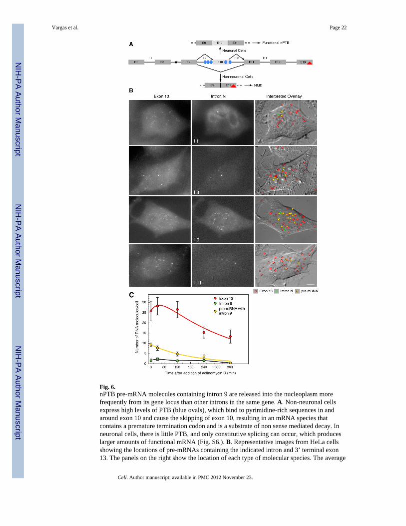
Fig. 6.nPTB pre-mRNA molecules containing intron 9 are released into the nucleoplasm morefrequently from its gene locus than other introns in the same gene. A. Non-neuronal cellsexpress high levels of PTB (blue ovals), which bind to pyrimidine-rich sequences in andaround exon 10 and cause the skipping of exon 10, resulting in an mRNA species thatcontains a premature termination codon and is a substrate of non sense mediated decay. Inneuronal cells, there is little PTB, and only constitutive splicing can occur, which produceslarger amounts of functional mRNA (Fig. S6.). B. Representative images from HeLa cellsshowing the locations of pre-mRNAs containing the indicated intron and 3’ terminal exon13. The panels on the right show the location of each type of molecular species. The average
Vargas et al. Page 22
Cell. Author manuscript; available in PMC 2012 November 23.
NIH
-PA Author Manuscript
NIH
-PA Author Manuscript
NIH
-PA Author Manuscript
number of molecules per cell are provided in Table 1. C. Kinetics of splicing anddegradation of pre-mRNAs containing intron 9 and exon 13 after the addition ofactinomycin D to the culture medium. The yellow line is an exponential decay curve fittedto the observed decrease in the pre-mRNA population. The red line is a fitted curve based onmodeling of reaction: pre-mRNA → mRNA → Φ.
Vargas et al. Page 23
Cell. Author manuscript; available in PMC 2012 November 23.
NIH
-PA Author Manuscript
NIH
-PA Author Manuscript
NIH
-PA Author Manuscript

NIH
-PA Author Manuscript
NIH
-PA Author Manuscript
NIH
-PA Author Manuscript
Vargas et al. Page 24
Table 1
Numbers of nPTB pre-mRNA molecules containing the indicated intron and exon 13 and their splicingproducts in HeLa cells. The numbers are mean values ± 95% confidence interval obtained from at least 20cells.
Intron pre-mRNA Spliced mRNA Spliced Intron
1 0.32±0.20 18.18±2.51 0.39±0.20
8 4.75±0.75 20.13±1.99 0.65±0.34
9 9.28±0.88 25.78±4.84 1.78±0.59
11 2.33±0.48 22.98±1.96 0.37±0.19
Cell. Author manuscript; available in PMC 2012 November 23.