Cartography of neurexin alternative splicing mapped by single-molecule long-read mRNA sequencing
9
Cartography of neurexin alternative splicing mapped by single-molecule long-read mRNA sequencing Barbara Treutlein a,b,1 , Ozgun Gokce c,1 , Stephen R. Quake a,b,d,2 , and Thomas C. Südhof c,d,2 Departments of a Bioengineering and c Molecular and Cellular Physiology, School of Medicine, b Department of Applied Physics, and d Howard Hughes Medical Institute, Stanford University, Stanford, CA 94305 Contributed by Thomas C. Südhof, February 24, 2014 (sent for review January 24, 2014) Neurexins are evolutionarily conserved presynaptic cell-adhesion molecules that are essential for normal synapse formation and synaptic transmission. Indirect evidence has indicated that exten- sive alternative splicing of neurexin mRNAs may produce hundreds if not thousands of neurexin isoforms, but no direct evidence for such diversity has been available. Here we use unbiased long-read sequencing of full-length neurexin (Nrxn)1α, Nrxn1β, Nrxn2β, Nrxn3α, and Nrxn3β mRNAs to systematically assess how many sites of alternative splicing are used in neurexins with a significant frequency, and whether alternative splicing events at these sites are independent of each other. In sequencing more than 25,000 full-length mRNAs, we identified a novel, abundantly used alterna- tively spliced exon of Nrxn1α and Nrxn3α (referred to as alterna- tively spliced sequence 6) that encodes a 9-residue insertion in the flexible hinge region between the fifth LNS (laminin-α, neurexin, sex hormone-binding globulin) domain and the third EGF-like se- quence. In addition, we observed several larger-scale events of alternative splicing that deleted multiple domains and were much less frequent than the canonical six sites of alternative splicing in neurexins. All of the six canonical events of alternative splicing appear to be independent of each other, suggesting that neurex- ins may exhibit an even larger isoform diversity than previously envisioned and comprise thousands of variants. Our data are con- sistent with the notion that α-neurexins represent extracellular protein-interaction scaffolds in which different LNS and EGF domains mediate distinct interactions that affect diverse func- tions and are independently regulated by independent events of alternative splicing. schizophrenia | neuroligin | cerebellin | LRRTM | autism N eurons form highly specific and complex patterns of synaptic connections that underlie all brain function (1–5). Such specific connections require trillions of chemically differentiated synapses whose identity may be shaped by interactions of specific pre- and postsynaptic signaling molecules, especially cell-adhe- sion molecules. The genome size is insufficient to encode such diversity, but a mixture of combinatorial expression patterns that pair different synaptic cell-adhesion molecules with each other and of distinct alternative splicing patterns that amplify the number of cell-adhesion molecules into a large number of isoforms may generate the number of transsynaptic interactions needed to account for the enormous diversity of synaptic connections. In Drosophila melanogaster, alternative splicing of the mRNAs encoding the Down syndrome cell-adhesion molecule can gen- erate nearly 20,000 protein isoforms whose structures specify axon bundling but not synapse formation (6). In mammals, alter- native splicing of neurexin and some protocadherin mRNAs can also produce thousands of isoforms, which may at least in the case of neurexins be involved in synapse formation (7–10). Neurexins are type I membrane proteins that were discovered as presynaptic receptors for α-latrotoxin (8, 9). Six principal neurexins (Nrxn1α–3α and Nrxn1β–3β) are synthesized from three genes (Nrxn1–Nrxn3), each of which expresses larger α- and shorter β-isoforms from independent promoters (11–13). All neurexin mRNAs are extensively alternatively spliced (8, 9, 14). α- and β-neurexins contain different extracellular sequences but identical transmembrane regions and short cytoplasmic tails. Specifically, the extracellular sequences of α-neurexins are composed of six LNS (laminin-α, neurexin, sex hormone-binding globulin) domains with three interspersed EGF-like repeats followed by an O-linked sugar attachment sequence and a con- served cysteine loop sequence (8, 15). In contrast, the extracel- lular sequences of β-neurexins comprise a short β-neurexin– specific sequence, and then splice into the sixth LNS domain of α-neurexins, from which point on β-neurexins are identical to α-neurexins (11). All α-neurexins are subject to alternative splicing at five canonical sites [SS#1–SS#5 (14)], of which SS#4 and SS#5 are also found in β-neurexins. The alternatively spliced sequences for SS#1–SS#4 are highly homologous among the three neurexins, whereas those of SS#5 differ among neurexins. Here the variable sequence in Nrxn1 encompasses only 3 resi- dues, whereas in Nrxn2 it is composed of 194 residues, and in Nrxn3 a variety of alternatively spliced sequences are observed that include inserts of 247 residues and sequences with in-frame stop codons, effectively producing secreted Nrxn3 isoforms (9, 14). Alternative splicing of neurexins is differentially regulated in different brain regions (10, 14, 16), exhibits a diurnal cycle (17), and is modulated by development, neurotrophins, and neuronal activity (18–21). Neurexins are known to bind to a large number of ligands, of which neuroligins, leucine-rich repeat transmembrane proteins, and cerebellins are best-characterized (22–28). All of these ligands Significance Neurexins are presynaptic cell-adhesion molecules that are essential for synapse formation and synaptic transmission. Extensive alternative splicing of neurexin transcripts may generate thousands of isoforms, but it is unclear how many distinct neurexins are physiologically produced. We used un- biased long-read sequencing of full-length neurexin mRNAs to systematically assess the alternative splicing of neurexins in prefrontal cortex. We identified a novel, abundantly used al- ternatively spliced exon of neurexins, and found that the dif- ferent events of alternative splicing of neurexins appear to be independent of each other. Our data suggest that thousands of neurexin isoforms are physiologically generated, consistent with the notion that neurexins represent transsynaptic protein- interaction scaffolds that mediate diverse functions and are regulated by alternative splicing at multiple independent sites. Author contributions: B.T., O.G., S.R.Q., and T.C.S. designed research; B.T. and O.G. per- formed research; B.T., O.G., S.R.Q., and T.C.S. analyzed data; and B.T., O.G., S.R.Q., and T.C.S. wrote the paper. The authors declare no conflict of interest. Data deposition: The sequences reported in this paper have been deposited in the NCBI Se- quence Read Archive (accession no. SRP039451). 1 B.T. and O.G. contributed equally to this work. 2 To whom correspondence may be addressed. E-mail: [email protected] or tcs1@ stanford.edu. This article contains supporting information online at www.pnas.org/lookup/suppl/doi:10. 1073/pnas.1403244111/-/DCSupplemental. www.pnas.org/cgi/doi/10.1073/pnas.1403244111 PNAS Early Edition | 1 of 9 NEUROSCIENCE PNAS PLUS
-
Upload
lmu-munich -
Category
Documents
-
view
1 -
download
0
Transcript of Cartography of neurexin alternative splicing mapped by single-molecule long-read mRNA sequencing

Cartography of neurexin alternative splicing mappedby single-molecule long-read mRNA sequencingBarbara Treutleina,b,1, Ozgun Gokcec,1, Stephen R. Quakea,b,d,2, and Thomas C. Südhofc,d,2
Departments of aBioengineering and cMolecular and Cellular Physiology, School of Medicine, bDepartment of Applied Physics, and dHoward Hughes MedicalInstitute, Stanford University, Stanford, CA 94305
Contributed by Thomas C. Südhof, February 24, 2014 (sent for review January 24, 2014)
Neurexins are evolutionarily conserved presynaptic cell-adhesionmolecules that are essential for normal synapse formation andsynaptic transmission. Indirect evidence has indicated that exten-sive alternative splicing of neurexin mRNAs may produce hundredsif not thousands of neurexin isoforms, but no direct evidence forsuch diversity has been available. Here we use unbiased long-readsequencing of full-length neurexin (Nrxn)1α, Nrxn1β, Nrxn2β,Nrxn3α, and Nrxn3β mRNAs to systematically assess how manysites of alternative splicing are used in neurexins with a significantfrequency, and whether alternative splicing events at these sitesare independent of each other. In sequencing more than 25,000full-length mRNAs, we identified a novel, abundantly used alterna-tively spliced exon of Nrxn1α and Nrxn3α (referred to as alterna-tively spliced sequence 6) that encodes a 9-residue insertion in theflexible hinge region between the fifth LNS (laminin-α, neurexin,sex hormone-binding globulin) domain and the third EGF-like se-quence. In addition, we observed several larger-scale events ofalternative splicing that deleted multiple domains and were muchless frequent than the canonical six sites of alternative splicing inneurexins. All of the six canonical events of alternative splicingappear to be independent of each other, suggesting that neurex-ins may exhibit an even larger isoform diversity than previouslyenvisioned and comprise thousands of variants. Our data are con-sistent with the notion that α-neurexins represent extracellularprotein-interaction scaffolds in which different LNS and EGFdomains mediate distinct interactions that affect diverse func-tions and are independently regulated by independent eventsof alternative splicing.
schizophrenia | neuroligin | cerebellin | LRRTM | autism
Neurons form highly specific and complex patterns of synapticconnections that underlie all brain function (1–5). Such
specific connections require trillions of chemically differentiatedsynapses whose identity may be shaped by interactions of specificpre- and postsynaptic signaling molecules, especially cell-adhe-sion molecules. The genome size is insufficient to encode suchdiversity, but a mixture of combinatorial expression patterns thatpair different synaptic cell-adhesion molecules with each otherand of distinct alternative splicing patterns that amplify thenumber of cell-adhesion molecules into a large number of isoformsmay generate the number of transsynaptic interactions neededto account for the enormous diversity of synaptic connections.In Drosophila melanogaster, alternative splicing of the mRNAsencoding the Down syndrome cell-adhesion molecule can gen-erate nearly 20,000 protein isoforms whose structures specifyaxon bundling but not synapse formation (6). In mammals, alter-native splicing of neurexin and some protocadherin mRNAs canalso produce thousands of isoforms, which may at least in the caseof neurexins be involved in synapse formation (7–10).Neurexins are type I membrane proteins that were discovered
as presynaptic receptors for α-latrotoxin (8, 9). Six principalneurexins (Nrxn1α–3α and Nrxn1β–3β) are synthesized fromthree genes (Nrxn1–Nrxn3), each of which expresses larger α- andshorter β-isoforms from independent promoters (11–13). Allneurexin mRNAs are extensively alternatively spliced (8, 9, 14).
α- and β-neurexins contain different extracellular sequencesbut identical transmembrane regions and short cytoplasmic tails.Specifically, the extracellular sequences of α-neurexins arecomposed of six LNS (laminin-α, neurexin, sex hormone-bindingglobulin) domains with three interspersed EGF-like repeatsfollowed by an O-linked sugar attachment sequence and a con-served cysteine loop sequence (8, 15). In contrast, the extracel-lular sequences of β-neurexins comprise a short β-neurexin–specific sequence, and then splice into the sixth LNS domain ofα-neurexins, from which point on β-neurexins are identical toα-neurexins (11). All α-neurexins are subject to alternativesplicing at five canonical sites [SS#1–SS#5 (14)], of which SS#4and SS#5 are also found in β-neurexins. The alternatively splicedsequences for SS#1–SS#4 are highly homologous among thethree neurexins, whereas those of SS#5 differ among neurexins.Here the variable sequence in Nrxn1 encompasses only 3 resi-dues, whereas in Nrxn2 it is composed of 194 residues, and inNrxn3 a variety of alternatively spliced sequences are observedthat include inserts of 247 residues and sequences with in-framestop codons, effectively producing secreted Nrxn3 isoforms (9,14). Alternative splicing of neurexins is differentially regulated indifferent brain regions (10, 14, 16), exhibits a diurnal cycle (17),and is modulated by development, neurotrophins, and neuronalactivity (18–21).Neurexins are known to bind to a large number of ligands, of
which neuroligins, leucine-rich repeat transmembrane proteins,and cerebellins are best-characterized (22–28). All of these ligands
Significance
Neurexins are presynaptic cell-adhesion molecules that areessential for synapse formation and synaptic transmission.Extensive alternative splicing of neurexin transcripts maygenerate thousands of isoforms, but it is unclear how manydistinct neurexins are physiologically produced. We used un-biased long-read sequencing of full-length neurexin mRNAs tosystematically assess the alternative splicing of neurexins inprefrontal cortex. We identified a novel, abundantly used al-ternatively spliced exon of neurexins, and found that the dif-ferent events of alternative splicing of neurexins appear to beindependent of each other. Our data suggest that thousands ofneurexin isoforms are physiologically generated, consistentwith the notion that neurexins represent transsynaptic protein-interaction scaffolds that mediate diverse functions and areregulated by alternative splicing at multiple independent sites.
Author contributions: B.T., O.G., S.R.Q., and T.C.S. designed research; B.T. and O.G. per-formed research; B.T., O.G., S.R.Q., and T.C.S. analyzed data; and B.T., O.G., S.R.Q., andT.C.S. wrote the paper.
The authors declare no conflict of interest.
Data deposition: The sequences reported in this paper have been deposited in the NCBI Se-quence Read Archive (accession no. SRP039451).1B.T. and O.G. contributed equally to this work.2To whom correspondence may be addressed. E-mail: [email protected] or [email protected].
This article contains supporting information online at www.pnas.org/lookup/suppl/doi:10.1073/pnas.1403244111/-/DCSupplemental.
www.pnas.org/cgi/doi/10.1073/pnas.1403244111 PNAS Early Edition | 1 of 9
NEU
ROSC
IENCE
PNASPL
US
bind to both α- and β-neurexins because they interact with thesixth LNS domain; strikingly, these interactions are differentiallyregulated by alternative splicing of neurexins at SS#4 in the sixthLNS domain (24, 26–31). In addition to these ligands, neurexinswere shown to bind to neurexophilin, dystroglycan, and CIRL/latrophilin (32–35).Despite extensive studies, the full extent of neurexin alterna-
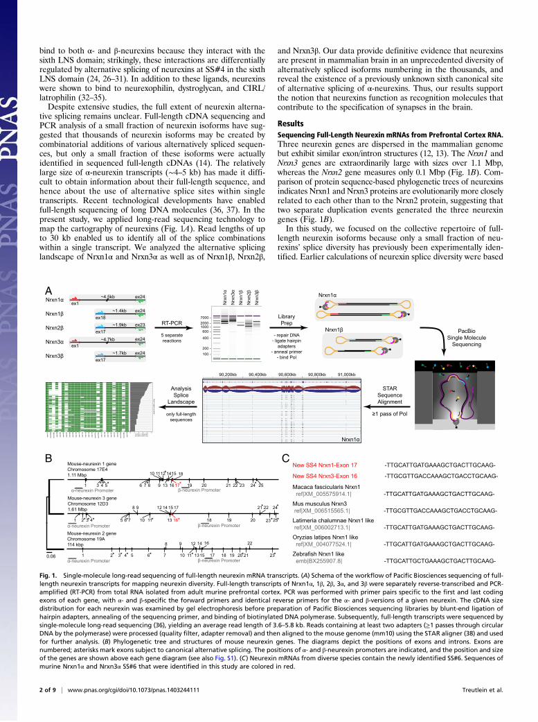
tive splicing remains unclear. Full-length cDNA sequencing andPCR analysis of a small fraction of neurexin isoforms have sug-gested that thousands of neurexin isoforms may be created bycombinatorial additions of various alternatively spliced sequen-ces, but only a small fraction of these isoforms were actuallyidentified in sequenced full-length cDNAs (14). The relativelylarge size of α-neurexin transcripts (∼4–5 kb) has made it diffi-cult to obtain information about their full-length sequence, andhence about the use of alternative splice sites within singletranscripts. Recent technological developments have enabledfull-length sequencing of long DNA molecules (36, 37). In thepresent study, we applied long-read sequencing technology tomap the cartography of neurexins (Fig. 1A). Read lengths of upto 30 kb enabled us to identify all of the splice combinationswithin a single transcript. We analyzed the alternative splicinglandscape of Nrxn1α and Nrxn3α as well as of Nrxn1β, Nrxn2β,
and Nrxn3β. Our data provide definitive evidence that neurexinsare present in mammalian brain in an unprecedented diversity ofalternatively spliced isoforms numbering in the thousands, andreveal the existence of a previously unknown sixth canonical siteof alternative splicing of α-neurexins. Thus, our results supportthe notion that neurexins function as recognition molecules thatcontribute to the specification of synapses in the brain.
ResultsSequencing Full-Length Neurexin mRNAs from Prefrontal Cortex RNA.Three neurexin genes are dispersed in the mammalian genomebut exhibit similar exon/intron structures (12, 13). The Nrxn1 andNrxn3 genes are extraordinarily large with sizes over 1.1 Mbp,whereas the Nrxn2 gene measures only 0.1 Mbp (Fig. 1B). Com-parison of protein sequence-based phylogenetic trees of neurexinsindicates Nrxn1 and Nrxn3 proteins are evolutionarily more closelyrelated to each other than to the Nrxn2 protein, suggesting thattwo separate duplication events generated the three neurexingenes (Fig. 1B).In this study, we focused on the collective repertoire of full-
length neurexin isoforms because only a small fraction of neu-rexins’ splice diversity has previously been experimentally iden-tified. Earlier calculations of neurexin splice diversity were based
*1 3 4 5 7 86 9 19 20 21 22 23 24 2513 16 17
18
******
10 1112 1514
α-neurexin Promoter β-neurexin Promoter
*
1 2 3 4 5 76
9
18 19 20
22
23
24
2513
17
16 **** 10 11
12 1514
α-neurexin Promoter β-neurexin Promoter
8 21*
*
1 2 5 6 18 19 20
22
2317* 10 1315
14
α-neurexin Promoter β-neurexin Promoter
8
21 *
1612
11
9
73 4* * * * *
Mouse-neurexin 3 geneChromosome 12D31.61 Mbp
Mouse-neurexin 1 gene Chromosome 17E41.11 Mbp
Mouse-neurexin 2 geneChromosome 19A 114 kbp
0.06
Nrxn1α
Nrxn1βex18
ex24~1.4kbex1
ex24~4.5kb
Nrxn3α
Nrxn3β
Nrxn2βex17
ex23~1.9kb
ex17ex24~1.7kb
ex1ex24
RT-PCR
~4.7kb
Nrx
n1α
Nrx
n1β
Nrx
n3α
Nrx
n3β
Nrx
n2β
7000 -2000 -1000 -600 -
400 -
100 -200 -
LibraryPrep
- repair DNA- ligate hairpin
adapters- anneal primer
- bind Pol
5 separatereactions
PacBioSingle Molecule
Sequencing
STARSequenceAlignment
≥1 pass of Pol
Nrxn1α
Nrxn1β
90,200kb 90,400kb 90,600kb 90,800kb 91,000kb
AnalysisSplice
Landscape
A
Nrxn1α
only full-lengthsequences
0 1 2 3 4 5Number of transcriptsper splice-variant, log
exon
01
e xon
03a
exon
03b
exon
04
exon
05
exon
06
exon
07
exon
08
exon
09
exon
10
exon
11
exon
12
exon
13
exon
14
exon
15
exon
16o
exon
16n
exon
17
exon
18
e xon
19
exon
20
exon
21
exon
22
exon
23a
exon
23b
exon
24
Macaca fascicularis Nrxn1 ref|XM_005575914.1| -TTGCATTGATGAAAGCTGACTTGCAAG-
Latimeria chalumnae Nrxn1 like ref|XM_006002713.1| -TTGCATTGATGAAAGCTGACTTGCAAG-
Oryzias latipes Nrxn1 like ref|XM_004077524.1| -TTGCATTGATGAAAGCTGACTTGCAAG-
New SS4 Nrxn1-Exon 17 -TTGCATTGATGAAAGCTGACTTGCAAG-
New SS4 Nrxn3-Exon 16 -TTGCGTTGACCAAAGCTGACCTGCAAG-
Zebrafish Nrxn1 like emb|BX255907.8| -TTGCATTGCTGAAAGCTGACTTGCAAG-
Mus musculus Nrxn3 ref|XM_006515565.1| -TTGCGTTGACCAAAGCTGACCTGCAAG-
B C
*** *
Fig. 1. Single-molecule long-read sequencing of full-length neurexin mRNA transcripts. (A) Schema of the workflow of Pacific Biosciences sequencing of full-length neurexin transcripts for mapping neurexin diversity. Full-length transcripts of Nrxn1α, 1β, 2β, 3α, and 3β were separately reverse-transcribed and PCR-amplified (RT-PCR) from total RNA isolated from adult murine prefrontal cortex. PCR was performed with primer pairs specific to the first and last codingexons of each gene, with α- and β-specific the forward primers and identical reverse primers for the α- and β-versions of a given neurexin. The cDNA sizedistribution for each neurexin was examined by gel electrophoresis before preparation of Pacific Biosciences sequencing libraries by blunt-end ligation ofhairpin adapters, annealing of the sequencing primer, and binding of biotinylated DNA polymerase. Subsequently, full-length transcripts were sequenced bysingle-molecule long-read sequencing (36), yielding an average read length of 3.6–5.8 kb. Reads containing at least two adapters (≥1 passes through circularDNA by the polymerase) were processed (quality filter, adapter removal) and then aligned to the mouse genome (mm10) using the STAR aligner (38) and usedfor further analysis. (B) Phylogenetic tree and structures of mouse neurexin genes. The diagrams depict the positions of exons and introns. Exons arenumbered; asterisks mark exons subject to canonical alternative splicing. The positions of α- and β-neurexin promoters are indicated, and the position and sizeof the genes are shown above each gene diagram (see also Fig. S1). (C) Neurexin mRNAs from diverse species contain the newly identified SS#6. Sequences ofmurine Nrxn1α and Nrxn3α SS#6 that were identified in this study are colored in red.
2 of 9 | www.pnas.org/cgi/doi/10.1073/pnas.1403244111 Treutlein et al.

on the assumption that all splice events are independent andsplicing can occur only at five canonical positions in the gene andsuggested the generation of more than 3,000 distinct neurexins
(14), but no direct evidence supports this supposition. To un-derstand the true diversity of neurexin mRNAs, we determined thefull-length sequences of thousands of neurexin mRNAs (Fig. 1A).
Fig. 2. Splice landscape of Nrxn1α. The transcript map visualizes the 247 unique alternatively spliced isoforms (rows) observed for the 2,574 full-lengthNrxn1α mRNAs sequenced. Exons (columns) are colored in green if present and in white if absent, and are numbered at the bottom (asterisks, exons withcanonical alternative splicing; for an explanation of the numbering, see Fig. S1). The domain structure of Nrxn1α is shown at the top (light blue hexagons, LNSdomains; black ovals, EGF-like domains; CHO, O-linked sugar modifications; TMR, transmembrane region) and is connected to the exons that encode therespective domains by dotted gray lines. The abundance of each splice isoform is shown in the bar graph (Right).
Treutlein et al. PNAS Early Edition | 3 of 9
NEU
ROSC
IENCE
PNASPL
US

All principal neurexin mRNAs (encoding Nrxn1α, Nrxn1β,Nrxn2β, Nrxn3α, and Nrxn3β) except for that of Nrxn2α wereamplified by primers specific to the first and last exons, andseparate sequencing libraries were prepared by ligating hairpinadapters to both ends of each DNA molecule such that the DNApolymerase can traverse each DNA molecule multiple timesduring rolling circle replication (Fig. 1A). Nrxn2α amplificationwas unsuccessful, possibly because of its lower abundance, se-quence content, and/or larger size. After filtering out all reads thatmissed the exons targeted by the amplification primers (damagedor misprimed PCR products), we obtained a total of 23,943 full-length mRNA reads (2,574 Nrxn1α, 1,653 Nrxn1β, 10,283 Nrxn2β,934 Nrxn3α, and 8,499 Nrxn3β cDNAs).
Identification of a New Conserved Alternatively Spliced α-NeurexinExon.We aligned filtered reads to the mouse genome (mm10) usingthe STAR sequence aligner (38). By comparing the full-lengthtranscript sequences with the characterized neurexin gene structures(12), we observed a previously unidentified, alternatively splicedexon in theNrxn1α andNrxn3α genes [alternatively spliced sequence6 (SS#6)]. The 9-residue amino acid sequence encoded by this al-ternatively spliced exon maps to the hinge region between EGF-Cand the fifth LNS domains of Nrxn1α and Nrxn3α (Fig. 1B) (39, 40).Although this new alternatively spliced exon was not characterizedpreviously, it is present in neurexin mRNAs in the public databasesand is evolutionarily conserved in zebrafish and macaque neurexinmRNAs (Fig. 1C). However, this exon is missing in the Nrxn2 gene.Phylogenetic analysis (Fig. 1B) suggests that neurexins are the resultof relatively recent gene duplication events, which first separatedNrxn2 from the two other neurexins and then diverged Nrxn1 andNrxn3 from each other, accounting for the absence of the exonencoding SS#6 from Nrxn2α.
Splice Landscape of Nrxn1α. We used the sequencing reads to gen-erate a transcript map of Nrxn1α. This map reveals that 247 uniquesplice isoforms (rows) are observed in 2,574 full-length transcripts,with 96 unique splice isoforms accounting for 90% of all detectedtranscripts (Fig. 2). In addition to events at the six canonical Nrxn1αsites of alternative splicing, we observed alternative splicing atnoncanonical sites that include all Nrxn1α exons. As an example,exons 13, 14, and 15 were spliced out in 5% of the Nrxn1α mRNAs(Fig. 2; see also Fig. 6). The majority of novel alternative splicingevents consisted of modular excisions of multiple exons: Whereas insome cases two canonical sites were spliced out together withinterjacent exons, in other cases novel splice donors were used.
Confirmation of a Novel Event of Modular Alternative Splicing. Toconfirm novel splicing events, we focused on a modular event thatexcises Nrxn1α exons 12–18 (12–17 in ref. 12). Using specific pri-mers, we amplified Nrxn1α exons 11–20 (11–19 in ref. 12) fromprefrontal cortex total RNA and observed two distinct bands: ahigher band of ∼1,500 bp matching the size of the mRNA includingall exons, and a lower band of ∼300 bp matching the size of themRNA lacking exons 12–18 (Fig. 3A). Sanger sequencing of thelower band confirmed that exons 12–18 are absent (Fig. 3B).
Splice Landscapes of Nrxn3α, 1β, 2β, and 3β. Using full-length readsof Nrxn3α, Nrxn1β, Nrxn2β, and Nrxn3β, we generated transcriptmaps of these neurexins to visualize their isoform diversity (Figs. 4and 5). For Nrxn3α, we detected 934 full-length mRNA sequencesthat encode 138 different unique splice variants, with 67 uniquesplice isoforms accounting for 90% of all detected transcripts. In-terestingly, we did not observe alternative splicing of the previouslyreported alternatively spliced exons 3 and 24b/c (23b/c in ref. 12),but we did detect alternative splicing of exons 5, 7, 8, 9, and 16,which have previously been reported to be invariant (Fig. 4). Incontrast to Nrxn1α, Nrxn3α novel splicing events are limited to thesecond and third LNS and EGF-B domains.
Regarding the shorter β-neurexins, we detected 1,653 full-length mRNA sequences for Nrxn1β, 10,283 full-length sequen-ces for Nrxn2β, and 8,499 full-length sequences for Nrxn3β,encoding 11, 9, and 41 splice variants in total, respectively. Forβ-neurexins, we were able to detect all previously identified al-ternative splicing events except for exons 24b and 24c (23b and23c in ref. 12) of Nrxn3. We detected 146 splicing events of thepreviously unreported alternatively spliced exon 22 in Nrxn3βbut not in the Nrxn3α isoforms.
Alternative Splicing Events of Nonadjacent Exon Clusters AreIndependent of Each Other. To examine whether alternativesplicing at different splice sites is coordinated, we analyzed thepairwise correlation between events of alternative splicing atdifferent sites in Nrxn1α and Nrxn3α. We detected no correlationand hence independent splicing of different splice sites (Fig. 6 Aand B). However, correlations were observed within canonicalsplice sites, such as Nrxn1α exons 3a and 4 (SS#1), or in the caseof modular excision events of exon blocks such as Nrxn3α exons5–9 (Fig. 6 A and B). In these cases, correlations are the result ofneighboring exons being part of the same splicing reaction.
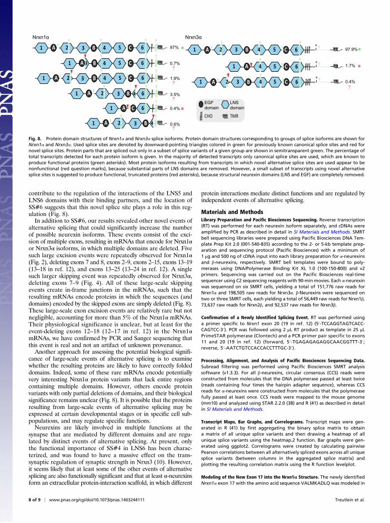
Effect of the Novel Alternatively Spliced Exon on the Structure ofNrxn1α and Nrxn3α. By using single-molecule long-read se-quencing, we have identified not only a new alternatively splicedexon (SS#6 Fig. 7) but also observed splicing of exons that werepreviously not considered to be subject to alternative splicing. Inmost of these newly identified events of alternative splicing,multiple exons were excised, resulting in the deletion of repeatedmodules in the neurexin protein domain structure. The questionarises whether these events of alternative splicing representaccidents of nature or whether they are physiologically relevant.If they were accidents, they should be random and disturb theprotein domain structure of a neurexin.To visualize the effect of novel alternative splicing events on
protein domain structure, we determined the location of novelsplice sites within the domain structure (Fig. 8). As neurexin proteindomains mostly consist of multiple exons, alternative splicing eventsmostly lead to the partial deletion of domains and produce non-functional proteins. However, some of the novel alternative splicingevents completely remove large structural modules, thereby creat-ing new neurexins that are likely to have new functional properties.Strikingly, we observed very few large-scale domain structure-changing events of alternative splicing in Nrxn3α, but several in-dependent events in Nrxn1α with dramatic effects. For example,some rare but repeatedly observed events of large-scale alternativesplicing of Nrxn1α completely remove all domains between thefirst and last LNS and EGF-like domains (Fig. 8), which are likelyto form functional neurexin isoforms with novel protein structures.We estimated that 97.4% of Nrxn1α transcripts and 99.6% ofNrxn3α transcripts generate functional proteins, based on theassumption that a transcript can be translated into a functional
A B
Fig. 3. Validation of newly identified alternatively spliced isoform of Nrxn1αlacking exons 12–18 by RT-PCR. (A) Agarose gel electrophoresis analysis of the PCRproduct obtained with primers specific to the end of exon 11 and beginning ofexon 20 of Nrxn1α. Twomajor splice variants are identified: a long DNA fragment(∼1,500 bp) corresponding to transcripts containing exons 12–18 as well as a shortDNA fragment (∼300 bp) corresponding to transcripts lacking exons 12–18. Notethat exon 19 encodes the N terminus of Nrxn1β and is alwaysmissing fromNrxn1αmRNAs (12). (B) Sanger sequencing of the short PCR product as obtained in A.
4 of 9 | www.pnas.org/cgi/doi/10.1073/pnas.1403244111 Treutlein et al.

protein if the correct reading frame is maintained and pre-dicted protein modules are likely either present or absent.
DiscussionWe used a single-molecule long-read sequencing approach forprofiling neurexin isoform diversity. Previous analyses of neurexinswere based on sequencing of a limited number of cDNAs and on
PCR-based methods (14). These methods either did not haveenough depth to map the complete diversity of neurexin mRNAs(cDNA sequencing) or analyzed individual splice sites separately(PCR), and thus did not provide information about the combinationof different splice sites within full-length transcripts. In contrast, thelong-read sequencing approach overcomes these limitations by se-quencing thousands of independent full-length mRNA transcripts.
Fig. 4. Splice landscape ofNrxn3α. The transcript map visualizes the 138 unique alternatively spliced isoforms (rows) observed for the 934 full-lengthNrxn3αmRNAssequenced. Exons (columns) are colored in green for coding and red for exons with in-frame stop codons if present and in white if absent, and are numbered atthe bottom (asterisks, exons with canonical alternative splicing; for an explanation of the numbering, see Fig. S1). The domain structure of Nrxn1α is shown at thetop and is connected to the exons that encode the respective domains by dotted gray lines. In the domain structure, the asterisk denotes a stop codon thatis present in some of the alternatively spliced exons and could produce secreted versions of neurexin-3. The abundance of each splice isoform is shown inthe bar graph (Right).
Treutlein et al. PNAS Early Edition | 5 of 9
NEU
ROSC
IENCE
PNASPL
US

Our study provides more than 25,000 full-length neurexin mRNAreads, allowing us to analyze all splicing events within single mRNAmolecules and to detect low-expressed isoforms.We found that neurexins are likely even more polymorphic
than previously thought. Full-length mRNA sequencing revealedan enormous diversity of α-neurexin isoforms in which no single
splice variant dominates (Figs. 2 and 4). The situation is muchsimpler for β-neurexins in which diversity is limited, and nearlyall variants can be explained by alternative splicing at the pre-viously described SS#4 and SS#5 (14). Based on our sequencingdata, how many different neurexins are there? For this calcula-tion, we need to know whether all alternative splicing events are
A B
C
D
E
F
Fig. 5. Splice landscape of β-neurexins. (A–C) Transcript maps visualizing unique splice isoforms (rows) observed for Nrxn1β (A; 1,653 transcripts, 11 splicevariants), Nrxn2β (B; 10,283 transcripts, 9 splice variants), and Nrxn3β (C; 8,499 transcripts, 41 splice variants). Exons (columns) are colored in green if presentand in white if absent, and are numbered at the bottom (asterisks, exons with canonical alternative splicing; for an explanation of the numbering, see Fig. S1).The domain structures of the respective β-neurexins are shown above the transcript maps and are connected to the exons that encode the respective domainsby dotted gray lines. SP denotes the signal peptide; the asterisk in the domain structure of Nrxn3β indicates a stop codon encoded by one of the alternativelyspliced exons. The abundance of each splice isoform is shown in the bar graph (Right). (D–F) Bar graphs visualizing the relative frequency of exon skipping forthe indicated exons in the Nrxn1β (D), Nrxn2β (E), and Nrxn3β mRNAs (F). Canonical alternatively spliced exons are marked by asterisks.
6 of 9 | www.pnas.org/cgi/doi/10.1073/pnas.1403244111 Treutlein et al.

independent of each other. To test this, we calculated thePearson correlation coefficient between all alternatively splicedsequences. We did not observe any correlation between canonicalsplice sites, including the newly identified alternatively splicedexon, suggesting that these sites of alternative splicing are usedindependent of each other. Cumulatively, our results suggest thatthe various events of alternative splicing can be used independentof each other in all possible combinations. Based on this conclu-sion, we calculated a total number of possible neurexin variants,taking into account only splice events that were observed morethan twice. We observed in this manner a minimal diversity of1,159 isoforms for Nrxn1α, 1,120 isoforms for Nrxn3α, and a totalof 152 isoforms for all three β-neurexins. Thus, earlier estimates of2,000–3,000 neurexin variants created by alternative splicing (8, 9,12, 14) may have been an underestimate, because our presentstudy arrived at the same numbers by analyzing only one brainregion and one developmental stage (prefrontal cortex of adultmice) and because Nrxn2αmRNAs were not examined, suggestingthat the true number of neurexin variants may be even higher.
An important observation of our present study is that wefound a new evolutionarily conserved exon that is alternativelyspliced in Nrxn1α and Nrxn3α, which we propose referring to asalternatively spliced sequence 6. Although we did not analyzeNrxn2α mRNAs in the current study, the exon encoding SS#6 isabsent from the Nrxn2α gene, suggesting that Nrxn2α lacksSS#6. According to the phylogenetic tree of neurexins, the Nrxn2gene separated from the common precursor gene to Nrxn1 andNrxn3 early in evolution, whereas Nrxn1 and Nrxn3 diverged later(Fig. 1). In mammals, the Nrxn2 gene is strikingly smaller (10-fold)than the Nrxn1 and Nrxn3 genes, and the intronic region of Nrxn1and Nrxn3 containing the exon encoding SS#6 is 14 and 60 timeslarger, respectively, than the corresponding intron in the Nrxn2gene (327 bp for the mouse genome).SS#6 encodes 9 residues that localize to the flexible hinge re-
gion between the fifth LNS domain (LNS5) and the third (C)EGF-like sequence of neurexins (Fig. 7A) (39, 40). This is the onlyregion of the Nrxn1α structure in which there are no interdomaincontacts between the LNS and EGF-like domains. The flexiblehinge region has been proposed to explain existing biochemicaldata suggesting that the sixth LNS domain (LNS6) behaves dif-ferently depending on whether it is examined on its own (as it ispresent in β-neurexins) or together with other LNS domains[as it is present in α-neurexins (39, 40)]. The hinge region may
A B
C
D
Fig. 6. Independent and coordinated splicing of canonical and novel splicesites for Nrxn1α and Nrxn3α. (A and B) Correlograms showing the pairwisecorrelation in splice behavior between alternatively spliced exons of (A)Nrxn1α and (B) Nrxn3α. The color bar denotes the Pearson correlation co-efficient from −1 (blue, anticorrelated splicing) through 0 (no correlation insplicing) to 1 (green, positively correlated splicing). Newly observed alter-natively spliced exons tend to be spliced out in a coordinated way, whereascanonical events of alternative splicing appear to occur independent of eachother. (C and D) Bar graphs visualizing the frequency of each exon to bespliced out for (C) Nrxn1α and (D) Nrxn3α. Canonical alternatively splicedexons (12) are marked by asterisks. Exons that are observed to be alterna-tively spliced in a coordinated way (A and B) show similar frequencies.
L2 L3 L4 L5
L6
EGF-B
EGF-C
SS#6GAAGTTGCATTGATGAAAGCTGACTTGCAAGGGCCC E V A L M K A D L Q G P
GAAGTTGCGTTGACCAAAGCTGACCTGCAAGGACCC E V A L T K A D L Q G P
Nrxn1α
Nrxn3α
···taacaaagTTGCGTTGACCAAAGCTGACCTGCAAGgtagag···
···ctaccTTGCAAGTCAGCTTTCATCAATGCAACtttgtca···
90847275│
mm10 Nrxn3 chr12 (AC_000034.1):90847301
│
mm10 Nrxn1 chr17 (AC_000039.1):94947883
│94947909
│
A
B
exon 1716 18
exon 1615 17
Fig. 7. Sequence and structure of newly identified alternatively splicedexons for Nrxn1α and Nrxn3α. (A) mRNA sequence and translated 9-residueamino acid sequence of the newly identified alternatively spliced Nrxn1αexon 17 and Nrxn3α exon 16 (SS#6). A possible extended conformation ofNrxn1α exon 17 was modeled into the crystal structure of Nrxn1α [PDB ID3QCW (39)] using Coot (42). SS#6 is located between the LNS5 and EGF-Cdomains, thereby extending the flexible hinge region between these struc-tural features. The image of the Nrxn1α protein structure was created inChimera (43). (B) Location of the newly identified exon of Nrxn1α andNrxn3α in the mouse genome (mm10). Exonic nucleotides are shown incapital letters, whereas intronic nucleotides are shown in lowercase letters.
Treutlein et al. PNAS Early Edition | 7 of 9
NEU
ROSC
IENCE
PNASPL
US
contribute to the regulation of the interactions of the LNS5 andLNS6 domains with their binding partners, and the location ofSS#6 suggests that this novel splice site plays a role in this reg-ulation (Fig. 8).In addition to SS#6, our results revealed other novel events of
alternative splicing that could significantly increase the numberof possible neurexin isoforms. These events consist of the exci-sion of multiple exons, resulting in mRNAs that encode for Nrxn1αor Nrxn3α isoforms, in which multiple domains are deleted. Fivesuch large excision events were repeatedly observed for Nrxn1α(Fig. 2), deleting exons 7 and 8, exons 2–9, exons 2–15, exons 13–19(13–18 in ref. 12), and exons 13–25 (13–24 in ref. 12). A singlesuch larger skipping event was repeatedly observed for Nrxn3α,deleting exons 7–9 (Fig. 4). All of these large-scale skippingevents create in-frame junctions in the mRNAs, such that theresulting mRNAs encode proteins in which the sequences (anddomains) encoded by the skipped exons are simply deleted (Fig. 8).These large-scale exon excision events are relatively rare but notnegligible, accounting for more than 5% of the Nrxn1α mRNAs.Their physiological significance is unclear, but at least for theevent-deleting exons 12–18 (12–17 in ref. 12) in the Nrxn1αmRNAs, we have confirmed by PCR and Sanger sequencing thatthis event is real and not an artifact of unknown provenance.Another approach for assessing the potential biological signifi-
cance of large-scale events of alternative splicing is to examinewhether the resulting proteins are likely to have correctly foldeddomains. Indeed, some of these rare mRNAs encode potentiallyvery interesting Nrxn1α protein variants that lack entire regionscontaining multiple domains. However, others encode proteinvariants with only partial deletions of domains, and their biologicalsignificance remains unclear (Fig. 8). It is possible that the proteinsresulting from large-scale events of alternative splicing may beexpressed at certain developmental stages or in specific cell sub-populations, and may regulate specific functions.Neurexins are likely involved in multiple functions at the
synapse that are mediated by different domains and are regu-lated by distinct events of alternative splicing. At present, onlythe functional importance of SS#4 in LNS6 has been charac-terized, and was found to have a massive effect on the trans-synaptic regulation of synaptic strength in Nrxn3 (10). However,it seems likely that at least some of the other events of alternativesplicing are also functionally significant and that at least α-neurexinsform an extracellular protein-interaction scaffold, in which different
protein interactions mediate distinct functions and are regulated byindependent events of alternative splicing.
Materials and MethodsLibrary Preparation and Pacific Biosciences Sequencing. Reverse transcription(RT) was performed for each neurexin isoform separately, and cDNAs wereamplified by PCR as described in detail in SI Materials and Methods. SMRTbell sequencing libraries were prepared using Pacific Biosciences DNA Tem-plate Prep Kit 2.0 (001-540-835) according to the 2- or 5-kb template prep-aration and sequencing protocol (Pacific Biosciences) with a minimum of1 μg and 500 ng of cDNA input into each library preparation for α-neurexinsand β-neurexins, respectively. SMRT bell templates were bound to poly-merases using DNA/Polymerase Binding Kit XL 1.0 (100-150-800) and v2primers. Sequencing was carried out on the Pacific Biosciences real-timesequencer using C2 sequencing reagents with 90-min movies. Each α-neurexinwas sequenced on six SMRT cells, yielding a total of 151,776 raw reads forNrxn1α and 198,505 raw reads for Nrxn3α. β-Neurexins were sequenced ontwo or three SMRT cells, each yielding a total of 56,449 raw reads for Nrxn1β,73,637 raw reads for Nrxn2β, and 92,537 raw reads for Nrxn3β.
Confirmation of a Newly Identified Splicing Event. RT was performed usinga primer specific to Nrxn1 exon 20 (19 in ref. 12) (5′-TCCAGGTAGTCACC-CAGTCC-3′). PCR was followed using 2 μL RT product as template in 25 μLPrimeSTAR polymerase (Clontech) and a PCR primer pair specific to exons11 and 20 (19 in ref. 12) (forward, 5′-TGAGAGAGAGGCAACGGTTT-3′;reverse, 5′-AATCTGTCCACCACCTTTGC-3′).
Processing, Alignment, and Analysis of Pacific Biosciences Sequencing Data.Subread filtering was performed using Pacific Biosciences SMRT analysissoftware (v1.3.3). For all β-neurexins, circular consensus (CCS) reads wereconstructed from molecules that the DNA polymerase passed at least twice(reads containing four times the hairpin adapter sequence), whereas CCSreads for α-neurexins were constructed from molecules that the polymerasefully passed at least once. CCS reads were mapped to the mouse genome(mm10) and analyzed using STAR 2.2.0 (38) and R (41) as described in detailin SI Materials and Methods.
Transcript Maps, Bar Graphs, and Correlograms. Transcript maps were gen-erated in R (41) by first aggregating the binary splice matrix to obtaina matrix of all unique splice variants and then drawing a heatmap of allunique splice variants using the heatmap.2 function. Bar graphs were gen-erated using ggplot2. Correlograms were created by calculating pairwisePearson correlations between all alternatively spliced exons across all uniquesplice variants (between columns in the aggregated splice matrix) andplotting the resulting correlation matrix using the R function levelplot.
Modeling of the New Exon 17 into the Nrxn1α Structure. The newly identifiedNrxn1α exon 17 with the amino acid sequence VALMKADLQ was modeled in
EGFdomain
LNSdomain
CHO
0.4%
Nrxn3α
1.9%
0.7%
3.5%
0.4%
0.6%
Nrxn1α
TMR
*
61 3A 2 C5B 4
6C541 BA 3
6C541 B3A 2
1 3A 2 B 4 6
61 A C
1 3A 2 B 4
6*
51 3A B2 4A C
1 4A 6*
5 C 1.7% *
1 3 B2 4A 6*
5 C
97% * 97.9%*
Fig. 8. Protein domain structures of Nrxn1α and Nrxn3α splice isoforms. Protein domain structures corresponding to groups of splice isoforms are shown forNrxn1α and Nrxn3α. Used splice sites are denoted by downward-pointing triangles colored in green for previously known canonical splice sites and red fornovel splice sites. Protein parts that are spliced out only in a subset of splice variants of a given group are shown in semitransparent green. The percentage oftotal transcripts detected for each protein isoform is given. In the majority of detected transcripts only canonical splice sites are used, which are known toproduce functional proteins (green asterisks). Most protein isoforms resulting from transcripts in which novel alternative splice sites are used appear to benonfunctional (red question marks), because substantial parts of LNS domains are removed. However, a small subset of transcripts using novel alternativesplice sites is suggested to produce functional, truncated proteins (red asterisks), because structural neurexin domains (LNS and EGF) are completely removed.
8 of 9 | www.pnas.org/cgi/doi/10.1073/pnas.1403244111 Treutlein et al.

an arbitrary, extended conformation into the Nrxn1α crystal structure [Pro-tein Data Bank (PDB) ID code 3QCW (39)] between E1088 and G1089 usingCoot (42). To accommodate the additional 9 amino acids of the new exon,Nrxn1α C-terminal domains EGF-C and LNS6 (G1089–V1355) were dislocatedfrom the N-terminal part of the protein by about 27 Å.
Effects of Modular Splicing Events on Protein Structure. Aggregated binarysplicematrices were created for the examined regions of Nrxn1α andNrxn3α in Rto identify all unique splice variants and their abundances. Splice variants weregrouped based on similarity of the corresponding protein domain structure. Allsplice variants containing only previously detected canonical splice events were
grouped together because they are known to produce functional neurexinproteins with only small changes in the overall protein domain structure. Allremaining splice variants corresponded to proteins with more substantialchanges in the protein domain structure that are suggested to be either non-functional (substantial parts of LNS domains spliced out) or functional (full LNS/EGF modules spliced out).
ACKNOWLEDGMENTS. We thank Jody Puglisi for sharing equipment. Thisstudy was supported by Grants R37 MH052804 from the National Institute ofMental Health and R01 NS077906 from the National Institute of Neurolog-ical Disorders and Stroke (to T.C.S.).
1. Sperry RW (1963) Chemoaffinity in the orderly growth of nerve fiber patterns andconnections. Proc Natl Acad Sci USA 50(4):703–710.
2. Van Essen DC (2013) Cartography and connectomes. Neuron 80(3):775–790.3. Bargmann CI, Marder E (2013) From the connectome to brain function. Nat Methods
10(6):483–490.4. Meinertzhagen IA, Lee CH (2012) The genetic analysis of functional connectomics in
Drosophila. Adv Genet 80:99–151.5. Kleinfeld D, et al. (2011) Large-scale automated histology in the pursuit of con-
nectomes. J Neurosci 31(45):16125–16138.6. Hattori D, et al. (2009) Robust discrimination between self and non-self neurites re-
quires thousands of Dscam1 isoforms. Nature 461(7264):644–648.7. Wu Q, Maniatis T (1999) A striking organization of a large family of human neural
cadherin-like cell adhesion genes. Cell 97(6):779–790.8. Ushkaryov YA, Petrenko AG, Geppert M, Südhof TC (1992) Neurexins: Synaptic cell
surface proteins related to the alpha-latrotoxin receptor and laminin. Science257(5066):50–56.
9. Ushkaryov YA, Südhof TC (1993) Neurexin III α: Extensive alternative splicing gen-erates membrane-bound and soluble forms. Proc Natl Acad Sci USA 90(14):6410–6414.
10. Aoto J, Martinelli DC, Malenka RC, Tabuchi K, Südhof TC (2013) Presynaptic neurexin-3alternative splicing trans-synaptically controls postsynaptic AMPA receptor trafficking.Cell 154(1):75–88.
11. Ushkaryov YA, et al. (1994) Conserved domain structure of β-neurexins. Unusualcleaved signal sequences in receptor-like neuronal cell-surface proteins. J Biol Chem269(16):11987–11992.
12. Tabuchi K, Südhof TC (2002) Structure and evolution of neurexin genes: Insight intothe mechanism of alternative splicing. Genomics 79(6):849–859.
13. Rowen L, et al. (2002) Analysis of the human neurexin genes: Alternative splicing andthe generation of protein diversity. Genomics 79(4):587–597.
14. Ullrich B, Ushkaryov YA, Südhof TC (1995) Cartography of neurexins: More than 1000isoforms generated by alternative splicing and expressed in distinct subsets of neu-rons. Neuron 14(3):497–507.
15. Gokce O, Südhof TC (2013) Membrane-tethered monomeric neurexin LNS-domaintriggers synapse formation. J Neurosci 33(36):14617–14628.
16. Ehrmann I, et al. (2013) The tissue-specific RNA binding protein T-STAR controls re-gional splicing patterns of neurexin pre-mRNAs in the brain. PLoS Genet 9(4):e1003474.
17. Shapiro-Reznik M, Jilg A, Lerner H, Earnest DJ, Zisapel N (2012) Diurnal rhythms inneurexins transcripts and inhibitory/excitatory synapse scaffold proteins in the bi-ological clock. PLoS ONE 7(5):e37894.
18. Zeng Z, Sharpe CR, Simons JP, Górecki DC (2006) The expression and alternativesplicing of α-neurexins during Xenopus development. Int J Dev Biol 50(1):39–46.
19. Rozic-Kotliroff G, Zisapel N (2007) Ca2+-dependent splicing of neurexin IIalpha. Bio-chem Biophys Res Commun 352(1):226–230.
20. Patzke H, Ernsberger U (2000) Expression of neurexin Ialpha splice variants in sym-pathetic neurons: Selective changes during differentiation and in response to neu-rotrophins. Mol Cell Neurosci 15(6):561–572.
21. Iijima T, et al. (2011) SAM68 regulates neuronal activity-dependent alternativesplicing of neurexin-1. Cell 147(7):1601–1614.
22. Ichtchenko K, et al. (1995) Neuroligin 1: A splice site-specific ligand for beta-neurexins. Cell 81(3):435–443.
23. Ichtchenko K, Nguyen T, Südhof TC (1996) Structures, alternative splicing, and neurexinbinding of multiple neuroligins. J Biol Chem 271(5):2676–2682.
24. Ko J, Fuccillo MV, Malenka RC, Südhof TC (2009) LRRTM2 functions as a neurexin li-gand in promoting excitatory synapse formation. Neuron 64(6):791–798.
25. de Wit J, et al. (2009) LRRTM2 interacts with Neurexin1 and regulates excitatorysynapse formation. Neuron 64(6):799–806.
26. Siddiqui TJ, Pancaroglu R, Kang Y, Rooyakkers A, Craig AM (2010) LRRTMs andneuroligins bind neurexins with a differential code to cooperate in glutamate syn-apse development. J Neurosci 30(22):7495–7506.
27. Uemura T, et al. (2010) Trans-synaptic interaction of GluRdelta2 and neurexinthrough Cbln1 mediates synapse formation in the cerebellum. Cell 141(6):1068–1079.
28. Matsuda K, Yuzaki M (2011) Cbln family proteins promote synapse formation byregulating distinct neurexin signaling pathways in various brain regions. Eur J Neu-rosci 33(8):1447–1461.
29. Boucard AA, Chubykin AA, Comoletti D, Taylor P, Südhof TC (2005) A splice code fortrans-synaptic cell adhesion mediated by binding of neuroligin 1 to α- and β-neurexins.Neuron 48(2):229–236.
30. Chih B, Gollan L, Scheiffele P (2006) Alternative splicing controls selective trans-syn-aptic interactions of the neuroligin-neurexin complex. Neuron 51(2):171–178.
31. Comoletti D, et al. (2006) Gene selection, alternative splicing, and post-trans-lational processing regulate neuroligin selectivity for β-neurexins. Biochemistry 45(42):12816–12827.
32. Petrenko AG, et al. (1996) Structure and evolution of neurexophilin. J Neurosci 16(14):4360–4369.
33. Missler M, Hammer RE, Südhof TC (1998) Neurexophilin binding to α-neurexins. Asingle LNS domain functions as an independently folding ligand-binding unit. J BiolChem 273(52):34716–34723.
34. Sugita S, et al. (2001) A stoichiometric complex of neurexins and dystroglycan inbrain. J Cell Biol 154(2):435–445.
35. Boucard AA, Ko J, Südhof TC (2012) High affinity neurexin binding to cell adhesionG-protein-coupled receptor CIRL1/latrophilin-1 produces an intercellular adhesioncomplex. J Biol Chem 287(12):9399–9413.
36. Eid J, et al. (2009) Real-time DNA sequencing from single polymerase molecules.Science 323(5910):133–138.
37. English AC, et al. (2012) Mind the gap: Upgrading genomes with Pacific Biosciences RSlong-read sequencing technology. PLoS ONE 7(11):e47768.
38. Dobin A, et al. (2013) STAR: Ultrafast universal RNA-seq aligner. Bioinformatics 29(1):15–21.
39. Chen F, Venugopal V, Murray B, Rudenko G (2011) The structure of neurexin 1α re-veals features promoting a role as synaptic organizer. Structure 19(6):779–789.
40. Miller MT, et al. (2011) The crystal structure of the α-neurexin-1 extracellular regionreveals a hinge point for mediating synaptic adhesion and function. Structure 19(6):767–778.
41. R Development Core Team (2009) R: A Language and Environment for StatisticalComputing (R Found Stat Comput, Vienna).
42. Emsley P, Cowtan K (2004) Coot: Model-building tools for molecular graphics. ActaCrystallogr D Biol Crystallogr 60(Pt 12 Pt 1):2126–2132.
43. Pettersen EF, et al. (2004) UCSF Chimera—A visualization system for exploratory re-search and analysis. J Comput Chem 25(13):1605–1612.
Treutlein et al. PNAS Early Edition | 9 of 9
NEU
ROSC
IENCE
PNASPL
US