Nova1 Regulates Neuron-Specific Alternative Splicing and Is Essential for Neuronal Viability
13
Neuron, Vol. 25, 359–371, February, 2000, Copyright 2000 by Cell Press Nova-1 Regulates Neuron-Specific Alternative Splicing and Is Essential for Neuronal Viability to regulate a wide variety of neuronal activities, including the subcellular distribution and protein interactions of NR1 NMDA receptors (Ehlers et al., 1995, 1998); the Kirk B. Jensen, B. Kate Dredge, Giovanni Stefani, Ru Zhong, Ronald J. Buckanovich, Hirotaka J. Okano, Yolanda Y. L. Yang, and Robert B. Darnell* physiology of glutamate (Sommer et al., 1990), NMDA Laboratory of Molecular Neuro-Oncology (Hollmann et al., 1993), and GABA A (Macdonald, 1995) The Rockefeller University receptors; and the ability of agrin to induce clustering New York, New York 10021 of acetylcholine receptors (Ferns et al., 1992). Alternative splicing of receptors is so widespread in the brain that it has been found in all major receptor subtypes, includ- Summary ing the glycine, GABA (Macdonald, 1995), dopamine (Pi- cetti et al., 1997), serotonin (Claeysen et al., 1998), opioid We have combined genetic and biochemical ap- (Zaki et al., 1996), and metabotropic glutamate (Conn proaches to analyze the function of the RNA-binding and Pin, 1997) receptors, although the physiologic rele- protein Nova-1, the paraneoplastic opsoclonus-myo- vance of many of these splice variants is not yet certain. clonus ataxia (POMA) antigen. Nova-1 null mice die In some instances, alternative splicing has been found to postnatally from a motor deficit associated with apo- generate a striking diversity of receptors. For example, ptotic death of spinal and brainstem neurons. Nova-1 alternative splicing generates over 1000 different recep- null mice show specific splicing defects in two inhibi- tor isoforms from three neurexin genes (Ullrich et al., tory receptor pre-mRNAs, glycine a2 exon 3A (GlyRa2 1995; Missler and Su ¨ dhof, 1998) and 576 protein variants E3A) and GABA A exon g2L. Nova protein in brain ex- from the cSlo potassium channel gene in the avian co- tracts specifically bound to a previously identified chlea (reviewed by Black, 1998), and may participate in GlyRa2 intronic (UCAUY) 3 Nova target sequence, and regulating the utilization of variable exons in the neural Nova-1 acted directly on this element to increase E3A cadherin-like genes to create a complex family of cell splicing in cotransfection assays. We conclude that surface adhesion proteins (Kohmura et al., 1998; Wu Nova-1 binds RNA in a sequence-specific manner to and Maniatis, 1999). The greatest degree of functional regulate neuronal pre-mRNA alternative splicing; the diversity can be generated when splicing of individual defect in splicing in Nova-1 null mice provides a model exons is regulated as independent events—a single mes- for understanding the motor dysfunction in POMA. sage that includes ten such alternatively spliced exons can generate 2 10 or 1024 different protein products. Our current understanding of how alternative splicing Introduction is regulated in mammalian cells derives almost exclu- sively from in vitro biochemical studies (Tacke and Man- One of the fundamental mechanisms generating diver- ley, 1999), which have implicated sequence-specific sity in cells is differential processing of pre-mRNA. Alter- RNA-binding proteins, including SR- and hnRNP-type native splicing can determine the subcellular location, proteins, in splicing regulation. Although these proteins molecular interactions, or function of proteins and is are ubiquitously expressed, alternative splicing can be able to generate different sets of proteins from single regulated in individual cells by varying their relative lev- RNA precursors. Alternative splicing is particularly evi- els, as in the cases of the SR protein ASF-SF2 and dent in the nervous system, where it contributes to the hnRNP A1 (reviewed by Horowitz and Krainer, 1994), complexity of neuronal function. by regulating their posttranslational modifications (e.g., Proof of the importance of alternative splicing in cell phosphorylation; see Petersen-Mahrt et al., 1999, and development emerged from a combination of genetic references therein) and by assembling the multiprotein and biochemical studies in Drosophila. Alternative splic- complexes of the splicing apparatus (Hertel et al., 1997). ing mediated by the Sxl protein is necessary and suffi- Ubiquitously expressed RNA-binding proteins have cient to regulate sex determination (reviewed by Baker, also been found to contribute to the regulation of neu- 1989; Tacke and Manley, 1999). Sxl is one of several ron-specific splice variants. Neuron-specific splicing of Drosophila sequence-specific RNA-binding proteins that calcitonin-CGRP pre-mRNA is regulated by the general regulate splicing by either competing for or disrupting factor polypyrimidine tract–binding protein (PTB), in ad- the binding of general splicing proteins, such as U2AF65 dition to unidentified factors (Lou et al., 1999). Neurons (Valcarcel et al., 1993). In contrast, Tra/Tra-2 act by at- insert a unique exon into the SH2 domain of c-src to tracting general splicing proteins to splice sites that are generate the n-src protein, and this involves a number otherwise poorly utilized (Tian and Maniatis, 1992). of generally expressed RNA-binding proteins, including Alternative splicing in neurons was first demonstrated PTB, hnRNP F, hnRNP H, and KSRP (Chan and Black, when a single pre-mRNA encoding calcitonin in thyroid 1997; Min et al., 1997; Chou et al., 1999). Within the cells was found to be differentially spliced to encode the nervous system, the generation of GABA A g2 exon in- neuropeptide neurotransmitter CGRP in neurons (Amara cluded (long, g2L) or excluded (short, g2S) forms is regu- et al., 1982). Alternative splicing was subsequently found lated by PTB and perhaps other proteins (Zhang et al., 1999). It remains unclear whether the regulation of tis- sue-specific splicing by general factors is able to ac- * To whom correspondence should be addressed (e-mail: darnelr@ rockvax.rockefeller.edu). count for the absolute differences in the ways in which
-
Upload
independent -
Category
Documents
-
view
4 -
download
0
Transcript of Nova1 Regulates Neuron-Specific Alternative Splicing and Is Essential for Neuronal Viability

Neuron, Vol. 25, 359–371, February, 2000, Copyright 2000 by Cell Press
Nova-1 Regulates Neuron-Specific AlternativeSplicing and Is Essential for Neuronal Viability
to regulate a wide variety of neuronal activities, includingthe subcellular distribution and protein interactions ofNR1 NMDA receptors (Ehlers et al., 1995, 1998); the
Kirk B. Jensen, B. Kate Dredge, Giovanni Stefani,Ru Zhong, Ronald J. Buckanovich, Hirotaka J. Okano,Yolanda Y. L. Yang, and Robert B. Darnell*
physiology of glutamate (Sommer et al., 1990), NMDALaboratory of Molecular Neuro-Oncology(Hollmann et al., 1993), and GABAA (Macdonald, 1995)The Rockefeller Universityreceptors; and the ability of agrin to induce clusteringNew York, New York 10021of acetylcholine receptors (Ferns et al., 1992). Alternativesplicing of receptors is so widespread in the brain thatit has been found in all major receptor subtypes, includ-Summarying the glycine, GABA (Macdonald, 1995), dopamine (Pi-cetti et al., 1997), serotonin (Claeysen et al., 1998), opioidWe have combined genetic and biochemical ap-(Zaki et al., 1996), and metabotropic glutamate (Connproaches to analyze the function of the RNA-bindingand Pin, 1997) receptors, although the physiologic rele-protein Nova-1, the paraneoplastic opsoclonus-myo-vance of many of these splice variants is not yet certain.clonus ataxia (POMA) antigen. Nova-1 null mice dieIn some instances, alternative splicing has been found topostnatally from a motor deficit associated with apo-generate a striking diversity of receptors. For example,ptotic death of spinal and brainstem neurons. Nova-1alternative splicing generates over 1000 different recep-null mice show specific splicing defects in two inhibi-tor isoforms from three neurexin genes (Ullrich et al.,tory receptor pre-mRNAs, glycine a2 exon 3A (GlyRa21995; Missler and Sudhof, 1998) and 576 protein variantsE3A) and GABAA exon g2L. Nova protein in brain ex-from the cSlo potassium channel gene in the avian co-tracts specifically bound to a previously identifiedchlea (reviewed by Black, 1998), and may participate inGlyRa2 intronic (UCAUY)3 Nova target sequence, andregulating the utilization of variable exons in the neuralNova-1 acted directly on this element to increase E3Acadherin-like genes to create a complex family of cellsplicing in cotransfection assays. We conclude thatsurface adhesion proteins (Kohmura et al., 1998; WuNova-1 binds RNA in a sequence-specific manner toand Maniatis, 1999). The greatest degree of functionalregulate neuronal pre-mRNA alternative splicing; thediversity can be generated when splicing of individualdefect in splicing in Nova-1 null mice provides a modelexons is regulated as independent events—a single mes-for understanding the motor dysfunction in POMA.sage that includes ten such alternatively spliced exonscan generate 210 or 1024 different protein products.
Our current understanding of how alternative splicingIntroductionis regulated in mammalian cells derives almost exclu-sively from in vitro biochemical studies (Tacke and Man-One of the fundamental mechanisms generating diver-ley, 1999), which have implicated sequence-specificsity in cells is differential processing of pre-mRNA. Alter-RNA-binding proteins, including SR- and hnRNP-typenative splicing can determine the subcellular location,proteins, in splicing regulation. Although these proteinsmolecular interactions, or function of proteins and isare ubiquitously expressed, alternative splicing can beable to generate different sets of proteins from singleregulated in individual cells by varying their relative lev-RNA precursors. Alternative splicing is particularly evi-els, as in the cases of the SR protein ASF-SF2 anddent in the nervous system, where it contributes to thehnRNP A1 (reviewed by Horowitz and Krainer, 1994),complexity of neuronal function.by regulating their posttranslational modifications (e.g.,
Proof of the importance of alternative splicing in cellphosphorylation; see Petersen-Mahrt et al., 1999, and
development emerged from a combination of geneticreferences therein) and by assembling the multiprotein
and biochemical studies in Drosophila. Alternative splic- complexes of the splicing apparatus (Hertel et al., 1997).ing mediated by the Sxl protein is necessary and suffi- Ubiquitously expressed RNA-binding proteins havecient to regulate sex determination (reviewed by Baker, also been found to contribute to the regulation of neu-1989; Tacke and Manley, 1999). Sxl is one of several ron-specific splice variants. Neuron-specific splicing ofDrosophila sequence-specific RNA-binding proteins that calcitonin-CGRP pre-mRNA is regulated by the generalregulate splicing by either competing for or disrupting factor polypyrimidine tract–binding protein (PTB), in ad-the binding of general splicing proteins, such as U2AF65 dition to unidentified factors (Lou et al., 1999). Neurons(Valcarcel et al., 1993). In contrast, Tra/Tra-2 act by at- insert a unique exon into the SH2 domain of c-src totracting general splicing proteins to splice sites that are generate the n-src protein, and this involves a numberotherwise poorly utilized (Tian and Maniatis, 1992). of generally expressed RNA-binding proteins, including
Alternative splicing in neurons was first demonstrated PTB, hnRNP F, hnRNP H, and KSRP (Chan and Black,when a single pre-mRNA encoding calcitonin in thyroid 1997; Min et al., 1997; Chou et al., 1999). Within thecells was found to be differentially spliced to encode the nervous system, the generation of GABAA g2 exon in-neuropeptide neurotransmitter CGRP in neurons (Amara cluded (long, g2L) or excluded (short, g2S) forms is regu-et al., 1982). Alternative splicing was subsequently found lated by PTB and perhaps other proteins (Zhang et al.,
1999). It remains unclear whether the regulation of tis-sue-specific splicing by general factors is able to ac-* To whom correspondence should be addressed (e-mail: darnelr@
rockvax.rockefeller.edu). count for the absolute differences in the ways in which

Neuron360
neurons and nonneuronal cells regulate the same mRNAs these mice is likely to be a direct consequence of theor whether they would be capable of generating the absence of Nova-1. Nova-1 in brain extracts specificallydiversity seen in different types of neurons. recognizes the GlyRa2 (UCAUY)3 sequence motif, and
An interesting set of candidates for mammalian tis- Nova-1 acts directly on the (UCAUY)3 element to upregu-sue-specific splicing regulators are neuron-specific late utilization of GlyRa2 E3A in cotransfection experi-RNA-binding proteins identified as paraneoplastic neu- ments. Taken together, these results demonstrate thatrologic disease (PND) antigens (Darnell, 1996; Grabow- Nova-1 is essential for postnatal motor neuron survival,ski, 1998). The Nova family of proteins were identified where it functions to regulate neuron-specific splicingas target antigens in the paraneoplastic opsoclonus- of inhibitory receptor pre-mRNAs.myoclonus ataxia (POMA) syndrome (Luque et al., 1991;Buckanovich et al., 1993; Yang et al., 1998), a disorder Resultsin which motor dysfunction suggests a defect in theinhibitory control of neurons in the brainstem and spinal Targeted Deletion of the Mouse Nova-1 Genecord. Nova proteins harbor three KH-type RNA-binding To assess the significance of Nova interactions withdomains and are expressed exclusively in neurons specific RNA ligands in neurons, we generated Nova-1within the central nervous system (CNS; Buckanovich null mice. We used homologous recombination to re-et al., 1993, 1996). Nova proteins are closely related to place the coding sequence of each of the first twohnRNP E1/E2 (Burd and Dreyfuss, 1994), which regulate Nova-1 exons, including the sequence encoding the ini-a-globin stability (Ostareck-Lederer et al., 1998) and tiator methionine, with a targeting vector encoding thehnRNP K, which shuttles RNA transcripts from the nu- neomycin resistance gene, which was flanked by loxPcleus to the cytoplasm (Michael et al., 1997), and both sites to allow Cre-mediated excision of the selectablehnRNP K and hnRNP E1/E2 regulate lipoxygenase-15 markers (Figure 1A). In addition, the cassette harboredmRNA translation in erythrocytes (Ostareck et al., 1997). an IRES-t-lacZ sequence (Mombaerts et al., 1996) toOther KH-type RNA-binding proteins include the Frag- allow visualization of Nova-1-expressing neurons. Afterile-X mental retardation gene product (Ashley et al., transfection into embryonic stem (ES) cells, 2 of 961993; Siomi et al., 1993) and a number of KH domain G418-resistant clones had undergone homologous re-proteins identified as splicing factors in yeast (MER-1; combination (Figure 1B). These cells were transfectedEngebrecht and Roeder, 1990), Drosophila (PSI; Siebel with a plasmid expressing Cre recombinase (pB5185)et al., 1995), and mammals (SF1, KSRP; Arning et al., to excise the loxP-HSV-tk-neomycin (LTNL) cassette,1996; Min et al., 1997). reanalyzed by Southern blot analysis (data not shown),
RNA selection experiments demonstrated that Nova and used for blastocyst injection. Germline transmissionproteins are sequence-specific RNA-binding proteins was obtained in one line, and the genotype was con-(Buckanovich et al., 1996; Buckanovich and Darnell, firmed by Southern blot (Figure 1C).1997; Yang et al., 1998). Full-length Nova-1 binds with We analyzed Nova protein expression in wild-type andlow-nanomolar affinity to a stem loop RNA harboring a Nova-1 null mice by Western blot analysis using POMAsequence-specific motif (UCAUY)3. This sequence led antisera, which recognize all Nova-related antigens. Into the identification of a candidate Nova-1 RNA target the forebrain, a single band of 52 kDa and several largerin (GlyRa2) pre-mRNA present in an intron 85 nt up- bands are recognized (Figure 1D), all of which corre-stream of the inhibitory glycine receptor a2 exon 3A spond precisely to the set of bands immunoprecipitated(GlyRa2 E3A; Buckanovich and Darnell, 1997). This exon by a Nova-2-specific peptide antibody (Yang et al.,is alternatively spliced in a mutually exclusive fashion 1998). In the hindbrain, additional bands of 52–55 kDawith a downstream exon (E3B; Kuhse et al., 1991). Immu-
are evident in wild-type mice, and these bands are re-noprecipitation of Nova from brain extracts specifically
duced and absent, respectively, in Nova-1 heterozygouscoprecipitated GlyRa2 pre-mRNA (Buckanovich and
and null mice, indicating that they are derived from theDarnell, 1997), providing evidence that a Nova-GlyRa2Nova-1 gene. This distribution of Nova-1 correspondsprotein-RNA complex forms in neurons. Recent X-raywith previous immunohistochemical and in situ hybrid-crystallography of a Nova-RNA complex confirms theization studies demonstrating that Nova-1 is specificallyspecificity of the interaction between Nova and the coreexpressed in the hindbrain, spinal cord, and hypothala-(UCAUY)3 motif (Lewis et al., 2000).mus and is absent in the forebrain (Buckanovich et al.,In this report, we combine genetic and biochemical1996; Yang et al., 1998). The distinct Nova-1 bands likelyanalyses of Nova-1 function to examine the hypothesiscorrespond to products of alternatively spliced Nova-1that Nova-1 regulates alternative splicing. We generatedtranscripts (Buckanovich et al., 1993) or to posttransla-Nova-1 null mice, which are born indistinguishable fromtional modifications of the protein. We conclude thattheir littermates but die after birth with a profound motorNova-1 null mice express no Nova-1 protein variantsfailure that correlates with apoptotic death of motorbut are unchanged in their level of expression of Nova-2neurons in the spinal cord and brainstem. Evaluation ofand Nova-2-related proteins.GlyRa2 pre-mRNA splicing in Nova-1 null mice reveals
a specific defect in the utilization of GlyRa2 E3A in theNova-1 Is Required for Postnatal Survivalbrainstem and spinal cord but not in the forebrain, aof Neurons in the Brainstemregion that does not express Nova-1. Moreover, a surveyand Spinal Cordof neuronal exon usage in Nova-1 null mice revealed anNova-1 null mice were phenotypically indistinguishableadditional defect in inclusion of the alternatively splicedfrom their littermates at birth. Thereafter, they demon-GABAA g2L cassette exon, which is preferentially usedstrated progressive motor dysfunction and died an aver-in the brainstem and spinal cord (Zhang et al., 1996).
The defect in GlyRa2 pre-mRNA alternative splicing in age of 7–10 days after birth (Figure 1E). Prior to their

Nova-1 Regulates Neuronal Splicing361
Figure 1. Generation of Nova-1 Null Mice
(AI) The wild type Nova-1 locus illustratedcontains the first and second coding exons(dark boxes, with initiator ATG indicated).(AII) A targeting construct was generated har-boring a genomic fragment (left: Xba-AlwNI,1.6 kb) flanking the initiator methionine, anIRES-t-lacZ LTNL insertion, and an intronicgenomic fragment flanking the second cod-ing exon (right: SacI-SacI 6kb).(AIII) The Nova-1 locus following Cre-medi-ated excision of the LTNL cassette. Restric-tion sites are indicated for SacI (S), HindIII(H), Xba (X), AlwNI (A), PacI (P), and EcoRI(E). The probe (S-X fragment) used for South-ern blot analysis is indicated.(B) Southern blot analysis of ES cells trans-fected with Nova-1 targeting construct. TheSouthern blot of ES cell clones digested withEcoRI was probed with a DNA fragment thatrecognized a 7 kb band in the wild-typeNova-1 locus and a 5 kb band in the recombi-nant Nova-1 locus. The fidelity of homologousrecombination was confirmed by probing theblot with a second probe flanking the rightarm of the targeting construct (data notshown).(C) Genotypic analysis of Nova-1 null mice.Southern blot analysis was performed on tailDNA and digested with SacI, using theNova-1 genomic S-X probe described in (A).The genotypes for the samples are wild-type(1/1), Nova-1 heterozygous (1/2), andNova-1 null (2/2).(D) Western blot analysis of Nova protein ex-pression. Extracts of mouse forebrain or hind-brain were made from wild-type, heterozy-gous, or Nova-1 null mice; run on SDS–PAGEgels; and blotted with POMA antisera, whichrecognize all Nova protein species (Nova-1and Nova-2; see Yang et al., 1998).(E) Survival curve of Nova-1 null mice and
heterozygote and wild-type littermates. Nova-1 null mice die on average between P7 and P10, whereas heterozygote mice are phenotypicallynormal and show no difference in survival from wild-type littermates. The ratio of genotypes is approximately 2:1:1 (het:wt:ko), suggestingthere are no embryonic lethal effects in mice lacking Nova-1.
death, Nova-1 null mice exhibited an action-induced nuclei, most notably in CN XII, and in the deep cerebellarnuclei but was not evident in regions that do not ex-tremulousness and overt motor weakness (Figure 2A),
although they maintained normal responses to sensory press Nova-1, including the dorsal spinal cord (Figures2F and 2G), neocortex, and hippocampus (data notstimuli. There was no abnormal phenotype in Nova-1
heterozygotes. shown). TUNEL staining revealed that the pyknotic cellscorresponded to cells undergoing apoptotic death (Fig-Pathologic examination of newborn Nova-1 null mice
revealed normal gross structure and histology in the ures 2H and 2I). Since Nova proteins are expressedexclusively in neurons, these results demonstrate thatforebrain, brainstem, spinal cord, and extra-CNS struc-
tures. Histologic stains for b-galactosidase revealed the hallmark pathologic change in postnatal Nova-1 nullmice is an increase in apoptotic death in hindbrain andweak b-galactosidase reactivity exclusively in CNS re-
gions previously identified by in situ hybridization to ventral spinal cord neurons.express Nova-1, including the ventral spinal cord, brain-stem nuclei, deep cerebellar nuclei, and hypothalamus GlyRa2 Splicing in Nova-1 Null Mice
Since GlyRa2 gene expression and physiology in rodent(Buckanovich et al., 1996; Yang et al., 1998; Figure 2C).These results confirm that our targeting cassette was in- spinal cord and brainstem undergo regulation after the
first week of postnatal life (Kuhse et al., 1991; Bechadeserted accurately into the Nova-1 locus and that Nova-1null neurons are confined to the subcortical structures et al., 1994; Singer et al., 1998), we bred Nova-1 null
mice into a CD1 genetic background, yielding Nova-1we had previously identified.Histologic stains of older Nova-1 null mice revealed null mice that lived for an average of 2–3 weeks after
birth. In these mice, the motor phenotype became par-a 4-fold increase in the number of pyknotic cells in theventral region of the spinal cord relative to littermates ticularly pronounced after z7–10 days, with weakness
and tremulousness, and with some mice showing clear(Figures 2G and 2D–2F). A survey of different areas re-vealed that the increase in pyknotic cells was most evi- signs of atrophy in the hindlimbs.
We examined GlyRa2 splicing in postnatal day 16dent in the ventral spinal cord, in specific brainstem
Neuron362
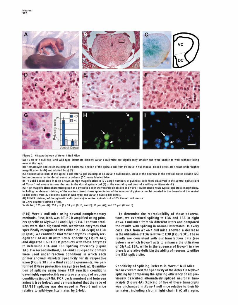
Figure 2. Histopathology of Nova-1 Null Mice
(A) P5 Nova-1 null (top) and wild-type littermate (below). Nova-1 null mice are significantly smaller and were unable to walk without fallingover at this age.(B) Hematoxylin and eosin staining of a horizontal section of the spinal cord from P5 Nova-1 null mouse. Boxed areas are shown under highermagnification in (D) and (dotted box) (F).(C) Horizontal section of the spinal cord after X-gal staining of P5 Nova-1 null mouse. Most of the neurons in the ventral motor column (VC)but not neurons in the dorsal sensory column (DC) were labeled blue.(D–F) Solid boxed area in (B) is shown at high magnification in (D). Large numbers of pyknotic cells were observed in the ventral spinal cordof Nova-1 null mouse (arrows) but not in the dorsal spinal cord (F) or the ventral spinal cord of a wild-type littermate (E).(G) High magnification photomicrograph of a pyknotic cell in the ventral spinal cord of a Nova-1 null mouse shows typical apoptotic morphology,including condensed staining of the nucleus. Inset shows quantitation of the number of pyknotic nuclei counted in the dorsal and the ventralspinal cords from 27 sections each of wild-type and Nova-1 null spinal cords.(H) TUNEL staining of the pyknotic cells (arrows) in ventral spinal cord of P5 Nova-1 null mouse.(I) DAPI counter staining of (H).Scale bar, 125 mm (B); 250 mm (C); 31 mm (D, E, and F); 10 mm (G); and 20 mm (H and I).
(P16) Nova-1 null mice using several complementary To determine the reproducibility of these observa-methods. First, RNA was RT-PCR amplified using prim- tions, we examined splicing to E3A and E3B in eighters specific to GlyRa2 E2 and GlyRa2 E4. Reaction prod- Nova-1 null mice from six different litters and compareducts were then digested with restriction enzymes that the results with splicing in normal littermates. In everyspecifically recognized sites either in E3A (SspI) or E3B case, RNA from Nova-1 null mice showed a decrease(BspMI). We confirmed that these enzymes uniquely rec- in the utilization of E3A relative to E3B (Figure 3C). Theseognized E3A or E3B (with .98% specificity; Figure 3AII) results are consistent with our transfection data (seeand digested E2-E4 PCR products with these enzymes below), in which Nova-1 acts to enhance the utilizationto determine E3A and E3B splicing efficiency (Figure of GlyRa2 E3A, while in the absence of Nova-1 in vivo3AI). In a second method, E3A- and E3B-specific primers there is a relative deficit in the ability of neurons to utilizewere used under reaction conditions in which each the E3A splice site.primer showed absolute specificity for its respectiveexon (Figure 3B). In a third set of experiments, we per-
Specificity of Splicing Defects in Nova-1 Null Miceformed RNase protection assays (see below). Quantita-We next examined the specificity of the defect in GlyRa2tion of splicing using linear PCR reaction conditionssplicing by comparing the splicing efficiency of six pre-gave highly reproducible results over a range of reactionviously described alternatively spliced neuronal tran-conditions (input RNA, PCR cycle number) and betweenscripts (Figure 4A). Splicing of five of these transcriptsanimals (see below), and demonstrated that the ratio ofwas unchanged in Nova-1 null mice relative to their lit-E3A/E3B splicing was decreased in Nova-1 null mice
relative to wild-type littermates by 2-fold. termates, including clathrin light chain B (ClaB), agrin,

Nova-1 Regulates Neuronal Splicing363
Figure 3. Aberrant GlyRa2 Splicing in Nova-1Null Mice
(AI) RT-PCR analysis of GlyRa2 E3A and E3Bsplicing utilization. Spinal cord RNA from aP16 Nova-1 null mouse and a wild-type lit-termate was analyzed by RT-PCR using32P-labeled E2 primer and unlabeled E4primer followed by SspI restriction digestionand autoradiography.(AII) Specificity of the restriction digest assay.PCR of a cDNA encoding the mature E2-E3A-E4 mRNA or the E2-E3B-E4 mRNA was di-gested with SspI. Phosphorimage analysis ofthe data revealed that .98% of the E2-E3A-E4 DNA was digested, while ,2% of the E2-E3B-E4 DNA was digested. Similar discrimi-nation of E3A and E3B products was seenwith BspMI digestions (data not shown).(BI) RT-PCR analysis of GlyRa2 E3A and E3Bsplicing utilization. Spinal cord RNA from aP18 Nova-1 null mouse and a wild-type lit-termate was analyzed by RT-PCR using32P-labeled E2 primer and unlabeled E3A orE3B primer followed by autoradiography.(BII) Specificity of the assay. cDNA clonesencoding the mature E2-E3A mRNA or theE2-E3B mRNA were PCR amplified with32P-labeled E2-E3A or E2-E3B primer sets.Analysis of this data on a phosphorimagerrevealed that E2-E3A or E2-E3B productscould not be detected using the mismatchedprimer pair (E2-E3B or E2-E3A primers, re-spectively).(C) Reproducibility of the GlyRa2 splicing de-fect in Nova-1 null mice. Spinal cord RNAfrom Nova-1 null mice and their littermateswith indicated genotypes was analyzed as in(B) for the presence of GlyRa2 E3A and E3B
isoforms. Data are presented as the ratio of E3A/E3B exon usage in each animal. PCR results were linear with respect to RNA input and cyclenumber. Actin RNA levels were measured in each sample by RT-PCR and differed by no more than 5%–10% between littermates (data not shown).
mGluR1, ICH-1, and n-src. However, alternative splicing shown). In forebrain of wild-type versus null mice, wefound no differences in alternative splicing for GlyRa2,of the GABAA receptor g2 subunit was significantly al-GABAA g2L, or ClaB mRNA or in steady-state levels oftered in Nova-1 null mice. The GABAA g2 transcript isactin or the spinal motor neuron marker choline acetyl-widely expressed in the brain, but the pre-mRNA is alter-transferase (ChAT) (Figure 5). We conclude that Nova-1natively spliced to include a cassette exon, g2L, prefer-mediates a specific effect on the alternative splicing ofentially in brainstem and spinal neurons (Zhang et al.,GlyRa2 and GABAA g2 receptor pre-mRNAs in a cell-1996). Examination of brainstem and spinal cord RNAautonomous manner.revealed a 2-fold difference in the E3A/E3B GlyRa2
splicing ratio and nearly a 3-fold difference in the g2L/g2S GABAA g2 splicing ratio in wild-type versus Nova-1 Genotype and Developmental Profilenull mice, averaged over 11 and 9 litters, respectively of Splicing Changes(Figure 4B). Similar results were obtained with RNase The level of Nova-1 protein varies in a dose-dependentprotection assays (data not shown), although these re- manner in wild-type mice relative to Nova-1 heterozy-sults were less reliable than RT-PCR in quantitating the gous and null mice (Figure 1D). To assess whether thelow-abundance GlyRa2 splice variants in a tissue sam- splicing defects of GlyRa2 and GABAA g2 RNAs wereple of high complexity, as anticipated (Foley et al., 1993). dependent on the level of Nova-1 protein, we examined
Although the GlyRa2 and GABAA g2 genes are ex- the utilization of these exons in mice as a function ofpressed throughout the brain, Nova-1 expression is ab- Nova-1 gene dosage. The GABAA g2L splicing defectsent in rostral structures, including the forebrain and varied from 1.5-fold in heterozygous mice to a 3-foldsensory thalamus (Buckanovich et al., 1996; Yang et al., change Nova-1 null littermates (Figures 6A and 6B).1998; Figure 1D). If aberrant GlyRa2 E3A and GABAA GlyRa2 E3A utilization was only abnormal in Nova-1 nullg2L splicing results directly from the lack of Nova-1 in mice, suggesting differences in the degree to whichneurons, splicing of these RNAs should be normal in the the Nova-1 protein is rate limiting for proper splicing offorebrain and abnormal in the spinal cord of individual GlyRa2 and GABAA g2 pre-mRNAs.Nova-1 null mice. Specific abnormalities in GlyRa2 and When we examined alternative splicing as a functionGABAA g2 splicing were found in the spinal cord and of age, we found that utilization of the GABAA g2L in-
creased in wild-type mice during the first 3 weeks ofhindbrain but not the forebrain (Figure 5; data not

Neuron364
Figure 5. GlyRa2 and GABAA g2 Alternative Splicing Defects AreRestricted to Nova-1-Expressing Regions of Brain in Nova-1 NullMice
Spinal cord or forebrain RNA was isolated from a P18 (P7 for GABAA
g2) Nova-1 null mice and a wild-type littermate and analyzed for theindicated splicing products by RT-PCR. Data are presented as inFigure 3C. Unlike the spinal cord, quantitation of phosphorimagerdata using RNA from forebrain revealed no differences in the ratio ofGlyRa2 E3A/E3B and g2L/g2S GABAA exon usage between Nova-1null animals and wild-type littermates. ClaB alternative splicingshowed no null:wild-type difference in either spinal cord or forebrain.Quantitation revealed no differences in the steady-state levels ofRNA encoding the motor neuron marker ChAT or actin in Nova-1null spinal cord relative to wild-type.
Mouse Brain Nova Binds Specificallyto (UCAUY)3 RNATo evaluate whether the GlyRa2 splicing defect evidentin Nova-1 null mice could be a direct action of Nova-1on GlyRa2 pre-mRNA, we first examined whether native
Figure 4. Specific Abnormalities in Alternative Splicing of GlyRa2 Nova protein from mouse brain is able to specificallyand GABAA g2 Transcripts in Nova-1 Null Mice bind the GlyRa2 intronic (UCAUY)3 element. Previous(A) RNA isolated from a P16 Nova-1 null mouse and a heterozygous work demonstrated that immunoprecipitates of Novalittermate was analyzed by RT-PCR with primers specific to the protein from brain extracts contain GlyRa2 pre-mRNAindicated genes. All PCR reactions were performed in duplicate (Buckanovich and Darnell, 1997) but did not define theusing minus-RT reactions (data not shown). LCB2 includes and
specific sequence to which Nova bound. To examineLCB3 lacks the neuron-specific EN exon in the ClaB mRNA (Wangthis question, a 90 nt intronic RNA surrounding theand Grabowski, 1996). Agrin0 lacks and agrin19 includes both Z siteGlyRa2 (UCAUY)3 element was transcribed in the pres-exons (Ferns et al., 1992). mGluR1a lacks and mGluR1b contains
an additional exon in the metabotropic glutamate receptor mRNA ence of 32P-UTP and ultraviolet (UV) cross-linked to pro-(Hollmann and Heinemann, 1994). ICH-1S includes and ICH-1L lacks teins in brain extracts. As a control, we also transcribedan additional exon in the IL-1b-converting enzyme mRNA (Wang et a mutant GlyRa2 RNA, (UAAUY)3, previously shown notal., 1994). n-src mRNA includes a neuron-specific exon in the c-src
to bind Nova-1 in vitro (Buckanovich and Darnell, 1997).message (Martinez et al., 1987).These mixtures were RNase treated, and Nova protein(B) Quantitation of the data presented in (A), together with additionalwas immunoprecipitated, run on SDS–PAGE gels, andRNA samples (the number of litters examined is indicated by n). The
ratio of long to short alternatively spliced products measured in transferred to nitrocellulose. A 32P-labeled RNA bandwild-type animals relative to the respective ratio in the correspond- superimposable on the 55 kDa Nova protein band wasing Nova-1 null littermate is presented. For GlyRa2, the ratio of E3A/ seen specifically in immunoprecipitates of extractsE3B in wild-type versus Nova-1 null littermate is depicted. The bars
cross-linked to the wild-type (UCAUY)3 but not the mu-represent the average change in wild-type:null exon usage for thetant (UAAUY)3 RNA (Figure 7). This result demonstratesnumber of litters examined; error bars represent the standard devi-that native neuronal Nova protein specifically binds toation.the GlyRa2 intronic (UCAUY)3 RNA.
Nova Regulates GlyRa2 Pre-mRNA Alternativedevelopment (Figure 6C), consistent with previous anal-yses (Wang and Grabowski, 1996; Zhang et al., 1996), Splicing in Cell Lines
We next examined whether Nova-1 is able to directlyand that a deficiency in exon utilization first becomesapparent at P7 in Nova-1 null mice (Figure 6C). In Nova-1 act on GlyRa2 pre-mRNA to regulate alternative splicing
of the E3A/E3B exons. We generated a minigene con-heterozygous mice, there was again an intermediatedeficit in GABAA g2 usage evident at P7. The GlyRa2 struct consisting of genomic DNA encoding GlyRa2 E2,
E3A, E3B, and E4 and several hundred nucleotides ofE3A/E3B ratio was unchanged in P0–P4 Nova-1 nullmice (,10% difference across multiple littermates) and intronic DNA on either side of these exons (Figure 8A).
To allow us to determine whether Nova-1 acts directlythereafter differed by 2-fold (data not shown).

Nova-1 Regulates Neuronal Splicing365
Figure 6. Changes in GlyRa2 and GABAA g2Alternative Splicing as a Function of GeneDosage and Development
(A) GABAA g2, GlyRa2, and c-src alternativeexon usage in the spinal cords of a P16 Nova-1null mouse and its heterozygous and wild-type littermates was analyzed. There was asignificant difference in alternative exon utili-zation in both the null and heterozygote micefor the GABAA g2 message, whereas theGlyRa2 E3A/E3B ratio is only disturbed in thenull mouse. Alternatively spliced n-src mRNAis unchanged in all three genotypes.(B) Data obtained from (A) and the indicatednumber of additional litters are shown.GlyRa2, GABAA g2, and n-src alternative exonusage in Nova-1 null and heterozygote miceis plotted using the alternative exon ratio ofthe wild-type mouse set to 1. Bars representthe average of the normalized exon use ratio;error bars indicate the standard deviation.(C) Developmental change in the GABAA g2L/g2S exon use ratio. Points represent singlemeasurements at the indicated develop-mental times.
on the GlyRa2 intronic (UCAUY)3 element, we also gener- on extracts of a neuroblastoma cell line, N2A, trans-fected with the wild-type (UCAU) or mutant (UAAU)ated a mutant minigene in which this element was mu-
tated to the nonbinding (UAAUY)3 sequence. These con- GlyRa2 minigene in the presence or absence of pcNova-1.A Nova-1 dose-dependent increase in splicing to E3Astructs were then transfected into three different cell
lines along with increasing amounts of a plasmid ex- (and compensatory decrease in E3B) is evident in cellstransfected with the wild-type UCAU but not the mutantpressing Nova-1 (pcNova-1).
Nova-1 transfection mediated an increase in GlyRa2 UAAU GlyRa2 minigene. We demonstrated that Nova-1was present in increasing amounts in cells transfectedE3A splicing in each of the three cell lines tested. Figure
8B illustrates the results of splicing assays performed with increasing amounts of pcNova-1 by Western blotanalysis (Figure 8C).
To confirm these results and analyze splicing to bothE3A and E3B, we repeated independent transfectionexperiments in N2A cells (Figure 8D). In cells transfectedwith the wild-type UCAU GlyRa2 minigene, Nova-1 re-producibly increased E3A splicing at the expense ofE3B splicing. Nova-1 mediated a 2-fold increase in E3Autilization, an effect similar in magnitude to the decreasein the E3A/E3B splicing ratio seen in Nova-1 null mice(Figures 3 and 4).
In N2A cells transfected with the mutant UAAU GlyRa2minigene, splicing to E3A was unchanged relative tothe wild-type minigene (Figure 8D). In the presence ofincreasing amounts of Nova, a paradoxical increase inE3B, at the expense of E3A, was seen. While the mecha-nism underlying this observation is unclear, a possibleFigure 7. Native Nova Protein Binds to the GlyRa2 (UCAUY)3 El-
ement explanation is that the UAAU mutation allowed detectionAn extract of mouse brainstem was incubated with 90 nt 32P-labeled of a cryptic Nova-1 binding site able to mediate an actionRNAs encoding either the GlyRa2 UCAU intronic sequence (lanes on E3B splicing. Taken together, these results demon-1, 2, 19, and 29) or mutant UAAU RNA (lanes 3, 4, 39, and 4’). Samples strate that the action of Nova to enhance E3A utilizationwere UV cross-linked, RNase treated, and immunoprecipitated with
in wild-type UCAU GlyRa2 pre-mRNAs was abolishedeither rabbit anti-Nova serum (lanes 1, 3, 19, and 39) or preimmuneby mutation in the intronic UCAU Nova-binding element.serum (lanes 2, 4, 29, and 49, run on SDS–PAGE gels and transferred
We repeated these experiments in HeLa cells andto a filter for Western blot analysis with POMA antiserum. The samefilter was also exposed directly to X-ray film to detect 32P-labeled 293T cells, two nonneuronal cell lines that do not ex-RNA (lanes 19–49). The arrow indicates the molecular size of Nova-1. press Nova (Figures 8E and 8F; data not shown).The strong reactivity present underneath the Nova-1 bands (lanes pcNova-1 cotransfection with the wild-type GlyRa2 min-1–4) is due to secondary antibody cross-reactivity with rabbit immu-
igene mediated a 2.5-fold increase in E3A splicing innoglobulin G. The z70 kDa band visible in lanes 1 and 3 is a proteinHeLa cells and a 2-fold increase in 293T cells. Somerelated to Nova-2 that immunoprecipitates specifically with POMA
antisera (Yang et al., 1998). additional effects were seen. In HeLa cells, the increase

Neuron366
Figure 8. Nova-1 Enhances GlyRa2 E3A Splic-ing in Heterologous Cell Lines
(A) The GlyRa2 minigene used for splicingassays in transiently transfected cells con-tains the complete GlyRa2 E2, a shortenedintron 2, the complete genomic sequence sur-rounding E3A and E3B, a shortened intron 3,and 233 nucleotides at the 59 end of E4. TheUCAU Nova-1 binding site in intron 2 is indi-cated by the schematic hairpin loop, and theUAAU mutant minigene is indicated by an X.(B) Nova-1-dependent effects on E3A splicingof the wild-type UCAU and mutant UAAUGlyRa2 minigenes. RNA from N2A cells trans-fected with the wild-type or mutant GlyRa2minigene and the indicated amount ofpcNova-1 plasmid were analyzed as in Fig-ure 3A.(C) Western blot using anti-T7 antibody show-ing the titration of T7-tagged Nova proteinlevels after N2A cells were cotransfected withthe GlyRa2 minigene and the indicatedamounts of pcNova-1.(D) Nova increases the ratio of E3A/E3B splic-ing from wild-type GlyRa2 pre-mRNA, but thiseffect is abrogated in the GlyRa2 mutantUAAU pre-mRNA. Three independent trans-fections were performed in which splicingproducts (E3A or E3B) were quantitated as inFigure 3A. Increasing amounts of pcNova-1were cotransfected with either the wild typeor mutant GlyRa2 minigene, as indicated. Re-sults are displayed as a ratio of E3A/E3B. Thetotal amount of spliced product is unchangedas a result of Nova titration; the increase inE3A usage is accompanied by a compensa-tory decrease in E3B utilization (data notshown). Reaction products were linear with
respect to input cDNA and PCR cycle number (data not shown). Results shown are the average of three independent transfections, and errorbars represent standard deviation. Similar results were obtained in two additional experiments (data not shown).(E and F) Nova facilitates E3A splicing in HeLa (E) and 293T (F) cells specifically in wild-type UCAU GlyRa2 transcripts. Western blot confirmedthat HeLa and 293T cells did not express Nova and that Nova transfection led to linear increases in Nova protein (data not shown). Resultsare the average of two (E) and four (F) independent transfections, and error bars represent standard deviation.
in E3A splicing was at the expense of E3B, while in 293T that Nova-1 functions to regulate alternative splicing inneurons by binding pre-mRNA in a sequence-specificcells the increase in E3A-spliced product was inversely
proportional to a decrease in an aberrant spliced prod- manner to activate exon inclusion. Nova-1 present inbrain extracts specifically cross-links the sequenceuct that contained neither E3A nor E3B (E2-E4; data not
shown). We also found that in cells transfected with the (UCAUY)3 but not the mutant sequence (UAAUY)3, con-sistent with previous coimmunoprecipitation experi-mutant UAAU GlyRa2 minigene, in the presence of Nova
there was no increase in E3A splicing but again a para- ments demonstrating that Nova-1 protein binds GlyRa2pre-mRNA in vivo. In cotransfection assays, GlyRa2 pre-doxical increase in E3B splicing, and in the absence of
Nova there was a small but consistent increase in E3A mRNAs harboring this (UCAUY)3 sequence show aNova-1 dose-dependent increase in utilization of thesplicing that is unexplained. Taken together, these ex-
periments demonstrate that Nova-1 is able to act directly alternatively spliced E3A, while mutant pre-mRNAs har-boring the (UAAUY)3 point mutant do not show this ef-to enhance the inclusion of GlyRa2 E3A in cell lines
and that this action is dependent on the integrity of the fect. In Nova-1 null mice, there is a reciprocal 2-folddecrease in the ratio of GlyRa2 E3A relative to E3Bintronic UCAU Nova binding site.splicing. These data provide converging lines of evi-dence that Nova-1 regulates GlyRa2 splicing by directlyDiscussionbinding to the pre-mRNA in neurons.
We also find a second specific defect in alternativeNova-1 Regulates Mammalian Tissue-Specificsplicing in Nova-1 null mice, which is deficient inclusionAlternative Splicingof the GABAA g2L exon. The defective exon utilizationWe have combined biochemical and genetic ap-of g2L and GlyRa2 E3A in Nova-1 null mice, togetherproaches to demonstrate that Nova-1 functions to regu-with the finding that Nova-1 enhanced E3A utilization inlate alternative splicing in neurons and found that theheterologous cells, suggests that Nova-1 most likelyabsence of Nova-1 leads to neuronal death in spinal
and brainstem neurons. We provide strong evidence acts to activate GlyRa2 E3A and GABAA g2L exon inclu-

Nova-1 Regulates Neuronal Splicing367
sion in neurons. An alternative, and not entirely exclu- 1996; Figures 1 and 2). Therefore, the identification ofNova-1 as a splicing factor provides a mechanism bysive, possibility is that Nova-1 activates splicing indi-which individual neurons can differentially regulaterectly by competing with a splicing inhibitor. Such ansplicing in a manner qualitatively distinct from that ofinterpretation would be consistent with the observationother cell types.that Nova action can be antagonized by a newly discov-
In previous studies of the mechanism of neuron-spe-ered brain-enriched variant of PTB (A. D. Polydoridescific splicing, intronic enhancer elements similar in na-et al., submitted). However, a strictly indirect action ofture to the GlyRa2 (UCAUY)3 sequences have been iden-Nova appears unlikely, since the action of Nova-1 intified. These include multiple elements that promotetransfected cells is abolished by mutation in an intronicneuron-specific exon inclusion in the c-src, calcitonin/RNA element.CGRP, GABAA g2, and agrin genes (Black, 1992; ZhangSeveral unanswered questions regarding tissue-spe-et al., 1996; Wei et al., 1997; Lou et al., 1999). Regulationcific splicing are addressed by the finding that Nova-1of splicing appears complex given that multiple ele-regulates alternative splicing in neurons. RNA-bindingments, both positive and negative, influence alternativeproteins such as Sxl and Tra/Tra-2 in Drosophila, identi-splicing events. For example, in c-src, in addition tofied by genetic approaches, act in a sequence-specificpositive acting signals identified as targets of an RNA-manner to regulate alternative splicing of specific targetbinding protein complex involving KSRP, hnRNP F, andtranscripts. However, genetics has not previously beenhnRNP H (Min et al., 1997; Chou et al., 1999), negativeapplied to the study of RNA-binding proteins in mam-elements are found in the polypyrimidine tract, upstreammals, and biochemical studies applied to complex splic-of the neuronal n-src exon, that interact with PTB (Chaning problems have led not to the identification of tissue-and Black, 1997) and downstream of the n-src exonspecific splicing factors, but to the identification ofthat interact with brain-enriched PTB (D. L. Black et al.,ubiquitously expressed RNA-binding proteins. Thesesubmitted).studies have yielded one clear model in which differen-
The mechanism of neuron-specific splicing in thetial regulation of splicing in mammals is generatedGABAA g2 gene has similar complexities (Ashiya et al.,through relatively subtle changes in the levels of such1995; Zhang et al., 1996, 1999). A number of negativeubiquitous splicing factors. For example, changing theelements surrounding the g2L exon were found to bindratio of the constitutively expressed hnRNP proteinPTB, and evidence was also found for positive actinghnRNP A1 to the SR protein ASF-SF2 mediates quantita-splicing elements. These observations, together withtive changes, ranging from 1.25-fold to severalfold inour data, suggest that Nova-1 may act in concert withcell transfection studies, in the utilization of proximalgeneral factors such as PTB or brain-enriched PTB toversus distal splice site choices at alternatively splicedregulate GABAA g2L exon utilization, which would beexons (Caceres et al., 1994). Our data demonstrate thatconsistent with the finding that Nova interacts with brainin addition to the role general factors play in regulatingPTB (A. D. Polydorides et al., personal communication).alternative splicing, tissue-specific factors that recog-GABAA g2L is preferentially included in the hypothala-nize specific RNA ligands exist in mammals. A workingmus, midbrain, brainstem, and spinal cord (Zhang et al.,model for Nova action is that it acts to enhance utilization1996), and this correlates precisely with the pattern ofof adjacent exons by recruiting general factors, such asNova-1 expression, supporting our finding of a role forSR proteins, to promote the assembly of a splicing-Nova-1 in generating the g2L splice variant in vivo.competent complex.
Nova-1, Neuronal Death, and Neurologic DiseaseNova-1 Regulates Neuron-Specific Several recent reports have implicated splicing defectsAlternative Splicing as a cause of motor neuron death in the spinal cord.Three families of strictly neuron-specific RNA-binding Dreyfuss and colleagues have determined that a proteinproteins have been identified. Musashi proteins are ex- mutated in the human disorder spinal muscular atrophypressed in mitotic neuronal precursors (Sakakibara et plays a critical role in the generation of snRNPs andal., 1996) and play a role in the early development of have suggested that the proper regulation of splicingthe nervous system in Drosophila. Nova (Buckanovich is necessary for postnatal survival of motor neuronset al., 1993; Yang et al., 1998) and Hu (Szabo et al., 1991) (Fischer et al., 1997). In addition, Lin et al. (1998) haveproteins were identified as PND target antigens. These reported that splicing defects in the glutamate trans-autoimmune neurologic disorders are believed to be porter EAAT2 are associated with the motor neuron dis-triggered when proteins normally sequestered from im- order amyotrophic lateral sclerosis. The phenotype ofmune surveillance in neurons are ectopically expressed Nova-1 null mice, which phenocopies many aspects ofin tumor cells (Darnell, 1996, 1998), leading to the devel- the human POMA syndrome (see below), together withopment of antigen-specific antibodies and T cells (Dar- the biochemical evidence presented here, provides anell, 1996; Albert et al., 1998, 1999). particularly compelling case illustrating splicing defects
Detailed analysis of the developmental and adult ex- that are associated with neuronal death in the ventralpression patterns of both Nova (Buckanovich et al., spinal cord.1993, 1996) and Hu (Okano and Darnell, 1997; Waka- Our results demonstrate that Nova-1 is essential formatsu and Weston, 1997) antigens revealed that their neuronal viability in mature neurons in postnatal mice.expression is entirely restricted to neurons throughout Nova-1 null neurons appear to undergo normal morpho-development. Nova-1 expression is not only specific to genesis, assessed histochemically, and axon pathfind-neurons, but to a subset of neurons in the hypothalamus, ing, assessed by electron microscopy of the neuromus-
cular junction at birth (J. Sanes and R. B. D., unpublishedbrainstem, and spinal cord (Buckanovich et al., 1993,

Neuron368
data). However, the TUNEL-positive and pyknotic nuclei (Lewis et al., 1999; R. B. D. et al., unpublished data).Moreover, data from the crystal structure of Nova KH3we observed in P5 Nova-1 null neurons reflect the end
stage of apoptotic neuronal death, and it is difficult to be (Lewis et al., 1999) and the Nova KH3-RNA complex(Lewis et al., 2000) demonstrate that Nova proteins di-certain at what age the neurons become dysfunctional.
Moreover, we cannot rule out the possibility that Nova-1 merize, even when bound to RNA.Neurons are likely to utilize some immune system-likemay also play a role in early neuronal development. For
example, Nova-2 is expressed together with Nova-1 in mechanism to generate diversity from a relatively smallnumber of primary transcripts (Darnell, 1998). The signif-early postmitotic neurons and is downregulated in Nova-
1-expressing neurons postnatally, suggesting that Nova-2 icance of a neuron-specific system that regulates alter-native splicing is that it provides a means for the genera-could rescue Nova-1 function before birth. The defect
in neuronal viability suggests either a loss of function, tion of such diversity. While genome rearrangementsmight occur in the brain (see Gao et al., 1998; Kohmurasuch as a failure of mechanisms necessary to ensure
survival of mature neurons, or a gain of function, such as et al., 1998; Serafini, 1999; Wu and Maniatis, 1999), todate alternative splicing and RNA editing are the onlyacquisition of a new toxic activity that mediates neuronal
death in Nova-1 null neurons. For example, aberrant relevant mechanisms known to operate in neurons.Nova proteins, acting in concert with other proteins,splicing of the a1A calcium channel in mice is believed
to result in a gain-of-function mutation, which leads to are likely to play an important role in generating thecomplexity of neuronal function.a delayed onset neurologic disorder manifest by ataxia,
motor seizures, and absence seizures (Fletcher et al.,1996). In Nova-1 null mice, defects in inhibitory receptor
Experimental Proceduressplicing could lead to a gain of function, such that gly-cine and/or GABA receptors become toxic, for example, Plasmid Constructsby creating imbalances between the neuronal inhibition The GlyRa2 minigene (cloned in pCDNA3, Invitrogen) corresponds toand excitation that lead to excitotoxic cell death. the following GlyRa2 E2 mouse genomic sequences: bases 81–276
(GenBank accession number X75842), 63–583 (GenBank accessionPatients with POMA have symptoms that suggest ab-number X75843), and 10–300 (GenBank accession number 75844).normal inhibitory control over brainstem and spinal mo-The mutated GlyRa2 minigene, in which the Nova-1 binding sitetor pathways (Luque et al., 1991). For example, opsoclo-59-UCAUCAUCAUUUUCAUUUUGUUU-39 was mutated to 59-UAA
nus is thought to result from failure of normal functioning UAAUAAUUUUAAUUAAGGUU-39, was generated by PCR. A cDNAof brainstem inhibitory glycinergic neurons, termed om- encoding T7-tagged Nova-1 was cloned into pcXHook (Invitrogen).nipause neurons (Averbuch-Heller and Remler, 1996), All DNA constructs generated by PCR were sequenced in their en-
tirety.and brainstem or spinal myoclonus is believed to relateto a failure of inhibition of motor neurons and may involveGABAergic or glycinergic systems (Rothwell, 1995; Cavi- PCR Primersness, 1996; Rajendra et al., 1997). Moreover, mice with The primers used were E2 F, 59-AGCTTTCTGCAAAGACCATGAC;the spasmodic mutation (Ryan et al., 1994; Saul et al., E4 R, 59-GAAGATCTCCAAATCCAAGGAATCATCTGGG; a2Gly R
E3A, 59-CATGGTGGTTTCTGTGACTGATC; a2Gly R E3B, 59-CATTG1994) and humans with hyperekplexia (Shiang et al.,TAGTTTCTGCTATTGACCCAAAG; a2Gly R E4, 59-TCCAAATCCAA1993) harbor defects in inhibitory glycine receptors andGGAATCATCTGGG; actin F, 59-GTGGGCCGCTCTAGGCACCA;suffer from myoclonic syndromes that bear marked simi-actin R, CCCCCCTGAACCCTAAGGCCAACCG; mGluR1 F, 59-CCT
larities to the motor symptoms evident in Nova-1 null GGGGTGCATGTTTACTCC; mGluR1 R, 59-AGGCCGTCTCGTTGGTmice and in POMA patients. Thus, the defects in splicing CTTCA; ICH-1 F, 59-GTCTCATCTTCATCAACTCC; ICH-1 R, 59-ATGof the inhibitory glycine and GABA receptors in Nova-1 CTAACTGTCCAAGTCTA; ClaB F, 59-ACCGAACAGGAGTGGCG
GGAG; ClaB R, 59-GGGGTCTCCTCCTTGGATTCT; g2 GABA F,null mice suggest a reasonable model for the mecha-59-GTATGGCACCCTGCATTATTTTGTC; g2 GABA R, 59-TTGAATGnism of motor dysfunction in both the murine and humanGTTGCTGATCTGGGACG; ChAT F, 59-ATGCCTATCCTGGAAAAsystems. In POMA, immunologic targeting of Nova pro-GGTCCC; ChAT R, 59-AGTGCTCCGAGCAAAGATCACAG; src F,
tein may lead to defects in inhibitory receptor function 59-CCAAGCTCTTCGGAGGCTTCAACTC; src R, 59-CACATAGTTGCand thereby to the excess motor activity evident in the TGGGGATGTAACCG; agrin F, 59-GGGATAGTTGAGAAGTCAGTGGdisorder. GGG; and agrin R, 59-CGAAGCCAGCGGTTGGTGTTG.
Complexity of Regulation of Neuronal Generation of Targeted MutationAlternative Splicing Genomic clones encoding Nova-1 were isolated from a phage library
derived from a 129 mouse (Stratagene). A 1.8 kb Xba-AlwNI fragmentThe identification of the glycine and GABAA receptorencoding sequences upstream of the initiator methionine and a 6.5pre-mRNAs as Nova-1 targets raises the question ofkb SacI fragment harboring sequences downstream of the secondthe extent to which Nova proteins are used to regulatecoding exon were cloned into pBS2 (Stratagene). A PacI linker wasalternative splicing in neurons. The Nova gene familyligated to a unique NotI site between these fragments to allow insert
itself consists of at least two (Buckanovich et al., 1993; of the IRES-t-lacZ LTNL cassette (Mombaerts et al., 1996). PlasmidYang et al., 1998) and possibly additional Nova gene was linearized with SalI and used to electroporate ES cells, which
were grown as described (Mombaerts et al., 1996), including 150family members (Fletcher et al., 1997; Yang et al., 1998;mg/ml of G418 starting 24 hr after transfection. Genomic DNA fromsee also Figure 1), and the Nova-1 and Nova-2 tran-individual colonies was digested with EcoRI and screened by South-scripts are themselves alternatively spliced (Buckano-ern blot with a 59 Nova-1 genomic probe (0.5 kb SacI-XbaI fragment).vich et al., 1993; Yang et al., 1998). Evaluation of NovaClones that had undergone homologous recombination were trans-
protein–protein interactions using biochemical and yeast fected with a plasmid expressing Cre recombinase (pB5185) to ex-two-hybrid assays suggests that Nova-1 and Nova-2 cise the LTNL cassette. Accurate homologous recombination in pos-
itive clones was confirmed by Southern blot with HindIII-digestedcan form homo- and heterotypic protein interactions
Nova-1 Regulates Neuronal Splicing369
genomic DNA and a 39 Nova-1 genomic probe (0.75 kb SacI frag- treatment of activated T cells in the cerebrospinal fluid of patientswith paraneoplastic cerebellar degeneration. Ann. Neurol., in press.ment). A homologous recombinant clone was injected into C57/BL6
blastocysts to produce germline chimeras. Amara, S.G., Jonas, V., Rosenfeld, M.G., Ong, E.S., and Evans, R.(1982). Alternative RNA processing in calcitonin gene expressiongenerates mRNAs encoding different polypeptide products. NatureHistology and TUNEL Assay298, 240–244.The mice were perfused with 4% buffered formaldehyde, and the
brains were removed, postfixed overnight, and embedded in paraffin Arning, S., Gruter, P., Bilbe, G., and Kramer, A. (1996). Mammalianblocks. Tissue sections (14 mm) were deparaffinized, rehydrated, splicing factor SF1 is encoded by variant cDNAs and binds to RNA.stained with hematoxylin and eosin, and visualized by light micros- RNA 2, 794–810.copy using a Zeiss Axioplan microscope. Pyknotic neurons were Ashiya, M., Zhang, L., and Grabowski, P.J. (1995). Regulated splicingquantitated by counting all pyknotic cells in each of 27 14 mm thick of gamma2 pre-messenger RNA in neuronal cells. Nucleic Acidssections of spinal cord from a single Nova-1 null mouse or a single Symp. Ser. 33, 215–216.wild-type littermate. For X-gal staining, whole-mount staining was
Ashley, C.T., Wilkinson, K.D., Reines, D., and Warren, S.T. (1993).performed as described (Mombaerts et al., 1996).FMR-1 protein: conserved RNP family domains and selective RNAApoptosis was assessed by the DNA terminal transferase nickbinding. Science 262, 563–566.end translation method, or TUNEL assay, as described previouslyAverbuch-Heller, L., and Remler, B. (1996). Opsoclonus. Semin. Neu-(Fuks et al., 1995), and visualized by fluorescence microscopy.rol. 16, 21–26.
Baker, B.S. (1989). Sex in flies: the splice of life. Nature 340, 521–524.RNA Preparation and RT-PCRBechade, C., Sur, C., and Triller, A. (1994). The inhibitory neuronalPurified RNA (Chomcynski and Sacchi, 1987) was reverse tran-glycine receptor. Bioessays 16, 735–744.scribed using random hexamers, and cDNA products were amplified
using PfuTurbo (Stratagene) with 40 pmol of each primer and 0.5 Black, D.L. (1992). Activation of c-src neuron-specific splicing bypmol of one g-32P-ATP-labeled primer. PCR products were con- an unusual RNA element in vivo and in vitro. Cell 69, 795–807.firmed by sequencing. PCR was linear with respect to input cDNA Black, D.L. (1998). Splicing in the inner ear: a familiar tune, but whatand cycle number (data not shown). are the instruments? Neuron 20, 165–168.T7 Transcription and UV Cross-Linking
Buckanovich, R.J., and Darnell, R.B. (1997). The neuronal RNA bind-RNAs for crosslinking were generated by PCR using the wild-typeing protein Nova-1 recognizes specific RNA targets in vitro and inor mutant GlyRa2 minigene, followed by in vitro transcription. Thevivo. Mol. Cell. Biol. 17, 3194–3201.primers used were T7primer1 (sense) (59-AGTAATACGACTCACTBuckanovich, R.J., Posner, J.B., and Darnell, R.B. (1993). Nova, theATAGGGATCATGCAGTTCTGGTTTAAT) and primer2 (antisense)paraneoplastic Ri antigen, is homologous to an RNA-binding protein(59-AGCTCCATCAACATCTGTGG), which amplified a 90 nt fragmentand is specifically expressed in the developing motor system. Neu-surrounding the Nova-1 binding site. Gel-purified PCR productsron 11, 657–672.were used as templates for in vitro transcription. Thirty microliters
of total cell extracts from mouse brainstem (50 ml/mg) were irradiated Buckanovich, R.J., Yang, Y.Y., and Darnell, R.B. (1996). The onco-with UV light for 15 min. The samples were treated with 80 U of neural antigen Nova-1 is a neuron-specific RNA-binding protein,RNAse A (Worthington) for 30 min at 378C. UV cross-linking reactions the activity of which is inhibited by paraneoplastic antibodies. J.were immunoprecipitated with a 5 ml polyclonal rabbit anti-Nova-1 Neurosci. 16 1114–1122.antisera in a total volume of 100 ml of lysis buffer. The immune Burd, C.G., and Dreyfuss, G. (1994). Conserved structures and diver-complexes were precipitated with protein A–Sepharose beads sity of functions of RNA-binding proteins. Science 265, 615–621.(Sigma), run on 10% SDS–PAGE gels, transferred to nitrocellulose,
Caceres, J.F., Stamm, S., Helfan, D.M., and Krainer, A.R. (1994).and exposed to film.
Regulation of alternative splicing in vivo by overexpression of antag-Cell Transfectiononistic splicing factors. Science 265, 1706–1709.N2A, HeLa, and 293T cells were grown to 60% confluence in 35Caviness, J.N. (1996). Myoclonus. Mayo Clin. Proc. 71, 679–688.mm dishes or in 6-well plates. 2.5 mg of total DNA, comprised of
0.25 mg of the wild-type or mutant pcDNA3 GlyRa2 minigene, 0.25 Chan, R.C., and Black, D.L. (1997). The polypyrimidine tract bindingmg of pCMV b-galactosidase, and variable amounts of pcNova-1 protein binds upstream of neural cell–specific c-src exon N1 toand empty pcXHook vector, were incubated with 95 ml of Dulbecco’s repress the splicing of the intron downstream. Mol. Cell. Biol. 17,modified Eagle’s medium (DMEM) and 5 ml of Fugene (Roche) for 4667–4676.15 min at room temperature. The mixture was then added to the Chomcynski, P., and Sacchi, N. (1987). Single-step method of RNAcells in a total volume of 2 ml 10% FBS-DMEM. After 48 hr, the cells isolation by acid guanidinium thiocyanate-phenol-chloroform ex-were collected and used for RNA extraction, protein extraction, and traction. Anal. Biochem. 162, 156–159.b-galactosidase assay. Western blot analysis and b-galactosidase
Chou, M.Y., Rooke, N., Turck, C.W., and Black, D.L. (1999). hnRNPassays were performed as described (Okano et al., 1999).H is a component of a splicing enhancer complex that activates ac-src alternative exon in neuronal cells. Mol. Cell. Biol. 19, 69–77.
AcknowledgmentsClaeysen, S., Faye, P., Sebben, M., Taviaux, S., Bockaert, J., andDumuis, A. (1998). 5-HT4 receptors: cloning and expression of new
The authors are grateful to Peter Mombaerts for advice on generat-splice variants. Ann. NY Acad. Sci. 861, 49–56.
ing Nova-1 null mice and for the IRES-t-lacZ LTNL cassette and toConn, P.J., and Pin, J.P. (1997). Pharmacology and functions ofNathaniel Heintz for critical review of the manuscript. This work wasmetabotropic glutamate receptors. Annu. Rev. Pharmacol. Toxicol.supported by the National Institutes of Health (RO1 NS34389), an37, 205–237.Irma T. Hirschl Career Scientist Award (R. B. D.), the Breast Cancer
Research Program (DAMD17-97-1-7097) (K. B. J.), and the Ataxia Darnell, R.B. (1996). Onconeural antigens and the paraneoplasticTelangectasia Children’s Project (H. J. O.). neurologic disorders: at the intersection of cancer, immunity and
the brain. Proc. Natl. Acad. Sci. USA. 93, 4529–4536.
Darnell, R.B. (1998). Immunologic complexity in neurons. NeuronReceived August 16, 1999; revised January 7, 2000.21, 947–950.
Ehlers, M.D., Tingley, W.G., and Huganir, R.L. (1995). RegulatedReferencessubcellular distribution of the NR1 subunit of the NMDA receptor.Science 269, 1734–1737.Albert, M.L., Darnell, J.C., Bender, A., Francisco, L.M., Bhardwaj, N.,
and Darnell, R.B. (1998). Tumor-specific killer cells in paraneoplastic Ehlers, M.D., Fung, E.T., O’Brien, R.J., and Huganir, R.L. (1998).cerebellar degeneration. Nat. Med. 4, 1321–1324. Splice variant–specific interaction of the NMDA receptor subunit
NR1 with neuronal intermediate filaments. J. Neurosci. 18, 720–730.Albert, M.L., Austin, L.M., and Darnell, R.B. (1999). Detection and

Neuron370
Engebrecht, J., and Roeder, G.S. (1990). MER1, a yeast gene re- Macdonald, R.L. (1995). Ethanol, gamma-aminobutyrate type A re-ceptors, and protein kinase C phosphorylation. Proc. Natl. Acad.quired for chromosome pairing and genetic recombination, is in-Sci. USA 92, 3633–3635.duced in meiosis. Mol. Cell. Biol. 10, 2379–2389.
Martinez, R., Mathey-Prevot, B., Bernards, A., and Baltimore, D.Ferns, M., Hoch, W., Campanelli, J.T., Rupp, F., Hall, Z.W., and(1987). Neuronal pp60c-src contains a six-amino acid insertion rela-Scheller, R.H. (1992). RNA splicing regulates agrin-mediated acetyl-tive to its non-neuronal counterpart. Science 237, 411–415.choline receptor clustering activity on cultured myotubes. Neuron
8, 1079–1086. Michael, W.M., Eder, P.S., and Dreyfuss, G. (1997). The K nuclearshuttling domain: a novel signal for nuclear import and nuclear ex-Fischer, U., Liu, Q., and Dreyfuss, G. (1997). The SMN–SIP1 complexport in the hnRNP K protein. EMBO J. 16, 3587–3598.has an essential role in spliceosomal snRNP biogenesis. Cell 90,
1023–1029. Min, H., Turck, C.W., Nikolic, J.M., and Black, D.L. (1997). A newregulatory protein, KSRP, mediates exon inclusion through an in-Fletcher, C.F., Lutz, C.M., O’Sullivan, T.N., Shaughnessy, J.D., Jr.,tronic splicing enhancer. Genes Dev. 11, 1023–1036.Hawkes, R., Frankel, W.N., Copeland, N.G., and Jenkins, N.A. (1996).
Absence epilepsy in tottering mutant mice is associated with cal- Missler, M., and Sudhof, T.C. (1998). Neurexins: three genes andcium channel defects. Cell 87, 607–617. 1001 products. Trends Genet. 14, 20–26.
Fletcher, C.F., Okano, H.J., Gilbert, D.J., Yang, Y.Y.L., Yang, C.W., Mombaerts, P., Wang, F., Dulac, C., Chao, S.K., Nemes, A., Mendel-Copeland, N.G., Jenkins, N.A., and Darnell, R.B. (1997). Murine chro- sohn, M., Edmondson, J., and Axel, R. (1996). Visualizing an olfactorymosomal locations of nine genes encoding homologs of human sensory map. Cell 87, 675–686.paraneoplastic neurologic disorder antigens. Genomics, in press. Okano, H.J., and Darnell, R.B. (1997). A hierarchy of Hu RNA bindingFoley, K.P., Leonard, M.W., and Engel, J.D. (1993). Quantitation of proteins in developing and adult neurons. J. Neurosci. 17, 3024–RNA using the polymerase chain reaction. Trends Genet. 9, 380–385. 3037.
Fuks, Z., Alfieri, A., Haimovitz-Friedman, A., Seddon, A., and Cordon- Okano, H.J., Park, W.Y., Corradi, J.P., and Darnell, R.B. (1999). TheCardo, C. (1995). Intravenous basic fibroblast growth factor protects cytoplasmic Purkinje antigen cdr2 downregulates Myc function: im-the lung but not mediastinal organs against radiation-induced apo- plications for neuronal and tumor cell survival. Genes Dev. 13, 2087–ptosis in vivo. Cancer J. Sci. Am. 1, 62. 2098.
Gao, Y., Sun, Y., Frank, K.M., Dikkes, P., Fujiwara, Y., Seidl, K.J., Ostareck, D.H., Ostareck-Lederer, A., Wilm, M., Thiele, B.J., Mann,Sekiguchi, J.M., Rathbun, G.A., Swat, W., Wang, J. et al. (1998). A M., and Hentze, M.W. (1997). mRNA silencing in erythroid differentia-critical role for DNA end-joining proteins in both lymphogenesis and tion—hnRNP K and hnRNP E1 regulate 15-lipoxygenase translationneurogenesis. Cell 95, 891–902. from the 39 end. Cell 89, 597–606.
Grabowski, P.J. (1998). Splicing regulation in neurons: tinkering with Ostareck-Lederer, A., Ostareck, D.H., and Hentze, M.W. (1998). Cy-cell-specific control. Cell 92, 709–712. toplasmic regulatory functions of the KH-domain proteins hnRNPs
K and E1/E2. Trends Biochem. Sci. 23, 409–411.Hertel, K.J., Lynch, K.W., and Maniatis, T. (1997). Common themesin the function of transcription and splicing enhancers. Curr. Opin. Petersen-Mahrt, S.K., Estmer, C., Ohrmalm, C., Matthews, D.A., Rus-Cell Biol. 9, 350–357. sell, W.C., and Akusjarvi, G. (1999). The splicing factor–associated
protein, p32, regulates RNA splicing by inhibiting ASF/SF2 RNAHollmann, M., and Heinemann, S. (1994). Cloned glutamate recep-binding and phosphorylation. EMBO J. 18, 1014–1024.tors. Annu. Rev. Neurosci. 17, 31–108.Picetti, R., Saiardi, A., Abdel Samad, T., Bozzi, Y., Baik, J.H., andHollmann, M., Boulter, J., Maron, C., Beasley, L., Sullivan, J., Pecht,Borrelli, E. (1997). Dopamine D2 receptors in signal transductionG., and Heinemann, S. (1993). Zinc potentiates agonist-induced cur-and behavior. Crit. Rev. Neurobiol. 11, 121–142.rents at certain splice variants of the NMDA receptor. Neuron 10,
943–954. Rajendra, S., Lynch, J.W., and Schofield, P.R. (1997). The glycinereceptor. Pharmacol. Ther. 73, 121–146.Horowitz, D.S., and Krainer, A.R. (1994). Mechanisms for selecting
59 splice sites in mammalian pre-mRNA splicing. Trends Genet. 10, Rothwell, J.C. (1995). Brainstem myoclonus. Clin. Neurosci. 3,100–106. 214–218.
Kohmura, N., Senzaki, K., Hamada, S., Kai, N., Yasuda, R., Wata- Ryan, S.G., Buckwalter, M.S., Lynch, J.W., Handford, C.A., Segura,nabe, M., Ishii, H., Yasuda, M., Mishina, M., and Yagi, T. (1998). L., Shiang, R., Wasmuth, J.J., Camper, S.A., Schofield, P., andDiversity revealed by a novel family of cadherins expressed in neu- O’Connell, P. (1994). A missense mutation in the gene encoding therons at a synaptic complex. Neuron 20, 1137–1151. alpha 1 subunit of the inhibitory glycine receptor in the spasmodic
mouse. Nat. Genet. 7, 131–135.Kuhse, J., Kuryatov, A., Maulet, Y., Malosio, M.L., Schmieden, V.,and Betz, H. (1991). Alternative splicing generates two isoforms of Sakakibara, S., Imai, T., Hamaguchi, K., Okabe, M., Aruga, J., Naka-the a2 subunit of the inhibitory glycine receptor. FEBS Lett. 283, jima, K., Yasutomi, D., Nagata, T., Kurihara, Y., Uesugi, S., et al.
(1996). Mouse-musashi-1, a neural RNA binding protein highly en-73–77.riched in the mammalian CNS stem cell. Dev. Biol. 176. 230–242.Lewis, H., Musunuru, K., Jensen, K.B., Carme, E., Chen, H., Darnell,Saul, B., Schmieden, V., Kling, C., Mulhardt, C., Gass, P., Kuhse, J.,R.B., and Burley, S.K. (2000). Sequence-specific RNA binding by aand Becker, C.M. (1994). Point mutation of glycine receptor alphaNova KH-domain: implications for paraneoplastic disease and the1 subunit in the spasmodic mouse affects agonist responses. FEBSfragile-X syndrome. Cell 100, 323–332.Lett. 350, 71–76.Lewis, H.A., Chen, H., Edo, C., Buckanovich, R.J., Yang, Y.Y., Musu-Serafini, T. (1999). Finding a partner in a crowd: neuronal diversitynuru, K., Zhong, R., Darnell, R.B., and Burley, S.K. (1999). Crystaland synaptogenesis. Cell 98, 133–136.structures of Nova-1 and Nova-2 K-homology RNA-binding do-
mains. Structure 7, 191–203. Shiang, R., Ryan, S.G., Zhu, Y.Z., Hahn, A.F., O’Connell, P., andWasmuth, J.J. (1993). Mutations in the alpha 1 subunit of the in-Lin, C.L.G., Bristol, L.A., Jin, L., Dykes-Hoberg, M., Crawford, T.,hibitory glycine receptor cause the dominant neurologic disorder,Clawson, L., and Rothstein, J.D. (1998). Aberrant RNA processinghyperekplexia. Nat. Genet. 5, 351–358.in a neurodegenerative disease: the cause for absent EAAT2, a
glutamate transporter, in amyotrophic lateral sclerosis. Neuron 20, Siebel, C.W., Admon, A., and Rio, D.C. (1995). Soma-specific expres-589–602. sion and cloning of PSI, a negative regulator of P element pre-mRNA
splicing. Genes Dev. 9, 269–283.Lou, H., Helfman, D.M., Gagel, R.F., and Berget, S.M. (1999). Polypy-rimidine tract–binding protein positively regulates inclusion of an Singer, J.H., Talley, E.M., Bayliss, D.A., and Berger, A.J. (1998).alternative 39-terminal exon. Mol. Cell. Biol. 19, 78–85. Development of glycinergic synaptic transmission to rat brain stem
motoneurons. J. Neurophysiol. 80, 2608–2620.Luque, F., Furneaux, H., Ferziger, R., Rosenblum, M., Wray, S.,Schold, S., Glantz, M., Jaeckle, K., Biran, H., Lesser, M., et al. (1991). Siomi, H., Siomi, M., Nussbaum, R., and Dreyfuss, G. (1993). TheAnti-Ri: an antibody associated with paraneoplastic opsoclonus and protein product of the fragile X gene, FMR1, has characteristics of
an RNA-binding protein. Cell 74, 291–298.breast cancer. Ann. Neurol. 29, 241–251.

Nova-1 Regulates Neuronal Splicing371
Sommer, B., Keinanen, K., Verdoorn, T.A., Wisden, W., Burnashev,N., Herb, A., Kohler, M., Takagi, T., Sakman, B., and Seeburg, P.H.(1990). Flip and flop: a cell-specific functional switch in glutamate-operated channels of the CNS. Science 249, 1580–1585.
Szabo, A., Dalmau, J., Manley, G., Rosenfeld, M., Wong, E., Henson,J., Posner, J.B., and Furneaux, H.M. (1991). HuD, a paraneoplasticencephalomyelitis antigen contains RNA-binding domains and ishomologous to Elav and sex lethal. Cell 67, 325–333.
Tacke, R., and Manley, J.L. (1999). Functions of SR and Tra2 proteinsin pre-mRNA splicing regulation. Proc. Soc. Exp. Biol. Med. 220,59–63.
Tian, M., and Maniatis, T. (1992). Positive control of pre-mRNA splic-ing in vitro. Science 256, 237–240.
Ullrich, B., Ushkaryov, Y.A., and Sudhof, T.C. (1995). Cartographyof neurexins: more than 1000 isoforms generated by alternativesplicing and expressed in distinct subsets of neurons. Neuron 14,497–507.
Valcarcel, J., Singh, R., Zamore, P.D., and Green, M.R. (1993). Theprotein Sex-lethal antagonizes the splicing factor U2AF to regulatealternative splicing of transformer pre-mRNA. Nature 362, 171–175.
Wakamatsu, Y., and Weston, J.A. (1997). Sequential expression androle of Hu RNA–binding proteins during neurogenesis. Development124, 3449–3460.
Wang, L., Miura, M., Bergeron, L., Zhu, H., and Yuan, J. (1994). Ich-1,an Ice/ced-3-related gene, encodes both positive and negative reg-ulators of programmed cell death. Cell 78, 739–750.
Wang, Z., and Grabowski, P.J. (1996). Cell- and stage-specific splic-ing events resolved in specialized neurons of the rat cerebellum.RNA 2, 1241–1253.
Wei, N., Lin, C.Q., Modafferi, E.F., Gomes, W.A., and Black, D.L.(1997). A unique intronic splicing enhancer controls the inclusion ofthe agrin Y exon. RNA 3, 1275–1288.
Wu, Q., and Maniatis, T. (1999). A striking organization of a largefamily of human neural cadherin-like cell adhesion genes. Cell 97,779–790.
Yang, Y.Y.L., Yin, G.L., and Darnell, R.B. (1998). The neuronal RNAbinding protein Nova-2 is implicated as the autoantigen targetedin POMA patients with dementia. Proc. Natl. Acad. Sci. USA 95,13254–13259.
Zaki, P.A., Bilsky, E.J., Vanderah, T.W., Lai, J., Evans, C.J., andPorreca, F. (1996). Opioid receptor types and subtypes: the deltareceptor as a model. Annu. Rev. Pharmacol. Toxicol. 36, 379–401.
Zhang, L., Ashiya, M., Sherman, T.G., and Grabowski, P.J. (1996).Essential nucleotides direct neuron-specific splicing of gamma 2pre-mRNA. RNA 2, 682–698.
Zhang, L., Liu, W., and Grabowski, P.J. (1999). Coordinate repressionof a trio of neuron-specific splicing events by the splicing regulatorPTB. RNA 5, 117–130.