A Multidomain Flexible Docking Approach to Deal with Large Conformational Changes in the Modeling of...
11
Structure Article A Multidomain Flexible Docking Approach to Deal with Large Conformational Changes in the Modeling of Biomolecular Complexes Ezgi Karaca 1 and Alexandre M.J.J. Bonvin 1, * 1 Bijvoet Center for Biomolecular Research, Faculty of Science, Utrecht University, Padualaan 8, 3584 CH Utrecht, The Netherlands *Correspondence: [email protected] DOI 10.1016/j.str.2011.01.014 SUMMARY Binding-induced backbone and large-scale confor- mational changes represent one of the major chal- lenges in the modeling of biomolecular complexes by docking. To address this challenge, we have developed a flexible multidomain docking protocol that follows a ‘‘divide-and-conquer’’ approach to model both large-scale domain motions and small- to medium-scale interfacial rearrangements: the flex- ible binding partner is treated as an assembly of subparts/domains that are docked simultaneously making use of HADDOCK’s multidomain docking ability. For this, the flexible molecules are cut at hinge regions predicted using an elastic network model. The performance of this approach is demonstrated on a benchmark covering an unprecedented range of conformational changes of 1.5 to 19.5 A ˚ . We show from a statistical survey of known complexes that the cumulative sum of eigenvalues obtained from the elastic network has some predictive power to indicate the extent of the conformational change to be expected. INTRODUCTION Proteins play a vital role in all kinds of biological processes through their complex interactions with other biomolecules and small ligands. Revealing all the functional steps in the lifetime of a protein requires 3D atomic-level information about the complexes it forms. This information can be obtained from clas- sical experimental techniques such as X-ray crystallography and NMR, although, considering the enormous number of complexes, the need for accurate and complementary computa- tional methods like docking is evident (Bonvin, 2006; Lensink and Mendez, 2008; Moreira et al., 2010; Pons et al., 2010; Ritchie, 2008; Zacharias, 2010). Docking is defined as the modeling of the structure of a complex (the bound conformation) starting from the free forms (unbound conformations) of the inter- action partners. This becomes an extremely challenging task when the conformation of a protein changes significantly upon complex formation (Andrusier et al., 2008; Bonvin, 2006; Lensink and Mendez, 2008; Zacharias, 2010). Various mechanisms have been proposed over the years to describe the binding process and its associated conformational changes. Fischer suggested that the active sites of the interact- ing molecules are complementary to each other, obeying a lock- and-key mechanism (Fischer, 1894). Koshland extended this model by proposing that the binding follows an induced fit rule, where the molecules adopt different conformations in order to fit into the active site of their interaction partner (Koshland, 1958). Later on, Kumar et al. reevaluated this issue from a statis- tical mechanics point of view and proposed the conformational selection model (Kumar et al., 2000): the native state of a protein exists in an ensemble of conformations sampling various states and environmental effects shift the equilibrium toward the bound conformation. Subsequent to this hypothesis, Grunberg et al. (2006) provided a consensus model, treating the concept of recognition and binding as two different processes with confor- mational selection at the basis of the recognition process, start- ing with the formation of an encounter complex. Conformational changes occurring upon binding can be clas- sified into four categories: local rearrangements, collective global motions, a mixture of the two, and binding-induced folding events. Local changes cover loop rearrangements (Andrusier et al., 2008), secondary structure element alterations and motions, etc. Global changes refer to large-scale domain motions like hinge and shear (Andrusier et al., 2008; Gerstein and Krebs, 1998; Gerstein and Echols, 2004). Binding-induced folding events are observed in specific recognition when the folding of one of (partially) unfolded partner occurs only upon binding (Bonvin, 2006; Mittag et al., 2010; Zacharias, 2010). Various strategies have been followed to model these different types of conformational changes (Andrusier et al., 2008; Bonvin, 2006; Lensink and Mendez, 2008; Moreira et al., 2010; Zacha- rias, 2010). For the lock-and-key cases, namely, when shape complementarily drives the interaction, rigid body algorithms (e.g., geometric hashing [Bachar et al., 1993] and Fast Fourier Transform [Katchalski-Katzir et al., 1992]) provide fast and accu- rate solutions. Small- to medium-scale backbone conforma- tional changes (in the range of 1–2 A ˚ ) can be modeled through algorithms that address the induced fit mechanism (mostly based on Molecular Dynamics and Monte Carlo simulations (Dominguez et al., 2003; Gray et al., 2003)). For some types of conformational changes, e.g., loop rearrangements or allosteric effects, this may not provide satisfactory results. Under such conditions, docking can be started from ensembles of structures (coming from NMR, Molecular Dynamics or Monte Carlo Simula- tions, Normal Mode Analysis, graph theoretic approaches .), Structure 19, 555–565, April 13, 2011 ª2011 Elsevier Ltd All rights reserved 555
-
Upload
independent -
Category
Documents
-
view
0 -
download
0
Transcript of A Multidomain Flexible Docking Approach to Deal with Large Conformational Changes in the Modeling of...
Structure
Article
A Multidomain Flexible Docking Approach toDeal with Large Conformational Changes in theModeling of Biomolecular ComplexesEzgi Karaca1 and Alexandre M.J.J. Bonvin1,*1Bijvoet Center for Biomolecular Research, Faculty of Science, Utrecht University, Padualaan 8, 3584 CH Utrecht, The Netherlands
*Correspondence: [email protected]
DOI 10.1016/j.str.2011.01.014
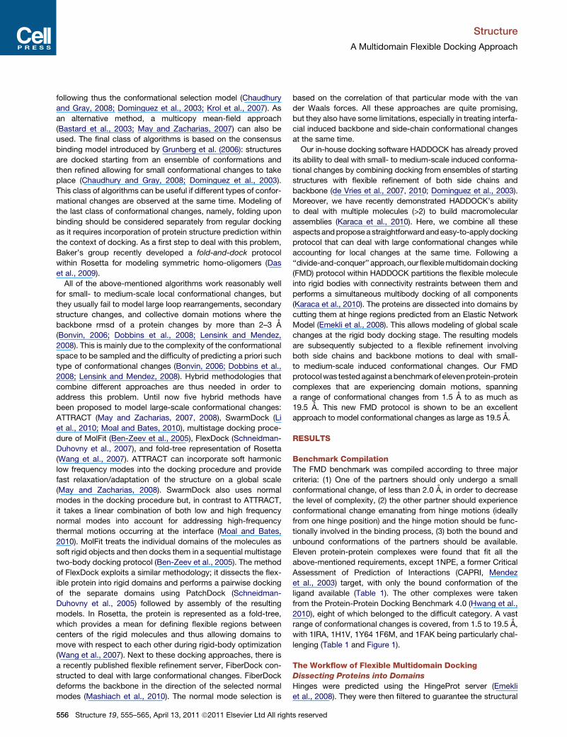
SUMMARY
Binding-induced backbone and large-scale confor-mational changes represent one of the major chal-lenges in the modeling of biomolecular complexesby docking. To address this challenge, we havedeveloped a flexible multidomain docking protocolthat follows a ‘‘divide-and-conquer’’ approach tomodel both large-scale domain motions and small-tomedium-scale interfacial rearrangements: the flex-ible binding partner is treated as an assembly ofsubparts/domains that are docked simultaneouslymaking use of HADDOCK’s multidomain dockingability. For this, the flexiblemolecules are cut at hingeregions predicted using an elastic network model.The performance of this approach is demonstratedon a benchmark covering an unprecedented rangeof conformational changes of 1.5 to 19.5 A. Weshow from a statistical survey of known complexesthat the cumulative sum of eigenvalues obtainedfrom the elastic network has some predictive powerto indicate the extent of the conformational changeto be expected.
INTRODUCTION
Proteins play a vital role in all kinds of biological processes
through their complex interactions with other biomolecules and
small ligands. Revealing all the functional steps in the lifetime
of a protein requires 3D atomic-level information about the
complexes it forms. This information can be obtained from clas-
sical experimental techniques such as X-ray crystallography
and NMR, although, considering the enormous number of
complexes, the need for accurate and complementary computa-
tional methods like docking is evident (Bonvin, 2006; Lensink
and Mendez, 2008; Moreira et al., 2010; Pons et al., 2010;
Ritchie, 2008; Zacharias, 2010). Docking is defined as the
modeling of the structure of a complex (the bound conformation)
starting from the free forms (unbound conformations) of the inter-
action partners. This becomes an extremely challenging task
when the conformation of a protein changes significantly upon
complex formation (Andrusier et al., 2008; Bonvin, 2006; Lensink
and Mendez, 2008; Zacharias, 2010).
Structure 19,
Various mechanisms have been proposed over the years to
describe the binding process and its associated conformational
changes. Fischer suggested that the active sites of the interact-
ing molecules are complementary to each other, obeying a lock-
and-key mechanism (Fischer, 1894). Koshland extended this
model by proposing that the binding follows an induced fit rule,
where the molecules adopt different conformations in order to
fit into the active site of their interaction partner (Koshland,
1958). Later on, Kumar et al. reevaluated this issue from a statis-
tical mechanics point of view and proposed the conformational
selection model (Kumar et al., 2000): the native state of a protein
exists in an ensemble of conformations sampling various states
and environmental effects shift the equilibrium toward the bound
conformation. Subsequent to this hypothesis, Grunberg et al.
(2006) provided a consensus model, treating the concept of
recognition and binding as two different processes with confor-
mational selection at the basis of the recognition process, start-
ing with the formation of an encounter complex.
Conformational changes occurring upon binding can be clas-
sified into four categories: local rearrangements, collective
global motions, a mixture of the two, and binding-induced
folding events. Local changes cover loop rearrangements
(Andrusier et al., 2008), secondary structure element alterations
and motions, etc. Global changes refer to large-scale domain
motions like hinge and shear (Andrusier et al., 2008; Gerstein
and Krebs, 1998; Gerstein and Echols, 2004). Binding-induced
folding events are observed in specific recognition when the
folding of one of (partially) unfolded partner occurs only upon
binding (Bonvin, 2006; Mittag et al., 2010; Zacharias, 2010).
Various strategies have been followed tomodel these different
types of conformational changes (Andrusier et al., 2008; Bonvin,
2006; Lensink and Mendez, 2008; Moreira et al., 2010; Zacha-
rias, 2010). For the lock-and-key cases, namely, when shape
complementarily drives the interaction, rigid body algorithms
(e.g., geometric hashing [Bachar et al., 1993] and Fast Fourier
Transform [Katchalski-Katzir et al., 1992]) provide fast and accu-
rate solutions. Small- to medium-scale backbone conforma-
tional changes (in the range of 1–2 A) can be modeled through
algorithms that address the induced fit mechanism (mostly
based on Molecular Dynamics and Monte Carlo simulations
(Dominguez et al., 2003; Gray et al., 2003)). For some types of
conformational changes, e.g., loop rearrangements or allosteric
effects, this may not provide satisfactory results. Under such
conditions, docking can be started from ensembles of structures
(coming from NMR, Molecular Dynamics or Monte Carlo Simula-
tions, Normal Mode Analysis, graph theoretic approaches .),
555–565, April 13, 2011 ª2011 Elsevier Ltd All rights reserved 555
Structure
A Multidomain Flexible Docking Approach
following thus the conformational selection model (Chaudhury
and Gray, 2008; Dominguez et al., 2003; Krol et al., 2007). As
an alternative method, a multicopy mean-field approach
(Bastard et al., 2003; May and Zacharias, 2007) can also be
used. The final class of algorithms is based on the consensus
binding model introduced by Grunberg et al. (2006): structures
are docked starting from an ensemble of conformations and
then refined allowing for small conformational changes to take
place (Chaudhury and Gray, 2008; Dominguez et al., 2003).
This class of algorithms can be useful if different types of confor-
mational changes are observed at the same time. Modeling of
the last class of conformational changes, namely, folding upon
binding should be considered separately from regular docking
as it requires incorporation of protein structure prediction within
the context of docking. As a first step to deal with this problem,
Baker’s group recently developed a fold-and-dock protocol
within Rosetta for modeling symmetric homo-oligomers (Das
et al., 2009).
All of the above-mentioned algorithms work reasonably well
for small- to medium-scale local conformational changes, but
they usually fail to model large loop rearrangements, secondary
structure changes, and collective domain motions where the
backbone rmsd of a protein changes by more than 2–3 A
(Bonvin, 2006; Dobbins et al., 2008; Lensink and Mendez,
2008). This is mainly due to the complexity of the conformational
space to be sampled and the difficulty of predicting a priori such
type of conformational changes (Bonvin, 2006; Dobbins et al.,
2008; Lensink and Mendez, 2008). Hybrid methodologies that
combine different approaches are thus needed in order to
address this problem. Until now five hybrid methods have
been proposed to model large-scale conformational changes:
ATTRACT (May and Zacharias, 2007, 2008), SwarmDock (Li
et al., 2010; Moal and Bates, 2010), multistage docking proce-
dure of MolFit (Ben-Zeev et al., 2005), FlexDock (Schneidman-
Duhovny et al., 2007), and fold-tree representation of Rosetta
(Wang et al., 2007). ATTRACT can incorporate soft harmonic
low frequency modes into the docking procedure and provide
fast relaxation/adaptation of the structure on a global scale
(May and Zacharias, 2008). SwarmDock also uses normal
modes in the docking procedure but, in contrast to ATTRACT,
it takes a linear combination of both low and high frequency
normal modes into account for addressing high-frequency
thermal motions occurring at the interface (Moal and Bates,
2010). MolFit treats the individual domains of the molecules as
soft rigid objects and then docks them in a sequential multistage
two-body docking protocol (Ben-Zeev et al., 2005). The method
of FlexDock exploits a similar methodology; it dissects the flex-
ible protein into rigid domains and performs a pairwise docking
of the separate domains using PatchDock (Schneidman-
Duhovny et al., 2005) followed by assembly of the resulting
models. In Rosetta, the protein is represented as a fold-tree,
which provides a mean for defining flexible regions between
centers of the rigid molecules and thus allowing domains to
move with respect to each other during rigid-body optimization
(Wang et al., 2007). Next to these docking approaches, there is
a recently published flexible refinement server, FiberDock con-
structed to deal with large conformational changes. FiberDock
deforms the backbone in the direction of the selected normal
modes (Mashiach et al., 2010). The normal mode selection is
556 Structure 19, 555–565, April 13, 2011 ª2011 Elsevier Ltd All righ
based on the correlation of that particular mode with the van
der Waals forces. All these approaches are quite promising,
but they also have some limitations, especially in treating interfa-
cial induced backbone and side-chain conformational changes
at the same time.
Our in-house docking software HADDOCK has already proved
its ability to deal with small- to medium-scale induced conforma-
tional changes by combining docking from ensembles of starting
structures with flexible refinement of both side chains and
backbone (de Vries et al., 2007, 2010; Dominguez et al., 2003).
Moreover, we have recently demonstrated HADDOCK’s ability
to deal with multiple molecules (>2) to build macromolecular
assemblies (Karaca et al., 2010). Here, we combine all these
aspectsandproposeastraightforwardandeasy-to-applydocking
protocol that can deal with large conformational changes while
accounting for local changes at the same time. Following a
‘‘divide-and-conquer’’ approach,our flexiblemultidomaindocking
(FMD) protocol within HADDOCK partitions the flexible molecule
into rigid bodies with connectivity restraints between them and
performs a simultaneous multibody docking of all components
(Karaca et al., 2010). The proteins are dissected into domains by
cutting them at hinge regions predicted from an Elastic Network
Model (Emekli et al., 2008). This allows modeling of global scale
changes at the rigid body docking stage. The resulting models
are subsequently subjected to a flexible refinement involving
both side chains and backbone motions to deal with small-
to medium-scale induced conformational changes. Our FMD
protocolwas testedagainst abenchmarkofelevenprotein-protein
complexes that are experiencing domain motions, spanning
a range of conformational changes from 1.5 A to as much as
19.5 A. This new FMD protocol is shown to be an excellent
approach to model conformational changes as large as 19.5 A.
RESULTS
Benchmark CompilationThe FMD benchmark was compiled according to three major
criteria: (1) One of the partners should only undergo a small
conformational change, of less than 2.0 A, in order to decrease
the level of complexity, (2) the other partner should experience
conformational change emanating from hinge motions (ideally
from one hinge position) and the hinge motion should be func-
tionally involved in the binding process, (3) both the bound and
unbound conformations of the partners should be available.
Eleven protein-protein complexes were found that fit all the
above-mentioned requirements, except 1NPE, a former Critical
Assessment of Prediction of Interactions (CAPRI, Mendez
et al., 2003) target, with only the bound conformation of the
ligand available (Table 1). The other complexes were taken
from the Protein-Protein Docking Benchmark 4.0 (Hwang et al.,
2010), eight of which belonged to the difficult category. A vast
range of conformational changes is covered, from 1.5 to 19.5 A,
with 1IRA, 1H1V, 1Y64 1F6M, and 1FAK being particularly chal-
lenging (Table 1 and Figure 1).
The Workflow of Flexible Multidomain DockingDissecting Proteins into Domains
Hinges were predicted using the HingeProt server (Emekli
et al., 2008). They were then filtered to guarantee the structural
ts reserved

Table 1. Selected Complexes for the Flexible Multidomain Docking Benchmark
Backbone Conformational
Change Range (A)
Complex ID Receptor IDa Ligand IDa Receptor Ligand
1IRAb (Schreuder et al., 1997) 1G0Y_R (Vigers et al., 2000) 1ILR_1 (Schreuder et al., 1995) 19.5 0.7
1H1Vb (Choe et al., 2002) 1D0N_B (Burtnick et al., 1997) 1IJJ_B (Bubb et al., 2002) 13.9 1.6
1Y64b (Otomo et al., 2005) 1UX5_A (Xu et al., 2004) 2FXU_A (Rizvi et al., 2006) 10.3 1.1
1F6Mb (Lennon et al., 2000) 1CL0_A (Lennon et al., 1999) 2TIR_A (Nikkola et al., 1993) 7.3 0.9
1FAKb (Zhang et al., 1999) 1QFK_HL (Pike et al., 1999) 1TFH_B (Huang et al., 1998) 6.0 1.0
1ZLIb (Arolas et al., 2005) 2JTO_A (Pantoja-Uceda et al., 2008) 1KWM_A (Barbosa Pereira
et al., 2002)
3.8 0.6
1E4Kb (Sondermann et al., 2000) 2DTQ_AB (Matsumiya et al., 2007) 1FNL_A (Zhang et al., 2000) 2.9 1.7
1IBRb (Vetter et al., 1999) 1F59_A (Bayliss et al., 2000) 1QG4_A (Kent et al., 1999) 2.9 1.1
1KKLb (Fieulaine et al., 2002) 1JB1_AB (Fieulaine et al., 2001) 2HPR (Liao and Herzberg, 1994) 2.6 0.5
1NPEc (Takagi et al., 2003) 1KLO_A (Stetefeld et al., 1996) 1NPE_A (Takagi et al., 2003) 1.8 —
1DFJb (Kobe and Deisenhofer, 1995) 2BNH_A (Kobe and Deisenhofer, 1996) 9RSA_B (Nachman et al., 1990) 1.5 0.7a The PDB and chain ID’s are indicated as PDB ID_chain ID.bDocking was performed starting from the unbound conformations of both receptor and ligand.c For this particular protein, as the unbound conformation of the ligand was not available, the docking was performed from the unbound conformation
of the receptor and the bound conformation of the ligand.
Structure
A Multidomain Flexible Docking Approach
integrity and dissect the flexible molecule in as few components
as possible in order to maintain the compactness of the
expected solution. This filtering is based on the domain motions
study of Hayward in which hinges were found to bemore likely at
the termini of a helices, b sheets, and in loops that connect
domains, less likely in the center of interdomain a helices, and
not likely within b sheets (Hayward, 1999). In addition, we also
discarded hinge predictions close to N- and C-terminal residues,
as it is highly improbable that such residues can have a hinge-like
behavior. Subsequently, the monomer was cut at the first
peptide bond following the predicted hinge residue. Short expla-
nations for the basis of the hinge selection for each protein can
be found in Table 2.
Defining Ambiguous Interaction and Connectivity
Restraints
The Ambiguous Interaction Restraints (AIRs) were generated via
a new HADDOCK server interface especially built to allow fine-
tuning of restraints in the case of multidomain docking (http://
haddock.chem.uu.nl/services/GenTBL/) (de Vries et al., 2010):
(1) Restraints for the interface of the rigid molecule were kept
ambiguous for the distinct interfaces of the separated flexible
molecule, (2) No AIRs were defined between the separated
domains of the flexible molecule (see Figure 2). The AIRs were
defined based on interface residues identified from the crystal
structure of the complexes. By choosing an ideal definition of
the interface (but not of the contacts made) we focus on the
problem of dealing with conformational changes. This repre-
sents thus a best-case scenario. To maintain the connectivity
between the separated chains, two connectivity restraint files
consisting of an unambiguous distance restraint between the C
and N termini of the separated domains were prepared. The first
file was used during the rigid body energy minimization (it0) and
semiflexible refinement in torsion angle space (it1), to enforce
a chain separation of no more than 10 A (upper distance
restraint). The second one was imposed during the final refine-
Structure 19,
ment in explicit solvent (water) in order to restore the connectivity
(to the real peptide distance, 1.3 A). As the N and C termini of the
cut domains are created artificially, they were kept uncharged.
Three residues on both sides of the separated domains were
defined as fully flexible to allow for more flexibility of the linker
region. All other HADDOCK parameters were left to their default
values. The summary of the workflow is illustrated in Figure 3.
Performance of Flexible Multidomain DockingBoth standard semiflexible two-body docking and the new FMD
protocol were applied to the benchmark for comparison. Two-
body docking could generate good solutions (two stars) for
1DFJ and 1NPE and acceptable ones (one star) for 1E4K and
1KKL. The best solutions were ranked at the top for the latter
three (Table 3). For the other cases, two-body docking failed
completely in generating any acceptable solutions. When the
new FMD protocol was applied, acceptable or better solutions
were obtained for each benchmark case (Table 3). The improve-
ment is impressive particularly for the seven cases (1IRA, 1H1V,
1Y64, 1F6M, 1FAK, 1ZLI, 1IBR) for which two-body docking was
failing: the best l-rmsd values of the top four challenging cases
decreased from 38.6 to 7.5 A for 1IRA, from 31.1 to 9.6 A for
1H1V, from 3.2 to 9.5 A for 1Y64 and from 39.4 to 8.7 A for
1F6M (note also the very high fraction of native contacts, Table
3). Besides providing acceptable-to-good solutions for all of
the cases, FMD-HADDOCK could rank them at the top using
the standard scoring scheme in HADDOCK consisting of
a weighted sum of van der Waals, electrostatic energies,
and an empirical desolvation term (Fernandez-Recio et al.,
2004) (HADDOCKscore = 1.0 Evdw + 0.2 Eelec + 1.0 Edesol). Further-
more, the quality of the models improved for 1E4K, 1KKL, 1NPE,
and 1DFJ, for which two-body docking already provided reason-
able solutions. 1E4K’s and 1KKL’s best models, ranked among
the top five, are now a two star (good) solution and 1NPE’s
top-ranking structure is almost a three star (high accuracy)
555–565, April 13, 2011 ª2011 Elsevier Ltd All rights reserved 557
A B C D
EF G H
I J K
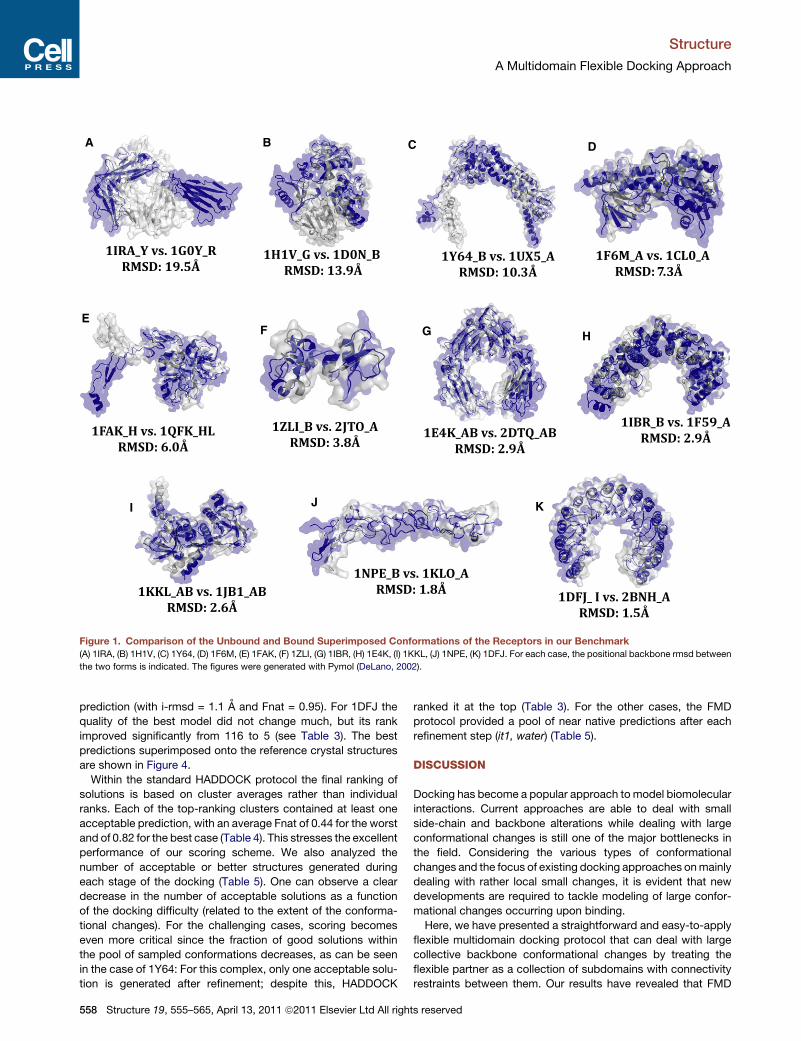
Figure 1. Comparison of the Unbound and Bound Superimposed Conformations of the Receptors in our Benchmark
(A) 1IRA, (B) 1H1V, (C) 1Y64, (D) 1F6M, (E) 1FAK, (F) 1ZLI, (G) 1IBR, (H) 1E4K, (I) 1KKL, (J) 1NPE, (K) 1DFJ. For each case, the positional backbone rmsd between
the two forms is indicated. The figures were generated with Pymol (DeLano, 2002).
Structure
A Multidomain Flexible Docking Approach
prediction (with i-rmsd = 1.1 A and Fnat = 0.95). For 1DFJ the
quality of the best model did not change much, but its rank
improved significantly from 116 to 5 (see Table 3). The best
predictions superimposed onto the reference crystal structures
are shown in Figure 4.
Within the standard HADDOCK protocol the final ranking of
solutions is based on cluster averages rather than individual
ranks. Each of the top-ranking clusters contained at least one
acceptable prediction, with an average Fnat of 0.44 for the worst
and of 0.82 for the best case (Table 4). This stresses the excellent
performance of our scoring scheme. We also analyzed the
number of acceptable or better structures generated during
each stage of the docking (Table 5). One can observe a clear
decrease in the number of acceptable solutions as a function
of the docking difficulty (related to the extent of the conforma-
tional changes). For the challenging cases, scoring becomes
even more critical since the fraction of good solutions within
the pool of sampled conformations decreases, as can be seen
in the case of 1Y64: For this complex, only one acceptable solu-
tion is generated after refinement; despite this, HADDOCK
558 Structure 19, 555–565, April 13, 2011 ª2011 Elsevier Ltd All righ
ranked it at the top (Table 3). For the other cases, the FMD
protocol provided a pool of near native predictions after each
refinement step (it1, water) (Table 5).
DISCUSSION
Docking has become a popular approach to model biomolecular
interactions. Current approaches are able to deal with small
side-chain and backbone alterations while dealing with large
conformational changes is still one of the major bottlenecks in
the field. Considering the various types of conformational
changes and the focus of existing docking approaches onmainly
dealing with rather local small changes, it is evident that new
developments are required to tackle modeling of large confor-
mational changes occurring upon binding.
Here, we have presented a straightforward and easy-to-apply
flexible multidomain docking protocol that can deal with large
collective backbone conformational changes by treating the
flexible partner as a collection of subdomains with connectivity
restraints between them. Our results have revealed that FMD
ts reserved
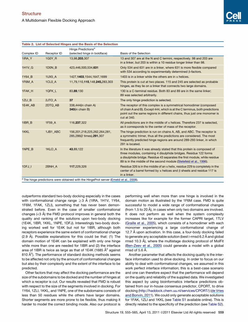
Table 2. List of Selected Hinges and the Basis of the Selection
Complex ID Receptor ID
Hinge Predictionsa
(selected hinge in boldface) Basis of the Selection
1IRA_Y 1G0Y_R 13,98,203,307 13 and 307 are at the N and C termini, respectively. 98 and 203 are
in a linker, but 203 is within a 10 residue longer linker than 98.
1H1V_G 1D0N_B 423,446,500,534,631 Only 534 and 631 are in a linker, where 631 is more flexible compared
with 534 according to experimentally determined b-factors.
1Y64_B 1UX5_A 1427,1403,1544,1647,1699 1403 is in a linker while the others are in a helices.
1F6M_A 1CL0_A 11,79,110,115,148,245,283,303 This protein is cut at two places. 115 and 245 are selected as probable
hinges, as they lie on a linker that connects two large domains.
1FAK_H 1QFK_L 83,89,130 130 is a C-terminal residue. Both 83 and 89 are in the same linker;
89 was selected arbitrarily.
1ZLI_B 2JTO_A 37 The only hinge prediction is selected.
1E4K_AB 2DTQ_AB 338,444(in chain A)
340(in chain B)
The receptor of this complex is a symmetrical homodimer (composed
of chain A andB). Except 444, which is at the C terminus, both predictions
point out the same regions in different chains, thus just one monomer is
cut at 340.
1IBR_B 1F59_A 116,237,322 All predictions are in the middle of a helices. Therefore 237 is selected,
as it corresponds to the center of mass of the receptor.
1KKL 1JB1_ABC 156,201,218,225,262,264,281,
285,286(2 times),291,307
The hinge prediction is run on chains A, AB, and ABC. The receptor is
a symmetric trimer, thus all the predictions are considered. The most
frequently predicted hinge regions are around 289-293 linker, in which
291 is located.
1NPE_B 1KLO_A 43,89,122 In the literature it was already stated that this protein is composed of
three modules, containing 4 disulphide bridges. Residue 122 forms
a disulphide bridge. Residue 43 separates the first module, while residue
89 is in the middle of the second module (Stetefeld et al., 1996).
1DFJ_I 2BNH_A 117,229,326 Residue 326 is in the middle of an a helix, residue 229 is completely in the
center of a barrel formed by a helices and b sheets and residue 117 is
in a linker.a The hinge predictions were obtained with the HingeProt server (Emekli et al., 2008).
Structure
A Multidomain Flexible Docking Approach
outperforms standard two-body docking especially in the cases
with conformational change range R3 A (1IRA, 1H1V, 1Y64,
1F6M, 1FAK, 1ZLI), something that has never been demon-
strated before. Even in the case of smaller conformational
changes (<3 A) the FMD protocol improves in general both the
quality and ranking of the solutions upon two-body docking
(1E4K, 1IBR, 1KKL, 1NPE, 1DFJ). Interestingly two-body dock-
ing worked well for 1E4K but not for 1IBR, although both
receptors experience the same extent of conformational change
(2.9 A). Possible explanations for this could be that: (1) The
domain motion of 1E4K can be explained with only one hinge
while more than one are needed for 1IBR and (2) the interface
area of 1IBR is twice as large as that of 1E4K (1685 A2 against
810 A2). The performance of standard docking methods seems
to be affected not only by the amount of conformational changes
but also by their complexity and the extent of the interface to be
predicted.
Other factors that may affect the docking performance are the
size of the subdomains to be docked and the number of hinges at
which a receptor is cut. Our results revealed that FMD is robust
with respect to the size of the segments involved in docking. For
1Y64, 1ZLI, 1KKL, and 1NPE, one of the subdomains consists of
less than 50 residues while the others have larger domains.
Shorter segments are more prone to be flexible, thus making it
harder to model the correct binding mode. Also our protocol is
Structure 19,
performing well when more than one hinge is involved in the
domain motion as illustrated by the 1F6M case. FMD is quite
successful to model a wide range of conformational changes
(from 1.5 to 20 A), in cases when only two domains are involved.
It does not perform as well when the system complexity
increases like for example for the former CAPRI target, 1TLV
(Graille et al., 2005), which consists of a homodimer with each
monomer experiencing a large conformational change of
12.7 A upon activation. In this case, a four-body docking failed
to generate any acceptable solution (best i-rmsd 6.2 A and global
rmsd 10.3 A), where the multistage docking protocol of MolFit
(Ben-Zeev et al., 2005) could generate a model with a global
rmsd of 5.6 A.
Another parameter that affects the docking quality is the inter-
face information used to drive docking. In order to focus on our
ability to deal with conformational changes, we assumed in this
work perfect interface information; this is a best-case scenario
and one can therefore expect that the performance will depend
on the quality and reliability of the supplied data.We investigated
this aspect by using bioinformatics interface predictions ob-
tained from our in-house consensus predictor, CPORT, to drive
docking (http://haddock.chem.uu.nl/services/CPORT/) (de Vries
and Bonvin, 2011). We could only generate acceptable solutions
for 1FAK, 1ZLI and 1KKL (see Table S1 available online). This is
directly related to the specificity of the prediction (see Table S2).
555–565, April 13, 2011 ª2011 Elsevier Ltd All rights reserved 559

A
10Å
it0/it1
1.3Å
0ÅÅ
/it1
waterB 1
AIRs
B2
Connec�vity Restraints
AIRs
Figure 2. Schematic Representation of the Restraints Used in Dock-
ing (Ambiguous Interaction and Connectivity Restraints) and of the
Dissection of the Flexible Partner into Subdomains
Table 3. Comparison of Two-Body and Flexible Multidomain
Docking Results
Flexible Multidomain Docking
PDB ID Quality / Ranka i-rmsd (A) l-rmsd (A) Fnat Fnonnat
1IRA + / 1 3.9 7.5 0.55 0.61
1H1V + / 11 4.6 9.6 0.49 0.67
1Y64 + / 5 3.9 9.5 0.48 0.72
1F6M + / 1 3.5 8.7 0.69 0.58
1FAK++ / 37 2.8 4.2 0.55 0.54
+ / 2 3.4 7.4 0.50 0.58
1ZLI ++ / 1 2.1 3.5 0.74 0.47
1E4K++ / 5 2.3 4.0 0.70 0.47
+ / 1 2.8 6.0 0.62 0.52
1IBR ++ / 1 2.3 4.0 0.63 0.57
1KKL ++ / 1 2.2 4.9 0.67 0.59
1NPE++ / 16 1.2 3.2 0.95 0.36
+ / 1 5.5 7.1 0.74 0.51
1DFJ++ / 5 2.0 7.1 0.68 0.56
+ / 1 2.3 8.3 0.60 0.61
Two-Body Dockingb
PDB ID Quality / Rank i-rmsd (A) l-rmsd (A) Fnat Fnonnat
1IRA - / 1 17.5 38.6 0.04 0.94
1H1V - / 1 11.9 31.1 0.08 0.94
1Y64 - / 1 10.3 32.2 0.07 0.92
1F6M - / 1 14.1 39.4 0.00 1.00
1FAK - / 1 11.4 28.5 0.01 0.99
1ZLI - / 1 14.8 25.3 0.02 0.99
1E4K + / 1 4.1 9.8 0.58 0.58
1IBR - / 1 9.6 30.9 0.11 0.90
1KKL + / 1 3.1 12.3 0.56 0.60
1NPE ++ / 1 1.7 6.1 0.85 0.45
1DFJ++ / 116 1.8 5.6 0.63 0.59
+ / 1 2.5 7.1 0.62 0.64
The quality is expressed according to CAPRI criteria (see Experimental
Procedures). The reported rank is the rank of the individual models prior
Structure
A Multidomain Flexible Docking Approach
Only for four cases (1FAK, 1ZLI, 1KKL, and 1DFJ), the specificity
of CPORT predictions was above 30% for both receptor and
ligand and for three of these acceptable to medium quality solu-
tions could be obtained. This is in line with what was observed in
systematic HADDOCK runs, where CPORT was used to drive
docking (de Vries and Bonvin, 2011).
In general, our FMD protocol could generate at least an
acceptable solution for each case, and even medium-quality
predictions for seven of them. The fraction of native contacts
for the best models was above 0.5 (the CAPRI threshold for
high quality predictions) for nine of them. The strength of FMD-
HADDOCK resides mainly in its ability to deal with large-scale
and small, induced conformational changes at the same time.
Our benchmark shares common cases with three other hybrid
methodologies (see Introduction): 1NPE and 1IBRwith FlexDock
(see Table 2 in Schneidman-Duhovny et al., 2007); 1DFJ and
1IBR with ATTRACT (see Table 5 in May and Zacharias, 2008)
and FiberDock (see Table 4 in Mashiach et al., 2010). Comparing
the best l-rmsds for 1IBR, FlexDock, FiberDock and FMD
produce similar somewhat better results than ATTRACT. FMD
however outperforms FlexDock in scoring. A similar situation is
Figure 3. Workflow of Flexible Multidomain Docking in HADDOCK
to clustering. See also Tables S1–S3.a The ranking is based on the HADDOCK score: 1.0 Evdw + 0.2 Eelec +
1.0 Edesol (see Results).bWhen no acceptable solution is generated, the values for the top ranked
structure are reported.
560 Structure 19, 555–565, April 13, 2011 ª2011 Elsevier Ltd All righ
observed for 1NPE. In the case of 1DFJ FMD, FiberDock and
ATTRACT perform well in both sampling and scoring, although
FiberDock’s best model has a better l-rmsd.
The excellent performance of the new FMD-HADDOCK
protocol represents already a major step in dealing with large
conformational changes occurring upon binding. One question,
however, remains: How can we predict which type of conforma-
tional change should be expected upon binding so that one can
choose the best suited method? Dobbins et al. (2008) pointed
out that Normal Mode Analysis can provide some discrimination
among the various types of conformational changes. They stated
that the slowest mode of a protein experiencing significant
conformational changes (Ca-rmsd > 2.0 A) has a 2.5 times lower
ts reserved

A BC D
E FG H
I J K
Figure 4. View of the Best HADDOCK Solutions Superimposed onto the Respective Reference Crystal Structures
The docked complexes are shown in magenta (ligand) and blue (receptor) and the reference crystal structure (only the receptor is shown) in gray: (A) 1IRA, (B)
1H1V, (C) 1Y64, (D) 1F6M, (E) 1FAK, (F) 1ZLI, (G) 1IBR, (H) 1E4K, (I) 1KKL, (J) 1NPE, (K) 1DFJ. The quality of the models is indicated as: number of stars / interface
rmsd (in A) / fraction of native contacts (see Experimental Procedures). The figures were generated with Pymol (DeLano, 2002).
Structure
A Multidomain Flexible Docking Approach
frequency than for proteins undergoing limited conformational
change (Ca-rmsd < 1.0 A). To further investigate this issue, we
considered a set of 268 proteins experiencing different levels
of conformational changes (0.3 A % Ca-rmsd % 19.5 A). These
were taken from the Docking Benchmark 4.0 (Hwang et al.,
2010), excluding multimers, antibody-antigen complexes and
proteins having less than 50 amino acids. For all single proteins,
eigenvalues from a Gaussian Network Model (GNM) (see Exper-
imental Procedures) (Haliloglu et al., 1997) were obtained from
the HingeProt web server (Emekli et al., 2008). They were
normalized and their cumulative sum (ranging from 0 to 1) was
calculated and plotted as a function of the number of modes.
This plot can be interpreted as the fraction of motion explained
by a given number of modes. Here we assumed that proteins
undergoing larger conformational changes have smaller GNM-
eigenvalues, corresponding to collective low frequency motions.
For these, the cumulative sum of the normalized GNM-eigen-
values should increase slower as a function of the number of
Structure 19,
modes than for rigid cases, in agreement with the observations
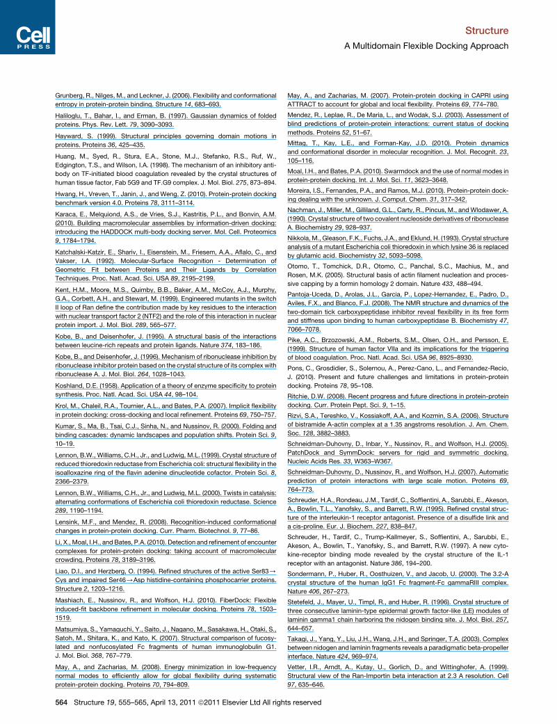
of Dobbins et al. (2008) (Figure S1). To test this assumption,
we developed a simple predictor, which predicts proteins as
being rigid or flexible based on the fraction of motion explained
by a given number of modes (see Supplemental Experimental
Procedures). This classifier performs with the best accuracy
(65 ± 16%, cross-validated accuracy) on the set of 268 proteins
when a 2.6 A rmsd cutoff is used to classify these proteins as
rigid and flexible and the cutoff for the fraction of motion
explained for the first 50 modes is taken as 0.08 (i.e., the cumu-
lative sum of the eigenvalues for the first 50 modes should
be <0.08 for the protein to be predicted as flexible). Our simple
predictor was able to classify the receptors of the FMD bench-
mark with 64% accuracy (with seven out of nine flexible cases
and one out of two rigid cases being correctly classified, see Fig-
ure 5). The cumulative sum of the normalized eigenvalues for
a given number of modesmay thus be deterministic in predicting
the range of the conformational change that could be expected.
555–565, April 13, 2011 ª2011 Elsevier Ltd All rights reserved 561
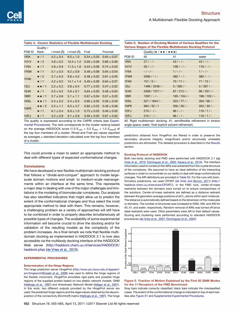
Table 4. Cluster Statistics of Flexible Multidomain Docking
PDB ID
Quality /
Rank i-rmsd (A) l-rmsd (A) Fnat Fnonnat
1IRA + / 1 4.2 ± 0.4 8.0 ± 1.0 0.54 ± 0.03 0.64 ± 0.07
1H1V + / 2 4.8 ± 0.2 10.4 ± 1.2 0.50 ± 0.06 0.66 ± 0.06
1Y64 + / 1 4.8 ± 0.6 11.3 ± 1.0 0.44 ± 0.06 0.74 ± 0.03
1F6M + / 1 3.1 ± 0.3 9.2 ± 0.8 0.49 ± 0.08 0.64 ± 0.04
1FAK+ / 2 3.7 ± 0.5 9.8 ± 4.5 0.48 ± 0.02 0.61 ± 0.05
+ / 1 4.2 ± 0.2 14.1 ± 1.4 0.49 ± 0.06 0.64 ± 0.07
1ZLI ++ / 1 2.2 ± 0.2 3.8 ± 0.4 0.77 ± 0.03 0.47 ± 0.02
1E4K + / 1 2.5 ± 0.2 5.9 ± 0.1 0.64 ± 0.05 0.46 ± 0.04
1IBR ++ / 1 2.7 ± 0.6 5.1 ± 1.1 0.67 ± 0.04 0.57 ± 0.03
1KKL ++ / 1 2.4 ± 0.2 5.4 ± 0.5 0.69 ± 0.05 0.56 ± 0.03
1NPE++ / 2 2.2 ± 1.1 6.5 ± 4.7 0.82 ± 0.22 0.46 ± 0.06
+ / 1 6.1 ± 0.4 8.9 ± 1.0 0.73 ± 0.03 0.46 ± 0.05
1DFJ + / 1 3.2 ± 0.8 9.1 ± 0.8 0.59 ± 0.02 0.67 ± 0.02
The quality is expressed according to the CAPRI criteria (see Experi-
mental Procedures). The rank corresponds to the cluster ranking based
on the average HADDOCK score (1.0 Evdw + 0.2 Eelec + 1.0 Edesol) of
the top four members of a cluster. Rmsd and Fnat are values reported
as averages ± standard deviation calculated over the top four members
of a cluster.
Table 5. Number of Docking Models of Various Qualities for the
Various Stages of the Flexible Multidomain Docking Protocol
Quality (+ / ++ / +++)
PDB ID it0 it1 water
1IRA 27 / - / - 43 / - / - 44 / - / -
1H1V 50 / - / - 108 / - / - 116 / - / -
1Y64 - / - / - 1 / - / - 1 / - / -
1F6M 2096 / - / - 382 / - / - 382 / - / -
1FAK 151 / 3 / - 70 / 11 / - 71 / 12 / -
1ZLI 1496 / 3246 / - 5 / 395 / - 3 / 397 / -
1E4K 2569 / 1371 / - 87 / 212 / - 98 / 201 / -
1IBR 1287 / - / - 195 / 104 / - 196 / 103 / -
1KKL 521 / 1844 / - 323 / 77 / - 304 / 96 / -
1NPE 984 / 32 / 1 356 / 26 / - 353 / 30 / -
1DFJ 210 / - / - 96 / - / - 116 / 1 / -
1DFJ 210 / - / - 96 / - / - 116 / 1 / -
it0, Rigid multidomain docking; it1, semiflexible refinement in torsion
angle space; water, final explicit solvent refinement.F
ract
ion
of t
he T
otal
Mot
ion
Exp
lain
ed
0.080.1
0.2
0.3
0.4
0.5
1IRA 1H1V 1Y64 1F6M 1FAK 1ZLI 1E4K 1IBR 1KKL 1NPE 1DFJ
19.5Å
13.9Å10.3Å
7.3Å
3.8Å
2.9Å
2.9Å
2.6Å
1.8Å
1.5Å6.0Å
Figure 5. Fraction of Motion Explained by the First 50 GNM Modes
for the 11 Receptors of the FMD Benchmark
Gray bars indicate correctly classified; black bars indicate the misclassified
cases. The extent of the conformational change is indicated on top of each bar.
See also Figure S1 and Supplemental Experimental Procedures.
Structure
A Multidomain Flexible Docking Approach
This could provide a mean to select an appropriate method to
deal with different types of expected conformational changes.
ConclusionsWe have developed a new flexible multidomain docking protocol
that follows a ‘‘divide-and-conquer’’ approach to model large-
scale domain motions and small- to medium-scale rearrange-
ments within an interface at the same time. This represents
a major step in dealing with one of the major challenges and limi-
tations in the modeling of biomolecular complexes. Our analysis
has also identified indicators that might allow us to predict the
extent of the conformational changes and thus select the most
appropriate method to deal with them. This remains, however,
a challenging problem as a variety of approaches will still have
to be combined in order to properly describe simultaneously all
possible types of changes. The availability of some experimental
information will become crucial to drive the docking and/or for
validation of the resulting models as the complexity of the
problem increases. As a final remark we note that flexible multi-
domain docking as implemented in HADDOCK 2.1 is now also
accessible via the multibody docking interface of the HADDOCK
Web server (http://haddock.chem.uu.nl/services/HADDOCK/
haddock.php) (de Vries et al., 2010).
EXPERIMENTAL PROCEDURES
Determination of the Hinge Regions
The hinge prediction server HingeProt (http://www.prc.boun.edu.tr/appserv/
prc/hingeprot/)(Emekli et al., 2008) was used to define the hinge regions of
the flexible monomers. HingeProt annotates rigid parts and possible hinge
regions of the supplied protein based on two elastic network models: GNM
(Haliloglu et al., 1997) and Anisotropic Network Model (Atilgan et al., 2001).
In this work, two different outputs provided by the HingeProt server are
used: the predicted hinge regions and the eigenvalues obtained by the decom-
position of the connectivity (Kirchhoff) matrix (Haliloglu et al., 1997). The hinge
562 Structure 19, 555–565, April 13, 2011 ª2011 Elsevier Ltd All righ
predictions obtained from HingeProt are filtered in order to preserve the
secondary structure integrity. Insignificant and/or structurally unreliable
predictions are eliminated. The detailed procedure is described in the Results
section.
Docking Protocol of HADDOCK
Both two-body docking and FMD were performed with HADDOCK 2.1 (de
Vries et al., 2010; Dominguez et al., 2003; Karaca et al., 2010). The interface
information used to construct the AIRswas extracted from the crystal structure
of the complexes. We assumed to have an ideal definition of the interacting
surfaces in order to concentrate on our ability to deal with large conformational
changes. The AIR definitions are provided in Table S3. For the runs with bioin-
formatics predictions, we used CPORT (de Vries and Bonvin, 2011) (http://
haddock.chem.uu.nl/services/CPORT/). In the FMD runs, center-of-mass
restraints between the domains were turned on to ensure compactness of
the solutions. Center-of-mass restraints are defined as a distance restraint
between the geometric average positions of all Ca atomswithin eachmolecule.
The distance is automatically defined based on the dimension of themolecules
or domains. The number of structures was increased to 5000, 400, and 400 for
it0, it1, and water, respectively. Random removal of AIRs was turned off since
ideal restraints were used. Other parameters were left to their default values.
Scoring and clustering were performed according to standard HADDOCK
procedures (de Vries et al., 2007; Dominguez et al., 2003).
ts reserved

Structure
A Multidomain Flexible Docking Approach
Assessment of the Structure Quality
The docking models were evaluated according to CAPRI criteria (Mendez
et al., 2003):
d Acceptable prediction (one star): i-rmsd % 4 A or l-rmsd % 10 A and
Fnat R 0.1
d Good prediction (two stars): i-rmsd % 2 A or l-rmsd % 5 A and Fnat R
0.3
d High-quality prediction (three stars): i-rmsd % 1 A or l-rmsd % 1 A and
nat R 0.5.
i-rmsd refers to the interface rmsd, l-rmsd to the ligand rmsd, calculated
over the backbone atoms of the ligand (rigid component) after fitting on the
receptor, and Fnat to the fraction of native contacts. Next to Fnat we also
provided Fnonnat, which is the fraction of incorrectly predicted contacts
given the modeled complex. A cluster was considered of one-, two-, or
three-star quality if at least one of its top four members was of the correspond-
ing quality.
SUPPLEMENTAL INFORMATION
Supplemental Information includes Supplemental Experimental Procedures,
one figure, and three tables and can be found with this article online at
doi:10.1016/j.str.2011.01.014.
ACKNOWLEDGMENTS
This work was supported by the Netherlands Organization for Scientific
Research (NWO) (VICI grant 700.56.442 to A.M.J.J.B.) and the European
Community (FP7 FP7 e-Infrastructure ‘‘e-NMR’’ and ‘‘WeNMR’’ projects, grant
numbers 213010 and 21301). Support from the national GRID Initiatives of
Belgium, Italy, Germany, the Netherlands (via the Dutch BiG Grid project),
Portugal, UK, South Africa, is acknowledged for the use of computing and
storage facilities.
Received: October 8, 2010
Revised: January 3, 2011
Accepted: January 10, 2011
Published: April 12, 2011
REFERENCES
Andrusier, N., Mashiach, E., Nussinov, R., and Wolfson, H.J. (2008). Principles
of flexible protein-protein docking. Proteins 73, 271–289.
Arolas, J.L., Popowicz, G.M., Lorenzo, J., Sommerhoff, C.P., Huber, R., Aviles,
F.X., and Holak, T.A. (2005). The three-dimensional structures of tick carboxy-
peptidase inhibitor in complex with A/B carboxypeptidases reveal a novel
double-headed binding mode. J. Mol. Biol. 350, 489–498.
Atilgan, A.R., Durell, S.R., Jernigan, R.L., Demirel, M.C., Keskin, O., and Bahar,
I. (2001). Anisotropy of fluctuation dynamics of proteins with an elastic network
model. Biophys. J. 80, 505–515.
Bachar, O., Fischer, D., Nussinov, R., and Wolfson, H. (1993). A computer
vision based technique for 3-D sequence-independent structural comparison
of proteins. Protein Eng. 6, 279–288.
Barbosa Pereira, P.J., Segura-Martin, S., Oliva, B., Ferrer-Orta, C., Aviles, F.X.,
Coll, M., Gomis-Ruth, F.X., and Vendrell, J. (2002). Human procarboxypepti-
dase B: three-dimensional structure and implications for thrombin-activatable
fibrinolysis inhibitor (TAFI). J. Mol. Biol. 321, 537–547.
Bastard, K., Thureau, A., Lavery, R., and Prevost, C. (2003). Docking macro-
molecules with flexible segments. J. Comput. Chem. 24, 1910–1920.
Bayliss, R., Littlewood, T., and Stewart, M. (2000). Structural basis for the inter-
action between FxFG nucleoporin repeats and importin-beta in nuclear traf-
ficking. Cell 102, 99–108.
Ben-Zeev, E., Kowalsman, N., Ben-Shimon, A., Segal, D., Atarot, T., Noivirt,
O., Shay, T., and Eisenstein, M. (2005). Docking to single-domain and
multiple-domain proteins: old and new challenges. Proteins 60, 195–201.
Structure 19,
Bonvin, A.M. (2006). Flexible protein-protein docking. Curr. Opin. Struct. Biol.
16, 194–200.
Bubb, M.R., Govindasamy, L., Yarmola, E.G., Vorobiev, S.M., Almo, S.C.,
Somasundaram, T., Chapman, M.S., Agbandje-McKenna, M., and McKenna,
R. (2002). Polylysine induces an antiparallel actin dimer that nucleates filament
assembly: crystal structure at 3.5-A resolution. J. Biol. Chem. 277, 20999–
21006.
Burtnick, L.D., Koepf, E.K., Grimes, J., Jones, E.Y., Stuart, D.I., McLaughlin,
P.J., and Robinson, R.C. (1997). The crystal structure of plasma gelsolin: impli-
cations for actin severing, capping, and nucleation. Cell 90, 661–670.
Chaudhury, S., and Gray, J.J. (2008). Conformer selection and induced fit in
flexible backbone protein-protein docking using computational and NMR
ensembles. J. Mol. Biol. 381, 1068–1087.
Choe, H., Burtnick, L.D., Mejillano, M., Yin, H.L., Robinson, R.C., and Choe, S.
(2002). The calcium activation of gelsolin: insights from the 3A structure of the
G4-G6/actin complex. J. Mol. Biol. 324, 691–702.
Das, R., Andre, I., Shen, Y., Wu, Y., Lemak, A., Bansal, S., Arrowsmith, C.H.,
Szyperski, T., and Baker, D. (2009). Simultaneous prediction of protein folding
and docking at high resolution. Proc. Natl. Acad. Sci. USA 106, 18978–18983.
de Vries, S.J., van Dijk, A.D.J., Krzeminski, M., van Dijk, M., Thureau, A., Hsu,
V., Wassenaar, T., and Bonvin, A.M.J.J. (2007). HADDOCK versus HADDOCK:
new features and performance of HADDOCK2.0 on the CAPRI targets.
Proteins 69, 726–733.
de Vries, S.J., van Dijk, M., and Bonvin, A.M. (2010). The HADDOCK web
server for data-driven biomolecular docking. Nat. Protoc. 5, 883–897.
de Vries, S.J., and Bonvin, A.M.J.J. (2011). CPORT: a consensus interface
predictor and its performance in prediction-driven docking with HADDOCK.
PLoS ONE, in press.
DeLano, W.L. (2002). The PyMOL Molecular Graphics System on World Wide
Web (http://www.pymol.org).
Dobbins, S.E., Lesk, V.I., and Sternberg, M.J. (2008). Insights into protein flex-
ibility: the relationship between normal modes and conformational change
upon protein-protein docking. Proc. Natl. Acad. Sci. USA 105, 10390–10395.
Dominguez, C., Boelens, R., and Bonvin, A.M.J.J. (2003). HADDOCK:
a protein-protein docking approach based on biochemical or biophysical
information. J. Am. Chem. Soc. 125, 1731–1737.
Emekli, U., Schneidman-Duhovny, D., Wolfson, H.J., Nussinov, R., and
Haliloglu, T. (2008). HingeProt: automated prediction of hinges in protein struc-
tures. Proteins 70, 1219–1227.
Fernandez-Recio, J., Totrov, M., and Abagyan, R. (2004). Identification of
protein-protein interaction sites from docking energy landscapes. J. Mol.
Biol. 335, 843–865.
Fieulaine, S., Morera, S., Poncet, S., Mijakovic, I., Galinier, A., Janin, J.,
Deutscher, J., and Nessler, S. (2002). X-ray structure of a bifunctional protein
kinase in complexwith its protein substrate HPr. Proc. Natl. Acad. Sci. USA 99,
13437–13441.
Fieulaine, S., Morera, S., Poncet, S., Monedero, V., Gueguen-Chaignon, V.,
Galinier, A., Janin, J., Deutscher, J., and Nessler, S. (2001). X-ray structure
of HPr kinase: a bacterial protein kinase with a P-loop nucleotide-binding
domain. EMBO J. 20, 3917–3927.
Fischer, E. (1894). Einfluss der Configuration auf die Wirkung der Enzyme.
(Berichte der deutschen Chemischen Gesellschaft) , pp. 2985–2993.
Gerstein, M., and Echols, N. (2004). Exploring the range of protein flexibility,
from a structural proteomics perspective. Curr. Opin. Chem. Biol. 8, 14–19.
Gerstein, M., and Krebs, W. (1998). A database of macromolecular motions.
Nucleic Acids Res. 26, 4280–4290.
Graille, M., Zhou, C.Z., Receveur-Brechot, V., Collinet, B., Declerck, N., and
van Tilbeurgh, H. (2005). Activation of the LicT transcriptional antiterminator
involves a domain swing/lock mechanism provoking massive structural
changes. J. Biol. Chem. 280, 14780–14789.
Gray, J.J., Moughon, S., Wang, C., Schueler-Furman, O., Kuhlman, B., Rohl,
C.A., and Baker, D. (2003). Protein-protein docking with simultaneous optimi-
zation of rigid-body displacement and side-chain conformations. J. Mol. Biol.
331, 281–299.
555–565, April 13, 2011 ª2011 Elsevier Ltd All rights reserved 563
Structure
A Multidomain Flexible Docking Approach
Grunberg, R., Nilges, M., and Leckner, J. (2006). Flexibility and conformational
entropy in protein-protein binding. Structure 14, 683–693.
Haliloglu, T., Bahar, I., and Erman, B. (1997). Gaussian dynamics of folded
proteins. Phys. Rev. Lett. 79, 3090–3093.
Hayward, S. (1999). Structural principles governing domain motions in
proteins. Proteins 36, 425–435.
Huang, M., Syed, R., Stura, E.A., Stone, M.J., Stefanko, R.S., Ruf, W.,
Edgington, T.S., and Wilson, I.A. (1998). The mechanism of an inhibitory anti-
body on TF-initiated blood coagulation revealed by the crystal structures of
human tissue factor, Fab 5G9 and TF.G9 complex. J. Mol. Biol. 275, 873–894.
Hwang, H., Vreven, T., Janin, J., and Weng, Z. (2010). Protein-protein docking
benchmark version 4.0. Proteins 78, 3111–3114.
Karaca, E., Melquiond, A.S., de Vries, S.J., Kastritis, P.L., and Bonvin, A.M.
(2010). Building macromolecular assemblies by information-driven docking:
introducing the HADDOCK multi-body docking server. Mol. Cell. Proteomics
9, 1784–1794.
Katchalski-Katzir, E., Shariv, I., Eisenstein, M., Friesem, A.A., Aflalo, C., and
Vakser, I.A. (1992). Molecular-Surface Recognition - Determination of
Geometric Fit between Proteins and Their Ligands by Correlation
Techniques. Proc. Natl. Acad. Sci. USA 89, 2195–2199.
Kent, H.M., Moore, M.S., Quimby, B.B., Baker, A.M., McCoy, A.J., Murphy,
G.A., Corbett, A.H., and Stewart, M. (1999). Engineered mutants in the switch
II loop of Ran define the contribution made by key residues to the interaction
with nuclear transport factor 2 (NTF2) and the role of this interaction in nuclear
protein import. J. Mol. Biol. 289, 565–577.
Kobe, B., and Deisenhofer, J. (1995). A structural basis of the interactions
between leucine-rich repeats and protein ligands. Nature 374, 183–186.
Kobe, B., and Deisenhofer, J. (1996). Mechanism of ribonuclease inhibition by
ribonuclease inhibitor protein based on the crystal structure of its complex with
ribonuclease A. J. Mol. Biol. 264, 1028–1043.
Koshland, D.E. (1958). Application of a theory of enzyme specificity to protein
synthesis. Proc. Natl. Acad. Sci. USA 44, 98–104.
Krol, M., Chaleil, R.A., Tournier, A.L., and Bates, P.A. (2007). Implicit flexibility
in protein docking: cross-docking and local refinement. Proteins 69, 750–757.
Kumar, S., Ma, B., Tsai, C.J., Sinha, N., and Nussinov, R. (2000). Folding and
binding cascades: dynamic landscapes and population shifts. Protein Sci. 9,
10–19.
Lennon, B.W., Williams, C.H., Jr., and Ludwig, M.L. (1999). Crystal structure of
reduced thioredoxin reductase from Escherichia coli: structural flexibility in the
isoalloxazine ring of the flavin adenine dinucleotide cofactor. Protein Sci. 8,
2366–2379.
Lennon, B.W., Williams, C.H., Jr., and Ludwig, M.L. (2000). Twists in catalysis:
alternating conformations of Escherichia coli thioredoxin reductase. Science
289, 1190–1194.
Lensink, M.F., and Mendez, R. (2008). Recognition-induced conformational
changes in protein-protein docking. Curr. Pharm. Biotechnol. 9, 77–86.
Li, X., Moal, I.H., and Bates, P.A. (2010). Detection and refinement of encounter
complexes for protein-protein docking: taking account of macromolecular
crowding. Proteins 78, 3189–3196.
Liao, D.I., and Herzberg, O. (1994). Refined structures of the active Ser83/
Cys and impaired Ser46/Asp histidine-containing phosphocarrier proteins.
Structure 2, 1203–1216.
Mashiach, E., Nussinov, R., and Wolfson, H.J. (2010). FiberDock: Flexible
induced-fit backbone refinement in molecular docking. Proteins 78, 1503–
1519.
Matsumiya, S., Yamaguchi, Y., Saito, J., Nagano, M., Sasakawa, H., Otaki, S.,
Satoh, M., Shitara, K., and Kato, K. (2007). Structural comparison of fucosy-
lated and nonfucosylated Fc fragments of human immunoglobulin G1.
J. Mol. Biol. 368, 767–779.
May, A., and Zacharias, M. (2008). Energy minimization in low-frequency
normal modes to efficiently allow for global flexibility during systematic
protein-protein docking. Proteins 70, 794–809.
564 Structure 19, 555–565, April 13, 2011 ª2011 Elsevier Ltd All righ
May, A., and Zacharias, M. (2007). Protein-protein docking in CAPRI using
ATTRACT to account for global and local flexibility. Proteins 69, 774–780.
Mendez, R., Leplae, R., De Maria, L., and Wodak, S.J. (2003). Assessment of
blind predictions of protein-protein interactions: current status of docking
methods. Proteins 52, 51–67.
Mittag, T., Kay, L.E., and Forman-Kay, J.D. (2010). Protein dynamics
and conformational disorder in molecular recognition. J. Mol. Recognit. 23,
105–116.
Moal, I.H., and Bates, P.A. (2010). Swarmdock and the use of normal modes in
protein-protein docking. Int. J. Mol. Sci. 11, 3623–3648.
Moreira, I.S., Fernandes, P.A., and Ramos, M.J. (2010). Protein-protein dock-
ing dealing with the unknown. J. Comput. Chem. 31, 317–342.
Nachman, J., Miller, M., Gilliland, G.L., Carty, R., Pincus, M., andWlodawer, A.
(1990). Crystal structure of two covalent nucleoside derivatives of ribonuclease
A. Biochemistry 29, 928–937.
Nikkola, M., Gleason, F.K., Fuchs, J.A., and Eklund, H. (1993). Crystal structure
analysis of a mutant Escherichia coli thioredoxin in which lysine 36 is replaced
by glutamic acid. Biochemistry 32, 5093–5098.
Otomo, T., Tomchick, D.R., Otomo, C., Panchal, S.C., Machius, M., and
Rosen, M.K. (2005). Structural basis of actin filament nucleation and proces-
sive capping by a formin homology 2 domain. Nature 433, 488–494.
Pantoja-Uceda, D., Arolas, J.L., Garcia, P., Lopez-Hernandez, E., Padro, D.,
Aviles, F.X., and Blanco, F.J. (2008). The NMR structure and dynamics of the
two-domain tick carboxypeptidase inhibitor reveal flexibility in its free form
and stiffness upon binding to human carboxypeptidase B. Biochemistry 47,
7066–7078.
Pike, A.C., Brzozowski, A.M., Roberts, S.M., Olsen, O.H., and Persson, E.
(1999). Structure of human factor VIIa and its implications for the triggering
of blood coagulation. Proc. Natl. Acad. Sci. USA 96, 8925–8930.
Pons, C., Grosdidier, S., Solernou, A., Perez-Cano, L., and Fernandez-Recio,
J. (2010). Present and future challenges and limitations in protein-protein
docking. Proteins 78, 95–108.
Ritchie, D.W. (2008). Recent progress and future directions in protein-protein
docking. Curr. Protein Pept. Sci. 9, 1–15.
Rizvi, S.A., Tereshko, V., Kossiakoff, A.A., and Kozmin, S.A. (2006). Structure
of bistramide A-actin complex at a 1.35 angstroms resolution. J. Am. Chem.
Soc. 128, 3882–3883.
Schneidman-Duhovny, D., Inbar, Y., Nussinov, R., and Wolfson, H.J. (2005).
PatchDock and SymmDock: servers for rigid and symmetric docking.
Nucleic Acids Res. 33, W363–W367.
Schneidman-Duhovny, D., Nussinov, R., and Wolfson, H.J. (2007). Automatic
prediction of protein interactions with large scale motion. Proteins 69,
764–773.
Schreuder, H.A., Rondeau, J.M., Tardif, C., Soffientini, A., Sarubbi, E., Akeson,
A., Bowlin, T.L., Yanofsky, S., and Barrett, R.W. (1995). Refined crystal struc-
ture of the interleukin-1 receptor antagonist. Presence of a disulfide link and
a cis-proline. Eur. J. Biochem. 227, 838–847.
Schreuder, H., Tardif, C., Trump-Kallmeyer, S., Soffientini, A., Sarubbi, E.,
Akeson, A., Bowlin, T., Yanofsky, S., and Barrett, R.W. (1997). A new cyto-
kine-receptor binding mode revealed by the crystal structure of the IL-1
receptor with an antagonist. Nature 386, 194–200.
Sondermann, P., Huber, R., Oosthuizen, V., and Jacob, U. (2000). The 3.2-A
crystal structure of the human IgG1 Fc fragment-Fc gammaRIII complex.
Nature 406, 267–273.
Stetefeld, J., Mayer, U., Timpl, R., and Huber, R. (1996). Crystal structure of
three consecutive laminin-type epidermal growth factor-like (LE) modules of
laminin gamma1 chain harboring the nidogen binding site. J. Mol. Biol. 257,
644–657.
Takagi, J., Yang, Y., Liu, J.H., Wang, J.H., and Springer, T.A. (2003). Complex
between nidogen and laminin fragments reveals a paradigmatic beta-propeller
interface. Nature 424, 969–974.
Vetter, I.R., Arndt, A., Kutay, U., Gorlich, D., and Wittinghofer, A. (1999).
Structural view of the Ran-Importin beta interaction at 2.3 A resolution. Cell
97, 635–646.
ts reserved
Structure
A Multidomain Flexible Docking Approach
Vigers, G.P., Dripps, D.J., Edwards, C.K., 3rd, and Brandhuber, B.J. (2000).
X-ray crystal structure of a small antagonist peptide bound to interleukin-1
receptor type 1. J. Biol. Chem. 275, 36927–36933.
Wang, C., Bradley, P., and Baker, D. (2007). Protein-protein docking with
backbone flexibility. J. Mol. Biol. 373, 503–519.
Xu, Y., Moseley, J.B., Sagot, I., Poy, F., Pellman, D., Goode, B.L., and Eck,
M.J. (2004). Crystal structures of a Formin Homology-2 domain reveal a teth-
ered dimer architecture. Cell 116, 711–723.
Structure 19,
Zacharias, M. (2010). Accounting for conformational changes during protein-
protein docking. Curr. Opin. Struct. Biol. 20, 180–186.
Zhang, E., St Charles, R., and Tulinsky, A. (1999). Structure of extracellular
tissue factor complexed with factor VIIa inhibited with a BPTI mutant. J. Mol.
Biol. 285, 2089–2104.
Zhang, Y., Boesen, C.C., Radaev, S., Brooks, A.G., Fridman, W.H., Sautes-
Fridman, C., and Sun, P.D. (2000). Crystal structure of the extracellular domain
of a human Fc gamma RIII. Immunity 13, 387–395.
555–565, April 13, 2011 ª2011 Elsevier Ltd All rights reserved 565