
Restoration of tamoxifen sensitivity in estrogen receptornegative breast cancer cells:...
10
Restoration of Tamoxifen Sensitivity in Estrogen Receptor– Negative Breast Cancer Cells: Tamoxifen-Bound Reactivated ER Recruits Distinctive Corepressor Complexes Dipali Sharma, 1 Neeraj K. Saxena, 2 Nancy E. Davidson, 4 and Paula M. Vertino 1,3 1 Winship Cancer Institute and Departments of 2 Medicine and 3 Radiation Oncology, Emory University School of Medicine, Atlanta, Georgia and 4 The Sidney Kimmel Comprehensive Cancer Center at Johns Hopkins, Baltimore, Maryland Abstract Breast tumors expressing estrogen receptor-A (ER) respond well to therapeutic strategies using selective ER modulators, such as tamoxifen. However, f30% of invasive breast cancers are hormone independent because they lack ER expression due to hypermethylation of ER promoter. Treatment of ER- negative breast cancer cells with demethylating agents [5-aza- 2¶-deoxycytidine (5-aza-dC)] and histone deacetylase (HDAC) inhibitors (trichostatin A) leads to expression of ER mRNA and functional protein. Here, we examined whether epigenetically reactivated ER is a target for tamoxifen therapy. Following treatment with trichostatin A and 5-aza-dC, the formerly unresponsive ER-negative MDA-MB-231 breast cancer cells became responsive to tamoxifen. Tamoxifen-mediated inhibi- tion of cell growth in these cells is mediated at least in part by the tamoxifen-bound ER. Tamoxifen-bound reactivated ER induces transcriptional repression at estrogen-responsive genes by ordered recruitment of multiple distinct chroma- tin-modifying complexes. Using chromatin immunoprecipita- tion, we show recruitment of two different corepressor complexes to ER-responsive promoters in a mutually exclusive and sequential manner: the nuclear receptor corepressor- HDAC3 complex followed by nucleosome remodeling and histone deacetylation complex. The mechanistic insight provided by this study might help in designing therapeutic strategies directed toward epigenetic mechanisms in the prevention or treatment of breast cancer. (Cancer Res 2006; 66(12): 6370-8) Introduction Estrogen is a key regulator for normal growth and differentiation of mammary glands as well as the malignant progression of breast cancer (1). Estrogen exerts its effects by binding to estrogen receptor (ER) that functions as transcription factor controlling cell proliferation and differentiation (2). Breast tumors that express ER are generally more amenable to endocrine therapy compared with ER-negative tumors that exhibit de novo resistance (3). This loss of ER is transcriptional in nature, without a high frequency of deletion, mutation, or other structural changes in ER gene (4). It is suggested that epigenetic modifications of cytosine residues in DNA and the NH 2 termini of histone proteins are responsible for the silencing of ER expression in ER-negative breast tumors (5). Because all endocrine therapies are designed to block ER function in some way, the identification of new therapies or strategies for sensitization of ER-negative breast cancer cells to selective ER modulators (SERM) treatment has become very important. Recent studies revealed that distinct coregulatory complexes modulate transcriptional activity of nuclear receptors. Ligand- bound nuclear receptors recruit multiple coactivator/corepressor complexes to modulate gene expression (6). Some of these coactivator complexes possess histone acetyltransferase activity and/or histone methyltransferase activity (7). In addition, nuclear receptors are involved in transrepression and active repression in the absence and presence of ligand through interactions with various coregulatory complexes (8). Whereas the role of coactiva- tors for ER is very well established, the importance of corepressors is still under investigation. An increasing number of ER corepressors have been reported in last few years, which interact with ER in the AF-1, DNA-binding domain/hinge, and ligand- binding domain (LBD)/AF-2 regions (9). SERMs, such as tamoxifen, are thought to inhibit ER function by passive processes, such as repositioning of helix 12, thereby blocking the coactivator binding, and active repression via recruitment of corepressor complexes (10). Some studies have indicated that tamoxifen-bound ER might interact with corepressor complexes containing histone deacetylase (HDAC) activity leading to chromatin condensation and gene silencing (9). Three class I HDAC-containing multiprotein complexes have been purified and characterized: the HDAC1/HDAC2-containing Sin3 and Mi2/nucleosome remodeling and histone deacetylation (NuRD) complexes and the HDAC3-containing nuclear receptor corepressor (NCoR) complex (11, 12). Some of the components of NuRD complex, such as HDAC1 and HDAC2 and two histone- binding proteins (RbAp46 and RbAp48), are also found in the Sin3 complex. In addition to HDAC activity, the NuRD complex has ATP-dependent nucleosome remodeling activity. A distin- guishing feature of NuRD is the presence of metastasis- associated proteins MTA1 and/or MTA2. The unique components of the Sin3 complex include Sin3, Sin-associated protein (SAP) 18, and SAP30. Specific components of these complexes may serve to couple them to specific repression systems (11). The NCoR complex was initially found to be involved in repression associated with unliganded retinoic acid receptor and thyroid hormone receptor (13). Subsequently, it was shown that ER can also interact with this corepressor in the presence of antagonist (14). NCoR is associated with HDAC3, TBL1 (transducin h-like protein), and TBLR1 in large protein complexes (15). Both TBL and TBLR1 might function as histone-binding proteins preferen- tially binding to histones H2B and H4 through their NH 2 - terminal region, correlating with their transcriptional repression function (16). Requests for reprints: Dipali Sharma, Winship Cancer Institute, Emory University, Clinic C, Room 4060, 1701 Uppergate Drive, Atlanta, GA 30322. Phone: 404-778-3265; Fax: 404-778-5530; E-mail: [email protected]. I2006 American Association for Cancer Research. doi:10.1158/0008-5472.CAN-06-0402 Cancer Res 2006; 66: (12). June 15, 2006 6370 www.aacrjournals.org Research Article Research. on February 23, 2016. © 2006 American Association for Cancer cancerres.aacrjournals.org Downloaded from
-
Upload
independent -
Category
Documents
-
view
2 -
download
0
Transcript of Restoration of tamoxifen sensitivity in estrogen receptornegative breast cancer cells:...

Restoration of Tamoxifen Sensitivity in Estrogen Receptor–
Negative Breast Cancer Cells: Tamoxifen-Bound Reactivated
ER Recruits Distinctive Corepressor Complexes
Dipali Sharma,1Neeraj K. Saxena,
2Nancy E. Davidson,
4and Paula M. Vertino
1,3
1Winship Cancer Institute and Departments of 2Medicine and 3Radiation Oncology, Emory University School of Medicine,Atlanta, Georgia and 4The Sidney Kimmel Comprehensive Cancer Center at Johns Hopkins, Baltimore, Maryland
Abstract
Breast tumors expressing estrogen receptor-A (ER) respondwell to therapeutic strategies using selective ER modulators,such as tamoxifen. However, f30% of invasive breast cancersare hormone independent because they lack ER expressiondue to hypermethylation of ER promoter. Treatment of ER-negative breast cancer cells with demethylating agents [5-aza-2¶-deoxycytidine (5-aza-dC)] and histone deacetylase (HDAC)inhibitors (trichostatin A) leads to expression of ER mRNA andfunctional protein. Here, we examined whether epigeneticallyreactivated ER is a target for tamoxifen therapy. Followingtreatment with trichostatin A and 5-aza-dC, the formerlyunresponsive ER-negative MDA-MB-231 breast cancer cellsbecame responsive to tamoxifen. Tamoxifen-mediated inhibi-tion of cell growth in these cells is mediated at least in part bythe tamoxifen-bound ER. Tamoxifen-bound reactivated ERinduces transcriptional repression at estrogen-responsivegenes by ordered recruitment of multiple distinct chroma-tin-modifying complexes. Using chromatin immunoprecipita-tion, we show recruitment of two different corepressorcomplexes to ER-responsive promoters in a mutually exclusiveand sequential manner: the nuclear receptor corepressor-HDAC3 complex followed by nucleosome remodeling andhistone deacetylation complex. The mechanistic insightprovided by this study might help in designing therapeuticstrategies directed toward epigenetic mechanisms in theprevention or treatment of breast cancer. (Cancer Res 2006;66(12): 6370-8)
Introduction
Estrogen is a key regulator for normal growth and differentiationof mammary glands as well as the malignant progression of breastcancer (1). Estrogen exerts its effects by binding to estrogenreceptor (ER) that functions as transcription factor controlling cellproliferation and differentiation (2). Breast tumors that express ERare generally more amenable to endocrine therapy compared withER-negative tumors that exhibit de novo resistance (3). This loss ofER is transcriptional in nature, without a high frequency ofdeletion, mutation, or other structural changes in ER gene (4). It issuggested that epigenetic modifications of cytosine residues inDNA and the NH2 termini of histone proteins are responsible forthe silencing of ER expression in ER-negative breast tumors (5).
Because all endocrine therapies are designed to block ER functionin some way, the identification of new therapies or strategies forsensitization of ER-negative breast cancer cells to selective ERmodulators (SERM) treatment has become very important.
Recent studies revealed that distinct coregulatory complexesmodulate transcriptional activity of nuclear receptors. Ligand-bound nuclear receptors recruit multiple coactivator/corepressorcomplexes to modulate gene expression (6). Some of thesecoactivator complexes possess histone acetyltransferase activityand/or histone methyltransferase activity (7). In addition, nuclearreceptors are involved in transrepression and active repression inthe absence and presence of ligand through interactions withvarious coregulatory complexes (8). Whereas the role of coactiva-tors for ER is very well established, the importance of corepressorsis still under investigation. An increasing number of ERcorepressors have been reported in last few years, which interactwith ER in the AF-1, DNA-binding domain/hinge, and ligand-binding domain (LBD)/AF-2 regions (9). SERMs, such as tamoxifen,are thought to inhibit ER function by passive processes, such asrepositioning of helix 12, thereby blocking the coactivator binding,and active repression via recruitment of corepressor complexes(10). Some studies have indicated that tamoxifen-bound ER mightinteract with corepressor complexes containing histone deacetylase(HDAC) activity leading to chromatin condensation and genesilencing (9).
Three class I HDAC-containing multiprotein complexes havebeen purified and characterized: the HDAC1/HDAC2-containingSin3 and Mi2/nucleosome remodeling and histone deacetylation(NuRD) complexes and the HDAC3-containing nuclear receptorcorepressor (NCoR) complex (11, 12). Some of the components ofNuRD complex, such as HDAC1 and HDAC2 and two histone-binding proteins (RbAp46 and RbAp48), are also found in theSin3 complex. In addition to HDAC activity, the NuRD complexhas ATP-dependent nucleosome remodeling activity. A distin-guishing feature of NuRD is the presence of metastasis-associated proteins MTA1 and/or MTA2. The unique componentsof the Sin3 complex include Sin3, Sin-associated protein (SAP)18, and SAP30. Specific components of these complexes mayserve to couple them to specific repression systems (11). TheNCoR complex was initially found to be involved in repressionassociated with unliganded retinoic acid receptor and thyroidhormone receptor (13). Subsequently, it was shown that ER canalso interact with this corepressor in the presence of antagonist(14). NCoR is associated with HDAC3, TBL1 (transducin h-likeprotein), and TBLR1 in large protein complexes (15). Both TBLand TBLR1 might function as histone-binding proteins preferen-tially binding to histones H2B and H4 through their NH2-terminal region, correlating with their transcriptional repressionfunction (16).
Requests for reprints: Dipali Sharma, Winship Cancer Institute, Emory University,Clinic C, Room 4060, 1701 Uppergate Drive, Atlanta, GA 30322. Phone: 404-778-3265;Fax: 404-778-5530; E-mail: [email protected].
I2006 American Association for Cancer Research.doi:10.1158/0008-5472.CAN-06-0402
Cancer Res 2006; 66: (12). June 15, 2006 6370 www.aacrjournals.org
Research Article
Research. on February 23, 2016. © 2006 American Association for Cancercancerres.aacrjournals.org Downloaded from

Earlier studies have shown that one mechanism leading to lossof ER expression in ER-negative breast cancer cells involvesepigenetic silencing associated with hypermethylation of the ERpromoter. Treatment of such cells with DNA methyltransferase(DNMT) and/or HDAC inhibitors leads to demethylation of the ERpromoter and reactivation of ER expression (17, 18). The findingsthat reactivated ER in ER-negative cells can function as atranscription factor for ER-responsive genes prompted us to askwhether this can also make hormone-unresponsive MDA-MB-231cells receptive to the antagonistic actions of tamoxifen. Here, weexplored this combinatory approach using both HDAC and DNMTinhibitors and found that it sensitizes ER-negative cells totamoxifen, leading to inhibition of cell proliferation. We furtherexamined the modulation of expression of ER target genes inresponse to both agonist and antagonist treatment. Usingchromatin immunoprecipitation and reimmunoprecipitation anal-ysis to examine the specific components of the corepressorcomplexes involved in active repression mediated by antagonist-bound reactivated ER, we show recruitment of the NuRD andNCoR complexes to ER-responsive promoters in response totreatment with tamoxifen. Interestingly, these multiprotein com-plexes bind in a distinctive manner, and time-course analysisindicates ordered recruitment, with the binding of the NCoRcomplex preceding that of NuRD complex.
Materials and Methods
Antibodies. Antibodies against poly(ADP-ribose) polymerase (PARP),caspase-3, cleaved caspase-3 (Asp175), HDAC1, HDAC2, HDAC3, HDAC5,
HDAC6, and HDAC7 were purchased from Cell Signaling Technology
(Danvers, MA). Anti-actin antibody was procured from Sigma (St. Louis,MO). The anti-ERa, Bax, Bcl-2, NCoR, TBL1, mSin3A, SAP18, SAP30,
MTA2, and Mi2 were purchased from Santa Cruz Biotechnology, Inc. (Santa
Cruz, CA). Anti-rabbit IgG and anti-mouse IgG were obtained from Upstate
Biotechnology (Charlottesville, VA).Cell culture, reagents, and treatments. The human breast cancer cell
lines MDA-MB-231 and MCF7 were grown in DMEM supplemented with 5%
fetal bovine serum (FBS; Hyclone, Logan, UT) and 2 Amol/L L-glutamine
(Invitrogen, Carlsbad, CA). For treatment, cells were seeded at a density of5 � 105 per 100-mm tissue culture dish in phenol red–free DMEM
supplemented with 5% charcoal-treated FBS (estrogen-free medium). After
24 hours, the estrogen-free medium was changed to estrogen-free mediumcontaining 2.5 Amol/L 5-aza-2¶-deoxycytidine (5-aza-dC; Sigma) for 72 hours
or 100 ng/mL trichostatin A (Wako Pure Chemical Industries Ltd., Osaka,
Japan) for 12 hours. For the combination study, 5-aza-dC was present in
culture for 72 hours and trichostatin A was added for the last 12 hours. Fortreatments with 17h-estradiol (E2) and 4-hydroxytamoxifen, cells untreated
or pretreated with 5-aza-dC/trichostatin A were treated with E2 (100 nmol/
L for 2 hours), 4-hydroxytamoxifen (1 Amol/L for 2 hours), or vehicle in fresh
estrogen-free medium for indicated time periods.RNA isolation and reverse transcription-PCR. Total cellular RNA was
extracted using the Trizol reagent kit (Life Technologies, Inc., Rockville,
MD) and quantified by UV absorption. Reverse transcription-PCR (RT-
PCR) was carried out according to our previously described method (18)using specific sense and antisense PCR primers for amplification. PCR
products were resolved by 2% agarose gel electrophoresis and visualized
by ethidium bromide staining. The primers used were ER-bindingfragment-associated antigen 9 (EBAG9) forward primer and 5¶-GCTACA-CAAGATTCTGCCT-3¶ and reverse primer 5¶-CTTCTTCATTAGCCGTTGTG-3¶, cathepsin D forward primer 5¶-TACATGATCCCCTGTGAGAAGGT-3¶and reverse primer 5¶-GGGACAGCTTGTAGCCTTTGC-3¶, c-Myc forwardprimer 5¶-GCCACGTCTCCACACATCAG-3¶ and reverse primer 5¶-TCTTGGCAGCAGGATAGTCCTT-3¶, insulin-like growth factor-I (IGF-I)
forward primer 5¶-TGCTCTTCAGTTCGTGTGTG-3¶ and reverse primer 5¶-TGGCATGTCACTCTTCACTC-3¶, and progesterone receptor (PR) forward
primer 5¶-TCATTACCTCAGAAGATTTGTTTAATC-3¶ and reverse primer5¶-TGATCTATGCAGGACTAGACAA-3¶.Western blot. Whole-cell lysates were prepared as described previously
(19). Proteins were quantified using the BCA protein assay kit (Pierce,
Rockford, IL). Proteins were resolved on SDS-polyacrylamide gels andtransferred to nitrocellulose membranes, and Western blot analyses were
done using previously described antibodies. Immunodetection was done
using Enhanced Chemiluminescence System (Amersham Pharmacia
Biotech, Inc., Piscataway, NJ) according to the manufacturer’s instructions.Cell viability assay. Cell viability assay was done by estimating
reduction of 2,3-bis[2-methoxy-4-nitro-5-sulfophenyl]-2H-tetrazolium-5-
carboxanilide inner salt (XTT) using a commercially available kit (Roche,
Basel, Switzerland) following the manufacturer’s instructions. MCF7 andMDA-MB-231 cells were plated in 96-well plates at an initial density of
4 � 103 per well for 24 hours. Cells were treated as described above, except
that 4-hydroxytamoxifen treatment (1 Amol/L) was given for 24 hours eitheralone or following 72-hour 5-aza-dC/trichostatin A treatment. XTT labeling
reagent was added to each culture well to attain a final concentration of
0.3 mg/mL. After 4-hour exposure at 37jC, absorbance was measured at 450
and 690 nm using a 96-well plate reader (SPECTRAmax PLUS, MolecularDevices, Sunnyvale, CA). Pilot experiments verified that the cell densities
were within the linear range of the XTT assay. A standard curve was
prepared using cell densities from 1 � 103 to 1 � 106, and the results were
calculated with respect to the number of cells.Chromatin immunoprecipitation. Chromatin immunoprecipitation
analyses were done using a published procedure (20) with following
modifications. Chromatin samples were sonicated on ice thrice for 10seconds each (i.e., until the average length of sheared genomic DNA was 1-1.5
kb) followed by centrifugation for 10 minutes. The immunoprecipitated DNA
was ethanol precipitated and resuspended in 25 AL H2O. Total input samples
were resuspended in 100 AL H2O and diluted 1:100 before PCR analysis.The primers for chromatin immunoprecipitation were EBAG9 forward
primer 5¶-ATTGTCTGCCCTTCGCCGT-3¶ and reverse primer 5¶-TTTGGA-GGCTGCGTGCTTT-3¶, cathepsin D forward primer 5¶-GGTTTCTCT-
GGAAGCCCTGTAG-3¶ and reverse primer 5¶-TCCTGCACCTGCTCCTCC-3¶,c-Myc forward primer 5¶-AGGCGCGCGTAGTTAATTCAT-3¶ and reverse
primer 5¶-CGCCCTCTGCTTTGGGA-3¶, and IGF-I forward primer 5¶-TTG-TCACCATGCCCAAAAAA-3¶ and reverse primer 5¶-TTGCGCAGGCTCTAT-CTGC-3¶. Initially, PCR was done with different numbers of cycles and/or
dilutions of input DNA to determine the linear range of amplification; all
results shown fall within this range. Following 28 to 30 cycles of amplification,
PCR products were run on 1% agarose gel and analyzed by ethidium bromidestaining. All chromatin immunoprecipitation assays were done at least thrice
with similar results.
Chromatin immunoprecipitation/reimmunoprecipitation. Chroma-
tin immunoprecipitation/reimmunoprecipitations on supernatants weredone following the same procedure as the primary immunoprecipitations.
Bead eluates from the first cycle of immunoprecipitation were incubated
with 10 mmol/L DTT at 37jC for 30 minutes and diluted 1:50 in dilution
buffer containing 1% Triton X-100, 2 mmol/L EDTA, 20 mmol/L Tris-HCl(pH 8.1), and 150 mmol/L NaCl followed by reimmunoprecipitation with
specific second antibodies.
Results
ER reexpression is induced by the HDAC and DNMTinhibitors. Although antiestrogen therapy targeting ER is themost successful therapy for breast cancer, a major problem is thatits use is limited to ER-positive breast cancers that generally have abetter prognosis (1, 3). Because ER-negative breast cancers aremore aggressive (21), alternative combinatorial therapies targetingER-negative breast cancers are urgently needed. Earlier studieshave shown that the ER promoter is hypermethylated and ERmRNA is absent in ER-negative breast cancer cells (5). One efficientway for targeted therapy for ER-negative hormone-independentbreast cancers could be to transform ER-negative into ER-positive
Sensitizing ER–Negative Breast Cancer Cells to Tamoxifen
www.aacrjournals.org 6371 Cancer Res 2006; 66: (12). June 15, 2006
Research. on February 23, 2016. © 2006 American Association for Cancercancerres.aacrjournals.org Downloaded from
breast cancer cells by gene therapy or ER gene reexpression. Weused the MDA-MB-231 breast cancer cells as a model of ER-negative breast cancers. This human cell line is particularly suitablefor preclinical studies because it is highly aggressive both in vitroand in vivo (22).
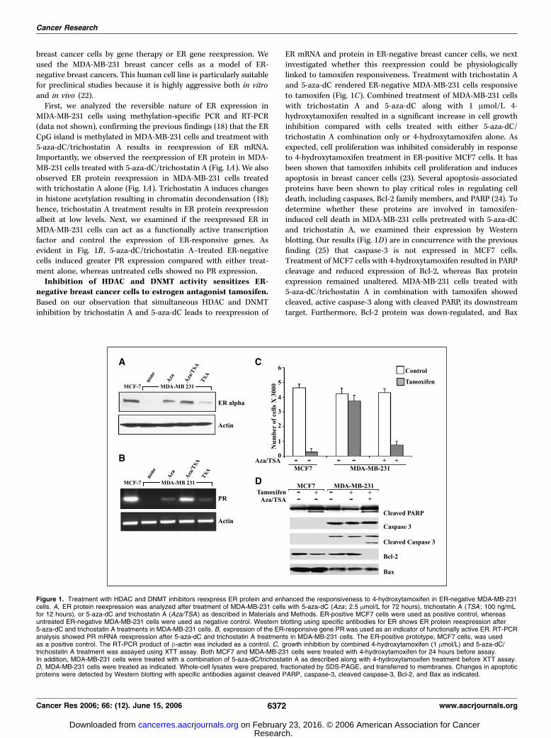
First, we analyzed the reversible nature of ER expression inMDA-MB-231 cells using methylation-specific PCR and RT-PCR(data not shown), confirming the previous findings (18) that the ERCpG island is methylated in MDA-MB-231 cells and treatment with5-aza-dC/trichostatin A results in reexpression of ER mRNA.Importantly, we observed the reexpression of ER protein in MDA-MB-231 cells treated with 5-aza-dC/trichostatin A (Fig. 1A). We alsoobserved ER protein reexpression in MDA-MB-231 cells treatedwith trichostatin A alone (Fig. 1A). Trichostatin A induces changesin histone acetylation resulting in chromatin decondensation (18);hence, trichostatin A treatment results in ER protein reexpressionalbeit at low levels. Next, we examined if the reexpressed ER inMDA-MB-231 cells can act as a functionally active transcriptionfactor and control the expression of ER-responsive genes. Asevident in Fig. 1B , 5-aza-dC/trichostatin A–treated ER-negativecells induced greater PR expression compared with either treat-ment alone, whereas untreated cells showed no PR expression.Inhibition of HDAC and DNMT activity sensitizes ER-
negative breast cancer cells to estrogen antagonist tamoxifen.Based on our observation that simultaneous HDAC and DNMTinhibition by trichostatin A and 5-aza-dC leads to reexpression of
ER mRNA and protein in ER-negative breast cancer cells, we nextinvestigated whether this reexpression could be physiologicallylinked to tamoxifen responsiveness. Treatment with trichostatin Aand 5-aza-dC rendered ER-negative MDA-MB-231 cells responsiveto tamoxifen (Fig. 1C). Combined treatment of MDA-MB-231 cellswith trichostatin A and 5-aza-dC along with 1 Amol/L 4-hydroxytamoxifen resulted in a significant increase in cell growthinhibition compared with cells treated with either 5-aza-dC/trichostatin A combination only or 4-hydroxytamoxifen alone. Asexpected, cell proliferation was inhibited considerably in responseto 4-hydroxytamoxifen treatment in ER-positive MCF7 cells. It hasbeen shown that tamoxifen inhibits cell proliferation and inducesapoptosis in breast cancer cells (23). Several apoptosis-associatedproteins have been shown to play critical roles in regulating celldeath, including caspases, Bcl-2 family members, and PARP (24). Todetermine whether these proteins are involved in tamoxifen-induced cell death in MDA-MB-231 cells pretreated with 5-aza-dCand trichostatin A, we examined their expression by Westernblotting. Our results (Fig. 1D) are in concurrence with the previousfinding (25) that caspase-3 is not expressed in MCF7 cells.Treatment of MCF7 cells with 4-hydroxytamoxifen resulted in PARPcleavage and reduced expression of Bcl-2, whereas Bax proteinexpression remained unaltered. MDA-MB-231 cells treated with5-aza-dC/trichostatin A in combination with tamoxifen showedcleaved, active caspase-3 along with cleaved PARP, its downstreamtarget. Furthermore, Bcl-2 protein was down-regulated, and Bax
Figure 1. Treatment with HDAC and DNMT inhibitors reexpress ER protein and enhanced the responsiveness to 4-hydroxytamoxifen in ER-negative MDA-MB-231cells. A, ER protein reexpression was analyzed after treatment of MDA-MB-231 cells with 5-aza-dC (Aza ; 2.5 Amol/L for 72 hours), trichostatin A (TSA ; 100 ng/mLfor 12 hours), or 5-aza-dC and trichostatin A (Aza/TSA ) as described in Materials and Methods. ER-positive MCF7 cells were used as positive control, whereasuntreated ER-negative MDA-MB-231 cells were used as negative control. Western blotting using specific antibodies for ER shows ER protein reexpression after5-aza-dC and trichostatin A treatments in MDA-MB-231 cells. B, expression of the ER-responsive gene PR was used as an indicator of functionally active ER. RT-PCRanalysis showed PR mRNA reexpression after 5-aza-dC and trichostatin A treatments in MDA-MB-231 cells. The ER-positive prototype, MCF7 cells, was usedas a positive control. The RT-PCR product of h-actin was included as a control. C, growth inhibition by combined 4-hydroxytamoxifen (1 Amol/L) and 5-aza-dC/trichostatin A treatment was assayed using XTT assay. Both MCF7 and MDA-MB-231 cells were treated with 4-hydroxytamoxifen for 24 hours before assay.In addition, MDA-MB-231 cells were treated with a combination of 5-aza-dC/trichostatin A as described along with 4-hydroxytamoxifen treatment before XTT assay.D, MDA-MB-231 cells were treated as indicated. Whole-cell lysates were prepared, fractionated by SDS-PAGE, and transferred to membranes. Changes in apoptoticproteins were detected by Western blotting with specific antibodies against cleaved PARP, caspase-3, cleaved caspase-3, Bcl-2, and Bax as indicated.
Cancer Research
Cancer Res 2006; 66: (12). June 15, 2006 6372 www.aacrjournals.org
Research. on February 23, 2016. © 2006 American Association for Cancercancerres.aacrjournals.org Downloaded from

protein expression remained unaltered. Our findings suggest that acombinatory treatment of ER-negative breast cancer cells with5-aza-dC/trichostatin A along with 4-hydroxytamoxifen induces celldeath as indicated by the activation of caspase-3 and cleavage ofPARP.Reexpression of ER in ER-negative cells renders ER-
responsive genes responsive to the antagonistic effects of 4-hydroxytamoxifen. Steroid receptors, such as ER, regulate genetranscription either by binding directly to the promoter of targetgenes or by binding indirectly through other transcription factors(1, 2). Genes regulated through direct ER binding, such as EBAG9and cathepsin D, typically harbor a classic hormone-responsiveelement (HRE) and are activated slowly (26, 27). In contrast, genesregulated by indirect binding of ER to nonclassic HREs are quicklyactivated, such as c-Myc and IGF-I (28, 29). We next examined the
difference in agonist-modulated versus antagonist-modulatedexpression of these genes in our experimental system. The ER-responsive genes showed a strong increase in expression in thepresence of E2 in ER-positive MCF7 cells, whereas tamoxifentreatment repressed the expression of these same genes (Fig. 2).ER-negative MDA-MB-231 cells exhibited no detectable expressionof EBAG9, whereas IGF-I showed very low levels of expression. ER-negative cells show robust levels of both cathepsin D and c-Mycsimilar to ER-positive cells. Importantly, the basal levels ofexpression were unaffected by both E2 and tamoxifen treatmentin ER-negative cells. Reexpression of ER in ER-negative cells with 5-aza-dC/trichostatin A treatment restored basal levels of expressionof ER-responsive genes EBAG9 and IGF-I and the expression wasinducible by E2 treatment. Treatment with tamoxifen inhibited theexpression of ER-responsive genes (Fig. 2). More importantly, ER-responsive genes that are constitutively expressed in MDA-MB-231cells and previously remained unaltered with either tamoxifen orE2 treatment became responsive to antagonist treatment in ER-negative cells reexpressing ER. Expression of both cathepsin D andc-Myc was inhibited strongly by tamoxifen treatment in MDA-MB-231 cells pretreated with 5-aza-dC/trichostatin A. These dataindicate that 5-aza-dC/trichostatin A pretreatment of ER-negativecells restores not only E2-dependent activation but also tamoxifen-dependent repression of ER target genes via reexpression of ER.Both HDAC2 and HDAC3 bind to the promoters of ER-
responsive genes in response to antagonist treatment.Recruitment of corepressors to the ER results in the formation ofmultisubunit corepressor complexes, including various HDACs,facilitating chromatin condensation (6, 9). To investigate thespecific corepressor complexes involved in antagonist-inducedrepression of ER-responsive genes in MDA-MB-231 cells reexpress-ing ER (MDA-MB-231/ER), we first sought to investigate thepattern of recruitment of various HDAC molecules on these ER-responsive gene promoters using chromatin immunoprecipitation.Treatment of 5-aza-dC/trichostatin A–pretreated MDA-MB-231cells with 4-hydroxytamoxifen induced a dramatic increase in theoccupancy of HDAC2 and HDAC3 at the EBAG9, cathepsin D,c-Myc, and IGF-I gene promoters (Fig. 3). In contrast, HDAC2 andHDAC3 were not recruited to the same genes in response to E2.Importantly, another member of class I HDACs, HDAC1, and
Figure 2. Expression levels of ER-responsive genes in ER-negative cellsreexpressing ER in response to E2 and 4-hydroxytamoxifen. MDA-MB-231cells were grown in estrogen-free medium and treated with 5-aza-dCand trichostatin A for 72 and 12 hours, respectively, to reexpress ERfollowed by fresh medium containing no treatment or treatment with E2 or4-hydroxytamoxifen (Tam ) for 2 hours as described in Materials and Methods.Total RNA was isolated, quantified, and subjected to cDNA synthesisfollowed by RT-PCR analysis using specific primer pairs. Differential modulationof expression of ER-responsive genes was observed in agonist- andantagonist-treated MDA-MB-231 cells reexpressing ER.
Figure 3. Profile of various HDAC molecules on the promoters of ER-responsive genes. Soluble chromatin was prepared from MDA-MB-231 cells pretreatedwith 5-aza-dC (2.5 Amol/L for 72 hours) and trichostatin A (100 ng/mL for 12 hours) followed by treatment with E2 (100 nmol/L for 2 hours), 4-hydroxytamoxifen(1 Amol/L for 2 hours), vehicle (�) as described in Materials and Methods and immunoprecipitated with 5 Ag specific antibodies against various class 1 and II HDACproteins overnight at 4jC. The immune complexes were pulled down with protein A agarose/salmon sperm DNA beads and washed extensively as described inMaterials and Methods, and cross-linking was reversed. The purified DNA was analyzed by PCR using primers spanning the EREs of EBAG9, cathepsin D, c-Myc,and IGF-I gene promoters. Recruitment of HDAC2 and HDAC3 was observed on all four promoters in response to tamoxifen treatment, whereas HDAC1, HDAC5,HDAC6, and HDAC7 were not recruited.
Sensitizing ER–Negative Breast Cancer Cells to Tamoxifen
www.aacrjournals.org 6373 Cancer Res 2006; 66: (12). June 15, 2006
Research. on February 23, 2016. © 2006 American Association for Cancercancerres.aacrjournals.org Downloaded from

certain members of class II HDACs, including HDAC5, HDAC6, andHDAC7, were not recruited to ER-responsive promoters (Fig. 3).Tamoxifen-bound reactivated ER recruits Mi2/NuRD core-
pressor complex but not Sin3A corepressor complex to ER-responsive genes. Biochemical studies from various laboratorieshave characterized three class I HDAC-containing corepressorcomplexes [i.e., the HDAC1/HDAC2-containing Sin3 (11) and Mi2/NuRD (12) complex and the HDAC3-containing NCoR complexes(13)]. Having shown that tamoxifen-bound reactivated ER is able toinduce occupancy of ER-responsive promoters by HDAC2 andHDAC3, we sought to determine which corepressor complexes arealso getting recruited to achieve active repression of these targetgenes. Chromatin immunoprecipitation analysis with specificantibodies to HDAC2 showed that tamoxifen-bound reactivatedER specifically recruits HDAC2 to the promoter region of EBAG9,cathepsin D, c-Myc, and IGF-I in an antagonist-dependent manner(Fig. 4A). Importantly, chromatin immunoprecipitation analysisusing specific antibodies to the components of Sin3 complex,mSin3A, SAP18, and SAP30 revealed that neither agonist norantagonist treatment of MDA-MB-231 cells reexpressing ERresulted in the recruitment of members of Sin3 complex at thepromoter region of ER-responsive genes (Fig. 4A). In contrast,reactivated ER recruited distinct components of NuRD complex inMDA-MB-231 cells treated with tamoxifen (Fig. 4B). 4-Hydroxyta-moxifen stimulated the ER-dependent recruitment of MTA1 andMi2 to ER-responsive promoters, whereas E2-bound reactivated ERdoes not induce recruitment of these proteins. Our results clearlyshow that antagonist-bound reactivated ER induces activerepression of target genes via engaging distinctive corepressorcomplexes. The recruitment of NuRD components occurredirrespective of the type of estrogen-responsive element (ERE) inthe various gene promoters.HDAC3 recruited by tamoxifen-bound reactivated ER is a
part of NCoR complex. Previous in vitro studies indicate that ERinteracts with NCoR and SMRT corepressors in the presence ofantagonist (14) and biochemical purification and characterizationof these complexes showed that both are associated with HDAC3(15). We thus sought to understand the participation of NCoRcorepressor complex in the mediation of active repression of ER-responsive genes via antagonist-bound reactivated ER. Interesting-ly, as observed for components of NuRD complex, the members ofNCoR complex, HDAC3, NCoR, and TBL1, associated with thepromoters of all ER-responsive genes included in this study in anantagonist-dependent manner (Fig. 4C). In contrast, E2 treatmentdid not induce recruitment of the NCoR complex components overIgG and vehicle controls. These data indicate a role of NCoRcomplex in active repression achieved by antagonist-boundreactivated ER in MDA-MB-231 cells.Tamoxifen-bound reactivated ER uses multiple corepressor
complexes. Having established the antagonist-specific recruitmentof the NuRD and NCoR corepressor complexes at the ER-responsive promoters by reactivated ER, we next investigated thepattern of recruitment of these complexes. The presence of twodifferent multiprotein corepressor complexes could be explainedby one of the following scenarios: both complexes get recruited tothe promoter region simultaneously either through differentanchor subunits or by a member of one multiprotein complexacting as an anchor for the other multiprotein complex. NCoR hasbeen found to interact with components of NuRD complex (30).Alternatively, the complexes may be recruited in an orderedmanner with one complex promoting or excluding the binding of
the other. To examine this, we used chromatin immunoprecipita-tion/reimmunoprecipitation to determine which subunits werepresent on the promoter at the same time. In these experiments,DNA bound by HDAC2 was immunoprecipitated using HDAC2antibodies. Then, both the precipitates and the supernatants weresubjected to reimmunoprecipitation with antibodies againstHDAC3, NCoR, or MTA1 representing the NCoR (HDAC3/NCoR)
Figure 4. Recruitment of HDAC2-Sin3, HDAC2-NuRD, and HDAC3-NCoRcorepressor complex to ER-responsive gene promoters. A, formaldehydecross-linked chromatin was immunoprecipitated with antibodies specific forHDAC2, mSin3A, SAP18, and SAP30 from MDA-MB-231 cells treated as inFig. 3. The immune complexes were pulled down with protein A agarose/salmonsperm DNA beads and washed extensively as described in Materials andMethods, and cross-linking was reversed. The purified DNA was analyzed byPCR using specific primers spanning the EREs of EBAG9, cathepsin D,c-Myc, and IGF-I gene promoters. Recruitment of signature subunits of theHDAC2-Sin3 complex was not observed. B, cross-linked chromatin-proteincomplexes were immunoprecipitated with antibodies specific for HDAC2, MTA2,Mi2, and MTA1 from MDA-MB-231 cells treated as indicated. The purifiedDNA was analyzed by PCR using primers spanning the EREs of EBAG9,cathepsin D, c-Myc, and IGF-I gene promoters. Recruitment of all four signaturesubunits of the HDAC2-NuRD complex was observed. C, formaldehydecross-linked chromatin was immunoprecipitated with antibodies specific forHDAC3, NCoR, and TBL1 from MDA-MB-231 cells treated as in Fig. 3. Theimmune complexes were pulled down with protein A agarose/salmon sperm DNAbeads and washed extensively as described in Materials and Methods,and cross-linking was reversed. The purified DNA was analyzed by PCR usingprimers spanning the EREs of EBAG9, cathepsin D, c-Myc, and IGF-I genepromoters. Recruitment of all three signature subunits of the NCoR-HDAC3complex was observed.
Cancer Research
Cancer Res 2006; 66: (12). June 15, 2006 6374 www.aacrjournals.org
Research. on February 23, 2016. © 2006 American Association for Cancercancerres.aacrjournals.org Downloaded from
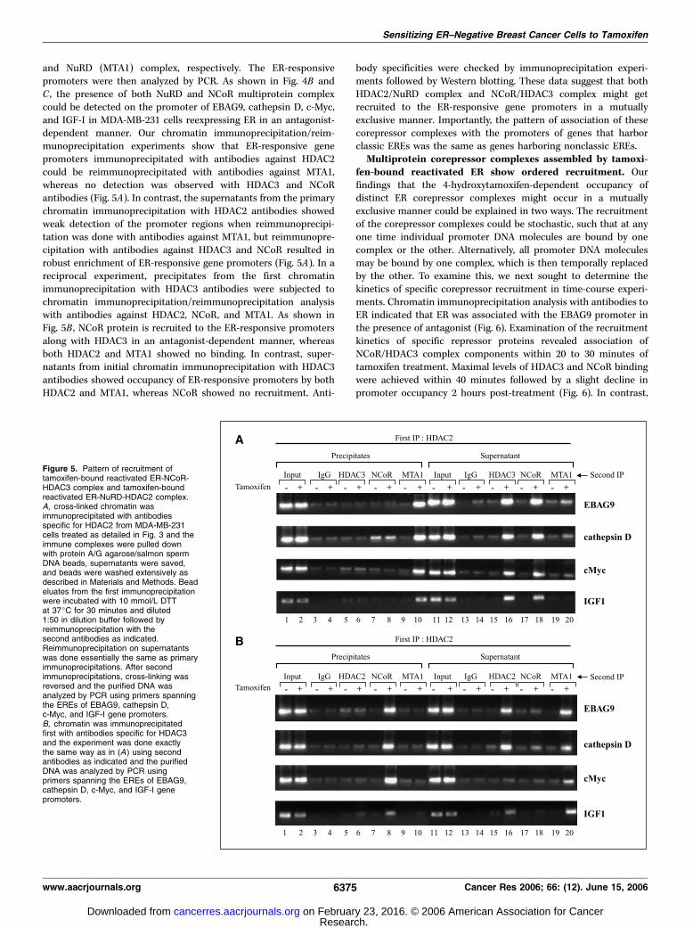
and NuRD (MTA1) complex, respectively. The ER-responsivepromoters were then analyzed by PCR. As shown in Fig. 4B andC , the presence of both NuRD and NCoR multiprotein complexcould be detected on the promoter of EBAG9, cathepsin D, c-Myc,and IGF-I in MDA-MB-231 cells reexpressing ER in an antagonist-dependent manner. Our chromatin immunoprecipitation/reim-munoprecipitation experiments show that ER-responsive genepromoters immunoprecipitated with antibodies against HDAC2could be reimmunoprecipitated with antibodies against MTA1,whereas no detection was observed with HDAC3 and NCoRantibodies (Fig. 5A). In contrast, the supernatants from the primarychromatin immunoprecipitation with HDAC2 antibodies showedweak detection of the promoter regions when reimmunoprecipi-tation was done with antibodies against MTA1, but reimmunopre-cipitation with antibodies against HDAC3 and NCoR resulted inrobust enrichment of ER-responsive gene promoters (Fig. 5A). In areciprocal experiment, precipitates from the first chromatinimmunoprecipitation with HDAC3 antibodies were subjected tochromatin immunoprecipitation/reimmunoprecipitation analysiswith antibodies against HDAC2, NCoR, and MTA1. As shown inFig. 5B , NCoR protein is recruited to the ER-responsive promotersalong with HDAC3 in an antagonist-dependent manner, whereasboth HDAC2 and MTA1 showed no binding. In contrast, super-natants from initial chromatin immunoprecipitation with HDAC3antibodies showed occupancy of ER-responsive promoters by bothHDAC2 and MTA1, whereas NCoR showed no recruitment. Anti-
body specificities were checked by immunoprecipitation experi-ments followed by Western blotting. These data suggest that bothHDAC2/NuRD complex and NCoR/HDAC3 complex might getrecruited to the ER-responsive gene promoters in a mutuallyexclusive manner. Importantly, the pattern of association of thesecorepressor complexes with the promoters of genes that harborclassic EREs was the same as genes harboring nonclassic EREs.Multiprotein corepressor complexes assembled by tamoxi-
fen-bound reactivated ER show ordered recruitment. Ourfindings that the 4-hydroxytamoxifen-dependent occupancy ofdistinct ER corepressor complexes might occur in a mutuallyexclusive manner could be explained in two ways. The recruitmentof the corepressor complexes could be stochastic, such that at anyone time individual promoter DNA molecules are bound by onecomplex or the other. Alternatively, all promoter DNA moleculesmay be bound by one complex, which is then temporally replacedby the other. To examine this, we next sought to determine thekinetics of specific corepressor recruitment in time-course experi-ments. Chromatin immunoprecipitation analysis with antibodies toER indicated that ER was associated with the EBAG9 promoter inthe presence of antagonist (Fig. 6). Examination of the recruitmentkinetics of specific repressor proteins revealed association ofNCoR/HDAC3 complex components within 20 to 30 minutes oftamoxifen treatment. Maximal levels of HDAC3 and NCoR bindingwere achieved within 40 minutes followed by a slight decline inpromoter occupancy 2 hours post-treatment (Fig. 6). In contrast,
Figure 5. Pattern of recruitment oftamoxifen-bound reactivated ER-NCoR-HDAC3 complex and tamoxifen-boundreactivated ER-NuRD-HDAC2 complex.A, cross-linked chromatin wasimmunoprecipitated with antibodiesspecific for HDAC2 from MDA-MB-231cells treated as detailed in Fig. 3 and theimmune complexes were pulled downwith protein A/G agarose/salmon spermDNA beads, supernatants were saved,and beads were washed extensively asdescribed in Materials and Methods. Beadeluates from the first immunoprecipitationwere incubated with 10 mmol/L DTTat 37jC for 30 minutes and diluted1:50 in dilution buffer followed byreimmunoprecipitation with thesecond antibodies as indicated.Reimmunoprecipitation on supernatantswas done essentially the same as primaryimmunoprecipitations. After secondimmunoprecipitations, cross-linking wasreversed and the purified DNA wasanalyzed by PCR using primers spanningthe EREs of EBAG9, cathepsin D,c-Myc, and IGF-I gene promoters.B, chromatin was immunoprecipitatedfirst with antibodies specific for HDAC3and the experiment was done exactlythe same way as in (A ) using secondantibodies as indicated and the purifiedDNA was analyzed by PCR usingprimers spanning the EREs of EBAG9,cathepsin D, c-Myc, and IGF-I genepromoters.
Sensitizing ER–Negative Breast Cancer Cells to Tamoxifen
www.aacrjournals.org 6375 Cancer Res 2006; 66: (12). June 15, 2006
Research. on February 23, 2016. © 2006 American Association for Cancercancerres.aacrjournals.org Downloaded from

recruitment of the NuRD complex components, HDAC2, Mi2, andMTA1, displayed considerably slower kinetics. Detectable occu-pancy of the EBAG9 promoter by NuRD components did not occuruntil 40 minutes after tamoxifen treatment, with maximal levelsoccurring 50 to 60 minutes post-treatment (Fig. 6). In summary, thedata in Fig. 6 suggest that tamoxifen-mediated repression of ER-responsive genes involves the sequential recruitment of multi-protein corepressor complexes with components of the NCoR/HDAC3 complex preceding that of NuRD/HDAC2 complex.Collectively, these result indicate that epigenetic reactivation ofthe ER gene in MDA-MB-231 cells sensitizes ER-negative cells tothe antagonistic effects of tamoxifen involving modulation ofexpression of ER-responsive genes via ordered recruitment ofdistinct corepressor complexes.
Discussion
The effects of endocrine therapy are primarily mediated throughthe ER; therefore, ER expression is a strong predictor of response toSERM treatment. Indeed, lack of ER expression is the dominantmechanism of de novo resistance to SERMs, such as tamoxifen(1–3). In addition, during breast cancer progression, many initiallyER-positive tumors lose ER expression and attain hormone un-responsiveness (4, 5). ER-negative tumors are more aggressive, andconsidering the ability of these tumors to metastasize and theirheterogeneity, new therapies or strategies for sensitization ofER-negative tumors to endocrine treatment are required.
Several enzymatic inhibitors targeting HDACs have beendeveloped with good in vivo bioavailability and intracellular
capability to inhibit HDAC. Preclinical studies and initial clinicaltrials indicate that HDAC inhibitors from different structuralclasses are very well tolerated and exhibit clinical activity against avariety of human cancers (31, 32). The hydroxamate trichostatin Awas shown previously to have an in vivo antitumor activity withdaily parenteral dosing associated with little systemic toxicity (33).The greatest potential of HDAC inhibitors lies in their ability tomodulate the activity of other therapeutic agents. Demethylatingagents, such as 5-aza-dC, are particularly interesting candidates,owing to the interaction of DNA methylation with histonedeacetylation in gene silencing of tumor suppressor genes.Combined treatment of trichostatin A or depsipeptide with 5-aza-dC has been shown to synergistically reactivate silenced tumorsuppressor genes in human cancer cells, including MLH1, TIMP3,CDKN2B, CDKN2A, gelsolin, and maspin (34, 35).
We have shown previously that treatment of ER-negative breastcancer cells with DNMT and HDAC inhibitors leads to reactivationof expression of functional ER (18). We explored this strategy forsensitizing ER-negative breast cancer cells to tamoxifen. We foundthat ER-negative breast cancer cells can be sensitized to antitumoreffects of tamoxifen by combined treatment with 5-aza-dC/trichostatin A, underscoring the importance of drugs having thepotential to derepress the expression of epigenetically silenced keygenes in cancer therapeutics. Previously, other laboratories havetransfected ERa recombinant cDNA into the MDA-MB-231 cells inan attempt to recover normal estrogen and antiestrogen respon-siveness. The unliganded ER was found to inhibit the invasivenessand growth of MDA-MB-231 cells transfected with ER (36).However, whereas E2 inhibited cell growth and invasiveness tosome extent, tamoxifen had no effect (37, 38), indicating thattransfecting ER into ER-negative breast cancer cells is not sufficientto sensitize these unresponsive cells to endocrine therapy. Inaddition, this approach is not directly translatable to clinicalstudies.
Tamoxifen is a potent ER antagonist and its pharmacology hasbeen reviewed extensively (39). Many studies focusing on themechanism of action of tamoxifen have indicated that thiscompound acts in both a cytostatic (causing G0-G1 arrest) and acytotoxic (inducing apoptosis) manner (40, 41). We show here thattamoxifen-induced growth inhibition of 5-aza-dC/trichostatin A–pretreated cells involves apoptosis as indicated by cleavage ofcaspase-3 and its downstream target PARP. Tamoxifen induceddown-regulation of Bcl-2 in MDA-MB-231 cells reexpressing ERwithout any alteration at Bax expression as reported earlier forMCF7 cells (42), indicating the involvement of Bcl-2 family intamoxifen-mediated cell death.
The findings that reactivation of ER can direct tamoxifen-dependent repression of endogenous ER target genes indicates that5-aza-dC/trichostatin A reactivated ER is able to interact with bothagonists and antagonists to modulate transcription. Tamoxifenmainly exerts its antiproliferative action by binding competitivelyto ER, modulating the ER-mediated transcriptional cascade.Agonist-bound ER adopts a conformation in which a-helices(3, 5, and 12) in the LBD form a hydrophobic cleft (AF-2), providinga binding surface for NR boxes (LXXLL motifs) in coactivators. ERantagonists, such as tamoxifen, have a bulky side chain thatsterically modulates the conformation of the hydrophobic cleft(AF-2) with helix 12 binding to the AF-2 cleft with its own intrinsicNR box, occluding the binding of coactivators. Antagonist-mediated inhibition of ER not only is a passive process resultingfrom repositioning of helix 12, thereby blocking the coactivator
Figure 6. Dynamics of assembly of tamoxifen-bound reactivated ER-NCoR-HDAC3 complex and tamoxifen-bound reactivated ER-NuRD-HDAC2 complex.Chromatin immunoprecipitation assays were done as described earlier.MDA-MB-231 cells were pretreated with 5-aza-dC (2.5 Amol/L for 72 hours) andtrichostatin A (100 ng/mL for 12 hours) as described in Materials and Methods,and cells were treated with 1 Amol/L 4-hydroxytamoxifen for varying lengthsof time as indicated above the lanes. Right, antibodies used for chromatinimmunoprecipitation. Recruitment of tamoxifen-bound reactivated ER toEBAG9 promoter was observed within 5 minutes of tamoxifen treatment, whereasNCoR-TBL1-HDAC3 complex gets recruited in 20 to 30 minutes followed by thebinding of the components of NuRD complex by 40 minutes of tamoxifentreatment.
Cancer Research
Cancer Res 2006; 66: (12). June 15, 2006 6376 www.aacrjournals.org
Research. on February 23, 2016. © 2006 American Association for Cancercancerres.aacrjournals.org Downloaded from

binding (10), but also involves the active recruitment ofcorepressors to form repressive ER complex at ER target genes.
To comprehend the molecular basis of repression of ER-responsive genes by tamoxifen-bound reactivated ER in ER-negative breast cancer cells, it is important to decipher the natureof corepressor complex involved in these antagonistic actions. Ourstudies with tamoxifen-bound reactivated ER show the formationof a distinct complex containing HDAC3, NCoR, and TBL1 onpromoter regions of ER-responsive genes. Other studies have alsoshown that HDAC3 is the major HDAC associated with NCoR/SMRT complexes and NCoR interacts directly with HDAC3 througha deacetylase-activating domain activating HDAC3 activity (15, 16).TBL1 then recognizes and binds the resultant deacetylated histonetails, further stabilizing the binding of this multiprotein complexleading to repression. TBL1 and TBLR1 are not required for HDAC3activity or initial binding of the NCoR/SMRT complex to nuclearreceptors, but they can interact with core histones to stabilize thebinding. This is similar to the role of RbAp46 and RbAp48 in NuRDcomplex. Whereas RbAp48 binds to H2A, H3, and H4, TBL1 bindpreferentially to H2B and H4. We observed binding of NuRDcomplex to the ER-responsive promoters in ER-negative breastcancer cells reexpressing functional ER, and this recruitment wasspecifically dependent on tamoxifen treatment as no binding wasobserved in the presence of estrogen.
Combinatorial utilization of multiple corepressor complexesmay be required to achieve physiologic levels of repression on somepromoters, whereas on other promoters different complexes mightget recruited independent of each other. NCoR directly interactswith nuclear receptors via its NR box-related conserved bipartiteNR interaction domain containing L/IXXI/VI sequence (16),anchoring NCoR/HDAC3 multiprotein complex. NCoR can alsointeract with components of both the SAP and the NuRDcomplexes (30), suggesting that NCoR and NuRD complexes couldbe corecruited to ER or other nuclear receptor gene targets. Ourchromatin immunoprecipitation/reimmunoprecipitation datashow that both NCoR/HDAC3 and NuRD complex bind to ER-responsive promoters containing either classic or nonclassic EREpromoters in a mutually exclusive manner. Mutually exclusivebinding of both NCoR and NuRD corepressor complexes rules outthe possibility of NCoR-mediated recruitment of NuRD complex atleast in tamoxifen-bound reactivated ER. Because human NuRDcomplex is a multisubunit protein complex, it is possible that itgets recruited using one of its own subunits as the anchoringprotein. Biochemical and immunofluorescence studies have shownthat MTA1 interacts directly with the ER (43). However, whetherMTA1 targets the NuRD complex to ER-responsive promoter hasnot been elucidated. Other candidate subunits of the NuRDcomplex are methyl-binding proteins, such as MBD2 and MBD3.Whereas human MBD3 does not recognize methylated DNA (44),MBD2 might direct the recruitment of NuRD complex tomethylated loci at target gene promoters (45). Our time-courseexperiments, however, suggest that a DNA methylation-mediatedmechanism is unlikely, as NuRD complex components were
observed at the EBAG9 promoter within 40 minutes of tamoxifentreatment. In addition, NuRD complex purified with HDAC1contains MBD2 (46), whereas a similar immunoaffinity purificationof HDAC2 generated a NuRD complex with no detectable MBD2(46). Our data show the recruitment of HDAC2 but not HDAC1containing NuRD complex at the ER-responsive promoters,suggesting that MBD2 is not involved in tamoxifen-mediatedrepression by reactivated ER. Further studies are needed to clearlyshow how NuRD complex binds to tamoxifen-bound reactivatedER and mediate repression of ER-responsive genes.
Our findings also provide evidence for an ordered recruitment ofNCoR complex followed by NuRD complex at distinct ER targetpromoters in ER-negative breast cancer cells via tamoxifen-boundreexpressed ER. Sequential recruitment of various cofactors hasbeen reported for regulation of various mammalian genes (19, 20).Given the ordered recruitment of corepressor complexes shownhere, our findings support a multistep model of tamoxifen-mediated repression by reactivated ER. NCoR complex coulddirectly interact with tamoxifen-bound reactivated ER resulting indeacetylation of local histones through recruitment of HDACactivity (15, 16). One possibility is that removal of the acetyl groupsfrom K9 and K14 of histone H3 (47) creates an environment thatpromotes the binding of Suv39H1/Clr4. The methylation of H3-K9by Suv39H1/Clr4 after HDACs remove the acetyl groups from K9and K14 of histone H3 (47) then serves as a binding site for thechromodomain of HP1/Swi6 (48, 49). NuRD complex contains Mi2/CHD family proteins that have a chromodomain (12) andbiochemical analysis have shown that the NuRD complexassociates with histone H3 when Lys9 is methylated (50). Thismodel is in accordance with the histone code hypothesis as thepattern of histone tail modifications serves as a recognition codefor the recruitment of cofactors resulting in modulation ofchromatin structure and function.
In conclusion, our findings are clinically important, as endocrinetherapy targeting ER has proven its efficacy with the developmentof antiestrogens and aromatase inhibitors. Sensitizing hormone-resistant ER-negative breast cancer cells to endocrine therapy bycombined treatment with DNMT inhibitors and HDAC inhibitorsprovides new treatment options for patients with de novoresistance. In addition, the elucidation of the specific corepressorcomplexes involved in the tamoxifen-bound reactivated ER-mediated repression of endogenous ER-responsive genes mighthelp in designing more combined therapies using other therapeuticagents and innovative drug delivery strategies.
Acknowledgments
Received 1/31/2006; revised 4/5/2006; accepted 4/11/2006.Grant support: U.S. Army Medical Research and Material Command W81WXH-04-
BC-030963, Susan G. Komen Breast Cancer Research Foundation grant BCTR 0503526(D. Sharma), and NIH grants CA88843 (N.E. Davidson) and CA077337 (P.M. Vertino).
The costs of publication of this article were defrayed in part by the payment of pagecharges. This article must therefore be hereby marked advertisement in accordancewith 18 U.S.C. Section 1734 solely to indicate this fact.
References
1. Platet N, Cathiard AM, Gleizes M, Garcia M. Estrogensand their receptors in breast cancer progression: a dualrole in cancer proliferation and invasion. Crit Rev OncolHematol 2004;51:55–67.
2. Klinge CM. Estrogen receptor interaction withestrogen response elements. Nucleic Acids Res 2001;29:2905–19.3. Johnston SR, Dowsett M, Smith IE. Towards amolecular basis for tamoxifen resistance in breastcancer. Ann Oncol 1992;3:503–11.
4. Roodi N, Bailey LR, Kao WY, et al. Estrogen receptorgene analysis in estrogen receptor-positive and estrogenreceptor negative primary breast cancer. J Natl CancerInst 1995;87:446–51.5. Ottaviano YL, Issa J-P, Parl FF, Smith HS, Baylin SB,Davidson NE. Methylation of the estrogen receptor
Sensitizing ER–Negative Breast Cancer Cells to Tamoxifen
www.aacrjournals.org 6377 Cancer Res 2006; 66: (12). June 15, 2006
Research. on February 23, 2016. © 2006 American Association for Cancercancerres.aacrjournals.org Downloaded from

Cancer Research
Cancer Res 2006; 66: (12). June 15, 2006 6378 www.aacrjournals.org
gene CpG island marks loss of estrogen receptorexpression in human breast cancer cells. Cancer Res1994;54:2552–5.6. Glass CK, Rosenfeld MG. The coregulator exchange intranscriptional functions of nuclear receptors. GenesDev 2000;14:121–41.7. Spencer TE, Jenster G, Burcin MM, et al. Steroidreceptor coactivator-1 is a histone acetyltransferase.Nature 1997;389:194–8.8. Zamir I, Harding HP, Atkins GB, et al. A nuclearhormone receptor corepressor mediates transcriptionalsilencing by receptors with distinct repression domains.Mol Cell Biol 1996;16:5458–65.9. Dobrzycka KM, Townson SM, Jiang S, Oesterreich S.Estrogen receptor corepressors—a role in human breastcancer. Endocr Relat Cancer 2003;10:517–36.10. Shiau AK, Barstad D, Loria PM, et al. The structuralbasis of estrogen receptor/coactivator recognition andthe antagonism of this interaction by tamoxifen. Cell1998;95:927–37.11. Zhang Y, Iratni R, Erdjument-Bromage H, Tempst P,Reinberg D. Histone deacetylases and SAP18, a novelpolypeptide, are components of a human Sin3 complex.Cell 1997;89:357–64.12. Xue Y, Wong J, Moreno GT, Young MK, Cote J, WangW. NURD, a novel complex with both ATP-dependentchromatin-remodeling and histone deacetylase activi-ties. Mol Cell 1998;2:851–61.13. Horlein AJ, Naar AM, Heinzel T, et al. Ligand-independent repression by the thyroid hormone recep-tor mediated by a nuclear receptor co-repressor. Nature1995;377:397–404.14. Smith CL, Nawaz Z, O’Malley BW. Coactivator andcorepressor regulation of the agonist/antagonist activityof the mixed antiestrogens, 4-hydroxytamoxifen. MolEndocrinol 1997;11:657–66.15. Li J, Wang J, Nawaz Z, Liu JM, Qin J, Wong J. Bothcorepressor proteins SMRT and NCoR exist in largeprotein complexes containing HDAC3. EMBO J 2000;19:4342–50.16. Yoon HG, Chan DW, Huang ZQ, et al. Purification andfunctional characterization of the human NCoR com-plex: the roles of HDAC3, TBL1 and TBLR1. EMBO J2003;22:1336–46.17. Yang X, Phillips D, Ferguson AT, Nelson WG,Herman JG, Davidson NE. Synergistic activation offunctional estrogen receptor (ER)-a by DNA methyl-transferase and histone deacetylase inhibition inhuman ER-a-negative breast cancer cells. Cancer Res2001;61:7025–9.18. Sharma D, Blum J, Yang X, Beaulieu N, MacleodAR, Davidson NE. Release of methyl CpG bindingproteins and histone deacetylase 1 from the estrogenreceptor a (ER) promoter upon reactivation in ER-negative human breast cancer cells. Mol Endocrinol2005;19:1740–51.19. Sharma D, Fondell JD. Temporal formation of distinctthyroid hormone receptor coactivator complexes inHeLa cells. Mol Endocrinol 2000;14:2001–9.
20. Sharma D, Fondell JD. Ordered recruitment ofhistone acetyltransferases and the TRAP/Mediatorcomplex to thyroid hormone-responsive promotersin vivo . Proc Natl Acad Sci U S A 2002;99:7934–9.21. Sheikh MS, Garcia M, Pujol P, Fontana JA, RochefortH. Why are estrogen-receptor-negative breast cancersmore aggressive than the estrogen-receptor-positivebreast cancers? Invasion Metastasis 1995;14:329–36.22. Price JE, Polyzos A, Zhang RD, Daniels MD.Tumorigenicity and metastasis of human breastcarcinoma cell lines in nude mice. Cancer Res 1990;50:717–21.23. Mandlekar S, Kong ANT. Mechanisms of tamoxifen-induced apoptosis. Apoptosis 2001;6:469–77.24. Gross A, McDonnell JM, Korsmeyer SJ. BCL-2 familymembers and the mitochondria in apoptosis. Genes Dev1999;13:1899–911.25. Li F, Srinivasan A, Wang Y, Armstrong RC, TomaselliKJ, Fritz LC. Cell-specific induction of apoptosis bymicroinjection of cytochrome c . Bcl-xL has activityindependent of cytochrome c release. J Biol Chem 1997;272:30299–305.26. Tsuchiya F, Ikeda K, Tsutsumi O, et al. Molecularcloning and characterization of mouse EBAG9, homologof a human cancer associated surface antigen: expres-sion and regulation by estrogen. Biochem Biophys ResCommun 2001;284:2–10.27. Augereau P, Miralles F, Cavailles V, Gaudelet C, ParkerM, Rochefort H. Characterization of the proximalestrogen-responsive element of human cathepsin Dgene. Mol Endocrinol 1994;51:693–703.28. Dubik D, Shiu RP. Mechanism of estrogen acti-vation of c-myc oncogene expression. Oncogene 1992;7:1587–94.29. Umayahara Y, Kawamori R, Watada H, et al. Estrogenregulation of the insulin-like growth factor I genetranscription involves an AP-1 enhancer. J Biol Chem1994;269:16433–42.30. Li J, Lin Q, Wang W, Wade P, Wong J. Specifictargeting and constitutive association of histone deace-tylase complexes during transcriptional repression.Genes Dev 2002;16:687–92.31. Gray SG, Ekstrom TJ. The human histone deacetylasefamily. Exp Cell Res 2001;262:75–83.32. de Ruijter AJ, van Gennip AH, Caron HN, Kemp S, vanKuilenburg AB. Histone deacetylase (HDACs): charac-terization of the classical HDAC family. Biochem J 2003;370:737–49.33. Vigushin DM, Ali S, Pace PE, et al. Trichostatin A is ahistone deacetylase inhibitor with potent antitumoractivity against breast cancer in vivo . Clin Cancer Res2001;7:971–6.34. Cameron EE, Bachman KE, Myohanen S, Herman JG,Baylin SB. Synergy of demethylation and histonedeacetylase inhibition in the re-expression of genessilenced in cancer. Nat Genet 1999;21:103–7.35. Primeau M, Gagnon J, Momparler RL. Synergisticantineoplastic action of DNA methylation inhibitor 5-aza-2¶-deoxycytidine and histone deacetylase inhibitor
depsipeptide on human breast carcinoma cells. Int JCancer 2003;103:177–84.36. Platet N, Cunat S, Chalbos D, Rochefort H, Garcia M.Unliganded and liganded estrogen receptors protectagainst cancer invasion via different mechanisms. MolEndocrinol 2000;14:999–1009.37. Garcia M, Derocq D, Freiss G, Rochefort H.Activation of estrogen receptor transfected into areceptor-negative breast cancer cell line decreases themetastatic and invasive potential of the cells. Proc NatlAcad Sci U S A 1992;89:11538–42.38. Jiang SY, Jordon VC. Growth regulation of estrogenreceptor-negative breast cancer cells transfected withcomplementary DNAs for estrogen receptor. J NatlCancer Inst 1992;84:580–91.39. Furr BJA, Jordan VC. The pharmacology and clinicaluses of tamoxifen. Pharmacol Ther 1984;25:127–205.40. Chen HM, Tritton TR, Kenny N, Absher M, Chiu JF.Tamoxifen induces TGF h1 activity and apoptosis ofhuman MCF-7 breast cancer cells in vitro . J CellBiochem 1996;61:9–17.41. Osbourne CK, Boldt DH, Clark GM, Trent JM. Effectsof tamoxifen on human breast cancer cell cycle kinetics:accumulation of cells in early G1 phase. Cancer Res1983;43:3583–5.42. Zhang GJ, Kimijima I, Onda M. Tamoxifen-inducedapoptosis in breast cancer cells relates to down-regulation of bcl-2, but not bax and bcl-X(L), withoutalteration of p53 protein levels. Clin Cancer Res 1999;5:2971–7.43. Hendrich B, Bird A. Identification and characteriza-tion of a family of mammalian methyl-CpG bindingproteins. Mol Cell Biol 1998;18:6538–47.44. Zhang Y, Ng HH, Erdjument-Bromage H, Tempst P,Bird A, Reinberg D. Analysis of the NuRD subunitsreveals a histone deacetylase core complex and aconnection with DNA methylation. Genes Dev 1999;13:1924–35.45. Humphrey GW, Wang Y, Russanova VR, et al. Stablehistone deacetylase complexes distinguished by thepresence of SANT domain proteins CoREST/kiaa0071and mta-L1. J Biol Chem 2001;276:6817–24.46. Mazumdar A, Wang RA, Mishra SK, et al. Transcrip-tional repression of oestrogen receptor by metastasis-associated protein 1 corepressor. Nat Cell Biol 2001;3:30–7.47. Rea S, Eisenhaber F, O’Carroll D, et al. Regulation ofchromatin structure by site-specific histone H3 methyl-transferases. Nature 2000;406:593–9.48. Bannister AJ, Zegerman P, Partridge JF, et al. Selectiverecognition of methylated lysine 9 on histone H3 by theHP1 chromodomain. Nature 2001;410:120–4.49. Lachner M, O’Carroll D, Rea S, Mechtler K, JenuweinT. Methylation of histone H3 lysine 9 creates a bindingsite for HP1 proteins. Nature 2001;410:116–20.50. Zegerman P, Canas B, Pappin D, Kouzarides T.Histone H3 lysine 4 methylation disrupts binding ofnucleosome remodeling and deacetylase (NuRD) repres-sor complex. J Biol Chem 2002;277:11621–4.
Research. on February 23, 2016. © 2006 American Association for Cancercancerres.aacrjournals.org Downloaded from

2006;66:6370-6378. Cancer Res Dipali Sharma, Neeraj K. Saxena, Nancy E. Davidson, et al. ER Recruits Distinctive Corepressor ComplexesNegative Breast Cancer Cells: Tamoxifen-Bound Reactivated
−Restoration of Tamoxifen Sensitivity in Estrogen Receptor
Updated version
http://cancerres.aacrjournals.org/content/66/12/6370
Access the most recent version of this article at:
Cited articles
http://cancerres.aacrjournals.org/content/66/12/6370.full.html#ref-list-1
This article cites 50 articles, 25 of which you can access for free at:
Citing articles
http://cancerres.aacrjournals.org/content/66/12/6370.full.html#related-urls
This article has been cited by 21 HighWire-hosted articles. Access the articles at:
E-mail alerts related to this article or journal.Sign up to receive free email-alerts
Subscriptions
Reprints and
To order reprints of this article or to subscribe to the journal, contact the AACR Publications
Permissions
To request permission to re-use all or part of this article, contact the AACR Publications
Research. on February 23, 2016. © 2006 American Association for Cancercancerres.aacrjournals.org Downloaded from