Alternative ERK5 regulation by phosphorylation during the cell cycle
BioMed CentralBMC Molecular Biology
ss
Open AcceResearch articlePhosphorylation at Ser473 regulates heterochromatin protein 1 binding and corepressor function of TIF1beta/KAP1Chiung-Wen Chang1, Han-Yi Chou1, Yu-Sheng Lin1, Kuo-Hsiang Huang1, Ching-Jin Chang2, Tsui-Chun Hsu1 and Sheng-Chung Lee*1,2,3Address: 1Institute of Molecular Medicine, College of Medicine, National Taiwan University, Taipei, Taiwan, 2Institute of Biological Chemistry, Academia Sinica, Taipei, Taiwan and 3Institute of Clinical Medicine, College of Medicine, National Taiwan University, Taipei, Taiwan
Email: Chiung-Wen Chang - [email protected]; Han-Yi Chou - [email protected]; Yu-Sheng Lin - [email protected]; Kuo-Hsiang Huang - [email protected]; Ching-Jin Chang - [email protected]; Tsui-Chun Hsu - [email protected]; Sheng-Chung Lee* - [email protected]
* Corresponding author
AbstractBackground: As an epigenetic regulator, the transcriptional intermediary factor 1β (TIF1β)/KAP1/TRIM28) has been linked to gene expression and chromatin remodeling at specific loci byassociation with members of the heterochromatin protein 1 (HP1) family and various otherchromatin factors. The interaction between TIF1β and HP1 is crucial for heterochromatinformation and maintenance. The HP1-box, PXVXL, of TIF1β is responsible for its interaction withHP1. However, the underlying mechanism of how the interaction is regulated remains poorlyunderstood.
Results: This work demonstrates that TIF1β is phosphorylated on Ser473, the alteration of whichis dynamically associated with cell cycle progression and functionally linked to transcriptionalregulation. Phosphorylation of TIF1β/Ser473 coincides with the induction of cell cycle gene cyclinA2 at the S-phase. Interestingly, chromatin immunoprecipitation demonstrated that the promoterof cyclin A2 gene is occupied by TIF1β and that such occupancy is inversely correlated with Ser473phosphorylation. Additionally, when HP1β was co-expressed with TIF1β/S473A, but not TIF1β/S473E, the colocalization of TIF1β/S473A and HP1β to the promoters of Cdc2 and Cdc25A wasenhanced. Non-phosphorylated TIF1β/Ser473 allowed greater TIF1β association with theregulatory regions and the consequent repression of these genes. Consistent with possibleinhibition of TIF1β's corepressor function, the phosphorylation of the Ser473 residue, which islocated near the HP1-interacting PXVXL motif, compromised the formation of TIF1β-HP1complex. Finally, we found that the phosphorylation of TIF1β/Ser473 is mediated by the PKCδpathway and is closely linked to cell proliferation.
Conclusion: The modulation of HP1β-TIF1β interaction through the phosphorylation/de-phosphorylation of TIF1β/Ser473 may constitute a molecular switch that regulates the expressionof particular genes. Higher levels of phosphorylated TIF1β/Ser473 may be associated with theexpression of key regulatory genes for cell cycle progression and the proliferation of cells.
Published: 1 July 2008
BMC Molecular Biology 2008, 9:61 doi:10.1186/1471-2199-9-61
Received: 9 April 2008Accepted: 1 July 2008
This article is available from: http://www.biomedcentral.com/1471-2199/9/61
© 2008 Chang et al; licensee BioMed Central Ltd. This is an Open Access article distributed under the terms of the Creative Commons Attribution License (http://creativecommons.org/licenses/by/2.0), which permits unrestricted use, distribution, and reproduction in any medium, provided the original work is properly cited.
Page 1 of 16(page number not for citation purposes)

BMC Molecular Biology 2008, 9:61 http://www.biomedcentral.com/1471-2199/9/61
BackgroundTranscriptional intermediary factor TIF1β and heterochro-matin protein 1 profoundly impact the regulation of thestructure and function of chromatin [1]. The heterochro-matin protein 1 (HP1) family of proteins (HP1α, HP1β,and HP1γ) participates in gene silencing by forming hete-rochromatic structures [2,3]. HP1 exhibits distinct nuclearlocalization patterns: HP1α nassociates with centromereswhile HP1β and HP1γ are largely localized in distinctnuclear regions. The nuclear arrangement of HP1 proteinsand TIF1β is differentiation pathway-specific, and appearsto be more important than changes in the levels of theseproteins, which are relatively stable during all of theinduced differentiation processes [4,5].
HP1 proteins comprise an N-terminal chromodomain, aC-terminal chromoshadow domain and a hinge domain[3,6,7]. The chromodomain functions as a protein inter-action domain, bringing together different proteins inmulti-protein complexes and recruiting them to hetero-chromatin. This domain binds to particular proteinswhich contain an HP1 box (PXVXL) and function at thetranscriptional level [2,8-10]. The chromoshadowdomain mediates the formation of homodimer. TIF1βinteracts with the chromodomain of dimeric HP1β via theHP1 box of TIF1β in the HP1-interaction domain [6]. TheTIF1β ntranscriptional repression activity depends on theinteraction between TIF1β and HP1 [11]. This interactionis essential in the relocation of TIF1β from euchromatin toheterochromatin that accompanies the differentiation ofprimitive endoderm-like cells [12]. TIF1β is proposed tofunction as a universal co-repressor protein for the KRABzinc finger protein (KRAB-zfp) superfamily of transcrip-tional repressors. The recruitment of HP1 proteins by theKRAB-TIF1β complex to specific loci within the genomevia the formation of heterochromatin-like complexes maythus silence gene activity. Gene-specific repression may bea consequence of the formation of such complexes [13].
It has been reported that TIF1β directly interacts with thehistone methyltransferase SETDB1, which methylates spe-cifically histone H3/Lys9 within euchromatin [14]. Deple-tion of endogenous levels of TIF1β by siRNA significantlyinhibited the KRAB-mediated transcriptional repressionof a chromatin template and inhibited cell cycle progres-sion. Cell death may occur if TIF1β is severely depleted bysiRNA knockdown [unpublished observations, [15-17]].Similarly, the knock-down of cellular levels of HP1 pro-teins and SETDB1 by siRNA attenuated KRAB-TIF1βrepression. The physiological targets and functions ofTIF1β remain unclear. Its interactions with chromatinmodification factors such as HDAC1, SETDB1 and HP1correlate with its activities in regulating the chromatinstructure and heterochromatin formation, resulting inepigenetic silencing of reporter genes [18]. A TIF1β-con-
taining multiprotein complex regulates various proteinactivities or genes [19]. The activity of TIF1β may be regu-lated by posttranslational modifications. For example,TIF1β is rapidly phosphorylated by members of the phos-phatidylinositol-3 kinase-like family of kinases followingDNA damage. Phosphorylated TIF1β colocalizes withnumerous damage response factors at DNA lesions [20].Recent genetic and proteomic studies in mice have shownthat TIF1β is a developmental regulatory protein perform-ing cellular function(s) that are critical to early embryonicdevelopment, and is a component of the interactive pro-tein network for the pluripotency of embryonic stem cells[21,22]. The binding of the TIF1β corepressor to the retro-virus primer binding site is responsible for the epigeneticsilencing of retrovirus transcription [17]. Therefore, theinteraction between TIF1β and HP1 or other transcriptionfactors may account for the regulation of gene expression.
Phosphorylation of both human and mouse TIF1β/Ser473 has been identified by nuclear phosphoproteinanalysis of HeLa and WEHI-231 cells [23,24]. Ser473 islocated in the HP1-interacting domain of TIF1β – close tothe HP1 box (amino acids 486–490, PXVXL). The conser-vation of TIF1β/Ser473 phosphorylation in various celllines from different species motivated this investigation ofthe functional significance of this modification.
The coil-coiled domain of TIF1β binds to E2F1 and inhib-its its activity [16]. The induction of cyclin A, Cdc2 andCdc25A genes depends on E2F [25,26]. Cyclin A is a cellcycle-regulating protein that participates in S-phase con-trol and mitosis in mammalian somatic cells. The pro-moter of cyclin A is repressed during the G1 phase of thecell cycle and is activated at S-phase entry [27]. Cdc2 per-mits the transition from G1 through S in conjunction withcyclin E [28]. These E2F downstream genes are importantto normal cell cycle progression [29].
This report shows that TIF1β participates in the regulationof cyclin A2, Cdc2 and Cdc25A gene expression. This regu-lation depends on the interaction between TIF1β andHP1β, which is itself regulated by the phosphorylationstate of TIF1β/Ser473. The experimental results suggestthat the TIF1β/Ser473-unphosphorylated form bindsmore strongly than the phosphorylated form to the pro-moters of Cyclin A2, Cdc2, and Cdc25A genes. The phos-phorylation/de-phosphorylation of TIF1β/Ser473 mayserve as a molecular switch regulating its interaction withHP1β and gene expression.
ResultsCharacterization of TIF1β and phosphorylated TIF1β/Ser473 antibodiesWestern blotting of interphase HeLa cell extract was per-formed to characterize the specificity of monoclonal anti-
Page 2 of 16(page number not for citation purposes)
BMC Molecular Biology 2008, 9:61 http://www.biomedcentral.com/1471-2199/9/61
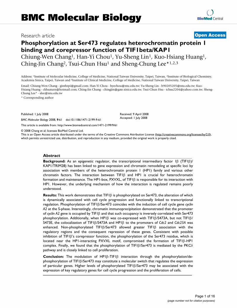
body to TIF1β, clone 20A1. A specific band of 100 kDawas detected (Figure 1A). 293T cell-expressed FLAG-TIF1βwas recognized either by 20A1 or monoclonal anti-FLAGantibody, M2 (Figure 1A), demonstrating that mono-clonal antibody 20A1 is specific to TIF1β.
The specificity of rabbit anti-phospho-Ser473 antibodywas characterized by Western blotting of interphase 293Tcell extract with rabbit anti-TIF1β/phospho-Ser473 anti-body (S473) or 20A1 monoclonal antibody. The phos-phorylated TIF1β/Ser473 signal was diminished after calfintestine alkaline phosphatase (CIP) treatment (Figure1B). Ectopically-expressed FLAG-TIF1β was immunopre-cipitated from 293T cells with M2 beads and treated withCIP to examine whether the signal was due to the phos-phorylation of TIF1β/S473. 20A1 and S473 antibodiesboth recognized FLAG-TIF1β, but the signal recognized bythe S473 antibody disappeared upon CIP treatment (Fig-ure 1C). Unlike the FLAG-TIF1β, mutants FLAG-TIF1β/S473A and FLAG-TIF1β/S473E were not detected by S473antibody (Figure 1C). These data demonstrate that while20A1 or S473 antibodies recognize the same protein,TIF1β, the S473 antibody specifically recognizes phos-phorylated TIF1β/S473.
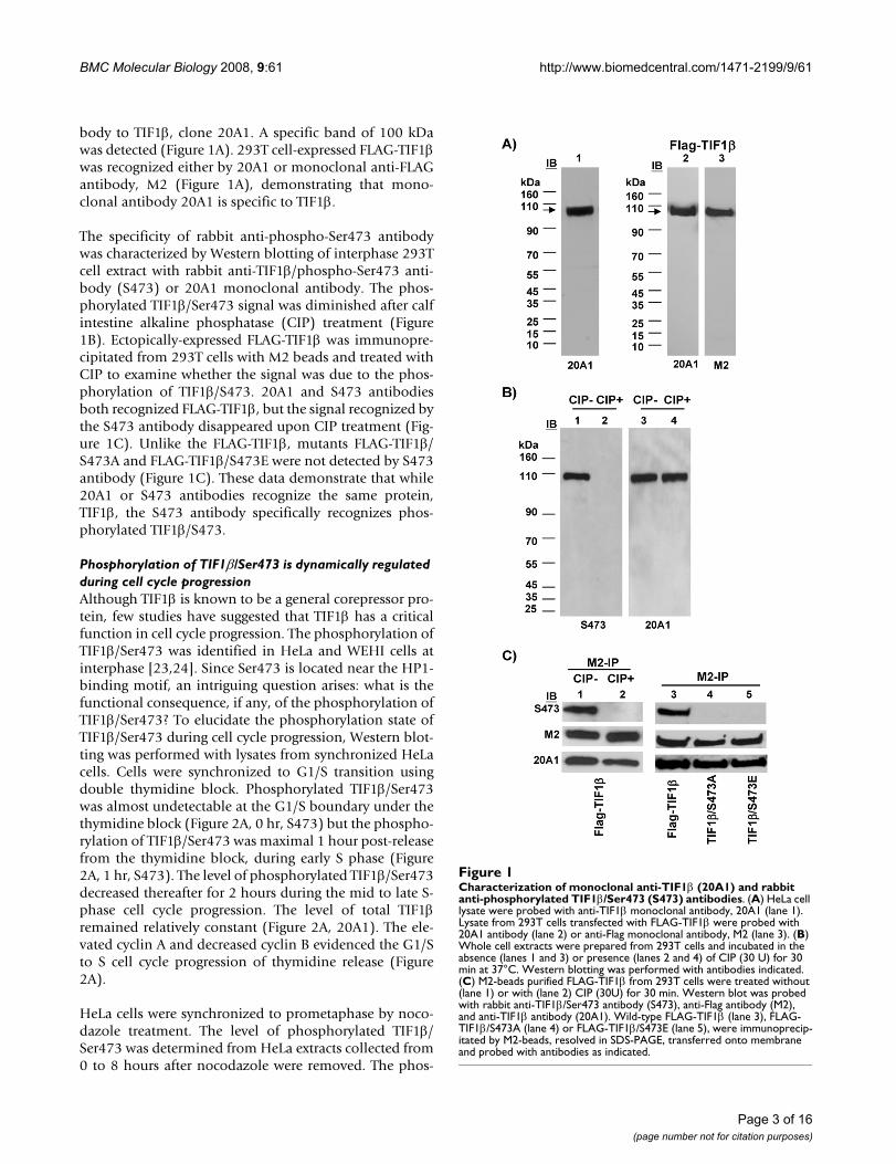
Phosphorylation of TIF1β/Ser473 is dynamically regulated during cell cycle progressionAlthough TIF1β is known to be a general corepressor pro-tein, few studies have suggested that TIF1β has a criticalfunction in cell cycle progression. The phosphorylation ofTIF1β/Ser473 was identified in HeLa and WEHI cells atinterphase [23,24]. Since Ser473 is located near the HP1-binding motif, an intriguing question arises: what is thefunctional consequence, if any, of the phosphorylation ofTIF1β/Ser473? To elucidate the phosphorylation state ofTIF1β/Ser473 during cell cycle progression, Western blot-ting was performed with lysates from synchronized HeLacells. Cells were synchronized to G1/S transition usingdouble thymidine block. Phosphorylated TIF1β/Ser473was almost undetectable at the G1/S boundary under thethymidine block (Figure 2A, 0 hr, S473) but the phospho-rylation of TIF1β/Ser473 was maximal 1 hour post-releasefrom the thymidine block, during early S phase (Figure2A, 1 hr, S473). The level of phosphorylated TIF1β/Ser473decreased thereafter for 2 hours during the mid to late S-phase cell cycle progression. The level of total TIF1βremained relatively constant (Figure 2A, 20A1). The ele-vated cyclin A and decreased cyclin B evidenced the G1/Sto S cell cycle progression of thymidine release (Figure2A).
HeLa cells were synchronized to prometaphase by noco-dazole treatment. The level of phosphorylated TIF1β/Ser473 was determined from HeLa extracts collected from0 to 8 hours after nocodazole were removed. The phos-
Characterization of monoclonal anti-TIF1β (20A1) and rabbit anti-phosphorylated TIF1β/Ser473 (S473) antibodiesFigure 1Characterization of monoclonal anti-TIF1β (20A1) and rabbit anti-phosphorylated TIF1β/Ser473 (S473) antibodies. (A) HeLa cell lysate were probed with anti-TIF1β monoclonal antibody, 20A1 (lane 1). Lysate from 293T cells transfected with FLAG-TIF1β were probed with 20A1 antibody (lane 2) or anti-Flag monoclonal antibody, M2 (lane 3). (B) Whole cell extracts were prepared from 293T cells and incubated in the absence (lanes 1 and 3) or presence (lanes 2 and 4) of CIP (30 U) for 30 min at 37°C. Western blotting was performed with antibodies indicated. (C) M2-beads purified FLAG-TIF1β from 293T cells were treated without (lane 1) or with (lane 2) CIP (30U) for 30 min. Western blot was probed with rabbit anti-TIF1β/Ser473 antibody (S473), anti-Flag antibody (M2), and anti-TIF1β antibody (20A1). Wild-type FLAG-TIF1β (lane 3), FLAG-TIF1β/S473A (lane 4) or FLAG-TIF1β/S473E (lane 5), were immunoprecip-itated by M2-beads, resolved in SDS-PAGE, transferred onto membrane and probed with antibodies as indicated.
Page 3 of 16(page number not for citation purposes)
BMC Molecular Biology 2008, 9:61 http://www.biomedcentral.com/1471-2199/9/61
Page 4 of 16(page number not for citation purposes)
Phosphorylation of TIF1β/Ser473 is dynamically regulated during cell cycle progressionFigure 2Phosphorylation of TIF1β/Ser473 is dynamically regulated during cell cycle progression. (A) HeLa cells were arrested at G1/S boundary by double thymidine block and released into S phase for three hours. The levels of TIF1β and phosphorylated TIF1β/Ser473 were probed with 20A1 and S473 antibodies. The protein levels of cyclin A, cyclin B1, or α-tubulin were also shown. (B) HeLa cells were synchronized by nocodazole (1 μM) treatment for 16 hours. Mitotic shake off cells were collected and released for 8 hours. The levels of TIF1β and phosphorylated TIF1β/Ser473 were probed with 20A1 and S473 antibodies. The protein levels of cyclin B1, phosphorylated Histone H3 S10 (H3S10), or α-tubulin were also probed as indicated. The relative phosphorylation level of TIF1β/Ser473 was compared by quantitative determination of the images from Western blots (by ImageGauge software, normal-ized to individual TIF1β level) at each time point (shown in the lower panel). (C) HeLa cells were arrested at G1 phase by serum starvation for three days and released to S phase by serum addition. The levels of TIF1β and phosphorylated TIF1β/Ser473 were probed with 20A1 and S473 antibodies. The pro-tein levels of cyclin A, cyclin B1, and α-tubulin were also probed as indicated. HeLa cells at 0 hr (G1) or 2 hours after serum addition (early-S) were immu-nostained with 20A1 (red) and S473 (green) antibodies, DNA was counter stained with DAPI (blue). (D) Immunostaining of ectopically expressed FLAG-TIF1βs. FLAG-TIF1β/S473A (column 1) or FLAG-TIF1β/S473E (column 2) and wild-type FLAG-TIF1β (column 3) were transiently transfected into HeLa cells. Cells were fixed at 36 hours after transfection and co-stained with M2 (red) and HP1β (green) antibodies. DNA was counter stained with DAPI (blue). Control immunostaining was negative when performed with secondary antibody alone (data not shown). Bars in C represent 30 μm and bars in D show 8 μm.
BMC Molecular Biology 2008, 9:61 http://www.biomedcentral.com/1471-2199/9/61
phorylated TIF1β/Ser473 level was highest during mitosis(Figure 2B, 0–2 hours, S473) and decreased thereafterthrough G1 phase (Figure 2B). The lower panel of Figure2B shows the level of phosphorylated TIF1β/Ser473 inrelation to that of total TIF1β at each time point. The M-phase marker cyclin B or phosphorylated histone H3S10identify specific stages of mitotic release (Figure 2B).
To further evaluate the phosphorylation state of TIF1β/S473 from G1 to S phase, HeLa cells were serum starvedfor three days to arrest in G1 phase. The level of phospho-rylated TIF1β/Ser473 was almost non-detectable in G1phase (Figure 2C, 0 hr, S473), reached a maximum 2hours after serum was added and declined thereafter (Fig-ure 2C, 2 hr, S473). The gradual increase in the level ofcyclin A or decrease in the level of cyclin B demonstratedthe specific cell cycle stage from G1 to S (Figure 2C). Thisdynamic, biphasic (S and M phases) appearance of phos-phorylated TIF1β/Ser473 suggests that TIF1β may beinvolved in regulating the cell cycle by the switching onand off of Ser473 phosphorylation.
Various cell cycle regulated proteins may exhibit changedactivity when phosphorylation modulates their stability.The total protein level of TIF1β remained approximatelyconstant while the level of phosphorylated TIF1β/Ser473fluctuated throughout the cell cycle. To further demon-strate this dynamic fluctuation of phosphorylated TIF1β/Ser473 level, HeLa cells were immunostained using S473antibody. The level of phosphorylated TIF1β/Ser473 inthe cells 2 hr after serum addition markedly exceeded thatin the serum-starved cells (Figure 2C, immunofluores-cence staining, compare early S and G1, S473). The pro-tein level and distribution of TIF1β during G1 were thesame as those of early S phase (Figure 2C, immunofluo-rescence staining, compare 20A1). Furthermore, in laterexperiments using over-expressed FLAG-TIF1βs, similarnuclear distribution patterns of over-expressed wild-typeFLAG-TIF1β, FLAG-TIF1β/S473A and FLAG-TIF1β/S473Ewere observed (Figure 2D, compare M2). HP1β stainingalso showed nuclear localization with over-expressedFLAG-TIF1βs (Figure 2D, compare HP1β in transfectedcells).
As a well known corepressor, TIF1β regulates gene expres-sion through direct binding to HP1 proteins in numeroustranscriptional-repressed loci. Thus, the immunostainingdata further indicate that TIF1β might control its co-regu-lator function in the nuclear microenvironment viaSer473 phosphorylation/dephosphorylation.
Phosphorylation of TIF1β/Ser473 compromises interaction between HP1β and TIF1βThe Ser473 is located close to the PXVXL (aa 486–490)-containing HP1 box (aa 483–497) (Figure 3A). To eluci-
date the role of TIF1β in regulating gene expression, weexamined whether its interaction with HP1β was affectedby the phosphorylation of S473. Immunoprecipitationand Western blotting experiments were performed using293T cells that were co-transfected with FLAG-TIF1β(TIF1β), FLAG-TIF1β/S473A (S473A) or FLAG-TIF1β/S473E (S473E) and HA-HP1β. The interaction betweenFLAG-TIF1β/S473A and HP1β was stronger than thatbetween FLAG-TIF1β/S473E or FLAG-TIF1β and HP1β(Figure 3B). To demonstrate that the reduced phosphor-ylation level of TIF1β/Ser473 was related to its strongerinteraction with HP1β, a pull-down assay was performedusing purified GST-HP1β from E. coli and FLAG-TIF1β,FLAG-TIF1β/S473A or FLAG-TIF1β/S473E from 293Tcells. GST-HP1β interacted with non-phosphorylated-mimetic FLAG-TIF1β/S473A 1.6 times more effectivelythan with phosphorylated-mimetic FLAG-TIF1β/S473E(Figure 3C). The similarity of the level of interactionbetween wild-type FLAG-TIF1β and GST-HP1β and that ofFLAG-TIF1β/S473A and GST-HP1β (1.39 vs. 1.63) mayreflect a lower phosphorylation level of wild-type FLAG-TIF1β here. To verify that TIF1β/Ser473 phosphorylationcompromises the interaction between TIF1β and HP1β,M2 or HA antibody pull-down assays were conductedusing in vitro translated HA-HP1β and FLAG-TIF1β, FLAG-TIF1β/S473A, or FLAG-TIF1β/S473E. The results of theM2 pull-down and the HA antibody pull-down assayindeed demonstrated that the interaction between HA-HP1β and FLAG-TIF1β/S473A is much stronger than thatbetween FLAG-TIF1β/S473E and FLAG-TIF1β (Figure3D). Taken together, these data demonstrate that thephosphorylation state of TIF1β/Ser473 influences theinteraction between TIF1β and HP1β both in vivo and invitro. When TIF1β/Ser473 is phosphorylated, its interac-tion with HP1β is compromised.
Although the TIF1β co-repressor function is known to berelated to HP1β, few studies have addressed the specificgene targets of TIF1β. TIF1β/S473 phosphorylation is up-regulated during the S phase in HeLa cells and the interac-tion between HP1β and TIF1β is compromised whenSer473 is phosphorylated. These observations suggest thatthe phosphorylation of TIF1β/Ser473 may regulate geneexpression by abolishing its interaction with HP1β.
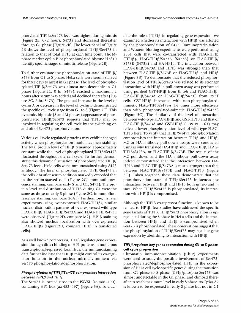
TIF1β regulates key genes expression during G1 to S-phase cell cycle progressionChromatin immunoprecipitation (ChIP) experimentswere used to study the possible involvement of Ser473-phosphorylated/dephosphorylated TIF1β in the expres-sion of HeLa cell cycle-specific genes during the transitionfrom G1 phase to S phase. TIF1β/phospho-Ser473 wasalmost undetectable in the G1 phase, and climbed there-after to reach maximum level in early S phase. As Cyclin A2is known to be expressed in early S phase but not in G1
Page 5 of 16(page number not for citation purposes)

BMC Molecular Biology 2008, 9:61 http://www.biomedcentral.com/1471-2199/9/61
Page 6 of 16(page number not for citation purposes)
Interaction between TIF1β and HP1β is compromised by phosphorylation of TIF1β/Ser473Figure 3Interaction between TIF1β and HP1β is compromised by phosphorylation of TIF1β/Ser473. (A) Schematic repre-sentation of domain organization of TIF1β protein. The position of Ser473 relative to the PXVXL motif is shown. (B) Western blot results of co-immunoprecipitation of over-expressed FLAG-TIF1β and HA-HP1β. FLAG-TIF1β/S473A, FLAG-TIF1β/S473E and wild-type FLAG-TIF1β were co-expressed with HA-HP1β into 293T cells. The level of ectopically expressed FLAG-TIF1βs (Input, M2) or M2-beads immunoprecipitated FLAG-TIF1βs (M2-IP, M2) was probed with M2 antibodies (TIF1β, M2). The level of ectopically expressed HA-HP1β (Input, HA) or M2-beads co-immunoprecipitated HA-HP1βs (M2-IP, HA) was probed with monoclonal anti-HA antibodies (HP1β, HA). (C) In vitro GST pull-down assay of TIF1βs by recombinant GST-HP1β. Wild-type and mutant FLAG-TIF1βs were expressed in 293T cells. These FLAG-TIF1βs were immunoprecipitated by M2 beads and eluted with M2 peptide. The levels of FLAG-TIF1βs from 1/10 input for each pull down assay were shown (lanes 8–10). Eluted FLAG-TIF1βs were incubated with GST-beads alone (lanes 1–3) or incubated with recombinant GST-HP1β bound glutathione-beads (lanes 4–6). The levels of GST-HP1β and FLAG-TIF1βs in each pull down assay were shown (lanes 1–6). Quantitation of the Western blots (by ImageGauge software) from each pull down sample (lanes 4–6) showing the relative level of FLAG-TIF1βs (A, TIF1β/S473A; E, TIF1β/S473E; W, wild-type TIF1β) pulled down by GST-HP1β, after normalization to individual GST-HP1β levels (right panel). (D) Interaction between in vitro translated FLAG-TIF1βs and HA-HP1β. Upper panel, In vitro translated [35S] FLAG-TIF1βs and [35S]-HA-HP1β were incubated for 30 min before being immunoprecipitated by M2 beads in the presence of 0.5 M NaCl. The immunoprecipitates were resolved by SDS-PAGE and followed by autoradiography to visual-ize HA-HP1β (upper right panel, M2-pull down). Lower panel, In vitro translated [35S] FLAG-TIF1βs and [35S] HA-HP1β were pulled down by protein-G coupled anti-HA antibody. The immunoprecipitated FLAG-TIF1βs are shown in the lower right panel (TIF1βs, IP). Supernatants after immunoprecipitation are also shown, as indicated (S).
BMC Molecular Biology 2008, 9:61 http://www.biomedcentral.com/1471-2199/9/61
phase, a reasonable question arises: Is the TIF1β/Ser473phosphorylation correlated with cyclin A2's expression?PCR primers encompassing the E2F site were used todetect DNA fragments corresponding to the promoterregion of cyclin A2. A much higher level of TIF1β was asso-
ciated with the cyclin A2 promoter at the G1/S boundarythan in early S phase (Figure 4A, upper panel). Theseresults are closely correlated with the rise in phosphor-ylated TIF1β/Ser473 in the early S phase, but not at theG1/S boundary (Figure 4A, lower left panel). Histone H3
TIF1β is involved in the regulation of expression of E2F downstream genes, cyclin A2, Cdc2 and Cdc25AFigure 4TIF1β is involved in the regulation of expression of E2F downstream genes, cyclin A2, Cdc2 and Cdc25A. (A) Binding of TIF1β to the promoter of cyclin A2 gene is correlated with its repression. HeLa cells were synchronized to G1/S boundary by double thymidine block and subsequently released for 1 hour to the early S phase. ChIP was performed as described in the Materials and Methods section. Equal loadings of input derived from G1/S and S cells were used for ChIP experiments (upper panels), using 20A1, H3K4diMe, and H3K9diMe antibodies. PCR was performed with input samples (1/50) and ChIP eluates. Relative differences between G1/S and S phase of each indicated probe on cyclin A2 promoter are shown in the lower right panels (comparing the cyclin A2 signal of G1/S and S phase from each probe with the same total area, by ImageGauge soft-ware). The protein level of TIF1β (20A1), α-tubulin and phosphorylated TIF1β/Ser473 (S473) from formaldehyde cross-linked samples are shown in the lower left panel. PCR for cyclin E promoter was used as a control (upper panel). (B) HeLa cells were arrested at G1 phase by serum starvation and released into early S phase by addition of serum to the culture for 2 hours. ChIP was performed as in (A) with 20A1, anti-H3K4diMe and anti-H3K9diMe antibodies (upper panel). Relative differences between G1 and S phase of each indicated probe on cyclin A2 promoter are shown in the lower right panels (by ImageGauge, as in A), while the protein levels are shown in the lower left panel. (C) Flow cytom-etry analysis. FLAG-TIF1β/S473A, FLAG-TIF1β/S473E and wild-type FLAG-TIF1β were over-expressed in 293T cells, collected 36 hours post-transfection and analyzed by flow cytometry after staining with propidium iodide, and the FCM histograms are shown in the upper panels. Cell cycle phase distributions (%) of TIF1β over-expressing cells were analyzed with the CellQuest software and shown in the lower left panel, according to the gated region from the upper panel (M1, subG1; M2, G1; M3, S; and M4, G2/M, by Cel-lQuest program), and the bar charts represented the cell cycle distribution (in total for 100%) are shown in the lower right panel (by Microsoft Excel). (D) The HP1β complex is preferentially associated with the promoters of Cdc2 and Cdc25A in TIF1β/S473A over-expressing interphase cells of HEK293T. FLAG-TIF1β, FLAG-TIF1β/S473A, FLAG-TIF1β/S473E and HA-HP-1β were ectopically expressed in HEK 293T cells for 36 hours and subjected to ChIP with the indicated antibodies (anti-tubulin, 10D8; anti-TIF1β, 20A1; anti-HA, HA). DNA from the immunoprecipitated chromatin was quantified by real-time PCR. Each result was normalized to input and the relative level to the transfected vector control. Results for the Cdc2 promoter (upper panels) and Cdc25A promoter (lower panels) are shown.
Page 7 of 16(page number not for citation purposes)
BMC Molecular Biology 2008, 9:61 http://www.biomedcentral.com/1471-2199/9/61
K9diMe was associated with the silencing of gene expres-sion. The ChIP results demonstrated that the level ofH3K9diMe correlated strongly with that of TIF1β. How-ever, histone H3K4diMe is known to associate withactively transcribed genes. Its association with the pro-moter region of cyclin A2 was inversely correlated withthat of TIF1β. Quantitative ChIP results comparing thecyclin A2 level in G1/S and S phase are presented under thepanel of PCR results obtained using each probe. As a con-trol, TIF1β was not associated with the promoter of cyclinE (Figure 4A, upper panel). TIF1β/phospho-Ser473 wasalso elevated 2 hours after serum was added (Figure 2C).ChIP was thus used to measure the level of TIF1β boundto the cyclin A2 promoter in G1-phase HeLa cells that hadbeen starved of serum for three days or S-phase HeLa cellsthat had been released from serum starvation. More TIF1βin the G1 phase cells was associated with the promoterregion of cyclin A2 (Figure 4B, upper panel). This findingis closely correlated with the elevated level of phosphor-ylated TIF1β/Ser473 in the S phase but not in the G1phase (Figure 4B, lower left panel). Quantitative ChIPresults, comparing cyclin A2 levels in the G1 and S phase,are presented beneath the PCR results obtained using eachprobe. Taken together, these results indicate that the asso-ciation of TIF1β with the promoter of cyclin A2 is corre-lated with silencing while dissociation is correlated withderepression of the promoter.
Immunostaining showed that over-expression of FLAG-TIF1β, FLAG-TIF1β/S473A, and FLAG-TIF1β/S473E didnot alter their nuclear localization (Figure 2D). A flowcytometric analysis was performed to test further theeffects of over-expressed FLAG-TIF1β, FLAG-TIF1β/S473Aand FLAG-TIF1β/S473E on cell cycle progression. Over-expression of the FLAG-TIF1β/S473A mutant caused anaccumulation of cells stalled at G2/M in 293T cells (Figure4C), while the profiles of cell cycle progression were sim-ilar in 293T cells over-expressing FLAG-TIF1β and FLAG-TIF1β/S437E, suggesting that the phosphorylation state ofTIF1β/Ser473 affects cell cycle progression. Quantitativereal-time PCR analysis of 293T cells revealed half theamount of cyclin A2 mRNA in the FLAG-TIF1β/S473A-overexpressing cells compared to the FALG-TIF1β orFALG-TIF1β/S473E-expressing cells (data not shown).The phenotype of the G2/M stalls and the reduction of thecyclin A2 mRNA level in TIF1β/S473A over-expressing293T cells are consistent with observations made in aGFP-cyclin A2 siRNA knockdown experiment publishedby Kenrick et al. [29]. Taken together, these results suggestthat disruption of TIF1β/Ser473 phosphorylation mayinfluence cell cycle-regulated gene expression, and thatcyclin A2 may be one of the TIF1β indirect target genes.These data also demonstrate that the Ser473 phosphoryla-tion/dephosphorylation status of TIF1β may regulate cellcycle progression.
To further examine the association of unphosphorylatedTIF1β/Ser473 with other cell cycle-regulated genes, suchas Cdc2 and Cdc25A, quantitative ChIP experiments wereconducted with HEK293T cells that had been transfectedwith HA-HP1β and FLAG-TIF1β, FLAG-TIF1β/Ser473A,and FLAG-TIF1β/Ser473E. When ChIP was performedwith HA monoclonal antibody, the association of HP1βwith the promoters of Cdc2 or Cdc25A in FLAG-TIF1β/Ser473A over-expressing cells was stronger than in FLAG-TIF1β/Ser473E-over-expressing cells (Figure 4D). WhenChIP was performed using 20A1 (which recognizes the N-terminal of TIF1β), no obvious difference between FLAG-TIF1β/Ser473A and FLAG-TIF1β/Ser473E was observed.Collectively, these results reveal that over-expressed HP1βand TIF1β/Ser473A may form a stronger complex andpreferentially associate with the promoter regions of Cdc2and Cdc25A genes more than over-expressed HP1β andTIF1β/Ser473E in interphase HEK293T cells.
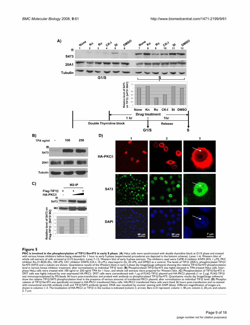
Phosphorylation of TIF1β Ser473 in S phase is mediated by PKC pathwayTIF1β/Ser473 phosphorylation is up-regulated in S phase.To identify which kinase(s) are involved in the phospho-rylation of TIF1β/Ser473 in the S phase, we used Westernblotting of cell extracts prepared from cells that had beentreated with a panel of kinase inhibitors. Among theinhibitors tested, only the PKC inhibitor (Ro-31-8820)exhibited significant inhibition, while the CK1 inhibitorhad a moderate effect on the phosphorylation of Ser473(Figure 5A). An inhibitory peptide (a substrate-mimeticfragment from aa 466–478 of TIF1β) that containedTIF1β/S471A/S473A also blocked TIF1β/Ser473 phos-phorylation (data not shown). Neither CaMK-II inhibitornor staurosporine had any effect (Figure 5A). To confirmthat the phosphorylation of TIF1β/Ser473 proceeds alongthe PKC pathway, the effect of 12-O-tetradecanoylphor-bol-13-acetate (TPA) on TIF1β/Ser473 phosphorylationin interphase HeLa cells was tested. TPA treatmentinduced dramatic phosphorylation of TIF1β/Ser473within 1 hour (Figure 5B). To further examine whethersubtype-PKCδ is involved in the phosphorylation ofTIF1β/Ser473, M2-immunoprecipitation and Westernblotting with S473 antibody were performed using cellextracts that were prepared from FLAG-TIF1β and HA-PKCδ-cotransfected 293T cells. The phosphorylation levelof TIF1β/Ser473 was double that of the control (Figure5C). Immunostaining also revealed an increase of theTIF1β/Phospho-Ser473 signal in HA-PKCδ-transfectedHeLa cells (Figure 5D). Although the majority of over-expressed PKCδ was located in the cytoplasm, a minorportion appeared in the nucleus (Figure 5D, column 3).Collectively, these results indicate that TIF1β/Ser473phosphorylation in S phase could be mediated by thePKCδ pathway, and that TIF1β/Ser473 phosphorylationcorrelated with the expression of target genes by weaken-
Page 8 of 16(page number not for citation purposes)
BMC Molecular Biology 2008, 9:61 http://www.biomedcentral.com/1471-2199/9/61
Page 9 of 16(page number not for citation purposes)
PKC is involved in the phosphorylation of TIF1β/Ser473 in early S phaseFigure 5PKC is involved in the phosphorylation of TIF1β/Ser473 in early S phase. (A) HeLa cells were synchronized with double thymidine block at G1/S phase and treated with various kinase inhibitors before being released for 1 hour to early S-phase (experimental procedures are depicted in the bottom scheme). Lanes 1–6, Western blot of whole cell extracts of cells arrested at G1/S boundary. Lanes 7–12, Western blot of early S-phase extracts. The inhibitors used were CaMK-II inhibitor KN93 (KN, 1 μM), PKC inhibitor Ro-31-8220 (Ro, 100 nM), CK1 inhibitor D4476 (CK-I, 10 μM,), staurosporin (St, 20 nM), and DMSO as a control. The levels of TIF1β (20A1), phosphorylated TIF1β/Ser473 (S473) and α-tubulin are shown. Quantitative results of the Western blots in early S phase (by ImageGauge software) showing the relative TIF1β/Ser473 phosphorylation level in various kinase inhibitor treatment, after normalization to individual TIF1β level. (B) Phosphorylated TIF1β/Ser473 was highly elevated in TPA-treated HeLa cells. Inter-phase HeLa cells were treated with 100 ng/ml or 250 ng/ml TPA for 1 hour, and whole cell extracts were prepared for Western blot. (C) Phosphorylation of TIF1β/Ser473 in 293T cells was highly induced by over-expressed HA-PKCδ. 293T cells were cotransfected with 1 μg of FLAG-TIF1β plasmid and HA-PKCδ plasmids (1 or 2 μg). FLAG-TIF1β was immunoprecipitated by M2-beads 36 hours post-transfection and probed with antibody to phosphorylated TIF1β/Ser473. Quantitative results (by ImageGauge software) show the relative TIF1β/S473 phosphorylation level in the presence of various amounts of transfected PKCδ plasmid, after normalization to individual TIF1β level. (D) Phospho-rylation of TIF1β/Ser473 was specifically increased in HA-PKCδ transfected HeLa cells. HA-PKCδ transfected HeLa cells were fixed 36 hours post-transfection and co-stained with monoclonal anti-HA antibody (red) and TIF1β/S473 antibody (green). DNA was visualized by counter staining with DAPI (blue). Different maginification of images are shown in columns 1–3. The localization of HA-PKCδ or TIF1β in the nucleus is indicated (column 3, arrow). Bars in D represent: column 1, 40 μm; column 2, 20 μm; and column 3, 7 μm.
BMC Molecular Biology 2008, 9:61 http://www.biomedcentral.com/1471-2199/9/61
ing TIF1β/HP1β interaction (and heterochromatin forma-tion).
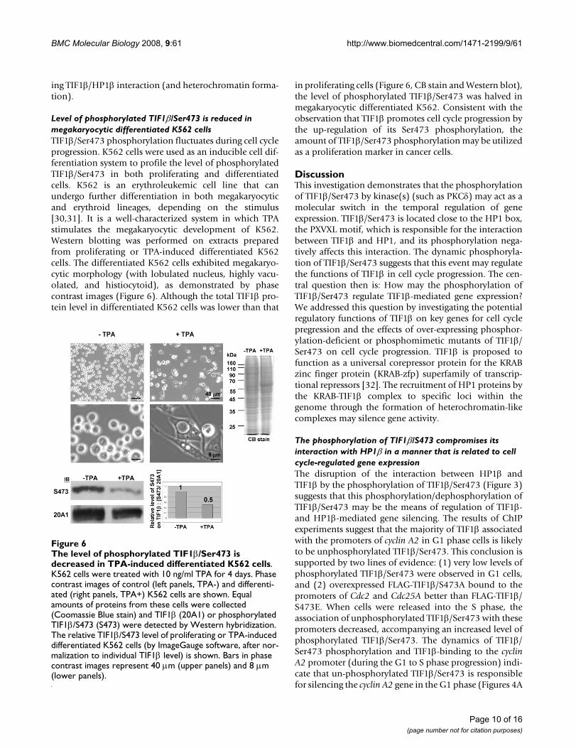
Level of phosphorylated TIF1β/Ser473 is reduced in megakaryocytic differentiated K562 cellsTIF1β/Ser473 phosphorylation fluctuates during cell cycleprogression. K562 cells were used as an inducible cell dif-ferentiation system to profile the level of phosphorylatedTIF1β/Ser473 in both proliferating and differentiatedcells. K562 is an erythroleukemic cell line that canundergo further differentiation in both megakaryocyticand erythroid lineages, depending on the stimulus[30,31]. It is a well-characterized system in which TPAstimulates the megakaryocytic development of K562.Western blotting was performed on extracts preparedfrom proliferating or TPA-induced differentiated K562cells. The differentiated K562 cells exhibited megakaryo-cytic morphology (with lobulated nucleus, highly vacu-olated, and histiocytoid), as demonstrated by phasecontrast images (Figure 6). Although the total TIF1β pro-tein level in differentiated K562 cells was lower than that
in proliferating cells (Figure 6, CB stain and Western blot),the level of phosphorylated TIF1β/Ser473 was halved inmegakaryocytic differentiated K562. Consistent with theobservation that TIF1β promotes cell cycle progression bythe up-regulation of its Ser473 phosphorylation, theamount of TIF1β/Ser473 phosphorylation may be utilizedas a proliferation marker in cancer cells.
DiscussionThis investigation demonstrates that the phosphorylationof TIF1β/Ser473 by kinase(s) (such as PKCδ) may act as amolecular switch in the temporal regulation of geneexpression. TIF1β/Ser473 is located close to the HP1 box,the PXVXL motif, which is responsible for the interactionbetween TIF1β and HP1, and its phosphorylation nega-tively affects this interaction. The dynamic phosphoryla-tion of TIF1β/Ser473 suggests that this event may regulatethe functions of TIF1β in cell cycle progression. The cen-tral question then is: How may the phosphorylation ofTIF1β/Ser473 regulate TIF1β-mediated gene expression?We addressed this question by investigating the potentialregulatory functions of TIF1β on key genes for cell cyclepregression and the effects of over-expressing phosphor-ylation-deficient or phosphomimetic mutants of TIF1β/Ser473 on cell cycle progression. TIF1β is proposed tofunction as a universal corepressor protein for the KRABzinc finger protein (KRAB-zfp) superfamily of transcrip-tional repressors [32]. The recruitment of HP1 proteins bythe KRAB-TIF1β complex to specific loci within thegenome through the formation of heterochromatin-likecomplexes may silence gene activity.
The phosphorylation of TIF1β/S473 compromises its interaction with HP1β in a manner that is related to cell cycle-regulated gene expressionThe disruption of the interaction between HP1β andTIF1β by the phosphorylation of TIF1β/Ser473 (Figure 3)suggests that this phosphorylation/dephosphorylation ofTIF1β/Ser473 may be the means of regulation of TIF1β-and HP1β-mediated gene silencing. The results of ChIPexperiments suggest that the majority of TIF1β associatedwith the promoters of cyclin A2 in G1 phase cells is likelyto be unphosphorylated TIF1β/Ser473. This conclusion issupported by two lines of evidence: (1) very low levels ofphosphorylated TIF1β/Ser473 were observed in G1 cells,and (2) overexpressed FLAG-TIF1β/S473A bound to thepromoters of Cdc2 and Cdc25A better than FLAG-TIF1β/S473E. When cells were released into the S phase, theassociation of unphosphorylated TIF1β/Ser473 with thesepromoters decreased, accompanying an increased level ofphosphorylated TIF1β/Ser473. The dynamics of TIF1β/Ser473 phosphorylation and TIF1β-binding to the cyclinA2 promoter (during the G1 to S phase progression) indi-cate that un-phosphorylated TIF1β/Ser473 is responsiblefor silencing the cyclin A2 gene in the G1 phase (Figures 4A
The level of phosphorylated TIF1β/Ser473 is decreased in TPA-induced differentiated K562 cellsFigure 6The level of phosphorylated TIF1β/Ser473 is decreased in TPA-induced differentiated K562 cells. K562 cells were treated with 10 ng/ml TPA for 4 days. Phase contrast images of control (left panels, TPA-) and differenti-ated (right panels, TPA+) K562 cells are shown. Equal amounts of proteins from these cells were collected (Coomassie Blue stain) and TIF1β (20A1) or phosphorylated TIF1β/S473 (S473) were detected by Western hybridization. The relative TIF1β/S473 level of proliferating or TPA-induced differentiated K562 cells (by ImageGauge software, after nor-malization to individual TIF1β level) is shown. Bars in phase contrast images represent 40 μm (upper panels) and 8 μm (lower panels).
Page 10 of 16(page number not for citation purposes)

BMC Molecular Biology 2008, 9:61 http://www.biomedcentral.com/1471-2199/9/61
and 4B). The observation that HP1β interacted stronglywith un-phosphorylated TIF1β/Ser473 is consistent withthe ChIP results concerning the over-expressed recom-binant variants, where over-expressed FLAG-TIF1β/S473Aassociated better with the promoter region of Cdc2 orCdc25A than that did FLAG-TIF1β/S473E (Figure 4D).
The treatment of cells with cyclin A2 siRNA led to an accu-mulation of cells in prophase and mitosis to a degree sim-ilar to that observed for cyclin B1, consistent with therequirement of cyclin A for G1/S and G2/M transitions[29]. Interestingly, the ChIP results (Figures 4A and 4B)and the effects of over-expressed FLAG-TIF1β/Ser473A oncell cycle progression (accumulation of G2/M cells, Figure4C) are consistent with published results for the siRNAknockdown of Cyclin A2 [29].
HP1β recruitment to E2F-binding element of Cdc2 andCdc25A promoters was affected by the phosphorylationstate of TIF1β/Ser473. The level of HP1β recruitment toCdc2 or Cdc25A promoter was increased when wild-typeFLAG-TIF1β or FLAG-TIF1β/S473A were ectopicallyexpressed. This association was compromised by thephosphomimetic mutant, S473E, which suggests thatHP1β recruitment is negatively regulated by phosphoryla-tion of TIF1β/Ser473. Likewise, ectopically expressedHP1β resulted in elevated recruitment of wild-type FLAG-TIF1β or FLAG-TIF1β/S473A. These observations providea clue that the recruitment of TIF1β and HP1β could workin a positive feedback manner.
The above results demonstrate that the dynamic intercon-version between unphosphorylated and phosphorylatedforms of TIF1β/Ser473 may be crucial in the regulation ofgene expression during cell cycle progression. Despiteextensive efforts to perform ChIP with rabbit anti-phos-phorylated TIF1β/Ser473 antibody, no significant resultwas obtained. A likely explanation of this failure is thatphosphorylated TIF1β/Ser473 is not associated with thepromoter of cyclin A2 in the G1 phase or, alternatively, theepitope in the multiprotein complex is masked from anti-body access.
The way in which TIF1β disassociates from the HP1-con-taining heterochromatin is unclear. However, the findingsherein provide a molecular explanation, which involvesPKC-mediated phosphorylation at TIF1β/Ser473 (Figure5).
Phosphorylated TIF1β/S473 level may serve as a proliferation markerTIF1β transcriptional repression activity depends on theinteraction between TIF1β and HP1β [11]. This interac-tion is essential to the relocation of TIF1β from euchroma-tin to heterochromatin that accompanies the
differentiation of primitive endoderm-like cells [12].TIF1β is known to interact differentially with HP1β andHP1γ in differentiated and non-differentiated cells [33].In non-differentiated cells, TIF1β/HP1 interaction occursonly in euchromatin and selectively involves HP1β andHP1γ, but not HP1α. In differentiated cells, on the otherhand, TIF1β selectively associates with HP1β in hetero-chromatin, while TIF1β and HP1γ interaction occurs onlyin euchromatin. These conclusions agree with the reducedlevel of phosphorylated TIF1β/Ser473 seen here in differ-entiated K562 cells (Figure 6). The results herein alsorevealed that un-phosphorylated TIF1β/Ser473 interactsmore strongly with HP1β than its phosphorylated coun-terpart (Figure 4B). These results further suggest that thephosphorylation of TIF1β/Ser473 regulates the differen-tial interaction between TIF1β and HP1β.
What may be the functional consequences of the phos-phorylation of TIF1β/Ser473 and its consequent dissocia-tion from HP1s must be addressed. TIF1β interacts withE2F [16], TRIP-Br and CBP/p300, and potentiates the co-activation of E2F-1/DP-1 by TRIP-Br protein [34]. Phos-phorylated TIF1β/Ser473 may thus be involved in this tri-partite functional interaction. This suggestion issupported by our findings that the induction of cyclin A2is accompanied by an increased level of phosphorylatedTIF1β/Ser473 and reduced binding of TIF1β to the pro-moter. A most provocative question that remains to beanswered is whether the Ser473-phosphorylated TIF1βmay interact preferentially with transcription factors andserve as a coactivator.
The results also demonstrated that the level of phosphor-ylated TIF1β/Ser473 peaks at early S- and M-phases. Twophosphorylation sites other than Ser473, Ser752 andSer757, were also identified in the course of this investiga-tion. Both sites are located in the bromodomain of TIF1β.The phosphorylation-deficient or phosphomimeticmutants of Ser757 did not influence the phosphorylationof Ser473 (Chang, unpublished results), suggesting thatthese phosphorylations may be independent events dur-ing mitosis. Other phosphorylation sites important forregulating the function of TIF1β have also been identified[35]. The functional consequences of Ser752 and Ser757phosphorylation remain to be investigated.
PKC mediated TIF1β/S473 phosphorylation may be involved in cell cycle progressionNumerous examples have demonstrated that PKCδ is cen-trally involved in cell proliferation and differentiation.The activation of PKCδ uniquely mediates insulin-induced proliferation: PKCδ is activated by insulin andinteracts with insulin receptor and IRS [36-38]. Insulin-activated PKCδ interacts with 3-phosphoinositide-dependent protein kinase to regulate Protein Kinase B
Page 11 of 16(page number not for citation purposes)
BMC Molecular Biology 2008, 9:61 http://www.biomedcentral.com/1471-2199/9/61
[39] and is responsible for STAT3 activation and keratino-cyte proliferation [40]. Although the over-expressed PKCδis mainly located in the cytoplasm (Figure 5D), it has beenshown here (Figure 5D) and by Kajimoto et al. to partiallylocalize to the nucleus [41]. The observation herein thatthe serum-stimulated phosphorylation of TIF1β/Ser473correlates strongly with G1/S progression and cyclin A2expression (Figure 4) uncovers a novel mechanism ofPKCδ-mediated G1 to S phase cell cycle progression.
Phosphorylation of TIF1β/Ser473, gene activation and stem cell proliferationThe dynamic phosphorylation of TIF1β/Ser473 duringcell proliferation and differentiation (Figures 2 and 6)suggests that phosphorylation/dephosphorylation is cru-cial for regulating the transcriptional activity of TIF1β.During the differentiation of embryonic carcinoma cells,the intracellular distribution of TIF1β changes from dif-fuse nuclear staining to discrete foci and colocalizes withheterochromatin [42]. The steady-state level of TIF1β isalso decreased. The reduced levels of phosphorylatedTIF1β/Ser473 as well as the lower steady-state TIF1β levelsin differentiated cells suggest that TIF1β may be mainlylocalized to heterochromatin in differentiated cells. Inembryonic stem cells, TIF1β is present in complexes withvarious pluripotent markers, including Rex-1, Dax-1 andNanog [22]. Since phosphorylated TIF1β/Ser473 seems tobe preferentially associated with cell proliferation, it isimportant to determine whether it resides in these com-plexes. In fact, the level of phosphorylated TIF1β/Ser473may serve as a proliferation marker. The epigenetic silenc-ing of retrovirus transcription is caused by the binding ofa TIF1β corepressor complex to retrovirus primer bindingsite [17]. The likely recruitment of TIF1β by a DNA-bind-ing KRAB-box containing zinc finger protein for PBS-mediated silencing should be addressed, and the questionof whether the regulation of the phosphorylation ofTIF1β/Ser473 or the formation of TIF1β corepressor com-plex is participating in retrovirus transcription should alsobe investigated.
ConclusionAlthough the TIF1β co-repressor function is known to berelated to HP1β, few studies have addressed the specificgene targets of TIF1β. We have found that cell cycle pro-gression is regulated by TIF1β. The key cell cycle regula-tory genes, Cyclin A2, Cdc2 and Cdc25A are targeted byTIF1β, and phosphorylation of TIF1β/Ser473 is associatedwith the activation of these genes. TIF1β/S473 phosphor-ylation is up-regulated during the S phase in HeLa cellsand the interaction between HP1β and TIF1β is compro-mised when Ser473 is phosphorylated. These observa-tions suggest that the phosphorylation of TIF1β/Ser473may regulate gene expression by abolishing its interaction
with HP1β. Thus, phosphorylation of TIF1β/Ser473 playsa crucial role in epigenetic regulation of gene expression.
MethodsAntibodiesMonoclonal antibody against TIF1β was generated byimmunizing BALB/c mice with recombinant TIF1β corre-sponding to the N-terminal region (amino acids 1–250).Clone 20A1 was used throughout this investigation. Phos-pho-specific polyclonal antibody (S473) was produced byimmunizing rabbits with KLH-conjugated peptide(VKRSRpSGEGEVC). The S473 antibody was purified bypeptide-agarose affinity column chromatography. Rabbitanti-H3K9diMe and H3K4diMe antibodies were obtainedfrom Upstate/Millipore, cyclin A and cyclin B antibodieswere from BD Science, and M2 monoclonal antibody andM2 beads were from Sigma.
Plasmids and chemicalsMouse pCMV-FLAG-TIF1β was cloned as described byChang et al. [43]. Site-directed mutagenesis was per-formed using pCMV-FLAG-TIF1β to create S473A andS473E mutants with PfuTurbo DNA Polymerase (Strata-gene). The primers, based on nucleotides 1691 to 1721 ofNM_005762, were designed as follows. For S473A:
Forward: 5'-GAAACGGTCCCGCGCAGGTGAGGG-3' and
Reverse: 5'-CCCTCACCTGCGCGGGACCGTTTC-3'.
For S473E:
Forward: 5'-GAAACGGTCCCGCGAAGGTGAGGG-3' and
Reverse: 5'-CCCTCACCTTCGCGGGACCGTTTC-3'.
HA-PKCδ was obtained from Dr. C. K. Chou, HA-HP1βand GST-HP1β plasmids were kindly provided by Dr.Pierre Chambon. PKC inhibitor Ro-31-8820 and calcium/calmodulin-dependent protein kinase-II (CaMK-II) inhib-itor KN-93 were from BIOMOL. Casein kinase1 (CK1)inhibitor D4476 was from Calbiochem. Staurosporin, 12-O-tetradecanoylphorbol-13-acetate (TPA), thymidine,and nocodazole were from Sigma. Ser473-specific inhibi-tory peptide SGVKRARAGEGEVrrrrrrrrr (r stands forarginine) was obtained from Genemed Synthesis (SouthSan Francisco, CA).
Cell culturesHeLa, HEK293, and HEK293T cells were cultured in Dul-becco's modified Eagle's medium plus 10% FCS and 100units/ml penicillin/streptomycin. For synchronization atmitotic phase, HeLa cells were treated with 1 μM nocoda-zole for 16 hours. Cells were collected by shake-off, rinsedwith PBS and cultured in complete medium. For synchro-
Page 12 of 16(page number not for citation purposes)
BMC Molecular Biology 2008, 9:61 http://www.biomedcentral.com/1471-2199/9/61
nization at G1/S phase, HeLa cells were treated with 2.5mM thymidine for 19 hours, released for 10 hours, treatedfor 19 hours again before release at various time points forcell cycle progression. For G1 phase synchronization,HeLa cells were serum-starved for 72 hours and then cul-tured in complete medium for various times. K562 cellswere maintained in RPMI-1640 with 10% FCS and 100units penicillin/streptomycin. K562 cells were induced todifferentiate by treating with TPA (10 ng/ml) for 4 days[30,31].
Whole cell extracts (WCEs) were prepared by treating thecells with lysis buffer [20 mM HEPES, pH 7.4, 200 mMNaCl, 0.5% Triton-X100, 20% glycerol, 1 mM EDTA, 1mM EGTA, 1 mM orthovanadate, and protease inhibitors(0.1 μg/ml each of aprotinin, leupeptin, pepstatin A, and10 mM PMSF) and centrifuging at 6,000 × g in a microcen-trifuge for 30 min at 4°C. Nuclear fractions were preparedby lysing the cells in HEPES buffer (pH 7.6) containing 10mM NaCl, 1.5 mM MgCl2, 20% glycerol, 0.2 mM EDTA,0.1% Triton X-100 and protease inhibitors for 10 min,centrifuged at 1,250 × g for 5 min and washed once withthe same buffer. Nuclear extracts were prepared in nuclearextraction buffer containing 25 mM Tris-HCl, pH 8.0, 0.5M NaCl, 1 mM EDTA, 10% glycerol, 0.2% NP-40 and pro-tease inhibitors for 30 min at 4°C followed by DNase Itreatment for 1 hour. For calf intestine alkaline phos-phatase (CIP) treatment, 293 T cells were lysed with CIPbuffer (50 mM Tris-HCl, pH 8.0, 0.1 M NaCl, 10 mMMgCl2, and protease inhibitors). The supernatants wereincubated with 30 U of CIP at 37°C for 30 min.
Immunoprecipitation, recombinant proteins, in vitro pull-down, and western blottingWCE was pre-cleaned with protein G beads. Antibody wasbound to protein G beads and mixed with pre-cleanedWCE for 2 hours at 4°C by gentle rotation. The immuno-complex was washed 3 times with immunoprecipitationbuffer before SDS sample buffer was added and proteinswere separated by SDS-PAGE. TIF1βs (FLAG-TIF1β/S473A, FLAG-TIF1β/S473E, and wild type FLAG-TIF1β)were co-transfected with HA-HA-HP1β into 293T cells.Nuclear extracts were used for M2 pull-down assay. GST-HP1β expressed in Escherichia coli DH5α was purified bybinding to glutathione beads. FLAG-tagged TIF1β, TIF1β/S473A and TIF1β/S473E were expressed in 293T cells andpurified by FLAG peptide elution of the M2 bead-boundproteins. For pull-down assays, glutathione bead-boundGST-HP1β was re-suspended in nuclear extraction bufferand incubated with the purified FLAG-TIF1β, FLAG-TIF1β/S473A, and FLAG-TIF1β/S473E.
In vitro transcription and translation[35S]Methionine-labeled FLAG-TIF1βs (FLAG-TIF1β,FLAG-TIF1β/S473A, and FLAG-TIF1β/S473E) and HA-
HP1β were prepared by TnT in vitro transcription/transla-tion kit (Promega).
Chromatin immunoprecipitation assaysChromatin immunoprecipitation (ChIP) assays were per-formed as described by Li et al. [44]. Briefly, synchronizedG1, G1/S, early S phase, or interphase cells were treatedwith 1.42% of formaldehyde for 10 min at room temper-ature. Nuclei from 1 × 10⁷ cells were resuspended in ChIPlysis buffer (50 mM Tris-HCl [pH 8.1], 1% SDS, 10 mMEDTA, 1× protease inhibitor cocktail) and used for eachimmunoprecipitation. After sonication on ice four to sixtimes for 10 seconds followed by centrifugation for 10min, the chromatin solution was diluted 10-fold withdilution buffer (5 μg/ml of salmon sperm DNA, 5 mg/mlBSA, 20 mM Tris-HCl [pH 8.1], 1% Triton X-100, 2 mMEDTA, 150 mM NaCl, 1× protease inhibitor cocktail). Aninput control of 100 μl of sonicated solution was savedand processed in parallel with the eluted immunoprecip-itates beginning at the cross-link reversal step. The chro-matin was pre-cleaned with protein G-agarose. Differentantibodies (TIF1β monoclonal antibody, 20A1; rabbitanti-H3K9diMe and H3K4diMe antibodies) were boundto protein G-agarose first in dilution buffer containing 5μg/ml of salmon sperm DNA and 5 mg/ml BSA. Chroma-tin complexes were then incubated with specific antibod-ies-protein G-agarose and rotated for 2–4 hours at 4°C.Immunoprecipitates were sequentially washed for 5 to 10min in wash buffer I (20 mM Tris-HCl [pH 8.1], 2 mMEDTA, 0.1% SDS, 1% Triton X-100, 150 mM NaCl), washbuffer II (20 mM Tris-HCl [pH 8.1], 2 mM EDTA, 0.1%SDS, 1% Triton X-100, 500 mM NaCl), wash buffer III(0.25 M LiCl, 1% NP-40, 1% deoxycholate, 1 mM EDTA,10 mM Tris-HCl [pH 8.1]), and then in TE buffer (threetimes). Washed beads were incubated at 65°C overnightto reverse protein-DNA cross-linking, and then each sam-ple was treated with 10 μg of proteinase K in proteinase Kbuffer for 6 h at 55°C. The eluted material was combinedin one tube, and the DNA was purified with the QIAquickPCR purification kit (Qiagen, Valencia, Calif.28104) andeluted in 50 μl of elution buffer. Extracted total inputswere diluted 1:50 and subjected to PCR analysis with indi-cated primers. Each PCR mixture contained 2 μl of immu-noprecipitate or input, 1 μM each primer, 0.4 mMdeoxynucleoside triphosphate mixture, 1× LA-Taq PCRbuffer, and 0.25 μl LA-TaqDNA polymerase in a total vol-ume of 50 μl. The E2F responsive primers for each pro-moter were as follows:
For Cyclin A2 promoter:
Forward: 5'-CTGCTCAGTTTCCTTTGGTTTACC-3';
Reverse: 5'-CAAAGACGCCCAGAGATGCAG-3';
Page 13 of 16(page number not for citation purposes)
BMC Molecular Biology 2008, 9:61 http://www.biomedcentral.com/1471-2199/9/61
and for Cyclin E promoter:
Forward: 5'-GCCGCCTGTCCATTCATCC-3';
Reverse: 5'-GGTCCTGTGGAGCCTGTAGCC-3'.
PCR was performed for 26 to 29 cycles with 1 min ofdenaturing at 94°C, annealing at 62°C, and extension at68°C. PCR results were analyzed by agarose gel (1.5%)electrophoresis.
For real time PCR, 5 μg of FLAG-TIF1β and HA-HP-1βexpression vectors were transfected into HEK 293T cells in10-cm dish at about 20% confluence. In each precipita-tion, 300 μg of total cell extract was used, with 50 μg oftotal cell extract as input. Antibodies for FLAG (M2) or HAwere used to probe recombinant TIF1β and HP1β on E2Ftargets of Cdc2 and Cdc25A promoters. Real-time PCR wasconducted on a Roche Light Cycler 480 using the follow-ing primers for Cdc2 promoter:
Forward: 5'-ACAGTAGGACGACACTC-3'; and
Reverse: 5'-GGATTCACCAATCGGGTAG-3';
and the following primers for Cdc25A promoter:
Forward: 5'-CTAGCTGCCATTCGGT-3'; and
Reverse: 5'-CTTCGCTGTTCTCCCA-3'.
ImmunostainingCells on cover slips were washed with 1× phosphate-buff-ered saline (PBS), fixed with 2% formaldehyde for 15min, washed with cold PBS for three times and furtherpermeablized with 1xPBS containing 0.5% Triton-X100for 5 min. Cells were blocked with 1% BSA for 30 min andincubated with indicated antibodies diluted in 1% bovineserum albumin/PBS, and probed with indicated primaryantibodies. Alexa 594-conjugated goat anti-mouse IgG(Molecular Probes, Inc) and Alexa 488-conjugated goatanti-rabbit IgG were used as secondary immunofluores-cent dyes. DAPI was used to visualize DNA. Stained cellswere analyzed with a Leica TCS SP2 Confocal SpectralMicroscope using a 63X/NA 1.4 oil immersion objectivelens.
Flow cytometryCells were collected and fixed with 70% ethanol for 30minutes at 0°C. Cells were stained with DNA-stainingsolution (25 μg/ml propidium iodide, 100 μg/ml RNaseA, and 0.5% Nonidet P-40 in PBS) at room temperaturefor 30 min. DNA content was analyzed from 10,000 cellscollected with a BD Biosciences flow cytometer in con-junction with the CellQuest software. The distribution of
cell populations in the cell cycle stage was gated with Cel-lQuest analysis program (M1, subG1; M2, G1; M3, S; andM4, G2/M). The cell cycle phase distribution (in total for100%) in each sample after CellQuest program analysis isshown. The percentage of the cell cycle phase shown intable is also presented with bar charts (by MicrosoftExcel).
RNA extraction and Real-time PCRThe level of gene expression was determined by real-timePCR. Briefly, total RNA was extracted from cells using Blueextract reagent (LTK, Inc., Taiwan) following the proce-dures recommended by the manufacturer. Samples of 5μg of total RNA were reverse transcribed using M-MLVreverse transcriptase (Promega) and an oligo dT primer.The primers used for real-time PCR were for Cyclin A2:
Forward: 5'-GCATGTCACCGTTCCTCCTT-3';
Reverse: 5'-CAGGGCATCTTCACGCTCTAT-3';
and for Actin:
Forward: 5'-GAGAAAATCTGGCACCACACC-3';
Reverse: 5'-ATACCCCTCGTAGATGGGCAC-3'.
All reactions were carried out using a 7300 Real-Time PCRSystem (Applied Biosystems) and ABsolute™ QPCR SYBR®
Green Mix (ABgene, Epsom, England). The amplificationwas carried out as follow: initial enzyme activation at94°C for 15 min, then 40 cycles of 94°C for 15 s and60°C for 1 min. A total of 50 ng of each diluted reversetranscription product was used for real-time PCR in a finalvolume of 25 μl containing 160 nM of each specificprimer and 1× ABsolute™ QPCR SYBR® Green Mix(ABgene). The relative level of Cyclin A2 gene expressionwas calculated according to the comparative Ct methodusing the 2-ΔΔCTCT formula, using the expression of Actin asan endogenous control.
Authors' contributionsCWC performed most experiments and participated in thewriting of the manuscript. HYC and TCH contributed topull-down experiments with in vitro translated proteins.YSL performed quantitative ChIP experiments with ectop-ically expressed plasmids. KHH and CJC contributed tothe cloning of recombinant plasmids. SCL generated20A1 and S473 antibodies. SCL supervised and conductedthe study and wrote the manuscript. All authors read andapproved the final manuscript draft.
AcknowledgementsWe thank Drs Pierre Chambon (Institute of Genetics and Cellular and Molecular Biology, Strasbourg, France) for HP1 plasmids and C. K. Chou (Department of Life Science, Chang Gung University, Taiwan) for HA-
Page 14 of 16(page number not for citation purposes)
BMC Molecular Biology 2008, 9:61 http://www.biomedcentral.com/1471-2199/9/61
PKCδ. This research was supported by a frontier science research grant from National Science Council of Taiwan (NSC96-2321-B002-008) and fund from the Institute of Biological Chemistry, Academia Sinica, Taiwan.
References1. Le Douarin B, Nielsen AL, Garnier JM, Ichinose H, Jeanmougin F, Los-
son R, Chambon P: A possible involvement of TIF1 alpha andTIF1 beta in the epigenetic control of transcription bynuclear receptors. EMBO J 1996, 15:6701-6715.
2. Eissenberg JC, Elgin SC: The HP1 protein family: getting a gripon chromatin. Curr Opin Genet Dev 2000, 10:204-210.
3. Wang G, Ma A, Chow CM, Horsley D, Brown NR, Cowell IG, SinghPB: Conservation of heterochromatin protein 1 function. MolCell Biol 2000, 20:6970-6983.
4. Grigoryev SA, Nikitina T, Pehrson JR, Singh PB, Woodcock CL:Dynamic relocation of epigenetic chromatin markersreveals an active role of constitutive heterochromatin in thetransition from proliferation to quiescence. J Cell Sci 2004,117:6153-6162.
5. Bártová E, Pacherník J, Kozubík A, Kozubek S: Differentiation-spe-cific association of HP1alpha and HP1beta with chromocen-tres is correlated with clustering ofTIF1beta at c these sites.Histochem Cell Biol 2007, 127:375-388.
6. Brasher SV, Smith BO, Fogh RH, Nietlispach D, Thiru A, Nielsen PR,Broadhurst RW, Ball LJ, Murzina NV, Laue ED: The structure ofmouse HP1 suggests a unique mode of single peptide recog-nition by the shadow chromo domain dimer. EMBO J 2000,19:1587-1597.
7. Eissenberg JC: Decisive factors: a transcription activator canovercome heterochromatin silencing. Bioessays 2001,23:767-771.
8. Nielsen AL, Sanchez C, Ichinose H, Cerviño M, Lerouge T, ChambonP, Losson R: Selective interaction between the chromatin-remodeling factor BRG1 and the heterochromatin-associ-ated protein HP1alpha. EMBO J 2002, 21:5797-5806.
9. Thiru A, Nietlispach D, Mott HR, Okuwaki M, Lyon D, Nielsen PR,Hirshberg M, Verreault A, Murzina NV, Laue ED: Structural basisof HP1/PXVXL motif peptide interactions and HP1 localisa-tion to heterochromatin. EMBO J 2004, 23:489-499.
10. Vassallo MF, Tanese N: Isoform-specific interaction of HP1 withhuman TAFII130. Proc Natl Acad Sci USA 2002, 99:5919-5924.
11. Nielsen AL, Ortiz JA, You J, Oulad-Abdelghani M, Khechumian R,Gansmuller A, Chambon P, Losson R: Interaction with membersof the heterochromatin protein 1 (HP1) family and histonedeacetylation are differentially involved in transcriptionalsilencing by members of the TIF1 family. EMBO J 1999,18:6385-6395.
12. Cammas F, Oulad-Abdelghani M, Vonesch JL, Huss-Garcia Y, Cham-bon P, Losson R: Cell differentiation induces TIF1beta associa-tion with centromeric heterochromatin via an HP1interaction. J Cell Sci 2002, 115:3439-3448.
13. Ryan RF, Schultz DC, Ayyanathan K, Singh PB, Friedman JR, Freder-icks WJ, Rauscher FJ 3rd: KAP1 corepressor protein interactsand colocalizes with heterochromatic and euchromatic HP1proteins: a potential role for Krüppel-associated box-zinc fin-ger proteins in heterochromatin-mediated gene silencing.Mol Cell Biol 1999, 19:4366-4378.
14. Schultz DC, Ayyanathan K, Negorev D, Maul GG, Rauscher FJ 3rd:SETDB1: a novel KAP1-associated histone H3, lysine 9-spe-cific methyltransferase that contributes to HP1-mediatedsilencing of euchromatic genes by KRAB zinc-finger pro-teins. Genes Dev 2002, 16:919-932.
15. Wang C, Ivanov A, Chen L, Fredericks WJ, Seto E, Rauscher FJ 3rd,Chen J: MDM2 interaction with nuclear corepressor KAP1contributes to p53 inactivation. EMBO J 2005, 24:3279-3290.
16. Wang C, Rauscher FJ 3rd, Cress WD, Chen J: Regulation of E2F1function by the nuclear corepressor KAP1. J Biol Chem 2007,282:29902-29909.
17. Wolf D, Goff SP: TRIM28 mediates primer binding site-tar-geted silencing of murine leukemia virus in embryonic cells.Cell 2007, 131:46-57.
18. Li H, Rauch T, Chen ZX, Szabó PE, Riggs AD, Pfeifer GP: The his-tone methyltransferase SETDB1 and the DNA methyltrans-ferase DNMT3A interact directly and localize to promoterssilenced in cancer cells. J Biol Chem 2006, 281:19489-19500.
19. Sripathy SP, Stevens J, Schultz DC: The KAP1 corepressor func-tions to coordinate the assembly of de novo HP1-demar-cated microenvironments of heterochromatin required forKRAB zinc finger protein-mediated transcriptional repres-sion. Mol Cell Biol 2006, 26:8623-8638.
20. White DE, Negorev D, Peng H, Ivanov AV, Maul GG, Rauscher FJ 3rd:KAP1, a novel substrate for PIKK family members, colocal-izes with numerous damage response factors at DNAlesions. Cancer Res 2006, 66:11594-11599.
21. Cammas F, Mark M, Dollé P, Dierich A, Chambon P, Losson R: Micelacking the transcriptional corepressor TIF1beta are defec-tive in early postimplantation development. Development2000, 127:2955-2963.
22. Wang J, Rao S, Chu J, Shen X, Levasseur DN, Theunissen TW, OrkinSH: A protein interaction network for pluripotency of embry-onic stem cells. Nature 2006, 444:364-368.
23. Beausoleil SA, Jedrychowski M, Schwartz D, Elias JE, Villén J, Li J, CohnMA, Cantley LC, Gygi SP: Large-scale characterization of HeLacell nuclear phosphoproteins. Proc Natl Acad Sci USA 2004,101:12130-12135.
24. Shu H, Chen S, Bi Q, Mumby M, Brekken DL: Identification ofphosphoproteins and their phosphorylation sites in theWEHI-231 B lymphoma cell line. Mol Cell Proteomics 2004,3:279-286.
25. Rayman JB, Takahashi Y, Indjeian VB, Dannenberg JH, Catchpole S,Watson RJ, te Riele H, Dynlacht BD: E2F mediates cell cycle-dependent transcriptional repression in vivo by recruitmentof an HDAC1/mSin3B corepressor complex. Genes Dev 2002,16:933-947.
26. Vigo E, Müller H, Prosperini E, Hateboer G, Cartwright P, MoroniMC, Helin K: CDC25A phosphatase is a target of E2F and isrequired for efficient E2F-induced S phase. Mol Cell Biol 1999,19:6379-6395.
27. Henglein B, Chenivesse X, Wang J, Eick D, Bréchot C: Structureand cell cycle-regulated transcription of the human cyclin Agene. Proc Natl Acad Sci USA 1994, 91:5490-5494.
28. Aleem E, Kiyokawa H, Kaldis P: Cdc2-cyclin E complexes regu-late the G1/S phase transition. Nat Cell Biol 2005, 7:831-836.
29. Kenrick M, Hancock S, Stubbs S, Thomas N, GE Healthcare, The May-nard Centre, Cardiff, UK: siRNA screening of the cell cycle withtwo dynamic GFP sensors. Discovery Matters 2005:18-19[http:w4.gelifesciences.com/APTRIX/upp00919.nsntenF6C9E078DD4E77C125704500085213?OpenDocument&Path=Catalog&Hometitle=Catalog&entry=1&newrel&LinkPart=C1256FC4003AED4FADC4C4C717CAEC125706A003C9C32_RelatedLinksNew].
30. Belhacène N, Maulon L, Guérin S, Ricci JE, Mari B, Colin Y, CartronJP, Auberger P: Differential expression of the Kell blood groupand CD10 antigens: two related membrane metallopepti-dases during differentiation of K562 cells by phorbol esterand hemin. FASEB J 1998, 12:531-539.
31. Rosson D, O'Brien TG: Constitutive c-myb expression in K562cells inhibits induced erythroid differentiation but not tetra-decanoyl phorbol acetate-induced megakaryocytic differen-tiatio. Mol Cell Biol 1995, 15:772-779.
32. Urrutia R: KRAB-containing zinc-finger repressor proteins.Genome Biol 2003, 4:231.
33. Cammas F, Janoshazi A, Lerouge T, Losson R: Dynamic and selec-tive interactions of the transcriptional corepressor TIF1beta with the heterochromatin protein HP1 isotypes duringcell differentiation. Differentiation 2007, 75:627-637.
34. Hsu SI, Yang CM, Sim KG, Hentschel DM, O'Leary E, Bonventre JV:TRIP-Br: a novel family of PHD zinc finger- and bromodo-main-interacting proteins that regulate the transcriptionalactivity of E2F-1/DP-1. EMBO J 2001, 20:2273-2285.
35. Li X, Lee YK, Jeng JC, Yen Y, Schultz DC, Shih HM, Ann DK: Role forKAP1 serine 824 phosphorylation and sumoylation/des-umoylation switch in regulating KAP1-mediated transcrip-tional repression. J Biol Chem 2007, 282:36177-36189.
36. Jain N, Zhang T, Kee WH, Li W, Cao X: Protein kinase C deltaassociates with and phosphorylates Stat3 in an interleukin-6-dependent manner. J Biol Chem 1999, 274:24392-24400.
37. Novotny-Diermayr V, Zhang T, Gu L, Cao X: Protein kinase Cdelta associates with the interleukin-6 receptor subunit glyc-oprotein (gp) 130 via Stat3 and enhances Stat3-gp130 inter-action. J Biol Chem 2002, 277:49134-49142.
Page 15 of 16(page number not for citation purposes)
BMC Molecular Biology 2008, 9:61 http://www.biomedcentral.com/1471-2199/9/61
Publish with BioMed Central and every scientist can read your work free of charge
"BioMed Central will be the most significant development for disseminating the results of biomedical research in our lifetime."
Sir Paul Nurse, Cancer Research UK
Your research papers will be:
available free of charge to the entire biomedical community
peer reviewed and published immediately upon acceptance
cited in PubMed and archived on PubMed Central
yours — you keep the copyright
Submit your manuscript here:http://www.biomedcentral.com/info/publishing_adv.asp
BioMedcentral
38. Shen S, Alt A, Wertheimer E, Gartsbein M, Kuroki T, Ohba M,Braiman L, Sampson SR, Tennenbaum T: PKCdelta activation: adivergence point in the signaling of insulin and IGF-1-inducedproliferation of skin keratinocytes. Diabetes 2001, 50:255-264.
39. Brand C, Cipok M, Attali V, Bak A, Sampson SR: Protein kinase Cδparticipates in insulin-induced activation of PKB via PDK1.Biochem Biophys Res Commun 2006, 349:954-962.
40. Gartsbein M, Alt A, Hashimoto K, Nakajima K, Kuroki T, Tennen-baum T: The role of protein kinase C delta activation andSTAT3 Ser727 phosphorylation in insulin-induced keratino-cyte proliferation. J Cell Sci 2006, 119:470-481.
41. Kajimoto T, Ohmori S, Shirai Y, Sakai N, Saito N: Subtype-specifictranslocation of the delta subtype of protein kinase C and itsactivation by tyrosine phosphorylation induced by ceramidein HeLa cells. Mol Cell Biol 2001, 21:1769-1783.
42. Cammas F, Herzog M, Lerouge T, Chambon P, Losson R: Associa-tion of the transcriptional corepressor TIF1beta with hete-rochromatin protein 1 (HP1): an essential role forprogression through differentiation. Genes Dev 2004,18:2147-2160.
43. Chang CJ, Chen YL, Lee SC: Coactivator TIF1beta interactswith transcription factor C/EBPbeta and glucocorticoidreceptor to induce alpha1-acid glycoprotein gene expres-sion. Mol Cell Biol 1998, 18:5880-5887.
44. Li X, Wong J, Tsai SY, Tsai MJ, O'Malley BW: Progesterone andglucocorticoid receptors recruit distinct coactivator com-plexes and promote distinct patterns of local chromatinmodification. Mol Cell Biol 2003, 23:3763-3773.
Page 16 of 16(page number not for citation purposes)
Copyright © 2022 FDOKUMEN