The evolution of African great ape subtelomeric heterochromatin and the fusion of human chromosome 2
14
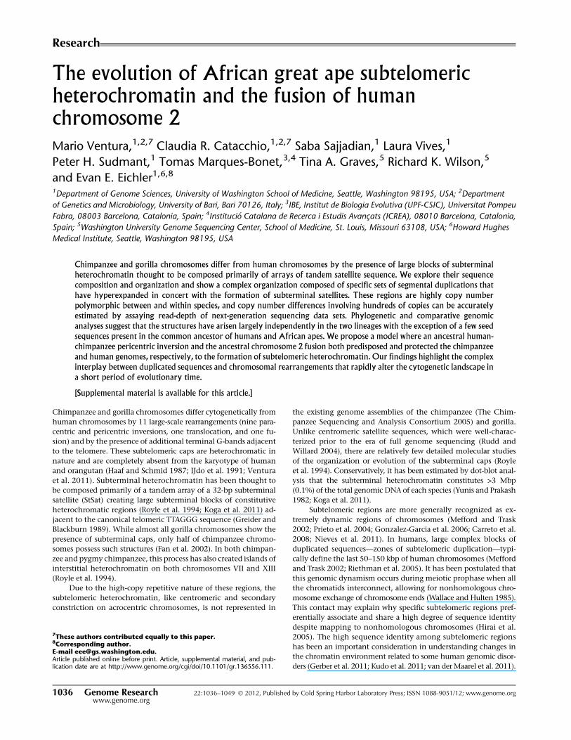
Research The evolution of African great ape subtelomeric heterochromatin and the fusion of human chromosome 2 Mario Ventura, 1,2,7 Claudia R. Catacchio, 1,2,7 Saba Sajjadian, 1 Laura Vives, 1 Peter H. Sudmant, 1 Tomas Marques-Bonet, 3,4 Tina A. Graves, 5 Richard K. Wilson, 5 and Evan E. Eichler 1,6,8 1 Department of Genome Sciences, University of Washington School of Medicine, Seattle, Washington 98195, USA; 2 Department of Genetics and Microbiology, University of Bari, Bari 70126, Italy; 3 IBE, Institut de Biologia Evolutiva (UPF-CSIC), Universitat Pompeu Fabra, 08003 Barcelona, Catalonia, Spain; 4 Institucio ´ Catalana de Recerca i Estudis Avanc xats (ICREA), 08010 Barcelona, Catalonia, Spain; 5 Washington University Genome Sequencing Center, School of Medicine, St. Louis, Missouri 63108, USA; 6 Howard Hughes Medical Institute, Seattle, Washington 98195, USA Chimpanzee and gorilla chromosomes differ from human chromosomes by the presence of large blocks of subterminal heterochromatin thought to be composed primarily of arrays of tandem satellite sequence. We explore their sequence composition and organization and show a complex organization composed of specific sets of segmental duplications that have hyperexpanded in concert with the formation of subterminal satellites. These regions are highly copy number polymorphic between and within species, and copy number differences involving hundreds of copies can be accurately estimated by assaying read-depth of next-generation sequencing data sets. Phylogenetic and comparative genomic analyses suggest that the structures have arisen largely independently in the two lineages with the exception of a few seed sequences present in the common ancestor of humans and African apes. We propose a model where an ancestral human- chimpanzee pericentric inversion and the ancestral chromosome 2 fusion both predisposed and protected the chimpanzee and human genomes, respectively, to the formation of subtelomeric heterochromatin. Our findings highlight the complex interplay between duplicated sequences and chromosomal rearrangements that rapidly alter the cytogenetic landscape in a short period of evolutionary time. [Supplemental material is available for this article.] Chimpanzee and gorilla chromosomes differ cytogenetically from human chromosomes by 11 large-scale rearrangements (nine para- centric and pericentric inversions, one translocation, and one fu- sion) and by the presence of additional terminal G-bands adjacent to the telomere. These subtelomeric caps are heterochromatic in nature and are completely absent from the karyotype of human and orangutan (Haaf and Schmid 1987; IJdo et al. 1991; Ventura et al. 2011). Subterminal heterochromatin has been thought to be composed primarily of a tandem array of a 32-bp subterminal satellite (StSat) creating large subterminal blocks of constitutive heterochromatic regions (Royle et al. 1994; Koga et al. 2011) ad- jacent to the canonical telomeric TTAGGG sequence (Greider and Blackburn 1989). While almost all gorilla chromosomes show the presence of subterminal caps, only half of chimpanzee chromo- somes possess such structures (Fan et al. 2002). In both chimpan- zee and pygmy chimpanzee, this process has also created islands of interstitial heterochromatin on both chromosomes VII and XIII (Royle et al. 1994). Due to the high-copy repetitive nature of these regions, the subtelomeric heterochromatin, like centromeric and secondary constriction on acrocentric chromosomes, is not represented in the existing genome assemblies of the chimpanzee (The Chim- panzee Sequencing and Analysis Consortium 2005) and gorilla. Unlike centromeric satellite sequences, which were well-charac- terized prior to the era of full genome sequencing (Rudd and Willard 2004), there are relatively few detailed molecular studies of the organization or evolution of the subterminal caps (Royle et al. 1994). Conservatively, it has been estimated by dot-blot anal- ysis that the subterminal heterochromatin constitutes >3 Mbp (0.1%) of the total genomic DNA of each species (Yunis and Prakash 1982; Koga et al. 2011). Subtelomeric regions are more generally recognized as ex- tremely dynamic regions of chromosomes (Mefford and Trask 2002; Prieto et al. 2004; Gonzalez-Garcia et al. 2006; Carreto et al. 2008; Nieves et al. 2011). In humans, large complex blocks of duplicated sequences—zones of subtelomeric duplication—typi- cally define the last 50–150 kbp of human chromosomes (Mefford and Trask 2002; Riethman et al. 2005). It has been postulated that this genomic dynamism occurs during meiotic prophase when all the chromatids interconnect, allowing for nonhomologous chro- mosome exchange of chromosome ends (Wallace and Hulten 1985). This contact may explain why specific subtelomeric regions pref- erentially associate and share a high degree of sequence identity despite mapping to nonhomologous chromosomes (Hirai et al. 2005). The high sequence identity among subtelomeric regions has been an important consideration in understanding changes in the chromatin environment related to some human genomic disor- ders (Gerber et al. 2011; Kudo et al. 2011; van der Maarel et al. 2011). 7 These authors contributed equally to this paper. 8 Corresponding author. E-mail [email protected]. Article published online before print. Article, supplemental material, and pub- lication date are at http://www.genome.org/cgi/doi/10.1101/gr.136556.111. 1036 Genome Research www.genome.org 22:1036–1049 Ó 2012, Published by Cold Spring Harbor Laboratory Press; ISSN 1088-9051/12; www.genome.org
-
Upload
independent -
Category
Documents
-
view
0 -
download
0
Transcript of The evolution of African great ape subtelomeric heterochromatin and the fusion of human chromosome 2
Research
The evolution of African great ape subtelomericheterochromatin and the fusion of humanchromosome 2Mario Ventura,1,2,7 Claudia R. Catacchio,1,2,7 Saba Sajjadian,1 Laura Vives,1
Peter H. Sudmant,1 Tomas Marques-Bonet,3,4 Tina A. Graves,5 Richard K. Wilson,5
and Evan E. Eichler1,6,8
1Department of Genome Sciences, University of Washington School of Medicine, Seattle, Washington 98195, USA; 2Department
of Genetics and Microbiology, University of Bari, Bari 70126, Italy; 3IBE, Institut de Biologia Evolutiva (UPF-CSIC), Universitat Pompeu
Fabra, 08003 Barcelona, Catalonia, Spain; 4Institucio Catalana de Recerca i Estudis Avancxats (ICREA), 08010 Barcelona, Catalonia,
Spain; 5Washington University Genome Sequencing Center, School of Medicine, St. Louis, Missouri 63108, USA; 6Howard Hughes
Medical Institute, Seattle, Washington 98195, USA
Chimpanzee and gorilla chromosomes differ from human chromosomes by the presence of large blocks of subterminalheterochromatin thought to be composed primarily of arrays of tandem satellite sequence. We explore their sequencecomposition and organization and show a complex organization composed of specific sets of segmental duplications thathave hyperexpanded in concert with the formation of subterminal satellites. These regions are highly copy numberpolymorphic between and within species, and copy number differences involving hundreds of copies can be accuratelyestimated by assaying read-depth of next-generation sequencing data sets. Phylogenetic and comparative genomicanalyses suggest that the structures have arisen largely independently in the two lineages with the exception of a few seedsequences present in the common ancestor of humans and African apes. We propose a model where an ancestral human-chimpanzee pericentric inversion and the ancestral chromosome 2 fusion both predisposed and protected the chimpanzeeand human genomes, respectively, to the formation of subtelomeric heterochromatin. Our findings highlight the complexinterplay between duplicated sequences and chromosomal rearrangements that rapidly alter the cytogenetic landscape ina short period of evolutionary time.
[Supplemental material is available for this article.]
Chimpanzee and gorilla chromosomes differ cytogenetically from
human chromosomes by 11 large-scale rearrangements (nine para-
centric and pericentric inversions, one translocation, and one fu-
sion) and by the presence of additional terminal G-bands adjacent
to the telomere. These subtelomeric caps are heterochromatic in
nature and are completely absent from the karyotype of human
and orangutan (Haaf and Schmid 1987; IJdo et al. 1991; Ventura
et al. 2011). Subterminal heterochromatin has been thought to
be composed primarily of a tandem array of a 32-bp subterminal
satellite (StSat) creating large subterminal blocks of constitutive
heterochromatic regions (Royle et al. 1994; Koga et al. 2011) ad-
jacent to the canonical telomeric TTAGGG sequence (Greider and
Blackburn 1989). While almost all gorilla chromosomes show the
presence of subterminal caps, only half of chimpanzee chromo-
somes possess such structures (Fan et al. 2002). In both chimpan-
zee and pygmy chimpanzee, this process has also created islands of
interstitial heterochromatin on both chromosomes VII and XIII
(Royle et al. 1994).
Due to the high-copy repetitive nature of these regions, the
subtelomeric heterochromatin, like centromeric and secondary
constriction on acrocentric chromosomes, is not represented in
the existing genome assemblies of the chimpanzee (The Chim-
panzee Sequencing and Analysis Consortium 2005) and gorilla.
Unlike centromeric satellite sequences, which were well-charac-
terized prior to the era of full genome sequencing (Rudd and
Willard 2004), there are relatively few detailed molecular studies
of the organization or evolution of the subterminal caps (Royle
et al. 1994). Conservatively, it has been estimated by dot-blot anal-
ysis that the subterminal heterochromatin constitutes >3 Mbp
(0.1%) of the total genomic DNA of each species (Yunis and Prakash
1982; Koga et al. 2011).
Subtelomeric regions are more generally recognized as ex-
tremely dynamic regions of chromosomes (Mefford and Trask
2002; Prieto et al. 2004; Gonzalez-Garcia et al. 2006; Carreto et al.
2008; Nieves et al. 2011). In humans, large complex blocks of
duplicated sequences—zones of subtelomeric duplication—typi-
cally define the last 50–150 kbp of human chromosomes (Mefford
and Trask 2002; Riethman et al. 2005). It has been postulated that
this genomic dynamism occurs during meiotic prophase when all
the chromatids interconnect, allowing for nonhomologous chro-
mosome exchange of chromosome ends (Wallace and Hulten 1985).
This contact may explain why specific subtelomeric regions pref-
erentially associate and share a high degree of sequence identity
despite mapping to nonhomologous chromosomes (Hirai et al.
2005). The high sequence identity among subtelomeric regions
has been an important consideration in understanding changes in
the chromatin environment related to some human genomic disor-
ders (Gerber et al. 2011; Kudo et al. 2011; van der Maarel et al. 2011).
7These authors contributed equally to this paper.8Corresponding author.E-mail [email protected] published online before print. Article, supplemental material, and pub-lication date are at http://www.genome.org/cgi/doi/10.1101/gr.136556.111.
1036 Genome Researchwww.genome.org
22:1036–1049 � 2012, Published by Cold Spring Harbor Laboratory Press; ISSN 1088-9051/12; www.genome.org
Recently, we identified the presence of specific segmental
duplications that have hyperexpanded within the subterminal
cap of both gorilla and chimpanzee (Cheng et al. 2005; Marques-
Bonet et al. 2009; Ventura et al. 2011). In this study, we perform
a detailed investigation into the organization and evolution of
subterminal caps using molecular cytogenetics, targeted clone-
based sequencing, and phylogenetic-based analyses. Based on the
evolutionary history of hominid chromosomal rearrangements,
we develop a model to explain the distribution and organization
of subtelomeric caps in gorilla and chimpanzee, as well as their
absence in humans. We hypothesize that ancestral chromosomal
rearrangements were important in catalyzing the spread of these
chromosome structures and that the fusion of human chromo-
some 2 squelched this process in humans by eliminating the
source of the StSat sequence shortly after human speciation.
Results
Subtelomeric cap distribution among gorillaand chimpanzee chromosomes
In order to test the localization of telomeric TTAGGG sequences
with respect to the 32-bp StSat, we began by performing bicolor
fluorescent in situ hybridization (FISH) experiments using bio-
tinylated oligonucleotides designed to the satellite sequence in
conjunction with a commercial PNA (peptide nucleic acid) corre-
sponding to telomeric TTAGGG sequence (Methods). As expected,
the telomeric satellite colocalizes with ‘‘caps’’ in both species,
mapping immediately proximal to the PNA telomere probe (Sup-
plemental Fig. S1). We were able to identify all chromosome arms
carrying StSat in chimpanzee (42/96 chromosome tips and inter-
stitial signals on chromosomes VII and XIII) and in gorilla (80/96
chromosome tips). Cap signals were detected on short arms of IIp and
IIq chromosomes in chimpanzee and on both arms of IIq in gorilla.
No traces of signals were observed on the long arms of chimpanzee
IIp and IIq or on the short arm of gorilla IIp. For gorilla, in particular,
we did not detect signals on any of the p arms of the acrocentric
chromosomes (IIp, IX, XIII, XIV, XV, XVIII, XXI, and XXII), except for
chromosome IIq showing signals on the p and q arms.
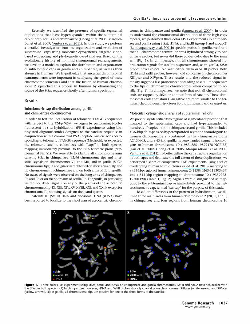
Satellite III (SatIII) DNA and ribosomal DNA (rDNA) have
been reported to localize to the short arm of acrocentric chromo-
somes in chimpanzee and gorilla (Jarmuz et al. 2007). In order
to understand the chromosomal distribution of these high-copy
repeats, we performed three-color FISH experiments in chimpan-
zee and gorilla using StSat, rDNA, and SatIII (group 1 and group 2)
(Bandyopadhyay et al. 2001b) specific probes. In gorilla, we found
that all chromosome termini or arms hybridized strongly to one
of these probes, but never did these probes colocalize to the same
arm (Fig. 1). In chimpanzee, not all chromosomes showed hy-
bridization signals for satellite sequences and, as in gorilla, StSat
probes never colocalized with either rDNA or SatIII probes. Both
rDNA and SatIII probes, however, did colocalize on chromosomes
XIIIpter and XIVpter. These results and the reduced signal in-
tensity suggest a less pronounced spreading of satellite sequences
to the tips of chimpanzee chromosomes when compared to go-
rilla (Fig. 1). In chimpanzee, we note that not all chromosomal
ends are capped by StSat or another form of satellite. These chro-
mosomal ends that stain G-negative are more similar to the ter-
minal chromosomal structures found in human and orangutan.
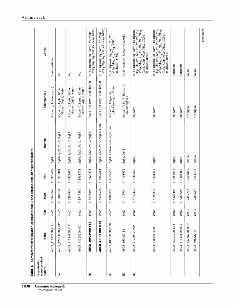
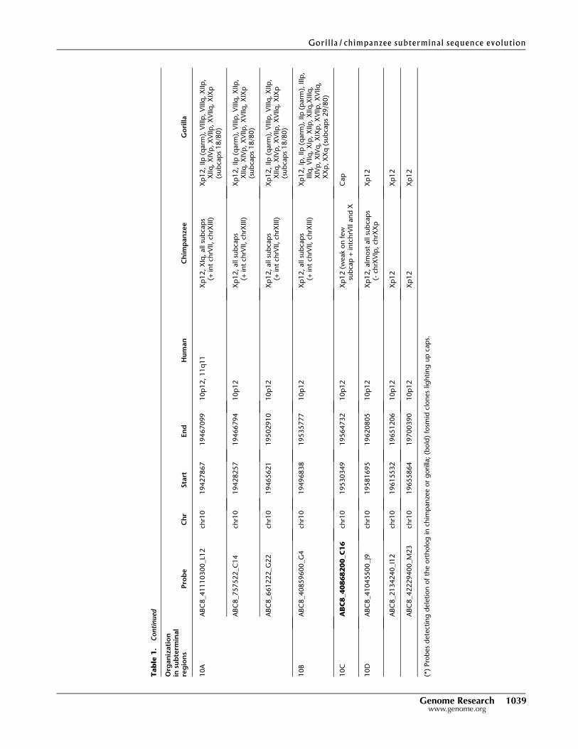
Molecular cytogenetic analysis of subterminal regions
We previously identified two regions of segmental duplication that
mapped to the subterminal caps and had hyperexpanded to
hundreds of copies in both chimpanzee and gorilla. This included
a 36-kbp chimpanzee-hyperexpanded segment homologous to
human chromosome 2, contained in the chimpanzee clone
AC150905, and a 45-kbp gorilla-hyperexpanded segment homolo-
gous to human chromosome 10 (19534885-19579478 NCBI35)
(Fan et al. 2002; Cheng et al. 2005; Marques-Bonet et al. 2009;
Ventura et al. 2011). To better define the cap structure organization
in both apes and delineate the full extent of these duplications, we
performed a series of comparative FISH experiments using a set of
overlapping human fosmid clones (Kidd et al. 2010) mapping to
a 463-kbp region of human chromosome 2 (113840263-114303469)
and a 341-kbp region mapping to chromosome 10 (19359773-
19700390) (Table 1; Fig. 2). Signals were distinguished as map-
ping to the subterminal cap or immediately proximal to the het-
erochromatic cap, termed ‘‘subcap’’ for the purpose of this study.
Based on differences in the pattern of hybridization, we de-
fined three main areas from human chromosome 2 (2B, C, and E)
in chimpanzee and four regions from human chromosome 10
Figure 1. Three-color FISH experiment using StSat, SatIII, and rDNA on chimpanzee and gorilla chromosomes. SatIII and rDNA never colocalize withthe StSat in both species. (A) In chimpanzee, however, rDNA and SatIII probes strongly colocalize on chromosomes XIIIpter (white arrows) and XIVpter(yellow arrows). (B) In gorilla, all chromosomal tips are positive for one of the three forms of the satellite.
Genome Research 1037www.genome.org
Gorilla/chimpanzee subterminal sequence evolution
Tab
le1
.C
om
para
tive
hyb
rid
izati
on
of
chro
mo
som
e2
an
dch
rom
oso
me
10
hyp
ere
xp
an
sio
ns
Org
an
izati
on
insu
bte
rmin
al
reg
ion
sP
rob
eC
hr
Sta
rtEn
dH
um
an
Ch
imp
an
zee
Go
rilla
ABC
8_41147200_H
23
chr2
113840263
113878692
2q
14
IIp
(parm
),IIp
(Cen
parm
)IIp
(in
ters
titi
al)
2A
ABC
8_42163800_E20*
chr2
113884171
113915881
2q
14,9p
24,9p
12,9q
13
IIp
(parm
),IIIp
ter,
IVq
ter,
VIIq
ter,
IXq
11,IX
qte
rIX
q
ABC
8_41115100_I1
7*
chr2
113894854
113929406
2q
14,9p
24,9p
12,9q
13
IIp
(parm
),IIIp
ter,
IVq
ter,
VIIq
ter,
IXq
11,IX
qte
rIX
q
ABC
8_43008200_P2*
chr2
113919580
113958771
2q
14,9p
24,9p
12,9q
13
IIp
(parm
),IIIp
ter,
IVq
ter,
VIIq
ter,
IXq
11,IX
qte
rIX
q
2B
AB
C8
_40
92
59
00
_F1
2ch
r2113978394
114020431
2q
14,9p
24,9p
12,9q
13
Cap
(+in
tch
rVII
an
dch
rXIII)
Ip,IIp
(qarm
),IIq
(parm
),V
q,V
IIIp
,V
IIIq
,IX
p,X
q,X
IIIq
(Sub
cap
14/8
0)
AB
C8
_41
13
41
00
_D2
2ch
r2114021524
114059681
2q
14,9p
24,9p
12,9q
13,su
bte
lC
ap
(+in
tch
rVII
an
dch
rXIII)
Ip,IIp
(qarm
),IIq
(parm
),V
q,V
IIIp
,V
IIIq
,IX
p,X
q,X
IIIq
(Sub
cap
14/8
0)
2C
ABC
8_40903600_D
20
chr2
114080059
114122590
2q
14,su
bte
lom
eri
csi
gn
als
on
IIp
(parm
),IIq
(parm
),su
bte
lst
ron
gon
VIq
ter,
Ip,IIp
(qarm
),IIq
(parm
),V
q,V
Ip,
VIIIp
,V
IIIq
,X
q,X
IIIq
,X
XIIq
(Sub
cap
16/8
0)
2D
ABC
8_660422_B2
chr2
114111934
114153971
2q
14,2q
11
IIp
(parm
),IIp
11,IIq
(parm
)d
oub
lesi
gn
als
IIp
(in
ters
titial)
,IIq
(parm
)(w
eak)
2E
ABC
8_2126940_M
24
chr2
114149372
114184630
2q
14
IIq
(parm
)Ip
,IIp
(qarm
),IIq
(parm
),IIq
(qarm
),IIIp
,IIIq
,IV
p,IV
q,V
p,V
q,V
Ip,V
Iq,
VIIIp
,V
IIIq
,X
p,X
q,X
Ip,X
Iq,X
IIIq
,X
IVq
,X
VIp
,X
IXp
,X
IXq
,X
XIIq
(Sub
cap
s38/8
0)
ABC
8_738840_B20
chr2
114196346
114231473
2q
14
IIq
(parm
)Ip
,IIp
(qarm
),IIq
(parm
),IIq
(qarm
),IIIp
,IIIq
,IV
p,IV
q,V
p,V
q,V
Ip,V
Iq,
VIIIp
,V
IIIq
,X
p,X
q,X
Ip,X
Iq,X
IIIq
,X
IVq
,X
VIp
,X
IXp
,X
IXq
,X
XIIq
(Sub
cap
s38/8
0)
ABC
8_45707700_L2
4ch
r2114223493
114258048
2q
14
IIq
(parm
)IIq
(parm
)
ABC
8_41123600_B12
chr2
114267007
114303469
2q
14
IIq
(parm
)IIq
(parm
)
ABC
8_43746700_B12*
chr1
019359773
19395890
10p
12
No
sig
nal
Xp
12
ABC
8_708622_I7
*ch
r10
19399597
19435714
10p
12
No
sig
nal
Xp
12
(conti
nued
)
Ventura et al .
1038 Genome Researchwww.genome.org
Tab
le1
.C
on
tin
ued
Org
an
izati
on
insu
bte
rmin
al
reg
ion
sP
rob
eC
hr
Sta
rtEn
dH
um
an
Ch
imp
an
zee
Go
rilla
10A
ABC
8_41110300_L1
2ch
r10
19427867
19467099
10p
12,11q
11
Xp
12,
XIq
,all
sub
cap
s(+
int
chrV
II,ch
rXIII)
Xp
12,IIp
(qarm
),V
IIIp
,V
IIIq
,X
IIp
,X
IIq
,X
IVp
,X
VIIp
,X
VIIq
,X
IXp
(sub
cap
s18/8
0)
ABC
8_757522_C
14
chr1
019428257
19466794
10p
12
Xp
12,
all
sub
cap
s(+
int
chrV
II,ch
rXIII)
Xp
12,IIp
(qarm
),V
IIIp
,V
IIIq
,X
IIp
,X
IIq
,X
IVp
,X
VIIp
,X
VIIq
,X
IXp
(sub
cap
s18/8
0)
ABC
8_661222_G
22
chr1
019465621
19502910
10p
12
Xp
12,
all
sub
cap
s(+
int
chrV
II,ch
rXIII)
Xp
12,IIp
(qarm
),V
IIIp
,V
IIIq
,X
IIp
,X
IIq
,X
IVp
,X
VIIp
,X
VIIq
,X
IXp
(sub
cap
s18/8
0)
10B
ABC
8_40859600_G
4ch
r10
19496838
19535777
10p
12
Xp
12,
all
sub
cap
s(+
int
chrV
II,ch
rXIII)
Xp
12,Ip
,IIp
(qarm
),IIp
(parm
),IIIp
,IIIq
,V
IIq
,X
Ip,X
IIp
,X
IIq
,XIIIq
,X
IVp
,X
IVq
,X
IXp
,X
VIIp
,X
VIIq
,X
Xp
,X
Xq
(sub
cap
s29/8
0)
10C
AB
C8
_40
86
82
00
_C1
6ch
r10
19530349
19564732
10p
12
Xp
12
(weak
on
few
sub
cap
+in
tch
rVII
an
dX
Cap
10D
ABC
8_41045500_J9
chr1
019581695
19620805
10p
12
Xp
12,
alm
ost
all
sub
cap
s(-
chrX
VIp
,ch
rXX
pX
p12
ABC
8_2134240_I1
2ch
r10
19615532
19651206
10p
12
Xp
12
Xp
12
ABC
8_42229400_M
23
chr1
019655864
19700390
10p
12
Xp
12
Xp
12
(*)
Pro
bes
dete
ctin
gd
ele
tion
of
the
ort
holo
gin
chim
pan
zee
or
gorilla
;(b
old
)fo
smid
clon
es
ligh
tin
gup
cap
s.
Gorilla/chimpanzee subterminal sequence evolution
Genome Research 1039www.genome.org
Figure 2. Duplication and fosmid probe map. The pattern of segmental duplications is shown for (A) a 463-kbp region of human chromosome 2(chr2:113840263-114303469) and (B) a 341-kbp region mapping to chromosome 10 (chr10:19359773-19700390) based on human genome anno-tation (NCBI build35). Computationally predicted human, chimpanzee, and gorilla duplications (red = excess depth-of-coverage of aligned whole-genome shotgun sequence) as well as a heat map indicating the copy number are displayed. Fosmid clone contigs are reported below each genomicregion. Human fosmid probes underlying each region are shown and grouped (2A–E and 10A–D) based on the pattern of hybridization to chimpanzee andgorilla subtelomeric cap and subcap regions. (HSA) Homo sapiens, (PTR) Pan troglodytes, (GGO) Gorilla gorilla.
Ventura et al .
1040 Genome Researchwww.genome.org
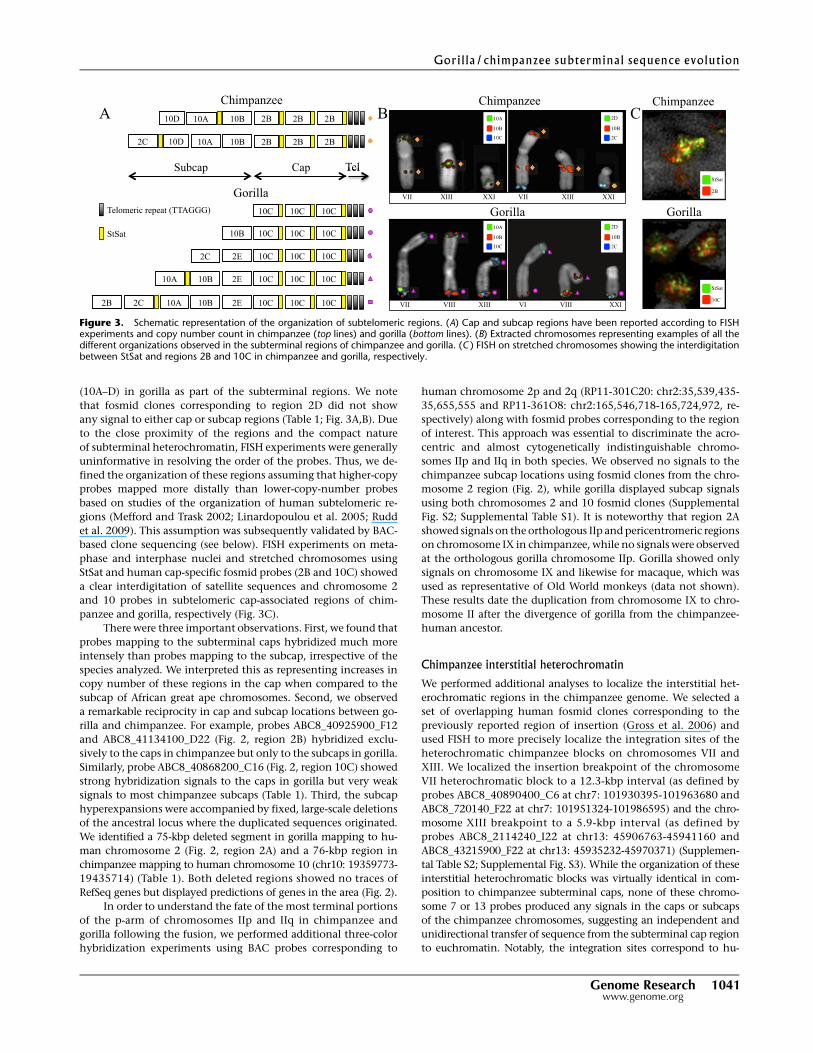
(10A–D) in gorilla as part of the subterminal regions. We note
that fosmid clones corresponding to region 2D did not show
any signal to either cap or subcap regions (Table 1; Fig. 3A,B). Due
to the close proximity of the regions and the compact nature
of subterminal heterochromatin, FISH experiments were generally
uninformative in resolving the order of the probes. Thus, we de-
fined the organization of these regions assuming that higher-copy
probes mapped more distally than lower-copy-number probes
based on studies of the organization of human subtelomeric re-
gions (Mefford and Trask 2002; Linardopoulou et al. 2005; Rudd
et al. 2009). This assumption was subsequently validated by BAC-
based clone sequencing (see below). FISH experiments on meta-
phase and interphase nuclei and stretched chromosomes using
StSat and human cap-specific fosmid probes (2B and 10C) showed
a clear interdigitation of satellite sequences and chromosome 2
and 10 probes in subtelomeric cap-associated regions of chim-
panzee and gorilla, respectively (Fig. 3C).
There were three important observations. First, we found that
probes mapping to the subterminal caps hybridized much more
intensely than probes mapping to the subcap, irrespective of the
species analyzed. We interpreted this as representing increases in
copy number of these regions in the cap when compared to the
subcap of African great ape chromosomes. Second, we observed
a remarkable reciprocity in cap and subcap locations between go-
rilla and chimpanzee. For example, probes ABC8_40925900_F12
and ABC8_41134100_D22 (Fig. 2, region 2B) hybridized exclu-
sively to the caps in chimpanzee but only to the subcaps in gorilla.
Similarly, probe ABC8_40868200_C16 (Fig. 2, region 10C) showed
strong hybridization signals to the caps in gorilla but very weak
signals to most chimpanzee subcaps (Table 1). Third, the subcap
hyperexpansions were accompanied by fixed, large-scale deletions
of the ancestral locus where the duplicated sequences originated.
We identified a 75-kbp deleted segment in gorilla mapping to hu-
man chromosome 2 (Fig. 2, region 2A) and a 76-kbp region in
chimpanzee mapping to human chromosome 10 (chr10: 19359773-
19435714) (Table 1). Both deleted regions showed no traces of
RefSeq genes but displayed predictions of genes in the area (Fig. 2).
In order to understand the fate of the most terminal portions
of the p-arm of chromosomes IIp and IIq in chimpanzee and
gorilla following the fusion, we performed additional three-color
hybridization experiments using BAC probes corresponding to
human chromosome 2p and 2q (RP11-301C20: chr2:35,539,435-
35,655,555 and RP11-361O8: chr2:165,546,718-165,724,972, re-
spectively) along with fosmid probes corresponding to the region
of interest. This approach was essential to discriminate the acro-
centric and almost cytogenetically indistinguishable chromo-
somes IIp and IIq in both species. We observed no signals to the
chimpanzee subcap locations using fosmid clones from the chro-
mosome 2 region (Fig. 2), while gorilla displayed subcap signals
using both chromosomes 2 and 10 fosmid clones (Supplemental
Fig. S2; Supplemental Table S1). It is noteworthy that region 2A
showed signals on the orthologous IIp and pericentromeric regions
on chromosome IX in chimpanzee, while no signals were observed
at the orthologous gorilla chromosome IIp. Gorilla showed only
signals on chromosome IX and likewise for macaque, which was
used as representative of Old World monkeys (data not shown).
These results date the duplication from chromosome IX to chro-
mosome II after the divergence of gorilla from the chimpanzee-
human ancestor.
Chimpanzee interstitial heterochromatin
We performed additional analyses to localize the interstitial het-
erochromatic regions in the chimpanzee genome. We selected a
set of overlapping human fosmid clones corresponding to the
previously reported region of insertion (Gross et al. 2006) and
used FISH to more precisely localize the integration sites of the
heterochromatic chimpanzee blocks on chromosomes VII and
XIII. We localized the insertion breakpoint of the chromosome
VII heterochromatic block to a 12.3-kbp interval (as defined by
probes ABC8_40890400_C6 at chr7: 101930395-101963680 and
ABC8_720140_F22 at chr7: 101951324-101986595) and the chro-
mosome XIII breakpoint to a 5.9-kbp interval (as defined by
probes ABC8_2114240_I22 at chr13: 45906763-45941160 and
ABC8_43215900_F22 at chr13: 45935232-45970371) (Supplemen-
tal Table S2; Supplemental Fig. S3). While the organization of these
interstitial heterochromatic blocks was virtually identical in com-
position to chimpanzee subterminal caps, none of these chromo-
some 7 or 13 probes produced any signals in the caps or subcaps
of the chimpanzee chromosomes, suggesting an independent and
unidirectional transfer of sequence from the subterminal cap region
to euchromatin. Notably, the integration sites correspond to hu-
Figure 3. Schematic representation of the organization of subtelomeric regions. (A) Cap and subcap regions have been reported according to FISHexperiments and copy number count in chimpanzee (top lines) and gorilla (bottom lines). (B) Extracted chromosomes representing examples of all thedifferent organizations observed in the subterminal regions of chimpanzee and gorilla. (C ) FISH on stretched chromosomes showing the interdigitationbetween StSat and regions 2B and 10C in chimpanzee and gorilla, respectively.
Gorilla/chimpanzee subterminal sequence evolution
Genome Research 1041www.genome.org
man-chimpanzee and human-chimpanzee-gorilla segmentally du-
plicated regions in chromosome VII and chromosome XIII, respec-
tively. Further, in the case of chromosome VII, the insertion point
maps within 1 Mbp from an inversion breakpoint specific to the
ancestral lineage of human and chimpanzee (Ventura et al. 2011).
Copy number variation between and within species
We performed two sets of experiments to assess the extent of var-
iation between and within species. To assess qualitative differences
in the chromosomal distribution, we first performed cohybridiza-
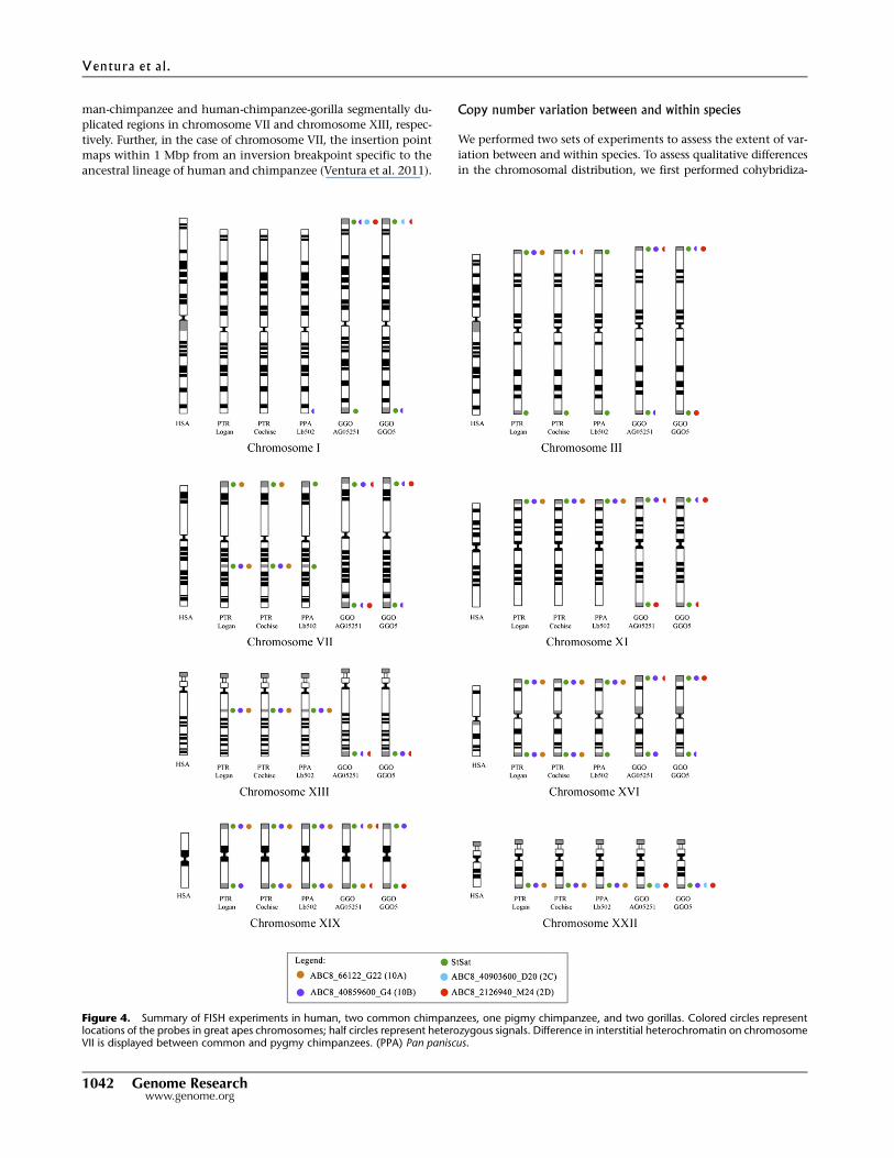
Figure 4. Summary of FISH experiments in human, two common chimpanzees, one pigmy chimpanzee, and two gorillas. Colored circles representlocations of the probes in great apes chromosomes; half circles represent heterozygous signals. Difference in interstitial heterochromatin on chromosomeVII is displayed between common and pygmy chimpanzees. (PPA) Pan paniscus.
Ventura et al .
1042 Genome Researchwww.genome.org
tion experiments on metaphase chromosomal spreads from two
gorillas, two chimpanzees, and one bonobo. Marked differences
in the distribution and signal intensity were observed, especially
when comparing homologous chromosomes in gorilla (Figs. 3B, 4),
where subterminal regions showed differences in the organization
of the cap (see chromosomes XIX and XXII). The two common
chimpanzees and the bonobo showed a much more uniform distri-
bution of probes, with the exception of the interstitial chromosome
VII heterochromatic block, which appears less complex in bonobo
when compared to the two common chimpanzees (Figs. 3B, 4).
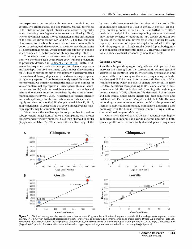
To obtain a quantitative assessment of copy number varia-
tion, we performed read-depth-based copy number predictions
as previously described in Sudmant et al. (2010). Briefly, next-
generation sequence reads were mapped to reference sequences
and read-depth was used to estimate copy number after correcting
for GC-bias. While the efficacy of this approach has been validated
for low- to middle-copy duplications, the dynamic range response
of high-copy repeats had not been previously tested. To assess this
more formally, we initially estimated the median copy number for
various subterminal cap regions in three species (human, chim-
panzee, and gorilla) and compared these values to the number and
relative fluorescence intensity normalized by the value of maxi-
mum fluorescence (VMF = 255). The relative fluorescence intensity
and read-depth copy number for each locus in each species were
highly correlated (r2 = 0.93–0.99) (Supplemental Table S3; Fig. 5;
Supplemental Fig. S4), suggesting that copy number, even for high-
copy repeats, may be accurately estimated.
We estimate the median species copy number for various
subcap regions ranges from 29 to 64 in chimpanzee with greater
diversity and lower copy number (12–55) than observed in gorilla
(Supplemental Table S3). We estimate the median copy of the
hyperexpanded segments within the subterminal cap to be 798
in chimpanzee compared to 1092 in gorilla. In contrast, all ana-
lyzed human genomes, as well as the Neanderthal, were either
predicted to be diploid for the corresponding segments or showed
only modest evidence of duplication (<14 copies). Adjusting for
the size of the probes and differences in copy number for each
segment, the amount of segmental duplication added to the cap
and subcap regions is strikingly similar (;46 Mbp) in both gorilla
and chimpanzee (Supplemental Table S3). This value exceeds the
initial estimates of StSat sequence by more than 10-fold.
Sequence analyses
Since the subcap and cap regions of gorilla and chimpanzee chro-
mosomes are missing from the corresponding primate genome
assemblies, we identified large-insert clones by hybridization and
sequenced the inserts using capillary-based sequencing methods.
We also used BLAST to search for sequence similarity with StSat
(contained in the pCht7 and pCht13 clones) (Royle et al. 1994) for
GenBank accessions corresponding to BAC clones carrying these
sequences within the nucleotide (nr/nt) and high-throughput ge-
nomic sequence (HTGS) collections. We identified 17 chimpanzee
and nine gorilla clones whose inserts had been sequenced and
had tracts of StSat sequence (Supplemental Table S4). The cor-
responding sequences were annotated as StSat, the presence of
segmental duplications in human, chimpanzee, and gorilla, and
homology with the human reference genome using a suite of
computational programs (Methods).
Our analysis showed that all 26 BAC sequences were highly
duplicated in chimpanzee and gorilla genomes and carried both
species-specific as well as ancestrally shared duplicated sequences
Figure 5. Distribution copy number counts versus fluorescence. Copy number estimates of sequence read-depth for each genomic region correlatestrongly (r2 = 0.99) with measured fluorescence intensity for every probe distributed on chromosome 2 and chromosome 10 (see Supplemental Table S3).Red arrows show the location of the single probe positive for cap, while blue arrows display the group of probes positive for subcap in (A) chimpanzee and(B) gorilla (left panels). The correlation rates reduce when hyperexpanded segments are excluded from the analysis (right panels).
Gorilla/chimpanzee subterminal sequence evolution
Genome Research 1043www.genome.org
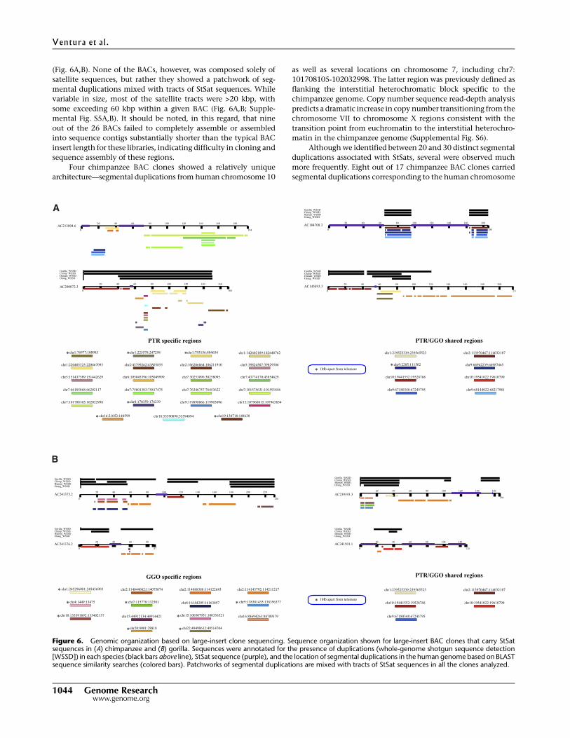
(Fig. 6A,B). None of the BACs, however, was composed solely of
satellite sequences, but rather they showed a patchwork of seg-
mental duplications mixed with tracts of StSat sequences. While
variable in size, most of the satellite tracts were >20 kbp, with
some exceeding 60 kbp within a given BAC (Fig. 6A,B; Supple-
mental Fig. S5A,B). It should be noted, in this regard, that nine
out of the 26 BACs failed to completely assemble or assembled
into sequence contigs substantially shorter than the typical BAC
insert length for these libraries, indicating difficulty in cloning and
sequence assembly of these regions.
Four chimpanzee BAC clones showed a relatively unique
architecture—segmental duplications from human chromosome 10
as well as several locations on chromosome 7, including chr7:
101708105-102032998. The latter region was previously defined as
flanking the interstitial heterochromatic block specific to the
chimpanzee genome. Copy number sequence read-depth analysis
predicts a dramatic increase in copy number transitioning from the
chromosome VII to chromosome X regions consistent with the
transition point from euchromatin to the interstitial heterochro-
matin in the chimpanzee genome (Supplemental Fig. S6).
Although we identified between 20 and 30 distinct segmental
duplications associated with StSats, several were observed much
more frequently. Eight out of 17 chimpanzee BAC clones carried
segmental duplications corresponding to the human chromosome
Figure 6. Genomic organization based on large-insert clone sequencing. Sequence organization shown for large-insert BAC clones that carry StSatsequences in (A) chimpanzee and (B) gorilla. Sequences were annotated for the presence of duplications (whole-genome shotgun sequence detection[WSSD]) in each species (black bars above line), StSat sequence (purple), and the location of segmental duplications in the human genome based on BLASTsequence similarity searches (colored bars). Patchworks of segmental duplications are mixed with tracts of StSat sequences in all the clones analyzed.
Ventura et al .
1044 Genome Researchwww.genome.org
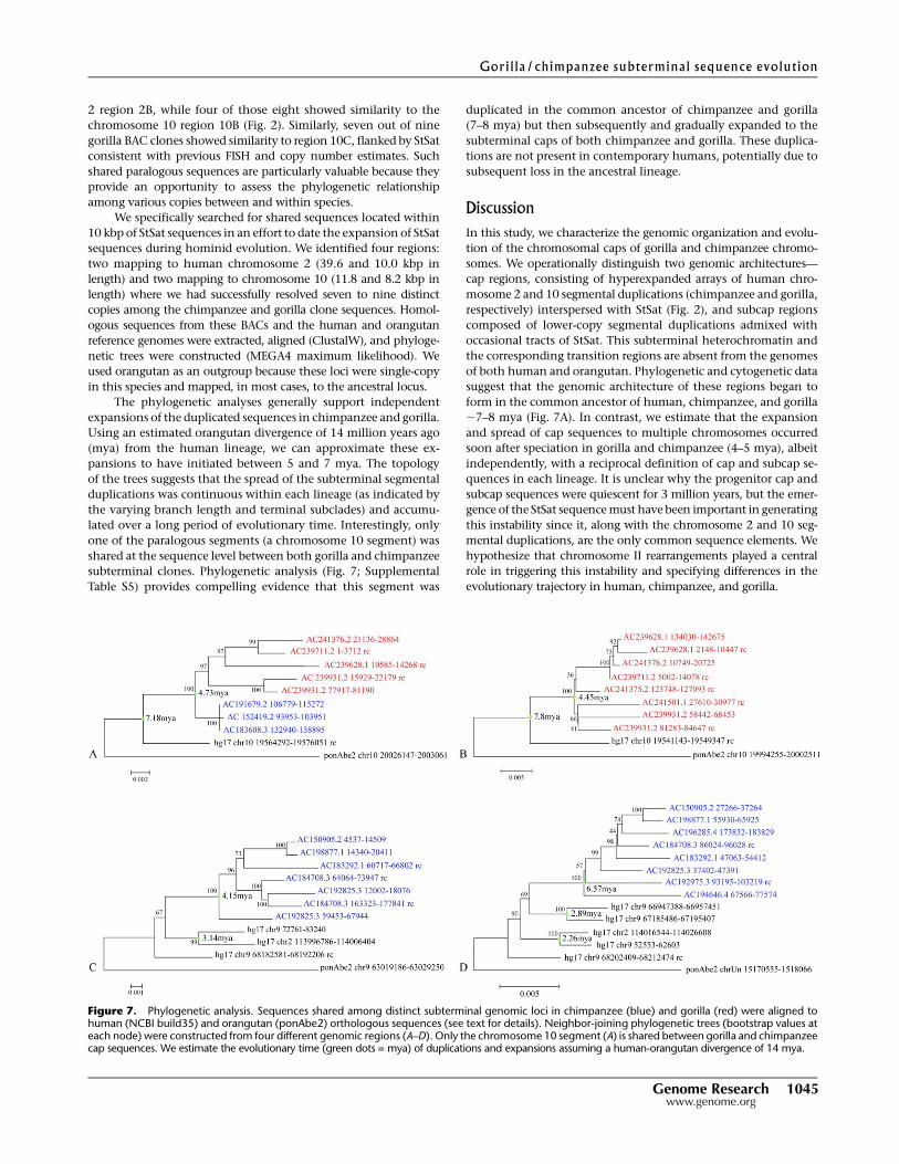
2 region 2B, while four of those eight showed similarity to the
chromosome 10 region 10B (Fig. 2). Similarly, seven out of nine
gorilla BAC clones showed similarity to region 10C, flanked by StSat
consistent with previous FISH and copy number estimates. Such
shared paralogous sequences are particularly valuable because they
provide an opportunity to assess the phylogenetic relationship
among various copies between and within species.
We specifically searched for shared sequences located within
10 kbp of StSat sequences in an effort to date the expansion of StSat
sequences during hominid evolution. We identified four regions:
two mapping to human chromosome 2 (39.6 and 10.0 kbp in
length) and two mapping to chromosome 10 (11.8 and 8.2 kbp in
length) where we had successfully resolved seven to nine distinct
copies among the chimpanzee and gorilla clone sequences. Homol-
ogous sequences from these BACs and the human and orangutan
reference genomes were extracted, aligned (ClustalW), and phyloge-
netic trees were constructed (MEGA4 maximum likelihood). We
used orangutan as an outgroup because these loci were single-copy
in this species and mapped, in most cases, to the ancestral locus.
The phylogenetic analyses generally support independent
expansions of the duplicated sequences in chimpanzee and gorilla.
Using an estimated orangutan divergence of 14 million years ago
(mya) from the human lineage, we can approximate these ex-
pansions to have initiated between 5 and 7 mya. The topology
of the trees suggests that the spread of the subterminal segmental
duplications was continuous within each lineage (as indicated by
the varying branch length and terminal subclades) and accumu-
lated over a long period of evolutionary time. Interestingly, only
one of the paralogous segments (a chromosome 10 segment) was
shared at the sequence level between both gorilla and chimpanzee
subterminal clones. Phylogenetic analysis (Fig. 7; Supplemental
Table S5) provides compelling evidence that this segment was
duplicated in the common ancestor of chimpanzee and gorilla
(7–8 mya) but then subsequently and gradually expanded to the
subterminal caps of both chimpanzee and gorilla. These duplica-
tions are not present in contemporary humans, potentially due to
subsequent loss in the ancestral lineage.
DiscussionIn this study, we characterize the genomic organization and evolu-
tion of the chromosomal caps of gorilla and chimpanzee chromo-
somes. We operationally distinguish two genomic architectures—
cap regions, consisting of hyperexpanded arrays of human chro-
mosome 2 and 10 segmental duplications (chimpanzee and gorilla,
respectively) interspersed with StSat (Fig. 2), and subcap regions
composed of lower-copy segmental duplications admixed with
occasional tracts of StSat. This subterminal heterochromatin and
the corresponding transition regions are absent from the genomes
of both human and orangutan. Phylogenetic and cytogenetic data
suggest that the genomic architecture of these regions began to
form in the common ancestor of human, chimpanzee, and gorilla
;7–8 mya (Fig. 7A). In contrast, we estimate that the expansion
and spread of cap sequences to multiple chromosomes occurred
soon after speciation in gorilla and chimpanzee (4–5 mya), albeit
independently, with a reciprocal definition of cap and subcap se-
quences in each lineage. It is unclear why the progenitor cap and
subcap sequences were quiescent for 3 million years, but the emer-
gence of the StSat sequence must have been important in generating
this instability since it, along with the chromosome 2 and 10 seg-
mental duplications, are the only common sequence elements. We
hypothesize that chromosome II rearrangements played a central
role in triggering this instability and specifying differences in the
evolutionary trajectory in human, chimpanzee, and gorilla.
Figure 7. Phylogenetic analysis. Sequences shared among distinct subterminal genomic loci in chimpanzee (blue) and gorilla (red) were aligned tohuman (NCBI build35) and orangutan (ponAbe2) orthologous sequences (see text for details). Neighbor-joining phylogenetic trees (bootstrap values ateach node) were constructed from four different genomic regions (A–D). Only the chromosome 10 segment (A) is shared between gorilla and chimpanzeecap sequences. We estimate the evolutionary time (green dots = mya) of duplications and expansions assuming a human-orangutan divergence of 14 mya.
Gorilla/chimpanzee subterminal sequence evolution
Genome Research 1045www.genome.org
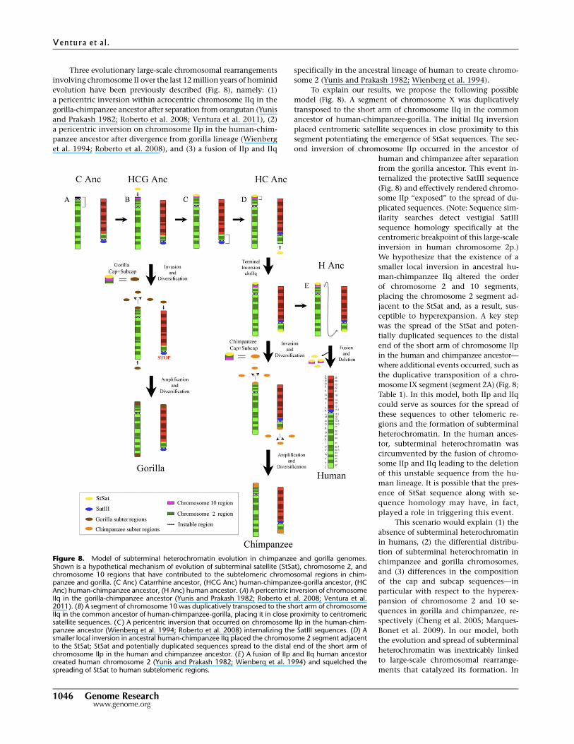
Three evolutionary large-scale chromosomal rearrangements
involving chromosome II over the last 12 million years of hominid
evolution have been previously described (Fig. 8), namely: (1)
a pericentric inversion within acrocentric chromosome IIq in the
gorilla-chimpanzee ancestor after separation from orangutan (Yunis
and Prakash 1982; Roberto et al. 2008; Ventura et al. 2011), (2)
a pericentric inversion on chromosome IIp in the human-chim-
panzee ancestor after divergence from gorilla lineage (Wienberg
et al. 1994; Roberto et al. 2008), and (3) a fusion of IIp and IIq
specifically in the ancestral lineage of human to create chromo-
some 2 (Yunis and Prakash 1982; Wienberg et al. 1994).
To explain our results, we propose the following possible
model (Fig. 8). A segment of chromosome X was duplicatively
transposed to the short arm of chromosome IIq in the common
ancestor of human-chimpanzee-gorilla. The initial IIq inversion
placed centromeric satellite sequences in close proximity to this
segment potentiating the emergence of StSat sequences. The sec-
ond inversion of chromosome IIp occurred in the ancestor of
human and chimpanzee after separation
from the gorilla ancestor. This event in-
ternalized the protective SatIII sequence
(Fig. 8) and effectively rendered chromo-
some IIp ‘‘exposed’’ to the spread of du-
plicated sequences. (Note: Sequence sim-
ilarity searches detect vestigial SatIII
sequence homology specifically at the
centromeric breakpoint of this large-scale
inversion in human chromosome 2p.)
We hypothesize that the existence of a
smaller local inversion in ancestral hu-
man-chimpanzee IIq altered the order
of chromosome 2 and 10 segments,
placing the chromosome 2 segment ad-
jacent to the StSat and, as a result, sus-
ceptible to hyperexpansion. A key step
was the spread of the StSat and poten-
tially duplicated sequences to the distal
end of the short arm of chromosome IIp
in the human and chimpanzee ancestor—
where additional events occurred, such as
the duplicative transposition of a chro-
mosome IX segment (segment 2A) (Fig. 8;
Table 1). In this model, both IIp and IIq
could serve as sources for the spread of
these sequences to other telomeric re-
gions and the formation of subterminal
heterochromatin. In the human ances-
tor, subterminal heterochromatin was
circumvented by the fusion of chromo-
some IIp and IIq leading to the deletion
of this unstable sequence from the hu-
man lineage. It is possible that the pres-
ence of StSat sequence along with se-
quence homology may have, in fact,
played a role in triggering this event.
This scenario would explain (1) the
absence of subterminal heterochromatin
in humans, (2) the differential distribu-
tion of subterminal heterochromatin in
chimpanzee and gorilla chromosomes,
and (3) differences in the composition
of the cap and subcap sequences—in
particular with respect to the hyperex-
pansion of chromosome 2 and 10 se-
quences in gorilla and chimpanzee, re-
spectively (Cheng et al. 2005; Marques-
Bonet et al. 2009). In our model, both
the evolution and spread of subterminal
heterochromatin was inextricably linked
to large-scale chromosomal rearrange-
ments that catalyzed its formation. In
Figure 8. Model of subterminal heterochromatin evolution in chimpanzee and gorilla genomes.Shown is a hypothetical mechanism of evolution of subterminal satellite (StSat), chromosome 2, andchromosome 10 regions that have contributed to the subtelomeric chromosomal regions in chim-panzee and gorilla. (C Anc) Catarrhine ancestor, (HCG Anc) human-chimpanzee-gorilla ancestor, (HCAnc) human-chimpanzee ancestor, (H Anc) human ancestor. (A) A pericentric inversion of chromosomeIIq in the gorilla-chimpanzee ancestor (Yunis and Prakash 1982; Roberto et al. 2008; Ventura et al.2011). (B) A segment of chromosome 10 was duplicatively transposed to the short arm of chromosomeIIq in the common ancestor of human-chimpanzee-gorilla, placing it in close proximity to centromericsatellite sequences. (C ) A pericentric inversion that occurred on chromosome IIp in the human-chim-panzee ancestor (Wienberg et al. 1994; Roberto et al. 2008) internalizing the SatIII sequences. (D) Asmaller local inversion in ancestral human-chimpanzee IIq placed the chromosome 2 segment adjacentto the StSat; StSat and potentially duplicated sequences spread to the distal end of the short arm ofchromosome IIp in the human and chimpanzee ancestor. (E ) A fusion of IIp and IIq human ancestorcreated human chromosome 2 (Yunis and Prakash 1982; Wienberg et al. 1994) and squelched thespreading of StSat to human subtelomeric regions.
Ventura et al .
1046 Genome Researchwww.genome.org
contrast, the fusion of chromosome IIp and IIq squelched this
process in humans by eliminating the source of the StSat sequence.
Since the phylogenetic analyses of both human and chimpanzee
suggest the spread of these segments occurred >4 mya, it follows
that the fusion of chromosome IIp and IIq occurred early during
evolution; otherwise, subterminal heterochromatin would have
evolved within our lineage.
We recognize, however, that this is only one of many possible
explanations that might account for the observed differences
among human, chimpanzee, and gorilla subterminal regions. It
may be also possible that functional constraints, such as the ex-
pression or suppression of critical genes in humans, may have
contributed to these dramatic differences between humans and
great apes. Human diseases such as fascioscapulohumeral mus-
cular dystrophy (FSHD), for example, arise from chromatin de-
regulation (Lemmers et al. 2010) near the telomere of human
chromosome 4. From an evolutionary perspective, it is known that
specific gene families have expanded immediately proximal to
the telomeres of human chromosomes. Of particular relevance in
this regard is the Wiskott-Aldrich Syndrome Protein and SCAR
Homolog (WASH) gene family (Linardopoulou et al. 2007). This
gene family has both expanded within the subtelomeric regions,
with a copy located specifically at the site of the chromosome 2
fusion. The function of the specific family members is not known,
but as a class, they are thought to be important in the reorganization
of the actin cytoskeleton in filopodia protrusions (Linardopoulou
et al. 2007).
Among hominids, chimpanzees are the only lineage wherein
heterochromatic blocks developed interstitially (chromosomes VII
and XIII). Our sequence and FISH analyses indicate that the
composition of these regions is indistinguishable from subtermi-
nal heterochromatin. Refined mapping of the breakpoints reveals
that the interstitial heterochromatin formed within segmentally
duplicated regions shared between human and chimpanzee for
chromosome VII and between human, chimpanzee, and gorilla for
chromosome XIII. Thus, it is quite likely that homology between
these interstitial blocks of segmental duplication and the sub-
telomeric region provided a gateway for the movement of StSat
and chromosome 2 segments internally (IJdo et al. 1991; Hirai et al.
2005). Once present, these sequences amplified, creating hetero-
chromatic interstitial G-band positive regions in this species. The
fact that the chromosome VII interstitial heterochromatin has not
fixed in the chimpanzee population may indicate that these in-
terstitial ‘‘colonizations’’ occurred relatively recently (Hirai et al.
2005) or, alternatively, are being deleted.
Hirai and colleagues (2005) described a retrotransposable
compound repeat DNA organization (RCRO) associated with sub-
terminal heterochromatin. The authors hypothesized that this
sequence was important in inducing and prolonging bouquet
formation during meiotic prophase specifically in chimpanzee
when compared to human and other primate chromosomes. Our
data reveal a much more complex organization of subterminal
heterochromatin that involved segmental duplication as opposed
to retrotransposition—although it is possible that the RCRO orig-
inally described is part of this architecture. Our copy number es-
timates and sequence analysis (Fig. 5) suggest that StSat and seg-
mental duplication expansions have added ;45–46 Mbp of highly
identical (1%–2% divergent) sequences to the ends of many gorilla
and chimpanzee chromosomes.
It has been previously reported that the presence of large
blocks of heterochromatin at the ends of chromosomes may alter
patterns of meiotic recombination (Miklos and Nankivell 1976). In
chimpanzee, it is, thus, possible that this architecture directly in-
terferes with synapsis during meiosis, resulting in an overall re-
duction of the total number of chiasma and a prolonged associa-
tion of telomeric regions during zygotene and pachytene. Indeed,
preliminary findings by Hirai and colleagues reported a mean
lower chiasma frequency in chimpanzee when compared to hu-
man (Hirai et al. 2005). While it is now known that the pattern of
fine-scale recombination differs significantly between human and
chimpanzee (Ptak et al. 2005), it will be interesting to determine
whether this difference is most pronounced in close proximity to
regions of subterminal heterochromatin. This will require both
higher quality sequence and fine-scale mapping of meiotic cross-
over events in these complex regions of human and great ape
chromosomes.
Methods
Fluorescent in situ hybridizationGorilla and chimpanzee BAC and human fosmid clones were usedas probes in FISH assays on human and great ape metaphasespreads. Metaphases from nonhuman primates (common chim-panzee, gorilla, bonobo, and rhesus monkey) were obtained fromlymphoblastoid or fibroblast cell lines; human metaphase spreadswere obtained from PHA-stimulated peripheral lymphocytes ofnormal donors by standard procedures. DNA extraction from BACsand fosmids was performed as already reported (Ventura et al.2001). FISH experiments were essentially performed as previouslydescribed (Ventura et al. 2004). Briefly, DNA probes were directlylabeled with Cy3-dUTP, Cy5-dUTP (GE Healthcare), or Fluorescein-dUTP (Invitrogen) by nick translation. Two hundred ng of labeledprobe were hybridized on metaphases spreads; hybridization wasperformed overnight at 37°C in 23 SSC, 50% (v/v) formamide,10% (w/v) dextran sulphate, 5 mg COT1 DNA (Roche), and 3 mgsonicated salmon sperm DNA, in a volume of 10 mL. Post-hybrid-ization washing was performed at 60°C in 0.13 SSC (three times,high stringency). Washes of interspecies hybridization experi-ments were performed at lower stringency: 37°C in 23 SSC, 50%formamide (3 3), followed by washes at 42°C in 23 SSC (3 3).Chromosome identification was obtained by DAPI staining, pro-ducing a Q-banding pattern. Digital images were obtained usinga Leica DMRXA epifluorescence microscope equipped with a cooledCCD camera (Princeton Instruments). Cy3, Cy5, Fluorescein, andDAPI fluorescence signals, detected with specific filters, were re-corded separately as grayscale images. Pseudocoloring and mergingof images were performed using Adobe Photoshop software.
Stretched chromosomes
Chimpanzee and gorilla chromosomes were stretched mechan-ically. Colcemid-treated lymphoblastoid cells were washed inphosphate-buffered saline (PBS), counted, and resuspended ina hypotonic solution (75 mM KCl, Cf: 60,000 cells/mL) for 15 min.Then, 0.5 mL of the suspension was cytocentrifuged (ShandonCytospin 3 centrifuge) onto coated glass slides (Thermo ShandonDouble Cytoslide) at 800 rpm for 4 min and fixed in methanol at�20°C for 15 min and in methanol:acetic acid 3:1 for 30 min. Theslides were aged at 80°C for 1 h before hybridization.
Oligo-FISH and PNA probes
Biotinylated primers were designed on the StSat sequence(59TCCATGTTTATACAGATAGCGGTG39-Bio) and directly used asprobes (0.001 mmol) in FISH experiments on chimpanzee andgorilla metaphase spreads. Probe and target were denaturated at
Gorilla/chimpanzee subterminal sequence evolution
Genome Research 1047www.genome.org
74°C for 6 min and hybridized o/n at 37°C in 23 SSC, 50% (v/v)formamide, 10% (w/v) dextran sulphate, and 3 mg sonicatedsalmon sperm DNA, in a volume of 10 mL. Slides were quicklywashed in 23 SSC, 50% formamide at room temperature, andsignals were detected (detection buffer: 1% BSA/13 SSC0/0.1Tween 20) with avidin-FITC at 37°C for 1 h. Excess fluorochromewas washed away in 43 SSC/0.1 Tween 20 at room temperature, andchromosomes were DAPI stained.
PNA probes hybridized to denatured telomeric sequences incells permeabilized in hot formamide. Two mL of ready-to-use Cy3-conjugated telomere PNA probe (Dako, Telomere PNA FISH Kits)were used as probes in fluorescent in situ cohybridization assaysto evaluate the respective localization of telomeric sequences andthe StSat on gorilla and chimpanzee chromosomes.
Satellite III and rDNA chromosomal distribution
To determine the chromosomal distribution, primer sets weredesigned from the known sequences of each SatIII subfamily(Bandyopadhyay et al. 2001a,b) (pE-1: GATTCGATTCCATTGCACTCG-GGACTGAAACAAAATGGAGACC; pE-2: ATGCAGCCTGGGTGACCT-AAGAATCCATACCACACC; pR-1: TGTGCCTCTGTGTTACAT-ACTGCCATCCTTTCCACC; pR-2: ACGCTGGGTGATGGAGTGAAATAC-ACTCCATTTCATTCCGCCGC; pR-4: TAAGCGTGGAATGGGTTTGAGC-CATCCGATTCCATTTCACTAC; pK-1: ATCGAATGGATTCCTAATTG-CGATATCTTCTGTTACACG; pW-1: AATGGGATGGAACCGAGTGG-CCTTTCATTTCAAGTCCCTTCGC) and usedto amplify genomic DNA by polymerase chain reactions (PCR) onhuman, chimpanzee, and gorilla genomic DNA. Amplified prod-ucts were then directly labeled with Fluorescein-dUTP by PCR la-beling and used as probes in FISH assays. The use of PCR labelingavoids the possible contamination from genomic DNA by nicktranslation labeling of PCR products. PCR labeling was carried outin a final volume of 20 mL that contained 100 ng PCR product, 2 mL103 reaction buffer (Invitrogen), 2 mL 50 mM MgCl2, 0.5 mL eachprimer (10 mM), 0.5 2 mM mL dACG, 2.5 mL 1 mM Fluorescein-dUTP, 5 mL 2% BSA, and 0.3 mL Taq polymerase (5 U/mL). For bothamplification reactions, the cycling parameters used were as fol-lows: 3 min initial denaturation at 94°C, followed by 30 cycles of:94°C for 30 sec, 56°C for 30 sec, and 72°C for 30 sec. Final extensionwas at 72°C for 10 min.
In order to assess rDNA localization in great apes, clonescontaining rDNA were extracted by a BLAST sequence similaritysearch of the human rDNA complete repeating unit (accession:U13369.1) against the HTGS database. Several human BAC cloneswere selected and tested; the clone giving the most consistent andreliable hybridization signals was chosen as a probe for the cohy-bridization experiments: CH507-159O11.
BAC sequencing
Chimpanzee and gorilla clones were selected for complete insertsequencing using capillary sequencing methods (McPherson et al.2001) in order to obtain high-quality finished sequence withinduplicated regions. Rearrangements were visualized using Miropeats(Parsons 1995) and previously described in-house visualization tools(Kidd et al. 2010).
Data accessA subset of clones was selected for complete insert sequencing. Thesequences have been deposited in GenBank (http://www.ncbi.nlm.nih.gov/genbank) under accession nos. AC198877.1, AC183608.3,AC192975.3, AC097005.1, AC194646.4, AC200072.3, AC191679.2,AC183292.1, AC150905.2, AC152419.2, AC204739.3, AC192628.1,AC196285.4, AC213004.6, AC184708.3, AC192825.3, AC145493.3,
AC239281.3, AC241375.2, AC239628.1, AC239393.3, AC239711.2,AC241376.2, AC241501.1, AC239640.2, AC239931.2.
AcknowledgmentsWe thank Tonia Brown for valuable comments in the preparationof this manuscript. The authors are grateful to Lisa Faust, MollieHerget, and the staff of the Lincoln Park Zoo for gorilla materialused in this study. We are also indebted to the large genome se-quencing centers for early access to BAC end sequence data fortargeted analysis of structural rearrangements. This work wassupported, in part, by NIH grants HG002385 and GM058815 toE.E.E. and NIH grant U54 HG003079 to R.K.W. P.H.S. is supportedby a Howard Hughes Medical Institute International Student Fel-lowship. E.E.E. is an investigator of the Howard Hughes MedicalInstitute.
References
Bandyopadhyay R, Berend SA, Page SL, Choo KH, Shaffer LG. 2001a.Satellite III sequences on 14p and their relevance to Robertsoniantranslocation formation. Chromosome Res 9: 235–242.
Bandyopadhyay R, McQuillan C, Page SL, Choo KH, Shaffer LG. 2001b.Identification and characterization of satellite III subfamilies to theacrocentric chromosomes. Chromosome Res 9: 223–233.
Carreto L, Eiriz MF, Gomes AC, Pereira PM, Schuller D, Santos MA. 2008.Comparative genomics of wild type yeast strains unveils importantgenome diversity. BMC Genomics 9: 524. doi: 10.1186/1471-2164-9-524.
Cheng Z, Ventura M, She X, Khaitovich P, Graves T, Osoegawa K, Church D,DeJong P, Wilson RK, Paabo S, et al. 2005. A genome-wide comparisonof recent chimpanzee and human segmental duplications. Nature 437:88–93.
The Chimpanzee Sequencing and Analysis Consortium. 2005. Initialsequence of the chimpanzee genome and comparison with the humangenome. Nature 437: 69–87.
Fan Y, Linardopoulou E, Friedman C, Williams E, Trask BJ. 2002. Genomicstructure and evolution of the ancestral chromosome fusion site in2q13-2q14.1 and paralogous regions on other human chromosomes.Genome Res 12: 1651–1662.
Gerber JC, Neuhann TM, Tyshchenko N, Smitka M, Hackmann K. 2011.Expanding the clinical and neuroradiological phenotype of 6q27microdeletion: Olfactory bulb aplasia and anosmia. Am J Med Genet A155: 1981–1986.
Gonzalez-Garcia M, Gonzalez-Sanchez M, Puertas MJ. 2006. The highvariability of subtelomeric heterochromatin and connections betweennonhomologous chromosomes, suggest frequent ectopicrecombination in rye meiocytes. Cytogenet Genome Res 115: 179–185.
Greider CW, Blackburn EH. 1989. A telomeric sequence in the RNA ofTetrahymena telomerase required for telomere repeat synthesis. Nature337: 331–337.
Gross M, Starke H, Trifonov V, Claussen U, Liehr T, Weise A. 2006. Amolecular cytogenetic study of chromosome evolution in chimpanzee.Cytogenet Genome Res 112: 67–75.
Haaf T, Schmid M. 1987. Chromosome heteromorphisms in the gorillakaryotype. Analyses with distamycin A/DAPI, quinacrine and5-azacytidine. J Hered 78: 287–292.
Hirai H, Matsubayashi K, Kumazaki K, Kato A, Maeda N, Kim HS. 2005.Chimpanzee chromosomes: Retrotransposable compound repeat DNAorganization (RCRO) and its influence on meiotic prophase andcrossing-over. Cytogenet Genome Res 108: 248–254.
IJdo JW, Baldini A, Ward DC, Reeders ST, Wells RA. 1991. Origin of humanchromosome 2: An ancestral telomere-telomere fusion. Proc Natl AcadSci 88: 9051–9055.
Jarmuz M, Glotzbach CD, Bailey KA, Bandyopadhyay R, Shaffer LG. 2007.The evolution of satellite III DNA subfamilies among primates. Am JHum Genet 80: 495–501.
Kidd JM, Graves T, Newman TL, Fulton R, Hayden HS, Malig M, Kallicki J,Kaul R, Wilson RK, Eichler EE. 2010. A human genome structuralvariation sequencing resource reveals insights into mutationalmechanisms. Cell 143: 837–847.
Koga A, Notohara M, Hirai H. 2011. Evolution of subterminal satellite (StSat)repeats in hominids. Genetica 139: 167–175.
Kudo H, Emi M, Ishigaki Y, Tsunoda U, Hinokio Y, Ishii M, Sato H, Yamada T,Katagiri H, Oka Y. 2011. Frequent loss of genome gap region in 4p16.3subtelomere in early-onset type 2 diabetes mellitus. Exp Diabetes Res2011: 498460. doi: 10.1155/2011/498460.
Ventura et al .
1048 Genome Researchwww.genome.org
Lemmers RJ, van der Vliet PJ, Klooster R, Sacconi S, Camano P, Dauwerse JG,Snider L, Straasheijm KR, van Ommen GJ, Padberg GW, et al. 2010.A unifying genetic model for facioscapulohumeral muscular dystrophy.Science 329: 1650–1653.
Linardopoulou EV, Williams EM, Fan Y, Friedman C, Young JM, Trask BJ.2005. Human subtelomeres are hot spots of interchromosomalrecombination and segmental duplication. Nature 437: 94–100.
Linardopoulou EV, Parghi SS, Friedman C, Osborn GE, Parkhurst SM,Trask BJ. 2007. Human subtelomeric WASH genes encode a newsubclass of the WASP family. PLoS Genet 3: e237. doi: 10.1371/journal.pgen.0030237.
Marques-Bonet T, Kidd JM, Ventura M, Graves TA, Cheng Z, Hillier LW, JiangZ, Baker C, Malfavon-Borja R, Fulton LA, et al. 2009. A burst of segmentalduplications in the genome of the African great ape ancestor. Nature457: 877–881.
McPherson JD, Marra M, Hillier L, Waterston RH, Chinwalla A, Wallis J,Sekhon M, Wylie K, Mardis ER, Wilson RK, et al. 2001. A physical map ofthe human genome. Nature 409: 934–941.
Mefford HC, Trask BJ. 2002. The complex structure and dynamic evolutionof human subtelomeres. Nat Rev Genet 3: 91–102.
Miklos GL, Nankivell RN. 1976. Telomeric satellite DNA functions inregulating recombination. Chromosoma 56: 143–167.
Nieves M, De Oliveira EH, Amaral PJ, Nagamachi CY, Pieczarka JC,Muhlmann MC, Mudry MD. 2011. Analysis of the heterochromatin ofCebus (Primates, Platyrrhini) by micro-FISH and banding patterncomparisons. J Genet 90: 111–117.
Parsons JD. 1995. Miropeats: Graphical DNA sequence comparisons.Comput Appl Biosci 11: 615–619.
Prieto P, Martin A, Cabrera A. 2004. Chromosomal distribution of telomericand telomeric-associated sequences in Hordeum chilense by in situhybridization. Hereditas 141: 122–127.
Ptak SE, Hinds DA, Koehler K, Nickel B, Patil N, Ballinger DG, Przeworski M,Frazer KA, Paabo S. 2005. Fine-scale recombination patterns differbetween chimpanzees and humans. Nat Genet 37: 429–434.
Riethman H, Ambrosini A, Paul S. 2005. Human subtelomere structure andvariation. Chromosome Res 13: 505–515.
Roberto R, Misceo D, D’Addabbo P, Archidiacono N, Rocchi M. 2008.Refinement of macaque synteny arrangement with respect to the
official rheMac2 macaque sequence assembly. Chromosome Res 16:977–985.
Royle NJ, Baird DM, Jeffreys AJ. 1994. A subterminal satellite locatedadjacent to telomeres in chimpanzees is absent from the humangenome. Nat Genet 6: 52–56.
Rudd MK, Willard HF. 2004. Analysis of the centromeric regions of thehuman genome assembly. Trends Genet 20: 529–533.
Rudd MK, Endicott RM, Friedman C, Walker M, Young JM, Osoegawa K,de Jong PJ, Green ED, Trask BJ. 2009. Comparative sequence analysisof primate subtelomeres originating from a chromosome fission event.Genome Res 19: 33–41.
Sudmant PH, Kitzman JO, Antonacci F, Alkan C, Malig M, Tsalenko A,Sampas N, Bruhn L, Shendure J, Eichler EE. 2010. Diversity of humancopy number variation and multicopy genes. Science 330: 641–646.
van der Maarel SM, Tawil R, Tapscott SJ. 2011. Facioscapulohumeralmuscular dystrophy and DUX4: Breaking the silence. Trends Mol Med 17:252–258.
Ventura M, Archidiacono N, Rocchi M. 2001. Centromere emergence inevolution. Genome Res 11: 595–599.
Ventura M, Weigl S, Carbone L, Cardone MF, Misceo D, Teti M, D’Addabbo P,Wandall A, Bjorck E, de Jong PJ, et al. 2004. Recurrent sites for newcentromere seeding. Genome Res 14: 1696–1703.
Ventura M, Catacchio CR, Alkan C, Marques-Bonet T, Sajjadian S, Graves TA,Hormozdiari F, Navarro A, Malig M, Baker C, et al. 2011. Gorilla genomestructural variation reveals evolutionary parallelisms with chimpanzee.Genome Res 21: 1640–1649.
Wallace BM, Hulten MA. 1985. Meiotic chromosome pairing in the normalhuman female. Ann Hum Genet 49: 215–226.
Wienberg J, Jauch A, Ludecke HJ, Senger G, Horsthemke B, Claussen U,Cremer T, Arnold N, Lengauer C. 1994. The origin of humanchromosome 2 analyzed by comparative chromosome mapping witha DNA microlibrary. Chromosome Res 2: 405–410.
Yunis JJ, Prakash O. 1982. The origin of man: A chromosomal pictoriallegacy. Science 215: 1525–1530.
Received December 16, 2011; accepted in revised form March 6, 2012.
Gorilla/chimpanzee subterminal sequence evolution
Genome Research 1049www.genome.org