GENES ON CHROMOSOME 22 INVOLVED IN THE ...
176
GENES ON CHROMOSOME 22 INVOLVED IN THE PATHOGENESIS OF CENTRAL NERVOUS SYSTEM TUMORS Ronald Leklllllle dil Deprez
-
Upload
khangminh22 -
Category
Documents
-
view
0 -
download
0
Transcript of GENES ON CHROMOSOME 22 INVOLVED IN THE ...
GENES ON CHROMOSOME 22 INVOLVED
IN THE PATHOGENESIS OF
CENTRAL NERVOUS SYSTEM TUMORS
Ronald Leklllllle dil Deprez

GENES ON CHROMOSOME 22 INVOLVED
IN THE PATHOGENESIS OF
CENTRAL NERVOUS SYSTEM TUMORS
GENEN, GELEGEN OP CHROMOSOOM 22, DIE BETROKKEN ZIJN BIJ
DE PATHOGENESE VAN TUMOREN VAN HET CENTRALE ZENUWSTELSEL
PROEFSCRRIFT
TER VERKRIJGING VAN DE GRAAD VAN DOCTOR
AAN DE ERASMUS UNIVERSITEIT ROTTERDAM
OP GEZAG V AN DE RECTOR MAGNIFICUS
PROF. DR. P.W.C. AKKERMANS M. LIT.
EN VOLGENS BESLUIT V AN RET COLLEGE V AN DEKANEN.
DE OPENBARE VERDEDIGING ZAL PLAATSVINDEN OP
WOENSDAG 2 NOVEMBER 1994 OM 11.45 UUR
DOOR
RONALD RENDRIKUS LEKANNE DIT DEPREZ
GEBOREN TE OEGSTGEEST
PROMOTmCOMMIssm
Promotor:
Overige leden:
Co-Promotor:
Prof. Dr. D. Bootsma
Prof. Dr. F.T. Bosman
Prof. Dr. A. Westerveld
Prof. Dr. J.H.J. Hoeijmakers
Dr. E.C. Zwarthoff
Dit proefschrift werd bewerkt binnen de vakgroep Pathologie van de Faculteit der
Geneeskunde en Gezondheidswetenschappen van de Erasmus Universiteit Rotterdam. Ret
onderzoek maakte deel uit van het Medisch Genetisch Centrum Zuid-West Nederland. Het
onderzoek en deze uilgave werden l1nancieel gesteund door de Nederlandse
Kankerbestrijding-Koningin Wilhelmina Fonds en de Maurits en Anna de Kock Stichting.
4
Eell ovenvillnillg lVordt Iliet ill kilometers
maar ill celltimeters be/wald. Will nu een
beetje, houd wat je hebt en will later
/log een beetje meer.
Louis L 'Amour
Voor Willeke
5
Contents
List of abbreviations 9
Chapter I Gene1'31 introduction 11
I Cancer is a genetic disease 13
2 Oncogenes and tumor suppressor genes 14
2.1 Oncogenes 14
2.2 Tumor suppressor genes 14
3 Molecular cloning and function of tumor suppressor genes 18
3.1 The Retinoblastoma (RbI) gene 18
3.2 Thep53 gene 19
3.3 The isolation of APC, MCC, DCC, hMSH2,
and hMLHl and their putative role in
colorectal tumorigenesis 22
3.4 The isolation of the wn and VHL genes 25
3.5 The RET proto-oncogene 27
3.6 The NFl gene 29
3.7 The NF2 gene 29
4 MENINGIOMA
4.1 Cells of origin 31
4.2 Frequency, incidence and occurrence 31
4.3 Anatomic localization 32
4.4 Histopathology 33
4.5 Etiology 34
4.6 Cytogenetic and molecular genetic studies
of meningiomas 35
4.7 Other tumors with loss or rearrangements
of chromosome 22 38
4.8 Chromosome 22 39
5 Scope of the thesis 40
6 References 41
6
Chapter II Isolation and characterization of 2S unique DNA mal'l~ers
for human chl'omosome 22
Appendix
Chapter III Cytogenetic, molecular genetic and pathological analyses
in 126 meningiomas
Chapter IV Familial anaplastic ependymoma: evidence of loss of
chromosome 22 in tumor cells
Chapter V A t(4;22) in a meningioma points to the localization
of a putative tumor suppressor gene
Chapter VI Molecular cloning of a gene disrupted by a balauced
translocation in a meningioma
Chapter VII Constitutional DNA-level alterations in chromosome 22
in a patient with multiple meningiomas
Chapter VIII Frequent NF2 gene transcript mutations in sporadic
meningiomas and vestibular schwannomas
Discussion and sununary
Samenvatting
Curl'iculum vitae
List of publications
Nawool'd
53
59
65
99
109
119
143
151
161
167
171
173
175
7

List of abbreviations
APC
bp
DCC
FAP
FISH
FMTC
GRAIL
HNPCC
kb
LOH
Mb
MCC
MEN
mRNA
MTC
NFl
NF2
ORF
Rb
RFLP
RT-PCR
SSCP
VHL
WAGR
WT
wt
YAC
Adenom~tous polyposis coli
basepair
Deleted in colorectal carcinoma
Familial polyposis coli
Fluorescent in situ hybridization
Familial medullary thyroid carcinoma
Gene recognition and analysis internet link
Hereditary non polyposis colorectal cancer
kilobase
Loss of heterozygosity
Megabase
Mutated in colorectal cancer
Multiple endocrine neoplasia
messenger RNA
Medullary thyroid carcinoma
Neurofibromatosis type I
Neurofibromatosis type 2
Open reading frame
Retinoblastoma
Restriction fragment length polymorphism
Reverse transciptase-polymerase chain reaction
Single strand conformation polymorphism
von Hippel-Lindau
Wilms' tumor, aniridia, genitourinary abnormalities, mental retardation
Wilms' tumor
Wild-type
Yeast artificial chromosome
9

10

Chapter I
General introduction
11

12
1 Cancer is a genetic disease
It is nowadays generally accepted that cancer can be considered as a genetic disease.
However, there are two clear differences between cancer and most other genetic diseases.
First, most cancers are caused essentially by somatic mutations, whereas all other genetic
diseases are caused solely by germline mutations. Second, for most cancer types at least two
mutations are required before a tumor may arise. For most pediatric and some adult tumors
only a limited number of mutations are supposed to be sufficient for tumor development
(Knudson, 1971, 1986; Haber and Housman, 1992). In contrast, most adult cancers appear
to require more than two genetic changes. This 'multiple-hit' concept of tumorigenesis stems
from several lines of evidence. First, most adult cancers show an exponential increase in
incidence with age, suggesting that the accumulation of different hits is involved in tumor
development (Vogelstein and Kinzler, 1993). Second, a dramatically increased incidence of
cancer has been observed in individuals with chromosome instability syndromes such as
Bloom's syndrome (Vijayalaxmi et aI., 1983; Seshadri et aI., 1987). Third, repeated
administration of mutagen to test animals is generally required before a tumor arises (Saffhill
et aI., 1985). Fourth, increasing evidence becomes available for the involvement of multiple
genetic alterations in common human tumors. The most extensively studied example is the
accumulation of genetic alterations observed in human colon tumors, in which mutations in
APe and ras found in adenomatous polyps have been classified as early events and mutations
in p53 and Dee as later events that may contribute to tumor progression (Fearon and
Vogelstein, 1990; section 3.3). In a specific human cancer, mutations in one particular gene
appear to precede those in others (Vogelstein and Kinzler, 1993), although after this first hit
the accumulation of changes rather than their specific order seems to be important for tumor
progression (Marx, 1989).
13
2 Oncogenes and tumor suppressor genes
Molecular genetic analysis of tumorigenesis has revealed two fundamental mechanisms of
neoplasia: 1) the activation of dominant oncogenes or 2) the inactivation of recessive tumor
suppressor genes (anti-oncogenes). Accumulating evidence suggests that the progression of
many tumors to full malignancy requires both types of changes in the tumor cell genome.
2.1 Oncogenes
First evidence for the existence of oncogenes came from the induction of sarcomas by the
Rous sarcoma retrovirus, containing the V-Sf'C oncogene (Martin, 1970). Later, other
retroviral oncogenes (v-olle) and their human homologues (c-olle) were discovered in various
cancers. Also the dissection of chromosomal translocations, tumor associated DNA
amplifications and the use of gene-transfer have resulted in the identification of more
oncogenes. Until now 100 or more of them have been isolated. Under normal conditions
proto-oncogenes are thought to be involved in the regulation of cell proliferation. They act
as growth factors or their receptors. Within the cell, they function as downstream modulators
in signal transduction pathways. Furthermore, the nuclear (proto-)oncoproteins may regulate
gene transcription in response to these signals (Hunter, 1991). Different mechanisms (point
mutation, amplification or translocation) are involved in the activation of a proto-oncogene
into an oncogene. Mutations in the proto-oncogenes occur in one of the two alleles of the
gene and they act in a dominant way relative to the wild-type allele. Therefore, they are
called 'gain of function' mutations that give normal cells neoplastic properties (Weinberg,
1989).
2.2 Tumor suppressor genes
Three lines of evidence support the existence of growth-constraining tumor suppressor genes:
1) somatic cell hybrids 2) familial cancer and 3) loss of heterozygosity in tumors.
The first evidence that tumor suppressor genes are involved in neoplastic
14
transformation came from somatic cell fusion experiments, which showed that fusion of
tumor cells with normal cells almost always results in the outgrowth of non tumorigenic
hybrids (Sager, 1985; Rarris, 1988). Sometimes these hybrid cells reverted back to a
tumorigenic state, which could be correlated with the loss of specific 'normal' chromosomes
(Stanbridge, 1990). These and other fusion experiments suggest that normal cells contain
genetic information capable of suppressing the neoplastic growth in the tumor cells. The
tumor cells have lost this information during their evolution from normal to tumor cells.
The second indication came from the existence of familial cancer. In 1971, Knudson
proposed the two-hit model for the development of retinoblastoma and one year later also for
Wilms' tumor (Knudson and Strong, 1972). This model was based on epidemiological
analyses of age of onset of multi focal (hereditary) versus single (sporadic) cases. It implies
that in heritable retinoblastoma the first hit is already present in the germline and only one
somatic hit is required for tumor formation. In sporadic cases two somatic mutational events
are needed, which explains the later age of onset in these patients. In 1973 Comings
suggested that the mutations were in the two alleles of the same gene. Definite proof for the
involvement of a recessive tumor suppressor gene was obtained when the nature of these
germline and somatic mutations became clear. First indications came from karyotypic
analyses of retinoblastoma tumor cells, which sometimes uncovered interstitial deletions that
involved chromosomal band 13ql4 (Yunis and Ramsay, 1978). Genetic analysis of blood and
tumor DNA pairs and the isolation of the retinoblastoma gene (Rbl) eventually confirmed
Knudson's theory (Cavenee et aI., 1983; Friend et aI., 1986; Fung et aI., 1987; Lee et aI.,
1987). These studies support the idea that a recessive tumor suppressor gene can contribute
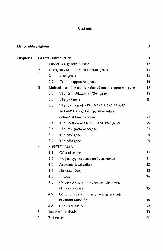
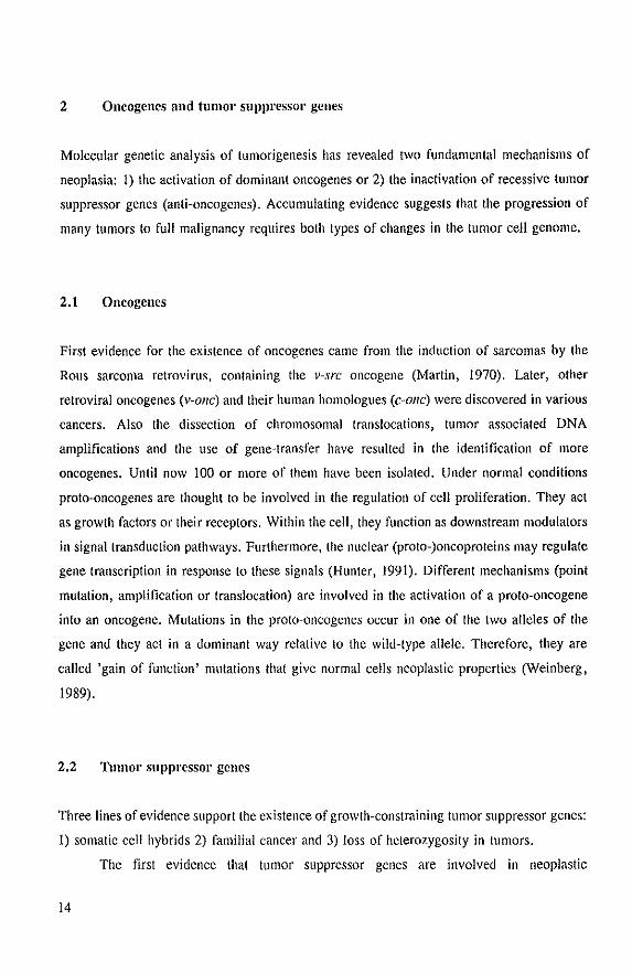
to the development of a tumor when both alleles have been inactivated (Fig. I). The study
of other familial cancers have contributed towards the elucidation of more cancer
susceptibility loci and genes. The isolated genes responsible for the small fraction of
hereditary cancers are very useful for the study of these and the commoner sporadic cancers.
Although the predisposing Cancer genes are phenotypically dominant, at the genetic level they
are recessive (both copies of the gene should be inactivated). The apparent dominant
inheritance of these diseases reflects the high probability that the second allele is hit in a
somatic cell.
The third clue came from consistent and specific chromosome deletions in tumor
cells. Karyotyping and 'loss of heterozygosity' (LOR) studies clearly showed loss of
IS
particular (parts ot) chromosomes in human tumors. The latter approach uses the frequent
occurrence of restriction fragment length polymorph isms (RFLPs) of some DNA probes or
polymorphic microsatellites in the human genome. This technique takes advantage of the
heterozygous state of a patient's normal (constitutional) DNA for a particular polymorphic
marker, loss of that part of the chromosome is the case when tumor tissue of the same patient
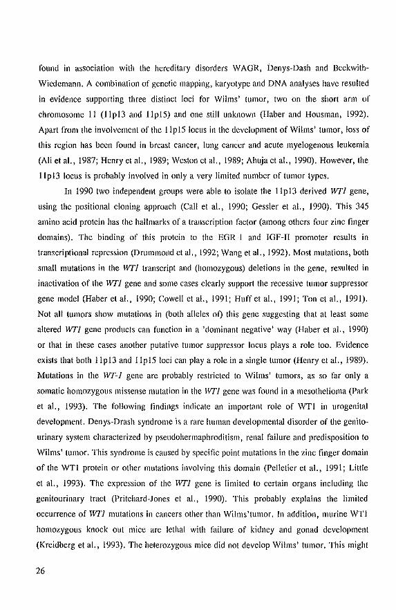
shows reduction or loss of one of the alleles (Fig. I).
Figure 1.
"",m&1 par, of ch,omolorn;>,
M
tumor pal. of c~'omo'omu
LOH study
""rmol tumor
M_ M-
,,-
Soul""rn bioi
The most commonly observed inactivation scheme for a recessive tumor suppressor gene. The
normal (wild-type) copies of the tumor suppressor gene are shown with the open boxes. In the
tumor both copies of the gene are inf'lctivated by a small mutation in one allele (black box) and
loss of the portion of the chromosome that harbours the second allele. Loss of heterozygosity
(LOH) of this chromosomal part can be detected on a Sonthern blot. This analysis uses the ability
of a polymorphic DNA marker, specific for the relevant region of the chromosome, to detect both
the maternal and paternal alleles (A I and A2). The comparison of normal and tumor DNA from
the same individual can distinguish whether one allele is lost in the tumor.
A comprehensive list of consistently observed allele losses in solid tumors has been described
by Seizinger et al. (1991). A combination of linkage analyses in cancer families and LOH
studies in both sporadic and familial tumors have led to the localization of a large number
of (mostly) tumor suppressor gene loci. The 'positional cloning' (Collins, 1992) andlor
candidate gene approach eventually resulted in the cloning of some of these genes. A number
of examples are listed in Table 1. In the following sections these genes are briefly described.
The products of tumor suppressor genes may be involved in the negative regulation
of cell growth, induction of terminal differentiation and/or apoptosis. The gene can contribute
16
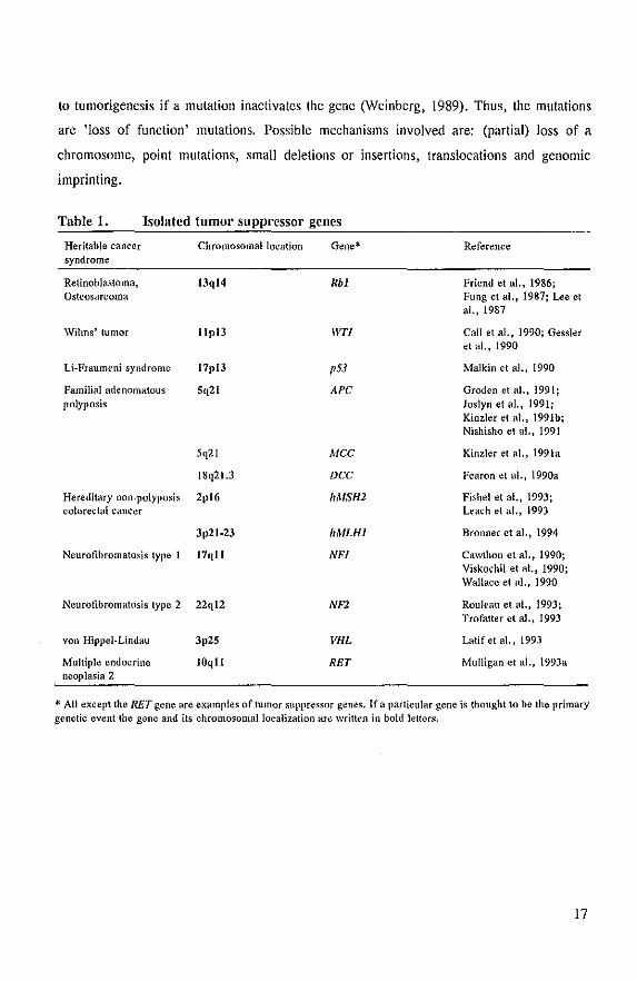
to tumorigenesis if a mutation inactivates the gene (Weinberg, 1989). Thus, the mutations
are 'loss of function' mutations. Possible mechanisms involved are: (partial) loss of a
chromosome, point mutations, small deletions or insertions, translocations and genomic
imprinting.
Table 1. Isolated tumor SUppl'essor genes
Heritable cancer Chromosomal location Gene* Reference syndrome
Retinoblastoma. 13ql4 Rbi Friend et aI., 1986: Osteosarcoma Fung et ai., 1987: ue et
ai., 1987
Wilms' tumor lIpl3 IVTI Call et al.. 1990; Gessler et al.. 1990
Li·Fraumeni syndrome 17pl3 p53 Malkin et a1.. 1990
Familial adenomatous 5q21 APe Groden et aI., 1991; polyposis Joslyn et al .• 1991;
Kinzler et a!., 1991b; Nishisho et al., 1991
5q21 Mee Kinzler et al., 1991a
18q21.3 Dee Fearon et al.. 1990a
Hereditary non-polyposis 2pl6 hMSH2 Fishel et al •• 1993; colorectal cancer Leach et ai., 1993
3p21-23 hMLHl Bronner et aI., 1994
Neurofibromatosis type I 17qll NFl Cawthon et a!., 1990; Viskochil et al.. 1990; Wallace et al., 1990
Neurofibromatosis type 2 22ql2 NFl Rouleau et al.. 1993; Trofalter et a!., 1993
von Hippel-Lindau 3p25 VHL Latifetal.,1993
MUltiple endocrine 10qll RET Mulligan et al.. 1993a neoplasia 2
* All except the RET gene are examples of tumor suppressor genes. If a particular gene is thought to he the primary genetic event the gene and its chromosomal localization are written in bold letters.
17
3 Moleculm' cloning and fUllction of tumol' suppl'essol' genes
In the last few years an enormous progress was seen in the cloning and/or identification of
genes involved in hereditary cancer syndromes and tumor progression. This section gives an
overview of some of these genes together with a short description of their putative function
and possible involvement in other tUlllor types.
3.1 The Retinoblastoma (RbI) gene
The RbI gene was isolated in 1986, on the basis of its primary role in retinoblastoma
development (Friend et a!., 1986; Fung et a!., 1987; Lee et a!., 1987). The gene
encompasses 200 kb on chromosome 13ql4 and encodes a 105 kD nuclear phosphoprotein.
Most alterations observed in the gene in retinoblastoma are deletions and nonsense mutations
and involve both alleles. These changes result in a truncated or absent protein and are in
agreement with the suspected loss of function mutations as was postulated for tumor
suppressor genes. Inactivating mutations of both copies of the gene occur in many other
tumor types including breast carcinoma, prostate carcinoma, osteosarcoma, soft tissue
sarcoma and small-cell lung carcinoma (Bookstein et al. I 1990a; Friend et al., 1987; Harbour
et a!., 1988; Hensel et a!., 1990; Lee el a!., 1988; T'Ang et a!., 1988; Varley et a!., 1989;
Yokota et a!., 1988). This indicates Ihat loss of Rb I function is important in the
tumorigenesis of many tumors. In hereditary retinoblastoma 5-10% of the patients also
develop osteosarcomas and soft tissue sarcomas. An explanation for the finding that they
have no increased risk for other tumors might be that for the other tumors mutations in other
genes must happen first. The growlh suppressing role of the wild-type (wt) Rb I protein was
clearly illuslrated by experiments in which the wt RbI gene was introduced into Rbi-deficient
tumor cells. These studies demonstrated suppression of cell-growth and tUlllorigenicity
(Huang et a!., 1988; Bookstein et a!., 1990b; Sumegi et a!., 1990; Takahashi et a!., 1991;
Goodrich et a!., 1992a).
Insight into the function of Rbi started with the finding that DNA tUlllor virus
encoded oncoproteins (EI A, large T antigen and E7) form complexes with the host cell Rb I
protein (DeCaprio el a!., 1988; Whyte et a!., 1988; Dyson et a!., 1989). The current idea
18
is that this interaction inactivates Rb I function by removing it from the growth regulating
machinery of the cell, equivalent to a deletion or mutation in the RbI gene. Other
experiments suggest that the Rbi protein is involved in cell cycle regulation, probably by
regulating the progression through the GI phase of the cell cycle. Among others, the Rbi
protein is phosphorylated in a cell-cycle dependent manner: hypophosphorylated Rb I protein
predominates in G I, whereas heavily phosphorylated forms appear just prior to the G I to S
transition and persist during the S, G2 and M phase (Buchkovich et aI., 1989; DeCaprio et
aI., 1989; Chen et aI., 1989). The Rbi protein is a substrate for different kinases involved
in the cell cycle (Matsushime et aI., 1992; Ewen et aI., 1993; Kato et aI., 1993). In addition,
all three viral oncoproteins bind only to the hypophosphorylated state of the Rb I protein
(Ludlow et aI., 1989). This suggests that the hypophosphorylated form of the Rbi protein
is active in cell growth control. Moreover, hypophosphorylated instead of
hyperphosphorylated Rbi protein is able to bind the transcription factor E2F (Chellappan et
aI., 1991; Shirodkar et aI., 1992). The interaction of Rb with E2F probably blocks the
transactivation of certain positively acting growth-regulating genes such as c-myc (Thalmeier
et aI., 1989). Further study showed physical interaction between the c-myc and Rbi proteins
and microinjection of cells with Rbi and c-myc results in the inhibition of the ability of the
Rbi protein to arrest the cell cycle (Rustgi et aI., 1991; Goodrich and Lee, 1992b). All these
data together suggest that the Rbi protein functions by negatively regulating the progression
through the cell cycle by the inhibition of transcription factors involved in entry into the S
phase.
3.2 The p53 gene
The nuclear phosphoprotein p53 was originally discovered in extracts of SV40 transformed
cells (Lane and Crawford, 1979). The protein was found in a complex with SV40 large T
antigen. In the mid-1980s the corresponding gene was cloned (Matlashewski et aI., 1984;
Lamp and Crawford, 1986) and assigned to chromosome 17p13.1 (van Tuinen et aI., 1988).
Originally p53 was classified as an oncogene because cotransfection of ras and p53
genes transformed embryo fibroblasts. However, the 1'53 cDNA clones used in these
experiments were later observed to contain missense mutations. More recent experiments
19
with wild-type (wI) p53 proved the opposite. From these studies wt p53 was found to be
involved in growth suppression and inhibition of transformation (Finlay et aI., 1989; Eliyahu
et aI., 1989; Baker et aI., 1990). Therefore, the wt p53 gene has clear characteristics of a
tumor suppressor gene.
Different mechanisms have been found in human tumors that alter p53 and in turn
inactivate wt p53 function. These alterations can occur at either the protein or DNA level.
At the protein level, the wt p53 protein was found in a complex with various DNA tumor
virus encoded oncoproteins (EIB, large T antigen and E6). In addition, p53 was found in a
complex with MDM2, a cellular protein with transforming properties (Momand et aI., 1992).
Furthermore, mutant p53 can bind with wt p53 (see below). The binding of these proteins
most likely inactivates p53 and thereby contributes to oncogenesis. Alterations at the DNA
level appear to be the most common mechanism. These alterations include the presence of
a mutations in the p53 gene and loss of the chromosomal region that bears this gene. Most
tumors show a mutation in one copy of the gene and loss of the second allele because the p53
containing portion of chromosome 17p is lost. In these cases p53 follows a recessive
mechanism of tumorigenesis. Mutation analyses of the p53 gene in human tumors revealed
two remarkable findings. First, mutations in this gene were the most common genetic
alteration yet identified in human cancer. These include cancers of the colon, lung,
oesophagus, breast, liver, brain and hemopoietic tissues (Hollstein et aI., 1991; Levine et aI.,
1991; Levine, 1992). Second, 80% of p53 mutations involve single base changes (missense
mutations). Most mutations are clustered in the central, highly conserved regions of the gene.
However, the types and sites of the mutations can vary amoung tumor types and probably
reflect exposure to specific etiologic agents (Harris and Hollstein, 1993). Many of the p53
missense mutations, which lead to an amino acid change and a full-length mutant protein,
show similar detrimental effects on protein conformation (Gannon et aI., 1990) and function
(Kern et aI., 1992; Vogelstein and Kinzler., 1992). On the protein level an increase in half
life from minutes to hours has been observed (Bartek et aI., 1991). Although most tumors
show abnormalities of both alleles, some tumors show only one mutated copy. This indicates
that in these cases p53 did not follow a recessive mechanism of tumor development. Instead
a 'dominant negative' mechanism was proposed in which only one mutated copy of p53 is
sufficient to contribute to tumor formation. One plausible explanation for this might be that
the abundantly expressed mutated p53 protein competes with and blocks the activity of the
20
endogenous wt p53 by forming mixed tetrameric complexes (Kern et aI., 1992; Harris et aI.,
1992; Hupp et aI., 1992). Reports have also appeared in which certain missense p53
mutations show some oncogenic properties as well (Wolf et aI., 1984; Dittmer et aI., 1993).
Thus, these missense p53 mutations can not only result in functional competition with wt p53
resulting in loss of function, but some of them have gained also new properties that result
in a gain of function.
Mutations in p53 were not only found in sporadic human cancers but also in the
germline of patients with the Li-Fraumeni cancer syndrome (Malkin et aI., 1990). These
patients are characterized by a high incidence of different cancers including early-onset breast
carcinoma, childhood sarcomas, and other neoplasms. However, other cancers, for example
colon and small cell lung carcinoma, with a high incidence of somatic mutations of pS3 are
hardly observed in Li-Fraumeni patients. This might be due to the capacity of a certain tissue
to divide once a mutation in 1'53 has occurred or is constitutionally present and thereby
increasing the chance of undergoing a second mutation in the gene. For instance breast tissue
displays an increased growth in adolescence, whereas colon and lung tissues have only a
limited potential to divide under normal conditions (Knudson, 1993). Germline mutations in
p53 are also, though very rarely, observed in childhood sarcomas (Toguchida et aI., 1992)
and among some women with breast cancer (Borresen et aI., 1992). Transgenic p53 knock
out mice (Donehower et aI., 1992) and mice expressing mutated p53 proteins (Lavigueur et
aI., 1989) showed that p53 is not important for embryonic development, because the mice
were viable and appeared completely normal, although they displayed an increased risk for
the development of some tumor types, particularly lymphomas.
Functional studies of the p53 protein have revealed that p53 plays a role in the control
of the cell cycle (Bischoffet aI., 1990; Stuzbecher et aI., 1990), processes involving DNA
repair, DNA replication, genomic instability (Kastan et aI., 1992; Yin et aI., 1992;
Livingstone et aI., 1992), and programmed cell death (Clarke et aI., 1993; Lowe et aI.,
1993a; Lowe et aI., 1993b). Although p53 occupies an important position in all the above
mentioned processes this protein is probably not required in normal cells. However, when
the cells are damaged, for instance by gamma-irradiation, the concentration of p53 increases.
This results in an arrest of the cell cycle, which allows the repair of DNA damage. Thus,
p53 is supposably acting as a kind of 'guardian of the genome' (Lane, 1992). The most likely
way by which p53 executes these various activities is through its ability to act as a
21
transcriptional modulator. p53 can act directly or indirectly as a transactivator of transcription
of different genes. Induction of transcription was observed in genes containing a p53 binding
site after binding 10 p53 (Kern el aI., 1991; EI-Deiry el aI., 1992; Kern el ai., 1992).
GADD45 (growth arresl and DNA damage inducible), an enzyme involved in DNA repair,
contains such a binding site. p53 was found 10 be necessary for Ihe induclion of Ihis enzyme
(Kaslan el aI., 1992). Repression of lranscriplion was found in genes conlaining a TATA
promoter sequence (Ginsberg et aI., 1991). This repression is probably Ihe consequence of
the ability of p53 10 bind to a componenl of the lranscriplion-initiation complex (Selo et aI.,
1992; Mack el aI., 1993). Recently,. gene has been cloned thai mighl at least in pari explain
Ihe biological fnnctions of p53. This gene, IVAFl/Cipl/Sdil (Wild-Iype p53-aclivated
fragment 1; Cdk-interacting protein I; Senescent cell-derived inhibitor), contains a p53-
binding sile, is highly induced by wild-type p53, can inhibit cell growth, is up-regulaled in
senescent cells, and is found in complexes important for cell cycle regulation (EI-Deiry et
aI., 1993; Harper et aI., 1993; Noda el aI., 1994). All Ihese fealures resemble Ihose
described for p53 and suggesl thai Ihis gene is a meaningful key effeclor of p53.
3.3 The isolation of APC, MCC, DCC, hMSH2 and hMLHl and their putative role
in colOl'ectal tumorigenesis
Colorectal tumorigenesis provides an excellent system to study and search for genetic
alterations involved in the different well-defined stages of colon tumor development. The
studies of bolh sporadic coloreclal cancer and especially Ihe familial polyposis coli (FAP)
syndrome, have resulted in more insight in the accumulation of different genetic alterations
during tumor progression. PAP patients are characterized by the appearance of hundreds or
even thousands of benign adellomalous polyps in the colonic mucosa some of which
evenlnally develop inlo carcinomas. The sludy of Ihis tumor type resulted in the isolalion of
Ihree differenllumor suppressor genes: DCC, MCC and APC (Fearon el aI., 1990a; Groden
et aI., 1991; Joslyn el .1., 1991; Kinzler el ai., 1991a; Kinzler el ai., 1991 b; Nishisho el ai.,
1991). Other hereditary conditions, which predispose to colorectal cancer, are known as
hereditary non polyposis coloreclal cancer (HNPCC). In HNPCC cases only diffuse polyps
more proximal in the colon are observed, which also might degenerate into carcinoma.
22
Recent studies in these families have resulted in the isolation of two other genes: hMSH2 and
hMLHl (Fishel et a!., 1993; Leach et a!., 1993; Bronner et a!., 1994).
The gene that predisposes to FAP, the adenomatous polyposis coli (APC) gene, has
been isolated and is located at chromosome 5q21 (Groden et a!., 1991; Joslyn et a!., 1991;
Kinzler et a!., 1991b; Nishisho et a!., 1991). Apart from the germline APC mutations in FAP
patients, somatic mutations were detected in sporadic colorectal cancer (Powell et a!., 1992).
The mutations most often involve deletions and nonsense mutations (Nagase and Nakamura,
1993). The localization of APC mutations may explain at least in part the difference in
phenotypic appearance (Nagase et a!., 1992; Nagase and Nakamura, 1993; Olschwang et aI.,
1993; Spirio et aI., 1993). One might speculate that a constitutional APC mutation might give
the mucosal cells a growth advantage, which increases the change of getting a second
mutation in the other allele that resuits in polyp formation. The APC gene encodes a large
2,843 amino acid protein with a coiled coil structure in the N-terminal part. No significant
resemblance to any known gene was found in the databases, which makes predictions about
its function difficult. However, recently immunoprecipitation experiments identified
interactions between APC and alpha and beta catenin (Rubinfeld et aI., 1993; Su et a!.,
1993). These experiments suggest that APC might interfere indirectly with cadherins,
proteins that mediate cell-cell interactions. Therefore, APC might be involved in cell
adhesion. Although most cases of familial polyposis have shown linkage to and mutations in
the APC gene (Spirio et aI., 1993), another family was recently described in which linkage
to the APC gene was excluded (Stella et aI., 1993).
About 150 kb proximal to the APC gene another gene was identified. This gene, MCC
(Mutated in Colorectal Cancers), is thought to contribute to colorectal tumorigenesis because
several sporadic colon carcinomas showed gross structural alterations or point mutations,
mostly missense mutations, in this gene (Kinzler et aI., 1991a; Nishisho et a!., 1991). In
addition, about 40% of the colorectal tumors revealed allelic loss of 5q21, which includes
the MCC and APC genes (Kinzler et a!., 1991a). No MCC mutations have been found in
FAP patients. The gene encodes a protein of 829 amino acids, which shows a short region
of homology with G protein-coupled receptors and contains a coiled coil structure. Further
study is required to investigate the role of this gene in colorectal neoplasia.
Another important gene involved in these carcinomas is the DCC (Deleted in
Colorectal Carcinomas) gene. This gene was identified because LOH studies showed frequent
23
loss of markers on the long arm of chromosome 18 (Vogelstein et aI., 1988; Fearon et aL,
1990a). Only about II % of early stage adenomatous polyps but over 70% of carcinomas
show loss of 18q (Vogel stein et aI., 1988; Cho et aI., 1994). This suggests that DCC plays
a role in advanced adenomas. The gene is expressed in most normal tissues, including
colonic mucosa, though its expression was greatly reduced or absent in most colorectal
carcinomas (Fearon et aI., 1990a). So far, one missense mutation has been identified in the
coding region of the gene (Cho et aI., 1994). The gene encodes a 190-kD transmembrane
phosphoprotein probably a cell surface receptor involved in adhesion to an extracellular
matrix or basement membrane component (Fearon et aI., 1990a). LOH for chromosome 18q
is also seen in breast tumors (Devilee et aI., 1991a) and gastric cancer (Dchino et aI., 1992).
To date germline mutations in this gene were not observed. This might indicate that DCC
is involved in tumor progression rather than tumor initiation.
In addition to alterations in these tumor suppressor genes other changes (genes) are
frequently involved such as K-ras mutations (Vogelstein et aI., 1988), loss andlor mutations
in the p53 gene on 17p (Baker et aI., 1989; Nigro et aI., 1989; Vogelstein, 1989), loss of
22q (Okamoto et aI., 1988; Vogelstein et aI., 1989; Miyaki et aI., 1990) and loss of the
1/1I/23-Hl gene on Ip35 (Leister et aI., 1990; Cohn et aI., 1991). Loss of Ip35 was most
frequently observed in the non metastatic colon tumors (Cohn et aI., 1991). The model for
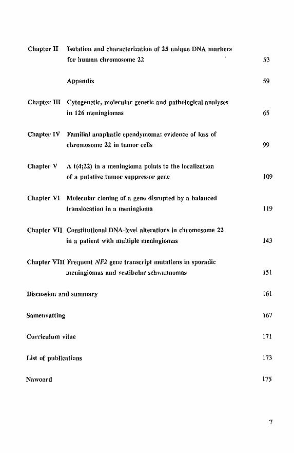
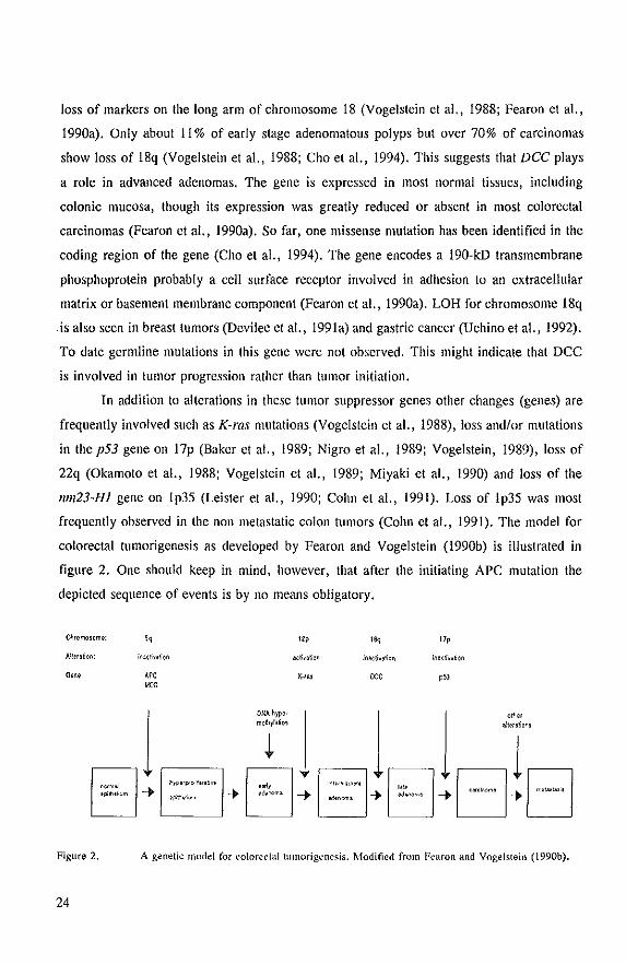
colorectal tumorigenesis as developed by Fearon and Vogelstein (1990b) is illustrated in
figure 2. One should keep in mind, however, that after the initiating APC mutation the
depicted sequence of events is by no means obligatory.
C~romOlemt:
Allera~on:
GeM:
Figure 2.
24
APC <ICC
'2p
K-/!S
I~ I7p
DCC
A genetic model for colorectal1umorigenesis. Modified from Fearon and Voge1stein (1990b).
In about 15% of all colon tumors another initiating mutation is likely. These tumors
reveal widespread instability of microsatellite sequences, whereas loss of heterozygosity for
chromosomal loci is mostly absent (Thibodeau et aI., 1993). This implicates that we deal
with a different group of colon tumors. Furthermore, these alterations were also found in
tumors from HNPCC patients. Linkage analysis in HNPCC families suggested that at least
three genes are involved: one at chromosome 2p16, another at chromosome 3p21-23, and a
third at an unidentified locus (Aaltonen et aI., 1993; Peltomiiki et aI., 1993; Lindblom et aI.,
1993). Both the candidate gene approach (Fishel et aI., 1993) and the positional cloning
approach (Leach et aI., 1993) have resulted in the identification of the hMSH2 gene at
chromosome 2p16. Mutations in this gene were found in sporadic colon tumors that showed
dinucleotide repeat instability and HNPCC patients. Both splice acceptor site mutations and
mis- and non-sense mutations were observed: The human hMSH2 gene shows high homology
with the prokaryotic MillS gene, which is part of the mismatch repair pathway in E. coli.
MutS mutants exhibit dinucleoide repeat instability like that observed in HNPCC patients
(Levinson and Gutman, 1987; Strand et aI., 1993). In vitro experiments with extracts of
HNPCC tumor cells showed a profound defect in strand-specific mismatch repair (Parsons
et aI., 1993). In addition, very recently a candidate mismatch repair gene on chromosome
3p has been isolated because of its homology with MIIlL, another gene involved in the
mismatch repair pathway in E. coli (Bronner et aI., 1994). This gene was called hMLHI. In
a chromosome 3-linked HNPCC family missense mutations were found in affected
individuals. Mutations in APe, K-ras and p53 are also observed in this group of tumors. It
might be that mutations in mismatch repair genes result in an increase risk of generating
mutations in other oncogenes and tumor suppressor genes. The hMSH2 and hMLHl genes
probably playa role in carcinogenesis by inducing a mutator phenotype. Yet, why would
mutations in these genes only predispose to specific tumors? Future studies should shed light
on this intriguing question.
3.4 The isolation of the WTl and VHL genes
Wilms' tumor is a pediatric kidney cancer which probably arises from blastemal kidney cells
(Beckwith et aI., 1990). Most tumors occur sporadically, however these tumors are also
25
found in association with the hereditary disorders WAGR, Denys-Dash and Beckwith
Wiedemann. A combination of genetic mapping, karyotype and DNA analyses have resulted
in evidence supporting three distinct loci for Wilms' tumor, two on the short arm of
chromosome II (llpl3 and llp15) and one still unknown (Haber and Housman, 1992).
Apart from the involvement of the 11p15 locus in the development of Wilms' tumor, loss of
this region has been found in breast cancer, lung cancer and acute myelogenous leukemia
(Ali et aI., 1987; Henry et aI., 1989; Westonetal., 1989; Ahujaetal., 1990). However, the
llpl3 locus is probably involved in only a very limited number of tumor types.
In 1990 two independent groups were able to isolate the 11pl3 derived WTl gene,
using the positional cloning approach (Call et aI., 1990; Gessler et aI., 1990). This 345
amino acid protein has the hallmarks of a transcription factor (among others four zinc finger
domains). The binding of this protein to the EGR-1 and IGF-II promoter results in
transcriptional repression (Drummond et aI., 1992; Wang et aI., 1992). Most mutations, both
small mutations in the wrl transcript and (homozygous) deletions in the gene, resulted in
inactivation of the WTJ gene and some cases clearly support the recessive tumor suppressor
gene model (Haber et aI., 1990; Cowell et aI., 1991; Huffet aI., 1991; Ton et aI., 1991).
Not all tumors show mutations in (both alleles 01) this gene suggesting that at least some
altered WTl gene products can function in a 'dominant negative' way (Haber et aI., 1990)
or that in these cases another putative tumor suppressor loclis plays a role too. Evidence
exists that both 11p13 and 11pl510ci can playa role in a single tumor (Henry et aI., 1989).
Mutations in the WT-J gene are probably restricted to Wilms' tumors, as so far only a
somatic homozygous missense mutation in the ~vrl gene was found in a mesothelioma (Park
et aI., 1993). The following findings indicate an important role of WTl in urogenital
development. Denys-Drash syndrome is a rare human developmental disorder of the genito
urinary system characterized by pseudohermaphroditism, renal failure and predisposition to
Wilms' tumor. This syndrome is caused by specific point mutations in the zinc finger domain
of the WTl protein or other mutations involving this domain (Pelletier et aI., 1991; Little
et aI., 1993). The expression of the WTl gene is limited to certain organs including the
genitourinary tract (Pritchard-Jones et aI., 1990). This probably explains the limited
occurrence of "WI'1 mutations in cancers other than Wilms'tumor. In addition, murine WTl
homozygous knock out mice are lethal with failure of kidney and gonad development
(Kreidberg et aI., 1993). The heterozygous mice did not develop Wilms' tUIllOr. This might
26
be explained by assuming that the number of target-cells in mice is too low and therefore a
second mutation in the other allele never occurs.
Another mechanism of inactivation which could be involved in the pathogenesis of
Wilms' tumor is genomic imprinting, because in more than 90 percent of the tumors maternal
allelic loss is observed. This imprinting is probably restricted to the IIpl5 locus because this
preferential loss is not the wn gene on II p 13 (van Heyningen and Hastie, 1992; Koi et al.,
1993). The preferential loss of the maternal allele might indicate that the paternal allele is
imprinted, resulting in loss of expression of the susceptibility gene from this locus. Other
mechanisms which might explain this finding are that imprinting could somehow enhance the
rate of induction of small mutations in the paternal allele or protect for mutations in the
maternally derived gene during gametogenesis. The net result in all of these models is
inactivation of both maternal and paternal alleles as could be expected when the predisposing
gene is a tumor suppressor gene. However, other mechanisms might be the outcome of this
imprinting effect when we deal with an oncogene for instance. More details about this issue
are provided in a very recent review about genomic imprinting (Tycko, 1994).
The von Hippel-Lindau (VHL) disease is a dominantly inherited cancer syndrome in
which in addition to renal cell carcinoma, also hemangioblastomas of the central nervous
system and retina and pheochromocytomas occur. However, Wilms' tumors are never found.
Recently, the VHL gene on chromosome 3p25-p26 was isolated and both large
(nonoverlapping) deletions and small mutations were observed in YHL kind reds and sporadic
renal cell carcinomas (Latif et aI., 1993). The gene probably encodes a 284 amino acid
protein without any significant homology to already isolated genes in the databases.
However, an acidic tandemly repeated pentamer is observed in the putative protein, which
suggests a role in signal transduction or cell adhesion (Latif et aI., 1993).
3.5 The RET Pl'oto-oncogene
The RET proto-oncogene, located on chromosome 1 Oq 11. 2, was originally isolated because
the gene became activated by rearrangement during NIH3T3 cell transformation (Takahashi
et aI., 1985). Later, it also appeared to playa role in human cancer because in 25% of
27
human papillary thyroid carcinomas this gene showed rearrangements (Grieco et aI., 1990).
The RET gene encodes a transmembrane tyrosine kinase protein with an as yet unknown
ligand.
In the dominantly inherited cancer syndromes multiple endocrine neoplasia type 2
(MEN 2A and B), familial medullary carcinoma (FMTC) , and the dominantly inherited
Hirschsprung's disease, which is a developmental disorder, specific mutations have been
found in the RET proto-oncogene (Donis-Keller et aI., 1993; Mulligan et aI., 1993a; Edery
et aI., 1994; Hofstra et aI., 1994; Romeo et aI., 1994). MEN 2 and FMTC are characterized
by the development of bilateral medullary thyroid carcinoma (MTC). In both MEN 2A and
B pheochromocytomas are observed and in MEN 2B a distinct more aggressive and complex
phenotype is apparent (Nanes and Catherwood, 1992). Hirschsprung's disease is
characterized by congenital absense of parasympathetic innervation in the lower intestinal
tract and cancer-association has never been described. The mutations observed in the RET
gene might explain the distict clinical phenotypes. The MEN2A and FMTC mutations were
aU missense mutations involving the 4 conserved cysteines in the extracellular region and
probably interfere with ligand binding (Donis-Keller et aI., 1993; Mulligan et aI., 1993a,b).
In MEN2B all mutations found so far were specific threonine to methionine substitutions in
codon 918 in the intracellular tyrosine kinase domain of RET. The same mutation was also
observed in 6 sporadic MTC tumors (Hofstra et aI., 1994). The nature of the mutations found
in Hirschsprung's disease however are different. These include deletions, nonsense and
missense mutations and are presumably loss of function mutations.
The RET mutations observed in the cancer syndromes suggest that the gene is an
oncogene rather than a tumor suppressor gene because: 1) very specific heterozygous amino
acid substitutions were observed and 2) chromosome 10 allelic loss was hardly ever found
in these tumors (Landsvater et aI., 1989; Nelkin et aI., 1989; Mulligan et aI., 1993a,b).
However, the observation that the RET protein dimerizes (Bongarzone et aI., 1993;
Rodriguez and Park, 1993) suggests that a dominant-negative mechanism can not be ruled
out. The presumed loss of function mutations found in Hirschsprung's disease indicate that
haplo-insufficiency (dosage-effect) is the underlying mechanism. Homozygous RET gene
knockout mice die soon after birth and show impairment in kidney development and lack
enteric neurons throughout the digestive tract (Schuchardt et aI., 1994).
28
3.6 The NFl gene
Neurofibromatosis type I (NFl), also known as von Recklinghausen neurofibromatosis, is
an autosomal dominant disorder with a heterogeneous clinical manifestation (Riccardi, 1981).
The hallmarks of NF I are: cutaneous or subcutaneous neurofibromas, cafe au lait spots and
Lisch nodules. The NFl gene, located on chromosome 17q11.2, was isolated in 1990 and
encodes a 2,818 amino acid protein (Cawthon et ai., 1990; Viskochil et ai., 1990; Wallace
et ai., 1990). The gene product, neurofibromin, shares high homology with GTPase activator
proteins (Buchberg et ai., 1990; Xu et ai., 1990a; Xu et ai., 1990b). The protein may
function as a negative regulator of the p21'''-mediated signal transduction pathway andlor
down stream of p21'~ (Bollag and McCormick, 1992). The possible interaction of
neurofibromin with microtubules suggests a role in mitosis and cell division (Seizinger,
1993). NFl gene mutations were observed in sporadic tumors that are also found in NFl
patients (neurofibroma, neurofibrosarcoma, pheochromocytoma and astrocytoma), but in
addition in neuroblastoma, malignant melanoma, a sporadic colon carcinoma and
myelodysplastic syndrome, diseases that do not occur in NFl patients (Li et ai., 1992).
Recent data suggest that the NFl gene functions at least in some of the tumors as a recessive
tumor suppressor gene, because homozygous deletions in the NFl locus have been found in
a neurofibrosarcoma from a NF 1 patient, some melanomas and some neuroblastomas
(Andersen et ai., 1993; Johnson et ai., 1993; Legius et ai., 1993).
3.7 The NF2 gene
Neurofibromatosis type 2 displays an autosomal dominant inheritance and is genetically
linked to chromosome 22 (Rouleau et ai., 1990). The hallmark for this disease is the
development of bilateral schwannomas of the eighth cranial nerve. Apart from this, multiple
spinal and intracranial schwannomas and meningiomas may appear in these patients (Evans
et ai., 1992a). Recently two independent groups have isolated the NF2 gene, which encodes
a 595 amino acid protein, called merlin (moesin-ezrin-radixin like protein, Trofatter et aI.,
1993) or schwannomin (Rouleau et ai., 1993). High homology of the N-terminal 340 residues
of this protein to the cytoskeleton associated proteins moesin, ezrin and radixin suggests a
29
role in mediating interactions between the cell membrane and the cytoskeleton. Mutations
were found in both the germline of NF2 patients and sporadic variants of tumors associated
with NF2 (Rouleau et aI., 1993; Trofatter et aI., 1993; Bianchi et aI., 1994; Jacoby et aI.,
1994; Ruttledge et aI., 1994; Chapter VIII). Most mutations result in frameshifts, which lead
to truncation of the protein, and presumably a loss of function. Almost all meningiomas with
complete or partial loss of chromosome 22 revealed mutations in the NF2 gene. These
findings support the view that the NF2 gene functions as a recessive tumor suppressor gene.
Mutations in the NF2 gene transcript were also observed in breast carcinoma and melanoma,
neoplasms unrelated to NF2. However, no mutations were observed in pheochromocytomas
and colon carcinomas in which chromosome 22q loss has frequently been observed (Bianchi
et aI., 1994).
30
4 MENINGIOMA
4.1 Cells of origin
In 1922 the name 'meningioma' was first introduced by Harvey Cushing for a benign tumor
of the meninges of the central nervous system. The meninges, the coverings of the brain and
spinal cord, are composed of three layers with at the outside the pachymeninx or dura mater
and at the inside the leptomeninges composed of the arachnoid and pia mater. Embryogenesis
of the meninges, at least in humans, is still not very well elucidated. Experimental work in
lower vertebrates indicates that the dura mater and probably also the arachnoid are
mesodermal in origin, whereas the pia mater probably is ectodermal in origin and is largely
derived from neural crest cells (Kepes, 1982; Russell and Rubinstein, 1989). Different lines
of evidence suggest that meningiomas arise from arachnoid cells (Kepes, 1982; Upton and
Weller, 1985; Russell and Rubinstein, 1989).
4.2 Frequency, incidence and occurrence
Intracranial meningiomas represent approximately 13-19% of all primary intracranial tumors
that are treated by surgery and an incidence of 12 % was recorded for the spinal meningiomas
(Russell and Rubinslein, 1989). The aclual incidence of meningiomas is probably much
higher because Ihese lumors are slowly growing and may slay asymplomatic in a large
number of cases. An eslimale of Ihe overal incidence of meningiomas could be calculated
from a Swedish sludy in which during a to-year period 172 meningiomas were found in
11,793 autopsies: a prevalence of 1.46% (Rausing el aI., 1970). However, in only 11 of
these cases meningiomas were the main cause of death.
Belween 2-3 limes as many females develop meningiomas as males and the highesl
incidence is observed in the fiflh and sixth decade of life (Zang, 1982). During childhood
meningiomas are dislinctly rare (Perilongo el aI., 1992). There are three patterns of
occurrence (Butti el aI., 1989; Domenicucci el aI., 1989; Russell and Rubinslein, 1989;
McDowell, 1990; Sieb et aI., 1992):
1) sporadic solitary cases;
31
2) as part of the hereditary syndrome neurofibromatosis type 2 (NF2);
3) multiple or familial aggregation of meningiomas.
The first group involves the majority of the cases and consists of tumors occurring randomly
(not associated with a hereditary syndrome) in the general population. The second one
involves meningiomas found in individuals with NF2. The diagnostic criteria for NF2 are
depicted in table 2 (Evans et aI., 1992b).
Table 2, Diagnostic cl'itel'ia ror Nfl
Bilateral vestibular schwannomas 01' family history of NF2 plus
1) unilateral vestibular schwannoma 01'
2) any two of: meningioma, glioma, neurofibroma, schwannoma, posterior
subcapsular lenticular opacities.
This disorder may be divided in at least two distinct forms. One is called Wishart type and
has an early onset, rapid course and multiple tumors in addition to bilateral vestibular
schwannomas. The other, Gardner type, is characterized by a later age of onset, a more
benign course and is usually limited to bilateral vestibular schwannomas (Evans et aI.,
1992b). In both categories meningiomas can occur. The third group consists of patients with
multiple or familial aggregation of meningiomas without evidence of NF2. In this group, at
least in some of the cases, a predisposing mutation is probably involved (Chapter VII). It
remains to be established whether these different groups arise as a result of different
mutations in one gene or that different genes or combination of genes are involved.
4.3 Anatomic localization
According to Russell and Rubinstein (1989) meningiomas can be found at seven different
sites in the body: I) intracranial; 2) spinal; 3) intraventricular; 4) orbital; 5) intrapetrous; 6)
(extra-) calvarial and 7) ectopic. The intracranial and spinal meningiomas are the most
common ones. These tumors are often further subdivided in base (sphenoid ridge, tuberculum
sellae, olfactory grooves, pontocerebellar angle, petrous ridge of the temporal bone, posterior
32

fossa and foramen magnum), convexity (parasagittaJ region and free convexity), falx cerebri,
intraventricular and spinal cord meningiomas (Casalone et al., 1990). Total surgical resection
is dependent on the anatomic localization of the tumor and influences the recurrence rate
(Black, 1991).
4.4 Histopathology
Microscopic examination of meningiomas by many neuropathologists resulted in 1979 in the
following WHO classification scheme (Ziilch, 1979):
1. meningotheliomatous (endotheliomatous, syncytial, arachnotheliomatous)
2. fibrous (fibroblastic)
3. transitional (mixture of type 1 and 2)
4. psammomatous
5. angiomatous
6. haemangioblastic
7. haemangiopericytic
8. papillary
9. anaplastic (malignant)
Other subtypes are also known but less common such as Iipoblastic and xanthomatous
meningiomas (Kepes, 1982). Most tumors show features corresponding with a mixture of
different histological subtypes, from which the first four occur most predominantly (de la
Monte et aI., 1986). From a histopathological point of view, most of these tumors are
considered to be benign and only very rarely truly malignant invasive meningiomas occur.
However, different degrees of anaplasia, which may to some extent influence biological
behaviour, can be identilied in all classes of benign tumors (Jiiiiskeliiinen et aI., 1985; de la
Monte et aI., 1986; Vagner-Capodano et aI., 1993). A significant association was observed
in this respect between the in vivo growth rate of meningiomas and the histopathological
grade of the tumors (JiiiiskeHiinen et aI., 1985). The grading system according to increasing
anaplasia used in this thesis involves the following six histological parameters: loss of
architecture, increased cellularity, nuclear pleomorphism, mitotic figures, focal necrosis and
brain infiltration (Hiaskeliiinen et aI., 1985; Vagner-Capodano et .1., 1993). Three different
33
grades can be distinguished using these criteria: grade I: benign; grade II: atypical and grade
III: anaplastic. It remains to be established whether this grading system is of clinical
relevance.
4.5 Etiology
From the literature different studies suggest that the following factors may be involved in the
etiology of meningioma. These include: I) genetic predisposition (section 4.2) 2) steroid
hormones and their receptors 3) radiation 4) head injury 5) SV-40-related papova viruses.
The postulated role of the steroid hormones and their receptors came from differenl
lines of evidence. First, a higher incidence of these tumors is observed in women and an
accelerated growth has been observed during pregnancy (Zang, 1982; Roelvink et aI., 1987).
In other studies this association was not found (Schlehofer et aI., 1992; and cited references),
Second, the majority of meningiomas and normal leptomeningeal tissue contain progesterone
receptors. The presence of oestrogen receptors is still a matter of discussion. The significance
of the progesterone receptors in these tumors was further investigated with in vitro
experiments using progesterone and progesterone inhibitors. The results of these experiments
are not very conclusive, although they might point to a slight growth promoting role for
progesterone (Koper et aI., 1990).
Radiation is nowadays a well established causative factor in meningioma development.
Meningiomas have occlirred with increased frequency and at an earlier age in people
receiving high-dose irradiation for the treatment of an intracranial growth or after low-dosage
scalp irradiation for fungal infections (Russell and Rubinstein, 1989).
A large number of reports going back as far as 1813 suggest the involvement of
previous head injury and the development of meningioma years later (AI-RodIJan and Laws,
1990), Some investigators found a statistically significant association between meningioma
occurrence and a history of head trauma, others observed site-specific head injuries with
subsequent tumor development at the same site years later (Russell and Rubinstein, 1989).
Different reports reviewed by Zang (1982) might support the role of SV40-related
papova viruses and the formation of meningiomas. In about 35 % of histological sections or
34
early cell cultures the presence of SV 40 related T antigens was described (Weiss et a1., 1975;
Scherneck et aI., 1979; Krieg et aI., 1981). However, these findings were not confirmed by
others (Merletti et a1., 1975).
4.6 Cytogenetic and molecular genetic studies of meningiomas
As early as 1967, cytogenetic studies showed non-random loss of one G-group chromosome
in meningiomas. This was the first specific chromosomal aberration observed in solid tumors
(Zang and Singer, 1967). The subsequent development of banding techniques and molecular
genetic studies revealed that monosomy of chromosome 22 is the hallmark for these tumors
(Zang, 1982; Seizinger et a1., 1987a; Dumanski et aI., 1990a). In a considerable number of
tumors loss of chromosome 22 is the only cytogenetically visible chromosomal alteration (AI
Saadi et a1., 1987; Maltby et a1., 1988; Vagner-Capodano et aI., 1993). These results
indicate that loss of genetic material from this chromosome is a primary and specific event
in the development of these tumors, which is reminiscent of the involvement of a tumor
suppressor gene(s) as is mentioned in section 2. In addition, other less common non random
chromosomal changes occur. The most frequent ones are loss or rearrangements of
chromosomes 1, 7, 14, 18 and 19 and these are thought to playa role in tumor progression
(Katsuyama et aI., 1986; AI Saadi et aI., 1987; Maltby et aI., 1988; Rey et aI., 1988;
Casalone et aI., 1990). The karyotypic evolution observed in meningiomas appears to be
associated with more anaplastic features (Vagner-Capodano et aI., 1993).
As mentioned earlier the majority of meningiomas are sporadic cases but they are also
found in patients with NF2 or in familial or multiple meningioma patients. Genetic linkage
studies in NF2 families, tumor deletion mapping in both sporadic and hereditary meningioma
cases resulted in the identification of at least three different loci on the long arm of
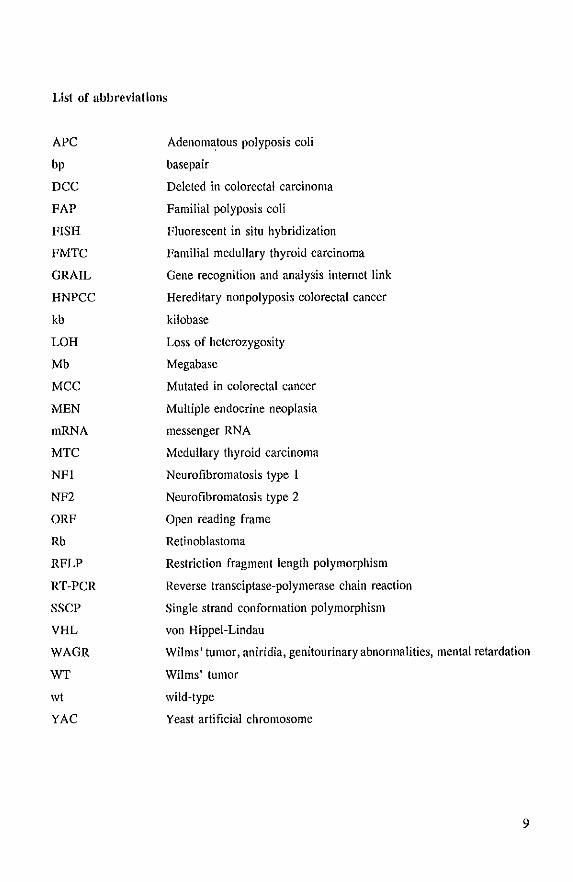
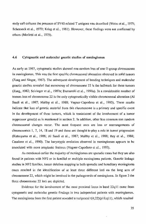
chromosome 22, which might be involved in the pathogenesis of meningioma. In figure 3 the
three chromosome 22 loci are depicted.
Evidence for the involvement of the most proximal locus in band 22q 11 came from
cytogenetic and molecular genetic findings in two independent patients with meningiomas.
The meningioma from the first patient revealed a reciprocal t(4;22)(pI6;qll), which resulted
35
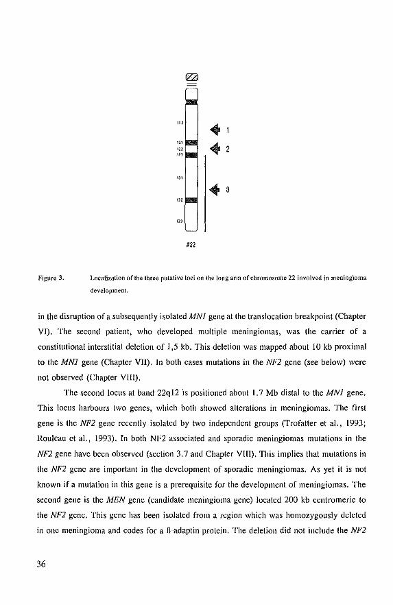
Figure J.
.3 '"
#22
Localization of the three putative loci on the long arm of chromosome 22 involved in meningioma
development.
in the disruption of a subsequently isolated MNI gene at the translocation breakpoint (Chapter
VI). The second patient, who developed multiple meningiomas, was the carrier of a
constitutional interstitial deletion of 1,5 kb. This deletion was mapped about 10 kb proximal
to the MNI gene (Chapter VII). In both cases mutations in the NF2 gene (see below) were
not observed (Chapter VIII).
The second locus at band 22q12 is positioned about 1.7 Mb distal to the MNI gene.
This loclis harbours two genes, which both showed alterations in meningiomas. The first
gene is the NF2 gene recently isolated by two independent groups (Trofatter et aI., 1993;
Rouleau et aI., 1993). In both NF2 associated and sporadic meningiomas mutations in the
NF2 gene have been observed (section 3.7 and Chapter VIII). This implies that mutations in
the NF2 gene are important in the development of sporadic meningiomas. As yet it is not
known if a mutation in this gene is a prerequisite for the development of meningiomas. The
second gene is the MEN gene (candidate meningioma gene) located 200 kb centromeric to
the NF2 gene. This gene has been isolated from a region which was homozygously deleted
in one meningioma and codes for a fi-adaptin protein. The deletion did not include the NF2
36
gene (Dumanski et aI., NNFF consortium on gene cloning, Ann Arbor, april 24·26, 1993).
However, Sanson et al (1993) observed a germline deletion in a NF2 family in which both
the NF2· and the MEN gene were lost. In this family, however, meningiomas were not
found. Therefore, further study is required to find out if this gene is involved in the
development of (a subset 01) meningiomas.
The most distal locus on chromosome 22 was defined because in two meningiomas
terminal deletions were observed telomeric to the NF2 gene (Dumanski et aI., 1990a; Rey
et aI., 1993). Since the most distal breakpoint was located telomeric to MB at 22q12.3 this
locus was mapped on 22qI2.3·qter. In addition, other reports describe chromosome 22
abnormalities restricted to this distal region. Two patients with multiple meningiomas were
described with a constitutional ring chromosome 22, resulting in loss of chromosome 22
sequences distal to 22q13 (Arinami et aI., 1986; Petrella et aI., 1993). This might suggest
that a gene is located in the deleted region or that the ring chromosomes lead to somatic
instability of chromosome 22. Unfortunately, in neither case cytogenetic analysis was carried
out on the tumor cells. Apart from this, in a sporadic vestibular schwannoma a reciprocal
translocation was found with the breakpoint at 22q13. No mutation in the NF2 gene was
observed in these tumor cells (Wolff et aI., NNFF consortium on gene cloning, Ann Arbor,
april 24·26, 1993).
Furthermore, linkage analysis in a family with mUltiple meningiomas and
ependymomas suggests that another meningioma locus distinct from NF2 is involved that
might be located on another chromosome (Pulst et al. 1993). In this study they excluded a
region of 15 eM on chromosome 22, which includes the NF2 gene, as the site for the
predisposing mutation.
All together these results indicate that different genes may be involved in the
development of vestibular schwan noma and meningioma and that further studies are required
to establish the importance of the individual genes.
Three groups have investigated whether genomic imprinting of chromosome 22 occurs
in sporadic meningiomas and schwannomas and NF2 schwannomas by examining the parental
origin of chromosome 22 loss. No preferential parental loss of this chromosome could be
observed. This suggests that genomic imprinting is not a common mechanism of gene
inactivation in these tumors (Fontaine et aI., 1990; Sanson et a1., 1990; Fontaine et aI.,
1991).
37
4.7 Other tumors with loss or I'earl'angements of chromosome 22
In addition to meningiomas and vestibular schwannomas, other tumors also show non random
loss of chromosome 22. Medullary thyroid carcinoma, an endocrine tumor also found in
MEN 2, showed LOH for chromosome 22q in approximately 10% of the cases. However,
in 40% of both sporadic and familial pheochromocytomas (MEN 2), deletions of (parts of)
the long arm of chromosome 22 have been observed, with the smallest region of overlap
between D22SIO and D22S22 (Khosla et aI., 1991; Tanaka et aI., 1992). This region
includes all three putative meningioma susceptibility loci (Fig 3, Delattre et aI., 1991).
Reviewing the data on 31 ependymomas, deletions and translocations of chromosome 22 were
observed in 13 of them (Rey et aI., 1987; Bown et aI., 1988; Dal Cin and Sandberg, 1988;
Griffin et aI., 1988; Jenkins et aI., 1989; Savard and Gilchrist, 1989; James et aI., 1990;
Ranson et aI., 1992; Sainati et aI., 1992; Weremowicz et aI., 1992). One ependymal tumor
showed a der(22)t(22;?)(qI1.2;?) and another one revealed loss of one chromosome 22 and
a balanced translocation at q 13. 3 in the remaining 22 homologue (Stratton et aI., 1989;
Weremowicz et aI., 1992). This might suggest that a gene at 22q13.3 is involved. Monosomy
and structural changes of chromosome 22 are the most consistent specific chromosomal
alterations observed in malignant mesothelioma, The structural abnormalities all revealed
breakpoints at 22qll (Fletjer et aI., 1989; Hagemeijer et aI., 1990). This is in the region
where we found the MNl gene. In leiomyoma, a small tumor derived from smooth muscle
most often of the uterus, also non random loss of chromosome 22 was found (Sreekantaiah
and Sandberg, 1991). In addition, chromosome 22 deletions have been observed in gliomas
and predominate in the higher malignancy grades. LOH studies in these tumors have
identified terminal deletions due to breakpoints at 22qI3, suggesting that this region might
be the location ofa glioma suppressor gene (Rey et aI., 1993). A few reports about rhabdoid
tumors describe monosomy 22 as the only cytogenetic change in two cases and a
der(22)t(9;22(p13;qll) with a deletion of 22qter distal to BCRL2 in another (Biegel et aI.,
1990; Biegel et aI., 1992). Recently a reciprocal t(1l;22)(pI5.5;qI1.23) was identified in
such a tumor (Newsham et aI., 1994). The breakpoint was mapped proximal to the NF2
locus and might be in the region where we mapped the MNI gene. It would be very
interesting to find out if this gene is involved. LOH studies in breast cancer revealed specific
deletions of chromosome 22 sequences, with a preference in the lobular carcinomas (Devilee
38
et aI., 1991b; Larsson et aI., 1990). In colon cancer Vogelstein et al. (1989) found in about
30% of the tumors loss of chromosome 22q. Miyaki et al.(1990) showed that chromosome
22 loss in colorectal cancer is a relatively late event. Less than 5% of the adenomas from
FAP patients showed loss of chromosome 22, whereas 33% of invasive carcinomas revealed
this loss. So far, mutations in the NF2 gene were observed in 1/69 breast tumors, 6/20
primary and melanoma metaslases, and 2/64 coloreclal carcinomas (Bianchi et aI., 1994;
Arakawa et aI., 1994) and 118 ependymomas (Rubio et aI., 1994). However, 5
pheochromocytomas and 30 aslrocylomas did not reveal mutations in this gene (Bianchi et
aI., 1994). Furlher sludy is required to elucidale whether the above mentioned genes andlor
other chromosome 22 specific genes are involved in the pathogenesis these tumors.
4.8 Chromosome 22
Chromosome 22 is the second smallest acrocentric chromosome, comprising approximately
56 Mb or 1.9% of Ihe haploid genome (Morton, 1991). The shorl arm (22p) only harbours
the ribosomal genes of the nucleolar organizer region, while Ihe long arm (22q) represents
the bulk of the chromosomal DNA. The long ann of the chromosome is involved in a
number of diseases. These include Di George syndrome, velo-cardia-facial syndrome, cat eye
syndrome and a number of cancers such as chronic myelocylic leukemia, Burkililymphoma,
Ewing sarcoma, meningiomas and vestibular schwannomas (McDermid et aI., 1993). The
past few years a lot of efforl has been put into Ihe genomic characterization of chromosome
22. This was facilitated by Ihe development of high-resolulion panels of somalic cell hybrids,
detailed linkage maps and the isolation of a large number of chromosome 22 specific DNA
markers (Duman ski et aI., 1990b; Budarf et aI., 1991; Delallre et aI., 1991; Dumanski et aI.,
1991; Fiedler et aI., 1991; Emanuel et aI., 1991; Frazer et aI., 1992; van Biezen et aI.,
1993; Emanuel et aI., 1993). Very helpful in the construclion of a genomic contig of this
chromosome is the availability of chromosome 22 specific cosmid libraries (de Jong et aI.,
1989). These cosmid and genomic YAC libraries have resulted in the isolation of a contig
of about 1100 kb on human chromosome 22ql2 (Xie et aI., 1993). Very recently a physical
map became available for 22qll-ql2 using pulsed-field gel electrophoresis. This long-range
restriction map spans approximately 11 Mb of DNA (McDermid et aI., 1993).
39
5 Scope of the thesis
Cytogenetic analysis and LOR studies of meningioma tumors points to the localization of (a)
tumor suppressor gene(s) on the long arm of chromosome 22 because:
in about 50% of the tumors one copy of chromosome 22 is lost and this is often the sole
chromosomal abnormality and
in a few percent of the cases additional aberrations concerning chromosome 22, such as
translocations (aU in band qll,q12) and partial loss of the long arm are found.
The purpose of this thesis was to identify and characterize (a) tumor suppressor gene(s),
which (is) are involved in the pathogenesis of meningioma. To reach this goal we started
with the isolation and characterization of single-copy probes for chromosome 22 (Chapter II).
Cytogenetic analysis and genotype analysis of the tumors were carried out in order to identify
the chromosomal aberrations in the tumors. Furthermore, other patient and tumor
characteristics were investigated and mutuaUy compared (Chapter Ill). The same approach
was used to study the involvement of chromosome 22 in a patient with familial anaplastic
ependymoma (Chapter IV). ]n order to localize a meningioma tumor suppressor gene on
chromosome 22 we took advantage of a meningioma (MN32) in which a reciprocal
t(4;22)(pI6;qll) had occurred. We hypothesized that this translocation disrupts a potential
tumor suppressor gene. To map this translocation breakpoint on chromosome 22 we made
hybrid ceUlines in which both reciprocal translocation products were segregated in different
hybrids (Chapter V). Long-range restriction mapping was used to further characterize the
breakpoint. One probe (D22S 193) recognized the translocation breakpoint on pulsed field
gels. Chromosome walking towards the translocation breakpoint and searching for transcribed
sequences in this region resulted in the isolation of the MNI gene (Chapter VI). Probes from
this region were used to investigate DNA from other meningiomas and blood of a patient
with multiple meningiomas. Southern blot analyses observed aberrant restriction fragments
in DNA isolated from a patient with multiple meningiomas (patient 55). The characterization
of these alterations are described in chapter VII. Findings from other groups (see 4.6), the
isolation of the MNI gene and the recent cloning of the NF2 gene (Rouleau et aI., 1993;
Trofatter et aI., 1993) raise a question about the relative contribution of these genes/loci in
the development of meningiomas. To investigate the extent in which the NF2 is involved we
performed mutation analysis of the NF2 gene transcript in our series of (mostly) sporadic
meningiomas, including tumors 32 and 55 (Chapter VIII).
40
6 References
Aaltonen LA, Peltomiiki P, Leach FS, Sistonen P, Pylkkiinen L, Mecklin JP, Jarvinen H, Powell SM et .11. (1993) Clues to the pathogenesis of familial colorectal cancer. Science 260:812-816.
Ahuja HO, Foti A. Zhau DJ, Cline MJ (1990) Analysis of proto-oncogenes in acute myeloid leukemia: loss of heterozygosity fOT the Ha-ras gene. Blood 75:819-822
Ali lV, Liderau R. 111eiIJel C, Callahan R (1987) Reduction to homozygosity of genes on chromosome 11 in human breast neoplasia. Science 238: 185-188.
AI-Rodhan NRF. Laws ER (1990) Meningioma: A historical study of the tumor and its surgical management. Neurosurg 26:832-847.
AI-Saadi A, Latimer F. Madercic M, Robbins T (1987) Cytogenetic studies of human brain tUmors and their clinical significance, Cancer Genet Cytogenet 26: 127-141.
Andersen LB, Fountain JW, Gutmann DR, Tarle SA, Glover TW, Dracopoli NC. Housman DE, Collins FS (1993) Mutations in the neurofibromatosis t gene in sporadic malignant melanoma cell lines, Nature Genet 3:I18-121.
Arakawa H, Hayashi N, Nagase H, Ogawa M, Nakamura Y (1994) Alternative splicing of the NF2 gene and its mutation analysis of breast ami coloredal cancers, Hum Mol Oen 3:565-568,
Arinami T, Kondo I, Hamaguchi H, Nakajima S (1986) Multifocal meningiomas in a patient with a constitutional ring chromosome 22, J Med Genet 23:178-180,
Baker SJ, Fearon ER, Nigro JM, Hamilton SR, Preisinger AC, Jessup M, van Tuinen P et al. (1989) Chromosome 17 deletions and p53 gene mutations in colorectal carcinomas, Science 244:217-221.
Baker SJ, Markowitz S, Fearon ER. Willson JK, Vogelstein B (1990) Suppression of human coloreclal carcinoma cell growth by wild-type p53, Science 249:912-915_
Bartek I, Bartkova J, Vojtesek B, Staskova Z, Lukas J, Rejthar A, Kovarik J et al. (1991) Aberrant expression of the p53 oncoprotein is a common feature ofa wide spectrum of human malignancies_ Oncogene 6: 1699-1703,
Beckwith JB, Kiviat NB, Bondio JF (1990) Nephrogenic resls, nephroblaslomatosis, and the pathogenesis of Wilms' tumor_ Pediatr PathoI1O:1-36.
Bianchi AB. Hara T, Ramesh V, Gao J, Klein-Szanto AlP, Morin F, Menon AG et al. (1994) Mutations in transcript isoforms of the neurofibromatosis 2 gene in multiple human tumour types. Nature Genet 6: 185· 192.
Biegel JA, Rorke LB, Packer RJ, Emanuel BS (1990) Monosomy 22 in rhabdoid or atypical tumors of the brain. J Neurosurg 73:710-714.
Biegel JA, Burk CD, Parmiter AH, Emanuel BS (1992) Molecular analysis of a partial deletion of 22q in a central nervous system rhabdoid tumor. Genes Chrom Cancer 5:104·108.
van Biezen NA, Lekanne Deprez RH, Thijs A, Heutink P, Oostra BA, Geurts van Kessel ARM, ZwarthoffEC (1993) Isolation and characterization of25 unique DNA markers for human chromosome 22. Genomics 15:206· 208.
BischoffJR, Friedman PN, Marshak DR, Prives C, Beach D. (1990) Human p53 is phosphorylated by p60-cde2 and cyetin B·cdc2. Proc Natl Acad Sci USA 87:4766·4770.
Black P McL (1991) Brain Tumors. New Engl J Med 324:1555-1564.
BoUag G, McCormick F (1992) NF is enough of GAP. Nature 356:663·664.
Bongarzone I, Monzini N, Borrello MG, Carcano C, Ferraresi G, Arighi E, Mondellini P et al. (1993) Molecular characterization of a thyroid tumor-specific transforming sequence fonned by the fusion of ret tyrosine kinase and the regulatory subunit RI alpha of cyclic AMP-dependent protein kinase A. Mol Cell Bioi 13:358-366.
Bookstein R, Rio P, Madreperla SA, Hong F, Allred C, Grizzle WE, Lee WH (l990a) Promoter deletion and loss of retinoblastoma gene expression in human prostate carcinoma. Proc Nat! Acad Sci USA 87:7762-7766.
41
Bookstein R, Shew JY, Chen PL, Scully P, Lee WH (1990b) Suppression of tumorigenicity of human prostate carcinoma cells by replacing a mutated Rb gene. Science 247:712-715.
Borresen AL, Andersen TI, Garber J, Barbier-Piraux N, Thorlacius S, Eyfjord J et al. (1992) Screening for genn line TP53 mutations in breast cancer patioents. Cancer Res 52:3234-3236.
Bown N, Pearson ADJ, Davidson EV, Gardner-Medvin D, Crawford P, Perry R (1988) Multiple chromosome rearrangements in a childhood ependymoma. Cancer Genet Cytogenet 36:25-30.
Bronner CE, Baker SM, Morrison PT, Warren G, Smith LG, Lescoe MK, Kane M et al (1994) Mutations in the DNA mismatch repair gene homologue hMLHI is associatetl with hereditary non-polyposis colon cancer. Nature 368:258-261.
Buchberg AM, Cleveland LS, Jenkins NA, Copeland NG (1990) Sequence homology share<:! by neurofibromatosis type-l gene and IRA-l and IRA-2 negative regulators of the RAS cyclic AMP pathway. Nature 347:291-294.
Buchkovich K, Duffy LA, Harlow E (1989) The retinoblastoma protein is phosphorylated during spe.cific phases of the cell cycle. Cell 58:1097-1105.
Budarf ML, McDennid HE, Sellinger B, Emanuel BS (1991) Isolation and regional localiwtion of 35 unique anonymous DNA markers for human chromosome 22. Genomics 10:996-1002.
Butti G, Assietti R, Casalone R, Paoletti P (1989) Multiple meningiomas: A clinical, surgical and cytogenetic analysis. Surg Neurol 31 :255-260.
Call KM, Glaser T, Ito CY, Buckler AJ, Pelletier J, Haber DA, Rose EA et al. (1990) Isolation and characterization of a zinc finger polypeptide gene at the human chromosome II Wilms' tumor locus. Cell 60;509-520.
Caselone R, Simi P, Granata P, Minelli E, Giudici A, Butti G, Solero CL (1990) Correlation between cytogenetic and histopathological findings in 65 human meninginmas. Cancer Genet Cytogenet 45:237-243.
Cavenee WK, Dryja TP, Phillips RA, Benedict WF, Godbout R, Gallie BL, Murphree AL, Strong LC, White RL (1983) Expression of recessive alleles by chromosomal mechanisms in retinoblastoma. Nature 305:779-784.
Cawthon RM, Weiss R, Xu G, Viskochil D, Culver M, Stevens J, Robertson M et Ill. (1990) A m<tlor segment of the neurofibromatosis type 1 gene: eDNA sequence, genomic structure, and point mutations. Cell 62: 193-201.
Chellappan SP, Hiebert S, Mudryj M, HorowitzJM, Nevins JR (1991) The E2F transcription factor is a cellular target for the Rb protein. Cell 65: 1053-1061.
Chen PL, Scully P, Shew JY, Wang JY, Lee WH (1989) Phosphorylation of the retinoblastoma gene product is modulated during the cell cycle and cellular differentiation. Cell 58: 1193-1198.
Cho KR, Oliner JD, Simons JW, Hedrick L, Fearon ER, Preisinger AC, Hedge P et al. (1994) The DeC gene: Structural analysis and mutations in coloreclal carcinomas. Oenomics 19:525-531.
Clarke AR, Purdie CA, Harrison DJ, Morris RO, Bird CC, Hooper ML, Wyllie AH (1993) Thymocyte apoplosis induced by p53-dependent and independent pathways. Nature 362:849-852.
Cohn KH, Wang P, Desolo-LaPaix P, Solomon WB, Patterson LG, Arnold MR, Weimar J el al. (1991) Association of nm23-H I allelic deletions with distant metastases in coloreclal carcinoma. Lancet 338: 722-724.
Collins FS (1992) Positional cloning: Let's not call it reverse any more. Nature Genet 1:3-6.
Comings DE (1973) A general theory of carcinogenesis. Proc Nail Acad Sci USA 70:3324-3328.
Cowell JK, Wadey RB, Haber DA t Call KM, Housman DE, Pritchard J (1991) Structural rearrangements of the WTI gene in Wilms' tumour cells. Oncogene 6:595-599.
Cushing H (1922): The meningiomas (dural endotheliomas): Their source and favoured seats of origin. Brain 45: 282-316.
Dal Cin P, Sandberg AA (1988) Cytogenetic findings in a supratentorial ependymoma. Cancer Genet Cytogenet 30;289-293.
DeCaprio JA, Ludlow JW, Figge J, Shew JY, Huang CM, Lee WH, Marsilio E, Paucha E, Livingston DM
42
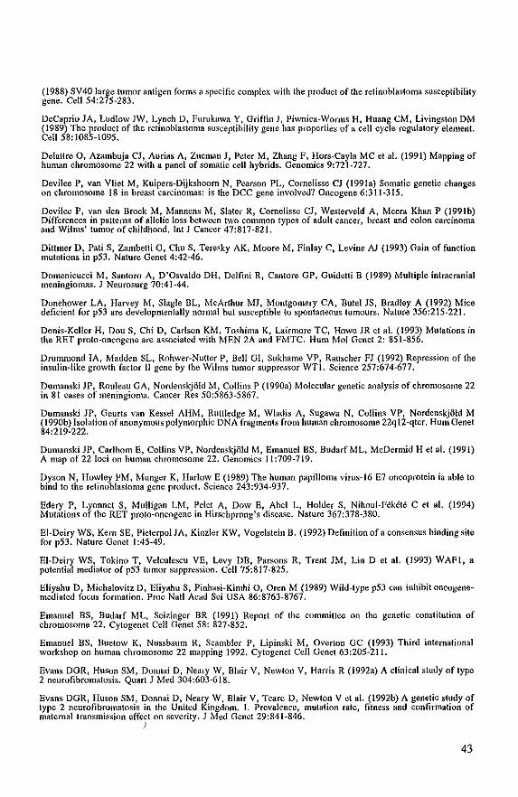
(1988) SV40 large tumor antigen forms a spedne complex with the product of the retinoblastoma susceptibility gene. Cell 54:275-283.
DeCaprio JA, Ludlow JW, Lynch 0, Furukawa Y, Griffin J, Piwnica-Worms H, Huang CM, Livingston DM (1989) The product of the retinoblastoma susceptibility gene has properties of a cell cycle regulatory element. Cell 58: 1085-1095.
Delattre 0, Azambuja CJ, Aurias A, Zucman J, Peter M, Zhang F. Hors-Cayla MC et aJ. (1991) Mapping of human chromosome 22 with a panel of somatic cell hybrids. Genomics 9:721-727.
Devilee P, van Vliet M, Kuipers-Dijkshoorn N, Pearson PL, Comelisse CJ (199Ia) Somatic genetic changes on chromosome 18 in breast carcinomas: is the DCC gene involved? Oncogene 6:311-315.
Devilee P, van den Broek M, Mannens M. Slater R. Cornelisse CJ, Westerveld A. Meera Khan P (l99Ib) Differences in patterns of aUelic loss between two common types of adult cancer, breast and colon carcinoma and Wilms' tumor of childhood. Int J Cancer 47:817-821.
Dittmer D, Pati S, Zambelli G, Chu S, Teresky AK, Moore M, Finlay C, Levine A1 (1993) Gain of function mutations in p53. Nature Genet 4:42-46.
Domenicucci M. Santoro A, D'Osvaldo DH, Delfllli R, Cantore GP, Guidetti B (1989) Multiple intracranial meningiomas. J Neurosurg 70:41-44.
Donehower LA, Harvey M, Slagle BL, McArthur MJ, Montgomery CA, RUlel JS, Bradley A (1992) Mice deficient for pS3 are developmentally normal but susceptible to spontaneous tumours. Nature 356:215-221.
Donis-Keller H, Dou S, Chi D, Carlson KM, Toshima K. Lairmore Te, Howe JR et a!. (1993) Mutations in the RET proto-oncogene are associated with MEN 2A and FMTC. Hum Mol Genet 2: 851-856.
Drummond lA, Madden SL, Rohwer-Nutter P, Bell GI, Sukhame VP, Rauscher FJ (1992) Repression of the insulin-like growth factor II gene by the Wilms tumor suppressor WTI. Science 257:674-677.
Dumanski JP, Rouleau GA, NordenskjOJd M. Collins P (1990a) Molecular genetic analysis of chromosome 22 in 81 cases of meningioma. Cancer Res 50:5863-5867.
Dumanski JP, Geurts van Kessel AHM, Ruttledge M, Wladis A, Sugawa N, Collins VP, NordenskjOld M (l990b) Isolation of anonymous polymorphic DNA fragments from human chromosome 22q 12-qter. Hum Genet 84:219·222.
Dumanski JP, earlbom E, Collins VP, NordenskjOid M, Emanuel BS, BudarfML, McDermid H et al. (1991) A map of 22 loci on human chromosome 22. Genomics 11:709-719.
Dyson N, Howley PM, Munger K, Harlow E (1989) The human papilloma virus-16 E7 oncoprotein ia able to bind to the retinoblastoma gene product. Science 243:934-937.
Edery P, Lyonnet S, Mulligan LM. Pelel A, Dow E, Abel L, Holder S, Nihoul-F~kt~te C el al. (1994) Mutations of the RET proto-oncogene in Hirschprung's disease. Nature 367:378-380.
EI-Deiry WS, Kern SE, Pieterpol JA, Kinzler KW, Vogelstein B. (1992) Definition of a consensus binding site for p53. Nature Genet 1 :45-49.
EI-Deiry WS, Tokino T, Velculescu VE, Levy DB, Parsons R, Trent JM, Lin D et a!. (1993) WAFI, a potential mediator of pS3 tumor suppression. Cell 75:817-825.
Eliyahu D, Michalovitz D, Eliyahu S, Pinhasi-Kimhi D, Oren M (1989) Wild-type pS3 can inhibit oncogenemediated focus formation. Proc Natl Acad Sci USA 86:8763-8767.
Emanuel BS, Budarf ML, Seizinger BR (l991) Report of the committee on the genetic constitution of chromosome 22. Cytogenet Cell Genet 58: 827~852.
Emanuel BS, Buetow K, Nussbaum R, Scambler P, Lipinski M, OVerton GC (1993) Third international workshop on human chromosome 22 mapping 1992. Cytogenet Cell Genet 63:205-211.
Evans DGR, Huson SM, Donnai 0, Neary W, Blair V, Newlon V, Harris R (l992a) A clinical study of type 2 neurofibromatosis. Quart J Med 304:603-618.
Evans DGR, Huson SM, Donnai 0, Neary W, Blair V, Teare 0, Newton V et al. (1992b) A genetic study of type. 2 neurofibromatosis in the United Kingdom. I. Prevalence, mutation rate, fitness and confinnation of maternal transmission effect on severity. J Moo Genet 29:841-846.
I
43
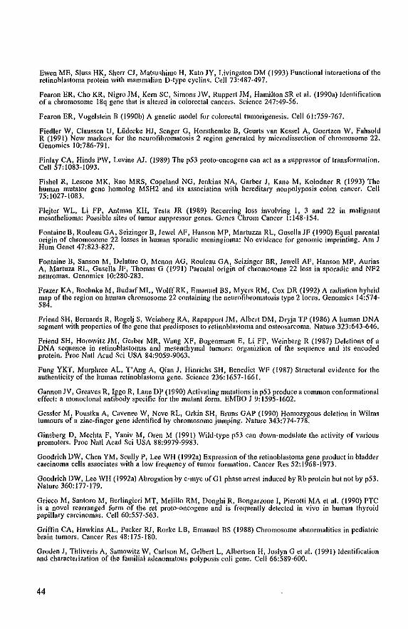
Ewen ME, Sluss HK, Sherr CJ, Matsushime H, Kato JY, Livingston DM (1993) Functional interactions of the retinoblastoma protein with mammalian D-Iype cyctins. Cell 73:487-497.
Fearon ER, Cho KR, Nigro JM, Kern SC, Simons JW, Ruppert JM, Hamilton SR et al. (1990a) Identification of a chromosome 18q gene that is altered in coiorectal cancers. Science 247:49-56.
Fearon ER, Vogelstein B (l990b) A genetic model for colorectaltumorigenesis. Cell 61:759-767.
Fiedler W, Claussen V, Liidecke HJ, Senger G, Horslhemke B, Geurts van Kessel A, Goertzen W, Fahsold R (1991) New markers for the neurofibromatosis 2 region generated by microdissection of chromosome 22. Genomics 10:786-791.
Finlay CA, Hinds PW, Levine AJ. (1989) The p53 proto-oncogene can act as a suppressor of transformation. Cell 57:1083-1093.
Fishel R, Lescoe MK, Rao MRS, Copeland NG, Jenkins NA, Garber J, Kane M, Kolodner R (1993) The human mutator gene homolog MSH2 and its association with hereditary nonpolyposis colon cancer. Cell 75:1027-1083.
Flejter WL, Li FP, Anlman KH, Testa JR (1989) Recurring loss involving I, 3 and 22 in malignant mesothelioma: Possible sites of tumor suppressor genes. Genes Chrom Cancer 1:148-J54.
Fontaine B, Rouleau GA, Seizinger B, Jewel AF, Hanson MP, Marluzza RL, Gusella JF (1990) Equal parental origin of chromosome 22 losses in human sporadic meningioma: No evidence for genomic imprinting. Am J Hum Genet 47:823-827.
Fontaine B, Sanson M, Delatlre 0, Menon AG, Rouleau GA, Seizinger BR, Jewell AF, Hanson MP, Aurias A, Martuza RL, Gusella JF, Thomas G (1991) Parental origin of chromosome 22 loss in sporadic and NF2 neuromas. Genomics 10:280-283.
Frazer KA, Boehnke M, BudarfML, WolffRK, Emanuel BS, Myers RM, Cox DR (1992) A radiation hybrid map of the region on human chromosome 22 containing Ihe neurofibromatosis type 2 locus. Genomics 14:574-584.
Friend SH, Bernards R, Rogelj S, Weinberg RA, Rapapport JM, Albert DM, Dryja TP (1986) A human DNA segment with properties of the gene that predisposes to retinoblastoma and osteosarcoma. Nature 323:643-646.
Friend SH, Horowitz JM, Gerber MR, Wang XP, Bogenmann E, Li FP, Weinberg R (1987) Deletions of a DNA sequence in retinoblastomas and mesenchymal tumors: organiztion of the sequence and its encoded protein. Proc Nail Acad Sci USA 84:9059-9063.
Fung YKT. Murphree AL, T'Ang A, Qian J, Hinrichs SH, Benedict WF (1987) Structural evidence for the authenticity of the human retinoblastoma gene. Science 236: 1657-1661.
Gannon JV, Greaves R. Jggo R, Lane DP (J990) Activating mutations in p53 produce a common conformational effect: a monoclonal antibody spedfic for the mutant form. EMBO J 9: 1595~ 1602.
Gessler M, Pouslka A, Cavenee W, Neve RL, Orkin SR, Bruns GAP (1990) Homozygous deletion in Wilms tumours of a zinc-finger gene identified by chromosome jumping. Nature 343:774-778.
Ginsberg D. Mechta F, Yaniv M, Oren M (1991) Wild-Iype p53 can down-modulate the activity of various promoters. Proc Nail Acad Sci USA 88:9979-9983.
Goodrich DW, Chen YM, Scully P, Lee WH (l992a) Expression of the retinoblastoma gene product in bladder carcinoma cells associates with a low frequency of tumor formation. Cancer Res 52: J968-J 973.
Goodrich DW, Lee WH (1992a) Abrogation by c-myc ofGl phase arrest induced by Rb protein but not by p53. Nature 360:177-179.
Grieco M, Santoro M. Berlingieri MT, Melillo RM. Donghi R, Bongarzone I, Pierotti MA et a1. (1990) PTC is a novel rearranged form of the rei proto-oncogene and is frequently detected in vivo in human thyroid papillary carcinomas. Cell 60:557-563.
Griffin CA, Hawkins AL, Packer RJ, Rorke LB, Emanuel BS (1988) Chromosome abnormalities in pediatric brain tumors. Cancer Res 48:175-180.
Groden J. Thliveris A, Samowitz W, Carlson M. Gelbert L, Albertsen H, Joslyn G el al. (1991) Identification and characterization of the familial adenomatous polyposis coli gene. Cell 66:589-600.
44
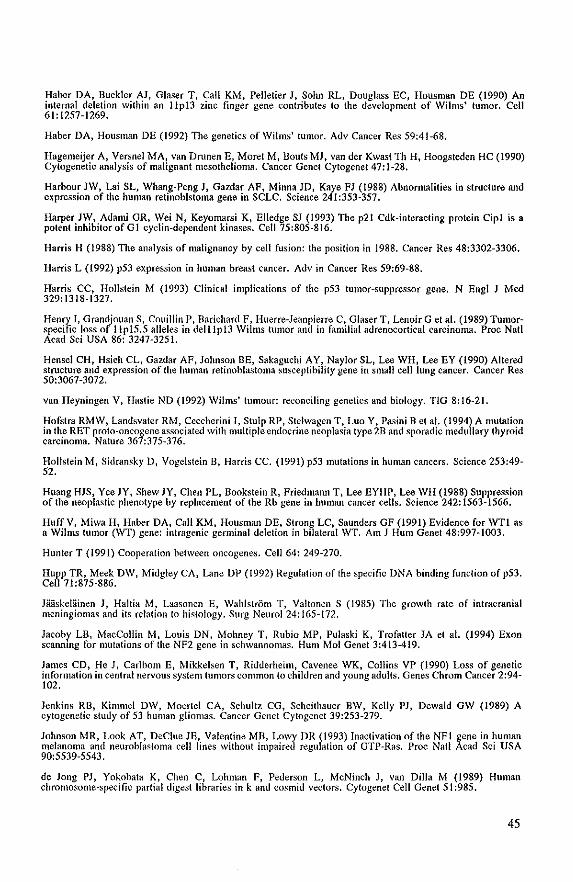
Haber DA, Buckler AJ, Glaser T, Call KM, Pelletier J, Sohn RL, Douglass EC, Housman DE (1990) An internal deletion within an IIp!3 zinc finger gene contributes to the development of Wilms' tumor. Cell 61:1257-1269.
Haber DA, Housman DE (1992) The genetics of Wilms' tumor. Adv Cancer Res 59:41-68.
Hagemeijer A, Versnel MA, van Drunen E, Moret M, Douts MJ, van der Kwast Th H, Hoogsteden HC (1990) Cytogenetic analysis of malignant mesothelioma. Cancer Genet Cytogenet47:1-28.
Harbour JW, Lai SL, Whang-Peng J, Gazdar AF, Minna JD, Kaye FJ (1988) Abnormalities in structure and expression of the human relinoblsloma gene in SCLC. Science 241:353-357.
Harper JW, Adami OR, Wei N, Keyomarsi K, Elledge SJ (1993) The p21 Cdk-interacting protein Cipl is a potent inhibitorofGI eyelin-dependent kinases. Cell 75:805-816.
Harris H (1988) The analysis of malignancy by cell fusion: the position in 1988. Cancer Res 48:3302-3306.
Harris L (1992) p53 expression in human breast cancer. Adv in Cancer Res 59:69-88.
Harris CC, Hollstein M (1993) Clinical implications of the p53 tumor-suppressor gene. N Engl J Med 329: 1318-1327.
Henry It Grandjouan S, Couillin P, Darichard F, Huerre-Jeanpierre C, Glaser T. Lenoir G et al. (1989) Tumorspecific loss of IlpIS.S alleles in deillpl3 Wilms tumor and in familial adrenocortical carcinoma. Proc Nat! Acad Sci USA 86: 3247-3251.
Hensel CH, Hsieh CL, Gazdar AF, Johnson DE, Sakaguchi AY, Naylor SL, Lee WH, Lee EY (1990) Altered structure and expression of the human retinoblastoma susceptibility gene in small cell lung cancer. Cancer Res 50:3067-3072.
van Heyningen V, Hastie ND (1992) Wilms' tumour: reconciling genetics and biology. TiO 8:16-21.
Hofstra RMW, Landsvater RM, Ceccherini T, Stulp RP, Stelwagen T. Luo Y. Pasini B et al. (1994) A mutation in the RET proto-oncogene associated with mUltiple endocrine neoplasia type 2B and sporadic medullary thyroid carcinoma. Nature 367:375-376.
Hollstein M, Sidransky D, Vogelstein B. Harris CC. (1991) p53 mutations in human cancers. Science 253:49-52.
Huang HJS, Yee JY, Shew JY, Chen PL, Bookstein R, Friedmann T, Lee EYHP, Lee WH (1988) Suppression of the neoplastic phenotype by replacement of the Rb gene in human cancer cells. Science 242: 1563-1566.
Huff V, Miwa H, Haber DA, Call KM, Housman DE, Strong Le, Saunders GF (1991) Evidence for WTl as a Wilms tumor (WT) gene: inlragenic germinal deletion in bilateral WT. Am J Hum Genet 48:997-1003.
Hunter T (1991) Cooperation between oncogenes. Cell 64: 249-270.
Hupp TR, Meek DW, Midgley CA, Lane DP (1992) Regulation of the specific DNA binding function of p53. Cell 71:875-886.
JiiliskeliHnen J, Haltia M, Laasonen E, Wahlstrom T, Valtonen S (1985) The growth rate of intracranial meningiomas and its relation to histology. Surg Neurol 24: 165-172.
Jacohy LB, MacCollin M, Louis DN, Mohney T, Rubio MP, Pulaski K, Trofatter JA et al. (1994) Exon scanning for mutations of the NF2 gene in schwannomas. HUIll Mol Genet 3:413-419.
James CD, He J, Carlbom E, Mikkelsen T, Ridderheim, Cavenee WK, Collins VP (1990) Loss of genetic information in central nervous system hlfllOrs common to children and young adults. Genes Chrom Cancer 2:94-102.
Jenkins RB, Kinmlei DW, Moertel CA, Schultz CG, Scheithauer BW, Kelly PJ, Dewald GW (1989) A cylogenetic study of 53 human gliomas. Cancer Genet Cytogenet 39:253-279.
Johnson MR, Look AT, DeClue JE, Valentine MD, Lowy DR (1993) Inactivation of the NFl gene in human melanoma and neuroblastoma cell line-s without impaired regulation of GTP-Ras. Proc Nat! Acad Sci USA 90:5539-5543.
de Jong PJ, Yokobata K, Chen C, Lohman F, Pederson L, McNinch J, van Dilla M (1989) Human chromosome-spedfic partial digest libraries in k and cos mid vectors. Cylogenet Cell Genet 51:985.
45
Joslyn G, Carlson M, Thlivris A, Albertsen H, Gelberl L, SamowHz W, Groden J et aL (1991) Identification of deletion mutations and three new genes at the familial polyposis locus. Cell 66:601·613.
Kaslan MR, Zham Q, EI-Deiry WS, Carrier F. Jacks T, Walsh WV, Plunkett BS, Vo¥eislein B. Fomace AJ, (1992) A mammalian cell cycle checkpoint pathway utilizing p53 and GADD45 IS defective in AtaxiaTelangiedasia. Cell 71: 587-597.
Kala JY. Matsushime H. Hiebert SW, Ewen ME, Sherr C] (1993) Direct binding of eyelin D to the retinoblastoma gene product and pRb phosphorylation by the cycJln D-dependenl kinase cdk4. Genes Dev 7: 331-342.
Kalsuyama }, Papenhausen PR, Herz F, gazivoda P, Hirano A, Koss LG (1986) Chromosome abnormalities in meningiomas. Cancer Genet Cytogenet 22:63-68.
Kepes JJ (1982) Meningiomas: biology, pathology and differential diagnosis, Masson Publishing USA Illc New York,
Kern SE, Kinz1er KW, Bruskin A, Jarosz D, Friedman P, Prives C, Vogelstein B (1991) Identification of p53 as a sequence spe<:ific DNA binding protein. Science 252: 1707-1711.
Kern SE, Pietenpol JA, Thiagalingam S, Seymour A, Kinzler KW, Vogelstein B (1992) Oncogenic forms of p53 inhibits p53-regulated gene expression. Science 256:827-830.
Khosla S, Patel VM, Hay ID, Schald DJ, Grant CS, van Heerden JA, Thibodeau SN (1991) Loss of heterozygosity suggests multiple genetic alteralions in pheochromocytomas and medullary thyroid carcinomas, J Clin Invest 87:1691-1699,
Kinzler KW, Nilbert MC, Vogeistein B, Bryan TM, Levy DB, Smith KJ, Preisinger AC el aL (199Ia) Identification of a gene located at chromosome 5q21 that is Illutated in colorectal cancers, Science 251: 1366-1370.
Kinzler KW, Nilhert MC, Su LK, Vogelstein B, Bryan TM, Levy DB, Smith KJ et al. (l99Ib) Identification ofFAP locus genes from chromosome 5q21. Science 253:661-665,
Knudson AG (1971) Mutation and Cancer: Statistical Study ofRetinoblaslollla. Proc Natl Acad Sci USA 68:820-823.
Knudson AG, Strong LC (1972) Mutation ami cancer: A model for Wilms' tumor of the kidney. J Nat Cancer Inst 48:313-324.
Knudson AG (1986) Genetics of human cancer, Annu Rev GeneI20:231-251.
Knudson AG (1993) Anlioncogenes and human cancer. Proc Natl Acad Sci USA 90:10914-10921.
Koi M, Johnson LA, Kalikin LM, Little PFR, Nakamura Y, Feinberg AP (1993) Tumor cell growth arrest caused by subchromosomal transferable DNA fragments from chrolllosome 11. Science 260;361-364,
Koper JW, Foekens JA, Braakman R, Lamberts SWJ (1990) Effects of progesterone on the response to epidennal growth factor and other growth factors in cultured human meningioma cells. Cancer Res 50:2604-2607.
Kreidberg JA, Sariola H, Loring JM, Maeda M, Pelletier J, Huusman D, Jaenisch R (1993) WT-I Is required for early kidney development. Cell 74:679-691.
Krieg P, Amlmann E, Jonas D, Fischer H, Zang KD, Sauer H (1981) Episomal simian virus 40 genomes in human brain tumors, Proc Nail Acad Sci USA 78:6446-6450.
Lamb P, Crawford LV (1986) Characterization of the human p53 gene. Mol Cell Bioi 6: 1379-1385.
Landsvaler RM , Mathew CG, Smith BA, Marcus EM. te Meerman GJ, Lips CJ, Geerdink RA et al. (1989) Development of multiple endocrine neoplasia type 2A does not involve substantial deletions of chromosome 10. Genonucs 4:246-250.
Lane DP, Crawford LV (1979) T antigen is bound to host protein in SV40-transformed cells, Nature 278:261-263.
Lane DP (1992) p53, guardian of the genome. Nature 358:15-16.
Larsson C, Bystrom C, Skoog L. Rotstein S, Nordenskjold (1990) Genomic allerations in human breast
46
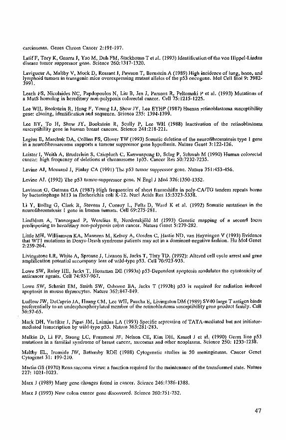
carcinomas. Genes Chrom Cancer 2:191-197.
LatifF, Tory K, Gnarra J, Yao M, Duh FM, Stackhouse T et al. (1993) Identification of the von Hippel-Lindau disease tumor suppressor gene. Science 260: 1317-1320.
Lavigueur A, Maltby V, Mock D, Rossant J, Pawson T, Bernslein A (1989) High incidence of lung, bone, and lymphoid tumors in transgenic mice overexpressing mutant alleles of the p53 oncogene. Mol Cell Bioi 9: 3982-3991.
Leach FS, Nicolaides NC, Papdopoulos N, Liu B, Jen J, Parsons R, Peltomaki P et al. (1993) Mutations of a MutS homolog in hereditary non-polyposis colorectal cancer. Cell 75:1215-1225.
Lee WH, Bookstein R, Hong F, Young U, Shew JY, Lee EYHP (1987) Human retinoblastoma susceptibility gene: cloning, identification and sC{juence. Science 235: 1394-1399.
Lee EY, To H, Shew JY, Bookstein R, Scully P, Lee WH (1988) Inactivation of the retinoblastoma susceptibility gene in human breast cancers. Science 241:218-221.
Legius E, Marchuk DA, Collins FS, Glover TW (1993) Somatic deletion of the neurofibromatosis type 1 gene in a neurofibrosarcoma supports a tumour suppressor gene hypothesis. Nature Genet 3: 122-126.
Leister I, Weith A, Bruderlein S, Cziepluch C, Kanwanpong 0, Sclag P, Schwab M (1990) Human colorectal cancer: high frequency of deletions at chromosome Ip35. Cancer Res 50:7232-7235.
Levine AJ, Momand J, Finlay CA (1991) The p53 tumor suppressor gene. Nature 351:453-456.
Levine N. (1992) The p53 tumor-suppressor gene. N Engl J Med 326:1350-1352.
Levinson G, Gutman GA (1987) High frequencies of short frameshins in poly-CAITG tandem repeats borne by bacteriophage M13 in Escherichia coli K-12. Nucl Acids Res 15:5323-5338.
Li Y, Dollag G, Clark R, Stevens J, Conroy L, Fults D, Ward K et al. (1992) Somatic mutations in the neurofibromatosis I gene in human tumors. Cell 69:275-281.
Lindblom A, Tannergard P, Werelius D, Nordenskji)ltl M (1993) Genetic mapping of a sfXond locus predisposing to hereditary non-polyposis colon cancer. Nature Genet 5:279-282.
Little MH, Williamson KA, Mannens M, Kelsey A, Gostlen C, Hastie ND, van Heyningen V (1993) Evidence that WTl mutations in Denys-Drash syndrome patients may act in a dominant-negative fashion. Hu Mol Genel 2:259-264.
Livingstone LR, White A, Sprouse J, Livanos E, Jacks T, Tlsty TO. (1992): Altered cell cycle arrest and gene amplification potential accompany loss of wild-type p53. Cell 70:923-935.
Lowe SW, Ruley HE, Jacks T, Housman DE (1993a) p53-Dependenl apoptosis modulates the cytotoxicity of anticancer agents. Cell 74:957-967.
Lowe SW, Schmitt EM, Smith SW, Osborne BA, Jacks T (l993b) p53 is required for radiation induced apoptosis in mouse thymocytes. Nature 362:847-849.
Ludlow JW, DeCaprio JA, Huang CM, Lee WH, Paucha E, Livingston DM (1989) SV40 large T antigen binds preferentially to an underphosphorylated member of the retinohlastoma susceptihility gene product family. Cell 56:57-65.
Mack DH, Vartikar J, Pipas JM, Laimins LA (1993) Specific repression ofTATA-mediated but not initialormediated transcription by wild-type p53, Nature 363:281-283.
Malkin D, Li FP, Strong LC, Fraumeni JF, Nelson CE, Kim DH, Kassel J et al. (1990) Germ line p53 mutations in a familial syndrome of breast cancer, sarcomas and other neoplasms. Science 250: 1233-1238.
Maltby EL, Ironside JW, Batlersby RDE (1988) Cytogenetic studies in 50 meningiomas. Cancer Genet Cytogenet 31: 199-210.
Martin GS (1970) ROllS sarcoma virus: a function required for the maintenance of the transformed stale. Nature 227: 1021-1023.
Marx J (1989) Many gene changes found in cancer. Science 246: 1386-1388.
Marx J (1993) New colon cancer gene discovered. Science 260:751-752.
47
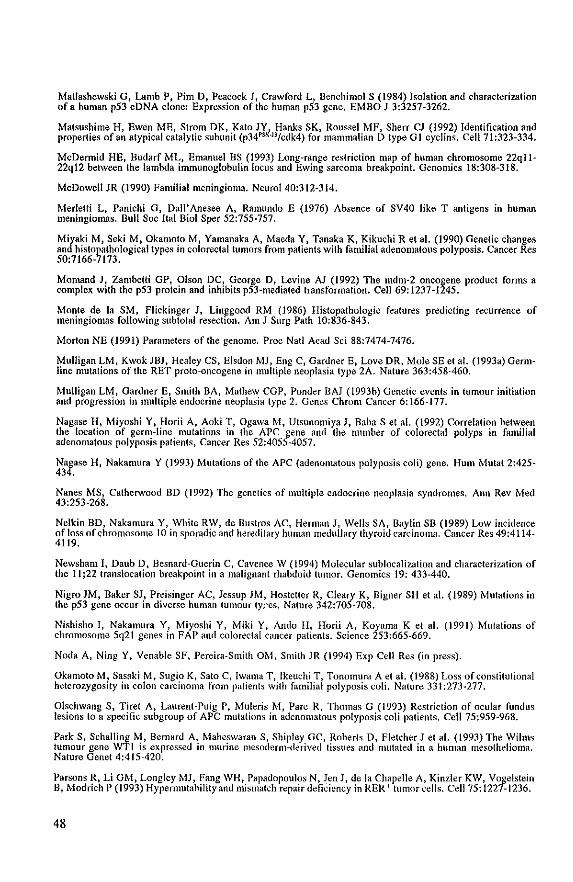
Matlashewski at Lamb P, Pim D, Peacock J, Crawford L. Benchimol S (1984) Isolation and characterization of a human p53 eDNA clone: Expression of the human p53 gene, EMBO J 3:3257-3262.
Matsushime H, Ewen ME, Strom DK, Kala JY, Hanks SK, Roussel MF, Sherr CJ (1992) Identification and properties of an atypical catalytic subunit (p34PSK•J3/cdk4) for mammalian D type G I cyclins. Cell 71 :323-334.
McDennid HE, Budarf ML, Emanuel BS (1993) Long-range restriction map of human chromosome 22ql 1-22q12 between the lambda immunoglobulin locus and Ewing sarcoma breakpoint. Genomics 18:308-318.
McDowell JR (1990) Familial meningioma. NeuroI40:312-314.
Merlelti L. Panichi at Oall' Anesee A. Ramunda E (1976) Absence of SV40 like T antigens in human meningiomas. Bull Soc Ital Bioi Sper 52:755-757.
Miyaki M, Seki M, Okamoto M, Yamanaka A, Maeda Y, Tanaka K, Kikuchi R et al. (1990) Genetic changes and histopathological types in colorectal tumors from patients with familial adenomatous polyposis. Cancer Res 50:7166-7173.
Momand J, Zambetti GP, Olson DC, George D, Levine AJ (1992) The mdm-2 oncogene product forms a complex with the p53 protein and inhibits p53-mediated transformation. Cell 69: 1237-1245.
Monte de 1a SM, Flickinger 1, Linll'good RM (1986) Histopathologic features predicting recurrence of meningiomas following subtotal resection. Am I Surg Path 10:836-843.
Morton NE (1991) Parameters of the genome. Proc Nat! Acad Sci 88:7474-7476.
Mulligan LM, Kwok JBl, Healey CS, Elsdon Ml, Eng C, Gardner E, Love DR, Mole SE et a!. (I 993a) Germline mutations of the RET proto-oncogene in multiple neoplasia type 2A. Nature 363:458-460.
Mulligan LM, Gardner E, Smith BA, Mathew CGP, Ponder BAJ (1993b) Genetic events in tumour initiation and progression in multiple endocrine neoplasia type 2. Genes Chrom Cancer 6: 166-177.
Nagase H, Miyoshi Y, Horii A, Aoki T, Ogawa M, Utsunomiya 1, Baba S et al. (1992) Correlation between the location of germ-line mutalions in the APC gene and the number of colorectal polyps in familial adenomatous polyposis patients, Cancer Res 52:4055-4057.
Nagase H, Nakamura Y (1993) Mutations of the APC (adenomatous polyposis coli) gene. Hum Mutat 2:425-434.
Nanes MS, Catherwood 3D (1992) The genetics of multiple endocrine neoplasia syndromes. Ann Rev Med 43:253-268.
Nelkin BD, Nakamura Y, White RW, de Bustros AC, Herman I, Wells SA, Baylin SB (1989) Low incidence of loss of chromosome to in sporadic and hereditary human medullary thyroid carcinoma. Cancer Res 49:4114-4119.
Newsham I, Daub 0, Besnard-Guerin C, Cavenee W (1994) Molecular sublocalization and characterization of the II ;22 translocation breakpoint in a malignant rhabdoid tumor, Genomics 19: 433-440.
Nigro 1M, Baker SI, Preisinger AC, Jessup JM, Hostetler R, Cleary K, Eigner SH et at. (1989) Mutations in the p53 gene occur in diverse human tumour ty,es. Nature 342:705-708.
Nishisho I, Nakamura Y, Miyoshi Y, Miki Y, Ando H, Horii A. Koyama K et a!. (1991) Mutations of chromosome 5q2t genes in FAP and colorectal cancer patients. Science 253:665-669.
Noda A, Ning Y, Venable SF, Pereira-Smith OM, Smith JR (1994) Exp Cell Res (in press).
Okamoto M, Sasaki M, Sugio K, Sato C, Iwamll T, lkeuchi T, Tonomura A et a!. (1988) Loss of constitutional heterozygosity in colon carcinoma from patients with familial polyposis coli. Nature 331 :273-277.
Olschwang S, Tiret A, Laurent-Puig p. Muleris M, Pare R, Thomas G (1993) Restriction of ocular rundus lesions to a specific subgroup of APC mutations in adenomatous polyposis coli patients. Cell 75:959-968.
Park S, Schalling M, Bernard A, Maheswaran S, Shipley GC, Roberts 0, Fletcher Jet a!. (1993) The Wilms tumour gene WTI is expressed in murine mesoderm-derived tissues and mutated in a human mesothelioma. Nature Genet 4:415-420.
Parsons R, Li GM, Longley Ml, Fang WH, Papadopoulos N. len I, de la Chapelle A, Kinzler KW, Vogelstein B, Modrich P (1993) Hypermutability and mismatch repair deficiency in RER + tumor cells. Cell 75: 1227-1236.
48
Pelletier J, Breunin~ W, Kashtan CE, Mauer SM, Manivel JC, Striegel JE, Houghton DC et al. (1991) Germline mutations m the Wilms' tumor suppressor gene are associated with abnonnal urogenital development in Denys-Drash syndrome. Cell 67:437-447.
Peltomaki P, Aaltonen LA, Sislonen P, Pylkkiinen L, M~klin JP, Jarvinen H, Green JS et al. (1993) Genetic mapping of a locus predisposing to human colorectal cancer. Science 260:810-816.
Perilon~o G, Sulton LN, Goldwein IW, Gusnard D, Schut L, Biegel JA, Rorke LB et al. (1992) Childhood meningIOmas: Experience in the modem imaging era, Pediatr Neurosurg 18:16-23.
Petrella R, Levine S, Wilmot PL, Ashar KD, Casamassima AC, Shapiro LR (1993) Multiple meningiomas in a patient with constitutional ring chromosome 22. Am J Med Genet 47:184-186.
Powell SM, Zilz N, Dearer-Barclay Y, Bryan TM, Hamilton SR, Thibodeau SN, Vogelstein D, Kinzler KW (1992) APC mutations occur early during colorectal tumorigenesis. Nature 359:235-237.
Pritchard-Iones K. Fleming S, Davidson D, Bickmore W, Porteous D, Gosden C, Bard 1 et al. (1990) The candidate Wilms' tumor gene is involved in genitourinary development. Nature 346:194-197.
Pulst SM, Rouleau GA, Marineau C, Fain P, Sieb jp (1993) Familial meningioma is not allelic to neurofibromatosis 2, Neurol 43:2096-2098.
Ranson DT, Ritland SR, Kimmel DW, Moerlel CA, Dahl RI, Scheithauer BW, Kelly PJ, Jenkins RB (1992) Cytogenetic and loss of heterozygosity studies in ependymomas, pilocyticastrocytomas and oligodendrogliomas. Genes Chram Cancer 5:348-356.
Rausing A, Ybo W, Slenflo J (1970) Intracranial meningioma: A population study of ten years. Acta Neurol Scand 46; 102-110.
Rey lA, Bello MJ, de Campos JM, Kusak ME, Moreno S (1987) Chromosomal composition of a series of 22 human low grade gliomas. Cancer Genet Cytogenet 29:223-237.
Rey JA, BelJo MJ, de Campos JM, Kusak E, Moreno S (1988) Chromosomal involvement secondary to -22 in human meningiomas. Cancer Genet Cytogenet 33:275-290.
Rey JA, Bello MJ. de Campos JM, Vaquero J, Kusak ME, Sarasa IL, Pestana A (1993) Abnormalities of chromosome 22 in human brain tumors determined by combined cytogenetic and mol~ular genetic approaches. Cancer Genet Cytogenet 66: 1-10.
Riccardi VM (1981) Von Recklinghausen Neurofibromatosis. New Engl J Med 305: 1617-1627.
Rodriguez GA, Park M (1993) Dimerizalion mediated through a leucine zipper activates the oncogenic potential of the met receptor tyrosine kinase. Mol Cell Bioi 13:6711-6722.
Roelvink NCA, Kamphorst W, van Alphen HAM, Ramanath R (1987) Pregnancy-related primary brain and spinal tumors. Arch Neural 44:209-215.
Romeo G, Ronchetlo P, Luo Y, Baone V, Seri M, Ceccherini I. Pasini B et al. (1994) Point mutations affecting the tyrosine kina~e domain of the RET proto-oncogene in Hirschsprung's disease. Nature 367:377~378.
Rouleau GA, Seizinger BR, Wertelecki W, Haines JL, Superneau DW, Martuza RL, GuseHa JF (1990) Flanking Markers Bracket the Neurofibromatosis Type 2 (NF2) Gene on chromosome 22. Am J Hum Genet 46:323-328.
Rouleau GA, Merel P, Lutchman M, Sanson M, Zucman I, Marineau C, Hoang-Xuan K et al. (1993) Alteration in a new gene encoding a putative membrane-organizing protein causes neuro-fibromatosis type 2. Nature 363:515~521.
Rubinfield B, Souza B, Albert I, Muller 0, Chamberlain SH, Masiarz FR, Munemitsu S, Polakis P (1993) Association of the APC gene product with fi-Catenin. Science 262: 1731-1733.
Rubio MP, Correa KM, Ramesh V, MacColiin MM, Jacoby LB, von Deimling A, Gusella IF, Louis DN (1994) Analysis of the neurofibromatosis 2 gene in human ependymomas and astrocytomas. Cancer Res 54:45~ 47.
Russell DS, Rubinstein U (1989) Pathology of tumours of the nervous system, 5th ed. Edward Arnold, pp 449-516.
Rustgi AK, Dyson N, Bernards R (1991) Amino-terminal domains of c-myc and N-myc proteins mediate
49
binding to the retinoblastoma gene product. Nature 352:541-544.
RuttledgeMH, Sarrazin J, Rangaratnam S, Phelan eM, Twist E, Merel P, Delaltre 0 et al. (1994) Evidence for the complete inactivation of the NF2 gene in the majority of sporadic meningiomas. Nature Genet 6: 180-184.
Sager R (1985) Genetic suppression of tumor famalian. Adv Cancer Res 44:43-68.
Saffhill R. Margison OP, O'Connor PJ (1985) Me<:hanisms of carcinogenesis induced by alkylating agents. BiO(:h Bioph Acta 823: 111-145.
Sainali L, Montaldi At Putti Me, Gian~aspero F. Rigobello L, Stella M, Zanesco L, Basso G (1992) Cytogenetic t(lliI7)(q13jq21) in a pediatnc ependymoma. Cancer Genet Cytogenet 59:213-216.
Sanson M, Deiatlre 0, Couturier J t Philippan It Cophignon J, Derome P, Rouleau GA, Thomas G (1990) Parental origin of chromosome 22 alleles lost in meningioma. Am J Hum Genet 47:877-880.
Sanson M, Marineau C, Desmaze C, Lutchman M, Ruliledge M, Baron C, Norod S el al. (1993) GermJine deletion in a neurofibromatosis type 2 kindred inactivates the NF2 gene and a candidate meningioma locus. Hu Mol Genet 2:1215-1220.
Savard ML, Gilchrist OM (1989) Ependymomas in two sisters and a maternal male cousin with mosaicism with monosomy 22 in tumour. Pediatr Neurosci 15:80-84.
Schemeck S, Liibbe L. Geissler E, Nisch G, Rudolph M, Wahlte H, Weickmann F, Zinmlermann W (1979) Detection of simian virus 40 related T-antigen in human meningiomas. Zentralbl Neurochir 40: 121-130.
Schlehofer B, BleUner M, Wahrendorf J (1992) Association hetween brain tumors and menopausal status. J Nat Cancer Inst 17:1346-1349.
Schuchardt A. D'Agati V, Larsson-Blomberg L, Costantini F, Pachnis V (1994) Defects in the kidney and enteric nervous system of mice lacking the tyrosine kinase receptor ReI. Nature 367:380-383.
Seizinger BR, De La Monte S, Atkins L, Gusella JF, Martuza RL (l987a) Molecular genetic approach to human meningioma: Loss of genes on chromosome 22. Proc Nail Acad Sci USA 84:5419-5423.
Seizinger BR, Klinger HP, Junien e, Nakamura Y, Le Beau M, Cavenee W. Emanuel Bet al. (1991) Report of the committee on chromosome ami gene loss in human neoplasia. Cytogenet Cell Genet 58: 1080-1096.
Seizinger BR (1993) NFl: a prevalent cause of tumorigenesis in human cancers? Nature Genet 3:97-99.
Seshadri R, Kutlaca RJ, Trainor K, Matthews C, Morley AA (1987) Mutation rale of normal ami malignant human lymphocytes. Cancer Res 47:407-409,
Seto E, Usheva A, Zambelli GP, Momand J, Horikoshi n, Weinmann R, Levine Al, Shenk T (1992) Wild-type p53 binds to the TATA-binding protein and represses transcription Proc Natl Acad Sci USA 89:12028-12032.
Shirodkar S, Ewen M, DeCaprio JA, Morgan J, Livingston DM. Chittenden T (1992) The transcription factor E2F interacts with the retinoblastoma product and a p107-cyclin A complex in a cell cycle-regulated manner. CeU68:ISH66.
Sieb JP, Puis! SM, Buch A (1992) Familial CNS tUmors. J Neurol 239:343-344,
Smith KJ, Johnson KA, Bryan TM, Hill DE, Markowitz S, Willson JKV, Paraskeva C et al. (1993) The APC gene product in normal and tumor cells. Proc Natl Acad Sci USA 90:2846-2850.
Spirio L, Olschwang S, Groden J, Robertson M, Samowitz W, Joslyn G, Gelbert L et al. (1993) Alleles of the APe gene: An allenuated form of familial polyposis, Cell 75:951-957.
Sreekantaiah C, Sandberg AA (1991) Clustering of aberrations to specific chromosome regions in benign neoplasms. Int J Cancer 48:194-198,
Stanbridge EJ (1990) Human tumor suppressor genes. Annu Rev Genet 24:615·657,
Stella A, Resla N, Gentile M, Susca F, Mareni e. Montera MP, Guanti G (1993) Exclusion of the APe gene as the cause of a variant form of familial adenomatous polyposis (PAP), Am J Hum Genet 53:1031-1037,
Strand M, Prolla TA, Liskay RM, Petes TO (1993) Destabilization of tracts of simple repetitive DNA inyeas! by mutations affecting DNA mismatch repair. Nature 365:274-276,
50
Stratton MR, Darling J, Lantos PL, Cooper CS, Reeves BR (1989) Cytogenetic abnonnalities in human ependymomas. Int J Cancer 44:579-581.
Stiirzbecher HW, Maimets T, Chumakov P, Brain R, Addison C, Simanis V, Rudge K et al. (1990) p53 interacts with p34cdc2 in mammalian cells: implications for cell cycle control and oncogenesis. Oncogene 5:795-801.
Su KL, Vogelstein B, Kinzler KW (1993) Association of the APe tumor suppressor protein with catenins. Science 262: 1734-1737.
Su LK, Jolmson KA, Smith KI, Hill DE, Vogelstein B, Kinzler KW (1993) Association between wild type and mutant APe gene products. Cancer Res 53:2728-2731.
Sume~i J, Uzvolgyi E, Klein G (1990) Expression of the Rb gene under the control of MuLV -LTR suppresses tumorigenicity of WERI-Rb-27 retinoblastoma cells in inmmnodefective mice. Cell Growth Diff 1:247-250.
Takahashi M. Ritz J, Cooper GM (1985) Activation of a novel human transforming gene, ret, by DNA rearrangement. Cell 42:581-588.
Takahashi R, Hashimoto T, Xu HJ, Hu SX, Matsui T, Miki T. Bigo-Marshall H et al. (1991) The retinoblastoma gene functions as a growth and tumor suppressor in human bladder carcinoma cells. Proc Nail Acad Sci USA 88:5257-526 t.
T'Ang A, Varley JM, Chakraborty S, Murphree AL, Fung YKT (1988) Structural rearrangement of the retinoblastoma gene in human breast carcinoma. Science 241:1797-1800.
Tanaka N, Nishisho I, Yamamoto M, Miya A, Shin E, Karakawa K, Fujita S et al. (1992) Loss of heterozygosity on the long arm of chromosome 22 in pheochromocytoma. Genes Chrom Cancer 5:399-403.
Thalmeier K, Synovzik H, Mertz R, Winnacker EL, Lipp M (1989) Nuclear factor E2F mediates basic transcription and trans-activation by EI of the human Myc promotor. Genes Dev 3:527-536.
Thibodeau SN, Bren G, Schaid D (1993) Microsatellite instability in cancer of the proximal colon. Science 260:816-819.
Toguchida J, Yamaguchi T, Dayton SH et al. (1992) Prevalence and spectrum of germline mutations ofthe p53 gene among patients with sarcomas. N Engl J Med 326:1301-1308.
Ton ce, Huff V, Call KM, Cohn S, Stong Le, Housman DE, Saunders OF (1991) Smallest region of overlap in Wilms tumor deletions uniquely implicates an IIp13 zinc finger as the disease locus. GenonUcs 10:293-297.
Trofaller JA, MacCollin MM, Rutter JL, Murrell JR, Duyao MP, Parry DM, Eldridge R et al. (1993) A novel moesin-, ezrin-, radixin-like gene is a candidate for the neurofibromatOSIs 2 tumor suppressor. Cell 72:791-800.
van Tuinen P, Dobyns WB, Rich DC, Summers KM, Robinson TJ, Nakamura Y, Ledbeller DH (1988) Molecular detection of microscopic ans submicroscopic deletions associated with Miiler-Dieker syndrome. Am J HUnl Genet 43:587-596.
Tycko B (1994) Genomic imprinting: Mechanism and role in human pathology. Am J Path 144:431-443.
Uchino S, Tsuda H, Noguchi M, Yokota J, Terada M, Saito T, Kobayashi M et al. (1992) Frequent loss of heterozygosity at the Dee locus in gastric cancer. Cancer Res 52:3099-3102.
Upton ML, Weller RO (1985) The morphology of cerebrospinal fluid drainage pathways in human araclmoid granulations. J Neurosurg 63:867-875.
Vagner-Capodano AM, Grisoli F, Gambarelli D, Sedan R, Pellet W, De Victor B (1993) Correlation between cytogenetic and histopathological findings in 75 human meningiomas. Neurosurg 32:892-900.
Varley JM, Armour J, Swallow JE, Jeffreys AJ, Ponder BA, T'Ang A, Fung YK et al. (1989) The retinoblastoma gene is frequently altered leading to loss of expression in primary breast tumours. Oncogene 4:725-729.
Viskochil D, Buchberg AM, Xu G, Cawthon RM, Stevens J, WolffRK, Culver M el al. (1990) Deletions and a translocation interrupt a cloned gene at the neurofibromatosis type 1 locus. Cell 62:187-192.
Vogel stein B, Fearon ER, Hamilton SR, Scott EK, Preisinger AC, Leppart M, Nakamura Y et al. (1988) Genetic alterations during colorectal tumor development. N Eng J Moo 319:525-532.
51
Vogelstein B, Fearon ER, Kern SC, Hamilton SR, Preisinger AC, Nakamura Y, White R (1989): Allelotype of colorectal carcinomas, Science 244:207-211,
Vogel stein B, Kinz1er KW (1992) p53 function and dysfunction, Cell 70:523-526,
Vogelstein B, Kinzler KW (1993) The multistep nature of cancer, TIG 9: 138-141.
Vijayalaxmi, Evans HJ, Ray JR, German J (1983) Bloom's syndrome: evidence for an increased mutation frequency in vivo, Science 221:851-853,
Wallace MR, Marchuk DA, Andersen LB, Letcher R, Odeh HM. Saulino AM et al. (1990) T{pe 1 neurofibromatosis gene: Identificationofa large transcript disrupted in three NFl patients, Science 249: 18 -186.
Wang ZY, Madden SL, Deuel TF, Rauscher F (1992) The Wilms' tumor gene rroduct, WTI, represses transcription of the platelet-derived growth factor A-cham gene, J Bioi Chem 267:2 999-22002,
Weinberg RA (1989) Oncogenes, antioncogenes, and the molecular bases of multistep carcinogenesis. Cancer Res 49:3713-3721.
Weiss AF, Port mann R, Fischer H, Simon J, Zang KD (1975) Sinuan virus 40 related antigens in three human meningiomas with defined chromosome loss. Proc Natl Acad Sci USA 72:609-613.
Weremowicz S, Kupsky WJ, Morton CC, Fletcher JA (1992) Cytogenetic evidence for a chromosome 22 tumor suppressor gene in ependymoma. Cancer Genet Cytogenet 61: 193-196.
Weston A, Willey JC, Modali R, Sugimura H, McDowell EM, Resau J, Light B et a!. (1989) Differential DNA sequence deletions from chromosomes 3, II, 13, and 17 in squamous-cell carcinoma, large-cell carcinoma, and adenoma of the human lung. Proc Natl Acad Sci USA 86:5099-5103.
Whyte P, Buchkovich 10, Horowitz JM, Friend SH, Raybuck M, Weinberg RA, Harlow E (1988) Association between an oncogene and an antioncogene: the adenovirus EtA proteins bind to the retinoblastoma gene product. Nature 334: 125-129.
WolfD, Harris NM, Roller V (1984) Reconstitution ofp53 expression in a non producer All-MuLV-transformed cell line by transfection of a functional p53 gene. Cell 38: 119-126.
Xie YO, Han FY, Peyrard M, Rultledge MH, Fransson [, de Jong P, Collins Jet al. (1993) Cloning of a novel, anonymous gene from a megabase-range Y AC and cosmid contig in the neurofibromatosis type 2/meningioma region on human chromosome 22qJ2. Hum Mol Genet 2:1361-1368.
Xu G, O'Connell P, Viskochil D, Cawthon R, Robertson M, Culver M, Dunn D et al. (1990a) The neurofibromatosis type I gene encodes a protein related to GAP. Cell 62:599-608.
Xu G, Lin n, Tanaka K, Dunn D. Wood D, Gesteland R, White R, et al. (l990b) The catalytic domain of the neurofibromatosis type I gene product stimulates ras GTPase and complements ira mutants ofS. cerevisiae. Cell 63:835-841.
Yin Y, Tainsky MA, BischoffFZ, Strong LC, Wahl GM (1992/ Wild-type p53 restores cell cycle control and inhibits gene amplification in cells with mutant p53 alleles. Cel 70:937-948.
Yokota J, Akiyama T, Fung YK, Benedict WF, Namba Y, Hanaoka M, Wada M et al. (1988) Altered expression of the retinoblastoma (Rb) gene in small-cell carcinoma of the lung. Oncogene 3:471-475.
Zang KD, Singer H (1967) Chromosomal constitution of meningiomas. Nature 216:84-85.
Zang KD (1982) Cytological and cytogenetical studies on human meningioma, Cancer Genet Cytogenet 6:249-274.
ZUlch KJ (1979) Histological typing of tumours of the central nervous system, Geneva: WHO.
52
Isolatioll alld characterimtioll of 25
ullique DNA markers for humall
chromosome 22
Gellomics 15:206-208, 1993
Chapter II
53

54
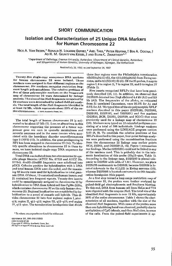
SHORT COMMUNICATION
Isolation and Characterization of 25 Unique DNA Markers for Human Chromosome 22
NICK A. VAN BIEZEN, '" RONALD H, LEKANNE DEPREZ," ABEL THIJS, '" PETER HEUTINK, t BEN A. OaSTRA, t AD H. M, GEURTS VAN KessEl,:f: AND EllEN C. ZWARTHOFF*,l
'Department of Pathology, Erasmus University, Rotterdam; tDepartment of Clinical Genetics, Rotterdam; and tDepartment of Human Genetics, University of Nij'megen, Nijmegen, The Netherlands
Received July 20, 1992; revised September 29, 1992
Twenty-five single-copy anonymous DNA markers for human chromosome 22 were Isolated. These markers wore assigned to four different regions on the chromosome. Sb; markers recognize restriction fragment length polymorphlsms. The relative positions of five of these polymorphic markers on the Cramework map of chromosome 22 were determined by linkage analysis. The sizes of the Noll fragments recognized by 22 markers were determined by pulsed-field gel analysIs. The total length of the Noll fragmentsldentlfled is at least 12 Mh, which represents about 20% of the enUre chromosome. t> 1993 A<adem!< P'OS!r.ln~.
The total length of human chromosome 22 is estimated to be about 57 Mb (7). Loss or alterations in this chromosome suggesting the presence of a tumor suppressor gene are seen in sporadic meningioma and acoustic -neuroma and in the same tumors when associated with the hereditary disorder neurofibromatosis type 2 (NF2) (15). In addition, the gene predisposing to NF2 has been mapped to chromosome 22 (14). To identify specific alterations in chromosome 22 in these tumors, we have isolated single·copy DNA sequences for chromosome 22.
Total DNA was isolated from two chromosome 22-specific phage libraries (ATCC No. 57733 and ATCC No. 57714). EcoRI-HindIlI fragments were subcloned into pUC9. Colonies positive for hybridization with X DNA and total human DNA were discarded, and the remaining 59 inserts were used for hybridization to total genomic DNA. Of these, 13 contained nonhuman inserts and 21 contained less frequent repeats. Twenty-five inserts could be unambiguously assigned to chromosome 22 by hybridization to DNA from hybrid cell line PgMe-25Nu, which contains chromosome 22 as the only human chromosome (5). Regional localization was established by hybridization to a panel of somatic cell hybrids. This divides chromosome 22 into four regions! region I, pterqll; region II, qll-qI2; region III, qt2-q13; and region IV, qI3-qter. The translocation breakpoints that divide
! To whom correspondence should be nddressed.
GRNOMICS 16,206-208 (1993) D-SM-'lMJ/93 $5.00 Copyright © 1993 by Academic Pre ... In~. An right, of tep,odu~tion in any form re;erved.
these four regions were the Philadelphia translocation t(9;22)(q34;ll) (5), the t{lt;22)(q24;12) from Ewingsarcoma, anda t(1;22)(42;13} (4). Ofthe 25 probes, 5 map to region I, 8 to region II, 7 to region III, and 5 to region IV (Table 1).
Five inserts recognized RFLPs that have been previously described (10, 11). In addition, we observed that D22S2Q5 detected two TaqI alleles of 4.4 kb (A1) and 2.9 kb (A2). The frequencies of the alleles, as estimated from 31 unrelated Caucasians, were 93.5% for At and 6.5% for A2. We typed five ofthe six polymorphic RFLP markers described in this paper (D22S181, D22S182, D22S183, D22S193, and D22S201) and five markers (D22S1Q, BCR, D22S1, D22S29, and IGLC) that were previously used for a linkage map of chromOsome 22 (13). Markers were typed on 13 extended pedigrees consisting of a total of .386 individuals. Linkage analyses were performed using the LINKAGE program version 5.03 (8, 9). To establish the relative positions of five RFLPs described in this paper, four-point linkage analyses were performed using the recombination fractions from the chromosome 22 linkage map anchor points BCR, D22S1, and D22S29 (3, 13). Figure 1 summari~es our findings. Marker D22S201 could not be linked toany of the markers used. This is probably due to the telomeric localization of this probe (22q13----qter, Table 1). According to the linkage map, D22S193 is placed telomeric to D22S29 with odds of 1.8:1. However, we place D22S193 centromeric to D22S29, because D22S29 is located telomeric to the t(t1;22) in Ewing sarcoma (13), whereas D22S193 is located centromeric to this translocation breakpoint (this paper).
As a first step toward a long-range restriction mup of chromosome 22, the probes were further analyzed by pulsed-field gel electrophoresis and Southern blotting. To this end, DNA from human cell lines HeLa and T24 were digested with the enzyme NotI. The total size of the identified Not! fragments is over 12 Mb, approximately 20% of the entire chromosome. Table 1 shows the characteristics of all markers, together with the size of the observed NotI fragments. With some of the probes more than one hybridizing band was observed, probably due to methylation of CpG islands, and thus NotI sites, in some of the cells. From the pulsed-field experiments it ap-
55
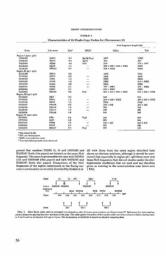
SHORT COMMUNICATION
TABLE 1
Characteristics or 25 Single· Copy Probes ror Chromosome 22
Noll fragment length (kb)
o no. Lab name Size' RFLP HeLa T24
Region I (pler··qll) D228181 NB17 0.7 BgllllTaql ND~ NRZ' D228182 NB35 0.8 Bglii ND ND 1)228183 NBS4 1.0 Psil 270 270 + 310· D228184 NBSS 1.1 360 + 680 + 630 + NRZ NHZ D22~185 NBI03 0.7 210 + NRZ NRZ
Region n (qll-q12) D228186 NB14 0.' 1000 1000 D228187 NB20 0.9 1000 1000 0228188 NB21 13 NRZ NHZ D228189 NB43 1.6 NRZ 670+ NRZ 0228190 NB62 0.6 NHZ NRZ D22S191 NB66 2.1 190 + NRZ 190+ NIlZ 0228192 NB97 0.7 680 + NRZ NRZ D22S193 NBI29 0.6 Pst! 265 + 410 + 630 + NRZ 265 + 630 + NRZ
Region lIT (q12-q!.1) D228194 NB7 0.3 500 500 022819~ NRH 1.1 420 + 500 + NRZ 420 + 500 + NItZ 022~196 NB74 1.6 NHZ NRZ 0228197 NBIOa 0.3 420 + 500 420 + 500 D228198 NB1l6 1.0 760 750 0228199 NUll7 1.9 350 350 0228200 NBil9 1.8 560 560
Begion IV (qI3-qter) 0228201 NB5 0.5 1;,ql 190 ND 0228202 NB8 0.8 D22~203 NB60 1.9 D228204 NB70 0.3 0228205 NB127 0.3 Taql
• ~ize insert in kb. b ND: not determined. , NRZ: nonresoll1tion zone. d Two hyhridiling bands were observed.
peared that markers D2281 (1, 6) and D228186 and D228187 (both this paper) are located on the same Noll fragment. The same is presumably the case with 022815 (12) and D228199 (this paper) and with D228195 and D228197 (both this paper). Comparison of the Noll fragments of the region centromeric to the Ewing sar· coma translocation as recently described by Budarfet al.
6:t 1.3 . 10~t
nr-I
NO ND 240 + 400 240 + 400 ND ND ND ]50
(2) with those from the same region described here shows no obvious relations, although it should be mentioned that especially in region qll-q12 there were very large Noll fragments that did not resolve under the elec· trophoresis conditions that we used and are therefore given as running in the nonresolution zone (sizes o\'er 1 Mb).
206:t 1:1.8
n n locus: 022St8t 022S163 022S182 02:2St93 Relare-nee
toci:
Odds
'" tOLe 022SI0
! t 0.00 004 001
LJ LJ 26:t 151:t
seR 022St 022$29
'" 0.12 008 oos! qter ! 1
LJ too.t
FIG. 1. Most likely order and se>:-Ilveraged recombination rates between gene markers On cbromosome 22. Reference lod were used as anchor points for placing four new markers On the map. The odds against im·ersion of the marker order are shown above or below the brackets lind lire based on multipoint linkllge aMlyse8. The i()(alization of D228193 is based on physical mapping data.
56
SHORT COMMUNICATION
ACKNOWLEDG~IENTS
This work was supported in part by a grant from the Dutch Cancer Sodety (RH.L.DJ. We are indebted to ProL Dr. A. Hagemeijer-Hausman and Ms. R. van Drunen for cytogenetic controls of the somatic cell hybrids.
REFERENCES
\. Barker, G., Schafer, M., and White, R. (984). He8trktion sites containing CpG show a higb frequency of polymorphiom in hu· man DNA. eel/3S: 131-138.
2. Bud<uf, M. L., McDermid, H. E., Se!linger, 8., and Emanuel, B. S. (1991). I~olation and regional localiUltion of 35 unique anonymous DNA markers for human chromosome 22. Genomics 10: 996-1002.
3. Emanuel, B. S., Budnrf, M. L., and Seizinger, B. R. (1991). Report ofthecommitlee on the genetic constitution of chromosome 22. Cy/ogcnet. Cdl Genet. 68: 827-8.52.
4. Geurts van Ke~8el, A .. 'IUre·Carel, C., De Klein, A., Gro;;veld, G., unoir, G., and Bootsma, D. (198.5). TransiQcation of QncQgene c-sis from chromosome 22 to chromo;;ome II in II Ewing SarCOma derived cell line. MoL CdL Riol. 6: 427-429.
5. Goyns, M. H., Young, B. D., Geurts van Kessel, A., De Klein, A., Grosveld, G., Bartram, C. R., and Bootsma, D. 0984). Regional mapping of the human immunoglohulin lambda light chain to the Philadelphia chromosome in chronic myeloid leukemia.vu· hrmill Res. 8: 5 17-553.
6. Julier, C., Lathrop, G. M., Re-ghis, A., Swjne-It, M·F., Lnlouel, J·M., and Kaplan, J·C. (198.'1). A linkage and physical map of chromosome 22, find some applications to gene mapping. Am. J. Hum. Genet. 42: 297-~08.
7. Korenberg, J. R., and Engels, W. R. (1978). Ru..<e ratio, DNA
content, and quinacrine brightness of human chromosomes. Proc. NaiL ,1cad. Sci. USA 75: 3382-3386.
8. Lathrop, G. M., and Lalouel, J. M. (1984). Easy calculations of lod SCOrCS and genetic risks on small Nmputers. Am. J. Hum. Gcnct. 36: 460-465.
9. Lathrop, O. M., Lalouel, J. M., Juliet, C., and Ott, J. (1984). Strategies for multilocus linkage analysis in humans. Proc. Nott. Awd. Sri. USA 81: 34·13-3446.
10. ukanne Deprez, R. H., van Hiezen, N. A., Heutink, P., Doej· hllrat, K. H. O. S., Do Klein, A, Geurts van Kessel, A. H. M., and Zwarthoff, E. C. (1991). A new polymorphic prohe on chromo· some 22: NB17 (D22S181). A new polymorphic probe on chromosome 22: NB3" (D22S182). A new polymorphic probe On
chromosome 22: NR84 (D22S183). A new polymorphic prohe on chromosome 22q: NB129 (D22S193). Nucleic Acids Res. 19: 686-687.
11. L€kanne Deprez, R. H., ,·an Biezen, N. A., Br<'ed\"eld, G. J., Boej· harnt, K. R. G. S., De Klein, A, Geurts ,·an Kessel, A. H. M., and Zwarthoff, E. C. (991). A new polymorphic probe on chromosome 22: NB5 (D22S201). Nudeic Acids Re.5. 19: 1963.
12. Rouleau, G. A., Kurnit, D. M., Neve, H. L., Bazanowsky:A., Pat· ter<on, D., and Gusella, J. F. (1988). 022815 a fetal bram eDNA v;ith Banll and Sael RFT,P. Nucleic Arids Res. 16: 1646.
13. HoulelHl, G. A., Haines,J. L., Baza.anowski,A., Colella·Crowley, A., Twfnther,J.A, Wuler, N. S.,Conneally, P. M.,and Gusella, ,J. F. (1989). A genetic linkage map of the long IIrm of human chromosome 22. GenGmics 4: 1-6.
14. HOlileau, G. A., Seizinger, B. fl., Wertelecki, W., Haines, J. L., Superneau, D. W., Martuw, H. L., and Gusella, J. F. (199{)). I"lanking markers hracket the neurollhromato:;is type 2 (NF2) gene on chromo;;orne 22. Am. J. Hum. Genrl. 40: 323-328.
15. S~ilinger, B. R., Rouleau, G., Ozilius, T,. J., Lane, A. H., St. George Hyslop, P., Huson, S., and Gusella, J. F. (1987h). Com· mon p;1!hogenic me.:hanism for three tumor types in bilateral acoustic neurofibrom~!osis. Science 236: 317-319.
57

58
Appendix
A lIew polymorphic probe 011 chromosome 22: NBl7 (D22S181)
Nllc/. Acids Res. 19:686, 1991
AileII' polymOlphic probe 011 chromosome 22: NB35 (D22S182)
Nllc/. Adds Res. 19:686, 1991
A lIew polymorphic probe 011 chromosome 22: NB84 (D22Sl83)
Nllc/. Acids Res. 19:687, 1991
A lIew polymorphic probe 011 chromosome 22q: NB129 (D22S193)
Nllc/. Acids Res. 19:687, 1991
A new polymO/phic probe 011 chromosome 22: NB5 (D22S20l)
Nllc/. Acids Res. 19:1963, 1991
59

60
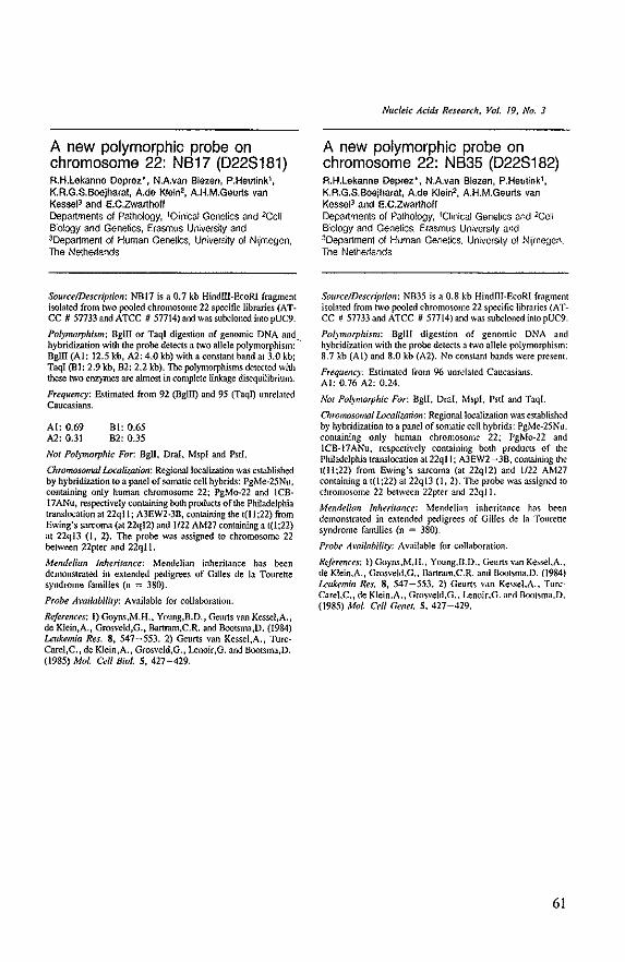
A new polymorphic probe on chromosome 22: NB17 (0228181) RH.lekanne Deprez', N.A.van Blazen, P,Heutinkl, K.R.G.S.8oejharat, A.da Kleln2, A.H.M.Geurts van Kesse!3 and E.C.Zwarthoff Departments 01 Pathology, lCiinical Genetics and 2CeH Biology and Genetics, Erasmus University and JOepartment of Human Genetics, University 01 Nljmegen, The Netherlands
SoufcelDescriplion: NBI7 is a 0.7 kb Hindl11-&oRI fragment isolated from two !XXlled chromosome 22 specific libraries (AT· CC # 57733 and ATCC # 57714) and was subcloned intopUC9.
Po/)morphism: Bgill or TaqI digestion of genomic DNA and hybridization with the probe detects a two allele polymorphism: " BglD (AI: 12,5 kb, A2: 4.0 kb) with a constant band at 3.0 kb; Taqi (Bl: 2.9 kb, B2: 2,2 kb). The poJymorphisms detected with these two enzymes are almost in complete linkage disequilibrium.
Frequency: Estimated from 92 (DglIl) and 95 (Taqn unrelated Caucasians.
AI: 0.69 A2: 0.31
BI: 0.65 82: 035
NOI Polymorphic For: 8gll, DmI, Mspl and Pstl.
ChromosomnllLxalit111ion: Regionallocalitation was established by hybridization to a panel of somatic ceU hybrids: PgMe-25Nu, containing only human chromosome 22; PgMo-22 and ICB-17ANu, respectively containing both products of the Philadelphia translocation at 22qll; A3EW2+3B, containing the t(ll ;22) from Ewing's sarwma (at 22q12) and 1/22 AM27 containing a 1(1;22) at 22ql3 (1, 2). The probe was assigned to chromosome 22 between 22pter and 22qll.
Mendelian Inheritance: Mendelian inheritance has been demonstrated in e .... lended pedigrees of Gilles de la rourette syndrome families (n = 380),
Probe Availability: Available for collaboration.
References: I) Goyns,M.H., Young,B.D., Geurts van Kessel,A" de Kiein,A., Grosveld,G" Bartram.C.R. and Bootsma,D. (1984) Leukemia Res. 8, 547~553, 2) Geurts van Kessel,A., Turccarel,C, , de Klein,A., GrosveJd,G., Lenoir,G. and Bootsma,D. (1985) Mol. Cell BioI,S, 427-429.
Nuefeic Adds Research, Vol. 19, No.3
A new polymorphic probe on chromosome 22: NB35 (0228182) A.H.Lekanne Deprez·, NAvan Blezen, P.Heutinkl, KA,G,S,Boejharal, Ada Kiein2, AH,M,Geurts van KesselJ and E.C.Zwarthoff Departments of Pathology, 1Clinical Genetics and 2eell Biology and Genetics. Erasmus UniverSity and 3Department of Human Genetics, University of N~megen, The Netherlands
Source/Description: NB35 is a 0.8 kb Hindm·&oRI fragment isolated from two pooled chromosome 22 specific libraries (ATCC /I 57733 and ATCC /I 57714) and was subdoned into pUC9.
Polymorphism: Bglll digestion of genomic DNA and hybridization with the probe detects a two allele polymorphism: 8,7 kb (AI) and 8,0 kb (A2), No constant bands were present.
Frequency: Estimated from 96 unrelated Caucasians, AI: 0,76 A2: 0,24,
11'01 PO(I7/lorphic For: BgU, Oral. Mspl. Pstl and Taql.
ChromoJOlllal Loca/i<JIn'on: Regional localization was established by hybriditation to a panel of so malic cell hybrids: PgMe-25Nu, containing only human chromosome 22; PgMo-22 and ICB·17ANu. respectively containing both products of the Philadelphia translocation at 22qll; A3EW2 ~ 3B, containing the t{lI;22) from Ewing's sarcoma (at 22q12) and 1/22 AM27 containing a t(l;22) at 22q13 (I, 2), The probe was assigned 10
chromosome 22 between 22pter and 22qll.
Mendelian Inheritance: Mendelian inheritance has been demonstrated in extended pedigrees of Gilles de la Tourette syndrome families (n = 380),
Probe Availability: Available for collaboration.
References: I) Goyns.M,H .. Young,B.D .. Geurts van KesseI.A .. de Klein.A., Grosveld,G., Bartram.C.R. and Bootsma.D. (1984) Leukemia Res, 8, 547-553. 2) Geuns van KesseI.A .. TurcCarel,C., de Klein,A., Grosve[d,G .. Lenoir.G, and BooLSma,D. (1985) Mol. Cell Genet,S. 427-429.
61
A new polymorphic probe on chromosome 22: NB84 (0228183) A.H.Lekanne Deprez', NAvan Biezen, P.Heutlnk1
,
K.R.G.S.8oejharat, Ada Klein2 , A.H.M.Geurts van Kessel3 and E,C.Zwarthoff Departments of Pathology, lClinicaJ Genetics and 2Cell Biology and Genetics. Erasmus University and JOepartment 01 Human Genetics, University of Nijmegen. The Netherlands
Source/Description: NB84 is a 1.0 kb HindJlI·EcoRI fragment isolated from Iwo pooled chromosome 22 specific libraries (ATCC 1/ 57733 and ATCC 1/ 57714) and is subdoned into pUC9.
Polmwrphism: Pstl digestion of genomic DNA and hybridization with the probe detects a two allele polymorphism: 4.6 kb (AI) and 4.0 kb (Al). No constant bands were present.
Frequency: E5tirnated from 91 unrelated Caucasians. AI: 0.19 A2: 0.81.
NOI PO(\'Inorphic For. Bgll, Bglll, DraI, Mspl and Taql.
Chmmosomalwcaliza!ion: Regionallocalizalion was established by hybridization to a panel of somatic cell hybrids: PgMe-25Nu, containing only human chromosome 22; PgMo-22 and IC8-17ANu, respectively containing both products of the Philadelphia translocation at 22ql 1; A3EW2- ]8, containing the t(lI:22) from Ewing's sarcoma (at 22q12) and 1122 AM27 containing a t((;22) at 22ql3 (I, 2). The probe was assigned to chromosome 22 between 22pter and 22ql L
Mendelian Inheritance: Mendelian inheritance has been demonstrated in extended pedigrees of Gilles de la TOllrette syndrome families (n = ]80).
Probe Ami/abiliTY: Available for collaboration.
References: I) Goyns.M.H., YOllflg,8.D., Geurts van Kessel,A., de Klein,A., Gros~'eld,G., Bartram,C.R. and Bootsma,D. (1984) Leukemia Res. 8, 547-553. 2) Geurts van Kessel,A., TurcCarel,C., de Klein.A .. Grosveld,G .. lenoir,G. and Bootsma,D. (1985) Mol. Cdl Bioi. S, 427-429.
62
Nudeic Acids Research, Vol. 19, No.3
A new polymorphic probe on chromosome 22q: NB129 (0228193) R.H.Lekanne Deprez', N.A.van Biezen, P.Heutinkl, K.R.G.S.Boejharal, A.de Klein2 , AH.M.Geurts van Kessel3 and E.C.Zwarthoff Departments of Pathology, !Clinical Genetics and "Celt Biology and Genetics, Erasmus University and 3Department of Human Genetics. University of Nijmegen, The Netherlands
SourcelDescn'prion: NDI29 is a 0.6 kb HindllI-EcoRl fragment isolated from twO pooled chromosome 22 specific libraries (ATCC # 5773] and ATCC # 57714) and is subdoned into pUC9.
Polymorphism: Pst! digestion of genomic DNA and hybridization with the probe detects a two allele polymorphism: 1.6 kb (AI) and 1.3 kb (Al). No constant bands were present.
Frequcncy: Estimated from 98 unrelated Caucasians AI: 0.45 Al: 0.55.
Not Polymorphic For: Bgll, BglII, DraI, MspI and TaqI.
Olromosonlalwcaliza.tion: Regionallocaliz.ation was established by hybridization to a panel of somatic cell hybrids: pgMe-25Nu, containing only human chromosome 22; PgMo-22 and ICB-17ANu, respectively containing both products of the Philadelphia translocation at 22qI I; A3EW2 - ]B, containing the t(lI;22) from Ewing's sarcoma (at 22q12) and 1122 AM27 containing a t(I;22) at 22qI3 (I, 2). The probe was assigned to the long arm of chromosome 22 between 22qll and 22q12.
Mendelian Inhcritance: Mendelian inheritance has been demonstrated in extended pedigrees of Gilles ,de la Tourette syndrome families (n =:0 380).
Probe Amilability: Available for collaboration.
References: 1) aoyns,M.H., Young,B.D., Geurts van Kessel,A., de K1ein,A., Grosveld,G., Bartram,C.R. and Bootsma,D. (1984) Leukemia Res. 8, 547-55]. 2) Geurts van Kessel,A., TurcCarel,C., de Klein,A., Grosveld,G" Lenoir,G. and Bootsma,D. (1985) Mol. Cell Bioi. 5, 427-429.
A new polymorphic probe on chromosome 22: NB5 (D22S201) R,H,Lekanne Deprez', NAvan Blezen, G.J.Breedveldl ,
K.R.G.S.Boejharal, A.de K1ein2, A.H.M.Geurts van Kessel3 and E,C.Zwarthoff Departments of Pathology, !C!inicaJ Genetics and 2Cell Biology and Genetics, Erasmus University and 3Department of Human Genetics. University of Nijmegen. The Netherlands
SOl/rce/Description: NBS is a O.S kb HindIII-&oRJ fragment isolated from two pooled chromosome 22 spedfic libraries (ATCC # 57733 and ATCC # 57714) and \\o"ES subcloned intopUC9.
Pol)morphism: Taql digestion of genomic DNA and hybridization with the probe dele.::ts a four allele polymorphism: 1,75 kb (AI), 1.6 kb (Al), 1.4 kb (A3) and 1.3 kb (A4). No constant bands were present.
Frequency: Estimated from 97 unrelated Caucasians. Al : 0.072 Al : 0.86 A3 : 0.OS2 A4 : 0.015
NOI Pol}1/Iorphic For. BgII, SgiII, Oral, Mspl and PstI.
O"onwsomal iAcaliwtion: Regional localization was established by hybridization to a panel of somatic cell hybrids: PgMe-25Nu, containing only human chromosome 22; PgMo-22 and ICB-17ANu, respectively containing both products of the Philadelphia translocation at 22qll; A3EW2-3B, containing the 1(11;22) from Ewing's sarcoma (at 22q12) and 1122 AM27 containing a t(1;22) at 22ql3 (I, 2). The probe was assigned to chromosome 22 between 22ql3 and 22qter.
Mendelian 1nhedtance: Mendelian inheritance has been demonstrated in extended pedigrees of Gilles de la TOllrette syndrome families (n = 380).
Probe Mailabiliry: Available for collaboration.
References: I)Goyns,M.H., Young,B.D., Geurts van Kessel.A., de Klein,A., Grosveld,G., Bartram,C.R. and Dootsma,D. (19&4) Leukemia Res. 8, 547-553. 2)Geurts van Kessel,A., TurcCarel,C., de Klein,A., Grosve!d,G., Lenoir,G. and Bootsma,D. (1985) Mol. Cell Bioi. S,427-429.
Nucleic Acids Research, Vol, 19, No, 8
63

64
Chapter III
Cytogenetic, molecular genetic and pathological
analyses in 126 meningiomas
submitted tor publication
65

66
Cytogenetic, Molecular Genetic and Pathological
Analyses in 126 Meningiomas
Ronald H. Lekanne Deprez*, Peter H. Riegman*, Ellen van Drunen#, Ursula L. Warringa*,
Nicole A, Groen', Stanislav Z, Stefanko', Jan W, Koper@, Cees LJ, Avezaat$, Paul G, H,
Mulder&, Ellen C, Zwarthoff', and Anne Hagemeijer#1
Departments of *Pathology, HCell Biology and Genetics, @Internal Medicine, $Neurosurgery, and &Epidemiology
and Biostatistics, Erasmus University, Rotterdam
Abstract
In a series of 126 meningiomas, tumor and patient characteristics wer'c investigated and
statistically analyzed. A combined cytogenetic and molecular genetic approach was used
to study chromosomal abnol'malities and loss of markel'S OIl chromosome 22q. This
approach was successfully applied to 93 meningiomas, In 66 cases complete or partial
loss of chromosome 22 was observed and in at least 12 of them this chromosome was
involved in structural abel'l'ations. In addition to chromosome 22 changes, chromosomes
1, 6, 11, 13, 14, 18, 19, X, and Y were also frequently involved in strue!u ... 1 and
numerical aberrations. Statistical analysis revealed a significant association between the
number of chromosomal abnormalities and tumor grade. Complex karyotypes
predominated in the group of grade II/III meningiomas. FUI'thel1110re, othel' variables
showed statistically (Ol' marginally statisticall,Y) significant differences. Meningiomas
from the convexity wel'e mOl'e often gl'ade IlIIII, displayed predominantly (pa.'lia]) loss
of chromosome 22 and had complex km'yotypes more often. These features were
fl'equently found in meningiomas from males. Base meningiomas on the other hand
occurred more often in females, they were usually grade I, showed loss of (palis 00
chromosome 22 less often and displayed fewer additional clll'omososmal abn0l111aJities.
67
Introduction
Meningiomas are considered to be histologically benign neoplasms that account for 13-19%
of all primary brain tumors treated by surgery (Russell and Rubinstein 1989), However, all
meningiomas may display, to a certain extent, atypical or anaplastic features (JaaskeHiinen
et aI., 1985),
Cytogenetically, meningiomas are the most extensively studied human solid tumors
and they display a consistent chromosome aberration (Zang and Singer, 1967; Zang, 1982),
These cytogenetic studies and molecular genetic analyses strongly suggest that loss of (parts
of) chromosome 22 is a primary event in the development of these tumors and also indicate
that several chromosome 22 loci might be involved in the process (Seizinger et aI., 1987;
Rey et aI., 1988; Dumanski et aI., 1990; Lekanne Deprez et aI., 1991a; Rey et aI., 1993;
Lekanne Deprez et aI., 1994). In addition, other less common non random chromosomal
changes occur in some tumors, which might playa role in tumor progression (Katsuyama et
aI., 1986; Al Saadi et aI., 1987; Casalone et aI., 1987; Maltby et aI., 1988; Rey et aI., 1988;
Vagner-Capodano et aI., 1993; Biegel et aI., 1994), One of these changes could also be the
primary cause in a subset of meningiomas without chromosome 22 involvement, as a
meningioma/ependymoma family did not show linkage to DNA markers flanking the NF2
locus on chromosome 22 (Pulst et aI., 1993). More complex chromosome abnormalities
appear to be associated with more atypical features (Vagner-Capodano et aI., (993),
Most meningiomas occur sporadically, although they are also found in patients with
the autosomal dominantly inherited predisposition for Neurofibromatosis type 2 (NF2) or in
multiple meningioma patients or very rarely as sole tumor type clustered in some families
(Butti et ai" 1989; Domenicicci et aI., 1989; Russell and Rubinstein, 1989; McDowell,
1990; Sieb et aI., (992), Positional cloning approaches have resulted in the isolation of the
NF2 gene on chromosome 22q12 and mutations were observed in both the germ-line of NF2
patients and in sporadic tumors (Rouleau et aI., 1993; Trofalter et aI., 1993), Recently,
mutation analyses of the NF2 gene have revealed mutations in about 30% of the sporadic
meningiomas (Lekanne Deprez et aI., 1994; Ruttledge et aI., 1994), This indicates that the
NF2 gene is important in the development of a large number of sporadic meningiomas,
although it is still very well possible that other loci on chromosome 22 and/or other
chromosomes are involved (see above). Studies on the association between chromosomal
68
abnormalities, histopathological and other tumor and patient characteristics of meningiomas
are scarce (Zang, 1982; Al Saadi et aI., 1987; Maltby et aI., 1988; Casalone et aI., 1990;
Sanson et aI., 1992; Vagner-Capodano et aI., 1993) and in only two of these studies
statistical analyses on some of these characteristics (Casal one et aI., 1990; Vagner-Capodano
et aI., 1993) were performed. Investigation of the different characteristics of meningiomas
could help in identifying different subclasses, which might explain the genetic alterations, not
affecting the NF2 gene, found in a subset of tumors. This study describes cytogenetic and
molecular genetic findings in a large number of meningiomas. A significant association was
found between characteristics like sex, site of tumor origin, histological subtype, grade,
chromosome 22 loss and number of chromosomal abnormalities.
Matedals and methods:
Tumor collection and tissue culture:
From November 1988 to September 1993 we collected 126 meningiomas (Table 1). If
possible, tumor material obtained at surgery was divided in three or more parts. These parts
were used for: 1) histopathology, 2) DNA isolation and 3) cytogenetic analysis. Tumor parts
were frozen and stored in liquid nitrogen prior to DNA isolation and pieces were used
directly for interphase in situ hybridization and tissue culture. Both preparation for tissue
culture and tissue culture conditions were as described elsewhere (Koper et aI., 1990).
Histological examination and localization:
Histopathological assessment was performed on formalin fixed 5 I'm paraffin embedded
tumor sections. The tumors were classified according to the WHO histological typing of
tumors of the central nervous system (ZUlch 1979) and Kepes et al. (1982). The following
subtypes or mixtures of them were identified in our series: transitional (T), fibroblastic (F),
meningotheliomatous (M), psammomatous (Ps) , angiomatous (A), papillomatous (P),
anaplastic (Ana), xanthomatous (X), lipoblastic (L), pleiomorphic (PI), oncocytic (0), and
haemangiopericytic (H).
The grading system used in this study was as described by JiUiskeUiinen et al. (1985).
In this system a higher grade corresponds to an increased degree of anaplasia, which is
69
defined by evaluating 6 histological parameters (loss of architecture, increased cellularity,
nuclear pleomorphism, mitotic figures, focal necrosis and brain infiltration). This resulted
in three different grades: grade I (benign), grade II (atypical), and grade III (anaplastic). The
grading o[ the meningiomas was carried out by one of us (SZS).
The anatomic sites of tumor origin were subdivided in convexity (C), base (B), falx
(F), and intraventricular (IV).
Cytogenetic analysis and in sitll hybridization:
Cytogenetic analysis was carried out on cultured samples, from passage a (pO) corresponding
to the first outgrowth of cells to passage 3 (p3). Cytogenetic results were only taken into
account when the cells were cultured for less than 50 days. Cells were harvested and fixed
according to standard methods. Chromosomes were identified using RFA, QFQ and GTG
banding techniques and described according to ISCN (1991).
Fluorescent in situ hybridization (FISH) was performed on interphase nuclei directly
obtained from the fresh tumor sample or after short term culture. Competitive in situ
hybridization was applied using at least two chromosome 22 specific cos mid or centromere
probes following the technique as reported by Arnoldus et al. (1991). For all probes used,
the number of signals per nucleus after hybridization was counted in at least 100 nuclei. As
a control, 34 lymphocyte cultures of the patients (when available) and 23 lymphocyte cultures
of normal donors were used. In both control groups we observed similar hybridization
results: one spot (apparent monosomy) in about 5 % of nuclei and two spots (disomy) in more
than 90% of the nuclei. This allowed us to conclude the absence of constitutional loss of
chromosome 22 in patients lymphocytes. Control data were lIsed to determine the sensitivity
of the technique used and to define the cut off rate (mean % of nuclei with one spot + two
times the standard deviation) of 13.5% and 12%, respectively above which a cell popUlation
with (partial) monosomy could be safely identified.
The following probes cited from centromere to telomere of chromosome 22 were
used: p22!l:2.1 (centromere, McDermid et al. 1986); 2 cosmids in the lambda
immunoglobulin light chain gene region: lambda 2.2, lambda 3.1 (Goyns et al., 1984); 4
cos mid probes 40D2, 58B12, 50BII, 76A4 ordered from centromere to telomere within 100
kb from D22S193 (Chapter VI); 29B5 (D22S72); 103DI2 (D22S56); 4 cosmid probes in the
NF2 region: 115D7 (NEFH), 121G 10, 96CIO, 74BI. The cosmid probes in the vicinity of
70
D22S193, D22Sn, D22S56 and NF2 were obtained by screening two chromosome 22-
specific cosmid libraries (LL22NCOI and LL22NC03: de Jong et aI., 1989; Zucman et aI.,
1992).
LOB analysis oj chromosome 22:
High-molecular-weight DNA was isolated from stored tumor tissue according to standard
procedures. Before DNA isolation the percentage of tumor cells was determined by
microscopic examination of a cryostat section of the tumor specimen. Only tumor samples
with more than 80% tumor cells were used for DNA analysis. Most specimens contained
more than 90% tumor cells. When available, constitutional DNA was isolated from
peripheral blood leucocytes. Restriction endonuclease digestion, agarose gel electrophoresis,
Southern transfer, hybridization to 32p labeled probes and autoradiography were performed
as reported (Lekanne Deprez et aI., 199Ia). The following DNA markers for loci on
chromosome 22 were used: D22S181, 022S183 (Lekanne Deprez et aI., 199Ib), D22SIO
(Hofker et aI., 1985), D22S182 (Lekanne Deprez et aI., 199Ib), D22S1 (Barker et aI.,
1984), D22S193 (Lekanne Deprez et aI., 199Ib), 17.13'(3 prime part of the MNI eDNA
17.1, which detects a Pvu II polymorphism, unpublished result), D22S29 (Rouleau et aI.,
1989), D22S45 (Budarf et aI., 1991), 022S201 (Lekanne Deprez et aI., 1991c) and D22S205
(van Biezen et aI., 1993). D22S201 and 022S205 are located distal to D22S193, but their
orientation relative to each other or D22S45 is not known. The other probes are ordered from
centromere to telomere. For most of the tumor DNA samples analyzed in this study
constitutional ONA was not available, therefore loss of heterozygosity (LOH) for a specific
marker was assumed when a heterozygous DNA sample showed clear reduction of intensity
of one of the 2 alleles.
SlatiSlics:
The statistical analyses were performed with the Statistical Package for the Social Sciences
(SPSS) and with the Epidemiological Graphics, Estimation, and Testing package (EGRET).
We used the X' test, the Exact Homogeneity test, and Fisher's exact test. A significant
association was concluded when P '" 0.05. The following variables were mutually analyzed:
the sex of the patients, the age at surgery (over and under 50 years of age), the site of tumor
origin, the histological subtype, the grade, the loss of (parts of) chromosome 22, and the
71
number of clonal chromosomal abnormalities (chr abn). To analyze the latter variable the
number of clonal chromosomal abnormalities was divided in two groups: ::;; 2 ehr abn or > 2 chr abn as seen by chromosome banding analysis.
Resulls
The 126 meningiomas that were studied are listed in table 1. This table shows the sex and
age at surgery of the patients, the site of tumor origin, the histological subtype, grade, and
the chromosome 22 status of the tumors. This series contains 13 tumors derived from 9
patients with two or more meningiomas, which were located at different sites, and 2
meningiomas from 2 sisters with NF2 (table 1). Of the liD single meningioma patients
without evidence of NF2, 82 were from females (average age 55.3) and 28 were from males
(average age 57.5). In the complete group of meningiomas the site of tumor origin was
subdivided in convexity 52.9% of the cases, base 37.2%, falx 7.4% and intraventricular
2.5%. Histopathologically 52.3% of the meningiomas were transitional, 16.7% fibroblastic,
11.9% meningotheliomatous and 19.1 % were mixed or had a different sUbtype. Grading of
the meningiomas resulted in 73% grade I (benign), 19.8% grade II (atypical) and 7.2% grade
III (anaplastic) tumors. The chromosome 22 status (last 3 columns in table I) is the result
of karyotyping, FISH and LOH analysis. These analyses were carried out depending on the
quantity of material available. FISH analysis of freshly obtained tumor cells was applied only
in the second half of this series and proved to be a very sensitive tool for the detection of
(partial) loss of chromosome 22 (table I and 2). In total 97 tumors could be examined with
at least one of these techniques. For the interpretation of (partial) loss of chromosome 22 and
number of clonal chromosomal abnormalities in the tumors (below and table 4), the tumors
10, 133 and 177 were excluded because discrepancies between the methods were found. This
was probably due to inhomogeneity in these tumors with regard to the percentage tumor vs
normal cells. Tumor 16 was excluded because only one cell could be analyzed. This cell,
however, showed a very abnormal karyotype, including loss of chromosome 22. Tumor 176
was excluded with regard to the interpretation of number of clonal chromosomal
abnormalities because incomplete cytogenetic data were available. FfSH analysis in this
tumor clearly showed loss of chromosome 22 (table I). Therefore, we established that in 66
72
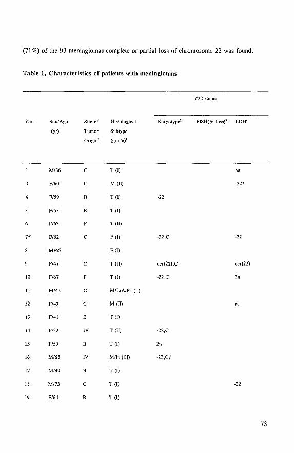
(71 %) of the 93 meningiomas complete or partial loss of chromosome 22 was found.
Table 1. Chal'actel'istics of patients with meningiomas
No.
3
4
5
6
7'
8
9
10
II
12
13
14
15
16
17
18
19
Sex/Age
(yr)
M/66
F/60
FIS9
FISS
F/63
F/62
M/6S
F/47
F/67
Mf43
F/43
F/41
FI22
FIS)
M/68
M/49
MI73
F/64
Site of
Tumor
Origin1
c
C
B
B
F
C
C
F
C
C
B
IV
B
IV
B
c
B
Histological
Subtype
(grade)l
T(D
M (lQ
T (Q
T(D
T (lQ
F(D
F(D
T GQ
T(D
Mill AlPs (1I)
MGQ
T(D
T GQ
T(D
M/H (Ill)
T (1)
T(D
T(D
Karyotype2
-22
-22,c
der(22),C
-22,C
-22,C
2n
-22,C?
#22 status
FISH(% loss)} LOH'
nc
-22*
-22
der(22)
2n
nc
-22
73
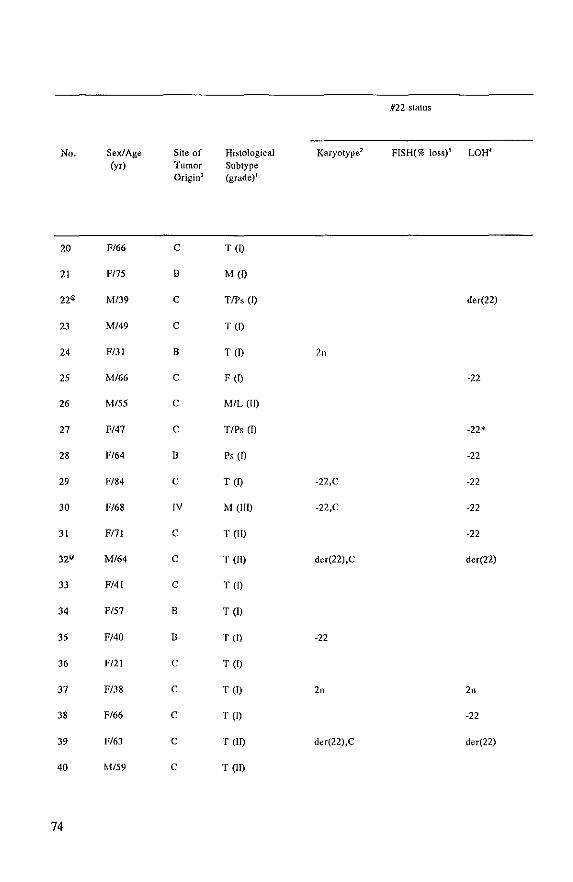
No.
20
21
22@
23
24
25
26
27
28
29
30
31
32@
33
34
35
36
37
38
39
40
74
Sex/Age (yr)
F/66
FI75
M/39
M/49
FIJI
M/66
MISS
F/47
F/64
F/84
F/68
FI71
M/64
F/41
FIS7
F/40
FJ21
F/38
F/66
F/63
M/59
Site of Tumor Originl
C
B
C
C
B
C
C
C
B
C
IV
C
C
C
B
B
C
C
C
C
C
Histological Subtype (grade)l
TW
M (I)
TlPs(l}
T (I)
T(I)
F (I)
MIL (II)
TIPs en p, (I)
T (I)
M (III)
TaT)
T (II)
T (I)
T (I)
T (I)
T (I)
T (I)
TW
T (II)
T (II)
#22 status
Karyotype2 FISH(% loss)} LOH l
der(22)
2"
·22
-22*
·22
-22,C ·22
-22,C ·22
·22
der(22),C der(22)
·22
2n 2"
·22
der(22),C der(22)
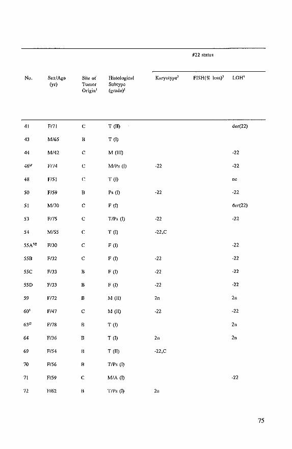
No.
41
43
44
46"
48
50
51
53
54
55A5@
55B
55e
55D
59
60'
63°
64
69
70
71
72
Sex/Age (y,)
F171
M/65
M/42
F174
F/51
F/59
M170
F175
M/55
F/JO
F/32
F/33
F/33
FI72
FJ47
F178
F/36
F/54
F/56
F/59
F/62
Site of Tumor Originl
e
B
e
e
e
B
e
e
e
e
e
B
B
B
e
8
B
8
B
e
8
Histological Subtype (grade)1
T (II)
T(I)
M (lID
M/Ps (1)
T (I)
p, m
F (D
TIPs (I)
T (I)
F (I)
Fm
Fm
Fm
M (II)
M (II)
T (I)
Tm
T (II)
TIPs (I)
MIA (D
TIPs (I)
#22 status
Karyotype2
d,,(22)
-22
-22 -22
nc
-22 -22
der(22)
-22 -22
-22,C
-22
-22 -22
-22 -22
-22 -22
2n 2n
-22 -22
2n
2n 2n
-22,C
-22
2"
75
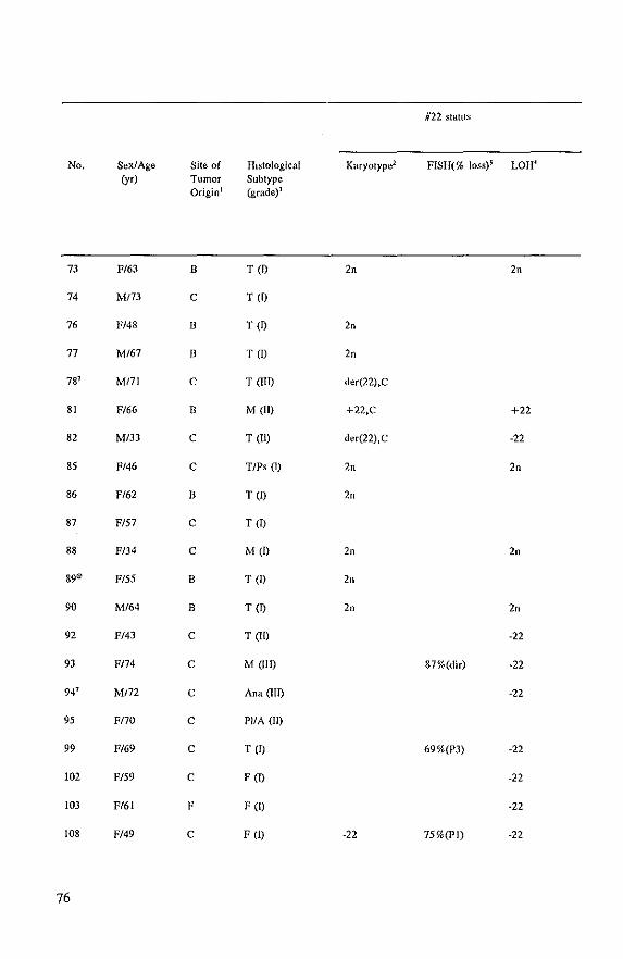
No.
73
74
76
77
78'
81
82
85
86
87
88
89"
90
92
93
94'
95
99
102
103
108
76
SexiAge (yr)
F/63
MJ73
F/48
Ml67
MI7I
F/66
M/33
F/46
F/62
FI57
F/34
FI55
MI64
F/43
F/74
M/72
F170
F/69
FI59
FI61
FI49
Site of Tumor Origin l
B
C
B
B
c
B
C
c
B
C
C
B
B
c
C
C
C
c
C
F
C
Histological Subtype (grade)1
Ten
T(O
T (Q
Tal
TOm
M (II)
T(IQ
TIP, (I)
T (Q
T (I)
M (I)
T(l)
T(Q
T(m
M (IIQ
Ana (lTO
PIIA (II)
T (I)
F (I)
F (I)
F (I)
Karyolype2
2n
2n
2n
der(22),C
+22,C
der(22),C
2n
2n
2n
2"
2"
-22
#22 slattls
FISH(% loss)' LOH4
2n
+22
-22
2n
2"
2n
-22
87%(dir) -22
-22
69%(P3) -22
-22
-22
75%(P1) -22
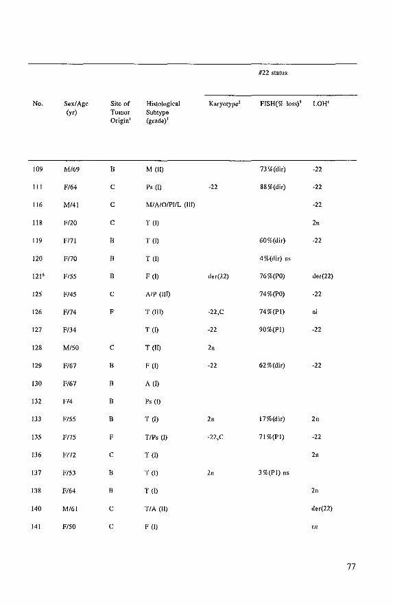
No.
109
111
116
118
119
120
121 6
125
126
127
128
129
130
132
133
135
136
137
138
140
141
SexlAge (yr)
MI69
FI64
MI41
F/20
FI7I
F/70
FISS
FI45
F174
FI34
MI50
FI67
FI67
FI4
FI55
FI7S
F172
FI53
FI64
M/61
FI50
Site of Tumor Origin'
B
C
C
C
B
B
B
C
F
C
B
B
B
B
F
C
B
B
C
C
Histological Subtype (grade)1
MOD
p, (I)
MI A/OIPIIL (lID
T (I)
Ten
T (I)
F (I)
AlP (liD
T (liD
Ten
T (ID
F(Q
A(D
Ps (I)
T (I)
TIP, (I)
T (I)
T (I)
T (I)
TIA aD
F(l)
#22 status
Karyotype! FfSH(% loss)1 LOH(
73%(dir) -22
-22 88%(dir) -22
-22
2n
60%(dir) -22
4 %(dir) ns
der(22) 76%(PO) der(22)
74%(PO) -22
-22,C 74%(PI) ni
-22 90%(PI) -22
2n
-22 62 %(dir) -22
2n 17%(dir) 2"
-22,C 71 %(FI) -22
2n
2n 3%(PI) IlS
2n
der(22)
"'
77
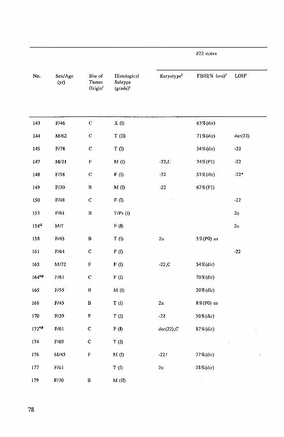
No.
143
144
145
147
148
149
ISO
153
154@
158
161
163
164~
165
166
170
172~
174
176
177
179
78
Sex/Age (yr)
F146
M/62
F178
M/21
FI58
FI30
FI48
F/61
MI?
FI45
FI64
M172
F/6!
FI55
FI45
FI39
F/61
F/69
M/45
FI61
F/JO
Site of Tumor Originl
C
C
C
F
C
B
C
B
B
C
F
C
B
B
F
C
C
F
B
Histological Subtype (grade)l
x (I)
l' an
1'(1)
M(I)
F(I)
M(I)
F(I)
TIPs (I)
Fm
l' (1)
F(D
F(I)
F (1)
Mm l' (I)
l' (I)
Fm
1'm
M(I)
T (I)
M (11)
Karyotype2
-22,C
·22
·22
2n
-22,C
2n
·22
der(22),C
-22'1
2n
#22 status
FISH(% loss)} LOH4
63%(dir)
71 %(dir) der(22)
54 %(dir) ·22
74%(PI) ·22
53 %(dir) -22*
67%(P1)
·22
2n
2n
5%(PO) ns
." 54%(dir)
70%(dir)
20%(dir)
8%(PO) ns
50%(dir)
87%(dir)
77%(dir)
28%(dir)
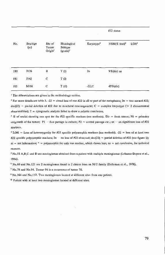
No.
180
181
183
Sex/Age (yr)
F176
F/42
M/56
Site of Tumor Origin l
B
c
c
Histological Subtype (grade)l
T(O
T (I)
T (ID
1 The abbreviations are given in the methodology section,
#22 status
Karyotype2 FISH(% loss)]
20 9 %(dir) ns
49%(dir)
2 For more details see table 3, -22 = clonal loss of one #22 in all or part of the metaphases; 2n = two normal #22;
der(22) = partial deletion of #22 due to structural rearrangement; C = complex karyotype (> 2 chromosomal
abnormalities); ? = cytogenetic analysis failed to draw a reliable conclusion,
] % of nuclei showing one spot for the #22 specific markers (see methods), Dir = fresh tumor; PO = primairy
outgrowth of the tumor; PI = first pa~sage in culture; P2 = second passage etc,; ns = no significant loss of#22
markers,
4 LOR = Loss of heterozygosity for #22 specific polymorphic markers (see methods), -22 = loss of at least two
#22 specific polymorphic markers; 2n = no loss of #22 observed; der(22) = partial deletion of #22 (see figure I);
oi = not informative; .. = polymorphic tor only one marker, which shows loss; ne = not conclusive, for technical
rea~ons,
5 No,S5 A,B,C and D are meningiomas obtained from a patient with multiple meningioma~ (Lekanne Deprez et aI.,
1994).
6 No.60 and No,121 are 2 meningiomas found in 2 sisters from an NF2 familY (Delle man et aI., 1978).
J No,78 and No.94. Tumor 94 is a recurrence 01 tumor 78.
8 No.164 and No, 172, Two meningiomas located at different sites from one patient.
<Zl Patient with at least two meningiomas located at different sites.
79
Table 2.
Technique
FISH
Cytogenetics
LOH
Comparison of FISH, cytogenetic and LOn techniqnes for detection of
(paltinl) loss of chl'omosome 22 in meningioma
Number of tumors (%)
Total* Disomy 22 (partial) loss of 22 not evaluable
30 5 (16.7%) 25 (83.3%) o
54 20 (37%) 34 (63%) o (13X: normal) (lOX: -22 only)
( 7X: other abn.) (24X: -22 and other
aim.)
69 13 (18.8%) 50 (72.5%) 6 (8.7%)
* The following tumors were excluded for rea~ons described in the result section, FISH: 1:33, 177; Cytogenetics:
10, 16, 133, 176. 177; LOH: 10, 133.
The results of cytogenelic analysis of 59 meningiomas cultured for less than 50 days
are shown in table 3. Longer culture times usually resulted in normal karyotypes, which was
probably due to outgrowth of normal cells initially present in low numbers in the cultures.
This was seen also when repeated cytogenetic analysis of meningiomas with an altered
karyotype resulted in normal karyotypes in the course of time (data nol shown). Thirteen
meningiomas showed a normal or non-clonal karyotype and in 41 cases clonal chromosomal
abnormalities were found. Twenty three of the cases with chromosomal abnormalities showed
::;; 2 changes, including chromosome 22 aberrations in 17 cases. In eighteen meningiomas
more than 2 chromosomal abnormalities, incuding chromosome 22, were identified. In at
least 12 meningiomas chromosome 22 was involved in structural abnormalities. These are
further discussed below. Partial or complete loss of chromosome 22 was the most frequently
observed abnormality (63%) in this series of meningiomas. In addition to this other
chromosomes also showed non random abnormalities in all or sublines of the cells. Of these,
chromosomes I (27.7%), 6 (13%), II (13%), 13 (14.8%), 14 (18.5%), 18 (13%), 19
(24.1 %), X (20.5%), and Y (20%) were most of len involved (table 3). Complete or partial
loss of these chromosomes was the predominant abnormality.
80
Table 3. Summary of chromosomal findings in 59 meningiomas
No. Day~ in Karyotypes andlor clonal rearrangements [no. of cells}
culture
4 P2 44,X,-X,-22 (161
7° 15 47,XX,+7,+21,-22 {lS)1
idem, 9q+ (3)
9 35 4S,XX,-19,-22, +dic(22pter-> ql1?:: 19p 12?- > qter) 16)1
42-44,idem 1p-,6q-,llp+,-21,+variable additional aburations (7)
10 4 43,XX,-14,-19,-22 [14]1
42,XX,tas(7; 19)(pter:qter),-14,-19 ,-22 12]
14 33 44.XX,dic( 1 qter- > q 1 0;: 19pter- > qter),add(2)(p21),der(2)t(2:8)(q 14?;q2I),
,dd(4)(q21),d,,(8)«2;8)(p21 ;q21),del( 12)(p 12),«14; 16)(q II;q 11-12),
d,,(l8)«2; 18)(q 14;p 11),-22 [10]1
46,XX 13]
15 19 46,XX [511
44-45, random losses [3)
16 17 S5,XX, Ip-[2x], lq-[2xl,-IO,-1I,-12,-16[2x),-20[2x),-22[2xi, +6 markers III
24 38 47,XX,+mar(?del(13)(q14q22)) 1161
29 27 45,XX,dic(l; 19)(lqter- >pI3:: 19q1l- >pter),del(t3)(qI3q21), +20,-22 [5)1
46,idem,+20 {II]!
4S,idem,+20,-21 [81.
Additional slnlctural aherrations were observed in 10 of the cells in the different
clones.
30 19 41 ,XX"dd(I)(p 12),dic(i ;2)(p I O;q I 0),-3,-4,d,,(6)<(1 ;6)(q 12;q 14),
d,,(8)«8; 131)(q24;q 141),d,I(lI)(q II ),-13 ,d,,(l6)«4; 16)(q2 t;p 13),
d,,(l8)<(lI; 18)(q 12;qI2),d,,(l9)<(3; t 9)(q II;q 12),-22 P]I
42,idem, +ring [6]1
84,XXXX,idem tetraploid,+2 ring [3}
32° 15 45,XY ,-I ,«4;22)(p 161;q 11),d,,(22)<(I ;22)(p II ;qll) [811
44,itlem,13r+ ,-14 [2)/44,idem,-IO, + 12, 13p+ ,-14 (6)1
45,idem,-IO, + 12, 13p+ ,-14, + 16 I12]/90,idem,tetraploid I21
81
No. Days in Karyotypes andlor clonal rearrangements {no. of cells] culture
35 6 45,XX,-22 [14]1
90,XXXX,-22,-22 [2]
37 9
39 49
14
50 14
53 28
54 35
55B~ 14
sse 10
55D 39
59 18
60' 35
64 49
69 32
82
46,XX"dd(2)(q22) [20]
45,XX,lq-,3q+,-10 [3]1
44.XX,2q·,·6,·7.lOp+.llp +, 13q+, 19q+ ,·22.22q +, + fragment [IO]I
45,XX,2qy 6,·7, lOp+, IIp+ ,·13, 19q+ ,·22,22q +, + fragment, + ring f3J1
46,XX [5]
45,XX,-22 [16]
45,XX,-22 [12]1
45,XX,,(17),-22 [3JI
44,XX,·22,tas(9; 17)(qter;pter)111
45,XX,-22 [1311
very aherrant tetraploid including tas 12}
43,XY,-14,-19,-22 [3]1
42,X,-Y,-14,-19,-22 [611
46,XY (61
45,XX,-22 [III
44,X,-X,-22 [811
40·43,idem,with random losses 151
45,XX,-22 12411
89,hypotetrapioid.-2212x],with additionHI tosses and rearrangements (lJ
45,XX,-22 [3JI
46,XX (2J
46,XX [16(
45,XX,-22 [4(1
46,XX [12J
46,XX [161
43,XX,der(1)t(l; J6)(p II ;qll),uic(6qter- > pI2:: 16q 11- >pter or 22q II-qter),-14,
-16,-22 [16(
No. Days in Karyotypes and/or clonal rearrangements [no. of cellsJ culture
72 21 46,XX 116J
73 14 46,XX [16]
76 21 46,XX 123J
77 18 46,XY 120)J
78' 5 Very aberrant hypodiploid to hypotetraploid karyotype. Clonal aberrations:
t(l;2)(q41?;q221),·19,19q+,22q+ [13J. Also many tas and HSR
81 25 56(54·59),X,·X, lp·, + Ip·, +3, +5,6q+, + 13, + 17, + 18,( + 19), +20,( +20),( +22),
+various markers 19JI
46,XX [lJ
82 3 44,der(X), Y, 1 p.,t( 1 j6)(]l32;p2l),2q.,.4,·5q., +6q·,6p + ,der(7), 1 Oq +,12'1·,
d,,(12)1(12; 17)(qlO;qlO),-15,-19,22q-,",,(22)1(2;22)(ql1 ;1;qll) 17JI
idem,+additional aberrations:9q+ or ·9,1Ip+ or ·11.·21 (IOJ
85 14 46,XX [l6J
86 35 46,XX,I(7;14)(q35;qll-l2) 19JI
46,XX [3)
88 32 46,XX 118JI
tetraploid 12]
89° 21 45,XX,-1l [JJI
47,XX,+X 14JI
46,XX 142J
90 38 46,XY 114JI
tetraploid 14]
108 10 45,XX,-22116JI
44,XX,dic(l3;22)(pll;pll),-22 [4]1
44·45,XX,·22,and variable tas and whole arm translocations (61
III 28 45,XX,-22 112JI
44,XX,·22,dic or las involving chromosome 1914]
121 6 49 46,XX,22q- 17]
83
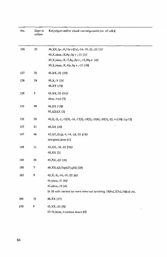
No. Days in Karyotypes and/or clonal rearrangements [no. of cells] cuJt\lfe
126 32 46,XX, Ip-,-6,11p+[2x],-14,-18,-21,-22 II]I
40.X,idem,-X,8q-,9p+,-13 [IJI
J9,X,idem.-X,-7 ,8q-,9p+ .-IJ,20p+ [4]1
J9,X,idem,-X,-8q-,9p+,-IJ flO]
127 32 45,XX,-22 1331
128 28 45,X,-Y 1311
46,XY 1131
129 5 45,XX,-22 (31(1
idem,+tas [71
133 49 46,XX 11311
92,XXXX 121
135 28 42,X,-X,-I,-1213I,-14,-17(3I,-1813(,-1916(,-20(3I,-22, +" 101
137 21 46,XX 1201
147 46 42,XY,i(lq),-4,-14,-18,-22 11811
letr<lpioid,idem [1]
148 11 44,XX,-18,-22 11011
46,XX 121
149 28 45,XX,-22 1161
158 7 46,XX,t(3;7)(pI2?;q35) 1201
163 8 42,X,-X.-14,-19,-22 (6]1
41,idem,-21 {6J1
41,idem,-lJ [4].
[cp17J
In 10 cells various tas were observed involving 19(9x],J{7x),14[6x] etc.
166 10 46,XX 117J
170 9 45,XX,-22 1911
43-44,idem,+random losses (91
84
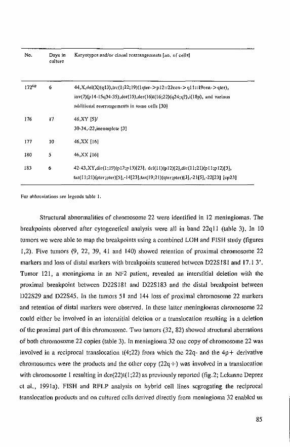
No. Days in Karyotypes and/or clonal rearrangements Ino. of cells] culture
lnS1l' 6 44,X,del(X)(q13),trc(1 ;22; 19)(lqter->p12::22cen- > ql1:: 19cen-> qter),
inv(7)(p 14-ISq34-3S),der(lS),der(16)t(16;22)(q24;q?),i( 18p), and various
176 17
177 10
180 5
183 6
additional rearrangements in some cells [30]
46,XY [5JI
30-34,-22,incomplete (3)
46,XX [161
46,XX /16J
42-43,XY ,die(l; 19)(p 12;p 13)[23J, d,I(11 )(p 12)[2J,di,(11 ;21)(P II;p 12)/3),
tas( II ;21)(pter;pter)[51. -14(23),tas( 19 ;21)(qter;pter)[3] ,-21 (S) ,-22[23] (cp231
For abbreviations see legends table I.
Structural abnormalities of chromosome 22 were identified in 12 meningiomas. The
breakpoints observed after cytogenetical analysis were all in band 22qll (table 3). In 10
tumors we were able to map the breakpoints using a combined LOH and FISH study (figures
1,2). Five tumors (9, 22, 39, 41 and 140) showed retention of proximal chromosome 22
markers and loss of distal markers with breakpail;ts scattered between D22S 181 and 17.1 3'.
Tumor 121, a meningioma in an NF2 patient, revealed an interstitial deletion with the
proximal breakpoint between D22S 181 and D22S 183 and the distal breakpoint between
D22S29 and D22S45. In the tumors 51 and 144 loss of proximal chromosome 22 markers
and retention of distal markers were observed. In these latter meningiomas chromosome 22
could either be involved in an interstitial deletion or a translocation resulting in a deletion
of the proximal part of this chromosome. Two tumors (32, 82) showed structural aberrations
of both chromosome 22 copies (table 3). In meningioma 32 one copy of chromosome 22 was
involved in a reciprocal translocation t(4;22) from which the 22q- and the 4p+ derivative
chromosomes were the products and the other copy (22q +) was involved in a translocation
with chromosome 1 resulting in der(22)t(l ;22) as previously reported (fig.2; Lekanne Deprez
et aI., 1991a). FISH and RFLP analysis on hybrid cell lines segregating the reciprocal
translocation products and on cultured cells derived directly from meningioma 32 enabled us
85
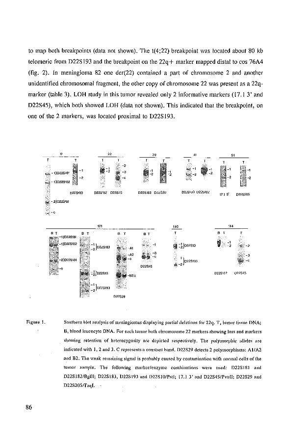
to map both breakpoints (data not shown). The t(4;22) breakpoint was located about 80 kb
telomeric from D22S 193 and the breakpoint on the 22q + marker mapped distal to cos 76A4
(fig. 2). In meningioma 82 one der(22) contained a part of chromosome 2 and another
unidentified chromosomal fragment, the other copy of chromosome 22 was present as a 22q
marker (table 3). LOR study in this tumor revealed only 2 informative markers (17.1 3' and
D22S45), which both showed LOR (data not shown). This indicated that the breakpoint, on
one of the 2 markers, was located proximal to D22S193.
Figure 1.
86
9 39 " 51 T T T T T T
~ -1 -, ~-11D22S181
1Ili*J- 1}022S182
iIlIi.·· ..... -, 'II! -, l1li-' -, "~\ I=! .' ~ -2 -, -,
WSl93 D225182 022345 D22S183 022310 D22$183 0225182 17.1 3' D22$205
.;,;. -2]022$181
-0
'" ''0 '" B T B T T B T T
-, 1:1]022510 f -, -, -, -3
-3 -, -1 ]0228193 ... ·-c
022845 )liM -2
-92'e 0228162 022545
022529
Southern blot analysis of meningiomas displaying partial deletions for 22q. T, iumof tissue DNA;
B, blood leucocyte DNA. For each tumor hoth chromosome 22 markers showing loss and markers
showing retention of heterozygosity are depicted respectively. The polymorphic alleles are
indicated with 1,2 and J, C represents a constant band, D22S29 detects 2 polymorph isms: AI/A2
and B2. The weak remaining signal is probahly caused by contamination with normal cells of the
tmnor sample, The following marker/enzyme comhinations were used: D22S181 and
D22S182/Bgl1l; 022S183, 022S193 and 022SIO/PstI; 17,1 3' and 022S45/PvuII; 022S29 and
D22S2051TaqI.
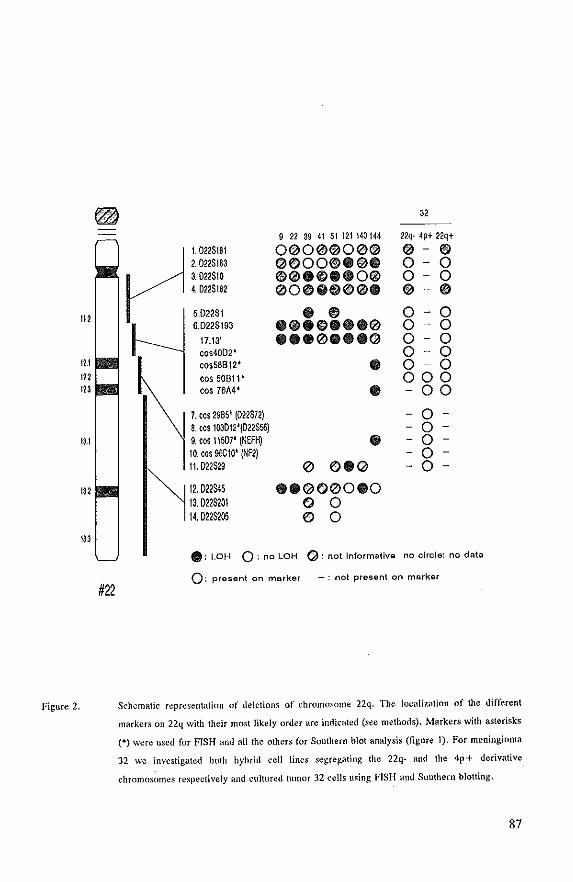
112
12.1 122 123
1).1
132
133
Figure 2.
~ 32 .. '
#22
9 22 39 41 51 121140144 22q· 4p+ 22q+
I. 022$181 0(2)00l®00® ® - ® 2.022$183 ®OOO®.®. 0- 0 3.022$10 ®®.® •• O® 0- 0 4.022$182 ®O® •• 0l® • • @ 5.D22$1 @ ® 0 - 0 6.022$193 @0@®.00® 0 - 0
17.13' 0 •• 00 •• (2) 0 - 0 cos40D2" 0 - 0 cos58B12* • 0 0 cos 50811* 000 cos 76A4* 0 - 00
7. co, 2985' (022S72) - 0 -
S. co, 103012'(022S56) - 0 -
9. '''11507' (NEFH) • - 0 -
10. ,os 96C10' (NF2) - 0 -
11. D22$29 0 0.(2) - 0 -
12.022345 .0(2)e>00.0 13.022$201 e> 0 14. D22$105 e> 0
.' LOH 0: noLOH ('): not Informative no circle: no data
0, present on marker - : not present on marker
Schematic representation of deletions of chromosome 22q. The localization of the different
markers on 22q with their most likely order are indicated (see methods). Markers with asterisks
(*) were used for FISH and all the others for Southern blot analysis (figure I). For meningioma
J2 we investigated hoth hybrid cell lines segregating the 22q- and the 4p+ derivative
chromosomes respectively and cultured tumor 32 cells using FISH and Southern blotting.
87
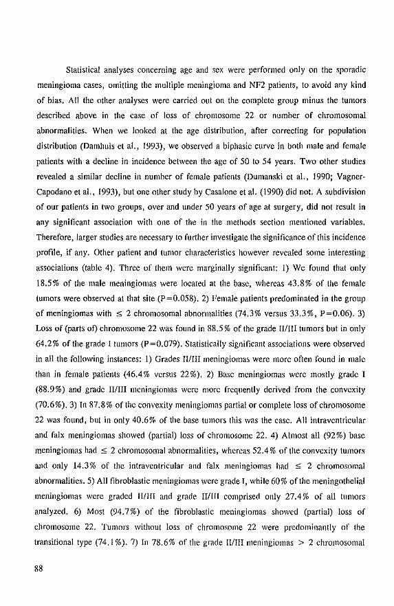
Statistical analyses concerning age and sex were performed only on the sporadic
meningioma cases, omitting the multiple meningioma and NF2 patients, to avoid any kind
of bias. All the other analyses were carried out on the complete group minus the tumors
described above in the case of loss of chromosome 22 or number of chromosomal
abnormalities. When we looked at the age distribution, after correcting for population
distribution (Damhuis et a!., 1993), we observed a biphasic curve in both male and female
patients with a decline in incidence between the age of 50 to 54 years. Two other studies
revealed a similar decline in number of female patients (Dumanski et a!., 1990; Vagner
Capodano et a!., 1993), but one other study by Casalone et a!. (1990) did not. A subdivision
of our patients in two groups J over and under 50 years of age at surgery I did not result in
any significant association with one of the in the methods section mentioned variables.
Therefore, Jarger studies are necessary to further investigate the significance of this incidence
profile, if any. Other patient and tumor characteristics however revealed some interesting
associations (table 4). Three of them were marginally significant: 1) We found that only
18.5% of the male meningiomas were located at the base, whereas 43.8% of the female
tumors were observed at that site (P=0.058). 2) Female patients predominated in the group
of meningiomas with :5 2 chromosomal abnormalities (74.3% versus 33.3%, P=0.06). 3)
Loss of (parts of) chromosome 22 was found in 88.5 % of the grade WIll tumors but in only
64.2% of the grade I tumors (P=0.079). Statistically significant associations were observed
in all the following instances: I) Grades WIll meningiomas were more often found in male
than in female patients (46.4% versus 22%). 2) Base meningiomas were mostly grade I
(88.9%) and grade WIll meningiomas were more frequently derived from the convexity
(70.6%). 3) In 87.8% of the convexity meningiomas partial or complete loss of chromosome
22 was found, but in only 40.6% of the base tumors this was the case. All intraventricular
and falx meningiomas showed (partial) loss of chromosome 22. 4) Almost all (92 %) base
meningiomas had::; 2 chromosomal abnormalities, whereas 52.4% of the convexity tumors
and only 14.3% of the intraventricular and falx meningiomas had $ 2 chromosomal
abnormalities. 5) All fibroblastic meningiomas were grade I, while 60% of the meningothelial
meningiomas were graded IIIIII and grade IIIIlI comprised only 27.4 % of all tumors
analyzed. 6) Most (94.7%) of the fibroblastic meningiomas showed (partial) loss of
chromosome 22. Tumors without loss of chromosome 22 were predominantly of the
transitional type (74.1 %).7) In 78.6% of the grade II/III meningiomas> 2 chromosomal
88

abnormalities were found, whereas in only 17.5% of the grade I tumors this was the case.
8) Loss of (parts 01) chromosome 22 was observed in 94.4% of the tumors with> 2
chromosomal abnormalities but in only 47.2% of the meningiomas with:::;; 2 chromosomal
abnormalities.
89
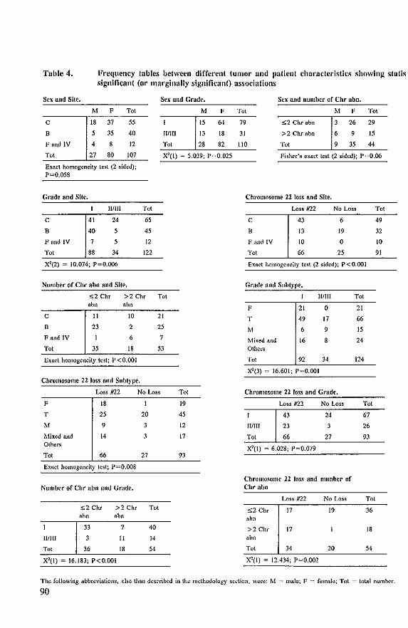
Tuble 4. Frequency tables between different tUI1101' und patient characteristics showing sinUs signil1cant (or mnrginully significant) associations
SI'X and Site. Se-x and Grade.
M F Tot M F
C 18 37 55 15 64
B 5 35 40 HlIII 13 18
F and IV 4 8 12 Tot 28 82
Tot 27 80 107 X2(1) = 5.029: P=O.025
Exact homogeneity test (2 sided)j P=Q,058
Grade and Site.
Will Tot
C 41 24 65
B 40 5 45
F and IV 7 5 12
Tot 88 34 122
X~(2) = 10.074; P-O.OO6
Number of Chr abn and Sitl'.
52 ehT >2 enr Tot .bn .bn
C II 10 21
B 23 2 25
F and IV 6 7
Tot 35 18 53
Exact homogeneity test; P<O.OOI
Chromosome 22 Joss and Subtype.
Loss 1122 No Loss
F 18
T 25 20
M 9
Mixed and 14 Others
Tot 66 27
Exact homogeneity tcst: P=O.OO8
Number or Chr abn Ilnd Grade,
s2 ehr >2 ehr Tot .bn .b"
33 7 40
IIIIII 3 II 14
Tot 36 18 54
X2(l) = 16,183; P<O,OOI
Tot
19
45
12
17
9J
Sex and number of Chr abn.
Tot M F Tot
79 s2 Chr abo 3 26 29
31 >2 Chrabn 6 9 15
110 Tot 9 35 44
Fisher's exact tcst (2 sided); P=O.06
Chromosome 22 loss Rnd Site.
Loss 1122 No Loss
c 43 6
8 13 19
F and IV 10 0
Tot 66 25
Exact homogeneity tcst (2 sided); P<O.OOI
Grade and Subtype.
111111
F 21 0
T 49 17
M 6 9
Mixed and 16 Others
Tot 92 34
XI(J) = 16.601; P=O.OOI
Chromosome 22 loss Ilnd Grade,
HlIII
Tot
Loss #22
43
23
66
~(1) = 6,018: P=O.079
No Loss
24
3
27
Chromosome 22 loss llnd number or ehr abn
Tot
21
66
15
24
124
Tot
67
26
9J
Loss #22 No Los~ Tot
s2 ehr 17 19 36
"bn
>2 ehr 17 18 abll
Tot 34 20 54
X2(l) - 12.434; P-O.O<X!.
Tot
49
32
10
91
The following abbreviations, else than descrihed in the methodology section, were: M = male; F = female; Tot = total number,
90
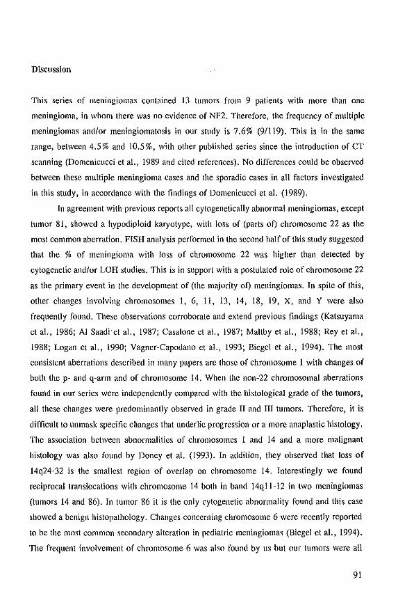
Discussion
This series of meningiomas contained 13 tumors from 9 patients with more than one
meningioma, in whom there was no evidence of NF2. Therefore, the frequency of multiple
meningiomas andlor meningiomatosis in our study is 7.6% (9/119). This is in the same
range, between 4.5% and 10.5%, with other published series since the introduction of CT
scanning (Domenicucci et a!., 1989 and cited references). No differences could be observed
between these multiple meningioma Cases and the sporadic cases in all factors investigated
in this study, in accordance with the findings of Domenicucci et a!. (1989).
In agreement with previous reports all cytogenetically abnormal meningiomas, except
tumor 81, showed a hypodiploid karyotype, with loss of (parts of) chromosome 22 as the
most common aberration. FISH analysis performed in the second half of this study suggested
that the % of meningioma with loss of chromosome 22 was higher than detected by
cytogenetic andlor LOH studies. This is in support with a postulated role of chromosome 22
as the primary event in the development of (the majority of) meningiomas. In spite of this,
other changes involving chromosomes 1, 6, 11, 13, 14, 18, 19, X, and Y were also
frequently found. These observations corroborate and extend previous findings (Katsuyama
et a!., 1986; AI Saadiet aI., 1987; Casalone et a!., 1987; Maltby et a!., 1988; Rey et a!.,
1988; Logan et a!., 1990; Vagner-Capodano et ai., 1993; Biegel et ai., 1994). The most
consistent aberrations described in many papers are those of chromosome 1 with changes of
both the p- and q-arm and of chromosome 14. When the non-22 chromosomal aberrations
found in our series were independently compared with the histological grade of the tumors,
all these changes were predominantly observed in grade" and III tumors. Therefore, it is
difficult to unmask specific changes that underlie progression or a more anaplastic histology.
The association between abnormalities of chromosomes 1 and 14 and a more malignant
histOlogy was also found by Doney et a!. (1993). In addition, they observed that loss of
14q24-32 is the smallest region of overlap on chromosome 14. Interestingly we found
reciprocal trallSlocations with chromosome 14 both in band 14qll-12 in two meningiomas
(tumors 14 and 86). In tumor 86 it is the only cytogenetic abnormality found and this case
showed a benign histopathology. Changes concerning chromosome 6 were recently reported
to be the most common secondary alteration in pediatric meningiomas (Biegel et al., 1994).
The frequent involvement of chromosome 6 was also found by us but our tumors were all
91
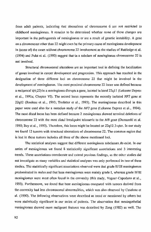
from adult patients, indicating that aberrations of chromosome 6 are not restricted to
childhood meningiomas. It remains to be determined whether some of these changes are
important in the pathogenesis of meningiomas or are a result of genetic instability. A gene
on a chromosome other than 22 might even be the primary cause of meningioma development
in (some of) the cases without chromosome 22 involvement as the studies of Ruttledge et al.
(1994) and Pulst et al. (1993) suggest that'in a subclaSS of meningiomas chromosome 22 is
not involved.
Structural chromosomal alterations are an important tool in defining the localization
of genes involved in cancer development and progression. This approach has resulted in the
designation of three different loci on chromosome 22 that might be involved in the
development of meningiomas. The most proximal chromosome 22 locus was defined because
a reciprocal t(4;22) in a meningioma disrupts a gene, located in band 22q II (Lekanne Deprez
et aI., 1991a; Chapter VI). The second locus represents the recently isolated NF2 gene at
22ql2 (Rouleau et aI., 1993; Trofatter et aI., 1993). The meningiomas described in this
paper were used also for a mutation study of the NF2 gene (Lekanne Deprez et aI., 1994).
The most distal locus has been defined because 2 meningiomas showed terminal deletions of
chromosome 22 with the most distal breakpoint telomeric to the MB gene (Dumanski et al.
1990; Rey et aI., 1993). Therefore, this locus might be located on 22qI2.3-qter. In our study
we found 12 tumors with structural aberrations of chromosome 22. The common region that
is lost in these tumors includes all three of the above mentioned loci.
The statistical analyses suggest that different meningioma subclasses,do exist. In our
series of meningiomas we found 8 statistically significant associations and 3 interesting
trends. These associations corroborate and extend previous findings, as the other studies did
not investigate as many variables and statistical analyses was only performed in two of these
studies. The statistically significant associations observed were that grade II/III meningiomas
predominated in males and that base meningiomas were mainly grade I, whereas grade 11/111
meningiomas were most often found in the convexity (this study, Vagner~Capodano et aI.,
1993). Furthermore, we found that base meningiomas compared with tumors derived from
the convexity had less chromosomal abnormalities, which was also observed by Casal one et
al. (1990). The following observations were described as trend or mentioned by others but
were statistically significant in our series of patients. The observation that meningothelial
meningiomas showed more malignant features was described by Zang (1982) as well. The
92
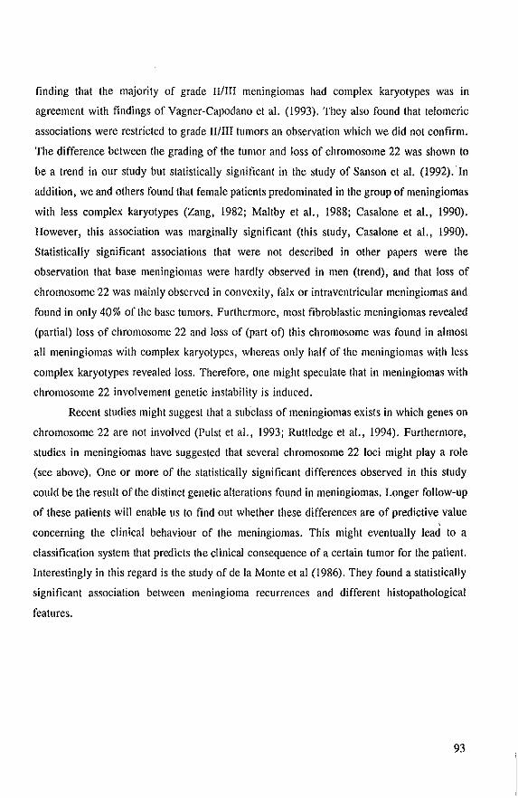
finding that the majority of grade WIll meningiomas had complex karyotypes was in
agreement with findings of Vagner-Capodano et al. (1993). They also found that telomeric
associations were restricted to grade Will tumors an observation which we did not confirm.
The difference between the grading of the tumor and loss of chromosome 22 was shown to
be a trend in our study but statistically significant in the study of Sanson et al. (1992). In
addition, we and others found that female patients predominated in the group of meningiomas
with less complex karyotypes (Zang, 1982; Maltby et aI., 1988; Casalone et al., 1990).
However, this association was marginally significant (this study, Casalone et aI., 1990).
Statistically significant associations that were not described in other papers were the
observation that base meningiomas were hardly observed in men (trend), and that loss of
chromosome 22 was mainly observed in convexity, falx or intraventricular meningiomas and
found in only 40% of the base tumors. Furthermore, most fibroblastic meningiomas revealed
(partial) loss of chromosome 22 and loss of (part of) this chromosome was found in almost
all meningiomas with complex karyotypes, whereas only half of the meningiomas with less
complex karyotypes revealed loss. Therefore, one might speculate that in meningiomas with
chromosome 22 involvement genetic instability is induced.
Recent studies might suggest that a suhclass of meningiomas exists in which genes on
chromosome 22 are not involved (Pulst et aI., 1993; Ruttledge et aI., 1994). Furthermore,
studies in meningiomas have suggested that several chromosome 22 loci might playa role
(see above). One or more of the statistically significant differences observed in this study
could be the result of the distinct genetic alterations found in meningiomas. Longer follow-up
of these patients will enable us to find out whether these differences are of predictive value
concerning the clinical behaviour of the meningiomas. This might eventually lead to a
classification system that predicts the clinical consequence of a certain tumor for the patient.
Interestingly in this regard is the study of de la Monte et al (1986). They found a statistically
significant association between meningioma recurrences and different histopathological
features.
93
Acknowledgements
This study was supported by a grant from the Dutch Cancer Foundation (EUR 89-06).
Support of the E.C. Biomed I Concerted Action: Molecular cytogenetics of solid tumors to
E.v.D. is acknowledged.
References
AI Saadi A, Latimer F, Madercic M, Robbins T (1987) Cytogenetic studies of human brain tumors and their clinical significance. Cancer Genet Cylogenet 26: 127-141.
Amoldus EPJ. NoorJermeer lA, Boudewijn Peters AC. Voormolen JHC, Bots GTAM, Raap AK, van Jer Ploeg M (1991) Tnlerphase cytogenetics of brain hlmors. Genes Chrom Cancer 3: 101-107.
Barker G, Schafer M, White R (1984) Restriction sites containing CpG show a higher frequency of polymorphism in human DNA. Cell 36: 131-138.
Diegel JA, Parmiter AH, Sutton LN, Rorke LB, Emanuel BS (1994) Abnormalities of chromosome 22 in pediatric meningiomas, Genes Chrom Cancer 9:81-87.
van Biezen NA, Lekanne Deprez RH, Thijs A, Heutink P, Ooslra BA, Geurts van Kessel AHM, ZwarthoffEC (1993) Tsolation and characterization of25 unique DNA markers for human chromosome 22. Genomics 15:206-208.
Budarf ML, McDermid HE, Sellinger D, Emanuel DS (1991) Isolation and regional localization of 35 unique anonymous DNA markers for human chromosome 22, Genomics 10,996-1002.
Bulli G, Assielli R, Casalone R, Paoletti P (1989) Multiple meningiomas: A clinical, surgical and cytogenetic analysis. Surg Neuml 31 :255·260.
Casalone R. Granata P, Simi P, Tarantino E, Butti G, Buonaguidi R, Faggionato F, Knerich R, Solero L (1987) Recessive cancer genes in meningiomas? An analysis of 31 cases, Cancer Genet Cytogenet 27: 145-159.
Casal one R, Simi P, Granata P, Minelli E, Giudici A, Butti G, Solem CL (1990) Correlation between cytogenetic and histopathological findings in 65 human meningiomas, Cancer Genet Cytogenet 45:237-243.
Damhuis RAM, Mol CAM, Hoekman- van der Male (1993) Frequentie en behandeling van kanker, lKR-Regio 1988-1991: pp 10.
Delleman JW, de Jong JGY, Bleeker GM (1978) Meningiomas in five members of a family over two generations, in one member simultaneously with acoustic neurinomas. Neural 28:567-570.
Domenicucci M. Santoro A, D'Osvaldo DB, Delfini R, Cantore GP, Guidetti B (1989) Multiple intracranial meningiomas. J Neurosurg 70:41-44.
Doney MK, Houseal TW, Soukup SW, Rutter JL, GuselJa JF, Schneider BF, Golden WL et a!. (1993) Identification of a putative tumor suppressor locus in 14q24-32 that is involved in formation and progression of meningiomas. Am J Bum Genet 53:293.
94
Dumanski JP, Roule~lU GA, Nordenskj6ld M, Collins VP (1990) Molecular genetic analysis of chromosome 22 in 81 cases of meningioma. Cancer Res 50:5863-5867.
Goyns MH, Young BD, Geurts van Kessel A, de Klein A, Grosveld G, Bartram CR, Boolsma D (1984) Regional mapping of the human immunoglobulin lambda light chain to the philadelphia chromosome in chronic myeloid leukaemia. Leukemia Res 8:547-553.
Hotter MH, Breuning MH, Bakker P, van Ommen GJB, Pearson PL (1985) An anonymous single copy chromosome 22 clone, D22SIO (22Cl-18), identifies an RFLP with Pstl. Nucleic Acids Res 13:7167.
ISCN (1991) Mitelman F (ed): Guidelines for Cancer Cytogenetics, Supplement to an Intemational System for Human Cytogenetic Nomenclature. Basel: S. Karger.
JiiaskeUiinen J, Haltia M. Laasonen E. Wahlstrom T, Vallonen S (1985) The growth rate of intracranial meningiomas and its relation to histlogy. Surg NeufoI24:165-172.
de Jong PJ, Yokohata K. Chen C, Lohman F, Pederson L, McNinch J, van Oilla M (1989) Human chromosome-specific partial digest libraries in k and cos mid vectors. Cytogenet Cell Genet 51 :985.
Katsuyama J. Papenhallsen PR, Herz F, Gazivoda P, Hirano A, Koss LG (1986) Chromosome abnormalities in meningiomas. Cancer Genet Cytogenet 22:63-68.
Kepes JJ (1982) Meningiomas, Biology, Pathology, Oifferentiill ~iagnosis, New York:Masson.
Koper JW, Foekens JA, Braakman R, Lamberts SWJ (1990) Effects of progesterone on the response to epidermal growth factor and other growth factors in cultured human meningioma cells. Cancer Res 50:2604-260).
Lekanne Deprez RH, Groen NA, van Bielen NA, Hagemeijer A, van Dronen E, Koper JW, Avezaat CJJ, Bootsma D, Zwarthoff EC (1991a) A t(4;22) in a meningioma points to the localization of a putative tUmorsuppressor gene. Am J Hum Genet 48:783-790.
Lekanne Deprez RH, van Biezen NA, Heutink P, BoejhHrat KRGS, de Klein A, Geurts van Kessel AHM, ZwarthoffEC (l99Ib) A new polymorphic probe on chromosome 22: NBI7 (D22SI81). A new polymorphic probe on chromosome 22: NB35 (022SI82). A new polymorphic probe on chromosome 22; NB84 (022S183). A new polymorphic probe on chromosome 22q: NBI29 (022S193). Nucleic Acids Res 19:686-687,
Lekanne Deprez RH, van Biezen NA, Breedveld GJ, Boejharat KRGS, de Klein A. Geurts van Kessel AHM, Zwarthoff EC (1991e) A new polymorphic probe on chromosome 22: NBS (022S201), Nucleic Acids Res, 19:1963.
Lekanne Deprez RH, Groen NA, Louz 0, Hagemeijer A, vall Drunen E, Koper JW, Avezaat CJJ, Bootsma D, van der Kwast TH, Zwarlhofr EC (1994) Constitutional DNA-level aberrations in chromosome 22 in a patient with multiple meningiomas. Genes Chrom Cancer 9: 124-128.
Lekanne Deprez RH, Bianchi AB, Groen NA, Seizinger BR, Hagemeijer A, van Drunen E, Bootsma D, Koper JW, Avezaat CJJ, Kley N, Zwarthoff EC. Frequent NF2 gene transcript mutations in sporadic meningiomas and vestibular schwannomas. Am J Hum Genet (in pres~).
Logan JA, Seizinger BR, Atkins L, Martuza RL (1990) Loss of the Y chromosome in meningiomas, Cancer Gene! Cytogenet 45:41-47.
Maltby EL, Ironside JW, Battersby RDE (1988) Cytogenetic studies in 50 meningiomas, Cancer Genet Cytogenet 31:199-210.
95
McDermid HE, Duncan AMV, Higgins MJ, Hamerton JL, Rector E, Brasch KR, White BN (1986) Isolation and characterization of an alpha-satellite repeated sequence from human chromosome 22, Chromosoma 94:228-234.
McDowell JR (1990) Familial meningioma, Neurology 40:312-314,
de la Monte SM, Flickinger J, Linggood RM (1986) Histopathologic features predicting recurrence of meningiomas following subtotal resection. Am J Surg Pathol 10:836-843,
Pulst SM, Rouleau GA, Marineau C, Fain P, Sieb JP (1993) Familial meningioma is not allelic to neurofibromatosis 2. Neural 43:2096-2098.
Rey JA, Bello MJ, de Campos JM, Kusak E, Moreno S (1988) Chromosomal involvement secondary to -22 in human meningiomas. Cancer Genet Cytogenet 33:275-290.
Rey lA, Bello MJ, de Campos JM, Vaquero I, Kusak ME, Sarasa JL, Pestana A (1993) Abnonnalities of chromosome 22 in human brain tumors determined by combined cytogenetic and molcular genetic approaches, Cancer Genet Cytogenet 66: 1-10.
Rouleau GA, Haines JL, Bamnowski A, Colella-Crowley A, Trofalter JA, Wexler NS, Conneally PM, Gusella JF (1989) A genetic linkage map of the long arm of human chromosome 22. Genomics 4: 1-6.
Rouleau GA, Merel P. Lutchman M, Sanson M, Zucman J, Marineau C, Hoang-Xuan K. Demczuk S. Desmaze C, Plougastel B, Pulst SM, Lenoir G, Bijlsma E, Fashold R, Dumanski J, de Jong P, Parry D, Eldrige R, Aurias A, Delattre J, Thomas G (1993) Alteration in a new gene encoding a putative membrane-organizing protein causes neuro-fibromatosis type 2. Nature 363:515-521.
Russell OS, Rubinstein U (1989) Tumours of the meninges and related tissues. In Arnold E (ed): Pathology of Tumours of the Nervous System, 5<h ed. London: Edward Arnold pp 452-453.
Rulliedge MH, Sarrazin J, Rangaratnam S, Phelan CM, Twist E, Merel P, Delaltre 0, Thomas G, Nordenskj6ld M, Collins VP, Dumanski JP, Rouleau GA (1994) Evidence for the complete inactivation of the NF2 gene in the majority of sporadic meningiomas. Nature Genet 6: 180-)84.
Sanson M, Richard S, Delaltre 0, Poliwka M, Mikol J, Philippon J, Thomas G (1992) Allelic loss on chromosome 22 correlates with histopathological predictors of recurrence of meningiomas. lnt J Cancer 50:391-394.
Seizinger BR, de la Monte S, Atkins L, Gusella JF, Martum RL (1987) Molecular genetic approach to human meningioma: Loss of genes on chromosome 22. Proc Natl Acad Sci USA 84:5419-5423.
Sieb JP, Pulst SM, Buch A (1992) Familial CNS tumors, J Neurol 239:343-344.
Trofatter JA, MacColiin MM, Rutter JL, Murrell JR, Duyao MP, Parry DM, Eldridge R, Kley N, Menon AG, Pulaski K, Haase VH, Ambrose CM. Munroe D, Dove C, Haines JL, Martllm RL. MacDonald ME. Seizinger DR, Short MP, Buckler AJ, Gusella JF (1993) A novel Moesin-, Euin-, Radixin-likeGene is a candidate for the Neurofibromatosis 2 tumor suppressor, Cell 72:79)-800.
Vagner-Capodano AM, Grisoli F, Gambarelli D, Sedan R, Pellet W, de Victor B (1993) Correlation between cytogenetic and histopathological findings in 75 human meningiomas. Neurosurg 32:892-900.
Zang KD, Singer H (1967) Chromosomal constitution of meningiomas. Nature 216:84-85.
Zang KD (1982) Cytological and cytogenetical studies on human meningioma, Cancer Genet Cytogenet6:249-274.
96

Zucman J, Delattre 0, Desmaze C, Plougastel n, Joubert I, Melot T. Peter M, de Jong P, Rouleau G, Aurias A, Thomas G (1992) Cloning and characterization of the ewing's sarcoma and peripheral neuroepithelioma 1(11;22) translocation breakpoints. Genes Chrom Cancer 5:271-277.
ZUlch KJ (1979) Histological typing of tumours of the central nervous system. Geneva: WHO.
97

98
Familial anaplastic ependymoma:
evidence of loss of chromosome 22 in
tumor cells
accepled for pllblication ill rhe 1. Neurol. NellroslI1g. P~)'chi(/tr)'
Chapter IV
99

100
Familial anaplastic ependymoma: evidence of loss of chromosome 22
in tumor cells
PCG Nijssen*, RH ukanne Deprez$, CC Tijssen*, A Hagemeijer&, EPJ Arnoldus+, JUM
Teepen#, R Ho!l@, MF Niermeyer!
Departments of *Neurology, HNeuropatho]ogy, @Pediatrics, St Elisabeth Hospital, Tilburg; Departments of
$Pathology, &CeJl Biology and Genetics, !Clinical Genetics, Erasmus University, Rotterdam; + University Hospital
of Leiden
Abstract
A family is presented with anaplastic ependymomas, histologically verified in 3 cases,
and neuroradiologically suggested in a 4th case. Two healthy brothers both had 2
affected sons. All 4 male patients were younger than 5 years old at the time of diagnosis.
Two boys died before the age of 3 years. Genotype analysis (using polymorphic DNA
mal'kers for chromosome 22 and interphase cytogenetic analysis) of olle of the tumors,
showed a subpopulation of tumor cells with monosomy of (pm1 00 chromosome 22.
Nonneoplastic cells of this patient showed a normal l<al·yotype. These findings give
further evidence for the role of a tumor suppressor gene on chromosome 22 in the
pathogenesis of familial ependymal tumors.
Introduction
Familial occurrence of brain tumors is infrequent, and has been reported especially for
medulloblastomas, glioblastomas and astrocytomas. Concurrence of ependymal tumors in 2
sisters made up the first report on familial brain tumors, but subsequent familial ependymal
tumors have been reported rarely (Tijssen et aI., 1982).
Cytogenetic and DNA studies on ependymal tumors are relatively scarce (Ranson et
aI., 1992). Reviewing the literature, deletions or trans locations of chromosome 22 were
101
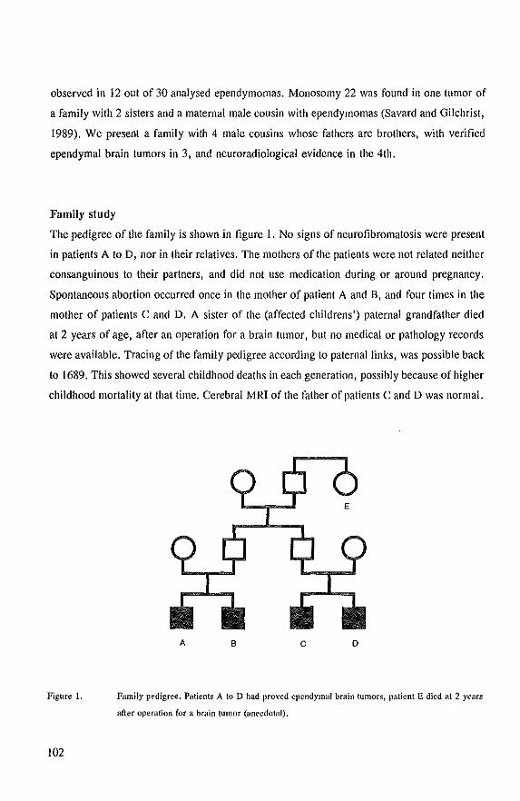
observed in 12 out of 30 analysed ependymomas. Monosomy 22 was found in one tumor of
a family with 2 sisters and a maternal male cousin with ependymomas (Savard and Gilchrist,
1989). We present a family with 4 male cousins whose fathers are brothers, with verified
ependymal brain tumors in 3, and neuroradiological evidence in the 4th.
Family study
The pedigree of the family is shown in figure 1. No signs of neurofibromatosis were present
in patients A to D, nor in their relatives. The mothers of the patients were not related neither
con sanguinous to their partners, and did not use medication during or around pregnancy.
Spontaneous abortion occurred once in the mother of patient A and B, and four times in the
mother of patients C and D. A sister of the (affected childrens') paternal grandfather died
at 2 years of age, after an operation for a brain tumor, but no medical or pathology records
were available. Tracing of the family pedigree according to paternal links, was possible back
to 1689. This showed several childhood deaths in each generation, possibly because of higher
childhood mortality at that time. Cerebral MRI of the father of patients C and D was normal.
Figure 1.
102
A B c o
Family pedigree. Patients A to D had proved ellendymal brain tumors, patient E died at 2 years
after operation fOf a brain tumor (anecdotal).
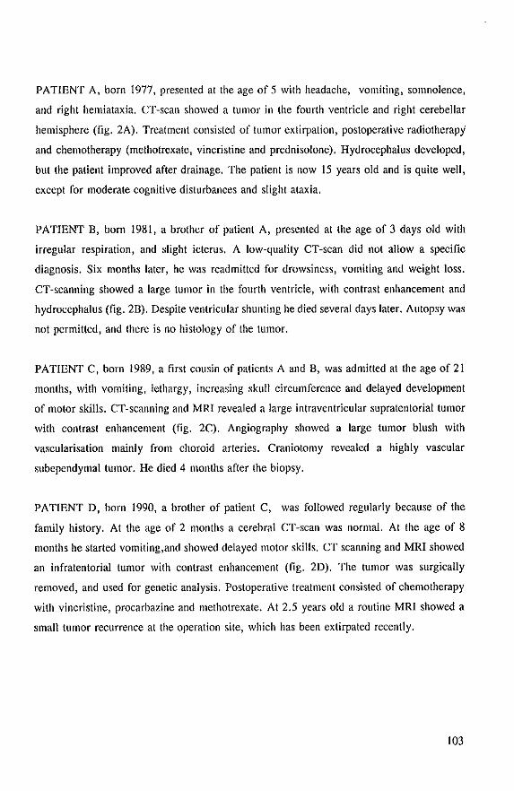
PATIENT A, born 1977, presented at the age of 5 with headache, vomiting, somnolence,
and right hemiataxia. CT-scan showed a tumor in the fourth ventricle and right cerebellar
hemisphere (fig. 2A). Treatment consisted of tumor extirpation, postoperative radiotherapy
and chemotherapy (methotrexate, vincristine and prednisolone). Hydrocephalus developed,
but the patient improved after drainage. The patient is now 15 years old and is quite well,
except for moderate cognitive disturbances and slight ataxia.
PATIENT D, born 1981, a brother of patient A, presented at the age of 3 days old with
irregular respiration, and slight icterus. A low-quality CT-scan did not allow a specific
diagnosis. Six months later, he was readmitted for drowsiness, vomiting and weight loss.
CT -scanning showed a large tumor in the fourth ventricle, with contrast enhancement and
hydrocephalus (fig. 2D). Despite ventricular shunting he died several days later. Autopsy was
not permitted, and there is no histology of the tumor.
PATIENT C, born 1989, a first cousin of patients A and D, was admitted at the age of 21
months, with vomiting, lethargy, increasing skull circumference and delayed development
of motor skills. CT-scanning and MRI revealed a large intraventricular supratentorial tumor
with contrast enhancement (fig. 2C). Angiography showed a large tumor blush with
vascularisation mainly from choroid arteries. Craniotomy revealed a highly vascular
subependymal tumor. He died 4 months after the biopsy.
PATIENT D, born 1990, a brother of patient C, was followed regularly because of the
family history. At the age of 2 months a cerebral CT-scan was normal. At the age of 8
months he started vomiting,and showed delayed motor skills. CT scanning and MRI showed
an infratentorial tumor with contrast enhancement (fig. 2D). The tumor was surgically
removed, and used for genetic analysis. Postoperative treatment consisted of chemotherapy
with vincristine, procarbazine and methotrexate. At 2.5 years old a routine MRI showed a
small tumor recurrence at the operation site, which has been extirpated recently.
103
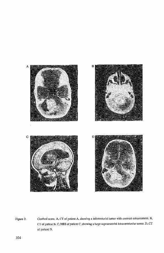
A
c
Figure 2.
104
B
Cerebral scans. A. CT of patient A. showing a infratentorial tumor with contrast enhancement. D.
CT of patient n. C. MRI of patient C. showing a large supratentorial intraventricular tumor. D, CT
of patient D.
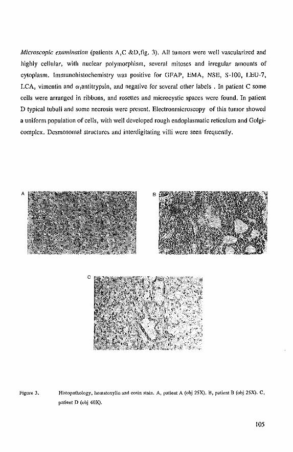
Microscopic examination (patients A,C &D,fig. 3). All tumors were well vascularized and
highly cellular, with nuclear polymorphism, several mitoses and irregular amounts of
cytoplasm. Immunohistochemistry was positive for GFAP, EMA, NSE, S-100, LEU-?,
LCA, vimentin and 0i1antitrypsin, and negative for several other labels. In patient C some
cells were arranged in ribbons, and rosettes and microcystic spaces were found. In patient
D typical tubuli and some necrosis were present. Electronmicroscopy of this tumor showed
a uniform population of cells, with well developed rough endoplasmatic reticulum and Golgi
complex. Desmosomal structures and interdigitating villi were seen frequently.
A
Figure 3,
B
c
Histopathology, hematoxylin and eosin stain. A, patient A (obj 25X), B, patient B (obj 25X). C,
patient D (obj 40X).
105
Genetic analysis
Fresh tumor tissue obtained from patient D at neurosurgery was used for DNA extraction
and interphase cytogenetic analysis. DNA extraction, Southern blotting and hybridization
were performed according 10 standard procedures. The following polymorphic DNA markers
for loci on chromosome 22 were used: D22S181, D22S183, D22SIO, D22S182, D22SI,
D22S 193, cos 50BII. The probes are ordered from centromere to telomere (van Biezen et
aI., 1993).
INTERPHASE CYTOGENETICS. Nuclei isolated from freshly removed tumor cells, were
investigated for aberrations of chromosomes #1, 6, 7, 10, 11, 17, 18, 22, X and Y by
fluorescent in situ hybridisation (FISH), according to Arnoldus et al (1990). In addition, we
used probes specific for the centromeric regions of chromosome 22 (p22!1 :2.1), and two
cosmid probes (cos 58BI2 & cos 50BII) that are located close to D22S193 on band 22qll.
One hundred cells were counted for each probe. Cultured lymphocytes from blood of an
unrelated subject were used as control.
Results
Cytogenetic analysis of respectively lymphocytes and fibroblasts in patients C and D showed
a normal male karyotype. The tumor of patient D was investigated in more detail. Interphase
cytogenetic analysis performed on freshly removed tumor tissue with probes for chromosome
#1,6,7,10,11, IS, 17, 18,X and Y revealed results in the normal range. This indicates a diploid
tumor, without gross chromosomal aberrations of these chromosomes.
Additional techniques were applied to investigate chromosome 22. Interphase
cytogenetics on freshly removed tumor tissue showed only one tluorescent spot in about 76%
of nuclei, using 3 probes for chromosome 22 (table I). Moreover, 7 polymorphic probes for
chromosome 22 were used to study possible loss of heterozygosity (LOH) in tumor DNA.
D22S193 was the only informative probe. This probe clearly showed LOH when DNA
derived from freshly removed tumor tissue was compared with DNA from fibroblasts of the
same patient. In addition, we looked at mutations in the recently cloned NF2 gene, using
RNA SSCP analysis. No evidence for mutations was observed in RNA from this tumor.
However, the same method showed mutations in 36% of 53 meningiomas and schwannomas
106
(Lekanne Deprez et aI., 1994).
Table 1. Interphase cytogenetic analysis of tumor of patient D
Number of spots per nucleus
Tissue Probes 0 2
fresh tumor p221l :2.1 6 73 21
cos 58BI2 8 76 16
cos 50BII 6 79 15
blood control* p221l :2.1 7 IO 83
cos 58BI2 8 13 79
*Cultured lymphocytes from blood of an unrelated subject.
Discussion
The histology and immunohistochemistry, intraventricular location, age of onset, and
identical neuroradiological patterns indicate that the tumors in all four patients are similar,
and of ependymal origin. While they are not typical ependymomas, they lack the major
criteria for ependymoblastomas. Considering age of onset, location, morphology and
immunohistochemistry, we designate these tumors as anaplastic ependymomas. Familial
ependymal tumors are very rare, and to our knowledge 9 families have been reported. These
include three families with 2 or more ependymal tumors (Tijssen et aI., 1982; Savard and
Gilchrist, 1989), and 5 ependymal tumors associated with glioma, plexus papilloma and
medulloblastoma (Tijssen et aI., 1982; Sato et aI., 1984). While coincidence may account for
part of these concurrences, the occurrence of 4 affected sons in a single generation, born
from 2 healthy brothers, strongly suggests the involvement of a genetic predisposing
mutation.
DNA and interphase cytogenetics of the tumor of patient D showed loss of (part) of
107
chromosome 22 in a major percentage of tumor celis, indicating the involvement of a tumor
suppressor gene. Although loss of chromosome 22 in tumor tissue might be a secondary
effect of tumorigenesis, the consistent observation of loss of chromosome 22 in our and other
reported cases, suggest a causal relationship. Because ependymomas are sometimes found in
neurofibromatosis type 2, we searched for mutations in the NF2 gene, in RNA from the
tumor of patient D. No mutation was detected, thus we have no evidence that the NF2 gene
is involved in this family. Howeyer, there are indications for additional loci on chromosome
22 involved in the development of NF2 related tumors.
The likely transmission of the disorder by 2 healthy brothers, their exclusively male
offspring, the occurrence of spontaneous abortions and the history of a brain tumor in a
paternal aunt, raise questions on the possible mode of inherilance. It cannot be explained by
simple classic mendelian genetics, and variation in expression of an autosomal dominant gene
is a proposed explanation, but lacks precision. Whether sex-related effects of the transmitting
parent (imprinting), or a gene with a variable insert size may be involved remains open to
speculation, and awaits the isolation and characterization of the gene involved.
Rcfel'ences
Arnoldus EPJ, Noordermeer lA, Peters AC, Voo,molen JHC, Bots GTAM, Raap AK, van der Ploeg M (1990) Interphase cytogenetics on brain tumors. Genes Chrom Cancer 3: 101-107.
van Biezen NA, Lekanne Deprez RH, Thijs A, Heutink p, Ooslra BA, Geurts van Kessel AHM, ZwarthoffEC (1993) Isolation and characterisation of25 unique DNA markers for human chromosome 22. Genomics 15:206-208.
Lekanne Deprez RH, Bianchi AB, Groen NA, Seizinger BR, Hagemeijer A, van Dronen E, Bootsma D, Koper JW, Avemat CJJ, Kley N, Zwarthoff EC (1994) Frequent NF2 gene transcript mutations in sporadic meningiomas and vestibular scbwannomas. Am J Hum Genet 54:1022-1029.
Ranson DT, Ritlaml SR, Kimmel DW, Moertel CA, Dabl RJ, Scheithauer BW, Kelly PI Jenkins RB (1992) Cytogenetic and loss of heterozygosity studies in ependymomas, pylocytic astrocytomas and oligodendrogliomas. Genes Chrom Cancer 5:348-356.
Salo T, Sbimoda A, Taskahashi T, Kurokawa H, Ando M, Goto S, Takamura H (1984) Congenital anaplastic ependymoma: a case report of familial glioma, Childs Brain 11:342-348.
Savard ML, Gilchrist DM (l989)Ependymomas in two sisters and a maternal male cousin with mosaicism with monosomy 22 in tumour. Pediatr Neurosci 15:80-84.
Tijssen CC, Halprin MR, Endtz U, Familial brain tumours. Marlinus Nijhoff Publishers 1982.
108

A t(4;22) ill a meningioma points to
the localization of a Plltative tllmor
sllppressor gene
Am. J. HUIIl. Genet. 48:783-790, 1991
Chapter V
109

110
Am. J. HI/m. Genet. 48:783-790, 1991
A t(4j22) in a Meningioma Points to the Localization of a Putative Tumor-suppressor Gene
Ronald H. Lekanne Deprez,· Nicole A. Groen,· Nick A. van Biezen,· Anne Hagemeijer, t Ellen van Drunen, t Jan W. Koper,t Cees J. J. Avezaat,§ Dirk Bootsma, t and Ellen C. Zwarthoff'
Departments of 'Pathology, teell B!ology and Genetics. llnternal Medicine. and §Neurosurgery. Erasmus University. Rotterdam
Summary
Cytogenetic analysis of meningioma cells from one particular patient (MN32) displayed the stem-line karyotype 45, XY, - 1, 4p+, 22q -, 22q+, which thus had rearrangements of both chromosomes 22. The 22q+ marker appeared as a dicentric: 22 pterql1::lpl1--+qter. The reciprocal product of this translocation has presumably been lost because it lacked a centromere. The 22q - chromosome also appeared to have lost sequences distal to band ql1. We assumed that this marker could have been the result of a reciprocal translocation between chromosomes 4 and 22. To investigate the 4p+ and 22q- chromosomes in more detail, human-hamster somatic cell hybrids were constructed that segregated the 22q- and 4p+ chromosomes. Southern blot analysis with DNA from these hybrids showed that sequences from 22q were indeed translocated to 4p+ and that reciprocally sequences from 4p were translocated to 22q-, demonstrating a balanced t{4;22)(pI6;ql1). On the basis of these results we presume that in this tumor a tumor-suppressor gene is deleted in the case of the 22q+ marker and that the t{4;22) disrupts the second allele of this gene. The latter translocation was mapped between 02251 and 022515, a distance of 1 eM on the linkage map of this chromosome. The area in which we have located the translocation is within the region where the gene predisposing to neurofibromatosis 2 has been mapped.
Introduction
The involvement of a tumor-suppressor gene in the etiology or pathology of a particular type of cancer is often indicated by loss of a specific chromosome or part of a chromosome (Cavenee et al. 1986). Until now five tumor-suppressor genes have been isolated; these include the retinoblastoma gene (Weinberg 1989), the p53 gene (Eliyahu et al. 1989; Finlay et al. 1989), a gene called "DeC" which is involved in the pathology of colorectal carcinoma (Fearon et al. 1990), a gene involved in Wilms tumor associated with the WAGR syndrome {Call et al. 1990; Gessler et al. 1990; Rose et al. 1990}, and the gene which is
Received Sept~mb~r 21, 1990; revision receivet:! December 18, 1990.
Address for correspondence and reprints: Dr. Ellen C. Zwarthoff, Department of Patholog)', Erasmus University, P.O. Box 1738,3000 DR Rotterdam, The Netherlands.
© 1991 by TheAmer;can Soon}, of HU[Jun GenetiCS, AU right; rtSe"cJ. 0002-9297/9114804-0018502.00
involved in the predisposition to neurofibromatosis 1 (NFl; Cawthon et al. 1990; Viskochil et al. 1990; Wallace et al. 1990).
Meningioma is a common benign mesenchymal intracranial or intraspinal tumor arising from arachnoidal cells surrounding the brain and spinal cord. Cytogenetic analysis and RFLP analysis of these tumors reveal a specific chromosomal aberration. In about 50% of the tumors one copy of chromosome 22 is lost (Zang 1982). In a few percent of the cases other aberrations of chromosome 22, such as translocations (breakpoints are mapped in region 22ql1-22q12) or deletions of the larger part of the long arm, are found {Katsuyama et al. 1986; Al Saadi et al. 1987; Casalone etal.1987;Dumanskietal. 1987;Meeseetal. 1987; Seizinger et al. 1987a; Maltby et al. 1988; Rey et:I1. 1988; Casartelli et al. 1989; Poulsglrd et al. 1989}. These findings suggest that inactivation of both alleles of a tumor-suppressor gene on chromosome 22 is a causal event in the development of meningioma.
The autosomal dominant disorder neurofibro-
1I1
matosis 2 (NF2) predisposes to acoustic neurinomas, meningiomas, and other tumors of the central nervous system, Loss of sequences on chromosome 22 has also been observed in tumors associated with NF2 (Seizinger et al. 1986; Seizinger et al. 1987b; Rouleau et a!. 1990). The gene predisposing to NF2 has been located between 022S1 and 022S28 by using linkage analysis (Rouleau et a!. 1990), At present it is not dear whether the gene predisposing to NF2 is the same as the gene involved in sporadic meningioma.
In the course of our search for the exact position of the meningioma locus on chromosome 22, we analyzed tumor cells from a patient in whom both copies of chromosome 22 were involved in translocations. One of the marker chromosomes was the result of a reciprocal translocation between chromosomes 4 and 22. We presume that this translocation has inactivated a tumor-suppressor gene. Using hybrid cell lines we mapped the translocation breakpoint to a position between 022S1 and 022S15.
Material and Methods
Patient
The patient, born in 1925, had a history of head injuries: in 1931 he had a car accident resulting in brain damage, and in 1947 he had a concussion of the brain, after which his sense of smell was impaired. In 1968 an olfactorial meningioma was removed surgically. In 1989 a second meningioma (MN32), located on the right temporal side of the brain, was removed. This tumor showed a combination of three histological types: synC}'tial, transitional, and fibroblastic. These are characteristic of all meningiomas. The localization of the tumor was completely outside the previous operation position, and it was not considered to be a local recurrence.
Tissue Culture, Hybrid Cell Lines, and Cytogenetics
The meningioma specimen was obtained within 30 min after surgery. Both preparation for tissue culture and tissue culture conditions were as described elsewhere (Koper et al. t 990), Chromosome analysis was carried out on cultured cells after one passage using G- (trypsin-Giemsa), R- (acridine orange}, and Q- (quinacrine) banding,
The somatic cell hybrids were constructed HC
cording to a method described elsewhere (Geurts van Kessel et al. 1981). Interspecies hybrid cell lines were obtained by fusion of thymidine kinase-deficient (TK -) A3 Chinese hamster cells with cultured menin-
112
Lekanne Deprez et al.
gioma cells. Inactivated Sendai virus was used as the fu·sogen. Independent hybrid clones were selected in FlO medium supplemented with HAT (hypoxanthine, aminopterin, and thymidine), 2 11M Ouabain, and 15% FCS (Biological Industries). The hybrid cell Jines were analyzed by R-bandingj at least 10 meta phases I hybrid cell line were studied. This analysis was repeated several times during culture of the hybrid cell lines. Possible rearrangements bet\\'een hamster and human chromosomes were verified using chromosome painting. This was done by biotinylation of human DNA and by in situ hybridization on metaphase spreads of the hybrid cell lines. The eventual presence of the marker 22q + , which resembles hamster chromosome 6, was also analyzed b)' using this technique.
DNA Extraction, Southern Blotting, HybridiZation, and Densitometric Analysis
High-molecular-weight ONA was isolated from cultured tumor cells (first passage) and from hybrid cell lines according to standard procedures. DNA samples were digested to completion with restriction enzymes (Boehringer Mannheim and Promega) and were separated by'electrophoresis, transferred to nylon membranes (Hybond N+; Amersham), and hybridized to radiolabeled probes according to a method described by Feinberg and Vogelstein (1983). Conditions for hybridization, are slightly modified from those described by Amasino et al. (1986). Blots were hybridized at 65°C in 0.25 M sodium phosphate pH 7.2, 0.25 M NaCl, 7% SOS (biochemical 44244; British Drug Houses), 1 mM EDTA, and 10% polyethylene glycol (AI, 6 x 101, biochemical 44271 ; British Orug Houses). After hybridization, blots were washed once in 3 X SSC, 0.1°/" SOS for 20 min at 65"C; once in 1 x sse, 0.1 % SOS for 20 min at 65°Cj and once for 20 min at 65°e in 0.3 X sse, 0.5% SOS. Membranes were exposed to Kodak XAR-5 film at -70°C with an intensifying screen, The following loci were examined: c-abl (0.6·kb EcoRIIBamHI fragment; Heisterkampet al. 1983), ber (a third-exon PstI c-ONA probe of 393 bp; Heisterkamp et a1. 1985), 022S1 (Barker et a!. 1984; Julier et al. 1988), 022S15 (Rouleau et al. 1988), myoglobin (MB, a third-exon PCR probe; Weller et a1. 1984), 022S201 (this probe has been localized on 22q13-qterj N, A. van Eiezen, unpublished data), 04S62 (Thayer et al. 1987), F5.53 (Bakkeretal. 1987),and04S125(Nakamuraetal. 1988). The sequence of these probes on the long arm of chromosome 22 is centromere-bcr-022S1-D22S15-MBc-sis-telomere. The location of these probes on the
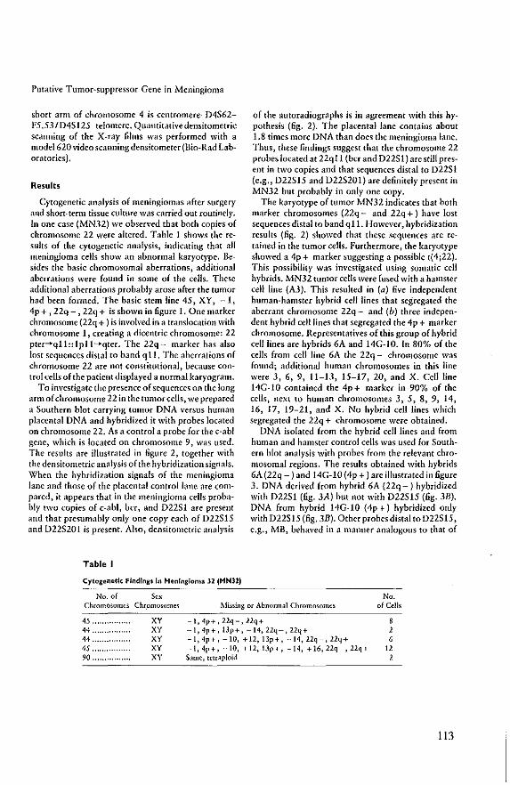
Putative Tumor-suppressor Gene in Meningioma
short arm of chromosome 4 is centromere-D4562-F5, 53/D45t25-telomere, Quantitative densitometric scanning of the X-ray films was performed with a model 620 video scanning densitometer (Bio-Rad Laboratories),
Results
Cytogenetic analysis of meningiomas after surgery and short-tenn tissue culture was carried out routinely, In one case (MN32) we observed that both copies of chromosome 22 were altered, Table 1 shows the results of the cytogenetic analysis, indicating that all meningioma cells show an abnormal karyotype, Hesides the basic chromosomal aberrations, additional aberrations were found in some of the celis, These additional aberrations probably arose after the tumor had been formed, The basic stem line 45, XY, -1, 4p + , 22q - , 22q + is shown in figure 1, One marker chromosome (22q + ) is involved in a translocation with chromosome 1, creating a dicentric chromosome: 22 pter-+qll ::1pl1-+qter. The 22q - marker has also lost sequences distal to band q 11, The aberrations of chromosome 22 are not constitutional, because control cells of the patient displayed a normal karyogram.
To investigate the presence of sequences on the long arm of chromosome 22 in the tumor cells. we prepared a 50uthern blot carrying tumor DNA versus human placental ONA and hybridized it with probes located on chromosome 22, As a control a probe for the c-abl gene, which is located on chromosome 9, was used, The results are illustrated in figure 2, together with the densitometric analysis of the hybridization signals, When the hybridization signals of the meningioma lane and those of the placental control lane are com· pared, it appears that in the meningioma cells probably two copies of c-abl, bcr, and D2251 are present and that presumably only one copy each of D22515 and D225201 is present. Also, densitometric analysis
Table I
Cytogenetic Findings In Henlngiom" 32 (MN32)
No. of Sex
of the autoradiographs is in agreement with this hypothesis (fig, 2). The placental lane contains about 1.8 times more ONA than does the meningioma lane, Thus, these findings suggest that the chromosome 22 probes located at 22qll (bcr and 02251) are still present in two copies and that sequences distal to 02251 (e,g" 022515 and D225201) are definitely present in MN32 but probably in only one copy,
The karyotype of tumor MN32 indicates that both marker chromosomes (22q - and 22q + ) have lost sequences distal to band q 11, However, hybridization results (fig. 2) showed that these sequences are retained in the tumor cells, Furthermore, the karyotype showed a 4p+ marker suggesting a possible t(4;22), This possibility was investigated using somatic cell hybrids, MN32 tumor cells were fused with a hamster cell line (A3), This resulted in (a) five independent human-hamster hybrid cell lines that segregated the aberrant chromosome 22q - and (b) three independent hybrid cell lines that segregated the 4p + marker chromosome. Representatives of this group of hybrid cell lines are hybrids 6A and 14G-1O. In 80%, of the cells from cell line 6A the 22q - chromosome was found; additional human chromosomes in this line were 3, 6, 9, It-B, 15-17,20, and X. Cell line 14G-1O contained the 4p+ marker in 90% of the cells, next to human chromosomes 3, 5, 8, 9, 14, 16, 17, 19-21, and X, No hybrid cell lines which segregated the 22q + chromosome were obtained,
DNA isolated from the hybrid cell lines and from human and hamster control cells was used for 50uthern blot analysis with probes from the relevant chromosomal regions, The results obtained with hybrids 6A (22q - ) and 14G-IO (4p + ) are illustrated in figure 3. DNA derived from hybrid 6A (22q -) hybridized with 02251 (.6g, 3A) but not with D22515 (fig, 3B), DNA from hybrid 14G-I0 (4p+) hybridized only with 022515 (fig, 3B), Other probes distal to D22515, e,g" MB, behaved in a manner analogous to that of
Chromosomes Chromosomes Mis5ing or Abnormal Chromosomes No.
of Cells
45,,,,,,,,
44 "" .. 44 " .... . 45 .. .. 90 ....... "
Xy XV XY XV XV
-i,4p+,22q-,22q+ -1,4p+, IJp+, -14, 22q-, 22q+ -I, 4p+, -10, +12, 13p+, -14, 22q-, 22q+ -1,4p+, -10, +12,13p+, -14, +16,22q-,22q+ Same, tetraploid
, 2 6
12 2
113
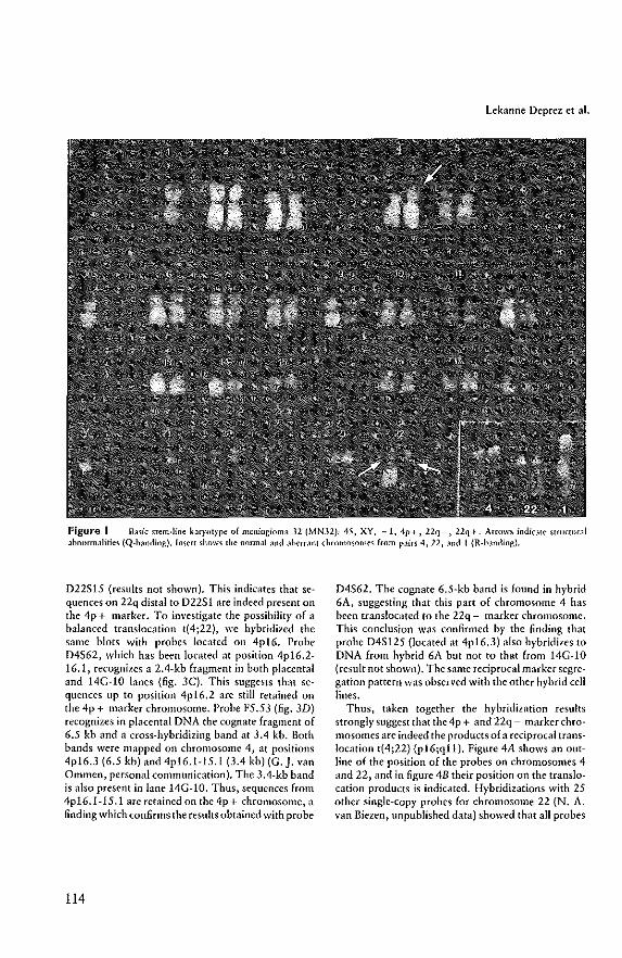
Lekanne Deprez et a!.
Figure I Basic stem·line brytlt)'pe of meningioma 32 (MN32): 45, XY, -I, 4p+, 22q-, 22q+. Arrows indicJte structurJI .1bnormalities (Q-b.mding). Insert shows the normal Jnd JberrJnt ,hromosomes from p.lirs 4, 22, Jnd I (R-bJnding).
022515 (results not shown). This indicates that sequences on 22q distal to 02251 are indeed present on the 4p + marker. To investigate the possibility of a balanced translocation t(4;22), we hybridized the same blots with probes located on 4p16. Probe D4562, which has been located at position 4p16.2-16.1, recognizes a 2.4-kb fragment in both placental and 14G-1O lanes (fig. 3C). This suggests that sequences up to position 4p16.2 are still retained on the 4p + marker chromosome. Probe F5.53 (fig. 3D) recognizes in placental DNA the cognate fragment of 6.5 kb and a cross-hybridizing band at 3.4 kb. Both bands were mapped on chromosome 4, at positions 4p16.3 (6.5 kb) and 4pI6.1-1S.1 (3.4 kb) (G. J. van Ommen, personal communication). The 3.4-kb band is also present in lane 14G-1O. Thus, sequences from 4p16.1-15.1 are retained on the 4p + chromosome, a finding which confirms the results obtained with probe
114
D4562. The cognate 6.5-kh band is found in hybrid 6A, suggesting that this part of chromosome 4 has been translocated to the 22q - marker chromosome. This conclusion was confirmed by the finding that probe 045125 (located at 4p16.3) also hybridizes to DNA from hybrid 6A but not to that from 14G-I0 (result not shown). The same reciprocal marker segregation pattern was observed with the other hybrid cell lines.
Thus, t,lken together the hybridization results strongly suggest that the4p + and 22q - markerchromosomes are indeed the products of a reciprocal translocation t(4;22) (p16;qU). Figure 4A shows an outline of the position of the probes on chromosomes 4 and 22, and in figure 4B their position on the translocation products is indicated. Hybridizations with 25 other single-copy probes for chromosome 22 (N. A. van Biezen, unpublished data) showed that all probes
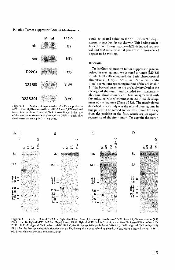
Putative Tumor-suppressor Gene in Meningioma
M pi ratio,
abl 1.67
bcr ND
D2281 1.86
D22815 3.34
D228201 3.80
Figure 2 Ancllpis of cop}' numher of different probes in 1-.INJ2.laneM, DNA isolJ[edfrom r-.INJ2.lanepl,DNA isolJ[ed from J hUIll,ln p!,J.(~ntJJ ~(mtf{}1 DNA. Also indicated is the ratio uf [he JreJ under the CI]n·e of placental Jnd MN.U sign.ll, after densitometric scanning. ND = not done.
A B
0 ~ ., , "' "' ii
OJ '" .. <'i
OJ '" ;! '" "' '" "' kb ---_., kb
14.1 _ 14.1 _
5,1 _ 5.1 4.7 - F= 4·5 - ·5-
2.8-2.8_ ..
H= 2.5-2.1-
1.7 - 2.0-1.7 _
could be located either on the 4p + or on the 22qchromosomes (results not shown). This finding underlines the conclusion that the t(4;22) is indeed reciprocal and that no substantial parts of chromosome 22 appear to be missing.
Discussion
To localize the putative tumor-suppressor gene involved in meningioma, we selected a tumor (MN32) in which all cells contained the basic chromosomal aberrations - 1, 4p + , 22q - , and 22q + , with additional aberrations appearing in some of the cells (table 1), The basic aberrations arc probably involved in the etiology of the tumor and included two structurally abnormal chromosomes 22. This is in agreement with the indic,lted role of chromosome 22 in the development of meningioma (Zang 1982), The meningioma described in our study was the second meningioma in this patient. The second tumor was found far away from the position of the first, which argues ag;linst recurrence of the first tumor. To explain the occur-
C D
0 0 ., ., " "' '" "' ii
OJ ;! _OJ :t '" "' a", "' kb kb
14.1 - 14.1 -
~' - S' -.7 = 1- 4.5 4 -2.8 -
2.8-2.5 _ 2.1
2.5-,., 20 2.1 _ 1.7-2.0-1.7 - I
Figure 3 Southern blots of DNA from (h)·hrid) cell line,. LJne pi, Hl!m,ln placental control DNA. lane AJ, Chinese hamster (A3) DNA. lane 6A, Hybrid MNJ2I A3 6A (l2:q -). bne 14G-IO, Hybrid MN321 A3 14G·IO (4p+ J. 11, Hilldm.diges[ed DNA probed with DllSI. B, E(oRI-dige>led DNA probed with D22515. C, EmRI.digested DNA probed with D4562. D, l/illdlll-dige>ted DNA probed with FS.D. Beside, the cognate hybridization signal at 6.S lb, there is ,lis!> a cross·h} briditing h,lnd (3.4 kh), which is located at 4p J 5 .1-16.1 (G. J. van Ommen, personJI communicJtion).
liS
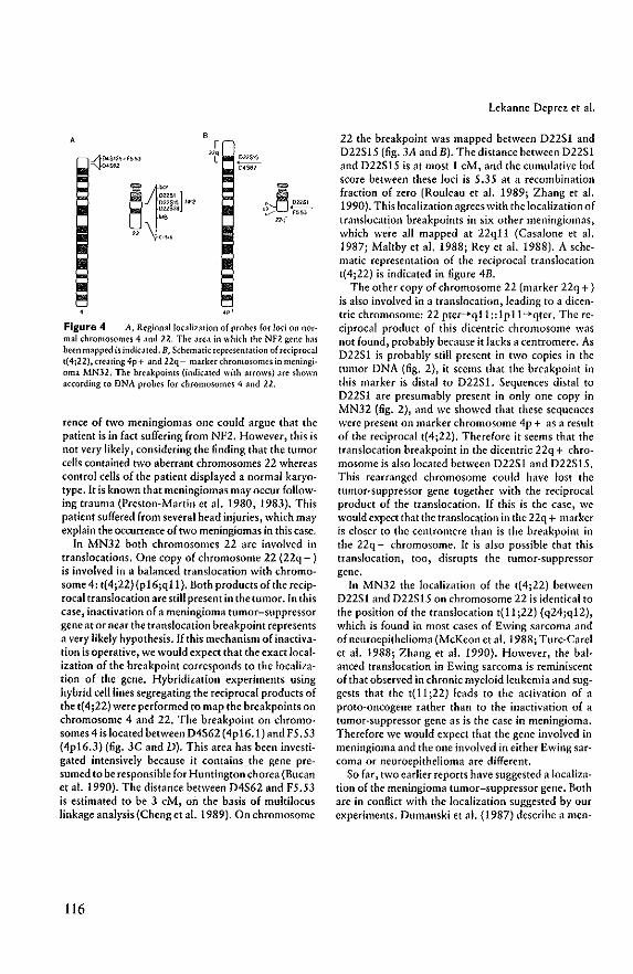
A
""'~"' S :;;~:'l '" D2:1S?S
"' n c',.,
Figure 4 A, Regional localization of probes for lod on no[mal ,hrOnlosomes 4 and 22. The area in which the NF2 gene has been mapped is indicated. B, Schematic repre-sentation of reciprocal t(4j22),crearing 4p+ and 22q- marker chromosomes inmeningi. oma MN32. The breakpoints (indicated with aHows) are shown accon:ling to DNA probes for chromosomes 4 and 22.
rence of two meningiomas one could argue that the patient is in fact suffering from NF2. However, this is not very likely, considering the finding that the tumor cells contained hVo aberrant chromosomes 22 whereas control cells of the patient displayed a normal karyotype. It is known that meningiomas may occur following trauma (Preston-Martin et al. 1980, 1983). This patient suffered from several head injuries, which may explain the occurrence of two meningiomas in this case.
In MN32 both chromosomes 22 are involved in translocations. One copy of chromosome 22 (22q - ) is involved in a balanced translocation with chromosome 4: t(4j22) (pI6jqll). Both products of the reciprocal translocation are still present in the tumor. In this case, inactivation of a meningioma tumor-suppressor gene at or near the translocation breakpoint represents a very likely hypothesis. If this mechanism of inactivation is operative, we would expect that the exact localization of the breakpoint corresponds to the localization of the gene. Hybridization experiments lIsing hybrid cell lines segregating the reciprocal products of the t(4;22) were performed to map the breakpoints on chromosome 4 and 22. The breakpoint on chromosomes 4 is located between 04S62 (4p16.1) and F5 .53 (4pI6.3) (fig. 3C and D). This area has been investigated intensively because it contains the gene presumed to be responsible for Huntington chorea (Bucan et al. 1990), The distance between 04S62 and F5.53 is estimated to be 3 eM, on the basis of multilocus linkage analysis(Cheng et al. 1989). On chromosome
116
Lekanne Deprez et al.
22 the breakpoint was mapped between D22S1 and D22S15 (fig. 3A and B). The distance between 022S1 and 022S15 is at most 1 eM, and the cumulative lod score between these loci is 5,35 at a recombination fraction of zero {Rouleau et aJ. 1989; Zhang et al. 1990}. Thislocalization agrees with the localization of translocation breakpoints in six other meningiomas, which were all mapped at 22qll (Casalone et al. 1987; Maltby et al. 1988; Rey et al. 1988). A schematic representation of the reciprocal translocation t(4;22) is indicated in figure 4B.
The other copy of chromosome 22 (marker 22q + ) is also involved in a translocation, leading to a dicentric chromosome: 22 pter--+qll::1pll--+qter. The reciprocal product of this dicentric chromosome was not found, probably because it lacks a centromere. As 022S1 is probably still present in two copies in the tumor DNA (fig. 2), it seems that the breakpoint in this marker is distal to 022S1. Sequences distal to D22$1 are presumably present in only one copy in MN32 (fig. 2), and we showed that these sequences were present on marker chromosome 4p + as a result of the reciprocal t(4;22), Therefore it seems that the translocation breakpoint in the dicentric 22q + chromosome is also located between 022S1 and 022S15. This rearranged chromosome could have lost the tumor-suppressor gene together with the reciprocal product of the translocation. If this is the case, we would expect that the translocation in the 22q + marker is closer to the centromere than is the breakpoint in the 22q - chromosome. It is also possible that this translocation, too, disrupts the tumor-suppressor gene,
In MN32 the localization of the t(4;22) between 022S1 and 022S15 on chromosome 22 is identical to the position of the translocation t(11 ;22) (q24;q12), which is found in most cases of Ewing sarcoma and of neuroepithelioma (McKeon et al. 1988; Turc-Carel et al. 1988; Zhang et al. 1990). However, the balanced translocation in Ewing sarcoma is reminiscent of that observed in chronic myeloid leukemia and suggests that the t(11;22) leads to the activation of a proto-oncogene rather than to the inactivation of a tumor-suppressor gene as is the case in meningioma. Therefore we would expect that the gene involved in meningioma and the one involved in either Ewing sarcoma or neuroepithelioma are different.
So far, two earlier reports have suggested a localization of the meningioma tumor-suppressor gene. Both are in conflict with the localization suggested by our experiments, Dumanski et al. (1987) describe a men-
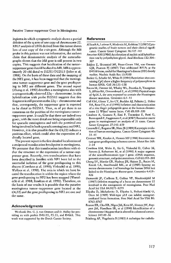
Putative Tumor-suppressor Gene in Meningioma
ingioma in which cytogenetic analysis shows a partial deletion of the q arm of one copy of chromosome 22, RFLP analysis of DNA derived from this tumor shows loss of one copy of the c-sis gene, Although the MB probe in this patient was not informative, the authors claim that densitometric analysis of the autoradiographs shows that the MB gene is still present in two copies, This suggests that localization of the tumorsuppressor gene should bedistal to MB, MB is approximately 20 eM distal to D22S1/D22S15 (julier et a!. 1988), On the basis of these data and the mapping of the NF2 gene, it has been suggested that the meningioma tumor-suppressor gene and the gene predisposing to NF2 arc different genes, The second report (Zhang et al. 1990) describes a meningioma also with a cytogenetically observed 22q - chromosome. In situ hybridization with probe D22S15 suggests that this fragment is still present on the 22q - chromosome and that, consequently, the suppressor gene is expected to be distal to D22S15, Thus, as of yet there is no consensus on the position of the meningioma tumor
·suppressor gene. It could be that there are indeed two genes, with the more distal one being responsible only for sporadic meningioma and with the proximal one being involved in both sporadic meningioma and NF2, However, it is also possible that the t{4;22) induces a position effect, which could alter the expression of a distally located gene,
The present report is the first detailed localization of a reciprocal translocation breakpoint in meningioma. We presume that this translocation interferes with either the structure or the expression of a tumor-suppressor gene. Recentl)', two translocations that have been described in families with Nfl have led to the successful isolation of the gene predisposing to this disease (Cawthon et al. 1990); Viskochil et a!. 1990; Wallace et al. 1990}. The area in which we have located the translocation is within the region where the gene predisposing to NF2 has been mapped {Werte!ecki et al. 1988; Rouleau et al. 1990}, Therefore, on the basis of our results it is possible that the putative meningioma tumor-suppressor gene located at the t{4;22) and the gene predisposing to NF2 are one and the same.
Acknowledgments
We thank Drs. G. J. van Ommen and D. Halley for providing us with probes D4S125, F5.53, and D4S62. This work was supported by the Dutch Cancer Society.
References
Al Saadi A, Latimer F, Madercie M, Robbins T (1987) Cytogenetic studies of brain tumors and their clinical significance. Cancer Genet Cytogenet 26:127-141
Amasino RM (1986) Acceleration of nucleic acid hybridization rate by polyethylene glycol. Anal Biochem 152:304-307
Bakker E, Skraastad MI, Fisser-Groen YM, van Ommen GJB, Pearson PJ (1987) Two additional RFLPs at the D4S I 0 locus, useful for Huntington's disease (HD)-family studies. Nucleic Acids Res 15:9100
Barker G, Schafer M, White R (1984) Restriction sites containing CpG show a higher frequency of polymorphism in human DNA, Cell 36:131-138
Bucan 1·f, Zimmer M, Whaley WL, PoustkaA, Youngman S, Allitto BA, Ormondroyd E, et al (1990) Physical maps of 4pI6.3, the area expected to contain the Huntington disease mutation. Genomics 6:1-15
Call KM, Glaser T, Ito CY, Buckler AJ, Pelletier J, Haber DA, Rose EA, et al (1990) Isolation and characterization of a zinc finger polypeptide gene at the human chromosome 11 Wilms' tumor locus. Cell 60:509-520
Casalone R, Granata P, Simi P, Tarantino E, Butti G, Buonaguidi F, Faggionato F, et al (1987) Recessive cancer genes in meningiomas? an analysis of 31 cases. Cancer Genet Cytogenet 27:145-159
Casartelli C, Rogatto SR, NetoJB (1989) Karyotypicevolution of human meningioma, Cancer Genet Cytogenet 40: 33-45
Cavenee WK, Koufos A, Hansen 1.fF (1986) Recessive mutant genes predisposing to human cancer. Mutat Res 168: 3-14
Cawthon RM, Weiss R, Xu G, Visko{:hil D, Culver M, Stevens J, Robertson M, et al (1990) A major segment of the neurofibromatosis type 1 gene: cDNA sequence, genomk structure, and point mutations. CeJl62:193-201
Cheng SV, Martin GR, Nadeau JH, Haines JL, Bucan M, Kozak CA, MacDonald ME, et al (1989) Synteny on mouse chromosome 5 of homologs for human DNA loci linked to the Huntington disease gene. Genomks 4:419-426
Dumanski JP, Carlbom E, Collins VP, Nordenskj61d M (1987) Deletion mapping of a locus on chromosome 22 in\'Olved in the oncogenesis of meningioma. Proc Nat! Acad Sci USA 84:9275-9279
Eliyahu D, Micha!ovitz D, Eliyahu S, Pinhasi-Kimhi 0, Oren M (l989) Wild-type p53 can inhibit oncogenemediated focus formation. Proe Nat! Acad Sd USA 86: 8763-8767
Fearon ER, Cho KR, Nigro JM, Kern SE, SimonsJW, Ruppert JM, Hamilton SR, et al (1990) Identification of a chromosome 18q gene that is altered in colorectal cancer. Science 247:49-56
Feinberg AP, Vogelstein B (1983) A technique for radiola-
117
beling DNA restriction endonuclease fragments to high specific activity. Anal Biochem 132:6-13
Finlay CA, Hinds PW, Levine AJ (1989) The p53 protooncogene can act as a suppressor of transformation. Cell 57:1083-1093
Gessler l\.1, Poustka A, Cavence W, Neve RL, Orkin SH, Bruns GAP (1990) Homozygous deletion in Wilms tumours of a zinc-finger gene identified by chromosome jumping. Nature 343:774-778
Genrts van Kessel AHM, ten Brinke H, Boere WAM, den Boer \'\IC, de Groot PG, Hagcmeijer A, t-,·Ieera Khann P, et al (1981) Characterization of the Philadelphia chromosume hy gene mapping. Cytogenet Cell Genet 30;83-91
Heisterkamp N, Groffen J, Stephenson JR (1983) The human v-abl celluLlr homolog. J Mol Appl Genet 2:57-68
Heisterkamp N, SWill K, GroffenJ, de Klein A, Grosveld G (1985) Structural organization of the her gene :lnd its role in the Ph' translocation. Nature 315:758-761
Julier C, Lathrop GM, Reghis A, Szajnert !>.l-F, Lalouel ]-M, Kaplan J-C (1988) A linkage and physical map of chromosome 22, and some applications to gene mapping. Am J Hwn Genet 42:297-308
Katsuyama J, Papenhausen PR, Herz F, Gazivoda P, Hirano A, Koss LG (1986) Chromosome abnormalities in meningiomas. Cancer Genet Cytogenet 22:63-68
Koper JW, FoekensJA, Braakman R, Lamberts SWJ (1990) Effects of progesterone on the response to epidermal growth factor and other growth factors in cultured human meningioma cells. Cancer Res 50:2604-2607
McKeon C, ThieleCJ, Ross RA, Kwan }"[, Triche Tj, Miser jS, Israel MA {I 988) Indistinguishable patterns of protooncogene expression in two distinct but closely related tumors: Ewing's sarcoma and neuroepithelioma. Cancer Rcs 48:4307-4311
Maltby EL, Ironside JW, Battersby RDE (1988) Cytogenetic studies in 50 meningiomas. Cancer Genet Cytogenet 31: 199-210
Meese E, Blin N, Zang KD (1987) Loss of heterozygosity and the origin of meningioma. Hum Genet 77:349-351
Nakamura Y, Culver M, O'Connell P, Leppert M, Lathrop GM, LalouelJM, White R (1988) Isolation and mapping of a polymorphic DNA sequence (pYNZ32) on chromosome 4p [D4S125}. Nucleic Acids Res 16:4186
Poulsgard L, R<;1nne M; Schr¢der HD (1989) Cytogenetic studies of 19 meningiomas and their clinical significance. Anticancer Res 9:109-112
Preston-Martin S, Paganini-Hill A, Henderson BE, Pike MC, Wood C (1980) Case-control study of intracranial meningiomas in woman in Los Angeles Count}', California. J Nat! Cancer Inst 65:67-73
Preston-Martin S, Yu MC, Henderson BE, RobertSC (1983) Risk factors for meningiomas in men in Los Angeles County. J Natl Cancer Inst 70:863-866
Rey )A, Bello Mj., de Gampos JM, Kpsak E, Moreno S (1988) Chromosomal involvement secondary to - 22 in human meningiomas. Cancer Genet Cytogenet 33:275-290
Rose EA, Glaser T, Jones C, Smith CL, Lewis "?H, Call
118
Lekanne Deprez et aL
KM, Minden M, et al (1990) Complete physical map of the WAGR region of llp13 localizes a candidate Wilms' tumor gene. Cell 60:495-508
Rouleau GA, HainesJL, Bazanowski A, Colella-Crowler A, Trofatter JA, Wexler NS, ConneaUy PM, et al (1989) A genetic linkage map of the long arm of human chromosome 22. Genomics 4:1-6
Rouleau GA, Kurnit DM, Ne~'e RL, Badnowsky A, P.uterson D, GuseUa jF (1988) D22DI5 a fetal brain cDNA with Banll and Sad RFLP. Nucleic Acid Res 16:1646
Rouleau GA Selzinger BR, "i/ertelecki W, Haines jL, Sn· perneau DW, Martuza RL, Gusella jF (1990) Flanking markers bracket the neurofibromatosis type 2 (NF2) gene on chromosome 22. Am J Hum Genet 46:323-328
Seizinger BR, de 1.1 Monte S, Atkins L, Gusella JF, Martuza RL (198711) },folecular genetic approach to humanmenin· gioma: loss of genes on chromosome 22. Proc Nat! Acad Sci USA 84:5419-5423
Seizinger BR, Martuza RL, Gusella JF (1986) Loss of genes on chromosome 22 in tumorigenesis' of human acoustic neuroma. Nature 322:644-647
Selzinger BR, Rouleau G, Ozilius LJ, I.ane AH, SI GeorgeHyslop P, Huson S, GusellaJF (1987b) Common pathogenetic mechanism for three tumor types in bilateral acoustic neurofibromatosis. Science 236:317-319
Tha}'er RE, Harper ME, Sawyer J, Singer MF, McBride OW (1987) Localization of a unique DNA sequence to band pI6 of human chromosome 4. Cytogenet cell Genet 45: 75-79
Ture-Carel C, Auria. A, Mugneret F, Lizard S, Sidaner I, Yolk C, Thiery JP, et al (1988) Chromosomes in Ewing's sarcoma. r. An evaluation of 85 cases and remarkable consistency of t{l1j22)(q24jq12). Cancer Genet Cytogenet 32:229-238
Viskochil D, Buchberg AM, Xu G, Cawthon R}'·f, Stevens j, WolffRK, Culver M, et al{1990) Deletions and a translocation interrupt a cloned gene at the neurofibromatosis type I locus. Cell 62:187-192
Wallace MR, Marchuk DA, Andersen LB, Letcher R, Odeh H~'(, Saulino AM, Fountain YW, et al (l990) Type 1 neurofibromatosis gene: identification of a large transcript disrupted in three NFl patients. Science 249:181-249
Weinberg RA (1990) Oncogenes, antioncogcnes, and the molecular bases of multistep carcinogenesis. Cancer Res 49:3713-3721
Weller P,jeffreys AJ, Wilson V, Blanchetot A (1984) Organization of the human myog!obin gene. EMBO J 3:439-446
Wertelecki W, Rouleau GA, Superneau DW, Forehand L W, Williams JP, Haines JL, Gusella JF (1988) Neurofibromatosis 2: clinical and linkage studies of a large kindred. N Engl J Med 319:278-283
Zang KD (1982) Cytological and cytogenetica! studies on human meningioma. Cancer Genet Cytogenet 6:249-274
Zhang FR, Delattre 0, Rouleau G, Couturier J, Lefran~ois D, Thomas G, Anrias A (1990) The neuroepithelioma breakpoint on chromosome 22 is proximal to the meningioma locus. Genomics 6:174-177
Molecular clolling of a gene
disrupted by a balanced
trallslocation in a meningioma
to be submilted
Chapter VI
119

120
Molecular Cloning of a Gene Disrupted by a Balanced
Translocation in a Meningioma
Ronald H. Lekanne Deprez', Peter H.J. Riegman*, Nicole A. Groen', Ursula L.
Warringa*, Nick A. van Biezen*, Aneo Molijn*, Dirk Bootsma#, Pieter de Jong@, Anil
Menon#, Nikolai Kley$, Bernd R. Seizinger$, and Ellen C. Zwarthoff*
Departments of *Pathology and Heel! Biology and Genetics, Erasmus University, Rotterdam; @Human Genome
Center, Lawrence Livermore National Laboratory, University of California, Livermore, California; HDepartrnent
of Molecular Genetics, University of Cincinnati Medical Center; $Oncology Drug Discovery, Department of
Molecular Genetics and Cell Biology, Bristol Myers-Squibb Pharmaceutical Research Institute, Princeton
Abstract
We have previously mapped a putative meningioma suppressor gene at the chromosome
22 breakpoint of a t(4;22)(pI6;qll), which was observed in meningioma 32. We now
report the isolation of a gene (MNl) from chromosome 22, which is disrupted by the
translocation breakpoint. The MNI gene spans about 70 kb and consists of at least two
large exons of approximately 4.7 kb and 2.8 kb. The 4.7 kb exon is disl'upted by the
t(4;22). TIlere is no obvious homology in the nucleotide and putative amino acid
sequences with other Imown genes. The gene is highly conserved in evolution and
nOl1hern blots show a ubiquitously expressed mRNA of 8 kb with an alternative 4.5 kb
transcript in skeletal muscle. In meningiomas the expression pattel'll is very variable.
Some, including meningioma 32, show no expression suggesting that the gene could
fUllction as a tumor suppressor gene fol' meningeal cells. Paradoxically, howevel', a very
high expression is sometimes also observed in meningiomas.
121
Introduction
Meningiomas are primary tumors of the arachnoidal layer of the meninges and they usually
occur as benign and slow-growing tumors, that account for 13%-19% of all intracranial and
25% of all intraspinal tumors (Russell and Rubinstein, 1989). The majority of Ihe
meningiomas are sporadic. However, they are also frequently observed in patients with the
hereditary disorder neurofibromatosis type 2 (NF2), in patients with multiple meningiomas
or (rarely) as familial meningiomas (Butti et aI., 1989; Domenicucci et aI., 1989; Russell and
Rubinslein, 1989; McDowell, 1990; Sieb el aI., 1992). This could suggest thallhere might
be a genetic predisposilion for the development of meningiomas dislincl from that for NF2.
Another indication for genetic heterogeneity is that recently linkage analysis in a
meningioma/ependymoma family excluded Ihe NF2 region (Pulst el aI., 1993). Both
cytogenetic and molecnlar genetic studies have shown that complete or partial loss of
chromosome 22 is a very frequent event in the development of meningiomas (Zang, 1982;
Seizinger et aI., 1987; Dumanski et aI., 1990) suggesling that a tumor suppressor gene(s)
located on the q-arm of chromosome 22 plays a crucial role in the pathogenesis of these
tumors. Similar results were obtained in both sporadic and NF2 associated (vestibular)
schwannomas (Seizinger et aI., 1986; Bijlsma et aI., 1992). The NF2 gene has been isolated
recently (Rouleau et aI., 1993; Trofatter et aI., 1993). Mulations in the gene, that were
observed in bolh sporadic and NF2 associated tumors, suggest that this gene is indeed
frequently involved in the tumorigenesis of meningiomas and vestibular schwannomas
(Rouleau et aI., 1993; Trofatter et aI., 1993; Bianchi et aI., 1994; Bourn et aI., 1994; Irving
et aI., 1994; Jacoby et aI., 1994; Lekanne Deprez et aI., 1994b; Pykett et aI., 1994;
RuttIedge et aI., 1994; Sainz et aI., 1994; Twist et aI., 1994). Apart from the putative
existence of a meningioma locus which was not linked to the NF2 region (pulst et al., 1993)
other evidence exists for aberrations on chromosome 22 which are not in the vicinity of the
NF2 gene. These include the telomeric region which shows LOR in some meningiomas
(Dumanski et aI., 1990; Rey et aI., 1993), a more proximal region in which we observed a
germline deletion in a patient with multiple meningiomas and a tumor associated balanced
t(4;22) (Lekanne Deprez et aI., 1991; Lekanne Deprez el aI., 1994a). In this paper we
describe the molecular analysis of the latter breakpoint and the identification and isolation
of a gene, MNI, which is disrupted by Ihe translocation breakpoint and which is presumed
122
to playa role in the development of meningiomas.
Methodology
PFGE, Southern and northern blots. Agarose plugs were prepared by imbedding 0.5xl0'
1'24 and 3x 10' hybrid (6A, 14G-IO, 9B) cells in low-melting temperature agarose (FMC).
Following deproteinization steps, DNA in the blocks was digested to completion with the
appropriate restriction enzymes. After digestion the blocks were rinsed with TE (IOmM Tris
pH 7.5, ImM EDTA) and one third was applied to a 1 % agarose gel. Electrophoresis was
carried out in 0.5xTBE for 20 hours at 12.5 'C, 160 V, 165 rnA and a switch time of 45 sec
using a Contour-clamped Homogeneous Electric Field (CHEF) system (BioRad). After
electrophoresis gels were stained with ethidium bromide and the DNA was transferred to
Hybond N+ filters (Amersham) by electroblotting in 0.5xTBE at 120 V, for 2 h in a cooled
BioRad transblot cell. DNA extraction, southern blotting, radiolabeling of probes,
hybridization and autoradiography were performed as reported (Lekanne Deprez et aI.,
1991). Northern blot analysis was carried out using northern filters containing 2 pg of poly
A + RNA from different human tissues (Human MTN, Clonetech, CAl and filters containing
glyoxal denatured total RNA isolated from primary meningioma specimens using the
guanidinium thiocyanate method (Chirgwin et aI., 1979). Total RNA (20 pg/lane) was
separated by electrophoresis on a I % agarose gel and transferred to a nylon membrane
(GeneScreen, NEN). Hybridization was in the presence of 50% formam ide at 42'C. After
washing twice with 2xSSC at 20'C, twice with 2xSSC, 0.1 % SDS for 20 min at 65 'C and
once with IxSSC at 200C the membranes were either exposed to Kodak XAR films at -70'C
using intensifying screens or the hybridization signals were evaluated using a phosphorimager
(Molecular Dynamics).
Screening for conserved sequences. Ten p.g of DNA from each species was digested with
BeoR! and electrophoresed, blotted, hybridized and washed as previously described (Lekanne
Deprez et aI., 1991). Hybridization and washing was at 600C. The final wash was performed
in 0.3xSSC, 0.1 %SDS for 20 minutes.
123
Hybrid cell lines, The somatic cell hybrids 6A, 14G-1O and 98 were derived by fusing A3
Chinese hamster cells with cultured meningioma 32 cells (MN32), harbouring the reciprocal
t(4;22)(P16;qll), Hybrids 6A segregates the 22q- derivative chromosome and hybrid 14G-IO
the 4p+ marker chromosome of this translocation as reported previously (Lekanne Deprez
et ai" 1991), Hybrid 98 harbours normal chromosome 22 with additional human
chromosomes 8,9, 13, 17,21 and Y, derived from normal cells of patient 32, PgMe25NU
is a mouse-human hybrid cell line with human chromosome 22 as its only human component
(Goyns et ai" 1984). Cells were grown in DMEM medium supplemented with 10% FCS,
penicillin and streptomycin.
Construction of a cosmid contig, D22S193 was used to start the isolation of a cosmid
contig, To generate the contig, two chromosome 22 specific cosmid libraries (LL22NCOI and
LL22NC03), gridded in microtiter plates and containing approximately four genome
equivalents of chromosome 22, were used (de long et aI., 1989; Zuckman et aI., 1992).
Isolated single copy fragments and cosmid DNA from previous screens were used for the
isolation of overlapping clones. Cos mid probes or other repeat containing probes were treated
as follows: before hybridization 100 nanograms of the cos mid DNA was digested with a
restriction enzyme, labeled by random priming using alpha "P-dATP and prehybridized for
2hr in 5xSSC at 65"C in a total volume of 500 1'1 containing 100 I'g of total sheared human
genomic DNA and 100 I'g of predigested cosmid DNA from the previous walk.
Hybridization, washing and autoradiography were as reported previously (Lekanne Deprez
et aI., 1991). The EcoR! map from this contig was made by hybridizing cos mid blots and
blots containing human genomic DNA with the isolated cosmids and single copy restriction
enzyme fragments from this region.
Screening of cDNA Iibral'ies, Two different humane fetal brain cDNA libraries were used.
About Ix 10' clones of an oligo(dT) primed Agtll cDNA library (Clonetech, HLl065) or the
same amount of an oligo(dT) and random primed AZAP II cDNA library (Stratagene,
936206) were plated on E.coli LE392 or PC2495 respectively. Filters were prepared
according to standard techniques, and the library was initially screened with isolated genomic
restriction fragments from the cos mid contig and subsequently with end fragments of the
isolated cDNAs. Fragments containing repetitive sequences were first prehybridized with
124
total sheared human genomic DNA before hybridization as described in the previous section.
Labeling, hybridization and washing conditions were as described elsewhere (Lekanne Deprez
et aI., 1991). positive plaques were purified and subcloned into pTZ plasmid or rescued as
pBluescript plasmid from the XZAP II phages.
cDNA sequencing. Sequencing was performed on both strands using double-stranded and/or
single stranded pTZ, pBluescript and Ml3 subcloned fragments. The isolated cDNAs and
subcloned fragments were sequenced using the universal and reverse primers, as well as
primers derived from the obtained sequence. The dideoxy chain termination and deaza G/ A
sequencing kits from Phannacia were used. The nucleotide sequence was compared with the
EMBL and NCBI databases to search for homologous sequences and the GRAIL (Gene
Recognition and Analysis Internet Link) program to define putative ORFs (Uberbacher and
Mural, 1991).
Results
Pulsed-field analysis of t(4;22)(p16;qU)
For the characterization of the reciprocal t(4;22)(pI6;qll) on chromosome 22 in meningioma
32 we used different probes from the 22qll region for analyses of membranes from pulsed
field gels. Figure I shows the result ofNruI digested DNA probed with D22S193. Lanes T24
and 9B contain DNA from cell lines with normal chromosome 22 and serve as controls; T24
is a human bladder carcinoma cell line and 9B a hamster/human hybrid cell line with normal
chromosome 22 from patient 32. In these lanes a fragment of approximately 120 kb is
recognized. In DNA isolated from hybrid 6A, which harbours the 22q- marker of the
reciprocal t(4;22), an aberrant 250 kb Nrul band is observed (Fig. I, lane 6A). DNA from
hybrid 14G-IO, containing the 4p+ derivative chromosome, shows no specific hybridization,
indicating that D22S193 is not located on that part of chromosome 22 that was translocated
to chromosome 4p (Fig. I, lane 14G 10). The other signals observed on this blot are probably
the result of aspecific hybridization with excess DNA in the non-resolution zone of the gel
(Fig. I, all lanes) and/or methylation differences of the Nrul sites (Fig. I , lane T24). In
addition, DNA digested with Mlu!, Not! and Sail showed altered bands in DNA from hybrid
125
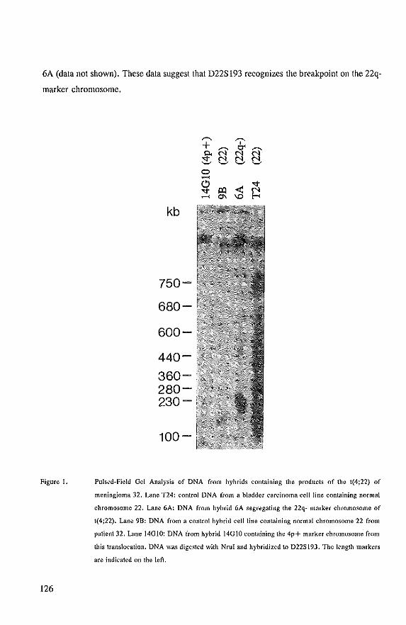
6A (data not shown), These data suggest that D22S193 recognizes the breakpoint on the 22q
marker chromosome.
Figure 1.
126
kb
750-
680-
600-
440-
360-280-230-
100-
""' + 0.
:::t o ..... (!) '<f' .......
'& N' N' N c:!- c:!- c:!-
< '<f' ~ ~ 0-. 'D
Pulsed-Field Gel Analysis of DNA from hybrids containing the products of the 1(4;22) of
meningioma 32. Lane T24: control DNA from a bladder carcinoma cell line containing normal
chromosome 22. Lane 6A: DNA from hybrid 6A segregating the 22q- marker chromosome of
1(4;22). Lane 9B: DNA from a control hybrid cell line containing normal chromosome 22 from
patient 32, Lane 14G1O: DNA from hybrid 14010 containing the 4p+ marker chromosome from
this translocation. DNA wa .. digested with NruI and hyhridized to D22SI9J. The length markers
are indicated on the let!.
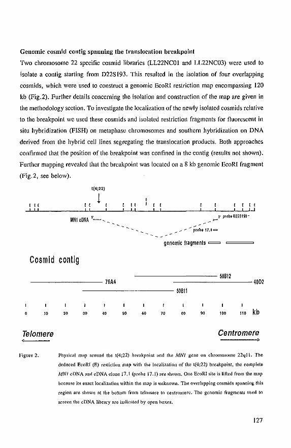
Genomic cosmid contig spanning the tl'anslocation breakpoint
Two chromosome 22 specific cosmid libraries (LL22NCOI and LL22NC03) were used to
isolate a contig starting from D22SI93. This resulted in the isolation of four overlapping
cosmids, which were used to construct a genomic EcoRI restriction map encompassing 120
kb (Fig.2). Further details concerning the isolation and construction of the map are given in
the methodology section. To investigate the localization of the newly isolated cosmids relative
to the breakpoint we used these cosmids and isolated restriction fragments for fluorescent in
situ hybridization (FISH) on metaphase chromosomes and southern hybridization on DNA
derived from the hybrid cell lines segregating the translocation products. Both approaches
confirmed that the position of the breakpoint was confined in the contig (results not shown).
Further mapping revealed that the breakpoint was located on a 8 kb genomic EcoRI fragment
(Fig.2, see below).
o
" ! ! ! ! ! !
1(4;22)
1 UNI eDNA "-, ,
Cosmid contlg
76M
" , , ! ! , !
3' probe 022$193 ~ ~
genomic fragments =
--------- 58B12 -------- 4002
---------- 50BlI
10 20 30 40 60 70 80 90 100 110 kb
Telomere Centromere
Figure 2. Physical map around the t(4;22) breakpoint and the MNI gene on chromosome 22qll. The
deduced EcoRI (E) restiction map with the localization of the t(4;22) breakpoint, the complete
MNl eDNA and cDNA clone 17,1 (probe 17.1) are shown. One EcoRI site is lifted from the map
because its exact localization within the map is unknown. The overlapping cosmids spanning this
region are shown at the bottom from telomere to centromere. The genomic fragments used to
screen the eDNA library are indicated by open hoxes.
127
Isolation and characterization of the cDNAs
To search for transcripts in the region covered by the contig, genomic fragments (Fig.2),
were used to screen a fetal brain cDNA library (Clontech, HLl065). Sequence analysis and
restriction mapping of the isolated cDNA clones proved that all clones were derived from the
same gene and one of them (cDNA 17.1) contained a poly (A) tail with a polyadenylation
signal 22 bp upstream. In total about 3 kb of the cDNA was isolated. The insert isolated
from one of these clones, cDNA probe 17.1, was used on a northern blot and recognized a
messenger of approximately 8 kb (Fig. 4). Therefore, the previous and another cDNA library
(Stratagene 936206) were screened again with probes located at the most 5' end from former
screens, Some clones derived from the second library contained composite inserts and
therefore were discarded unless carefully checked. Tn total overlapping cDNAs were isolated
which together span approximately 7.5 kb of the messenger. We do not know yet if we
isolated a full-length cDNA. We have called the gene from which these cDNAs were derived
MNI.
Restriction mapping of cDNA clones and subcloned genomic fragments from this
region showed that the isolated 7.5 kb transcript consists of two large exons of 4.7 kb at the
5' end and 2.8 kb at the 3' end (data not shown). The 5' exon-intron boundary was
sequenced and revealed a consensus splice acceptor site (data not shown), The localization
of the gene on the isolated chromosome 22 contig is depicted in figure 2 and shows that the
gene spans approximately 70 kb and is interrnpted by one 60 kb intron.
Mapping of the tmnsloeation bl'eakpoint
Fine mapping of the breakpoint was done on mapping panels with 2 genomic probes and a
cDNA probe located in the 8 kb genomic EcoRI fragment in which the breakpoint was found
(Fig. 3). EcoRI restriction patterns obtained with these probes on DNA extracted from
cultured meningioma 32 tumor cells (MN32), hybrids harbouring the 22q- (6A) or 4p+
(l4G-IO) marker chromosomes of meningioma 32 and controls (human, 9B, A3, PgMe25Nu
and mouse) are shown in figure 3. Genomic probe A, located at the tclomeric site of this 8
kb genomic fragment, detects in addition to the normal 8 kb band an aberrant band of 7 kb
in meningioma 32 DNA (Fig. 3A, lane MN32). The 8 kb band corresponds to hybridization
with the other copy of chromosome 22 present in the tumor. The aberrant band is the result
of hybridization with the breakpoint containing fragment on the 4p+ marker. Furthermore,
128
probe A is conserved in hamster DNA and detects a fragment of about 7 kb (Fig. 3A, lane
A3). Therefore, DNA from 14010 shows hybridization with two fragments of about 7 kb,
the 4p+ breakpoint fragment and the hamster homologue (Fig. 3A, lane 14010). The 7 kb
fragments observed in lanes 6A and 9B and the 20 kb fragment in PgMe25NU are also the
result of hybridization with conserved hamster and mouse sequences, respectively (Fig. 3A).
Oenomic probe B, located at the centromeric site of the 8 kb fragment, detects in
meningioma 32 DNA the normal 8 kb band and a 2.5 kb band (Fig. 3B, lane MN32). The
2.5 kb band is also found in DNA from hybrid 6A indicating that this band is produced by
the translocation breakpoint (Fig. 3B, lane 6A). These results prove that the translocation
breakpoint is located between probe A and B (Fig. 3, physical map). The localization of the
MNl gene relative to the t(4;22) in meningioma 32 was investigated with cDNA probe C,
which is part of the 5' exon and is located between the genomic probes A and B. Figure 3C
shows the EcoRI restriction fragments which hybridize with cDNA probe C. This probe
recognizes both products of the reciprocal t(4;22), as the 2.5 kb fragment is derived from
the 22q- marker (Fig. 3C, compare lanes MN32 and 6A) and the 7.0 kb fragment is the
result of the hybridization with the 4p+ marker (Fig. 3C, compare lanes MN32 and 140-
10). These results show that the 5' exon of the MNI gene is disrupted by the translocation
in meningioma 32. Figure 3C also shows weakly hybridizing fragments of approximately 6
and 7.5 kb (lanes human, MN32). This is the result of hybridization with homologous human
sequences that are not located on chromosome 22. In PgMe25NU, a hybrid ceHline with
only human chromosome 22 in a mouse background, these fragments were not observed (Fig
3C, lane PgMe25NU). cDNA probe C detects in both hamster and mouse DNA an EcoRI
fragment of 8.5 kb, suggesting that this DNA fragment is highly conserved (Fig 3C, lanes
A3, mouse). In the hybridizations shown in figure 3 it is obvious that the normal 8 kb EcoRI
band is underrepresented in tumor DNA (lane MN32). The normal copy of the MNI gene
in tumor 32 is located on a der(22)t( I ;22)(p II ;q II) with the breakpoint on chromosome 22
between cos 76A4 and the NF2 gene. The underrepresentation of this chromosome in MN32
DNA preparations was also observed when we used RFLP probes for the more proximal
region of chromosome 22. However, in in situ hybridizations with cosmids from the MNI
gene region on MN32 metaphase spreads, no deviation from the contribution of both marker
chromosomes 22 was observed (results not shown). Therefore, we must conclude that the
apparent underrepresentation of the der(22)t( I ;22) in DNA preparations is an artefact and is
129
probably caused by the less efficient release of this chromosome during cell lysis.
kb
Figure 3,
130
21
genomic; E
probe A eDNA:
t(4;22) E
= probe B
5' MN1 axon ,SS\,S\SS'''SS\\\\\''SSSSSSSSSSS'J
Telomere +---
probe C
Centromere
5,1 -
4.3 -
probe A probe n prohe C
Fine mapping of the 1(4;22) breakpoint in meningioma 32. On top the physical map of the 8 kb
genomic fragment with the localization of the 5' MNJ exon, the translocation breakpoint and the
probes (genomic probes A and H, eDNA probe C) used for the mapping are shown. Below {he
hybridizations of EcoRI digested DNA with probes A, Band C are shown. Lane Human: control
DNA from leucocytes of an unrelated individual. Lane MN32: DNA (rom cultured nleningioma
32 cells. Lanes 6A, 14G1O and 9B, see figure 1 legend for a description. Lane A3: Chinese
hamster (A3) control DNA, Lane PgMe25NU: control DNA from (mouse) hybrid PgMe25NU
containing only human chromosome 22, Lane Mouse: control mouse DNA, The sizes of the
markers are indicated on the left,
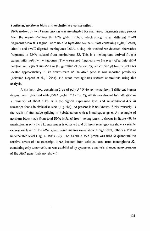
Southerll, 1l0l1hern blots and evolutional'Y conservation.
DNA isolated from 71 meningiomas was investigated for rearranged fragments using probes
from the region spanning the MNI gene. Probes, which recognize all different EcoRI
fragments from this region, were used to hybridize southern blots containing BglII, EcoRI,
HindllI and Pvull digested meningioma DNA. Using this method we detected alternative
fragments in DNA isolated from meningioma 55. This is a meningioma derived from a
patient with multiple meningiomas. The rearranged fragments are the result of an interstitial
deletion and a point mutation in the germline of patient 55, which disrupt two EcoRI sites
located approximately 10 kb downstream of the MNI gene as was reported previously
(Lekanne Deprez et aI., 1994a). No other meningiomas showed aberrations using this
analysis.
A northern blot, containing 2 I'g of poly A + RNA extracted from 8 different human
tissues, was hybridized with eDNA probe 17.1 (Fig. 2). All tissues showed hybridization of
a transcript of about 8 kb, with the highest expression level and an additional 4.5 kb
transcript found in skeletal muscle (Fig. 4A). At present it is not known if this transcript is
the result of alternative splicing or hybridization with a homologous gene. An example of
northern blots made from total RNA isolated from meningiomas is shown in figure 4B. In
meningiomas only the 8 kb messenger is observed and different meningiomas show a variable
expression level of the MNI gene. Some meningiomas show a high level, others a low or
undetectable level (Fig. 4, lanes 1-7). The fi-actin eDNA probe was used to quantitate the
relative levels of the transcript. RNA isolated from cells cultured from meningioma 32,
containing only tumor cells, as was established by cytogenetic analysis, showed no expression
of the MNI gene (data not shown).
I31
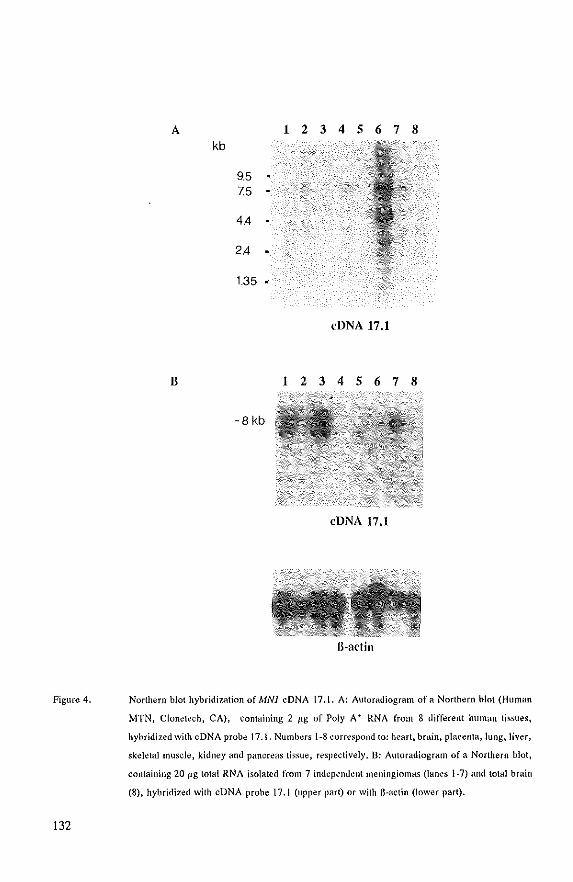
Figure 4.
132
A 1 2 3 4 5 6 7 8 kb
9.5 7.5
4.4
2.4
1.35 •
eDNA 17.1
B 1 2 3 4 5 678
-8 kb
eDNA 17.1
Northern blot hybridi7.<ition of MNl cDNA 17.1. A: Autoradiogram of a Northern blot (Human
MTN, Clonetech, CA), containing 2 Itg of Poly A + RNA from 8 dift~rent human tissues,
hybridized with cDNA probe 17.1. Numbers 1-8 correspond to: heart, brain, placenta, lung, liver,
skeletal m\lSc!e, kidney and pancreas tissue, respectively. B: Autoradiogram of a Northern blot,
containing 20 I-lg total RNA isolated from 7 independent metlingiol1la..~ (lanes 1-7) and \o\al brain
(8), hybridized with cDNA prohe 17.1 (upper part) or with ll-actin (lower part).
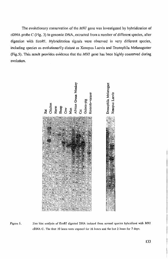
The evolutionary conservation of the MNI gene was investigated by hybridization of
cDNA probe C (Fig. 3) to genomic DNA, extracted from a number of different species, after
digestion with EcoRI. Hybridization signals were observed in very different species,
including species as evolutionarily distant as Xenopus Laevis and Drosophila Melanogaster
(Fig.S). This result provides evidence that the MNI gene has been highly conserved during
evolution.
Figure 5.
~ ~ "" ~ g,n
~ ~ .~ ~
.~ f ~ ~ t.:> :E ~ 5
g 8 ~ L~ ~ " ~
~
"" II 0 g.
& ~ M 'S E 8 ~ :ll 0
U t.:> ~ Q
Zoo blot analysis of EcoRT digested DNA isolated from several species hybridized with MN}
eDNA C. The first 10 lanes wefe exposed for 16 hours and the last 2 lanes for 7 days.
133
cDNA sequence analysis
Sequence analysis was performed in both orientations on different selected overlapping MNI
cDNA clones. Sequencing this MNI transcript was not an easy task, as the first exon (4713
nucleotides) has a high overall GC content (69%). The numerous sequence artifacts
encountered were dissolved by sequencing other independent cDNA clones, subcloned
fragments andlor by using shifted internal primers with and without the interchange of dATP
and dGTP for 7-deaza dATP and 7-deaza dGTP in the sequencing reactions. The nucleotide
sequence which we have analyzed at the moment this chapter was written did not predict a
single contiguous ORF. Therefore, we used the GRAIL program to deduce the potential
protein coding regions from the nucleotide sequence. The program predicts two putative
ORFs with a reasonable length with the quality score excellent protein coding region. The
amino acid sequences of both ORFs between the stop codons are depicted in figure 6. The
most 5' ORF (Fig. 6A) contains 6 methionine codons, of these the ones at positions 53 and
88 show the closest match with the Kozak consensus sequence for translation initiation sites
(Kozak, 1987). Upstream of this ORF 3 additional ATGs are situated, all followed by stop
codons (data not shown). The 3' ORF (Fig. 6B) contains 8 methionine codons, of these the
ones at positions 88, 275, 548 and 651 fit best with the Kozak consensus sequence.
The MNI nucleotide sequence revealed two trinucleotide (CAG) repeat sequences
located between both ORFs. These repeats were investigated for tumor associated length
variation in our series of meningioma DNAs and control DNAs lIsing radioactive peR with
primers flanking the repeats and separation on sequence gels (Verkerk et aI., 1991). The
most 5' CAG repeat was not polymorphic in any of the 114 DNA samples analyzed,
including 89 meningiomas. However, the other CAG repeat was polymorphic and detected
three alleles differing six or nine nucleotides from the smallest allele respectively. The
frequencies of the alleles, as estimated from 92 unrelated DNAs of Caucasian origin, were
5% for the smallest, 73% for the middle one and 22% for the largest fragment. In 63
meningioma DNA samples all three alleles were recognized, but no increase in length was
observed (data not shown).
134
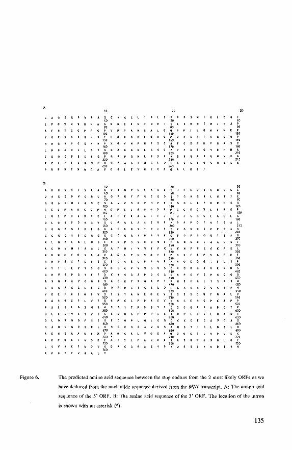
Figure 6.
A
" l ... aGAP~RAGC
" EPOV~SR~AGQGE ;0
AfH1CQP GPV~PAH
'M YGF~ARG Sf HAGG
no
Pl PPPSH
" W E G L S H ., SAlCep? LG
'" lQAQPVHG
'" HG PC PHO GG
'" MfaG?O
';0 HeG lGYGGAAGGlGSQ P P A e ,.,
'" ESCQPE GPG GHlP!) H S G A
'" ", pel lOQSPWRAA FHGlP S SSG S 0
'" '" PRR~lHQGAVO EYWY~~ GAL G
" " AOGVAESAAAVR A Q L G , , , " YKGGPVGGlAOPH " rE~EGGSTGAG .,
G l 0 Q f
" , , , " II ~ M P
'" ,. , '" GAS C
'" · . , '" , , , '" ,
" G Q G G l
" , " ;0
OQAPHlAaeSA , , , P P G 0 n.
, . , , 'M
SGlPAOCGPHD no
lQEPHRIIPGE
, , . , · , . '" GAGVGl , , , '" G G Q P G A A G ~ , , no
GGGGSSGGGGGGG
".
p p P G G , . , "
'" , ... , , 200 , ",
, , , , ,
, . . ,,,
f R G P
, , '" , '" , , "" p P SAG
'" , . . , , 0 • , , , ". , , , KPSS(O~lFGQSC , , '" ,
'" '" '" , , • • • l GAP . , , H '( '( H P EG(R(~
'" '" S Q W E GAAVAG , , , . , , , , , , , '" , '" . , , , , , , '" · , ES1SG~OG~
'" G H V S , , f 0 '( Y $ A ". , , , ,
GAAVGGSSAG
'" , , , G 0 Q P 0
'" , , , s 0 E V S , , ,,,
'" , , , 0 ,
'" VSGGGGRGRGR ,,, APOSGGAPGVS
'" . , , P H E K A l
'" lOGGA(S
'M
'" , , '" • 0 ,
'" • 0 0
'" , , '" , . ,,,
E 0 e v s S~WPQAlV ,,, , , SRSPlV1GS ,,, , , , , , , , , GYGAGEHGPKAP
'M POSYGGGGGPGHP
'" , '" ,
• • • , , · , • • • • , , . . , . A e H S , ,
,., SSSG"P
,,, POETHP ". '" , OaPlGl(GGK(G
'" , , , '" Y II P A 0
'"
". SEAV(SAH .M
(AlVOSAOOO'(T
, , , 60' , ."
CAV~AS
, , U. , . ." , no , , , . H S K E '" A~,(ASAS , , , . , n, , . , .
, , no
TOOYGOA(AR n.
I' A A l ,
, , ". . , , . , 0
The predicted amino acid sequence between the stop codons from the 2 most likely ORFs as we
have deduced from the nucleotide sequence derived from the MNl tramcript. A: The amino acid
sequence of the 5' ORF. B: The amino acid sequence of the 3' ORF. The location of the intron
is shown with an asterisk (*).
135
Discussion
Characterization of the reciprocal t(4;22)(pI6;qll) in meningioma 32 has led to the
identification of a new gene, consisting of at least two exons of about 4.7 kb (5' end) and
2.8 kb (3' end). The entire 5' exon and genomic sequences upstream (results not shown) are
extremely GC rich. This area apparently is a very large GC island. The translocation
breakpoint is located in the 5' exon, which strongly suggests that the gene is directly
involved in the tumorigenesis of meningioma 32. Therefore, we have called this gene MNl,
for candidate meningioma gene I. Many reports describe the use of reciprocal translocations
for the isolations of cancer genes. Specific chromosomal translocations are involved in the
activation of proto-oncogenes as is the case in many leukemias (Solomon et aI., 1991). In
addition, they can also inactivate a tumor suppressor gene. For instance, in neurofibromatosis
type I (NFl) two reciprocal translocations involving band 17ql1.2 resulted in the successful
isolation of the NFl tumor suppressor gene (Cawthon et aI., 1990; Viskochil et aI., 1990;
Wallage et aI., 1990). In agreement with the observed loss of chromosome 22 in
meningiomas this might suggest that the MNl gene is a tumor suppressor gene and that the
translocation in meningioma 32 inactivates the gene. Northern blot analysis of meningioma
32 RNA supports this. Southern blot analysis of71 meningiomas with probes from the MNI
region revealed a point mutation and a 1.5 kb homozygous deletion in one tumor (55). This
tumor was from a patient with multiple meningiomas. Further analysis showed that the
alterations were found also in the gennline of this patient and were located about 10 kb
downsteam of the MNl gene (I.ekanne Deprez et aI., 1994a). One might speculate that these
alterations interfere with MNl gene function. However, this gene was highly expressed in
meningioma 55. Northern analysis of the MNl gene in other meningiomas also showed high
expression in some and low or no expression at all in others. This might indicate that the
expression of the MNl gene is deregulated in meningiomas. Further study at the protein level
is however needed to investigate this in more detail.
Although the nucleotide sequence as we have obtained so far would suggest 2 possible
ORFs, in vitro transcription/translation experiments suggest that only one large protein of
approximately 3.8 kb is formed (Molyn et aI., personal communication). Therefore, we
expect that both ORFs are part of this large protein and that some sequence artifacts are still
present in the I kb that separate them. The scanning model for the initiation of translation
136
postulates that the first AUG codon in the mRNA located in the right context defines the
beginning of the protein (Kozak, 1991). Therefore, we expect that the methionine at position
53 (Fig.6B), which is the first AUG with a reasonable Kozak sequence, is the start point for
translation. Be<:ause in the upstream leader sequence 4 other AUG codons are found and this
sequence is very GC-rich (highly structured) we presume that the transcript is poorly
translated. These features are observed in many proto-oncogenes, transcription factors,
receptors, signal transduction components and proteins involved in the immune response
(Kozak, 1991). All these proteins playa critical regulatory role and one might speculate that
the unfavourable translation context of the MNI gene is a way of tightly regulating its
expression. Amino acid sequences deduced from both the predicted ORFs (Fig.6A,B) and
the nucleotide sequences of the transcript were analyzed for homology with other genes using
the EMBL and NCB! databases. No significant homology was obtained which could predict
a putative function for the gene. That the gene product must have an important function is,
however, suggested by the finding that the gene is highly conserved during evolution. An
almost complete alignment was found with two identical expressed sequence tags (gene bank
accession numbers Z28469 and HSXT02462). Furthermore, two CAG repeats, probably
located in the putative ORF, were observed. Instability (expansion) of polymorphic
trinucleotide repeats has been found to be responsible for a number of (neurological) diseases
(Warren and Nelson, 1993). Expansion of the CAG repeats in the MNI gene, however, was
not found in meningiomas. Further analysis of these repeats in patients with
neurodegenerative disorders is under way.
The neurofibromatosis type 2 gene (NF2) was recently isolated and is located about
1.7 Mbp distal to the MNI gene on the q-arm of chromosome 22 (McDermid et aI., 1993;
Rouleau et aI., 1993; Trofalter et aI., 1993). Mutation analysis of the NF2 gene transcript
in sporadic meningiomas revealed mutations, which probably inactivate the gene, in about
30% of the tumors (Rultledge et aI., 1994; Lekanne Deprez et aI., 1994b). However, in both
meningiomas (32, 55) with aberrations in or close to the MNI gene, no mutations in the NF2
gene were found, neither by using RT-PCR-SSCP (Lekanne Deprez et aI., 1994) or by SSCP
analysis of exons 1-12 on the DNA level (Zwarthoff et aI., unpublished results). This
corroborates the putative role of the MNI gene in the development of (a subset of)
meningiomas. Interestingly, both these meningiomas were from multiple meningioma
patients. Moreover, most partial chromosome 22 deletions found in meningiomas so far
137
involve loss of both the MNl and the NF2 gene. Reciprocal translocations in meningiomas
are very rare and the three reported cases are located in region 22q II (Casal one et al., 1990;
Lekanne Deprez et aI., 1991) and 22ql2 (Maltby et aI., 1988), bnt none except ours was
precisely mapped to investigate MNl or NF2 gene involvement. To further adress the
question about the extent of the contribution of the MNl gene to meningioma development
a detailed mutation study of the MNl gene is necessary. This gene might also be involved
in other tumors with known chromosome 22 deletions, such as breast cancer (Larsson et al.,
1990), colon cancer (Okamoto et aI., 1988), ependymomas (James et aI., 1990), gliomas
(Rey et aI., 1993), pheochromocytomas (Khosla et aI., 1991; Tanaka et aI., 1992) and
rhabdoid tumors (Biegel et aI., 1990; Newsham et aI., 1994).
Acknowledgements
This study was supported by a grant from the Dutch Cancer Foundation (EUR 89-06, EUR
93-467, EUR 93-653) and the Maurits and Anna de Kock Stichting. We thank Frank van der
Panne for photography.
References
AmolJus EPI, Noordermeer lA, Boudewijn Peters ACt Voonnolen JHC, Dols GTAM, Raap AK, van der Ploeg M (1991) Interphase cytogenetics of brain tumors. Genes Chrom Cancer 3: 101·107.
Bianchi AB, Ham T. Ramesh V, Gao J, Klein-Szanto AJP, Morin F, Menon AG, Trofatter JA, Gusella JF, Seizinger BR, Kley N (1994) Mutations in transcript isoforms of the neurofibromatosis 2 gene in multiple human tumour Iypes. Nature Genet 6: 185-192.
Biegel JA, Burk CD, Parmiter AH. Emanuel BS (l990) Monosomy 22 in rhabdoid or atypical tumors of the brain. J Neurosurg 73:710-714.
van Bieren NA, Lekanne Deprez RH, Thijs A, Heutink P, Oostra BA, Geurts van Kessel AHM, ZwarthoffEC (1993) Isolation and characteri:z..1tion of 25 unique DNA markers for human chromosome 22. GenomiC's 15:206-208.
Bourn D, Carter SA, Mason S, Gareth 0, Evans R, Strachan T (1994) Germline mutations in the neurofibromatosis type 2 tumour suppressor gene. Hum Mol Genet 3: 813-816.
Butli G, Assielti R, Casalone R, Paoletti P (1989) Multiple meningiomas: A clinical. surgical and cytogenetic analysis. Surg NeuroI31:255-260.
138
Bijlsma EK, Brouwer~Mladin R, Bosch DA, Westerveld A, Hulsehos TJM (1992) Molecular characterization of chromosome 22 deletions in schwannomas. Genes Chrom Cancer 5:201~205.
Casalone R, Simi P, Granata P, Minelli E, Giudici A, Butti G, Solero CL (1990) Correlation between cytogenetic and histopathological findings in 65 human meningiomas. Cancer Genet Cytogenet 45:237~243.
Cawthon RM, Weiss R, Xu G, Viskochil D, Culver M, Stevens J, Robertson M, Dunn D, Gesleland R, O'Connell P, White R (1990) A major segment of the neurofibromatosis type I gene: cDNA sequence, genomic structure and point mutations. Cell 62: 193~201.
Chirgwin 1M, Przybyla AE, MacDonald RJ, Rutter Wl (1979) Isolation of biologically active ribonucleic acid from sources enriched in ribonuclease. Biochemistry 18:5294-5299.
Domenicucci M, Santoro A, D'Osvaldo DH, Delfini R, Cantore GP, Guideui B (1989) Multiple intracranial meningiomas. J Neurosurg 70:41-44.
Dumanski lP, Rouleau GA, Nordenskjold M, Collins VP (1990) Molecular genetic analysis of chromosome 22 in 81 cases of meningioma. Cancer Res 50:5863-5867,
Goyns MH, Young BD, Geurts van Kessel A, de Klein A, Grosveld G, Bartram CR, Bootsma D (1984) Regional mapping of the human immunoglohulin lambda light chain to the philadelphia chromosome in chronic myeloid leukaemia. Leukemia Res 8:547-553.
Irving RM, Moffat DA, Hardy DO, Barton DE, Xuereb JH, Maher ER (1994) Somatic NF2 gene mutations in familial and non-familial vestibular schwannoma, Hum Mol Genet 3:347-350,
lacoby LB, MacColiin M, Louis DN, Mohney T, Rubio MP, Pulaski K, Trofalter lA, Kley N, Seizinger B, Ramesh V, Gusella 1 (1994) Exon scanning for mutation of the NF2 gene in schwannomas. Hum Mol Genet 3:413-419.
lames CD, Hel, Carlbom E, Mikkelsen T, Ridderheim P~A, Cavenee WK, Collins VP (1990) Loss of genetic information in central nervous system tumors common to children and young adults. Genes Chrom Cancer 2:94~ 102.
de long Pl, Yokobata K, Chen C, Lohman F, Pederson L, McNinch J, va Dilla M (1989) Cytogenet Cell Genet 51:985.
Khosla S, Patel VM, Hay ID, Schald DJ, Grant CS, van Heerden JA, Thibodeau SN (1991) Loss of heterozygosity suggests multiple genetic alterations in pheochromocytomas and medullary thyroid carcinomas, J Clin Invest 87:1691~1699.
Kozak M (1987) An analysis of 5'-noncoding sequences from 699 vertebrate messenger RNAs. Nuel Acids Res 15:8125-8148.
Kozak M (1991) An analysis of vertebrate mRNA sequences: intimations of translational control. 1 Cell Bioi 115:887·894.
L1rsson C, Bystrom C, Skoog L, Rotstein S, Nordenskjold (1990) Genomic alterations in human breast carcinomas, Genes Chrom Cancer 2: 191-197,
Lekanne Deprez RH, Groen NA, van Biezen NA, Hagemeijer A, van Drunen E, Koper lW, Avezaat Cll, Boolsma D, Zwarlhofr EC (199Ia) A 1(4;22) in a meningioma poinls 10 the localization of a putative (Umorsuppressor gene. Am 1 Hum Genet 48:783-790.
Lekanne Deprez RH, Groen NA, LOllz D, Hagemeijer A, van Drunen E, Koper JW, Avezaal CJJ, Bootsma
139
D, van der Kwast TH, Zwarthoff BC (1994a) Constitutional DNA-level aberrations in chromosome 22 in a patient with multiple meningiomas. Genes Chrom Cancer 9: 124-128.
Lekanne Deprez RH, Bianchi AB, Groen NA, Seizinger BR, Hagemeijer A, van Drunen E, Bootsma D, Koper JW, Avezaat CJJ, Kley N, Zwarthoff BC (1994b) Frequent NF2 gene transcript mutations in sporadic meningiomas and vestibular schwannomas. Am J Hum Genet 54:1022-1029.
Maltby EL, Ironside JW, Battersby RDE (1988) Cytogenetic studies in 50 meningiomas. Cancer Genet Cytogenet 31: 199-210.
McDemlid HE, Budarf ML, Emanuel BS (1993) Long-range restriction map of human chromosome 22qll-22q12 between the lambda immunoglobulin locus and the Ewing Sarcoma breakpoint. Genomics 18:308-318.
McDowell JR (1990) Familial meningioma. Neurology 40:312-314.
Newsham I, Daub D, Desnard-Guerin C, Cavenee W (1994) Molecular sublocalization and characterization of the 1l;22 translocation breakpoint in a malignant rhahdoid tumor. Genomics 19:433-440.
Okamoto M, Sasaki M, Sugio K, Sato C, Iwama T, Ikeuchi T, Tonomura A et al. (1988) Loss of constitutional heterozygosity in colon carcinoma from patients wilh familial polyposis coli. Nature 331:273-277.
Pulst SM, Rouleau GA, Marineau C, Fain P, Sieb JP (1993) Familial meningiomas is not allelic to neurofibromatosis 2. Neurol 43:2096-2098.
Pykett MJ, Murphy M, Hamish PR, George DL (1994) The neurofibromatosis 2 (NF2) tumor suppressor gene encodes multiple alternatively spliced transcripts. Hum Mol Genet 3:559-564.
Rey JA, Bello MJ; de Campos JM, Vaquero J, Kusak ME, Sarasa JL, Pestana A (1993) Abnornlalilies of chromosome 22 in human brain tumors determined by combined cytogenetic and molecular genetic approaches. Cancer Genet Cytogenet 66:1-10.
Rouleau GA, Merel P, Lutchman M, Sanson M, Zucman J, Marineau C, Hoang-Xuan K, Demczuk S, Desmaze C, Plougastel n, Pulst SM, Lenoir G, Bijlsma E, Fashold R, Dumanski J, de Jong P, Parry D, Eldrige R, Aurias A, Delattre J, Thomas G 1993) Alteration in a new gene encoding a putative membrane-organizing protein causes neuro-fibromatosis type 2. Nature 363:515-521.
Russell DS, Rubinstein U (1989) Tumours of the meninges ami related tissues. In Arnold E (ed): Pathology of tumours of the nervous system, 51b ed. London: Edward Arnold. pp 452-453.
Rutlledge MH, Sarrazin J, Rangaratnam S, Phelan CM, Twist E, Merel P, Delatlre 0, Thomas G, NordenskjOid M, Collins VP, Dumanski JP, Rouleau GA (1994) Evidence for the complete inactivation of the NF2 gene in the majority of sporadic meningiomas. Nature Genet 6: 180-184.
Sainz J, Huynh OP, Figueroa K, Ragge NK, Baser ME, Pulst SM (1994) Mutations of the neurofibromatosis type 2 gene and lack of the gene product in vestibular schwannomas. Hum Mol Genet 3:885-891.
Seizinger DR, Marluza RL, Gusella JF (1986) Loss of genes on chromosome 22 in tumorigenesis of human acoustic neuroma. Nature 322:644-647.
Seizinger BR, de la Monte S, Atkins L, Gusella JF, Martuza RL (1987) Molecular genetic approach to human meningioma: Loss of genes on chromosome 22. Proc Natl Acad Sci USA 84:5419-5423.
Sieb JP, Pulst SM, Buch A (1992) Familial CNS tumors. J Neurol 239:343-344.
Solomon E, Borrow J, Goddard AD (1991) Chromosome aberrations and cancer. Science 254: 1153-1160.
140
Tanaka N, Nishisho I, Yamamoto 1'.1, Miya A, Shin E, Karakawa K, Fujita S et al. (1992) Loss of heterozygosity on the long arm of chromosome 22 in pheochromocytoma. Genes Chrom Cancer 5:399-403.
TrofatterJA, MacColiin MM, RulterJL, Murrell JR, Duyan MP, Parry OM, Eldridge R, Kley N, Menon AG, Pulaski K, Haase VH, Ambrose eM, Munroe D, Bove C, Haines JL, Martuza RL, MacDonald ME, Seizinger BR, Short MP, Buckler Al, Gusella JF (1993) A novel Moesin-, Ezrin-, Radixin-Iike Gene is a candidate for the Neurofibromatosis 2 tumor suppressor. CelJ 72:791-800.
Twist EC, Ruttlooge MH, Rousseau M, Sanson M, Papi L, Merel P, Delattre 0, Thomas G, Rouleau GA (1994) The neurofibromatosis type 2 gene is inactivated in scilwannomas. Hum Mol Genet 3: 147-151.
Uberbacher EC, Mural RJ (1991) Locating protein-coding regions in human DNA sequences by a multiple sensor-neural network approach. Proc Nail Acad Sci USA 88: 11262-11265.
Verkerk AJMH, Pierelti M. Sutcliffe JS, Fu Y-H, Kunl DPA, Pizzuti A, Reiner 0, Richards S, Victorai MF, Zhang F, Eussen E, v Onm1en GB, et al. (1991) Identification of a gene (FMR-I) containing a CGG repeat coincident with a breakpoint cluster region exhibiting length varation in fragile X syndrome. Cell 65:905-914.
Viskochil D, Buchberg AM, Xu G, Cawthon RM, Stevens J, WolffRK, Culver M, Carey JC, Copeland NG, Jenkins NA, White R, O'Connell P (1990) Deletions and a translocation interrupt a cloned gene at the neurofibromatosis type 1 locus. Cell 62:187-192.
Wallage MR, Marchuk DA, Andersen LB, Letcher R, Odeh HM, Saulino AM, Fountain JW, Brereton At Nicholson J, Mitchell AL, Brownstein BH, Collins FS (1990) Type 1 Neurofibromatosis gene: Identifiction of a large transcript disrupted in three NFl patients. Science 249: 181-186.
Warren ST, Nelson DC (l993) Trinucleotide repeats in neurological disease. Curr Opin NeurobioI3:752-759.
Zang KD (1982) Cytological and cytogenetical studies on human meningioma. Cancer Genet Cytogenet 6:249· 274.
Zucman J, Delatlre 0, Desmaze C, Plougastel, Isabelle Joubert, Melot T, Peter M, de Jong P, Rouleau G, Audas A, Thomas G (1992) Cloning and characterization of the Ewing's Sarcoma and Peripheral Neuroepithelioma t(II;22) translocation breakpoints. Genes Chrom Cancer 5:271-277.
141

142
Constitutional DNA-level alterations
in chromosome 22 in a patient with
mUltiple mellingiomas
Gelles ChroJ/l, Callct'!" 9:124-/28, 1994
Chapter VII
143

144
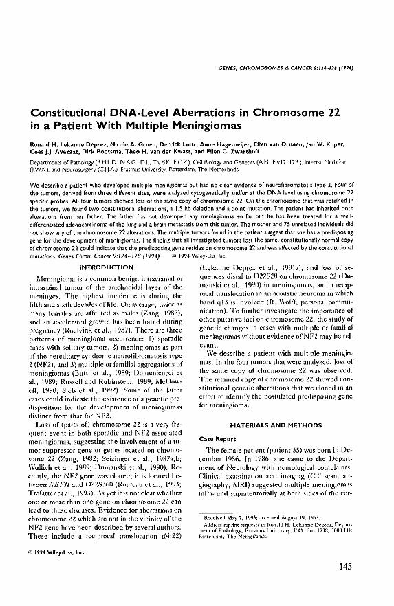
GENES, CHROMOSOMES & CANCER 9;124-128 (1994)
Constitutional DNA-level Aberrations in Chromosome 22 in a Patient With Multiple Meningiomas
Ronald H. Lekanne Deprez. Nicole A. Groen, Derrick Louz. Anne HagemeiJer, Ellen van Drunen.lan W. Koper, Cees J.J. Avezaat. Dirk Bootsma. Theo H. van der Kwast, and Ellen C. Zwarthorf
Departments of Pathology (RHLD" NAG. DL T.v.d K, E.CZ), Ceil B:o!ogy and Genetics (A H. E.v.o" D,B,), Internal t-1edcine (J,W,K), and Neurosurgery (C J J A). Erasmus University. Rotterdam. The Netherlands
We describe a patient who developed multiple meningiomas but had no clear evidence of neurofibromatosis type 2. Four of the tumors, derived from three dirferent sites, were analyzed cytogenetically andlor at the DNA level using chromosome 22 speclnc probes. AU four tumors showed loss of the same copy of chromosome 22. On the chromosome that was retained in the tumors, we found two constitutional aberrations, a 1.5 kb deletion and a point mutation. The patient had Inherited both alterations from her father. The father has not developed any menlngiom<lS so far but he has been treated for a well· differentiated adenocarclnoma of the lung and a brain metastasis (rom this tumor. The mother and 75 unrelated Individuals did not show any of the chromosome 22 alterations. The multiple tumors found in the patient suggest that she has a predisposing gene for the development of meningiomas. The finding that all investigated tumors lost the same, constitutionally normal copy of chromosome 22 could indicate that the predisposing gene resides on chromosome 22 and was affected by the constitutional mutations. Genes Chrom Cancer 9:/14-128 (1994). ;D 1994 Wlley.Liss. Inc.
INTRODUCTION (Lekanne Deprez et al., 199Ia), and loss of se·
1teningioma is a common benign intracranial or quences distal to D22S28 on chromosome 22 (Du· intraspinal tumor of the arachnoidal layer of the manski et aI., 1990) in meningiomas, and a recip· meninges. The highest incidence is during the rocal translocation in an acoustic neuroma in which fifth and sixth decades of life. On average, twice as band qJ3 is invoh'ed (R. Wolff, personal commu-many females are affected as males (Zang, 1982), nkation). To further investigate the importance of and an a('ceierated growth has been found during other putati\'c loci on chromosome 22, the study of pregnanc), (Roeh'ink et al., 11)87). There arc three genetk changes in cases with multiple or familial patterns of meningioma occurrence: I) sporadic meningiomas without evidence of NF2 may be rel-eases with solitary tumors, 2) meningiomas as part evant. of the hereditary syndrome neurofibromatosis type We describe a patient with multiple meningin+ 2 (NF2), and 3) multiple or familial aggregations of mas. In the four tumors that were analyzed, loss nf meningiomas {Butti et aI., 1989; Domenicucci et the same copy of chromosome 22 was observed. aI., 11)89; Russell and Rubinstein, 1989; 1lcDow· The retained copy nf chromosome 22 showed ('on-eil, 1990; Sieb et aI., 1992). Some of the latter stitutional genetic aberrations that we cloned in an ('ases could indicate the existence of a genetic pre- effort to identify the postulated prcdisposing gene disposition for the development of meningiomas for meningioma, distinct from that for NF2.
Lnss of (parts of) chromosome 22 is a very frequent event in both sporadic and NF2 associated meningiomas, suggesting the involvement of a (U+
mor suppressor gene or genes located on chromo· some 22 (Zang, 1982; Seizinger et aI., 1987a,b; Wullich e{ aI., 1989; Dumanski et aI., 19(0). Recently, the NF2 gene was cloned; it is located be· tween NEFf! and D22SJ60 (Ruuleau et aI., 1993; 'l'rofatteret al., 1993). As yet it is not clear whether one or more than one gene on chromosome 22 can lead to these diseases. Evidence for aberrations on chromosome ZZ which arc not in the vicinity of the NF2 gene have been described by several authors. These include a reciprocal translocation t(4;22)
© 1994 Wlley.Uss, Inc.
MATERIALS AND METHODS
Case Report
The female patient (patient 55) was born in December 1956. In 19K6, she came to the Department of Neurology with neurological complaints. Clinical examination and imaging (CT scan, an· giography, 11Rl) suggested multiple meningiomas infra- and supmtentorially at both sides of the cer-
Heccived May 7, 1993: J{(cpled AUj;;u.sI 19, 1'193. Auun;~s n;prim requesl< to RonJld II. Lckanne Deprez, Depart·
mem of Pathology, Erasmus Unil'cr;iIY, P.O. 110" 17.18, JOOO DR Rouwhm. The Ncrhcrl.l[]us.
145
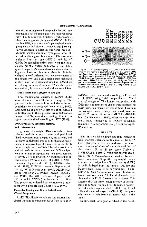
CHROMOSOME 11 ABERRATIONS
ebellopontine angle and intraspinally. In 1987, several pamsagittal meningiomas were removed surgically. The tumors were histologically diagnosed as fibrous meningiomas (designated ~[N55A). In December 1989, a reCLlrrence of a parasagittal meningioma on the left side was removed and histologically diagnosed as a fibrous meningioma (:..tN55B). Multiple small nodules of hyperplasia were observed in this region. In October 19l)O, two meningiomas from the right (:"IN55C) and the left (i\IN550) cerebellopolltine angle were excised at an inten'al of 2 weeks; both were of the fibrous type. The patient's parents have no history of meningiomas. However, her t~lther (born in 1929) developed a well-differentiated adenocarcinoma of the lung in 19H4 and 2 years latera brain metastasis of this tumor. A CT scan performed in 1990 did not reveal any intracmnial tumors. When this paper W,IS written, he was alive ilntl without complaints.
Tissue Culture and Cytogenetic Analysis
The meningioma specimens (MN55B,C,D) were ohtained within 3{) min after surgery. The preparation for tissue culture and tissue culture l'Onditions were as described (Koper et aI., 1990). Chromosome analysis WilS carried Ollt on cultured cells after one to three passages using R-(acridine omnge) and Q-(quinacrine) banding. The karyotypes were described aCl'onling to ISeN (1991).
DNA Extraction, Southern Blotting, and Hybridization
High molecular weight DNA was isolated from I:ultured and fresh tumor ti.~sue and peripheral blood leucocytes from the patient, her parents, and unrelated individuals according tu standard procedures. The percentage of tumor cells in the fresh tumor sample was est,lhlished by microscopic examination of a frozen tissue section. DNA analyses were performed as reported by Lekanne Deprez e( a!. (199b). The following DNA markers for loci on chromosome 22 were used: D2ZSIHI, D2ZS183 (Lekanne Deprez et aI., 1<)9Ib), D2ZSIO (liolker et aI., 19B5), 022S1H2 (Lekanne Deprez et aI., 19<)1 Il), DZ2S1 (Barker et ai., 19B4), D22SI<J3 (Lekanne Oeprel. et aI., 199Ib), D2ZS45 (Uudarf et aI., 1991), D22S201 (Lebtnne Deprez et al., 1<)9lc), and D2ZS20S (yan Biezen et al., 1<)<).1). The probes are ordered from centromere to telomere when possible (van Diezen et ,d., 19<).1).
Molecular Cloning and Characterization of Cloned Fragments
A XEMBL4 libnuy containing size-fractionated, EroRI digested meningioma DNA from patient SS
146
A2-A3-c-
Figure I. loss of heceroqgosity for 022545 in DNA from leucocytel and meningiomas of patient 55. CI, C2, and CJ: Control DNA! from leucocytes of three lInrellted Indtyidu~ls. PBLS5M and F: DNA (rom leucocytes of the mother (H) and the father (F) of patient 55. PBL55: DNA from leu<:ocytes o( patient 55. HNS5A·D: DNA from meningioma MN55A. B, C. and D of patient 55. f'vull digested DNA was probed with D22545. On the left are the observed fragment sizes as described by Budari et aJ. (1991). A2:. allele 2 (H kb). A3: allele 3 (2.1 kb), and C: a constant band of 1.8 kb.
(~IN55B) was constructed according to Frisehauf et al. (1983) using XEMRIA predigested EroRI ~lrms (Stratagene). The library was probed with 022S193, and four phage clones were isolated ~md their restriction maps were established. This map was compared with the map constructed from control DNA isolated from the C~IU) XE~lllL3 library (de Klein et aI., 1986). When relevant, dOllble-stranded sequencing of pUCI9 subcloned fragments was performed using a sequencing kit (Pharmacia).
RESULTS
FOUf intracranial meningiomas from patient 55 were analyzed cytogenetically andlof at the DNA level. Cytogenetic analyses performed on shortterm cultures of three of them showed loss of chromosome 22 in all the cases Crable 1; ~fN55D,C,D). Tumor ~fN55I3 also showed loss of one of the X chromosomes in most of the cells. Nine chromosome 22 specific polymorphic probes were used to analyze loss of heterozygosity (LOI I) in DNA isolated from the tumors. 022SlO and D22S45 were found to be polymorphic. The results with D22S45 arc shown in Figure I, showing loss of maternal allele A.t Identical results were ohwined with D22SIO (results not shown). This suggests that the same (paternal) l'OPy of chromosome 22 is presen'ed in aU four tumors. The presence of residual signal of the lost allele (Fig. I) and l'ells with a normal karyotype Crable I) were probably due to an admixture of normal cells in the tumor.
In our search for a gene involved in the de\'d-
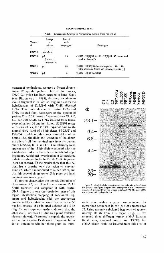
LfKANNf DfPREZ fT AL
TABLE I. CytogenetiC Findings In Meningioma Tumors from Patient 55
Passage No. of Tumor '0 cells # culture karyotyped
MN5SA Not done
MNSSB pO IS (primary outgrowth)
MNSSC pi 25
MNS5D p)
opment of meningioma, we used different chromosome 22 specific probes, One of thc probcs, D22St9J, which ha<; been mapped to band 22qtl (van Biezen et aI., 1993), detected an aberrant EmRI fragment in patient 55, Figure 2 shows the hybridization of 022S193 with RmRf digestep DNA. This probc dctccts, in control DNA and DNA isolated from leucocytes of the mother of patient 55, a 2.6 kb E(oIU fragment (lanes CI, C2, C\ and PBL55~1). In DNA isolated from leucocytes of patient 55 and her father, D22S193 recognizes two alleles, the 2,6 kh fragment and an abnormal sil.ed hand of 15 kb (lanes PBL55F and PBL55). In addition, this probe showed los'> of the normal (2,6 kb) allele and retention of the abnormal allele in all four meningiomas from the patient (lanes i\fN55A, B, C, and D). The relatively weak appeiH~mee of the IS kb allele compared with the 2.6 kb allelc is due to less eftlcient transfer of larger fragmcnts. Additional in\'estiglHion of 75 unrelated indi\'idu~lls showed only the 2.6 kb EroRI fragment (data not shown). These results show that this paticnt has a constitutional abermtion on chromosome 22, which she inherited from her father, lmd that this copy of chromosome 22 is preserved in all meningi!lmas investigated.
To further eharaeterizc the genetic alteration of chromosome 22, we doncd the abcf[;lIlt 15 kb EroRI fragment and compared it with control DNA. Figure 3 shows the rcstriction map of this rcgion. Restriction mapping of subcloned fmgmems ~lIld hybridization with the appropriate probes cstablished that one E(oRI site in p;uient 55 was lost because of an internal deletion of 1,5 kb (Fig. J), and sequell!.~e analysis showed that the other A"(QRI site was lost due to a point mutation (data not shown), These results cxptJin the appearance of the aberrant IS kb EtoRI fragment. In order to determine whether these germline muta-
Karyotype
4S,XX, - 22[ I JI44,X, - X, - 22{8]140-H, idem, with random losses [5]
4S,XX,-22[24]/89, hypotetrap!old, -22, -22, with additional losses and rearrangements II]
4S,XX, - 22[ 31/46,XX[2]
kb
23.1-
9.4-
6.6-
4.4-
2.3-2.0-
Figure 2. Analysis of the constitutional aberrations iJl patient 5S and her parenl.$- See Figure I legend for a description of the DNA slmples used. E,,,RI d'gested DNA was probed with 022S193. The sizes of the markers are indICated on the left.
tions were within a genc, wc searched for transcribed sequences in this part of chromosome 22, Using genomic subdoned fragmems of approximately 10 kb from this region (Fig, 3), we scrccned three different human eDNA libraries (fewl bmin, temporal cortex, and T47lJ). No cDNA clones could be isolatcd from this area of
147
CHROMOSOME 11 ABERRATIONS
E BP E HP E XO D B
Control: I I II II
=
0228193: -
Figure 3. Physl<a1 map from the area <ontainlng the <onstitutional alterations o( p~tient 55. Control: Ph)'Slcai map (or control DNA. The aberrations observed In patient 55 are Indicated by an open box (or the 1.5 kb deletion and an :uterisk «) (or the point mutation. Probe
chromosome 22. Also, zoo blots, containing hamster, mouse, and mt DNA, showed no clear conserved sequences, except for a weak conservation of 022S193 (data not shown).
DISCUSSION
We postulated that the multiple meningiomas probably arose as a consequence of a predisposing germline mutation distinct from the gene predisposing to NF2. LOH study with 022510 and 022545 performed in four meningiomas derived from patient 55 suggested loss of the same copy of chromusome 22. This result was further supported by the study of 0225193. The finding that the same copy of chromosome 22 was lost in all four tumors could mean that the posmlated germline mutation is on the copy of chromosome 22 which is retained in the tumor cells. In the multiple men¥ ingioma patient we investigated, the retained copy of chromosome 22 had two constitutional alter¥ ations, namely, a deletion of 1.5 kb and a point mutation, both of which were detected with probe D22S193. The same abnormalities were not found in 75 unrelated individuals. This suggests that they either represent a \'ery rare polymorphism or that they might be involved in the pathogenesis of the multiple meningioma syndrome of patient 55. We investigated the latter possibility by searching for transcribed sequences in the region surrounding the deletion and point mutation but found no ev¥ idence for a gene in the immediate vicinilY of the aberrations. However, we recently cloned a gene which is located approximately 10 kb from the germ¥ line aberrations in patient 55. This gene was cloned because it is disrupted by the reciprocal translocation t(4;22) found in a meningioma (Le¥ kanne Deprez et aI., 1991a; Lekanne Deprez, un¥ published results). Thus it is possible that the al¥
148
PEX X H H B B H BE
!II I I I I
• L-J 1 kb
D22S1931s ind,eated by ablack oox. The fragment used to screEn (DNA I,braries Is indiuted by the lutched box. Abbreviations: B. 8gm: D. Oral; E. E(oRI: H. Hirxl!!l; p. Psti; X. Xba1. Not all Oml. Pst!. and Xbalsites are shown.
terations found in patient 55 interact with the expression of this gene and that this plays a role in meningioma development. Further investigations arc necessary to explore this hYP9thesis .
. An alternative explanation for the retention of the same copy of t:hromosome 22 in the four mcn¥ ingiomas could be that this copy has been subject to genomic imprinting. The postulated germline mutation could be in the imprinting gene, which does not have to reside on chromosome 22, a situ¥ ation analogous to that proposed for some familial cases of Wilms' tumor (Haber and Housman, 1992). At present, we consider this explanation less likely in view of the fact that no evident:c for ge¥ nomic imprinting of chromosome 22 has been found in sporadic meningiomas and NF2 (Fontaine et aI., 1990, 1991; Sanson et aI., 1990).
Only two reports have described constitutional chromosome abnormalities and the occurrence of meningioma. Once a Robertsonian t(14;22) was found in familial meningioma (Bolger et aI., 1985), In another patient with multiple meningioolas, a ring chromosome 22 was found, resulting in dele¥ tion of sequences between 22qB .. 1 and 22qter (Arinami et aI., 1986). It is conceivable that these abnormalities lead to somatic instability of chromosome 22. In neither case was cytogenetic analysis performed on the tumors to investigate this p()ssi~ bility. We therefore conclude that the findings prc¥ sented in this paper gi\'c a better indication for the proposed existence of a predisposing gene on chro¥ mosome 22 in multiple/familial meningiomas.
The father of patient 55 is also a carrier of the <lherations but has not developed any meningio~ mas. He is now 63 years old. The age of onset in seven families with multiple meningiomas was reported to vary between 8 and 72 years. Thus he could still be at risk. An alternative explanation
LEKANNE DEPRF2 ET AL
could be that he is a case of mosaicism and that at least the meninges are not affected by the nmta· tions. Interestingly, he de\'cloped lung cancer at age 55 and a brain metastasis of this tumor 2 years later. Both were diagnosed as well·differcmiated adenocarcinomas, As far as we are aware, the com· bination of meningiomas and lung cancer has not been described before.
ACKNOWLEDGMENTS
This study was supported by a grant from the Dutch Cancer Foundation (EUR 89·06).
REFERENCES
ArinJmi T, Kondo I, Ham.guchi II, N1lujima.s (1',1::;6) .\Iultifoc'JI meningionlJs in J p,Hicnt with J cOllltinltiondl ring chromosome 22. J .\Icd Genct n:178-180.
Illrker G, Schafer .\I, White R (I'13-t) RCllriction sitcs containing CpG sho\\' a higher frc'lucnq' ofpol)'morphilm 'In humall D;""A. Ccll 36: ]JI-U8.
\-In Rielen NA, Leklnne D~prcl RH, Thijs A, lIeutink 1', OOlfll DA, GCUlts nn Ke,scl AH~I, Z\\allhoff Ee (199.1) IsoiJtion Jnd char~\.'terilation of 25 unique DNA mJrkers fOI humln chromosome 22. Genomic; IS:206-208.
Dolgcr Gil, St.lmberg C~IJ, Kirsch IR, Ho1li, GF, Sdl\\JrL DF, Thonus Gil (1985) Chromosome mnslocJtiun t(l4;U) and the on(ogcnc (c'-sis) vlriant in J pedigree \Iith £JmiliJI meningiomJ. N Eng!] Med 312:564-567.
BudJIf ML, McDermid HE, Sellinger B, EmJnud [IS (19')i) Js<r 1,1[ion Jnd region]1 10cJliLltion of .15 unique anonymous D:-JA malkels for human chromosome U. Genomk, 10:996-1002.
Buni (i, A,,;ctti H, Cas.llone H. Pao1ctti I' (19,~9) ~lultiple men ingiom.l>: ,\ dinkJI, ~urgiol and cytogenetic Jnll) ,L,. Surg ~eurol 31:255-2(,0.
DomcniclKd M, SJntoro A, U'Owaldo DB, Delfini It CJntore GP, Guideni U (19::\9) ~Iultiple intrJcranill mcningiom3s. J Neum,urg 70:41-+-1.
DumJn~ki JP, RouleJU GA, Nordcnskjold '\1, Collins \1' (1990) Mole(ubr gcnetic Jn~lys;s of chromosome 22 in :11 CJleS of meningioml. Cancer Re, 50:5S63-5867.
Fontaine Il, ROUlelU GA, Sci~inger D, Jellel AF, HJnson ,\IP, ,\l.lrtull.3. I{[" Gu~el1J JF (1990) E<jIUI p.lrcnt.ll origin of ~hrumo"wme U losses in human sporJdic- meningioJlu: Xo el"idence for genomic imprinting. Am J Hum Genet 47:8lJ-RD.
FomJinc II, Samon M, Delmre 0, ,\!cnon AG, ROlileJU GA, sci~· inger IlR, Jewell AF, HJI\Ion ,\IP, l\uriasA, MJrtULl HI., Gusel,lJ JF, Thomas (j (1991) Puenul origin of chromosome U 10% In sporJdic and NF2 neuroml.l. Gcnomic, 1O:280-2I'!3.
FrisfhJuf A.\-I, Lehfa(h H, I'ollllka A, .\Iurray N (191'3) Lambda replacement vcctors carrying polylinka Icquences. J ,\101 Bioi 170::127-H-U.
Haber DA, Housman DE (1992) The genetics of\\'ilms' tumor. Adl' Clncer Res 59:41-6:'1.
Ho{ker MH, Brellning '\111, Blkkcr F, \-an Ommen GJU, Pearson PL (l9g5) An anonrmOll\ single copy chromosome 22 done, D22SIO (22CI-18), identifies an RfLP with Pstl. Nucleic Add~ RCI 1.\:7167.
ISCN (1 ':191) Mitd mJn F (cd); Guidelines forC.lIlccr C)'togenetiu,
Supplement to an International S)'stem for Human Cytogenetic Nomencl1ture. ihsel: S. Karger.
de Klein A, van Agthol'en '1', Groffen C, Hdsterbmp N, Graffen }, Groweld G (1986) ~Iolecuhl anal)""sil of both tramloc.lti.on product:S of a Philldelphil-positive C~IL patient. Nucleic ACids Res 14:7071-7081.
Koper JW, foekem JA, BraJkmln It Lamberts SWJ (1990) EITeclS of progesterone on the rcsponse to epidermal grOlnh factor Jnd other groI\th fJctors in (ulolred humJn meningioma ceUs, Cancer Res 50:Z6()..1.-26ll7.
Lebnne Deprez RH, Grocn NA, nn Bielen NA, Hagemeijer ,\, ..n Drunen E, Koper Jv.', ,\\"eLlat CJJ, Bootsilla D, ZII'.l:,hoff EC (1'1913) A t(4;22) in a meningioma points to the localiLluon of a putative tumor-suppressor gene. Am J Hum Genet 48:783:--790.
LekJnne Deprez. RH, vall Biezcn N,\, Heutink P, DoeJhlf.ll KHGS, de Klein A, GCUr15 van Kesse! AH,\I, Zwarthoff EC (199Ib) A ncw pu)ymorphi~ probe on chromosome 22q: NDI29 (DllSI93). Nudeic Acids Rcs 19:686-687.
Lck.lnnc Deprez RII, \'In Ilioen NA, Brcedvcld GJ, BoejhJfat KHGS, de Klein A, Oeuns van Kesscl AHM, ZlI'arthoff EC (l9'ilc) A new polymorphic probe on chromosome ll: Nil,; (/)llS201). Nucleic Acids Res 19:1963.
MeDo\l"elJ JR (1990) FJmilill meningioma. Neurology 40;312-314. Roell'ink NCA, Kamphor;t W, van Alphen IIA,\I, Ramlnath H
(1987) Pregnlnly-rel1ted primH)" hrain and spinal tumors. Arch NellwI44:20'}-215.
RoulcJll G,\, Merel P, Lutchman ,\1, SansOln M, Zucman J, ~IJJineau C, Hoang-Auan K, Dcmc7uk S, DeSmJle C, PJ~uJ:l.Itel 1I,I'ulsi S~I, LenoirG, Ilijlsma E, fashold R, Dumanskl J, de Jong P, Palry 0, Eldrige R, AUli1S A, Dehme J, Thomas G (1993) Altcration in J ne\\" genc encoding 1 putativc memhrane· orgJni7iog protein (JUICS ncuro-fibroml{Osis t)l'c 2. Nature 363: S15-521.
Russell /)S, Hubinstein LJ (1989) Tumouls of thc meninges Jnd related tissues. In Arnold E (ed): PJthOllogy of Tumours of the Nervous System, 5th ed. London: Edward Arnold. pp -I52-4S.1,
Salllon M, Delmre 0, Couturier], Philippon J, (:':'phignon J, Deroille P, ROlileau (M, Thonus G (1990) PJrentJl origin Df (hromosume U alleles lost in mcningionlJ. Am] Hum Gcnct 47:R77-Sgo.
SeiLinger IIR, de lJ '\Iont~ S, Atkins L, Guselb JF, ,\l.lrtULl I{L (1987a) ~lole(ul.Jr genetic approJch to hum~n meningioma: Loss of genes on chromosome U. Prn( NJtl ,\c.ld Sci USA 84:5419-542-'.
SeilingerBR, Rouleau G, Olelius LJ, Lane AH, St. George-Hplop P, lIuson S, GuselJ.l Jf, ~!artUz.l HI. (l987b) Common PJthog~netic' mechanism for three tumor types in hihtera) Jc'(ju~tic nellrofibwm.llUsis. Science 136:JI7-319.
Skb 11', PuIS! S~I, Buch A (1992) FamiliJI CNS {umors.] NClirol 2W:3-13-34-1.
Trofmer JA, ~bd"'-Ollin ~1.\1, Ruuer JI., ~hlrfelJ JR, Duyao MP, Parry D~I, Eldridge R, Klcy N, Menon AG, Pub.ski K, HlJ\e "II, Ambrosc C~I, ~hlllfOe D.llol·c C, Haines JL, ~IHtuz.l RL, .\IJcDonald ,\IE, Seizinger rm, Short ~IP, Ducklel Aj, GusdJJ JF (1993) A nOl'd Mocsin·, Euin-, Radixin-like gene is a (Jndidate for the ncurofibromJtosis 2 tumor suppressor. Cell 72:791-800.
\\'uHich D, Kie(hlc-Schwut: M, ~IJ)fr.lnk L, Schempp W (198,9) C) IOgenclic Jnd in situ DNA-hybridi~~tion studies in intrac~JnJJI tumors of a patient \\ith ((nm] nellfOfibrumalUsis. Hum (,cnct 82:JI-,H.
ZJng KD (1982) CytologicJI Jnd crtogenetical studies on hUIllJn meningioma. Cancer Genet Cytogenet 6:149-274.
149

150
Frequent NF2 gene transclipt
mutatiolls ill sporadic meningiomas alld
vestibular sc/zwanllomas
Am. j, HUIII. Genet, 54:1022-1029, 1994
Chapter VIII
151

152
Am.'. HIIIII. Gellet. 54:1022-1029, 1994
Frequent NF2 Gene Transcript Mutations in Sporadic Meningiomas and Vestibular Schwannomas
Ronald H. Lekanne Deprez,' Albert B. Bianchi," Nicole A. Groen,' Bernd R. Selzlnger," Anne HagemelJer,t Ellen van Drunen,t Dirk Bootsma,f Jan W. Koper,* Cees J. J. Avezaat,§ Nikolai Kley," and Ellen C. Zwarthoff*
DepHtmenu of -Pathology, IGenetlcs, 'Internal Medicine and iNeurosurgery, Erasmus Un1yerslty, Rotterdam; and l'Oncology Drug Discovery, DepArtment of Molecular Genetics and Cell Biology, Bristol Myers-Squibb Pharmaceutical Reselrch Institute, Princeton
Summary
The gene for the hereditary disorder neurofibromatosis type 2 (NF2), which predisposes for benign CNS tumors such as vestibular schwannomas and meningiomas, has been assigned to chromosome 22 and recently has been isolated. Mutations in the NF2 gene were found in both sporadic meningiomas and vestibular schwannomas. However. so far only 6 of the 16 exons of the gene have been analyzed. In order to extend the analysis of an im'olvement of the NF2 gene in tbe sporadic counterparts of these NF2-related tumors. we have used reverse transcriptase-PCR amplification followed by SSCP and DNA sequence analysis to screen for mutations in the coding region of the NF2 gene. Analysis of the NF2 gene transcript in 53 unrelated patients with meningiomas and vestibular schwannomas re,'ealed mutations in 32% of the sporadic meningiomas (n = 44), in 50% of the sporadic vestibular schwannomas (n = 4). in 100% of the tumors found in NF2 patients (n = 2). and in one of three tumors from multiple-meningioma patients. Of the 18 tumors in which a mutation in the NF2 gene transcript was observed and the copy number of chromosome 22 could be established. 14 also showed loss of {parts 00 chromosome 22, This suggests that in sporadic meningiomas and NF2-associated tumors the NF2 gene functions as a recessive tumor-suppressor gene. The mutations detected resulted mostly in frameshifts, predicting truncations starting within the N-terminal half of the putative protein.
Introduction
Neurofibromatosis typc 2 (NF2) is a dominant hereditary disorder that predisposes patients to the development of a number of benign eNS tumors, These are t}'picaU}' (bilateral) vestibular schwannomas (tumors of the eighth cmllial nerve). (multiple) meningiomas, schwannomas, and ependymomas, The incidence of NF2 is ........ 1:40.000. with >950/0 penetrance (Kanter et
Reeeh'ed October 20, 1993; accepted for publication February 17, 1994.
Address for correspondence and reprint,: Dr. Ronald lekanne De· prez, Department of Pathology, Erasmus Unil'ef"Sit)', P.O. Box 1738, 3000 DR Rotterdam, The Netherlands; or Dr. Nikolai Klef, DepJ.rt~ ment of Mole;;ular Genetics and Cell Biology, flri'itol M)'ef"S Squibb PhaIW:lct'utical Research Institute, P.O. Box 4000, Princeton, NY 08543·4000. (j) 1994 by The AmencJn So;:iet)" of HUm..ln Gene!ic$. All nght, resened. 0002-9197/94/5406..0() !DSOl.OO
al. 1980; Evans et al. 1992). The tumors that occur in NF2 patients also occur sporadically, For instance. intracmnial meningiomas represent ........ 13%-19% of all primary brain tumors that arc treated by surgery (Russell and Rubinstein 1989). Theactual incidence of meningiomas is probably higher. since they are observed in 33% of intracranial neoplasms found incidentally at necropsy (Wood et aL 1957),
The NF2 gene has been assigned 10 ~hromosome 22 by tumor deletion studies and linkage mapping (Seizinger et al. 1986. 198711, 1987b; Rouleau et al. 1990) and recentlr has been isolated (Rouleau et al. 1993; Trofatter et al. 1993). The gene presumablr encodes a 595-amino-acid protein called "merlin," or "schwan· nom in." of which the N-terminal340 residues display a high homology to Illoesin. ezein, and radixin. These proteins have been postulated to playa role in mediating interactions between the cell membmne and the cytoskeleton. In some NF2-related tumors the inacti·
153
NF2 Mut.nions and Meningiomas
vating germ-line mutation was accompanied by loss of heterozygosity (LOH) of chromosome 22 (Rouleau et a!. 1993; Trofatter et al. 1993), suggesting II classical tumor-suppressor-gene model analogous to that originally proposed by Knudson (1971)_
In spor-adk meningiomas and schwannomas, LOH of chromosome 22 is a frequent event, suggesting loss or inactiv,ltion of a tumor-suppressor gene(s) on chromosome 22 as the underlying mechanism for the development of these tumors (Z.1ng 1982; Seizinger ct a11986, 1987a; Dumanski et al. 1990; Bijlsma et al. 1992), In a study by Rouleau et al. (1993), 30 meningiomas and 30 vestibular schwannomas from either NF2 or sporadic patients were analyzed for mutations by denaturing gr-adient gel electrophoresis analysis of 6 of the 16 exons of the gene. In tot.\I, six mutations were observed. Even when the limited region of the gene that so far has been investigated is considered, this raises the question about the extent in which the NF2 gene is involved-in particular, because there are indications of additional loci on chromosome 22 that may playa role in the development of these tumors (Dumanski et a!. 1990; Lekanne Deprez et a!. 1991; D_ R_ Cox, personal communication).
In the present p,\per we describe the analysis of mutations in the NF2 gene in 44 spomdic meningiomas, in 4 sporadic vestibular schwannomas, in 2 tumors from NFl patients, and in 3 tumors from patients with multiple meningiomas. Mutations were detected in onethird (t9 /53) of the cases, and these were often (14/18) associated with loss of chromosome 22. This suggests that in a significant number of sporadic NF2-related tumors, the NF2 gene plays an important role in tumor development.
Material and Methods
Tumor Samples and RNA Isolation Fifty-three Wmor samples were obtained from pa
tients during surgery. The tumors were classified according to the World Health Organization histological typing of tumor's of the CNS (Ziilch 1979). Th~ tumors were called "sporadic" when there was no family history for NF2 and when they were solitary cases. Patients were diagnosed as ha\,jng NF2 when they fulfilled the criteria for NF2 according to the National Institute of Health Consensus Conference Statement on Neurofibromatosis (l987). When more than two meningiomas that did not fulfill the criteria for NF2 were found in one patient, this patient was considered as having multiple meningiomas. The tissue samples were kept
154
frozen in liquid nitrogen until needed. The percentage of tumor cells in the tumor specimen W;lS established h)' microscopic examination of ;1 frozen tissue section. Most specimens contain >90% tumor cells. These samples and one cultured meningioma were used for tOI;11 RNA isolation llsing the guanidinium Ihiocyanate method (Chirgwin et al. 1979).
RNA peR Amplification First-strand cDNA synthesis was carried out by dena
turing 1.5}.tg of total RNA at 65°C for 10 min in 12 ~1I of diethylpyrocarhonate-treated water with 30 pmol of each of the primers A3/J' and B3/3'_ The denatured RNA was chilled on ice for 2 min and incubated for 10 min at room temperature and then for 60 min at 42°C in a final volume of 20 Jt\ containing 4 }.tl 5 X reversetranscriptase buffer (Promega), 10 mM OTT, 0.5 mM each dNTP, and 200 units of re\'erse transcriptase (cat-llog M530; Promega). The reaction was terminated by heating to 95°C for 5 min and cooling on ice. A first amplification by PCR, resuiting in the amplification products A and B, was perfonned by combining the following reagents in a 50-J.li re.'l.ction: 2 JtI of the reverse-transcribed product,S J.lIIO X PCR buffer (Bochringer-Mannhcim), 200 }.tl\'f each dNTP, 1.5 units of Taq DNA pol}'merase (perkin Elmer/Roche), 20 pmol of each primer (product A, Al/5' and A3/3'; and product B, Bt/5' and B3/3'). Nested PCR amplifications Al (AIlS' and AI/3'). A2 (A2/5' and A2/3'), and A3 (A31 5' and A3/3') were performed using t Jli of amplification product A, Nested PCR amplifications Bl (Bl/5' and B1/31. B2 (B2/5' and 1l2/3'). and B3 (B3/5' and B3/3') were performed with 1 ~tI of amplification product B. The amplified products were sepamted on a 3% agarose gel {NuSieve 3:t; FMC}. The conditions for nested PCR were identical to those for the amplification of fragments A and B. Amplification was carried out in a GeneAmp 9600 machine (Perkin Elmer), with the following parameters: initial denaturation for 5 min at 94°C, 32 three-step cycles (denaturation at 94°C for 15 s, annealing at 58°C for 15 s, and elongation at noc for 1 min 15 s), and 3 min at n°c' The oligonucleotide primers used in reverse transcription and peR amplification were as depicted in table 1.
SSCP Analysis For SSCP analysis, identical nested PCR reactions
were performed as described abovc, except that 20 ~M dCTP and O.t 111 of [uYp] dCTP (3,000 Ci/mmol; Du Pont-New England Nuclear) wcre added. After amplification, peR products were diluted (1:6) with 0.1%
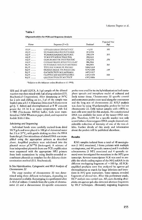
Lekanne Deprez ct al.
Table I
Oligonucleotides for peR and Sequence Analysis
Expected Size Name Sequen(:e (5'_ 3') Position' (hpj
Al/5' . CATGGCCGGGCCATCGCrrCC -1/21 3J5 A1/3' . CCTGAACCAGCTCCTCTTCAGC 313/3.14 A2/5' . TCAAAGGAAGAACCAGTCACC 259/279 3D A2/3' . TCA(;CrrCATCCCTGGCTCG 592/611 A3/5' . GGAGAGAAHACr(;CnCCTAC 555/576 370 A3fT ,. CATAAATAGATCATGGTTCCCGAT 901/924 BI/5' ,. CCTCAAAGCTTCGTGTTAATAAGC 860/883 365 1\1/3' HCCTGCTCAGCCTCTGCGGC 1204/1224 B2/5' . GGAGGCAAAACnCTGGCCCAG 1179/1200 3J9 B2/}' . GACAGGCTGTCACCAATGA(;G 1497/1517 B3/5' . CAATTCCAGCACCGTTGCCTCC 1457/1478 ,350 1\3/3' ,., .. GGGTGGCTGGGTCACCTGCT 1787/1806
• Rd . .J.til'C 10 the initi.J.tion codon (ROllloll et ,II. 1993).
SDS and 10 mM EDTA, A S-li! sample of the diluted reaction was then mixed with 5 Jtl of stop solution (U,S. Biochemical Corporation). After denaturing at 94°C for 2 min and chilling on icc, 3 ~I of the s<lmple was loaded onto a 0,5 X ,Mutation Detection Enhancemenc gel (J- T. Baker) and electrophoresed at g W constant power for 14-16 h at room temperature, with 0_6 X TBE (Tris-borate EDTA) buffer. Gels were transferred to 3 MM Whatman paper, dried, and exposed to Kodak XAR-S film.
Subc/oning and Sequencing Indi\'idual bands were carefull}' excised from dried
SSCP gels and were placed in 100 ~(/ of deionized \v<lter for 3 hat .37°C, with gentle shaking to clute the DNA from the gel. Five microliters of eluted DNA was reamplified using the appropriate primers as described above. Amplified products were subcloned into the plasmid vector pC1v1™IJ (lnyitrogen). A mixture of four independent plasmids from one PCR amplification wcre sequenced with the appropriatc NFl primers from both orientations by lIsing double-stranded recombinant plasm ids as template for the dideoxy chaintermination method (U.S. Biochemical).
In Situ Hybridization. Cytogenetic and RFLP Analysis of Chromosome 22
The copy number of chromosome 22 W,lS determincd using three diffcrent techniques, depending on the material available. KaryOlyping was performed after 3-50 d of culture. Cosmids from the q arm of chromosome 22 and a chromosome 22-specific centromere
probe were used for in situ h}'bridizatiori on both metaphase spreads and interphase nuclei of cultured and fresh tumor tissue. Chromosome 22-specific cosmid and centromere probes were used along the centromere and the long ann of chromosome 22. RFLP analysis was done by using 10 polymorphic probes for loci on chromosome 22. Onl}' tumor s<lmples with >80QA) tumor cells were used for this anal}'sis, No constillltional DNA W.1S aV<lilable for most of the tumor DNA sam· pies. Thereforc, LOH for a specific marker was onl}' scored when a heterozrgous DNA sample showed considerable reduction of intcnsit}' of one of the two alleles. Further details of this stud}' and information about the probes will be published elsewhere.
Results
RNA samples isolated from a total of 48 meningiomas (1 NF2 associated, 3 from patients with multiple meningiomas, and 44 sporadic tumors) and 5 vestibular schwannomas (1 NF2 associated and 4 sporadic tumors) were invcstigated for mutations in the NF2 gene tmnscript. Reverse-transcript.1se peR was used to amplif}' the whole coding region of the Nfl mRNA in six different overlapping fragments of -- 350 bp. All PCR~ amplified products were first analyzed b}' agarose gel electrophoresis to search for large deletions and inscr~ tions in NF2 gene transcripts. Some tumors revealed fragments of altered size. After this preliminary stud}', SSCP analysis was curied out. All cases showing alterations on agarosc gels also detected aberrant fragments b}' SSCP techniques. Aberrantly migrating fr.lgnlents
155
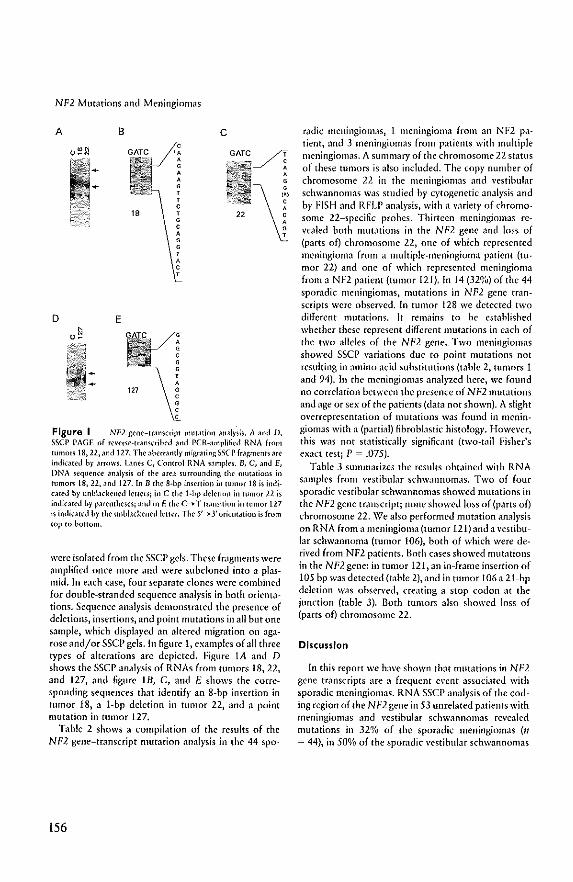
NF2 Mutations and Meningiomas
A B
D E
, ,
, , , , , ,
c
Figure I NF2 gcne-transcript mutJtion Jnal,';is. A and D, S$CP PAGE of rCI'CfSNrJnscribcd and PCR~amplilicd RNA from tumors 18,22, Jlld 127, The aherrandy migrating sseI' frJgmenrs are indkat~d h,' MfOW5. Lanes C, Control RNA sJmples, B, C, ,md E, DNA sequence In,llysis of the area surrounding the mutations in tumo~ 18, 22,and 127. In B the 8·bp insertion in tumor 18 is indi· ,.lIed by unbbckencd lcuc~; in C the I.bp deletion in tumor 22 is indkJ[cd by pJrcntheses; and in E the C--+ T transition in tumor 127 is indicated by the unbbckened letter, The 5'---+3' orientation is from top to bottom.
were isolated from the SSCP gels. These fragments were 'lmplified once more and were subcloned into a plasmid. In each case, four separate clones were combined for double-stranded sequence analysis in both orientations. Sequence analysis demonstrated the presence of deletions, insertions, and point mutations in all but one sample, which displayed an altered migration on agarose and/or SSC!> gels. In figure I, examples of all three types of alterations arc depicted. ~igure lA and D shows the SSCP analysis of RNAs from tumors 18,22, and 127, and figure IB, C, and E shows the corresponding sequences that identify an g.bp insertion in tumor 18, a 1-bp deletion in tumor 22, and a point mutation in tumor 127.
Table 2 shows a compilation of the results of the NI'2 gene-transcript mutation analysis in the 44 spa-
156
radic meningiomas, 1 meningioma from an NF2 patient, and 3 meningiomas from patients with multiple meningiomas. A summary of the chromosome 22 status of these tumors is also included. The copy numher of chromosome 22 in the meningiomas and vestibular schwannomas was studied by cytogenetic analysis and by ~ISH and RFLP analysis, with a variety of chromosome 22-specific probes. Thirteen meningiomas revealed both mut;Uions in the NF2 gene and loss of (parts of) chromosome 22, one of which represented meningioma from a multiple-meningioma patient (tumor 22) and one of which represented meningioma from a NF2 patient (tumor 121). In 14 (32%) of the 44 sporadic meningiomas, mutations in NF2 gene transcripts were observed. In tumor 128 we detected two different mutations. It remains to be estahlished whether these represent different mutations in each of the two alleles of the NF2 gene., Two meningiomas showed SSCP variations due to point mutations not resulting in ,Imino acid substitutions (table 2, tumors 1 and 94). In the meningiomas analyzed here, we found no correlation between the presence of NF2 mutations and age or sex of the patients (data not shown). A slight overrepresentation of mutations was found in meningiomas with a (partial) fibroblastic histology. However, this was not statistically significant (two-lilil Fisher's exact test; P = .075).
Tahle 3 summarizes the results obtained with RNA samples from vestibular schw;ltlnomas. Two of four sporadic vestibular schwannomas showed mutations in the NF2 gene transcript; none showed loss of (parts of) chromosome 22. We also performed mutation analysis on RNA from a meningioma (tumor 121) and a \'estibu~ lar schwannoma (tumor 106), both of which were derived from NF2 patients. Both cases showed Illut;ltions in the NF2gene! ill tumor 121, an in-frame insertion of 105 bp was detected (t;lble 2), and in tumor 106 a 21-bp deletion was ohserved, creating a stop codon at the junction (table 3). Both tumors also showed loss of (parts of) chromosome 22.
Discussion
In this report we have shown that mutations in Nf2 gene transcripts are a frequent event associated with sporadic meningiomas. RNA SSCP analysis of the coding region of the N"'2 gene in 53 unrelated p.uients with meningiomas ,md vestibular schwannomas revealed mutations in 32% of the sporadic meningiomas (11 = 44), in 50% of the sporadic vestihular schwannomas
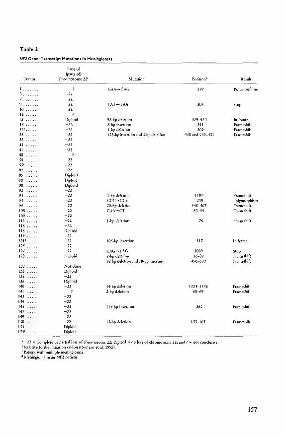
Table 2
Nfl Gene-Transcript Mutations In Meningiomas
Loss of (pMtsofj
Tumor Chromosome 21' Mutation Positionb
GAA_GAG 387 ], -22 7 -22 9, -22 TAT .... TAA 303 10 -22 12 15 Diploid 96.hp deletion 329-424 IS -22 8.bp insertion 241 U -22 l.bp deletion 209 15 -22 128.hp imcrtion and 3.6p deletion 448 and 449-451 32 -22 JS -22 41 -22 4S
54 -22 55' -22 81 -12 85 Diploid 88 Diploid 90 Diploid 91 -12 93 -12 l.bp deletion 1107 94 -22 Gee_GeT 219 99 -22 20.bp deletion 448-467 103 -22 CAA_CT SJ ... S4 109 -22 111 -22 I.bp deletion 76 116 -22 118 ... Diploid 119 -22 121d -22 IOS·hp insertion 517 125 -22 127 -22 CAG ..... TAG 1009 128 Diploid 2.bp deletion 36-37
82.bp deletion jnd 18.bp insertion 496-577 130 Not done 133 Diploid 13,~ -22 136 Diploid 140 -22 14.6p deletion 1223-1236 141 ? 2.6p deletion 68-69 143 -22 144 -22 145 -22 119.6p insertion 364 147 -22 149 -22 150 -22 I 3.bp deletion 153-165 153 Diploid 154' . Diploid
• -22 "" Complete or partial loss of chromosome 22; Diploid"" no loss of chromosome 22; and? = not conclush'e. b Relative to the initiation codon (Roule:lll et.Il. 1993). 'Patient with multiple meningiomas. d Meningi('mj in.In NF2 patient.
Result
!'olymorphism
Swp
In frame Frameshift Frameshift FrJmCShift
FrJmelhift Polrmorphism Framcshift FrJmcshift
Fr.lmelhift
In frJme
Stop Frameshift Framelhift
Frameshift Frameshift
Frameshift
FrJmeshift
157
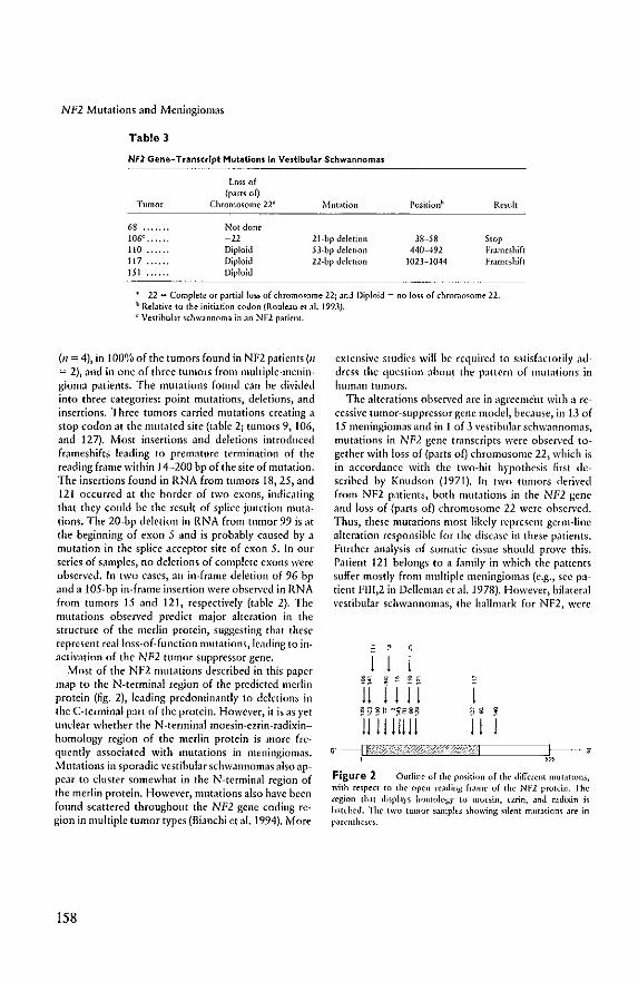
NF2 Mutations and Meningiomas
Table 3
Nfl Gene-TranscrIpt Mutations In Vestibular Schwannomas
Loss of (pamo£)
Tumor Chromosome 22' Mutation Positionb Rt"sult
68 ... Not done 106e
• -12 21+bp deletion 38-58 Stop 110 Diploid n.bp deletion 440-492 Frame-shift 117 Diploid 22-bp deletion 1023-1044 Frameshi£! 151 Diploid
• -12 = Complete or partial loss of chromosome 22; and Diploid = no loss of ,hromosome 22. b Relative \0 the initiation codon (f{ouleJu et at 1993). < Vestibular schwannoma in an NFl patienl.
(11 = 4), in tOOO,'o of the tumors found in NF2 patients (It = 2), and in one of three tumors from multiple-meningioma patients. The mutations found can be divided into three categories: point mmations, deletions, and insertions. Three tumors carried mutations creating a stop codon at the mutated site (table 2; tumors 9, 106, and 127). Most insertions and deletions introduced frameshifts leading to premature termination of the reading kame within 14-200 bp of the site of mutation. The insertions found in RNA from tumors 18,25, and 121 occurred at the border of two exom, indicating that they could be the result of splice-junction mutations. The 20-bp deletion in RNA from tumor 99 is at the beginning of exon 5 and is probably caused by a mutation in the splice acceptor site of exon 5. In our series of snmples, no deletions of complete exons were observed, In two cases, an in-frame deletion of 96 bp nnd a 105-bp in-frame insertion were obsen'ed in RNA from tumors 15 and 121, respectively (table 2). The mutations observed predici major alteration in the structure of the merlin protein, suggesting that these represent realioss-of-fullction mutations, leading to inactivation of the NF2 tumor-suppressor gene.
Most of the NF2 mutations described in this paper lIIap to the N-terminal region of the predicted merlin protein (fig. 2), leading predominantly to dektions in the C-terminal part of the protein. However, it is as yet unclear whether the N-terminal moesin-ezrin-radixin~ homology region of the merlin protein is more fre~ quently associated with mutations in meningiomas. Mutations in sporadic vestibular schwannomas also appear to cluster somewhat in the N-terminal region of the merlin protein, However, mutations also have been found scattered throughout the NF2 gene coding region in multiple tumor types (Bianchi etal. 1994). More
158
extensive studies will be required to s.ltisfactorily ad~ dress the question about the pattern of lllutntio/lS in human tUlIlors.
The alterations observed nre in agreeme'tIt with a recessi\'e tumor-suppressor gene model, because, in 13 of 15 meningiomns and in 1 of 3 vestibular schwannomas, mutations in NF2 gene transcripts were obsen'ed together with loss of {parts 00 chromosome 22, which is in accordance with the two-hit hypothesis first described by Knudson (1971). In two tumors derived from Nfl patients, both nmt.lIions ill the NF2 gelle and loss of (parts of) chromosome 22 were obsen'ed. Thus, these mutations most likely represent germ-line alteration responsible for the disease in these p<ltients. Further analysis of somatic tissue should prove this, Patient 121 belongs to .t family in which the patients suffer mostly from multiple meningiomas (e.g., sec patient FIII,2 in Detleman et al. 1978). However, bilateml vestibular schw,1nnomas, the hallmark for NF2, wcre
J J 2~ j~~5
JJ [J J J 8 ~ ~
J J J
Figure 2 Outline of the positinn of the (lifferent mutations, \\ilh respen to the open reading frame of the NF2 rroteill. The region that dilpLtrs homology to mocsin, cmn, and radixin is h.ncbed. The two tumor S.lJllpleS ~ho\\ing silent mllt~linm Jre in parentheses.
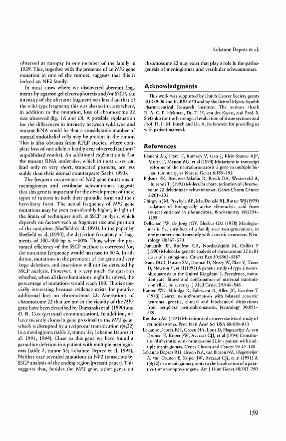
observed at autopsy in one member of the f'lmily in 1939. This, together with the presence of an NF2 gene mutation in one of the tumors, suggests that this is indeed an Nfl family.
In most cases where we discovered abemmt fragments by agarose gel electrophoresis and/or SSC!>, the intensity of the abermnt fmgment was less than that of the wild-type fragment; this was also so in cases where, in addition to the mutation, loss of chromosome 22 was observed (6g_ lA and D)_ A possible explanation for the differences in intensity hetween wild-type and mutant RNA could be that a considerable number of normal endothelial cells may he present in the tumor. This is also obvious from RFLP studies, where complete loss of one allele is hardly ever observed (authors' unpublished results), An additional explanation is th.n the mUl<lnt RNA molecules, which in most cases can lead only to yery short, tnmcated proteins, Me less stable than their 1l0rnJ;l1 counterparts (Sachs 1993).
The frequent occurrence of NF2 gene mutations in meningiomas and vestibular schwannomas suggests that this gene is important for the de\'Clopment of these types of tumors in both their sporadic form and their hereditary form. The actual frequency of NF2 gene mutations may he even consider<1bly higher, in light of the limib of techniques such its sscr llnalysis, which depends on f.lctors such as fragment size .lIld position of thc mutation (Sheffield ec al. 1993)_ In the paper by Sheffield et al. (1993), the dcrection frequency of fragments of 300-400 bp is ----600:}/0_ Thus, when the presumed efficiency of the sscr method is wrrecled for, the mutation frequency would incre.tse to 50%_ In llddition, Illut.uions in the promoter of the gene .lIlt! very huge deletions and insertions will not be detected by ssep anlllysis. However, it is very Illuch the question whether, when all chese limilalions might be solved, the percentage of mutations would rcach 100. This is cspecially interesting because evidence exists for pllt.lIive additional loci Oil chromosome 22. Aberrations of chromosome 22 that are not in the vicinity of the NF2 gene havc hecn described by Dumanski et ai, (1990) and D. R. Cox (person.ll cOllllllunicalion). In addition, we have recently cloned a gene proximal to the NP2 gene, which is disrupted hy a reciprocal transloc.ttion t(4;22) in illlleningiolllll (tllble 2, tumor 32; Lekanne Deprez et al. 1991, 1994), Closc to this gene we have found il
germ-line deletion ill a patient with multiple meningiomas (table 2, tumor 55; Lekannc Deprez et al. 1994), Neither case re\'ealed mut.lIions in NF2 tf;1llscripcs by sse!> analysis of the coding region (present paper). This suggests that, bcsides the NF2 gene, other genes on
Lebnne Deprez et lli.
chromosome 22 may exist that playa role in the pathogenesis of meningiomas and vestibular schW;lnnomas,
Acknowledgments
This work W.IS supported by Dut(:h Can(:er Society grants EUR89-06 and EUR93-653 and by the Bristol Myers-Squibb l'harmllceufical Rese,m:h Institute_ The authors thank R, A. C. p, Hekman, Dr. T. H. van der Kwast, and Prof. S. Stefanko for the histological e\'.llllation of tissue sections and Prof. H. I,'. M, Busch and Dr, A, Rubinstein for providing us with patient material.
References
Bianchi All, Hara T, Ramesh V, Gao j, Klein-Szanto A)P, Morin F, Menon AG, et ;11 (1994) Mutations in transcript isoforms of the neuro6bromalOsis 2 gene in multiple human tumour types Nature Genet 6:185-192
Biilsma EK, Brollwer-Mbdin R, Bosch DA, Westefycld A, Hulsehns Tj (1992) , .... Iolewlar characterization of chromusome 22 deletions in schw.lIlnon1.1so Genes Chrom Cancer 5:201-205
Chirgwin)}'I, Przyb)Ola Af, },lacDonald R), Rutter W) (1979) isolation of biologically active ribonucleic acid from sources enriched in rihonuclease. Binchemistry 18:5294-5299
Delleman )\'(', de JongjGY, Bleeker GM (1978) Meningium.1S in Ii\'e members of ;1 f.lmily over two gener.ltions, in one member simultaneously with .leollstie neurom.1S, Neurology 28:567-570
Olltllanski Ji', Rouleall GA, Nordcnskjold M, Collins P (1990) },·Iolecular genetic analysis uf chromosome 22 in 81 cases of meningioma. Cancer Res 50:5863-5867
Evans OeR, IlllSon S}'I, Oonn.li 0, NeJI)' \V, Blair V, Teare D, Newton V, et al (1992) A genetic stud)' of type 2 neurolibronutosis in the United Kingdom. I. Prey.llence, mutation rate, fitness and confirmation of maternal transmission elfect on severity.) Med Genet 29:841-846
Kanter \VIt, Eldridge R, Fabric.mt R, Allen JC, Koerber T (1980) Centml neurofibromatosis with bilateral acoustic neuroma: genetic, clinical and biochemical distinctions f[{Jm peripheral neuro6bromatosis, Neurology 30:851-859
Knudson AG (1971) ,\iutation and cancer: statistical study of rctinohlastoma_ Proc Nail Acad Sci USA 68:820-823
Lekanne Deprez RH, Groen NA, Louz 0, Hagemeijcr A, y.m Dnlllen f, Koper )\'\', Avezaat CJ), et al (1994) Constitutional aberr.Hions in chromosome 22 in a patient with multiple meningiomaso Genes Chrom and Cancer 9:124-128
Lebnne Ocprcz RH, Groen NA, van Biezen NA, Hagemeijer A, van Drunen E, Koper jW, AveZJ.at CJ), et al (1991) A t(4;22) in a meningiomll points to the localization of a putative tumur-suppressor gene, Am J Hum Genet 48:783-790
159
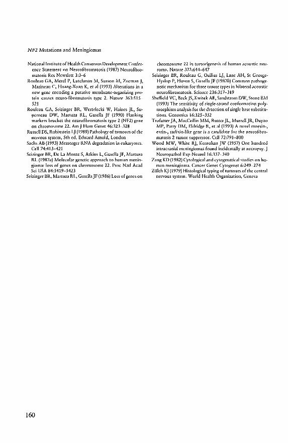
NF2 Mutations and Meningiomas
National Institute of Health Consensus Development Conference Statement on Neurofibromatosis (1987) Neurofibromatosis Res Newslett 3:]-6
Rouleau GA, Mere! P, lutchman M, Sanson i\f, Zucmanj, Marineau C, Hoang-Xuan K, et a\ (199]) Alterations in.\ new gene encoding a putative membrane-organizing protein causes neuro-fihromatosis type 2. Nature 36]:515-521
Rouleau GA, Seizinger HR, Wertelecki W, Haines jl, Superneau OW, Martuza RL, Gusella jF (1990) Flanking markers bracket the neurofibromatosis type 2 (NF2) gene on chromosome 22. Am J Hum Genet 46:]2]-]28
Russell OS, Rubinstein Lj (1989) Pathology of tumours of the nervous system, 5th cd. Edward Arnold, london
Sachs AB (1993) Messenger RNA degradation in eukaryotes. Cell 74:413-421
Sci zinger BR, De La Monte S, Atkins t, Gusella jF, l'o.-1artUz.1 RL (1987a) .Molecular genetic approach to human meningioma: loss of genes on chromosome 22. Proc Nat! ACJ.d Sci USA 84:5419-5423
Seizinger BR, MartUz.1 RL, GusclbJF (1986) loss of genes on
160
chromosome 22 in tumorigenesis of human acoustic neuroma. Nature 322:644-647
Selzinger BR, Rouleau G, Ozilius Lj, Lane AH, St GeorgeH)'slop P, Huson S, Gusella jl; (1987b) Common pathogenetic mechanism for three tumor types in hilateral acoustic neurofibromatosis. Science 236:317-319
Sheffield VC, Beck )S, Kwilek AE, Sandstrom DW, SlOne EM (1993) The sensitivity of single-strand conformation pol}'morphism analysis for the detection of single base substitutions. Genomics 16:]25-3]2
Trofatter jA, MacCollin MM, Rutter Jl, Murrell JR, Duyao lI.H', Parry DM, Eldridge R, et al (199]) A novel moesin-, ezrin-, radixin-like gene is a candidate for the neurofibromatosis 2 mmor suppressor. Cell 72:79J-800
\'l/ood MW, White Rj, Kernohan j\,\1 (1957) One hundred intracranial meningiomas (ound incidentally at necropsy. J Neuropathol Exp NeuroI16:]37~]40
Zang KD (1982) Cyw!ogical and cytogenctkal studies on human meningioma. Cancer Genet Cytogenet 6:249-274
zulch KJ (1979) Histological typing of tumoucsQf the central nen'ous system. World Health Organization, Geneva
Summary and Discussion
Meningioma is a common benign central nervOllS system tumor (eNS) arising from
arachnoidal cells surrounding the brain and spinal cord. Cytogenetic studies as early as 1967
and molecular genetic analyses revealed that loss of chromosome 22 is the most frequent
aberration observed in these tumors. In additioll, 50% of the patients with the autosomal
dominant inherited disorder NF2 have meningiomas. Linkage analysis in affected families
has assigned the predisposing gene to chromosome 22. This indicates that both NF2 and
sporadic meningioma presumably are the result of mutations in one Of more genes on
chromosome 22. Consistent and specific chromosome deletions suggest the involvement of
tumor suppressor genes. A tumor suppressor gene plays a role in tumorigenesis when both
alleles of the gene are'inactivated. Loss of such a gene is one of the mechanisms involved
in functional inactivation. Therefore, a ttlillor suppressor gene(s) on chromosome 22 is (are)
probably involved in the development of meningiomas. The goal of the work described in
this thesis was to localize and isolate this (these) gene(s) on chromosome 22.
In order to identify the region(s) on chromosome 22 responsible for meningioma development
we first collected meningiomas for cytogenetic and DNA analysis. At that time only a limited
number of markers from this chromosome were available. Therefore, a chromosome 22
specific phage library was used for the isolation of additional probes. The 25 isolated single
copy probes were further characterized by testing whether they recognized restriction
fragment length polymorphisms (RFLPs) on human genomic DNA and they were regionally
localized on chromosome 22 using a panel of somatic cell hybrids. The polymorphic probes
were used to construct a linkage map (Chapter II).
These probes and markers obtained from other laboratories were subsequently used to
investigate our collection of meningiomas. Chapter III describes the results of the combined
cytogenetic and molecular genetic study in OLlr series of meningiomas. Furthermore, the
observed chromosomal changes were compared with other patient and tumor characteristics
and statistically analyzed. In accordance with other reports loss of chromosome 22 was most
frequently observed. We found complete or partial loss of this chromosome in 71 % of the
tumors. Statistical analysis revealed a signiticant association between the number of
161
chromosomal abnormalities and tumor grade. Complex karyotypes predominated in the group
of grade IIIIIl meningiomas. This suggests that in the progression of meningiomas multiple
genetic hits are required. In addition, other variables showed statistically significant
differences indicating that meningioma subclasses exist. For instance, meningiomas from the
convexity were more often grade WIll, displayed predominantly (partial) loss of chromosome
22 and had complex karyotypes more often. These features were frequently found in
meningiomas from males. Base meningiomas on the other hand occurred more often in
females, they were usually grade I, showed loss of (parts 01) chromosome 22 less often and
displayed fewer additional chromosomal abnormalities. Future studies of patient survival,
recurrence rate and invasive growth of the tumors should establish whether these observations
are of clinical importance.
The combined cytogenetic and molecular genetic approach was also applied to an anaplastic
ependymoma, which revealed loss of markers on the long arm of chromosome 22 (Chapter
IV). This tumor was obtained from an 8 month old boy born from a family in whom two
healthy brothers each had two sons with anaplastic ependymomas. Normal cells from this boy
showed a normal karyotype. This finding provides evidence for the role of a tumor
suppressor gene on chromosome 22 in the pathogenesis of ependymal tumors. The clustering
of ependymal tumors in four nephews from one family suggests that a predisposing germ-line
mutation is involved in the pathogenesis. However, because neither the fathers nor the
grandparents developed ependymomas there is no evidence for a simple dominant mode of
inheritance of the disease in this family. A possible explanation could be that there is
mendelian inheritance of the disease but that the inheritance involves a gene whic.h is
inactivated by paternal imprinting rather than by an inactivating mutation. An alternative
model is based on, for instance the fragile X syndrome, in which the expansion of a
trinucleotide repeat in the FMR-J gene can continue over several generations until! a final
step suddenly reveals the disease in members of the same generation.
Cytogenetic analysis of our meningiomas revealed one tumor, meningioma 32, with a
reciprocal t(4;22)(pI6;q 11). We hypothesized that this translocation disrupts a potential tumor
suppressor gene. To further investigate this translocation we fused the meningioma cells with
a hamster cell line. This resulted in hybrids segregating one of the two products of the
162
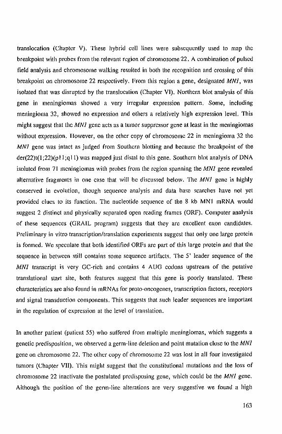
translocation (Chapter V). These hybrid cell lines were subsequently used to map the
breakpoint with probes from the relevant region of chromosome 22. A combination of pulsed
field analysis and chromosome walking resulted in both the recognition and crossing of this
breakpoint on chromosome 22 respectively. From this region a gene, designated MNl, was
isolated that was disrupted by the translocation (Chapter VI). Northern blot analysis of this
gene in meningiomas showed a very irregular expression pattern. Some, including
meningioma 32, showed no expression and others a relatively high expression level. This
might suggest that the MNl gene acts as a tumor suppressor gene at least in the meningiomas
without expression. However, on the other copy of chromosome 22 in meningioma 32 the
MNl gene was intact as judged from Southern blotting and because the breakpoint of the
der(22)t(I;22)(pll;q II) was mapped just distal to this gene. Southern blot analysis of DNA
isolated from 71 meningiomas with probes from the region spanning the MNl gene revealed
alternative fragments in one case that will be discussed below. The MNl gene is highly
conserved in evolution, though sequence analysis and data base searches have not yet
provided clues to its function. The nucleotide sequence of the 8 kb MNI mRNA would
suggest 2 distinct and physically separated open reading frames (ORF). Computer analysis
of these sequences (GRAIL program) suggests that they are excellent exon candidates.
Preliminary in vitro transcriptionltransiation experiments suggest that only one large protein
is formed. We speculate that both identified ORFs are part of this large protein and that the
sequence in between still contains some sequence artifacts. The 5' leader sequence of the
MNI transcript is very GC-rich and contains 4 AUG codons upstream of the putative
translational start site, both features suggest that this gene is poorly translated. These
characteristics are also found in mRNAs for proto-oncogenes, transcription factors, receptors
and signal transduction components. This suggests that such leader sequences are important
in the regulation of expression at the level of translation.
In another patient (patient 55) who suffered from multiple meningiomas, which suggests a
genetic predisposition, we observed a germ-line deletion and point mutation close to the MNl
gene on chromosome 22. The other copy of chromosome 22 was lost in all four investigated
tumors (Chapter VII). This might suggest that the constitutional mutations and the loss of
chromosome 22 inactivate the postulated predisposing gene, which could be the MNI gene.
Although the position of the germ-line alterations are very suggestive we found a high
163
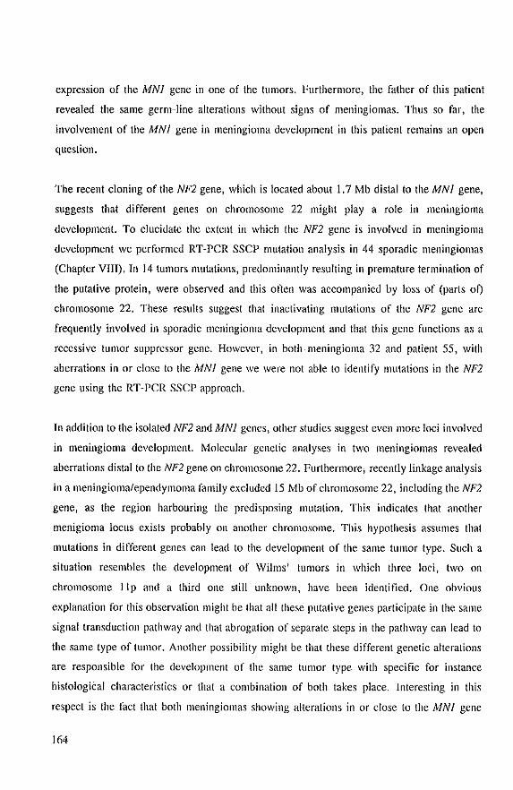
expression of the MNI gene in one of the tumors. Furthermore, the father of this patient
revealed the same germ-line alterations without signs of meningiomas. Thus so far, the
involvement of the MNI gene in meningioma development in this patient remains an open
question.
The recent cloning of the NF2 gene, which is located about I, 7 Mb distal to the MN 1 gene,
suggests that different genes on chromosome 22 might play a role in meningioma
development. To elucidate the extent in which the NF2 gene is involved in meningioma
development we performed RT-PCR SSCP mutation analysis in 44 sporadic meningiomas
(Chapter VIII). Tn 14 tumors mutations, predominantly resulting in premature termination of
the putative protein, were observed and this often was accompanied by loss of (parts ot)
chromosome 22. These results suggest that inactivating mutations of the NF2 gene are
frequently involved in sporadic meningioma development and that this gene functions as a
recessive tumor suppressor gene. However, in both meningioma 32 and patient 55, with
aberrations in or close to the MNI gene we were not able to identify mutations in the NF2
gene using the RT-PCR SSCP approach.
In addition to the isolated NF2 and MNI genes, other studies suggest even more loci involved
in meningioma development. Molecular genetic analyses in two meningiomas revealed
aberrations distal to the NF2 gene on chromosome 22. Furthermore, recently linkage analysis
in a meningioma/ependymoma family excluded 15 Mb of chromosome 22, including the NF2
gene, as the region harbouring the predisposing mutation. This indicates that another
menigioma loclls exists probably on another chromosome. This hypothesis assllmes that
mutations in different genes can lead to the development of the same tumor type. Such a
situation resembles the development of \Vilms' tumors in which three loci, two on
chromosome IIp and a third one still unknown, have been identified. One obvious
explanation for this observation might be that all these putative genes participate in the same
signal transduction pathway and that abrogation of separate steps in the pathway can lead to
the same type of tumor. Another possibility might be that these different genetic alterations
are responsible for the development of the same tumor type with specific for instance
histologiCal characteristics or that a combination of both takes place. Interesting in this
respect is the fact that both meningiomas showing alterations in or close to the MNJ gene
164
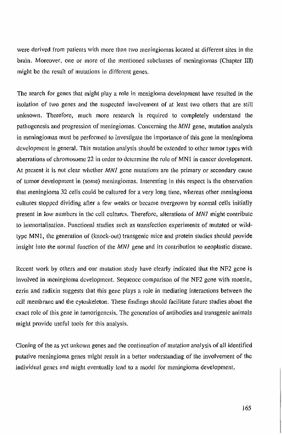
were derived from patients with more than two meningiomas located at different sites in the
brain. Moreover, one or more of the mentioned subclasses of meningiomas (Chapter III)
might be the result of mutations in different genes.
The search for genes that might playa role in menigioma development have resulted in the
isolation of two genes and the suspected involvement of at least two others that are still
unknown. Therefore, much more research is required to completely understand the
pathogenesis and progression of meningiomas. Concerning the MNI gene, mutation analysis
in meningiomas must be performed to investigate the importance of this gene in meningioma
development in general. This mutation analysis should be extended to other tumor types with
aberrations of chromosome 22 in order to determine the role of MN I in cancer development.
At present it is not clear whether MNI gene mutations are the primary or secondary cause
of tumor development in (some) meningiomas. Interesting in this respect is the observation
that meningioma 32 cells could be cultured for a very long time, whereas other meningioma
cultures stopped dividing after a few weaks or became overgrown by normal cells initially
present in low numbers in the cell cultures. Therefore, alterations of MNI might contribute
to immortalisation. Functional studies such as transfection experiments of mutated or wHd
type MNl, the generation of (knock-out) transgenic mice and protein studies should provide
insight into the normal function of the MNI gene and its contribution to neoplastic disease.
Recent work by others and our mutation study have clearly indicated that the NF2 gene is
involved in meningioma development. Sequence comparison of the NF2 gene with moesin,
ezrin and radixin suggests that this gene plays a role in mediating interactions between the
cell membrane and the cytoskeleton. These findings should facilitate future studies about the
exact role of this gene in tumorigenesis. The generation of antibodies and transgenic animals
might provide useful tools for this analysis.
Cloning of the as yet unkown genes and the continuation of mutation analysis of all identified
putative meningioma genes might result in a better understanding of the involvement of the
individual genes and might eventually lead to a model for meningioma development.
165

166
Samenvatting
Meningeomen zUn goedaardige tumoren van het centrale zenuwsteisel (CZS) en ontstaan uit
cellen afkomstig van het vIies, dat de hersenen en het ruggemerg omgeeft. Chromosoom
studies, reeds uitgevoerd vanaf 1967, en moleculair-genetische analyses hebben Iaten zien,
dat verIies van chromosoom 22 de meest frequente afwijking in deze tumoren is. Daarnaast
kamen meningeomen frequent voor bij patienten met de dominant overervende aandoening
NF2. KoppeIings-onderzoek in NF2 families heeft aangetoond, dat het verantwoordeIijke gen
op chromosoom 22 gelocaliseerd moet zijn. Dit wijst erop dat lOwel NF2 aIs sporadische
meningeomen waarschijnlijk veroorzaakt wordt door een of meerdere genen op chromosoom
22. Het consequent aanwezig zijn van een specifieke chromosomale afwijking, in dit geval
verlies van chromosoom 22, doet vermoeden dat we hier te maken hebben met een
tumorsuppressorgen (genen). Een tumorsuppressorgen draagt bij tot het ontstaan van een
tumor, wanneer beide copieen van het gen ge'inactiveerd zijn. Verlies van een dergeJijk gen
is een van de mechanism en am het gen te inactiveren. Deze bevindingen veronderstellen dat
ten minste een tU1110rsuppressorgen op chromosoom 22 betrokken is bij het ontstaan van
meningeomen. Het doel van het werk, beschreven in dit proefschrift, was om zulke genen
op chromosoom 22 te isoleren en Ie bestuderen.
Het onderzoek werd opgestart met de isolatie van DNA merkers, die specifiek voor
chromosoom 22 zijn. Bij deze merkers hebben we onderzocht of ze in staat waren
polymorfismen in restrictie enzym knipplaatsen te detecteren in normaal menselijk DNA.
Voor de IocaUsatie van de merkers op chromosoom 22 hebben we gebruik gemaakt van
DNA, gelsoleerd uit een aantal hybride cellijnen, die stukken van chromosoom 22 bevatten.
De polymorfe merkers zijn ook gebruikt om een genetische koppelingskaart van een deel van
chromosoom 22 Ie maken.
Onze polymorfe merkers en merkers, die van andere laboratoria afkomstig waren, hebben
we gebruikl om DNA, ge'isoleerd uit onze serie meningeomen en een aantal andere tumoren
van het CZS, te bestuderen op afwijkingen van chromosoom 22. De tumoren zijn vervolgens
op chromosomaal niveau nader gekarakteriseerd door gebruik Ie maken van cytogenetische
analysetechnieken. Deze methoden stelden ons in staat om (delen van) chromosomen aan te
167
wijzen, die rnogelijk voor rneningeoorn ontwikkcling van beJang zouden kunnen zijn. De
resultaten van deze gecombineerde studie zijn vergeleken met een aantal ken mer ken van de
patienten en hun· tumoren. In overeensternming met de bevindingen van andere
onderzoeksgroepen was verlies van chromosoom 22 de meest voorkornende chromosornale
afwijking. Ook het anaplastiseh ependymoom vertoonde verlies van ehromosoom 22. In 71 %
van de meningeomen werd volledig of gedeeJtelijk verlies van dit ehromosoom geobserveerd.
Statistische analyse toonde een significant verschil tussen het aantal chromosomale
afwijkingen en de gradering van de tumor. Complexe karyogrammen werden vaker gevonden
in tumoren van graad II en Ill. Dit doet vermoeden dat bij de progressie van meningeomen
meerdere genetische gebeurtenissen een rol spelen. De statistisch significante verschillen, die
tussen de variabelen gevonden waren, suggereren dat meningeomen in verschillende
subgroepen onder te verdelen zijn. Convexiteit meningeomen toonden meer verlies van (deJen
van) chromosoom 22, meerdere chromosomale afwijkingen, waren in hoofdzaak van graad
II en III en kwamen vaak bij mannen voor. Meningeomen geJocaliseerd in de basis
daarentegen vertoonden weinig verlies van (delen van) chromosoom 22, weinig andere
chromosomale afwijkingen, waren meestal van graad I en werden vaker bij vrouwen
gevonden.
Chromosoorn-analyse resulteerde in de identificatie van cen tumor, meningeoom 32, met een
gebalaneeerde t(4;22)(pI6;qll). Onze werkhypothese was dat deze transloeatie een
tumorsuppressorgen kapot zou maken en dat het verder in kaart brengen van het translocatie
breukpunt ons zou kunnen helpen bij het isoleren van dit gen. Hiervoor hebben we de
tumorcellen gefuseerd met een hamster-cellijn, waardoor twee hybride cellijnen verkregen
werden, die elk een translocatieproduct bevatten. Dit maakte het mogelijk om met
ehromosoom 22 specilieke merkers de ligging van het breukpunt te bepalen. Ben van deze
merkers bleek het breukpunt te herkennen. Met behulp van genomisehe en eDNA banken
werd het gebied van chromosoom 22, waarin het breukpunt geJegen is, ge'isoleerd en een gen
in ditgebied gejdentifieeerd. Nadere plaatsbepaling van dit gen, door ons MNI genoemd,
toonde aan dat het door de transloeatie kapot gemaakt werd. Verdere karakterisatie van het
gen door middel van sequentie-analyse en vergeJijking met genen-banken hebben geen
aanwijzingen voor zijn mogelijke funetie opgeleverd. Het gen is wei sterk geeonserveerd
gedurende de evolutie. Southern-blot teehnieken zijn vervolgens gebruikt om afwijkingen in
168
het gebied rondom het MNI gen te bestuderen in 71 andere meningeomen. In een van deze
tumoren, afkomstig van een patient met meerdere individuele meningeomen (patient 55),
detecteerden we afwijkingen in dit gebied. Deze afwijkingen bleken oak aanwezig te zijn in
DNA gelsoleerd nit bloed van patient 55. De afwijkingen, een intersitiele dele tie en een punt
mntatie, bevinden zieh op ongeveer 10 kb afstand van het MNI gen. Op dit moment is niet
bekend of deze afwijkingen voor de meningeomen in deze patient predisponeren en de funetie
van het MNI gen belnvloeden. Nader onderzoek moet uitwijzen hoe belangrijk dit gen is voor
de ontwikkeling van meningeomen.
Op grand van literatuurgegevens zijn er aanwijzingen dat meerdere genen cen rol zouden
kuonen spelen bij mcningeoom ontwikkeling. Recentelijk is het op cluoillosoom 22 gelegen
NF2 gen geeloneerd. Bovendien doen afwijkingen distaal van het NF2 gen in een tweetal
meningeomen vermoeden dat cen derde, nog niet gc'isoleerd, gen cell rol ZOli kunnen speJen.
Verder laat koppelingsonderzoek in cen ependymoma/meningeoma familie zien, dat een groat
deel van chromosoom 22, waaronder het NF2 gen, niet verantwoordelijk is voor het ontstaan
van de ziekte in deze familie. Waarsehijnlijk is het predisponerende gen op een ander
ehramosoom gelegen. Mutatie-studie van het NF2 gen hebben we uitgevoerd om de ral van
dit gen bij meningeoom ontwikkeling te onderzoeken. In 30% van de tumoren was het
mogelijk mutaties op te sporen. Bijna aUe mutaties zouden leiden tot een sterk verkart NF2
eiwit, waardoor het eiwit waarschijnlijk ge'inactiveerd wordt. In de meeste tumoren gingen
de mutaties samen met verlies van (delen van) chromosoom 22. Deze bevindingen doen sterk
vermoeden dat we hier te maken hebben met een recessief tumorsuppressorgen en dat dit gen
een belangrijke ral vervult bij het ontstaan van meningeomen. EelIter dit sluit de
betrokkenheid van andere genen niet lIit. Verder onderzoek zal moeten bepaJen wat de
(onderlinge) fllnctie van deze genen is bij het proces, dat aan de ontwikkeling van
meningeomen ten grondslag ligt.
169

170
30 juni 1965
1984
1984 - 1988
1989 - 1994
1994 -
Curriculum vitae
geboren te Oegstgeest
eindexamen VWO aan het Rijnlands Lyceum te Oegstgeest
studie Gezondheidswetenschappen aan de Faculteit der
Geneeskllnde Rijksllniversiteit van Leiden (cum laude)
Bijvakstages:
1 Afdeling Klinische Oncologie, Academisch Ziekenhllis
Leiden
begeleider: Dr. P.L Schrier
2 Afdeling Klinische Oncologie, Academisch Ziekenhuis
Leiden
begeleider: Prof. Dr. F.J. Cleton
3 Afdeling Nierziekten, Academisch Ziekenhuis Leiden
begeleider: Prof. Dr. M.R. Daha
4 Afdeling Medische Biochemie, Sylvius Laboratorium
Leiden
begeleider: Dr. J.A. Maassen
Hoojdvakstage:
Afdeling Medische Biochemie, Sylvius Laboratorium Leiden
begeleider: Dr. LA. Maassen
Promotieonderzoek, afdeling Pathologie, Erasmus Universiteit
Rotterdam
promotor:
co-promotor:
Prof. Dr. D. Bootsma (afd. Genetica)
Dr. B.C. Zwarthoff
Wetenschappelijk onderzoeker, afdeling Anatomie &
Embryologie, Academisch Medisch Centrum, Amsterdam
171

172
List of publications
I) van 't Veer U, van den Berg-Bakker LAM, Hennens RPMG, Lekanne Deprez RH,
and Schrier PI (1988). High frequency of mas oncogene activation detected in the
NIH-3T3 tumorigenicity assay. Oncogene Res 3:247-254
2) Lekanne Deprez RH, Potter van Loon BJ, van der Zon GCM, Moller W, Lindhout
D, Klinkhamer MP, Krans HMJ and Maassen JA (1989). Individuals with only one
allele for a functional insulin receptor have a tendency to hyperinslIlinacmia but not
to hyperglycaemia. Diabetologia 32:740-744
3) Lekanne Deprez RH, Groen NA, van Biezen NA, Hagemeijer A, van Drunen E,
Koper JW, Avezaat CJJ, Bootsma D, Zwarthoff EC (1991). A t(4;22) in a
meningioma points to the localization of a putative tumor-suppressor gene. Am J Hum
Genet 48:783-790
4) Lekanne Deprez RH, van Biezen NA, Heutink P, Boejharat KRGS, de Klein A,
Geurts van Kessel AHM and Zwarthoff EC (1991). A new polymorphic probe on
chromosome 22: NBI7 (D22SI81). A new polymorphic probe on chromosome 22:
NB35 (D22SI82). A new polymorphic probe on chromosome 22: NB84 (D22SI83).
A new polymorphic probe on chromosome 22q: NBI29 (D22SI84). Nucleic Acids
Res 19:686-687
5) Lekanne Deprez RH, van Biezen NA, Breedveld GJ, Boejharat KRGS, de Klein A,
Geurts van Kessel AHM and Zwart hoff EC (1991). A new polymorphic probe on
chromosome 22: NB5 (D22S201). Nucleic Acids Res 19: 1963
6) van Biezen NA, Lekanne Deprez RH, Thijs A, Heutink P, Oostra BA, Geurts van
Kessel AHM and Zwarthoff EC (1992). Isolation and characterization of 25 unique
DNA markers for human chromosome 22. Gcnomics 15:206-208
173
1) Lekanne Deprez RH, Groen NA, Louz D, Hagemeijer A, van Drunen E, Koper JW,
Avezaat CJJ, Bootsma D, van der Kwast TH, Zwarthoff EC (1994) Constitutional
DNA-Level Aberrations in chromosome 22 in a patient with multiple meningiomas.
Genes Chrom Cancer 9:124-128
8) Lekanne Deprez RH, Bianchi AB, Groen NA, Seizinger BR, Hagemeijer A, van
Drunen E, Bootsma D, Koper JW, Avezaat CJJ, Kley N, Zwarthoff EC (1994)
Frequent NF2 Gene transcript mutations in sporadic meningiomas and vestibular
schwannomas. Am J Hum Genet 54: 1022-1029
9) Nijssen PCG, Lekanne Deprez RH, Tijssen CC, Hagemeijer A, Arnoldus EPJ,
Teepen JUM, Holl R, Niermeijer MF. Familial anaplastic ependymoma: evidence
of loss of chromosome 22 in tumor cells. J Neurol Neurosurg Psychiatry (in press)
10) Lekanne Deprez RH, Riegman PHJ, Groen NA, Warringa UL, van Biezen NA,
Molijn A, Bootsma D, de Jong P, Menon A, Kley N, Seizinger BR, Zwarthoff EC.
Molecular cloning of a gene disrupted by a reciprocal tra~slocation in a meningioma.
(to be submitted)
11) Lekanne Deprez RH, Riegman PH, van Drunen E, Warringa UL, Groen NA,
Stefanko SZ, Koper JW, Avezaat CJJ, Mulder PGM, ZwarthoffEC, Hagemeijer A.
Cytogenetic, molecular genetic and pathological analyses in 126 meningiomas.
(submitted)
174
Nawoord
Dit boekje is 101 sland gekomen na vijf jaar onderzoek op de afdeling Palhologie.
Terugkijkend kom ik 101 de eonclusie dal deze periode voor mij aile ingredienlen bevalle, die
voor de loekomsl van een (ex-)promovendus zeer waardevol kunnen zijn. Hel was een lijd
van hard werken, enerverende ontdekkingen ('ups en downs'), leerzame ontwikkelingen, en
stirnulerende samenwerkingen binnen de Erasmus Universiteit en daarbuiten. Dat ik altijd met
veel plezier op hellab gewerkl heb, dank ik aan mijn direkle eollega's. Zonder de indruk Ie
wekken volledig Ie zijn, wil ik op deze plaals diegenen, die voor mijn promolie van grool
belang zijn geweest, met name naemell.
Hel enlhousiasme voor moleeulair biologiseh onderzoek heb ik in hel bijzonder
opgedaan lijdens mijn hoofvakslage bij de vakgroep Medisehe Bioehemie van hel Sylvius
Laboratorium in Leiden, onder de bezielende leiding van Ton Maassen. Ton, jij hebt mij
enorm gestimuleerd en een grate roi gespeeJd in de keuzes, die ik tijdens en na mijn studie
gemaakl heb.
In Rotterdam had ik hel genoegen 'geeoaehed' Ie worden door Ellen Zwarthoff. Ellen,
jij wisl mij allijd enorm Ie moliveren en was bereid mij Ie allen lijde mel raad en daad ler
zijde te staan. Heelleerzaam vond ik jOtlW kritische houding ten aanzien van het onderzoek
en de manier waarop onderzoeksresultaten verbaal dan wei schriftelijk gepresenteerd konden
worden. Oak denk ik mel plezier lerug aan de menige (lees: melige) uurljes sequenlies lezen.
Van onschatbare waarde voar de uitvoering van menig experiment was jij, Nicole.
Ik kende je al vanuil Leiden ell was heel blij mel jOllw komsl naar Rotterdam. We waren al
spoedig zo op e1kaar ingespeeld dal een half woord voldoende was om elkaar Ie begrijpen.
Jouw bereidwillige, werkluslige ell pasilieve houding zal ik niel vergelen. In de begin periode
heen Nick mij de beginselen van cloneren en laler ook 'pulsed field analyses' bijgebrachl.
Veel dank hiervoor. Ben woord van dank is oak op zijn plaals voor Peler (mijn compuler
held), Arnold (altijd even hulpvaardig en in voor een leuk gesprek), Ursula (RFLP analyses)
en de studenten Abel, Krishna, en Derrick voor hun practische bijdrage, interesse, en
gezelligheid. Frank van der Panne bedankt vocr het leveren van de vele fotc's en dia's.
Professor Boctsma, u wil ik bedanken voar het in mij gestelde vertrouwen, uw
belangslelling voor hel onderzoek en mijll loekomslplannen en hel snelle nakijken van de
manuscripten. Professor Hagemeijer wi! ik bed an ken voar de goede samenwerking, die vocr
175

het onderzoek onontbeerlijk was. Beste Anne, jij stand altijd klaar am mijn manuscripten
zorgvuldig na te kijken, zelfs wanneer de enige tijd diejou nag restte de vroege morgen was.
Ellen van Drunen, ookjouw naam mag hier niet ontbreken. Ik heb de samenwerking metjou
als zeer prettig ervaren en wil je in het bijzonder bedanken voor de cytogenetica, die jij
grotendeels voor je rekening nam.
I would like to thank our colleagues from Bristol Myers (Princeton, New Jersey),
Bernd Seizinger, Nikolai Kley and Albert Bianchi for giving l11e the opportunity to do the
mutation analyses of our series of meningiomas in their lab. Nikolai and Mary I will never
forget the wonderful days at the beach and the way you taught l11e how to surf.
Professor Avezaat en Jan-Willem Koper wil ik bedanken voor het respectievelijk
aanleveren en in kweek brcngen van het vocr dit onderzoek zo bclangrijke meningeoma
materiaal. De leden van de kleine promotiecommissie wil ik bedanken vocr het kritisch lezen
van dit proefschrift.
Tot slot wi! ik mijn familie en vrienden bedanken voor hun interesse en stelln.
Willeke, de steun die van jou uitging is eigenlijk niet te beschrijvcn. Heel erg veel dank
hiervoor.
176