OralCard: A bioinformatic tool for the study of oral proteome
Upload
divine-impressions-prophetic-artCategory
view
1download
0
Copyright 2004 by the Genetics Society of America
Genetic and Bioinformatic Analysis of 41C and the 2R Heterochromatinof Drosophila melanogaster: A Window on the
Heterochromatin-Euchromatin Junction
Steven H. Myster,* Fei Wang,†,1 Robert Cavallo,‡,1 Whitney Christian,†,1 Seema Bhotika,†
Charles T. Anderson† and Mark Peifer*,†,‡,2
†Department of Biology, ‡Curriculum in Genetics and Molecular Biology and *Lineberger Comprehensive Cancer Center,University of North Carolina, Chapel Hill, North Carolina 27599-3280
Manuscript received September 2, 2003Accepted for publication November 12, 2003
ABSTRACTGenomic sequences provide powerful new tools in genetic analysis, making it possible to combine
classical genetics with genomics to characterize the genes in a particular chromosome region. Theseapproaches have been applied successfully to the euchromatin, but analysis of the heterochromatin haslagged somewhat behind. We describe a combined genetic and bioinformatics approach to the base ofthe right arm of the Drosophila melanogaster second chromosome, at the boundary between pericentricheterochromatin and euchromatin. We used resources provided by the genome project to derive a physicalmap of the region, examine gene density, and estimate the number of potential genes. We also carriedout a large-scale genetic screen for lethal mutations in the region. We identified new alleles of the knownessential genes and also identified mutations in 21 novel loci. Fourteen complementation groups mapproximal to the assembled sequence. We used PCR to map the endpoints of several deficiencies and usedthe same set of deficiencies to order the essential genes, correlating the genetic and physical map. Thisallowed us to assign two of the complementation groups to particular “computed/curated genes” (CGs),one of which is Nipped-A, which our evidence suggests encodes Drosophila Tra1/TRRAP.
EUKARYOTIC chromosomes are organized into do- position, organization, and regulation of the hetero-mains termed euchromatin and heterochromatin chromatin has lagged behind, but recent sequencing
(reviewed in Henikoff 2000; Grewal and Elgin 2002). efforts and functional studies have begun to shed newEuchromatin is composed primarily of single-copy DNA light on the structure and function of the heterochro-and is condensed during mitosis and decondensed dur- matin.ing interphase. In contrast, heterochromatin is largely One interesting property of heterochromatin is thatcomposed of repetitive DNA that remains condensed it can silence euchromatic genes that are placed withinduring interphase, is replicated late in S-phase, and is it by chromosomal rearrangements such as translocationsrelatively gene poor. Heterochromatin is concentrated or transposable element insertions (reviewed in Grewalnear telomeres and in the pericentric region spanning and Elgin 2002). This silencing property is epigeneticthe centromere. However, despite or perhaps because and is clonally inherited at a cellular level, resulting inof its repetitive nature, heterochromatin has several im- variegated expression—a phenomenon termed posi-portant functions. It contains the centromere in most tion-effect variegation. This type of silencing occurs ineukaryotes and plays important roles in meiotic pairing organisms as diverse as yeasts, Drosophila, and mam-and sister chromatid cohesion (reviewed in Henikoff mals. Although heterochromatin in general is highly2000; Sullivan et al. 2001). Advances in whole-genome repetitive, many single-copy genes, which must havesequencing have provided great insights into the com- unique mechanisms of escaping gene silencing, areposition and organization of genes in euchromatin and located there.also have provided geneticists with tools to extend ge- Our best understanding of the centromere and ofnetic analysis to a new level by comprehensively charac- the mechanisms of heterochromatic silencing comesterizing a region using a combination of classical ge- from budding yeast (reviewed in Moazed 2001; Cleve-netic, reverse genetic, and bioinformatics tools (e.g., land et al. 2003). However, its genome differs from thatAshburner et al. 1999). Our understanding of the com- of multicellular eukaryotes and even from that of some
other fungi in the small size of its centromere and therelatively low levels of repetitive and heterochromatic
1These authors contributed equally to this work. DNA. Drosophila is an excellent model for studying2Corresponding author: Department of Biology, CB#3280, Coker Hall, heterochromatin in an animal. It provided the first ex-University of North Carolina, Chapel Hill, NC 27599-3280.
E-mail: [email protected] amples of position-effect variegation (Muller 1930)
Genetics 166: 807–822 ( February 2004)

808 S. H. Myster et al.
and is where the genetic basis of this phenomenon is purpose in Drosophila is the P element. In a concertedeffort, P elements that disrupt �25% of all essential locibest understood (reviewed in Wallrath 1998). In addi-
tion, genetic experiments defined a minimal centro- in Drosophila were collected (Spradling et al. 1999).However, few were inserted in heterochromatin. Twomeric region and revealed some of the cis sites and
transacting factors necessary for its segregation (re- reasons for this seem plausible (and are not mutuallyexclusive): P elements might transpose into heterochro-viewed in Sullivan et al. 2001).
Further, the Drosophila genome is well characterized. matin at reduced frequency, or heterochromatic inser-tions might not be recognized due to the silencing ofIn Release 1 of the genome, most of the 120 Mb of the
euchromatic genome were represented as complete and the selectable markers used to follow them. Dimitri etal. (1997) successfully used the LINE-like I factor tocontiguous sequence (Adams et al. 2000). The hetero-
chromatin was less completely assembled. However, the mutagenize the heterochromatin, suggesting that it cantranspose into this region.recently released new whole-genome shotgun sequence
assembly (WGS3) greatly increased assembly of the peri- Two strategies were developed to allow recovery ofheterochromatic P-element insertions. Roseman et al.centric heterochromatin (Celniker et al. 2002; Hoskins
et al. 2002). Release 3 of the genome also provided (1995) generated a P element, the SUPorP, in whichthey flanked the white� marker by insulator elements toimproved gene annotation (Misra et al. 2002) and a
more comprehensive look at transposon content (Kamin- protect it from silencing. When this P element was used,heterochromatic insertions were obtained, suggestingker et al. 2002), both of which are relevant to the hetero-
chromatin. In addition, Gary Karpen’s group defined that silencing was a major reason for the previous diffi-culties. Yan et al. (2002) utilized a P element carryinga minimal functional centromere using genetic tech-
niques and characterized it by molecular mapping and the yellow� selectable marker and have screened forinsertions with variegated expression, allowing them topartial sequencing (Sun et al. 1997, 2003).
Together, these analyses reveal that heterochromatin efficiently collect insertions in the centric heterochro-matin. These opened up a powerful new approach foris not a single entity. The �420-kb functional centro-
mere is composed of large blocks of simple repeat satel- genetic analysis in the heterochromatin, and these Pelements are now being used in ongoing systematic ef-lite DNA (350 kb) interspersed with more complex se-
quence composed of transposons (Sun et al. 2003). In forts to generate P-element insertions in additionalgenes and regions (Yan et al. 2002; H. Bellen, R. Hos-contrast, the sequence at the euchromatin-heterochro-
matin junction is largely composed of transposable ele- kins, R. Levis, G. Luo, G. M. Rubin and A. C. Sprad-ments (at least �50% of a characterized contig in the ling, unpublished data; http://flypush.imgen.bcm.tmc.2L heterochromatin; Hoskins et al. 2002), with single- edu/pscreen/).copy genes interspersed at a density much lower than We describe below a genetic and bioinformatic analy-that found in the standard euchromatin (one gene per sis of the 2R euchromatin-heterochromatin junction.50 kb, approximately sixfold lower than that in the eu- We built on earlier genetic work in the region, carryingchromatin; Hoskins et al. 2002). The accumulation of out a large-scale genetic screen for essential genes, andtransposons in the heterochromatin is an interesting used the genetic and bioinformatics tools developed byand conserved phenomenon (reviewed in Dimitri and the Drosophila genome project to connect the geneticJunakovic 1999) that may reflect the low meiotic re- and physical maps, providing an example of how genet-combination rate in the region or may suggest func- ics and bioinformatics can be integrated to analyze thetional roles for transposons in the structure or function Drosophila heterochromatin.of the heterochromatin.
Classical genetics has also been used to study theheterochromatin. For example, the pericentric hetero- MATERIALS AND METHODSchromatin of the right arm of the second chromosome
Bioinformatics: Analysis was done using tools and databases(2R) of Drosophila was the target of several genetic of the Berkeley Drosophila Genome Project (BDGP; www.fruitscreens that identified a number of essential loci (Hil- fly.org) and the National Center for Biotechnology Informa-liker 1976; Dimitri 1991; Dimitri et al. 1997; Rollins tion (NCBI; www.ncbi.nlm.nih.gov). For analysis of the “com-
puted/curated genes” (CGs) in the region, each predictedet al. 1999). However, the number of loci identifiedprotein was used as a query in a protein BLAST search of thegenetically does not approach the number of predictednonredundant protein database at NCBI. If no significantgenes in the region (Hoskins et al. 2002), suggesting match was found, then the predicted coding sequence was
that the screens did not reach saturation and/or that translated in all frames and used to query the same database,many predicted genes are either not genes at all or not using a translated BLAST search. Some transposon matches
were found via the latter search type. Figure 2 was createdessential.using scaffold maps of the BDGP Armview viewer (www.fruitOne way to link genetic loci and those defined byfly.org/cgi-bin/annot/arm_view.pl), as well as complete se-sequence is via transposon mutagenesis. Transposonsquences of each scaffold as annotated in GenBank. For analysis
provide a molecular tag that allows one to relatively of the repetitive DNA in the vicinity of the p120 gene, weeasily determine which gene is disrupted by a given began with the 27 kb of sequence beginning �3 kb upstream
of the p120 start site (this limit of the region analyzed wasmutation. The most common transposon used for this

809The Heterochromatin-Euchromatin Junction
imposed by the presence of an unsequenced region of scaffold region. We began with a P element inserted between p120and the neighboring gene, the SUPorP strain, KG01086 (H.AE002751 beginning there) and extending through the next
downstream gene, CG17486. One- to 2-kb segments across this Bellen, R. Hoskins, R. Levis, G. Luo, G. M. Rubin and A. C.Spradling, unpublished data; http://flypush.imgen.bcm.region were used as queries of BLAST searches of the repeats
and transposons database, using FlyBlast. We also searched tmc.edu/pscreen/), and backcrossed it to y w; Pin/CyO threetimes to segregate the insertion at 41C away from additionalthe full Drosophila genome to look for repetitive DNA that
is not included in the repeats database and searched the pre- P-element insertions on other chromosomes, selecting by eyecolor for the loss of additional insertions. The retention ofdicted genes and expressed sequence tag (EST) databases for
matches to potential coding sequences. the KG01086 element was confirmed by PCR amplification ofthe insertion junction using a P-element-specific primerConstructing a physical map of the region: To construct a(P-out, 5�-ccgcggccgcggaccaccttatgttatttc-3�) and a primer lo-physical map of the region, we began by assuming that the scaf-cated �7.7 kb downstream of p120 (5�-ccgtctttaagcacgagtacafolds containing p120 and Nipped-B, AE002751 and AE003040,cag-3�). To mobilize the element, KG01086 was crossed to amust map to the region on the basis of their genetic or physicalstrain carrying a source of transposase (w; Sp/CyO; Sb�2-3/map positions (Rollins et al. 1999; Myster et al. 2003). We thenTM6 Tb) and single males carrying both KG01086 and theattempted to order these with respect to the scaffolds fromtransposase were crossed to y w; Pin/CyO. Progeny carryingAE30788 to the right that were part of the assembled releaseKG01086 but not the transposase were scored for changes in1 genome and to identify additional unassigned scaffolds thateye and body color due to mobilization or deletion of themight map to this region. We began with the sequence-taggedelement and backcrossed to establish stable lines. Each linesite (STS) content map of bacterial artificial chromosomeswas crossed to the p120 deficiency line M(2)41A2/SM1, and(BACs) generated by BDGP (http://www.fruitfly.org/seq_tools/progeny were scored for viability. DNA was isolated from het-displays/ArmView.html). STSs in the region were used toerozygotes containing both the deficiency chromosome andBLAST search the entire fly genome, using FlyBlast (http://the mobilized KG01086 chromosome for PCR analysis. Initialwww.fruitfly.org/blast/) to search for matches to scaffolds,tests used three primer pairs: one spanned the KG01086 in-BACs, or predicted genes. This identified several candidatesertion (p120 side forward primer 5�-ccgtctttaagcacgagtacascaffolds that mapped to the region and allowed us to tenta-cag-3�and CG17486 side reverse primer 5�-agcagacaactgcatgttively order them. We then used selected regions of thesegtgcac-3�), and the second and third pair involved use of ascaffolds (in particular, genes that mapped onto them) asP-element primer to the inverted terminal repeats (P-out, seeBLAST queries, confirming and extending our hypotheticalabove) paired with each of the genomic primers flanking themap. We also used BAC end sequences that were not in theinsertion. All lines missing one or both junction fragments inoriginal STS content map as BLAST queries. These allowedthe initial assay were analyzed further with primer pairs in theus to make a proposed tiling path of BACs across the region.p120 and CG17486 coding regions to assess if the deletionsFinally, we used the sequence of BACR11B22, which was com-extended into these genes. Six hundred crosses were screenedplete, to more accurately order and orient the scaffolds at thefor mobilization. A total of 401 independent lines were estab-right end of our map and to identify one additional scaffoldlished and assayed by PCR. Mobilization events fell into thethat mapped to this region (AE003064).following classes: 287 lines lost both the yellow and white mark-Fly stocks: Canton-S, cn bw, vlc07022, Bub1k03113, rl, Df(2R)ers (y� w�), 48 lines were y� w�, 5 lines were y� w�, 2 linesM41A8, Df(2R)M41A10, Df(2R)nap1, M(2)41A2, and w*; wgSp-1/were w� y variegated, 5 lines were w� y variegated, 18 linesCyO; ry506 Sb1 P{ry�t7.2�Delta2-3}99B/TM6B, Tb� were providedhad lighter eye color, and 36 lines had darker eye color. DNAby the Bloomington Stock Center and mutations are describedwas isolated from one to two flies using a scaled-down versionin FlyBase (flybase.bio.indiana.edu/). The Cy Kr GFP line isof the BDGP protocol (http://www.fruitfly.org/about/meth-described in Casso et al. (2000). l(2)41Ae34-14 , l(2)41Af 45-72
ods/inverse.pcr.html). PCR conditions were: 3 min at 95�,(Hilliker 1976), IR3, IR23 (Dimitri et al. 1997), and allfollowed by 35 cycles of 95� for 30 sec, 60� for 1 min, and 72�Nipped alleles (Rollins et al. 1999) were used in complementa-for 1 min.tion tests. All tests were performed at 25�.
Deficiency endpoint mapping and mutation identification:Ethyl methanesulfonate mutagenesis: The 25 mm ethylDeficiency lines were rebalanced over CyO KrGFP (Casso etmethanesulfonate (EMS) was fed to flies in 1% sucrose ac-al. 2000) and homozygous deficiency [non-green fluorescentcording to standard procedures (Grigliatti 1998). In sevenprotein (GFP)] embryos were picked for DNA isolation. DNAindependent rounds of mutagenesis �6000 cn bw males werewas isolated as in Gloor et al. (1993) and PCR reactions weremutagenized and crossed to Df(2R)M41A8 al /SM1 to captureperformed as described above. Primer pairs for genes are in listedindividual mutagenized and balanced second chromosomes.in supplemental Table 1 at www.genetics.org/supplemental/.These males were crossed to a p120 deficiency line that hadThe predicted coding regions and intron-exon boundariesthe recessive marker al recombined onto the deficiency chro-of CG2905 were sequenced from Nipped-A alleles l(2)NC116,mosome [Df(2R)M41A8 al /SM1]. al is also carried on SM1.l(2)NC186 (both from this study), and Nipped-A357.2 (RollinsThis allowed us to distinguish the deficiency used in the screenet al. 1999). Genomic DNA isolation and PCR amplificationfrom mutations generated by mutagenesis. Crosses werewere performed as described above, using balanced flies asscored for the presence of unbalanced flies. From crosses that starting material. PCR products were separated on agarosecontained only balanced progeny, indicating the presence gels, extracted, and directly sequenced using the ABI PRISMof a new mutation lethal over the deficiency chromosome, BigDye Terminator cycle sequencing ready reaction kit withbalanced males and females carrying the mutated chromo- AmpliTaq DNA polymerase on a 3100 genetic analyzer (Ap-some were identified by the presence of aristae and stocks plied Biosystems, Foster City, CA). Amplification and sequenc-were established. A total of 6284 individual mutagenized males ing primer information are available upon request.were crossed to the deficiency line and 226 lines that are
lethal over the deficiency were established. Twenty-four linescomplemented the deficiency on the retest and were dis-
RESULTScarded, 29 died before genetic analysis was completed, and45 lines were unhealthy and could not be maintained. A total
Rationale: Our interest in the genetics and molecularof 128 lines were placed on the genetic map.genetics of the heterochromatin-euchromatin junctionP-element mobilization mutagenesis: We desired to make
small deletions and recover local transpositions in the p120 of 2R was initiated by the fact that p120, a gene of

810 S. H. Myster et al.
interest to our lab, maps to this region. We thus began quenced BDGP BAC clones with both STSs and genesfrom these scaffolds, to further test our map. These dataparallel genetic and molecular genetic analysis of this
region, building on earlier genetic work in the region, allowed us to derive a proposed physical map of theregion (Figure 1).and utilizing the genetic and bioinformatics tools devel-
oped by the Drosophila genome project. We carried In 2002 the BDGP/Celera Genomics genome projectreleased an improved whole-genome shotgun assemblyout a screen for essential genes that map to this region
and then used genetic and molecular methods to con- (WGS3; Celniker et al. 2002; Hoskins et al. 2002). Thisincludes an assembled sequence of much of the 2Rnect the genetic and physical maps. Our goal was to
integrate genetics and bioinformatics and thus obtain heterochromatin, including the entire region we ana-lyzed: the Release 3 scaffold AE003788 is a high-qualitynew insights into the Drosophila heterochromatin.
The heterochromatin defined by high-resolution band- finished sequence that encompasses the Release 2 scaf-folds AE003024, AE003064, AE003056, and AE003788;ing of mitotic chromosomes differs somewhat from the
heterochromatin as defined on polytene chromosomes, while the WGS3 2R wgs3 centromere extension scaffoldencompasses Release 2 scaffolds AE002751, AE003040,where unamplified sequences form the chromocenter.
This was clarified by parallel fluorescent in situ hybrid- AE003032, and AE002760. Our proposed physical mapis fully consistent with the Release 3 sequence assembly,ization (FISH) analysis of mitotic and polytene chromo-
somes, using BACs from the 2R heterochromatin as testifying to its quality. Our proposed physical map alsoagrees with the mapping of BACs by FISH on bothprobes (Corradini et al. 2003). These data suggest that
the BACs that together span the heterochromatic region mitotic and polytene chromosomes (Corradini et al.2003).h46 on mitotic chromosomes hybridize to 41C–E as
defined on the polytene chromosomes. We refer to the We next examined each CG assigned by the BDGP/Celera genome project (Rubin et al. 2000; Hoskins etentire region as 41C for simplicity.
Bioinformatic analysis of the 2R euchromatin/hetero- al. 2002; Misra et al. 2002) to the scaffolds in the region.We performed BLAST searches of the NCBI nonredun-chromatin junction: Previous work defined a number
of lethal complementation groups in the 2R heterochro- dant protein database to determine whether each CGwas conserved in other organisms and whether any ofmatin (Hilliker 1976; Dimitri et al. 1997; Rollins et
al. 1999) and mapped these relative to several deficienc- its relatives had a known or predicted function. A smallnumber appear to be transposons or transposon rem-ies. In addition, the BDGP in collaboration with Celera
Genomics generated both a physical map (Hoskins et nants (Table 1). Most of the remaining CGs have strongsupport as bona fide genes, as they have clear orthologsal. 2000) and sequence information (Adams et al. 2000;
Celniker et al. 2002; Hoskins et al. 2002; http://www. or sequence relatives in other species (Table 2; Figure2), many with known or inferred functions.fruitfly.org/). While most of 2R was assembled into a
continuous sequence in the initial whole-genome assem- Previous analysis demonstrated that average genedensity in the heterochromatin is quite low. In the por-bly (Adams et al. 2000), the 41C region was not (the
Release 3 sequence does fully assemble the region; see tion of the WGS3 heterochromatic sequence in scaffoldslarge enough to be annotated in detail, average genebelow). In that initial analysis, p120 mapped to the most
proximal of the scaffolds assembled (AE002751), with density was 1 gene per 42 kb (287 genes in 12.1 Mb;Hoskins et al. 2002). This contrasts with the genome-p120 the most proximal sequenced gene then defined.
One additional scaffold was assigned to 41C (AE002760), wide average of 1 gene per 9 kb (Adams et al. 2000;Misra et al. 2002). In the 594-kb light region, which iswhich was thought to lie between p120 and the rest of
2R (which began with scaffold AE003788). The BDGP in the 2L pericentric heterochromatin, gene density was1 gene per 50 kb (Hoskins et al. 2002). To comparealso assigned numerous BAC clones to the region (Hos-
kins et al. 2000) and mapped numerous STSs, most 41C to these, we analyzed the annotated scaffolds of theregion (Misra et al. 2002), creating a picture of genederived from BAC end sequences.
We used this information as a starting point to attempt density across 41C (Figure 2; this extends further distalto the region covered in Figure 1). Gene density throughto derive a physical map of the region (Figure 1). One
additional scaffold, AE003040, which was at that point much of the region is quite low. The region can beroughly divided into four parts on the basis of geneunassigned to a chromosome, clearly belonged in this
region, as it carries Nipped-B, which genetically maps to density. In the most proximal region (2R wgs3 centro-mere extension; 11 genes in �345 kb), gene density is41C (Rollins et al. 1999). We then carried out BLAST
searches of the Release 2 sequence scaffolds with STSs low—1 gene per 32 kb. Next is a region of �210 kbcontaining no predicted genes (all of AE003788 exceptmapped to the region by the BDGP (using the FlyBlast
server; http://www.fruitfly.org/blast/). This identified its distal end). Next most distal is a long region withlow gene density (1 gene per 29 kb; AE003787–several additional scaffolds as candidates that might map
to the region and allowed us to tentatively order them AE003786; 20 genes per 585 kb). Gene density thenincreases fairly abruptly in the most proximal scaffoldwith respect to the scaffolds known to map to this region.
We then carried out BLAST searches of partially se- to a density similar to that of most of the euchromatic

811The Heterochromatin-Euchromatin Junction
Figure 1.—Physical mapof 41C. The centromere isto the left and the euchro-matic region of 2R is to theright. At the center are theRelease 2 scaffolds thatmapped to the region whenwe began our analysis, whichwe ordered and oriented onthe basis of matches to se-quences identified in theoverlapping BAC clones (seetext for details). Gaps are in-dicated by spaces and scaf-fold size is in kilobases. Thelocations of selected geneson each scaffold that carriesgenes are indicated belowthe scaffold name. Abovethe scaffolds is an overlap-ping set of BAC clones
covering the region. Below the scaffold are some of the STSs (Hoskins et al. 2000) that support the map (see text for furtherdetails). At the bottom are the WG3 sequence assemblies (Celniker et al. 2002; Hoskins et al. 2002). Our map fully supportstheir sequence assembly.
genome (1 gene per 7.1 kb in the first 50 kb of et al. 2002). A total of 52% of the 20.7-Mb WGS3 hetero-chromatic sequence is accounted for by transposableAE003785; 7 genes per 50 kb). A similar regional organi-
zation was previously observed in the heterochromatin- elements, and 78% of the repetitive sequence repre-sented LTR retrotransposons (Hoskins et al. 2002). Ineuchromatin junction of the X and 2L (Adams et al.
2000; Hoskins et al. 2002)—in each case a region devoid contrast, only �4% of the euchromatin is composed oftransposons (Kaminker et al. 2002). To obtain a moreof genes was found intervening between regions of low-
ered gene density. detailed view of a sample of the 41C region, we analyzed27 kb of sequence in the vicinity of p120 (Figure 3), ofEarlier analyses of the X and 2L (Adams et al. 2000;
Hoskins et al. 2002), along with the analysis of individual which 4 kb (�15%) is composed of exons of p120 andheterochromatic genes (referenced in Hoskins et al.2002), suggest that the low gene density has two causes.
TABLE 1First, many heterochromatic genes have large introns(Adams et al. 2000; Hoskins et al. 2002) relative to the CGs that may be transposons or other repetitive DNAgenome as a whole (Mount et al. 1992; Adams et al.2000; Misra et al. 2002). Second, within the regions of CG40290, repetitive in the genome—7 BLAST matches with
P(n) � e � 10.low gene density are stretches devoid of genes (e.g.,Hoskins et al. 2002). We observed similar features in
CG40279 � CG17479, retrotransposon. BLAST match tothe 41C region. Many genes on the most proximal scaf-Q9N9Z1 “ENDONUCLEASE/REVERSEfold (e.g., CG40293, p120ctn, and Nipped-B) and in theTRANSCRIPTASE,” P(n) � e � 8.
more proximal region of the more distal scaffolds (e.g.,d4, Ogt, CG30437, and CG30438) are interrupted by CG40280 � CG17478, possible retrotransposon. BLASTlarge introns, a feature that is less frequent for genes match of predicted protein to BAA95569 RNase H and
integrase-like protein [Bombyx mori]; 53% identical overon the most distal scaffold (AE003785). Second, withinfirst 30 of 55 amino acids. P(n) � 0.24. When translatedthe regions of low gene density are several shorterin all three frames, three additional matches tostretches (40–50 kb each) devoid of genes. Our analysisBAA95569 are found. P(n) � 6e � 6.thus reinforces the picture derived from the earlier anal-
yses of the X and 2L (Adams et al. 2000; Hoskins et al. CG30442, possible retrotransposon. Repetitive in genome—82002), which suggested that the heterochromatin does BLAST matches with P(n) � e � 10. BLAST matchnot have a sharp boundary with the euchromatin, but to CAC59744 putative retrovirus-like env glycoprotein
[Drosophila virilis]; 33% identity over 65 of 126 amino acids.rather that gene density rises and repetitive DNA con-P(n) � 0.046.tent decreases gradually across several megabases.
The heterochromatin-euchromatin junctions thus farCG1294, repetitive in the genome—24 BLAST matches withanalyzed (X, 2L) are rich in repetitive DNA, as is the
P(n) � e � 10. Best BLAST protein match is AAB66824rolled region of 2R, which is deeper in the heterochroma- Tel1, a transposable element of D. virilis. P(n) � 1e � 5.tin (e.g., Miklos et al. 1988; Adams et al. 2000; Hoskins

812 S. H. Myster et al.
TABLE 2
Annotation of predicted proteins in 41C
2R WGS3 centromere extension (11 genes)CG40278 (�CG18001), 70 aa, ribosomal protein L38.Closest human hit: NP_000990.1, ribosomal protein L38 (P(n) � 5e � 14).
CG40293, 333 aa, Ste-20-like protein kinase.Has close Drosophila relatives including Frayed (P(n) � 8e � 21), CG5169.Closest human hit: AAG48269.1, breast cancer antigen NY-BR-96 (P(n) � 4e � 41).
p120ctn (CG17484), 781 aa, p120 family, adherens junction component.Closest human hit: AAB97957, Arm-repeat protein NPRAP (P(n) � e � 128).
CG17486, 564 aa, possible asparagine synthetase.Closest human hit: NP_061921.1, hypothetical protein (P(n) � 8e � 73).Closest hit with known function: NP_578800.1, asparagine synthetase [Pyrococcus furiosus] (P(n) � 1e � 12).
CG17883, 312 aa, TBC domain protein.Closest human hit: NP_653229.1, chromosome 20 open reading frame 140 (P(n) � 1e � 62).
Nipped B (CG17704), 2053 aa, chromosomal adherin family member.Closest human hit: NP_597677, IDN3 protein (P(n) � 0.0).
CG40282, 128 aa, and CG40287 (�CG17706), 77 aa. Both closely related to Drosophila NonA (e.g., (P(n) �1e � 40), but transcribed in opposite directions.
Rearranged and diverged; pseudogene?
CG17082, 629 aa, Rho GAP.Closest human hit: NP_277050.1, MacGAP protein (P(n) � 4e � 22).
CG12547, 717 aa, N-terminal thioredoxin domain, C-terminal NHL repeat.Closest human hit: XP_089702.1, hypothetical protein (P(n) � e � 103).
CG17528, 560 aa, doublecortin kinase-like.Closest human hit: NP_004725.1, doublecortin and CaM kinase-like 1 (P(n) � 2e � 66).
CG40285 (�CG14464), 127 aa. Has human relative of unknown function.Closest human hit: NP_689529.1, hypothetical protein (P(n) � 9e � 14).
AE003788 (one gene)TpnC41C (CG2981), 154 aa, troponin C.Closest relatives other Drosophila proteins, e.g., CG7930 (P(n) � 7e � 40).Closest human hit: AAH08437, calmodulin 2 (P(n) � 8e � 25).
AE003787 (11 genes)CG3107, 1112 aa, metalloprotease.Closest human hit: NP_055704.1, metalloprotease 1 (pitrilysin family) (P(n) � 0.0).
CG2944, 349 aa, SSB1 homolog.Closest human hit: XP_045247.2, SPRY domain-containing SOCS box protein SSB-1 (P(n) � e � 115).
CG3136, 739 aa, bZIP family transcription factor.Closest human hit: NP_004372.2, cAMP-responsive element-binding protein-like 1 (P(n) � 3e � 13).
CG2905, 3435 aa, Tra1/TRRAP, part of SAGA acetyltransferase/transcriptional adaptor complex.Closest human hit: NP_003487, transformation/transcription domain-associated protein (P(n) � 0.0).
d4 (CG2682), 495 aa, homolog of requiem PhD finger.Closest human hit: NP_006259.1, requiem; apoptosis response zinc-finger protein (P(n) � 2e � 48).
Ogt (CG10392), 1059 aa, O-glycosyltransferase.Closest human hit: NP_858059, O -linked GlcNAc transferase isoform 2 (P(n) � 0.0).
CG10465, 301 aa, BTB domain protein.Closest human hit: Q13829, TNF--induced protein, B12 BTB domain homolog (P(n) � 5e � 90).
CG10395, 281 aa, PAP-1-binding protein.Closest human hit: NP_112578.1, PAP-1-binding protein (P(n) � 3e � 14).
(continued)

813The Heterochromatin-Euchromatin Junction
TABLE 2
(Continued)
CG30441, 126 aa intraflagellar transport protein 20.Closest human hit: AAH02640, intraflagellar transport protein IFT20 (P(n) � 1e � 9).
CG10396, 162 aa, cytochrome C oxidase polypeptide IV.Closest human hit: P13073, cytochrome C oxidase polypeptide IV (P(n) � 5e � 23).
CG10417, 662 aa, protein phosphatase 2C gamma.Closest human hit: O15355, protein phosphatase 2C gamma (P(n) � 8e � 87).
AE003786 (8 genes)CG30437, 733 aa, laccase.Several others in fly genome [e.g., CG7871 (P(n) � 1e � 57), CG5959].Hit in another insect: CAD20461, laccase, venom protein, parasitic wasp (P(n) � e � 117).Matches in fungi, plants, nematodes, but not in mammals.
CG32838, 733 aa, another laccase, 70% identical to CG30437.
CG30440, 1057 aa, Ost/trio-like rho GEF.Closest human hit: AAA52172.1, DBL-transforming protein (P(n) � 6e � 74).
CG30438, 413 aa, putative UDP-glycosyltransferase.�5 in flies (e.g., CG6658, CG6644, UGT35A (P(n) � 1e � 59).Closest human hit: AAA83406, UDP-Glucuronosyltransferase (P(n) � 2e � 56).
CG12408, 140 aa, troponin C relative.Best matches are in Drosophila, including TpnC41C (P(n) � 1e � 41).
CG17510, 98 aa, related to Fis1.Closest human hit: AF151893, human Fis1 (role in mitochondrial fission) (P(n) � 3e � 6).
CG17508, 321 aa, has human homolog of unknown function.Closest relative: Drosophila CG15403 (P(n) � 5e � 39).Closest human hit: NP_543011.1, chromosome 20, open reading frame 108 (P(n) � 6e � 38).
CG11665, 442 aa, monocarboxylate transporter.Closest relatives are several similar proteins in Drosophila, e.g., CG8271 (P(n) � 3e � 12).Closest human hit: O60669, monocarboxylate transporter 2.
AE003785 (7 genes up to and including Vulcan)CG1344, 641 aa, Arm/HEAT repeat, N-terminal kinase like domain.Closest human hit: NP_065156.1, hypothetical protein LOC57147 (P(n) � 2e � 51).
CG8426, 948 aa, Cdc39/NOT3 transcriptional repressor homolog.Closest human hit: NP_055331.1, CCR4-NOT transcription complex, subunit 3 (P(n) � 2e � 74).
CG8245, 343 aa, has human homolog of unknown function.Closest human hit: NP_078863.1, hypothetical protein (P(n) � 5e � 35).
CG1298, 264 aa.Related to Sinuous � CG10624 (P(n) � 1e � 22).No close human hits.
CG11066, 655 aa, Serine protease, prophenoloxidase activating factor?Closest relative is Drosophila CG40160 (P(n) � 7e � 23).Insect relative with known function: CAC12665.1, prophenoloxidase activating factor (P(n) � 6e � 20).Closest human hit: (likely NOT ortholog) NP_000883.1, plasma kallikrein B1 precursor (P(n) � 9e � 16).
CG17337, 462 aa, glutamate carboxypeptidase.Closest human hit: CAC69883.1, glutamate carboxypeptidase (P(n) � e � 172).
Vulcan (CG8390), 605 aa.Closest human hit: T03306, PSD-95/SAP90-associated protein-2 (P(n) � 9e � 14).
aa, amino acid.
814 S. H. Myster et al.
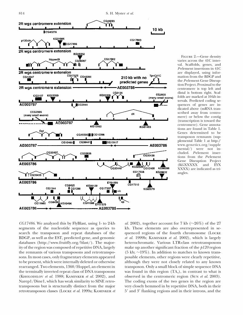
Figure 2.—Gene densityvaries across the 41C inter-val. Scaffolds, genes, andP-element insertions in 41Care displayed, using infor-mation from the BDGP andthe P-element Gene Disrup-tion Project. Proximal to thecentromere is top left anddistal is bottom right. Scaf-folds are marked at 10-kb in-tervals. Predicted coding se-quences of genes are in-dicated above (mRNA tran-scribed away from centro-mere) or below the contig(transcription is toward thecentromere). Gene annota-tions are found in Table 1.Genes determined to betransposon remnants (sup-plemental Table 1 at http://www.genetics.org/supplemental/) were not in-cluded. P-element inser-tions from the P-elementGene Disruption Project(KGXXXXX and EYXXXXX) are indicated as tri-angles.
CG17486. We analyzed this by FlyBlast, using 1- to 2-kb al. 2002), together account for 7 kb (�26%) of the 27kb. These elements are also overrepresented in se-segments of the nucleotide sequence as queries to
search the transposon and repeat databases of the quenced regions of the fourth chromosome (Lockeet al. 1999b; Kaminker et al. 2002), which is largelyBDGP, as well as the EST, predicted gene, and genomic
databases (http://www.fruitfly.org/blast/). The major- heterochromatic. Various LTR-class retrotransposonsmake up another significant fraction of the p120 regionity of the region was composed of repetitive DNA, largely
the remnants of various transposons and retrotranspo- (5 kb; �19%). In addition to matches to known trans-posable elements, other regions were clearly repetitive,sons. In most cases, only fragmentary elements appeared
to be present, which were internally deleted or otherwise although they were not closely related to any knowntransposon. Only a small block of simple sequence DNArearranged. Two elements, 1360/Hoppel, an element in
the terminally inverted repeat class of DNA transposons was found in this region (TAn), in contrast to what isobserved in the centromeric region (Sun et al. 2003).(Kholodilov et al. 1988; Kaminker et al. 2002), and
Narep1/Dine1, which has weak similarity to SINE retro- The coding exons of the two genes in the region arevery closely hemmed in by repetitive DNA, both in theirtransposons but is structurally distinct from the major
retrotransposon classes (Locke et al. 1999a; Kaminker et 5� and 3� flanking regions and in their introns, and the

815The Heterochromatin-Euchromatin Junction
Figure 3.—The region surroundingp120 is highly repetitive. An analysis of23 kb of sequence spanning the p120gene and extending distal to CG17486is presented. The exons and introns ofthe two genes are displayed as blackboxes and thin lines, respectively. Thegray box indicates an incorrectly anno-tated “fifth exon” of p120, which wasremoved in WGS3. The majority of thesequence surrounding these two genesis highly repetitive. Lines with arrowheadsrepresent blocks of sequence that are iden-tifiable as remnants of known transposons.Colored rectangles represent other re-petitive DNA, as indicated in the key.Portions of the remaining DNA may berepetitive as well.
p120 3� untranslated region includes a retrotransposon We then carried out a screen for lethal mutationsuncovered by Df(2R)M41A8 (Figure 4B). We EMS muta-remnant. In the light region of 2L, exons are also embed-
ded in repetitive DNA (Hoskins et al. 2002). genized males carrying an isogenic second chromosomemarked with the recessive visible markers cn and bw andA genetic screen for essential genes in the p120 re-
gion: Having this picture of predicted gene content as a crossed them to females carrying a second chromosomebalancer (see materials and methods). Balanced F1foundation, we initiated genetic analysis of the essential
genes in the region. Our initial goal was to obtain muta- males were individually mated to balanced females car-rying Df(2R)M41A8, and crosses were screened for thosetions in p120, which encodes a component of the cell-
cell adherens junction and is well conserved in all ani- in which all of the progeny carried the balancer—i.e.,stocks in which a new mutation that was lethal overmals thus far examined (reviewed in Anastasiadis and
Reynolds 2000). We began with the hypothesis that the al Df(2R)M41A8 chromosome had been induced.Unbalanced progeny were also scored for visible pheno-p120 would be an essential gene and thus set out to
collect lethal mutations in the region to which it mapped. types. Candidate lethal or visible mutations were re-tested to verify the original result. We screened 6284We first used in situ hybridization to polytene chromo-
somes to map p120 to a region in 41C defined by the chromosomes and recovered 226 lethal mutations and5 visible mutations. The 5 visible mutants all share theoverlap between Df(2R)M41A10 and Df(2R)M41A8
(Myster et al. 2003; Figure 4A). In addition, we deter- same partially penetrant phenotype when trans -hetero-zygous with Df(2R)M41A8: they have ectopic wing veinsmined that p120 was not deleted by Df(2R)nap1, but was
deleted by Df(2R)M41A4 (data not shown). Our analysis posterior to longitudinal vein 5. To date, these have notbeen analyzed further.used polytene chromosomes, which have lower cytologi-
cal resolution than mitotic chromosomes. Previous anal- Placing deficiencies on the physical map and usingthem to map new mutations: In addition to the defi-ysis of mitotic chromosomes suggests that Df(2R)M-
41A10 removes the entire 2R mitotic heterochromatin ciencies we initially analyzed, we obtained from othersor generated (see below) a number of other deficiencies(h39–h46), while Df(2R)M41A4 deletes only h46 (Dimi-
tri 1991), suggesting that p120 maps to h46, a conclu- in the region, many of which were smaller than thatused for the screen (for purposes of this analysis, wesion supported by the recent work of Corradini et al.
(2003). We obtained from our colleagues alleles of the hypothesize that these represent deficiencies ratherthan more complex rearrangements—while the latterknown genes in the region: Nipped-A, Nipped-B, l(2)41Ae,
l(2)41Af, l(2Rh)IR3, and l(2Rh)IR23 (Hilliker 1976; possibility remains, the data below are consistent withmost or all being simple deficiencies). We characterizedDimitri et al. 1997; Rollins et al. 1999). We also initi-
ated a search for new mutations. We selected Df(2R)M- existing and newly generated chromosomal deficienciesin two ways: we mapped their endpoints on the physical41A8 for further genetic analysis as it was the smallest
deficiency in our initial analysis that removed p120, and map by PCR, and we characterized them genetically bycrossing them both to the preexisting complementationit was a relatively healthy stock.

816 S. H. Myster et al.
us to place all of the mutations into complementationgroups, many of which were ordered with respect toone another (Figure 6; unordered complementationgroups are joined by brackets). Interestingly, 14 of thecomplementation groups map more centromere proxi-mal within the heterochromatin, in a region proximalto the contiguously assembled sequence (Hoskins et al.2002). The deficiencies also allowed us to connect thegenetic and physical maps by providing common pointsof reference. We mapped alleles of the cloned geneNipped-B (Rollins et al. 1999), as well as mutations inp120 and CG17486 (from the P-mobilization screen de-scribed below), providing three additional anchor pointsbetween the physical and genetic maps. Interestingly,our EMS screen generated many deficiencies, in addi-tion to the expected point mutations (Figure 6). Someare relatively small and fail to complement alleles atonly two loci whereas others are quite large and fail tocomplement all of the mutant genes generated in thescreen. Two deficiencies extend even more distally, fail-ing to complement bub1, which lies outside Df(2R)M41A8.
Generating additional deletions in the p120 region:None of the complementation groups from our initialanalysis was a good candidate for a mutation in p120.Only one initially mapped to the same deficiency inter-
Figure 4.—Mutagenesis screen to identify lethal mutations val as p120, and sequencing of the p120 coding regionat the 2R heterochromatin/euchromatin junction. (A) Pre-from that mutant line [l(2)41Af] revealed no mutations.viously identified lethal loci and overlapping deficiencies inWe thus needed an alternate approach. Fortunately,the region. Df(2R)M41A8 and Df(2R)M41A10 remove p120
whereas Df(2R)nap1 does not (Myster et al. 2003). (B) Outline by this point the P-element screen/Gene Disruptionof the strategy for the EMS mutagenesis screen to generate Project of the Bellen/Rubin/Spradling labs had begunrecessive lethal mutations uncovered by Df(2R)M41A8 (see generating and mapping new P-element insertions (H.materials and methods for details).
Bellen, R. Hoskins, R. Levis, G. Luo, G. M. Rubinand A. C. Spradling, unpublished data; http://flypush.imgen.bcm.tmc.edu/pscreen/) and had used as one ofgroups in the region and to our newly generated muta-
tions. Deficiency endpoints were mapped by PCR ampli- their P elements the SUPorP P element. This carries awhite� gene surrounded by insulator elements from thefication from multiple DNA preparations from single
homozygous deficiency embryos (selected using a GFP- suppressor of Hairy wing, helping insulate the gene fromchromosomal position effects (Roseman et al. 1995). Itmarked balancer), using primer pairs throughout the
region. For each DNA preparation we used a set of also carries a yellow� gene outside the insulators. Previ-ous work suggested that insertions of this P elementprimers from outside the region as a positive control
for the quality of the DNA, and we used a wild-type would be more effectively recovered from the hetero-chromatin (Roseman et al. 1995), and this has beenstrain as a positive control for each primer pair. Because
of the repetitive nature of most of the DNA, we selected borne out in our region of interest. The P-element GeneDisruption Project recovered at least 14 new P-elementprimer pairs from the coding sequence of predicted
genes, with the result that our resolution is limited by insertions in 41C, 12 of which are SUPorP insertions(Figure 2). Most are intergenic insertions and are viable.the density of predicted genes in a region. This an-
chored the deficiency map on the physical map (Figure Even these SUPorP insertions are biased toward the moredistal scaffolds.5). Our mapping of Df(2R)M41A10 is also consistent
with the mapping of BAC clones by FISH onto chromo- One of these insertions, KG01086, is �7 kb 3� to p120and 2 kb 5� of CG17486. This insertion is viable andsomes carrying this deficiency (Corradini et al. 2003).
We then characterized the lethal mutations we gener- fertile. We mobilized this insertion (see materials andmethods), generating 401 putative mobilizations fromated in our screen (Figure 6). We first crossed them to
a subset of the deficiencies in the region, allowing us 600 crosses. Among these was one relatively large defi-ciency, Df(2R)247, which deletes many genes (Figuresto assign them to given deficiency intervals. We then
crossed them to additional deficiencies, known muta- 5 and 6). We used this deficiency in the mapping ofcomplementation groups described above. The mobili-tions in the region, and to one another. This allowed

817The Heterochromatin-Euchromatin Junction
Figure 5.—Placing deficiencies on the physical map. At the top is a diagram of the physical map (this is only roughly toscale—a correctly scaled version is presented in Figure 2). Genes used in defining the endpoints of deficiencies are indicatedabove each scaffold. The region indicated by a dotted line has not been assembled into finished sequence and thus was notincluded in this analysis. The centromere is to the left. Deficiency endpoints were determined by PCR amplification of codingsequence from the indicated genes, using homozygous mutant genomic DNA as a template (see materials and methods).Asterisks indicate deficiencies generated by mobilization of the KG1086 SUPorP P element.
zation of KG01086 also generated smaller deficiencies ciencies: TpnC41C, CG3107, CG2944, CG3136, and CG2905(Figure 6). We initially used RT-PCR to analyze tran-confined to the immediate region of p120 and its neigh-
boring genes. We mapped these 401 lines using a stan- scripts from 10 different Nipped-A alleles, hypothesizingthat one of these alleles might not produce a stabledard set of PCR reactions, searching for deletions with
one endpoint in the P element and the other in flanking mRNA. However, a product of the predicted size wasgenerated, using exonic primers designed to amplifyDNA (the mapping of those that delete p120 is de-
scribed in detail in Myster et al. 2003). We began with CG3107, CG2944, CG3136, and CG2905 (data not shown).Of the five genes in the region, CG2905 is the largest,two primer pairs: one within the P-element inverted
repeat and one in the DNA flanking the insertion to spanning �35 kb, containing 15 predicted exons, andencoding a 3435-amino-acid predicted protein that isthe right or left. We scored for the presence or absence
of a PCR product and used a primer pair from outside the homolog of mammalian TRRAP and yeast Tra1(Grant et al. 1998; Kusch et al. 2003). Because Nipped-Athe region as a positive control for the quality of the
DNA preparation. For lines in which one end of the P was mutated the most frequently in our screen (36 al-leles), we hypothesized that Nipped-A alleles might haveelement was deleted, we then used primer pairs within
the coding exons of p120, CG40293, and CG17486 to mutations in the CG2905 gene. The CG2905 codingregion was sequenced from three alleles of Nipped-A [twoamplify DNA from homozygous mutant lines to deter-
mine the extent of the deletion (for all PCR reactions, EMS-induced alleles generated in our study (l(2)NC116and l(2)NC186) and a -ray-induced allele (Nipped-A357.2;the KG01086 strain was used as a positive control). Rep-
resentative PCR data for the strains deleting p120 can Rollins et al. 1999)] . In l(2)NC116, a G-to-A transitionwas identified in the first base of the intron followingbe seen in Myster et al. (2003). This revealed deletions
in both directions of a variety of sizes. One, Df(2R)Dark2, exon 4. This position lies in the highly conserved GTdinucleotide in the 5� splice site consensus sequencedeletes CG17486, but does not extend into Nipped B (as
determined genetically). This deletion is viable, demon- (Mount et al. 1992) and thus should disrupt propersplicing (Figure 7, top). In l(2)NC186, an A-to-T transver-strating that CG17486 is not essential. Two others delete
p120 and do not extend into the next gene—the pheno- sion that is predicted to result in a nonconservativevaline to aspartic acid missense mutation at amino acidtype of these is described in detail elsewhere (Myster et
al. 2003)—but they are viable and fertile, demonstrating 885 was identified. This lies in a region conserved inthe Drosophila, human, and Arabadopsis homologs: allthat p120 is not essential. Finally, Df(2R)244 deletes both
p120 and CG40293. This deletion is viable and fertile, have valine at this position (Figure 7, bottom). No muta-tions in the CG2905 predicted coding region were iden-demonstrating that CG40293 is also not essential.
Correlating the genetic and physical maps: We then tified in Nipped-A357.2, but due to the complex genomicstructure of NippedA, it may be that this -ray-inducedused our alignment of the genetic and physical maps
to identify a candidate for the Nipped-A gene. Nipped-A allele results from a DNA rearrangement with a break-point in an intron or 5� to the coding sequences.was originally identified as a modifier of the phenotype
of the effects of certain cut mutations on the wing. It is We identified one additional anchor between the ge-netic and physical maps by examining additional SUPorPthe sole complementation group that fails to comple-
ment both the Nipped-D and Df(2R)nap1 deficiency insertion lines in 41C whose physical location has beendetermined by sequence analysis of the insertion junc-strains. Five predicted genes are removed by these defi-

818 S. H. Myster et al.
Figure 6.—Correlating the genetic andphysical maps. At the right is the set of over-lapping deficiencies correlated with thephysical map, as displayed in Figure 5. Tothe left of them is the genetic map, withlethal complementation groups ordered onthe basis of complementation data with theset of overlapping deficiencies (distanceson the genetic map are arbitrary). The or-der of loci within deficiency intervals hasnot been determined, and brackets join lociunordered with respect to one another.The number of alleles for each complemen-tation group generated in our screens isindicated in parentheses. Dashed lines rep-resent anchor points where complementa-tion groups (including the nonessentialgenes CG40293, p120, and CG17486) havebeen assigned to mutations in identifiedgenes. At the far left are deficiencies gener-ated in our EMS mutagenesis screen, theendpoints of which have been mapped ge-netically but that have not been placed onthe physical map. The extent of each dele-tion was determined by complementationtesting against the lethal loci. Multiple de-ficiencies that fail to complement the sameloci are listed together. The distal endpointof two deficiency lines has not been deter-mined and is indicated by an arrowhead.

819The Heterochromatin-Euchromatin Junction
somes by FISH with BACs from the assembled sequencesuggests that these will map in chromosome region h45or more proximal (Corradini et al. 2003)]. Thus thisregion of the heterochromatin may contain many essen-tial loci that remain to be molecularly analyzed (theonly molecularly characterized mutation in this proxi-mal region is rolled). While we clearly did not reachsaturation (many complementation groups have a singleallele), our data also begin to suggest that the hetero-
Figure 7.—Nipped-A mutations affect CG2905/Tra1/ chromatin may contain many nonessential althoughTRRAP. (Top) Nucleotide sequence of the junction between conserved loci. We directly identified three such genes,exon 4 and the downstream intron of CG2905, from cn bw,
p120, CG40293, and CG17486, and the excess of identi-the isogenic stock in which the mutations were induced, andfied CGs to genetically identified genes in the regionNipped-A116. This mutation alters the conserved GT dinucleo-
tide that is an essential part of the splice donor site (Mount between p120 and vulcan suggests that a number ofet al. 1992). (Bottom) A portion of the predicted amino acid other loci may be nonessential.sequence of CG2905 (amino acids 878–897) and the corre- Heterochromatin and unique coding DNA sequences:sponding region of its human and Arabidopsis homologs.
The pericentric heterochromatin is composed of theIdentical residues are indicated by white type in black boxes,centromeric region, whose composition is largely simplewhile similar amino acids are indicated by black type in gray
boxes. The valine residue affected by Nipped-A186 is indicated. sequence DNA arranged in tandem repeats (Sun et al.1997, 2003), and the “boundary region,” where transpo-sons comprise much of the sequence (e.g., Hoskins et
tion. Of 17 insertion lines, only 1, KG10496, appears to al. 2002). Genes are absent from the centromere (withbe lethal, as assessed by the presence or absence of the exception of scattered retrotransposons), while inhomozygous flies in the stocks. This line is inserted into the boundary region gene density is quite low, confinedthe coding region of CG8426, a predicted transcription to small islands of unique coding sequences inter-factor. The physical location predicts that the insertion spersed throughout. Previous mutational screens identi-
fied a small number of lethal loci in the heterochroma-line would fail to complement DF(2R)M41A8 and Df(2R)tin of 2R and in this study we report the generation ofnap1 and would complement Nipped-D and M(2)41A2.additional alleles of each (Hilliker 1976; Dimitri etOur complementation tests confirmed this (data notal. 1997; Rollins et al. 1999). Surprisingly, our screensshown). Testing of EMS lines in the region identifiedidentified a number of new lethal loci in the heterochro-l(2)NC136 as allelic to the KG10496 insertion (Figure 6).matin proximal to the published sequence. This sug-gests that there may be many more essential loci inthis region of the heterochromatin than was previouslyDISCUSSIONthought and fits well with recent work by the genome
Our interest in the proximal region of the second project that predicts �450 genes in the heterochroma-chromosome was initiated by the fact that p120, a gene tin as a whole (Hoskins et al. 2002; Misra et al. 2002).that encodes a component of adherens junctions, is One reason for the earlier underestimate of essentiallocated at polytene band 41C. Core components of ad- gene number in the 2R heterochromatin is that earlierherens junctions are required to establish cellular adhe- screens likely did not reach saturation, which is alsosive contacts and mutations in many adherens junction likely for our own screen, as described above. A secondcomponents are embryonic lethal (for reviews see Yap et potential cause of the previous underestimate is theal. 1997; Tepass et al. 2001). With the goal of identifying apparent tendency for mutagens to generate deficien-mutations in p120, two screens for mutations in the cies at a high rate in this region (Figure 6, left; seeregion were performed. In addition to providing infor- below). For example, we interpret our data to suggestmation about the phenotype of p120, which was recently that one of the lethal loci described in an earlier screen,published elsewhere (Myster et al. 2003), the screen l(2)41Ae (Hilliker 1976), may be a deletion, as it failsprovides an example of how classical genetic approaches to complement seven of our complementation groupscan be combined with sequence information, annota- (Figure 6). If this is the correct interpretation of thesetion, and bioinformatics tools emerging from the ge- data, it would suggest that the number of lethals in thenome project to create a combined genetic and bio- region has been underestimated. However, two caveatsinformatics picture of a heterochromatic region, as a to this conclusion must be noted. First, Hilliker offeredresource for future work on the genes within it. an alternate explanation for the behavior of l(2)41Ae in
We identified lethal mutations in 26 loci, 21 of which complementation tests. He suggested that it is a complexappear to be novel. Fourteen of the complementation locus, with an unusual degree of intraallelic comple-groups map in the heterochromatin proximal to the mentation, and thus suggested that all of the comple-
mentation groups in this region are alleles of a singleassembled sequence [recent analysis of mitotic chromo-

820 S. H. Myster et al.
complex locus. This is a possibility, although we favor p120. An added advantage of screening at the molecularlevel is that nonessential mutations can be identified.our own interpretation. Second, as some of our comple-Our screen revealed that mutations in p120, CG40293,mentation groups contain only a single allele, their pat-and CG17486 are viable and fertile. These illustrate howtern of complementation is slightly less secure than thatthe growing bank of P-element insertions in the hetero-of complementation groups with multiple alleles.chromatin will be a great resource to identify or analyzeThe SUPorP transposable element allows genetic ac-both lethal and nonessential heterochromatic loci incess to heterochromatin: The repetitive nature of het-the future.erochromatin made it challenging to clone, sequence,
Mutagens and repetitive DNA: EMS is generally con-and correctly assemble in large-scale sequencing efforts.sidered to be a point mutagen, and previous mutagene-Recent efforts have made inroads into these regions ofsis of the euchromatin supports this (e.g., Gray et al.the genome (Hoskins et al. 2002; Sun et al. 2003). It is1991). We were thus surprised to find that many of ourestimated that �60 Mb of heterochromatin are in theEMS-generated alleles (Figure 6, left), as well as allelesgenome of Drosophila females and 90 Mb of hetero-generated in earlier screens in the region (Rollinschromatin in males (Celniker et al. 2002; Hoskins etet al. 1999), are deficiencies that fail to complemental. 2002). A genomics-based estimate of total hetero-multiple loci. We suspect that the repetitive DNA in thechromatic gene number will have to await the comple-region may contribute to this. After induction of a newtion of the sequence in this region, but current estimatesmutation, the cellular repair mechanisms are activatedsuggest that there are �450 genes are in the heterochro-and use the complementary strand as a template formatin as a whole (Hoskins et al. 2002; Misra et al. 2002).repair. Due to the highly repetitive nature of the region,Genetics-based approaches provide an alternative meth-we imagine that in the process of repairing individualod for identifying genes in heterochromatin. P-elementbase-pair mutations, misalignment could occur, resultingtransposons can effectively insert into heterochromatinin a looping out of a region of DNA. This could lead(Roseman et al. 1995; Yan et al. 2002), and thus the lowto the generation of a deficiency.number of insertions previously identified in hetero-
Anchoring the genetic and physical maps: Our EMSchromatin is probably due to its gene silencing proper-screen generated additional alleles of each of the pre-ties (for a review see Weiler and Wakimoto 1995), pre-viously identified loci in the p120 region, including 36venting the expression of the scorable markers. Whennew mutations in Nipped-A and 16 new mutations inwe started our work no known P-element insertions wereNipped-B. Conversely, 16 of the newly identified comple-close to p120, ruling out P-element mobilization as amentation groups contain a single member. Taken to-viable mutagenesis strategy. Fortunately, modified P ele-gether, these results imply that some loci are highly
ments have provided access to the heterochromatin.mutable and our screens are probably not saturating.
The SUPorP was designed to allow efficient insertion in The published Drosophila genomic sequence and itssilenced regions. In it, the white� selectable marker is annotation provide a powerful data set that could beflanked by insulator elements carrying Suppressor of used to learn more about our many newly identifiedhairy wing binding sites, effectively blocking the silenc- loci (Adams et al. 2000 ; Celniker et al. 2002; Hoskinsing properties of the heterochromatin (Roseman et al. et al. 2002; Misra et al. 2002). We used the set of overlap-1995). Yan et al. (2002) utilized this element as well, ping deficiency strains to genetically order many of ourscreening for variegated expression of the yellow� se- complementation groups with respect to each other andlectable marker, which is not flanked by insulators ele- exploited the deficiency lines as an inroad to correlatements. Together, these efforts have allowed the identi- the genetic and physical maps. In addition, the Nipped-Bfication of many new insertions into previously untagged gene was previously analyzed at the molecular level andregions of the genome (Yan et al. 2002; H. Bellen, R. thus provided an additional anchor point between theHoskins, R. Levis, G. Luo, G. M. Rubin et al., unpub- two maps (Rollins et al. 1999). We focused on the 14lished data; http://flypush.imgen.bcm.tmc.edu/pscreen/). complementation groups that are distal to p120. Nipped-
These insertions provide the ability to genetically ma- A is the only locus that fails to complement both thenipulate the surrounding region, both through the di- Nipped-D and Df(2R)nap1 deficiencies (see Figure 6).rect insertional inactivation of genes and through mobi- Five genes are predicted to be located in this interval.lization of the P elements to create new insertions or Interestingly, one of these genes, CG2905, is very large,deletions. Our work provides an illustration of each of with 15 exons encoding a 3435-amino-acid protein. Duethese. We found that l(2)NC136 is allelic to the KG10496 to the high mutability of Nipped-A (36 alleles in our EMSinsertion. After mobilizing the P element in the p120/ screen) we suspected that CG2905 encoded Nipped-A.CG17486 intragenic region, we identified five deficien- This appears to be the case, as mutations in the CG2905cies of variable length extending in both directions from gene were identified in two alleles of Nipped-A that werethe original insertion site among 600 mobilization generated in this study (Figure 7).events. In addition, a local hop identified an additional Nipped-A was originally identified in a screen for genes
that modified the phenotype of a regulatory allele oflethal complementation group [l(2)309] proximal to

821The Heterochromatin-Euchromatin Junction
genome of Drosophila melanogaster : the Adh region. Genetics 153:the cut gene (Rollins et al. 1999). cut has a complex179–219.
regulatory region, with distant enhancers that regulate Casso, D., F. Ramirez-Weber and T. B. Kornberg, 2000 GFP-taggedbalancer chromosomes for Drosophila melanogaster. Mech. Dev.tissue-specific expression. The cut mutation used in the91: 451–454.screen was caused by an insertion of the gypsy retro-
Celniker, S. E., D. A. Wheeler, B. Kronmiller, J. W. Carlson, A.transposon, which has the ability to block interactions Halpern et al., 2002 Finishing a whole-genome shotgun: release
3 of the Drosophila melanogaster euchromatic genome se-of distal enhancers with promoters. Mutations in a num-quence. Genome Biol. 3: RESEARCH0079.ber of different genes were isolated in this screen. They
Cleveland, D. W., Y. Mao and K. F. Sullivan, 2003 Centromeresinclude mutations of transcriptional regulators like and kinetochores: from epigenetics to mitotic checkpoint signal-
ing. Cell 112: 407–421.Chip and Mastermind, as well as mutations in Nipped-B,Corradini, N., F. Rossi, F. Verni and P. Dimitri, 2003 FISH analysisa member of a family of proteins involved in chromatid
of Drosophila melanogaster heterochromatin using BACs and Pcohesion, chromosome condensation, and DNA repair. elements. Chromosoma 112: 26–37.
Dimitri, P., 1991 Cytogenetic analysis of the second chromosomeOur identification of Nipped-A as a mutation in CG2905heterochromatin of Drosophila melanogaster. Genetics 127: 553–fits into this picture, as CG2905 encodes the fly homolog564.
of Tra1/TRRAP, a component of SAGA/GNAT-type Dimitri, P., and N. Junakovic, 1999 Revising the selfish DNA hy-pothesis: new evidence on accumulation of transposable elementsmultiprotein histone acetyltransferase complexes (Grantin heterochromatin. Trends Genet. 15: 123–124.et al. 1998; Kusch et al. 2003). Tra1/TRRAP is a distant Dimitri, P., B. Arca, L. Berghella and E. Mei, 1997 High genetic
relative of ATM, the gene mutated in the human disease instability of heterochromatin after transposition of the LINE-like I factor in Drosophila melanogaster. Proc. Natl. Acad. Sci.ataxia-telangiectasia (reviewed in Shiloh 2000), and isUSA 94: 8052–8057.thus thought by analogy to be a protein kinase or possi- Gloor, G. B., C. R. Preston, D. M. Johnson-Schlitz, N. A. Nassif,
bly a lipid kinase. Our identification of Nipped-A with R. W. Phillis et al., 1993 Type I repressors of P-element mobil-ity. Genetics 135: 81–95.Tra1/TRRAP opens the way for genetic analysis of the
Grant, P. A., D. Schieltz, M. G. Pray-Grant, J. R. Yates, III androle of this protein complex in transcriptional regula- J. L. Workman, 1998 The ATM-related cofactor Tra1 is a com-tion in Drosophila. ponent of the purified SAGA complex. Mol. Cell 2: 863–867.
Gray, M., A. Charpentier, K. Walsh, P. Wu and W. Bender, 1991Our alignment of the genetic and physical maps pro-Mapping point mutations in the Drosophila rosy locus usingvides a framework for future molecular identification denaturing gradient gel blots. Genetics 127: 139–149.
studies. It is our hope that future investigators will utilize Grewal, S. I., and S. C. Elgin, 2002 Heterochromatin: new possibili-ties for the inheritance of structure. Curr. Opin. Genet. Dev. 12:our reagents and view of the heterochromatin-euchro-178–187.
matin region of 2R as a starting point for examining Grigliatti, T. A., 1998 Mutagenesis, pp. 55–83 in Drosophila: APractical Approach, edited by D. B. Roberts. Oxford Universitythe function of the genes in this interesting region ofPress, New York.the genome.
Henikoff, S., 2000 Heterochromatin function in complex genomes.Biochim. Biophys. Acta 1470: 01–08.We thank P. Cayirlioglu for advice on and V. Morel for assistance
Hilliker, A. J., 1976 Genetic analysis of the centromeric heterochro-with P-element mobilization; K. Maners for helping with complemen-matin of chromosome 2 of Drosophila melanogaster: deficiency map-tation tests; R. Hoskins, A. Hilliker, J. Gates, B. McCartney, M. Price,ping of EMS-induced lethal complementation groups. Geneticsand the two anonymous reviewers for comments on the manuscript; 83: 765–782.
and members of the Peifer lab for helpful discussions. We are also Hoskins, R. A., C. R. Nelson, B. P. Berman, T. R. Laverty, R. A.very grateful to A. Hilliker, P. Dimitri, D. Dorsett, the P-element Gene George et al., 2000 A BAC-based physical map of the majorDisruption Project, and the Bloomington Drosophila Stock Center autosomes of Drosophila melanogaster. Science 287: 2271–2274.
Hoskins, R. A., C. D. Smith, J. W. Carlson, A. B. Carvalho, A. Halpernfor fly stocks and are especially grateful to R. Rollins and D. Dorsettet al., 2002 Heterochromatic sequences in a Drosophila whole-as well as R. Hoskins and the Berkeley Drosophila Genome Project forgenome shotgun assembly. Genome Biol. 3: RESEARCH0085–0085.sharing unpublished data and for many helpful discussions. This work
Kaminker, J. S., C. M. Bergman, B. Kronmiller, J. Carlson, R.was supported by National Institutes of Health grant GM47857 to M.Svirskas et al., 2002 The transposable elements of the Drosoph-
Peifer. S. H. Myster was supported by National Institutes of Health ila melanogaster euchromatin: a genomics perspective. GenomeNational Research Service Award GM19888, R. Cavallo by a Depart- Biol. 3: RESEARCH0084.ment of Defense Breast Cancer Research Program predoctoral fellow- Kholodilov, N. G., V. N. Bolshakov, V. M. Blinov, V. V. Solovyovship, C. T. Anderson by the Pfizer Summer Undergraduate Research and I. F. Zhimulev, 1988 Intercalary heterochromatin in Dro-
sophila. III. Homology between DNA sequences from the Y chro-Fellowship program, C. T. Anderson and S. Bhotika by Thompsonmosome, bases of polytene chromosome limbs, and chromosomeUndergraduate Research awards, and M. Peifer in part by a Depart-4 of D. melanogaster. Chromosoma 97: 247–253.ment of Defense Breast Cancer Research Program Career Develop-
Kusch, T., S. Guelman, S. M. Abmayr and J. L. Workman, 2003ment Award and by the Welsh Distinguished Term Professorship. Two Drosophila ada2 homologues function in different multipro-tein complexes. Mol. Cell. Biol. 23: 3305–3319.
Locke, J., L. T. Howard, N. Aippersbach, L. Podemski and R. B.Hodgetts, 1999a The characterization of DINE-1, a short, in-terspersed repetitive element present on chromosome 4 and inLITERATURE CITEDthe centric heterochromatin of Drosophila melanogaster. Chro-
Adams, M. D., S. E. Celniker, R. A. Holt, C. A. Evans, J. D. Gocayne mosoma 108: 356–366.et al., 2000 The genome sequence of Drosophila melanogaster. Locke, J., L. Podemski, K. Roy, D. Pilgrim and R. Hodgetts, 1999bScience 287: 2185–2195. Analysis of two cosmid clones from chromosome 4 of Drosophila
Anastasiadis, P. Z., and A. B. Reynolds, 2000 The p120 catenin melanogaster reveals two new genes amid an unusual arrange-family: complex roles in adhesion, signaling and cancer. J. Cell ment of repeated sequences. Genome Res. 9: 137–149.Sci. 113: 1319–1334. Miklos, G. L., M. T. Yamamoto, J. Davies and V. Pirrotta, 1988
Ashburner, M., S. Misra, J. Roote, S. E. Lewis, R. Blazej et al., Microcloning reveals a high frequency of repetitive sequencescharacteristic of chromosome 4 and the beta-heterochromatin1999 An exploration of the sequence of a 2.9-Mb region of the

822 S. H. Myster et al.
of Drosophila melanogaster. Proc. Natl. Acad. Sci. USA 85: 2051– Shiloh, Y., 2000 ATM: sounding the double-strand break alarm.Cold Spring Harbor Symp. Quant. Biol. 65: 527–533.2055.
Misra, S., M. A. Crosby, C. J. Mungall, B. B. Matthews, K. S. Spradling, A. C., D. Stern, A. Beaton, E. J. Rhem, T. Laverty et al.,1999 The Berkeley Drosophila Genome Project gene disruptionCampbell et al., 2002 Annotation of the Drosophila melanogas-
ter euchromatic genome: a systematic review. Genome Biol 3: project: single P-element insertions mutating 25% of vital Dro-sophila genes. Genetics 153: 135–177.RESEARCH0083.
Moazed, D., 2001 Common themes in mechanisms of gene silenc- Sullivan, B. A., M. D. Blower and G. H. Karpen, 2001 Determiningcentromere identity: cyclical stories and forking paths. Nat. Rev.ing. Mol. Cell 8: 489–498.
Mount, S. M., C. Burks, G. Hertz, G. D. Stormo, O. White et al., Genet. 2: 584–596.Sun, X., J. Wahlstrom and G. Karpen, 1997 Molecular structure1992 Splicing signals in Drosophila: intron size, information
content, and consensus sequences. Nucleic Acids Res. 20: 4255– of a functional Drosophila centromere. Cell 91: 1007–1019.Sun, X., H. D. Le, J. M. Wahlstrom and G. H. Karpen, 2003 Se-4262.
Muller, H. J., 1930 Types of viable variations induced by X-rays in quence analysis of a functional Drosophila centromere. GenomeRes. 13: 182–194.Drosophila. J. Genet. 22: 299–334.
Myster, S. H., R. Cavallo, C. T. Anderson, D. T. Fox and M. Peifer, Tepass, U., G. Tanentzapf, R. Ward and R. Fehon, 2001 Epithelialcell polarity and cell junctions in Drosophila. Annu. Rev. Genet.2003 Drosophila p120catenin plays a supporting role in cell
adhesion but is not an essential adherens junction component. 35: 747–784.Wallrath, L. L., 1998 Unfolding the mysteries of heterochromatin.J. Cell Biol. 160: 433–449.
Rollins, R. A., P. Morcillo and D. Dorsett, 1999 Nipped-B, a Curr. Opin. Genet. Dev. 8: 147–153.Drosophila homologue of chromosomal adherins, participates Weiler, K. S., and B. T. Wakimoto, 1995 Heterochromatin andin activation by remote enhancers in the cut and Ultrabithorax gene expression in Drosophila. Annu. Rev. Genet. 29: 577–605.genes. Genetics 152: 577–593. Yan, C. M., K. W. Dobie, H. D. Le, A. Y. Konev and G. H. Karpen,
Roseman, R. R., E. A. Johnson, C. K. Rodesch, M. Bjerke, R. N. 2002 Efficient recovery of centric heterochromatin P-elementNagoshi et al., 1995 A P -element containing suppressor of insertions in Drosophila melanogaster. Genetics 161: 217–229.hairy-wing binding regions has novel properties for mutagenesis Yap, A. S., W. M. Brieher and B. M. Gumbiner, 1997 Molecularin Drosophila melanogaster. Genetics 141: 1061–1074. and functional analysis of cadherin-based adherens junctions.
Rubin, G. M., M. D. Yandell, J. R. Wortman, G. L. Gabor Miklos, Annu. Rev. Cell Dev. Biol. 13: 119–146.C. R. Nelson et al., 2000 Comparative genomics of the eukary-otes. Science 287: 2204–2215. Communicating editor: J. Tamkun
Copyright © 2022 FDOKUMEN