Plasmodium falciparum heterochromatin protein 1 binds to tri-methylated histone 3 lysine 9 and is...
11
2596–2606 Nucleic Acids Research, 2009, Vol. 37, No. 8 Published online 6 March 2009 doi:10.1093/nar/gkp115 Plasmodium falciparum heterochromatin protein 1 binds to tri-methylated histone 3 lysine 9 and is linked to mutually exclusive expression of var genes Karla Pe ´ rez-Toledo 1 , Ana Paola Rojas-Meza 1,2 , Liliana Mancio-Silva 2 , Nora Adriana Herna ´ ndez-Cuevas 1 , Dulce Maria Delgadillo 1 , Miguel Vargas 1 , Santiago Martı´nez-Calvillo 3 , Artur Scherf 2 and Rosaura Hernandez-Rivas 1, * 1 Departamento de Biomedicina Molecular, Centro de Investigacio ´ n y de Estudios Avanzados del Instituto Polite ´ cnico Nacional (IPN), Apartado Postal 14-740, 07360, Me ´ xico D. F., Me ´ xico, 2 Institut Pasteur, Unite ´ de Biologie des Interactions Ho ˆ te-Parasite – CNRS URA2581. 25, Rue du Dr. Roux, 75724 Paris, France and 3 Unidad de Biomedicina, Facultad de Estudios Superiores Iztacala, Universidad Nacional Auto ´ noma de Me ´ xico. Av. de los Barrios 1, Col. Los Reyes Iztacala, Tlalnepantla, Edo. de Me ´ xico, CP 54090, Me ´ xico Received December 1, 2008; Revised February 6, 2009; Accepted February 10, 2009 ABSTRACT Increasing experimental evidence shows a promi- nent role of histone modifications in the coordinated control of gene expression in the human malaria parasite Plasmodium falciparum. The search for the histone-mark-reading machinery that translates histone modifications into biological processes, such as formation of heterochromatin and antigenic variation is of foremost importance. In this work, we identified the first member of a histone modification specific recognition protein, an orthologue of heterochromatin protein 1 (PfHP1). Analysis of the PfHP1 amino-acid sequence revealed the presence of the two characteristic HP1 domains: a chromo- domain (CD) and a chromo shadow domain (CSD). Recombinant CD binds to di- and tri-methylated lysine 9 from histone H3, but not to unmodified or methylated histone H3 in lysine 4. PfHP1 is able to interact with itself to form dimers, underlying its potential role in aggregating nucleosomes to form heterochromatin. Antibodies raised against PfHP1 detect this molecule in foci at the perinuclear region. ChIP analysis using anti-PfHP1 shows that this protein is linked to heterochromatin of subtelo- meric non-coding repeat regions and monoallelic expression of the major virulence var gene family. This is the first report implicating an HP1 protein in the control of antigenic variation of a protozoan parasite. INTRODUCTION Plasmodium falciparum, the causal protozoan agent of the most severe form of human malaria, has a complex life cycle with two different hosts, the Anopheles mosquito and humans. In order to complete its life cycle, P. falciparum invades different types of cells and self-propagates in very distinct environments in the mosquito (gut, hemolymph and salivary glands) as well as in the human host (skin, liver and erythrocytes). Each of these distinct environ- ments exerts selective pressure related to morphological changes that force P. falciparum to exhibit differential gene expression during its life cycle (1,2). A surprising finding was the identification of a relatively low number of genes encoding transcription factors in the parasite, although the basal core transcriptional machinery and the protein-coding genes involved in nucleosome assembly and regulation of chromatin structure were found to be conserved (3–5). To date, no specific DNA-binding proteins have been identified other than PfMyb1 (6) and the ApiAp2 gene family (7). This suggested that epigenetic mechanisms play a significant role in controlling gene expression in Plasmodium (3,8–10). The importance of reversible chromatin modifications involving histone modifications and the chromatin-associated protein PfSir2 was first demonstrated for a subtelomeric gene family (var gene family) involved in the control of anti- genic variation in P. falciparum (10). Knock out of PfSir2 resulted in the de-repression of a fraction of the members of the var gene family (11). A screen for histone modifica- tions revealed that H3K9me3 plays a particular role in var gene repression (12,13) and H3K4me2/3 in transcriptional *To whom correspondence should be addressed. Tel: +(52) 55 5747 3325; Fax: +(52) 55 5747 3938; Email: [email protected] The authors wish it to be known that, in their opinion, the first two authors should be regarded as joint First Authors. ß 2009 The Author(s) This is an Open Access article distributed under the terms of the Creative Commons Attribution Non-Commercial License (http://creativecommons.org/licenses/ by-nc/2.0/uk/) which permits unrestricted non-commercial use, distribution, and reproduction in any medium, provided the original work is properly cited. by guest on January 22, 2015 http://nar.oxfordjournals.org/ Downloaded from
-
Upload
independent -
Category
Documents
-
view
4 -
download
0
Transcript of Plasmodium falciparum heterochromatin protein 1 binds to tri-methylated histone 3 lysine 9 and is...
2596–2606 Nucleic Acids Research, 2009, Vol. 37, No. 8 Published online 6 March 2009doi:10.1093/nar/gkp115
Plasmodium falciparum heterochromatin protein 1binds to tri-methylated histone 3 lysine 9 and islinked to mutually exclusive expression of var genesKarla Perez-Toledo1, Ana Paola Rojas-Meza1,2, Liliana Mancio-Silva2,
Nora Adriana Hernandez-Cuevas1, Dulce Maria Delgadillo1, Miguel Vargas1,
Santiago Martınez-Calvillo3, Artur Scherf2 and Rosaura Hernandez-Rivas1,*
1Departamento de Biomedicina Molecular, Centro de Investigacion y de Estudios Avanzados del InstitutoPolitecnico Nacional (IPN), Apartado Postal 14-740, 07360, Mexico D. F., Mexico, 2Institut Pasteur, Unitede Biologie des Interactions Hote-Parasite – CNRS URA2581. 25, Rue du Dr. Roux, 75724 Paris, France and3Unidad de Biomedicina, Facultad de Estudios Superiores Iztacala, Universidad Nacional Autonoma deMexico. Av. de los Barrios 1, Col. Los Reyes Iztacala, Tlalnepantla, Edo. de Mexico, CP 54090, Mexico
Received December 1, 2008; Revised February 6, 2009; Accepted February 10, 2009
ABSTRACT
Increasing experimental evidence shows a promi-nent role of histone modifications in the coordinatedcontrol of gene expression in the human malariaparasite Plasmodium falciparum. The search forthe histone-mark-reading machinery that translateshistone modifications into biological processes,such as formation of heterochromatin and antigenicvariation is of foremost importance. In this work, weidentified the first member of a histone modificationspecific recognition protein, an orthologue ofheterochromatin protein 1 (PfHP1). Analysis of thePfHP1 amino-acid sequence revealed the presenceof the two characteristic HP1 domains: a chromo-domain (CD) and a chromo shadow domain (CSD).Recombinant CD binds to di- and tri-methylatedlysine 9 from histone H3, but not to unmodifiedor methylated histone H3 in lysine 4. PfHP1 is ableto interact with itself to form dimers, underlyingits potential role in aggregating nucleosomes toform heterochromatin. Antibodies raised againstPfHP1 detect this molecule in foci at the perinuclearregion. ChIP analysis using anti-PfHP1 shows thatthis protein is linked to heterochromatin of subtelo-meric non-coding repeat regions and monoallelicexpression of the major virulence var gene family.This is the first report implicating an HP1 proteinin the control of antigenic variation of a protozoanparasite.
INTRODUCTION
Plasmodium falciparum, the causal protozoan agent of themost severe form of human malaria, has a complex lifecycle with two different hosts, the Anopheles mosquito andhumans. In order to complete its life cycle, P. falciparuminvades different types of cells and self-propagates in verydistinct environments in the mosquito (gut, hemolymphand salivary glands) as well as in the human host (skin,liver and erythrocytes). Each of these distinct environ-ments exerts selective pressure related to morphologicalchanges that force P. falciparum to exhibit differentialgene expression during its life cycle (1,2). A surprisingfinding was the identification of a relatively low numberof genes encoding transcription factors in the parasite,although the basal core transcriptional machinery andthe protein-coding genes involved in nucleosome assemblyand regulation of chromatin structure were found tobe conserved (3–5). To date, no specific DNA-bindingproteins have been identified other than PfMyb1 (6) andthe ApiAp2 gene family (7). This suggested that epigeneticmechanisms play a significant role in controlling geneexpression in Plasmodium (3,8–10). The importanceof reversible chromatin modifications involving histonemodifications and the chromatin-associated proteinPfSir2 was first demonstrated for a subtelomeric genefamily (var gene family) involved in the control of anti-genic variation in P. falciparum (10). Knock out of PfSir2resulted in the de-repression of a fraction of the membersof the var gene family (11). A screen for histone modifica-tions revealed that H3K9me3 plays a particular role in vargene repression (12,13) and H3K4me2/3 in transcriptional
*To whom correspondence should be addressed. Tel: +(52) 55 5747 3325; Fax: +(52) 55 5747 3938; Email: [email protected]
The authors wish it to be known that, in their opinion, the first two authors should be regarded as joint First Authors.
� 2009 The Author(s)This is an Open Access article distributed under the terms of the Creative Commons Attribution Non-Commercial License (http://creativecommons.org/licenses/by-nc/2.0/uk/) which permits unrestricted non-commercial use, distribution, and reproduction in any medium, provided the original work is properly cited.
by guest on January 22, 2015http://nar.oxfordjournals.org/
Dow
nloaded from
activity and epigenetic memory (12). These dynamic his-tone modifications occur mainly in the 50-UTR regionsof var genes. It was shown that histone modificationsalso play a major role in the regulation of non-var genes(12,14–16). The identification of factors able to ‘read’specific histone marks and translate them into changesin gene activity remains elusive in Plasmodium.
In this work, we identified the first plasmodial proteinthat displays features of the histone-reading machinerythat binds specifically to H3K9me2/3. The identifiedprotein is an orthologue to heterochromatin protein 1(PfHP1) and shows a specific location to perinuclearfoci. Chromatin immunoprecipitation (ChIP) assaysreveal that the PfHP1 protein is a component of subtelo-meric chromatin. Furthermore, our data links PfHP1 toepigenetically silenced var genes, since an active var geneis devoid of PfHP1 in its 50-UTR.
MATERIALS AND METHODS
Parasites
Plasmodium falciparum FCR3 strain was cultivatedaccording to standard culture conditions (17). Panningassays for selection of FCR3 parasites that transcribevar genes associated with CD36 and CSA binding havebeen done as previously described (18).
Recombinant proteins
A 801 bp DNA fragment of the PfHP1 gene was obtainedby RT–PCR. The sequences of direct and reverse oligonu-cleotides were: PfHP1EcoRI 50-CGGAATTCATGACGGGTCAGATGAAGAA-30 and PfHP1XhoI 50-CCGCTCGAGTTAAGCTGTACGGTATCTTAG-30, respectively.The PCR fragment was digested, retrieved and insertedinto the EcoRI-XhoI-digested pGEX4T1 vector(Amersham) fused to a GST-tag. The resulting constructwas sequenced and called GST-PfHP1. The primersPfHP1-CDEcoRI 50-CGGAATTCATGACAGGGTCAGATGAAGAA-30 and PfHP1-CDXhoI 50-CCGCTCGAGCTCATTAGCTTTCGATAAAAAATT-30 were usedto amplify a DNA fragment of 204 bp that containsPfHP1 chromodomain (CD) (PfHP1-CD). Integrity ofall recombinants clones was confirmed by sequencingwith 50 and 30 primers to the pGEX cloning site (Phar-macia). All of them were transformed into Escherichia coliDH5a strain. Expression of GST fusion protein wasinduced with 0.5mM IPTG at 308C for 4 h. GST fusionproteins were purified with glutathione sepharose(Pharmacia) by standard methods. The PfHP1 DNAfragment of 801 bp was cloned into BamHI 50-CGGAATTCATGACGGGTCAGATGAAGAA-30 and NotI sites50-ATAAGAATGCGGCCGCTTAAGCTGTACGGTATCTTAG-30 of the pROEXTM HTb vector (Invitrogen).
6HisPfHP1 protein was induced in BL21 bacterial cells byaddition of 0.6mM IPTG. Recombinant proteins werepurified using nickel-nitriloacetic acid resin (TALON)according to manufacturer protocol. Purified recombinantproteins were verified by Western blot.
Production of PfHP1 antibodies
The GST-PfHP1 recombinant protein was purified asrecommended by the manufacturers (see above). Forpolyclonal antibodies against PfHP1, two rabbits wereinoculated i.c. with 100 mg of GST-PfH1 protein (firstdose). The fusion protein was emulsified with completeadjuvant (Sigma). Following the inoculation series, ani-mals were sacrificed and serum was collected. Immunoglo-bulins were purified from serum by protein A-sepharose(Pharmacia) chromatography, following standardprocedures.
Nuclear and cytoplasmic extracts preparation
Nuclear and cytoplasmic extracts were prepared as pre-viously described (10,19) with some modifications.Briefly, 5� 109 parasites of an asynchronous cultureof FCR3P. falciparum strain were isolated from infectederythrocytes by saponin lysis, resuspended in 1ml of lysisbuffer (10mM HEPES, pH 7.9, 10mM KCl, 0.1mMEDTA, 0.1mM EGTA, 1mM DTT, 0.65% NP-40)and incubated 30min a 48C. Then, parasites were lysedby 200 strokes on a prechilled douncer homogenizer.The nuclei were collected by centrifugation. The super-natant containing the cytoplasmic fraction was recoveredand kept at �808C. The nuclei were purified by sucrosegradient centrifugation. For this purpose, the nuclei wereresuspended in 1ml of lysis buffer and the resultingsuspension was layered on 3ml of lysis buffer containing0.34M sucrose. The nuclei were resuspended in 100 ml ofextraction buffer (20mM HEPES, pH 7.9, 0.4M NaCl,1mM EDTA, 1mM EGTA and 1mM DTT). Following15min of vigorous shaking at 48C, the extract was centri-fuged and the supernatant containing nuclear proteinswas collected. All buffers used in this protocol containedprotease inhibitors (Complete, Roche).
Immunofluorescence microscopy
Immunofluorescence assays were performed as previouslydescribed (20). The parasites were fixed in suspensionwith 4% paraformaldehyde solution for 15min on ice.Next, the fixed parasites were incubated with the primaryantibody for 60min at room temperature followed byincubation for 30min with a secondary antibody conju-gated with fluorochrome and deposited on microscopeslides. The final antibody dilutions were rabbit anti-PfHP1 1:500, rat anti-PfSir2 1:50, rat anti-PfNop1 1:50,Alexa Fluor 568 goat anti-rabbit highly cross-absorbed1:500 and Fluorescein-conjugated goat anti-rat1:500.The samples were analysed in a Nikon microscope.
ChIP and dot-blots
The ChIP assay was performed as previously described(10) with minor modifications. The parasites were treatedwith saponin and cross-linked in 1% formaldehyde for10min at 378C. Chromatin fragments were incubatedovernight at 48C with 2.5 ml of rabbit polyclonal antibodyanti-PfHP1 or preimmune serum. The immunoprecipi-tated DNA was radioactively labelled and hybridizedwith a Hybond N+ membrane dot-blotted with 35 ng of
Nucleic Acids Research, 2009, Vol. 37, No. 8 2597
by guest on January 22, 2015http://nar.oxfordjournals.org/
Dow
nloaded from

Telomeric, TARE1, TARE2, TARE2-3, TARE3, TARE6repeats as previously published (20). The 50-UTR regionof var genes (UpsB, UpsC and UpsE) were obtained aspreviously described (10). For input DNA samples, analiquot of lysate used in the immunoprecipitation was pro-cessed along with the rest of the samples. Quantificationof the signal was done with the ImageQuant software(Molecular Dynamics, Sunnyvale). The amount ofDNA immunoprecipitated in each ChIP correspondingto the rate of the bound and input (B/I) values andrepresent percentage of input.
In vitro binding assays
The His-PfHP1 protein was denatured and renaturedby dialysis against 8M urea, followed by 6M, 4M, 2M,1M and 0.5M urea and finally with nickel-bindingbuffer without urea. 40 mg of purified His-PfHP1 fusionprotein was incubated with Nickel-nitriloacetic acid resin(TALON) (50 ml) according to manufacturer protocol.Once the His-PfHP1 protein was immobilized onTALON resin, it was mixed with 2mg/ml total bacteriallysate that over-expressed GST-PfHP1 protein fusion andincubated for 2 h at 48C with 1ml binding buffer (20mMTris–HCl pH 8.0, 100mM NaCl). Next, the beads werewashed three times with 1ml washing buffer (20mMTris–HCl pH 8.0, 100mM NaCl, 15mM ImidazolpH 8.0). Bound proteins were eluted with sample bufferand boiled before loading on a SDS–PAGE gel. Afterelectrophoresis, proteins were transferred to HybondECL nitrocellulose membrane (Amersham) as previouslydescribed (21) and the blot was probed with anti-GSTand Anti-6His-Tag–specific antibodies. Rabbit polyclonalantibodies against GST and polyhistidine domain
6His-probe were purchased from Santa CruzBiotechnology, Inc.
Dot-blot overlay assays
The dot-blot assays were performed as described bySambrook et al., 1989 (22). Two hundred nanogramsof the GST-CD protein (GST-PfHP1-CD) were dottedto Hybond ECL nitrocellulose (Amersham) and blockedusing 48C fat-free dried milk in TBS-Tween (10mM Tris–HCl, 150mM NaCl, 1% Tween-20, pH 7.5). Membraneswere incubated overnight at 48C in BC100 buffer (25mMHEPES, pH 7.6; 100mM NaCl, 1mM MgCl2, 0.5mMEGTA, 0.1mM EDTA, 10% glycerol, 1mM DTT and0.2mM PMSF) with 2 mg or different amounts of biotyni-lated peptides: H3K9me3 (Upstate), H3K9me2(Washington Biotechnology), H3K9ac (Upstate),H3K4me3 (Upstate), H3K4me2 (Washington Biotechnol-ogy) and non-modified H3 (Washington Biotechnology).The membranes were washed twice (10min each) withBC-100 plus 0.05% NP40 and twice with BC200 (25mMHEPES, pH 7.6; 200mM NaCl, 1mM MgCl2, 0.5mMEGTA, 0.1mM EDTA, 10% glycerol, 1mM DTT,0.2mM PMSF and 0.05% NP-40). Membranes wereincubated with streptavidin, coupled to HRP (1:10 000dilutions in TBS plus 1% BSA) 30min at 378C. Finallythe membranes were washed seven times with TBS plus1% Tween-20 and bound peptides were detected by
chemiluminescence using an ECL kit. Quantification ofthe signal was done with the ImageQuant software(Molecular Dynamics, Sunnyvale).
Molecular modelling of PfHP1 protein
Homology modeling of the PfCD and PfCSD complexeswas performed with MOE package (Molecular OperatingEnvironment, http://www.chemcomp.com) using thecrystallographic structure of CD from mouse HP1b(19–73) and CSD from S. pombe SpSwi6 (261–320) astemplate (PDB ID 1APO and 1E0B, respectively).Search for templates and sequence alignments were donewith BLAST, with minor adjustments in the alignmentbased on template structure inspection to facilitate indelmodelling. For the search of the conformation of replacedside chains we used a library of rotamers included inMOE; for indel modelling, a loop library of this packagewas used. By random combination of these libraries weconstructed 1000 alternative models and minimized themto reach a gradient of 0.1 kcal/(mol A8). The model withthe best packing was subjected to more extensive mini-mization, until an RMS gradient lower than 0.05 kcal/(mol A8) was obtained, and used thereafter for the struc-tural analysis. Minimizations used the CHARMM22 forcefield with its partial-charge assignments for protein atoms.The stereochemical quality of the model was verified withMOE and with the Mol-Probity server (http://kinemage.biochem.duke.edu/molprobity).
RESULTS
Identification and modelling of the HP1 orthologue inP. falciparum
The HP1 protein is highly preserved through evolutionfrom Schizosaccharomyces pombe to human, and partici-pates in the formation of highly condensed chromatinby recognizing the di- or tri-methylated histone H3lysine 9 (H3K9me2 or H3K9me3) (23). In order to deter-mine the existence of a protein similar to HP1 and itspossible participation in heterochromatin formation inP. falciparum, we performed a search in the P. falciparumdatabank (www.plasmodb.org/plasmo). This analysisallowed us to identify the PFL1005c gene, which ispredicted to encode a 30-kDa protein. A detailed analysisof its amino-acid sequence with the SMART programrevealed the presence of the two characteristic HP1domains defined to date: a CD located between aminoacids 6 and 60, and a low-homology chromo shadowdomain (CSD) that spans from amino acids 197–267.Hereafter, this protein encoded by PFL1005c gene iscalled PfHP1.
The P. falciparum CD’s highest identity (66%) is withthe mouse CD, while the CSD’s highest identity is withS. pombe CSD (37%) (Figure 1A). Similar to whathas been reported in other eukaryotes, the PfHP1 CDhas three of the most conserved aromatic cage residues(F9, W30 and Y33); in mice, these residues recognizethe H3K9me3, and abrogate silencing when they aremutated (24).
2598 Nucleic Acids Research, 2009, Vol. 37, No. 8
by guest on January 22, 2015http://nar.oxfordjournals.org/
Dow
nloaded from

Secondary structure analysis of the PfHP1 CD showedhigh similarity to that described in mouse, since it presentsthe three ß chains, as well as the a2 helix structures(Figure 1A). This suggests that PfHP1 protein mightbe able to recognize the H3K9me2 and/or H3K9me3,similarly to the HP1 protein in higher eukaryotes. Withrespect to the CSD, PfHP1 consists of three beta chainsand two a helixes (a1 and a2), as described in S. pombeand other eukaryotes. It also presents some of the residuesreported to be important for dimerization, which in turnare necessary for heterochromatin formation (Figure 1A).
In order to determine whether the CD and CSDdomains of the PfHP1 protein have a tertiary structuresimilar to that described in S. pombe and other eukaryotes,we made homology models, taking as a reference themouse HP1 CD domain and Swi6 protein CSD ofS. pombe. The tertiary structure obtained showed thatPfHP1 CD and CSD domains have a folding similar tothat of the mouse CD and S. pombe CSD (Figure 1B).
Dimer formation in S. pombe is centred on helix 2,which interacts symmetrically with the helix 2 of theother monomer, and the primary contacts involve aminoacids L161, Y164 and L168. In PfHP1, L161 and L168 are
present, but Y164 has been replaced by a L. Nevertheless,in mouse Y164 has also been changed and that does notaffect dimer formation (25). Therefore, amino acids L161,L164 and L168 could play a role in PfHP1 dimer forma-tion (Figures 1C–C2). In the case of S. pombe, it has beenfound that the bonding of the two monomers through thealpha 2 helix generates a hydrophobic groove that favoursprotein–protein interactions (25). As seen in Figure 1C,the PfHP1 CSD folding generates a hydrophobic groovethrough which the PfHP1 monomers can interact withthemselves. Finally, the overlap of S. pombe CSD homo-dimers (magenta–yellow) and CSD of PfHP1 (red–green)showed similar structural organization (Figure 1D).In conclusion, the predicted tertiary structure suggeststhat PfHP1 might interact with histone H3 lysine9 methylated, as well as participate in the formation ofhomodimers.
In vitro binding of the PfHP1 CD to H3K9me2andH3K9me3 peptides
The CD in eukaryotes is responsible for recognizingH3K9me2 or H3K9me3 (26,27). To determine whetherthe PfHP1 protein is able to perform the same function
Figure 1. Structural modelling of the chromo domain (CD) and chromoshadow domain (CSD) of PfHP1. (A) Schematic diagram showing thecomplete ORF and location of CD and CSD of PfHP1 separated by the hinge. Numbers correspond to residue positions for each domain. Theprimary sequence alignment of CD from mouse HP1ß (19–73 aa) and PfHP1 (6–60 aa) was used for the modelling of the CD domain.The boxes indicate the aromatic cage residues potentially involved in the recognition of the H3K9me peptide. Conserved residues that form acomplementary surface responsible for H3 peptide recognition in the CD from Drosophila are indicated in red (24). The primary sequence alignmentof CSD from S. pombe (261–320 aa) and from PfHP1 (197–267 aa) was used to perform the model of the CSD domains. Residues implicated in theformation of the dimer interface are enclosed in boxes. Residues that have shown to be important for dimer formation in S. pombe (25) are indicatedwith pink letters. Both alignments were performed with ClustalW and used for modelling with the program MOE (www.chemcomp.com). Thesecondary structure elements are shown above the alignment: bars represent a-helices (a-1 and a-2) and arrows represent b-strands (b1–b3). (B)Tertiary structure of CD and CSD domains of PfHP1. The structure is conserved between mouse and P. falciparum CD domains, and between S.pombe and P. falciparum CSD domains. (C) Cartoon representation of the CSD dimer. The two monomers are indicated in red. The molecularsurface of the putative PfHP1-CSD dimer is shown. (C1) A putative monomer is shown by cartoon and the other one by molecular surface. Surfaceconvex zones are shown in red (protuberances); concave zones that correspond to binding or recognition sites are shown in green (hydrophobicregions) and the polar surface is denoted in blue. (C2) The hydrophobic region and the crucial L residues that contact the other subunit in theformation of the dimer are indicated. (D) Superposition of the 3D models of the CSD domains homodimers from Swi6 (magenta–yellow) and PfHP1homodimers (green–red). The CSD architecture is strongly conserved between S. pombe and P. falciparum.
Nucleic Acids Research, 2009, Vol. 37, No. 8 2599
by guest on January 22, 2015http://nar.oxfordjournals.org/
Dow
nloaded from
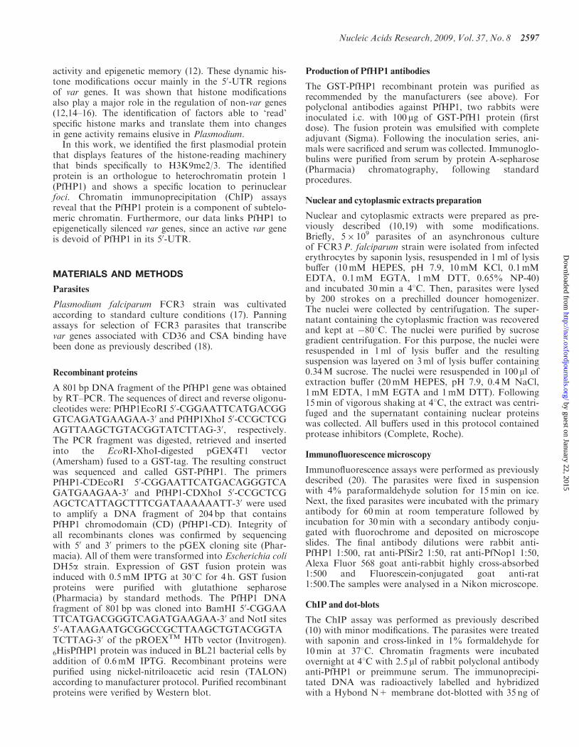
in malaria parasites, a soluble GST fusion protein corres-ponding to the CD was produced. This protein was pur-ified and immunoblotted with anti-GST antibodies(Figure 2A). A band of the predicted size (33 kDa) wasobserved together with minor bands, which are probablydegradation products often seen for GST fusion proteins.The GST-PfHP1-CD protein was then used to investigateits binding specificity to different histone modifications.The GST-PfHP1-CD protein was dotted on nylon mem-branes and incubated with the following biotinylated his-tone H3 peptides: H3K9me3, H3K9me2, H3K9ac,H3K4me3, H3K4me2 and unmodified histone H3.Binding of the peptides to the CD of PfHP1 was detectedusing streptavidin conjugated to HRP. Signal wasobserved only with H3K9me2 and H3K9me3(Figure 2B). To show that the modified histone H3 pep-tides did not bind the recombinant GST-CD proteinthrough GST, the GST protein was used as a negativecontrol (data not shown). These results demonstrate thatthe CD from PfHP1 protein is able to interact toH3K9me2 and H3K9me3, but not to H3K9ac,H3K4me2, H3K4me3 or to unmodified histone H3.To determine the affinity of the GST-PfHP1-CD protein
for both H3K9me3 and H3K9me2 peptides, dot blot bind-ing-assays were performed using a fixed amount of GST-PfHP1-CD fusion protein with different concentrationsof peptides. Densitometry analysis showed that in vitroGST-PfHP1-CD binds three times more efficiently toH3K9me3 than to H3K9me2 (Figure 2C).
Recombinant PfHP1 forms homodimers
Cowieson et al. demonstrated that recombinant CSD in itsunmodified form is able to dimerize in solution through
the alpha helixes, forming a non-polar cavity where pro-teins that have the P�V�L pentapeptide can bind (28).To demonstrate that PfHP1 is able to form dimers assuggested by the tertiary structure, we generated recombi-nant PfHP1 proteins fused to GST (GST-PfHP1) and tosix histidines (His-PfHP1). Inclusion bodies containingHis-PfHP1 were incubated in the presence of urea, rena-tured and incubated with Nickel-beads. These beads werewashed and the bound protein was eluted, analysed bySDS–PAGE, blotted and tested with the anti-histidineantibodies. These antibodies recognized a 33 kDa protein,which corresponds to the monomeric form of PfHP1(Figure 3A).
We next incubated lysates from bacterial cells over-expressing the GST-PfHP1 fusion protein (55 kDa)with the renatured His-PfHP1 fusion protein bound tothe nickel beads. Bound proteins were eluted, separatedby SDS–PAGE and immunoblotted with anti-histidineand anti-GST antibodies (Figure 3B). The Western blotanalysis confirmed that His-PfHP1 interacts specificallywith GST-PfHP1. GST alone (26 kDa) did not bind toHis-PfHP1 under the same conditions (Figure 3B).Similar results were obtained when we used purifiedGST-PfHP1 and His-PfHP1 fusion proteins in the pull-down assays (data not shown). In summary, our in vitroexperiments suggest the ability of PfHP1 to form dimers,which appears to be important for HP1-mediated hetero-chromatin formation (see model in Figure 6A).
PfHP1 localizes at the nuclear periphery
To further investigate the function of PfHP1, we raisedpolyclonal antibodies against the recombinant GST-PfHP1. Immunoblot analysis of parasite nuclear extracts
Figure 2. PfHP1 binds to histone H3 peptide methylated at lysine 9. (A) Total proteins from DH5a strain expressing the CD of PfHP1 before (NI)and after IPTG induction (I), as well as the purified GST-PfHP1-CD fusion protein (P), were separated by SDS–PAGE and stained with Coomassieblue (left). An anti-GST antibody was used to detect the presence of the fusion protein (right). (B) Purified protein GST-PfHP1-CD was assayed forbinding to histone H3 peptides. 200 ng of GST-CD protein (GST-PfHP1-CD) were dotted to a nitrocellulose membrane and incubated with 2 mg ofthe following biotinylated peptides: H3K9me3, H3K9me2, H3K9ac, H3K4me3, H3K4me2 and unmodified H3. (C) Two hundred nanograms ofGST-PfHP1-CD fusion protein were dotted to nitrocellulose and incubated with decreasing amounts of H3K9me3 and H3K9me2 biotinylatedpeptides (left panels). A representative result of two independent experiments is shown. The right panel shows a quantitative presentation of theGST-PfHP1-CD affinity to the peptides.
2600 Nucleic Acids Research, 2009, Vol. 37, No. 8
by guest on January 22, 2015http://nar.oxfordjournals.org/
Dow
nloaded from
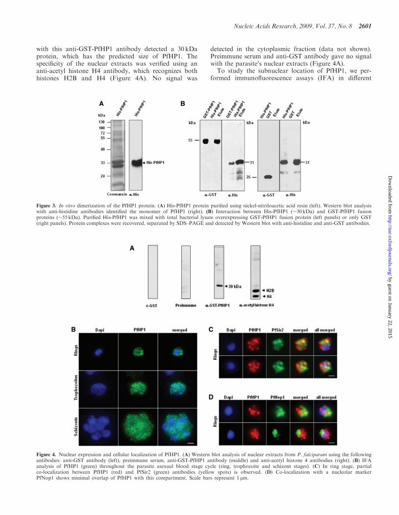
with this anti-GST-PfHP1 antibody detected a 30 kDaprotein, which has the predicted size of PfHP1. Thespecificity of the nuclear extracts was verified using ananti-acetyl histone H4 antibody, which recognizes bothhistones H2B and H4 (Figure 4A). No signal was
detected in the cytoplasmic fraction (data not shown).Preimmune serum and anti-GST antibody gave no signalwith the parasite’s nuclear extracts (Figure 4A).To study the subnuclear location of PfHP1, we per-
formed immunofluorescence assays (IFA) in different
Figure 4. Nuclear expression and cellular localization of PfHP1. (A) Western blot analysis of nuclear extracts from P. falciparum using the followingantibodies: anti-GST antibody (left), preimmune serum, anti-GST-PfHP1 antibody (middle) and anti-acetyl histone 4 antibodies (right). (B) IFAanalysis of PfHP1 (green) throughout the parasite asexual blood stage cycle (ring, trophozoite and schizont stages). (C) In ring stage, partialco-localization between PfHP1 (red) and PfSir2 (green) antibodies (yellow spots) is observed. (D) Co-localization with a nucleolar markerPfNop1 shows minimal overlap of PfHP1 with this compartment. Scale bars represent 1 mm.
Figure 3. In vitro dimerization of the PfHP1 protein. (A) His-PfHP1 protein purified using nickel-nitriloacetic acid resin (left). Western blot analysiswith anti-histidine antibodies identified the monomer of PfHP1 (right). (B) Interaction between His-PfHP1 (�30 kDa) and GST-PfHP1 fusionproteins (�55 kDa). Purified His-PfHP1 was mixed with total bacterial lysate overexpressing GST-PfHP1 fusion protein (left panels) or only GST(right panels). Protein complexes were recovered, separated by SDS–PAGE and detected by Western blot with anti-histidine and anti-GST antibodies.
Nucleic Acids Research, 2009, Vol. 37, No. 8 2601
by guest on January 22, 2015http://nar.oxfordjournals.org/
Dow
nloaded from
stages of the 48-h asexual cycle. PfHP1 appears to formnuclear foci during the entire cycle (Figure 4B). In ringstages (�16 h), PfHP1 locates predominantly to peri-nuclear space, whereas in trophozoite stages (�30 h) andschizonts (�40 h) the number of foci increases and seemsto redistribute in the nucleoplasm.To test whether PfHP1 localizes to telomeric perinuclear
clusters we performed dual IFA using anti-PfHP1 andanti-Sir2 antibodies. We observed that PfHP1 localizeswith some PfSir2 fluorescent signals (Figure 4C). BecausePfSir2 is not an exclusive marker for telomeric clusters andanti-PfSir2 also stains the nucleolus (10), we carried out adual IFA using an antibody against a nucleolar protein(anti-PfNop1). We observed that PfHP1 did not (oronly marginally) co-localize with the nucleolar marker
(Figure 4D), indicating that the PfHP1 foci co-localizingwith PfSir2 correspond indeed to telomeric foci.
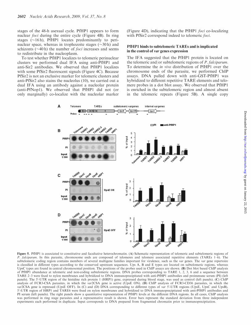
PfHP1 binds to subtelomeric TAREs and is implicatedin the control of var genes expression
The IFA suggested that the PfHP1 protein is located onthe telomeric and/or subtelomeric regions of P. falciparum.To determine the in vivo distribution of PfHP1 over thechromosome ends of the parasite, we performed ChIPassays. DNA pulled down with anti-GST-PfHP1 washybridized to different repetitive TARE elements and telo-mere probes in a dot blot assay. We observed that PfHP1is enriched in the subtelomeric region and almost absentin the telomeric repeats (Figure 5B). A single copy
Figure 5. PfHP1 is associated to constitutive and facultative heterochromatin. (A) Schematic representation of telomeric and subtelomeric regions ofP. falciparum. In this parasite, chromosome ends are composed of telomeres and telomere associated repetitive elements (TAREs 1–6). Thesubtelomeric coding region contains members of several multigene families important for virulence, such as the var genes. The var gene repertoireis classified in different types according to the conserved upstream sequences. Ups A, B and E types are located on subtelomeric regions, whereasUpsC types are found in central chromosomal position. The positions of the probes used in ChIP assays are shown. (B) Dot blot based ChIP analysisof PfHP1 abundance at telomeric and non-coding subtelomeric regions. DNA probes corresponding to TARE 1, 2, 3, 6 and a sequence betweenTARE 2–3 were fixed to nylon membranes and hybridized to DNA immunoprecipitated with anti-PfHP1 antibodies and preimmune serum (PI) (leftpanels). The 50-UTR region of the histidine rich protein 1 (HRP1) gene, expressed during blood stage, was used as control (left panels). (C) ChIPanalysis of FCR3-CSA parasites, in which the var2CSA gene is active (UpsE ON). (D) ChIP analysis of FCR3-CD36 parasites, in which thevar2CSA gene is repressed (UpsE OFF). In (C) and (D) DNA corresponding to different types of var 50-UTR regions (UpsE, UpsC and UpsB),50-UTR region of HRP1 and TARE6 were fixed on nylon membranes and hybridized to DNA immunoprecipitated with anti-PfHP1 antibodies andPI serum (left panels). The right panels show a quantitative representation of PfHP1 levels at the different DNA regions. In all cases, ChIP analysiswas performed in ring stage parasites and a representative result is shown. Error bars represent the standard deviation from three independentexperiments each performed in duplicate. Input corresponds to DNA prepared from fragmented chromatin prior to immunoprecipitation.
2602 Nucleic Acids Research, 2009, Vol. 37, No. 8
by guest on January 22, 2015http://nar.oxfordjournals.org/
Dow
nloaded from
blood-stage expressed gene (HRP1) did not immunopreci-pitate with the anti-GST-PfHP1 antibody (Figure 5B).These results show that PfHP1 is associated to chromatinacross subtelomeric repeat elements, most likely mediatingchromatin compaction.
To determine whether PfHP1 has a role in the silencingof subtelomeric var genes, we performed ChIP assaysusing two isogenic parasite populations: FCR3-CSA thatexpresses a specific subtelomeric var gene (var2CSA), andFCR3-CD36 in which the same gene is repressed. Wefound that the var2CSA upstream region (UpsE) is immu-noprecipitated by anti-GST-PfHP1 antibodies when thisgene is inactive (FCR3-CD36 population, Figure 5D)but not when it is active (FCR3-CSA population,Figure 5C). Other types of var gene upstream regionsrepresenting subtelomeric and chromosome central varmembers (UpsB and UpsC, respectively), which are ina repressed state in these parasite populations, wereimmunoprecipitated by anti-PfHP1 antibodies. Thus,our data show that PfHP1 is linked to repression ofsubtelomeric but also to central var genes.
DISCUSSION
In this work, we identified and characterized PfHP1, thefirst protein from P. falciparum that binds specifically toa histone modification, thus demonstrating the existenceof histone-mark-reading machinery in this pathogen. Ourin vitro results demonstrated that PfHP1 specificallyrecognizes H3K9me2 and H3K9me3 peptides by itsCD. The affinity of this protein seems to be greater fortri-methylated than for di-methylated peptide (Figure 2C),suggesting that the degree of methylation at histone H3K9may lead to the formation of more or less tight chromatin.As predicted by its tertiary structure (Figure 1), PfHP1is able to interact with itself (Figure 3B), which suggestsa role in the formation and maintenance of heterochroma-tin, similarly to its orthologues in S. pombe, Drosophilaand human (29). Furthermore, our data reinforce theidea that the machinery involved in heterochromatinformation in Plasmodium is more similar to metazoathan to S. cerevisiae. It also shows that the HP1 proteinis not exclusive of metazoan, as it has been proposed (30).
Nuclear staining with anti-PfHP1 suggested that thisprotein is located at the perinuclear region visible asfoci similar to PfSir2 and PfOrc1, two proteins knownto be components of subtelomeric heterochromatin inP. falciparum (10,20). In contrast to PfSir2 and PfOrc1,our co-localization experiments demonstrated that PfHP1does not stain the parasite nucleolus and may thereforenot participate in the silencing of rDNA genes in bloodstage parasites.
The notion that PfHP1 is a component of telomericheterochromatin was further demonstrated by showingthe binding of PfHP1 to subtelomeric chromatin(Figure 5). ChIP studies clearly showed that the anti-PfHP1 antibody immunoprecipitated subtelomeric regionsas well as members of the var gene family, and reactpoorly with telomeric repeats. Like in yeast, the telomeresof P. falciparum are mostly not packed in nucleosomes
whereas the subtelomeric chromatin contains nucleosomes(31). The association of PfHP1 with the subtelomericchromatin correlates well with the nucleosomal nature ofHP1 heterochromatinization function.Importantly, our data show that PfHP1 protein is
linked to monoallelic expression of the major virulencegene family in this human pathogen. The data stronglysuggest that PfHP1 must be removed from the varupstream regions to allow gene expression. Our ChIPassays demonstrated that PfHP1 is able to bind not onlyto subtelomeric but also to central var genes (Figure 5).Consistent with these results, P. falciparum genome-widehigh-resolution ChIP analysis of histone mark H3K9me3shows a restricted localization to telomeres, subtelomeresand internal chromosomal regions that contain virulencegene families (Lopez-Rubio, JJ and Scherf A. personalcommunication). It is very likely that PfHP1 displaysa genome-wide distribution similar to H3K9me3. The pre-sence of PfHP1 in internal chromosomal domains mayexplain the partial co-localization with telomeric PfSir2foci in the IFA analysis.In mammals, the HP1 family is composed of three
variants (alpha, beta and gamma), which are codedby distinct genes (32). These proteins are involved indifferent events: HP1 alpha and beta in heterochromatinformation, and HP1 gamma in euchromatin formation(29). We have found only one hp1 gene in P. falciparumgenome and this one was confirmed by Southern blotanalysis (data not shown). This raises the questionof whether a single PfHP1 protein performs differentfunctions. The hinge region of HP1 protein is highlyamenable to post-translational modifications especiallyphosphorylation (29). In addition, mutations in thisregion have been shown to affect the location, interactionsand functions of HP1 (33). For example, Ser83 phosphor-ylation of HP1 gamma is important for its localizationonly in euchromatic regions (34). In silico analysis ofPfHP1 protein indicates that a number of post-transla-tional changes, such as: sumoylation, ubiquitination andphosphorylation, may occur mainly in the hinge region(Perez-Toledo K and Hernandez-Rivas R unpublisheddata). It is therefore possible that different post-transla-tional modifications may define distinct functions ofPfHP1. Another component of the subtelomeric chroma-tin, PfSir2, has recently been shown to be sumoylated (35).It will be interesting to investigate whether mutation in thePfHP1 hinge region will affect function and nuclearlocation.PfHP1 knock-out parasites would be a valuable tool
to further investigate the role in gene silencing inP. falciparum. So far, all our attempts to disrupt the hp1gene had failed. This is possibly due to the fact thatonly one copy exists in the genome. It may also be dueto additional essential roles of PfHP1 possibly relatedwith chromosome integrity and/or nuclear organizationin this parasite. Inducible loss-of-function systemsappear now to be available for P. falciparum that mayallow PfHP1 phenotype analysis (36).In line with our data, we propose a model in
which PfHP1 works in concert with a histone deacetylaseand a histone methyltransferase in heterochromatin
Nucleic Acids Research, 2009, Vol. 37, No. 8 2603
by guest on January 22, 2015http://nar.oxfordjournals.org/
Dow
nloaded from

formation (Figure 6A). In our model, a histone deacety-lase PfSir2 (10,37), is first recruited to chromatin anddeacetylates H3K9 residues. In a subsequent step, wehypothesize that the H3K9 methyltransferase orthologuePfKMT1 (15) methylates the H3K9 residues. As a con-sequence, PfHP1 binds to H3K9me3, dimerizes and pro-motes chromatin condensation. This leads to PfHP1spreading into the coding regions, thus contributing tothe reversible silencing of virulence factor gene familiesimmediately adjacent of TAREs (Figure 6B). Whatrecruits PfHP1 to central var genes remains guesswork.In mammals, targeting of HP1 to chromatin requiresnot only K9 methylation but also a direct protein–proteininteraction with SUV39 (an orthologue of PfKMT1)(38). HP1 can also interact with Orc1, as it has beendemonstrated in Drosophila and Xenopus (39,40). Thespreading of PfHP1 along the different TAREs is mostlikely controlled by complex machinery including knowncomponents such as PfSir2, PfOrc1 and PfKMT1. Furtherstudies are necessary to determine potential protein–protein interaction between PfHP1 and other chromatincomponents by TAP-tag or pull down assays.In conclusion, this work identifies a key component
of the epigenetic machinery involved in heterochromatinformation. We demonstrated that PfHP1 is closely relatedto its counterpart in higher eukaryotes, since it is able toform homodimers and binds methylated histone H3K9.PfHP1 is the first plasmodial protein identified that recog-nizes a specific histone modification, which is linked genesilencing of virulence factor gene families located adjacent
to TAREs and some chromosome internal regions. Uponactivation of a single gene member in a mutually exclusivefashion, PfHP1 is absent of the promoter region of theactivated gene. This is a major conceptual advance toour knowledge and may have a big impact on the allelicexclusion field in other pathogens and higher eukaryotes.Together with the previously identified PfSir2, PfHP1might form the core of the subtelomeric heterochromatinmachinery in P. falciparum. Our anti-PfHP1 antibodiesare a new tool to investigate facultative and constitutiveheterochromatin formation in malaria parasites.Telomere-linked control of phenotypic variation is animportant issue in malaria pathogenesis and a betterknowledge of the epigenetic factors controlling those isvital for understanding the disease and to develop newanti-parasite control strategies.
ACKNOWLEDGEMENTS
We would like to thank J.J. Lopez-Rubio, L. Riviere andF. Iracheta Tovar for critical comments.
FUNDING
Consejo Nacional de Ciencia y Tecnologia [45687/A-1to R.H.R.]; Institut Pasteur (EGIDE) and CONACYT[No 172799 to A.P.R.M.]; European Commission(BioMalPar) [contract No: LSPH-CT-2004-503578];the ‘Fonds dedie: Combattre les Maladies Parasitaires’Sanofi Aventis – Ministere de la Recherche; ANR
Figure 6. Hypothetical model for heterochromatin formation at P. falciparum chromosome ends. (A) PfHP1-mediated heterochromatin modeldepends on the action of the histone deacetylase PfSir2 and histone methyltransferase PfKMT1 (10,15,37). (B) General view of the known chromatincomponents at P. falciparum subtelomeres. Spreading of heterochromatin along the different TAREs into adjacent coding regions probably involvesthe cooperation of PfHP1, PfSir2 and PfKMT1. The role of PfOrc1 in this process remains unknown.
2604 Nucleic Acids Research, 2009, Vol. 37, No. 8
by guest on January 22, 2015http://nar.oxfordjournals.org/
Dow
nloaded from

[ANR-06-MIME-026-01 to A.S.] and Fundacao para aCiencia e Tecnologia, Portugal [SFRH/BD/11756/2003to L.M.S.]. Funding for open access charge: ConsejoNacional de Ciencia y Tecnologıa [56836-Q to LSA].
Conflict of interest statement. None declared.
REFERENCES
1. Bozdech,Z., Llinas,M., Pulliam,B.L., Wong,E.D., Zhu,J. andDeRisi,J.L. (2003) The transcriptome of the intraerythrocyticdevelopmental cycle of Plasmodium falciparum. PLoS Biol., 1, E5.
2. Le Roch,K.G., Zhou,Y., Blair,P.L., Grainger,M., Moch,J.K.,Haynes,J.D., De la Vega,P., Holder,A.A., Batalov,S., Carucci,D.J.et al. (2003) Discovery of gene function by expression profiling ofthe malaria parasite life sycle. Science, 301, 1503–1508.
3. Aravind,L., Iyer,L.M., Wellems,T.E. and Miller,L.H. (2003)Plasmodium biology: genomic gleanings. Cell, 115, 771–785.
4. Templeton,T.J., Iyer,L.M., Anantharaman,V., Enomoto,S.,Abrahante,J.E., Subramanian,G.M., Hoffman,S.L.,Abrahamsen,M.S. and Aravind,L. (2004) Comparative analysisof apicomplexa and genomic diversity in eukaryotes. Genome Res.,14, 1686–1695.
5. Callebaut,I., Prat,K., Meurice,E., Mornon,J.P. and Tomavo,S.(2005) Prediction of the general transcription factors associatedwith RNA polymerase II in Plasmodium falciparum: conservedfeatures and differences relative to other eukaryotes. BMCGenomics, 6, 100.
6. Gissot,M., Briquet,S., Refour,P., Boschet,C. and Vaquero,C. (2005)PfMyb1, a Plasmodium falciparum transcription factor, is requiredfor intra-erythrocytic growth and controls key genes for cell cycleregulation. J. Mol. Biol., 346, 29–42.
7. De Silva,E.K., Gehrke,A.R., Olszewski,K., Leon,I., Chahal,J.S.,Bulyk,M.L. and Llinas,M. (2008) Specific DNA-binding byapicomplexan AP2 transcription factors. Proc. Natl Acad. Sci. USA,105, 8393–8398.
8. Hakimi,M.-A. and Deitsch,K.W. (2007) Epigenetics in apicomplexa:control of gene expression during cell cycle progression,differentiation and antigenic variation. Curr. Opin. Microbiol., 10,357–362.
9. Ralph,S.A. and Scherf,A. (2005) The epigenetic control of antigenicvariation in Plasmodium falciparum. Curr. Opin. Microbiol., 8,434–440.
10. Freitas-Junior,L.H., Hernandez-Rivas,R., Ralph,S.A.,Montiel-Condado,D., Ruvalcaba-Salazar,O.K., Rojas-Meza,A.P.,Mancio-Silva,L., Leal-Silvestre,R.J., Gontijo,A.M., Shorte,S. et al.(2005) Telomeric heterochromatin propagation and histoneacetylation control mutually exclusive expression of antigenicvariation genes in malaria Parasites. Cell, 121, 25–36.
11. Duraisingh,M.T., Voss,T.S., Marty,A.J., Duffy,M.F., Good,R.T.,Thompson,J.K., Freitas-Junior,L.H., Scherf,A., Crabb,B.S. andCowman,A.F. (2005) Heterochromatin silencing and locusrepositioning linked to regulation of virulence genes in Plasmodiumfalciparum. Cell, 121, 13–24.
12. Lopez-Rubio,J.J., Gontijo,A.M., Nunes,M.C., Issar,N., HernandezRivas,R. and Scherf,A. (2007) 50 flanking region of var genesnucleate histone modification patterns linked to phenotypic inheri-tance of virulence traits in malaria parasites. Mol. Microbiol., 66,1296–1305.
13. Chookajorn,T., Dzikowski,R., Frank,M., Li,F., Jiwani,A.Z.,Hartl,D.L. and Deitsch,K.W. (2007) Epigenetic memory at malariavirulence genes. Proc. Natl Acad. Sci. USA, 104, 899–902.
14. Cui,L., Miao,J., Furuya,T., Li,X. and Su,X.Z. (2007) PfGCN5-mediated histone H3 acetylation plays a key role in gene expressionin Plasmodium falciparum. Eukaryot. Cell, 6, 1219–1227.
15. Cui,L., Fan,Q. and Miao,J. (2008) Histone lysine methyltransferasesand demethylases in Plasmodium falciparum. Int. J. Parasitol., 38,1083–1097.
16. Miao,J., Fan,Q., Cui,L. and Li,J. (2006) The malaria parasitePlasmodium falciparum histones: organization, expression, andacetylation. Gene, 369, 53–65.
17. Trager,W. and Jensen,J.B. (1976) Human malaria parasites incontinuous culture. Science, 193, 673–675.
18. Scherf,A., Hernandez-Rivas,R., Buffet,P., Bottius,E., Benatar,C.,Pouvelle,B., Gysin,J. and Lanzer,M. (1998) Antigenic variationin malaria: in situ switching, relaxed and mutually exclusivetranscription of var genes during intra-erythrocytic developmentin Plasmodium falciparum. EMBO J., 17, 5418–5426.
19. Ruvalcaba-Salazar,O.K., del Carmen Ramirez-Estudillo,M.,Montiel-Condado,D., Recillas-Targa,F., Vargas,M. andHernandez-Rivas,R. (2005) Recombinant and native Plasmodiumfalciparum TATA-binding-protein binds to a specific TATAbox element in promoter regions. Mol. Biochem. Parasitol., 140,183–196.
20. Mancio-Silva,L., Rojas-Meza,A.P., Vargas,M., Scherf,A. andHernandez-Rivas,R. (2008) Differential association of Orc1 and Sir2proteins to telomeric domains in Plasmodium falciparum. J. CellSci., 121, 2046–2053.
21. Towbin,H., Staehelin,T. and Gordon,J. (1979) Electrophoretictransfer of proteins from polyacrylamide gels to nitrocellulosesheets: procedure and some applications. Proc. Natl Acad. Sci.USA, 76, 4350–4354.
22. Sambrook,J. and Rusell,D.W. (2001) Molecular Cloning aLaboratory Manual. Cold Spring Harbor Laboratory Press,Cold Spring Harbor, NY.
23. Hiragami,K. and Festenstein,R. (2005) Heterochromatinprotein 1: a pervasive controlling influence. Cell Mol. Life Sci., 62,2711–2726.
24. Jacobs,S.A. and Khorasanizadeh,S. (2002) Structure of HP1chromodomain bound to a lysine 9-methylated histone H3 tail.Science, 295, 2080–2083.
25. Brasher,S.V., Smith,B.O., Fogh,R.H., Nietlispach,D., Thiru,A.,Nielsen,P.R., Broadhurst,R.W., Ball,L.J., Murzina,N.V. andLaue,E.D. (2000) The structure of mouse HP1 suggests a uniquemode of single peptide recognition by the shadow chromodomain dimer. EMBO J., 19, 1587–1597.
26. Lachner,M., O’Carroll,D., Rea,S., Mechtler,K. and Jenuwein,T.(2001) Methylation of histone H3 lysine 9 creates a binding sitefor HP1 proteins. Nature, 410, 116–120.
27. Bannister,A.J., Zegerman,P., Partridge,J.F., Miska,E.A.,Thomas,J.O., Allshire,R.C. and Kouzarides,T. (2001) Selectiverecognition of methylated lysine 9 on histone H3 by the HP1chromo domain. Nature, 410, 120–124.
28. Cowieson,N.P., Partridge,J.F., Allshire,R.C. and McLaughlin,P.J.(2000) Dimerisation of a chromo shadow domain and distinctionsfrom the chromodomain as revealed by structural analysis. Curr.Biol., 10, 517–525.
29. Lomberk,G., Wallrath,L. and Urrutia,R. (2006) TheHeterochromatin Protein 1 family. Genome Biol., 7, 228.
30. Sautel,C.F., Cannella,D., Bastien,O., Kieffer,S., Delphine,A.,Garin,J., Tardieux,, I. Belrhali,H. and Hakimi,M.-A. (2007)SET8-mediated methylations of histone H4 lysine 20 mark silentheterochromatic domains in apicomplexan genomes. Mol. Cell Biol.,27, 5711–5724.
31. Figueiredo,L.M., Pirritt,L.A. and Scherf,A. (2000) Genomicorganisation and chromatin structure of Plasmodiumfalciparum chromosome ends. Mol. Biochem. Parasitol., 106,169–174.
32. Jones,D.O., Mattei,M.G., Horsley,D., Cowell,I.G. and Singh,P.B.(2001) The gene and pseudogenes of Cbx3/mHP1 gamma. DNASeq., 12, 147–160.
33. Zhao,T., Heyduk,T. and Eissenberg,J.C. (2001) Phosphorylationsite mutations in heterochromatin protein 1 (HP1) reduce oreliminate silencing activity. J. Biol. Chem., 276, 9512–9518.
34. Lomberk,G., Bensi,D., Fernandez-Zapico,M.E. and Urrutia,R.(2006) Evidence for the existence of an HP1-mediated subcodewithin the histone code. Nat. Cell Biol., 8, 407–415.
35. Issar,N., Roux,E., Mattei,D. and Scherf,A. (2008) Identification ofa novel post-translational modification in Plasmodium falciparum:Protein SUMOylation in different cellular compartments. CellMicrobiol., 10, 1999–2011.
36. Armstrong,C.M. and Goldberg,D.E. (2007) An FKBPdestabilization domain modulates protein levels in Plasmodiumfalciparum. Nat. Methods, 4, 1007–1009.
Nucleic Acids Research, 2009, Vol. 37, No. 8 2605
by guest on January 22, 2015http://nar.oxfordjournals.org/
Dow
nloaded from

37. Merrick,C.J. and Duraisingh,M.T. (2007) Plasmodium falciparumSir2: an unusual sirtuin with dual histone deacetylase andADP-ribosyltransferase activity. Eukaryot. Cell, 6, 2081–2091.
38. Blasco,M.A. (2007) The epigenetic regulation of mammaliantelomeres. Nat. Rev. Genet., 8, 299–309.
39. Pak,D.T., Pflumm,M., Chesnokov,I., Huang,D.W., Kellum,R.,Marr,J., Romanowski,P. and Botchan,M.R. (1997) Association of
the origin recognition complex with heterochromatin and HP1 inhigher eukaryotes. Cell, 91, 311–323.
40. Huang,D.W., Fanti,L., Pak,D.T., Botchan,M.R., Pimpinelli,S. andKellum,R. (1998) Distinct cytoplasmic and nuclear fractions ofDrosophila heterochromatin protein 1: their phosphorylationlevels and associations with origin recognition complex proteins.J. Cell Biol., 142, 307–318.
2606 Nucleic Acids Research, 2009, Vol. 37, No. 8
by guest on January 22, 2015http://nar.oxfordjournals.org/
Dow
nloaded from