Thrombomodulin-dependent effect of factor VLeiden mutation on factor XIII activation
Upload
independentCategory
view
1download
0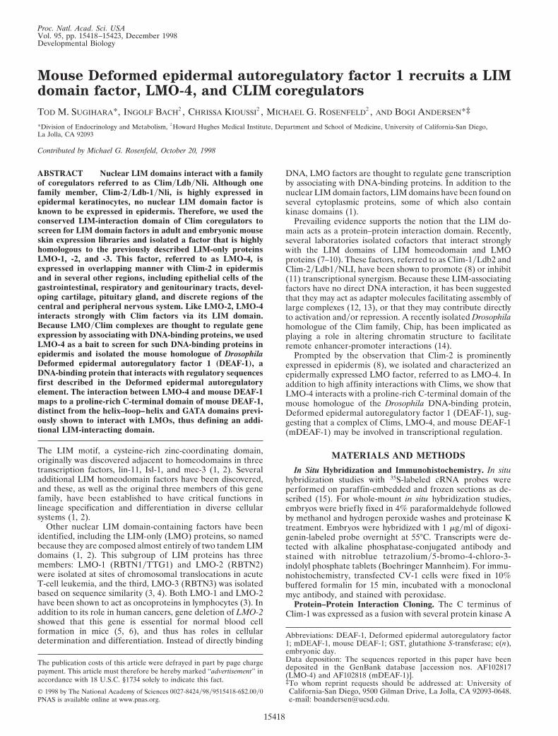
Proc. Natl. Acad. Sci. USAVol. 95, pp. 15418–15423, December 1998Developmental Biology
Mouse Deformed epidermal autoregulatory factor 1 recruits a LIMdomain factor, LMO-4, and CLIM coregulators
TOD M. SUGIHARA*, INGOLF BACH†, CHRISSA KIOUSSI†, MICHAEL G. ROSENFELD†, AND BOGI ANDERSEN*‡
*Division of Endocrinology and Metabolism, †Howard Hughes Medical Institute, Department and School of Medicine, University of California-San Diego,La Jolla, CA 92093
Contributed by Michael G. Rosenfeld, October 20, 1998
ABSTRACT Nuclear LIM domains interact with a familyof coregulators referred to as ClimyLdbyNli. Although onefamily member, Clim-2yLdb-1yNli, is highly expressed inepidermal keratinocytes, no nuclear LIM domain factor isknown to be expressed in epidermis. Therefore, we used theconserved LIM-interaction domain of Clim coregulators toscreen for LIM domain factors in adult and embryonic mouseskin expression libraries and isolated a factor that is highlyhomologous to the previously described LIM-only proteinsLMO-1, -2, and -3. This factor, referred to as LMO-4, isexpressed in overlapping manner with Clim-2 in epidermisand in several other regions, including epithelial cells of thegastrointestinal, respiratory and genitourinary tracts, devel-oping cartilage, pituitary gland, and discrete regions of thecentral and peripheral nervous system. Like LMO-2, LMO-4interacts strongly with Clim factors via its LIM domain.Because LMOyClim complexes are thought to regulate geneexpression by associating with DNA-binding proteins, we usedLMO-4 as a bait to screen for such DNA-binding proteins inepidermis and isolated the mouse homologue of DrosophilaDeformed epidermal autoregulatory factor 1 (DEAF-1), aDNA-binding protein that interacts with regulatory sequencesfirst described in the Deformed epidermal autoregulatoryelement. The interaction between LMO-4 and mouse DEAF-1maps to a proline-rich C-terminal domain of mouse DEAF-1,distinct from the helix–loop–helix and GATA domains previ-ously shown to interact with LMOs, thus defining an addi-tional LIM-interacting domain.
The LIM motif, a cysteine-rich zinc-coordinating domain,originally was discovered adjacent to homeodomains in threetranscription factors, lin-11, Isl-1, and mec-3 (1, 2). Severaladditional LIM homeodomain factors have been discovered,and these, as well as the original three members of this genefamily, have been established to have critical functions inlineage specification and differentiation in diverse cellularsystems (1, 2).
Other nuclear LIM domain-containing factors have beenidentified, including the LIM-only (LMO) proteins, so namedbecause they are composed almost entirely of two tandem LIMdomains (1, 2). This subgroup of LIM proteins has threemembers: LMO-1 (RBTN1yTTG1) and LMO-2 (RBTN2)were isolated at sites of chromosomal translocations in acuteT-cell leukemia, and the third, LMO-3 (RBTN3) was isolatedbased on sequence similarity (3, 4). Both LMO-1 and LMO-2have been shown to act as oncoproteins in lymphocytes (3). Inaddition to its role in human cancers, gene deletion of LMO-2showed that this gene is essential for normal blood cellformation in mice (5, 6), and thus has roles in cellulardetermination and differentiation. Instead of directly binding
DNA, LMO factors are thought to regulate gene transcriptionby associating with DNA-binding proteins. In addition to thenuclear LIM domain factors, LIM domains have been found onseveral cytoplasmic proteins, some of which also containkinase domains (1).
Prevailing evidence supports the notion that the LIM do-main acts as a protein–protein interaction domain. Recently,several laboratories isolated cofactors that interact stronglywith the LIM domains of LIM homeodomain and LMOproteins (7–10). These factors, referred to as Clim-1yLdb2 andClim-2yLdb1yNLI, have been shown to promote (8) or inhibit(11) transcriptional synergism. Because these LIM-associatingfactors have no direct DNA interaction, it has been suggestedthat they may act as adapter molecules facilitating assembly oflarge complexes (12, 13), or that they may contribute directlyto activation andyor repression. A recently isolated Drosophilahomologue of the Clim family, Chip, has been implicated asplaying a role in altering chromatin structure to facilitateremote enhancer-promoter interactions (14).
Prompted by the observation that Clim-2 is prominentlyexpressed in epidermis (8), we isolated and characterized anepidermally expressed LMO factor, referred to as LMO-4. Inaddition to high affinity interactions with Clims, we show thatLMO-4 interacts with a proline-rich C-terminal domain of themouse homologue of the Drosophila DNA-binding protein,Deformed epidermal autoregulatory factor 1 (DEAF-1), sug-gesting that a complex of Clims, LMO-4, and mouse DEAF-1(mDEAF-1) may be involved in transcriptional regulation.
MATERIALS AND METHODS
In Situ Hybridization and Immunohistochemistry. In situhybridization studies with 35S-labeled cRNA probes wereperformed on paraffin-embedded and frozen sections as de-scribed (15). For whole-mount in situ hybridization studies,embryos were briefly fixed in 4% paraformaldehyde followedby methanol and hydrogen peroxide washes and proteinase Ktreatment. Embryos were hybridized with 1 mgyml of digoxi-genin-labeled probe overnight at 55°C. Transcripts were de-tected with alkaline phosphatase-conjugated antibody andstained with nitroblue tetrazoliumy5-bromo-4-chloro-3-indolyl phosphate tablets (Boehringer Mannheim). For immu-nohistochemistry, transfected CV-1 cells were fixed in 10%buffered formalin for 15 min, incubated with a monoclonalmyc antibody, and stained with peroxidase.
Protein–Protein Interaction Cloning. The C terminus ofClim-1 was expressed as a fusion with several protein kinase A
The publication costs of this article were defrayed in part by page chargepayment. This article must therefore be hereby marked ‘‘advertisement’’ inaccordance with 18 U.S.C. §1734 solely to indicate this fact.
© 1998 by The National Academy of Sciences 0027-8424y98y9515418-6$2.00y0PNAS is available online at www.pnas.org.
Abbreviations: DEAF-1, Deformed epidermal autoregulatory factor1; mDEAF-1, mouse DEAF-1; GST, glutathione S-transferase; e(n),embryonic day.Data deposition: The sequences reported in this paper have beendeposited in the GenBank database [accession nos. AF102817(LMO-4) and AF102818 (mDEAF-1)].‡To whom reprint requests should be addressed at: University ofCalifornia-San Diego, 9500 Gilman Drive, La Jolla, CA 92093-0648.e-mail: [email protected].
15418
phosphorylation sites and glutathione S-transferase (GST).Lambda gt11 libraries from neonatal and embryonic mouseskin were screened with a 32P-labeled protein as described (8).
Yeast Two-Hybrid Screening. Baits were expressed as fu-sions with the GAL4 DNA-binding domain, and library andtarget plasmids were expressed as fusions with the GAL4activation domain. b-galactosidase units were calculated ac-cording to standard methods (CLONTECH, Matchmakertwo-hybrid system).
Protein–Protein Interaction Assays. Coimmunoprecipita-tions were performed as described, by using 35S-labeled pro-teins and either a monoclonal myc antibody or a polyclonalrabbit antisera recognizing Clim-1 and Clim-2 (8). GST-interaction assays have been described (8).
Mammalian Two-Hybrid System. LMO-4 was cloned as afusion with the GAL4 DNA-binding domain and domains ofmDEAF-1 were cloned in-frame with the VP-16 activation
domain, both under control of the simian virus 40 enhancerpromoter. Interaction was tested by transfecting these plasmidsinto HeLa cells along with a luciferase reporter under thecontrol of GAL4 binding sites and a minimal promoter.Transfections and luciferase assays were performed as de-scribed (15).
RESULTS
Cloning of LMO-4 from Epidermis. Previously, we showedby RNase protection assays that Clim-2 is highly expressed inskin (8). To investigate the precise expression pattern ofClim-2 in skin, we performed in situ hybridization studies andfound that Clim-2 was expressed at its highest levels in, but notrestricted to, the basal compartment of interfollicular epider-mis (Fig. 1A). In addition, strong expression was observed indeveloping hair follicles during embryogenesis (Fig. 1 A). In
FIG. 1. Clim-2 and LMO-4 exhibit overlapping expression in epidermis. In situ hybridization of Clim-2 (A–C) and LMO-4 (D–F) in skin. (A)Clim-2 expression at e17.5. (B and C) Clim-2 expression in adult hair follicles. (D) LMO-4 expression at e17.5. (E and F) LMO-4 expression inadult hair follicles. (G) Comparison of the amino acid sequence of mouse LMO-4 with mouse LMO-1, -2, and -3. Conserved residues in the twoLIM domains are shaded. BL, basal layer; HF, hair follicle; ORS, outer root sheath; M, matrix cells; DP, dermal papilla; SG, sebaceous gland.
Developmental Biology: Sugihara et al. Proc. Natl. Acad. Sci. USA 95 (1998) 15419
adult hair, Clim-2 was expressed at its highest levels in matrixcells and the outer root sheath (Fig. 1 B and C).
Because Clim factors are known to associate with nuclearLIM proteins, these studies suggested the possibility that anovel LIM domain gene might be expressed in epidermis. Tosearch for epidermal LIM factors, we cloned the C-terminal100 amino acids of Clim-1, encompassing the conserved regionresponsible for LIM domain interactions, into a GST bacterialexpression vector containing several protein kinase A phos-phorylation sites, thus allowing labeling of the interactiondomain with 32P in vitro. This radiolabeled protein was used toscreen mouse lgt11 expression libraries from neonatal andembryonic day (e) 14.5 embryonic skin. Several interactingclones were obtained from the neonatal skin library, and oneinteracting clone was isolated from the embryonic skin library,all of which represented mRNAs transcribed from the samegene. Sequence analyses predicted that these cDNAs encodeda protein with two tandem LIM domains, but no othersequence homology, similar to the LIM-only factors LMO-1,-2, and -3 (Fig. 1G), and thus we refer to this factor as LMO-4.A search of GenBank databases showed that a homologoushuman gene, referred to as Human Breast Tumor Autoanti-gen, had been deposited in the GenBank database (accessionno. U24576) and that an expressed sequence tag encompassingthis sequence had been mapped to human chromosome 1,reference interval D1S203-D1S2865. While this manuscriptwas in preparation, another group reported the isolation ofmouse LMO-4 (16).
In situ hybridization experiments showed that LMO-4 wasexpressed in epidermis with the highest levels in hair follicles,especially in the outer root sheath, sebaceous glands, andmatrix cells (Fig. 1 E and F). While LMO-4 transcripts wereless abundant in interfollicular epidermis than in hair follicles,they were clearly detectable in, although not limited to, thebasal layer (Fig. 1D). The expression pattern of LMO-4overlaps with that of Clim-2 (compare with Fig. 1 A–C) bothin hair follicles and interfollicular epidermis, consistent withthe notion that these proteins could be components of thesame transcriptional complex in epidermis.
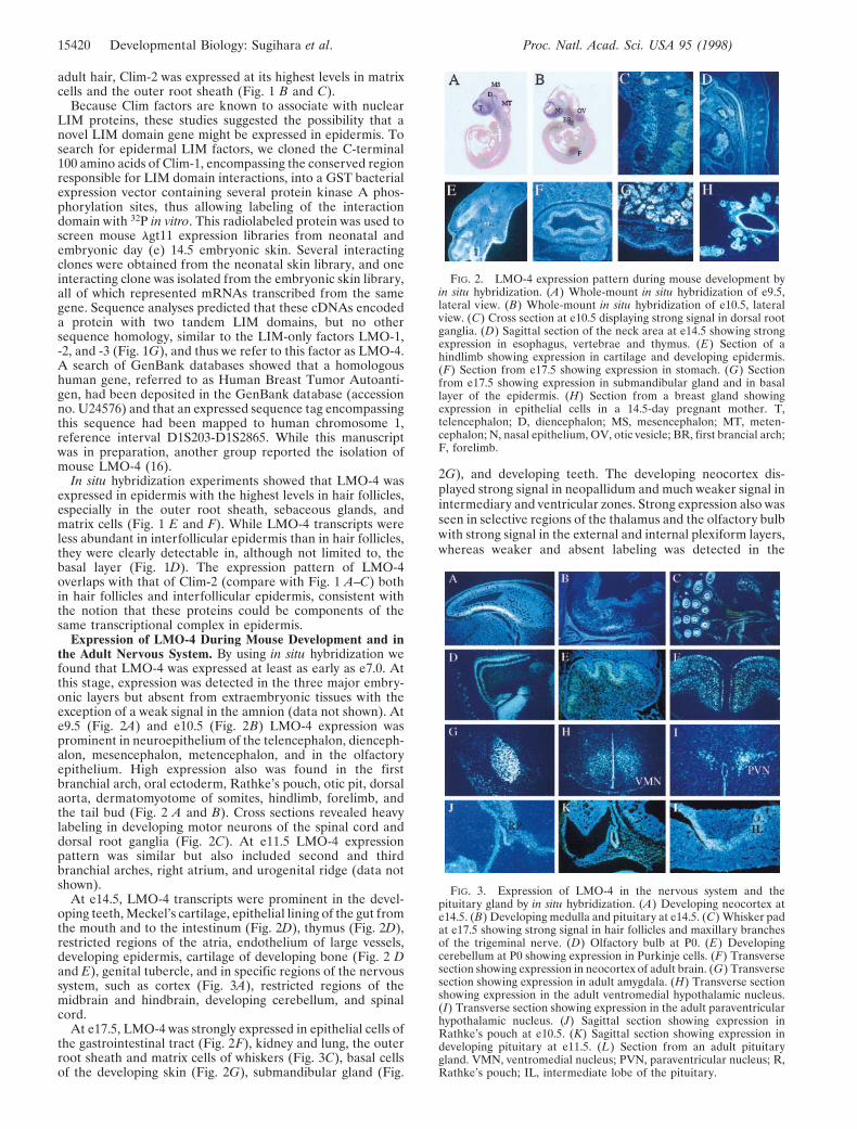
Expression of LMO-4 During Mouse Development and inthe Adult Nervous System. By using in situ hybridization wefound that LMO-4 was expressed at least as early as e7.0. Atthis stage, expression was detected in the three major embry-onic layers but absent from extraembryonic tissues with theexception of a weak signal in the amnion (data not shown). Ate9.5 (Fig. 2A) and e10.5 (Fig. 2B) LMO-4 expression wasprominent in neuroepithelium of the telencephalon, dienceph-alon, mesencephalon, metencephalon, and in the olfactoryepithelium. High expression also was found in the firstbranchial arch, oral ectoderm, Rathke’s pouch, otic pit, dorsalaorta, dermatomyotome of somites, hindlimb, forelimb, andthe tail bud (Fig. 2 A and B). Cross sections revealed heavylabeling in developing motor neurons of the spinal cord anddorsal root ganglia (Fig. 2C). At e11.5 LMO-4 expressionpattern was similar but also included second and thirdbranchial arches, right atrium, and urogenital ridge (data notshown).
At e14.5, LMO-4 transcripts were prominent in the devel-oping teeth, Meckel’s cartilage, epithelial lining of the gut fromthe mouth and to the intestinum (Fig. 2D), thymus (Fig. 2D),restricted regions of the atria, endothelium of large vessels,developing epidermis, cartilage of developing bone (Fig. 2 Dand E), genital tubercle, and in specific regions of the nervoussystem, such as cortex (Fig. 3A), restricted regions of themidbrain and hindbrain, developing cerebellum, and spinalcord.
At e17.5, LMO-4 was strongly expressed in epithelial cells ofthe gastrointestinal tract (Fig. 2F), kidney and lung, the outerroot sheath and matrix cells of whiskers (Fig. 3C), basal cellsof the developing skin (Fig. 2G), submandibular gland (Fig.
2G), and developing teeth. The developing neocortex dis-played strong signal in neopallidum and much weaker signal inintermediary and ventricular zones. Strong expression also wasseen in selective regions of the thalamus and the olfactory bulbwith strong signal in the external and internal plexiform layers,whereas weaker and absent labeling was detected in the
FIG. 3. Expression of LMO-4 in the nervous system and thepituitary gland by in situ hybridization. (A) Developing neocortex ate14.5. (B) Developing medulla and pituitary at e14.5. (C) Whisker padat e17.5 showing strong signal in hair follicles and maxillary branchesof the trigeminal nerve. (D) Olfactory bulb at P0. (E) Developingcerebellum at P0 showing expression in Purkinje cells. (F) Transversesection showing expression in neocortex of adult brain. (G) Transversesection showing expression in adult amygdala. (H) Transverse sectionshowing expression in the adult ventromedial hypothalamic nucleus.(I) Transverse section showing expression in the adult paraventricularhypothalamic nucleus. (J) Sagittal section showing expression inRathke’s pouch at e10.5. (K) Sagittal section showing expression indeveloping pituitary at e11.5. (L) Section from an adult pituitarygland. VMN, ventromedial nucleus; PVN, paraventricular nucleus; R,Rathke’s pouch; IL, intermediate lobe of the pituitary.
FIG. 2. LMO-4 expression pattern during mouse development byin situ hybridization. (A) Whole-mount in situ hybridization of e9.5,lateral view. (B) Whole-mount in situ hybridization of e10.5, lateralview. (C) Cross section at e10.5 displaying strong signal in dorsal rootganglia. (D) Sagittal section of the neck area at e14.5 showing strongexpression in esophagus, vertebrae and thymus. (E) Section of ahindlimb showing expression in cartilage and developing epidermis.(F) Section from e17.5 showing expression in stomach. (G) Sectionfrom e17.5 showing expression in submandibular gland and in basallayer of the epidermis. (H) Section from a breast gland showingexpression in epithelial cells in a 14.5-day pregnant mother. T,telencephalon; D, diencephalon; MS, mesencephalon; MT, meten-cephalon; N, nasal epithelium, OV, otic vesicle; BR, first brancial arch;F, forelimb.
15420 Developmental Biology: Sugihara et al. Proc. Natl. Acad. Sci. USA 95 (1998)
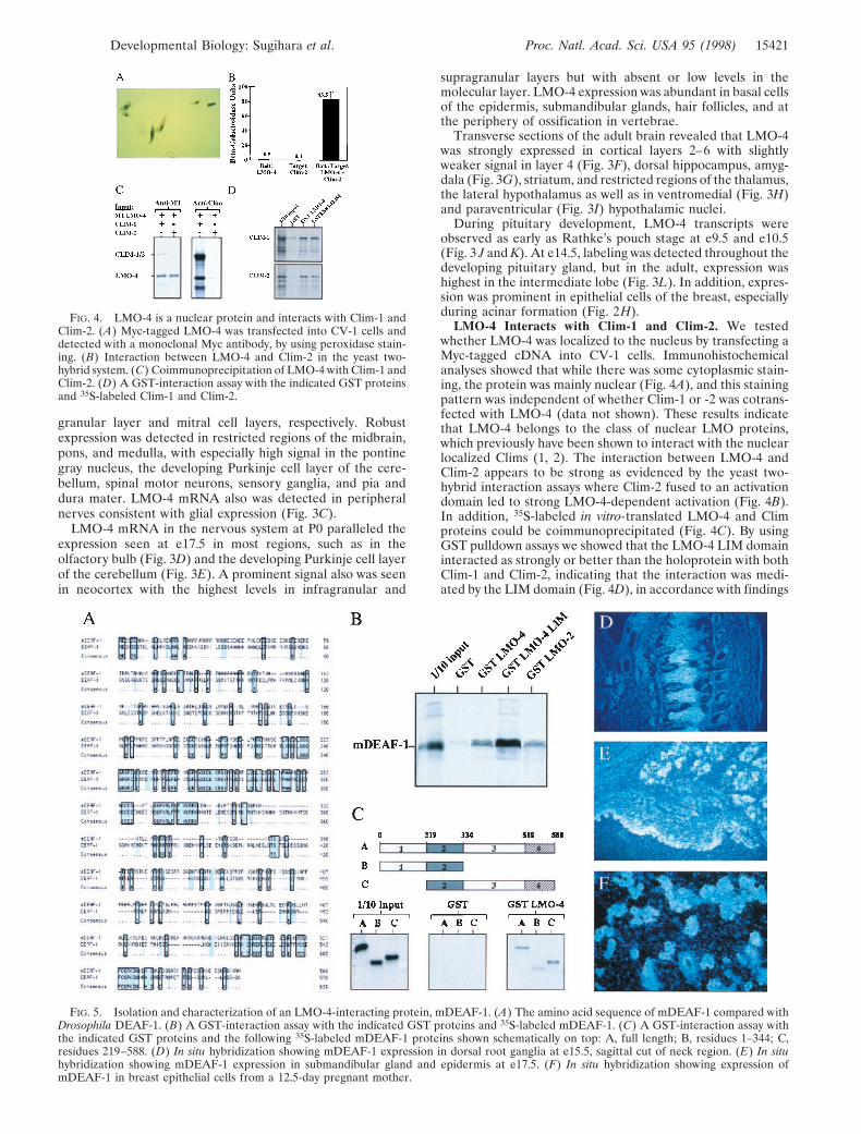
granular layer and mitral cell layers, respectively. Robustexpression was detected in restricted regions of the midbrain,pons, and medulla, with especially high signal in the pontinegray nucleus, the developing Purkinje cell layer of the cere-bellum, spinal motor neurons, sensory ganglia, and pia anddura mater. LMO-4 mRNA also was detected in peripheralnerves consistent with glial expression (Fig. 3C).
LMO-4 mRNA in the nervous system at P0 paralleled theexpression seen at e17.5 in most regions, such as in theolfactory bulb (Fig. 3D) and the developing Purkinje cell layerof the cerebellum (Fig. 3E). A prominent signal also was seenin neocortex with the highest levels in infragranular and
supragranular layers but with absent or low levels in themolecular layer. LMO-4 expression was abundant in basal cellsof the epidermis, submandibular glands, hair follicles, and atthe periphery of ossification in vertebrae.
Transverse sections of the adult brain revealed that LMO-4was strongly expressed in cortical layers 2–6 with slightlyweaker signal in layer 4 (Fig. 3F), dorsal hippocampus, amyg-dala (Fig. 3G), striatum, and restricted regions of the thalamus,the lateral hypothalamus as well as in ventromedial (Fig. 3H)and paraventricular (Fig. 3I) hypothalamic nuclei.
During pituitary development, LMO-4 transcripts wereobserved as early as Rathke’s pouch stage at e9.5 and e10.5(Fig. 3 J and K). At e14.5, labeling was detected throughout thedeveloping pituitary gland, but in the adult, expression washighest in the intermediate lobe (Fig. 3L). In addition, expres-sion was prominent in epithelial cells of the breast, especiallyduring acinar formation (Fig. 2H).
LMO-4 Interacts with Clim-1 and Clim-2. We testedwhether LMO-4 was localized to the nucleus by transfecting aMyc-tagged cDNA into CV-1 cells. Immunohistochemicalanalyses showed that while there was some cytoplasmic stain-ing, the protein was mainly nuclear (Fig. 4A), and this stainingpattern was independent of whether Clim-1 or -2 was cotrans-fected with LMO-4 (data not shown). These results indicatethat LMO-4 belongs to the class of nuclear LMO proteins,which previously have been shown to interact with the nuclearlocalized Clims (1, 2). The interaction between LMO-4 andClim-2 appears to be strong as evidenced by the yeast two-hybrid interaction assays where Clim-2 fused to an activationdomain led to strong LMO-4-dependent activation (Fig. 4B).In addition, 35S-labeled in vitro-translated LMO-4 and Climproteins could be coimmunoprecipitated (Fig. 4C). By usingGST pulldown assays we showed that the LMO-4 LIM domaininteracted as strongly or better than the holoprotein with bothClim-1 and Clim-2, indicating that the interaction was medi-ated by the LIM domain (Fig. 4D), in accordance with findings
FIG. 4. LMO-4 is a nuclear protein and interacts with Clim-1 andClim-2. (A) Myc-tagged LMO-4 was transfected into CV-1 cells anddetected with a monoclonal Myc antibody, by using peroxidase stain-ing. (B) Interaction between LMO-4 and Clim-2 in the yeast two-hybrid system. (C) Coimmunoprecipitation of LMO-4 with Clim-1 andClim-2. (D) A GST-interaction assay with the indicated GST proteinsand 35S-labeled Clim-1 and Clim-2.
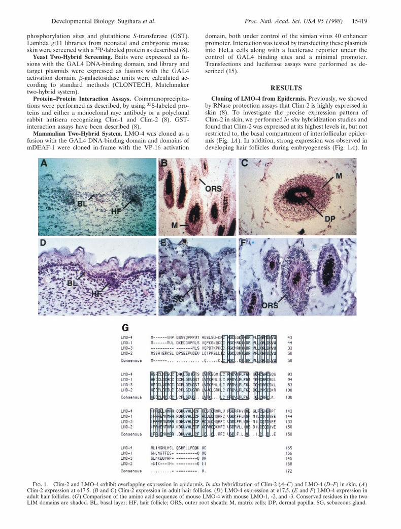
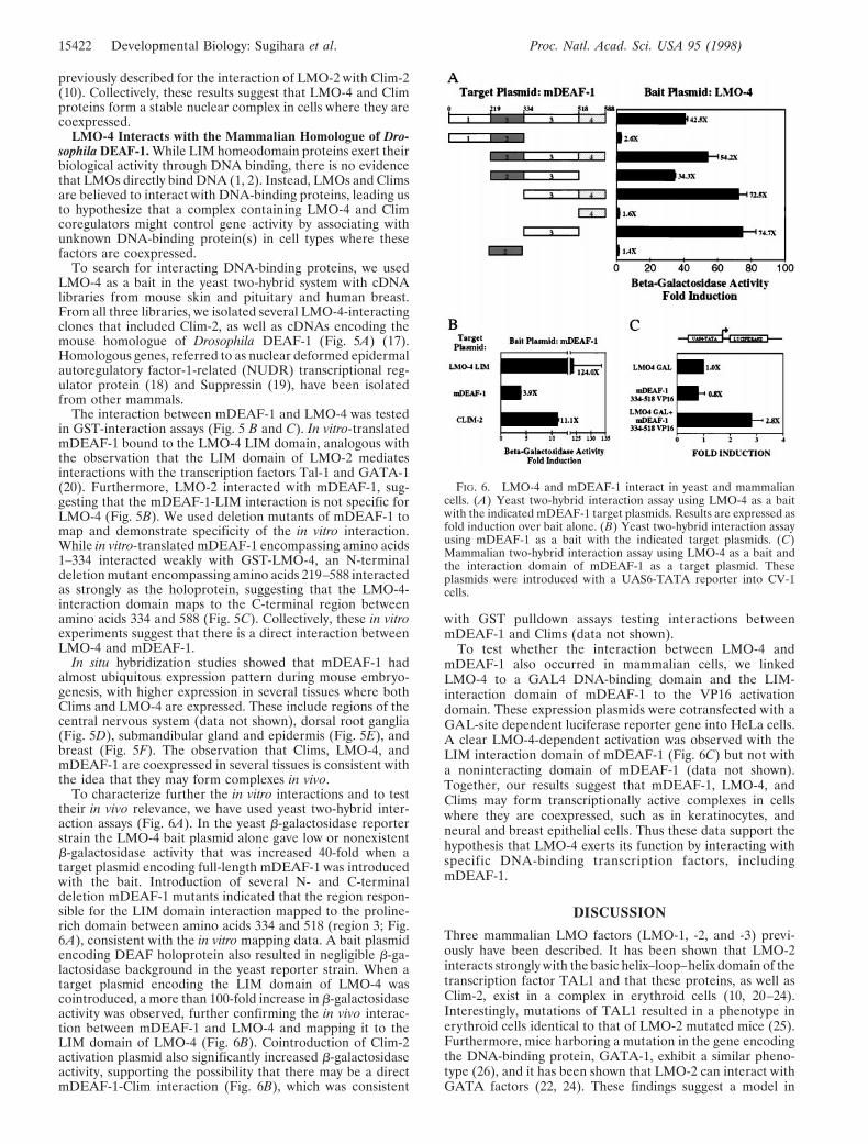
FIG. 5. Isolation and characterization of an LMO-4-interacting protein, mDEAF-1. (A) The amino acid sequence of mDEAF-1 compared withDrosophila DEAF-1. (B) A GST-interaction assay with the indicated GST proteins and 35S-labeled mDEAF-1. (C) A GST-interaction assay withthe indicated GST proteins and the following 35S-labeled mDEAF-1 proteins shown schematically on top: A, full length; B, residues 1–344; C,residues 219–588. (D) In situ hybridization showing mDEAF-1 expression in dorsal root ganglia at e15.5, sagittal cut of neck region. (E) In situhybridization showing mDEAF-1 expression in submandibular gland and epidermis at e17.5. (F) In situ hybridization showing expression ofmDEAF-1 in breast epithelial cells from a 12.5-day pregnant mother.
Developmental Biology: Sugihara et al. Proc. Natl. Acad. Sci. USA 95 (1998) 15421
previously described for the interaction of LMO-2 with Clim-2(10). Collectively, these results suggest that LMO-4 and Climproteins form a stable nuclear complex in cells where they arecoexpressed.
LMO-4 Interacts with the Mammalian Homologue of Dro-sophila DEAF-1. While LIM homeodomain proteins exert theirbiological activity through DNA binding, there is no evidencethat LMOs directly bind DNA (1, 2). Instead, LMOs and Climsare believed to interact with DNA-binding proteins, leading usto hypothesize that a complex containing LMO-4 and Climcoregulators might control gene activity by associating withunknown DNA-binding protein(s) in cell types where thesefactors are coexpressed.
To search for interacting DNA-binding proteins, we usedLMO-4 as a bait in the yeast two-hybrid system with cDNAlibraries from mouse skin and pituitary and human breast.From all three libraries, we isolated several LMO-4-interactingclones that included Clim-2, as well as cDNAs encoding themouse homologue of Drosophila DEAF-1 (Fig. 5A) (17).Homologous genes, referred to as nuclear deformed epidermalautoregulatory factor-1-related (NUDR) transcriptional reg-ulator protein (18) and Suppressin (19), have been isolatedfrom other mammals.
The interaction between mDEAF-1 and LMO-4 was testedin GST-interaction assays (Fig. 5 B and C). In vitro-translatedmDEAF-1 bound to the LMO-4 LIM domain, analogous withthe observation that the LIM domain of LMO-2 mediatesinteractions with the transcription factors Tal-1 and GATA-1(20). Furthermore, LMO-2 interacted with mDEAF-1, sug-gesting that the mDEAF-1-LIM interaction is not specific forLMO-4 (Fig. 5B). We used deletion mutants of mDEAF-1 tomap and demonstrate specificity of the in vitro interaction.While in vitro-translated mDEAF-1 encompassing amino acids1–334 interacted weakly with GST-LMO-4, an N-terminaldeletion mutant encompassing amino acids 219–588 interactedas strongly as the holoprotein, suggesting that the LMO-4-interaction domain maps to the C-terminal region betweenamino acids 334 and 588 (Fig. 5C). Collectively, these in vitroexperiments suggest that there is a direct interaction betweenLMO-4 and mDEAF-1.
In situ hybridization studies showed that mDEAF-1 hadalmost ubiquitous expression pattern during mouse embryo-genesis, with higher expression in several tissues where bothClims and LMO-4 are expressed. These include regions of thecentral nervous system (data not shown), dorsal root ganglia(Fig. 5D), submandibular gland and epidermis (Fig. 5E), andbreast (Fig. 5F). The observation that Clims, LMO-4, andmDEAF-1 are coexpressed in several tissues is consistent withthe idea that they may form complexes in vivo.
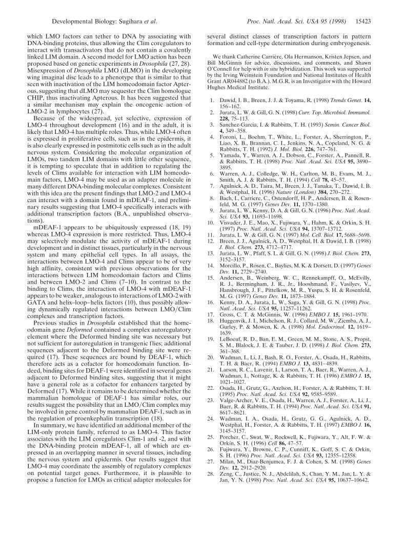
To characterize further the in vitro interactions and to testtheir in vivo relevance, we have used yeast two-hybrid inter-action assays (Fig. 6A). In the yeast b-galactosidase reporterstrain the LMO-4 bait plasmid alone gave low or nonexistentb-galactosidase activity that was increased 40-fold when atarget plasmid encoding full-length mDEAF-1 was introducedwith the bait. Introduction of several N- and C-terminaldeletion mDEAF-1 mutants indicated that the region respon-sible for the LIM domain interaction mapped to the proline-rich domain between amino acids 334 and 518 (region 3; Fig.6A), consistent with the in vitro mapping data. A bait plasmidencoding DEAF holoprotein also resulted in negligible b-ga-lactosidase background in the yeast reporter strain. When atarget plasmid encoding the LIM domain of LMO-4 wascointroduced, a more than 100-fold increase in b-galactosidaseactivity was observed, further confirming the in vivo interac-tion between mDEAF-1 and LMO-4 and mapping it to theLIM domain of LMO-4 (Fig. 6B). Cointroduction of Clim-2activation plasmid also significantly increased b-galactosidaseactivity, supporting the possibility that there may be a directmDEAF-1-Clim interaction (Fig. 6B), which was consistent
with GST pulldown assays testing interactions betweenmDEAF-1 and Clims (data not shown).
To test whether the interaction between LMO-4 andmDEAF-1 also occurred in mammalian cells, we linkedLMO-4 to a GAL4 DNA-binding domain and the LIM-interaction domain of mDEAF-1 to the VP16 activationdomain. These expression plasmids were cotransfected with aGAL-site dependent luciferase reporter gene into HeLa cells.A clear LMO-4-dependent activation was observed with theLIM interaction domain of mDEAF-1 (Fig. 6C) but not witha noninteracting domain of mDEAF-1 (data not shown).Together, our results suggest that mDEAF-1, LMO-4, andClims may form transcriptionally active complexes in cellswhere they are coexpressed, such as in keratinocytes, andneural and breast epithelial cells. Thus these data support thehypothesis that LMO-4 exerts its function by interacting withspecific DNA-binding transcription factors, includingmDEAF-1.
DISCUSSION
Three mammalian LMO factors (LMO-1, -2, and -3) previ-ously have been described. It has been shown that LMO-2interacts strongly with the basic helix–loop–helix domain of thetranscription factor TAL1 and that these proteins, as well asClim-2, exist in a complex in erythroid cells (10, 20–24).Interestingly, mutations of TAL1 resulted in a phenotype inerythroid cells identical to that of LMO-2 mutated mice (25).Furthermore, mice harboring a mutation in the gene encodingthe DNA-binding protein, GATA-1, exhibit a similar pheno-type (26), and it has been shown that LMO-2 can interact withGATA factors (22, 24). These findings suggest a model in
FIG. 6. LMO-4 and mDEAF-1 interact in yeast and mammaliancells. (A) Yeast two-hybrid interaction assay using LMO-4 as a baitwith the indicated mDEAF-1 target plasmids. Results are expressed asfold induction over bait alone. (B) Yeast two-hybrid interaction assayusing mDEAF-1 as a bait with the indicated target plasmids. (C)Mammalian two-hybrid interaction assay using LMO-4 as a bait andthe interaction domain of mDEAF-1 as a target plasmid. Theseplasmids were introduced with a UAS6-TATA reporter into CV-1cells.
15422 Developmental Biology: Sugihara et al. Proc. Natl. Acad. Sci. USA 95 (1998)
which LMO factors can tether to DNA by associating withDNA-binding proteins, thus allowing the Clim coregulators tointeract with transactivators that do not contain a covalentlylinked LIM domain. A second model for LMO action has beenproposed based on genetic experiments in Drosophila (27, 28).Misexpression of Drosophila LMO (dLMO) in the developingwing imaginal disc leads to a phenotype that is similar to thatseen with inactivation of the LIM homeodomain factor Apter-ous, suggesting that dLMO may sequester the Clim homologueCHIP, thus inactivating Apterous. It has been suggested thata similar mechanism may explain the oncogenic action ofLMO-2 in lymphocytes (27).
Because of the widespread, yet selective, expression ofLMO-4 throughout development (16) and in the adult, it islikely that LMO-4 has multiple roles. Thus, while LMO-4 oftenis expressed in proliferative cells, such as in the epidermis, itis also clearly expressed in postmitotic cells such as in the adultnervous system. Considering the molecular organization ofLMOs, two tandem LIM domains with little other sequence,it is tempting to speculate that in addition to regulating thelevels of Clims available for interaction with LIM homeodo-main factors, LMO-4 may be used as an adapter molecule inmany different DNA-binding molecular complexes. Consistentwith this idea are the present findings that LMO-2 and LMO-4can interact with a domain found in mDEAF-1, and prelimi-nary results suggesting that LMO-4 specifically interacts withadditional transcription factors (B.A., unpublished observa-tions).
mDEAF-1 appears to be ubiquitously expressed (18, 19)whereas LMO-4 expression is more restricted. Thus, LMO-4may selectively modulate the activity of mDEAF-1 duringdevelopment and in distinct tissues, particularly in the nervoussystem and many epithelial cell types. In all assays, theinteractions between LMO-4 and Clims appear to be of veryhigh affinity, consistent with previous observations for theinteractions between LIM homeodomain factors and Climsand between LMO-2 and Clims (7–10). In contrast to thebinding to Clims, the interaction of LMO-4 with mDEAF-1appears to be weaker, analogous to interactions of LMO-2 withGATA and helix–loop–helix factors (10), thus possibly allow-ing dynamically regulated interactions between LMOyClimcomplexes and transcription factors.
Previous studies in Drosophila established that the home-odomain gene Deformed contained a complex autoregulatoryelement where the Deformed binding site was necessary butnot sufficient for autoregulation in transgenic flies; additionalsequences adjacent to the Deformed binding site were re-quired (17). These sequences are bound by DEAF-1, whichtherefore acts as a cofactor for homeodomain function. In-deed, binding sites for DEAF-1 were identified in several genesadjacent to Deformed binding sites, suggesting that it mighthave a general role as a cofactor for enhancers targeted byDeformed (17). While it remains to be determined whether themammalian homologue of DEAF-1 has similar roles, ourresults suggest the possibility that an LMOyClim complex maybe involved in gene control by mammalian DEAF-1, such as inthe regulation of proenkephalin transcription (18).
In summary, we have identified an additional member of theLIM-only protein family, referred to as LMO-4. This factorassociates with the LIM coregulators Clim-1 and -2, and withthe DNA-binding protein mDEAF-1, all of which are ex-pressed in an overlapping manner in several tissues, includingthe nervous system and epidermis. Our results suggest thatLMO-4 may coordinate the assembly of regulatory complexeson potential target genes. Furthermore, it is plausible topropose a function for LMOs as critical adapter molecules for
several distinct classes of transcription factors in patternformation and cell-type determination during embryogenesis.
We thank Catherine Carriere, Ola Hermanson, Kristen Jepsen, andBill McGinnis for advice, discussions, and comments, and ShawnO’Connell for help with in situ hybridization. This work was supportedby the Irving Weinstein Foundation and National Institutes of HealthGrant AR044882 (to B.A.). M.G.R. is an Investigator with the HowardHughes Medical Institute.
1. Dawid, I. B., Breen, J. J. & Toyama, R. (1998) Trends Genet. 14,156–162.
2. Jurata, L. W. & Gill, G. N. (1998) Curr. Top. Microbiol. Immunol.228, 75–113.
3. Sanchez-Garcia, I. & Rabbitts, T. H. (1993) Semin. Cancer Biol.4, 349–358.
4. Foroni, L., Boehm, T., White, L., Forster, A., Sherrington, P.,Liao, X. B., Brannan, C. I., Jenkins, N. A., Copeland, N. G. &Rabbitts, T. H. (1992) J. Mol. Biol. 226, 747–761.
5. Yamada, Y., Warren, A. J., Dobson, C., Forster, A., Pannell, R.& Rabbitts, T. H. (1998) Proc. Natl. Acad. Sci. USA 95, 3890–3895.
6. Warren, A. J., Colledge, W. H., Carlton, M. B., Evans, M. J.,Smith, A. J. & Rabbitts, T. H. (1994) Cell 78, 45–57.
7. Agulnick, A. D., Taira, M., Breen, J. J., Tanaka, T., Dawid, I. B.& Westphal, H. (1996) Nature (London) 384, 270–272.
8. Bach, I., Carriere, C., Ostendorff, H. P., Andersen, B. & Rosen-feld, M. G. (1997) Genes Dev. 11, 1370–1380.
9. Jurata, L. W., Kenny, D. A. & Gill, G. N. (1996) Proc. Natl. Acad.Sci. USA 93, 11693–11698.
10. Visvader, J. E., Mao, X., Fujiwara, Y., Hahm, K. & Orkin, S. H.(1997) Proc. Natl. Acad. Sci. USA 94, 13707–13712.
11. Jurata, L. W. & Gill, G. N. (1997) Mol. Cell. Biol. 17, 5688–5698.12. Breen, J. J., Agulnick, A. D., Westphal, H. & Dawid, I. B. (1998)
J. Biol. Chem. 273, 4712–4717.13. Jurata, L. W., Pfaff, S. L. & Gill, G. N. (1998) J. Biol. Chem. 273,
3152–3157.14. Morcillo, P., Rosen, C., Baylies, M. K. & Dorsett, D. (1997) Genes
Dev. 11, 2729–2740.15. Andersen, B., Weinberg, W. C., Rennekampff, O., McEvilly,
R. J., Bermingham, J. R., Jr., Hooshmand, F., Vasilyev, V.,Hansbrough, J. F., Pittelkow, M. R., Yuspa, S. H. & Rosenfeld,M. G. (1997) Genes Dev. 11, 1873–1884.
16. Kenny, D. A., Jurata, L. W., Saga, Y. & Gill, G. N. (1998) Proc.Natl. Acad. Sci. USA 95, 11257–11262.
17. Gross, C. T. & McGinnis, W. (1996) EMBO J. 15, 1961–1970.18. Huggenvik, J. I., Michelson, R. J., Collard, M. W., Ziemba, A. J.,
Gurley, P. & Mowen, K. A. (1998) Mol. Endocrinol. 12, 1619–1639.
19. LeBoeuf, R. D., Ban, E. M., Green, M. M., Stone, A. S., Propst,S. M., Blalock, J. E. & Tauber, J. D. (1998) J. Biol. Chem. 273,361–368.
20. Wadman, I., Li, J., Bash, R. O., Forster, A., Osada, H., Rabbitts,T. H. & Baer, R. (1994) EMBO J. 13, 4831–4839.
21. Larson, R. C., Lavenir, I., Larson, T. A., Baer, R., Warren, A. J.,Wadman, I., Nottage, K. & Rabbitts, T. H. (1996) EMBO J. 15,1021–1027.
22. Osada, H., Grutz, G., Axelson, H., Forster, A. & Rabbitts, T. H.(1995) Proc. Natl. Acad. Sci. USA 92, 9585–9589.
23. Valge-Archer, V. E., Osada, H., Warren, A. J., Forster, A., Li, J.,Baer, R. & Rabbitts, T. H. (1994) Proc. Natl. Acad. Sci. USA 91,8617–8621.
24. Wadman, I. A., Osada, H., Grutz, G. G., Agulnick, A. D.,Westphal, H., Forster, A. & Rabbitts, T. H. (1997) EMBO J. 16,3145–3157.
25. Porcher, C., Swat, W., Rockwell, K., Fujiwara, Y., Alt, F. W. &Orkin, S. H. (1996) Cell 86, 47–57.
26. Fujiwara, Y., Browne, C. P., Cunniff, K., Goff, S. C. & Orkin,S. H. (1996) Proc. Natl. Acad. Sci. USA 93, 12355–12358.
27. Milan, M., Diaz-Benjumea, F. J. & Cohen, S. M. (1998) GenesDev. 12, 2912–2920.
28. Zeng, C., Justice, N. J., Abdelilah, S., Chan, Y. M., Jan, L. Y. &Jan, Y. N. (1998) Proc. Natl. Acad. Sci. USA 95, 10637–10642.
Developmental Biology: Sugihara et al. Proc. Natl. Acad. Sci. USA 95 (1998) 15423
Copyright © 2022 FDOKUMEN