IMPACT FACTOR - Eurosurveillance
176
Vol. 14 · Issues 13–25 · Apr–Jun 2009 www.eurosurveillance.org EUROPEAN CENTRE FOR DISEASE PREVENTION AND CONTROL In this edition Euroroundup • National Hand Hygiene Campaigns in Europe 2000-2009 Also • Estimating the Global Burden of Foodborne Diseases - a collaborative effort • Preparedness for the prevention and control of influenza outbreaks on passenger ships in the EU: the SHIPSAN TRAINET project communication NOW LISTED FOR IMPACT FACTOR
-
Upload
khangminh22 -
Category
Documents
-
view
1 -
download
0
Transcript of IMPACT FACTOR - Eurosurveillance

Vol . 14 · Issues 13–25 · Apr–Jun 2009
www.eurosurveillance .org
EUROPEAN CENTRE FOR DISEASE PREVENTION AND CONTROL
In this edition
Euroroundup
•NationalHandHygieneCampaignsin Europe 2000-2009
Also
•EstimatingtheGlobalBurdenofFoodborneDiseases-acollaborativeeffort
•PreparednessforthepreventionandcontrolofinfluenzaoutbreaksonpassengershipsintheEU:theSHIPSANTRAINETprojectcommunication
NOWLISTED FORIMPACTFACTOR

Con t e n t s
Peer-reviewedEuropeaninformationoncommunicablediseasesurveillanceandcontrol
The opinions expressed by authors contributing to Eurosurveillance do not necessarily reflect the opinions of the European Centre for Disease Prevention and Control (ECDC) or the Editorial team or the institutions with which the authors are affiliated. Neither the ECDC nor any person acting on behalf of the ECDC is responsible for the use which might be made of the information in this journal.
EDI TOR IALS • Internet surveillance systems for early alerting of health threats 200
• Multilocus variable number of tandem repeats analysis (MLVA) - a reliable tool for rapid investigation of Salmonella Typhimurium outbreaks 202
• European Immunization Week goes viral 203
• The global impact of hand hygiene campaigning 205
• Avian influenza A(H5N1) – current situation 207
SURVE I L L ANCE AND OUTBREAK REPORTS • Nation-wide prospective surveillance of Clostridium difficile infections in hospitals in Belgium, July 2007-June 2008 208
• Ongoing rubella outbreak in Austria, 2008-2009 211
• Mumps epidemiology in the Mid-West of Ireland 2004-2008: increasing disease burden in the university/college setting 215
• Characterisation of Neisseria meningitidis C strains causing two clusters in the north of Italy in 2007 and 2008 220
• Travel-associated Legionnaires’ disease in Europe in 2007 222
• Investigation of the spread of brucellosis among human and animal populations in southeastern Bulgaria, 2007` 227
RESEARCH ART ICL ES • Evaluation of a patient referral contact tracing programme for hepatitis B and C virus infection in drug injectors 232
• Development of a new nomenclature for Salmonella Typhimurium multilocus variable number of tandem repeats analysis (MLVA) 237
• The emergence of Clostridium difficile PCR ribotype 027 in Denmark – a possible link with the increased consumption of fluoroquinolones and cephalosporins? 242
• Endemic hepatitis E in two Nordic countries 246
• Can the Swedish new variant of Chlamydia trachomatis (nvCT) be detected by UK NEQAS participants from seventeen European countries and five additional countries/regions in 2009? 255
• Rapid spread of drug-resistant influenza A viruses in the Basque Country, northern Spain, 2000-1 to 2008-9 259
• Clinical laboratory practices for the detection of rotavirus in England and Wales: can surveillance based on routine laboratory testing data be used to evaluate the impact of vaccination? 262
• KPC-2-producing Klebsiella pneumoniae infections in Greek hospitals are mainly due to a hyperepidemic clone 268
• Autochthonous cystic echinococcosis in patients who grew up in Germany 273
• A methodological approach to investigating a nationwide clinical specimen contamination problem in England 280
• Application of RT-PCR for diagnosis of respiratory syncytial virus and human metapneumovirus infections in Bulgaria, 2006-7 and 2007-8 285
• Clinical and epidemiological aspects of parvovirus B19 infections in Ireland, January 1996-June 2008 290
Vol . 14 · Issues 13–25 · Apr–Jun 2009
EditorialTeamBased at the European Centre for Disease Prevention and Control (ECDC), 171 83 Stockholm | Sweden
TelephoneNumber:+46 (0)8 586 01138 or +46 (0)8 586 01136
Faxnumber:+46 (0)8 586 01294
E-mail: [email protected]
Editor-in-ChiefKarl Ekdahl
ManagingEditorInes Steffens
Scientific Editors Kathrin Hagmaier Renata Mikolajczyk
AssistantEditors Alina Buzdugan Ingela Söderlund
AssociateEditorsAndrea Ammon, ECDC, Stockholm, Sweden
Mike Catchpole, Health Protection Agency, London, United Kingdom
Denis Coulombier, ECDC, Stockholm, Sweden
Christian Drosten, Universitätsklinikum Bonn, Bonn, Germany
Johan Giesecke, ECDC, Stockholm, Sweden
Herman Goossens, Universiteit Antwerpen, Antwerp, Belgium
David Heymann, London, United Kingdom
Karl Kristinsson, Landspitali University Hospital, Reykjavik, Iceland
Irena Klavs, National Institute of Public Health, Ljubljana, Slovenia
Daniel Lévy-Bruhl, Institut de Veille Sanitaire, Paris, France
Richard Pebody, Health Protection Agency, London, United Kingdom
Panayotis T. Tassios, University of Athens, Athens, Greece
Hélène Therre, Institut de Veille Sanitaire, Paris, France
Henriette de Valk, Institut de Veille Sanitaire, Paris, France
Sylvie van der Werf, Institut Pasteur, Paris, France
EditorialBoardSee inner back cover
LayoutFabrice Donguy / Martin Wincent
WebmasterSami Dufva
www.eurosurveillance.org
©Eurosurveillance,2009

Vol . 14 · Issues 13–25 · Apr–Jun 2009
Con t e n t s
PERSPECT IVES • Is there a need for anti-rabies vaccine and immunoglobulins rationing in Europe? 295
• Implementation of a national electronic reporting system in Lithuania 298
• VACSATC (Vaccine safety: attitudes, training and communication): Why such a project? 304
• Estimating the Global Burden of Foodborne Diseases - a collaborative effort 308
• Influenza A(H5N1): an overview of the current situation 312
• Preparedness for the prevention and control of influenza outbreaks on passenger ships in the EU: the SHIPSAN TRAINET project communication 316
EUROROUNDUPS • National Hand Hygiene Campaigns in Europe, 2000-2009 320
MEE T ING REPORTS • Impact of immigration on HIV and tuberculosis epidemiology in the Euro-Mediterranean area 327
L E T T ERS • Prevention of congenital rubella and congenital varicella in Europe 330
• Authors’ reply: Prevention of congenital rubella and congenital varicella in Europe 331
• National pneumococcal vaccination programmes for children in Europe, 2001-2007: update from Ireland 332
• National pneumococcal vaccination programmes for children in Europe, 2001-2007: update from Turkey 333
• Authors’ reply: National pneumococcal vaccination programmes for children in Europe, 2001-2007 – updated table 334
RAPID COMMUNICAT IONS • First identification of class A carbapenemase-producing Klebsiella pneumoniae in the Republic of Ireland 337
• Outbreak of Clostridium difficile 027 in North Zealand, Denmark, 2008-2009 339
• Outbreak of Clostridium difficile 027 infection in Vienna, Austria 2008-2009 A 342
• Shigella sonnei infections in Norway associated with sugar peas, May – June 2009 344
• Imported fresh sugar peas as suspected source of an outbreak of Shigella sonnei in Denmark, April – May 2009 346
• Outbreak of hepatitis A among men who have sex with men in Barcelona, Spain, September 2008 – March 2009 348
• Anaphylaxis following unnecessary meningococcal chemoprophylaxis of a healthcare worker 351
• Chikungunya infection confirmed in a Belgian traveller returning from Phuket (Thailand) 354
• Trends in the epidemiology of dengue fever and their relevance for importation to Europe 356
• An outbreak caused by hantavirus in the Black Sea region of Turkey, January – May 2009 359
• Sustained intensive transmission of Q fever in the south of the Netherlands, 200 361
• Trichinellosis acquired in Senegal from warthog ham, March 2009 364
• Norwegians approve of the health authorities’ strategy to communicate worst case pandemic scenarios 366
All material in Eurosurveillance is in the public domain and may be used and reprinted without special permission. However, the source should be cited properly and we suggest adding a link to the exact page on the Eurosurveillance website.
Articles published in Eurosurveillance are indexed in PubMed/MEDLINE

2 0 0 www.eurosurveillance.org
Ed i t o r ials
I n t e r n e t s u r v e I l l a n c e s y s t e m s f o r e a r ly a l e r t I n g o f h e a lt h t h r e at s
J P Linge1, R Steinberger1, T P Weber1, R Yangarber2, E van der Goot1, D H Al Khudhairy1, N I Stilianakis ([email protected])11. Joint Research Centre (JRC), European Commission, Ispra (VA), Italy2. Department of Computer Science, University of Helsinki, Helsinki, Finland
This article was published on 2 April 2009. Citation style for this article: Linge JP, Steinberger R, Weber TP, Yangarber R, van der Goot E, Al Khudhairy DH, Stilianakis NI. Internet surveillance systems for early alerting of health threats. Euro Surveill. 2009;14(13):pii=19162. Available online: http://www.eurosurveillance.org/ViewArticle.aspx?ArticleId=19162
In order to gather a comprehensive picture of potential epidemic threats, public health authorities increasingly rely on systems that perform epidemic intelligence (EI). EI makes use of information that originates from official sources such as national public health surveillance systems as well as from informal sources such as electronic media and web-based information tools. All these sources are employed to enhance risk monitoring with the purpose of early alerting and initial risk assessment. In this context Paquet et al. [1] distinguish between indicator-based risk monitoring and event-based risk monitoring. As indicator-based monitoring relies on classical routine surveillance, many systems will use methods and data sources familiar to most epidemiologists and public health officials. The event-based component of EI is in contrast rather new; its methods, strengths and limitations are generally not widely known in the public health community. The purpose of this editorial is thus to provide an overview of the methods used in pro-active event-based monitoring and to put them into context with regard to the structured indicator-based monitoring such as that described in the article on the Lithuanian electronic surveillance system published in this issue of Eurosurveillance [2].
More and more national and international public health agencies employ systematic event detection systems using informal sources (news wires, media sources or websites) on the internet to monitor the potential threat of emerging and re-emerging infectious diseases. Such web-based event detection is the first step in EI systems designed to provide early warning signals to public health institutions. A number of different systems have been developed for this purpose. There is, however, still the need to emphasise some fundamental differences between the available systems and to identify the challenges that lie ahead. Existing event detection systems can be classified into three categories.
First, news aggregators collect articles from several sources, usually filtered by language or country. Users gain easy access to many sources through a common portal, but still need to examine each individual article.
Second, automatic systems such as the Medical Information System (MedISys) (http://medusa.jrc.it/) [3], Pattern-based Understanding and Learning System (PULS) (http://puls.cs.helsinki.fi/medical/) [3], HealthMap (http://www.healthmap.org/) [4], and BioCaster Global Health Monitor (http://biocaster.nii.ac.jp/) [5] go beyond the mere gathering task by adding a series of analysis steps. Automatic systems differ in their levels of analysis,
in the range of information sources, their language coverage, the speed of delivering information and visualisation methods. HealthMap currently covers five languages, BioCaster seven languages, and MedISys more than 40 languages. While HealthMap mainly relies on Google News, World Health Organization (WHO) news feeds, ProMED-Mail (http://www.promedmail.org/) [6], and Eurosurveillance as sources, MedISys monitors ProMED-Mail, web sites of national public health
authorities, specialist web sites (including Eurosurveillance), news from about twenty news wires, plus a balanced list of approximately 2,200 news sources from around the world, hand-selected with a view of ensuring a geographic balance.
Analysis steps may include: recognition of relevant terms (names of diseases, symptoms and organisations), recognition and disambiguation of geographical locations mentioned in the articles, grouping related articles into clusters, and extraction of full events from the news, providing the users with aggregated information about the disease, the number of cases, as well as time and place of an outbreak. Ideally, news items should be clustered across languages and national borders. Most systems focus on recognising communicable diseases and visualise the location of the extracted events on geographical maps. As a domain-specific application of the Europe Media Monitor (EMM) system, MedISys covers not only the whole range of chemical, biological, radiological and nuclear threats (CBRN), but also allows using a filter to only show outbreak-related information. MedISys additionally monitors trends and calculates alert levels per disease and per country, by comparing the number of recent news items with averages. PULS, which is integrated with MedISys, extracts event data from the
In order to gather a comprehensive picture
of potential epidemic threats, public health
authorities increasingly rely on systems that
perform epidemic intelligence (EI).

www.eurosurveillance.org 2 0 1
English MedISys articles and produces searchable outbreak data in table format.
All automatic systems will clearly benefit from better machine-translation software so that a more diverse range of sources can be tapped. Ideally, a summary of each article should be shown in the original language together with its translation.
Third, moderated systems such as ProMED-Mail [6], GPHIN (Global Public Health Intelligence Network) [7] and ARGUS [8] rely on a group of analysts to scan available news sources. The analysts take into account information from individual web sites, aggregator sites, automatic systems, and other sources such as reports from medical practitioners and health authorities. In combination with its Rapid News Service (RNS) tool, MedISys also allows for manual moderation.
There are fundamental differences in these approaches. Non-moderated systems are able to search the web and display new articles without time delay in an unbiased manner. Moderated systems show fewer irrelevant news items (fewer false positives). However, moderator bias represents a risk (false negatives); users might have a different focus than the moderators.
For users who need to react to threats quickly and possess the man-power to entertain their own monitoring effort, automatic systems are appealing because of the detection speed. Other users might prefer to wait for human-moderated feeds.
Technical implementation of aggregators is straight-forward, but for both automatic and moderated systems, many challenges lie ahead. Redundancy is a major issue. Naturally, news agencies, online and printed news sources, national and international authorities or blogs may report the same event in different ways at various time points. This often leads to misclassification of events and overestimation of impact. Furthermore, feedback loops are created when automatic systems accept input from moderated systems (or vice versa). In any moderated approach, long-term funding or volunteer participation is necessary to maintain the analyst base.
Automatic approaches are the only option to sieve relevant information out of the abundant pool of multilingual media sources in real time. However, human moderation is needed eventually.
A further challenge for the future will be to improve the transition from risk monitoring to risk assessment. Recent approaches on extracting patterns of influenza-related search terms from queries stored by Google and Yahoo [9, 10] showed that patterns of searches matched with official influenza surveillance data, thus indicating that search-term analysis could be a useful complementary tool to surveillance. However, although search-term analysis and event-based monitoring can provide an important signal of a potential outbreak, the data gathered is usually not detailed or reliable enough to estimate relevant epidemiological parameters of incipient outbreaks and the methods are prone to false alarms.
Lithuania’s electronic reporting system described in this issue of Eurosurveillance [2] is an example of an indicator-based component of EI which allows the collection of structured data at country level. Such national information is typically fed into the European Surveillance System (TESSy) [11] of the European Centre for Disease Prevention and Control (ECDC) which collects surveillance data on infectious diseases at the European Union
(EU) level to support outbreak detection, risk assessment, outbreak investigation and control measures. This is complemented by the Early Warning and Response System (EWRS) which establishes permanent communication between public health authorities in the EU member states [12].
References
1. Paquet C, Coulombier D, Kaiser R, Ciotti M. Epidemic intelligence: a new framework for strengthening disease surveillance in Europe. Euro Surveill. 2006;11(12):pii=665. Available from: http://www.eurosurveillance.org/ViewArticle.aspx?ArticleId=665
2. Domeika M, Kligys, G, Ivanauskiene O, Mereckiene J, Bakasenas V, Morkunas B, Berescianskis D, Wahl T, Stenqvist K. Implementation of a national electronic reproting system in Lithuania. Euro Surveill. 2009;14
3. Steinberger R, Fuart F, van der Goot E, Best C, von Etter P, Yangarber R. Text mining from the web for medical intelligence. In: Fogelman-Soulié F. Perrotta D, Piskorski J, Steinberger R, editors. Mining Massive Data Sets for Security, IOS Press, Amsterdam, 2008. p. 295-310. Available from: http://langtech.jrc.it/Documents/2009_MMDSS_Medical-Intelligence.pdf
4. Freifeld CC, Mandl KD, Reis BY, Brownstein JS. HealthMap: global infectious disease monitoring through automated classification and visualization of Internet media reports. J Am Med Inform Assoc. 2008;15(2):150-7.
5. Collier N, Doan S, Kawazoe A, Goodwin RM, Conway M, Tateno Y, Ngo QH, Dien D, Kawtrakul A, Takeuchi K, Shigematsu M, Taniguchi K. BioCaster: detecting public health rumors with a Web-based text mining system. Bioinformatics. 2008, 24, 2940-2941.
6. Madoff LC, ProMED-mail: an early warning system for emerging diseases. Clin Infect Dis. 2004;39(2):227-32.
7. Mykhalovskiy E, Weir L. The Global Public Health Intelligence Network and early warning outbreak detection: a Canadian contribution to global public health. Can J Public Health. 2006;97(1):42-4.
8. Wilson JM, Polyak, MG, Blake JW, Collmann J. A heuristic indication and warning staging model for detection and assessment of biological events. J Am Med Inform Assoc. 2008;15(2):158–71.
9. Polgreen PM, Chen Y, Pennock DM, Nelson FD. Using internet searches for influenza surveillance, Clin Infect Dis. 2008;47(11):1443-8.
10. Ginsberg J, Mohebbi MH, Patel RD, Brammer L, Smolinski MS, Brilliant L. Detecting influenza epidemics using search engine query data, Nature. 2009;457(7232):1012-14.
11. Amato-Gauci A, Ammon A. ECDC to launch first report on communicable diseases epidemiolog y in the European Union. Euro Surveill. 2007;12(23):pii=3213. Available from: http://www.eurosurveillance.org/ViewArticle.aspx?ArticleId=3213
12. Guglielmetti P, Coulombier D, Thinus G, Van Loock F, Schreck S. The Early Warning and Response System for communicable diseases in the EU: an overview from 1999 to 2005. Euro Surveill. 2006;11(12):pii=666. Available from: http://www.eurosurveillance.org/ViewArticle.aspx?ArticleId=666

2 0 2 www.eurosurveillance.org
Ed i t o r ials
M u lt i l o c u s va r i a b l e n u M b e r o f ta n d e M r e p e at s a n a ly s i s (M lva ) - a r e l i a b l e to o l f o r r a p i d i n v e s t i g at i o n o f s a l M o n e l l a t y p h i M u r i u M o u t b r e a k s
M Heck ([email protected])11. Centre for Infectious Disease Control, National Institute for Public Health and the Environment (Rijksinstituut voor
Volksgezondheid en Milieu, RIVM), Bilthoven, the Netherlands
This article was published on 16 April 2009. Citation style for this article: Heck M. Multilocus variable number of tandem repeats analysis (MLVA) - a reliable tool for rapid investigation of Salmonella Typhimurium outbreaks. Euro Surveill. 2009;14(15):pii=19177. Available online: http://www.eurosurveillance.org/ViewArticle.aspx?ArticleId=19177
Salmonella enterica subsp. enterica serovar Typhimurium is a frequently occurring foodborne pathogen which causes many sporadic cases worldwide and is frequently the responsible agent in outbreaks of gastroenteritis. In elucidating outbreaks involving consumption of contaminated food, source tracing in a timely manner is imperative. Furthermore, it is important for risk managers to be able to accurately attribute sporadic cases to specific animal host species and to understand transmission routes of S. Typhimurium. Epidemiological meaningful subdivision of this serotype is therefore indispensable.
Phage-typing and pulsed field gel electrophoresis (PFGE) are among the methods most frequently applied. Both have been used sucessfully but have the disadvantage that reading the typing results is difficult to standardise, which hampers the exchange of typing results between laboratories and the construction of international reference databases. Source tracing or attribution using these methods fails when a frequently occurring important phagetype like DT104 (or PT 4 within Salmonella Enteritidis) that may have different sources, cannot be further subdivided.
Unambiguous typing results are critical in both detecting outbreaks and determining their source. Multilocus variable number of tandem repeats analysis (MLVA) is a PCR-based method that has recently become a widely used highly discriminatory molecular method for typing S. Typhimurium. It is based on amplification and fragment analysis of five repeat loci. MLVA has the advantages of typing methods based on PCR (low cost, short time, and easy to perform) that are independent of equipment and yield unambiguous typing data. For the latter purpose, the authors of the article published in today’s issue of Eurosurveillance [1] developed a set of reference strains that can be used for easy normalisation of fragment sizes in each laboratory. According to the authors MLVA turned out to have a discriminatory power similar to that of phage typing and PFGE. Their results suggest that MLVA are reliable in epidemiological studies, including analyses of outbreaks and transmission routes. The authors propose a simple and definitive universal nomenclature based on the fragment size of tandem repeat loci allowing the comparison of MLVA profiles between laboratories. A further advantage of this nomenclature is that it allows easy recognition of related but slightly different MLVA-profiles that may be epidemiologically linked.
We strongly believe that molecular typing is the way forward and MLVA is a step in that direction. Nevertheless, at present it cannot fully replace the older typing techniques irrespective of all its advantages. Still faster methods are necessary for timely intervention both in outbreaks and during quality control along the food chain. Furthermore, epidemiological significance of related strains would be greater if molecular methods more fully exploited the phylogenetic information in the DNA of Salmonella.
References
1. Larsson JT, Torpdahl M, Petersen RF, Sørensen G, Lindstedt BA, Nielsen EM. Development of a new nomenclature for Salmonella Typhimurium multilocus variable number of tandem repeats analysis (MLVA). Euro Surveill. 2009;14(15):pii=19174. Available from: http://www.eurosurveillance.org/ViewArticle.aspx?ArticleId=19174

www.eurosurveillance.org 2 03
Ed i t o r ials
E u r o p E a n I m m u n I z at I o n W E E k g o E s v I r a l
R Martin1, O Nørgaard1, J V Lazarus ([email protected])11. Communicable Diseases Unit,World Health Organization Regional Office for Europe, Copenhagen, Denmark
This article was published on 23 April 2009. Citation style for this article: Martin R, Nørgaard O, Lazarus JV. European Immunization Week goes viral. Euro Surveill. 2009;14(16):pii=19180. Available online: http://www.eurosurveillance.org/ViewArticle.aspx?ArticleId=19180
Immunisation is one of the most cost-effective public health interventions, saving between two and three million lives worldwide annually. In addition, an extra two million lives could be saved with the introduction of vaccines such as meningococcal, pneumoccocal and rotavirus vaccines. Each year, 2.5 million children worldwide still die of diseases that can be prevented with vaccination [1].
While many new vaccines will soon be on the market, several challenges still remain concerning the existing vaccines and immunisation policies, even in the World Health Organization (WHO) European Region where vaccination uptake at the national level is generally high, with rates over 90% [2]. However, these figures conceal the fact that many vaccinations are not administered in a timely way – i.e. according to the recommended national vaccination schedules – as well as the disparities in vaccination coverage at subnational levels. Both factors increase the risk of outbreaks of vaccine-preventable diseases, such as measles; and indeed outbreaks of measles have again been occurring in western Europe since 2006 [3]. Regardless of the European country there are pockets of susceptible populations, contributing to an estimated 600,000 children (based on the coverage rates) in the Region, that miss their routine vaccination annually.
These susceptible populations, which include certain ethnic and religious minorities as well as some migrant populations, are not vaccinated because they often lack the knowledge about the importance of immunisation or access to the services. In some extreme cases, the willingness to vaccinate is influenced by an unfounded scepticism among parents [4] about the effectiveness and safety of vaccines, fuelled by anti-vaccination movements with dubious motives.
These issues were recently pointed out in editorials published in Vaccine [5], the Weekly Epidemiological Record [6] and the European Journal of Public Health [7], which addressed the importance and the future of immunisation in Europe, and clearly stated the need to keep timely immunisation high on the agenda and boost routine immunisation programmes.
European Immunization WeekEuropean Immunization Week (EIW) is an annual event,
held in April. It provides a framework for politicians and health professionals in the WHO European Region to analyse and address the challenges of immunisation at national and subnational levels.
Activities include the promotion of timely vaccination by carrying out a range of targeted advocacy activities as well as concrete outreach activities to reach vulnerable and hard-to-reach groups.
Since its inception in 2005, EIW has grown considerably. In 2008, 32 countries participated in the initiative, covering three quarters of the Region’s population. They organised a wide range of immunisation-related activities involving parents, children, healthcare workers, policy-makers, politicians and the media. Fourteen countries reported targeting vulnerable and hard-to-reach groups, varying from minority populations, such as the Roma and migrants – including foreign workers and political refugees – to abandoned children, religious objectors, prisoners,
the military, hepatitis B risk groups and geographically hard-to-reach groups.
Several countries organised outreach activities to assess people’s vaccination status and inform them about the importance of timely vaccination and
where these could be obtained. Supplementary immunisation activities resulted in almost two million persons being immunised during EIW.
As the initiative was born from a resolution adopted in 2005 by all the European Region’s Member States to work towards the elimination of measles and rubella in the Region by 2010, many countries placed extra emphasis on measles vaccination, for example by organising consultations for policy-makers to address remaining challenges to measles elimination, trainings for healthcare workers to properly register administered vaccinations, as well as by addressing young adults directly and raising their awareness about the importance of knowing their immunisation status and following up on doses needed beyond childhood [8].
EIW 2009For its fourth EIW, 20-26 April, the World Health Organization
is leveraging innovative Internet-based viral techniques and social media to advocate for immunisation across Europe. The initiative, launched in 36 countries, is spearheaded by an animated YouTube video that aims to spread the EIW message by word-of-mouth (virally) online as well as drive users to an informative website (www.euro.who.int/eiw2009). Social networking sites Facebook, VKontakte and StudiVZ are being used to reinforce the message.
Starting on 22 April, millions of individuals were contacted electronically and encouraged to view a short video prepared by the WHO Regional Office for Europe. The potential perils facing
European Immunization Week (EIW)
is an annual event, held in April.

2 0 4 www.eurosurveillance.org
young children are presented in a film available on 16 video-sharing websites and more than 120 social communication sites, blogs and discussion forums. The campaign website (www.euro.who.int/eiw2009) contains sections on reasons to vaccinate, myths about vaccination, questions and answers and links to recent reports on outbreaks of vaccine-preventable diseases in the European Region.
This week’s edition of Eurosurveillance joins these efforts with a selection of articles on immunisation issues, which reminds us of the urgency of advocating for vaccination in Europe. For instance, D Schmid et al. [9] describe an ongoing outbreak of rubella in two provinces in Austria. One hundred and forty three cases have occurred since October 2008, 20 of them in soldiers in different military camps. The authors question whether the 2010 target for measles and rubella elimination in the entire European Region is realistic. In another article, D Whyte et al. [10] discuss the epidemiology of mumps in Ireland, noting a high proportion of cases in the age group 15-24 years in the Mid-West of Ireland. The authors therefore stress the importance to increase awareness of the disease in the school, college and university settings. Preventive measures implemented to limit mumps transmission in these settings over recent years in the Mid-West of Ireland include vaccination of close contacts, isolation for five days and hand hygiene.
Next, C Fazio et al. [11] report the results of molecular analyses of Neisseria meningitidis serogroup C strains obtained from two outbreaks of invasive meningococcal disease in northern Italy. The paper highlights the importance of molecular typing in identifying new variants and detecting hyper-virulent clones, which are crucial in monitoring and preventing the disease. The last paper in this issue describes the European Union-funded “Vaccine safety: attitudes, training and communication” (VACSATC) project [12], established in 2006 to study perceptions of immunisation and vaccine safety, to improve training of healthcare professionals on vaccine safety and to improve the availability of information on vaccine safety on the Internet that adheres to good information practices.
Beyond 2009Given that at least 26 outbreaks of vaccine-preventable
infections in the European Region have been described in the literature since early 2008 [13] (and there were likely many more not written up), there is good reason for all countries in the Region to be vigilant. It is also interesting to note that in 2005–2006, measles epidemics in six former Soviet Union countries accounted for over 75% of cases reported in the Region. This reversed in 2007–2008, when seven western European countries accounted for over 75% of the reported cases.
Hopefully, more parts of the world will join the efforts of Europe, as well as the Vaccine Week in the Americas, in marking European Immunization Week as an extra push to boost routine immunisation programmes. The vaccination of children and risk groups remains a year-round activity and should therefore be kept high on the national health policy agenda all year long.
AcknowledgementsWe would like to thank A Häggblom and N Jagessar for input on earlier drafts of this article.
References
1. World Health Organization, UNICEF. Global Immunization Data. January 2008. Geneva: World Health Organization; 2008. Available from: http://www.who.int/immunization/newsroom/Global_Immunization_Data.pdf
2. World Health Organization Regional Office for Europe. EURO Immunization Monitor. 2009 Feb;4:1-9. Available from: http://www.euro.who.int/document/CPE/Euro_Immun_Mon_Feb_2009.pdf
3. Muscat M, Bang H, Wohlfahrt J, Glismann S, Mølbak K; EUVAC.NET Group. Measles in Europe: an epidemiological assessment. Lancet. 2009;373(9661):383-9.
4. Falagas ME, Zarkadoulia E. Factors associated with suboptimal compliance to vaccinations in children in developed countries: a systematic review. Curr Med Res Opin. 2008;24(6):1719-41.
5. Jagessar N, Lazarus JV, Laurent E, Matic S, Emiroglu N. Immunization: mind the gap. Vaccine. 2008;26(52):6736-7.
6. Progress towards measles elimination in WHO’s European Region, 2005–2008. Weekly Epidemiological Record. 2009;84(8):57-64. Available from: http://www.who.int/entity/wer/2009/wer8408.pdf
7. Ricciardi W. The old Edward Jenner and the new public health: the future of vaccines in Europe. Eur J Public Health. 2008;18(4):353.
8. WHO Regional Committee for Europe resolution EUR/RC55/R7 on strengthening national immunization systems through measles and rubella elimination and prevention of congenital rubella infection in WHO’s European Region. Copenhagen: World Health Organization Regional Office for Europe; 2005. Available from: http://www.euro.who.int/Governance/resolutions/2005/20050920_3
9. Schmid D, Kasper S, Kuo H, Aberle S, Holzmann H, Daghofer E, Wassermann-Neuhold M, Feenstra O, Krischka C, Allerberger F. Ongoing rubella outbreak in Austria, 2008-2009. Euro Surveill. 2009;14(16):pii=19184. Available from: http://www.eurosurveillance.org/ViewArticle.aspx?ArticleId=19184
10. Whyte D, O’Dea F, McDonnell C, O’Connell NH, Callinan S, Brosnan E, Powell J, Monahan R, FitzGerald R, Mannix M, Greally T, Dee A, O’Sullivan P. Mumps epidemiology in the Mid-West of Ireland 2004-2008: increasing disease burden in the university/college setting. Euro Surveill. 2009;14(16):pii=19182. Available from: http://www.eurosurveillance.org/ViewArticle.aspx?ArticleId=19182
11. Fazio C, Neri A, Tonino S, Carannante A, Caporali MG, Salmaso S, Mastrantonio P, Stefanelli P. Characterisation of Neisseria meningitidis C strains causing two clusters in the north of Italy in 2007 and 2008. Euro Surveill. 2009;14(16):pii=19179. Available from: http://www.eurosurveillance.org/ViewArticle.aspx?ArticleId=19179
12. Alvarez-Pasquín MJ, Heijbel H, Yarwood J, Van Damme P, VACSATC partners. VACSATC (Vaccine safety: attitudes, training and communication): Why such a project?. Euro Surveill. 2009;14(16):pii=19181. Available online: http://www.eurosurveillance.org/ViewArticle.aspx?ArticleId=19181
13. World Health Organization. European Immunization Week. Recent outbreaks in the European Region. [Accessed 22 April 2009] Available from: http://www.euro.who.int/eiw2009/20090316_2

www.eurosurveillance.org 2 0 5
Ed i t o r ials
T h e g l o b a l i m pa c T o f h a n d h yg i e n e c a m pa i g n i n g
C Kilpatrick ([email protected])1, B Allegranzi1, D Pittet1,2
1. World Health Organization (WHO) Patient Safety, WHO Headquarters, Geneva, Switzerland2. Infection Control Programme, University of Geneva Hospitals and Faculty of Medicine, Geneva, Switzerland
This article was published on 30 April 2009. Citation style for this article: Kilpatrick C, Allegranzi B, Pittet D. The global impact of hand hygiene campaigning. Euro Surveill. 2009;14(17):pii=19191. Available online: http://www.eurosurveillance.org/ViewArticle.aspx?ArticleId=19191
Improving and sustaining hand hygiene is a long-term challenge, as those who are already involved in efforts of improvement are aware. Strategies need to be applied on many levels and include training and the change of behaviour and culture. These strategies take many years to implement and embed within healthcare settings. On 5 May, the World Health Organization (WHO) highlights the importance of hand hygiene and launches guidelines and tools on hand hygiene, based on the next phase of a patient safety work programme ‘Save LIVES: Clean Your Hands’.
Since 2005, the WHO ‘First Global Patient Safety Challenge’ has aimed to promote and support a multimodal improvement strategy for hand hygiene, as Magiorakos et al. highlight in the opening of their paper on national hand hygiene campaigns in Europe, 2000-2009, published in this issue of Eurosurveillance [1].
The first phase of the ‘Clean Care is Safer Care’, patient safety work programme (2005-2008) saw the following initiatives under the ‘First Global Patient Safety Challenge’ come to fruition:
• Some 120 countries have pledged to address healthcare-associated infection through cleaner, safer care. Many of these have undertaken a range of activities since pledging;
• The ‘Advanced Draft Guidelines on Hand Hygiene in Health Care’, published in 2006 [2] and a suite of implementation tools have been developed and tested. This included support for eight pilot sites and over 300 additional, complementary test sites as well as a review of the current evidence and the involvement of a core group of international experts. The finalised guidelines are designed to present WHO member states and all professionals in the infection control specialty with evidence-based direction on how to improve hand hygiene compliance in the short, medium and long term. They also aim to direct on how to prevent infections and reduce the burden of clinical disease, to which poor hand hygiene contributes;
• Global awareness was raised regarding healthcare-associated infections and how the implementation of multimodal improvement strategies can contribute to their reduction;
• The creation of a global network of campaigning nations has been supported in order to share knowledge and build solidarity between those committed to improving hand hygiene in healthcare facilities.
“National programmes do not necessarily employ campaign approaches; however, national health improvement programmes have been shown in many cases to use elements of campaigning and mass media involvement to good effect” [3]. Other recent healthcare campaigns with demonstrable success, have focused
not only on hand hygiene but have also included for example, prudent use of antibiotics [4].
The ‘First Global Patient Safety Challenge’ has, over the last three years, attempted to track the activities of national campaigns. It is encouraging to observe Magiorakos et al. additionally acknowledging the importance and value of undertaking
such activities and being in communication with those in their regions who are actively working on hand hygiene improvement.
In 2007, WHO conducted its first survey and meeting of campaigning nations. Seventeen countries reported to be undertaking 20 national or sub-national campaigns [5]. In 2009, a similar survey was conducted and a total of 38 nations and sub-nations with campaigns have been recorded. Those with responsibility for leading these campaigns have been identified and information has also been gathered on whether these campaigns are ‘stand alone’ or part of wider healthcare associated activities and work programmes. A report of the 2009 survey will be published in the coming months.
Magiorakos et al. note that activities have taken place irrespective of whether the countries had already pledged to WHO to reduce healthcare-associated infections through cleaner safer care or not. Their article adds to the current body of knowledge on such activities. It is also important to note that 70% of campaign coordinators acknowledge the importance of the WHO pledge as a catalyst, and 89% and 73%, respectively, state that the WHO guidelines and implementation tools for hand hygiene improvement are used as a reference (WHO, unpublished data). However, the ‘First Global Patient Safety Challenge’ recognised at an early stage that pledging and other publicised activities do not always lead to action at the point of care. In addition, national campaigns, once started, do not always continue.
A WHO Patient Safety 2009 initiative has been established to catalyse progress and to further move action from pledging to
Working collaboratively both locally and
globally will ensure that lessons can be learned
and the best efforts can be made to save lives
through clean hands.

2 0 6 www.eurosurveillance.org
the point of patient care. This will be the next phase of the ‘First Challenge’s work on Clean Care is Safer Care’ [6].
This initiative ‘SAVE LIVES: Clean Your Hands’ has, as of April 2009, seen a total of 3,863 healthcare facilities registering their interest and commitment, which equates to a combined staff of over 3.6 million people. The healthcare facilities are based in different countries and territories and represent an increasing level of engagement in the global push to highlight hand hygiene as one of the best ways of reducing healthcare-associated infections.
On 5 May, 2009:
• WHO ‘Guidelines on Hand Hygiene in Health Care’ will be formally launched. The guidelines feature the steps required for a national strategy for action on hand hygiene improvement;
• The revised ‘Guide to Implementation’ and an associated toolkit will also be launched
• The revised web pages featuring a wide range of updated information that should support all those campaigning for improved hand hygiene will go live.Government pledging, and at times associated funding, as
described for some of the countries in the article by Magiorakos et al. [1], continues to have its place. On 5 May 2009, France will become the most recent country to sign the WHO pledge.
Moving forward, WHO’s ’First Global Patient Safety Challenge’ aims to publish a range of scientific articles featuring data from the activities at the collaborative pilot sites in each of the WHO regions. In addition, it intends to present an overview of these data at a patient safety event in London on 15 December 2009. The ‘First Challenge’ team also aims to continue to promote and support the ‘SAVE LIVES: Clean Your Hands’ initiative on 5 May every year. The vision for this annual event is that each country and where appropriate each healthcare facility, would present and celebrate their advances in hand hygiene improvement and the impact that this had on reducing the burden of disease attributable to healthcare-associated infections. At the same time, overview and country-specific articles such as the one by Magiorakos et al., would be truly valuable and add to the evidence base of infection prevention and control.
Sustainability of hand hygiene compliance is a long way off. Working collaboratively both locally and globally will ensure that lessons can be learned and the best efforts can be made to save lives through clean hands.
References
1. Magiorakos AP, Suetens C, Boyd L, Costa C, Cunney R, Drouvot V, et al. National hand hygiene campaigns in Europe 2000 - 2009. Available from: http://www.eurosurveillance.org/ViewArticle.aspx?ArticleId=19190
2. World Health Organization. Guidelines on hand hygiene in health care (advanced draft). Global patient safety challenge 2005-2006: “Clean care is safer care”. Geneva: WHO; 2006. Available from: http://www.who.int/patientsafety/events/05/HH_en.pdf
3. Perz JF, Craig AS, Coffey CS, Jorgensen DM, Mitchel E, Hall S, et al. Changes in antibiotic prescribing for children after a community-wide campaign. JAMA. 2002;287(23):3103–9.
4. Allegranzi B, Storr J, Dziekan G, Leotsakos A, Sax H, Pittet D, et al. The 1st Global Patient Safety Challenge (GPSC) catalyzing hand hygiene (HH) national campaigns worldwide. (Oral presentation) ‘47th Interscience Conference on Antimicrobial Agents and Chemotherapy’. Chicago; 2007 Sep 13-20.
5. WHO World Alliance for Patient Safety. Report of the First Meeting of Representatives from countries running national/sub-national hand hygiene campaigns. Geneva: WHO; 2007. Available from: http://www.who.int/patientsafety/events/07/ps_meeting_report_geneva_29august.pdf
6. World Health Organization. Guideline on hand hygiene in health care. Geneva: WHO; 2009. Available from: http://www.who.int/gpsc/en/

www.eurosurveillance.org 2 0 7
Ed i t o r ials
A v i A n i n f l u e n z A A (H5n1 ) – c u r r e n t s i t u At i o n
A Melidou ([email protected])11. National Influenza Centre for northern Greece, B Dept of Microbiology, Medical School, Aristotle University of Thessaloniki,
Thessaloniki, Greece
This article was published on 7 May 2009. Citation style for this article: Melidou A. Avian influenza A(H5N1) – current situation. Euro Surveill. 2009;14(18):pii=19199. Available online: http://www.eurosurveillance.org/ViewArticle.aspx?ArticleId=19199
The A(H5N1) influenza virus has re-emerged in 2003 in Asia, Africa, the Pacific Region as well as Europe and since then has become endemic in some countries. The virus is usually highly pathogenic and is associated with high morbidity and overall mortality rates that reach 61%. The cumulative number of confirmed human cases from 2003 to 2009 is 423 cases, with 258 deaths [1]. During the current year, sporadic human infections have occurred only in Egypt, China and Vietnam. In Egypt, no deaths have been reported from a total of 17 confirmed human cases in 2009, which could be an indication of altered pathogenicity of the circulating strains [2]. The article of JP Dudley published in this issue of Eurosurveillance [3] examines the age- and sex-specific rates of infection and mortality for human cases of avian influenza A(H5N1) virus in Egypt, concluding that they differ markedly from those recorded in other countries. Accelerated evolution of H5N1 was previously reported in the area, and was possibly linked to the vaccine program, as evolved circulating strains can escape from recent vaccines [4].
Ongoing research is focused on the development of appropriate vaccines against A(H5N1) circulating strains for use in humans. Clade 2.2 A(H5N1) influenza viruses that have been associated with human infections in Egypt since September 2008 are the ones with the most geographically disperse distribution and have caused outbreaks in poultry in over 60 countries in Asia, Africa and Europe. Human infections in China and Vietnam have been associated with clade 2.3 viruses. A number of reassortants have completed and others are awaiting regulatory approval to be used in vaccine production in affected areas. As antigenic heterogeneity occurs, vaccine candidates are being re-evaluated [4].
While at the moment attention is focused on the recent emergence of a new influenza A(H1N1) virus, other influenza viruses, including the avian influenza A(H5N1) strains, are still a cause for concern. With studies such as the one presenting data from Egypt the importance of constant monitoring of the geographic spread and epidemiology of circulating strains, and the determination of their genetic and antigenic characteristics is highlighted.
References
1. World Health Organization. Cumulative number of confirmed human cases of avian influenza A(H5N1) reported to WHO. [Accessed 6 May 2009] Available from: http://www.who.int/csr/disease/avian_influenza/country/cases_table_2009_05_06/en/index.html
2. United States Naval Medical Research Unit No. 3 (NAMRU-3). Influenza Activities Report. February – March 2009. Available from: http://www.geis.fhp.osd.mil/GEIS/SurveillanceActivities/Influenza/Reports/NAMRU3_March_2009.pdf
3. Dudley JP. Age-specific infection and death rates for human A(H5N1) avian influenza in Egypt. Euro Surveill. 2009;14(18):pii=19198. Available from: http://www.eurosurveillance.org/ViewArticle.aspx?ArticleId=19198
4. World Health Organization. Antigenic and genetic characteristics of H5N1 viruses and candidate vaccine viruses developed for potential use in human vaccines. 2009 February. Available from: http://www.who.int/csr/disease/avian_influenza/guidelines/200902_H5VaccineVirusUpdate.pdf
While at the moment attention is focused on the recent
emergence of a new influenza A(H1N1) virus, other
influenza viruses, including the avian influenza A(H5N1)
strains, are still a cause for concern.
2 0 8 www.eurosurveillance.org
S urve i ll an ce an d ou t b reak re p o r t s
N at i o N - w i d e p r o s p e c t i v e s u r v e i l l a N c e o f c l o s t r i d i u m d i f f i c i l e i N f e c t i o N s i N h o s p i ta l s i N B e l g i u m , J u ly 2007 - J u N e 2008
M L Lambert ([email protected])1, K Mertens1, I Ramboer1, M Delmée2, C Suetens1,3
1. National Surveillance of Infections in Hospitals (NSIH), Epidemiology Unit, Institute of Public Health, Brussels, Belgium2. National reference laboratory for Clostridium difficile, St Luc Hospital, Catholic University Leuven, Brussels, Belgium3. Current address: European Centre for Disease Prevention and Control (ECDC), Stockholm, Sweden
This article was published on 9 April 2009. Citation style for this article: Lambert ML, Mertens K, Ramboer I, Delmée M, Suetens C. Nation-wide prospective surveillance of Clostridium difficile infections in hospitals in Belgium, July 2007-June 2008. Euro Surveill. 2009;14(14):pii=19169. Available online: http://www.eurosurveillance.org/ViewArticle.aspx?ArticleId=19169
We report here baseline data from the first year of compulsory surveillance of Clostridium difficile infections (CDI) in hospitals in Belgium. Between 1 July 2007 and 30 June 2008, 2,704 CDI were reported: 12% were recurrent and 66% were hospital-associated (half of which occurred 15 days or more after admission). CDI was considered the cause of death (direct or indirect) for 10% of the episodes. The median incidence of CDI was 1.5 per 1,000 admissions and 1.9 per 10,000 hospital-days for all cases, and 0.9 per 1,000 admissions, and 1.1 per 10,000 hospital-days for hospital-associated cases. Further investigation of risk stratification by average length of stay in the reporting hospitals is warranted as a way to improve the comparability of indicators across hospitals and surveillance systems. In spite of methodological issues, the surveillance of CDI in Belgian hospitals has been able to produce robust baseline data that should allow monitoring of trends at hospital and national level, and provide a basis for international comparisons. Remaining challenges are to define and monitor targets for the control of CDI, and to improve the individual feed-back of data at hospital level.
Introduction Clostridium difficile associated infection (CDI) is now recognised
as a major cause of morbidity and mortality in hospitals [1]. Robust data are key for setting accurate targets to control the disease and for monitoring how these targets are achieved, but to our knowledge, no incidence data originating from prospective nation-wide surveillance systems have yet been published in peer-reviewed journals.
In this article, we report baseline data from the first year of compulsory surveillance of CDI in hospitals in Belgium in the period from July 2007 to June 2008. These data include a basic description of CDI and the distribution of incidence rates across reporting hospitals. The essence of a surveillance system is to provide comparable data, i.e. data that can be compared over time, between hospitals and between surveillance systems. We also discuss methodological issues such as the importance of declaring zero cases and the risk stratification of hospitals per length of stay.
Materials and methods Prospective surveillance of CDI in hospitals in Belgium started in
July 2006 and has since 1 July 2007 been compulsory by federal
law for all hospitals, with the exception of psychiatric hospitals and hospitals providing chronic care of less than 150 beds (although their voluntary participation is encouraged). Hospitals are requested to report all their CDI over at least one six-month period per year (termed in this article first and/or second “semester”).Therefore the unit of our analyses is a “hospital semester”, although some hospitals contributed data for the entire year. Registration and data entry are web-based and are separate processes. There is no explicit requirement to declare zero cases, making it impossible to differentiate between “no cases” and “no reporting”.
Case-definitions for CDI, hospital-associated (HA) case and recurrent case follow recommendations from the European Working Group on C. difficile [2]. Hospital data are limited to denominators from which the average length of stay can be calculated (number of admissions per number of hospital-days). Incidences are computed as number of CDI per 1,000 admissions (or cumulative incidence), and as number of CDI per 10,000 hospital-days (or incidence density).
Stata 10 statistical package was used for data analysis.
ResultsFrom 1 July 2007 to 30 June 2008, 130 hospitals registered
2,704 CDI over 229 surveillance semesters. We excluded 31 of the 229 (14%) surveillance semesters, for which no denominator data were available. This left 198 surveillance semesters (120 hospitals) for the analysis of incidence rates.
Description of casesOf 2,704 CDI 58% occurred in female patients, 12% were
recurrent cases and 66% occurred more than two days after admission (hospital-associated CDI). CDI was considered as cause of death (direct or indirect) for 10% of the cases. Median age was 78 years, 75% of cases were in patients 65 years-old or older.
Most CDI occur late in the course of the hospital stay: 75% of HA cases occurred eight days or more after admission (Figure 1).
Description of hospital-semestersTable 1 shows the distribution of 198 hospitals semesters in
terms of size of the hospitals (number of beds, admissions), length

www.eurosurveillance.org 2 0 9
of stay and total hospital-days during the surveillance period. There are clear outliers in the distribution.
For 37 of the198 hospital-semesters (19%), no cases were reported. CDI being a rare event, it is plausible for small hospitals to have zero cases over six months, but less so for larger hospitals. However 20 (54%) of the 37 hospital-semesters without cases were in hospitals reporting more than 30,000 hospital-days for the semester.
Incidences The distributions across hospital-semesters are shown in Table
2 and Figure 2 (note that many hospitals contributed data for the full year).
The median incidence of HA-CDI across hospitals in Belgium was 1.1 cases per 10,000 hospital-days, but the figure shows clearly that the mode was below one case per 10,000 hospital days, which is partly due to the large number of hospitals not reporting cases (whether zero case, or not reporting). It further shows outliers, which presumably reflect outbreaks, including a very severe one (12 cases per 10,000 hospital-days).
We compared characteristics of hospital-semesters and incidences of HA CDI across hospital-semesters, categorised according to their average length of stay.
This table shows that hospitals with patients staying for an average period of 14 days or more tend to be smaller than those with a length of stay shorter than 14 days. They more often do not report CDI cases, but when they do, report higher rates.
DiscussionThe median incidence of all CDI across Belgian hospitals in
the period from July 2007 to June 2008 was 1.5 per 1,000 admissions, an incidence higher than the median of 1.1 per 1,000 admissions reported in the first European survey (212 laboratories in eight countries surveyed in the year 2000) [3]. By contrast, the median incidence of all CDI in 35 hospitals in Germany in 2007 was 5.6 per 10,000 hospital-days [4], compared to 1.9 in Belgium (July 2007-June 2008). In Germany, the proportion of hospital-associated cases was 73%, compared to 66% in Belgium. In England, the mean incidence of all CDI in patients 65 years-old or older ranged from 17.5 to 27.4 per 10,000 hospital-days in 2007, depending on the region [5]. In a representative sample of community hospitals in the United States, the mean CDI incidence was 1.3 CDI per 1,000 discharges in 2005 (ICD_9 code 0845) [6]. Overall, the data available for comparisons (i.e. data from a representative sample of hospitals) are rather scarce.
It is noteworthy that 50% of HA cases occurred more than 15 days after admission, reflecting not only the risk associated with a longer exposure [7], but also presumably the fact that a longer period of stay is a proxy for the severity of the condition of the hospitalised patients, in itself a risk factor for CDI.
Since there is no requirement to explicitly report zero cases, hospitals that do not report at all are included in the evaluation as reporting zero cases. CDI being a rare event (in the absence of outbreaks), having zero cases over a period of six months is plausible in small hospitals but less likely in larger hospitals, and our data are therefore an underestimation of the real rates, because for these hospitals reporting no case more likely indicates
F i g u r e 1
Time from hospital admission to beginning of CDI in hospital-associated cases (n=1,794 cases occurring more than two days after admission)
CDI: Clostridium difficile infections
0 5 10 15 20 25 30 35 40 45 50 55 >60
0
5
10
15
20
Days
Median
Prop
orti
on o
f ho
spit
al-a
ssoc
iate
d ca
ses
(%)
T a b l e 1
Characteristics of hospitals contributing data for CDI surveillance in Belgium, July 2007-June 2008 (n=198 hospital-semesters)
Percentiles P10 P25 P50 P75 P90 Max
Beds 126 197 294 467 822 1,809
Average length of stay [days] 6.2 6.8 7.8 9.6 13.7 127.5
Admissions 1,170 3,168 5,040 7,758 13,871 32,145
Hospital-days 17,488 27,389 37,706 59,962 106,774 248,112
CDI: Clostridium difficile infections
T a b l e 2
Incidences of CDI in hospitals in Belgium, July 2007-June 2008 (for 198 hospital-semesters)
All cases Hospital-associated cases
per 1,000 admissions
per 10,000 hospital-days
per 1,000 admissions
per 10,000 hospital-days
Mean of means
2.5 2.3 1.9 1.5
95% CI 1.8-3.3 2.0-2.5 1.2-2.5 1.3-1.7
Percentiles
P10 0.0 0.0 0.0 0.0
P25 0.6 0.7 0.3 0.4
P50 1.5 1.9 0.9 1.1
P75 2.8 3.4 1.9 2.3
P90 4.8 4.5 3.1 3.3
Maximum
56.4 13.9 46.7 12.0
CDI: Clostridium difficile infections; CI: confidence interval.

21 0 www.eurosurveillance.org
no reporting. This shows the importance of explicit reporting of zero cases, which we are now implementing in Belgium (hospitals registering online for a new surveillance period, will be requested to actively declare closure of the previous reporting period, with or without cases). On the other hand, given that surveillance of CDI in Belgium is compulsory for only one half of a year, we cannot exclude that hospitals would prefer to report on semesters with higher CDI incidence, or outbreak. This would lead to an overestimation of the real rates at national level. In Belgium, hospitals with a longer average period of stay tend to be smaller than those with a shorter stay: our data show that they are more likely not to report cases, but they are also more likely to report high rates when reporting cases, supporting the hypothesis of a bias towards reporting in case of outbreaks. Finally, 13% of surveillance semesters were excluded from the calculation of rates because of missing denominator data, but we do not know if this created a bias, or not.
An issue particularly important for international comparisons is the different mix of hospitals contributing to the computation of
national CDI incidence estimates. In Belgium, “acute” hospitals accommodate a varying number of beds for chronic patients. In that respect, stratifying hospitals based on patients’ average length of stay might be more appropriate than a stratification of acute versus chronic hospitals based on administrative definitions at national level. We intend to investigate further whether risk stratification by average length of stay of the reporting hospital is a way to improve the comparability of indicators across hospitals, and across surveillance systems.
ConclusionsAlthough it raises methodological issues, such as the need for an
explicit reporting of zero cases, the surveillance of CDI in hospitals in Belgium has been able to produce robust baseline data that can improve our understanding of the epidemiology of the disease. It should also allow for a monitoring of trends at hospital and national level and provide a basis for international comparisons. Remaining challenges are to define and monitor targets for the control of CDI, and to improve the usefulness of data at hospital level through result-oriented individual feed-back.
References
1. Suetens C. Clostridium difficile: summary of actions in the European Union. Euro Surveill. 2008;13(31):pii=18944. Available online: http://www.eurosurveillance.org/ViewArticle.aspx?ArticleId=18944
2. Kuijper EJ, Coignard B, Tüll P; ESCMID Study Group for Clostridium difficile; EU Member States; European Centre for Disease Prevention and Control. Emergence of Clostridium difficile-associated disease in North America and Europe. Clin Microbiol Infect. 2006;12 Suppl 6:2-18.
3. Barbut F, Delmée M, Brazier JS, Petit JC, Poxton IR, Rupnik M, et al. A European survey of diagnostic methods and testing protocols for Clostridium difficile. Clin Microbiol Infect. 2003;9(10):989-96.
4. KISS Krankenhaus-Infektions-Surveillance-System. Modul CDAD-KISS. [KISS Hospital Infections Surveillance System. Module CDAD-KISS]. Berlin: National Reference Centre for Surveillance of Nosocomial Infections; 2008 Sept 9. Availabe from: http://www.nrz-hygiene.de/dwnld/CDAD_Referenzdaten_2007.pdf. [In German].
5. Health Protection Agency. Surveillance of Healthcare Associated Infections Report: 2008. London: Health Protection Agency; July 2008. Available from: http://www.hpa.org.uk/web/HPAwebFile/HPAweb_C/1216193833496
6. Zilberberg MD, Shorr AF, Kollef MH. Increase in adult Clostridium difficile–related hospitalizations and case-fatality rate, United States, 2000–2005. Emerg Infect Dis. 2008;14(6):929-31.
7. Barbut F, Petit JC. Epidemiology of Clostridium difficile-associated infections. Clin Microbiol Infect. 2001;7(8):405-10.
F i g u r e 2
Incidences of hospital-associated CDI in hospitals in Belgium, July 2007-June 2008 (n=198 hospital-semesters)
CDI: Clostridium difficile infections
0
10
20
30
Hos
pita
l-se
mes
ters
(%)
0 1 2 3 4 5 6 7 8 9 10 11 12 13
Cases per 10,000 hospital-days
T a b l e 3
Hospital characteristics and incidence of CDI , by average length of stay of reporting hospitals. Belgium, July 2007-June 2008
Average length of stay < 14 days ³14 days
Hospital-semesters (n) 181 17
Hospital-semesters not reporting cases (n) 31 (17%) 6 (35%) Chi2=3.4, p=0.07
Number of beds, median 319 166
Number of admissions, median 5,239 503
Number of hospital-days per semester, median 39,357 20,350
HA CDI per 10,000 hospital-days
P25 0.4 0.0
P50 1.1 1.1
P75 2.2 3.5
Hospital-semesters with more than three HA cases per 10,000 hospital-days 23 (13%) 6 (35%) Chi2=6.3, p=0.01
CDI: Clostridium difficile infections; HA: hospital-associated.

www.eurosurveillance.org 211
Surve i ll an ce an d ou t b reak re p o r t s
O n g O i n g r u b e l l a O u t b r e a k i n a u s t r i a , 2008 -2009
D Schmid ([email protected])1, S Kasper1, HW Kuo1,2, S Aberle3, H Holzmann3, E Daghofer4, M Wassermann-Neuhold5, O Feenstra5, C Krischka6, F Allerberger1
1. Austrian Agency for Health and Food Safety (AGES), Vienna, Austria2. Centers for Disease Control, Taipei, Taiwan 3. Institute of Virology, National Reference Laboratory, Medical University Vienna, Vienna, Austria4. Medical University Graz, Graz, Austria5. Public Health Authority Styria, Graz, Austria6. Public Health Authority Burgenland, Eisenstadt, Austria
This article was published on 23 April 2009. Citation style for this article: Schmid D, Kasper S, Kuo H, Aberle S, Holzmann H, Daghofer E, Wassermann-Neuhold M, Feenstra O, Krischka C, Allerberger F. Ongoing rubella outbreak in Austria, 2008-2009. Euro Surveill. 2009;14(16):pii=19184. Available online: http://www.eurosurveillance.org/ViewArticle.aspx?ArticleId=19184
Since October 2008, a total of 143 cases of rubella have affected the two Austrian provinces Styria and Burgenland. The index case occurred in mid-October 2008, but was not notified to the public health authorities until February 2009, when the Austrian Agency for Health and Food Safety was asked to investigate a cluster of 32 rubella cases (24 laboratory-confirmed and eight clinically suspected cases). No case of rubella had been reported in the two affected provinces between February 2007 - when statutory notification for rubella was implemented - and mid-October 2008. 113 of the 143 cases (79%) were confirmed: 101 (89.3% of the 113 cases) clinical-laboratory confirmed and 12 clinical-epidemiological confirmed. Thirty cases fulfilled the criteria of a probable outbreak case only (laboratory results or data on epidemiological link are pending). For 140 outbreak cases data on age was known; the median age was 19 years (range: 2-60 years). 20 cases occurred in soldiers in seven military camps in the area. 55 cases (38.5 %) were female. One case of a laboratory-confirmed rubella infection, affecting an unvaccinated pregnant 18-years old native Austrian in the early first trimenon of pregnancy, led to voluntary abortion.
BackgroundIn Austria, rubella has been a notifiable disease since 2007.
From February 2007 to the end of September 2008 a total of 13 cases of rubella were reported to the public health authority (seven in 2007 and six in 2008 including September). In the pre-vaccination aera, rubella was endemic in Austria, with large epidemics occurring every few years. Rubella vaccination was introduced in 1984 with a monocomponent vaccine targeting 11-13 year-old girls and seronegative mothers after delivery. Rubeaten® (Berna Biotech Ltd.) or Ervevax® (GlaxoSmithKline) were used until 1994. A two-dose measles, mumps and rubella (MMR) vaccination programme was launched nationwide in 1994; the two doses were given at the ages of 14-18 months and six years. The vaccine used throughout the programme was MMRII® (Merck). From 2001 until the end of 2008 the vaccine Priorix® (GlaxoSmithKline) was used; as of 2003, the vaccination scheme was changed and the second dose was given already four weeks after the first dose. Since 2009, the vaccine MMR VaxPro® (Sanofi Pasteure MSD) has been in use. The available nationwide data on the proportion of rubella susceptibles in the Austrian population by age-group and sex are limited.
Rubella is a viral disease that usually presents as a mild febrile rash illness with adenopathia in adults and children; 20%-50% of infected persons are asymptomatic. The infection is acquired through direct contact with nasopharyngeal secretions containing the virus or through droplet spread of nasopharyngeal secretions. Laboratory diagnosis of rubella is required, since clinical diagnosis is often inaccurate. According to the case definitions proposed by the European Commission [1],
laboratory confirmation should be based on the detection of a significant rise in rubella immunoglobulin G (IgG) antibody titres in the serum between acute and convalescent phase or on the isolation of rubella virus from nasal, blood, throat, urine, or cerebrospinal fluid specimens, on the detection of rubella virus nucleic acid by reverse transcription PCR (RT-PCR) in one of these clinical specimens, or – in an outbreak situation – on the detection of rubella-specific immunoglobulin M (IgM) antibody in serum or saliva [1]. An epidemiologically confirmed rubella case is defined as a patient with a febrile generalised rash illness of acute onset and an epidemiological link to a laboratory-confirmed case [1].
Rubella is of high public health importance owing to teratogenic effects that can result from congenital rubella infection (CRI) during the first trimester of pregnancy, leading to miscarriage, stillbirth, or infants with a pattern of birth defects, known as congenital rubella syndrome (CRS). CRS occurs in up to 90% of infants born to women who are infected with rubella during the first 10 weeks of pregnancy [2,3].
Outbreak description On February 10, 2009 the Austrian Agency for Health and
Food Safety (AGES) was informed about a cluster of 24 laboratory-confirmed cases of rubella infection and another eight clinical suspected cases by the provincial public health authority Styria. The 32 cases were notified between calendar week 3 and calendar week 7 from nine of the 17 public health districts in the Austrian province Styria (total population: approximately 1,208,000). Half of the 32 notified cases were soldiers who were currently doing their mandatory military service (six months duty). Seven military camps were affected in Styria and one in the province Burgenland (total population: approximately 283,000). All soldiers with rubella were hospitalised in a military hospital.
212 www.eurosurveillance.org
The index case - not related to the military camps – had already occurred in mid-October 2008; the case was not notified to the public health authorities until February 2009. Of the 32 cases,
29 cases resided in nine of the 17 public health districts in Styria and three cases in three of the nine public health districts in the Burgenland.
No case of rubella had been reported in the provinces Styria and Burgenland (combined population: 1.5 million) between February 2007 - when statutory notification for rubella was implemented - and mid-October 2008.
The following is a preliminary report of the ongoing outbreak of rubella in Austria. Aim of our ongoing outbreak investigation is to ascertain the vaccination coverage among the outbreak cases, the number of congenital rubella infections and to identify possible target groups for additional vaccination campaigns.
MethodsThe outbreak was described by time, place and person. A
confirmed outbreak case was defined (1) as a patient with a febrile generalised rash illness of acute onset, (2) who fulfilled one of the criteria of a laboratory-confirmed rubella infection or who was epidemiologically linked to a patient with laboratory-confirmed rubella infection, and (3) who fell sick after 15 October in the Austrian provinces Styria or Burgenland.
A probable outbreak case was defined (1) as a patient with a febrile generalised rash illness of acute onset and in whom a healthcare worker suspected rubella, and (2) who fell sick after 15 October in the provinces Styria or Burgenland.
F i g u r e 1
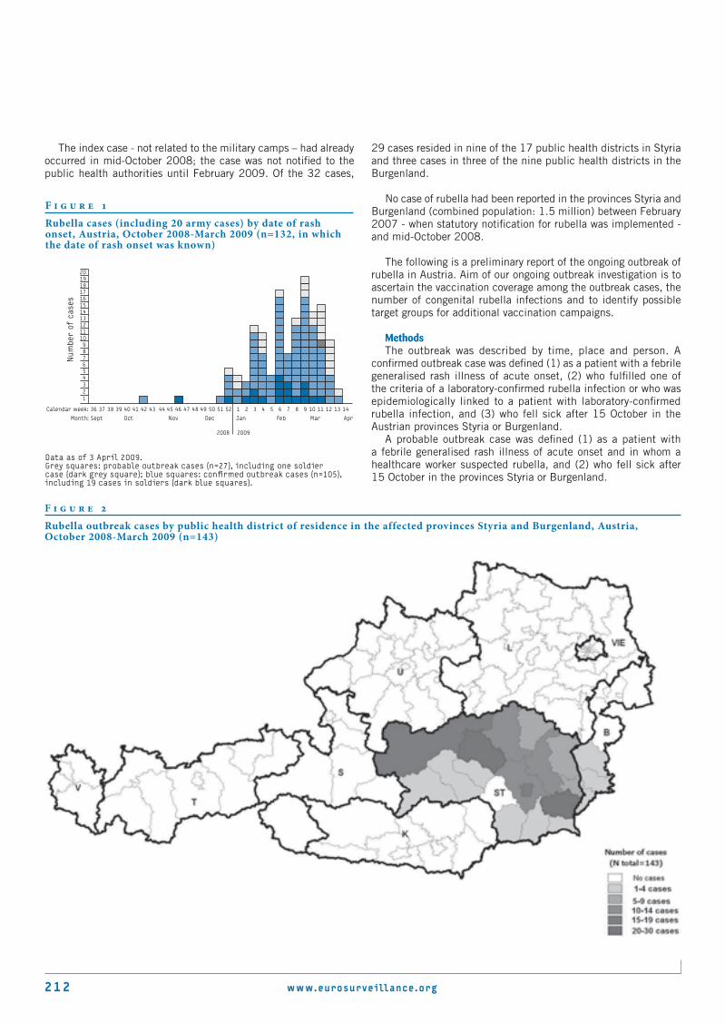
Rubella cases (including 20 army cases) by date of rash onset, Austria, October 2008-March 2009 (n=132, in which the date of rash onset was known)
Num
ber
of c
ases
2019181716151413121110987654321
Calendar week: 36 37 38 39 40 41 42 43 44 45 46 47 48 49 50 51 52 1 2 3 4 5 6 7 8 9 10 11 12 13 14
Month: Sept Oct Nov Dec Jan Feb Mar Apr
2008 2009
Data as of 3 April 2009.Grey squares: probable outbreak cases (n=27), including one soldier case (dark grey square); blue squares: confirmed outbreak cases (n=105), including 19 cases in soldiers (dark blue squares).
F i g u r e 2
Rubella outbreak cases by public health district of residence in the affected provinces Styria and Burgenland, Austria, October 2008-March 2009 (n=143)

www.eurosurveillance.org 213
A suspected rubella case was defined as a patient who presented with fever and a maculopapular rash among the contact persons of outbreak cases, and was reported by the outbreak cases.
Case finding occurred as follows: Cases of rubella infection laboratory-confirmed by the Austrian reference laboratory and cases of rubella notified to the public health authority were reported to AGES. Rubella outbreak cases were asked to name further individuals with febrile generalised rash illness of acute onset among their contacts. Information on demographics, date of rash onset, and contact with laboratory-confirmed cases were obtained by telephone interviews conducted with 143 cases; for 57 of these cases the vaccination status could be ascertained based on their vaccination documents. For active case finding, local physicians were asked to collect blood samples from all incident patients with a generalised rash for serological examination.
ResultsBetween October 2008 and March 2009, a total of 143 cases
fulfilled the outbreak case definition. Of these, 113 cases (79%) were confirmed outbreak cases of rubella: 101 (89.3% of the 113 cases) were confirmed clinically and by laboratory result, and 12 were confirmed clinically and epidemiologically. Thirty cases fulfilled only the criteria of a probable outbreak case; the procedure of laboratory or epidemiological confirmation is still ongoing for these cases. For 132 outbreak cases, the date of rash onset was known (illustrated in Figure 1).
Figure 2 shows the regional distribution by public health district of the cases’ residence; 140 cases had their residence in Styria (affecting 16 of 17 public health districts) and three outbreak cases were resident in Burgenland (affecting three of the nine public health districts).
A further 21 suspected rubella cases (not included in Figure 1 and 2) were named by confirmed outbreak cases.
One case of laboratory-confirmed rubella infection occurred in an unvaccinated 18 year-old pregnant native Austrian. As the infection occurred in the early first trimenon of pregnancy, a
voluntary abortion was performed. Already one year earlier, this woman had been identified as susceptible to rubella infection after delivery of her first child.
Of the 143 outbreak cases, 55 (38.5 %) were female. For 140 outbreak cases, data on age were known. The median age was 19 years (range: 2-60 years). A total of 136 cases (97% of 140 cases) were older than 15 years. The age group of 15-24 year-olds was most affected (88.6%; 124 of 140). Among the female cases, the age-group 15-19 years (67%; 35 of 52) was affected most, followed by the age-group 20-24 years (23%; 12 of 52). No female cases occurred in the age-group 25-39 years; two female cases occurred in the age-group 40-49 years (Figure 3).
To date, the vaccination status is known for 57 outbreak cases. Twelve cases (22%), including eight female cases, were vaccinated with one dose of rubella vaccine; none of them had received two doses. The remaining 45 outbreak cases were unvaccinated.
In the two most affected age groups, the 15-19 year-olds (n=32 in which vaccination status was known) and the 20-24 year-olds (n=24 in which vaccination status was known), the distribution of vaccination status by sex was as follows: in the age group 15-19 years, 11 of the 17 (65%) female cases were unvaccinated, while all 15 male cases were unvaccinated; in the age group 20-24 years, two of the four female cases and 14 of the 18 male cases were unvaccinated.
Outbreak control measuresMMR vaccination was immediately offered to any unvaccinated
persons by public health officers and general practitioners in Styria. Although the rubella vaccine was offered at no cost, only 180 doses of MMR vaccine were administered as part of the outbreak control measures in February and March 2009.
DiscussionBefore the introduction of routine rubella vaccination, rubella
outbreaks were common [4]. Recent outbreaks in Europe identified susceptible groups [5], e.g. in 2003 in immigrants from Latin America to Spain [6,7] and in 2005 in a religious community in the Netherlands [8]. Rubella, together with measles, is targeted for elimination in the WHO European region, with the objective for 2010 to eliminate endemic measles, endemic rubella, and to prevent congenital rubella infection (<1 case of CRS per 100,000 live births) [9]. Introduction of rubella vaccination programme has led to decreased circulation of the virus resulting in a reduced probability of wild virus exposure. If then vaccine coverage falls below a threshold of approximately 80%, there is an increase in CRS, due to accumulation of susceptibles among unvaccinated adult females [10]. According to the strategic plan for eliminating measles and rubella, and for preventing congenital rubella infection in the WHO European Region a total of 95% of the Member States should have administered, by January 2009, at least one dose of rubella vaccine to ≥95% of all children at the national level or to ≥90% of children in all first administrative levels [9]. In Austria there are no nationwide reliable data available on MMR vaccine coverage for individuals born before 1997. The official estimate of MMR vaccine coverage with at least one dose of the birth cohorts 1997 to 2007 was 84%.
Among the currently known outbreak cases, 90% of the female cases were 15-24 years-old. The index case, who occurred in mid-October 2008, was a teenage girl. No data on the contact pattern
F i g u r e 3
Rubella outbreak cases by age-group and sex, Austria, October 2008-March 2009 (n=140, in which the date of birth was known)
0
5
10
15
20
25
30
35
40
45
0-4
5-9
10-1
4
15-1
9
20-2
4
25-2
9
30-3
4
35-3
9
40-4
4
45-4
9
50-5
4
55-5
9
60-6
465
+
Age group [years]
Num
ber
of c
ases
female
male

214 www.eurosurveillance.org
during her infectious period are available to date. The second case occurred in mid-November 2008 and was the first case among soldiers. The other 19 outbreak cases in soldiers occurred between calendar week 52, 2008 and calendar week 11, 2009. To our knowledge the affected military camps implemented - except for isolation of the rubella patients in the military hospital - no other activities to control the outbreak.
Postnatal rubella is a mild infection and many cases are subclinical. Therefore, there may be substantial underreporting of cases among the general population. The clustering of cases among soldiers in this outbreak is more likely to be due to increased awareness and more reliable reporting to the public authorities in this population group.
In Austria, soldiers doing their mandatory military service are usually allowed to stay with their families during weekends, and in the second half of their six months duties may even sleep outside the barracks during the week. Three non-army outbreak cases had an epidemiological link to army cases.
In the setting of an outbreak, supplementary immunisation activities undertaken with the aim of interrupting transmission of rubella virus are the most effective preventive measure [11]. Obviously, the additional vaccination activities implemented by the local Austrian public health authorities have not been able to interrupt the rubella spread in the general population so far.
The documented voluntary abortion because of CRS risk affected a native Austrian who had been not vaccinated in childbed after her first delivery a year earlier although she had been identified as non-immune to rubella infection. This is a salutary reminder that vaccine programmes require a suitable public health infrastructure if unintended adverse consequences are to be avoided. However, national immunisation programmes are increasingly threatened by a combination of public and political complacency regarding the value of immunisation, and by the disturbing rise in the influence of anti-vaccination groups and their dangerously misleading advocacy campaigns.
An outbreak of mumps among adolescents and young adults in 2006 and an outbreak of measles affecting primarily the age-group ≥10 years in 2008, demonstrated already that additional MMR vaccination campaigns targeting the age group of ≥10 year-olds are highly required in order to prevent outbreaks of mumps, measles and rubella in Austria in the future [12,13]. Whether the statement ’The WHO European Region is well on its way to achieving targets for measles and rubella prevention and strengthening the control of vaccine preventable diseases in childhood‘ as published in Eurosurveillance in June 2003 [14] still holds true in April 2009, might be a matter of controversy.
References
1. COMMISSION DECISION of 28/IV/2008 amending Decision 2002/253/EC laying down case definitions for reporting communicable diseases to the Community network under Decision No 2119/98/EC of the European Parliament and of the Council. Official Journal of the European Union, 18 Juni 2008
2. Heymann DL. Control of Communicable Diseases Manual. 19th ed. Washington D.C.: American Public Health Association; 2008.
3. Miller E, Cradock-Watson JE, Pollock TM. Consequences of confirmed maternal rubella at succesive stages of pregnancy. Lancet 1982;2(8302):781-4.
4. Rafila A, Marin M, Pistol A, Nicolaiciuc D, Lupulescu E, Uzicanin A, et al. A large rubella outbreak, Romania – 2003. Euro Surveill. 2004;9(4):pii=457. Available from: http://www.eurosurveillance.org/ViewArticle.aspx?ArticleId=457
5. Spiteri G, Fenech Magrin AM, Muscat M. A cluster of rubella in Malta, December 2007 – January 2008. Euro Surveill. 2008;13(16):pii=18840. Available from: http://www.eurosurveillance.org/ViewArticle.aspx?ArticleId=18840
6. Lemos C, Ramirez R, Ordobas M, Guibert DH, Sanz JC, Garcia L, et al. New features of rubella in Spain: the evidence of an outbreak. Euro Surveill. 2004;9(4):pii=463. Available from: http://www.eurosurveillance.org/ViewArticle.aspx?ArticleId=463
7. Red de Vigilancia Epidemiológica de la Comunidad de Madrid. Outbreak of rubella in the Madrid region, Spain, 2005. Euro Surveill. 2005;10(27):pii=2742. Available from: http://www.eurosurveillance.org/ViewArticle.aspx?ArticleId=2742
8. van der Veen Y, Hahné S, Ruijs H, van Binnendijk R, Timen A, van Loon A, et al. Rubella outbreak in an unvaccinated religious community in the Netherlands leads to cases of congenital rubella syndrome. Euro Surveill. 2005;10(47):pii=2839. Available from: http://www.eurosurveillance.org/ViewArticle.aspx?ArticleId=2839
9. World Health Organization. Eliminating measles and rubella and preventing congenital rubella infection, WHO European Region strategic plan 2005-2010. Updated reprint 2006. Available from: http://www.euro.who.int/document/E87772.pdf [accessed 25 March, 2009].
10. Anderson RM, May RM. Vaccination against rubella and measles: quantitative investigations of different policies. J Hyg (Lond). 1983;90(2):259-325.
11. Centers for Disease Control and Prevention. Rubella Outbreak --- Arkansas, 1999. MMWR Morb Mortal Wkly Rep 2001;50(50):1137-9.
12. Schmid D, Holzmann H, Abele S, Kasper S, König C, Meusburger S, et al. An ongoing multi-state outbreak of measles linked to non-immune anthroposophic communities in Austria, Germany, and Norway, March-April 2008. Euro Surveill. 2008;13(16):pii=18838. Available from: http://www.eurosurveillance.org/ViewArticle.aspx?ArticleId=18838
13. Schmid D, Holzmann H, Alfery C, Wallenko H, Popow-Kraupp TH, Allerberger F. Mumps outbreak in young adults following a festival in Austria, 2006. Euro Surveill. 2008;13(7):pii=8042. Available from: http://www.eurosurveillance.org/ViewArticle.aspx?ArticleId=8042
14. Hanon FX, Ska JJ, Wassilak SS, Emiroglu N. WHO European Region’s strategy for elimination of measles and congenital rubella infection. Euro Surveill. 2003;8(6):pii=415. Available from: http://www.eurosurveillance.org/ViewArticle.aspx?ArticleId=415

www.eurosurveillance.org 215
Surve i ll an ce an d ou t b reak re p o r t s
M u M p s e p i d e M i o l o g y i n t h e M i d -W e s t o f i r e l a n d 2004 -2008 : i n c r e a s i n g d i s e a s e b u r d e n i n t h e u n i v e r s i t y / c o l l e g e s e t t i n g
D Whyte ()1, F O’Dea1, C McDonnell2, N H O’Connell2, S Callinan1, E Brosnan1, J Powell2, R Monahan2, R FitzGerald1, M Mannix1, T Greally1, A Dee1, P O’Sullivan1
1. Department of Public Health, Health Service Executive (West), Limerick, Ireland2. Serology Department, Mid-Western Regional Hospital, Limerick, Ireland
This article was published on 23 April 2009. Citation style for this article: Whyte D, O’Dea F, McDonnell C, O’Connell NH, Callinan S, Brosnan E, Powell J, Monahan R, FitzGerald R, Mannix M, Greally T, Dee A, O’Sullivan P. Mumps epidemiology in the Mid-West of Ireland 2004-2008: increasing disease burden in the university/college setting. Euro Surveill. 2009;14(16):pii=19182. Available online: http://www.eurosurveillance.org/ViewArticle.aspx?ArticleId=19182
Mumps is a contagious vaccine-preventable viral disease that is experiencing a revival in students attending second and third level colleges. Large mumps outbreaks have been reported in several countries despite the presence of childhood immunisation programmes over many years, including measles, mumps, and rubella (MMR) vaccination. In 2008, 1,377 cases of mumps were notified in Ireland and 1,734 in the first three months of 2009 (provisional data). This paper reviews the recent epidemiology of mumps in the Mid-West region of Ireland and highlights preventive measures. A substantial proportion of cases were not laboratory-confirmed and it is important that doctors continue to notify suspected cases. In the Irish Mid-West, data from enhanced surveillance show a high proportion of mumps in the age group 15-24 years. Complications were uncommon and rarely severe. Where data were available, over half of the cases did not recall having received two doses of MMR, but most recalled one dose. Parents should continue to ensure children receive both MMR vaccinations so that uptake is optimal for protection. Steps were taken to increase awareness of the disease in the school, college and university settings. Preventive measures implemented to limit mumps transmission in the school/college setting over recent years included vaccination of close contacts, isolation for five days and hand hygiene.
IntroductionMumps (or infectious parotitis) is an acute infection caused by
an RNA virus of the family Paramyxoviridae. It is spread directly from infected person to susceptible person by sneezing, droplets and close contact. Mumps can present with mild influenza-like symptoms which may include fever, headache and painful swollen salivary (usually parotid) glands [1]. Complications are usually infrequent but infection can progress to meningitis, deafness as well as orchitis, oophoritis or pancreatitis (inflammation of the testicles, ovaries or pancreas). Mumps infection during pregnancy is not associated with congenital malformations [2]. The incubation period is 16-18 days (range: 14-25 days), and recent data suggest an infected person is contagious during the period from two days before to five days after onset of symptoms [3].
Mumps is a vaccine-preventable disease. The measles, mumps and rubella (MMR) vaccine offers safe and effective protection against these diseases and is provided free of charge
to children at the age of 12-15 months (MMR1) as part of the Irish Primary Childhood Immunisation Programme (PCIP) [4]. Prior to 1996, there was no structured PCIP, only recommendations on immunisation. Electronic records of childhood vaccinations in Ireland for national and regional uptake monitoring are only available from 1996. Documentation of MMR status in people born prior to 1996 relies on manual records. Immunisation programmes rely on achieving over 95% uptake of MMR vaccine to protect the population from disease – especially the most susceptible, children under two years of age. Vaccine uptake in some areas of Ireland has only lately recovered to levels of over 90% after public confidence in combination vaccines was eroded by published research about possible side effects that has since been rejected [5]. Uptake levels in Ireland for MMR1 at the age of 24 months averaged below 80% until 2004 but reached 89% in 2008 (90% in the Mid-West) [6].
In 1989, about 700 mumps cases were notified in Ireland, but this number declined to between 30 and 40 cases annually after MMR1 was introduced in 1988. MMR2 (second dose of vaccination) was introduced in 1992 for 11-12 year-olds. Case numbers rose again in 1996-1997 (300-400) but subsequently fell back to 30-40 annual cases until the year 2002. In line with a recommendation from 1999, the age for MMR2 was brought forward to the age of 4-5 years in 2001. In 2008, 1,377 cases were reported in Ireland, half of whom were laboratory-confirmed. In the context of even higher infection levels observed in Ireland since the beginning of 2009, (a greater than ten-fold increase on 2008), it is timely to review the epidemiology of mumps infection in the Irish Mid-West over the preceding five year period to help explain the factors which may have been of influence.
In recent years a large number of cases of mumps have been reported in young adults, arising from transmission of the virus in so-called ‘third level colleges’ (colleges/universities attended by students aged 18 years and over), but also in some secondary schools (with 12-18 year-old students). Mumps outbreaks in third level colleges have been reported in Ireland since 2004 [7] and in several other countries [8-11]. This paper reviews the epidemiology of mumps infection in the Mid-West of Ireland from 2004 to 2008 and describes the source, demography of cases, risk factors and the spectrum of illness and examines the role of preventive measures.

216 www.eurosurveillance.org
Geographically, the Mid-West of Ireland includes the administrative counties of Clare, Limerick and North Tipperary with a population of 361,028 people. There are three large university/college institutes in Limerick city and smaller third level colleges in Tipperary including the national Garda (Police) Training College.
MethodsSuspected or confirmed mumps cases have been subject to
mandatory notification in Ireland since 1988 (and outbreaks since 2004) and must be reported to the Medical Officer of Health (MOH) in the Mid-West by medical doctors and laboratories [12]. Notifiers should have regard to Irish case definitions for mumps [13], which is based on the case definition of the European Union, but it adds the classification ‘possible case’ for clinical cases that are not laboratory-confirmed or are epidemiologically linked to a confirmed case. Where possible, enhanced surveillance data (on complications, symptoms, travel, etc.) were collected by medical officers in the regional public health department. Data on MMR vaccination history were based largely on family or doctor recall rather than on records. A copy of the national enhanced surveillance form for mumps is available online (http://www.ndsc.ie/hpsc/A-Z/VaccinePreventable/Mumps/SurveillanceForms/) from the Health Protection Surveillance Centre in Dublin. This core dataset is larger
and more disease-specific than the minimal dataset required under legislation for the generic list of notifiable diseases.
Data on clinical notifications from general practitioners and hospital doctors were collated by the Department of Public Health where notifications to the MOH are recorded. Laboratory data notified from the Department of Serology, Mid-Western Regional Hospital, Limerick and the National Virus Reference Laboratory (UCD) were collated. Laboratory notified cases were confirmed by the detection of mumps specific IgM immunoglobulin in serum or oral fluid specimens. The test is an IgM class capture enzyme immunoassay (MACEIA, Microimmune Ltd., United Kingdom). The reported sensitivity and specificity of the assay for serum samples is 94.7% and 95.9%, respectively, when compared to IgM antibody capture radioimmunoassay (MACRIA). For oral fluid samples, the sensitivity and specificity compared to MACRIA is 92.6% and 100%, respectively. Data were collected, entered into secure databases and analysed in MS Excel and SPSS. Analysis by age, sex, symptoms, complications and vaccination history was confined to the enhanced surveillance dataset.
ResultsFrom 1 January 2004 to 31 December 2008, 319 mumps
notifications were received by the Mid-West MOH. Three records were removed as duplicates and 14 were re-classified as denotified as laboratory data became available, leaving 302 records. Over the five year period, 109 laboratory-confirmed notifications (36%) and 193 clinical notifications (64%) were recorded. Enhanced surveillance was completed for 186 mumps cases (71% of 262 notified cases); 116 of them were clinical notifications only, 50 were laboratory confirmed only, and 20 were notified clinically and laboratory-confirmed (see Table 1).
Figure 1 shows all notified mumps cases, inclusive of those cases for whom enhanced surveillance information was available. With the exception of the academic year 2006-7, the number of mumps cases was relatively low in summer, and peaked in autumn in the months after third level colleges resume (Figure 1). An intervention with MMR vaccination was carried out by public
F i g u r e 1
Mumps cases with enhanced surveillance data and other mumps notifications, by year and month, Mid-West of Ireland, 2004-2008 (n=263)
0
5
10
15
20
25
30
35
40
1 2 3 4 5 6 7 8 9 10 11 12
2004 (n=16) 2005 (n=96) 2006 (n=24) 2007 (n=6) 2008 (n=121)
Month/year
Mum
ps c
ases
Enhanced surveillance data
Other cases
1 2 3 4 5 6 7 8 9 10 11 12 1 2 3 4 5 6 7 8 9 10 11 12 1 2 3 4 5 6 7 8 9 10 11 12 1 2 3 4 5 6 7 8 9 10 11 12
T a b l e 1
Mumps cases for whom enhanced surveillance data were available, by notification type, Mid-West of Ireland, 2004-2008 (n=186)
Notification Type (enhanced surveillance)
Year Notified Clinical Laboratory and clinical Laboratory Total
2004 4 0 1 5
2005 57 3 12 72
2006 8 0 3 11
2007 4 0 1 5
2008 43 17 33 93
2004-2008 116 (62%) 20 (11%) 50 (27%) 186 (100%)

www.eurosurveillance.org 217
health/university student health services in one large university following a peak in mumps infections in April 2005. This could be an explanation for the change in pattern in 2006-7.
The age range of mumps cases was 1.4 years to 79 years (average 22.9 years, median 20.4 years). There was a slight preponderance of cases in males over females in all years except 2007 (data not shown): from 2004 to 2008, the male:female ratio was 1.4:1. For the period from 2004 to 2008, Figure 2 illustrates the age distribution of mumps cases by sex in the Mid-West. Of 186 cases, 123 were in the age group of 15-24 year-olds (66%). This distribution reflects the ongoing transmission since 2004 of mumps virus in susceptible people attending third level colleges in Ireland.
For 186 cases over the five year period, symptoms and complications are shown by sex in Table 2. Nausea, earache, headache and vomiting were mentioned by seven cases as symptoms or complications. Complications of mumps disease were uncommon in female patients (1/78) while 13% of the male cases (14/108) reported complications. Seven cases (four male and three female) were hospitalised, 5% of the 144 cases for whom such information was available. The outcome ’recovered’ was stated for 28% of cases (52/186). No deaths were reported.
Investigation of the cases to determine the likely place of acquisition of mumps infection implicated several settings but university/college and secondary schools were reported in 54 cases (71% of 75 cases for whom this information was acquired), four in secondary schools and three in primary schools. The University of Limerick, a campus-style third level college, was associated with the vast majority of the infections acquired in university/college and with some related cases. Two mumps outbreaks were reported from the Mid-West – one in the community and one mixed community/college event.
Twelve cases (8% of cases where data were given) occurred in foreign-born nationals. Travel (25 days before onset) was reported in 16 cases, but only four had travelled “overseas” (two to the United Kingdom, one to North America and one to Africa). One case was reported as acquired overseas. Travel within Ireland appeared consistent with students commuting to their home counties.
Data on childhood MMR vaccination were ascertained for a large proportion of these mumps cases (78%; 146/186) and are shown in Table 3. MMR vaccination was not evident in the older cases (over 25 years of age), which is not unexpected. Where data were provided in young adults (15-24 years), 7% of cases (7/103) reported not receiving any MMR, 49% (51/103) had at least one MMR (MMR1) and 44% (45/103) reported receiving two doses (MMR2).
DiscussionHistorically, in the Irish setting, mumps occurred in children
between the ages of five and 15 years, although the disease
T a b l e 2
Mumps cases with enhanced surveillance data showing symptoms and complications, by sex, Mid-West of Ireland, 2004-2008 (n=186)
Males (n=108) Females (n=78)
Symptoms Yes No Not given/unknown Yes No Not given/unknown
Parotitis swelling 98 (91%) 3 7 69 (88%) 5 4
Bilateral parotitis 46 (43%) 49 13 26 (33%) 43 9
Fever 53 (49%) 41 14 20 (26%) 44 14
Complications
Meningitis 1 (1%) 95 12 0 69 9
Encephalitis 1 (1%) 94 13 0 69 9
Orchitis/Oophoritis 12 (11%) 82 14 0 69 9
Deafness 0 95 13 0 69 9
Mastitis 0 94 14 0 68 10
Pancreatitis 1 (1%) 94 13 1 (1%) 69 8
Due to the small numbers, we did not undertake a statistical analysis.
F i g u r e 2
Age distribution of mumps cases for whom enhanced surveillance data were available, by sex, Mid-West of Ireland, 2004-2008 (n=183*)
2 1
4
12 13
41
35 25
22
5
33
2
1
4
1
1
22
4
Male FemaleSex
Count
1-4
5-9
10-14
15-19
20-24
25-34
35-44
45-54
55-64
65+
1-4
5-9
10-14
15-19
20-24
25-34
35-44
45-54
55-64
65+
50 40 30 20 10 0 5040302010
Age
grou
p [ye
ars]
Age group [years]
*Age for three of the 186 cases unknown.
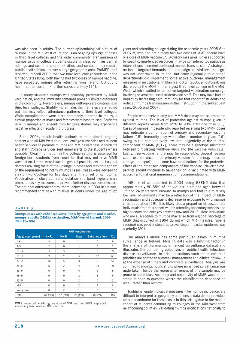
218 www.eurosurveillance.org
was also seen in adults. The current epidemiological picture of mumps in the Mid-West of Ireland is an ongoing upsurge of cases in third level colleges and the wider community. Transmission of mumps virus in college students occurs in classroom, residential settings and social or sports activities, and contacts may require public health follow-up over a large geographic area. ProMED mail reported, in April 2009, that two third level college students in the United States (US), both having had two doses of mumps vaccine, have suspected mumps after returning from Ireland. US public health authorities think further cases are likely [14].
In many students mumps was probably prevented by MMR vaccination, and the immunity conferred probably limited outbreaks in the community. Nevertheless, mumps outbreaks are continuing in third level colleges. Slightly more males than females are affected but this may reflect attendance patterns to third level colleges. While complications were more commonly reported in males, a similar proportion of males and females were hospitalised. Students ill with mumps and absent from lectures/studies may experience negative effects on academic progress.
Since 2004, public health authorities maintained ongoing contact with all Mid-West third level college authorities and student health services to promote mumps and MMR awareness in students and staff. College services sent email alerts to the students where possible. Clear information in the college setting is essential for foreign-born students from countries that may not have MMR vaccination. Letters were issued to general practitioners and hospital doctors advising them of the upsurge in cases and reminding them of the requirement to notify mumps cases. Cases were advised to stay off work/college for five days after the onset of symptoms. Vaccination of close contacts, isolation and hand hygiene were promoted as key measures to prevent further disease transmission. The national outbreak control team, convened in 2004 in Ireland, recommended that new third level students under the age of 25
years and attending college during the academic years 2005-6 to 2007-8, who had not already had two doses of MMR should have one dose of MMR vaccine [7]. Advisory measures, unless supported by specific, ring-fenced resources, may be considered too passive as interventions to control continued mumps transmission. A strategic, national, targeted immunisation campaign in third level colleges was not undertaken in Ireland, but some regional public health departments did implement some active outbreak management measures in institutions. In March and April 2005, an outbreak was declared by the MOH in the largest third level college in the Mid-West, which resulted in an active targeted vaccination campaign involving several thousand students and staff. This may have had an impact by increasing herd immunity for that cohort of students and reduced mumps transmission in this institution in the subsequent years, 2006 and 2007.
People who received only one MMR dose may not be protected against mumps. The level of protection against mumps given in different reports varies from 65% to 90% after one dose [11]. Cases of mumps in people who reported receiving two MMR doses may indicate a combination of primary and secondary vaccine failure [15]: Immunity may wane after a number of years [16], owing to the comparatively low immunogenicity of the mumps component of MMR [8,17], There may be a genotype mismatch between circulating wildtype virus and the vaccine virus [18]; Lastly, true vaccine failure may be responsible. Several reasons could explain uncommon primary vaccine failure (e.g. incorrect storage, transport), and some have implications for the protective effect of the other two components of the vaccine. Nevertheless, parents should continue to have their child vaccinated with MMR according to national immunisation recommendations.
DiRenzi et al. reported in 2004 using ESEN2 data that approximately 80-85% of individuals in Ireland aged between 15 and 24 years were immune to mumps and that this relatively low level of immunity may be a reflection of the impact of MMR vaccination and subsequent decrease in exposure to wild mumps virus circulation [19]. It is likely that a proportion of susceptible individuals from this cohort will be attending secondary schools and higher education colleges between now and 2013. More individuals who are susceptible to mumps may arise from a global shortage of MMR that occurred in 1994 during which MR (measles, rubella vaccine) was used instead, as preventing a measles epidemic was a priority [20].
Our analysis underlines some particular issues in mumps surveillance in Ireland. Missing data was a limiting factor in the analysis of the mumps enhanced surveillance dataset and illustrates the competing objectives in public health infectious disease surveillance. In crisis situations such as an outbreak priorities are shifted to outbreak management and clinical follow-up at the expense of timely and complete surveillance. Analysis was confined to mumps notifications where enhanced surveillance was undertaken, hence the representativeness of this sample may be prone to some bias. Accuracy and objectivity of MMR vaccination status is open to question where the classification depended on recall rather than records.
Traditional epidemiological measures, like mumps incidence, are difficult to interpret as geography and census data do not provide a clear denominator for these cases in this setting due to the mobile cohort of students commuting to colleges in the Mid-West from neighbouring counties. Validating mumps notifications nationally to
T a b l e 3
Mumps cases with enhanced surveillance by age group and measles, mumps, rubella (MMR) vaccination, Mid-West of Ireland, 2004-2008 (n=186)
MMR vaccination
Age group ( years) MMR1 MMR2 None Data not given All
1-4 5 0 1 0 6
5-9 2 0 1 0 3
10-14 5 3 1 0 9
15-19 21 23 4 12 60
20-24 30 22 3 8 63
25-34 6 2 4 13 25
35-44 0 0 4 2 6
45-54 0 0 2 3 5
55-64 0 0 2 0 2
>65 0 0 2 2 4
Not given 0 2 1 0 3
Total 69 (37%) 52 (28%) 25 (13%) 40 (22%) 186
MMR1: reported receiving one dose of MMR vaccine; MMR2: reported receiving two doses of MMR vaccine.

www.eurosurveillance.org 219
avoid duplication and overestimation remains a challenge, although the full implementation of a national computerised infectious disease reporting system (CIDR) may improve surveillance in future. Public health relies greatly on general practitioners and hospital clinicians notifying cases. More cases of mumps are reported clinically than are confirmed and notified by the laboratories. While mumps may have some classical symptoms and signs on presentation, clinicians may confirm cases by either serum IgM serology or by non-invasive salivary IgM testing. However, testing at the appropriate time in the clinical course of disease is important consideration in order to avoid apparently conflicting results [21,22].
A national outbreak control team was re-convened in 2009 and has recommended re-enforcing the present measures and adopting further active interventions, regionally and nationally, to control future transmission of mumps at secondary schools and third level colleges in Ireland.
AcknowledgementsThe patience and assistance of all the staff in the Health Protection Surveillance Centre is acknowledged. The Department of Public Health is appreciative of all notifications, especially mumps, from general practitioners and hospital clinicians. The work of the doctors and nurses and administrative staff in the colleges of the Mid-West is also acknowledged. Special thanks to Ms Orla Hanrahan, Surveillance Officer, and Ms Ann Sweeney and Ms Breda Tuohy, Surveillance Assistants, for diligent and timely data input in the Department of Public Health.
References
1. Falk WA, Buchan K, Dow M, Garson JZ, Hill E, Nosal M, et al. The epidemiology of mumps in Southern Alberta 1980-1982. Am J Epidemiol. 1989;130(4):736-49.
2. Siegel M. Congenital malformations following chickenpox, measles, mumps, and hepatitis. Results of a cohort study. JAMA. 1973;226(13):1521-4.
3. Polgreen PM, Bohnett LC, Cavanaugh JE, Gingerich SB, Desjardin LE, Harris ML, et al. The duration of mumps virus shedding after the onset of symptoms. Clin Infect Dis. 2008;46(9):1447-9.
4. Health Service Executive (HSE). National Immunisation Office. Protect Health – Immunise (Website on the internet). [Accessed April 2009]. Available from: http://www.immunisation.ie
5. Andreae MC, Freed GL and Katz SL. Safety concerns regarding combination vaccines. Perspective of select European countries. Hum Vaccin. 2005;1(1):1-5.
6. Health Service Executive (HSE). Health Protection Surveillance Centre. Summary Immunisation Uptake Statistics. Dublin: HSE. 2009. Available from: http://www.hpsc.ie/hpsc/AZ/VaccinePreventable/Vaccination/ImmunisationUptakeStatistics/ImmunisationUptakeStatisticsSummary/File,954,en.pdf (Last accessed April 2009)
7. Gee S, O’Flanagan D, Fitzgerald M, Cotter S. Mumps in Ireland, 2004-2008. Euro Surveill. 2008;13(18):pii=18857. Available from: http://www.eurosurveillance.org/ViewArticle.aspx?ArticleId=18857 (Last accessed April 2009)
8. Conly J, Johnston B. Is mumps making a comeback? Can J Infect Dis Med Microbiol. 2007; 18(1):7–9.
9. Centers for Disease Control and Prevention (CDC). Mumps epidemic—Iowa, 2006. MMWR Morb Mortal Wkly Rep. 2006; 55(13):366–8.
10. Cortese MM, Jordan HT, Curns AT, Quinlan PA, Ens KA, Denning PM, et al. Mumps vaccine performance among university students during a mumps outbreak. Clin Infect Dis. 2008;46(8):1172-80.
11. Savage E, Ramsay M, White J, Beard S, Lawson H, Hunjan R, et al. Mumps outbreaks across England and Wales in 2004: observational study. Brit Med J. 2005; 330(7500): 1119-20.
12. Government of Ireland. Infectious Diseases (Amendment) Regulations, 1988. Statutory Instrument S.I. No. 288 of 1988. Available from: http://www.irishstatutebook.ie/1988/en/si/0288.html (Last accessed April 2009)
13. Health Protection Surveillance Centre. Case Definition for Notifiable Diseases 2004. Available from: http://www.ndsc.ie/hpsc/A-Z/VaccinePreventable/Mumps/CaseDefinitions/File,2850,en.pdf (Last accessed April 2009)
14. ProMED-mail. Mumps - USA: (Massachusetts) ex Ireland. Archive Number 20090417.1455. 17 April 2009. Available from: http://www.promedmail.org/pls/otn/fp=2400:1001:4405464973023774::NO::F2400_P1001_BACK_PAGE,F2400_P1001_PUB_MAIL_ID:1010,77078
15. Vandermeulen C, Roelants M, Vermoere M, Roseeuw K, Goubau P, Hoppenbrouwers K. Outbreak of mumps in a vaccinated child population: a question of vaccine failure? Vaccine. 2004;22(21-22): 2713–6.
16. Cohen C, White JM, Savage EJ, Glynn JR, Choi Y, Andrews N, et al. Vaccine effectiveness estimates, 2004-2005 mumps outbreak, England. Emerg Infect Dis. 2007;13(1):12–7.
17. Harling R, White JM, Ramsay ME, Macsween KF, van den Bosch C. The effectiveness of the mumps component of the MMR vaccine: A case control study. Vaccine. 2005;23(31): 4070-4.
18. Kaaijk P, van der Zeijst BA, Boog MC and Hoitink CW. Increased mumps incidence in the Netherlands: Review on the possible role of vaccine strain and genotype . Euro Surveill. 2008;13(26):pii=18914. Available from: http://www.eurosurveillance.org/ViewArticle.aspx?ArticleId=18914 (Last accessed April 2009)
19. Di Renzi M, Jackson S, Gee S, Cotter S. Increase in mumps in Ireland in late 2004. Euro Surveill. 2004;8(52):pii=2608. Available from: http://www.eurosurveillance.org/ViewArticle.aspx?ArticleId=2608 (Last accessed April 2009)
20. Centers for Disease Control and Prevention (CDC). Mumps epidemic – United kingdom, 2004-2005. MMWR Morb Mortal Wkly Rep. 2006; 55(7):173-5.
21. Reid F, Hassan J, Irwin F, Waters A, Hall W, Connell J. Epidemiologic and diagnostic evaluation of a recent mumps outbreak using oral fluid samples. J Clin Virol. 2008;41(2):134-7.
22. Cunningham C, Faherty C, Cormican M, Murphy AW. Importance of clinical features in diagnosis of mumps during a community outbreak. Ir Med J. 2006;99(6):171-3.
22 0 www.eurosurveillance.org
S urve i ll an ce an d ou t b reak re p o r t s
C h a r a C t e r i s at i o n o f n e i s s e r i a m e n i n g i t i d i s C s t r a i n s C a u s i n g t w o C l u s t e r s i n t h e n o r t h o f i ta ly i n 2007 a n d 2008
C Fazio1, A Neri1, S Tonino1, A Carannante1, M G Caporali2, S Salmaso2, P Mastrantonio ([email protected])1, P Stefanelli1
1. Department of Infectious, Parasitic and Immune-mediated Diseases, Istituto Superiore di Sanità, Rome, Italy2. National Centre for Epidemiology, Surveillance and Health Promotion, Istituto Superiore di Sanità, Rome, Italy
This article was published on 23 April 2009. Citation style for this article: Fazio C, Neri A, Tonino S, Carannante A, Caporali MG, Salmaso S, Mastrantonio P, Stefanelli P. Characterisation of Neisseria meningitidis C strains causing two clusters in the north of Italy in 2007 and 2008. Euro Surveill. 2009;14(16):pii=19179. Available online: http://www.eurosurveillance.org/ViewArticle.aspx?ArticleId=19179
Two clusters of invasive meningococcal disease in the north of Italy both due to serogroup C/ST-11 clonal complex are here described. The objective of the investigation was to analyse the phenotype and the genotype of meningococci involved in the two clusters which were of national relevance due to the fatal outcome of the majority of cases (six of the total of 10 cases). All the strains were C:2a:P1.5 ST-11/ET-37 clonal complex. Two pulsed field gel electrophoresis (PFGE) and variable number tandem repeats (VNTR) profiles were identified, one for each cluster. VNTRs were different from those detected in Italy for C/ST-11 strains isolated from sporadic cases in the same period. This laboratory surveillance report highlights the importance and the crucial role of molecular characterisation to confirm the relatedness among meningococci responsible for clusters of cases.
IntroductionMeningococcal disease remains a major childhood infection in
Europe, with a considerable number of cases appearing also in other age groups, notably young adults. The incidence of serogroup C disease substantially declined with the introduction of conjugate meningococcal C vaccine in the national vaccination programmes of several countries [1]. However, the C/ST-8 and the C/ST-11 strains are currently the two hyper-virulent meningococcal lineages involved in a significant proportion of serogroup C invasive disease worldwide [2].
In Italy, the notification of invasive meningococcal disease to the local health authorities and to the Ministry of Health has been mandatory since 1983. Through national surveillance of bacterial meningitis, established in 1994, the National Reference Laboratory (NRL) at the Istituto Superiore di Sanità in Rome each year receives an average of 80% of strains isolated by local hospital laboratories throughout the country. The disease, characterised by low national incidence (0.3/100,000 inhabitants) and by sporadic cases, has in the last three years mainly been caused by serogroup B meningococci (64%).
Since the end of 2007, two clusters of serogroup C meningococcal disease have been detected in two different administrative regions in Italy. Due to the severity and fatal outcome of cases, these clusters were of national relevance, and the strains have been fully characterised at the NRL. The molecular characteristics of the ten strains involved in the two clusters are reported here.
MethodsIsolates of meningococci received at the NRL were subcultured
for serogroup confirmation by slide agglutination with commercial antiserum (Remel Europe, United Kingdom). Serotypes and serosubtypes were determined by standard whole-cell ELISA with monoclonal antibodies (purchased from NIBSC, UK) [3]. Susceptibilities to penicillin G, rifampicin, ciprofloxacin and ceftriaxone were determined by E-test method (AB Biodisk, Solna, Sweden), according to the manufacturer’s instructions.
The breakpoints were those recommended by the European Monitoring Group for Meningococci EMGM [4].
Molecular analyses by multilocus sequence typing (MLST), variable number of tandem repeats (VNTR) typing and pulsed field gel electrophoresis (PFGE) were performed following the procedures described elsewhere [5-7].
ResultsThe two clusters of serogroup C meningococci occurred in a
group of seven adolescents/young adults and in three adults in two different but bordering Italian regions, Veneto and Lombardy, in December 2007 and in July 2008, respectively.
The outbreak in Veneto has already been described from an epidemiological aspect and in terms of management [8]. The outcome was fatal in three of the seven cases.
From 13 to 15 July 2008, three cases of fatal septicaemia in patients aged 34, 48 and 51 years occurred in a limited geographical area of the Lombardy region. Family members and people who had been in contact with the patients were given chemoprophylaxis. Thorough investigation by the local health authorities did not show any social or institutional link between the three cases, and none could be identified as specifically at risk on the basis of the information obtained. All cases from both events, were laboratory-confirmed at the regional level by culture and the serogroup of Neisseria meningitidis was identified. At the NRL, the phenotypic and genotypic characteristics of the ten isolates were further determined. The strains showed the antigenic formula C:2a:P1.5 and were fully susceptible to penicillin, rifampicin, ceftriaxone and ciprofloxacin. All of them belonged to ST-11/ET37 clonal complex (cpx) as identified by MLST.
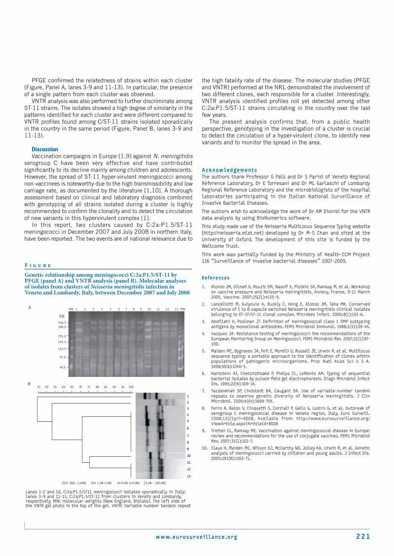
www.eurosurveillance.org 2 21
PFGE confirmed the relatedness of strains within each cluster (Figure, Panel A, lanes 3-9 and 11-13). In particular, the presence of a single pattern from each cluster was observed.
VNTR analysis was also performed to further discriminate among ST-11 strains. The isolates showed a high degree of similarity in the patterns identified for each cluster and were different compared to VNTR profiles found among C/ST-11 strains isolated sporadically in the country in the same period (Figure, Panel B, lanes 3-9 and 11-13).
DiscussionVaccination campaigns in Europe [1,9] against N. meningitidis
serogroup C have been very effective and have contributed significantly to its decline mainly among children and adolescents. However, the spread of ST-11 hyper-virulent meningococci among non-vaccinees is noteworthy due to the high transmissibility and low carriage rate, as documented by the literature [1,10]. A thorough assessment based on clinical and laboratory diagnosis combined with genotyping of all strains isolated during a cluster is highly recommended to confirm the clonality and to detect the circulation of new variants in this hypervirulent complex [1].
In this report, two clusters caused by C:2a:P1.5/ST-11 meningococci in December 2007 and July 2008 in northern Italy, have been reported. The two events are of national relevance due to
the high fatality rate of the disease. The molecular studies (PFGE and VNTR) performed at the NRL demonstrated the involvement of two different clones, each responsible for a cluster. Interestingly, VNTR analysis identified profiles not yet detected among other C:2a:P1.5/ST-11 strains circulating in the country over the last few years.
The present analysis confirms that, from a public health perspective, genotyping in the investigation of a cluster is crucial to detect the circulation of a hyper-virulent clone, to identify new variants and to monitor the spread in the area.
AcknowledgementsThe authors thank Professor G Palù and Dr S Parisi of Veneto Regional Reference Laboratory, Dr E Torresani and Dr ML Garlaschi of Lombardy Regional Reference Laboratory and the microbiologists of the hospital laboratories participating in the Italian National Surveillance of Invasive Bacterial Diseases.
The authors wish to acknowledge the work of Dr AM Dionisi for the VNTR data analysis by using BioNumerics software.
This study made use of the Neisseria MultiLocus Sequence Typing website (http://neisseria.mlst.net) developed by Dr M-S Chan and sited at the University of Oxford. The development of this site is funded by the Wellcome Trust.
This work was partially funded by the Ministry of Health-CCM Project 116 “Surveillance of invasive bacterial diseases” 2007-2009.
References
1. Alonso JM, Gilmet G, Rouzic EM, Nassif X, Plotkin SA, Ramsay M, et al. Workshop on vaccine pressure and Neisseria meningitidis, Annecy, France, 9-11 March 2005. Vaccine. 2007;25(21):4125-9.
2. Lancellotti M, Guiyoule A, Ruckly C, Hong E, Alonso JM, Taha MK. Conserved virulence of C to B capsule switched Neisseria meningitidis clinical isolates belonging to ET-37/ST-11 clonal complex. Microbes Infect. 2006;8(1):191-6.
3. Abdillahi H, Poolman JT. Definition of meningococcal class 1 OMP subtyping antigens by monoclonal antibodies. FEMS Microbiol Immunol. 1988;1(3):139-44.
4. Vazquez JA. Resistance testing of meningococci: the recommendations of the European Monitoring Group on Meningococci. FEMS Microbiol Rev. 2007;31(1):97-100.
5. Maiden MC, Bygraves JA, Feil E, Morelli G, Russell JE, Urwin R, et al. Multilocus sequence typing: a portable approach to the identification of clones within populations of pathogenic microorganisms. Proc Natl Acad Sci U S A. 1998;95(6):3140-5.
6. Hartstein AI, Chetchotisakd P, Phelps CL, LeMonte AM. Typing of sequential bacterial isolates by pulsed-field gel electrophoresis. Diagn Microbiol Infect Dis. 1995;22(4):309-14.
7. Yazdankhah SP, Lindstedt BA, Caugant DA. Use of variable-number tandem repeats to examine genetic diversity of Neisseria meningitidis. J Clin Microbiol. 2005;43(4):1699-705.
8. Ferro A, Baldo V, Cinquetti S, Corziali P, Gallo G, Lustro G, et al. Outbreak of serogroup C meningococcal disease in Veneto region, Italy. Euro Surveill. 2008;13(2):pii=8008. Available from: http://www.eurosurveillance.org/ViewArticle.aspx?ArticleId=8008
9. Trotter CL, Ramsay ME. Vaccination against meningococcal disease in Europe: review and recommendations for the use of conjugate vaccines. FEMS Microbiol Rev. 2007;31(1):101-7.
10. Claus H, Maiden MC, Wilson DJ, McCarthy ND, Jolley KA, Urwin R, et al. Genetic analysis of meningococci carried by children and young adults. J Infect Dis. 2005;191(8):1263-71.
F i g u r e
Genetic relationship among meningococci C:2a:P1.5/ST-11 by PFGE (panel A) and VNTR analysis (panel B). Molecular analyses of isolates from clusters of Neisseria meningitidis infection in Veneto and Lombardy, Italy, between December 2007 and July 2008
45 50 55 60 65 70 75 80 85 90 95 100
3
5
7
10
12
1
2
4
6
8
9
11
13
3
5
7
10
12
1
2
4
6
8
9
11
13
B
A
Kb
242.5
291.0
388.0436.5
48.5
97.0
145.5
DICE (Opt: 1.00%) (Tol 1.0%-1.0%) (H>0.0% S>0.0%) [0.0% - 100.0%]
MW 1 2 3 4 5 6 7 8 9 10 11 12 13 MW
Lanes 1-2 and 10, C:2a:P1.5/ST11 meningococci isolated sporadically in Italy;lanes 3-9 and 11-13, C:2a:P1.5/ST-11 from clusters in Veneto and Lombardy, respectively. MW: molecular weights (New England, Biolabs). The left side of the VNTR gel photo is the top of the gel. VNTR: Variable number tandem repeat

222 www.eurosurveillance.org
S urve i ll an ce an d ou t b reak re p o r t s
T r av e l - a s s o c i aT e d l e g i o n n a i r e s ’ d i s e a s e i n e u r o p e i n 2007
C A Joseph ([email protected])1, R Yadav1, K D Ricketts1, on behalf of the European Working Group for Legionella Infections1
1. Respiratory and Systemic Infections Department, Health Protection Agency Centre for Infections, London, United Kingdom
This article was published on 7 May 2009. Citation style for this article: Joseph CA, Yadav R, Ricketts KD, on behalf of the European Working Group for Legionella Infections. Travel-associated Legionnaires’ disease in Europe in 2007. Euro Surveill. 2009;14(18):pii=19196. Available online: http://www.eurosurveillance.org/ViewArticle.aspx?ArticleId=19196
Nine hundred and forty six cases of travel-associated Legionnaires’ disease were reported to the European Surveillance Scheme for Travel Associated Legionnaires’ Disease (EWGLINET) with onset during 2007; 890 were confirmed and 56 were presumptive. Twenty eight cases died, giving a case fatality rate of 3.0%. 8.2% of cases were diagnosed by culture, an important increase from 5.2% in 2006. One hundred and twelve new clusters were identified; the largest involved nine cases. Sixteen of these clusters (14.3%) occurred in countries outside EWGLINET, and three involved cruise ships. Twenty nine of the new clusters (25.9%) would not have been detected without the EWGLINET scheme. A total of 151 investigations were conducted in Europe, 42 of which were conducted at re-offending sites (where additional cases had onset after a report was received to say that investigations and control measures had been satisfactorily conducted). The names of 13 accommodation sites were published on the European Working Group for Legionella Infections (EWGLI) website; 11 of these were situated in Turkey.
IntroductionCases of Legionnaires’ disease are often associated with
overnight stays in public accommodation sites which may be visited by individuals from all over the world. As such, a cluster of cases of Legionnaires’ disease at a public accommodation site may involve nationals from more than one country and if the countries concerned do not share information on their cases, these clusters can go undetected. In 1987, the European Working Group for Legionella Infections (EWGLI) established a surveillance system known as ‘The European Surveillance Scheme for Travel Associated Legionnaires’ Disease’ (EWGLINET) with the aim of identifying clusters of travel-associated cases in Europe that may not be detected by national surveillance systems alone, and initiating investigation and control measures at such sites.
In 2002, EWGLI members introduced the European Guidelines for Control and Prevention of Travel Associated Legionnaires’ Disease [1], to standardise investigation and control measures conducted at cluster sites. These were endorsed by the European Commission in 2003. The history and current activities of EWGLI are described further on its website (www.ewgli.org).
This paper provides results and commentary on cases of travel-associated Legionnaires’ disease reported to EWGLINET with onset in 2007.
MethodsEach of the countries that participate in the EWGLINET scheme
run their own national surveillance schemes for Legionnaires’ disease, which collect information on cases occurring in their residents. In order to ensure that every country reports their data to EWGLINET in a consistent manner, standardised case definitions have been developed [2]. When a travel-associated case is identified that meets these definitions, it is reported to EWGLINET’s coordinating centre at the Health Protection Agency Centre for Infections in London. The coordinating centre maintains a database of all cases that have been reported to the scheme since its inception, and this is searched each time a new case is added to determine whether it is a single case or part of a cluster. These are defined in the following way [2]:
• A single case: A person who stayed, in the two to ten days before onset of illness, at a public accommodation site that has not been associated with another case of Legionnaires’ disease within two years.
• A cluster: Two or more cases who stayed at or visited the same accommodation site in the two to ten days before onset of illness and whose onset is within the same two-year period.
In 2002, EWGLI determined that the investigations conducted in response to EWGLINET single and cluster case notifications should be standardised. To this end, the group introduced the European Guidelines for Control and Prevention of Travel Associated Legionnaires’ Disease [1]. In response to the notification of a single case of Legionnaires’ disease associated with an accommodation site, the collaborator in the country of infection is informed and is required to send the site a checklist for minimising risk of legionella infections so that the site can ensure it is following best practice. At this stage no further actions at the international level are required because the epidemiological evidence suggesting that the site is the source of infection is relatively low, although further investigations may be conducted locally.
However, if a collaborator receives a EWGLINET notification of a cluster associated with an accommodation site in their country, they are required to initiate a full investigation of the site. First, a risk assessment and initial control measures must be implemented within two weeks, and a ‘Form A’ report returned to the coordinating centre to record that preliminary measures have been completed. Second, environmental sampling must be carried out and control measures completed within a further four weeks, and a ‘Form B’

www.eurosurveillance.org 2 23
report returned to the coordinating centre to record the completion and results of the investigation.
Because these measures are deemed to be important for the protection of public health across Europe, EWGLINET will publish details of any cluster site which is not properly investigated, or where the investigation is not completed on time, on its public website (www.ewgli.org). The information is then in the public domain and individual travellers or tour operators can choose for themselves whether or not to contract with these sites. The notice is removed once the relevant forms have been received.
If a site has been associated with a cluster and investigated under the guidelines, but is subsequently linked with a further case within a two year period, it is termed a ‘re-offending’ site and a complete re-investigation is required. If two cases have more than one accommodation site in common during their incubation periods, it is not possible to know which site may have caused the infections. This situation is termed a ‘complex cluster’, and each site involved is investigated separately.
Results Cases and outcomesIn 2007 the EWGLINET surveillance scheme had 35
collaborating countries of which 21 reported a total of 942 cases of travel-associated Legionnaires’ disease with onset during 2007 (England and Wales, Scotland and Northern Ireland have been counted as one country). This compares with 18 countries that reported 921 cases in 2006. The United States, a country not part of the official network, reported a further four cases in American citizens who had travelled to Europe. This brought the total number of cases reported to the EWGLINET scheme with onset in 2007 to 946, a small increase of 2.7% on the number reported in 2006 and a 25.3% increase on the number of cases in 2005 (Figure 1). The mean interval between onset and report to EWGLINET was 28 days in 2007 compared with 36 days in 2006 (due to the late report of some cases from Spain) and 29 days in 2005.
The majority of the cases reported in 2007 were from the following countries: United Kingdom (236 cases), France (181), Italy (153) and the Netherlands (137) (Table 1). This represents 74.7% (707 cases) of the total number of cases for the year.
The high occurrence of infection in males continues, with cases in males outnumbering those in females at a ratio of 2.6:1 (686 males and 260 females, compared with a ratio of 2.8:1 in 2006). As in previous years, cases in 2007 mainly occurred in the older age groups with peaks in the 50-59 year age group for men (median 59 years) and the 60-69 year group in women (median 61 years).
In 2007 the peak month for onset of cases was September (157 cases), compared with August in 2006 (162 cases), continuing the established pattern of high incidence during the summer period often seen with this travel-associated disease specific network.
Outcomes were provided for 470 (49.7%) cases, and 28 deaths were notified (3.0% of the total cases). This case fatality rate is slightly less than that in 2006 (33 deaths, 3.6%). The 28 deaths were reported for cases aged 42 to 86 years (median 69 years); 23 were male and five were female. The majority of deaths in 2007 were associated with single cases (17 cases, 60.7%), although less than in 2006 when 87.9% of the reported deaths were linked to single cases. The remaining deaths in each year were associated with clusters.
MicrobiologyThe 2007 dataset comprised cases diagnosed by urinary antigen
detection, culture, serology and PCR. Under the EWGLINET case definition, 890 cases were classified as confirmed and 56 were classified as presumptive [2]. The confirmed cases consisted of 805 cases diagnosed primarily by urinary antigen detection (85.1%, a decrease from 89.2% in 2006), 78 cases diagnosed by culture (8.2%, compared with 5.2% in 2006), and seven cases diagnosed by serology fourfold rise as L. pneumophila serogroup 1 (0.7% compared with 0.5% in 2006). The presumptive cases consisted of a further eight cases diagnosed by serology fourfold rise as the main method of diagnosis (six diagnosed as L. pneumophila serogroup unknown and two as non-serogroup 1) (0.8% compared with 0.7% in 2006), 38 diagnosed primarily by single high titre (4.0%, compared with 3.6% in 2006) and 10 diagnosed primarily by PCR (1.1%, up from 0.8% in 2006).
F i g u r e 1
Number of travel-associated Legionnaires’ disease cases reported to EWGLINET since the scheme began in 1987 (n=7,295)
0
100
200
300
400
500
600
700
800
900
1000
1988
1989
1990
1991
1992
1993
1994
1995
1996
1997
1998
1999
2000
2001
2002
2003
2004
2005
2006
2007
Num
ber
of c
ases
T a b l e 1
Countries reporting more than 10 cases of travel-associated Legionnaires’ disease to EWGLINET in 2007
Number of cases
Country of report 2006 2007
United Kingdom 250 236
France 174 181
Italy 130 153
The Netherlands 158 137
Spain 73 68
Sweden 28 41
Denmark 26 31
Austria 14 21
Norway 12 17
Belgium 16 15
Finland 6 14
Ireland 9 11
Note: In addition, ten other countries (including the United States), reported fewer than 10 cases and are not listed here

224 www.eurosurveillance.org
Travel Cases visited a total of 73 different countries during their
incubation periods in 2007 (Figure 2). One hundred and eighteen cases (12.5%) visited countries outside the EWGLINET scheme and 11 cases were associated with cruise ships. Sixty six cases visited more than one European country, and three visited more than one country outside Europe. The four countries most frequently associated with infection were Italy, France, Spain and Turkey and together they accounted for 58.6% of the total 2007 data set
(554 cases). Italy accounted for 227 (24.0%) cases, France 142 (15.0%), Spain 119 (12.6%) and Turkey 66 (7.0%).
Of the 227 cases associated with travel in Italy, 48.9% of the infections occurred among Italian nationals travelling in their own country (111 cases). For France this proportion was higher, 69.7% of cases visiting sites in France were French nationals (99 cases). For Spain 33 cases were travelling internally in their own country (27.7%). There were no Turkish nationals among the cases reported with travel to Turkey; 23 cases came from the Netherlands (34.8%) and 22 from the United Kingdom (33.3%). The proportion of cases associated with clusters in Italy was 37.9% (86 cases). In France the proportion was 17.6% (25 cases), and in Spain 26.9% (32 cases). The proportion of cases associated with clusters in Turkey increased to levels seen in previous years, at 47.0% (31 cases).
ClustersThe number of new clusters identified in 2007 was 112
compared with 124 in 2006, 94 in 2005 and 85 in 2004 (this does not include clusters which were identified in previous years and were associated with a subsequent case in 2007 (‘cluster updates’); these clusters are included in the previous years’ figures). This represents a decrease of 9.7% in the number of new clusters notified from 2006. A total of 278 cases (29.4%) were part of clusters in 2007. Twenty nine of the new clusters (25.9%) consisted of a single case that was reported by each of two or more countries; these would not ordinarily have been detected by national surveillance systems alone.
The largest cluster detected during 2007 involved nine cases (the same as in 2006), one of whom died, following a Baltic cruise in July and August. The ship was carrying approximately 723 passengers and 329 crew members. Seven of the cases were females and two were males; the cases were aged between 59-86 years. Investigations showed the ship’s water system to be the likely source of infection.
The 2007 clusters were detected across 24 countries and on three cruise ships. Italy was associated with the highest number (39), followed by France (17), Turkey (12), Spain (7), Greece (4) and Portugal (3) (Table 2). Of the remaining clusters, 16 (14.3%) occurred in countries outside EWGLINET, a slight increase on the 12.1% outside EWGLINET in 2006.
The seasonal pattern of clusters remains in line with the high incidence of cases during the summer period, with 93 of the clusters in 2007 (83.0%) occurring between May and October. Clusters were also detected in all the other months of 2007 outside this period (by date of onset of the second case in the cluster).
Investigations and publicationOne hundred and thirty one accommodation sites were
associated with the 112 new clusters in 2007 (some of the clusters were complex clusters involving more than one accommodation site). Twenty two of the total number of sites (16.8%) were located in countries that had not signed up to follow the European guidelines, leaving 109 new cluster sites that required EWGLINET investigations. In addition, 42 sites were associated with cluster updates issued in 2007 where additional cases were detected after investigations had been completed and control measures were reported as satisfactory (‘re-offending sites’). The guidelines require that these sites are re-investigated; accordingly, EWGLINET
T a b l e 2
Countries where two or more clusters of travel-associated Legionnaires’ disease occurred in 2007, EWGLINET data
Country of infection Number of clusters
Europe
Italy 39
France 17
Turkey 12
Spain 7
Greece 4
Portugal 3
Bulgaria 2
Germany 2
Malta 2
United Kingdom 2
Non-Europe
India 3
Thailand 3
United Arab Emirates 2
Other
Cruise ships 3
Note: In addition eleven other countries were associated with only one cluster and are not listed here
F i g u r e 2
Countries visited by 10 or more cases of travel-associated Legionnaires’ disease in 2007, by type of case, EWGLINET data
Note: A further 58 countries were visited by less than 10 cases, and do not feature on this graph.
0 50 100 150 200 250
Belgium
Tunisia
Bulgaria
Ship
USA
Netherlands
Portugal
Thailand
Germany
UK
GreeceTurkey
SpainFrance
Italy
Coun
try
of i
nfe
ctio
n
Total number of cases
Single casesCluster cases

www.eurosurveillance.org 2 2 5
requested a total of 151 investigations to be conducted in 2007, a similar number to the 146 investigations required in 2006.
Eighty two (54.3%) of the 151 Form B reports submitted to the coordinating centre reported that Legionella spp. was isolated from water samples taken at the accommodation site. This compares with 66.4% of reports with positive sampling results in 2006. Of the remaining 69 sites investigated, 66 (43.7% of the total) reported that legionella was not detected in samples, and three ‘Form B’ reports (2.0% of the total) reported ‘unknown’ results due to site closures.
Of the 82 sites where Legionella spp. was isolated from the water, L. pneumophila serogroup 1 was isolated from 57 sites (69.5%), at 12 sites the isolates were non-serogroup 1 (other species or serogroups) (14.6%), and the reports for 13 sites did not include enough information to categorise them in this way (15.9%).
There were 42 instances where additional cases were associated with a site after it had been investigated (‘re-offending sites’). Three of these re-offences occurred at the same site; thus 40 distinct sites were associated with further cases in 2007 subsequent to a previous cluster. This compares with 33 re-offending sites in 2006, two of which re-offended twice (31 distinct sites). Nineteen of the re-offending sites in 2007 were situated in Italy, eight in Spain, six in Turkey, two in France, and one each in Austria, Czech Republic, Germany, Latvia, Portugal. Twenty three of the 42 reinvestigations (54.8%) returned positive samples (compared with 21 out of 35 reinvestigations in 2006 (60.0%). Four of the re-offending sites were part of a complex cluster (where the cases involve more than one accommodation site as a potential source).
Thirteen accommodation sites were published on the EWGLI website during 2007 for failure to return Form A or Form B reports on time, or for failure to implement appropriate control measures within the required period. These sites were located in Turkey (11), Italy (1) and France (1). This represents a significant increase from the four site names published during 2006 (nine publications in 2005, and four in 2004).
The European guidelines do not require an investigation to be carried out at sites associated with a single case report. However Italy, and occasionally other countries, do carry out such investigations and in 2007 reports were received for 107 single case sites (82 sites in 2006), of which 48 (44.9%) were reported positive for Legionella spp. One of these reports was received from Turkey, one from Latvia, and the rest from Italy.
DiscussionTravel-associated Legionnaires’ disease continues to represent a
significant public health burden in many European countries and impacts disproportionately on otherwise healthy individuals as a consequence of their travel abroad or within their own country. Improved ascertainment and better reporting to EWGLINET has increased the number of cases linked to travel from less than one hundred in 1989 to almost one thousand in 2007. Whilst this rise in cases is formidable, it probably remains an underestimate of the true incidence of travel-associated legionella infection since many studies continue to highlight the issues of underdiagnosis and underreporting of Legionnaires’ disease [3,4].
As in previous years the four countries most frequently associated with cases continue to be France, Italy, Spain and Turkey. A large
proportion of cases from both Italy and France are people travelling internally within their own country. Since both countries have well established surveillance systems, these cases are likely to be due to differences in travel patterns. People from northern Europe will travel to the warmer countries of southern Europe for holidays, whilst those of southern Europe will tend to vacation closer to home.
Because the number of visitors or internal travellers in France, Italy and Spain is large, rates of infection per million visitors are much lower than in Turkey which receives fewer visitors. In 2007, rates of infection per million visitors from the UK were 2.93 for Spain compared with 13.47 for Turkey, reflecting the fact that 41 cases were reported from more than 14 million visitors from the UK to Spain compared with 22 cases from 1.6 million UK visitors to Turkey [5]. Rates of infection for other nationals such as the Dutch are known to be very high in relation to travel to Turkey [6], and more than two thirds of the 66 cases linked to Turkey in this dataset are UK and Dutch residents. The greater number of clusters detected in Turkey (12) than in Spain (7), along with the high rate of infection, suggest that control and prevention measures in tourist accommodations in Turkey are less well managed. This is also borne out by the fact that 11 of the 13 clusters published on the EWGLI website in 2007 were located in Turkey. It is hoped that Turkish health officials will take note of these findings.
The increase in the number of cases diagnosed by bacterial culture is to be welcomed since these may contribute to identifying the source of infection in accommodation sites where positive environmental samples have also been obtained. A legionella-positive environmental sample on its own is not sufficient to determine the source of infection although the likelihood that the accommodation is the source increases when clusters of two or more cases with onset of illness close together in time are linked to a site. The fall in the proportion of cluster sites with positive sampling results in 2007 (54.3% compared with 66.4% in 2006) is a return to the level observed in previous years. These levels can be compared with those reported in a French study of public accommodation sites not known to be linked to cases of Legionnaires’ disease, where 18.3% were positive [7]. That the percentage of positive sites was so much higher in the EWGLINET scheme reflects the targeted nature of the cluster investigations.
At the time of report, many collaborators do not know the clinical outcome of their cases (almost 50% of cases had an unknown outcome in 2007). Hence the very low mortality rate recorded by the scheme may be the result of these unknown outcomes. Accordingly, the increased proportion of deaths linked to clusters compared with single cases in 2007 could be due to the follow-up of these clusters and better ascertainment of mortality data for this group of cases.
Travel-associated Legionnaires’ disease linked to countries outside Europe is continuing to rise, as is the proportion of tourists aged 70 years or more who are visiting these countries [8]. These susceptible active elderly are at increased risk from legionella infections in countries outside the EWGLINET scheme where less well developed legionella control and prevention programmes exist. Although EWGLINET clusters that occur outside Europe are reported by EWGLI via the World Health Organization (WHO) to the ministry of health in the country concerned, minimal information on investigation and control measures is relayed back to the scheme. The Preparedness and Response Unit of the European Centre for

22 6 www.eurosurveillance.org
Disease Prevention and Control (ECDC) hosted a meeting in October 2007 between representatives of EWGLINET, the major international tour operators and the European Commission to address how this situation might be improved and several recommendations were made that are now being discussed at the international level. Since tour operators are always informed of clusters outside Europe, their role in supporting local investigations will be crucial in taking forward some of the recommendations.
In January 2010 EWGLINET will move its coordinating centre to ECDC. Transition of the scheme will take place during 2009 in order to ensure a smooth transfer of reporting and responding to cases of travel-associated Legionnaires’ disease thereafter.
AcknowledgementsThis work is funded by the European Centre for Disease Prevention and Control. We would like to thank all of the collaborators* for reporting their cases and all of the people involved in public health control and prevention programmes for travel-associated Legionnaires’ disease.
* The list of EWGLINET collaborators is available at the following URL address: http://www.ewgli.org/collaborators.htm
References
1. European Working Group for Legionella Infections. European Guidelines for Control and Prevention of Travel Associated Legionnaires’ Disease. London: PHLS; 2002. [Accessed 12 January 2009] Available from: http://www.ewgli.org/data/european_guidelines.htm
2. European Working Group for Legionella Infections. Case Definitions. [Accessed 12 January 2009]. Available from: http://www.ewgli.org/ewglinet/case_definitions.htm
3. Rota MC, Cawthorne A, Bella A, Caporali MG, Filia A, D’Ancona F; Legionellosis Working Group. Capture-recapture estimation of underreporting of legionellosis cases to the National Legionellosis Register: Italy 2002. Epidemiol Infect. 2007;135(6):1030-6.
4. Van Hest NA, Hoebe CJ, Den Boer JW, Vermunt JK, Ijzerman EP, Boersma WG, et al. Incidence and completeness of notification of Legionnaires’ disease in The Netherlands: covariate capture-recapture analysis acknowledging regional differences. Epidemiol Infect. 2008;136(4):540-50.
5. Office for National Statistics. MQ6 Transport Travel and Tourism. Overseas Travel and Tourism. Quarter 4 2007. Table 23. [Accessed 12 January 2009] Available from: http://www.statistics.gov.uk/downloads/theme_transport/mq6-q4-2007.pdf
6. Dijkstra F, van Gageldonk-Lafeber AB, Brandsema P, Friesema IHM, Robert-Du Ry van Beest Holle M, et al. Jaarrapportage respiratoire infectieziekten 2007/2008 [Annual report on respiratory infections 2007/2008]. RIVM-briefrapportnummer 210231003. Bilthoven: RIVM; 2008. Available from: http://www.rivm.nl/bibliotheek/rapporten/210231003.html
7. Institut de Veille Sanitaire. Pratiques des Ddass devant un cas isolé de légionellose non nosocomiale et non thermale en 2002 [Practices of the Ddass in management of a single case of legionellosis not linked to healthcare or thermal setting in 2002]. Paris : Institut de Veille Sanitaire ; February 2006. [Accessed 12 January 2009] Available from: http://www.invs.sante.fr/publications/2006/legio_ddass_2002/legio_ddass_2002.pdf
8. Office for National Statistics. International Passenger Survey 2000 to 2007. [Accessed 12 January 2009] Available from: http://www.statistics.gov.uk/statbase/Product.asp?vlnk=1905&More=N
www.eurosurveillance.org 2 27
Surve i ll an ce an d ou t b reak re p o r t s
I n v e s t I g at I o n o f t h e s p r e a d o f b r u c e l l o s I s a m o n g h u m a n a n d a n I m a l p o p u l at I o n s I n s o u t h e a s t e r n b u l g a r I a , 2007
V Tzaneva ([email protected])1, S Ivanova2, M Georgieva2, E Tasheva2
1. University Hospital, Stara Zagora, Bulgaria2. Regional Inspectorate for Public Health Protection and Control, Haskovo, Bulgaria
This article was published on 30 April 2009. Citation style for this article: Tzaneva V, Ivanova S, Georgieva M, Tasheva E. Investigation of the spread of brucellosis among human and animal populations in southeastern Bulgaria, 2007. Euro Surveill. 2009;14(17):pii=19187. Available online: http://www.eurosurveillance.org/ViewArticle.aspx?ArticleId=19187
Three human cases of brucellosis were reported in summer 2007 in the region of Haskovo in southeastern Bulgaria. Subsequently, the regional veterinary and public health authorities carried out investigations to determine the spread of infection in domestic animals and in the human population. As a result, over 90,000 animals were tested, and 410 were found infected with brucellosis. The screening of 561 people believed to have been at risk of infection yielded 47 positive results. The majority of these persons had direct contact with domestic animals or had consumed unpasteurised dairy products. The investigations revealed evidence of disease among animals in the region and a considerable risk to humans, thus emphasising the need for effective prevention and control programmes.
IntroductionBrucellosis, also called undulant fever or Malta fever, is a zoonotic
infection caused by Gram-negative bacteria of the genus Brucella. Brucella melitensis affects predominantly small ruminants, such as goats and sheep, B. аbortus is found mainly in cattle and B. suis in pigs, whereas B. canis occurring in dogs is the least common. Transmission to humans may take place by direct contact with affected animals or their post-partum/post-abortion secretions, by ingestion of contaminated, unpasteurised food (mainly dairy products), and by inhalation of infected aerosols. The potential to infect humans and animals through aerosol exposition has raised the possibility of deliberate use of Brucella spp. as a biological weapon [1].
The mean incubation period in humans is 2-10 weeks, but could range from several days up to six months. The symptoms include intermittent fever, chills, asthenia, fatigue, weakness, malaise, arthralgias, low back pain, headache, anorexia. Chronic untreated brucellosis can lead to osteoarticular or, less commonly, genitourinary complications, in some cases even death [2,3].
Globally, brucellosis remains a serious problem, with more than 500,000 cases per year worldwide. In Europe, brucellosis affects mainly the Mediterranean countries, but the epidemiology of this infection has been changing over the past decade due to various sanitary, socioeconomic, and political factors, and to international travel [4]. In 2006, a total of 1,313 human cases, of which 955 were confirmed, were reported in the European Union (EU)
countries, representing a notification rate of 0.20 per 100,000. Twelve countries reported zero cases. The highest notification rates per 100,000 were reported by Greece (1.1), Italy (0.78), Portugal (0.72) and Spain (0.3) [5].
In Bulgaria, since 1903, only sporadic cases had been reported in humans. However, during the last few years, the numbers increased; 37 cases were reported in 2005 and 11 in 2006 [6,7]. In 2007, in the course of the investigations described in this paper, 50 cases were identified in the province of Haskovo in southeastern Bulgaria (Figure 1), which brought the total number of cases registered in the country to 57.
The investigations reported here were undertaken after three cases with clinical symptoms and laboratory confirmation of brucellosis had been detected in the town of Harmanli (two cases) and a nearby village of Valche pole (one case) in the Haskovo region. The objectives were to determine the spread of disease in domestic animals, conduct active case-finding in the human population potentially exposed to infection, identify risk factors and provide recommendations for appropriate control and response measures.
Methods Case investigationBrucellosis has been a notifiable disease in Bulgaria since 1903.
The current case definition is based on the EU case definitions [8] as stated in the 2005 national legislation on registration, notification and reporting of communicable diseases [9].
Cases were interviewed using a standard questionnaire collecting information on the epidemiology and clinical presentation of brucellosis: contact with animals and consumption of unpasteurized dairy products, and possible symptoms, such as malaise, fever, chills, sweats, headache, neck pain, low back pain, joint pain, muscle pain, occasionally diarrhoea, constipation, anorexia, weight loss, and abdominal pain.
Epizootiological studyIn the region of residence of the first three reported cases of
brucellosis an epizootiological investigation was carried out by the Haskovo regional inspectorate for public health protection and

228 www.eurosurveillance.org
control (RIOKOZ) and the regional veterinary services (RVMS). Serologic screening of domestic animals – goats, sheep, cattle, horses and donkeys – was carried out in 10 localities: Valche pole village in Ljubimetch municipality, Harmanli town and five villages in Harmanli municipality, Mramor village and Ustrem village in Topolovgrad municipality, and Levka village in Svilengrad municipality. Sera samples were obtained from a total of 90,345 animals. The first animals were screened following the notification of the first case from Valche pole in August 2007. Then the area was broadened to include places inhabited by the two subsequent cases reported in September and the neighbouring localities and the investigations continued with periodic screening performed every three months.
The serologic tests conducted were Rose Bengal and complement binding reaction. The positive samples were sent for confirmation to the reference laboratory for brucellosis in the National Diagnostic Scientific Veterinarian Medical Institute where Rose Bengal, complement binding reaction and ELISA were performed.
Study in the human populationThe total population living in the area is 22,335 inhabitants. We estimated the size of the population exposed to risk of
infection, by identifying those who may have had contact with infected animals or consumed products originating from these animals (families living at the farms where cases in animals were detected and their visitors). From the veterinarians who performed the epizootiological study we received information on owners of animals which tested positive for brucellosis. On the basis of this data, a list of farmers whose animals had tested positively for brucellosis was established. To this list, we added their families and friends and relatives who had visited the farm and therefore may have had contact with the infected animals.
In total 561 persons were identified for serologic screening and 581 sera were tested. The serologic investigation was carried out in the laboratory for hazardous infections in the National Center of Infectious and Parasitic Diseases (NCIPD) and in the reference laboratory for brucellosis in the National Diagnostic Scientific Veterinarian Medical Institute.
Three single serologic tests were performed for each person. The samples were accompanied with information about the name, age and place of residence of the contact person. Rose Bengal and Wright tests were carried out for 329 people and Brucellacapt, Rose Bengal and Wright tests were performed for 232 persons.
We also performed a survey using a standard questionnaire collecting information about possible contact with animals and consumption of unpasteurised food products as well as clinical signs and symptoms indicating brucellosis. All 561 persons were surveyed.
Results Case investigationIn August and September 2007 the regional inspectorate for
public health protection and control (RIOKOZ) in Haskovo was notified about three cases of brucellosis in residents of the region – one from village Valche pole and two from the town Harmanli. In all three cases the diagnosis of brucellosis was laboratory-confirmed according to the case definition [7,8]. The disease developed in two women and one man. The data from the extended clinical-epidemiological investigation are as follows:
Case 1 In the end of July 2007, a 62-year-old woman was admitted to
hospital in Plovdiv with symptoms of fever, low back pain, urine frequency and dysuria, muscle pains and shivering. The initial diagnosis was pyelonephritis but further tests in August yielded positive result for brucellosis by ELISA. The patient history revealed that the woman lived in the village Valche pole during the spring and summer periods. Her sister’s family breeded domestic animals and the patient consumed milk products originating from these animals without preliminary heat treatment. In the course of subsequent epizootiological investigations, four animals from this farm – sheep and goats – tested positive for brucellosis.
Case 2 A man, aged 62 years, presented with symptoms of fever, chills
and low back pain in the end of May 2007. The initial diagnosis was pyelonephritis and an outpatient treatment was initiated. In July the patient was again with fever, chills and joint pain. A maculo-papulous rash appeared and he was admitted to the infectious diseases ward in the hospital in Haskovo with diagnosis of Marseilles fever (boutonneuse fever), but the disease was not confirmed serologically. He remained febrile and developed inflammation of the testis and epididymis. On the basis of the patient history – breeding domestic animals and consumption of milk products without the necessary heat treatment – a suspicion of brucellosis was raised and a serological test performed. The results obtained in mid-September were positive.
Later screening of his animals also yielded positive results.
T a b l e
Results of active case-finding among the population at risk of brucellosis infection in Haskovo province in southeastern Bulgaria, 2007
Place of residence Study population
Number of people who tested positive for brucellosis
Those who tested positive for brucellosis
Consumption of milk products without heat treatment
Contact with animals positive for brucellosis
Ljubimetch municipality: Valche pole village 158 5 4 4
Harmanli municipality 243 29 29 20
Topolovgrad municipality: Ustrem and Mramor village 150 13 13 11
Svilengrad municipality: Levka village 10 - - -
Total 561 47 46 35

www.eurosurveillance.org 2 29
Case 3 A 74-year-old woman, resident of Harmanli, fell ill in September
2007 with fever, gastrointestinal symptoms and loss of weight. In the course of the diagnostic process a positive result for brucellosis was obtained. The patient kept domestic animals – her goats had given birth to dead kids in December 2006 and January 2007. She had consumed milk products without the necessary heat treatment.
Epizootiological investigationIn total, 90,345 animals - goats, sheep, cattle, horses and
donkeys – from 10 localities in the region were tested for brucellosis. Of these, 403 small ruminants (sheep and goats) and seven cattle were found infected. During the time of the screening none of the animals had symptoms of the disease, but some of the owners reported miscarriages and stillbirths in their farm animals during the winter and spring of 2007.
As a result, 83 small farms were identified where animals with positive serology for brucellosis had been detected - 16 in Valche pole village (Ljubimetch municipality), 28 in the town of Harmanli, 38 in Mramor village (Toplovgrad municipality) and one in Levka village (Svilengrad municipality). Sporadic cases of infected animals were also detected in the villages Dositeevo, Dripchevo, Ivanovo, Cherna mogila and Nadejden from municipality Harmanli and Ustrem village from municipality Topolovgrad, where individual, small stock breeding farms exist.
Study in the human populationOf the 561 people screened for brucellosis, 47 tested positive.
Five of these were residents of village Valche pole, 29 of the town Harmanli and 13 of village Mramor.
All 561 individuals were also interviewed. The survey confirmed that those infected had contact with infected animals or consumed unpasteurised milk products.
The results of these investigations are shown in the Table.
The results of the serological screening indicate that as an outcome of the epizootic process in the region the infection has spread to people: besides the three index cases with manifestation of symptoms, 47 persons with positive serology but without clinical signs and symptoms of the disease were identified.
The majority of these people had contact with infected animals and/or consumed unpasteurised milk products.
Among 158 people tested in village Valche pole in municipality Ljubimetch, five had positive serology. For four of these there was evidence that they had domestic animals in their individual farms that had tested positive for brucellosis and had consumed milk products without the adequate heat treatment. In municipality Harmanli, of the 243 investigated people 29 had positive serology. Of these all reported ingestion of milk products without the necessary heat treatment and 20 had animals that had tested positive. In Topolovgrad municipality, Ustrem and Mramor villages, of the 150 people tested, 13 had positive serology.
The majority of people who tested positive for brucellosis were over the age of 45 years and the largest age group was that of 64 years and older (Figure 2). The small number of cases among children could be explained by the fact that they more rarely than adults have contact with the animals. The proportion of men was 52% (Figure 3).
The majority of persons who tested positive for brucellosis had contact with animals via their occupation, either in individual farms as farmers or herdsmen, or as veterinarians (Figure 4).
Discussion and conclusionThe occurrence of brucellosis in humans is directly linked to
the epizootic of animal brucellosis. The sources of infection are domestic animals and known risk factors for the development of the disease are direct contact with animals and consuming of unpasteurized milk and related dairy products [10,11]. The results of the joint investigations in the animal and human population reported here indicated that the spread of brucellosis in the region of Haskovo was notable and that the consumption of unpasteurised dairy products was a widespread common practice among the local population.
The testing of domestic animals followed by the screening of people who may have been at risk of infection proved to be a timely
F i g u r e 1
Map of Haskovo province in southeastern Bulgaria where human and animal cases of brucellosis were detected in 2007
F i g u r e 2
Age distribution of human cases of brucellosis in the province of Haskovo, Bulgaria, 2007 (n=50)
0 0 0 0
23 3
4
9
23
1 12
1 1
0
5
10
15
20
25
1 1-4 5-9 10-14 15-19 20-24 25-29 30-34 35-39 40-44 45-49 50-54 55-59 60-64 64+
Age group (years)
Num
ber
of c
ases

23 0 www.eurosurveillance.org
and adequate response to the detection of the first three human cases in the region, and the collaboration between the veterinary and public health authorities was very good.
Further measures included providing organisational and methodological support to all general practitioners in the region to raise awareness of brucellosis and ensure quick diagnosis and adequate treatment of infected patients. Information on the symptoms of brucellosis and ways of preventing infection was also given to the general public in printed booklets and online material published on the RIOKOZ website.
Brucellosis is a rare disease and physicians may not be aware of the initial clinical symptoms and of the diagnostic procedures necessary for the verification of the disease. Clinical symptoms
F i g u r e 4
Occupation reported by human cases of brucellosis in the province of Haskovo, Bulgaria, 2007 (n=50)
1 (2%)3 (6%) 4 (8%)
42 (84%)
0
5
10
15
20
25
30
35
40
45
Farmers Herdsmen Veterinarians Others
are not specific and patients may consult different specialists, which results in diagnostic delay. Treatment is difficult because of the intracellular nature of the infection and possibility for chronification. The therapeutic strategies are characterised by long duration and high cost of treatment [12,13].
The events described in this paper emphasize the importance to develop a national programme and response protocol for prevention of brucellosis, and to improve the laboratory diagnostics. It is necessary to define in what intervals the serologic tests of the affected patient should be performed to document the response to therapy and when and who should be screened among the contacts. We also believe that general practitioners should work closely with specialists in infectious diseases in treatment and follow-up of patients with brucellosis.
The results of our epidemiological investigations indicate that control and eradication programmes among animals and proper food safety should be regarded as priority measures in prevention of brucellosis. This is crucial especially considering that there is no human vaccine available.
As a result of the events described here the veterinary authorities have continued the periodic screening of animals performed every three months in the region affected. A special programme regulates the preventive and control measures undertaken in case of brucellosis detected in animals.
Regarding human cases, a questionnaire for patients with brucellosis has been developed at the national level. Training courses have been offered to general practitioners and infectious diseases specialists on etiology, diagnosis and epidemiology of brucellosis.
In 2008, one case of brucellosis with clinical presentation of the disease and 10 asymptomatic cases were notified in Bulgaria.
References
1. Hoover D, Friedlander A. Brucellosis. In: Zajtchuk R, editor. Textbook of Military Medicine: Medical Aspects of Chemical and Biological Warfare. Washington, DC: US Department of the Army, Surgeon General, and the Borden Institute; 1997. p. 513-21.
2. Corbel MJ. Brucellosis: an overview. Emerg Infect Dis. 1997;3(2):213-21.
3. Young EJ. An overview of human brucellosis. Clin Infect Dis. 1995;21(2):283-9.
4. Pappas G, Papadimitriou P, Akritidis N, Christou L, Tsianos EV. The new global map of human brucellosis. Lancet Infect Dis. 2006;6(2):91-9.
5. European Centre for Disease Prevention and Control. Annual Epidemiological Report on Communicable Diseases in Europe 2008. Report on the status of communicable diseases in the EU and EEA/EFTA countries. Available from: http://www.ecdc.europa.eu/en/Publications/AER_report.aspx
6. Tiholova M, Gotseva A, Baev M, Tchenkova M. Brucellosis – a reemerging disease in the country [in Bulgarian]. Infectology. 2006;43(2):44-7.
7. Tiholova M, Gotseva A, Baev M, Tchenkova M. Clinical investigation of first Brucellosis cases during an outbreak in Bulgaria [in Bulgarian]. Infectology. 2006;43(1):35-6.
8. Commission Decision of 28 April 2008 amending Decision 2002/253/EC laying down case definitions for reporting communicable diseases to the Community network under Decision No 2119/98/EC of the European Parliament and of the Council (notified under document number C(2008) 1589) (Text with EEA relevance). 2008/426/EC. Official Journal L 159, 18/06/2008 P. 0046 - 0090. Available from: http://eur-lex.europa.eu/LexUriServ/LexUriServ.do?uri=OJ:L:2008:159:0046:01:EN:HTML
9. Regulation on the procedure for registration, notification and reporting of communicable diseases [in Bulgarian]. Regulation nr 21, 18 July 2005. State Gazette, nr 62, 29 July 2005.
F i g u r e 3
Sex distribution of human cases of brucellosis in the province of Haskovo, Bulgaria, 2007 (n=50)
26 (52%)
24 (48%)
men women

www.eurosurveillance.org 231
10. Mishal J, Ben-Israel N, Levin Y, Sherf S, Jafari J, Embon E, Sherer Y. Brucellosis outbreak: analysis of risk factors and serologic screening. Int J Mol Med. 1999;4(6):655-8.
11. Vorou R, Gkolfinopoulou K, Dougas G, Mellou K, Pierroutsakos I, Papadimitriou T. Local brucellosis outbreak on Thassos, Greece: a preliminary report. Euro Surveill. 2008;13(25):pii=18910. Available from: http://www.eurosurveillance.org/ViewArticle.aspx?ArticleId=18910
12. Akova M, Uzun O, Akalin HE, Hayran M, Unal S, Gür D. Quinolones in treatment of human brucellosis: comparative trial of ofloxacin-rifampin versus doxycycline-rifampin. Antimicrob Agents Chemother. 1993;37(9):1831-4.
13. Ersoy Y, Sonmez E, Tevfik MR, But AD. Comparison of three different combination therapies in the treatment of human brucellosis. Trop Doct. 2005;35(4):210-2.

23 2 www.eurosurveillance.org
R esearch ar ti cles
E va l u at i o n o f a pat i E n t r E f E r r a l c o n ta c t t r a c i n g p r o g r a m m E f o r h E pat i t i s B a n d c v i r u s i n f E c t i o n i n d r u g i n j E c to r s
D D Brewer ([email protected])1, H Hagan2
1. Interdisciplinary Scientific Research, Seattle, United States2. National Development and Research Institutes, Inc., New York City, United States
This article was published on 9 April 2009. Citation style for this article: Brewer DD, Hagan H. Evaluation of a patient referral contact tracing programme for hepatitis B and C virus infection in drug injectors. Euro Surveill. 2009;14(14):pii=19170. Available online: http://www.eurosurveillance.org/ViewArticle.aspx?ArticleId=19170
Effective contact tracing for hepatitis B virus (HBV) and hepatitis C virus (HCV) infection could enhance disease control, especially in populations with low HBV vaccination rates and high prevalence of untreated HCV infection. We evaluated a low-cost approach to HBV/HCV contact tracing in injection drug users (IDUs). Index cases (n=26) were IDUs who seroconverted to HBV and/or HCV during a prospective cohort study in Seattle. Interviewers elicited index cases’ recent injection partners and administered recall cues and other techniques to boost recall. Index cases received vouchers for free hepatitis testing, which they were to give to locatable partners. Persons redeeming vouchers also received small monetary incentives. Most (26/40) seroconverters participated in the paid contact interviews. Index cases reported many partners (mean=17), and in the aggregate, index cases indicated they could refer more than one third of their elicited partners for testing. Overall, only 17 persons were ultimately referred and just eight of these were confirmed as partners sought for referral. The supplementary elicitation techniques, and especially the recall cues, increased reporting of injection partners substantially. The injection network constructed from reported partnerships was mostly connected and cyclic. Successful contact tracing in IDUs likely requires active involvement by public health staff to locate and notify exposed injection partners.
IntroductionThe two basic approaches to contact tracing for infectious disease
are provider referral and patient referral [1,2]. In provider referral, health workers elicit infected persons’ contacts, obtain identifying and locating information on those contacts, and then locate them to notify them of their exposure and provide counselling, testing, treatment and/or vaccination. In patient referral, infected persons are instructed by health workers to notify their contacts and refer them for testing, treatment and/or vaccination.
Contact tracing for hepatitis B and C viruses (HBV and HCV) in most communities defaults to patient referral or even no contact tracing at all. In some communities, contact tracing may also be initiated by public health staff in response to a case report or a request from a clinician or patient, but such efforts may be quite limited due to the extremely low rate of HBV/HCV case reporting, particularly for injection drug users (IDUs) [3,4]. Health departments in a few communities, however, have implemented
routine, proactive HBV control programmes that include tracing index cases’ sexual and household contacts [5,6]. To our knowledge, no routine contact tracing programmes have been described in the literature for HBV in IDUs or for HCV in any population.
Nevertheless, there are potential disease control benefits of effective HBV/HCV contact tracing programmes for IDUs. IDUs have low rates of HBV vaccination (in recent studies, 4-22% of IDUs without active or past infection had been vaccinated [7-9]) and contact tracing offers an opportunity to vaccinate susceptibles who have had risky contact with infected persons. Also, through HCV contact tracing, many HCV-infected IDUs can be identified and evaluated for treatment of their infections. IDUs treated for HCV infection, including those who continue to inject, show effective sustained virologic responses comparable to patients without a history of illicit drug injection, even over long post-treatment follow-up periods [10].
In this article, we report an evaluation of a patient referral contact tracing programme for HBV and HCV infection in IDUs. We also describe the impact of supplementary interviewing techniques for eliciting contacts and assess epidemiologically relevant aspects of the injection network ascertained through the programme.
Methods ParticipantsParticipants were IDUs who seroconverted to HBV or HCV during
a prospective cohort study of incident HCV infection conducted in Seattle, United States (US), between 2004 and 2006. IDUs were recruited into the cohort study from the county jail, two needle exchange programmes, street locations where IDUs were present, and by referral from another research study and from eligible participants. Individuals who had injected drugs at least once in the previous six months, spoke English, were 15 years of age or older and tested negative for HCV antibody were eligible for the cohort study. Cohort study participants were scheduled for HBV and HCV antibody testing and counselling every six months during the study period. Sera were screened for HCV antibody (anti-HCV) with a third generation enzyme immunoassay (Abbott Laboratories, Chicago). Sera were screened for hepatitis B core antibody (anti-HBc) using an enzyme immunoassay (Abbott Laboratories, Chicago). Anti-HBc-seronegative participants, some of whom may have been previously

www.eurosurveillance.org 233
vaccinated, were referred to free HBV vaccination services near the study office. (None of the study staff were licensed nursing or medical providers and consequently were legally prohibited from vaccinating participants.) Seroconversion was determined by the appearance of anti-HCV or anti-HBc in a previously seronegative individual.
A total of 211 IDUs completed at least one of the follow-up assessments. During the course of the cohort study, 36 participants seroconverted to HBV and/or HCV, and 23 (64%) agreed to participate in the contact tracing study, which was formally separate from the cohort study. (Unfortunately, we did not collect data on reasons for not participating in the contact tracing study.) All seroconverters were referred to a hepatitis clinic for free medical evaluation. Four additional IDUs with prevalent HCV infection at baseline were also invited, inadvertently, to participate in the contact tracing study; three of them agreed to participate. For ease of presentation, we group these additional cases with the prospectively identified seroconverters. Thus, the overall participation rate in the contact tracing study was 65% (26/40). Participants in the contact tracing study received USD 20 for their participation, and all provided written informed consent. The University of Washington Human Subjects Review Committee approved the study.
Interviewing proceduresFour trained study staff served as interviewers for this study
(including one who had served as an interviewer in a prior HCV contact tracing study [11]). At the beginning of the interview, interviewers explained to index cases the purposes of contact tracing and how the interview related to those goals. Interviewers then asked index cases to recall their injection partners during the 12 months preceding the interview, a period which encompassed most or all of the time after index cases had seroconverted. Injection partners were defined as persons who had injected drugs together with an index case, regardless of whether they had shared needles and including persons who injected the index case and persons whom the index case injected.
Interviewers began eliciting partners by asking index cases to list their partners freely. When index cases indicated they did not have or could not recall any more partners, interviewers prompted non-specifically (e.g. “who else did you inject drugs with …?”). Interviewers prompted in this way until the index case insisted he or she could recall no additional partners. Next, interviewers slowly read the list of elicited partners back to the index case to ensure the partners were correctly recorded, and then prompted non-specifically again.
At this point, interviewers briefly explained to index cases that our past research showed that people often forget some of their partners. Interviewers then administered location and network recall cues to elicit additional injection partners [12-14]. We used the same 17 locally-relevant and empirically-derived location cues for enhancing recall of injection partners that we had used previously with Seattle IDUs [13,14]. For the location cues, interviewers asked index cases to think of all the people they had met or injected with at a particular place (e.g. a motel) during the recall period, and to list any additional partners if they had forgotten to mention them earlier. For the network cues, interviewers read back the list of partners elicited so far in the interview. For each partner, the interviewer asked the index case to think of other people who interact with or know that partner and to list those with whom the
index case had injected during the recall period but whom they had forgotten to mention earlier.
After elicitation, index cases indicated those partners whom they knew how to locate and those whom they planned to refer to the contact tracing study. Interviewers collected first names or street nicknames for all elicited partners.
Referral proceduresIndex cases received numbered vouchers to give to the partners
whom they agreed to refer to the study for testing. Each voucher indicated that it could be redeemed for USD 5-15 for participation in a confidential study on drug use and health, and listed a telephone number for scheduling an appointment. Interviewers coached index cases how to refer their partners. Index cases were to emphasise that hepatitis was highly prevalent in IDUs and that study participation involved free hepatitis testing, and they were also to highlight the benefits of such testing. Index cases were not instructed to report their seroconversions to their partners or notify them of specific potential exposure to HBV and/or HCV.
When referred persons scheduled their appointments by telephone or came to the study site to redeem their vouchers, interviewers determined whether they matched any of the partners whom the corresponding index cases had agreed to refer. Matching was based on the voucher redeemer’s name, voucher number and name of the person the redeemer said they had been referred by. Referred persons who did not appear to match the sought partners were still eligible to participate. Voucher numbers were not required for participation, if the index case accompanied the referred person to the study site. Each referred person was paid USD 5 for redeeming the voucher and was then offered free counselling and antibody testing for HBV and HCV. Those who received counselling and testing were paid an additional USD 10. Persons testing anti-HBc-negative were referred for free HBV vaccination and those testing positive for HBV or HCV were referred to the hepatitis clinic for free medical evaluation.
Statistical analysisWe computed descriptive univariate statistics on index cases’
characteristics, contact tracing outcomes, and the number of partners recalled at different stages in the interview, as well as proportional increases in the number elicited during different stages. To assess the representativeness of participating seroconverters, we compared them with seroconverters who did not participate in the contact tracing study in terms of demographics, drug use and injection risk behaviours, using appropriate measures and tests of association. We also calculated Pearson correlations for selected predictors of the number of additional partners elicited by the supplementary techniques. In addition, we computed Pearson correlations for each index case between whether a partner was elicited before or by the recall cues and whether a partner was locatable. We summarised these coefficients with the mean correlation weighted by the number of partners elicited and the associated cumulative Z score [15]. Finally, we produced a graph of the injection network with a spring embedder algorithm as implemented in NetDraw 2.4 (Analytic Technologies, Lexington) (we manually repositioned a few nodes for clarity). In a Note at the end of this article, we describe procedures for identifying elicited partners uniquely.

23 4 www.eurosurveillance.org
Results Index case characteristicsThere were no meaningful or statistically significant differences
between seroconverters who participated in contact tracing and those who did not in terms of age, sex, race, education, employment, welfare, marital status, homelessness, recent incarceration, hepatitis B vaccination, hepatitis A vaccination, depression, behavioural sexual orientation, recent mental health treatment, current methadone treatment, age at first injection, recent needle exchange participation, or estimated numbers of recent injection partners/needle or syringe sharing partners/injection partners with whom other injection paraphernalia were shared. Twenty-two index participants seroconverted to HCV and four seroconverted to HBV; one seroconverted to both HBV and HCV at the same assessment. Index cases included 20 men and six women, and their mean age was 31 years (median=30, range=17-46). Seventy-two percent of index cases were white, 12% were American Indian or Alaskan Native, and 16% belonged to another ethnic/racial group.
Contact tracing outcomesIndex cases reported a mean of 17 (median=16, range=2-58)
injection partners. Twenty-three of the 26 index cases agreed to refer one or more partners. Of the 447 elicited partners, 160 (36%) were sought for referral. (One index case was interviewed on the second to last day of the study, and therefore was not asked to refer his eight partners). Seventeen (10%) referral vouchers, linked to nine index cases, were redeemed. Only eight of those vouchers were matched with confidence to a partner sought for referral by the corresponding index case, although two of these redeemers denied being current injectors. The available evidence suggested a further three persons redeeming vouchers were probably partners of the corresponding index case. Ten of 16 tested contacts were anti-HCV positive, and three of 14 were anti-HBc-positive. There were no meaningful differences in HCV or HBV prevalence between contacts matched to sought partners and those presenting vouchers who were not matched to sought partners. Some index cases spontaneously indicated why they would not refer partners. The reasons included the belief that most partners were already HCV infected (n=2 index cases), court order to stay out of a drug area where most partners were located (n=1), refusal to “hunt down” partners (n=1), partner
in inpatient drug treatment or hospital (n=2), and a dying partner who posed no transmission risk (n=1).
Impact of supplementary elicitation techniquesIndex cases listed a mean of nine and median of eight injection
partners before the supplementary elicitation techniques were administered. The Table shows that each supplementary elicitation technique elicited a noteworthy number and proportion of additional injection partners beyond those elicited in prior stages of the interview. Non-specific prompting and reading back the list each elicited additional injection partners from approximately half of the index cases. The additional partners reported in each of these stages represented 11-20% increases in the number of partners elicited on average. Most index cases listed additional injection partners in response to the location and network cues, and each set of these additional partners boosted the number elicited by 14-29% on average. The supplementary techniques, in combination, elicited additional injection partners from 89% of index cases, and resulted in a mean of eight additional partners reported (essentially doubling the number elicited, on average).
The location cues were moderately more potent than the network cues. For the typical index case, 0.21 additional injection partners were elicited per location cue and 0.14 additional partners were elicited per network cue.
The number of partners index cases recalled before administration of the recall cues correlated positively with the number recalled in response to the location cues (r=.45, p<.05) and network cues (r=.26, p>.05). The number of freely recalled partners, however, was only weakly related to the number of additional partners elicited from nonspecific prompting (r=.13, p>.05) and reading back the list (r=.15, p>.05).
Eighteen index cases recalled partners both before the cues and by the cues; the two groups varied in locatability. Partners elicited by the cues tended to be somewhat less likely to be locatable or sought for referral (weighted mean r= -0.27, cumulative Z= -4.3, p<.001, median = -.20, range = -.82 to .68; 83% negative). In the aggregate, for all 26 index cases, injection partners elicited
T a b l e
Effectiveness of the supplementary elicitation techniques
Stage of elicitation % listing partners Mean number listed (SD)
Mean % increase (SD)a
Free recall 100 9.4 (8.1)b ---
Supplementary techniques
Non-specific prompting 62 1.5 (1.8) 20 (26)
Reading back the list 42 1.2 (2.0) 11 (19)
Location cues 73 3.5 (3.2) 29 (27)
Network cues 58 1.6 (3.0) 14 (26)
Combined 89 7.8 (6.3)c 96 (87)d
Total 100 17.2 (12.2)e ---
Note: Summary based on 26 index cases. aPercent increase over partners elicited up to that point in interview;bMedian = 8, range = 1 to 43;cMedian = 8, range = 0 to 20;dMedian = 78, range = 0 to 360; eMedian = 15.5, range = 2 to 58.SD: standard deviation.
F i g u r e
Main component of injection network among index cases
Triangles: index cases; circles: partners (who did not also participate as index cases).

www.eurosurveillance.org 23 5
by the supplementary techniques were somewhat less likely to be locatable or to be sought for referral than those elicited during free recall (27% versus 47%).
Description of injection networkThe Figure shows the main component (set of persons connected
directly or indirectly by reported injection contact) of the injection network, based on reports from all 26 index cases. Index cases are represented as triangles, and partners who did not also participate as index cases are shown as circles. The main component includes 14 of the 26 index cases; other index cases were not linked to each other directly or indirectly, as far as we could determine. We excluded 76% of elicited partners from our analysis of the injection network because we could not identify them uniquely (the partners’ first names were not rare, we judged their street nicknames not to be unique, or index cases referred to them with descriptions only). For clarity, partners mentioned by only one index case are not represented in the Figure, even if they had a rare first name or unique nickname.
Despite the severe restrictions on the data included for analysis, the measured network is fairly connected and cyclic (a cycle is a path involving more than two nodes that starts and ends at a particular node and, when it is traversed, each node is visited only once). However, index cases’ recall periods were only 38% concurrent. That is, the sum of the pair-wise temporal overlap between index cases’ date-specific recall periods was 38% of the sum of the maximum possible overlap of index cases’ recall periods (i.e. if all index cases had been interviewed on the same day). This means that some of the connectedness (and corresponding transmission potential) shown in the Figure might not have been present had the timing of partnerships been considered.
DiscussionMost IDUs who had recently seroconverted to HBV and/or HCV
agreed to participate in patient referral contact tracing. Index cases reported many partners in contact interviews, and in the aggregate, index cases indicated they could refer more than one third of their elicited partners for testing. However, very few persons were ultimately referred and only half of them were confirmed as partners sought for referral. The supplementary elicitation techniques, and especially the recall cues, increased reporting of injection partners substantially. In all respects, our results mirrored prior evaluations of these techniques in other studies [13,14], and the evidence of the techniques’ effectiveness is now strong enough to make these techniques part of the standard interviewing practice for eliciting injection and sex partners [12]. Although partners elicited by the techniques were less likely to be locatable in the present study, technique-elicited partners are as likely as freely recalled partners to be infected with HCV [14]. The observed injection network was significantly connected and cyclic, as in previous studies of injection networks [11,16,17]. These structural characteristics are associated with epidemic spread of HBV, HCV, human immunodeficiency virus (HIV), and other sexually transmitted diseases [11,17-23]. The degree of connectivity and cyclicity we observed represents a minimum; with more complete data, network connectivity and cyclicity could only increase.
Our contact tracing procedures differed from conventional patient referral approaches in several ways that could have influenced the outcome. Index cases referred their partners for testing without notifying them of their specific exposure to HBV and/or HCV, although index cases were instructed to emphasise the
high rate of infection among IDUs. Nonetheless, partners might not have appreciated the urgency for testing given this less personalised notification. Also, we paid index cases to participate in a contact interview. Many public health officials might not choose to make such payments for routine contact tracing, and it is unknown whether infected injectors would be willing to participate in contact tracing without such remuneration. Similarly, we paid referred partners for testing, and that might have increased the chance that a referred partner presented for testing. Furthermore, after completing the baseline interview in the cohort study, participants could refer other IDUs (regardless of whether they had injected together) to the cohort study. Referred persons were screened for eligibility and paid incentives, and index cases also received a “bounty” payment for each person successfully referred. Participants could make such referrals only at this initial point in the cohort study. We lacked sufficient resources in the contact tracing study to make payments to index cases for each partner referred. Index cases may have been less motivated to refer partners after having had the opportunity earlier to refer more easily located persons (any drug injectors) and earn additional money for doing so.
Despite these limitations, our contact tracing results are likely applicable to the investigation of any infectious disease in IDUs. To our knowledge, all prior successful contact tracing efforts with IDUs have been based on provider referral [17,24-29]. The spontaneous comments from some of our index cases about why they would not refer partners indicate that practical barriers to successful patient referral may be common among IDUs. Nonetheless, IDUs are willing to participate in contact tracing, but generally prefer that health workers locate and notify partners [25]. Our results suggest that provider referral is an essential ingredient of any contact tracing effort in IDUs. Moreover, highly connected injection networks imply that HBV/HCV transmission still might be controlled – to some extent – through contact tracing, even if some infected IDUs do not participate [30].
We identified index cases and reported partners as uniquely as possible by:
1. Partner nicknames mentioned multiple times that we judged to be uncommon(e.g. names similar to “Dragon”, “Twist”, and “Crocodile”) and thus very likely to refer to the same person in this local setting;
2. Partner first names mentioned multiple times that were rare in the general population and thus very likely to refer to the same person (<0.1 persons expected to have a particular first name among the total number of partners mentioned by the index cases, as estimated from the Social Security Administration’s first name database stratified by decade of birth (http://www.ssa.gov/OACT/babynames/), weighting by the frequency of index cases by birth decade, and accounting for whether a name was used for females or males; birth decade weighting was based on decades of index cases’ births, because Seattle IDUs tend to inject with similar age partners [11]);
3. Rare first names (by the same criterion) mentioned by index cases as partner names and which also were the names of other index cases.
By these criteria, the 16 reported partners included in analysis were uniquely identified by five nicknames, seven male first names, and four female first names. We performed simulations in which we randomly sampled first names from the Social Security Administration database with replacement for sample sizes equal to that of named partners in our study. Most simulation trials yielded no rare (<0.1 expected mentions) first names that were sampled two or more times, indicating our criterion was conservative (simulation details available on request).
AcknowledgementsWe thank Eileen Hough for study coordination, Erica Seddig, Dawn Spellman, and Robert Yoon for data collection, Rong Lee for data management, Barbara Leigh and Peggy Peterson for overall project management, and John Roberts, Jr. for helpful discussions. This research was supported by National Institutes of Health grant no. R01DA015026.

23 6 www.eurosurveillance.org
References
1. Toomey KE, Cates W Jr. Partner notification for the prevention of HIV infection. AIDS. 1989;3(Suppl 1):S57-S62.
2. Potterat JJ, Rothenberg R. The case-finding effectiveness of a self-referral system for gonorrhea: a preliminary report. Am J Public Health. 1977;67(2):174-6.
3. Hagan H, Snyder N, Hough E, Yu T, McKeirnan S, Boase J, et al. Case-reporting of acute hepatitis B and C among injection drug users. J Urban Health. 2002;79(4):579-85.
4. Crofts N, Jolley D, Kaldor J, van Beek I, Wodak A. Epidemiology of hepatitis C virus infection among injecting drug users in Australia. J Epidemiol Community Health. 1997;51(6):692-7.
5. Gunn RA, Weinberg MS, Borntrager D, Murray PJ. Partner notification for persons with chronic hepatitis B virus infection: use of a syphilis model service. Sex Transm Dis. 2006;33(7):437-40.
6. van Steenbergen JE, Baayen D, Peerbooms PG, Coutinho RA, Van Den Hoek A. Much gained by integrating contact tracing and vaccination in the hepatitis B antenatal screening program in Amsterdam, 1992-1999. J Hepatol. 2004;40(6):979-85.
7. Altice FL, Bruce RD, Walton MR, Buitrago MI. Adherence to hepatitis B virus vaccination at syringe exchange sites. J Urban Health. 2005;82(1):151-61.
8. Kuo I, Sherman SG, Thomas DL, Strathdee SA. Hepatitis B virus infection and vaccination among young injection and non-injection drug users: missed opportunities to prevent infection. Drug Alcohol Depend. 2004;73(1):69-78.
9. Ompad DC, Galea S, Wu Y, Fuller CM, Latka M, Koblin B, et al. Acceptance and completion of hepatitis B vaccination among drug users in New York City. Commun Dis Public Health. 2004;7(4):294-300.
10. Grebely J, deVlaming S, Duncan F, Viljoen M, Conway B. Current approaches to HCV infection in current and former injection drug users. J Addict Dis. 2008;27(2):25-35.
11. Brewer DD, Hagan H, Sullivan DG, Muth SQ, Hough ES, Feuerborn NA, et al. Social structural and behavioral underpinnings of hyperendemic Hepatitis C virus transmission in drug injectors. J Infect Dis. 2006;194(6):764-72.
12. Brewer DD, Potterat JJ, Muth SQ, Malone PZ, Montoya PA, Green DA, et al. Randomized trial of supplementary interviewing techniques to enhance recall of sexual partners in contact interviews. Sex Transm Dis. 2005;32(3):189-93.
13. Brewer DD, Garrett SB. Evaluation of interviewing techniques to enhance recall of sexual and drug injection partners. Sex Transm Dis. 2001;28(11):666-77.
14. Brewer DD, Hagan H, Hough ES. Improved injection network ascertainment with supplementary elicitation techniques. Int J STD AIDS. 2008;19(3):188-91.
15. Rosenthal R. Meta-analytic procedures for social research. Newbury Park (CA): Sage Publications; 1991.
16. Rothenberg RB, Potterat JJ, Woodhouse DE, Muth SQ, Darrow WW, Klovdahl AS. Social network dynamics and HIV transmission. AIDS. 1998;12(12):1529-36.
17. Vogt TM, Perz JF, Van Houten CK Jr, Harrington R, Hansuld T, Bialek SR, et al. An outbreak of hepatitis B virus infection among methamphetamine injectors: the role of sharing injection drug equipment. Addiction. 2006;101(5):726-30.
18. Friedman SR, Neaigus A, Jose B, Curtis R, Goldstein M, Ildefonso G, et al. Sociometric risk networks and risk for HIV infection. Am J Public Health. 1997;87(8):1289-96.
19. Rothenberg RB, Sterk C, Toomey KE, Potterat JJ, Johnson D, Schrader M, et al. Using social network and ethnographic tools to evaluate syphilis transmission. Sex Transm Dis. 1998;25(3):154-60.
20. Potterat JJ, Phillips-Plummer L, Muth SQ, Rothenberg RB, Woodhouse DE, Maldonado-Long TS, et al. Risk network structure in the early epidemic phase of HIV transmission in Colorado Springs. Sex Transm Infect. 2002;78(Suppl 1):i159-63.
21. Potterat JJ, Muth SQ, Rothenberg RB, Zimmerman-Rogers H, Green DL, Taylor JE, et al.. Sexual network structure as an indicator of epidemic phase. Sex Transm Infect. 2002;78(Suppl 1):i152-8.
22. Potterat JJ, Rothenberg RB, Muth SQ. Network structural dynamics and infectious disease propagation. Int J STD AIDS. 1999;10(3):182-5.
23. Rothenberg R, Kimbrough L, Lewis-Hardy R, Heath B, Williams OC, Tambe P, et al. Social network methods for endemic foci of syphilis. Sex Transm Dis. 2000;27(1):12-8.
24. Barata LC, Andriguetti MT, de Matos MR. Outbreak of malaria induced among users of injectable drugs. Rev Saude Publica. 1993;27(1):9-14. [In Portuguese].
25. Levy JA, Fox SE. The outreach-assisted model of partner notification with IDUs. Public Health Rep. 1998;113(Suppl 1):160-9.
26. Munday PE, McDonald W, Murray-Sykes KM, Harris JRW. Contact tracing in hepatitis B infection. Br J Vener Dis. 1983;59(5):314-6.
27. Poulin C, Gyorkos TW, MacPhee J, Cann B, Bickerton J. Contact-tracing among injection drug users in a rural area. Can J Public Health. 1992;83(2):106-8.
28. Poulin C, Gyorkos T, Joseph L, Schlech W, 3rd, Lee S. An epidemic of hepatitis B among injection drug users in a rural area. Can J Public Health. 1992;83(2):102-5.
29. Mercedes L, Heymans T, Saavedra J, Martimo J, Hooker C. Investigation of a cluster of acute hepatitis C virus (HCV) infections in injecting drug users (IDU), Minnesota, 2001. National STD Prevention Conference; 2002 March; San Diego, United States.
30. Potterat JJ, Muth JB, Murray C. Partner notification. Ann Intern Med. 1990;113(6):481.
www.eurosurveillance.org 237
R esearch ar ti cles
D e v e l o p m e n t o f a n e w n o m e n c l at u r e f o r S a l m o n e l l a t y p h i m u r i u m m u lt i l o c u S va r i a b l e n u m b e r o f ta n D e m r e p e at S a n a ly S i S (m lva )
J T Larsson1, M Torpdahl1, R F Petersen1, G Sørensen2, B A Lindstedt3, E M Nielsen ([email protected])11. Statens Serum Institut, Copenhagen, Denmark2. National Food Institute, Technical University of Denmark, Copenhagen, Denmark3. Norwegian Institute of Public Health, Oslo, Norway
This article was published on 16 April 2009. Citation style for this article: Larsson JT, Torpdahl M, Petersen RF, Sørensen G, Lindstedt BA, Nielsen EM. Development of a new nomenclature for Salmonella Typhimurium multilocus variable number of tandem repeats analysis (MLVA). Euro Surveill. 2009;14(15):pii=19174. Available online: http://www.eurosurveillance.org/ViewArticle.aspx?ArticleId=19174
Multilocus variable number of tandem repeats analysis (MLVA) has recently become a widely used highly discriminatory molecular method for typing of the foodborne pathogen Salmonella Typhimurium. This method is based on amplification and fragment size analysis of five repeat loci. To be able to easily compare MLVA results between laboratories there is a need for a simple and definitive nomenclature for MLVA profiles. Based on MLVA results for all human S. Typhimurium isolates in Denmark from the last five years and sequence analysis of a selection of these isolates, we propose a MLVA nomenclature that indicates the actual number of repeat units in each locus. This nomenclature is independent of the equipment used for fragment analysis and, in principle, independent of the primers used. A set of reference strains is developed that can be used for easy normalisation of fragment sizes in each laboratory.
IntroductionSalmonella enterica subsp. enterica serovar Typhimurium is
one of the most important foodborne pathogens in industrialised countries. This Salmonella serovar often causes foodborne outbreaks, and there is a need for highly discriminatory typing of isolates to be able to detect and investigate outbreaks. Multilocus variable number of tandem repeats analysis (MLVA), especially the method described by Lindstedt et al. [1], has been increasingly used for typing of human, animal and food isolates in several countries. This method has shown to provide the high discrimination necessary for surveillance and outbreak investigations of S. Typhimurium [2-7]. The fairly simple procedure of MLVA and the possibility of converting the results into a simple text string with discrete numbers are some of the advantages of MLVA as compared to pulsed-field gel electrophoresis (PFGE) and other typing methods based on band patterns.
Many food products are distributed internationally and are thereby posing a risk of causing foodborne disease outbreaks affecting more than one country. Several recent examples of such international foodborne outbreaks [8-11] have highlighted the need for comparability of typing results between laboratories in order to be able to perform effective case finding and source tracing.
The MLVA procedure specifically developed for S. Typhimurium is based on PCR amplification of five variable number of tandem repeats (VNTR) loci followed by detection of the fragment sizes using capillary electrophoresis with an internal size standard in each sample [1]. In principle, the five fragment sizes should be easily comparable between laboratories; however, the fragment analysis is not fully comparable when using different sequencers, polymers, fluorescent labels, etc. [12]. With the precision needed for MLVA methods based on these relatively short repeat units (6 bp and up), the designation of allele numbers is therefore not as uncomplicated as first expected.
In this study, we analyse the VNTR regions of the five loci used in the widely accepted MLVA method for S. Typhimurium [1]. The exact fragment sizes and the actual number of repeat units of different alleles are determined by sequencing. On the basis of these results, we suggest a simple and rational nomenclature for naming of MLVA patterns. This nomenclature is independent of the equipment and materials used for fragment analysis, theoretically independent of the primers used, and in accordance with the principles agreed on by a group of scientists from European reference laboratories participating in a MLVA workshop held in Copenhagen in May 2008.
Methods Bacterial isolates Isolates were selected from a collection of approximately 4,000
MLVA-typed, primarily human Salmonella Typhimurium isolates collected at the Statens Serum Institut in Copenhagen and at the National Food Institute, Technical University of Denmark. The MLVA profiles are stored in a BioNumerics database. Eighty-one isolates were selected in order to cover most of the alleles for each locus that are registered in the database. One or more of the five VNTR loci were sequenced for these isolates.
Among these 81 isolates, 31 were selected as a set of reference strains. Together, these reference strains (Table 1) cover most of the size range reported by a number of European reference laboratories and the collection covers alleles well spread over the size range for each of the five MLVA loci.

23 8 www.eurosurveillance.org
MLVA MLVA was performed using the same primers as previously
described [1] but with a changed dye set from DS-34 to DS-30 for primer labelling. STTR9 and STTR6 were labelled with 6-FAM™, STTR5 and STTR3 with HEX™ and finally STTR10 was labelled with NED™. The size marker was the same GenFlo-625 as in [1] but with a label change from TAMRA to ROX. The primers were used in a single multiplex PCR followed by detection on an ABI310 [6].
Sequencing For sequencing of the VNTR loci, genomic DNA was isolated
from bacterial isolates using the PrepMan Ultra kit (Applied Biosystems). For sequencing of STTR3 and STTR5, new primers were designed to include a larger part of the flanking region than what is obtained with the primers used for MLVA. The primers used for the initial PCR and for sequencing are listed in Table 2. Capillary electrophoresis was performed using an ABI3130xl (Applied Biosystems).
Data analysis Sequencing data were imported, corrected and analysed with
BioNumerics (Applied Maths NV). Sequence alignment and visual analysis of the corrected data were performed using Jalview [13].
Results The DNA sequences of the repeat region as well as the flanking
regions of the VNTR loci were determined for the 81 S. Typhimurium isolates selected from our collection of Danish isolates. For each locus, between 50 and 80 sequences were analysed. Sequence results confirmed that the loci STTR5, SSTR6 and STTR10 have 6-bp repeat units and that STTR9 has 9-bp repeat units. STTR3 has a combination of two repeat units measuring 27 bp and 33 bp, respectively.
For each locus, the repeated unit was determined by comparing up to 80 sequences and manually assigning the correct start and stop (Table 3). In STTR9, STTR6 and STTR10, the repeat units were identical in all strains and repeats. In STTR5 and STTR3, some ambiguity was seen in the repeat unit, and in the case of STTR5 there was also an ambiguous base in the 5′ flanking region (Table 3, Figure). For these two loci, the VNTR region is located inside a coding DNA sequence, and therefore the repeat unit was also analysed on the translated level with the requirement that the repeat unit must be located in the correct reading frame. This gave a much clearer overview of where the repeat starts or stops.
The flanking regions of VNTRs contain various amounts of ‘partial repeats’ - bases that are the same as the first or last part of the repeat unit. If the repeat is located in non-coding regions there is no assistance to what should be the ‘real’ repeat. As an example, the STTR6 repeat unit could be any of gcaagg/caaggg/aagggc/agggca/gggcaa. With no help from translation the first one in sequence was consequently chosen. This approach was also taken for STTR10 and STTR9.
Discussion There is a long tradition of international standardisation of
phenotypic typing methods, e.g. serotyping. With the current shift towards molecular typing methods there is also a need for standardisation of these, and the standardisation of pulsed-field gel electrophoresis (PFGE) for foodborne pathogens by PulseNet [14,15] is a successful example of such an international standard.
T a b l e 2
Sequencing primers used in the study of Salmonella Typhimurium isolates at the Statens Serum Institut in Denmark
STTR9-F 5′-AGA GGC GCT GCG ATT GAC GAT A-3′
STTR9-R 5′-CAT TTT CCA CAG CGG CAG TTT TTC-3′
STTR5-seqF 5′-TTA TTA TTC TGA GCA CCG C-3′
STTR5-seqR 5′-TGA TAC GCT TTT GAC GTT GC-3′
STTR6-F 5′-TCG GGC ATG CGT TGA AAA-3′
STTR6-R 5′-CTG GTG GGG AGA ATG ACT GG-3′
STTR10-F 5′-CGG GCG CGG CTG GAG TAT TTG-3′
STTR10-R 5′-GAA GGG GCC GGG CAG AGA CAG C-3′
STTR3-seqF 5′-GAA AAA CGC GCA AAA CTC TC-3′
STTR3-seqR 5′-GCC ACT GGT TGT CCT GTT CT-3′
T a b l e 1
Reference strains of Salmonella Typhimurium sequenced at the Statens Serum Institut in Denmark (n=31)
Strain MLVA fragment sizes MLVA profile
STm-SSI01 198-235-342-371-490 6-9-13-10-211
STm-SSI02 207-271-336-383-517 7-15-12-12-311
STm-SSI03 216-247-NA–NA-490 8-11-NA-NA-211
STm-SSI04 225-265-NA-NA-490 9-14-NA-NA-211
STm-SSI05 171-253-330-437-517 3-12-11-21-311
STm-SSI06 171-277-342-455-517 3-16-13-24-311
STm-SSI07 171-295-324-NA-490 3-19-10-NA-211
STm-SSI08 171-307-330-NA-490 3-21-11-NA-211
STm-SSI09 162-319-396-389-523 2-23-22-13-212
STm-SSI10 162-325-NA–NA-463 2-24-NA-NA-111
STm-SSI11 162-337-306-359-523 2-26-7-8-212
STm-SSI12 162-247-342-365-523 2-11-13-9-212
STm-SSI13 171-271-348-377-517 3-15-14-11-311
STm-SSI14 171-265-354-449-517 3-14-15-23-311
STm-SSI15 162-253-408-359-523 2-12-24-8-212
STm-SSI16 162-241-414-359-550 2-10-25-8-312
STm-SSI17 171-265-438-NA-517 3-14-29-NA-311
STm-SSI18 162-247-342-335-523 2-11-13-4-212
STm-SSI19 162-235-336-341-523 2-9-12-5-212
STm-SSI20 171-277-342-485-517 3-16-13-29-311
STm-SSI21 180-235-300-359-616 4-9-6-8-314
STm-SSI22 162-301-342-377-469 2-20-13-11-12
STm-SSI23 162-277-318-395-484 2-16-9-14-310
STm-SSI24 180-283-312-347-265 4-17-8-6-105
STm-SSI25 162-253-342-347-298 2-12-13-6-106
STm-SSI26 171-283-378-407-517 3-17-19-16-311
STm-SSI27 189-253-312-371-436 5-12-8-10-11
STm-SSI28 189-259-300-353-337 5-13-6-7-8
STm-SSI29 171-223-360-497-517 3-7-16-31-311
STm-SSI30 162-211-288-389-370 2-5-4-13-9
STm-SSI31 171-253-306-NA-571 3-12-7-NA-511
“NA” designates a locus not present. The fragment sizes are the true size according to sequence results. The MLVA profile is based on the number of repeated units as described in Tables 3 and 4.
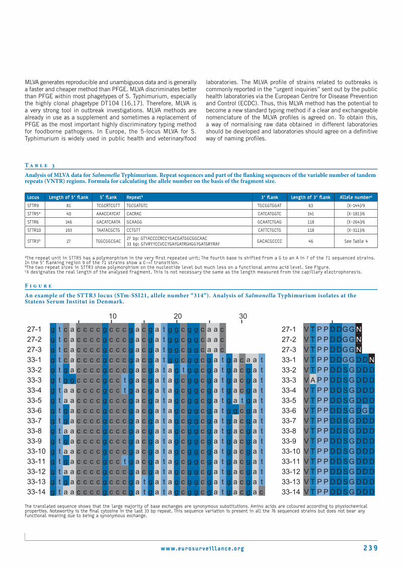
www.eurosurveillance.org 23 9
MLVA generates reproducible and unambiguous data and is generally a faster and cheaper method than PFGE. MLVA discriminates better than PFGE within most phagetypes of S. Typhimurium, especially the highly clonal phagetype DT104 [16,17]. Therefore, MLVA is a very strong tool in outbreak investigations. MLVA methods are already in use as a supplement and sometimes a replacement of PFGE as the most important highly discriminatory typing method for foodborne pathogens. In Europe, the 5-locus MLVA for S. Typhimurium is widely used in public health and veterinary/food
laboratories. The MLVA profile of strains related to outbreaks is commonly reported in the “urgent inquiries” sent out by the public health laboratories via the European Centre for Disease Prevention and Control (ECDC). Thus, this MLVA method has the potential to become a new standard typing method if a clear and exchangeable nomenclature of the MLVA profiles is agreed on. To obtain this, a way of normalising raw data obtained in different laboratories should be developed and laboratories should agree on a definitive way of naming profiles.
T a b l e 3
Analysis of MLVA data for Salmonella Typhimurium. Repeat sequences and part of the flanking sequences of the variable number of tandem repeats (VNTR) regions. Formula for calculating the allele number on the basis of the fragment size.
Locus Length of 5′ flank 5’ flank Repeatb 3′ flank Length of 3′ flank Allele numberc
STTR9 81 TCGCRTCGTT TGCGATGTC TGCGGTGGAT 63 (X-144)/9
STTR5a 40 AAACCAYCAT CACRAC CATCATGGTC 141 (X-181)/6
STTR6 146 GACATCAATA GCAAGG GCAATCTGAG 118 (X-264)/6
STTR10 193 TAATACGCTG CCTGTT CATTCTGCTG 118 (X-311)/6
STTR3b 27 TGGCGGCGAC27 bp: GTYACCCCRCCYGACGATGGCGGCAAC33 bp: GTVRYYCCVCCYGAYGATRGHGGYGATGRYRAY
GACACGCCCC 46 See Table 4
aThe repeat unit in STTR5 has a polymorphism in the very first repeated unit; The fourth base is shifted from a G to an A in 7 of the 71 sequenced strains. In the 5′ flanking region 9 of the 71 strains show a C→T transition.bThe two repeat sizes in STTR3 show polymorphism on the nucleotide level but much less on a functional amino acid level. See Figure.cX designates the real length of the analysed fragment. This is not necessary the same as the length measured from the capillary electrophoresis.
F i g u r e
An example of the STTR3 locus (STm-SSI21, allele number “314”). Analysis of Salmonella Typhimurium isolates at the Statens Serum Institut in Denmark.
The translated sequence shows that the large majority of base exchanges are synonymous substitutions. Amino acids are coloured according to physiochemical properties. Noteworthy is the final cytosine in the last 33 bp repeat. This sequence variation is present in all the 76 sequenced strains but does not bear any functional meaning due to being a synonymous exchange.

24 0 www.eurosurveillance.org
The raw data obtained by fragment analysis by capillary electrophoresis have systematic deviations from the actual size of the fragment. This depends on the DNA composition, the applied instrument, polymers used, etc. Therefore, the measured fragment sizes should be normalised to the actual size to ensure the comparability between laboratories. A set of reference strains with verified fragment sizes which covers the range of the most common alleles for each locus is presented in Table 1. This set offers the possibility for each laboratory to normalise their raw data to the actual fragment sizes.
Hitherto, the naming of profiles has been based on a string of arbitrary allele numbers that do not directly reflect the numbers of repeat units in the loci [1]. Rather, the fragment sizes are binned into allele size categories and then assigned an allele number. There are several advantages of naming the MLVA profiles as the string of five numbers showing the actual number of repeat units in each of the five loci. This way, the MLVA profile can be deduced without looking it up in a table of allele numbers, e.g. maintained on a website. When comparing different MLVA profiles, the difference in number of repeat units in a specific locus can be seen directly. In particular, this is important in outbreak situations where it is relevant to assess whether isolates with deviations in the MLVA profile should be considered part of the outbreak. With a similar MLVA for E. coli O157, it has been suggested that loss or gain of one repeat unit is more likely to occur in epidemiologically related isolates [18]. Furthermore, this definitive nomenclature is independent of the primers used for amplification of the fragments. In principle, this means that the allele numbers obtained in laboratories that use other MLVA protocols that also include these VNTR loci will be identical. For example, PulseNet US has developed 7-locus MLVA protocol for S. Typhimurium that includes the five loci in the European method, but using different primers and therefore obtaining different fragment sizes [Eija Hyytia-Trees, personal communication]. However, in case of polymorphisms in the primer regions a difference in MLVA profile can be obtained, e.g. a fragment can be obtained using one primer pair whereas no
product might be obtained by another primer pair (i.e. assigned as a null allele). However, this should not be of major concern as the sequence analysis of this study shows that the flanking regions are highly conserved.
For loci STTR9, STTR6 and STTR10, our definitions of the size of the flanking regions are in full agreement with those suggested by Gilbert [19]. Our analysis shows that the flanking region for STTR5 is 6 bp longer while in the case of STTR3 the flanking region is 33 bp shorter. The analysis of STTR3 sequences showed that the final nucleotide of the 33 bp repeat units were thymidine for all repeat units except for the very last repeat in each VNTR region, where a T→C transition was present (Figure). This sequence variation was seen in all the 76 sequenced STTR3 loci. This could warrant exclusion of the final 33 bp repeat, but after analysing the sequences we find that although there is an extended polymorphism in the STTR3 repeat units (an example is seen in Figure) the translated sequence is well preserved. The transition in the last repeat unit is also a synonymous mutation (Figure) and we find that this unit should be part of the VNTR region.
The suggested definition of the VNTR region in these five loci is that the region should only contain whole number of repeats. This results in a simple integer designating the number of complete repeat units in each locus. After sequencing up to 80 strains in each loci, it is clear that the flanking region is almost totally conserved (Table 3). ‘Half repeats’ might indeed be active in a mechanism that changes the repeat number, but from a surveillance perspective these fractions of repeats just add complexity without additional informational value.
For the VNTR loci with 6 bp and 9 bp repeat units, the proposed nomenclature is straightforward as the allele numbers can be assigned by a simple calculation based on the analysed fragment size (Table 3). Furthermore, these allele numbers can be translated into the commonly used and previously described system of arbitrary allele numbers. STTR3 pose a more complicated situation as this locus can possess both 27 bp and 33 bp repeat
T a b l e 4
Frequent alleles in the STTR3 locus of Danish Salmonella Typhimurium isolates and the assignation of allele number.
Fragment size 27bp repeats 33bp repeats Allele number Previous allele number*
337 0 8 8 08
370 0 9 9 07
436 0 11 11 05
451 3 9 309 01
463 1 11 111 01
469 0 12 12 01
490 2 11 211 02
496 1 12 112 02
517 3 11 311 03
523 2 12 212 03
544 4 11 411 04
550 3 12 312 04
572 5 11 511 04
616 3 14 314 Not assigned
*According to the allele number system previously described [1].

www.eurosurveillance.org 241
units. The original assignation of allele numbers came around this problem by making large bins for each allele. Thereby, different combinations of the two repeat units were assigned the same allele number (Table 4). This means a loss of discriminatory power. There are several possibilities for assigning allele numbers to the STTR3 locus that more accurately reflect the composition of the alleles seen in this locus. For example, the locus can be treated as two separate loci with 27 bp and 33 bp repeat units, respectively, so that the total MLVA type is a string of six numbers. However, this would give more weight to the STTR3 locus, e.g. when constructing dendrograms, and complicate the transition from the previously used profile assignations. Another possibility is to simply use the fragment size in basepairs as has been decided for comparison between Australian laboratories [19]. This is a simple solution, but only practical if some kind of bins are established as the accuracy of determining the fragment size is at least +/- 1 bp when using the same instrument [20]. Analysis of the fragment sizes found in around 4,000 MLVA typed isolates and the sequence analysis of STTR3 in almost 80 isolates have shown a general pattern for STTR3: STTR3 mainly consists of between 0 and 5 27-bp repeat units and between 8 and 14 33-bp repeat units.
Furthermore, not all combinations of these seem to occur. In our reference set we have included some rare variants with even fewer 33 bp repeats. These short variants make up for around 0.1% of our total S. Typhimurium database and should mainly be considered useful for machine calibration purposes and not for creating bins. Considering these restrictions, it is possible to predict the number of repeat units of each size based on the fragment size even if an inaccuracy of up to +/- 2 bp is allowed. For STTR3, we therefore propose that the allele number is a combination of the number of repeat units of each size, either as a four digit number, e.g. 0114 or simply 114 (1 27-bp repeat and 14 33-bp repeats) (Table 4). Omission of the leading zeros is suggested for more easy data handling using software such as BioNumerics or Excel.
Theoretically, the number of repeat units can be zero even though the VNTR locus is present, i.e. a PCR product is obtained as the flanking region is present. We have not been able to verify the presence of such alleles among our S. Typhimurium strains, but we have seen this for other serotypes. We propose that such alleles should be assigned 0. Additionally, it is fairly common that a PCR product is not obtained for one or more loci. The naming of such absent loci should be distinguished from loci with 0 repeats, and therefore, we suggest that these are assigned NA.
The suggested nomenclature presents a rational and scientifically based way of assigning names to MLVA profiles in a standardised manner. A collection of reference strains with MLVA fragment sizes determined by DNA sequencing offers a possibility of normalising the raw data obtained by each laboratory. A number of laboratories in Europe and North America have agreed to test this approach.
AcknowledgementsWe thank Christian Vråby Pedersen for technical assistance. We would like to thank the participants of the MLVA workshop “An attempt to harmonise MLVA methods for foodborne pathogens in Europe” held in Copenhagen in May 2008 for fruitful discussions that led to an agreement that a new nomenclature for S. Typhimurium MLVA profiles should be based on the principles that we have applied in this study.
References1. Lindstedt BA, Vardund T, Aas L, Kapperud G. Multiple-locus variable-number
tandem-repeats analysis of Salmonella enterica subsp. enterica serovar Typhimurium using PCR multiplexing and multicolor capillary electrophoresis. J Microbiol Methods. 2004;59(2):163-72.
2. Ethelberg S, Sørensen G, Kristensen B, Christensen K, Krusell L, Hempel-Jørgensen A, et al. Outbreak with multi-resistant Salmonella Typhimurium DT104 linked to carpaccio, Denmark, 2005. Epidemiol Infect. 2007;135(6):900-7
3. Hopkins KL, Maguire C, Best E, Liebana E, Threlfall EJ. Stability of multiple-locus variable-number tandem repeats in Salmonella enterica serovar Typhimurium. J Clin Microbiol. 2007;45(9):3058-61.
4. Isakbaeva E, Lindstedt BA, Schimmer B, Vardund T, Stavnes TL, Hauge K, et al. Salmonella Typhimurium DT104 outbreak linked to imported minced beef, Norway, October – November 2005. Euro Surveill. 2005;10(45):pii=2829. Available from: http://www.eurosurveillance.org/ViewArticle.aspx?ArticleId=2829
5. Nygard K, Lindstedt BA, Wahl W, Jensvoll L, Kjelsø C, Mølbak K, et al. Outbreak of Salmonella Typhimurium infection traced to imported cured sausage using MLVA-subtyping. Euro Surveill. 2007;12(11):pii=3158. Available from: http://www.eurosurveillance.org/ViewArticle.aspx?ArticleId=3158
6. Torpdahl M, Sørensen G, Ethelberg S, Sandø G, Kammelgard K, Jannok Porsbo L. A regional outbreak of S. Typhimurium in Denmark and identification of the source using MLVA typing. Euro Surveill. 2006;11(5):pii=621. Available from: http://www.eurosurveillance.org/ViewArticle.aspx?ArticleId=621
7. Torpdahl M, Sørensen G, Lindstedt BA, Nielsen EM. Tandem repeat analysis for surveillance of human Salmonella Typhimurium infections. Emerg Infect Dis. 2007;13(3):388-95
8. Ethelberg S, Lisby M, Torpdahl M, Sørensen G, Neimann J, Rasmussen P, et al. Prolonged restaurant-associated outbreak of multidrug-resistant Salmonella Typhimurium among patients from several European countries. Clin Microbiol Infect. 2004;10(10):904-10.
9. Kivi M, Hofhuis A, Notermans DW, Wannet WJ, Heck ME, Van De Giessen AW, et al. A beef-associated outbreak of Salmonella Typhimurium DT104 in The Netherlands with implications for national and international policy. Epidemiol Infect. 2007;135(6):890-9.
10. O’Flanagan D, Cormican M, McKeown P, Nicolay N, Cowden J, Mason B, et al. A multi-country outbreak of Salmonella Agona, February - August 2008. Euro Surveill. 2008;13(33):pii=18956. Available from: http://www.eurosurveillance.org/ViewArticle.aspx?ArticleId=18956
11. Pezzoli L, Elson R, Little CL, Yip H, Fisher I, Yishai R, Anis E, et al. Packed with Salmonella--investigation of an international outbreak of Salmonella Senftenberg infection linked to contamination of prepacked basil in 2007. Foodborne Pathog Dis. 2008;5(5):661-8.
12. de Valk HA, Meis JF, Bretagne S, Costa JM, Lasker BA, Balajee SA, et al. Interlaboratory reproducibility of a microsatellite-based typing assay for Aspergillus fumigatus through the use of allelic ladders: proof of concept. Clin Microbiol Infect. 2009;15(2):180-7.
13. Waterhouse AM, Procter JB, Martin DMA, Clamp M, Barton GJ. Jalview Version 2 - a multiple sequence alignment editor and analysis workbench. 2009. Bioinformatics doi: 10.1093/bioinformatics/btp033
14. Swaminathan B, Barrett TJ, Hunter SB, Tauxe RV. PulseNet: the molecular subtyping network for foodborne bacterial disease surveillance, United States. Emerg Infect Dis. 2001;7(3):382-9.
15. Swaminathan B, Gerner-Smidt P, Ng LK, Lukinmaa S, Kam KM, Rolando S, et al. Building PulseNet International: an interconnected system of laboratory networks to facilitate timely public health recognition and response to foodborne disease outbreaks and emerging foodborne diseases. Foodborne Pathog Dis. 2006;3(1):36-50.
16. Lindstedt BA, Heir E, Gjernes E, Kapperud G. DNA fingerprinting of Salmonella enterica subsp. enterica serovar Typhimurium with emphasis on phage type DT104 based on variable number of tandem repeat loci. J Clin Microbiol. 2003;41(4):1469-79.
17. Best EL, Lindstedt BA, Cook A, Clifton Hadley FA, Threlfall EJ. Multiple-locus variable-number tandem repeat analysis of Salmonella enterica subsp. enterica serovar Typhimurium: comparison of isolates from pigs, poultry and cases of human gastroenteritis. J Appl Microbiol. 2007;103(3):565-72.
18. Noller AC, McEllistrem MC, Pacheco AG, Boxrud DJ, Harrison LH (2003) Multilocus variable-number tandem repeat analysis distinguishes outbreak and sporadic Escherichia coli O157:H7 isolates. J Clin Microbiol. 2003;41(12):5389-97.
19. Gilbert GL (2008) Using MLVA to type strains of Salmonella Typhimurium in New South Wales. N S W Public Health Bull. 2008;19(1-2):29-31.
20. Lindstedt BA, Torpdahl M, Nielsen EM, Vardund T, Aas L, Kapperud G. Harmonization of the multiple-locus variable-number tandem repeat analysis method between Denmark and Norway for typing Salmonella Typhimurium isolates and closer examination of the VNTR loci. J Appl Microbiol. 2007;102(3):728-35.

24 2 www.eurosurveillance.org
R esearch ar ti cles
T h e e m e r g e n c e o f c l o s T r i d i u m d i f f i c i l e P c r r i b oT y P e 027 i n d e n m a r k – a P o s s i b l e l i n k w i T h T h e i n c r e a s e d c o n s u m P T i o n o f f l u o r o q u i n o l o n e s a n d c e P h a l o s P o r i n s ?
L Søes ([email protected])1, K Mølbak2, S Strøbæk3, K Truberg Jensen4, M Torpdahl1, S Persson1, M Kemp1, K E Olsen1
1. Department of Bacteriology, Mycology and Parasitology, Statens Serum Institut, Copenhagen, Denmark2. Department of Epidemiology, Statens Serum Institut, Copenhagen, Denmark3. Department of Infection Control, Sydvestjysk Hospital, Esbjerg, Denmark4. Department of Clinical Microbiology, Sydvestjysk Hospital, Esbjerg, Denmark
This article was published on 16 April 2009. Citation style for this article: Søes L, Mølbak K, Strøbæk S, Truberg Jensen K, Torpdahl M, Persson S, Kemp M, Olsen KE. The emergence of Clostridium difficile PCR ribotype 027 in Denmark – a possible link with the increased consumption of fluoroquinolones and cephalosporins?. Euro Surveill. 2009;14(15):pii=19176. Available online: http://www.eurosurveillance.org/ViewArticle.aspx?ArticleId=19176
Increasing rates of Clostridium difficile infection (CDI) with an unusual, severe course have been reported in several countries; this rise has partly been ascribed to the emergence of a virulent strain, C. difficile PCR ribotype 027 (CD027). An intriguing question is whether this could be related to increasing consumption of broadspectrum antibiotics. From 1997 to 2007, the number of hospital discharges in Denmark with the diagnosis enterocolitis caused by C. difficile increased from eight to 23 per 100,000 hospital discharges. This increase was proportional to a concomitant rise in the consumption of fluoroquinolones and cephalosporins. The first outbreak of CD027 in Denmark occurred from October 2006 to August 2007 and included 13 patients, most of them elderly, admitted to three hospitals in the same region. Most of the patients had overlapping periods of admission. All patients had been treated with broadspectrum antibiotics, in particular cephalosporins and fluoroquinolones, prior to positive culture of CD027. Thirty days after confirmation of diagnosis, three of the 13 patients had died. Taken together, the data support the hypothesis that the increasing use of certain broadspectrum antibiotics may be related to a possible increase of C. difficile infection, and show that the specific contribution by CD027 in its emergence needs to be determined.
IntroductionInfection with toxin-producing strains of Clostridium difficile is
a common cause of diarrhoea and varies from mild to severe cases of diarrhoea. Cases are frequently antibiotic-associated and occur mostly in hospitals. Pseudomembranous colitis in already impaired patients e.g. with an underlying condition is a serious manifestation of C. difficile infection (CDI) and can result in death.
Reports from North America, Europe and Japan have drawn attention to a recently discovered strain of C. difficile that is characterised as PCR ribotype 027, toxinotype III (CD027) [1-4]. This strain has an increased pathogenic capacity, possibly a higher infectious potential and a particular resistance profile. The increased pathogenicity is thought to be associated with an enhanced production of toxin A and toxin B caused by mutations
in a regulatory gene, but the fact that this strain in addition produces a binary toxin CDT may also contribute to increased pathogenicity. This strain has caused severe outbreaks of CDI in hospital environments, but has also been described as the cause of outbreaks and sporadic cases outside hospitals [2-4].
The aims of the present report are to summarise national hospital data with a discharge diagnosis of CDI and to describe the first outbreak of CD027 in Denmark.
MethodsBecause of the international emergence of CD027 and the
subsequent recommendations from ECDC [5], we obtained hospital discharge data on CDI in Denmark from 1997 to 2007 and conducted a retrospective characterisation of C. difficile isolates from November 2006 to March 2007. In addition, Statens Serum Institut (SSI) asked Danish departments of clinical microbiology to continuously report C. difficile findings and to forward isolates for typing on suspicion of an outbreak or severe disease.
The hospital discharge data were obtained from the statistics of the Danish National Board of Health (http://sundhedsdata.sst.dk). Specifically, we obtained the annual aggregated number of discharges with the ICD10 diagnosis code DA04.7 (“enterocolitis caused by C. difficile”, i.e. enterocolitis independent of PCR ribotype) as well as the annual number of all discharges from somatic hospitals, i.e. hospitals treating only somatic and not psychiatric diseases. Data about consumption of fluoroquinolones and cephalosporins were obtained from The Danish Integrated Antimicrobial Resistance Monitoring and Research Programme (DANMAP) [6].
Isolates of C. difficile were characterised by PCR ribotyping, toxin gene profiles, and deletion studies undertaken by the National Reference Laboratory for Enteropathogenic Bacteria at SSI.
Stool samples were cultured on cycloserine cefoxitin fructose agar (CCFA) (SSI Diagnostica, Hillerød, Denmark) in an atmosphere composed of 86% N2, 7% H2 and 7% CO2 at 37°C for 48 hours.

ww.eurosurveillance.org 243
Colonies with typical morphology and distinctive odour were identified. The colonies were analysed by 5-plex PCR directed towards tcdA, tcdB, cdtA, cdtB and 16S rDNA and by sequencing of the 5’-end of tcdC in order to search for premature stop codons and internal deletions [7]. PCR ribotyping was performed according to Bidet et al. [8].
Results Hospital discharges of CDI in DenmarkThe aggregated number of discharges of enterocolitis caused by
C. difficile increased from 86 (eight per 100,000 discharges) in 1997 to 282 (23 per 100,000 discharges) in 2007. In the same period, the consumption of fluoroquinolones and cephalosporins used in primary healthcare and hospitals taken together, increased
from 384 to 1,162 kg and from 626 to 2,285 kg active component per annum, respectively (see Figure 1) [6].
Detection of CD027 in DenmarkIn the retrospective survey, isolates obtained between November
2006 and March 2007 were characterised; eight CD027 cases were found (Figure 2). The isolates came from eight hospitalised patients from the Region of Southern Denmark (the former Ribe County). Seven of the patients had been admitted to the same small hospital A, while the last case was a patient in another hospital in the same local area. Prompted by this cluster, active surveillance for CD027 was established in the area, and an additional 22 isolates of C. difficile were received between June and August 2007, of which five were CD027 (see Figure 2).
Thus, a total of 13 patients with CD027 were identified. Mean age was 79 years (age range 64 to 96 years), and 10 cases were women. The patients were admitted to hospital in the period October 2006 to July 2007. Nine of the patients were admitted to the same medical ward at the small hospital A, which consisted of only the one ward and a surgical day clinic. Most of these patients had overlapping periods of admission. The CD027-positive stool sample from one of these patients was requested by the general practitioner 13 days after the patient’s discharge from hospital. The other eight were obtained during admission. The remaining four patients were admitted to three different medical departments at the larger hospital B. Two of these patients had overlapping periods of admission at the same ward. One of these four patients was moved to another medical ward at another small hospital C (see Figure 3).
The isolates were all PCR ribotype 027, carried the binary toxin gene, had an 18 bp deletion in the regulatory gene tcdC, and a 1 bp deletion at position 117 of tcdC. They were all resistant to fluoroquinolones (including moxifloxacin), but susceptible to erythromycin and clindamycin. Interestingly, at the same time and in the same geographical area, but unrelated to the outbreak
F i g u r e 2
Number of patients with Clostridium difficile infection caused by CD027, Denmark, October 2006-August 2007 (n=13)
0
1
2
3
4
5
Oct Nov Dec Jan Feb Mar Apr May Jun Jul Aug
No.
of
case
s
Primary clusterDetected in follow-up surveillance
2006 2007
F i g u r e 1
Annual number of hospital discharges with enterocolitis caused by Clostridium difficile (ICD10 diagnosis code DA04.7) and annual consumption of fluoroquinolones and cephalosporins for human use, Denmark, 1997-2007
0
50
100
150
200
250
300
1997 1998 1999 2000 2001 2002 2003 2004 2005 2006 2007
Year
No.
of
disc
harg
es w
ith
an I
CD10
-cod
eDA
04.
7
0
500
1000
1500
2000
2500
Kg a
ctiv
e co
mpo
nen
t
Discharges with an ICD10-diagnosis code DA04.7 Fluoroquinolone consumption Cephalosporin consumption
Source: [6]
F i g u r e 3
Distribution of cases of CD027 in the three hospitals, Denmark, October 2006-August 2007
Hospital CMinor specialised unit
connected with Hospital B;October 2006-March 2007: one case of CD027
transferred fromHospital B to Hospital C
Hospital BMajor acute-care hospital;
October 2006-March 2007: one case of CD027;June 2007-August 2007: three cases of CD027;
All four cases confirmed CD027
Hospital AMinor specialised unit
connected with Hospital B;October 2006-March 2007: seven cases of CD027;
June 2007-August 2007: two cases of CD027;All nine cases confirmed CD027

24 4 www.eurosurveillance.org
another isolate was found that also carried the binary toxin gene, had the 18 bp and the 1 bp deletion at position 117 in the regulatory gene tcdC, but was not PCR ribotype 027. In contrast to the CD027 strains it was sensitive to moxifloxacin.
As this cluster of 13 cases was detected in a setting with ample possibilities of transmission and at the time represented the only detection of CD027 in Denmark, it is reasonable to assume that an outbreak with CD027 occurred during this period. Multilocus variable-number tandem-repeat analysis (MLVA) or restriction endonuclease analysis (REA) [9,10] will be performed in order to elucidate the connection between the isolates.
All of the 13 patients were treated with broadspectrum antibiotics prior to positive culture of CD027. Eleven patients received cephalosporins and nine fluoroquinolones; seven received both cephalosporins and fluoroquinolones, either simultaneously or consecutively. Thirty days after confirmation of diagnosis, three of the 13 patients had died. It is unknown if the deaths were directly attributable to C. difficile.
DiscussionIt is not known with certainty why the number of patients
discharged after an episode of enterocolitis caused by C. difficile is increasing. However, it is certain that the patients with a discharge diagnosis of ICD10 code DA04.7 only comprise a modest fraction of the true number of cases. In 2007, 1,342 culture-confirmed cases of C. difficile infections were reported to the national surveillance system in Denmark (25 per 100,000 population). Surveillance was established in 2007. Data before this is therefore not available. Although increased diagnostic activity and awareness may play a role, it is also likely that changes in the strains’ pathogenicity are important contributing factors to the emergence of CDI. This includes the appearance of CD027 and possibly other hypervirulent strains. Several factors may be of importance to understand the emergence of C. difficile and in particular of CD027. The CD027 strain is resistant to the newer fluoroquinolones, including moxifloxacin, and it has been suggested that this may be the main reason for its wide dissemination [2,3]. This hypothesis is supported by the almost parallel increase in CDI discharge diagnoses and the consumption of fluoroquinolones as illustrated in Figure 1. However it should be emphasised that resistance to moxifloxacin and several other fluoroquinolones is also seen in other C. difficile PCR ribotypes [11,12]. Furthermore, increased use of other broadspectrum antibiotics including cephalosporins may also be related to the emergence of C. difficile since the same almost parallel increase is observed in CDI discharge diagnoses and consumption of cephalosporins (Figure 1).
However, these possible relations should be interpreted
with caution. Other circumstances may also be of considerable importance, such as the increasing challenges in the area of hospital hygiene. For example, increased virulence of C. difficile resulting in pronounced diarrhoeal symptoms may have promoted spread and cross-infection within healthcare institutions, possibly because of dissemination of spores by incontinent patients [3]. The emergence of C. difficile and CD027 in particular is likely to be a result of environmental as well as person-to-person transmission in healthcare facilities rather than solely a result of increased antibiotic pressure. Finally, demographic changes such as an age distribution with an increasing proportion of elderly people and
changes in the patterns of hospitalisation towards increased “turn-over” of patients may also contribute.
The recognition of the outbreak of CD027 in this particular geographical area of Denmark may not be an isolated observation. The initial cluster was detected in a convenience sample of stool specimens from diarrhoeal patients as part of a project including molecular characterisation of C. difficile isolates. Hence, it is conceivable that the cases discovered only represent the tip of the iceberg. On a voluntary basis, strains from all different geographical areas of Denmark are now being submitted for surveillance to the National Reference Laboratory for Enteropathogenic Bacteria to identify CD027.
Although we cannot conclude a cause-and-effect relation between the increase in fluoroquinolone and cephalosporin consumption and the increase in CDI discharge diagnoses, we consider it important to present these data to stimulate additional research. Studies are needed to determine the burden of disease associated with CD027 and other hypervirulent C. difficile strains, while integrated public health and microbiological surveillance should be established to determine trends, detect clusters in healthcare institutions, and facilitate more focused infection control. To prevent spread, it is essential to focus on hospital hygiene and promote prudent antibiotic policies, including the limitation of unnecessary use of broadspectrum antibiotics, including fluoroquinolones and cephalosporins.
AcknowledgementsWe wish to thank J. Nevermann Jensen for extensive technical assistance.
References
1. McDonald LC, Killgore GE, Thompson A, Owens RC, Kazakova SV, Sambol SP, et al. An epidemic, toxin gene-variant strain of Clostridium difficile. N Engl J Med. 2005;353(23):2433-41.
2. Loo VG, Poirier L, Miller MA, Oughton M, Libman MD, Michaud S, et al. A predominantly clonal multi-institutional outbreak of Clostridium difficile-associated diarrhea with high morbidity and mortality. N Engl J Med. 2005;353(23):2442-9.
3. Warny M, Pepin J, Fang A, Killgore G, Thompson A, Brazier J, et al. Toxin production by an emerging strain of Clostridium difficile associated with outbreaks of severe disease in North America and Europe. Lancet. 2005;366(9491):1079-84.
4. Kuijper EJ, Barbut F, Brazier JS, Kleinkauf N, Eckmanns T, Lambert ML, et al. Update of Clostridium difficile-associated disease due to PCR ribotype 027 in Europe, 2008. EuroSurveill 2008;13(31):pii=18942. Available from: http://www.eurosurveillance.org/ViewArticle.aspx?ArticleId=18942
5. Kuijper EJ, Tüll P. Emergence of Clostridium difficile-associated disease in Canada, the United States of America and Europe. Background document prepared on behalf of the European Study Group for Clostridium difficile (ESGCD) and European Centre for Disease Prevention and Control (ECDC). 2006 March 3. Available from: http://www.ecdc.europa.eu/documents/pdf/Cl_dif_v2.pdf
6. DANMAP 2007. Use of antimicrobial agents and occurrence of antimicrobial resistance in bacteria from food animals, foods and humans in Denmark. DANMAP – The Danish Integrated Antimicrobial Resistance Monitoring and Research Programme. 2008 September. ISSN 1600-2032. Available from: http://www.danmap.org
7. Persson S, Torpdahl M, Olsen KE. New multiplex PCR method for the detection of Clostridium difficile toxin A (tcdA) and toxin B (tcdB) and the binary toxin (cdtA/cdtB) genes applied to a Danish strain collection. Clin Microbiol Infect. 2008;14(11):1057-64.
8. Bidet P, Barbut F, Lalande V, Burghoffer B, Petit JC. Development of a new PCR-ribotyping method for Clostridium difficile based on ribosomal RNA gene sequencing. FEMS Microbiol Lett. 1999;175(2):261-66.
ww.eurosurveillance.org 24 5
9. Fawley WN, Freeman J, Smith C, Harmanus C, van den Berg RJ, Kuijper EJ et al. Use of Highly Discriminatory Fingerprinting to Analyze Clusters of Clostridium difficile Infection Cases Due to Epidemic Ribotype 027 Strains. J Clin Microbiol. 2008;46(3):954-60.
10. Killgore G, Thompson A, Johnson S, Brazier J, Kuijper E, Pepin J, et al. Comparison of Seven Techniques for Typing International Epidemic Strains of Clostridium difficile: Restriction Endonuclase Analysis, Pulsed-Field Gel Electrophoresis, PCR-Ribotyping, Multilocus Sequence Typing, Multilocus Variable-Number Tandem-Repeat Analysis, Amplified Fragment Length Polymorphism, and Surface Layer Protein A Gene Sequence Typing. J Clin Microbiol. 2008;46(2):431-7.
11. Wilcox MH, Fawley W, Freeman J, Brayson J. In vitro activity of new generation fluoroquinolones against genotypically distinct and indistinguishable Clostridium difficile isolates. J Antimicrob Chemother. 2000;46(4):551-6.
12. Alonso R, Peláez T, González-Abad MJ, Alcalá L, Muñoz P, Rodríguez-Créixems M et al. In vitro activity of new quinolones against Clostridium difficile. J Antimicrob Chemother. 2001;47(2):195-7.

24 6 www.eurosurveillance.org
R esearch ar ti cles
E n d E m i c h E pat i t i s E i n t w o n o r d i c c o u n t r i E s
H Norder ([email protected])1, L Sundqvist1, L Magnusson1, S Østergaard Breum2, M Löfdahl3, L E Larsen2, C K Hjulsager2, L Magnius1,B E Böttiger4, F Widén5
1. Department of Virology, Swedish Institute for Infectious Disease Control (Smittskyddsinstitutet, SMI), Solna, Sweden2. Department of Veterinary Diagnostics and Research, National Veterinary Institute, Technical University of Denmark (DTU),
Copenhagen, Denmark3. Department of Epidemiology, Swedish Institute for Infectious Disease Control (Smittskyddsinstitutet, SMI), Solna, Sweden4. Department of Virology, Statens Serum Institut, Copenhagen, Denmark5. Department of Biomedical Sciences and Veterinary Public Health, Swedish University of Agricultural Sciences,
Uppsala, Sweden
This article was published on 14 May 2009. Citation style for this article: Norder H, Sundqvist L, Magnusson L, Østergaard Breum S, Löfdahl M, Larsen LE, Hjulsager CK, Magnius L, Böttiger BE, Widén F. Endemic hepatitis E in two Nordic countries. Euro Surveill. 2009;14(19):pii=19211. Available online: http://www.eurosurveillance.org/ViewArticle.aspx?ArticleId=19211
Antibodies against hepatitis E virus (anti-HEV) were found in 248 Swedish and Danish patients between 1993 and 2007. Most patients were symptomatic and tested for anti-HEV due to travel abroad. Among patients with known country of infection, most were infected in Asia, mainly on the Indian subcontinent. However, 29 patients were infected in Europe, nine of these had HEV IgM and/or HEV RNA in serum. In sera from 65 of 141 tested patients HEV RNA could be detected, and 63 strains could be typed by limited sequencing within ORF2. HEV RNA was found in sera from 71% of the patients with HEV IgM and IgG and in 18% of the patients with only detectable HEV IgG. It was also found up to three weeks after the onset of disease in 67% of the patients with known date of onset. Patients infected in Europe were infected by genotype 3, and were older than those infected by genotype 1 (mean age 55.3 vs 30 years, p<0.001). Since it is known that genotype 3 can infect domestic pigs, HEV strains from 18 piglets in 17 herds in Sweden and Denmark were sequenced. Phylogenetic analyses of the genotype 3 strains showed geographical clades and high similarity between strains from patients and pigs from the same area. There are thus autochthonous hepatitis E cases in Scandinavia, and there are probably many undiagnosed ones. Patients with hepatitis of unknown etiology should therefore be investigated for anti-HEV even if they have not been outside Europe, since infections acquired from pigs or other animals should be taken into consideration.
IntroductionHepatitis E virus (HEV) is a non-enveloped positive-stranded
RNA virus of 27-34 nm in diameter [1]. It is the only member of the genus Hepevirus in the family Hepeviridae. The genome is approximately 7.2 kb in length and encodes three open reading frames, from ORF1 to ORF3. ORF1 encodes for enzymes important for replication and transcription, ORF2 encodes for a capsid protein and ORF3 for a small protein of 122 or 123 amino acids that interacts with cellular proteins and contributes to viral replication. There is only one serotype but based on genetic diversity HEV strains are classified into four genotypes designated with Arabic numerals 1 to 4. The genotypes are further divided into up to seven subtypes designated with Roman characters a – g, each with distinct geographical distribution [2]. Genotypes 1 and 2 only infect
humans, mainly in Asia, and Africa, where they are endemic and may cause large outbreaks. Genotype 2 has been found causing outbreaks in Mexico and Africa. Strains of the other two genotypes, 3 and 4, have been shown to infect not only humans, but also domestic pigs, wild boars, deer, and other mammals. These two genotypes have not been reported to cause outbreaks. In endemic countries, as India, genotype 1 infects humans, while HEV isolates from swine belong to genotype 3 or 4 [3]. However, genotype 3 strains have also been isolated from sporadic human cases of hepatitis E, and from domesticated pigs in several European countries, in the United States (US) and in Japan, while genotype 4 strains have been found in humans and pigs exclusively in Asia, as China, Taiwan, Japan and Vietnam [4-7].
Hepatitis E is transmitted mainly by the faecal-oral route, usually through contaminated drinking water. Usually, the infection is self-limited, although some persons develop fulminant hepatitis. In pregnant females the illness is particularly severe with up to 20% fatality rate in the third trimester, but it may be even higher in patients with underlying chronic liver disease [8,9]. Chronic hepatitis E infections have also been described in transplant patients on immunosuppressive treatment [10].
Hepatitis E was previously considered to mainly affect the inhabitants of or travellers to Asia and Africa, due to high endemicity in these parts of the world. However, in recent years there have been several reports on autochtonous hepatitis E cases in Europe, including United Kingdom (UK), the Netherlands and France [6,7,11,12], and also in the US, New Zealand and Japan [4,13,14]. There have also been increasing numbers of reports on high seroprevalence in Europe and the US. Antibodies againts HEV (anti-HEV) were found in 17% of blood donors in the UK and in France, in 21-33% of blood donors and 50% of farmers in Denmark and 5 to 9% of the general population and 13% of veterinarians in Sweden [11,15-18]. These data indicate that there is a high prevalence of hepatitis E infections also in Europe, albeit most infections are subclinical and most of them may be zoonotic. The study presented here was performed to investigate which genotypes of HEV were imported to Denmark and Sweden between 1993 and 2007, and to find out if there were any endemic HEV strains and,

www.eurosurveillance.org 247
if so, to determine their relation to HEV strains obtained from pigs in these countries.
Materials and methods Identification of human cases with hepatitis EPatients with a recent travel history and with clinical signs of
hepatitis not caused by hepatitis A, B, C or D virus were investigated for hepatitis E at the Swedish Institute for Infectious Disease Control (Smittskyddsinstitutet, SMI), Solna, Sweden. Sera from these patients were tested for HEV IgG and IgM by a kit using two recombinant HEV antigens corresponding to structural region of the HEV (Diagnostic Biotechnology, Singapore). Between 1993 and 2006, all samples were also tested for HEV IgG and IgM by using the until then commercially available ELISA kits from Abbott Laboratories (Abbott Laboratories, Chicago, IL). All reactive sera were tested for HEV RNA by PCR and the amplified fragments were sequenced.
Identification of pigs infected by HEVHEV RNA was isolated and sequenced from 18 HEV strains from
pigs. The strains were obtained from two HEV prevalence studies (Breum, unpublished; Widén et al., unpublished). Eight strains were from piglets from seven Danish pig herds and 10 strains were from piglets from 10 Swedish herds.
RNA extractionHEV RNA was extracted from 200 ul serum from humans or
faecal suspension from pigs using QIAamp UltraSense Virus Kit (Qiagen, GmbH, Germany) as described by the manufacturer. Five ul RNA were used for cDNA synthesis in 20 ul mix containing 5 ul 5X First Strand buffer (Invitrogen, Life Technologies, Carlsbad,
CA), 10 mM DTT (Invitrogen, Life Technologies, Carlsbad, CA), 0.5 mM dNTP (Thermo Scientific, Abgene®, Epsom, UK), 100 U Superscript II Reverse Transcriptase (Invitrogen, Life Technologies, Carlsbad, CA), 0.5 ul RNasin (Promega, Madison, US) and 0.1 U random hexamere primers (Roche Diagnostics, GmbH, Germany). Reverse transcription was performed at room temperature for 15 minutes and then at 42°C for two hours.
Nested PCR in the RdRp domain of ORF1 regionA nested PCR was carried out in a 50 ul reaction with 5 ul cDNA,
0.06 ul of 0.2 mM of each primers ISP-4232 and EAP-4576 [19], 5 ul 10X Taq.buffer general, 2 mM MgCl2 (Applied Biosystems, Roche Molecular Systems, New Jersey, US), 0.2 mM dNTP (Thermo Scientific, Abgene®, Epsom, UK) and 4 U Taq polymerase (Thermo Scientific, Abgene®, Epsom, UK). The PCR reaction was carried out for 40 cycles with denaturation at 94°C for 20s, annealing at 60°C for 30s and extension at 72°C for 60s. The second round reaction was carried out similarly but with 5 ul first round product instead of cDNA, 2.5 mM MgCl and 0.06 ul of 0.2 mM of each primer ISP-4232 and IAP-4561 [19].
Nested PCR in the ORF2 regionTwo different nested PCRs for amplification of the ORF2 region
were performed. PCR:1 was carried out in a 50 ul reaction with 10 ul cDNA, 0.1 ul of 0.2 mM primer HE110 [14], 0.119 ul of 0.2 mM primer HE041 (14), 5 ul 10X Taq.buffer general, 2.5 mM MgCl2, 0.2 mM dNTP, 6 U Taq polymerase. The PCR reaction was carried out for 40 cycles of denaturation at 94°C for 20s, annealing at 56°C for 20s and extension at 72°C for 60s. The second round reaction was carried out with 5 ul PCR product, primers HE110 and HE3159 [20] with reagents and cycling as in the first round.
T a b l e 2
Hepatitis E virus (HEV) RNA detection in serum samples from Swedish and Danish patients (1993-2007) with anti-HEV IgM and IgG or anti-HEV IgG only
Patient origin N Anti-HEV
IgM + IgGAnti-HEVIgG only HEV RNA positive (%) HEV RNA in IgM + IgG
positive seraHEV RNA in IgG only
positive seraNumber of
sequenced strains
Sweden 82 44 38 44 (57 %) 38 (86 %) 9 (24 %) 44
Denmark 59 36 23 21 (36 %) 19 (53 %) 2 (9 %) 21
Total 141 80 (57 %) 61 (43 %) 65 (46 %) 57 (71 %) 11 (18 %) 63 (97 %)
T a b l e 1
Age and sex distribution of patients from Sweden and Denmark (1993-2007) with serological markers against hepatitis E virus (HEV)
Number of patients with anti-HEV IgM Number of patients with anti-HEV IgG only
Sweden Denmark Sweden Denmark
Age M F M F Sub-total M F M F Sub-total Total
0-9 0 0 2 0 2 0 1 2 0 3 5
10-19 8 1 3 1 13 2 2 3 0 7 20
20-29 13 8 8 6 35 8 4 9 2 23 58
30-39 4 4 6 2 16 7 3 24 7 41 57
40-49 4 0 2 1 7 9 4 14 3 30 37
50-59 0 1 1 1 3 5 5 11 10 31 34
60-69 2 3 3 0 8 2 5 9 2 18 26
>70 1 0 0 0 1 3 3 1 3 10 11
Total 32 17 25 11 85 36 27 73 27 163 248
M = male, F = female
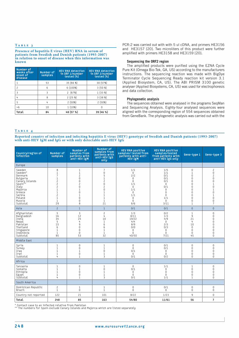
24 8 www.eurosurveillance.org
PCR:2 was carried out with with 5 ul cDNA, and primers HE3156 and HE3157 [20]. Two microliters of this product were further amplified with primers HE3158 and HE3159 [20].
Sequencing the ORF2 regionThe amplified products were purified using the EZNA Cycle
Pure Kit (Omega Bio-Tek, GA, US) according to the manufacturers instructions. The sequencing reaction was made with BigDye Terminator Cycle Sequencing Ready reaction kit version 3.1 (Applied Biosystem, CA, US). The ABI PRISM 3100 genetic analyser (Applied Biosystems, CA, US) was used for electrophoresis and data collection.
Phylogenetic analysisThe sequences obtained were analysed in the programs SeqMan
and Sequencing Analysis. Eighty-four analysed sequences were aligned with the corresponding region of 554 sequences obtained from GeneBank. The phylogenetic analysis was carried out with the
T a b l e 4
Reported country of infection and infecting hepatitis E virus (HEV) genotype of Swedish and Danish patients (1993-2007) with anti-HEV IgM and IgG or with only detectable anti-HEV IgG
Country/region of infection
Number of samples
Number of samples from patients with anti-HEV IgM
Number of samples from patients with anti-HEV IgG
only
HEV RNA positive samples/ tested from patients with anti-
HEV IgM
HEV RNA positive samples/tested
from patients with anti-HEV IgG only
Geno-type 1 Geno-type 3
Europe
SwedenSweden*DenmarkBulgariaCanary IslandsSpain**ItalyMajorca GreeceSerbiaPolandRussiaSubtotal
81712221121129
1120020101008
70512020111121
1/10
2/200
1/10
1/10
1/100
6/6
1/51/10/10/10/10
0/1000
1/10
3/11
0100000000001
2020010101007
Asia 2 1 1 0/1 0/1 0 0
AfghanistanBangladeshIndiaNepalPakistanThailandSingaporeIndonesiaSubtotal
5163451561185
312244900053
24101661132
1/38/1119/224/48/90/000
40/50
0/21/34/90
2/30/300
7/21
192141000045
000000000
Middle East
SyriaTurkeyIraqIranSubtotal
11114
00101
11013
00
0/10
0/1
0/10/100
0/2
00000
00000
Africa
TanzaniaSomaliaEthiopiaEgyptSubtotal
11114
01001
10113
00/100
0/1
1/1000
1/1
10001
00000
South America
Dominican RepublicBrazil
21
10
11
00
0/10
00
00
Country not reported 122 21 101 8/22 1/23 9 0
Total 248 85 163 54/80 11/61 56 7
* Contact case to an infected relative from Pakistan** The numbers for Spain exclude Canary Islands and Majorca which are listed separately
T a b l e 3
Presence of hepatitis E virus (HEV) RNA in serum of patients from Swedish and Denish patients (1993-2007) in relation to onset of disease when this information was known
Number of weeks after onset of disease
Number of samples
HEV RNA detection in ORF 1/number
tested (%)
HEV RNA detection in ORF 2/number
tested (%)
1 53 35 (66 %) 30 (57%)
2 6 6 (100%) 3 (50 %)
3 3 2 (67%) 1 (33 %)
4 8 2 (25 %) 3 (38 %)
5 4 2 (50%) 2 (50%)
>6 10 1 (10%) 0
Total 84 48 (57 %) 39 (46 %)

www.eurosurveillance.org 24 9
PHYLIP package version 3.65 [21]. Evolutionary distances were using the F84 algorithm in the DNADIST program with transition/transversion ratio of 4.29. Phylogenetic trees were constructed using UPGMA and Neighbor-joining method in the NEIGHBOR program in the PHYLIP package. The trees were visualized using the program Tree View, version 1.6.6. Bootstrap analysis of 1,000 replicas was perfomed with the programs SEQBOOT and CONSENSE in the PHYLIP package.
Results There was no significant difference in age and sex distribution
between the patients from Sweden compared with those from Denmark (Table 1). Anti-HEV IgM and IgG was found in 85 patients, 57 (67%) of those were males. There was also a predominance of males, 109/163 (67%), among patients in whom only anti-HEV IgG without detectable IgM was found (Table 1). The mean age of the patients with anti-HEV IgM was 31.5 years, while those with only detectable anti-HEV IgG were older with a mean age of 43.6 years.
HEV RNA could be detected in serum from 65 of 141 tested anti-HEV positive patients (Table 2). The PCR in the ORF1 region was more sensitive and could amplify 63 of the strains, while 51 of the strains were amplified in the ORF2 region. HEV strains could be amplified in 68% of the sera from patients with IgM anti-HEV, as well as in 18% of sera from patients with detectable anti-HEV IgG only (Table 2).
The time of onset of disease in relation to the time of sample collection was known for 84 patients (Table 3). All six patients sampled 2-3 weeks after onset had detectable HEV RNA in serum when ORF 1 was amplified, while only three of these patients had detectable HEV RNA when ORF2 was amplified. In two patients HEV RNA was detected as long as five weeks after onset of illness.
The countries of infection, known for 126 (51%) of the patients, were mainly in Asia with India, Pakistan and Bangladesh as dominating countries (Table 4). Twenty-nine patients (23%) were infected in Europe, while the rest were infected in the Middle East, Africa or South America (Table 4). Six of the patients infected in Europe were injecting drug users (IDUs) and one case in Sweden was a contact of an HEV-infected relative from Pakistan [22].
The PCR amplified regions could be sequenced for 63 of the 65 PCR amplified isolates and 56 patients were found infected with genotype 1, while seven were infected with genotype 3 (Table 4). Those with genotype 1 had all been infected in Asia and Africa, apart from the Swedish contact of a case from Pakistan, while all those with genotype 3 were infected in Europe.
There was a predominance of males in both groups, with five males among the seven patients infected by genotype 3 and 43 males among the 56 patients infected by genotype 1. The patients infected by genotype 3 were older than those with genotype 1. The mean age of patients infected with genotype 3 was 55.3 years, while the mean age of those with genotype 1 was 30 years (p<0.001; unpaired t-test).
HEV RNA could be detected in six faecal samples from six out of 10 piglets tested (in six out of 10 Swedish breeding herds) and in eight samples from piglets originating from seven Danish breeding herds. All piglets were found infected with genotype 3.
In the phylogenetic analysis all isolates could be allocated to either genotype 1 or 3 (Figure 1). It was also found that genotype 3 could be subdivided into two major clades, here tentatively designated 3-I and 3-II (Figures 1 and 2b). Subtypes 3a, c, and d clustered in clade 3-I, while strains of subtype 3e, g, and f were found in clade 3-II (Figures 1 and 2b). This sequenced region of ORF2 was not available in GenBank for subtypes 3h and 3j. The 371 nucleotides of the genomic region coding for the methyltransferase was available for these subtypes and from three genotype 3f strains and was compared with the corresponding region of the genotype 3 strain from a Swedish pig (accession number EU360977). The nucleotide sequence of the Swedish pig strain diverged by 81–84% from subtype 3h and j in this region, whereas it was 88-89% similar to subtype 3f. Sequences of the same subtypes were similar in 88-90% to each other and in 84-86% to the sequences of the other subtypes. Based on this comparison, the Swedish strains found in clade 3-II may belong to subtype 3f.
Even if most genotype 1 sequences available in GenBank originate from India and Nepal, there was a geographical clustering with these strains and 1a, 1b and 1c strains from China, Japan and Kyrgyzstan forming one cluster, while another cluster was formed by 1d and 1e strains from Africa (Figure 2a). In our study, the majority of patients infected on the Indian subcontinent were infected by 1a. The sequences from isolates from India and Pakistan were similar to strains available in GenBank from these countries and from Nepal, while those from patients infected in Bangladesh were found on a separate branch. However, one strain from a patient infected with 1a in Tanzania was more similar to strains from India than to strains from Africa, and was thus an exception.
The strains found in clade 3-I were from Asia, mainly Japan, South Korea and China, and the US. Two strains in our study were found in this clade, one was from a Swedish pig herd the other was from a woman infected in Serbia/Montenegro and was similar to a Japanese strain (AB094212). All other genotype 3 isolates in this study were found in clade 3-II and clustered according to geographical origin (Figure 2b). There were two major subclusters within 3-II one was formed by 3f strains from Europe the other by subtype 3e and 3g strains from Japan, Mongolia and Kyrgyzstan. There was geographical clustering also within the clade formed by European isolates. One branch was formed by strains from Spain and France, one with strains from the Netherlands and France and one with Swedish and Danish strains intermixed with three strains from Spain (Figure 2b). The isolates from one Swede and one Dane infected in Spain were similar to strains from Spanish pigs. The strains from individuals infected in Sweden or Denmark were all similar to strains from Swedish and Danish pigs (Figure 2b). Pig strains from two Swedish breeding herds were found similar to Japanese and Mongolian strains within clade 3-II.
DiscussionHepatitis E is not considered a major public health problem in
non-endemic countries. This study confirms that most cases of hepatitis E in Scandinavia are imported from Asia. However, several cases have been infected in Europe, which is generally regarded as a non-endemic region. There have been rather few reported cases of autochthonous hepatitis E in European countries to date [11,19,21], although several reports have shown a seroprevalence ranging from 5 to 33% in the adult population in Europe, Japan and the US [11,15-18]. This indicates that hepatitis E is not uncommon in these countries, although most infections are subclinical or inapparent.

2 5 0 www.eurosurveillance.org
F i g u r e 1
Phylogenetic tree based on 279 nucleotides of the capsid region of ORF 2 in 638 hepatitis E virus (HEV) strains
The branches with strains of known subtypes are marked with the subtype designation. The accession numbers of the strains with known subtypes according to Lu et al. 2006 [2] are given at the nodes with lines separating strains belonging to different subtypes. The figures at the internal nodes are boot strap values of 1,000 replicas.
0.1
AB094267AB094215AB094207AB094203AB094240AF466664AF516179AY641398.AB105903AB074918AB074920AB107368AB094212AF466683AF466667AF466675
AF466678AF060669
AF466681AB115543AB107366AF466660AF082843AF466676AB094238
AY714267AY714271
AY714270AY714272AY714268AF336291AF336299AF466662
AY115488
AB096756AB091394AB154830AB094275AB115544
AB073910
AB105900AB189072
AB189074AB189073
AB189070AB189075
AB189071AB079763AB088418AB154829AB112743AB105898
AB094279AB094305AB115542AB073912AF296166
AF296165AF296167
AF336297AF336298
AF336293AY032756
AY323506AF336296
AF336294AF332620
AF336292
AY032759AF336295
AY032757AY032758
AB094233AB094231AB094250AF503512AB073911AB093535AF503511AF455784AB074917AB161717AB161719AB107367AB105895AB105894AB097811AB097812AB099347AB091395AB200239AB079762AB108537
AF117278
AF117276
AF296162
AF302068
AF296163
AF117279
AY596313
AY596311AY596309
AY596319AY596315AF324504
AF505859AF505860
AB124818AF117280
AF117281AF296161
AB085977AB116173AB116178AB116176AB116181AB116160
AB116207AB116199
AB116190AB116186
AB085967AB085955
AB085952AB085958AB085960AF459438D10330M73218AB085950X99441AF185822L08816D11092M80581AY697427AB085965AB085976X98292AY230202AY204877
3
4
1
2
3 - I
3 - II
d
c
f
g
aj
e
b
a
b
cd e
a
b
c
d
e
g
72
67
100
100
100
100
89
M74506
AB094267AB094215AB094207AB094203AB094240AF466664AF516179AY641398.AB105903AB074918AB074920AB107368AB094212AF466683AF466667AF466675
AF466678AF060669
AF466681AB115543AB107366AF466660AF082843AF466676AB094238
AY714267AY714271
AY714270AY714272AY714268AF336291AF336299AF466662
AY115488
AB096756AB091394AB154830AB094275AB115544
AB073910
AB105900AB189072
AB189074AB189073
AB189070AB189075
AB189071AB079763AB088418AB154829AB112743AB105898
AB094279AB094305AB115542AB073912AF296166
AF296165AF296167
AF336297AF336298
AF336293AY032756
AY323506AF336296
AF336294AF332620
AF336292
AY032759AF336295
AY032757AY032758
AB094233AB094231AB094250AF503512AB073911AB093535AF503511AF455784AB074917AB161717AB161719AB107367AB105895AB105894AB097811AB097812AB099347AB091395AB200239AB079762AB108537
AF117278
AF117276
AF296162
AF302068
AF296163
AF117279
AY596313
AY596311AY596309
AY596319AY596315AF324504
AF505859AF505860
AB124818AF117280
AF117281AF296161
AB085977AB116173AB116178AB116176
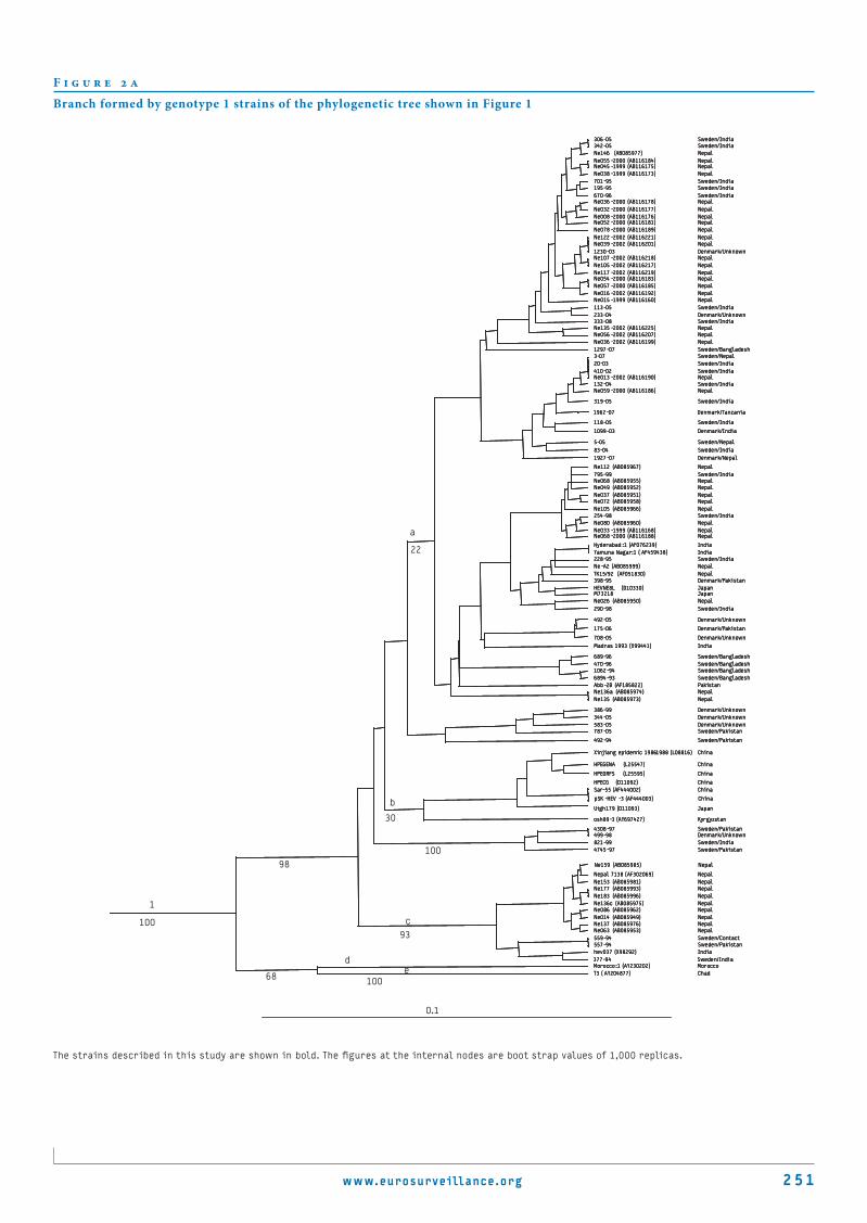
www.eurosurveillance.org 2 51
F i g u r e 2 a
Branch formed by genotype 1 strains of the phylogenetic tree shown in Figure 1
The strains described in this study are shown in bold. The figures at the internal nodes are boot strap values of 1,000 replicas.
306-05 Sweden/India342-05 Sweden/IndiaNe146 (AB085977) NepalNe055 -2000 (AB116184) NepalNe045 -1999 (AB116175) NepalNe038 -1999 (AB116173) Nepal701-95 Sweden/India195-95 Sweden/India670-96 Sweden/IndiaNe036 -2000 (AB116178) NepalNe032 -2000 (AB116177) NepalNe008 -2000 (AB116176) NepalNe052 -2000 (AB116181) NepalNe078 -2000 (AB116189) NepalNe122 -2002 (AB116221) NepalNe039 -2002 (AB116201) Nepal1230-03 Denmark/UnknownNe107 -2002 (AB116218) NepalNe105 -2002 (AB116217) NepalNe117 -2002 (AB116219) NepalNe054 -2000 (AB116183) NepalNe057 -2000 (AB116185) NepalNe016 -2002 (AB116192) NepalNe015 -1999 (AB116160) Nepal113-05 Sweden/India233-04 Denmark/Unknown333-08 Sweden/IndiaNe135 -2002 (AB116225) NepalNe056 -2002 (AB116207) NepalNe036 -2002 (AB116199) Nepal1297-07 Sweden/Bangladesh3-07 Sweden/Nepal20-03 Sweden/India410-02 Sweden/IndiaNe013 -2002 (AB116190) Nepal132-04 Sweden/IndiaNe059 -2000 (AB116186) Nepal
319-05 Sweden/India
1962-07 Denmark/Tanzania
118-05 Sweden/India
1099-03 Denmark/India
5-05 Sweden/Nepal83-04 Sweden/India1927-07 Denmark/Nepal
Ne112 (AB085967) Nepal795-99 Sweden/IndiaNe068 (AB085955) NepalNe049 (AB085952) NepalNe037 (AB085951) NepalNe072 (AB085958) NepalNe105 (AB085966) Nepal254-98 Sweden/IndiaNe080 (AB085960) NepalNe033 -1999 (AB116168) NepalNe068 -2000 (AB116188) Nepal
Hyderabad :1 (AF076239) IndiaYamuna Nagar:1 ( AF459438) India228-95 Sweden/IndiaNe -A2 (AB085999) NepalTK15/92 (AF051830) Nepal398-95 Denmark/PakistanHEVNE8L (D10330) JapanM73218 JapanNe026 (AB085950) Nepal290-98 Sweden/India
492-05 Denmark/Unknown
175-06 Denmark/Pakistan
708-05 Denmark/Unknown
Madras 1993 (X99441) India
689-96 Sweden/Bangladesh470-96 Sweden/Bangladesh1062-94 Sweden/Bangladesh6894-93 Sweden/BangladeshAbb-2B (AF185822) PakistanNe136a (AB085974) NepalNe135 (AB085973) Nepal
386-99 Denmark/Unknown344-05 Denmark/Unknown583-05 Denmark/Unknown787-05 Sweden/Pakistan
492-94 Sweden/Pakistan
Xinjiang epidemic 1986-1988 (L08816) China
HPEGENA (L25547) China
HPEORFS (L25595) China
HPECG (D11092) ChinaSar-55 (AF444002) China
pSK -HEV -3 (AF444003) China
Uigh179 (D11093) Japan
osh89-3 (AY697427) Kyrgyzstan
4308-97 Sweden/Pakistan499-98 Denmark/Unknown821-99 Sweden/India4745-97 Sweden/Pakistan
Ne159 (AB085985) Nepal
Nepal 7138 (AF302069) NepalNe153 (AB085981) NepalNe177 (AB085993) NepalNe183 (AB085996) NepalNe136c (AB085975) NepalNe086 (AB085962) NepalNe014 (AB085949) NepalNe137 (AB085976) NepalNe063 (AB085953) Nepal559-94 Sweden/Contact557-94 Sweden/Pakistanhev037 (X98292) India377-94 Sweden/IndiaMorocco :1 (AY230202) MoroccoT3 ( AY204877) Chad
a
b
c
de
10093
10068
98100
30
22
0.1
306-05 Sweden/India342-05 Sweden/IndiaNe146 (AB085977) NepalNe055 -2000 (AB116184) NepalNe045 -1999 (AB116175) NepalNe038 -1999 (AB116173) Nepal701-95 Sweden/India195-95 Sweden/India670-96 Sweden/IndiaNe036 -2000 (AB116178) NepalNe032 -2000 (AB116177) NepalNe008 -2000 (AB116176) NepalNe052 -2000 (AB116181) NepalNe078 -2000 (AB116189) NepalNe122 -2002 (AB116221) NepalNe039 -2002 (AB116201) Nepal1230-03 Denmark/UnknownNe107 -2002 (AB116218) NepalNe105 -2002 (AB116217) NepalNe117 -2002 (AB116219) NepalNe054 -2000 (AB116183) NepalNe057 -2000 (AB116185) NepalNe016 -2002 (AB116192) NepalNe015 -1999 (AB116160) Nepal113-05 Sweden/India233-04 Denmark/Unknown333-08 Sweden/IndiaNe135 -2002 (AB116225) NepalNe056 -2002 (AB116207) NepalNe036 -2002 (AB116199) Nepal1297-07 Sweden/Bangladesh3-07 Sweden/Nepal20-03 Sweden/India410-02 Sweden/IndiaNe013 -2002 (AB116190) Nepal132-04 Sweden/IndiaNe059 -2000 (AB116186) Nepal
319-05 Sweden/India
1962-07 Denmark/Tanzania
118-05 Sweden/India
1099-03 Denmark/India
5-05 Sweden/Nepal83-04 Sweden/India1927-07 Denmark/Nepal
Ne112 (AB085967) Nepal795-99 Sweden/IndiaNe068 (AB085955) NepalNe049 (AB085952) NepalNe037 (AB085951) NepalNe072 (AB085958) NepalNe105 (AB085966) Nepal254-98 Sweden/IndiaNe080 (AB085960) NepalNe033 -1999 (AB116168) NepalNe068 -2000 (AB116188) Nepal
Hyderabad :1 (AF076239) IndiaYamuna Nagar:1 ( AF459438) India228-95 Sweden/IndiaNe -A2 (AB085999) NepalTK15/92 (AF051830) Nepal398-95 Denmark/PakistanHEVNE8L (D10330) JapanM73218 JapanNe026 (AB085950) Nepal290-98 Sweden/India
492-05 Denmark/Unknown
175-06 Denmark/Pakistan
708-05 Denmark/Unknown
Madras 1993 (X99441) India
689-96 Sweden/Bangladesh470-96 Sweden/Bangladesh1062-94 Sweden/Bangladesh6894-93 Sweden/BangladeshAbb-2B (AF185822) PakistanNe136a (AB085974) NepalNe135 (AB085973) Nepal
386-99 Denmark/Unknown344-05 Denmark/Unknown583-05 Denmark/Unknown787-05 Sweden/Pakistan
492-94 Sweden/Pakistan
Xinjiang epidemic 1986-1988 (L08816) China
HPEGENA (L25547) China
HPEORFS (L25595) China
HPECG (D11092) ChinaSar-55 (AF444002) China
pSK -HEV -3 (AF444003) China
Uigh179 (D11093) Japan
osh89-3 (AY697427) Kyrgyzstan
4308-97 Sweden/Pakistan499-98 Denmark/Unknown821-99 Sweden/India4745-97 Sweden/Pakistan
Ne159 (AB085985) Nepal
Nepal 7138 (AF302069) NepalNe153 (AB085981) NepalNe177 (AB085993) NepalNe183 (AB085996) NepalNe136c (AB085975) NepalNe086 (AB085962) NepalNe014 (AB085949) NepalNe137 (AB085976) NepalNe063 (AB085953) Nepal559-94 Sweden/Contact557-94 Sweden/Pakistanhev037 (X98292) India377-94 Sweden/IndiaMorocco :1 (AY230202) MoroccoT3 ( AY204877) Chad
1
306-05 Sweden/India342-05 Sweden/IndiaNe146 (AB085977) NepalNe055 -2000 (AB116184) NepalNe045 -1999 (AB116175) NepalNe038 -1999 (AB116173) Nepal701-95 Sweden/India195-95 Sweden/India670-96 Sweden/IndiaNe036 -2000 (AB116178) NepalNe032 -2000 (AB116177) NepalNe008 -2000 (AB116176) NepalNe052 -2000 (AB116181) NepalNe078 -2000 (AB116189) NepalNe122 -2002 (AB116221) NepalNe039 -2002 (AB116201) Nepal1230-03 Denmark/UnknownNe107 -2002 (AB116218) NepalNe105 -2002 (AB116217) NepalNe117 -2002 (AB116219) NepalNe054 -2000 (AB116183) NepalNe057 -2000 (AB116185) NepalNe016 -2002 (AB116192) NepalNe015 -1999 (AB116160) Nepal113-05 Sweden/India233-04 Denmark/Unknown333-08 Sweden/IndiaNe135 -2002 (AB116225) NepalNe056 -2002 (AB116207) NepalNe036 -2002 (AB116199) Nepal1297-07 Sweden/Bangladesh3-07 Sweden/Nepal20-03 Sweden/India410-02 Sweden/IndiaNe013 -2002 (AB116190) Nepal132-04 Sweden/IndiaNe059 -2000 (AB116186) Nepal
319-05 Sweden/India
1962-07 Denmark/Tanzania
118-05 Sweden/India
1099-03 Denmark/India
5-05 Sweden/Nepal83-04 Sweden/India1927-07 Denmark/Nepal
Ne112 (AB085967) Nepal795-99 Sweden/IndiaNe068 (AB085955) NepalNe049 (AB085952) NepalNe037 (AB085951) NepalNe072 (AB085958) NepalNe105 (AB085966) Nepal254-98 Sweden/IndiaNe080 (AB085960) NepalNe033 -1999 (AB116168) NepalNe068 -2000 (AB116188) Nepal
Hyderabad :1 (AF076239) IndiaYamuna Nagar:1 ( AF459438) India228-95 Sweden/IndiaNe -A2 (AB085999) NepalTK15/92 (AF051830) Nepal398-95 Denmark/PakistanHEVNE8L (D10330) JapanM73218 JapanNe026 (AB085950) Nepal290-98 Sweden/India
492-05 Denmark/Unknown
175-06 Denmark/Pakistan
708-05 Denmark/Unknown
Madras 1993 (X99441) India
689-96 Sweden/Bangladesh470-96 Sweden/Bangladesh1062-94 Sweden/Bangladesh6894-93 Sweden/BangladeshAbb-2B (AF185822) PakistanNe136a (AB085974) NepalNe135 (AB085973) Nepal
386-99 Denmark/Unknown344-05 Denmark/Unknown583-05 Denmark/Unknown787-05 Sweden/Pakistan
492-94 Sweden/Pakistan
Xinjiang epidemic 1986-1988 (L08816) China
HPEGENA (L25547) China
HPEORFS (L25595) China
HPECG (D11092) ChinaSar-55 (AF444002) China
pSK -HEV -3 (AF444003) China
Uigh179 (D11093) Japan
osh89-3 (AY697427) Kyrgyzstan
4308-97 Sweden/Pakistan499-98 Denmark/Unknown821-99 Sweden/India4745-97 Sweden/Pakistan
Ne159 (AB085985) Nepal
Nepal 7138 (AF302069) NepalNe153 (AB085981) NepalNe177 (AB085993) NepalNe183 (AB085996) NepalNe136c (AB085975) NepalNe086 (AB085962) NepalNe014 (AB085949) NepalNe137 (AB085976) NepalNe063 (AB085953) Nepal559-94 Sweden/Contact557-94 Sweden/Pakistanhev037 (X98292) India377-94 Sweden/IndiaMorocco :1 (AY230202) MoroccoT3 ( AY204877) Chad

2 5 2 www.eurosurveillance.org
F i g u r e 2 b
Branch formed by the genotype 3 strains forming branch 3-II shown in Figure 1
The human genotype 3 strains are underlined; those described in this study are in addition shown in bold. The HEV sequences from domestic pigs described in this study are shown in bold italic. The figures at the internal nodes are boot strap values of 1,000 replicas.
Swine 25604 (DQ315757) Spain
Swine 65903 (DQ315748) Spain
Swine 24404 (DQ315756) Spain
Swine 63004 (DQ315765) Spain
Swine 60604 (DQ315760) Spain
Swine 70603 (DQ315751) Spain
Swine 60504 (DQ315759) Spain
Swine 60904 (DQ315763) Spain
Swine 67204 (DQ315770) SpainSwine 69003 (DQ315750) Spain
Swine 60804 (DQ315762) Spain
Swine 12804 (DQ315752) Spain
Swine 62904 (DQ315764) Spain
Swine 63104 (DQ315766) Spain
Swine 65103 (DQ315745) Spain
Swine 60304 (DQ315758) Spain
Swine 65203 (DQ315746) SpainSwine 15104 (DQ315753) Spain
Swine 15204 (DQ315754) Spain
Swien FRSPP05 (EF050797) France
Swine SpswCV2 (AY323506) Spain
Swine SpswfCV5 (DQ093566) Spain
Swine SPSWSCV17 (DQ141126) Spain
Swine SPSWSCV14 (DQ141123) Spain
Swine SpswsCV4 (DQ093565) Spain
Swine SPSWFCV31 (EF523417) Spain
Human 1459 -06 Sweden/Spain
Swine SPSWFCV32 (EF523418) Spain
Swine SpswfCV3 (DQ093564) Spain
Swine SpswmCV6 (DQ093567) Spain
Swine NLSW97 (AF336296) The Netherlands
Swine NLSW82 (AF336294) The Netherlands
Swine NLSW15 (AF332620) The Netherlands
Swine FR79 (EF494704) France
Swine NLSW28 (AF336292) The Netherlands
Swine NLSW91 (AY032759) The Netherlands
Swine NLSW85 (AF336295) The Netherlands
Swine NLSW76 (AY032757) The Netherlands
Swine NLSW50 (AY032758) The Netherlands
Human 525 -07 Denmark/Spain
Swine SpswmCV7 ( DQ093568) Spain
Swine SPSWMCV11 (DQ141120) Spain
Swine 295 -08 Denmark
Swine 18004 (DQ315755) Spain
Swine 294 -07 Denmark
Swine 292 -07 Denmark
Swine 298 -07 Sweden
Human 26 -02 Sweden
Swine 284 -07 Sweden
Human 560 -05 Sweden
Swine 326 -07 Sweden
Swine 292 -08 Denmark
Human 392 -08 Denmark
Swine 289 -08 Denmark
Swine 288 -08 Denmark
Swine 291 -08 Denmark
Swine 290 -08 Denmark
Swine 296 -08 Denmark
Swine 293 -08 Denmark
Swine 325 -07 Sweden
Swine 63504 (DQ315768) Spain
Swine 63604 (DQ315769) SpainSwine 63404 (DQ315767) Spain
Swine swX07-E1 (EU360977) Sweden
Swine 667 -05 Sweden
Swine 331 -07 Sweden
Swine 328 -07 Sweden
Human 38 -08 Denmark
Swine SPSWFCV2 (EF523412) Spain
Swine swJ8-10 (AB094235) Japan
Swine P354/1/02 ( AF503511) UK
SwineFR18 (EF494700) France
Swine swJ8-8 (AB094233) Japan
Swine swJ8-5 (AB248521) Japan
Swine swJ8-9 (AB094234) Japan
Swine swJ8-1 (AB094226) Japan
Swine swJ8-6 (AB094231) Japan
Swine swJ8-3 (AB094228) Japan
Swine swJ5-1 (AB094216) Japan
Swine swJ12-1(AB094250) Japan
Swine swJ12-3 (AB094252) Japan
Human HE -JA26 (AB194284) Japan
Swine 670 -05 Sweden
Swine 299 -07 Sweden
Swine 1970 -07 Sweden
Swine P143/11/02 ( AF503512) UK
Swine swJ791 (AB073911) Japan
Human HE -JA04 -1911 (AB248520) Japan
Human HEV -Sendai (AB093535) Japan
Swine swMN06 -C1056 (AB290313) Mongolia
Swine swMN06 -C1058 (AB290119) Mongolia
Swine swMN06 -C1054 (AB290115) Mongolia
Swine Osh 205 (AF455784) Kyrgyzstan
Swine UIAS268 (DQ061078) Russia
3 - II
e
f
g
67 66
100
68
53
48
38
4547
Swine 25604 (DQ315757) Spain
Swine 65903 (DQ315748) Spain
Swine 24404 (DQ315756) Spain
Swine 63004 (DQ315765) Spain
Swine 60604 (DQ315760) Spain
Swine 70603 (DQ315751) Spain
Swine 60504 (DQ315759) Spain
Swine 60904 (DQ315763) Spain
Swine 67204 (DQ315770) SpainSwine 69003 (DQ315750) Spain
Swine 60804 (DQ315762) Spain
Swine 12804 (DQ315752) Spain
Swine 62904 (DQ315764) Spain
Swine 63104 (DQ315766) Spain
Swine 65103 (DQ315745) Spain
Swine 60304 (DQ315758) Spain
Swine 65203 (DQ315746) SpainSwine 15104 (DQ315753) Spain
Swine 15204 (DQ315754) Spain
Swien FRSPP05 (EF050797) France
Swine SpswCV2 (AY323506) Spain
Swine SpswfCV5 (DQ093566) Spain
Swine SPSWSCV17 (DQ141126) Spain
Swine SPSWSCV14 (DQ141123) Spain
Swine SpswsCV4 (DQ093565) Spain
Swine SPSWFCV31 (EF523417) Spain
Human 1459 -06 Sweden/Spain
Swine SPSWFCV32 (EF523418) Spain
Swine SpswfCV3 (DQ093564) Spain
Swine SpswmCV6 (DQ093567) Spain
Swine NLSW97 (AF336296) The Netherlands
Swine NLSW82 (AF336294) The Netherlands
Swine NLSW15 (AF332620) The Netherlands
Swine FR79 (EF494704) France
Swine NLSW28 (AF336292) The Netherlands
Swine NLSW91 (AY032759) The Netherlands
Swine NLSW85 (AF336295) The Netherlands
Swine NLSW76 (AY032757) The Netherlands
Swine NLSW50 (AY032758) The Netherlands
Human 525 -07 Denmark/Spain
Swine SpswmCV7 ( DQ093568) Spain
Swine SPSWMCV11 (DQ141120) Spain
Swine 295 -08 Denmark
Swine 18004 (DQ315755) Spain
Swine 294 -07 Denmark
Swine 292 -07 Denmark
Swine 298 -07 Sweden
Human 26 -02 Sweden
Swine 284 -07 Sweden
Human 560 -05 Sweden
Swine 326 -07 Sweden
Swine 292 -08 Denmark
Human 392 -08 Denmark
Swine 289 -08 Denmark
Swine 288 -08 Denmark
Swine 291 -08 Denmark
Swine 290 -08 Denmark
Swine 296 -08 Denmark
Swine 293 -08 Denmark
Swine 325 -07 Sweden
Swine 63504 (DQ315768) Spain
Swine 63604 (DQ315769) SpainSwine 63404 (DQ315767) Spain
Swine swX07-E1 (EU360977) Sweden
Swine 667 -05 Sweden
Swine 331 -07 Sweden
Swine 328 -07 Sweden
Human 38 -08 Denmark
Swine SPSWFCV2 (EF523412) Spain
Swine swJ8-10 (AB094235) Japan
Swine P354/1/02 ( AF503511) UK
SwineFR18 (EF494700) France
Swine swJ8-8 (AB094233) Japan
Swine swJ8-5 (AB248521) Japan
Swine swJ8-9 (AB094234) Japan
Swine swJ8-1 (AB094226) Japan
Swine swJ8-6 (AB094231) Japan
Swine swJ8-3 (AB094228) Japan
Swine swJ5-1 (AB094216) Japan
Swine swJ12-1(AB094250) Japan
Swine swJ12-3 (AB094252) Japan
Human HE -JA26 (AB194284) Japan
Swine 670 -05 Sweden
Swine 299 -07 Sweden
Swine 1970 -07 Sweden
Swine P143/11/02 ( AF503512) UK
Swine swJ791 (AB073911) Japan
Human HE -JA04 -1911 (AB248520) Japan
Human HEV -Sendai (AB093535) Japan
Swine swMN06 -C1056 (AB290313) Mongolia
Swine swMN06 -C1058 (AB290119) Mongolia
Swine swMN06 -C1054 (AB290115) Mongolia
Swine Osh 205 (AF455784) Kyrgyzstan
Swine UIAS268 (DQ061078) Russia

www.eurosurveillance.org 2 53
In our study HEV RNA was detected in 67% of sera sampled within three weeks after onset of illness from patients with this information known. This is in accordance with an HEV RNA detection rate of 56-59% in sera sampled 15 to 20 days after onset in Chinese patients with hepatitis E [23]. However, in our study HEV RNA was also detected in 18% of sera from patients with anti-HEV IgG only, which is an unexpectedly high frequency and has not been described earlier, since the presence of IgG in the absence of detectable IgM is considered a marker of past infection. Anti-HEV IgG may persist for several years after infection, but whether lifelong immunity is conferred remains uncertain [24]. There is only one serotype of HEV, but it is not known if reinfections induce IgG response only or if also the IgM levels become elevated. It is also not known if there is a viremic phase during a reinfection when the level of IgG is low and the immune response has been elicited towards another HEV genotype. Since most of the patients in this study were from Scandinavia and it is known that there is a rather high seroprevalence against HEV in Sweden and Denmark [16-18] it is possible that individuals with low level antibodies towards genotype 3 when infected with genotype 1 developed disease and viraemia with anti-HEV IgG elevation only.
In this study the only case infected by genotype 1 in Europe was epidemiologically linked to a case from Pakistan. All other patients infected in Europe were infected with genotype 3 strains. These individuals were mainly males and were 20–25 years older than the cases infected by genotype 1. This is in accordance with previous recent reports from the UK, France and Germany showing that genotype 3 is the autochthonous genotype of HEV, which gives disease mainly in males over the age of 50 [15,25].
Since autochthonous hepatitis E in humans in Europe has been caused by strains with 99–100% identity to European swine HEV [26], the suspected route of infection is through direct contact with pigs or other infected mammals or by foodborne transmission. Foodborne transmission was described in Japan in patients infected after consumption of undercooked pig liver or meat from wild boar or deer [27-29]. This route of infection may occur also in Europe since HEV has been detected in commercial pig liver sold in groceries and there is a high HEV seroprevalence in many pig herds [30,31]. Phylogenetic analysis of the genotype 3 strains revealed that most Asian and American strains belong to one major clade and that the European strains belong to another clade. There were also geographical clades of the genotype 3 strains, and strains from patients infected in Sweden and Denmark were similar to strains from Swedish and Danish piglets, while patients infected in Spain had genotype 3 strains similar to those of Spanish pigs. This pattern has previously not been described and enables a possible identification of the country of origin of the strain infecting the patient. This in turn may help to trace the source of infection and to identify a possible food item from that country.
Antibodies to HEV have been shown to be prevalent among blood donors and apart from the faecal-oral and foodborne route HEV may be transmitted also through blood or blood products as has been reported from Japan, Saudi Arabia, France, and the UK [32-34]. HEV has also been reported to be transmitted by organ transplantations [35]. Some organ-recipients have developed chronic hepatitis E infection [10]. In our study the viraemia lasted for a relatively long period in most patients. Thus, transmission of HEV by blood or blood products may theoretically also occur in Sweden. Bloodborne transmission may also occur through injecting drug use. In our study six of the patients were IDUs among those
with HEV IgG but no detectable HEV IgM or RNA. More than 60% of Swedish IDUs have anti-HEV [17], which further supports the conclusion that hepatitis E may be transmitted parenterally in this cohort of individuals. The high seroprevalence indicates that most probably IDUs are frequently reinfected with HEV. The IDUs in our study were investigated for HEV infection due to elevated transaminases. However, genotype 3 reinfections have been shown not to induce elevation of liver enzymes or detectable HEV IgM among patients on hemodialysis in Japan [36], but HEV RNA was not looked for in these patients. It is thus not known if reinfections with genotype 3 cause viraemia. Lack of HEV RNA in the sera from the anti-HEV IgG positive IDUs may either indicate that reinfection with genotype 3 does not give rise to viraemia, or that a continuous low-level exposure to HEV keeps the immune status at a level preventing reinfection with HEV, or that there is a long lasting immunity with detectable HEV IgG.
Hepatitis E in developed countries has a natural history that
differs from classical hepatitis E in endemic areas. In the study presented here we have shown that HEV genotype 3 strains are indigenous in Sweden and Denmark, with high similarity between strains infecting humans and pigs. Prospective studies are needed to define the incidence of autochthonous infections in Scandinavia. It is also important to determine whether and how the spread occurs from pigs to humans and if there are other animal sources for zoonotic transmission of HEV, since genotype 3 appears to be a primarily animal virus that crosses the species barrier. In conclusion, hepatitis E should thus be considered in the diagnosis of patients with acute hepatitis, regardless of travel history.
AcknowledgementsThis work was supported by the European Commission DG Research Quality of Life Program, 6th Framework (EVENT, SP22-CT-2004-502571) and by the European Commission DG Research Biotechnology, Agriculture and Food Research programme, 6th Framework Programme (Pathogen Combat, Food-CT-2005-007081), the European Commission DG Research Network for the Prevention and Control of Zoonoses, (MedVetNet, FOOD-CT-2004-506122) and the Sandberg Foundation, Sweden.
References
1. Balayan MS, Andjaparidze AG, Savinskaya SS, Ketiladze ES, Braginsky DM, Savinov AP, et. al. Evidence for a virus in non-A, non-B hepatitis transmitted via the fecal-oral route. Intervirology. 1983;20(1):23-31.
2. Lu L, Li C, Hagedorn CH. Phylogenetic analysis of global hepatitis E virus sequences: genetic diversity, subtypes and zoonosis. Rev Med Virol. 2006;16(1):5-36.
3. Shukla P, Chauhan UK, Naik S, Anderson D, Aggarwal R. Hepatitis E virus infection among animals in northern India: an unlikely source of human disease. J Viral Hepat. 2007;14(5):310-7.
4. Schlauder GG, Dawson GJ, Erker JC, Kwo PY, Knigge MF, Smalley DL, et al. The sequence and phylogenetic analysis of a novel hepatitis E virus isolated from a patient with acute hepatitis reported in the United States. J Gen Virol. 1998;79(Pt3):447-56.
5. Takahashi K, Iwata K, Watanabe N, Hatahara T, Ohta Y, Baba K, et al. Full-genome nucleotide sequence of a hepatitis E virus strain that may be indigenous to Japan. Virology. 2001;287(1):9-12.
6. Mansuy JM, Peron JM, Abravanel F, Poirson H, Dubois M, Miedouge M, et al. Hepatitis E in the south west of France in individuals who have never visited an endemic area. J Med Virol. 2004;74(3):419-24.
7. Ijaz S, Arnold E, Banks M, Bendall RP, Cramp ME, Cunningham R, et al. Non-travel-associated hepatitis E in England and Wales: demographic, clinical, and molecular epidemiological characteristics. J Infect Dis. 2005;192(7):1166-72.
8. Hamid SS, Atiq M, Shehzad F, Yasmeen A, Nissa T, Salam A, et al. Hepatitis E virus superinfection in patients with chronic liver disease. Hepatology. 2002;36(2):474-8.

2 5 4 www.eurosurveillance.org
9. Khuroo MS, Kamili S. Aetiology, clinical course and outcome of sporadic acute viral hepatitis in pregnancy. J Viral Hepat. 2003;10(1):61-9.
10. Haagsma EB, van den Berg AP, Porte RJ, Benne CA, Vennema H, Reimerink JH, et al. Chronic hepatitis E virus infection in liver transplant recipients. Liver Transpl. 2008;14(4):547-53.
11. Dalton HR, Thurairajah PH, Fellows HJ, Hussaini HS, Mitchell J, Bendall R, et al. Autochthonous hepatitis E in southwest England. J Viral Hepat. 2007;14(5):304-9.
12. Widdowson MA, Jaspers WJ, van der Poel WH, Verschoor F, de Roda Husman AM, Winter HL, et al. Cluster of cases of acute hepatitis associated with hepatitis E virus infection acquired in the Netherlands. Clin Infect Dis. 2003;36(1):29-33.
13. Dalton HR, Fellows HJ, Gane EJ, Wong P, Gerred S, Schroeder B, et al. Hepatitis E in New Zealand. J Gastroenterol Hepatol. 2007;22(8):1236-40.
14. Mizuo H, Suzuki K, Takikawa Y, Sugai Y, Tokita H, Akahane Y, et al. Polyphyletic strains of hepatitis E virus are responsible for sporadic cases of acute hepatitis in Japan. J Clin Microbiol. 2002;40(9):3209-18.
15. Mansuy JM, Abravanel F, Miedouge M, Mengelle C, Merviel C, Dubois M, et al. Acute hepatitis E in south-west France over a 5-year period. J Clin Virol. 2009;44(1):74-7.
16. Christensen PB, Engle RE, Hjort C, Homburg KM, Vach W, Georgsen J, et al. Time trend of the prevalence of hepatitis E antibodies among farmers and blood donors: a potential zoonosis in Denmark. Clin Infect Dis. 2008;47(8):1026-31.
17. Sylvan SP. The high rate of antibodies to hepatitis E virus in young, intravenous drug-abusers with acute hepatitis B-virus infection in a Swedish community: a study of hepatitis markers in individuals with intravenously or sexually acquired hepatitis B-virus infection. Scand J Infect Dis. 1998;30(4):429-30.
18. Olsen B, Axelsson-Olsson D, Thelin A, Weiland O. Unexpected high prevalence of IgG-antibodies to hepatitis E virus in Swedish pig farmers and controls. Scand J Infect Dis. 2006;38(1):55-8.
19. Zhai L, Dai X, Meng J. Hepatitis E virus genotyping based on full-length genome and partial genomic regions. Virus Res. 2006;120(1-2):57-69.
20. Meng XJ, Purcell RH, Halbur PG, Lehman JR, Webb DM, Tsareva TS, et al. A novel virus in swine is closely related to the human hepatitis E virus. Proc Natl Acad Sci U S A. 1997;94(18):9860-5.
21. Felsenstein J. PHYLIP: Phylogeni inference package; version 3.52c. Seattle (WA, US): University of Washington; 1993.
22. Söderström A, Hammas B, Magnius L, Norkrans G. [A secondary case of hepatitis E. Since 1990 only one of the 17 imported cases was transmitted] Lakartidningen. 1995;92(50):4807-8.
23. Zhao ZY, Ruan B, Shao H, Chen ZJ, Liu SL. Detection of hepatitis E virus RNA in sera of patients with hepatitis E by polymerase chain reaction. Hepatobiliary Pancreat Dis Int. 2007;6(1):38-42.
24. Aggarwal R, Krawczynski K. Hepatitis E: an overview and recent advances in clinical and laboratory research. J Gastroenterol Hepatol. 2000;15(1):9-20.
25. Dalton HR, Stableforth W, Thurairajah P, Hazeldine S, Remnarace R, Usama W, et al. Autochthonous hepatitis E in Southwest England: natural history, complications and seasonal variation, and hepatitis E virus IgG seroprevalence in blood donors, the elderly and patients with chronic liver disease. Eur J Gastroenterol Hepatol. 2008; 20(8):784-90.
26. Herremans M, Vennema H, Bakker J, van der Veer B, Duizer E, Benne CA, et al. Swine-like hepatitis E viruses are a cause of unexplained hepatitis in the Netherlands. J Viral Hepat. 2007;14(2):140-6.
27. Yazaki Y, Mizuo H, Takahashi M, Nishizawa T, Sasaki N, Gotanda Y, et al. Sporadic acute or fulminant hepatitis E in Hokkaido, Japan, may be food-borne, as suggested by the presence of hepatitis E virus in pig liver as food. J Gen Virol. 2003;84(Pt9):2351-7.
28. Michitaka K, Takahashi K, Furukawa S, Inoue G, Hiasa Y, Horiike N, et al. Prevalence of hepatitis E virus among wild boar in the Ehime area of western Japan. Hepatol Res. 2007;37(3):214-20.
29. Matsuura Y, Suzuki M, Yoshimatsu K, Arikawa J, Takashima I, Yokoyama M, et al. Prevalence of antibody to hepatitis E virus among wild sika deer, Cervus nippon, in Japan. Arch Virol. 2007;152(7):1375-81.
30. Bouwknegt M, Lodder-Verschoor F, van der Poel WH, Rutjes SA, de Roda Husman AM. Hepatitis E virus RNA in commercial porcine livers in The Netherlands. J Food Prot. 2007;70(12):2889-95.
31. Meng XJ, Dea S, Engle RE, Friendship R, Lyoo YS, Sirinarumitr T, et al. Prevalence of antibodies to the hepatitis E virus in pigs from countries where hepatitis E is common or is rare in the human population. J Med Virol. 1999;59(3):297-302.
32. Matsubayashi K, Kang JH, Sakata H, Takahashi K, Shindo M, Kato M, et al. A case of transfusion-transmitted hepatitis E caused by blood from a donor infected with hepatitis E virus via zoonotic food-borne route. Transfusion. 2008;48(7):1368-75.
33. Boxall E, Herborn A, Kochethu G, Pratt G, Adams D, Ijaz S, et al. Transfusion-transmitted hepatitis E in a ‘nonhyperendemic’ country. Transfus Med. 2006;16(2):79-83.
34. Colson P, Coze C, Gallian P, Henry M, De Micco P, Tamalet C. Transfusion-associated hepatitis E, France. Emerg Infect Dis. 2007;13(4):648-9.
35. Kamar N, Mansuy JM, Esposito L, Legrand-Abravanel F, Peron JM, Durand D, et al. Acute hepatitis and renal function impairment related to infection by hepatitis E virus in a renal allograft recipient. Am J Kidney Dis. 2005;45(1):193-6.
36. Mitsui T, Tsukamoto Y, Yamazaki C, Masuko K, Tsuda F, Takahashi M, et al. Prevalence of hepatitis E virus infection among hemodialysis patients in Japan: evidence for infection with a genotype 3 HEV by blood transfusion. J Med Virol. 2004;74(4):563-72.
www.eurosurveillance.org 2 5 5
R esearch ar ti cles
C a n t h e S w e d i S h n e w va r i a n t o f C h l a m y d i a t r a C h o m at i S ( n v Ct ) b e d e t e C t e d b y U K n e QaS pa r t i C i pa n t S f r o m S e v e n t e e n e U r o p e a n C o U n t r i e S a n d f i v e a d d i t i o n a l C o U n t r i e S / r e g i o n S i n 2009?
M Unemo ([email protected])1, A Rossouw2, V James2, C Jenkins2
1. National Reference Laboratory for Pathogenic Neisseria, Department of Laboratory Medicine, Clinical Microbiology, Örebro University Hospital, Örebro, Sweden
2. UK National External Quality Assessment Scheme (UK NEQAS) for Microbiology, Health Protection Agency Centre for Infection, London, United Kingdom
This article was published on 14 May 2009. Citation style for this article: Unemo M, Rossouw A, James V, Jenkins C. Can the Swedish new variant of Chlamydia trachomatis (nvCT) be detected by UK NEQAS participants from seventeen European countries and five additional countries/regions in 2009?. Euro Surveill. 2009;14(19):pii=19206. Available online: http://www.eurosurveillance.org/ViewArticle.aspx?ArticleId=19206
In 2006, a new variant of Chlamydia trachomatis (nvCT) was reported in Sweden. The nvCT contains a deletion that includes the targets for the C. trachomatis genetic diagnostic single-target systems from Roche Diagnostics and Abbott Laboratories. Roche and Abbott have now developed certified dual-target assays that can detect the nvCT. This study examined the nucleic acid amplification tests (NAATs) currently used (in 2009) for C. trachomatis detection in laboratories from 17 European countries and five countries/regions outside Europe that are participating in the United Kingdom (UK) National External Quality Assessment Service (NEQAS). It further examined changes in these laboratories’ testing strategy during the period from 2006 to 2009, and their performance regarding nvCT detection. A UK NEQAS blinded nvCT specimen was distributed to all 283 participating laboratories, which were asked to analyse the specimen according to their routine C. trachomatis diagnostic protocols for endocervical swabs. BD ProbeTec was the most commonly used NAAT, followed by Cobas Amplicor, Cobas TaqMan, and Aptima. From 2006 to 2009, the use of Cobas Amplicor, which does not detect the nvCT, decreased, but it was still used by 22% (n=57) of responding participants in 59% of the countries, 54 of these 57 used it as first assay. Virtually all of the other participants detected the nvCT correctly. Laboratories using commercial or in house NAATs that do not detect the nvCT are encouraged to carefully monitor their C. trachomatis incidence, participate in effective internal and external quality assurance and controls schemes, and to consider changing their testing system.
IntroductionIn most middle- and high-resource settings nucleic acid
amplification tests (NAATs) are the most commonly used tests for rapid, highly sensitive and specific detection of Chlamydia trachomatis.
In 2006, a new variant of C. trachomatis (nvCT), which contains a 377 bp deletion in the cryptic plasmid, was reported in Sweden [1,2]. This deletion includes the genetic targets for commercially available single-target systems that were at the time used worldwide, namely the Amplicor C. trachomatis/Neisseria
gonorrhoeae (CT/NG) test, the Cobas Amplicor CT/NG test, and the Cobas TaqMan CT/NG test (Roche Diagnostics), as well as the RealTime CT/NG test (Abbott Laboratories). Subsequently, nvCT was identified in high proportions (10-65%) in most counties across Sweden. The affected NAATs were used in two thirds of the Swedish counties, and many thousands of false negative samples were reported [3-5]. Previous studies, using ompA gene sequencing and a new multilocus sequence typing (MLST), showed that the nvCT seems to be of clonal nature, belonging to genotype E and displaying a unique MLST sequence type [3,5]. Other commercial genetic diagnostic systems that are internationally available, such as a) the BD ProbeTec ET (Becton Dickinson), b) the Aptima CT and Aptima Combo 2 (Gen-Probe), c) the artus C. trachomatis PCR Kit (Qiagen), d) the artus C. trachomatis Plus PCR Kit (Qiagen), and e) the CHLAMYDIA tr. Q - PCR Alert Kit (Nanogen), were able to identify the nvCT; these NAATs target(s) are a) the cryptic plasmid (outside the deletion), b) specific 23S and 16S rRNA sequences, c) the ompA gene, d) both the ompA gene and the cryptic plasmid (outside the deletion), and e) the cryptic plasmid (outside the deletion), respectively.
Both Abbott Laboratories and Roche Diagnostics have now designed new sensitive and specific dual-target assays, namely the Abbott RealTime CT/NG (Abbott; new version, CE mark-certified in January 2008) that targets another sequence of the cryptic plasmid in addition to the sequence affected by the nvCT deletion, and the Cobas TaqMan CT v2.0 (Roche; CE mark-certified in June 2008) that detects the chromosomal ompA gene in addition to the sequence affected by the nvCT deletion [4]. Despite active surveillance and a number of studies performed in many countries [6], only sporadic cases of nvCT have so far been reported outside Scandinavia, e.g. in France [7], Ireland [8], and Scotland [9].
The aims of this report were to describe the NAATs currently used (in 2009) for C. trachomatis detection in laboratories from European countries (n=17) and countries/regions outside Europe (n=5) that are participating in the United Kingdom (UK) National External Quality Assessment Service (NEQAS). It further aimed to

2 5 6 www.eurosurveillance.org
identify changes in these laboratories’ testing strategy during the period from 2006 to 2009, and to highlight their performance regarding detection of the nvCT.
Materials and MethodsThe UK NEQAS distributes clinically relevant and educational
specimens for external quality assessment (EQA). In the UK NEQAS scheme for C. trachomatis detection (‘Molecular’), at present there are 283 participating laboratories (274 laboratories from 17 European countries and nine laboratories from five countries/regions outside Europe). However, most of the participating laboratories are in the UK (see Table 1). For surveillance and educational purposes, a blinded EQA specimen (Specimen 9119 in UK NEQAS Distribution 2402, issued in January 2009, as well as blinded specimens of three wildtype C. trachomatis strains) containing the nvCT, 1.67-3x10⁴ elementary bodies per ml of reconstituted lyophilised specimen, was prepared as previously described [10]. Vacuum integrity and moisture content (<2%) of the freeze-dried specimen were validated and approved before distribution to all 283 participants. The laboratories were requested to reconstitute the specimen in molecular grade water and analyse the specimen according to their routine protocols for detecting C. trachomatis from an endocervical swab.
Results Nucleic acid amplification tests (NAATs) used in 2009 for C. trachomatis diagnostics and changes in testing strategy during 2006-2009Of the 283 laboratories participating in the scheme, 261
(92.2%) returned results on the nvCT specimen. In 2009, BD ProbeTec was the most commonly used main NAAT (39.5% of laboratories), followed by Cobas Amplicor (20.7%), Cobas TaqMan (16.1%), and Aptima (5.7%) (Table 1).
During the period from 2006 to 2009, the use of Cobas Amplicor decreased. However, it was still used as main NAAT in 2009 by 54 participants in 13 (59.1%) of the countries. In contrast, the numbers of laboratories using Cobas TaqMan, Abbott, and Nanogen Q-PCR have increased (Figure 1).
Detection of the Swedish new variant of C. trachomatis (nvCT) The reporting laboratories used more than seven different
commercial assays, in house single-target (n=7) or multi-target (n=3) real-time PCR assays, or did not specify their method (n=8). Twelve of the laboratories used two different assays (Table 2). However, specific testing algorithms used for routine diagnostics in these laboratories were not accessible.
T a b l e 1
Countries and laboratories, including the main diagnostic assay used, participating in the UK NEQAS scheme for molecular detection of Chlamydia trachomatis in 2009
CountryNo. of
participatinglaboratories
CobasAmplicor(Roche)a
CobasTaqMan v2.0
(Roche)
Abbott RealTime (Abbott)
BD ProbeTec(Becton
Dickinson)
Aptima Combo 2
(Gen-Probe)
NanogenC. tr. Q-PCR
Alert(Nanogen)
artus(Qiagen)b
In house single-target
real-time PCRc
In house multi-
target real-time PCRc
Unspecified method
Not returning results
Austria 5 2 - - 1 - - 1 - - 1 -
Belgium 5 1 - - 2 - - 2 - - - -
Croatia 1 1 - - - - - - - - - -
Denmark 4 - 1 - 1 - - - 1 - - 1
Finland 5 - 2 - 1 - - - - - - 2
Germany 1 - - - - - - - - - - 1
Greece 1 - - - - - - - - - - 1
Hong Kong 2 1 - - - - - - - - - 1
Ireland 10 3 2 1 1 1 - - - 1 - 1
Israel 3 3 - - - - - - - - - -
Italy 42 6 3 3 7 - 7 2 2 - 4 8
Kuwait 1 - - - - - - - - - - 1
Macao 1 1 - - - - - - - - - -
Malta 1 1 - - - - - - - - - -
Netherlands 7 2 - - 3 1 - - 1 - - -
Norway 5 - 2 - 2 - - - - - 1 -
Portugal 5 - - 1 2 - 1 - - - 1 -
Slovenia 2 - 1 - - - - - 1 - - -
South Africa
2 1 - - - - - - - - - 1
Sweden 8 - 1 1 4 - - - - - - 2
Switzerland 14 10 - 2 1 - - - 1 - - -
United Kingdom
158 22 30 4 78 13 3 1 1 2 1 3
Total 283 54 42 12 103 15 11 6 7 3 8 22
a A few laboratories used Amplicor CT/NG (Roche). However, it was not possible to determine the exact number.b Both artus C. trachomatis PCR Kit (omp1 gene; Qiagen) and artus C. trachomatis Plus PCR Kit (ompA gene and cryptic plasmid; Qiagen) were used. However, it was not possible to determine how many laboratories used which kit.c Details about in house assays were often reported and could not be accessed retrospectively.

www.eurosurveillance.org 2 57
Eighty percent (n=209) of the laboratories correctly reported the presence of C. trachomatis in the nvCT specimen (Figure 2). The majority (94%, 51/54) of the laboratories using Cobas Amplicor as their first assay reported a false negative result, as expected. However, one laboratory using Cobas Amplicor, an assay that can not detect the nvCT, reported a false positive result. Furthermore, two additional laboratories reported an equivocal result: They used Cobas Amplicor, which was negative, but to confirm their results used Aptima and Cobas TaqMan, which detected the nvCT correctly. The reasons for using this double testing strategy were not available. Presumably it does not reflect their routine diagnostics of all C. trachomatis samples. Furthermore, one laboratory using the Abbott system reported a negative result. All remaining laboratories reported a positive result (Figure 2).
Discussion and conclusionsThis report highlights the NAATs currently used (in 2009)
for C. trachomatis detection in laboratories from 22 countries participating in the UK NEQAS scheme, alterations in their testing strategy during the period from 2006 to 2009, and their performance regarding detection of the nvCT.
Most of the laboratories (94%) using Cobas Amplicor, the second most common assay, as their first assay, reported an expected false negative result for the nvCT. However, two laboratories reported an equivocal result, i.e. negative with the Cobas Amplicor, but positive with an additional assay that detected the nvCT. One laboratory using the Cobas Amplicor assay reported a false positive result. This result suggests incorrect reporting either of the type of assay that was used or of the result, misinterpretation of the results, mix-up of specimens or contamination with other C. trachomatis strain or PCR amplicon.
One laboratory that was using the Abbott system and should have detected the nvCT, reported a negative result. A possible explanation could be that the older RealTime CT/NG test, the single-target assay that does not detect the nvCT, was used instead of the new Abbott RealTime CT/NG dual-target test. It is unlikely to reflect a sensitivity issue because the nvCT specimen contained a high number of elementary bodies per ml.
All other assays including the new Abbott RealTime CT and Roche Cobas TaqMan v2.0 performed well.
Laboratories that are still using Amplicor CT/NG, Cobas Amplicor CT/NG, and in house NAATs targeting the nvCT deletion in the cryptic plasmid are encouraged to monitor their C. trachomatis incidence in order to quickly identify unexplained significant declines in the normal or estimated local incidence and to alert reference centres about it. In addition, they are strongly encouraged to consider the feasibility of changing to a diagnostic method that can detect the nvCT, because using an additional NAAT on all negative samples is not feasible in the longer term.
Ideally, clinicians submitting samples to these laboratories should be objectively informed about the problem to diagnose the nvCT. An unexplained significant decline in incidence may be due to the emergence of nvCT. However, as other undetected mutants may emerge, monitoring of the incidence rate and participation of all laboratories in effective internal and external quality assurance and controls schemes are crucial.
F i g u r e 1
Diagnostic assays (main NAAT) used by participating laboratories in the UK NEQAS scheme for molecular detection of Chlamydia trachomatis from 2006 to 2009*
*The total number of participating laboratories and laboratories returning results (in parenthesis) was 221 (100%), 263 (95.8%), 278 (100%), and 283 (92.2%), in 2006, 2007, 2008, and 2009, respectively.NAAT: nucleic acid amplification test; NEQAS: National External Quality Assessment Service.
0
5
10
15
20
25
30
35
40
45
Coba
s Am
plicor
(Roc
he)
Coba
s TaqM
an (R
oche
)
RealTime
(Abb
ott)
BD Pro
beTec (BD)
Aptima
(Gen
-Pro
be)
Nano
gen Q-
PCR (Nan
ogen
)
artu
s (Qiage
n)
In hou
se PCR
Unsp
ecified met
hod
NAATs used for C. trachomatis diagnostics
Perc
enta
ge o
f pa
rtic
ipan
ts
Jan-06Jan-07Jan-08Jan-09
T a b l e 2
Combination of assays used in laboratories reporting using more than one assay for molecular detection of Chlamydia trachomatis in the UK NEQAS scheme in 2009
First assay Second assay No. of laboratories
Cobas Amplicor (Roche) Cobas TaqMan v2.0 (Roche) 3
Cobas Amplicor (Roche) Aptima Combo 2 (Gen-Probe) 1
Cobas TaqMan v2.0 (Roche) Cobas Amplicor (Roche) 1
Cobas TaqMan v2.0 (Roche) Aptima Combo 2 (Gen-Probe) 1
Cobas TaqMan v2.0 (Roche) Nanogen C. tr. Q-PCR Alert (Nanogen) 1
BD ProbeTec (Becton Dickinson) Cobas Amplicor (Roche) 1
BD ProbeTec (Becton Dickinson) Aptima Combo 2 (Gen-Probe) 1
BD ProbeTec (Becton Dickinson) In house single-target real-time PCR 1
In house single-target real-time PCR artus (Qiagen) 1
Unspecified assay Cobas Amplicor (Roche) 1

2 5 8 www.eurosurveillance.org
Based on the present study, it is obvious that a substantial number of laboratories in many European countries can still not detect the nvCT. However, the study only included laboratories participating in the UK-NEQAS scheme and thus gives a far from complete picture regarding the situation in the whole of Europe. The coverage in many participating countries was limited and it cannot be excluded that by selecting laboratories that are members of EQAS such as UK NEQAS a bias for high performance centres is introduced. Furthermore, no countries in eastern Europe were represented. In several of these countries, there are many shortcomings in the diagnosis of C. trachomatis and use of internationally available commercial NAATs is rare [11,12]. Some of the nationally produced and in house NAATs that are in use for diagnosis of C. trachomatis [11] may have their target in the nvCT plasmid deletion.
Even if the nvCT so far has been mainly detected in the Scandinavian countries, regular national and international surveillance, evaluation of the C. trachomatis diagnostic assays that are used, participation in external quality assessments including different diagnostic methods, and general evaluation of diagnostic guidelines are crucial. It cannot be excluded that the nvCT or other undetected mutants, e.g. C. trachomatis variants that do not contain the cryptic plasmid [13], are in a stage of early transmission in several countries. These mutants have a diagnostic selective advantage, can spread rapidly due to an accumulation of undetected and untreated cases that escape contact tracing, and may even possess biological advantages.
In comparison with wildtype C. trachomatis strains, no significant differences in symptoms and signs, sequelae, antimicrobial susceptibility, bacterial growth characteristics, cells/DNA load in NAAT samples have been associated with nvCT [3,4,14]. However, the incidence of nvCT in many Swedish counties has remained high and is even increasing in several counties using BD ProbeTec, an assay targeting a sequence outside the nvCT deletion. It has still not been ruled out whether the nvCT possesses particularly strong survival capabilities or other biological advantages over wildtype C. trachomatis strains. Further studies will soon be reported, which undertake a comprehensive phenotypic and genetic
characterisation of the nvCT strain, estimate statistically the time point of emergence of the nvCT in certain Swedish counties, and follow the transmission of the nvCT in several Swedish counties, using Roche/Abbott and BD ProbeTec.
In general, more frequent and comprehensive internal and external quality assessment and quality assurance of different diagnostic methods may be required for many infectious agents worldwide, not just for C. trachomatis. The distributed control samples included in these exercises should reflect not only currently transmitted strains, but also temporally, geographically and genetically diverse strains. Ideally, most NAATs would use several species-specific targets in multicopy essential genes, giving diagnostic assays high sensitivity, specificity, and preventing false negative results due to different types of mutations.
References
1. Ripa T, Nilsson P. A variant of Chlamydia trachomatis with deletion in cryptic plasmid: implications for use of PCR diagnostic tests. Euro Surveill. 2006;11(45):pii=3076. Available from: http://www.eurosurveillance.org/ViewArticle.aspx?ArticleId=3076
2. Ripa T, Nilsson PA. A Chlamydia trachomatis strain with a 377-bp deletion in the cryptic plasmid causing false-negative nucleic amplification tests. Sex Transm Dis. 2007;34(5):255-6.
3. Unemo M, Olcén P, Agné-Stadling I, Feldt A, Jurstrand M, Herrmann B, et al. Experiences with the new genetic variant of Chlamydia trachomatis in Örebro county, Sweden – proportion, characteristics and effective diagnostic solution in an emergent situation. Euro Surveill. 2007;12(4):pii=699. Available from: http://www.eurosurveillance.org/ViewArticle.aspx?ArticleId=699
4. Hadad R, Fredlund H, Unemo M. Evaluation of the new COBAS TaqMan CT Test v2.0 and the impact on the proportion of the new variant of Chlamydia trachomatis (nvCT) by introduction of diagnostics detecting nvCT (LightMix 480HT PCR) in Örebro county, Sweden. Sex Transm Infect. 2008 Dec 5. [Epub ahead of print].
5. Herrmann B, Törner A, Low N, Klint M, Nilsson A, Velicko I, et al. Emergence and spread of Chlamydia trachomatis variant, Sweden. Emerg Infect Dis. 2008;14(9):1462-5.
6. Savage EJ, Ison CA, van de Laar MJW; European Surveillance of Sexually Transmitted Infections (ESSTI). Results of a Europe-wide investigation to assess the presence of a new variant of Chlamydia trachomatis. Euro Surveill. 2007;12(10):pii=736. Available from: http://www.eurosurveillance.org/ViewArticle.aspx?ArticleId=736
7. de Barbeyrac B, Raherison S, Cado S, Normandin F, Clerc M, Clairet V, et al. French situation concerning the Swedish Chlamydia trachomatis variant. Euro Surveill. 2007;12(10):pii=740. Available from: http://www.eurosurveillance.org/ViewArticle.aspx?ArticleId=740
8. Lynagh Y, Walsh A, Crowley B. First report of the new variant strain of Chlamydia trachomatis in Ireland. Epi-Insight. 2007;8(4). Available from: http://www.ndsc.ie/hpsc/EPI-Insight/Volume82007/File,2283,en.pdf
9. Health Protection Scotland. New variant Chlamydia trachomatis in Scotland. HPS Weekly Report 42(2008/39). Available from: http://www.hps.scot.nhs.uk/ewr/redirect.aspx?id=39092
10. Chalker VJ, Vaughan H, Patel P, Rossouw A, Seyedzadeh H, Gerrard K, et al. External quality assessment for detection of Chlamydia trachomatis. J Clin Microbiol. 2005;43(3):1341-7.
11. Shipitsyna E, Zolotoverkhaya E, Agne´-Stadling I, Krysanova A, Savicheva A, Sokolovsky E, et al. First evaluation of six nucleic acid amplification tests (NAATs) widely used in the diagnosis of Chlamydia trachomatis in Russia. J Eur Acad Dermatol Venereol. 2009;23(3):268-76.
12. Domeika M, Savicheva A, Sokolovskiy E, Ballard R, Unemo M, on behalf of the Eastern European Network for Sexual and Reproductive Health (EE SRH Network). Quality enhancements and quality assurance of laboratory diagnosis of sexually transmitted infections (STIS) in Eastern Europe. Int J STD AIDS. 2009;20(5):365-7.
13. Magbanua JP, Goh BT, Michel CE, Aguirre-Andreasen A, Alexander S, Ushiro-Lumb I, et al. Chlamydia trachomatis variant not detected by plasmid based nucleic acid amplification tests: molecular characterisation and failure of single dose azithromycin. Sex Transm Infect. 2007;83(4):339-43.
14. Bjartling C, Johnson A, Persson K. Epidemiology and clinical observations of new variant Chlamydia trachomatis infection. Proceedings of the sixth meeting of the European Society for Chlamydia Research; 2008 July 1-4; Aarhus, Denmark. Abstract O14.
F i g u r e 2
Results and diagnostic method for detection of the new variant of Chlamydia trachomatis (nvCT) from 261 NEQAS laboratories in 22 countries in 2009
6
8
10
11
15
103
11
42
3 51
1
0 20 40 60 80 100 120
artus (Qiagen)
Unspecified method
In house PCR
Nanogen Q-PCR (Nanogen)
Aptima (Gen-Probe)
BD ProbeTec (BD)
RealTime (Abbott)
Cobas TaqMan (Roche)
Cobas Amplicor (Roche)
NAA
Ts u
sed
for
C. t
rach
omat
is d
iagn
osti
cs
Number of laboratories
Positive/Equivocal
Negative

www.eurosurveillance.org 2 5 9
R esearch ar ti cles
R a p i d s p R e a d o f d R u g - R e s i s ta n t i n f l u e n z a a v i R u s e s i n t h e B a s q u e C o u n t R y , n o R t h e R n s pa i n , 2000 -1 to 2008 -9
D Vicente1,2, G Cilla1,2, M Montes1,2, J Mendiola1, E Pérez-Trallero ([email protected])1,2,3
1. Microbiology Service and Reference Laboratory for Influenza Infections of the Basque Country, Hospital Donostia, San Sebastián, Spain
2. Biomedical Research Centre Network for Respiratory Diseases (CIBERES), San Sebastián, Spain3. Department of Preventive Medicine and Public Health, Faculty of Medicine, University of the Basque Country, San Sebastián,
Spain
This article was published on 21 May 2009. Citation style for this article: Vicente D, Cilla G, Montes M, Mendiola J, Pérez-Trallero E. Rapid spread of drug-resistant influenza A viruses in the Basque Country, northern Spain, 2000-1 to 2008-9. Euro Surveill. 2009;14(20):pii=19215. Available online: http://www.eurosurveillance.org/ViewArticle.aspx?ArticleId=19215
A worldwide increase of adamantane-resistant influenza A(H3N2) and oseltamivir-resistant influenza A(H1N1) viruses has been observed in recent years. The aim of this study was to analyse the prevalence of antiviral drug-resistant influenza A in a region of northern Spain. Resistance to adamantanes was detected in 45.3% (68/150) of influenza AH3 viruses analysed for the period from 2000-1 to 2008-9. Adamantane-resistance was absent in our region during the 2000-1 to 2002-3 influenza seasons. However, after the first adamantane-resistant virus (characterised as A/Fujian/411/2002) was detected in the 2003-4 season, a rapid increase in the proportion of resistant strains was observed (4.9% [2/41], 80% [8/10] and 100% [53/53] in the 2004-5, 2006-7 and 2008-9 seasons, respectively). Four of the first five adamantane-resistant AH3 viruses detected were isolated from adult patients, but the subsequent spread was observed mainly among children. No resistance to adamantanes was detected among the 65 influenza AH1 viruses analysed throughout the study period. Among the 172 influenza A (76 AH1 and 96 AH3) viruses analysed, five strains (AH1 with mutation H274Y) showed oseltamivir resistance, and all were detected in the last season. Amantadine use was very scarce in our region, and oseltamivir was not used at all; therefore the increase of resistance was attributed to imported drug-resistant influenza viruses.
IntroductionOnly four licensed influenza antiviral agents are currently
available: the adamantanes - amantadine and rimantadine, and the neuraminidase inhibitors - zanamivir and oseltamivir. Amantadine hydrochloride was approved in the United States (US) in 1966 for chemoprophylaxis of influenza A(H2N2) and has been used to prevent and treat influenza A for more than three decades [1]. Although adamantanes, which block the function of the M2 protein, can reduce the severity and duration of influenza A infection in healthy adults, their use has been limited due to rapid selection of resistant viruses during treatment. In recent years, a high percentage of influenza A(H3N2) viruses circulating in Asia, America and eastern Europe have shown resistance to adamantanes [2]. Southern Europe seems to have escaped this problem but resistance to oseltamivir has been observed since the beginning of the 2007-8 influenza season among influenza A(H1N1) viruses [3].
The aim of this study was to determine the prevalence of resistance to adamantanes and neuraminidase inhibitors (oseltamivir and zanamivir) in influenza A isolates obtained during nine seasons (2000-1 to 2008-9) in the Basque Country, northern Spain, a region that borders the southwest of France.
Materials and methodsThe study was performed in the Microbiology Department of
Hospital Donostia, which is the Reference Laboratory for Influenza Infections in the Basque Country, and has been integrated in the Spanish Influenza Surveillance System since 1998. Of the available 587 respiratory samples that tested positive by cell culture for influenza A virus, 282 (48%) were selected for the susceptibility study. All the minority subtype strains and an unselected sample of the predominant subtype circulating in each season were included. Most of the strains included in the study were obtained from consecutive patients who had consulted physicians participating in the Spanish Sentinel Influenza Surveillance System; a smaller proportion (19%) of isolates studied were obtained from patients admitted to or treated in the emergency department of our hospital. The age and gender of patients included in this study represent the normal distribution of people with influenza in our region. An aliquot of 400 µL of all the original samples was conserved at -80ºC until susceptibility studies were performed. The distinct number of clinical samples studied in each season was dependent on differences in the season-to-season dynamics of influenza A viruses. The exact numbers of isolates of AH1 and AH3 viruses tested in each season and for each drug class are listed in the Table. The resistant strains mentioned in this study have not previously been reported in any other publication, except for four AH1 oseltamivir-resistant strains reported to the European Influenza Surveillance Scheme (http://www.eiss.org/).
Genotypic resistance was detected by sequencing of viral genome fragments and identification of mutations previously associated with drug-resistance. Viral RNA was extracted from respiratory samples using the bioMérieux NucliSENS easyMAG system (bioMérieux, Marcy l’Etoile, France). Transcription of RNA to cDNA was performed with M-MuLVreverse transcriptase (Promega, Madison, WI, US) using random primers. An M2 gene fragment (330 bp) [4,5] and a neuraminidase gene fragment

2 6 0 www.eurosurveillance.org
(708 bp) [6,7] were amplified for analysis of adamantane and neuraminidase inhibitor susceptibility, respectively. Amplified gene fragments were sequenced in an ABI PRISM 3100 Genetic Analyser (Applied Biosystems, Foster City, CA, US) and amino acid-deduced sequences were obtained. M2 and neuraminidase sequences were analysed to identify mutations previously associated with antiviral resistance. The amino acid substitutions L26F, V27A, A30T, S31N and G34E in the M2 protein were associated with resistance to adamantanes [4,8], the amino acid substitutions H274Y, E119V and R292K in the neuraminidase protein were associated with resistance to oseltamivir, and substitutions Y155H and I222T with resistance to zanamivir [9]. Phylogenetic analysis of the haemagglutinin gene was performed at the Reference Center for Influenza Surveillance in Spain (Instituto de Salud Carlos III).
ResultsMutations conferring resistance to adamantanes were detected
in 31.6% (68/215) of the influenza A viruses studied over nine seasons (2000-1 to 2008-9). Resistance to adamantanes was detected in 45.3% (68/150) of influenza AH3 viruses, while no influenza AH1 viruses with mutations conferring resistance were found (0/65) (Table).
The first case of adamantane-resistance was detected in a sample obtained in November 2003 from a 37-year-old woman with typical influenza symptoms (high temperature, headache, muscle ache and respiratory symptoms). Phylogenetic analysis of the haemagglutinin gene confirmed its similarity with the A/Fujian/411/2002 strain. The proportion of strains resistant to adamantanes among AH3 viruses was 0% (0/20) from 2000-1 to 2002-3 season, 7.9% (5/63) from 2003-4 to 2005-6 season and 94% (63/67) during the last three seasons (Table, Figure). Four of the first five cases of adamantane-resistant AH3 viruses were detected in young adults (25 to 47 years old), but the subsequent eight cases were detected in children. Throughout the study period, resistance to adamantanes was more frequently detected among children (32/80, 40%) than adults (36/135, 26.7%) (P<0.05). All adamantane-resistant strains except one showed serine-to-asparagine (S31A) amino acid substitution at position 31. The
remaining strain showed glycine-to-glutamic acid (G34E) amino acid substitution at position 34.
Among 172 influenza A (76 AH1 and 96 AH3) isolates analysed for resistance to oseltamivir, five strains (AH1 with mutation H274Y) were found resistant. All of these were characterised as A/Brisbane/59/2007 (H1N1)-like AH1. The first was isolated in November 2008 from an 18-year-old man with typical influenza symptoms. Resistance to zanamivir was not detected in any of the 172 isolates studied.
DiscussionThe results of this study show that, unlike the situation
before 2004-5, most of the influenza AH3 virus strains currently circulating in our region are resistant to adamantanes (100%
T a b l e
Number of influenza A viruses included in the genetic analysis of antiviral resistance during nine influenza seasons (2000-1 to 2008-9) in the Basque Country, northern Spain.
Season
Adamantanes Oseltamivir*
Predominant virus in the seasonAH1 AH3 AH1 AH3
investigated/resistant investigated/resistant investigated/resistant investigated/resistant
2000-1 7/0 1/0 1/0 0/0 A/B
2001-2 3/0 18/0 0/0 7/0 A(H3N2)/B
2002-3 13/0 1/0 3/0 0/0 B/A(H1N1)
2003-4 0/0 18/1 0/0 2/0 A(H3N2)
2004-5 0/0 41/2 0/0 5/0 A(H3N2)
2005-6 19/0 4/2 8/0 0/0 A(H1N1)/B
2006-7 0/0 10/8 0/0 20/0 A(H3N2)
2007-8 18/0 4/2 59/0 9 /0 A(H1N1)/B
2008-9 5/0 53/53 5/5 53/0 A(H3N2)
Total 65/0 150/68 76/5 96/0
* Resistance to zanamivir was not detected in any of the 172 influenza A viruses studied
F i g u r e
Triannual distribution of adamantane-resistant influenza AH3 virus strains detected between the 2000-1 and 2008-9 seasons in the Basque Country, northern Spain
0
10
20
30
40
50
60
70
80
2000-2003 2003-2006 2006-2009
Num
ber
of
isol
ates
0
10
20
30
40
50
60
70
80
90
100
Perc
enta
ge
of a
dam
anta
ne-
resi
stan
t st
rain
s
susceptible
resistant

www.eurosurveillance.org 2 61
resistance in the 2008-9 season). In 2005, genetic studies confirmed the emergence of adamantane-resistant influenza AH3 strains in China and Hong Kong [10]. In the United States, the frequency of adamantane-resistance increased from 1.9% during the 2003-4 influenza season to 11% during the 2004-5 season [11]. Since then, a growing number of resistant AH3 viruses have been reported in several countries, with 100% resistance reached in some Asian countries [2]. In most cases, the amino acid substitution detected (S31N) was the same as that detected in the present study. Anti-M2-resistant strains easily emerge during treatment with adamantanes [12]. Rimantadine is not licensed in Spain, while amantadine is available on prescription only, is not included in any over-the-counter cold remedies, and its use in our region is scarce. The number of defined daily doses per 1,000 inhabitants per day [13] of amantadine in 2007 in this region, which has approximately 2 million inhabitants, was 0.13 and was mainly limited to the treatment of Parkinson’s disease. Therefore, the high resistance rate detected in our region is probably due to importation of resistant strains from other areas. Although the first few cases occurred in adults, the full spread across the region occurred mainly through children.
In January 2008, the emergence of resistance to oseltamivir among influenza A(H1N1) viruses was reported in Europe [3]. The results of analysis of early winter isolates revealed that 20% of the European strains were resistant to oseltamivir but retained sensitivity to zanamivir and adamantanes [3,14]. Up to June 2008, 52 countries worldwide reported similar results. The viruses carried a specific neuraminidase mutation (H274Y) that confers high-level resistance to oseltamivir in N1-containing influenza viruses [3,9,15]. Despite the spread of resistance across Europe, in Spain only two out of 108 (1.9%) A(H1N1) strains previously studied showed the H274Y mutation [14]. In our study, no mutations conferring neuraminidase inhibitor resistance were detected among the influenza A viruses (43 AH3 and 71 AH1 strains) analysed between the 2000-1 and 2007-8 seasons. However, during the 2008-9 season of the five influenza AH1 strains isolated, all five showed the H274Y mutation conferring oseltamivir resistance. Neither the patients nor their closest contacts had received oseltamivir treatment, which suggested that, as occurred with the first adamantane-resistant viruses, these viruses were already resistant before infecting these patients. The north of our region flanks the border with France, where 46.6% of the A(H1N1) viruses studied during the 2007-8 season showed oseltamivir resistance [14].
The present study reveals, once again, how resistance can appear in a region without prior pressure from antiviral drugs and how resistant strains can rapidly disseminate among the population. In addition to promoting influenza vaccination among the general population, research into new anti-influenza agents that could counteract the effects of this resistance should be stimulated.
References
1. Harper SA, Fukuda K, Uyeki TM, Cox NJ, Bridges CB; Advisory Committee on Immunization Practices (ACIP), Centers for Disease Control and Prevention (CDC). Prevention and control of influenza. Recommendations of the Advisory Committee on Immunization Practices (ACIP). MMWR Recomm Rep. 2005;54(RR-8):1-40.
2. Deyde VM, Xu X, Bright RA, Shaw M, Smith CB, Zhang Y, et al. Surveillance of resistance to adamantanes among influenza A(H3N2) and A(H1N1) viruses isolated worldwide. J Infect Dis. 2007;196(2):249-57.
3. Lackenby A, Hungnes O, Dudman SG, Meijer A, Paget WJ, Hay AJ, et al. Emergence of resistance to oseltamivir among influenza A(H1N1) viruses in Europe. Euro Surveill. 2008;13(5):pii=8026. Available from: http://www.eurosurveillance.org/ViewArticle.aspx?ArticleId=8026
4. Saito R, Oshitani H, Masuda H, Suzuki H. Detection of amantadine-resistant influenza A virus strains in nursing homes by PCR-restriction fragment length polymorphism analysis with nasopharyngeal swabs. J Clin Microbiol. 2002;40(1):84-8.
5. Chan CH, Lin KL, Chan Y, Wang YL, Chi YT, Tu HL, et al. Amplification of the entire genome of influenza A virus H1N1 and H3N2 subtypes by reverse-transcription polymerase chain reaction. J Virol Methods. 2006;136(1-2):38-43.
6. Takao S, Shimazu Y, Fukuda S, Kuwayama M, Miyazaki K. Neuraminidase subtyping of human influenza a viruses by RT-PCR and its application to clinical isolates. Jpn J Infect Dis. 2002;55(6):204-5.
7. Mak A, Rahmanian R, Lei V, Lawrence D, Krajden M, Brunham RC, et al. Longitudinal analysis of genotype distribution of influenza A virus from 2003 to 2005.J Clin Microbiol. 2006;44(10):3583-8.
8. Saito R, Sakai T, Sato I, Sano Y, Oshitani H, Sato M, et al. Frequency of amantadine-resistant influenza A viruses during two seasons featuring cocirculation of H1N1 and H3N2. J Clin Microbiol. 2003;41(5):2164-5.
9. Monto AS, McKimm-Breschkin JL, Macken C, Hampson AW, Hay A, Klimov A, et al. Detection of influenza viruses resistant to neuraminidase inhibitors in global surveillance during the first 3 years of their use. Antimicrob Agents Chemother. 2006;50(7):2395-402.
10. Bright RA, Medina MJ, Xu X, Perez-Oronoz G, Wallis TR, Davis XM, et al. Incidence of adamantane resistance among influenza A (H3N2) viruses isolated worldwide from 1994 to 2005: a cause for concern. Lancet. 2005;366(9492):1175-81.
11. Centers for Disease Control and Prevention (CDC). High levels of adamantane resistance among influenza A (H3N2) viruses and interim guidelines for use of antiviral agents--United States, 2005-06 influenza season. MMWR Morb Mortal Wkly Rep. 2006;55(2):44-6.
12. Englund JA, Champlin RE, Wyde PR, Kantarjian H, Atmar RL, Tarrand J, et al. Common emergence of amantadine- and rimantadine-resistant influenza A viruses in symptomatic immunocompromised adults. Clin Infect Dis. 1998;26(6):1418-24.
13. WHO Collaborating Centre for Drug Statistics Methodology. Anatomic Therapeutic Chemical (ATC) classification index with Defined Daily Doses (DDDs). WHO Collaborating Centre for Drug Statistics Methodology: Oslo, Norway; 2003. Available from: http://www.whocc.no/atcddd
14. Meijer A, Lackenby A, Hungnes O, Lina B, van-der-Werf S, Schweiger B, et al. Oseltamivir-resistant influenza virus A (H1N1), Europe, 2007-8 season. Emerg Infect Dis. 2009;15(4):552-560.
15. de Jong MD, Tran TT, Truong HK, Vo MH, Smith GJ, Nguyen VC, et al. Oseltamivir resistance during treatment of influenza A (H5N1) infection. N Engl J Med. 2005;353(25):2667-72.

2 6 2 www.eurosurveillance.org
R esearch ar ti cles
C l i n i C a l l a b o r ato r y p r a C t i C e s f o r t h e d e t e C t i o n o f r otav i r u s i n e n g l a n d a n d W a l e s : C a n s u r v e i l l a n C e b a s e d o n r o u t i n e l a b o r ato r y t e s t i n g d ata b e u s e d to e va l u at e t h e i m pa C t o f va C C i n at i o n ?
C J Atchison ([email protected])1, B A Lopman1,2, C J Harris3, C C Tam1, M Iturriza Gómara4, J J Gray4
1. Infectious Diseases Epidemiology Unit, Department of Epidemiology and Population Health, London School of Hygiene and Tropical Medicine, London, United Kingdom
2. Gastrointestinal, Emerging and Zoonotic Infections Department, Centre for Infections, Health Protection Agency, London, United Kingdom
3. Standards Unit, Department of Evaluations and Standards, Centre for Infections, Health Protection Agency, London, United Kingdom
4. Enteric Virus Unit, Virus Reference Department, Centre for Infections, Health Protection Agency, London, United Kingdom
This article was published on 21 May 2009. Citation style for this article: Atchison CJ, Lopman BA, Harris CJ, Tam CC, Iturriza Gómara M, Gray JJ. Clinical laboratory practices for the detection of rotavirus in England and Wales: can surveillance based on routine laboratory testing data be used to evaluate the impact of vaccination?. Euro Surveill. 2009;14(20):pii=19217. Available online: http://www.eurosurveillance.org/ViewArticle.aspx?ArticleId=19217
Two rotavirus vaccines have recently been licensed in Europe. Rotavirus surveillance data in many European countries are based on reports of laboratory-confirmed rotavirus infections. If surveillance data based on routine laboratory testing data are to be used to evaluate the impact of vaccination programmes, it is important to determine how the data are influenced by differences in testing practices, and how these practices are likely to affect the ability of the surveillance data to represent trends in rotavirus disease in the community. We conducted a survey of laboratory testing polices for rotavirus gastroenteritis in England and Wales in 2008. 60% (94/156) of laboratories responded to the survey. 91% of reporting laboratories offered routine testing for rotavirus all year round and 89% of laboratories offered routine rotavirus testing of all stool specimens from children under the age of five years. In 96% of laboratories, rotavirus detection was presently done either by rapid immunochromatographic tests or by enzyme-linked immunosorbent assay. Currently, rotavirus testing policies among laboratories in England and Wales are relatively homogenous. Therefore, surveillance based on laboratory testing data is likely to be representative of rotavirus disease trends in the community in the most frequently affected age groups (children under the age of five years) and could be used to help determine the impact of a rotavirus vaccine.
IntroductionTwo rotavirus vaccines with comparably good safety and
efficacy profiles are now licensed for use [1,2]. In England and Wales the introduction of rotavirus vaccination is currently under consideration. However, some countries have already introduced them into routine childhood immunisation schedules with good effect [3,4]. In the United States, in February 2006, the Advisory Committee on Immunization Practices recommended “RotaTeq®”, a live, oral, human-bovine reassortant rotavirus vaccine for routine use in infants [5]. Preliminary analysis of the national surveillance data for 2007-8 indicated that during the rotavirus season (July
2007 to May 2008) there were fewer cases and that the timing of the peak in incidence was delayed by two to four months compared to previous seasons [3]. This provides the first indication, post-licensure, that rotavirus vaccination reduces the burden of rotavirus disease in a large population and is consistent with the effects of vaccination seen for other childhood diseases [6].
In England the estimated rate of rotavirus gastroenteritis in the community is 7.1 cases per 1,000 persons per year [7]. Though mortality is rare [8], rotavirus is recognised as a major burden on health services. The annual incidence of rotavirus hospitalisations in England is approximately 4.5 per 1,000 children under the age of five years [9,10]. Each year rotavirus is estimated to be responsible for 14,300 hospitalisations, 29,700 accident and emergency consultations and 90,600-133,400 general practice consultations in children under the age of five years in England and Wales [10]. The cost to the National Health Service is estimated to be GBP 14.2 million per year [10].
Current burden of disease estimates are, in part, generated using the national rotavirus surveillance data. Evaluating the need for and the impact of a rotavirus vaccine in the United Kingdom (UK) will rely partly on these surveillance data. At present, surveillance in England and Wales is based on reports of laboratory-confirmed rotavirus infections from over 150 clinical microbiology laboratories. Rotavirus reports show marked seasonality, currently peaking between February and March each year [9]. The majority of reported laboratory-confirmed rotavirus infections occur in children under the age of five years (94% of all reports in which the patient’s age is recorded) [9].
However, only a fraction of community cases are reported to national surveillance. It has been estimated that for every rotavirus case reported to national surveillance in England there are 1.5 positive laboratory investigations, 11.3 cases who present to

www.eurosurveillance.org 2 63
general practice, and overall 35 cases in the community [7]. Using the rotavirus national surveillance data to investigate population disease patterns or potentially, to evaluate the impact of vaccination, requires that trends in laboratory-confirmed rotavirus infections are representative of trends in rotavirus gastroenteritis in the population. Variations in reporting practices, criteria for rotavirus testing and the diagnostic methods used, either between laboratories or from year to year, may create biases when using laboratory testing for surveillance data. If testing is only offered at certain times of year or in certain age groups, seasonal patterns of rotavirus disease in the population will be distorted in the national surveillance data. Understanding the effect of biases in laboratory testing and reporting practices on national data is fundamental to understanding the extent to which patterns observed in the surveillance data reflect underlying community trends. This study aims to examine how laboratory policies for rotavirus testing and reporting have affected rotavirus surveillance data since 1984.
MethodsThe Health Protection Agency (HPA) Centre for Infections
receives reports of laboratory-confirmed rotavirus infections for England and Wales. Reporting is on a voluntary basis but is strongly encouraged. All reports have mandatory data fields for reporting laboratory, patient identifier, age, sex, pathogen, specimen type and specimen date. Laboratories feed reports into a set of database modules (some still send printed reports or paper report forms) and these are electronically transferred to regional HPA units which
F i g u r e 3
Age distribution of laboratory-confirmed rotavirus reports, England and Wales, 1998-2007
Source: Health Protection Agency rotavirus national surveillance data.
0%
10%
20%
30%
40%
50%
60%
70%
80%
90%
100%
1998 1999 2000 2001 2002 2003 2004 2005 2006 2007
Year
Perc
enta
ge o
f ro
tavi
rus
repo
rts
60 months and over
24-59 months
12-23 months
0-11 months
F i g u r e 2
Average weekly number of laboratory-confirmed rotavirus reports in England and Wales, 1984-2007
Source: Health Protection Agency rotavirus national surveillance data.
0
100
200
300
400
500
600
700
800
900
0 4 8 12 16 20 24 28 32 36 40 44 48 52
Week
No.
of
rota
viru
s re
port
s
F i g u r e 1
Weekly number of laboratory-confirmed rotavirus reports in England and Wales, 1984-2007
The dashed line indicates the start of 1999, the year in which most laboratories switched to using ELISAs or rapid immunochromatographic tests for rotavirus testing.Source: Health Protection Agency rotavirus national surveillance data.
0
200
400
600
800
1000
1200
1400
1600
1800
1984
w1
1985
w1
1986
w1
1987
w1
1988
w1
1989
w1
1990
w1
1991
w1
1992
w1
1993
w1
1994
w1
1995
w1
1996
w1
1997
w1
1998
w1
1999
w1
2000
w1
2001
w1
2002
w1
2003
w1
2004
w1
2005
w1
2006
w1
2007
w1
Year (week no.)
No.
of
rota
viru
s re
port
s
F i g u r e 4
Mean number of reported rotavirus infections per reporting laboratory and number of reporting laboratories in England and Wales, 1984-2007
Source: Health Protection Agency rotavirus national surveillance data.
0
20
40
60
80
100
120
1984
1985
1986
1987
1988
1989
1990
1991
1992
1993
1994
1995
1996
1997
1998
1999
2000
2001
2002
2003
2004
2005
2006
2007
Year
Mea
n no
. of
rot
avir
us r
epor
ts p
er l
abor
ator
y
0
20
40
60
80
100
120
140
160
180
200
No.
of
repo
rtin
g la
bora
tori
es
Reports per laboratory
No. of laboratories

2 6 4 www.eurosurveillance.org
collect the reports before transferring them to ’LabBase’, the national laboratory reporting database at the Centre for Infections [11].
Medical microbiology laboratories reporting to the HPA include the National Health Service (NHS) and regional or collaborating HPA laboratories. These laboratories are mostly based within hospitals and all provide a clinical diagnostic microbiology service to both primary and secondary healthcare providers. Regional and collaborating HPA laboratories, in addition, provide specialist advice and support to other laboratories and microbiology services for health protection purposes. From a total of 208 NHS and HPA laboratories in England and Wales in 2007 [12,13], 156 were responsible for reporting cases of laboratory-confirmed rotavirus infections to national surveillance.
In May 2008 we distributed, by email, a structured questionnaire to the manager and consultant microbiologist (usually a medically-qualified doctor specialised in the diagnostics and management of infections) in each of these 156 laboratories. These laboratories were contacted directly using details available from the Department of Health [13], or via the regional consultant microbiologist who distributed the questionnaire to laboratories in their region. Two email reminders were sent if laboratories had not responded by August 2008. The survey included questions on the following (see Table):
1. The number of stools tested and positive for rotavirus in 2007,
2. Diagnostic tests used for rotavirus detection,3. Policies on screening by age,4. Months of the year in which routine rotavirus testing was
performed,5. Other indications for testing,6. Dates and details of changes to testing policies over the
period 1990-2007.
‘Routine’ rotavirus testing was defined as rotavirus testing carried out on all stool specimens from gastroenteritis cases fitting a policy’s inclusion criteria.
We used analysis of variance [14] to investigate whether certain testing policies were associated with higher positivity rates for rotavirus detection in stool specimens tested, and whether certain characteristics of a laboratory were associated with higher reporting efficiencies. Reporting efficiency was defined as the percentage of laboratory-confirmed rotavirus infections detected by a laboratory that were reported to LabBase. This was determined by dividing the number of rotavirus reports from a laboratory in LabBase in 2007 by the number of positive rotavirus specimens from that laboratory in the same year (survey question). This gives an indication of how efficient a laboratory was at reporting rotavirus diagnoses to national surveillance. For example, a reporting efficiency for a laboratory of 20% would mean that one in five rotavirus infections detected by that laboratory were reported or transferred to national surveillance.
To determine the effects of changes in diagnostic testing methods on long-term trends in national surveillance data, linear regression models were fitted to estimate whether the number of reports in a year were associated with the proportion of cases in that year diagnosed by a particular diagnostic test.
Results The England and Wales rotavirus surveillance data (LabBase)
A total of 290,708 laboratory-confirmed rotavirus infections were reported in England and Wales between 1984 and 2007. Rotavirus reports showed marked seasonality that was regular and consistent over the surveillance period (Figure 1).
The rise in the number of rotavirus reports typically began in November and fell back to baseline in June. The peak in reported rotavirus infections was between February and April when 65%-70% of all reports occurred each year (Figure 2).
56% of laboratory-confirmed rotavirus infections were in male patients and 94% of all reports were in children under the age of five years. Information on age or date of birth of rotavirus cases was consistently recorded in LabBase from 1998 onwards. The age distribution of cases did not change over the surveillance period 1998-2007 (Figure 3) and cases in all age groups showed a similar seasonal pattern.
The number of rotavirus reports in England and Wales increased dramatically from the early 1990s (Figure 1). This sudden increase coincided with a rise in the average annual number of reports per reporting laboratory from 1989 (Figure 4) and an increase in the number of laboratories reporting each year from 1989 (Figure 4). While similar numbers of total annual reports have been received over the last 15 years, the number of contributing laboratories has declined slightly in the present decade compared with the 1990s (Figure 4).
In the surveillance data, basic information was also available on which type of diagnostic test was used in each reported laboratory-confirmed rotavirus infection. Prior to 1990 most laboratories did not report the method of rotavirus detection. Between 1990 and 1997 electron microscopy (EM) was the most frequent diagnostic test used. In 1998, there was a dramatic shift to enzyme-linked immunosorbent assay (ELISA) and rapid immunochromatographic tests (RITs), which subsequently predominated (Figure 5).
F i g u r e 5
Diagnostic tests used for rotavirus detection in reported laboratory-confirmed rotavirus infections in England and Wales, 1984-2007
Source: survey answers.
0%
20%
40%
60%
80%
100%
1984
1985
1986
1987
1988
1989
1990
1991
1992
1993
1994
1995
1996
1997
1998
1999
2000
2001
2002
2003
2004
2005
2006
2007
Year
Perc
enta
ge o
f di
agno
stic
tes
ts p
erfo
rmed
Unknown
EM
Latex Agglutination
ELISAs/RITs

www.eurosurveillance.org 2 6 5
The Laboratory Survey of Rotavirus Testing PoliciesResponseNinety-four of 156 (60%) microbiology laboratories in England
and Wales returned completed questionnaires.
Current diagnostic methods used Most laboratories used RITs as their first line diagnostic
method for rotavirus detection, either dual adenovirus/rotavirus RIT or single rotavirus RIT (Table). ELISAs were the second most common test used. Only 4% of laboratories currently used EM or latex agglutination to detect rotavirus in stool specimens.
Seasonal policies for testing 91% (86/94) of laboratories routinely tested for rotavirus all year
round. The exceptions were one laboratory which routinely tested in all months except July, four laboratories which routinely tested only from October to May, two laboratories which routinely tested only from January to April and one laboratory which routinely tested only from July to December (Table).
Age policies for testingThere was some variation in the age policies currently used for
testing (Table). Complete testing for rotavirus in stool specimens from gastroenteritis cases in children under the age of five years was routinely performed in most laboratories (89%, 84/94). The two laboratories that routinely tested only in ≥65 year-olds served hospitals that did not have a paediatric department. Of the laboratories that routinely tested for rotavirus in children only (all age policies up to and including <16 year-olds), 43% had a policy whereby an institutional or hospital outbreak of diarrhoea in ≥65 year-olds would be an additional indication for rotavirus testing.
Other testing policiesOther indications for rotavirus testing included stool specimens
sent from immunocompromised patients, nursery workers, outbreaks in paediatric wards, adult outbreaks when testing for norovirus was PCR-negative and all liquid stool specimens (Table). All laboratories tested for rotavirus in response to a specific clinical request, but 38% stated that the request would be referred to a Consultant Microbiologist if the patient from whom the stool specimen was collected did not meet any of the routine testing criteria.
Testing policies associated with higher positivity ratesNo associations were found between the mean rotavirus positivity
rates and the diagnostic method, seasonal or age policy currently used by laboratories (p values ≥ 0.1 for all testing policies investigated). The sample size for this analysis was small, as 38% of laboratories did not provide positivity rates. This resulted in wide confidence intervals for our estimates.
Laboratory reportingAll laboratories had a policy to report all rotavirus-positive
specimens to the HPA Centre for Infections. On average, 71% (range 22-111%) of rotavirus infections detected by a given laboratory corresponded to a case report from that laboratory in LabBase in 2007. Reporting efficiencies over 100% could have resulted from errors during data input or delayed reporting. No associations were found between reporting efficiencies and rotavirus testing policies, affiliation of the laboratory to the HPA, whether a laboratory received specimens from more than one hospital or whether these hospitals were paediatric hospitals or had paediatric departments (p values ≥ 0.1 for all laboratory characteristics investigated).
Changes to laboratory practicesThirty-nine of 94 (41%) laboratories provided data on whether
testing policies changed over the last 15 years. Of the 32 laboratories (34% of all laboratories in the survey) reporting a change, 14 changed only the brand of the commercial assay they used and 18 changed the type of diagnostic method used, although only 11 of the 32 laboratories reporting changes could give the dates of when these changes occurred. Laboratories tended to switch from using ELISA, latex agglutination or EM to RITs from about 2000. These observations were consistent with information from the national database described above, which demonstrated
T a b l e
Routine laboratory testing policies for rotavirus in England and Wales in 2007 (survey of 94 laboratories)
Testing Policy No. of laboratories (%)
First line diagnostic method (n=94)
ELISA 22 (23%)
Electron microscopy 2 (2.1%)
Latex agglutination 2 (2.1%)
Dual adenovirus/rotavirus RIT 34 (36%)
Single rotavirus RIT 34 (36%)
Seasonal policies for testing (n=94)
All year 86 (91%)
All months except July 1 (1.1%)
October to May 4 (4.3%)
January to April 2 (2.1)
July to December 1 (1.1%)
Age policies for testing, in years (n=94)
< 3 6 (6.4%)
< 5 58 (62%)
< 6 4 (4.3%)
< 8 1 (1.1%)
< 10 1 (1.1%)
< 12 3 (3.2%)
< 16 8 (8.5%)
< 2 and ≥ 65 2 (2.1%)
< 5 and ≥ 60 1 (1.1%)
< 5 and ≥ 65 8 (8.5%)
≥ 65 2 (2.1%)
Other indications for testing (n=94)
Clinician’s request 94 (100%)
Diarrhoeal outbreak in ≥ 65 year-olds 35 (37%)
Diarrhoeal outbreak in paediatric ward 11 (12%)
Adult diarrhoeal outbreak when norovirus PCR-negative 4 (4.3%)
All liquid stools 1 (1.1%)
Stool specimens from immunocompromised patients 12 (13%)
Stool specimens from nursery workers 2 (2.1%)
ELISA: enzyme-linked immunosorbent assay; RIT: rapid immunochromatographic tests.

2 6 6 www.eurosurveillance.org
a national shift in diagnostic testing practices from using EM to ELISA or RITs after 1998 (Figure 4). If the surveillance data had been affected by this shift in diagnostic practice, one might have expected an artificial rise in the overall numbers of reported cases after the late 1990s as ELISA and RITs are more sensitive and less specific then EM for rotavirus detection [15]. However, we found no association between annual number of laboratory reports and the proportion of cases diagnosed by each diagnostic method (p values ≥ 0.1 for all diagnostic methods). Using LabBase and our survey results, we identified 59 laboratories that, from 1999 onwards, tested more than 90% of stool specimens for rotavirus each year by ELISAs or RITs.
DiscussionThis study demonstrated that rotavirus testing policies in
laboratories contributing to surveillance in England and Wales were reasonably consistent in 2007-8. The majority of laboratories were using RITs to detect rotavirus in stool specimens and were offering routine rotavirus testing all year round in children under the age of five years. These testing criteria for rotavirus are in accordance to those recommended in the National Standard Methods [16]. These are a set of standard operating procedures and guidance notes developed by the Standards Unit at the HPA to establish minimum best practice quality and efficiency in clinical microbiology laboratories in the UK.
No particular testing policy was found to be associated with higher positivity rates for rotavirus detection. This was unexpected, since laboratories testing only children under the age of five years might be expected to have higher positivity rates than those also testing older age groups. However, 38% of laboratories did not provide positivity rates. The resulting small sample size and wide confidence intervals may explain our failure to detect any associations. We reported that in 2007, on average, one in 1.4 (71%) rotavirus infections detected by a laboratory resulted in a case report from that laboratory to the national surveillance database “LabBase”. This estimate is consistent with a previous study which reported that for one rotavirus case reported to national surveillance in England there were 1.5 laboratory-positive investigations [7].
In addition, we demonstrated how the number of rotavirus reports can be dramatically influenced by sudden changes in the number of laboratories reporting, and therefore why long term trends in the England and Wales rotavirus surveillance data must be interpreted with caution. Changes in the number of laboratories reporting and in the mean number of reports per laboratory both occurred around 1989. These changes coincided with a doubling of the number of rotavirus reports in England and Wales during the same period. During the late 1980s, developments in rotavirus vaccine research took place and there was a renewed interest in rotavirus epidemiology [17]. This could account for the changes in laboratory reporting practices seen at this time. A slight reduction in the number of reporting laboratories was observed towards the end of the study period. We attribute this decline to recent changes in the delivery of microbiology services in the UK that have resulted in the closing and merging of microbiology laboratories as well the sharing of services between laboratories. This would also explain why the fall in number of reporting laboratories did not coincide with a fall in the overall number of laboratory-confirmed rotavirus infections reported.
Our survey results are in contrast to the findings of a previous study which looked at policies for rotavirus testing in eight
laboratories in the East of England region between 1990 and 1998 [18]. That study reported marked differences in age and seasonal testing policies between laboratories. Due to the small sample size, their results are less likely to be representative of laboratories across England and Wales than ours. Our national survey may have failed to detect those earlier findings from 10 years ago because the laboratories previously studied may have closed or merged with other laboratories since then. It is also possible that changes in practices from 10 years ago or more were not reported because staff responsible for testing in the past and able to recall such a change may no longer work in the laboratory.
Our survey is subject to limitations. There was a poor response (41% of surveyed laboratories answered) to survey questions regarding changes to testing policies over the last 15 years. However, given the regularity of the seasonal pattern of laboratory-confirmed rotavirus reports, it is reasonable to assume that either few changes in policy took place or that the changes had little effect on the surveillance data. Our conclusions cannot be extended to laboratories that do not report cases of rotavirus to the HPA as we only surveyed reporting laboratories. Non-reporting laboratories will not influence surveillance data as they do not contribute any reports. Sixty-two of 156 (40%) laboratories did not respond to the survey. Differences between responders and non-responders might have resulted in bias. Non-responders may be laboratories that have little interest and testing experience in rotavirus disease. They may also be the laboratories with poor reporting efficiencies or inconsistent rotavirus testing policies, and therefore did not respond because they were unwilling to disclose this information.
Our survey of clinical laboratory practices for rotavirus testing in England and Wales suggests that it may be reasonable to assume that seasonal patterns in rotavirus surveillance data based on reports of laboratory-confirmed rotavirus infections are representative of patterns of rotavirus disease in children under the age of five years. Specifically, surveillance data are representative of cases for which a specimen is tested, not necessarily all rotavirus cases. As most laboratories do not test routinely in adults, the patterns of disease in this age group are less likely to be represented in the surveillance data. This is not likely to be a problem as vaccine policy questions relate primarily to children. If clinical testing policies remain as they are at present, the surveillance data could be used to assess the impact of rotavirus vaccination on the seasonality of rotavirus infections in England and Wales.
However, laboratory testing practices are not the only factor influencing how accurately the surveillance data reflect the epidemiological trends of rotavirus disease. Surveillance data represent only a fraction of cases occurring in the community as only a minority seek medical attention, and of these, stool specimens are investigated for only a fraction [7]. Therefore, surveillance data also reflect healthcare-seeking behaviour of parents of young children suffering from diarrhoea, and clinical practices regarding stool sampling of those children. If care seeking or stool sampling practices change with the advent of vaccination, there would be temporal biases in the laboratory-based data. This would limit its value in evaluating the impact of a vaccination programme, even if laboratory testing practices remain unchanged. In this respect, key additional data to be collected would be the number of negative tests, so that the proportion positive for rotavirus can be assessed. We recommend that this is collected nationally in the period following licensure of a new vaccine. It may also be possible

www.eurosurveillance.org 2 67
to introduce national guidelines for the sampling of children with diarrhoea to standardise practice.
Most laboratories in England and Wales started using ELISA and RITs for rotavirus testing after 1999. These tests have higher sensitivity but lower specificity than previously used diagnostics. Therefore, using data subsequent to 1999 would provide the most appropriate baseline information against which post-licensure trends can be assessed (see Figure 1). We have identified 59 laboratories that predominantly used ELISAs or RITs after 1999. Data from these laboratories would yield the clearest baseline information (i.e. secular trends independent of diagnostic testing issues). Assuming they continue to use these methods post-licensure, evaluations using data from these laboratories would minimise biases.
In order to assess the effectiveness of a rotavirus vaccine it will be crucial to link the surveillance data to vaccination history in child health records. If vaccination is introduced, those responsible for monitoring its effects should consider encouraging laboratories to broaden their age-based testing policies. Vaccination is likely to increase the age of infection [6] and this may be missed by the surveillance data if age policies remain restricted to the youngest age groups. Other national surveillance centres in Europe may benefit from performing a similar survey of laboratory practices for rotavirus testing to aid in the interpretation of their surveillance data and in anticipation of vaccination.
AcknowledgementsWe thank all the laboratories who participated in the study for their contribution. We thank Dr Tim Wreghitt and Dr Jane Stockley for their help with the distribution of the questionnaires.
References
1. Vesikari T, Matson DO, Dennehy P, Van Damme P, Santosham M, Rodriguez Z, et al. Safety and efficacy of a pentavalent human-bovine (WC3) reassortant rotavirus vaccine. N Engl J Med. 2006;354(1):23-33.
2. Ruiz-Palacios GM, Perez-Schael I, Velazquez FR, Abate H, Breuer T, Clemens SC, et al. Safety and efficacy of an attenuated vaccine against severe rotavirus gastroenteritis. N Engl J Med. 2006;354(1):11-22.
3. Centers for Disease Control and Prevention (CDC). Delayed onset and diminished magnitude of rotavirus activity--United States, November 2007-May 2008. MMWR Morb Mortal Wkly Rep. 2008;57(25):697-700.
4. de Oliveira LH, Danovaro-Holliday MC, Matus CR, Andrus JK. Rotavirus vaccine introduction in the Americas: progress and lessons learned. Expert Rev Vaccines. 2008;7(3):345-53.
5. Parashar UD, Alexander JP, Glass RI; Advisory Committee on Immunization Practices (ACIP), Centers for Disease Control and Prevention (CDC). Prevention of rotavirus gastroenteritis among infants and children. Recommendations of the Advisory Committee on Immunization Practices (ACIP). MMWR Recomm Rep. 2006;55(RR-12):1-13.
6. Anderson RM, May RM. Infectious Diseases of Humans. Dynamics and Control. 1st ed. Oxford: Oxford University Press; 1991.
7. Wheeler JG, Sethi D, Cowden JM, Wall PG, Rodrigues LC, Tompkins DS, et al. Study of infectious intestinal disease in England: rates in the community, presenting to general practice, and reported to national surveillance. The Infectious Intestinal Disease Study Executive. BMJ. 1999;318(7190):1046-50.
8. Jit M, Pebody R, Chen M, Andrews N, Edmunds WJ. Estimating the number of deaths with rotavirus as a cause in England and wales. Hum Vaccin. 2007;3(1):23-6.
9. Ryan MJ, Ramsay M, Brown D, Gay NJ, Farrington CP, Wall PG. Hospital admissions attributable to rotavirus infection in England and Wales. J Infect Dis. 1996;174 Suppl 1:S12-8.
10. Harris J, Jit M, Cooper D, Edmunds W. Evaluating rotavirus vaccination in England and Wales Part I. Estimating the burden of disease. Vaccine. Vaccine. 2007 May 16;25(20):3962-70.
11. Laboratory Reporting To The Health Protection Agency: Guide For Diagnostic Laboratories. London: Health Protection Agency; revised Sept 2008. Available from: http://www.hpa.org.uk/web/HPAwebFile/HPAweb_C/1194947381307
12. Velazquez FR, Matson DO, Calva JJ, Guerrero L, Morrow AL, Carter-Campbell S, et al. Rotavirus infections in infants as protection against subsequent infections. N Engl J Med. 1996;335(14):1022-8.
13. Directory of Microbiology Laboratories. London: Department of Health; 2007. [Accessed April 2008]. Available from: http://www.dh.gov.uk/en/Publichealth/Patientsafety/Microbiologyandinfectioncontrol/DH_4135669
14. Kirkwood B. Comparison of means from several groups: analysis of variance. In: Kirkwood B, Sterne J, editors. Essential Medical Statistics. Malden, MA: Blackwell Publishing Company; 2003. p. 80-6.
15. Knipe DM, Howley PM, editors. Fields Virology. 4th ed. Philadelphia: Lippincott, Williams and Wilkins; 2001.
16. Standards Unit, Evaluation and Standards Laboratory. Gastroenteritis: Sporadic cases. London: Health Protection Agency; 2007 Jul 27. VSOP 2 Issue 5. Available from: http://www.hpa-standardmethods.org.uk/documents/vsop/pdf/vsop2.pdf
17. Midthun K, Kapikian AZ. Rotavirus vaccines: an overview. Clin Microbiol Rev. 1996;9(3):423-34.
18. Willocks LJ, Wreghitt TG. Laboratory policies on testing for rotavirus affect surveillance data. PHLS East Epidemiology and Virology Subcommittees. Commun Dis Public Health. 2000;3(2):115-20.

2 6 8 www.eurosurveillance.org
R esearch ar ti cles
K P C-2 - P r o d u C i n g K l e b s i e l l a p n e u m o n i a e i n f e C t i o n s i n g r e e K h o s P i ta l s a r e m a i n ly d u e to a h y P e r e P i d e m i C C l o n e
P Giakoupi1, H Maltezou2, M Polemis1, O Pappa1, G Saroglou3, A Vatopoulos ([email protected])1, the Greek System for the Surveillance of Antimicrobial Resistance4
1. Department of Microbiology, National School of Public Health Athens, Greece2. Department for Interventions in Health-Care Facilities, Hellenic Center for Disease Control and Prevention, Athens, Greece3. Department of Internal Medicine, Faculty of Nursing, University of Athens, Athens, Greece4. The participating hospitals are listed at the end of the article
This article was published on 28 May 2009. Citation style for this article: Giakoupi P, Maltezou H, Polemis M, Pappa O, Saroglou G, Vatopoulos A, the Greek System for the Surveillance of Antimicrobial Resistance. KPC-2-producing Klebsiella pneumoniae infections in Greek hospitals are mainly due to a hyperepidemic clone. Euro Surveill. 2009;14(21):pii=19218. Available online: http://www.eurosurveillance.org/ViewArticle.aspx?ArticleId=19218
To verify the presence of Klebsiella pneumoniae carbapenemase-producing (KPC–producing) Klebsiella pneumoniae in Greece, we asked 40 Greek hospitals participating in the Greek System for the Surveillance of Antimicrobial Resistance (GSSAR) to apply a combination of the modified Hodge test plus EDTA synergy test on all K. pneumoniae clinical isolates obtained from February 2008 which displayed reduced susceptibility to carbapenems (MIC of imipenem ≥ 1 mg/L). The presence of the blaKPC gene was confirmed by PCR and sequencing. This procedure revealed the presence of KPC-2 in isolates from 173 patients in 18 hospitals during a period of 11 months. Of these, 166 isolates belonged to a single pulsotype a fact consistent with possible epidemic spread, whereas the remaining seven isolates were further classified into four different pulsotypes. blaKPC-2 gene was found to be transferable by conjugation in the four pulsotypes other than the prevailing one. The emergence of a new carbapenemase gene in Greece, where high resistance rates to carbapenems in K. pneumoniae due to the spread of the VIM type metalloenzyme have been observed, emphasises the urgent need for the implementation of public health measures in the field of infection control and antibiotic consumption. It also underlines the need to supplement surveillance systems based on susceptibility data with the surveillance of resistance mechanisms.
Introduction Resistance to carbepenems is one of the major threats for
treatment of infections caused by Gram-negative bacteria, and the production of carbapenemases is the most important molecular mechanism both clinically and epidemiologically. Carbapenemases are beta-lactamases and are divided into two major molecular groups, differentiated by the hydrolytic mechanism in the active site. The first group contains at least one zinc atom at the active site, establishing them as metalloenzymes, represented mainly by Verona integron-encoded metallo-beta-lactamase (VIM) and IMP-type carbapenemases. The second group utilises serine at the active site and its main representatives are Klebsiella pneumoniae carbapenemase (KPC) type enzymes belonging to the Bush group 2f [1].
Two publications, one in late 2007 and the other in early 2008, reported infections due to KPC-producing K. pneumoniae in two patients, one in Sweden and the other in France. Both patients had originally been hospitalised in Crete, Greece [2,3].
Following these reports, in February 2008, the Department for interventions in healthcare facilities at the Hellenic Center for Disease Control and Prevention (HCDCP) in collaboration with the Greek System for the Surveillance of Antimicrobial Resistance (GSSAR) initiated a study aimed at confirming the presence of such clinical strains in Greece, and assessing the extent of their spread in the Greek hospitals. The objective was also to investigate the genetic relatedness of the respective bacterial strains and the transferability of the blaKPC-harbouring plasmids.
In this paper we report the preliminary results of this study. Part of these results have been presented at the 19th European Congress of Clinical Microbiology and Infectious Diseases (ECCMID) in Helsinki in May 2009.
Materials and methods Study designWritten guidelines on how to detect isolates producing KPC
were distributed to microbiology laboratories of the 40 hospitals that participate in the GSSAR*. These hospitals can be considered representative of all Greek hospitals, geographically, by type and by size. The laboratories were asked to screen all K. pneumoniae isolates displaying reduced susceptibility to carbapenems (minimum inhibitory concentration [MIC] of imipenem ≥ 1 mg/L), and, subsequently, send those identified as possible KPC-producing isolates to the microbiology laboratory at the National School of Public Health (NSPH) for confirmation and further analysis.
Susceptibility testing Susceptibility tests were performed by the agar dilution method
according to the guidelines of the Clinical and Laboratory Standards Institute (CLSI) [4]. MIC of imipenem was determined by Etest according to the instructions of the manufacturer (AB Biodisk, Solna, Sweden).

www.eurosurveillance.org 2 69
Phenotypic detection of the KPC enzyme Preliminary phenotypic detection of the KPC enzyme performed
by the hospital laboratories was based on the combination of a bioassay test - the Hodge (cloverleaf) test - and the EDTA synergy test [5].
A possible case of KPC-producing K. pneumoniae was defined as an isolate which displayed reduced susceptibility to carbapenems (MIC of imipenem ≥1mg/L) and tested positive in modified Hodge test for the presence of carbapenemase activity, and negative in EDTA synergy test for the presence of metalloenzymes.
A possible case of VIM-producing K. pneumoniae was defined as an isolate which displayed reduced susceptibility to carbapenems (MIC of imipenem ≥1mg/L) and tested positive in Hodge (cloverleaf) test for the presence of carbapenemase activity and positive in EDTA synergy test for the presence of metalloenzymes.
Both tests were negative in strains not producing carbapenemase.
Hodge (cloverleaf) test (bioassay) The indicator organism, Escherichia coli ATCC 25922, at a
turbidity of 0.5 McFarland standard, was used to inoculate the surface of a Mueller Hinton agar plate, and the test strain was heavily streaked from the centre to the plate periphery. After the plate was allowed to stand for 15 minutes at room temperature, a 10μg IPM disk was placed at the centre of the streak, and the plate was incubated overnight. The presence of an even slightly distorted inhibition zone was interpreted as a positive result for carbapenem hydrolysis
EDTA synergy testThe test strain, at a turbidity of 0.5McFarland standard, was
used to inoculate the surface of a Mueller Hinton agar plate. Disks of imipenem (10 μg), meropenem (10 μg), ceftazidime (30 μg) and piperacillin (100 μg) were placed at a 20mm centre-to-centre distance from a disk containing 930μg EDTA. The plate was incubated overnight. The presence of distorted inhibition zones to either antibiotic disk was interpreted as a positive result for metallo-beta-lactamase production
Confirmation of the presence of the blaKPC gene The presence of the blaKPC gene was confirmed by PCR using
forward and reverse primers proposed by Queenan and Bush [1], and subsequent sequencing on both strands of the PCR products.
Molecular typing was performed by pulsed field gel electrophoresis (PFGE) of XbaI-restricted genomic DNA as described previously [6]. Restriction fragments were separated through a 1% agarose using a contour-clamber homogeneous electric field DRIII apparatus (BioRad, Milano, Italy). Gel Compar II was used for classification of the isolates into PFGE types.
Strains blaVIM and extended-spectrum beta-lactamase (ESBL)-
producing K. pneumoniae isolates used in this study for the quality assessment of the phenotypic tests came from the collection of the microbiology laboratory at NSPH.
Conjugation Conjugal transfer of antibiotic resistance was performed
in mixed broth cultures as described previously [7] using the
Escherichia coli strain 1R716 (StrR, lac-) as a recipient. Transconjugant clones were selected in McConkey agar containing streptomycin 1000μg/ml plus ampicillin 100μg/ml.
Results From February 2008 until December 2008, 21 hospitals sent
us a total of 225 K. pneumoniae isolates, from an equal number of patients, phenotypically considered as possible KPC-producers. Hospitals sent different numbers of isolates (ranging from one to 37). Further analysis by PCR and sequencing at the NSPH laboratory confirmed 173 (77%) isolates from 18 hospitals to harbour blaKPC-2 gene. The remaining 52 isolates were found to be VIM-producers.
Interestingly, when the two phenotypic tests were repeated at the NSPH, the results indicated possible KPC production in 171 of the 173 PCR-confirmed KPC-2-producing isolates and in none of the 52 VIM-producing isolates. Two isolates that showed a positive bioassay test and a positive EDTA synergy test due to VIM-1 production, exhibited resistance to aztreonam and were found to concurrently produce KPC-2 enzyme.
The validity of the proposed combination of the phenotypic tests for the detection of the various carbapenemases was further evaluated using 34 VIM-producing and 41 ESBL-producing K. pneumoniae available in the microbiology laboratory at NSPH. The tests were able to identify all but three VIM-producing isolates which displayed a falsely negative bioassay and a positive EDTA test. The results were negative for all ESBL-producing isolates.
To estimate the probable period of emergence of the KPC-2-producing K. pneumoniae in Greece, all carbapenem-non-susceptible isolates in the collection of the microbiology laboratory at NSPH (which serves as the reference centre for carbapenem-resistant Enterobacteriaceae) were screened and found negative for the presence of blaKPC-2 gene. This collection of samples covered a period of seven years (January 2001 – December 2007).
Geographical distributionThe 171 confirmed KPC-2-producing K. pneumoniae isolates
were obtained from three hospitals in Crete, 14 hospitals in the Athens – Piraeus area, and one hospital in Thessaloniki.
PFGE patterns of the XbaI restriction fragments of KPC-2-producing K. pneumoniae isolates are shown in the Figure. Isolates producing KPC-2 were classified into five pulsotypes displaying 90% similarity within each type. Pulsotype A included 166 isolates, pulsotype B consisted of one isolate, and pulsotypes C, D and E included two isolates each. The two isolates producing both VIM-1 and KPC-2 belonged to pulsotype C (Table).
Pulsotype A was found in all but one hospital a fact consistent with possible epidemic spread, whereas pulsotypes B and C were found exclusively in Crete, each in different hospitals, together with pulsotype A. Pulsotype E was found in one hospital in Crete and in one hospital in Athens, whereas pulsotype D was found only in one hospital in Athens.
Pulsotype A was also found to be indistinguishable from the clinical strains isolated from patients in Sweden and France initially hospitalised in Greece [2,3], as well as from a patient transferred

270 www.eurosurveillance.org
to France from Israel and already known in Israel to be infected by this strain [V. Jarlier, personal communication].
Sensitivity testingSensitivity testing revealed that all KPC-2-producing isolates
were resistant to the combinations of penicillin with beta-lactamase inhibitors, as well as to ceftazidime and aztreonam and non-susceptible to cefoxitin, cefotaxime and cefepime. Colonies observed within the ellipses of inhibition made determination of the imipenem MIC difficult (see also reference 8). Concerning other drug classes, isolates of A, C and D types were resistant to aminoglucosides (except gentamycin), cotrimoxazole and quinolones and only tetracycline and tigecycline were shown efficacious in all types (Table). Type B strain was sensitive to all other drug classes tested.
ConjugationConjugal transfer was attempted with two isolates per hospital for
pulsotype A and with all isolates belonging to the other pulsotypes. Transconjugants at a rate of 10-6 were recovered from isolates of pulsotype B, C, D and E but not from the prevailing pulsotype A. Presence of blaKPC-2 gene in all transconjugants was confirmed by PCR (only the blaKPC-2 gene and not the blaVIM gene from the pulsotype C isolate was transferred). Transconjugants were resistant to combinations of penicillin with beta-lactamase inhibitors and aztreonam, but were susceptible to all other oxyimino-beta-lactams. MIC values of imipenem were two to five doubling dilutions higher than that of the recipient (recipient’s MIC of imipenem was 0.25 mg/L), but remained within the fully susceptible area as determined by the CLSI criteria [6]. Transconjugants from pulsotypes B, C and E were susceptible to all other drug classes, whereas in transconjugant from pulsotype D resistance to all other drug classes except quinolones was transferred. Work is in progress for the further characterisation of the genetic environment of the blaKPC-2 gene.
DiscussionThe occurrence of KPC-producing K. pneumoniae seems to be
an emerging public health problem in various parts of the world [9,10], although, to date, widespread hospital outbreaks have been reported mainly in the United States [11,12] and Israel [13,14]. Greece seems to be the third country facing a similar widespread problem. International cooperation through the early publication of reports [2,3] proved very helpful for the timely mobilisation of the Public Health System in Greece and the early detection of this epidemic.
A combination of the modified Hodge (cloverleaf) test with the EDTA synergy test was used by the hospital laboratories in Greece for the preliminary detection of the KPC-producing K. pneumoniae. Similar approaches have been described for the phenotypic detection of the carbapenemase-producing Enterobacteriaceae [15,16,17,18]. However various authors suggest the use of boronic acid disk potentiation tests for the detection of the KPC enzymes [19,20,21]. Although in the reference laboratory this approach showed high sensitivity and specificity for both detection of carbapenemases and discrimination of the KPC and VIM enzymes, the high number of errors in identifying the type of carbapenemase among the test results obtained initially by the hospital laboratories,
T a b l e
Summary of epidemiological data and information on antibiotic susceptibility and transferability of the KPC-2-producing Klebsiella pneumoniae isolates described in this study
Pulsotype Number of isolates Number of hospitals Resistance to other drug
classes*blaKPC gene transferred via
conjugationOther drug classes
transferred
A 166 18 an, net, tb, spt, sxt, c, cip No
B 1 1 — Yes
C 2 1 an, net, tb, spt, sxt, cip Yes —
D 2 1 gm, an, net, tb, spt, sxt, c, cip Yes gm an net tb spt sxt c
E 2 2 net, tb, spt, sxt, c, cip Yes —
*Abbreviations:an - amikacin, net - metilmicin, tb - tobramycin, spt - spectinomycine, sxt - cotrimοxazole, c - chloramphenicole, cip - ciprofloxacin, gm - gentamicin.— : negative result
F i g u r e
PFGE analysis of XbaI-digested genomic DNA from representative KPC-2-producing Klebsiella pneumoniae described in this study
Legend:lanes 1 and 14: Lambda Ladder PFG Marker (New England Biolabs, Ipswich, MA); lanes 2-7: pulsotype A; lane 8: pulsotype B; lane 9: pulsotype C; lane 10: pulsotype D; lane 11: pulsotype E; lanes 12 and 13: VIM-producing K. pneumoniae

www.eurosurveillance.org 271
underline the importance of experience needed in performing and interpreting such tests.
Furthermore it is well recognised that these enzymes confer low level resistance, lower than the established breakpoints [8]. Since this study was confined to strains exhibiting a MIC value of imipenem not less than 1 mg/L, the possible presence of undetected strains exhibiting lower MICs can not be excluded and thus it is possible that the overall prevalence has been underestimated.
These represented the major limitations of our study which made it difficult to assess the exact prevalence of the KPC-producing isolates in Greece as well as the exact date of the first isolation in each hospital.
With these limitations in mind, however, it can be deduced, mainly from the two published reports on Swedish and French patients hospitalised in Crete [2,3], from the results of an epidemiological study subsequently performed in Crete [22] and from the fact that no KPC-producing K. pneumoniae were found in the collection of the reference laboratory, that the first KPC-2-producing isolates seem to have emerged in Crete in spring 2007 [2,22]. The rapid mainly monoclonal epidemic spread in the rest of the country could at least partly be explained by the movement of patients among hospitals, a well known practice in Greece.
Currently, the KPC-2 enzyme seems to spread in Greece in K. pneumoniae and other Enterobacteriaceae in parallel with the well established VIM-type [23]. Data from the Greek System for the Surveillance of Antimicrobial Resistance (www.mednet.gr/whonet) show that there has been an increase in the resistance rates to imipenem in K. pneumoniae during the last three years [23].
Consumption of antibiotics in hospitals in Greece, overall and of the newer beta-lactam antibiotics (third generation cephalosporins and carbapenems), is reported to be the highest in Europe [24]. Carbapenems and third generation cephalosporins can act as selective factors for both blaVIM and blaKPC genes. Interestingly, VIM- producing K. pneumoniae were shown to cause a polyclonal epidemic in Greece [5,23], while KPC-2-producing K. pneumoniae isolates were found to belong to a single PFGE genotype in the vast majority of cases. Genetic homogeneity is probably consistent with the recent introduction and clonal spread of KPC-2-producing isolates. It also implies that infection control is an important public health strategy for the containment of the KPC-producing mechanism.
The spread of KPC-2 via indistinguishable pulsotypes, as
described in this study, was also shown in outbreaks in New York [25]. However, the location of blaKPC-2 gene on transferable plasmids as in the case of pulsotypes B to E observed in this study, could contribute to its further spread among clones and bacterial species. Τransferable plasmids indistinguishable by restriction profile analysis were implicated in the dissemination of KPC-2 in various instances [26]. Moreover, the ability of blaKPC-2 gene to coexist with blaVIM gene observed in this study, as well as with other newer beta-lactam-resistant determinants recently described [9] can lead to difficult to treat bacterial infections.
The observed similarity between the Greek major clone and the isolate from Israel could be regarded as consistent with the possible spread of the Israeli clone in Greece, a hypothesis that must be further evaluated. It is important to note however that in
Israel, the blaKPC-2 gene was found on six different pulsotypes of K. pneumoniae [13] whereas the blaKPC-3 gene was found to spread monoclonally [13]. Recently, the possible spread of strains carrying the blaKPC-3 gene from Israel to the United Kingdom has been reported [27].
In conclusion, resistance to carbapenems in K. pneumoniae in Greece seems to be due to the contemporary spread of two resistance mechanisms: the already established VIM type metalloenzyme characterised mainly by polyclonal spread and transferable plasmids [23] and the KPC-2 shown in our study to spread mainly monoclonally in an epidemic mode. Currently, there are no confirmed clinical data to assess the possible implication of the presence of carbapenemase-producing organisms in infections treated with carbapenems [23,28]. However, our data emphasise the urgent need for implementation of public health measures in the field of infection control and antibiotic consumption. They also underline the inadequacy of the surveillance systems that are exclusively based on antibiotic susceptibility data in elucidating the resistance phenomenon, and thus emphasise the need to supplement these systems with the surveillance of the resistance mechanisms at the molecular level. Understanding these complex processes at the hospital, country, national and even international level is an important prerequisite for instituting properly designed public health measures.
* The following hospitals of the Greek System for the Surveillance of Antimicrobial Resistance (http://www.mednet.gr/whonet/) participated in the present study:
“Venizelio” General Hospital, Herakleion, Crete (M Ventouri, V Liakou); “Onassio” Cardiac Surgery Centre, Athens (A Tasouli, S Geroulanos); General Hospital of Chania, Crete (G Alevraki, K Tsaferaki); “Evangelismos” General Hospital, Athens (E Platsouka, O Paniara); “Agios Panteleimon” General Hospital, Nikaia, Piraeus (P Karle, D Mylona-Petropoulou);“Agios Pavlos” General Hospital, Thessaloniki (H Kakasi, B Galanopoulou-Gkiousera); Naval Hospital, Athens (E Mournianakis, G Tottos); “Agia Olga” General Hospital, Athens (I Mellas, Z Roussou); 251 Air Force Hospital, Athens (H Douma–Zaharopoulou, G Katsanis); ”Amalia Flemming” General Hospital, Melissia, Athens (A Karaitianou, G Kouppari); “Tzannio” General Hospital, Piraeus (O Zarkotou, K Themeli-Digalaki); Emergency Hospital “KAT”, Athens (S Tsiplakou, V Papaioannou); Hippocration General Hosapital, Athens (A Xanthaki, M Toutouza); “G. Gennimatas” General Hospital, Athens (H Vagiakou, H Malamou-Ladas); “Asklipiio” General Hospital, Voula, Piraeus (D Kairis, X Koutsia); “Sotiria” General Hospital, Athens (H Moraitou, S Kanavaki); “Sismanoglio General Hospital, Athens (M Kanellopoulou, E Papafrangas); “Demokritus” University General Hospital, Aleksandroupoli (M Panopoulou, S Ktenidou-Kartali); General Hospital of Ierapetra, Ierapetra, Crete (M Kotsidoniotali); “Attiko” University General Hospital, Athens (S Vourli, L Zerva); “Laiko” General Hospital, Athens (A Pantazatou, A Avlami).
AcknowledgementsThe National Early Warning System for the Emergence of New Mechanisms of Antibacterial Resistance and the Greek System for the Surveillance of Antimicrobial Resistance are sponsored by the Hellenic Centre for Infectious Disease Control (KEELPNO) at the Ministry of Health, Athens, Greece.
We are grateful to K Tegmark-Wisell, O Thompson, V Jarlier, W Sougakoff and P Nortmann for providing KPC (+) strains and relative epidemiologic information.
References
1. Queenan AM, Bush K. Carbapenemases: the versatile β-lactamases. J Clin Microbiol Rev. 2007;20:440-58
2. Tegmark Wisell K, Hæggman S, Gezelius L, Thompson O, Gustafsson I, Ripa T, et al. Identification of Klebsiella pneumoniae carbapenemase in Sweden. Euro Surveill. 2007;12(51):pii=3333. Available from: http://www.eurosurveillance.org/ViewArticle.aspx?ArticleId=3333
3. Cuzon G, Naas T, Demachy MC, Nordmann P. Plasmid-mediated carbapenem-hydrolyzing beta-lactamase KPC-2 in Klebsiella pneumoniae isolate from Greece. Antimicrob Agents Chemother. 2008;52(2):796-7. Epub 2007 Dec 3.

272 www.eurosurveillance.org
4. Clinical and Laboratory Standards Institute. 2007. Performance Standards for Antimicrobial Susceptibility Testing – 17th Informational Supplement; Approved Standard M100-S17. Clinical and Laboratory Standards Institute, Wayne, PA.
5. Lee K, Chong Y, Shin HB, Kim YA, Yong D, Yum JH. Modified Hodge and EDTA-disk synergy tests to screen metallo-beta-lactamase-producing strains of Pseudomonas and Acinetobacter species. Clin Microbiol Infect. 2001;7(2):88-91.
6. Giakkoupi P, Xanthaki A, Kanelopoulou M, Vlahaki A, Miriagou V, Kontou S, et al. VIM-1 metallo-β-lactamase-producing Klebsiella pneumoniae strains in Greek hospitals. J Clin Microbiol. 2003;41(8):3893-6.
7. Vatopoulos AC, Phillippon A, Tsouvelekis LS, Komninou Z, Legakis NJ. Prevalence of a transferable SHV 5 type β lactamase in clinical isolates of Klebsiella pneumoniae and Escherichia coli in Greece. J Antimicrob Chemother. 1990;26(5):635-48.
8. Tenover FC, Kalsi RK, Williams PP, Carey RB, Stocker S, Lonsway D, et al. 2006 Carbapenem resistance in Klebsiella pneumoniae not detected by automated susceptibility testing. Emerg Infect Dis. 2006;12(8):1209-13.
9. Petrella S, Ziental-Gelus N, Mayer C, Renard M, Jarlier V, Sougakoff W. Genetic and structural insights into the dissemination potential of the extremely broad-spectrum class A β-lactamase KPC-2 identified in an Escherichia coli strain and an enterobacter cloacae strain isolated from the same patient in France. Antimicrob Agents Chemother. 2008;52(10):3725-36. Epub 2008 Jul 14.
10. Wei ZQ, Du XX, Yu YS, Shen P, Chen YG, Li LJ. Plasmid-mediated KPC-2 in a Klebsiella pneumoniae isolate from China. Antimicrob Agents Chemother. 2007;51(2):763-5. Epub 2006 Dec 4.
11. Chiang T, Mariano N, Urban C, Colon-Urban R, Grenner L, Eng RH, et al. 2007. Identification of carbapenem-resistant K.pneumoniae harboring KPC enzymes in New Jersey. Microb Drug Resist. 2007;13(4):235-39.
12. Smith Moland E, Hanson ND, Herrera VL, Black JA, Lockhart TJ, Hossain A, et al. Plasmid-mediated, carbapenem-hydrolysing β_-lactamase, KPC-2, in Klebsiella pneumoniae isolates. J Antimicrob Chemother. 2003;51(3):711-4.
13. Leavitt A, Navon-Venezia S, Chmelnitsky I, Schwaber MJ, Carmeli Y. 2007. Emergence of KPC-2 and KPC-3 in carbapenem-resistant Klebsiella pneumoniae strains in an Israeli hospital. Antimicrob Agents Chemother. 2007;51(8):3026-9. Epub 2007 Jun 11.
14. Navon-Venezia S, Chmelnitsky I, Leavitt A, Schwaber MJ, Schwartz D, Carmeli Y. Plasmid-mediated imipenem-hydrolyzing enzyme KPC-2 among multiple carbapenem-resistant Escherichia coli clones in Israel. Antimicrob Agents Chemother. 2006;50(9):3098-101.
15. Anderson KF, Lonsway DR, Rasheed JK, Biddle J, Jensen B, McDougal LK, et al. Evaluation of methods to identify the Klebsiella pneumoniae carbapenemase in Enterobacteriaceae. J Clin Microbiol. 2007;45(8):2723-5. Epub 2007 Jun 20.
16. Galani I, Rekatsina PD, Hatzaki D, Plachouras D, Souli M, Giamarellou H. Evaluation of different laboratory tests for the detection of metallo-beta-lactamase production in Enterobacteriaceae. J Antimicrob Chemother. 2008;61(3):548-53. Epub 2008 Jan 25.
17. Kotlovsky T, Shalginov R, Austin L, Sprecher H. Rapid detection of blaKPC-positive Klebsiella pneumoniae in a clinical setting. Eur J Clin Microbiol Infect Dis. 2009 Mar;28(3):309-11. Epub 2008 Aug 22
18. Yigit H, Queenan AM, Anderson GJ, Domenech-Sanchez A, Biddle JW, Steward CD, et al. Novel carbapenem-hydrolyzing β-lactamase, KPC-1, from a carbapenem-resistant strain of Klebsiella pneumoniae. Antimicrob Agents Chemother. 2001;45(4):1151-61.
19. Doi Y, Potoski BA, Adams-Haduch JM, Sidjabat HE, Pasculle AW, Paterson DL. Simple Disk-Based Method for Detection of KPC-Type {beta}-Lactamase by use of a boronic acid compound. J Clin Microbiol. 2008 Dec;46(12):4083-6. Epub 2008 Oct 15.
20. Tsakris A, Kristo I, Poulou A, Markou F, Ikonomidis A, Pournaras S. First occurence of KPC-2-possessing Klebsiella pneumoniae in a Greek hospital and recommendation for detection with boronic acid disc tests. J Antimicrob Chemother. 2008 Dec;62(6):1257-60. Epub 2008 Sep 4.
21. Tsakris A, Kristo I, Poulou A, Themeli-Digalaki K, Ikonomidis A, Petropoulou D, et al. Evaluation of boronic acid disk tests for differentiating KPC-possessing Klebsiella pneumoniae isolates in the clinical laboratory. J Clin Microbiol. 2009;47(2):362-7. Epub 2008 Dec 10
22. Maltezou HC, Giakkoupi P, Maragos A, Bolikas M, Raftopoulos V, Papahatzaki H, et al. Outbreak of infections due to KPC-2-producing Klebsiella pneumoniae in a hospital in Crete (Greece). J Infect. 2009;58(3):213-9. Epub 2009 Feb 26.
23. Vatopoulos A. 2008 High rates of metallo-β-lactamase-producing Klebsiella pneumoniae in Greece. A review of the current evidence. EuroSurveill. 2008;13(4):pii= 8023; Available from: http://www.eurosurveillance.org/ViewArticle.aspx?ArticleId=8023
24. Vander Stichele RH, Elseviers MM, Ferech M, Blot S, Goossens H. Hospital consumption of antibiotics in 15 European countries: results of the ESAC retrospective data collection (1997-2002). J Antimicrob Chemother. 2006 ;58(1):159-67. Epub 2006 May 12
25. Bratu S, Mooty M, Nichani S, Landman D, Gullans C, Pettinato B, et al. Emergence of KPC-possessing Klebsiella pneumoniae in Brooklyn, New York: epidemiology and recommendations for detection. Antimicrob Agents Chemother. 2005l;49(7):3018-20.
26. Cai JC, Zhou HW, Zhang R, Chen GX. 2008. Emergence of Serratia marcescens, Klebsiella pneumoniae, and Escherichia coli isolates possessing the plasmid-mediated carbapenem-hydrolyzing beta-lactamase KPC-2 in intensive care units of a Chinese hospital. Antimicrob Agents Chemother. 2008;52(6):2014-8. Epub 2008 Mar 10.
27. Woodford N, Zhang J, Warner M, Kaufmann ME, Matos J, MacDonald A, et al. Arrival of Klebsiella pneumoniae producing KPC carbapenemase in the United Kingdom. J Antimicrob Chemother. 2008;62(6):1261-4. Epub 2008 Sep 23.
28. Nordmann P, Cuzon G, Naas T. The real threat of Klebsiella pneumoniae carbapenemase-producing bacteria. Lancet Infect Dis. 2009;9:228-36.

www.eurosurveillance.org 273
R esearch ar ti cles
A u to c h t h o n o u s c y s t i c e c h i n o c o c c o s i s i n pAt i e n t s w h o g r e w u p i n g e r m A n y
J Richter ([email protected])1, A Orhun1, B Grüner2, I Müller-Stöver1, S Reuter1, T Romig3, D Häussinger1, P Kern2
1. Tropical Medicine Unit, University Hospital for Gastroenterology, Hepatology and Infectious Diseases, Heinrich-Heine-University, Düsseldorf, Germany
2. Comprehensive Infectious Diseases Center (CIDC), Division of Infectious Diseases and Clinical Immunology, University Hospital of Ulm, Germany
3. Department of Parasitology, University Hohenheim, Stuttgart, Germany
This article was published on 4 June 2009. Citation style for this article: Richter J, Orhun A, Grüner B, Müller-Stöver I, Reuter S, Romig T, Häussinger D, Kern P. Autochthonous cystic echinococcosis in patients who grew up in Germany. Euro Surveill. 2009;14(22):pii=19229. Available online: http://www.eurosurveillance.org/ViewArticle.aspx?ArticleId=19229
Human cystic echinococcosis (CE) is a widespread zoonosis. Cases occurring in Germany are considered to result from imported infection and it is unclear if Echinococcus granulosus (sensu lato) is still transmitted in Germany. Therefore, exposure was investigated in 15 patients with cystic echinococcosis (7 female, 8 male; age-range 16-68, with a median of 48 years) who grew up in Germany. Fourteen patients had most likely acquired their infection in rural Germany, 11 from local dogs, one from an imported dog, two without obvious dog contacts. Taking into account multiple conceivable confounding factors might also account for some of infections: contacts with imported dogs or contact with dogs during travel in highly endemic regions, and ingestion of food contaminated by worm ova, whether in Germany or abroad. However, in at least two cases autochthonous transmission is beyond doubt, because these patients had never left Germany. The long pre-symptomatic development of cystic echinococcosis does not allow for a precise evaluation of the actual epidemiological situation. Compulsory notification of human cystic echinococcosis is an important instrument in the surveillance of the disease in humans. Regular inquiries at laboratories carrying out work in the field of veterinary medicine and at slaughterhouses, supervision of dogs at risk as well as genetic investigations on the strain or species of the causal agent of cystic echinococcosis are needed.
IntroductionEchinococcosis is a zoonosis occurring worldwide. Two
forms of echinococcosis can affect humans: alveolar and cystic echinococcosis. The causal agent of alveolar echinococcosis is Echinococcus multilocularis. It is found in foxes, dogs, cats and wolves. The main host, the fox, contracts E. multilocularis mostly from eating rodents. E. multilocularis is known to sporadically transmit to humans in Germany [1, 2]. The domestic dog is the most frequent main host for E. granulosus and life cycles occur between dogs and different domestic animals including sheep or pigs. Worldwide human cystic echinococcosis following infection with several forms of the heterogeneous E. granulosus complex, accounts for most cases of human echinococcosis. The worldwide incidence of cystic echinococcosis is estimated to amount to 100,000 to 300,000 cases annually [3, 4]. Pastoral populations in East Africa, Kazakhstan, Kyrgizstan, northwest-China and
Tibet are particularly at risk. In Europe, human infections occur predominantly in the south and east [3, 4].
In Germany cystic echinococcosis was known to be transmitted autochthonously until the sixties. Nowadays, however, cystic echinococcosis is perceived as an infection of migrants acquired in their countries of origin. Established transmission cycles are considered to have been interrupted in Germany by the improvement of hygiene in slaughterhouses, preventing the access of dogs to infected organs of slaughtered animals. Sporadically, cystic echinococcosis is registered in German individuals, but the high mobility of the population and the long-lasting pre-symptomatic phase precludes the possibility of reconstructing where the infection had been acquired.
At present, it is not clear, whether or not transmission of cystic echinococcosis to humans still occurs in Germany. Since 2001, cases of cystic echinococcosis are a mandatorily notifiable disease that needs to be reported to the Robert Koch-Institut (RKI, German national public health institute). Between 2001 and 2007 some 413 of notified cases were identified as new infections. Notifying doctors communicated the most probable source of infection in 296 of these 413 cases. Among these 296 cases more than one sixth (56 cases) of infections were deemed as having been acquired in Germany [5]. Therefore, we attempted to identify particular risks and the most probable source of infection by conducting a survey among patients with cystic echinococcosis who grew up in Germany.
Patients, materials and methodsPatients were recruited among individuals diagnosed with cystic
echinococcosis, who grew up in Germany, and attended our regional referral centers in Germany, between 1999 and 2008. Patients were given detailed information on the study and asked for their consent to participate. Criteria for the definite diagnosis of cystic echinococcosis were imaging findings (ultrasound, computerized tomography and magnetic resonance imaging) showing a typical morphology for cystic echinococcosis. The findings were, classified according to the recommendations of the World Health Organization (WHO) - Informal Working Group on Echinococcosis [6]. Patients with transitional partially solidified cysts (WHO-CE

274 www.eurosurveillance.org
4) are sometimes difficult to diagnose on imaging findings alone: these cases were only included when other parameters (histology, detection of hooks or protoscolices in cystic fluid, antibodies to E. granulosus) supported or confirmed the imaging findings [7-9] (Table). Treatment and follow up were performed according to the stage of the disease [3,7,9].
Patients were asked to answer a detailed questionnaire concerning their entire life history and living conditions in all places where they had lived, with emphasis on urban or rural environment, dog contacts, whether they knew if slaughtering was controlled or not in the area they lived, and possibilities of an accidental transportation of parasite ova from dog faeces to raw food by cockroaches or flies. Patients were asked to present a detailed life-long travel history answering the same questions as in the questionnaire above. Where patients reported contacts to dogs, information on the origin and history of displacements of the dogs was also obtained.
Results History of exposure Twentytwo patients with cystic echinococcosis, who had grown
up in Germany, were recruited for the survey. Seven of them were excluded because their data were incomplete. The 15 remaining German cystic echinococcosis patients, seven female, eight male, were able to give exhaustive information to answer the questionnaire. Their age at the date of diagnosis was 16 to 68 years (median 48 years). Detailed results on their history of travel and exposure to dogs and findings (laboratory and imaging) are shown in the Table. Since patients were uncertain about possible transmission risks, other than the two mentioned above, the cumulative duration of dog contacts in and outside Germany was defined as the best measurable risk factor.
Only two patients (n° 4 and n° 6) did not recall contacts with dogs. These two patients mainly had lived in Germany, although one patient (n° 4) had stayed for some months in a high risk area, Northern Africa, the other patient (n° 6) had travelled in areas with a high incidence of cystic echinococcosis only on holidays.
Among the patients who recalled having been in contact with dogs, one patient (n° 9) reported an extended stay in a rural area of central Italy, where he had kept dogs. For many years he also owned dogs in Germany.
Two patients (n° 13 and n° 14) had never left Germany even for short periods. For 10 patients the cumulative time of exposure to dogs was longest in Germany. One of them, patient n° 11, had imported his dog from Hungary to Germany, whereas the others had been exposed to local dogs only. Some of the latter patients may have also occasionally been exposed to cystic echinococcosis outside Germany, such as patient n° 8, a medical doctor, who had worked in Brazil for four years. However, he had lived in an area of very low endemicity and he did not recall any contact with local dogs there [4; 10]. Although patient n° 7 had lived for some time in highly endemic regions he did not remember any contact with dogs during these stays. Five of the patients reported only short holidays in endemic countries but did not remember any contact with local dogs (n° 2, n° 5, n° 10, n° 12, n° 15). Patient n° 3 had taken her pet dog with her on holidays to Italy (Riccione, Emilia- Romagna).
DiscussionUnexpectedly, in the majority of cases included in our study,
infection by a local dog was the most likely explanation of cystic echinococcosis in patients who grew up in Germany. In two of the 15 cases there is no doubt about autochthonous infection, because they have never in their life-time travelled outside Germany. Our hypothesis of autochtonous infection in Germany may be confuted in some other cases where infection might also be interpreted as a travel associated disease [11].
The probability of autochthonous transmission depends on the prevalence of cystic echinococcosis in domestic animals, on the access of dogs to raw slaughter offal or to infected animal carcasses and the intensity and duration of contact between dogs and humans. Dog ownership, in particular the duration of dog ownership is the best established risk factor for human cystic echinococcosis [12]. Sometimes, humans may become infected without contact to dogs; indirect transmission occurs, when arthropods such as flies or cockroaches or birds transport ova of E. granulosus from dog excrements on raw food, e.g. salad [12-18]. In a rural environment, small children may also become infected when they accidentally ingest ova after crawling on the floor which has been contaminated by excrements of an infected dog. The type of water supply (i.e. tap water, wells) has also been suggested to be associated with the risk of human cystic echinococcosis. In a highly endemic rural area of Kazakhstan five out of 120 selected soil samples contained eggs of E. granulosus [19]. Obviously, no patient in our series could exclude these conceivable indirect ways of transmission. Indirect transmission most likely accounts for those two of our patients who did not recall any dog contact and may account for some other case, although an occasional dog contact which has been forgotten cannot be ruled out completely. However, the risk of indirect transmission by such sporadic events appears to be much lower than a long-lasting contact to a dog that is harbouring adult worms and thus constantly excreting worm ova over a time period of up to 22 months [10, 12-18].
Unfortunately, in Germany reliable data on the actual prevalence of cystic echinococcosis in domestic animals are not available. Infections of cattle were sporadically reported in Germany until the nineties [10; 20]. The prevalence of E. granulosus infections in dogs is assumed to be very low. The only vertical analysis available revealed 43 E. multilocularis cases but no E. granulosus confirmed by molecular analysis out of more than 21,000 specimens of dog excrements sent by Veterinary Medical Clinics to a German Veterinary Medicine laboratory in 2004 and 2005 [21]. This observation, however, cannot be taken as representative, because it can be assumed that rural free raging dogs are grossly underrepresented in this sample.
Controlled slaughtering and inspection of meat, as well as routine deworming of dogs have contributed to an almost complete disappearance of E. granulosis in Germany and many neighbouring countries. However, active foci are still present in countries close to Germany and frequently visited by Germans such as Poland and the Mediterranean countries [10; 20; 22].
A persistence of a reservoir of cystic echinococcosis in Germany cannot be excluded and new risks may arise, such as importation of infected dogs from endemic areas without deworming as well as the illegal slaughtering of domestic animals. Recently the re-introduction of cystic echinococcosis to slaughterhouses of a non-

www.eurosurveillance.org 275
endemic country has been observed in the Netherlands, where infected cattle had been imported from Romania [23].
As a possibility to more reliably identify the sources of human cystic echinococcosis in Germany, genetic investigations of the parasite could be helpful. In former times, cystic echinococcosis of domestic animals in Germany most frequently occurred in cattle. It can therefore be assumed that cystic echinococcosis in Germany was due to the genetically distinct cattle strain (G5 or E. ortleppi). An old persisting endemicity would be due to this agent. Genotypic analysis could indicate the origin of the infectious agent: sheep- and buffalo strains (G1/G3) are endemic in the Mediterranean region, and the pig strain (G7 or E. canadensis) is endemic in eastern Europe [9; D’Amelio, personal communication, 2007].
The actual risk of transmission is very difficult to determine because of the very slow development and persistence of cysts in patients for years or even decades. Considering the size and morphology of the cysts in our patients, infection must have taken place many years before diagnosis. The two older patients, who had never travelled outside Germany and who had inactive cysts, might have been infected in a period before cystic echinococcosis control had been completely achieved in Germany. This view may be supported by observations from other countries where cystic echinococcosis had been eradicated in animals but cysts were found in humans for many years after transmission had been interrupted [24,25]. To fully exclude sporadic infection acquired in endemic regions in the younger patients is impossible. Nowadays, it is difficult to find young Germans who have never travelled abroad and infections which are transmitted at present will most probably be discovered in humans only after years or even decades.
Furthermore, false negative serology results occur frequently, especially in young cysts (WHO-CL, WHO-CE1) and inactive cysts (WHO-CE5) [3, 6-10, 13, 17, 26, 27]. Obviously, laboratory notifications miss those cases where specific antibodies are not yet detectable. An inquiry among German pathologists showed a number of approximately 70 new cases of cystic echinococcosis detected each year, between 1995 and 2001 [28]. In our series, histopathology had been performed in less than half of the cases. Moreover, some cases of cystic echinococcosis in German patients may be misclassified as alveolar echinococcosis, because the notion that alveolar echinococcosis is endemic in Germany is common, whereas cystic echinococcosis is considered a disease of migrants. Reporting clinicians may also overestimate risks of acquiring cystic echinococcosis abroad because they believe that it cannot be transmitted in Germany. Thereby, cystic echinococcosis cases estimated of having been acquired in Germany are likely to be underreported. Furthermore, differential diagnosis between congenital cysts and cystic echinococcosisis is sometimes difficult [29]. Finally, many medical doctors in Germany are not yet sufficiently familiar with the notification procedures and these have not found their place in medical routine. Therefore, the real number of cystic echinococcosiscases is likely to exceed the number of notified cases. This notion is supported by a recent study on alveolar echniococcosis where a threefold higher incidence was found compared with the national surveillance figures [30].
The main limitations of our study are the impossibility to retrospectively assess transmission risks besides dog contacts and the high mobility of Germans with frequent stays and holidays in endemic areas. Nevertheless, despite of these difficulties, it
must be acknowledged that autochthonous transmission of cystic echinococcosis in our patient series had occurred without doubt at least in two of 15 cases investigated and that the cumulative duration of dog contacts in the majority of the remaining patients was highest within Germany.
At present it is not possible to assess the actual risk of cystic echinococcosis transmission in Germany. The difficulties arise from: the very long delay between infection and diagnosis; the permanence of (apparently) inactive cysts which do not cause symptoms for years or even decades, insufficient data on the frequency of cysts found in slaughtered animals including the possible practice of uncontrolled slaughtering, and insufficient data on the incidence of E. granulosus infection in local dogs.
ConclusionsCystic echinococcosis is a worldwide zoonosis, which occurs
sporadically also in Germany. The results of our study strongly support the notion that a significant proportion of the sporadic infections are due to autochthonous transmission in Germany, although cystic echinococcosis may also be acquired from dogs imported from a region of high endemicity or during a stay in a highly endemic region. The retrospective design of our study does not permit an estimation of the actual risk of transmission of cystic echinococcosis in Germany. However, new threats have to be taken into account. The European Union is expanding and animal imports are likely to increase especially from highly endemic countries in Europe. Epidemiological figures on the actual transmission are difficult to obtain. Therefore, compulsory notification of human cystic echinococcosis is one instrument for surveillance that has to be maintained. Regular inquiries in veterinary medicine laboratories and slaughterhouses, investigation of dogs at risk of infection as well as genetic investigations on the strain or species of the causal agent of cystic echinococcosis are also justified.
AcknowledgementsWe thank the patients for their enthusiastic support. The Paul-Ehrlich-Society of Chemotherapy supported the establishment of the German Echinococosis Registry in Ulm, Germany. This work is part of medical theses by A Orhun and B Just.
References
1. Kern P, Bardonnet K, Renner E, Auer H, Pawlowski Z, Ammann RW et al. European Echinococcosis Registry. European echinococcosis registry: human alveolar echinococcosis, Europe, 1982-2000. Emerg Infect Dis. 2003;9(3):343-9.
2. Schweiger A, Ammann RW, Candinas D, Clavien PA, Eckert J, Gottstein B et al. Human alveolar echinococcosis after fox population increase, Switzerland. Emerg Infect Dis. 2007;3(6):878-82.
3. Kern P, Reuter S, Kratzer W, Buttenschoen K. Treatment of cystic echinococcosis. Dtsch Med Wochenschr. 2001;19;126(3):51-4.
4. Romig T, Epidemiology of echinococcosis, Langenbecks Arch Surg. 2003;388: 209-17
5. Robert Koch-Institut. Infektionsepidemiologisches Jahrbuch meldepflichtiger Krankheiten 2001-2007. ( yearbooks of epidemiology of infections in Germany). Available from: http://www.rki.de/cln_100/nn_196322/DE/Content/Infekt/Jahrbuch/jahrbuch__node.html?__nnn=true
6. World Health Organization (WHO). Puncture Aspiration Injection and Re-aspiration – an option for the treatment of cystic echinococcosis. WHO Informal Working Group on Echinococcosis (WHO-IWGE) 2001; WHO/CDS/CSR/APH/2001. Available from: http://whqlibdoc.who.int/hq/2001/WHO_CDS_CSR_APH_2001.6.pdf
7. Junghanss T, da Silva AM, Horton J, Chiodini PL, Brunetti E. Clinical management of cystic echinococcosis: state of the art, problems, and perspectives. Am J Trop Med Hyg. 2008;79(3):301-11.

276 www.eurosurveillance.org
8. Wellinghausen N, Kern P. A new ImmunoCAP assay for detection of Echinococcus multilocularis-specific IgE. Acta Trop. 2001; 79(2): 123-7.
9. Kern P. Echinococcus granulosus infection: clinical presentation, medical treatment and outcome. Langenbecks Arch Surg. 2003;388(6): 413-20.
10. Eckert J, Schantz PM, Gasser RB, Torgerson PR, Bessonov AS, Movsesian SO, et al. Geographic distribution and prevalence. In: Eckert J, Gemmell MA, Meslin FX, Pawlowski ZS (eds.): WHO/OIE manual on echinococcosis in humans and animals: a public health problem of global concern. World Organization for Animal Health 2001, Paris, pp. 100-142, 265. Available from: http://whqlibdoc.who.int/publications/2001/929044522X.pdf
11. Brunetti E, Gulizia R, Garlaschelli AL, Filice C. Cystic echinococcosis of the liver associated with repeated international travels to endemic areas. J Travel Med. 2005; 12(4):225-8.
12. Larrieu EJ, Costa MT, del Caprio M, Moguillansky S, Bianchi G, Yadon ZE. A case-control study of the risk factors for cystic echinococcosis among the children of Rio Negro province, Argentina. Ann Trop Med Parasitol. 2002;96(1): 43-52.
13. Brunetti E, Filice C, Genchi, C. Genere Echinococcus in: de Carneri I edt. Parassitologia generale e umana. Casa Editrice Ambrosiana 13th edition, 2004, Milan Italy. pp.:317 – 329.
14. Torgerson PR, Karaeva RR, Corkeri N, Abdyjaparov TA, Kuttubaev OT, Shaikenov RS. Human cystic echinococcosis in Kyrgystan: an epidemiological study. Acta Trop. 2003;85(1): 51-61
15. Dowling PM, Abo-Shehada MN, Torgerson PR. Risk factors associated with human cystic echinococcosis in Jordan: results of a case-control study. Ann Trop Med Parasitol. 2000;94(1):69-75.
16. Dowling PM, Torgerson PR. A cross-sectional survey to analyse the risk factors associated with human cystic echinococcosis in an endemic area of mid-Wales. Ann Trop Med Parasitol. 2000;(3)94:241-5.
17. Carmona C, Perdomo R, Carbo A, Alvarez C, Monti J, Grauert R et al. Risk factors associated with human cystic echinococcosis in Florida, Uruguay: results of a mass screening study using ultrasound and serology. Am J Trop Med Hyg. 1998;58(5):599-605.
18. Wang Q, Qiu JM, Schantz P, He JG, Ito A, Liu FJ. Investigation of risk factors for development of human hydatidosis among household raising livestock in Tibetan areas of Western Sichuan province. Zhongguo Ji Sheng Chong Wue Yu Ji Sheng Chong Bing Za Zhi 2001;19(2): 93-6.
19. Shaikenov BS, Rysmukhambetova AT, Massenov B, Deplazes P, Mathis A, Torgerson PR. Short report: the use of a polymerase chain reaction to detect Echinococcus granulosus (G1 strain) eggs in soil samples. Am J Trop Med Hyg. 2004;71(4):441-3.
20. Romig T, Dinkel A, Mackenstedt U. The present situation of echinococcosis in Europe. Parasitology Int. 2006;55 Suppl:S187 – 191.
21. Dyachenko V, Pantchev N, Gawlowska S, Vrhovec MG, Bauer C. Echinococcus multilocularis infections in domestic dogs and cats from Germany and other European countries. Vet Parasitol. 2008;157(3-4):244-253.
22. Montinaro L, Sisinni AG, Sartorelli P. L’echinococcosi: un’endemia persistente. G Ital Med Lav Ergon. 2004;26(3):202-7.
23. Aalten M, Züchner L, Bruinier E, Holzhauer M, Wouda W, Bergsteede F et al.. Reintroduction of E. granulosus by import of cows in the Netherlands. Tijdschr Diergeneeskd. 2008;133(21): 898-902.
24. Beard T.C. The elimination of echinococcosis from Iceland. Bull World Health Organ. 1973;48(6):653-60.
25. Koea JB. Cystic lesions of the liver: 6 years of surgical management in New Zealand. N Z Medical J. 2008;121(1277): 61-9.
26. Gavidia CM, Gonzalez AE, Zhang W, McManus DP, Lopera L, Ninaquispe B, et al. Diagnosis of cystic echinococcosis, central Peruvian highlands. Emerg. Inf Dis. 2008;14,(2):260-6.
27. Hosch W, Junghanss T, Werner J, Düx M: Imaging methods in the diagnosis and therapy of cystic echinococcosis. Rofo. 2004;176(5):679-87
28. Mohlitz U, Junghanss T, Razum O. Zystische Echinokokkose: eine Befragung von Pathologen dient der Surveillance und führt zu praktischen Schlussfolgerungen. RKI Epidemiologisches Bulletin 2005;38: 347 – 349, Germany.
29. Grüner B, Kratzer W, Buttenschön K, Kern P, Reuter S. A case of sporadic Echinococcus granulosus infection originary from Southern Germany. Infection. 2008;36(1):78-81.
30. Jorgensen P, an der Heiden M, Kern P, Schöneberg, Krause G, Alpers K. Underreporting of Human Alveolar Echinococcosis, Germany. Emerg Infect Dis. 2008;14 (6):935-7.

www.eurosurveillance.org 277
Pati
ent-
n°.
age
at d
ate
of
diag
nosi
s (y
ears
)/ s
ex
(m =
mal
e, f
= f
emal
e)
Hist
ory
of e
xpos
ure
Diag
nost
ic fi
ndin
gs
Coun
trie
s of
ex
posu
reCu
mul
ativ
e du
rati
on o
f st
ayPe
t co
ntac
t
Cum
ulat
ive
dura
tion
of
pet
co
ntac
ts
(wee
ks)
Clin
ical
sy
mpt
oms
Cond
itio
ns o
f di
scov
ery
of
Cyst
ic e
chni
occc
ois
Eosi
noph
ils/
μl (
norm
al
≤400
); (
norm
al
≤4%
)
IgE
(IU/
ml)
(nor
mal≤1
00)
Sero
log
yIH
A*
(<32
-1)
Sero
log
y EL
ISA
†Ot
her
inve
stig
atio
nsIm
agin
g±
1 16 f
emal
e
Germ
any
(rur
al)
16 y
ears
Freq
uent
visi
t to
gr
andp
aren
ts w
ho
slau
ghte
red
dom
esti
c an
imal
s at
hom
e. G
rand
fa
ther
als
o ha
d CE
832
Non
eAc
cide
nta
lly
by u
ltra
soun
d≤4
00/μ
l≤4
%≤1
00neg
ativ
en.
a.§
IgE-
CAP-
RAST
spe
cific
for
CE¶
His
topa
thol
ogy
posi
tive
Live
r cy
st
CE 1
Ital
y3
wee
ksn.
a.n.
a.
2 19 f
emal
e
Germ
any
(rur
al))
19 y
ears
Dogs
in t
he
neig
hbou
rhoo
d98
8Ab
dom
inal
di
scom
fort
By u
ltra
soun
d be
caus
e of
ab
dom
inal
com
plai
nts
100/
μl1.
9%36
5Po
sitive
n.a.
His
topa
thol
ogy
posi
tive
ac
tive
Liv
er
cyst
Ital
y (S
ardi
nia)
2 w
eeks
Non
eNon
e
Mor
occo
2 w
eeks
Non
e Non
e
3 27 f
emal
e
Germ
any
22 y
ears
Ow
n d
ogs
988
Non
eAc
cide
nta
lly
by u
ltra
soun
d 28
2/μl
3.
0%18
Posi
tive
Posi
tive
# CB
R neg
**IF
T neg
His
topa
thol
ogy
posi
tive
Live
r cy
st C
E 5
Croa
tia(
beac
h)3
wee
ksNon
eNon
e
Ital
y (b
each
) 3
wee
ksBr
ough
t he
r pe
t al
ong
from
Ger
man
y3
4 38 m
ale
Germ
any
38 y
ears
Non
eNon
eSe
vere
bac
k pa
in
Acci
denta
lly
by u
ltra
soun
d pe
rfor
med
dur
ing
a di
ssec
tion
of
a re
nal
arte
ry
≤400
/μl
≤4%
33.5
Posi
tive
n.a.
IgE-
CAP-
RAST
spe
cific
for
CE: 1
Wes
tern
-Blo
t po
sitive
Li
ver
cyst
CE4
New
zeal
and
6 m
onth
sNon
eNon
e
Nor
ther
n A
fric
a±±
5 m
onth
sNon
eNon
e
Japa
n10
wee
ksNon
eNon
e
5 38
mal
e
Germ
any
(rur
al)
38 y
ears
Yes
1872
Mil
d ab
dom
inal
di
scom
fort
Acci
denta
lly
by a
po
sttr
aum
atic
ult
raso
und
afte
r a
ski
acci
dent
≤400
/μl
≤4%
≤100
Posi
tive
n.a.
.
IgE-
CAP-
RAST
spe
cific
for
CE: 0
His
topa
thol
ogy
posi
tive
Rupt
ured
liv
er
cyst
wit
h ba
cter
ial
supe
rinfe
ctio
n
Uni
ted
Stat
es10
wee
ksNon
eNon
e
Turk
ey3
wee
ksNon
eNon
e
Liba
non
1 w
eek
Non
eNon
e
Tune
sia
1 w
eek
Non
eNon
e
6 41 f
emal
e
Germ
any
(rur
al)
41 y
ears
Non
eNon
eNon
eAc
cide
nta
lly
by u
ltra
soun
d ≤4
00/μ
l≤4
%≤1
00Po
sitive
n.a.
IgE-
CAP-
RAST
spe
cific
for
CE: 1
Mul
tipl
e li
ver
cyst
s
Spai
n (Ca
nari
an
Isla
nds)
3 w
eeks
Non
eNon
eHis
topa
thol
ogy
posi
tive
Med
iter
rane
an3
wee
ksNon
eNon
e
Yugo
slav
ia (be
ach)
3 w
eeks
Non
eNon
e
Ital
y(be
ach)
3 w
eeks
Non
eNon
e
Ta
bl
e
Dem
ogra
phic
dat
a, h
isto
ry o
f exp
osur
e, c
lini
cal s
ympt
oms
and
find
ings
pri
or to
dia
gnos
is in
pat
ient
s w
ith
cyst
ic e
chin
ococ
cosi
s w
ho g
rew
up
in G
erm
any

278 www.eurosurveillance.org
7 47 m
ale
Germ
any
(rur
al)
18 y
ears
Yes
936
Non
e
Acci
denta
lly
by u
ltra
soun
d pe
rfor
med
bec
ause
of
abdo
min
al p
ain d
ue t
o ac
ute
appe
ndic
itis
≤200
/ μ
l≤4
.4%
211
Neg
ativ
eNeg
ativ
e
Calc
ified
ki
dney
cys
ts,
calc
ified
sp
leen
cys
ts
Net
herl
ands
(ru
ral)
4 ye
ars
Non
eNon
e
Peru
(ru
ral)
8 m
onth
sNon
eNon
e
Fran
ce (ru
ral)
2 m
onth
sNon
eNon
e
Nig
eria
(ru
ral)
1 m
onth
Non
eNon
e
Gree
ce (ru
ral)
1 m
onth
Non
eNon
e
Russ
ia (ur
ban)
1 Mon
atNon
eNon
e
8 48 m
ale
Germ
any
(par
tial
ly
rura
l)34
yea
rsYe
s31
2Co
ugh
Ches
t X
ray
per
form
ed
beca
use
of c
ough
ing
reve
aled
a p
ulm
onar
y sh
adow
≤900
/ μ
l≤2
0,4%
3940
Posi
tive
Posi
tive
W
este
rn-B
lot
Posi
tive
His
topa
thol
ogy
Posi
tive
lLng
cyst
live
r cy
st C
E 3
Braz
il (ur
ban)
4 ye
ars
Non
eNon
e
Port
ugal
(ur
ban)
2 m
onth
sNon
eNon
e
9 50 m
ale
Germ
any
(rur
al)
37 y
ears
Yes
1508
Mil
d ab
dom
inal
di
scom
fort
Acci
denta
lly
by u
ltra
soun
d ≤1
00 / μ
l≤0
.9%
184
Neg
ativ
eNeg
ativ
eLi
ver
cyst
CE
4
Ital
y (r
ural
)6
year
sYe
s31
2
Spai
n (ru
ral)
4 m
onth
sYe
s18
10
60 f
emal
e
Germ
any
(rur
al)
60 y
ears
Dog
of h
er b
roth
er78
0Non
eAc
cide
nta
lly
by u
ltra
soun
d ≤1
00 μ
l≤2
.5%
6Neg
ativ
ePo
sitive
Calc
ified
sp
leen
cys
t
Aust
ria
(rur
al)
Once
yea
rly
for
1-3
wee
ks e
ach
Non
eNon
e
Spai
n (Ca
nary
Isl
ands
)2
wee
ksNon
eNon
e
Swit
zerl
and
2 w
eeks
Non
eNon
e
Net
herl
ands
2 w
eeks
Non
eNon
e
Fran
ce2
wee
ksNon
eNon
e
Turk
ey2
wee
ksNon
eNon
e
Gree
ce2
wee
ksNon
eNon
e
11
63 m
ale
Germ
any
(mos
tly
rura
l)41
yea
rsYe
s13
34Dy
spne
a
Acci
denta
lly
by u
ltra
soun
d pe
rfor
med
for
dif
fere
nti
al
diag
nosi
s of
acu
te a
tria
l fibr
illa
tion
≤200
/ μ
l≤2
,1%
19Neg
ativ
ePo
sitive
Live
r cy
st C
E 2
Hun
gary
(hun
ting)
2 w
eeks
Yes,
ow
n d
og i
mpo
rted
fr
om H
unga
ry
624
(con
tact
Co
nti
nue
d in
Ge
rman
y)
Aust
ria
(hun
ting)
2 w
eeks
2
Fran
ce (ur
ban)
12 d
ays
Non
eNon
e
Port
ugal
(be
ach)
7 da
ysNon
eNon
e
Gree
ce (be
ach)
5 da
ysNon
eNon
e
Net
herl
ands
7 da
ysYe
s1
Yugo
slav
ia (be
ach)
5 da
ysNon
eNon
e

www.eurosurveillance.org 279
12
66 f
emal
e
Germ
any
(rur
al)
66 y
ears
Yes
728
Non
eAc
cide
nta
lly
by r
adio
logy
show
ing
a sp
leni
c ca
lcifi
cati
on
≤100
/ μ
l≤1
,1%
38Neg
ativ
eND*
****
***
Calc
ified
sp
leen
cys
t
Spai
n (ru
ral)
8 da
ysNon
eNon
e
Ital
y (r
ural
)8
days
Non
eNon
e
13
67 m
ale
Germ
any
(rur
al)
67 y
ears
Yes
3328
Non
eEp
igas
tic
pain
,, pa
lpab
le
abdo
min
al m
ass
≤200
/ μ
l ≤3
,4%
46Po
sitive
Neg
ativ
eLi
ver
cyst
s CE
1
14
67 f
emal
eGe
rman
y (r
ural
)67
yea
rsYe
s 2
964
Abdo
min
al
disc
omfo
rtIn
crea
sed
live
r en
zym
es
duri
ng
rout
ine
scre
enin
g≤5
5,7
/ μl
≤1%
10,3
Posi
tive
Po
sitive
IgE-
CAP-
RAST
spe
cific
for
CE: 3
His
topa
thol
ogy
Posi
tive
, Mic
rosc
opy
of c
yst
flui
d (h
ooks
, pr
otos
coli
ces)
Mul
tipl
e iv
er c
ysts
in
dif
fere
nt
deve
lopm
ent
st
ages
15
68
mal
e
Germ
any
(rur
al)
26 y
ears
Yes
1144
Non
e
Acci
denta
lly
by a
po
sttr
aum
atic
ult
raso
und
scre
enin
g af
ter
a sk
i ac
cide
nt
≤100
/ μ
l≤3
,4%
130
Posi
tive
Po
sitive
W
este
rn B
lot
posi
tive
Live
r cy
st C
E 5
Indi
a (h
olid
ay t
rip)
3 w
eeks
Non
eNon
e
Spai
n (Ca
nary
Isl
ands
, be
ach)
3 w
eeks
Non
eNon
e
Repu
blic
of
Sout
h Af
rica
(be
ach)
2 w
eeks
Non
eNon
e
±±
Are
as
of
hig
h en
dem
icit
y in
clude
: M
edit
erra
nea
n c
ountr
ies
such
as
Por
tuga
l, S
pain
, Cen
tral
and
Ital
y,(s
outh
ern p
art
, Sard
inia
and
Sic
ily,
), F
rance
(so
uth
ern p
art
), f
orm
er Y
ugo
slav
ia i
ncl
udin
g Cro
ati
a, Gre
ece,
Turk
ey,
Leban
on; Nor
ther
n A
fric
a; Kaza
khst
an, Ky
rgyz
stan
, Nep
al, part
s of
Chin
a.
Are
as
of
inte
rmed
iate
ende
mic
ity
incl
ude
: part
s of
Cen
tral
Euro
pe
(Hunga
ry, part
s of
Fran
ce), S
outh
Am
eric
a, In
dia
, W
est
Afr
ica, so
uth
ern A
fric
a.
Are
as
wit
h sp
orad
ic c
ase
s re
por
ted: Cen
tral
Euro
pe
(Ger
man
y, S
wit
zerl
and, Aust
ria, Ben
elux,
Sca
ndin
avia
, Unit
ed S
tate
s, M
exic
o [4
,10,1
2-18
,20,2
2,2
6]n.a
. =
not
avail
abl
e,IH
A =
imm
uno
hae
maggl
uti
nati
on,
† E
LISA
= en
zym
e-li
nke
d i
mm
unos
orben
t ass
ay
±Im
agin
g: cl
ass
ifica
tion
of
live
r cy
sts
acco
rdin
g to
WHO [
6];
CE
= cy
stic
ech
inoc
occo
sis,
§
n.a
.*¶ C
AP
RAST
= ra
dio
alle
rgos
orben
t te
st [
8];
#CBR
= co
mpl
emen
t bin
din
g re
acti
on,
** I
FT =
im
munofl
uor
esce
nce
tes
t †
†ND
= not
done

28 0 www.eurosurveillance.org
R esearch ar ti cles
A m e t h o d o l o g i c A l A p p r o A c h to i n v e s t i g At i n g A n At i o n w i d e c l i n i c A l s p e c i m e n c o n tA m i n At i o n p r o b l e m i n e n g l A n d
L Pezzoli ([email protected])1,2, C Campbell1, T L Lamagni1, E Johnson3, A Saei1, G Duckworth1
1. Health Protection Agency, (HPA) Centre for Infections, London, United Kingdom2. European Programme for Intervention Epidemiology Training (EPIET)3. Mycology Reference Laboratory Regional HPA Laboratory, Bristol, United Kingdom
This article was published on 11 June 2009. Citation style for this article: Pezzoli L, Campbell C, Lamagni TL, Johnson E, Saei A, Duckworth G. A methodological approach to investigating a nationwide clinical specimen contamination problem in England. Euro Surveill. 2009;14(23):pii=19234. Available online: http://www.eurosurveillance.org/ViewArticle.aspx?ArticleId=19234
Outbreaks of pseudo-infection due to contamination of specimens have been described, often as localised incidents. From August 2006, several English hospital laboratories began to refer an unusually high number of isolates of the fungus Paecilomyces variotii from clinical specimens to the national mycology reference laboratory for microbiological testing. We describe the methods used during the outbreak investigation in order to provide infection control specialists with an overview of how such national incidents may be investigated. We surveyed the hospitals reporting the contamination problem and conducted microbiological and environmental sampling. We applied analytical epidemiology to supply chain data, comparing the supply lines of key equipment to affected and unaffected hospitals in England. The survey was useful to describe procedures and equipment in use in the hospitals reporting the problem. The microbiological aspects of the investigation helped us understand how the fungal spores were distributed in the hospital environment. In the supply chain investigation we used data that was previously only used for logistical purposes. Overall the investigation were methodologically challenging, with no existing protocol to guide the investigators. To our knowledge, this is a novel approach to the investigation of such a widespread contamination problem, affecting geographically disparate hospitals at the same time.
IntroductionHospital equipment contamination can lead to a so called
pseudo-infection: the isolation of a pathogen in clinical specimens without clinical relevance [1]. Outbreaks of pseudo-infection are referred to as pseudo-outbreaks. Clinical specimen contamination in multiple hospitals occurs, they are however more commonly seen in the form of localised problems due to inadequate sampling techniques, presence of the contaminants (e.g. fungal spores) in the hospital or laboratory environment, or decontamination failures [2-6]. Simultaneous pseudo-outbreaks in multiple hospitals are rare, being more likely the result of contamination in single hospitals or laboratories [7-9]. A pseudo-outbreak involving Ochrobactrum anthropii contamination of blood culture bottles occurred in the United Kingdom in 2001 [10]. It is important to investigate such incidents even in the absence of clinical infections as the laboratory results may lead to patients being treated with
drugs which may be toxic or cause side effects and which are often expensive.
Between August and September 2006, 34 laboratories across England and one from Northern Ireland reported identification of 77 isolates of the fungus Paecilomyces variotii from clinical specimens, primarily blood cultures, to the Health Protection Agency (HPA) Mycology Reference Laboratory (MRL) for species confirmation [11]. Given the unusually high number of isolates (the MRL would usually receive only five or six P. variotii isolates per year) [11] the MRL subsequently notified the healthcare-associated infection and antimicrobial resistance department of the HPA Centre for Infections of this increase. Initial communication with referring laboratories indicated that the fungus had been isolated directly from blood culture bottles, that different blood culture systems were used in the hospitals and that, in most instances, the isolates were considered not to be clinically significant. Contamination of blood sampling equipment was therefore hypothesized and a national Incident Management Team (IMT) established. The team included experts in epidemiology of hospital acquired infections, mycology, and laboratory standards [11-13].
We describe the methods used to investigate the outbreak in order to provide infection control and public health specialists with an overview of how such national incidents may be investigated and to provide recommendations for future investigations.
InvestigationTo our best knowledge, no standardised or field-tested methods
existed to guide the investigation for this multisite outbreak of pseudo-fungaemia. We devised and pursued four investigative strands following active case finding which included:
• constructing and performing a descriptive survey; • characterising samples microbiologically; • performing environmental investigations; and • investigating the supply chain.
Active case findingThe IMT notified the Medicines and Healthcare products
Regulatory Agency (MHRA) of the fact that an unusually high amount of clinical samples from across the country were found

www.eurosurveillance.org 28 1
positive with Paecilomyces variotii and of the planned investigation. Relevant experts in microbiology, infection control and public health were alerted about the event and the forthcoming investigation through an article in the Communicable Disease Report (CDR) Weekly public health bulletin [12] and an email alert cascaded to all consultant microbiologists in England via the HPA Regional Microbiology Network. All stakeholders were asked to notify the investigation team of any isolates of Paecilomyces variotii after 1 July 2006. Alerts were also transmitted via the relevant public health bodies of Northern Ireland (Department of Health, Social Services and Public Safety), Wales (National Public Health Service for Wales) and Scotland (Health Protection Scotland). Furthermore, an article was published in Eurosurveillance to generate information about whether a rise in Paecilomyces variotii isolates had been noticed elsewhere in Europe [11].
Descriptive surveyPreliminary information indicated that the fungus was being
identified directly in blood culture bottles from two different brands of blood culture systems. We conducted a questionnaire survey in order to understand how samples were taken in the affected hospitals and to generate hypotheses on the source of the contamination.
Methods We sent a questionnaire to staff of every hospital laboratory
that reported an isolate of Paecilomyces to collect descriptive data on the contaminated specimens and asked about all species isolated, including non-Paecilomyces contaminants, the specialty from which the contaminated samples were referred and the procedures and equipment used for collection of the samples. We also asked if the laboratory had made any changes in the supplies of equipment or in the standard procedures used for blood sampling and processing the samples. Furthermore, we asked about the assumed clinical relevance of the findings and if antifungal therapy had been initiated for patients that were associated with the positive Paecilomyces samples.
The questionnaire was sent via email to the reporting laboratories, which then had the option to send it back via email or post. Data from the questionnaire were entered into a customized MS Access database. Analysis was conducted with MS Excel and STATA version 8 (Stata, College Station, TX).
Lessons learned With the survey we were able to describe how the contaminated
samples were collected in the hospitals and how they were processed in the laboratories, although we were not able to formulate hypotheses to test. To collect timely, accurate and comprehensive information to identify the source of a pseudo-infection with a questionnaire is difficult. We speculate that the investigation of pseudo-outbreaks due to contamination of equipment is of little priority for physicians and hospital microbiologists and this leads to a low response rate and delay in responding. In our case the questionnaires provided insufficient answers and further investigation was required. In order obtain results and a high response rate, a web based survey may be more suitable for such incidents instead of sending a questionnaire via email as we did for this investigation.
Microbiological characterisationSince different species of Paecilomyces present differences in
response to treatment (antifungal sensitivity), the differentiation
between members of the Paecilomyces complex is clinically relevant [14].
Methods Isolates received by the MRL were initially characterised using
phenotypic identification methods in which the macroscopic and microscopic morphology was examined. Strains were then subjected to broth microdilution susceptibility testing with a range of antifungal agents with systemic activity by means of the National Committee on Clinical Laboratory Standards (NCCLS now Clinical and Laboratory Standards Institute - CLSI) method for filamentous fungi M38-A [15]. Paecilomyces environmental isolates and isolates from clinical specimens were sent by the MRL to a laboratory in the Netherlands (the Centraalbureau voor Schimmelcultures Fungal Biodiversity Centre), which specialises in typing mould isolates, for sequencing of part of the beta-tubulin gene in order to compare the profiles of the isolates.
Lessons learnedMolecular typing of these organisms requires highly specialised
laboratories. This may present difficulties in logistics, turnaround time and cost. In order to overcome this current limitation we recommend typing a representative sample of isolates received in any similar occurrences.
Environmental investigation Environmental contamination, such as through Aspergillus
spores released during construction work, is known to play a part in fungal infections [7,16]. Information on the ecology of Paecilomyces indicates that it is commonly associated with soil and decaying vegetable matter and has been proven to colonise also plastic surfaces, saline solutions and water damaged organic material like wood, cardboard or fiberboard [17-19]. Consequently our investigation included environmental investigations to assess whether:
• any equipment implicated could be identified; • evidence could be provided to prove that contaminated
equipment had been in, and contaminated the patient care areas sampled, and
• specimens for typing could be provided [20].
MethodsThe hospitals that reported isolates (specimens taken less than
four weeks before notification to the IMT) were targeted to increase the chance of any contaminated equipment still being present on the premises. We asked laboratories reporting Paecilomyces to undertake microbiological sampling of premises and equipment used when, or associated with, sampling eventually found to be positive. To increase the chance of any contaminated equipment still being present on the premises we asked only the hospitals that had found isolates within four weeks before notification to the IMT to perform microbiological investigations.
Samples of consumable equipment (i.e. syringes, needles, skin swabs, adaptor caps and butterfly needles) obtained from the wards where the blood samples were taken were either sent to the HPA Mycology Reference Laboratory for testing or tested at site of collection. Where possible, equipment belonging to the same batch as that used during the contamination episode was requested, as well as sampling of the outside packaging of these items for testing of fungal contamination (e.g. empty box from skin swabs), especially if there was any suggestion of spoilage. Environmental swabs of the areas surrounding the patient bed or other patient

28 2 www.eurosurveillance.org
care areas, where the original positive samples were obtained, were also requested. Settle plates for environmental sampling of fungal spores were also positioned conveniently, depending on the respective location, above head-height in the same areas. The environmental samples were sent for molecular typing to verify if the same strain was implicated both in clinical and environmental isolates.
Lessons learnedEnvironmental investigation was in many cases delayed, because
the IMT became aware of the majority of positive isolates only after it had sent out the alerts. However, we recommend keeping the interval between collection of contaminated clinical samples and environmental analysis to a minimum. Environmental sampling in warehouses that supply equipment to affected hospitals and hospital storage areas should also be considered.
Supply chain investigation When using a traditional case-control study to analyse pseudo-
outbreaks it may difficult to select the appropriate controls, because all controls may share the same exposures as the cases (i.e. processed by same technician, using same equipment, etc.) thus making internal comparison inappropriate [21]. Other analytical approaches could be employed that compare sites (hospitals or wards) affected by a particular problem with the unaffected ones [22,23].
Because of the practical constraints discussed above, we decided to take a further investigative approach, analysing the supply chain of consumable equipment to the NHS (National Health Service) Acute Hospital Trusts (hospitals). Supply chain investigations are usually used in outbreaks of foodborne diseases to trace back the affected food items [24,25]. In our investigation we focused on the supply chain of blood taking equipment, but instead of using the standard retrospective approach, we analysed how the supply-chain differed between affected and unaffected hospitals.
In England, hospital equipment is supplied to hospitals via a centralised system, which is managed by the NHS Supply Chain, a subsidiary of DHL (Dalsey, Hillblom and Lynn) express mail services. This agency holds all the information on the equipment supplied to the hospitals in an electronic database, published twice a year. The catalogue has almost 50,000 entries, one for each product supplied. Each product is identified by a unique National Product Code (NPC). The NHS Supply Chain can identify which products are distributed to each hospital, when and how they are transported and in what quantity. The database provides information if specific products were returned to the sender and in what quantity, but information on the reasons for returning is not given. The NHS Supply Chain operates through six different stations
F i g u r e
Schematic outline of the supply chain of hospital equipment, England, 2006
T a b l e
Equipment categories for the analysis of supply chain data from England, Health Protection Agency investigation, 2006
National Health Service (NHS) supply chain category designation Items included in the category (examples)
Blood collection systems Blood sample tubes, needles, etc.
Clinical sundries Kidney dishes, trays
Gloves Latex or vinyl gloves, with/without powder
Haberdashery Towels, tapes
Hand washing Hand towels, paper towels
Intravenous cannulae and accessories Cannulae, catheters
Laboratory Blood culture media, blood specimen tubes
Paper and Hygiene Paper rolls
Sterile services Mono-use pulp trays or kidney dishes
Syringes needles and associated products Syringes, needles
Trolley covers Drapes
Wipes and applicators Dry wipes, disinfectant wipes

www.eurosurveillance.org 283
across England; for each of these stations there is a warehouse that stores all the supplies for the area covered. In general each individual hospital is supplied by one warehouse, although goods may be transferred between warehouses (Figure).
MethodsAs contamination of blood sampling equipment was suspected,
the investigation focused only on the products involved in blood taking listed in the catalogue. We created a short list from the catalogue of products likely to have been used in or around blood sampling procedures (Table).
For each product, information was obtained about the supplying warehouse, the brand, the supplier, the quantity supplied, when it was delivered and to which hospital. It was therefore possible to identify if a product was supplied to a hospital that reported the isolation of Paecilomyces, or to one that did not report this problem. This information was available only for English hospitals since the NHS Supply Chain operates only in England.
We designed a cohort study including all NHS Acute Hospital Trusts (hospitals) in England.
A case was defined as an affected hospital and a non-case as an unaffected hospital. We were interested in measuring the likelihood of a product being supplied to a hospital, so each one of the single entries (products) in the reduced catalogue was multiplied by each hospital to which it was supplied. According to a hierarchical approach, from large categories to smaller ones, we considered the following risk factors: supplying warehouse, product brand, supplier, product category. Due to the size of the database it was not possible to use every single product as a risk factor. We focused on single products if positive associations were found in the broad categories mentioned above.
Univariate analysis was first undertaken, followed by multivariable analysis (a logistic regression model with random components). We also used a log-linear model to investigate the following variables in the NHS product catalogue: supplier (supplying company if different from NHS supply chain), section (equipment category), and storing warehouse. The model included two correlated random effects corresponding to the two versions of the supply data, one created for the period before the contamination problem was first noticed and one for the period after it became evident. This model allows any possible changes in the supply-chain that may have explained the problem.
We used STATA version 8 (Stata, College Station, TX) and SAS Version 9 (Cary, NC: SAS Institute Inc) for analysis.
Lessons learned With this investigation we discovered that very accurate and
comprehensive data on the supplies to hospitals can be obtained in a timely way in England and possibly elsewhere where a central logistic authority exists in the public health system. We did not have any indication of the source of contamination, so we could not use the supply chain data for tracing back any potentially contaminated equipment. Contaminated equipment was, however, not found. In this investigation it took one month between our first enquiries to NHS Supply Chain and obtaining the data in a format suitable for analysis, but this time could be shortened now that we are aware of what kind of data is available and how to process it.
The involvement of the supply chain authority happened when the outbreak was already tailing off. In case of a similar problem
occurring again, we recommend earlier involvement by transmitting alerts not only to health professionals but also directly to the supply chain authorities. Even with detailed analysis of the supply chain, it still can be difficult to identify the exact source of contamination because the transfer of goods between warehouses could spread the contaminant throughout the supply chain. Similarly, cross-contamination of equipment that shared the same storing area for a time may occur, creating multiple sources of contamination which are difficult to disentangle through the use of epidemiological analysis.
Discussion and conclusions We developed an investigation protocol combining microbiological
and epidemiological techniques. When more traditional investigative approaches (descriptive epidemiology and environmental sampling) proved to be insufficient to identify the origin of the contamination problem we applied analytical epidemiology to supply chain data. To our knowledge this use of supply chain data is a novel approach to the investigation of such a widespread contamination problem, affecting geographically disparate hospitals at the same time. We used a traditional cohort study, using the supply catalogue in the same way as the food menu would be used in a “classic” wedding food-poisoning outbreak. The large size of the dataset, with almost one thousand different products possibly implicated, and the fact that these data are normally intended for logistical purposes (e.g. ordering of hospital supplies) made this approach unusual. We experienced some methodological challenges investigating this problem, because there was no existing protocol to guide the IMT. We believe that documenting the methodological and organisational aspects of this investigation could inform future investigation of similar problems in the United Kingdom or elsewhere.
AcknowledgementsThe authors wish to thank all the microbiologists and the infection control teams who assisted the investigation and the staff of NHS Supply Chain. We also wish to thank Peter Hoffman for his review of the questionnaire and his valuable suggestions and David Powell for his design of the survey database.
References
1. Centers for Disease Control and Prevention (CDC) Outbreaks of pseudo-infection with Cyclospora and Cryptosporidium--Florida and New York City, 1995. MMWR Morb Mortal Wkly Rep. 1997;46:354-8.
2. Beekmann SE, Diekema DJ, Doern GV. Determining the clinical significance of coagulase-negative staphylococci isolated from blood cultures. Infect Control Hosp Epidemiol. 2005;26(6):559-66.
3. Bekeris LG, Tworek JA, Walsh MK, Valenstein PN. Trends in blood culture contamination: a College of American Pathologists Q-Tracks study of 356 institutions. Arch Pathol Lab Med. 2005;129:1222-5.
4. Hall KK, Lyman JA. Updated review of blood culture contamination. Clin Microbiol Rev. 2006; 19(4):788-802.
5. Qamruddin A, Khanna N, Orr D. Peripheral blood culture contamination in adults and venepuncture technique: prospective cohort study. J Clin Pathol. 2008;61(4):509-13.
6. Schabrun S, Chipchase L. Healthcare equipment as a source of nosocomial infection: a systematic review. J Hosp Infect. 2006; 63(3):239-45.
7. Freeman J, Rogers K, Roberts S. Pseudo-outbreak of Aspergillus keratitis following construction in an ophthalmology ward. J Hosp Infect 2007; 67(1):104-5.
8. Medeiros EA, Lott TJ, Colombo AL, Godoy P, Coutinho AP, Braga MS, et al. Evidence for a pseudo-outbreak of Candida guilliermondii fungemia in a university hospital in Brazil. J Clin Microbiol. 2007;45(3):942-7.
9. Laurel VL, Meier PA, Astorga A, Dolan D, Brockett R, Rinaldi MG. Pseudoepidemic of Aspergillus niger infections traced to specimen contamination in the microbiology laboratory. J Clin Microbiol.1999;37(5):1612-6.
28 4 www.eurosurveillance.org
10. Public Health Laboratory Service. Ochrobactrum anthropi pseudobacteraemias. Commun Dis Rep CDR Wkly [serial online] 2001;11.
11. Lamagni TL, Campbell C, Pezzoli L, Johnson E. Unexplained increase in Paecilomyces variotii blood culture isolates in the UK. Euro Surveill. 2006;11(46):pii=3080. Available from: http://www.eurosurveillance.org/ViewArticle.aspx?ArticleId=3080
12. Health Protection Agency. Suspected cases of Paecilomyces variotii pseudofungaemia. Commun Dis Rep CDR Wkly [serial online] 2006; 16(44).
13. Health Protection Agency. Investigation into the increase in Paecilomyces variotii isolates in the UK from blood culture and other sterile sites. Health Protection Report [serial online] 2007;1(16).
14. Castelli MV, Alastruey-Izquierdo A, Cuesta I, Monzon A, Mellado E, Rodriguez-Tudela JL et al. Susceptibility testing and molecular classification of Paecilomyces spp. Antimicrob Agents Chemother. 2008; 52(8):2926-8.
15. National Committee for Clinical Laboratory Standards. Reference method for broth dilution antifungal susceptibility testing of filamentous fungi. Approved standard. Wayne, PA: National Committee for Clinical Laboratory Standards, 2002.
16. Hruszkewycz V, Ruben B, Hypes CM, Bostic GD, Staszkiewicz J, Band JD. A cluster of pseudofungemia associated with hospital renovation adjacent to the microbiology laboratory. Infect Control Hosp Epidemiol. 1992;13(3):147-50.
17. Young VL, Hertl MC, Murray PR, Lambros VS. Paecilomyces variotii contamination in the lumen of a saline-filled breast implant. Plast Reconstr Surg. 1995;96(6):1430-4.
18. Kluczek-Turpeinen B, Tuomela M, Hatakka A, Hofrichter M. Lignin degradation in a compost environment by the deuteromycete Paecilomyces inflatus. Appl Microbiol Biotechnol. 2003; 61(4):374-9.
19. Kapoor KK, Jain MK, Mishra MM, Singh CP. Cellulase activity, degradation of cellulose and lignin and humus formation by cellulolytic fungi. Ann Microbiol (Paris). 1978;129 B(4):613-20.
20. Labombardi VJ, O’brien AM, Kislak JW. Pseudo-outbreak of Mycobacterium fortuitum due to contaminated ice machines. Am J Infect Control. 2002; 30(3):184-6.
21. McNeil MM, Davis BJ, Solomon SL, Anderson RL, Shulman ST, Gardner S, et al. Ewingella americana: recurrent pseudobacteremia from a persistent environmental reservoir. J Clin Microbiol. 1987;25(3):498-500.
22. Yu IT, Xie ZH, Tsoi KK, Chiu YL, Lok SW, Tang XP, et al. Why did outbreaks of severe acute respiratory syndrome occur in some hospital wards but not in others? Clin Infect Dis. 2007; 44(8):1017-25.
23. Kool JL, Carpenter JC, Fields BS. Effect of monochloramine disinfection of municipal drinking water on risk of nosocomial Legionnaires’ disease. Lancet.1999; 353(9149):272-7.
24. Ammendrup S, Barcos LO. The implementation of traceability systems. Rev Sci Tech. 2006; 25(2):763-73.
25. Kivi M, Hofhuis A, Notermans DW, Wannet WJ, Heck ME, VAN De Giessen AW, et al. A beef-associated outbreak of Salmonella Typhimurium DT104 in The Netherlands with implications for national and international policy. Epidemiol Infect. 2007 135(6):890-9

www.eurosurveillance.org 28 5
R esearch ar ti cles
A p p l i c At i o n o f Rt-p c R f o R d i A g n o s i s o f R e s p i R Ato R y s y n c y t i A l v i R u s A n d h u m A n m e tA p n e u m o v i R u s i n f e c t i o n s i n B u l g A R i A , 2006 -7 A n d 2007 -8
S Pavlova ([email protected])1, T Hadzhiolova1, P Abadjieva2, R Kotseva1
1. National Laboratory of Influenza and Acute Respiratory Diseases, National Center of Infectious and Parasitic Diseases, Sofia, Bulgaria
2. Second Multi-Profile Hospital for Active Treatment, Sofia, Bulgaria
This article was published on 11 June 2009. Citation style for this article: Pavlova S, Hadzhiolova T, Abadjieva P, Kotseva R. Application of RT-PCR for diagnosis of respiratory syncytial virus and human metapneumovirus infections in Bulgaria, 2006-7 and 2007-8. Euro Surveill. 2009;14(23):pii=19233. Available online: http://www.eurosurveillance.org/ViewArticle.aspx?ArticleId=19233
We describe here the results of respiratory syncytial virus (RSV) detection by reverse transcription polymerase chain reaction (RT-PCR) during two consecutive seasons, from December 2006 to February 2007 and from October 2007 to March 2008, performed in the National Laboratory of Influenza and Acute Respiratory Diseases, Bulgaria. A total number of 278 nasopharyngeal samples obtained from hospitalised children up to the age of five years were investigated for these two seasons. During the first season, the aetiological role of RSV was confirmed in 56 of 148 samples (37.8%) compared to 11 of 130 samples (8.5%) during the second season. Since the beginning of January 2008, RT-PCR for the detection of the recently identified human metapneumovirus (HMPV) has also been introduced in Bulgaria. This virus has been demonstrated as the aetiological agent in 13 out of 81 samples (16%) from children of the same age group. The use of RT-PCR allows the detection of a broader spectrum of viruses causing respiratory diseases, as well as better discrimination of the aetiological agents in clinically similar cases.
IntroductionAcute respiratory infections (ARIs) represent a considerable
health problem in infants and children. Despite the great number of viruses causing ARIs (more than 200), influenza viruses of type A and B, respiratory syncytial viruses (RSV), parainfluenza viruses, and adenoviruses are indicated traditionally among the most important aetiological agents of respiratory system diseases. However, in addition to previously known viruses, a number of respiratory viruses have been recently identified as causative agents of lower respiratory illnesses in children: human metapneumovirus
(HMPV), human coronavirus (HCoV-NL63), human bocavirus (HBoV) [1-3].
RSV is the major cause of bronchiolitis and pneumonia during the first years of life. Children with underlying illnesses such as congenital heart disease and bronchopulmonary dysplasia are at increased risk for severe infections due to RSV. In addition, RSV is increasingly recognised as an important pathogen in other groups, including immunocompromised patients and the elderly [4-6].
HMPV was first identified in the Netherlands in 2001 using a PCR designed for the identification of unknown agents multiplying in cell cultures [1]. Together with RSV, HMPV has quickly assumed an important position among the rest of the respiratory pathogens, particularly regarding early childhood diseases [7-9]. Clinical symptoms of HMPV infection seem to be indistinguishable from RSV infections. Major clinical manifestations of the infection caused by these two viruses in infants and young children are bronchiolitis and pneumonia [2,10,11].
Although routine diagnostic methods for respiratory viruses, including virus cultivation on cell culture, are robust, PCR for the detection of viruses in respiratory samples has also been shown to be useful because it offers an enhanced sensitivity combined with rapid detection [12,13]. In the past five years, RT-PCR has been applied as a highly sensitive and specific method for the diagnosis of RSV in the National Laboratory of Influenza and Acute Respiratory Diseases in Bulgaria. Since the beginning of 2008,
T a b l e 1
Number of positive initial specimens tested by different methods for RSV in children under five, Bulgaria, 2006-7 (n=148 samples tested)
Number ofspecimens testedIsolation on cell cultures
RT-PCR- positive specimensSpecimens positive for CPE Specimens, confirmed by rapid tests
148 24 (16.2%) 16 (10.8%) 56 (37.8%)
RSV: respiratory syncytial virus

28 6 www.eurosurveillance.org
RT-PCR has been used for HMPV detection in clinical samples as well [14-16].
The purpose of this study was to provide data about the detection of RSV and HMPV by RT-PCR in hospitalised children up to the age of five years during the period from 2006 to 2008 in Bulgaria.
Materials and methods Clinical materialNasopharyngeal samples were obtained by gently rubbing
the deep nasal turbinate bilaterally with sterile swabs (Viral CulturetteTM system, Becton Dickinson, Fisher Scientific), combined with a third swab from the posterior pharynx. Swabs were transported to the laboratory on the same day, dipped into a vial containing 2 ml saline and divided into aliquots. A fresh aliquot was used to inoculate a cell culture and the remaining aliquots were stored for PCR testing and further investigation. The samples were obtained from hospitalised children up to the age of five years admitted to a paediatric unit of the Second Multi-Profile Hospital for Active Treatment, Sofia, with signs and symptoms of an upper respiratory tract infection, bronchiolitis, or pneumonia. In the 2006-7 season, samples were also taken from Lozenetz Hospital and two orphanages in Sofia. During the two seasons investigated, approximately 1,900 children were covered by these hospitals and
by the two orphanages. The following indicators were taken into consideration for children’s hospitalisation: infants younger than five years and high risk infants (prematurely born infants, children with bronchopulmonary dysplasia and congenital heart diseases, dystrophia, children born from twin pregnancies).
Cell cultureSamples (0.2 ml) were inoculated on the day of collection onto
HEp-2 and MRC-5 cells. The cell cultures were moved to the Cell Cultures Laboratory, National Center of Infectious and Parasitic Diseases (NCIPD). Cultures were observed daily during 10 days for cytophatic effect (CPE). RSV produces a characteristic CPE consisting of syncytia formation. When the CPE had reached 50% or more of the monolayer, the culture supernatant was aspirated for subsequent virus identification. RSV infection was confirmed by enzyme immunoassay (EIA) membrane test for the rapid and qualitative detection of RSV (Directigen RSV, Becton Dickinson) [15].
RNA Extraction RNA was extracted using Trizol LS reagent (Invitrogen)
or RiboSorb (Sacace) kits, according to the manufacturer’s instructions.
RT-PCRRT-PCR for RSV detection was performed by ABGene iT-One Step
RT-PCR (Invitrogen), using specific primers directed against a 278 nt fragment in the highly conservative region of the nucleocapsid gene of RSV (position 858-1135) [18].
RT-PCR for HMPV detection was performed by Qiagen® One Step RT-PCR Kit (Qiagen) kit using specific primers directed against a 416 nt fragment of the matrix protein gene of HMPV [17].
RNA extracted from the RSV reference strain, maintained through multiple passaging in a HEp-2 cell line, was used as a positive control for RSV. As positive control for HMPV, extracted RNA received from the Cantacuzino Institute, Romania was used [17]. As negative control distilled water was used.
Results First season (2006-7)A total of 148 samples from hospitalised children were tested by
RT-PCR for RSV during this first period of study. Table 1 demonstrates that 56 (37.8%) samples were RSV-positive. In the parallel testing of all of the samples on cell cultures, 24 samples (16.2%) showed a CPE, expressed mainly in round-cell degeneration of the monolayer without the appearance of the syncytia characteristic for RSV-infected cells. In addition, the culture supernatants were tested by a rapid immuno-chromatographic test assay membrane test
December 2006 – February 2007
Week 49 Week 50-51 Week 52-1 Week 2 Week 3 Week 4 Week 5 Week 6
Number of specimens tested 12 12 5 38 26 28 10 17
Number of positive specimens (percent) 2 (16.6%) 3 (25%) 4 (80%) 32 (84.2%) 7 (26.9%) 5 (17.9%) 1 (10%) 2 (11.8%)
RSV: respiratory syncytial virus
T a b l e 2
Distribution by week of the RT-PCR-positive specimens for RSV in children under five, Bulgaria, 2006-7 (n=148 samples tested)
F i g u r e
Distribution of samples positive for RSV and influenza viruses in children under five, Bulgaria, 2006-7 and 2007-8
RSV: respiratory syncytial virus
0
20
40
60
80
100
120
October November December January February March
Num
ber
of
posi
tive
sam
ple
s
Positive samples for influenza, 2006-7 (n= 115)
Positive samples for influenza, 2007-8 (n=33)
Positive samples for RSV, 2006-7 (n=56)
Positive samples for RSV, 2007-8 (n=11)

www.eurosurveillance.org 287
(Directigen RSV) as soon as the CPE had spread over at least 50% of the monolayer (between 48 h and 10 days post inoculation). This test was positive in 16 of the samples cultured (10.8% of all tested samples).
Laboratory-confirmed cases of RSV (Figure) showed a peak in January 2007 (48 RSV-positive results). A lower number of positive samples was detected in December 2006 and February 2007 (a total of eight positive samples).
In Table 2, the distribution of the positive specimens is indicated by weeks, which demonstrates that during the peak period, over 84% of samples were RSV-positive.
On the background of the advancing influenza epidemic in the beginning of 2007, RSV infections peaked almost two weeks earlier. It is worth noting that the RSV curve was based on testing of children under the age of five years, received from December 2006 to February 2007, while the influenza curve was based on all clinical specimens (n=732) received in the laboratory during the period from October 2006 to March 2007 in relation to the laboratory follow-up of the epidemic circulation of the seasonal influenza viruses. Nevertheless, the similar course of the two curves is evident, which confirms that both viruses – RSV and influenza A virus - contribute simultaneously to the morbidity rate attributable to influenza and other acute respiratory infections (Figure) [19,20]. The results obtained indicate an increase in RSV infections from the end of December 2006, and a decrease from mid-January 2007 (Table 2).
The age distribution of the 56 positive samples for RSV according to their clinical diagnosis is presented in Table 3. The largest number of RSV positive samples was recovered from the youngest age group from 0 to 12 months (37 positive samples).
Second season (2007-8)As a continuation of the work done in the first season, RT-PCR
was applied as a routine method for confirmation of RSV directly to clinical materials. The successful application of RT-PCR for RSV diagnostics in the season 2006-7 encouraged us to include during the season 2007-8 diagnostics of the newly emerging pathogen HMPV.
A total of 130 nasopharyngeal swabs were tested for RSV in the period from October 2007 to March 2008 (Table 4). The clinical material originated from hospitalised children up to the age of five years. Eighty-one swabs were received after the beginning of 2008 and they were tested in parallel for both RSV and HMPV. Using RT-PCR, the presence of RSV RNA was confirmed in 11 samples (8.5%), and 13 samples (16.1%) were positive for HMPV.
The age distribution of positive RSV and HMPV cases is presented in Table 5. The largest number of HMPV positive samples was recovered from the youngest age group from 0 to 12 months (10 positive samples). RSV confirmation has been comparatively lower in all age groups for the second period than for the first.
Based on the number of laboratory-confirmed RSV cases found, RSV infections were limited to individual cases in the season 2007-8, in contrast to the 2006-7 season, when an RSV epidemic was observed (Figure). In contrast to the CPE caused by the RSV isolated in 2007, which resulted in round cell degeneration of the monolayer, the strains isolated in 2008 caused RSV-typical syncytia in the monolayer. It is possible that this was due to differences in the biological characteristics of the strains isolated in the two seasons, indicating that they may belong to different RSV strains. This will be an object for further investigations.
Superimposed on the RSV results in the Figure, there is a curve showing the number of clinical samples found positive for influenza during the period under discussion. While the peak of the RSV and influenza activity coincided in the season 2006-7, the season 2007-8 saw first a peak in influenza-positive samples and, after the influenza activity had decreased, a peak in RSV-positive samples. Nevertheless, morbidity due to acute respiratory infections during this second season remained constant, indicating that RSV and influenza contribute together to the overall morbidity [20].
T a b l e 3
Distribution of RSV-positive samples in children under five by age group and clinical diagnosis, Bulgaria, 2006-7 (n=56)
Clinical diagnosis
Age groups
0 - 12 months 1 - 3 years 4 - 5 years
RSV RSV RSV
Acute bronchiolitis 20 7 0
Bronchitis or acute rhinopharyngitis 15 6 0
Pneumonia 2 3 3
RSV: respiratory syncytial virus
T a b l e 4
Distribution by month of samples positive by RT-PCR for RSV and HMPV in children under five, Bulgaria, 2007-8 (n=130 samples tested)
October 2007 – March 2008*
October 2007 November 2007 December 2007 January 2008 February 2008 March 2008
Number of samples tested for RSV 18 13 1842 32 7
Number of samples tested for HMPV 0 0 0
Number of positive samples 0 0 0 3 RSV2 HMPV
8 RSV8 HMPV 3 HMPV
*A total of 130 samples were tested for RSV, 81 of which were also tested for HMPVRSV: respiratory syncytial virus; HMPV: human metapneumovirus
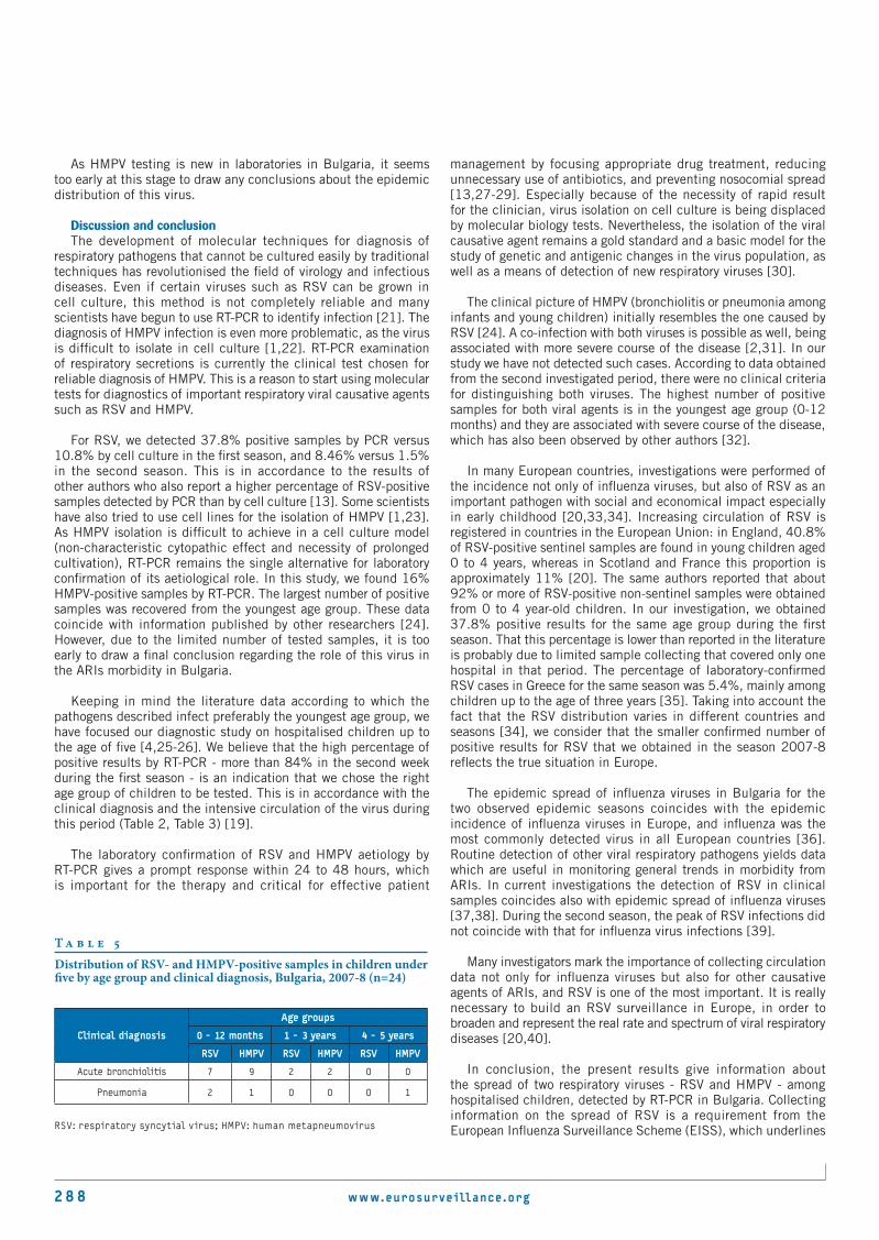
28 8 www.eurosurveillance.org
As HMPV testing is new in laboratories in Bulgaria, it seems too early at this stage to draw any conclusions about the epidemic distribution of this virus.
Discussion and conclusionThe development of molecular techniques for diagnosis of
respiratory pathogens that cannot be cultured easily by traditional techniques has revolutionised the field of virology and infectious diseases. Even if certain viruses such as RSV can be grown in cell culture, this method is not completely reliable and many scientists have begun to use RT-PCR to identify infection [21]. The diagnosis of HMPV infection is even more problematic, as the virus is difficult to isolate in cell culture [1,22]. RT-PCR examination of respiratory secretions is currently the clinical test chosen for reliable diagnosis of HMPV. This is a reason to start using molecular tests for diagnostics of important respiratory viral causative agents such as RSV and HMPV.
For RSV, we detected 37.8% positive samples by PCR versus 10.8% by cell culture in the first season, and 8.46% versus 1.5% in the second season. This is in accordance to the results of other authors who also report a higher percentage of RSV-positive samples detected by PCR than by cell culture [13]. Some scientists have also tried to use cell lines for the isolation of HMPV [1,23]. As HMPV isolation is difficult to achieve in a cell culture model (non-characteristic cytopathic effect and necessity of prolonged cultivation), RT-PCR remains the single alternative for laboratory confirmation of its aetiological role. In this study, we found 16% HMPV-positive samples by RT-PCR. The largest number of positive samples was recovered from the youngest age group. These data coincide with information published by other researchers [24]. However, due to the limited number of tested samples, it is too early to draw a final conclusion regarding the role of this virus in the ARIs morbidity in Bulgaria.
Keeping in mind the literature data according to which the pathogens described infect preferably the youngest age group, we have focused our diagnostic study on hospitalised children up to the age of five [4,25-26]. We believe that the high percentage of positive results by RT-PCR - more than 84% in the second week during the first season - is an indication that we chose the right age group of children to be tested. This is in accordance with the clinical diagnosis and the intensive circulation of the virus during this period (Table 2, Table 3) [19].
The laboratory confirmation of RSV and HMPV aetiology by RT-PCR gives a prompt response within 24 to 48 hours, which is important for the therapy and critical for effective patient
management by focusing appropriate drug treatment, reducing unnecessary use of antibiotics, and preventing nosocomial spread [13,27-29]. Especially because of the necessity of rapid result for the clinician, virus isolation on cell culture is being displaced by molecular biology tests. Nevertheless, the isolation of the viral causative agent remains a gold standard and a basic model for the study of genetic and antigenic changes in the virus population, as well as a means of detection of new respiratory viruses [30].
The clinical picture of HMPV (bronchiolitis or pneumonia among infants and young children) initially resembles the one caused by RSV [24]. A co-infection with both viruses is possible as well, being associated with more severe course of the disease [2,31]. In our study we have not detected such cases. According to data obtained from the second investigated period, there were no clinical criteria for distinguishing both viruses. The highest number of positive samples for both viral agents is in the youngest age group (0-12 months) and they are associated with severe course of the disease, which has also been observed by other authors [32].
In many European countries, investigations were performed of the incidence not only of influenza viruses, but also of RSV as an important pathogen with social and economical impact especially in early childhood [20,33,34]. Increasing circulation of RSV is registered in countries in the European Union: in England, 40.8% of RSV-positive sentinel samples are found in young children aged 0 to 4 years, whereas in Scotland and France this proportion is approximately 11% [20]. The same authors reported that about 92% or more of RSV-positive non-sentinel samples were obtained from 0 to 4 year-old children. In our investigation, we obtained 37.8% positive results for the same age group during the first season. That this percentage is lower than reported in the literature is probably due to limited sample collecting that covered only one hospital in that period. The percentage of laboratory-confirmed RSV cases in Greece for the same season was 5.4%, mainly among children up to the age of three years [35]. Taking into account the fact that the RSV distribution varies in different countries and seasons [34], we consider that the smaller confirmed number of positive results for RSV that we obtained in the season 2007-8 reflects the true situation in Europe.
The epidemic spread of influenza viruses in Bulgaria for the two observed epidemic seasons coincides with the epidemic incidence of influenza viruses in Europe, and influenza was the most commonly detected virus in all European countries [36]. Routine detection of other viral respiratory pathogens yields data which are useful in monitoring general trends in morbidity from ARIs. In current investigations the detection of RSV in clinical samples coincides also with epidemic spread of influenza viruses [37,38]. During the second season, the peak of RSV infections did not coincide with that for influenza virus infections [39].
Many investigators mark the importance of collecting circulation data not only for influenza viruses but also for other causative agents of ARIs, and RSV is one of the most important. It is really necessary to build an RSV surveillance in Europe, in order to broaden and represent the real rate and spectrum of viral respiratory diseases [20,40].
In conclusion, the present results give information about the spread of two respiratory viruses - RSV and HMPV - among hospitalised children, detected by RT-PCR in Bulgaria. Collecting information on the spread of RSV is a requirement from the European Influenza Surveillance Scheme (EISS), which underlines
T a b l e 5
Distribution of RSV- and HMPV-positive samples in children under five by age group and clinical diagnosis, Bulgaria, 2007-8 (n=24)
Clinical diagnosis
Age groups
0 - 12 months 1 - 3 years 4 - 5 years
RSV HMPV RSV HMPV RSV HMPV
Acute bronchiolitis 7 9 2 2 0 0
Pneumonia 2 1 0 0 0 1
RSV: respiratory syncytial virus; HMPV: human metapneumovirus

www.eurosurveillance.org 28 9
the necessity of collection of data regarding the incidence of this virus in different European countries. The investigations reported here are a priority for the National Laboratory of Influenza and Acute Respiratory Diseases in Bulgaria in order to confirm the participation of some more widely distributed viruses, as causative agents of ARIs, first and foremost in paediatric pathology.
AcknowledgementsWe would like to thank our colleagues from the Laboratory of Influenza, Cantacuzino Institute, Bucharest, Romania, for providing us with primers for HMPV and RT-PCR protocol for the detection of this virus.
References
1. Van den Hoogen BG, de Jong JC, Groen J, Kuiken T, de Groot R, Fouchier RA, et al. A newly discovered human pneumovirus isolated from young children with respiratory tract disease. NatMed 2001;7(6):719–24.
2. Choi EH, Lee HJ, Kim SJ, Eun BV, Kim NH, Lee JA, et al. The association of newly identified respiratory viruses with lower respiratory tract infections in Korean children, 2000-2005. Clin Infect Dis 2006;43(5):585–92.
3. Ebihara T, Endo R, Ma X, Ishiguro N, Kikuta H. Detection of human coronavirus NL63 in young children with bronchiolitis. J Med Virol.75(3):463–5.
4. Hall CB. Respiratory syncytial virus and parainfluenza virus. N Engl J Med. 2001;344(25):1917-28.
5. Collins PL, Chanock RB, Murphy BR. Respiratory syncytial virus. In B. N. Fields (ed.), Virology. Forth edition. Philadelphia: Lippincott Williams and Wilkins, Philadelphia. 2001;1443-85.
6. Walsh EE, Falsey AR, Hennesey PA. Respiratory syncytial virus and other infections in persons with chronic cardiopulmonary disease. Am J Respir Crit Care Med. 1999;160(3):791-5.
7. Dollner H, Risnes K, Radtke A, Nordbo SA. Outbreak of human metapneumovirus infection in norwegian children. Pediatr Infect Dis J. 2004;23(5):436-40.
8. Van den Hoogen BG, van Doornum GJ, Fockens JC, Cornelissen JJ, Beyer WE, de Groot R, et al. Prevalence and clinical symptoms of human metapneumovirus infection in hospitalized patients. J Infect Dis. 2003;188(10):1571-7.
9. McIntosh K, McAdam AJ. Human metapneumovirus-an important new respiratory virus. N Engl J Med. 2004;350(5): 431-3.
10. Falsey AR, Erdman D, Anderson LJ, Walsh EE. Human metapneumovirus infections in young and elderly adults. J Infect Dis. 2003;187(5):785-90.
11. Wolf DG, Greenberg D, Kalkstein D, Shemer-Avni Y, Givon-Lavi N, Saleh N, et al. Comparison of human metapneumovirus, respiratory syncytial virus and influenza A virus lower respiratory tract infections in hospitalized young children. Pediatr Infect Dis J. 2006;25(4):320-4.
12. Stockton J, Ellis JS, Saville M, Clewley JP, Zambon MC. Multiplex PCR for typing and subtyping influenza and respiratory syncytial viruses. J Clin Microbiol. 1998;36(10): 2990-5.
13. Falsey AR, Formica MA, Walsh EE. Diagnosis of respiratory syncytial virus infection: comparison of reverse transcription PCR to viral culture and serology in adults with respiratory illness. J Clin Microbiol. 2002;40(3):817-20.
14. Hadzhiolova T, Kotseva R, Kalvachev Z, Dundarova D, Mladenova Z. Application of a complex of laboratory methods for identification of Influenza viruses and RSV in participation of Laboratory of Influenza and ARD in Second European Qualitative Control. Infectology. 2004; XLI: 21-26.
15. Hadzhiolova T, Pavlova S, Kotseva R. Possibilities for laboratory diagnosis of Respiratory Syncytial Virus. Biotechnology. 2005;2(19):67-7111.
16. Pavlova S, Hadzhiolova T, Kotseva R, Abadgieva P. Application of RT-PCR for detection of RSV in hospitalized children. Infectology. 2007;1:119-22.
17. ecu C, Orasanu D, Sima A, Mihai M, Alexandrescu V, Matei D, et al.: First detection of human metapneumovirus in children with respiratory infections in Romania. Roum Arch Microbiol Immunol. 2007;66(1-2):37-40.
18. Valdivia A, Savón C, Chacón D, Sarmiento L, Morier L, Otero A, et al. Analysis of respiratory syncytial virus in clinical samples by reverse transciptase-polymerase chain reaction restriction mapping. Mem Inst Oswaldo Cruz.1997;92(3):389-93.
19. Meury S, Zeller S, Heininger U. Comparison of clinical characteristics of influenza and respiratory syncytial virus infection in hospitalised children and adolescents. Eur J Pediatr. 2004;163(7):359–63.
20. Meerhoff TJ, Fleming D, Smith A, Mosnier A, van Gageldonk-Lafeber AB, Paget WJ, et al. Surveillance recommendations based on an exploratory analysis of respiratory syncytial virus reports derived from the European Influenza Surveillance System. BMC Infect Dis.2006;6:128.
21. Zambon MC, Stockton JD, Clewley JP, Fleming DM. Contribution of influenza and respiratory syncytial virus to community cases of influenza-like illness: an observational study. Lancet. 2001;358(9291):1410–6.
22. Van den Hoogen BG, Osterhaus DM, Fouchier RA. Clinical impact and diagnosis of human metapneumovirus infection. Pediatr Infect Dis J. 2004;23(1 Suppl): S25-32.
23. Abiko C, Mizuta K, Itagaki T, Katsushima N, Ito S, Matsuzaki Y, et al. Outbreak of human metapneumovirus detected by use of the Vero E6 cell line in isolates collected in Yamagata, Japan, in 2004 and 2005. J Clin Microbiol. 2007;45(6):1912–9.
24. Garcia-Carcia ML, Calvo C, Martin F, et al. Human metapneumovcirus infections in hospitalized infants in Spain. Archives of Disease in Childhood. 2006; 91:290-295
25. Selwyn BJ. The epidemiology of acute respiratory tract infection in young children: comparison of findings from several developing countries. Coordinated Data Group of BOSTID Researchers. Rev Infect Dis. 1990;12 Suppl 8:870-88
26. Garcia-Garcia ML, Calvo C, Perez-Brena P, De Cea JM, Acosta B, Casas I. Prevalence and clinical characteristics of human metapneumovirus infections in hospitalized infants in Spain. Pediatr Pulmonol. 2006;41(9):863-71.
27. Wihley DM, Syrmis MW, Mackay IM, Sloots TP. Detection of human respiratory syncytial virus in respiratory samples by LightCycler reverse transcriptase PCR. J Clin Microbiol. 2002;40(12): 4418-22.
28. Groothuis JR, Wooden KA, Katz R, Robertson AD, McBride JT, Hall CB, et al. Early ribavirin treatment of respiratory syncytial viral infection in high-risk children. J. Pediatr. 1990;117(5):792-8.
29. O`Callaghan CA. Prevention of nosocomial respiratory syncytial virus infection. Lancet. 1993;341(8838):182.
30. Nunes IF, Moura FE. Isolation of respiratory syncytial virus from nasopharyngeal aspirates stored at 20 degrees C from one to fifteen months after collection. Mem Inst Oswaldo Cruz. 2006;101(4):451-3.
31. Greensill J, McNamara PS, Dove W, Flanagan B, Hart CA. Human metapneumovirus in severe respiratory syncytial virus bronchiolitis. Emerg Infect Dis. 2003;9(3):372-5.
32. Ginocchio CC, Manji R, Lotlikar M, Zhang F. Clinical evaluation of the NucliSENS analyte-specific reagents for the detection of human metapneumovirus in pediatric respiratory specimens. J Clin Microbiol. 2008;46(4):1274-80. Epub 2008 Feb 13.
33. Domegan L, O’Donnell J, Cunney R, O’Kelly E, Dooley S. Increased respiratory syncytial virus activity in Ireland. Euro Surveill. 2002;6(49):pii=1980. Available from: http://www.eurosurveillance.org/ViewArticle.aspx?ArticleId=1980
34. Terletskaia-Ladwig E, Enders G, Schalasta G, Enders M. Defiding the timing of respiratory syncytial virus (RSV) outbreaks: an epidemiological study. BMC Infect Dis. 2005;5(1):20.
35. Gioula G, Melidou A, Exindari M, Chatzidimitriou D, Kyriazopoulou-Dalaina V. Contribution of RSV and hMPV to patients with influenza-like infections during 2006-2007 season in northern Greece, 2008; Abstract. 18th European Congress of Clinical Microbiology and Infectious Diseases, Barcelona, Spain
36. Arkema JM, Meijer A, Meerhoff TJ, Van Der Velden J, Paget WJ, European Influenza Surveillance Scheme EISS). Epidemiological and virological assessment of influenza activity in Europe, during the 2006-2007 winter. Euro Surveill. 2008;13(34):pii=18958. Available from: http://www.eurosurveillance.org/ViewArticle.aspx?ArticleId=18958
37. Kojouharova M, Kurchatova A, Vladimirova N, et al. News Bulletin of the National Center of Infectious and Parasitic Diseases (NCIPD) [in Bulgarian]. 2008;6:4-40.
38. Izurieta HS, Thomson WW, Kramarz P, Shay DK, Davis RL, DeStefano F, et al. Influenza and the rates of hospitalization for respiratory disease among infants and young children. N Engl J Med. 2000;342(4):232-9.
39. Gasparini R, Ansaldi F, Banfi F, Ferro G, et al. The impact of influenza and respiratory syncytial virus in a 0–14-year children cohort: a comparison with the national network and between methods for influenza virological surveillance. International Congress Series. 2004; 329-33.
40. Meerhoff TJ, Paget WJ, Aguilera JF, van der Velden J. Harmonising the virological surveillance of influenza in Europe: results of an 18-country survey. Virus Res. 2004;103(1-2):31-3.

29 0 www.eurosurveillance.org
R esearch ar ti cles
C l i n i C a l a n d e p i d e m i o l o g i C a l a s p e C t s o f pa r v o v i r u s B19 i n f e C t i o n s i n i r e l a n d , J a n u a r y 1996 - J u n e 2008
N Nicolay ([email protected])1,2, S Cotter1
1. Health Protection Surveillance Centre (HPSC), Dublin, Ireland2. European Programme for Intervention Epidemiology Training (EPIET)
This article was published on 25 June 2009. Citation style for this article: Nicolay N, Cotter S. Clinical and epidemiological aspects of parvovirus B19 infections in Ireland, January 1996-June 2008. Euro Surveill. 2009;14(25):pii=19249. Available online: http://www.eurosurveillance.org/ViewArticle.aspx?ArticleId=19249
Parvovirus B19 infection may be mistakenly reported as measles or rubella if laboratory testing is not performed. As Europe is seeking to eliminate measles, an accurate diagnosis of fever/rash illnesses is needed. The main purpose of this study was to describe the epidemiological pattern of parvovirus B19, a common cause of rash, in Ireland between January 1996 and June 2008, using times series analysis of laboratory diagnostic data from the National Virus Reference Laboratory. Most diagnostic tests for presumptive parvovirus B19 infection were done in children under the age of five years and in women of child-bearing age (between 20-39 years-old). As a consequence, most of the acute diagnoses of B19 infection were made in these populations. The most commonly reported reasons for testing were: clinical presentation with rash, acute arthritis, influenza-like symptoms or pregnancy. The time series analysis identified seasonal trends in parvovirus B19 infection, with annual cycles peaking in late winter/spring and a six-year cycle for parvovirus B19 outbreaks in Ireland.
Introduction Human parvovirus B19 infection is the cause of erythema
infectiosum, or “slapped cheek” disease, a fever/rash illness occurring most frequently in childhood. The clinical presentation of parvovirus B19 infection is sometimes mistakenly diagnosed as rubella or measles. Although typically a mild, self-limiting disease, the infection can cause severe adverse outcomes in certain groups. In pregnant women infection can result in foetal death or hydrops foetalis, and among individuals with haematological disorders, complications such as anaemia or aplastic crisis can occur [1].
An accurate diagnosis of fever/rash illness is necessary not only for case management but also for public health control activities, particularly in outbreak situations in which measles or rubella is suspected [2]. As Europe seeks to eliminate measles as part of the World Health Organization’s European strategy it is important that fever/rash illnesses are accurately diagnosed and that parvovirus B19 infection is not mistakenly reported as measles or rubella [3,4]. The lack of commercially available, convenient and non-invasive diagnostic tests for parvovirus B19 may play a role in the misdiagnosis of measles and rubella cases [5,6]. Because many individuals with fever/rash illnesses are not routinely tested, each year many notified measles and rubella cases are not laboratory-confirmed. In Ireland in 2007, for instance, only 20 of 53 notified measles cases were laboratory-confirmed [7].
No data are available on the prevalence of parvovirus B19 infection in the Irish population, nor on the pattern of disease incidence in Ireland. As the infection is often asymptomatic, it is difficult to have a comprehensive picture of disease incidence. Due to the limited information available to us on the epidemiology of B19 in Ireland, we collaborated with the National Virus Reference Laboratory (NVRL) on a study to describe which population groups were most commonly tested for parvovirus B19, and to describe, using the pattern of laboratory diagnosis of acute infection, the epidemic pattern of acute parvovirus B19 in Ireland between January 1996 and June 2008.
Materials and methods The NVRL is the main diagnostic facility in Ireland for the
diagnosis of parvovirus B19 infection. During our study we identified three regional hospitals which also offer local testing but they represented a minority of all tests done in Ireland. Upon suspicion of acute parvovirus B19 disease, a clinician may request diagnostic testing. Serum samples are sent to the diagnostic laboratory either directly, by individual clinicians, or via any of the hospitals’ microbiological laboratories. Acute infection is diagnosed by the detection of parvovirus B19-specific immunoglobulin M (IgM). These samples are tested by enzyme immunoassay (EIA) in serum or plasma (Parvovirus B19 IgM (mu capture) EIA, Biotrin International).
To estimate the incidence of laboratory-confirmed disease in Ireland, information relating to each individual testing positive for parvovirus B19-specific IgM was extracted from the laboratory information system at the NVRL and sent to the Health Protection Surveillance Centre (HPSC) for analysis. The initial database consisted of a listing of all the positive tests performed at the NVRL with details on place and date of blood samples, age and sex of the patient, clinical symptoms associated with disease or an underlying condition consistent with this diagnosis, results for parvovirus IgM testing and parvovirus IgG testing in laboratory-confirmed acute cases. To eliminate duplicate results originating from patients presenting to clinicians for the same event, all the line listings were reviewed. Duplicates were defined as similar records based on same dates of birth, sex, place of testing, within a period of three days. Duplicate records were excluded from subsequent analysis. Age and sex distribution, clinical features according to the age of the patient, time and place of occurrence of positive tests for parvovirus IgM were described.

www.eurosurveillance.org 29 1
Times series analysis was carried out for the series of laboratory-confirmed acute parvovirus B19 cases reported per month. A linear function was used for analysing secular trends. In order to describe cycles and seasonality in the series, secular trend was removed and data were log transformed for stabilising minor changes in the variance along the series. From the new working series, cyclic components were identified using a Fast Fourier Transformation. Cycles with energy above the upper limit of the 95% confidence interval for the mean energy of the cycles were estimated using the least squares period. An equation with the following structure was obtained:
where A is the amplitude,θ is the phase and p is the period of cosine function of significant cycles.
Analyses were performed using Stata V9.2 (Stata Corporation) and the Fourier Transformation was done using R V2.8.1 (R foundation, www.r-project.org).
Results Descriptive results Parvovirus B19 tests undertaken by NVRL Between January 1996 and June 2008, a total of 12,430 tests
for parvovirus B19 were carried out at the NVRL. Of those, 546
−∑
pxAxfy θπ2cos)(
F i g u r e 1
Requests for parvovirus B19-specific immunoglobulin M detection by year, National Virus Reference Laboratory, Ireland, January 1996-June 2008 (n=11,437)
0
200
400
600
800
1000
1200
1400
1600
1996 1997 1998 1999 2000 2001 2002 2003 2004 2005 2006 2007 2008
Year
Num
ber
of r
eque
sts
Negative
Positive
F i g u r e 2
Requests for parvovirus B19-specific immunoglobulin M detection by age group and sex, National Virus Reference Laboratory, Ireland, January 1996 -June 2008 (n=11,437)
0
5
10
15
20
25
<5 5-9 10-14 15-19 20-29 30-39 40-49 >50 Unknown
Age group
Perc
enta
ge (%)
UnknownMaleFemale
T a b l e
Most common clinical associations observed in patients with parvovirus B19-specific immmunoglobulin M by age group, National Virus Reference Laboratory, Ireland, January 1996-June 2008
Age group < 15 years(n=60)*
>=15 years(n=190)*
Total(n=198)* †
Clinical features No. of reports (% Total)
Rash 28 (34) 52 (63) 83 (42)
“Slapped cheek” appearance 5 (71) 2 (28) 7 (3.5)
Acute arthritis 7 (16) 35 (79) 44 (22)
Fever 9 (32) 18 (64) 28 (14)
Influenza-like symptoms 1 (4) 24 (92) 26 (13)
Pregnancy 0 23 (96) 24 (12)
Intrauterine death 0 8 (100) 8 (4)
Hydrops fetalis 0 1 (100) 1 (0.5)
Anaemia 11 (73) 4 (26) 15 (8)
Haemophilia/Sickle cell anaemia 7 (70) 1 (10) 10 (5)
Sore throat 3 (42) 4 (57) 7 (4)
Lymphadenopathy 0 5 (100) 5 (3)
Headache 3 (75) 1 (25) 4 (2)
Bone marrow transplant 2 (100) 0 2 (1)
* The total number of symptoms exceeds the total number of cases as more than one symptom could be mentioned per case.† Age is unknown for eight patients for whom clinical details were given.
F i g u r e 3
Number of positive tests for parvovirus B19-specific Immunoglobulin M by age and sex, National Virus Reference Laboratory, Ireland, January 1996-June 2008 (n=497)*
* Age is unknown for 15 patients and sex is unknown for two patients under the age of 10 years
0
20
40
60
80
100
120
140
160
180
0-9 10-19 20-29 30-39 40-49 50-59 60-69 70-79 80-89 90-99
Age group
Num
ber
of
posi
tive
tes
ts
Female
Male

29 2 www.eurosurveillance.org
were positive, and 993 duplicates tests were identified. Following de-duplication, 514 (4.5%) acute cases of parvovirus B19 were identified out of 11,437 tests performed (Figure 1).
The number of test requests increased over the time period, from 500 requests in January 1996 to 1,388 in 2007. The proportion of IgM-positive tests varied depending on the year and the seasonality of parvovirus B19 in the community.
Most samples (27%) originated from children under the age of 10 years, the majority of whom were under five years old (19% of all events). The next largest age group was the age group of 30-39 year-olds (23%), followed by 20-29 year-olds (16%). Females were more likely to be tested than males (64% of all requests), most marked in the women of child-bearing age; 77% of requests were made for the 30-39 year-old group and 83% for the 20-29 year-olds (Figure 2).
Positive tests (n=514)Overall, 76% of all positive tests occurred in female patients,
giving a female:male ratio of 3.3:1 (Figure 3). The median age of all cases for whom the age was known was 31 years (range 7 days to 92 years); information on age was missing for 15 positive patients. Males tested positive were more likely to be younger than females (p<10-3). The median age for male patients was nine years and for female patients 33 years.
A total of 168 positive tests (32.7%) belonged to patients under the age of 15 years; 137 positive tests (21.3%) occurred in children between 0 and 9 years of age, with equal distribution between male and female children; 160 (31.1%) positive tests originated from patients between 30 and 39 years of age, 90% of whom were female. Of the 514 IgM-positive cases, 300 (58.4%) were also positive for IgG.
Regional distribution of events There was marked regional distribution of positive tests. Most
of the positive IgM samples (60.3%) were from the former Eastern Health Board region (encompassing the Dublin metropolitan region among others). Samples originating from the North Western Health Board, the Midland Health Board and the North Eastern Health Board represented 7.8%, 5.8% and 5.4% of positive tests, respectively. The lowest number of positive test results came from samples taken in the Western Health Board and in the Mid-Western Health Board regions (2.4%).
Clinical information provided with diagnostic samplesClinical information accompanied the request for parvovirus
B19-specific IgM testing for 198 (38.5%) of patients who tested positive for parvovirus IgM (Table). Parvovirus B19 infection was characterised by a variable combination of symptoms: rash, influenza-like symptoms, joint pain and haematologic abnormalities were often reported.
The most common symptom reported in parvovirus B19 IgM-positive patients was a rash (n=83, 42%) but the typical “slapped cheek” appearance was mentioned in only seven patients as shown in the Table. Joint pain was reported in 44 patients (22.1%) and was more common in adults than children. Fever was the third most commonly reported symptom (14%). Anaemia was the reason for testing for 15 patients (8%), and these were mainly young patients
with 11 (68%) under the age of 15 years. Among these 11, six had sickle cell anaemia and four had haemophilia.
Finally, 24 women (12%) were tested because they were pregnant (the specific circumstances however were not reported for the majority of them). Among those women, eight experienced an abortion or a miscarriage (one in 1996, two in 1999, one in 2000, one in 2004, two in 2005 and one in 2008).
Other documented symptoms reported at the time of diagnostic test request were varied and often unspecific, including reporting influenza-like symptoms, fever and fatigue (30 patients, 15.1%). Three parvovirus infections occurred in patients known to be immunocompromised (Hodgkin’s disease, renal transplant). One case was due to occupational exposure (unspecified).
F i g u r e 4
Monthly series of positive parvovirus B19-specific immunoglobulin M, National Reference Virus Laboratory, Ireland, January 1996-June 2008
0
5
10
15
20
25
1996
1997
1998
1999
2000
2001
2002
2003
2004
2005
2006
2007
2008
Year
Num
ber
of
posi
tive
tes
ts
F i g u r e 5
Periodogram of parvovirus B19 monthly series, Ireland, January 1996-June 2008*
* Periods were identified using Fast Fourier Transformation.
0
1
2
3
4
5
6
7
150.0 75.0 50.0 37.5 30.0 25.0 21.4 18.8 16.7 15.0 13.6 12.5 11.5 10.7 10.0 9.4 8.8 8.3 7.9 7.5 7.1 6.8 6.5 6.3
Spectral density
Threshold

www.eurosurveillance.org 29 3
Seasonal pattern and periodicity of epidemic yearsThe 514 acute cases of parvovirus B19 were reported over a
period of 149 months between January 1996 to June 2008 (Figure 4). The average number of cases diagnosed per month was 3.4 (standard deviation (SD) +/- 3.7). The maximum number of cases was diagnosed during April 1999 (n=20). An increasing trend in the number of acute cases was observed over the study period.
The Fast Fourier Transformation analysis identified two statistically significant components, one annual and the other every 75 months, i.e. approximately six years (Figure 5). Annual cycles peak during winter/spring : each year the majority of cases occurred between March and July.
Discussion In Ireland as well as in most other European countries, parvovirus
B19 infection is not a notifiable disease, and neither clinical descriptive data nor epidemiological data are readily available. To our knowledge, this is the first Irish study which attempts to describe the clinical and epidemiological pattern of parvovirus infection based on acute cases identified by the reference laboratory.
Our initial objective was to describe the epidemic pattern of parvovirus infection. We identified a periodicity of six years and an annual seasonality pattern, with most cases diagnosed between March and July. Our results showed that there was no sex difference in testing patterns in young children, which was to be expected as parvovirus B19 affects both sexes equally. In older age groups, testing is done predominantly in women; during pregnancy, the disease can lead to severe adverse outcomes for the foetus. Of 24 parvovirus B19 positive women known to be pregnant in this report, nine experienced a foetal loss, of whom four had acquired the infection during a time of increased incidence (two in 1999 and two in 2005). A prospective study in the United Kingdom estimated the risk of transplacental infection at 30%, with 5-9% of foetal loss reported [8]. As the number of pregnancies as well as the coverage of the screening in pregnant women are unknown for our study period, the data are not comparable. Nevertheless, they remind us of how severe the disease can be in pregnant women and of the consequences for the foetus. A better knowledge of the prevalence of parvovirus B19 in the Irish population is needed for further interpretation.
Our data were obtained from the Irish National Reference Virus Laboratory. It is situated in Dublin, the metropolitan area of which accounts for approximately one quarter of the Irish population. Although this may explain in part why the majority of tests came from the Eastern region (including Dublin), it does not fully explain the under-representation of other regions in these results. The extent to which other alternative testing may be done at local level is a possibility. However, we could identify only three other regional hospitals that undertook testing, and the number of positive tests performed there during the period under investigation was small (n=50). Despite this lack of regional representation we believe that our data represent a fair approximation of the current endemic situation of symptomatic parvovirus B19 in Ireland. The overrepresentation of the Dublin area most likely reflects increased awareness of testing and submission to the laboratory by both clinicians and the local hospital laboratories. There is no reason to suspect that there is a connection between geography and susceptibility to this common illness. The epidemiology of parvovirus B19 infection has many of the characteristics of other common childhood communicable diseases which were common in
the pre-vaccine aera (e.g. measles, rubella or mumps), all of which demonstrated outbreak years followed by periods of low incidence before the next outbreak. However, our data are unlikely to be fully representative of the true distribution of acute parvovirus B19 infection in the general Irish population due to a testing bias for certain population groups (young children and women attending maternity hospitals). It is likely that people tested in the present study represent the most seriously affected cases or the population considered to be most at risk.
Because human parvovirus infection is not a notifiable disease in Ireland, we cannot test the assumption that IgM-positive results from a reference laboratory are representative of the pattern of acute infection in the community. Nevertheless previous studies have used data from reference laboratories to describe the seasonality of parvovirus B19 [9,10]. In Ireland, measles is a notifiable disease. By comparing the pattern of measles-specific positive samples tested by NVRL with the distribution pattern of measles notifications to the HPSC between 2000 and 2008, we find a similar trend, with an increase in laboratory testing for measles during periods of increased notification, thus supporting the main assumption we made for parvovirus B19. Published data on the seasonal activity of parvovirus B19 in temperate countries are limited. According to our data most of the cases occurred during winter and late spring. Based on the times series analysis, the periodicity of epidemic years is six years, which is concordant with data published for some other developed countries [10-12]. However we cannot exclude that the pattern of the disease may present with one or two consecutive epidemic years: both 2004 and 2005 had a substantial number of positive tests. Even though the data from a reference laboratory are not as informative as the data which could be provided by a national surveillance system, they can provide helpful insight into the epidemiological pattern of non-notifiable diseases. Awareness of the normal epidemiology of parvovirus B19 can help clinicians who are confronted with patients with rash illnesses in the differential diagnosis for all compatible rash illness, especially measles.
Assuming a periodicity of six years, we can expect the next epidemic year in the coming two years (2011). We hope that this study will alert clinicians and increase diagnostic testing of all rash illnesses as they present. Numerous studies have highlighted the difficulty in making an accurate diagnosis of rash diseases [5,6,13,14]. The development of new and non-invasive technology that allows the sampling of oral fluid to diagnose viral infections such as measles [15], mumps [16], and hepatitis A and B [17] has increased the number of laboratory-confirmed diagnoses in many countries. Such testing has been found to be both sensitive and specific and is routinely used in many countries in case diagnosis. Commercial tests for the serological diagnosis of parvovirus B19 are available, but none are validated for use on oral fluid samples. Such a diagnostic tool would be invaluable, particularly when investigating fever/rash illnesses in young children [2] who are not so ill as to require hospitalisation but are usually seen by general practitioners (GPs) in the community. Anecdotal reports indicate that Irish GPs are reluctant to undertake phlebotomy in such paediatric cases and hence accurate laboratory confirmation of fever illness is often not done. A recently developed test to diagnose acute parvovirus B19 infection using oral fluid samples is now being assessed by the NVRL as part of a collaborative study with HPSC and clinicians around the country.

29 4 www.eurosurveillance.org
The value in adding parvovirus testing to enhanced measles surveillance has been demonstrated in South Australia where measles, rubella and parvovirus testing are included in routine measles surveillance. Despite a low overall rate of measles testing, this was particularly obvious in an inter-epidemic period when most notified measles cases were not measles [18]. Between 2% and 10% of suspected measles cases tested in South Australia between 1999 and 2004, were parvovirus B19 cases [19]. An added value was also shown when including parvovirus testing in the rubella surveillance programme [14].
In countries in the elimination phase for measles and rubella, a better knowledge of the epidemiology of parvovirus B19 may help clinicians in the differential diagnosis of common rash diseases. Meanwhile, a better laboratory confirmation of common rash illnesses is required to improve the quality of national data and public health action. The anticipated availability of an oral fluid test for parvovirus B19 will be useful in this aim.
Vivamus tempor mi quis quam. Fusce tempus, ante sed tincidunt ornare, nisi urna viverra enim, eget venenatis dui ante ut eros.
AcknowledgementsWe would like to acknowledge Dr Jeff Connell and Joanne Moran, National Virus Reference Laboratory, Dublin, Ireland for providing the data as well as Dr Francisco Luquero for his statistical support.
References
1. Young NS, Brown KE. Parvovirus B19. N Engl J Med. 2004;350(6):586-97.
2. Goncalves G, Correia AM, Palminha P, Rebelo-Andrade H, Alves A. Outbreaks caused by parvovirus B19 in three Portuguese schools. Euro Surveill. 2005;10(6):pii=549. Available from: http://www.eurosurveillance.org/ViewArticle.aspx?ArticleId=549
3. Strategic plan for measles and congenital rubella infection in the WHO European Region. Copenhagen: World Health organisation Regional Office for Europe; 2003. Available from: http://www.euro.who.int/document/e81567.pdf
4. Surveillance guidelines for measles and congenital rubella infection in the WHO European Region. Copenhagen: World Health Organization Regional Office for Europe; 2003. Available from: http://www.euro.who.int/document/e82183.pdf
5. Davidkin I, Valle M, Peltola H, Hovi T, Paunio M, Roivainen M, et al. Etiology of measles- and rubella-like illnesses in measles, mumps, and rubella-vaccinated children. J Infect Dis. 1998;178(6):1567-70.
6. Ramsay M, Reacher M, O’Flynn C, Buttery R, Hadden F, Cohen B, et al. Causes of morbilliform rash in a highly immunised English population. Arch Dis Child. 2002;87(3):202-6.
7. Health Protection Surveillance Centre. Annual Report 2007. Available from: http://www.ndsc.ie/hpsc/AboutHPSC/AnnualReports/File,3377,en.pdf.
8. Miller E, Fairley CK, Cohen BJ, Seng C. Immediate and long term outcome of human parvovirus B19 infection in pregnancy. Br J Obstet Gynaecol. 1998;105(2):174-8.
9. Enders M, Weidner A, Enders G. Current epidemiological aspects of human parvovirus B19 infection during pregnancy and childhood in the western part of Germany. Epidemiol Infect. 2007;135(4):563-9.
10. Kelly HA, Siebert D, Hammond R, Leydon J, Kiely P, Maskill W. The age-specific prevalence of human parvovirus immunity in Victoria, Australia compared with other parts of the world. Epidemiol Infect. 2000;124(3):449-57.
11. Vyse AJ, Andrews NJ, Hesketh LM, Pebody R. The burden of parvovirus B19 infection in women of childbearing age in England and Wales. Epidemiol Infect. 2007;135(8):1354-62.
12. Zaaijer HL, Koppelman MH, Farrington CP. Parvovirus B19 viraemia in Dutch blood donors. Epidemiol Infect. 2004;132(6):1161-6.
13. Davidkin I, Jokinen S, Paananen A, Leinikki P, Peltola H. Etiology of mumps-like illnesses in children and adolescents vaccinated for measles, mumps, and rubella. J Infect Dis. 2005;191(5):719-23.
14. Oliveira MI, Curti SP, Figueiredo CA, Afonso AM, Theobaldo M, Azevedo RS, et al. Rash after measles vaccination: laboratory analysis of cases reported in Sao Paulo, Brazil. Rev Saude Publica. 2002;36(2):155-9.
15. Perry KR, Brown DW, Parry JV, Panday S, Pipkin C, Richards A. Detection of measles, mumps, and rubella antibodies in saliva using antibody capture radioimmunoassay. J Med Virol. 1993;40(3):235-40.
16. Reid F, Hassan J, Irwin F, Waters A, Hall W, Connell J. Epidemiologic and diagnostic evaluation of a recent mumps outbreak using oral fluid samples. J Clin Virol. 2008;41(2):134-7.
17. Thieme T, Yoshihara P, Piacentini S, Beller M. Clinical evaluation of oral fluid samples for diagnosis of viral hepatitis. J Clin Microbiol. 1992;30(5):1076-9.
18. Lambert SB, Kelly HA, Andrews RM, Catton MC, Lynch PA, Leydon JA, et al. Enhanced measles surveillance during an interepidemic period in Victoria. Med J Aust. 2000;172(3):114-8.
19. Kelly H, Leydon J. Letter to the editor: outbreaks caused by parvovirus B19. Euro Surveill. 2005;10(9): pii=565. Available from: http://www.eurosurveillance.org/ViewArticle.aspx?ArticleId=565

www.eurosurveillance.org 29 5
P e rspec tives
I s t h e r e a n e e d f o r a n t I - r a b I e s va c c I n e a n d I m m u n o g l o b u l I n s r at I o n I n g I n e u r o p e ?
H Bourhy ([email protected])1, M Goudal1, A Mailles2, M Sadkowska-Todys3, L Dacheux1, H Zeller4
1. WHO Collaborative Center for Reference and Research on Rabies, National Reference Centre for Rabies, Institut Pasteur, Paris, France
2. Institut de veille sanitaire, Saint-Maurice, France3. National Institute of Public Health, Warsaw, Poland4. Preparedness and Response Unit, European Centre for Disease Prevention and Control (ECDC), Stockholm, Sweden
This article was published on 2 April 2009. Citation style for this article: Bourhy H, Goudal M, Mailles A, Sadkowska-Todys M, Dacheux L, Zeller H. Is there a need for anti-rabies vaccine and immunoglobulins rationing in Europe?. Euro Surveill. 2009;14(13):pii=19166. Available online: http://www.eurosurveillance.org/ViewArticle.aspx?ArticleId=19166
Rabies is a lethal encephalitis caused by a lyssavirus and transmitted from animals to humans via bite wound, scratch wound, or licking of mucous membranes. It is preventable by timely administration of post-exposure prophylaxis (PEP) consisting of four or five doses of rabies vaccine combined, in the most severe cases of exposures, with anti-rabies immunoglobulin (RIG). Although the rabies incidence in humans remains low, rabies is still present in some European countries. Moreover, rabid animals imported from enzootic areas are reported every year in rabies-free areas. These importations threaten the rabies-free status of terrestrial animals in western European countries and challenge the public health surveillance system and the health structures responsible for rabies prophylaxis and control. The importations frequently result in the prescription of a large number of PEP including RIG, especially in western European countries. The situation is inverted in some central and eastern European countries where RIG is underprescribed. Only a limited number of rabies vaccines and particularly of RIG are licensed for use in Europe. Their availability is also limited, a situation that may become worse in the future. It therefore seems important to study the possibility of comparing and unifying national PEP guidelines in Europe, if needed, and to generate effective solutions in the event of a shortage of anti-rabies biological products and RIG in particular, such as rationing these products.
IntroductionRabies is a lethal encephalitis caused by a lyssavirus which
is transmitted from animals to humans via bite wound, scratch wound, or licking of mucous membranes [1]. Human-to-human transmission has not been proven. However, some cases of rabies transmission through organ transplantation have been described [2,3]. Since Louis Pasteur’s discovery of the rabies vaccine, rabies has been a disease that can be prevented through the timely administration of post-exposure prophylaxis (PEP). Today, PEP consists of four or five doses of rabies vaccine administered on three to five visits. Anti-rabies immunoglobulin (RIG) is given in addition, if the exposure fulfils the criteria of Category III as defined by the recommendations given by the World Health Organization (WHO) [4,5].
Rabies is still present on the European continent, although some countries have rabies-free status according to the criteria of the World Organisation for Animal Health (OIE). Its incidence in humans remains low (fewer than five human cases per year) owing to the strict application of PEP and to veterinary rabies control measures in domesticated and wild animal populations.
The main indigenous animal reservoirs are dogs in eastern
European countries and on the borders with the Middle East, foxes in central and eastern Europe, racoon dogs in north-eastern Europe and insectivorous bats throughout the entire territory [6]. In addition, cases of rabid animals imported from enzootic areas outside Europe are reported every year, which shows the permeability of borders and travellers’ lack of awareness of the rabies risk [7]. These importations constantly threaten the rabies-free status of terrestrial animals in western European countries. The associated risk also complicates the decision concerning human PEP when the biting animal is not accessible for rabies assessment (clinical examination and/or laboratory examination) [6,8]. In view of the complexity of rabies epidemiology in the European Union (EU), it is important to keep health professionals, particularly physicians and veterinarians, updated in order to maintain vigilance. Recommendations to improve rabies control in animals and prevention of human transmission have recently been published in the WHO Expert Consultation on Rabies [4].
The objective of this paper is to review the current situation in the EU countries regarding the needs for rabies vaccine and anti-rabies immunoglobulins as well as the risk of a potential shortage, using as examples the current practice in France and in Poland.
Different usage of rabies biological products in EuropeData on the use of rabies vaccine and anti-rabies immunoglobulin
and on the number of PEP in Europe are scarce. Therefore, we will mainly focus our report on two countries, France and Poland, that have implemented centralised surveillance.
In France, data from 2007 showed that 3,631 people (47% of all people who sought medical care in anti-rabies centres) received PEP treatment with 11% of them receiving RIG. In February 2008, two cases of autochthonous rabid dogs lead to the prescription of

29 6 www.eurosurveillance.org
241 PEP in people who had been bitten, 34 of whom also received RIG, in accordance with the French and WHO recommendations. The index case was a dog illegally imported from Morocco [9]. Following this event, France lost its rabies-free status according to the OIE criteria. Since then, no other case of canine or feline rabies have been diagnosed in non-travelling animals, which makes us confident that the veterinary control measures taken after the incident have been effective in controlling further spread of the virus. In November 2008, a rabid dog imported to France from Spain was identified. The three month-old animal was found to be infected by a strain phylogenetically very close to those circulating in Morocco, indicating a potential recent importation from Morocco (unpublished results). Of 32 people who were in contact with this dog, seven received PET including vaccine and RIG, 18 received vaccine only and the remaining seven people were not considered at risk and therefore did not receive any PET. Unfortunately, such episodic importations of rabies-infected dogs are not rare. Between 2000 and 2008, seven rabid dogs had been illegally imported into France from Africa. For each imported rabid dog, between two and 187 people with direct contact had received post-exposure vaccination, and nearly 15% of them had also received anti-rabies immunoglobulin.
Several other rabies-free countries in Europe have also reported importation of rabid animals in the past (e.g. Belgium, Switzerland, and the United Kingdom). These episodes further supported the recommendation of prescribing PEP for patients bitten by a dog of unknown origin or suspected to come from an enzootic country. The recent re-emergence of fox rabies in Italy has stressed further that rabies in non-flying wildlife is not completely under control in Europe and that it can re-infect areas from which it was eliminated years before [10]. Consequently, the periodical re-introduction of rabies in any of the EU countries has an immediate impact on the number of PEP interventions, i.e. the number of rabies vaccine and immunoglobulin doses used in EU.
The number of reported human exposures to bats in Europe has also increased in recent years. In these cases, patients received RIG together with the vaccine in accordance with national and WHO guidelines [4]. In France alone, an average of 100 people receive PEP including RIG after exposure to bats every year.
On the other hand, RIG may be underprescribed in some countries in central and eastern Europe. In Poland, for example, PEP is administered to about 7,000 people every year (54,767 patients in total during the period from 2001 to 2007), and only 0.8% of these patients also receive RIG. In the same time period, 644 individuals received PEP after a contact with bats and only 4.7% of them received RIG. In these countries, a strict application of WHO guidelines would therefore immediately lead to an increase in the use of RIG in particular.
Risk of vaccine shortage According to the number of rabies vaccine sold every year and
in the absence of more precise data, we can estimate that world-wide, at least 15 million PEP are administered annually. The EU, the United States (US) and Canada only represent 1% of the global consumption. European producers have implemented high quality control standards for the production of rabies vaccines and immunoglobulin. The two European producers supply about 25% of the rabies vaccine doses used annually worldwide.
An official health advisory report published in June 2008 by the US Centers for Disease Control and Prevention (CDC) indicated a temporary decrease in human rabies vaccine supplies in the US [11,12]. The two European producers (Sanofi Pasteur and Novartis) are the only suppliers of rabies vaccine for the use in humans in the US. Supplies of rabies vaccine went down in the US after Sanofi Pasteur started renovations in the French production facility for the IMOVAX rabies vaccine (produced on human diploid cells) in June 2007, and after Novartis had to suspend its supply to the US and the EU in September 2007 following an inspection conducted by the US Food and Drug Administration (FDA) (http://www.fda.gov/foi/warning_letters/archive/s6644c.pdf). The renovations conducted by Sanofi Pasteur are expected to be completed by mid-2009 and the registration of IMOVAX (the rabies vaccine produced in this facility) by the end of 2009. Novartis started building a new rabies vaccine production facility in Germany in May 2008. It is expected to be fully operational in 2011 [11,13].
As a consequence, the US CDC strongly recommend that
healthcare providers, public health authorities at state and local level, animal control officials, as well as the public take immediate steps to ensure appropriate use of human rabies biological products. The US CDC stressed that the judicious and appropriate use of rabies vaccine is crucial in order to avert a situation that puts individuals exposed to rabies at increased risk due to depleted vaccine supplies [13]. Therefore the use of rabies vaccine is restricted to situations meeting the criteria indicated in the recommendations [13]. Regarding pre-exposure prophylaxis in the US, priority is given to those at greatest risk of rabies exposure (e.g. people working in rabies laboratories, animal control officers, veterinary staff or wildlife workers), taking into consideration the available rabies vaccine supplies. For groups at lower risks of exposure (e.g. travellers and veterinary students), the US CDC proposes to suspend pre-exposure prophylaxis until the vaccine supply levels are restored.
The availability of rabies vaccines in Europe differs from that in North America where only vaccines produced in chicken embryo cell culture or human diploid cells are licensed. In Europe, vaccine is produced in Vero cells in large amounts and widely used in Europe, particularly in France, as well as in Asia and Africa. It represents a possible alternative in the event of a shortage of the two other products.
Risk of RIG shortageThe stock of specific human RIG is more limited and it has
been known for some time that there is a world-wide shortage [14]. Only three to five million doses of anti-rabies immunoglobulin are produced and sold every year. Considering that the number of doses used in one protocol of PEP varies according to the patient’s weight in kg, no more than an estimated 2-5% of patients seeking PEP can have received anti-rabies immunoglobulin. The current level of production does not cover the needs. According to WHO estimates, about 60% of the people seeking care for PEP do not receive an injection of anti-rabies immunoglobulin, although they fall into the category of exposure that would require it [4,15,16]. This is mainly due to difficulties with access to this biological product, but also to limited production compared to the world-wide demand. In Europe, two types of purified anti-rabies immunoglobulin are produced, human (HRIGs) and equine (ERIGs). The entire production of HRIGs, which is limited due to the lack of plasma donors, is almost exclusively sold in the US and Europe. Therefore any increase

www.eurosurveillance.org 297
in demand may cause problems. However, ERIGs are now highly purified, well tolerated and have been demonstrated to be efficient in post-exposure treatment [17]. They are produced in large amounts and may be a suitable alternative in case of a shortage of HRIG, although they have not yet been licensed in Europe. Other products of good efficacy and safety manufactured outside Europe could also be used as a complementary source of supply. Cocktails of monoclonal antibodies have also been recently developed for this purpose [18]. Although promising, the first licence of this type of product cannot be expected before 2012 or 2013.
DiscussionIn France and Poland, recommendations for rabies PEP (both
vaccine and immunoglobulin) followed national guidelines and/or WHO guidelines which recommend that people should receive PEP when bitten by an animal suspected to be infected by rabies. Clinicians make an individual risk assessment for each patient bitten or scratched, and decide to administer rabies vaccine with or without immunoglobulins according to the general recommendations, epidemiological data and the category of the bite. The veterinary situation is taken into account in this assessment, namely the species of the biting animal and the possibility of carrying out examination of the animal if it can be identified. Although no study has investigated the actual prescription practices, it is suspected that some PEP prescriptions are not based on the guidelines [19]. In Europe, practices vary, relying either on special anti-rabies centres (such as in France) or on private general practices (such as in Germany). Furthermore, there seems to be large variations in the use of PEP and especially RIG between European countries, with some countries overreacting (for example France) and others underprescribing (like Poland). Therefore, it would be important to review and analyse practices in the EU, as has been done in North American countries [20].
The risk of a potential shortage of rabies vaccine seems limited in Europe. However, it is important to note that the risk of a potential shortage of RIG in the event of an unplanned increase in demand or a limitation in supply is shared by many countries in Europe and other continents [8,21-23]. The availability of other RIG that have proved their efficacy and safety and that are presently not widely licensed in this area constitutes a possible alternative.
Note added in proofSince the time of submission of this paper, an European consultation was conducted at the European Centre for Disease Prevention and Control (ECDC) in Stockholm on 15 January 2009. The group of experts gathered at this occasion further emphasised the need to review the rabies epidemiological situation in Europe. It also recommended to map practices and usage of anti-rabies biological products in Europe in order to be able to propose effective options for optimisation, as has been done for other vaccines [24]. The conclusions of this meeting will be available from ECDC.
References
1. Hemachudha T, Laothamatas J, Rupprecht CE. Human rabies: a disease of complex neuropathogenetic mechanisms and diagnostic challenges. Lancet Neurol. 2002;1(2):101-9.
2. Hellenbrand W, Meyer C, Rasch G, Steffens I, Ammon A. E-alert 18 February: Cases of rabies in Germany following organ transplantation. Euro Surveill. 2005;10(8):pii=2917. Available from: http://www.eurosurveillance.org/ViewArticle.aspx?ArticleId=2917
3. Jackson AC. Rabies: new insights into pathogenesis and treatment. Curr Opin Neurol. 2006 Jun;19(3):267-70.
4. WHO Expert Consultation on rabies. World Health Organ Tech Rep Ser. 2005;931:1-88, back cover.
5. Rabies Vaccines. WHO position paper. Wkly Epidemiol Rec. 2007;82(49/50):425-35.
6. Bourhy H, Dacheux L, Strady C, Mailles A. Rabies in Europe in 2005. Euro Surveill. 2005;10(11):pii=575. Available from: http://www.eurosurveillance.org/ViewArticle.aspx?ArticleId=575
7. Servas V, Mailles A, Neau D, Castor C, Manetti A, Fouquet E, et al. An imported case of canine rabies in Aquitaine: investigation and management of the contacts at risk, August 2004-March 2005. Euro Surveill. 2005;10(11):pii=578. Available from: http://www.eurosurveillance.org/ViewArticle.aspx?ArticleId=578
8. Gautret P, Soula G, Adamou H, Soavi MJ, Delmont J, Rotivel Y, et al. Rabies postexposure prophylaxis, Marseille, France, 1994-2005. Emerg Infect Dis. 2008;14(9):1452-4.
9. French multidisciplinary investigation team. Identification of a rabid dog in France illegally introduced from Morocco. Euro Surveill. 2008;13(11):pii=8066. Available from: http://www.eurosurveillance.org/ViewArticle.aspx?ArticleId=8066
10. De Benedictis P, Gallo T, Iob A, Coassin R, Squecco G, Ferri G, et al. Emergence of fox rabies in North-Eastern Italy. Euro Surveill. 2008;13(45):pii=19033. Available from: http://www.eurosurveillance.org/ViewArticle.aspx?ArticleId=19033
11. Reserves of human-use rabies vaccine could alleviate supply worries. JAVMA News. 2008, September 15. American Veterinary Medical Association. Available from: http://www.avma.org/onlnews/javma/sep08/080915a.asp
12. Rabies vaccine supply situation. [Accessed 11 March 2009]; Atlanta: Centers for Disease Control and Prevention; 2009 February 6. Available from: http://www.cdc.gov/rabies/news/RabVaxupdate.html
13. Manning SE, Rupprecht CE, Fishbein D, Hanlon CA, Lumlertdacha B, Guerra M, et al. Human rabies prevention--United States, 2008: recommendations of the Advisory Committee on Immunization Practices. MMWR Recomm Rep. 2008;57(RR-3):1-28.
14. Haupt W. Rabies--risk of exposure and current trends in prevention of human cases. Vaccine. 1999;17(13-14):1742-9.
15. Dodet B, Africa Rabies Expert Bureau (AfroREB), Adjogoua EV, Aguemon AR, Amadou OH, Atipo AL, et al. Fighting rabies in Africa: The Africa Rabies Expert Bureau (AfroREB). Vaccine. 2008;26(50):6295-8.
16. Dodet B, Goswami A, Gunasekera A, de Guzman F, Jamali S, Montalban C, et al. Rabies awareness in eight Asian countries. Vaccine. 2008;26(50):6344-8.
17. Quiambao BP, Dytioco HZ, Dizon RM, Crisostomo ME, Laot TM, Teuwen DE. Rabies Post-Exposure Prophylaxis in the Philippines: Health Status of Patients Having Received Purified Equine F(ab’)(2) Fragment Rabies Immunoglobulin (Favirab). PLoS Negl Trop Dis. 2008;2(5):e243.
18. Bakker AB, Python C, Kissling CJ, Pandya P, Marissen WE, Brink MF, et al. First administration to humans of a monoclonal antibody cocktail against rabies virus: safety, tolerability, and neutralizing activity. Vaccine. 2008;26(47):5922-7.
19. Peigue-Lafeuille H, Bourhy H, Abiteboul D, Astoul J, Cliquet F, Goudal M, et al. [Human rabies in France in 2004: update and management]. Med Mal Infect. 2004;34(12):551-60. [In French].
20. Huot C, De Serres G, Duval B, Maranda-Aubut R, Ouakki M, Skowronski DM. The cost of preventing rabies at any cost: post-exposure prophylaxis for occult bat contact. Vaccine. 2008;26(35):4446-50.
21. Albouy-Llaty M, Le Moal G, Bouche G, Dupuis A, Becq-Giraudon B, Migeot V. [Imogam rage prescription assessment in the rabies prophylaxis center of Poitiers]. Med Mal Infect. 2007;37(2):108-11. [In French].
22. Moran GJ, Talan DA, Mower W, Newdow M, Ong S, Nakase JY, et al. Appropriateness of rabies postexposure prophylaxis treatment for animal exposures. Emergency ID Net Study Group. Jama. 2000 23-30;284(8):1001-7.
23. Sadkowska-Todys M, Łabuńska E.. [Rabies in Poland in 2002]. Przegl Epidemiol. 2004;58(1):143-52. [In Polish]
24. Uscher-Pines L, Barnett DJ, Sapsin JW, Bishai DM, Balicer RD. A systematic analysis of influenza vaccine shortage policies. Public Health. 2008;122(2):183-91.

29 8 www.eurosurveillance.org
P e rspec tives
I m p l e m e n tat I o n o f a n at I o n a l e l e c t r o n I c r e p o r t I n g s y s t e m I n l I t h u a n I a
M Domeika ([email protected])1,2, G Kligys3, O Ivanauskiene3, J Mereckiene4, V Bakasenas5, B Morkunas6,D Berescianskis7, T Wahl8, K Stenqvist9,10
1. Department of Medical Sciences, Uppsala University, Uppsala, Sweden2. East Europe Committee of the Swedish Health Community, Stockholm, Sweden3. Kaunas Public Health Center, Kaunas, Lithuania4. Vilnius Public Health Center, Lithuania5. The State Public Health Service (SPHS) under the Ministry of Health, Lithuania6. Lithuanian Center for Communicable Diseases Prevention and Control, Lithuania7. Computer technology company “Compidea”, Vilnius, Lithuania8. Department of Communicable Disease Control and Prevention, Stockholm County, Sweden9. Hivprevention i Västra Götaland, Region Västra Götaland, Sweden10. Department of Social Medicine, Sahlgrenska Academy, University of Gothenburg, Sweden
This article was published on 2 April 2009. Citation style for this article: Domeika M, Kligys G, Ivanauskiene O, Mereckiene J, Bakasenas V, Morkunas B, Berescianskis D, Wahl T, Stenqvist K. Implementation of a national electronic reporting system in Lithuania. Euro Surveill. 2009;14(13):pii=19165. Available online: http://www.eurosurveillance.org/ViewArticle.aspx?ArticleId=19165
Electronic reporting systems improve the quality and timeliness of the surveillance of communicable diseases. The aim of this paper is to present the process of the implementation and introduction of an electronic reporting system for the surveillance of communicable diseases in Lithuania. The project which started in 2002 was performed in collaboration between Lithuania and Sweden and was facilitated by the parallel process of adapting the surveillance system to European Union (EU) standards. The Lotus-based software, SmittAdm, was acquired from the Department of Communicable Diseases Control and Prevention of Stockholm County in Sweden and adopted for Lithuania, resulting in the Lithuanian software, ULISAS. A major advantage of this program for Lithuania was the possibility to work offline. The project was initiated in the two largest counties in Lithuania where ULISAS had been installed and put in use by January 2005. The introduction was gradual, the national level was connected to the system during late 2005, and all remaining counties were included during 2006 and 2007. The reporting system remains to be evaluated concerning timeliness and completeness of the surveillance. Further development is needed, for example the inclusion of all physicians and laboratories and an alert system for outbreaks. The introduction of this case-based, timely electronic reporting system in Lithuania allows better reporting of data to the European Centre for Disease Prevention and Control (ECDC) and the World Health Organization (WHO) compared to the former reporting system with paper-based, aggregated data.
IntroductionWell-functioning surveillance systems for communicable
diseases are fundamental in providing information needed to take appropriate and timely measures. Studies from countries with electronic reporting systems show that such systems improve the quality and timeliness of the surveillance [1-4]. In addition, there is need for an integrated European surveillance system so that the epidemiology of communicable diseases can be compared between
countries and early warning systems can function in an international perspective. The present project was initiated in order to improve the Lithuanian system for the surveillance of communicable diseases which hitherto had been time consuming to administer and only paper-based, aggregated data had been reported to the national level. Duplication of data was also a problem since aggregated data was sent in parallel from both local and county level to the national centre. During the project period, Lithuania joined the European Union (EU). Consequently, the Lithuanian law on communicable diseases was adapted to the EU standards [5]. The main goal of this paper is to present the introduction process of the electronic surveillance system for communicable diseases ULISAS in Lithuania, a project that was performed with financial support and expertise from Sweden.
Materials and methods Organisation of the surveillance of communicable diseases in LithuaniaLithuania has 3.4 million inhabitants and is organised in ten
counties, each with one Public Health Centre (PHC). Each county has one or more Territorial Public Health Centers (TPHC), altogether 36 in the country. The PHC has an overall responsibility for the surveillance of communicable disease within the county [6]. Notifiable diseases are reported from the PHC to the Lithuanian Centre for Communicable Diseases Prevention and Control (CCDPC) as shown in Figure 1.
Physicians from a total of 1,257 primary care centres, hospitals and polyclinics and 21 laboratories report notifiable diseases by post, fax or e-mail to the TPHC and the PHC within 72 hours according to the rules and regulations. In unusual situations, for example the occurrence of plague and yellow fever, notifications should be sent by post, fax or e-mail, within 12 hours. The clinical notifications contain full patient identity and a unique personal identification number which is issued to all Lithuanian residents.

ww.eurosurveillance.org 29 9
Exceptions are the sexually transmitted infections (STI) and cases of human immunodeficiency virus (HIV), which are reported with a specially designed code, so that the personal identity is not revealed. The number of clinical notifications amounts to about 55,000 per year in the whole country. The epidemiologic investigations of individual cases and outbreaks are performed by epidemiologists at the TPHC and the PHC and reported on standardised paper forms to the CCDPC. For STI and HIV, physicians perform the epidemiological investigation and report weekly to the TPHC and PHC. Prior to the development of the electronic reporting system, aggregated data on 82 notifiable diseases collected at the TPHC and the PHC had been summarised using a standard statistical form at the end of every month and sent to the CCDPC in paper format. In addition, aggregated data had been reported yearly to the CCDPC using 17 different statistical forms.
Organisation of the surveillance of communicable diseases in SwedenNotifiable diseases are reported by physicians to the County
Medical Officer (CMO) at the county level and to the Swedish Institute for Infectious Disease Control (Smittskyddsinstitutet, SMI) at the national level. The CMO has an overall responsibility for the surveillance in his or her county. A national electronic surveillance system, SmiNet-1, had been operational from 1997 and in use by 16 of 21 counties since the beginning of the 2000s [3]. With this system, notifications were mainly sent in paper format to the CMO and manually entered at the county level, with the exception of the infectious diseases clinics and laboratories which were connected to the electronic reporting system. After a technical revision of SmiNet-1 in 2001, a new web-based system, SmiNet-2, was developed and implemented in 2004 [1]. Parallel to this, the Department of Communicable Diseases Control and Prevention at the county of Stockholm developed a program for electronic reporting, SmittAdm, which has been in use from 1998. This
F i g u r e 1
Data flow in the Lithuanian national reporting system of communicable diseases before and after the introduction of ULISAS electronic system
Case detection(physician)*
Data flow before introduction of ULISAS
Territorial Public Health Centers (36)
Public Health Centers (10)
National Center for Communicable Disease Prevention and Control
Service Ministry of Health
Data flow after introduction of ULISAS
Case detection(physician)*
Public Health Centers (10)
State Public Health Service
Aggregated data; monthly by e-mail
Indivudual data, daily, on-line
Ongoing introduction of the daily, on-line reporting, but temporarily both daily and aggregated data is reported
Territorial Public Health Centers (36)
Ministry of Health
National Center for Communicable Disease Prevention and Control
*Case data – notification by phone, fax, mail and e-mail according to the legal requirements

3 0 0 www.eurosurveillance.org
program supplemented SmiNet-1 with functions for administrative and judicial tasks which according to the Swedish Communicable Diseases Act are performed at the county level. SmittAdm and SmiNet-1, both being built in Lotus Notes, were easily connected. The staff at the county level used SmiNet-1 only for submitting notifications to the national level; data was then replicated to SmittAdm for further regional work and analysis. SmittAdm was also used by the second largest region, Västra Götaland, and three other counties.
Project organisation The project was initiated in 2002 by the Swedish Institute for
Infectious Disease Control (SMI) and the PHC in Kaunas with financial support from the East Europe Committee of the Swedish Health Care Community (SEEC).
The project manager (of Lithuanian origin, which facilitated communication) was based at SMI and a coordinating study group was formed in Lithuania consisting of epidemiologists from the national level and the counties of Kaunas and Vilnius, and an external IT specialist. This group defined the requirements for an electronic reporting system in Lithuania. Such a system should enable timely reporting of individual data to the county and national level, ascertain a uniform quality of notifications for the whole
country, and support the integration of laboratory and clinical notifications. Due to limited access to the internet the system had to allow working offline at the TPHC and the PHC. Since the project was not fully financed from the beginning, extra costs for staff were to be avoided. The study group was responsible for: 1) revising the surveillance procedures and the list of notifiable diseases according to the EU requirements, 2) creating adequate epidemiological forms for notifiable diseases, 3) studying the present Swedish electronic surveillance systems and participating in the process of developing and adapting the software for Lithuania, 4) establishing an action plan for the implementation of the system in the Lithuanian organisation, 5) assuring the provision of hardware and software for participating units, and 6) organising training for users at all levels.
Project sitesThe overall goal was to include all counties and the national
level in the project. The plan was to start the project at the PHC of Kaunas and Vilnius, counties with the largest populations in Lithuania. The authorities in Kaunas and Vilnius were motivated to introduce an electronic surveillance system; they identified its potential to reduce their work load and to improve not only the surveillance at the national, but also at the county level. The next target would be the national level, the CCDPC, followed by the remaining eight counties in Lithuania. The reason why this step by step approach had to be taken was that the resources were limited at the national level, not allowing the CCDPC to be involved from the start, and that the project at this point lacked financing for the whole country.
Results Revision of notifiable diseases and notification formsThe number of diseases notifiable in Lithuania was revised and
reduced from 82 to 76. The revisions were made in accordance with the Commission Decision No 2003/542/EC [7]. The lists of notifiable diseases and microorganisms were regulated by law by the Ministry of Health of the Republic of Lithuania in May 2004 and January 2005 respectively. New epidemiological forms were elaborated for nine groups of diseases and defined by the Director of the State Public Health Service under the Ministry of Health in June 2004 [9].
Selection of electronic reporting system and development of the software Since the Lithuanian authorities wanted to see the result at
county level before a central server was to be established, it was important to focus on the needs at the PHC level. SmiNet-1 and SmittAdm were both suitable in the respect that they allowed working offline, which was necessary since the internet was not sufficiently available for the majority of the PHC and TPHC. An advantage of SmittAdm was that this program, contrary to SmiNet-1, allowed integration of patient records and notes on contact tracing and outbreaks, facilitating work at the county level. Thus SmittAdm was chosen as the model for the Lithuanian software; a contributing factor for the choice was that SmittAdm was easily and quickly accessible to buy from the county of Stockholm. At the time SmiNet-2 had not been implemented in Sweden yet and would not be suitable since it was web-based [1].
Software and hardwareIn 2004, SmittAdm was acquired from the county of Stockholm
and translated into Lithuanian. Subsequently, a new program, the System for Data Collection and Analysis of Communicable Diseases
F i g u r e 2
Electronic reporting system of communicable diseases ULISAS in Lithuania, the county level

ww.eurosurveillance.org 3 0 1
(ULISAS), very similar to SmittAdm, was created in collaboration with an IT company, COMPIDEA. ULISAS uses the IBM Lotus Domino and Lotus Script and Java programming languages. The minimal requirement for ULISAS servers was any commercially available server with Pentium 2GHz, 2GB RAM, 2 HDD and 36 GB each, running Notes Domino 6.5 and higher (currently 8.0.2) on Linux or Windows servers and for workstations simple commercially available Windows computers with Lotus Notes 6.5 and higher. At the national level the requirements for the DB2 Lotus Domino server was 3 GHz, 4GB RAM, 300GB HDD.
Databases and architecture of the systemThe ULISAS database is built in Lotus Notes. The staff from
COMPIDEA monitors all activities and provides continuous support to users at the TPHC and PHC. The dataflow of the electronic reporting system is shown in Figure 1. The electronic reporting system with workstations and servers at the county and the national levels is shown in Figures 2 and 3.
The TPHC: There is usually one Lotus Notes client per TPHC. Data is regularly exchanged between the PHC and the TPHC by replication so that the TPHC client can work offline if necessary. If more data entry places are needed, it is possible to install extra, standalone Lotus Notes clients which also replicate to the PHC database. The main three physical .nfs files contain patient records, notifications and staff activity records. The number of fields with defined entry values varies between 33 and 54 between the different records, seven fields are obligatory. Data quality is assured by validation during data entry against a set of validity rules. Further notes concerning a patient or an investigation can be entered in commentary fields. All case records related to one individual over time can be linked to one another. Data is transferred to the county server automatically every hour or manually by the operator at any time.
The PHC: Several Lotus Notes clients or work units are included in a LAN with one Lotus Domino server. The county server keeps records from all TPHC clients in the county, backup and a
F i g u r e 3
Electronic reporting system of communicable diseases ULISAS in Lithuania, the national level

3 0 2 www.eurosurveillance.org
historical archive. Backup data can be transferred to an external USB HDD 300GB, allowing space to be maintained at the server. Data is regularly exchanged between the PHC and the CCDPC by replication, and when the internet is inaccessible or slow, the PHC server repeats replication in the following communication interval.
Laboratories: So far only one laboratory in the county of Kaunas has joined the system. Data is entered manually and the Lotus Notes workstation replicates data to the PHC and then to the CCDPC. It is possible to link a laboratory notification to the corresponding clinical notification.
The CCDPC: Several work units in the LAN at the national level are connected to a Lotus Domino server. Data from the databases in the ten counties is replicated to the central database; the CCDPC has an additional relational database based on DB2 version 8.2 in order to integrate the databases from each county. Defined fields from the records are exported to DB2 for further statistical analysis in SPSS. All data is archived in the central server.
Present organisation of the surveillance of communicable diseases in Lithuania Case-based data is reported continuously via the electronic
reporting system from the TPHC to the PHC and further to the CCDPC as demonstrated in Figure 1. Since the national level has not yet developed a system for analysis of the electronic reports, the old and new reporting systems still work in parallel. However, a change has been made so that the TPHC submits reports to the PHC only, which means that duplicates are avoided.
Implementation processDuring the preparatory phase, 2002-2004, the main task of the
project leader was to mediate collaboration between specialists from Kaunas PHC, the SMI and the Department of Communicable Diseases Control and Prevention, Stockholm County in Sweden. It was also important to stimulate a dialogue between the pioneering counties and the national Lithuanian authorities concerning the full implementation of ULISAS. By January 2005, ULISAS had been implemented in the counties of Kaunas and Vilnius with two servers and nineteen workstations at the PHC and the TPHC. The IT company trained the senior county epidemiologists who thereafter trained the remaining staff. During 2005, export functions for statistical analysis were developed. Later during 2005, the central server at the national level was installed and connected to the existing servers at the county levels. By 2006, a further six counties with 35 workstations were connected to the reporting system. The last two counties joined the system in 2007 when a total of 70 workstations were functioning. Personnel were trained as soon as their local working stations were installed and all throughout the project. Contracts for long term distance maintenance of the software and the hardware were signed. During 2009, servers are to be installed at the national level allowing all local servers to be connected into one national system.
Further financial and political supportIn April 2004, the Director of the State Public Health Care
Service under the Ministry of Health issued an order to initiate “The Study for the Implementation of the Computerised Program for Epidemiological Surveillance of Communicable Diseases at Kaunas and Vilnius Public Health Centres” [9]. This resulted in the provision of hardware for the CCDPC and a further six counties. The last two counties were included with financial support from the Swedish- Lithuanian project. The full integration of the reporting
system with the national level will be supported by a Lithuanian-Norwegian project during 2009. The fact that costs for staff at the TPHC and PHC levels was reduced facilitated the financing of the project. This can be exemplified by the county of Kaunas where the implementation of the new system with centralised organisation of the work process resulted in a reduction of costs for statisticians by 75 percent and for IT support by 85 percent. Staff members at the TPHC and the PHC were made redundant. The IT company use remote control in combination with hotline support, server administration and back-ups are managed centrally. The total costs for the development of the electronic reporting system is estimated to 60,000 EURO, the cost for hardware not being included since existing hardware was used. The yearly maintenance and support of the system amount to 12,000-15,000 EURO.
Data outputThe national analysis is still based on monthly aggregated data
from the ten counties and the reports to the ECDC the WHO have not yet been changed. The PHC of Kaunas developed a website during the project where statistics in the form of tables, graphs and maps were presented [10]. A corresponding website will be accessible at the CCDPC after the full integration of the national level during 2009.
Data securityEach individual user at the TPHC, PHC and CCDPC is given a
Lotus Notes ID file protected by a password. Users have varying degree of access rights to the system depending on his or her function. All data in the database is encrypted and all data is transferred through encrypted channels. A governmental agency provides internet access for the system. A control system for further quality assurance is developed by the IT company during 2008-2009.
DiscussionIn this paper we outline the structure and implementation of
ULISAS, a new comprehensive electronic reporting system for the surveillance of communicable diseases in Lithuania. The process, which started in 2000, has led to a change from paper-based aggregated monthly data at the county and national level to a timely case-based electronic reporting system. Parallel to this, the number of notifiable diseases was standardised according to the EU case definitions. The initiative and establishment of ULISAS was a joint venture between Lithuania and Sweden, the communication between the two counties and Sweden and financing through the SEEC being of vital importance [10]. Political engagement and further financial support was facilitated by the new Lithuanian legislation in 2001 on communicable diseases and the EU directives concerning notifiable communicable diseases [5].
The organisation of the surveillance of communicable diseases in Lithuania and Sweden are similar, the main difference is the existence in Lithuania, but not in Sweden, of local public health centres, TPHC. Epidemiologists at the TPHC perform epidemiological investigations on patients who have been reported with notifiable communicable diseases by the physicians [6,8]. For diseases belonging to the STI group, the same as in Sweden, a physician is responsible for the epidemiological investigation [12]. The main objective of the planned cooperation between the two countries was that Lithuania should take advantage of the Swedish experiences concerning electronic reporting systems. At the start of the project, Sweden had a national electronic reporting system in use, SmiNet-1, built in Lotus Notes. Since SmiNet-1

ww.eurosurveillance.org 3 03
did not have functions for administrative notes and records on patients and contact tracing a complementary program, SmittAdm, had been developed by the county of Stockholm. The Lithuanian project group chose SmittAdm as the prototype for the Lithuanian reporting system because it met the requirements at both county and national level, most importantly the possibility to work offline. A disadvantage with the choice of a Lotus Notes based program lay in creating export functions for statistical analysis and reporting, i.e. tasks that are not primarily performed with Lotus Notes. The new Swedish electronic reporting system, SmiNet-2, was under development during the study period but was not an alternative for Lithuania since it was web-based.
The implementation of the system in Lithuania started in 2004 and by 2007 the whole country had been covered with a total of 70 workstations and trained staff at the county level. The bottom-up policy with the work process starting at the county level was crucial for the completion of the project. The two counties with the largest populations initiated the project from the Lithuanian side and were able to develop the program from the requirements at the county level. The staff in these counties with the heaviest workload was motivated to change to an electronic reporting system. In addition, they were able to initiate the present project since they had access to hardware through previous state-supported programs. The national level had not been involved until late 2005 after the system had been established in the two pilot counties. This was in accordance with the initial plan and due to the fact that resources were lacking at the national level, and that it had to be proven that the system worked before the national level was connected. Financing was a risk factor in this project, since resources were limited and financial support was granted step by step. This explains why the national level has still not been fully integrated in the project, still lacking instruments for the analysis and data output. For comparison, the Swedish reporting system SmiNet-1, which was in use between 1997 and 2004, was not implemented in all counties [3]. This may be due to the fact that the Swedish organisation is decentralised and that some counties had developed their own tailor-made systems.
ULISAS needs to be further developed. Physician and laboratory notifications from the whole country should join the system in the future and algorithms for the detection of outbreaks should be elaborated. When access to the internet is stable at all levels a web application may be developed so that ULISAS can be extended to private clinics and physicians. The future work and development of ULISAS will be supported by the National Public Health Strategy Implementation Plan in 2006- 2013 [13].
The impact of ULISAS on the surveillance of communicable diseases in Lithuania remains to be evaluated. When the national level has joined to full extent it will be possible to leave the old system and to analyse the data from the new case-based electronic reporting system, to present data on the national website and to adapt better to the European surveillance system administered by ECDC and reporting of data to the WHO. According to the ECDC there is a wide variability in the design and effectiveness of the surveillance systems between countries [14]. With ULISAS, Lithuania has developed an important tool for further adaption to the EU directives.
AcknowledgementsThe present study was supported by a grant from the East Europe Committee of the Swedish Health Care Community, SIDA. We would also express our gratitude to the head of the Department of Communicable Disease Control and Prevention, Uppsala County - Staffan Sylvan and Stockholm County - Inger Berggren Palme, as well as the former head of the Department of Epidemiology of the Swedish Institute for Infectious Disease Control (Smittskyddsinstitutet, SMI) Johan Giesecke, for consultations and support.
References
1. Rolfhamre P, Janson A, Arneborn M, Ekdahl K. SmiNet-2: Description of an internet-based surveillance system for communicable diseases in Sweden. Euro Surveill. 2006;11(5):pii=626. Available from: http://www.eurosurveillance.org/ViewArticle.aspx?ArticleId=626
2. National Electronic Disease Surveillance System (NEDSS): a standards-based approach to connect public health and clinical medicine. J Public Health Manag Pract 2001;7(6):43-50.
3. Jansson A, Arneborn M, Ekdahl K. Sensitivity of the Swedish statutory surveillance system for communicable diseases 1998-2002, assessed by the capture-recapture method. Epidemiol Infect. 2005;133(3):401-407.
4. Ward M, Brandsema P, van Straten E, Bosman A. Electronic reporting improves timeliness and completeness of infectious disease notification, The Netherlands, 2003. Euro Surveill. 2005;10(1):pii=513. Available from: http://www.eurosurveillance.org/ViewArticle.aspx?ArticleId=513
5. Seimas of the Republic of Lithuania. Law on the Amendment to the Law on Prevention and Control of Communicable Diseases in Humans, Nr. X-1149, 2007-05-24, Žin., 2007, Nr. 64-2454 (2007-06-09). Available from: http://www3.lrs.lt/pls/inter3/dokpaieska.showdoc_bin?p_id=157347
6. Kligys G. Epidemiological surveillance and control system of communicable diseases: management principles of sexually transmitted infections. Materials of the Conclusive conference of the Lithuanian-Swedish project “Improvement of the prevention and control of sexually transmitted infections in Lithuania”. 26 May 2005, Vilnius, Lithuania. p. 45-48. Available from: http://www.medsci.uu.se/klinbakt/stigup/Interesting%20from%20conferences/05%2005%2026%20Kaunas%20conf/Angliskas_visas.pdf
7. Commission Decision of 17 July 2003 amending Decision 2000/96/EC as regards the operation of dedicated surveillance networks (notified under document number C(2003) 2522) (Text with EEA relevance). 2003/542/EC. Official Journal of the European Union. 24.07.2003. L 185/55-58. Available from: http://eur-lex.europa.eu/LexUriServ/LexUriServ.do?uri=OJ:L:2003:185:0055:0058:EN:PDF
8. Ministry of Health: Statutory epidemiological registration and reporting order of the epidemiological registration subjects 2002:673; 2004:V-397; 2005:V-38. Available from: http://www.sam.lt/lt/main/teisine_informacija/ministro_isakymai?id=24806
9. Public Health Service (SPHS) under the Ministry of Health. Available from: http://www.vvspt.lt/teises.php?p=ta_visakymai
10. Kaunas Public Health Centre. Available from: http://kaunas.vvspt.lt
11. Ivanauskiene O. The surveillance of communicable diseases in Lithuania after implementing the computerised epidemiological data management program at Kaunas and Vilnius Public Health Centers. Materials of the Conclusive conference of the Lithuanian-Swedish project “Improvement of the prevention and control of sexually transmitted infections in Lithuania”. 26 May, 2005, Vilnius, Lithuania. P. 48-52. Available from: http://www.medsci.uu.se/klinbakt/stigup/Interesting%20from%20conferences/05%2005%2026%20Kaunas%20conf/Angliskas_visas.pdf
12. Ministry of Health of Lithuania. The epidemiological surveillance procedures of sexually transmitted infections, HIV transmittance and HIV disease in primary health care centres and public health institutions, 2003: V-117. Available from: http://www.sam.lt/lt/main/teisine_informacija/ministro_isakymai?id=24856
13. Government of the Republic of Lithuania: National public health strategy implementation plan of 2006-2013. Available from: http://www3.lrs.lt/pls/inter3/dokpaieska.showdoc_bin?p_id=292274
14 Amato-Gauci A, Ammon A. ECDC to launch first report on communicable diseases epidemiolog y in the European Union. Euro Surveill. 2007;12(23):pii=3213. Available from: http://www.eurosurveillance.org/ViewArticle.aspx?ArticleId=3213

3 0 4 www.eurosurveillance.org
P e rspec tives
VACSATC (V A C C i n e S A f e T y : AT T i T u d e S , T r A i n i n g A n d C o m m u n i C AT i o n ) : W h y S u C h A p r o j e C T ?
M J Alvarez-Pasquín ([email protected])1, H Heijbel2, J Yarwood3, P Van Damme4, VACSATC partners5
1. Madrilenian Heath Service, Area 2. Spanish Society of Family and Community Medicine, Madrid, Spain2. Swedish Institute for Infectious Disease Control, Solna, Sweden3. Department of Health, London, United Kingdom4. University of Antwerp, Antwerp, Belgium5. The VACSATC partners are listed at the end of the article
This article was published on 23 April 2009. Citation style for this article: Alvarez-Pasquín MJ, Heijbel H, Yarwood J, Van Damme P, VACSATC partners. VACSATC (Vaccine safety: attitudes, training and communication): Why such a project?. Euro Surveill. 2009;14(16):pii=19181. Available online: http://www.eurosurveillance.org/ViewArticle.aspx?ArticleId=19181
The Vaccine safety: attitudes, training and communication (VACSATC) project was established in 2006 to study perceptions of immunisation and vaccine safety, to improve training of healthcare professionals on vaccine safety and to improve the availability of information on vaccine safety on the internet that adheres to good information practices. The three year project is funded by the European Commission’s Directorate General for Health and Consumers and by the partners. The project complements the activities of the Vaccine Safety Net project and the Vaccine European New Integrated Collaboration Effort (VENICE) project.
Background Vaccinations against life-threatening diseases are one of the
greatest public health achievements in history. Literally millions of premature deaths have been prevented, and countless more children have been saved from disfiguring illness [1]. Though some risks are unavoidable when dealing with vaccines, the medical, social and economic benefits they confer have led countries in Europe to establish childhood vaccination programmes to stop the spread of preventable diseases. In some countries, the programmes are based on recommendations while in others childhood immunisations have been made mandatory [2,3].
Today, however, vaccines are becoming a victim of their own successes. Many individuals have never witnessed the debilitating diseases against which vaccines protect, and this has led to complacency about necessary immunisations [1]. The risk of side effects of medicinal products - and therefore also of vaccines - are often not effectively communicated to the public, media and healthcare professionals. Especially the relation between risks and benefits of vaccination and the risk of not being vaccinated are not communicated well, as is information on how the number and seriousness of side effects relate to the number of vaccines administered. Anti-vaccination sentiment is growing in many European countries, in large part due to the controversial and hotly disputed link between immunisation and autism, between Hepatitis B vaccination and multiple sclerosis in France, and between sudden death and convulsions and human papillomavirus immunisation in Austria, Germany, Spain and other countries, despite a lack of evidence for such a causal relationship.
The results of many surveys on attitudes to immunisation demonstrate that mothers believe that the measles, mumps, rubella (MMR) vaccine protects against diseases that are not serious. The surveys have also shown that MMR is the vaccine least likely to be considered safe [4-10]. On the other hand a study by Smith et al. published in 2007 found that the proportion of parents in the United Kingdom (UK) who believe the MMR vaccine to be a greater risk than the diseases against which it protects had fallen from 24% to 14% since 2002. The proportion of people in that study in the UK who rejected vaccination completely remained stable in 2006 at just 6% [11]. The most significant finding from this latest survey is that there was a gradual and sustained increase in the proportion of parents who considered that the MMR vaccine was completely safe or posed just a slight risk, from 60% in 2002 to 74% in 2006. Clearly parents in the UK are not sure about the safety of the vaccine and the danger posed by the diseases that it protects against [11]. Not much is known about the situation in other countries and how it is changing.
The consequences of low vaccination coverage are serious not only for unvaccinated children, but also for society as a whole. ’Herd immunity’ (a critical proportion of the population being immune to a particular infection that is spread from person to person, so that natural transmission of the infection is effectively inhibited) is threatened, and outbreaks of diseases reoccur that were thought to be under control [12]. The decision-making process regarding childhood immunisation is complex. Parents require information that is up to date, tailored to their individual needs and provided by health professionals who are well informed [13]. The role of well-trained healthcare staff in giving advice and an opportunity to discuss vaccination with concerned parents cannot be overemphasised [11,14].
Given the impact of concerns about vaccine safety on vaccine coverage, the issue needs to be addressed by healthcare professionals offering vaccines [9]. Primary care physicians, paediatricians, family doctors, nurses and midwives as the most common contact points between parents and the immunisation delivery system, are most likely to be exposed to parental concerns about vaccine safety and have an important role to play in providing parents with balanced advice on this topic [10,15,16]. Physicians,

www.eurosurveillance.org 3 0 5
nurses, midwifes and other healthcare professionals should increase their efforts to build honest and respectful relationships with patients, especially when parents express concerns about vaccine safety or have misconceptions about the benefits and risks of vaccination [17,18]. Medical and paramedical students should therefore receive adequate pre-service training in vaccinology already at the level of nursing schools and universities, although other strategies could be used in the post-graduate period (training, reminder/recall interventions, incentives, etc.).
Not only healthcare professionals but also school personnel trained in vaccine safety may serve as a valuable source of vaccine information for parents. Public information campaigns [19] and the use of mobile teams [20] also play a role in disseminating reliable information on vaccines. Among the factors influencing individuals’ perception of vaccines are religious and philosophical beliefs, freedom of choice and individualism, as well as misinformation and over-perception of risk [1,10,21,22].
The context in which patients search for health information has changed dramatically with the growth of the internet, progress in telemedicine, and changes in the coverage of health issues in the media. Increasingly, individuals search for information online before talking with their physician [23]. Although the precise effect of increasing use of the internet for health information is unclear, it seems that the internet worsens fears regarding vaccination safety. Anti-vaccination sites express a range of concerns related to vaccine safety, relying heavily on emotional appeal to convey their messages [24]. The most common characteristic of vaccine-critical websites is the inclusion of statements linking vaccinations with specific adverse events, especially idiopathic chronic diseases such as multiple sclerosis, autism, and diabetes [25]. Sites with factual refutation strategies alone are unlikely to counter the highly rhetorical appeals of such sites [26].
Responding to the needs of improved information on immunisations Recognising the need of web-based information that is objective
and based on science, the World Health Organization (WHO) Global Advisory Committee on Vaccine Safety established the Vaccine Safety Net Project in 2003. The project has developed criteria for good quality websites. Websites are evaluated and those that meet the criteria in content and credibility are listed on the WHO website at http://www.who.int/immunization_safety/safety_quality/vaccine_safety_websites/en/index.html.
The need for good training of healthcare personnel has also been recognised by WHO and educational material has been made available at http://www.who.int/immunization_safety/.
Another initiative to improve training is the tutorial “Addressing Parents Concerns About Childhood Immunizations: A Tutorial for Primary Care Providers”, developed by B. Levi (Penn State College of Medicine in the United States). This tutorial has the potential to enhance communication between parents and primary healthcare providers and, more generally, to improve clinicians’ response to the growing resistance toward routine childhood immunisations [27].
In Europe, training materials have been produced as well, such as: the brochure “Argumentarium: Kinder impfen? Ja! Wieso?” (”Argumentarium: Vaccinating children? Yes! Why?”) in Switzerland (http://www.bag.admin.ch/shop/00047/00140/index.
html?lang=de), an Immunization Update Net by Junta de Andalucía in Spain (http://www.juntadeandalucia.es/salud/andaluciaessalud/bolet/portada.asp?id=14), and a complete document about immunisation learning standards “National Minimum Standards for Immunisation Training” in the UK, which offers a wide range of information (http://www.hpa.org.uk/web/HPAwebFile/HPAweb_C/1196942164323).
The Vaccine safety: attitudes, training and communication (VACSATC) projectThe Vaccine safety, attitudes, training and communication
(VACSATC) project (www.vacsatc.eu) was established in 2006 to study perceptions of immunisation and vaccine safety, to improve training of healthcare professionals regarding vaccine safety and to improve the availability of information on vaccine safety on the internet which adheres to good information practices. The project, funded by the European Commission’s Directorate General for Health and Consumers and by the partners in the project, will run for three years. The project complements the activities of WHO and the Vaccine European New Integrated Collaboration Effort (VENICE) project (http://venice.cineca.org/the_project.html) coordinated by the Istituto Superiore di Sanitá in Rome.
The reasons for establishing the VACSATC project were:
• The infectious agents as well as rumours and concerns about vaccine safety cross country borders. The problems cannot be resolved by action in a single country.
• Further improvements could be made through sharing of experiences in different countries, for instance on risk communication, perception of the population’s attitudes regarding vaccines, etc.
• The participation of centres of excellence will lead to improved quality and rapid dissemination of best practices, for example in training on immunisation, information about vaccines for the public, etc.
• Vaccine safety initiatives in individual countries are often inadequately funded [28].
The activities of the project are divided into work packages (WP). The objectives of three of them are given below:
WP5: To collect and summarise published material on perceptions of vaccination and carry out pilot and full scale studies on attitudes and perception,
WP6: To improve immunisation training for medical and paramedical personnel.
WP7: To increase the number of websites with information on vaccine safety and the number of websites that meet the WHO Vaccine Safety Net criteria for good information practices.
These three work packages use the same approach, namely to review the current status of the three aspects, attitudes, training and communication about immunisation, to share the expertise in partner organisations in order to develop a tool kit of best practices and to implement improvements at national level.
WP 5 is concerned with attitudes to vaccine-preventable diseases, immunisations and adverse events following immunisation (AEFI). At the beginning of the project, participants were invited to share studies on the subject, and at the same time the UK Department of Health performed a literature search. The number of good quality

3 0 6 www.eurosurveillance.org
studies in the published literature was limited. Partners were also asked to identify studies in their own countries that examined attitudes to vaccine-preventable diseases, immunisations and AEFI. The main purpose was to describe the work already done and to gain a better understanding of parental attitudes across Europe. A secondary aim was to explore the possibility of developing a common approach for the participating countries. Thirty papers were assessed and emerging issues noted. Twenty-eight papers focused on childhood immunisations and the remaining two focused on high-risk groups above the age of 65 years who refused influenza immunisation. The main practical conclusions arising from this review were that the level of investigation into parental attitudes varies widely from country to country and that such approaches are not well developed: There was no common methodology to investigate the parental attitudes. Financial resources and number of staff available for this kind of research vary widely across the participating countries. The participants agreed on ten themes for future questionnaires that can be used to prepare questionnaires locally.
WP6 focuses on training on immunisation including vaccine safety and communication on vaccination. At the kick-off meeting in Lund, Sweden, in October 2006 it was agreed that due to the diversity of the healthcare systems, a definition of target groups in the participating countries was needed: Who immunises and who provides information on immunisation? In February 2007, a ‘Setting the scene’ questionnaire was drafted and distributed to the participating countries. One conclusion of the ’Setting the scene’ phase was the necessity to improve the training on immunisation, and a strategy and tool were developed to evaluate the current training in immunisation and vaccine safety, addressed to curriculum managers and students at medical universities and nursing schools. As vaccinology is poorly addressed during the training of future healthcare workers although immunisation is a responsibility of all healthcare workers, an international vaccination course will be offered to medical, nursing and midwifery students in summer 2009 at the University of Antwerp (Belgium) that probably will be repeated every year. (see: http://www.ua.ac.be/main.aspx?c=.CEVSUMMERSCHOOL&n=71545). A set of common criteria for good training in immunisation and vaccine safety will be identified by the end of the project.
The aim of WP7 is to improve dissemination of information on vaccine safety on the websites of the partner organisation and to increase the number of websites that meet the quality criteria of Vaccine Safety Net. An assessment of the partners’ websites has been carried out. Documents on best practices and a web-based “library” have been developed in http://www.vacsatc.eu/LibraryWindow.aspx. There are now 16 websites in Europe that are certified by the WHO Vaccine Safety Net Project.
The VACSATC project started with 16 partners in 14 countries. Subsequently, a further five partners from four different institutions started collaborating with the project. Plans to expand the work and the number of partners are on the way.
AcknowledgementsWe thank Alison Brunier (World Health Organization, Switzerland) for her support on the work of websites development and advice.
VACSATC partners:FN Aksakal (Gazi University Faculty of Medicine, Department of Public Health, Turkey); S Andrén (Swedish Institute for Infectious Disease Control, Sweden); B Aronsson (Södersjukhuset AB, Sweden); E Augustynowitcz (National Institute of Hygiene, Poland); A Bechini (Department of Public Health, University of Florence, Italy); M Bejersten (Swedish Institute for Infectious Disease Control, Sweden); M Blennow (Södersjukhuset AB, Sweden); M Bergsaker (Norwegian Institute of Public Health, Norway); S Boccalini (Department of Public Health, University of Florence, Italy); P Bonanni (Department of Public Health, University of Florence, Italy); I Czumbel (National Center for Epidemiology, Hungary); C Fu (Swedish Institute for Infectious Disease Control, Sweden); TI Gerhardsen (World Health Organization Regional Office for Europe, Denmark); M Grgic-Vitek (Institute of Public Health, Slovenia); K Habersaat (World Health Organization Regional Office for Europe, Copenhagen); IL Haugen (Norwegian Institute of Public Health, Norway); N Kerbo (Health Protection Inspectorate, Estonia); A Kraigher (Institute of Public Health of the Republic of Slovenia); H Lundin (Department of Informatics, Lund University, Sweden); MA Mayer (Collegi Oficial de Metges de Barcelona, Spain); R Muchl (Federal Ministry of Health, Family and Youth, Austria), E Netterlid (Swedish Institute for Infectious Disease Control, Sweden); JA Navarro (Spanish Association of Vaccinology, Spain); H Nökleby (Norwegian Institute of Public Health, Norway); A Pistol (Institute of Public Health - Bucharest, Romania); H Pors Muniz (Norwegian Institute of Public Health, Norway); M Robinson (Department of Health, UK); A Stanescu (Institute of Public Health, Romania); P Stefanoff (National Institute of Hygiene, Poland); J Tuells (University of Alicante, Spain); N Vladimirova (National Center of Infectious and Parasitic Diseases, Bulgaria); A Vorsters (University of Antwerp, Belgium).
References
1. Calandrillo SP. Vanishing vaccinations: why are so many Americans opting out of vaccinating their children? Univ Mich J Law Reform. 2004;37(2):353-440.
2. Stampi S, Ricci R, Ruffilli I, Zanetti F. Compulsory and recommended vaccination in Italy: evaluation of coverage and non-compliance between 1998-2002 in Northern Italy. BMC Public Health. 2005;5(1):42.
3. Topuzoglu A, Ozaydin GA, Cali S, Cebeci D, Kalaca S, Harmanci H. Assessment of sociodemographic factors and socio-economic status affecting the coverage of compulsory and private immunization services in Istanbul, Turkey. Public Health. 2005;119(10):862-9.
4. Gerdes J, Thorsen T. So dangerous are not measles, mumps and rubella...A qualitative survey of causes of MMR vaccination refusal in the county of Vejle. Ugeskr Laeger. 2006;168(33):2670-4. [In Danish].
5. Hilton S, Petticrew M, Hunt K. Parents’ champions vs. vested interests: who do parents believe about MMR? A qualitative study. BMC Public Health. 2007;7:42.
6. Hilton S, Hunt K, Petticrew M. MMR: marginalised, misrepresented and rejected? Autism: a focus group study. Arch Dis Child. 2007;92(4):322-7.
7. Zucs AP, Crispin A, Eckl E, Weitkunat R, Schlipkoter U. Risk factors for undervaccination against measles in a large sample of preschool children from rural Bavaria. Infection. 2004;32(3):127-33.
8. Rotily M, Guagliardo V, Fontaine D, Garros B, Mayer C, Arrighi J, et al. Evaluation of measles, mumps and rubella vaccine coverage in 3 year old children in twelve French counties. Time-trends and related factors. Rev Epidemiol Sante Publique. 2001;49(4):331-41. [In French].
9. Carrasco-Garrido P, Gil de Miguel A, Hernández Barrera V, Vázquez-Fernández del Pozo S, Jiménez-Trujillo I, Jiménez-García R. Spanish parents’ knowledge about vaccination in their children in the decade 1993-2003. Data by autonomous communities. Vacunas. 2006;4:144-50. [In Spanish].
10. Alfredsson R, Svensson E, Trollfors B, Borres MP. Why do parents hesitate to vaccinate their children against measles, mumps and rubella? Acta Paediatr. 2004;93(9):1232-7.
11. Smith A, Yarwood J, Salisbury DM. Tracking mothers’ attitudes to MMR immunisation 1996-2006.Vaccine. 2007;25(20):3996-4002.
12. Muscat M, Glismann S, Bang H. Measles in Europe in 2001-2002. Euro Surveill. 2003;8(6):pii=414. Available online: http://www.eurosurveillance.org/ViewArticle.aspx?ArticleId=414
13. Smailbegovic MS, Laing GJ, Bedford H. Why do parents decide against immunization? The effect of health beliefs and health professionals. Child Care Health Dev. 2003;29(4):303-11.
www.eurosurveillance.org 3 0 7
14. Dannetun E, Tegnell A, Hermansson G, Giesecke J. Parents’ reported reasons for avoiding MMR vaccination. A telephone survey. Scand J Prim Health Care. 2005;23(3):149-53.
15. Freed GL, Clark SJ, Hibbs BF, Santoli JM. Parental vaccine safety concerns. The experiences of pediatricians and family physicians. Am J Prev Med. 2004;26(1):11-4.
16. Hansen LF. The knowledge about measles, mumps and rubella among parents in the county of Roskilde. Ugeskr Laeger. 2002;164(49):5748-52.
17. Smith PJ, Kennedy AM, Wooten K, Gust DA, Pickering LK. Association between health care providers’ influence on parents who have concerns about vaccine safety and vaccination coverage. Pediatrics. 2006;118(5):e1287-92.
18. Casiday R, Cresswell T, Wilson D, Panter-Brick C. A survey of UK parental attitudes to the MMR vaccine and trust in medical authority. Vaccine. 2006;24(2):177-84. Vaccine. 2006 Jan 12;24(2):177-84.
19. Salmon DA, Moulton LH, Omer SB, Chace LM, Klassen A, Talebian P, et al. Knowledge, attitudes, and beliefs of school nurses and personnel and associations with nonmedical immunization exemptions. Pediatrics. 2004;113(6):e552-9.
20. Harmanci H, Gürbüz Y, Torun SD, Tümerdem N, Ertürk T. Reasons for non-vaccination during national immunization days: a case study in Istanbul, Turkey. Public Health. 2003;117(1):54-61.
21. Bonanni P, Bergamini M. Factors influencing vaccine uptake in Italy. Vaccine. 2001;20 Suppl 1:S8-12.
22. Torun SD, Bakirci N. Vaccination coverage and reasons for non-vaccination in a district of Istanbul. BMC Public Health. 2006;6:125.
23. Hesse BW, Nelson DE, Kreps GL, Croyle RT, Arora NK, Rimer BK, et al. Trust and sources of health information: the impact of the Internet and its implications for health care providers: findings from the first Health Information National Trends Survey. Arch Intern Med. 2005;165(22):2618-24.
24. Wolfe RM, Sharp LK, Lipsky MS. Content and design attributes of antivaccination web sites. JAMA. 2002;287(24):3245-8.
25. Zimmerman RK, Wolfe RM, Fox DE, Fox JR, Nowalk MP, Troy JA, et al. Vaccine criticism on the World Wide Web. J Med Internet Res. 2005;7(2):e17.
26. Davies P, Chapman S, Leask J. Antivaccination activists on the World Wide Web. Arch Dis Child. 2002;87(1):22-5.
27. Levi BH. Addressing Parents’ Concerns About Childhood Immunizations: A Tutorial for Primary Care Providers. Pediatrics. 2007;120(1):18-26.
28. Directorate General for Health and Consumers. VACSATC: Vaccine Safety: attitudes, training and communication. Brussels: European Commission; Available from: http://ec.europa.eu/health/ph_projects/2005/action2/action2_2005_3_en.htm

3 0 8 www.eurosurveillance.org
P e rspec tives
E s t i m at i n g t h E g l o b a l b u r d E n o f f o o d b o r n E d i s E a s E s - a c o l l a b o r at i v E E f f o r t
T Kuchenmüller ([email protected])1, S Hird1, C Stein1, P Kramarz2, A Nanda2, A H Havelaar3
1. Department of Food Safety, Zoonoses and Foodborne Diseases, World Health Organization, Geneva, Switzerland2. European Centre for Disease Prevention and Control, Stockholm, Sweden3. Laboratory for Zoonoses and Environmental Microbiology, National Institute for Public Health and the Environment, Bilthoven,
the Netherlands
This article was published on 7 May 2009. Citation style for this article: Kuchenmüller T, Hird S, Stein C, Kramarz P, Nanda A, Havelaar AH. Estimating the Global Burden of Foodborne Diseases - a collaborative effort. Euro Surveill. 2009;14(18):pii=19195. Available online: http://www.eurosurveillance.org/ViewArticle.aspx?ArticleId=19195
Illness and death from diseases caused by unsafe food are a constant threat to public health security as well as socio-economic development throughout the world. The full extent of the burden and cost of foodborne diseases associated with pathogenic bacterial, viral and parasitic microorganisms, and food contaminated by chemicals is still unknown but is thought to be substantial. The World Health Organization (WHO) Initiative to estimate the global burden of foodborne diseases aims to fill the current data gap and respond to the increasing global interest in health information. Collaborative efforts are required to achieve the ambitious task of assessing the foodborne disease burden from all causes worldwide. Recognising the need to join forces, the WHO Initiative has assembled an alliance of stakeholders which share and support the Initiative’s vision, intended objectives and outcomes. One important collaborator is the European Centre for Disease Prevention and Control (ECDC) which has embarked on a burden of disease study covering at least 18 foodborne diseases in nearly 30 countries.
Burden of foodborne diseasesAll countries have limited resources with which to address the
health needs of their populations. Decision makers therefore need access to high-quality scientific evidence in order to prioritise resource allocation and improve public health in the most efficient and effective manner possible [1].
Surveillance data are often considered as one of the main evidence bases underpinning public health policy decisions. However traditional surveillance systems tend to capture merely a fraction of the existing disease burden. For data on foodborne diseases to be included, the affected persons need to seek medical care, provide a specimen, and test positive on laboratory tests. Moreover, the results have to be reported to the relevant health authorities [2]. The spectrum of pathogens causing infectious diseases is vast, and the diversity of these diseases makes it difficult to use surveillance data to set priorities to enable the best use of resources [3]. In addition, there are few surveillance systems which capture and attribute human illness due to infections following the ingestion of specific foods or sequelae that may be associated with foodborne infections, such as Guillain-Barré syndrome following campylobacteriosis, or epilepsy associated with neurocysticercosis following infection with the parasite Taenia solium.
Using the burden of disease methodology enables public health officials to circumvent some of the problems posed by the difficulty to report properly the incidence of foodborne diseases. ‘Burden of disease’ has been defined as the incidence and/or prevalence of morbidity, disability, and mortality associated with acute and chronic manifestations of disease [4]. The overall burden of disease is estimated using various composite measures of population health status such as the disability-adjusted life year (DALY), which is a time-based measure that combines years of life lost due to premature mortality and years of life lost due to time lived in disability or states of less than full health [5].
The burden of disease metric has been used extensively by the World Health Organization (WHO) and others to describe the global, regional and national burden from diseases [5]. Although some countries have recently quantified the national burden of foodborne diseases [6,7] the overall burden of these diseases has not been fully described to date.
Why estimate the global burden of foodborne diseases?Through the globalisation of food marketing and distribution,
both accidentally and deliberately contaminated food products can affect the health of people in numerous countries at the same time. This has been demonstrated by recent events surrounding melamine contamination in food [8]. Moreover, foodborne diseases appear to be emerging more frequently than ever before and the capacity of public health authorities to apply conventional control measures does not seem to be developing at the same speed [9]. A recent publication in Nature has shown that approximately 30% of all emerging infections over the past 60 years were caused by pathogens commonly transmitted through food [10]. This trend is compounded by the growing industrialisation of food and feed production as well as intensive farming which catalyses the appearance and spread of pathogens (e.g. prions associated with Bovine spongiform encephalopathy (BSE) leading to new variant Creutzfeldt-Jakob disease (vCJD) in humans during the 1990s which was caused by the use of meat and bone meal in the production of animal feeds [11]).
Diarrhoeal diseases alone - a considerable proportion of which is foodborne - kill 2.2 million people globally every year [12], but the burden arising from all foodborne diseases is clearly larger.

www.eurosurveillance.org 3 0 9
The heaviest share of the disease burden occurs in poor countries and jeopardises international development efforts, including the achievement of the Millennium Development Goals (MDGs). The MDG’s are eight specific development goals that aim to combat extreme poverty around the world, to be met by 2015 and that were endorsed at the UN Millennium Summit in 2000 [13]. Indeed, several analyses have shown that to attain MDG 4 which focuses on reducing the under-five mortality rate by two thirds between 1990 and 2015, renewed efforts are needed to prevent and control diarrhoea, among other diseases [12].
In order to generate data on the full extent and cost of foodborne diseases, the WHO Department of Food Safety, Zoonoses and Foodborne Diseases (FOS) launched the Initiative during an international consultation in 2006 [4]. The Initiative aims to provide the first ever quantitative description of foodborne disease burden by 2011, when estimates of the burden of foodborne diseases worldwide will be generated according to age, sex and WHO regions for a defined list of causative agents of microbial, parasitic, and chemical origin. This information will enable policy-makers and others to:
• appropriately allocate resources to foodborne disease, prevention and control efforts;
• monitor and evaluate food safety measures; • develop new food safety standards; • assess the cost-effectiveness of interventions; • quantify the burden in monetary costs, and • attribute human illness to specific food sources to support risk
management strategies [2].
Foodborne Disease Burden Epidemiology Reference Group (FERG) - an external expert group advising WHOOne of the main recommendations of the 2006 consultation was
to establish a Foodborne Disease Burden Epidemiology Reference
Group (FERG) which would advise the WHO on the generation of comprehensive foodborne disease burden estimates. The principles behind the FERG are based on a detailed analysis of lessons learnt from other external WHO expert groups, such as the Monitoring and Evaluation Reference Group (MERG) for malaria or the Child Health Epidemiology Reference Group (CHERG) [14].
The FERG is a group which unites disciplines that do not traditionally tend to collaborate, such as: risk assessment and epidemiology, microbiology, virology, parasitology, toxicology and disease and exposure modelling. This multidisciplinary approach enables the group to generate comprehensive data from all major foodborne diseases. The FERG is mandated to:
• assemble, appraise and report on existing burden of foodborne disease estimates;
• conduct epidemiological reviews of mortality, morbidity and disability for each of the major foodborne diseases as determined by the FERG (for more details see the meeting report, [9]);
• provide models for the estimation of foodborne disease burden where data are lacking;
• develop cause and source attribution models to estimate the proportion of diseases that are foodborne, and
• develop user-friendly tools for foodborne disease burden studies at country level.
The FERG operates through a Core Group, five Task Forces, and ad hoc Resource Advisers.
The WHO Secretariat carries out logistic, administrative, and technical support functions (Figure).
Since its establishment, the FERG has met twice to (a) decide on priority causative agents for which burden data should be generated (for more details see the meeting report, [9]), (b)
F i g u r e
Composition and structure of the WHO Initiative to Estimate the Global Burden of Foodborne Diseases [9]

31 0 www.eurosurveillance.org
develop extensive workplans guiding the WHO Secretariat on the work to be commissioned, and (c) appraise the progress made with commissioned work. Major pieces of review, research and modelling work have been undertaken by externally commissioned scientists for the following causative agents:
• chemicals/toxins: cyanide from cassava, aflatoxin, dioxins, peanut allergens;
• parasites: intestinal protozoa, Fasciola hepatica, Taenia solium, Echinococcus multilocularis;
• enteric pathogens: global burden of diarrhoeal diseases in persons older than five years of age.
First interim results are expected in 2009. A peer-review system involving external reviewers increases the quality and scientific rigour of the work of the FERG.
The Task Force on Source Attribution (task force 4), aiming to attribute the relevant fraction of disease burden to the specific food source responsible, commenced its work in April 2008. The fifth FERG Task Force on country studies will commence its work in June 2009. This task force will increase the capacity of countries to conduct their own foodborne disease burden assessments. Eighteen country studies are envisaged (three in each of the six WHO regions), and will provide first-hand field data, fill data gaps identified by the FERG, and help validate the burden results generated by modelling approaches.
Partnerships - joining efforts for results The multifactorial nature of foodborne diseases necessitates
close collaboration between the WHO Initiative and a large number of partners and stakeholders, to bring together necessary expertise and resources, and minimise duplication of efforts. The Initiative is capitalising on existing WHO in-house experience with staff from several WHO departments dealing with diseases of potentially foodborne origin (including child health, parasitic and neglected tropical diseases, water and sanitation, and others), working with the Initiative.
Collaboration with external stakeholdersThe Initiative relies on an alliance of external collaborators
and partners who provide technical expertise, information sharing platforms, networking possibilities and/or financial support. Through the FERG members, more than 30 internationally renowned scientific institutions from all over the world have been linked with the Initiative. WHO has established close technical collaboration with several organisations involved in major global and regional burden of disease initiatives, including (among others):
• The European Centre for Disease Prevention and Control (ECDC) which has embarked on a burden of disease study covering nearly 30 countries and up to 49 infectious diseases, of which at least 18 can also be transmitted by food (see also the section below on collaboration with the ECDC).
• The Institute for Health Metrics and Evaluation (IHME) in Seattle which is updating the Global Burden of Disease data for the year 2005, the year of reference. The risk factor ‘unsafe food’ will not be examined by IHME, but will instead be assessed by the WHO Initiative due to its specific knowledge in this area.
• The International Collaboration on Enteric Disease Burden of Illness Studies which facilitates communication between experts who have conducted burden of enteric or foodborne infectious disease studies.
• Med-Vet-Net, a European research network for zoonoses, which will produce estimates of the disease burden and cost of illness of (selected) foodborne and zoonotic pathogens in eight European countries.
The WHO has assembled and continues to expand an alliance of funding agencies and in kind supporters for the FERG, to ensure that no individual agency, foundation, or government can exert undue influence on the Initiative. The WHO and other institutions (such as the Ministry of Health, Welfare and Sport, the Netherlands; the Centers for Disease Control and Prevention and the United States Department of Agriculture, United States; the Ministry of Health, Labour and Welfare, Japan; the Department of Health, United Kingdom) continue to make considerable financial investments in the Initiative. The WHO is currently discussing additional funding options with a number of governmental and non-governmental donors.
Stakeholder events The Initiative has implemented a detailed communication
strategy covering internal and external information sharing, mechanisms for accountability, as well as all aspects of advocacy. Key food safety stakeholders were invited to the first formal meeting of the FERG in November 2007 to give their input to the Initiative. This involvement proved to be very fruitful, and the input received from the stakeholders was endorsed in the technical deliberations of the FERG [9].
The second FERG meeting (17 to 21 November 2008) also incorporated a stakeholder gathering. Representatives from more than 30 institutions (including the WHO Member States, bi- and multilateral organisations, agricultural and food industry, consumer groups, academia as well as scientific and public media) attended the event in November 2008. Stakeholders welcomed the WHO’s effort to estimate the foodborne disease burden.
Working group sessions at the meeting provided an opportunity for all participants to interact directly with the Initiative and the FERG members and to give relevant suggestions in the areas of communications, advocacy and policy [15].
Collaboration with the ECDCThe WHO has a global mandate to assemble health information,
assist countries to shape the health research agenda, set norms and standards, monitor and assess health trends and provide technical support to countries. The ECDC is responsible for identifying, assessing and communicating current and emerging threats to human health from infectious diseases within the European Union (EU) [16]. The WHO and the ECDC work closely together in order to avoid duplicating efforts and to make the best use of limited resources.
In 2006 the ECDC recognised that a composite measure of disease burden, such as DALY, could be used to guide public health policy and action in the area of communicable diseases [17]. Therefore a three-month pilot study to explore the potential of the disease burden concept for seven communicable diseases was conducted [18].
A study called “Present and Future Burden of Communicable Diseases in Europe” (BCoDE) will build on the pilot results, and will make use of existing methodologies such as those developed by the
www.eurosurveillance.org 3 11
WHO for its Global Burden of Disease Study [19]. The ECDC project is planned to start in 2009 with the initial phase (methodology development, field testing and full burden study) estimated to last four years. The burden of disease estimates will subsequently be updated on a regular basis.
While there is some overlap between the two studies with regards to the diseases (about one third of the diseases covered in the EU-wide study involving foodborne pathogens are also being investigated by the FERG), the effort of the WHO Initiative focuses on the global picture of all major foodborne diseases, including those resulting from chemical and numerous parasitic hazards which are not covered by the ECDC’s study. Additionally, the FERG aims to attribute causes of disease burden to particular food commodities, where possible. To ensure a synergistic approach, scientists from the ECDC and all relevant networks are represented as advisers on the FERG.
ConclusionsAssessing the global burden of foodborne diseases from all major
causes using summary health metrics in the form of the DALY is needed to help decision makers allocate appropriate resources to food safety control and prevention. To tackle this large task, the Initiative to Estimate the Global Burden of Foodborne Diseases combines the WHO’s public health leadership capacity with the independent expert advice of FERG, and relies on an inter-sectoral alliance of partners and stakeholders.
Multi-stakeholder partnerships work best if aligned with the
strategic interests of each party. This is the case for the ECDC and the WHO Initiative. Both institutions aim to estimate the burden of foodborne diseases by capitalising on their respective strengths. The ECDC will generate burden data on communicable diseases (including those transmitted by food) for European countries whereas the WHO will focus on the global burden of foodborne diseases from all major causes. Based on complementary strengths, this process will enable both institutions to avoid duplication of efforts, share technical expertise and data, as well as ensure comparability of burden results.
The WHO Initiative is continuously seeking to broaden its cooperation with external partners. The annual stakeholder meetings have proven to be an effective platform for fostering constructive dialogue and interaction between the WHO, the FERG and the food safety stakeholder community. These meetings will increase in size and importance to further catalyse international collaboration and funding for effective foodborne diseases prevention and intervention measures.
Authors’ disclaimerThe findings and conclusions in this publication are those of the authors and do not necessarily represent the decisions or policies of their respective institutions.
AcknowledgementsThe authors wish to thank J Takkinen and T Westrell, Surveillance Unit, European Centre for Disease Prevention and Control.
References
1. World Health Organization. The evidence-informed policy network (EVIPNet). Available from: http://evipnet.bvsalud.org/php/index.php?lang=en
2. World Health Organization. Initiative to Estimate the Global Burden of Foodborne Diseases. Available from: http://www.who.int/foodsafety/foodborne_disease/ferg/en/index.html
3. Krause G, Working Group on Prioritisation at Robert Koch Institute. Prioritisation of infectious diseases in public health – call for comments. EuroSurveill. 2008;13(40):pii=18996. Available from: http://www.eurosurveillance.org/images/dynamic/EE/V13N40/art18996.pdf
4. World Health Organization. WHO consultation to develop a strategy to estimate the global burden of foodborne diseases. Geneva: World Health Organization; 2006. p. vii. Available from: http://www.who.int/foodsafety/publications/foodborne_disease/fbd_2006.pdf
5. Murray CJL, Lopez AD, editors. The global burden of disease: a comprehensive assessment of mortality and disability from diseases, injuries, and risk factors in 1990 and projected to 2020. Cambridge (MA): Harvard University Press; 1996.
6. Kreijl CF van, Knaap AGAC, Raaij JMA van, editors. Our food, our health. Healthy diet and safe food in the Netherlands. Bilthoven: RIVM; 2006. Available from: http://www.rivm.nl/bibliotheek/rapporten/270555009.pdf
7. World Health Organization. WHO consultation to develop a strategy to estimate the global burden of foodborne diseases. Geneva, 2006. p. 16-17. Available from: http://www.who.int/foodsafety/publications/foodborne_disease/fbd_2006.pdf
8. World Health Organization. Melamine-contamination event. Available from: http://www.who.int/foodsafety/fs_management/infosan_events/en/index.html
9. World Health Organization. WHO Initiative to Estimate the Global Burden of Foodborne Diseases. First formal meeting of the Foodborne Disease Burden Epidemiology Reference Group. Geneva, 2007. Available from: http://www.who.int/foodsafety/publications/foodborne_disease/burden_nov07/en/index.html
10. Jones KE, Patel NG, Levy MA, Storeygard A, Balk D, Gittleman JL, et al. Global trends in emerging infectious diseases. Nature. 2008;451(7181):990-3.
11. Smith PG, Bradley R. Bovine spongiform encephalopathy (BSE) and its epidemiology. Br Med Bull. 2003;66(1):185-98.
12. World Health Organization. The Global Burden of Disease. 2004 Update. Geneva, 2008. Available from: http://www.who.int/healthinfo/global_burden_disease/GBD_report_2004update_full.pdf
13. United Nations. End Poverty 2015. Millennium Development Goals. Available from: http://www.un.org/millenniumgoals/
14. Stein C, Kuchenmüller T, Hendrickx S, Prüss-Űstün A, Wolfson L, Engels D, et al. The global burden of disease assessments – WHO is responsible? Negl Trop Dis. 2007;1(3):1-8.
15. World Health Organization. WHO Initiative to Estimate the Global Burden of Foodborne Diseases. Increasing impact through collaboration. Foodborne Disease Stakeholder Meeting, 20 November 2008, Geneva. Geneva, 2009. Available from: http://www.who.int/foodsafety/foodborne_disease/ferg1/en/index.html
16. European Centre for Disease Prevention and Control (ECDC) [homepage on the internet]. Available from: http://ecdc.europa.eu/en/About_us/Default.aspx
17. Jakab Z. Why a burden of disease study?. Euro Surveill. 2007;12(12):pii=750. Available from: http://www.eurosurveillance.org/ViewArticle.aspx?ArticleId=750
18. van Lier EA, Havelaar AH, Nanda A. The burden of infectious diseases in Europe: a pilot study. Euro Surveill. 2007;12(12):pii=751. Available from: http://www.eurosurveillance.org/ViewArticle.aspx?ArticleId=751
19. World Health Organization. Global Burden of Disease project. Available from: http://www.who.int/topics/global_burden_of_disease/en/

312 www.eurosurveillance.org
P e rspec tives
I n f l u e n z a a (H5n1 ) : a n o v e r v I e w o f t H e c u r r e n t s I t u at I o n
A Melidou ([email protected])1, G Gioula1, M Exindari1, D Chatzidimitriou1, E Diza-Mataftsi1
1. National Influenza Centre for northern Greece, B Dept of Microbiology, Medical School, Aristotle University of Thessaloniki, Thessaloniki, Greece
This article was published on 21 May 2009. Citation style for this article: Melidou A, Gioula G, Exindari M, Chatzidimitriou D, Diza-Mataftsi E. Influenza A(H5N1): an overview of the current situation. Euro Surveill. 2009;14(20):pii=19216. Available online: http://www.eurosurveillance.org/ViewArticle.aspx?ArticleId=19216
Influenza viruses continue to threaten the world with a new pandemic. While currently attention is focused on the newly emerged A(H1N1) virus, the avian influenza A(H5N1) virus is still a cause of concern. Extended research is focused on the genetic evolution of the viruses, as well as their susceptibility to available antiviral drugs. One of the major priorities of the World Health Organization is to develop candidate vaccines, four of which are already licensed for use in the European Union. Since the last influenza pandemic in 1968, our knowledge of the influenza virus and its biology has greatly increased, revealing new avenues in the research for antiviral strategies and the development of effective vaccines.
IntroductionInfluenza viruses continue to threaten the world with a new
pandemic. While currently attention is focused on the newly emerged influenza A(H1N1) virus spreading around the globe, the avian influenza A(H5N1) virus is still a cause for concern, not only as a threat in itself but also in combination with the new influenza A(H1N1) epidemic. The newly emerged influenza A(H1N1) strain is spreading rapidly to the human population, which indicates sustained human-to-human transmission, compared to the avian A(H5N1) strains which are highly pathogenic, but with limited ability for human-to-human transmission. No one can surmise the effect of an A(H1N1) spread to the countries where A(H5N1) is endemic. For this reason, continuous influenza surveillance and global monitoring of influenza infections is critical at this point.
Since the re-emergence of the A(H5N1) influenza virus in 2003 in Asia, Africa, the Pacific Region, Europe and the Middle East, the virus has become endemic in some countries, and continues to cause outbreaks in poultry. More importantly, it is now causing sporadic human infections that are associated with high morbidity and mortality rates. Evidently, should an avian influenza pandemic occur, the outcome is likely to be very severe. It is thus of great importance to monitor the emergence of such infections both in poultry and in humans, to isolate and characterise the circulating viruses and to invest in antiviral susceptibility testing and vaccine development.
The World Health Organization is coordinating the global response to human cases of H5N1 avian influenza and monitoring the corresponding threat of an influenza pandemic. The cumulative number of cases of A(H5N1) virus infections reported to WHO until 15 May 2009, was 424 with 261 subsequent deaths, accounting
for 61% mortality rate (Figure) [1]. 2006 was the year with the highest number of reported cases and a case fatality ratio of 63% [2]. The reported number of cases declined after that, probably reflecting the successful monitoring and detection of infections in poultry and humans. Fatality rates were high in all age groups, but were the highest in persons between 10 and 39 years of age, regardless of their sex. Cases occurred all year round.
Genetic characterisation of circulating virusesThe hemagglutinin sequences of circulating influenza A(H5N1)
viruses are classified into distinct clades. Recent human clade 1 infections have been limited to Cambodia, Thailand and Viet Nam. Clade 2.1 viruses have continued to circulate in poultry and have caused human infections in Indonesia, while clade 2.2 viruses have the most diverse distribution, with outbreaks in birds in over 60 countries in Africa, Asia and Europe and human infections in Azerbaijan, Bangladesh, China, Djibouti, Egypt, Iraq, Nigeria, Pakistan and Turkey. Clade 2.3.4 viruses have been responsible for human infections in China, Lao People’s Democratic Republic, Myanmar and Viet Nam. Since September 2008, human infections have been limited to China, Viet Nam, Cambodia, Egypt and Indonesia [3].
A number of recent reports highlight the importance of mutations in A(H5N1) avian influenza viruses, indicating that these genetic variations may increase the possibility of a new pandemic. Influenza viruses are inherently unstable, due to their segmented RNA genome and the lack of a genetic proofreading mechanism that allows undetected errors that occur during replication. Since the first documentation of human infection with the A(H5N1) avian influenza virus in 1997, the virus has undergone several changes. These changes have influenced the patterns of virus transmission and have spread amongst domestic and wild birds. Human infections are still considered a relatively uncommon event as the virus does not spread easily from birds to humans or from human to human. Trustworthy prediction of the evolution of influenza viruses cannot be made, as it is almost impossible to identify whether or when the A(H5N1) virus might obtain the characteristics needed to spread among humans and there is also a lack of knowledge as to which specific mutations will allow human-to-human transmission of the virus [4].
Fortunately, the A(H5N1) viruses have not yet demonstrated the capacity for efficient and sustained human-to-human transmission, although limited transmission is believed to be the cause of

www.eurosurveillance.org 3 13
some family clusters of cases [5]. Since those sporadic family clusters of A(H5N1) cases may be the first suggestion of a viral or epidemiologic change, they are being thoroughly investigated in order to determine any direct human-to-human transmission of the virus [6]. Such clusters involving highly probable human-to-human transmission have been documented in Egypt, China, Thailand, Vietnam, Indonesia and Pakistan [7,8]. Studies have also shown a higher prevalence of A(H5N1) antibodies among healthcare workers exposed to A(H5N1) patients in comparison with the prevalence among non-exposed healthcare workers. These findings constitute the epidemiological evidence that A(H5N1) viruses were indeed transmitted from patients to healthcare workers, who then possibly had an asymptomatic infection [9]. Such unconfirmed cases have a potentially huge impact on the case fatality ratio and could indicate that the A(H5N1) virus is probably less lethal than currently assumed.
Furthermore, it was recently observed that undetected A(H5N1) cases may be occurring in Egypt, given the unusual age-specific and sex-specific case incidence and fatality rates, which can be partly attributed to the existence of undetected fatal or non-fatal atypical or asymptomatic human A(H5N1) infections [8]. Asymptomatic human infections with A(H5N1) have been also reported from China, Vietnam, Japan, Thailand, and Korea although limited investigations suggest that the frequency of asymptomatic or clinically mild A(H5N1) virus infection have been rare since 2003 [10]. Most human cases have demonstrated the increased pathogenicity of the A(H5N1) strains.
Tumpey and colleagues, who reconstructed the A(H1N1) virus of 1918, have identified a number of common points between the viruses of Spanish and the avian A(H5N1) influenza. It was concluded that it is especially the polymerase, the hemagglutinin (HA) and neuraminidase (NA) genes that caused the extreme virulence and that the sequences of the polymerase proteins (PA, PB1, and PB2) of the 1918 virus differ by only 10 amino acids from the avian influenza viruses [4]. Human forms of seven out of the 10 amino acids have already been identified in currently circulating influenza A(H5N1) viruses. It is likely that also the other mutations will eventually emerge and make the A(H5N1) virus better suited for human-to-human transmission.
Another important factor is the change of the HA protein to a binding preference for alpha 2,6 sialic acid, which is the major form in the human respiratory tract. In avian viruses the HA protein preferentially binds to alpha 2,3 sialic acid, which is the major form in the avian enteric tract. It has been shown that only a single amino acid change can result in the change of this binding preference. Altogether it seems that only a few mutations are needed to make the A(H5N1) avian influenza virus a pandemic virus, with possible mortality rates resembling the rates of the Spanish flu, which killed over 40 million people worldwide. Taubenberger et al. have recently showed that the 1918 virus was initially an avian virus, like the A(H5N1) [11].
In February 2004 and May 2005, the influenza A(H5N1) virus was detected in pigs in Viet Nam and Indonesia, respectively, increasing fears of the emergence of new variant strains. Along with the continuing pattern of virus circulation in poultry, the occurrence in swine raised the level of concern about the possible evolution of the virus into a strain with pandemic potential, as pigs may act as a mixing vehicle, in which influenza viruses can recombine with genetic reassortment.
In order to detect any variations that might lead to the development of a potentially pandemic strain, WHO influenza reference laboratories, in cooperation with the national influenza centres of affected countries, are isolating circulating influenza viruses and monitoring their variations with molecular techniques.
Vaccine developmentOne of the major priorities of WHO is to develop candidate
vaccines with representative A(H5N1) viruses from all currently circulating clades. As of February 2009, a number of A(H5N1) reassortants have completed the regulatory approval; these reassortants belong to clades 1, 2.1, 2.2, 2.3.4 and 4 and have been developed by: National Institute for Biological Standards and Control (NIBSC), United Kingdom; Centers for Disease Control and Prevention (CDC), United States (US); Food and Drug Administration (FDA), US; and a consortium of St Jude Children’s Research Hospital US, University of Hong Kong, China and National Institute of Allergy and Infectious Disease, US (SJ/HKU/NIAID). A number of reassortant viruses that belong to clades 2.2, 2.3.2 and 7 are prepared and awaiting regulatory approval and there are also two viruses (clade 2.3.4 A/chicken/Hong Kong/AP156/2008-like and clade 7 A/chicken/Viet Nam/ NCDV-03/2008) that have been proposed by WHO for candidate vaccine preparation [3].
The procedure for licensing in Europe is centralised through the European Medicines Agency (EMEA), although national authorisation may still occur at the level of individual countries. To date, there are four licensed pre-pandemic and pandemic vaccines in the European Union. The first approved pre-pandemic vaccine is Prepandrix; it is an A(H5N1) adjuvanted vaccine manufactured by GlaxoSmithKline (GSK) plc, that could potentially protect against a range of different emerging H5N1 strains. The second is Daronrix vaccine, also developed by GSK, which contains inactivated influenza viruses of the A/Viet Nam/1194/2004 (H5N1) strain. When the World Health Organization declares a pandemic, Novartis is approved by EMEA to adapt Focetria vaccine to contain the pandemic strain. In addition, Baxter’s A(H5N1) vaccine, Celvapan, is the first approved pandemic vaccine that is cell-cultured based. A number of other countries, including US, Australia, Japan and China, also have licensed products [12].
F i g u r e
Human cases (n=424) and deaths (n=261) caused by influenza A(H5N1) virus infection, 2003-2009
Source: World Health Organization. Cumulative number of confirmed human cases of avian influenza A(H5N1) reported to WHO. 15 May 2009 [1].
0
20
40
60
80
100
120
140
2003 2004 2005 2006 2007 2008 2009
Year
Num
ber
of
case
s
Cases
Deaths

314 www.eurosurveillance.org
On 12-13 February 2009, the Department of Initiative for Vaccine Research (IVR) of WHO convened the 4th meeting on “Evaluation of pandemic influenza prototype vaccines in clinical trials”. Among A(H5N1) vaccines that have been evaluated, the egg-derived split/subunit, oil-in-water adjuvanted vaccines have demonstrated dramatic antigen–sparing, cross-clade immune responses, and effective priming. The MF59-adjuvanted A(H5N1) vaccine developed by Novartis is being evaluated in phase II trials and Sanofi-Pasteur’s AF03-adjuvanted A(H5N1) vaccine is undergoing phase II trials. Other market-approved A(H5N1) vaccine formulations include egg–derived, alum-adjuvanted whole or split virus vaccines in Japan (Biken), China (Sinovac) and Australia (CSL) [13]. The safety and immunogenicity of several A(H5N1) vaccines have been confirmed for both children and the elderly, while the evaluation of prototype pandemic vaccines for these groups is in progress. However, more data need to be accumulated, especially for the very young age groups from six months to three years of age, as in the event of a pandemic, priority immunisations will target the young, the elderly and the individuals that belong to high risk groups [13].
The development, the clinical trials and the licensing process of A(H5N1) vaccines is progressing and it is the responsibility of national authorities to decide on the use of one or more of these for the production of pilot lots of vaccine, depending on the geographical spread, epidemiology and antigenic and genetic properties of A(H5N1) viruses that are circulating in the area. A number of countries have been stockpiling such vaccines. Clinical trials are under way to evaluate vaccination schedules and to detect cross-immunity by vaccines containing viruses from different clades.
Antiviral susceptibilityUntil the production of vaccines for prophylaxis against influenza
A(H5N1) virus infection is completed, antiviral drugs are the first line of defence. For the treatment of seasonal influenza, two drug categories are currently commercially available, the neuraminidase (NA) inhibitors: oseltamivir and zanamivir, and the matrix protein 2 (M2) inhibitors: amantadine and rimantadine. Early administration of these drugs can reduce the severity and duration of illness from seasonal influenza viruses [14].
Though clinical data related to A(H5N1) infections are limited, it has been shown that early administration of NA inhibitors can decrease the severity of the disease and increase the prospects of survival. In case of a pandemic, the A(H5N1) virus is expected to be susceptible to the NA inhibitors. M2 inhibitors could also be administered against pandemic influenza, however resistance to these drugs may occur rapidly thus reducing their efficacy against the virus. In addition, a high percentage of currently circulating avian influenza A(H5N1) strains is already fully resistant to those drugs [15].
Concerning the NA inhibitors, some of the limitations for many countries are the low production capacity and the economic restrain. Due to the complex and time consuming manufacturing process, the producer of oseltamivir has to build a manufacturing capacity to meet the demands of the global market.
WHO has reserved a certain amount of oseltamivir for use in the first areas affected by an emerging pandemic virus. Based on mathematical modelling studies, the drugs could be utilised
for protection purposes at the beginning of a pandemic in order to delay its international spread and gain time to complete the vaccine supply. Influenza surveillance in the affected areas needs improvement, especially regarding the detection of clusters of cases which are closely related in time and place, in order to increase the chances that WHO’s rapid intervention will be successful [16,17].
As antiviral susceptibility profiles are changing in various affected areas, combined treatment with both available antiviral drug classes is also a possibility. It is important to clarify whether in a pandemic situation, highly pathogenic A(H5N1) influenza viruses that will have acquired affinity for human rather than avian respiratory epithelium, will also have altered susceptibility to NA inhibitors, which is considered the first line of defence. Relevant studies have not shown such a relation [18].
Resistance to antiviral drugs in influenza viruses can emerge following medication or may result from natural variation. The essential task of the recognition of influenza virus variants resistant to these drugs is accomplished by a select group of the global experts that are members of the Neuraminidase Inhibitor Susceptibility Network, organised by WHO [14,19]. Recent reports on the drug-resistance of the seasonal A(H1N1) virus strains from countries of the northern hemisphere, show a high percentage of strains resistant to oseltamivir. A total of 30 countries have reported resistance to oseltamivir in A(H1N1) viruses, whereas A(H3N2) strains seem to be susceptible to oseltamivir and resistant to adamantanes [20,21].
Basic research on influenza viruses provides a much better understanding of the biology of the virus and offers the possibility of the development of new antiviral drugs [22]. Antibodies against HA that can neutralise virus infection can be potentially developed into effective influenza prophylaxis. Several candidate antibodies against A(H5N1) have been identified, and have found to be effective in neutralising the virus infectivity in tissue culture and experimental animals. Furthermore, short interfering (si)RNAs, that are able to inhibit the expression of specific genes by inducing sequence-specific degradation of target mRNA, have been designed against conserved sequences in the influenza A virus nucleoprotein, polymerase and matrix genes. These siRNAs are able to suppress virus replication and significantly reduce virus yields in tissue culture, and in the lungs of infected mice [22]. In addition, molecules that mimic the structures of the double stranded RNA replicative intermediates, essential for replication, are also considered to be potential drugs against influenza. Such molecules are not produced in the host cell, and their presence in mammalian cells stimulates an antiviral response. Although the in vitro data obtained seem promising, it remains to be established if this approach will be effective in preventing influenza virus infection in humans. Similarly, treatment of cells with chloroquine elevates the endosomal pH, and previous studies have demonstrated its inhibitory effects on influenza virus replication [22].
Since the last influenza pandemic in 1968, our knowledge of the influenza virus and its biology has greatly increased, revealing new avenues in the research for antiviral strategies and the development of effective vaccines. It is clear that the development of vaccines will limit the spread of a pandemic strain and new antiviral strategies will provide new means in countering a new pandemic. However, it is likely that during a pandemic, people that live in many parts of the world will not be able to afford the costs of prevention and
www.eurosurveillance.org 3 15
treatment. One of the major challenges in a new pandemic will be the availability of anti-influenza virus vaccines and drugs that can be easily produced on a mass scale, and distributed to all parts of the world.
References
1. 1. World Health Organization. Cumulative number of confirmed human cases of avian influenza A(H5N1) reported to WHO. 15 May 2009. Available from: http://www.who.int/csr/disease/avian_influenza/country/cases_table_2009_05_15/en/index.html
2. Epidemiology of WHO-confirmed human cases of avian influenza A(H5N1) infection. Wkly Epidemiol Rec. 2006;81(26):249-57.
3. World Health Organization. Antigenic and genetic characteristics of H5N1 viruses and candidate vaccine viruses developed for potential use in human vaccines. February 2009. Available from: http://www.who.int/csr/disease/avian_influenza/guidelines/200902_H5VaccineVirusUpdate.pdf
4. Tumpey TM, Basler CF, Aguilar PV, Zeng H, Solórzano A, Swayne DE, et al. Characterization of the reconstructed 1918 Spanish influenza pandemic virus. Science. 2005;310(5745):77-80.
5. Ungchusak K, Auewarakul P, Dowell SF, Kitphati R, Auwanit W, Puthavathana P, et al. Probable person-to-person transmission of avian influenza A(H5N1). N Engl J Med. 2005 Jan 27;352(4):333-40.
6. World Health Organization. Guidelines for investigation of human cases of avian influenza A(H5N1). October 2006. Available from: http://www.un.org.vn/who/avian/docs%5CWHOHQ200610.pdf
7. Nicoll A. (Yet) another human A(H5N1) influenza case and cluster – when should Europe be concerned?. Euro Surveill. 2008;13(15):pii=18833. Available from: http://www.eurosurveillance.org/ViewArticle.aspx?ArticleId=18833
8. Dudley JP. Age-specific infection and death rates for human A(H5N1) avian influenza in Egypt. Euro Surveill. 2009;14(18):pii=19198. Available from: http://www.eurosurveillance.org/ViewArticle.aspx?ArticleId=19198
9. Buxton Bridges C, Katz JM, Seto WH, Chan PK, Tsang D, Ho W, et al. Risk of influenza A (H5N1) infection among health care workers exposed to patients with influenza A (H5N1), Hong Kong. J Infect Dis. 2000;181(1):344-8.
10. Uyeki TM. Global epidemiology of human infections with highly pathogenic avian influenza A (H5N1) viruses. Respirology. 2008;13 Suppl 1:S2-9.
11. Taubenberger JK, Reid AH, Lourens RM, Wang R, Jin G, Fanning TG. Characterization of the 1918 influenza virus polymerase genes. Nature. 2005;437(7060):889-93.
12. European Medicines Agency. [Homepage on the internet]. Available from: http://www.emea.europa.eu/
13. World Health Organization. The 5th WHO meeting on evaluation of pandemic influenza prototype vaccines in clinical trials, 12-13 February 2009, Geneva. Available from: http://www.who.int/vaccine_research/diseases/influenza/meeting_120209/en/
14. World Health Organization. Antiviral drugs: their role during a pandemic. 2005 November. Available from: http://www.who.int/csr/disease/avian_influenza/antivirals2005_11_3
15. World Health Organization. Influenza research at the human and animal interface. Report of a WHO working group. WHO: Geneva, Switzerland; 2006. Available from: http://www.who.int/csr/resources/publications/influenza/WHO_CDS_EPR_GIP_2006_3C.pdf
16. Le MT, Wertheim HF, Nguyen HD, Taylor W, Hoang PV, Vuong CD, et al. Influenza A H5N1 clade 2.3.4 virus with a different antiviral susceptibility profile replaced clade 1 virus in humans in northern Vietnam. PLoS ONE. 2008;3(10):e3339.
17. Crusat M, de Jong MD. Neuraminidase inhibitors and their role in avian and pandemic influenza. Antivir Ther. 2007;12(4 Pt B):593-602.
18. Ilyushina NA, Govorkova EA, Gray TE, Bovin NV, Webster RG. Human-like receptor specificity does not affect the neuraminidase-inhibitor susceptibility of H5N1 influenza viruses. PLoS Pathog. 2008;4(4):e1000043.
19. Hayden F, Klimov A, Tashiro M, Hay A, Monto A, McKimm-Breschkin J, et al. Neuraminidase inhibitor susceptibility network position statement: antiviral resistance in influenza A/H5N1 viruses. Antivir Ther. 2005;10(8):873-7.
20. Melidou A, Kyriazopoulou V, Diza E, Alexiou S, Pierroutsakos Y. Antiviral resistance of influenza A (H3N2) strains isolated in northern Greece between 2004 and 2007. Euro Surveill. 2009;14(4):pii=19104. Available from: http://www.eurosurveillance.org/ViewArticle.aspx?ArticleId=19104
21. World Health Organization. Influenza A(H1N1) virus resistance to oseltamivir - 2008/2009 influenza season, northern hemisphere. 18 March 2009. Available from: http://www.who.int/csr/disease/influenza/H1N1webupdate20090318%20ed_ns.pdf
22. Sugrue RJ, Tan BH, Yeo DS, Sutejo R. Antiviral drugs for the control of pandemic influenza virus. Ann Acad Med Singapore. 2008;37(6):518-24

316 www.eurosurveillance.org
P e rspec tives
P r e Pa r e d n e s s f o r t h e P r e v e n t i o n a n d c o n t r o l o f i n f l u e n z a o u t b r e a k s o n Pa s s e n g e r s h i P s i n t h e e u : t h e s h i P san tr a i n et P r o j e c t c o m m u n i c at i o n
V A Mouchtouri1, N Black2, G Nichols3, T Paux4, T Riemer5, J Rjabinina6, C Schlaich5, C Menel Lemos7, J Kremastinou8,C Hadjichristodoulou ([email protected])1, for SHIPSAN TRAINET project1
1. Department of Hygiene and Epidemiology, Faculty of Medicine, University of Thessaly, Larissa, Greece2. Health Protection Agency, Local and Regional Services, United Kingdom3. Health Protection Agency, Centre for Infections, London, United Kingdom4. Department of Alert, Response and Preparedness, General Directorate of Health, Ministry of Health and Solidarity, Paris, France5. Hamburg Port Health Center, Institute of Occupational and Maritime Medicine, Hamburg, Germany6. Health Protection Inspectorate, Tallinn, Estonia7. Executive Agency for Health and Consumers, European Commission, Luxembourg8. Department of Public and Administrative Health, National School of Public Health, Athens, Greece
This article was published on 28 May 2009. Citation style for this article: Mouchtouri VA, Black N, Nichols G, Paux T, Riemer T, Rjabinina J, Schlaich C, Menel Lemos C, Kremastinou J, Hadjichristodoulou C, SHIPSAN TRAINET project. Preparedness for the prevention and control of influenza outbreaks on passenger ships in the EU: the SHIPSAN TRAINET project communication. Euro Surveill. 2009;14(21):pii=19219. Available online: http://www.eurosurveillance.org/ViewArticle.aspx?ArticleId=19219
Passenger ships carry a large number of people in confined spaces. A case of the new influenza A (H1N1) virus aboard a passenger ship is an expected event and would lead to rapid spread of the virus, if preventive measures are not in place. However, many cruise lines have detailed policies and procedures to deal with cases of influenza like illness (ILI). The EU SHIPSAN and SHIPSAN TRAINET projects include in their objectives guidelines for the prevention and control of communicable diseases aboard passenger ships. A literature review showed that from 1997 to 2005, nine confirmed outbreaks of influenza were linked to passenger ships, with attack rates up to 37%. It is important to establish and maintain a surveillance system for ILI aboard passenger ships, in order to systematically collect data that can help to determine the baseline illness levels. Monitoring these will enable early identification of outbreaks and allow timely implementation of control measures.
IntroductionTravel has played a major role in the transmission of the new
influenza A (H1N1) virus throughout the world. Since April 2009, when the virus was recognised in Mexico, up to 25 May 2009, a total of 46 countries have officially reported 12,515 cases of new influenza A (H1N1) infection [1]. Within Europe, a total of 360 confirmed cases have been reported by 19 European Union (EU) and European Free Trade Association (EFTA) countries [2]. About 84% of the patients (149 out of 178 – data up to 6 May 2009) for whom travel history was available, reported recent travel to Mexico or USA and among the non travellers 52% reported contact with a returning traveller from Mexico [3]. To our knowledge, up to now all transmission to new countries has been through travel by air or by land.
Means of transport where large numbers of people gather, including airplanes and passenger ships, can provide the place for the spread of disease from person to person or indirect transmission
(e. g. contaminated surfaces). Within the EU, a large number of people travel by passenger ships, including ferries and cruise ships, for transport or leisure purposes. There were about 410 million passenger visits through EU ports in 2007 [4]. Even though to date there have been no confirmed cases of the new influenza A (H1N1) virus among passenger ship travellers, guidelines and protocols for the prevention of a potential introduction and control of the spread of influenza on board passenger ships have been prepared or are currently under preparation by governmental agencies [5], the passenger ship industry [6] as well as the International Maritime Health Association (IMHA) [7].
During a cruise or ferry voyage, passengers and crew members spend much of their time indoors. Passengers and crew may be from several nations and can intermingle for extended periods of time in semi enclosed areas. Shipboard activities and events such as dining, games, and movies increase the likelihood of contact between passengers and sometimes with crew as well [8]. The virus is easily spread from person to person by inhalation of the air that contains droplets from infected people who cough or sneeze, or by transferring the virus directly by hand or from surfaces contaminated by droplets to mucus membranes of the eyes, nose and mouth.
This paper describes the EU SHIPSAN TRAINET project activities that are related to the prevention and control of influenza outbreaks on board passenger ships.
SHIPSAN projectIn 2006, the European Union project SHIPSAN (www.shipsan.
eu) was established and funded by the Directorate General for Health and Consumers of the European Commission in order to assess the usefulness for an integrated common programme for communicable diseases surveillance and hygiene inspections in Europe. In the frame of this project, public health risks that may

www.eurosurveillance.org 3 17
occur on passenger ships were assessed and a review of the relevant legislation and literature on communicable diseases outbreaks, including respiratory infections, was conducted. Based on this information, proposals were prepared for the prevention and control of communicable diseases on passenger ships. The literature review showed among other things, that high attack rates of influenza have been reported in closed settings such as cruise ships [9]. From 1997 to 2005, nine confirmed outbreaks of influenza, linked to passenger ships, have been described in the scientific literature [8,10-13], including two in Europe (Mediterranean countries, United Kingdom and Germany). The infectious agent in seven out of the nine outbreaks was influenza A virus, in one it was influenza B and in one it was influenza A and B virus. A total of 898 cases have been reported including passengers and crew members. The attack rate ranged between 0.5 to 37%. However, it should be noted that many of the passengers are more than 65 years old, belonging to a high risk group for complications. The reported outbreaks highlight the need to develop criteria for determining when an outbreak is occurring and for effective surveillance protocols so that early and targeted prevention efforts may be instituted [14]. The SHIPSAN partnership proposals (as described in the final report of the project) on what needs to be done in the EU included: standardised syndromic surveillance for influenza like illness (ILI) on board passenger ships, outbreak management guidelines for port health authorities and crew members, web-based communication between ports and hygiene standards and protocols.
EU SHIPSAN TRAINET project The proposals formulated as a result of the SHIPSAN project are
now being implemented within the EU SHIPSAN TRAINET project which started in 2008 and will be completed in May 2011. This project foresees the development of: a) harmonised communicable diseases surveillance including ILI syndrome by using standardised reporting forms, b) a manual providing hygiene standards (e.g. for disinfection and cleaning), and outbreak management guidelines for airborne diseases, c) training of port health personnel and crew members on hygiene issues and outbreak management and d) a communication network for collection and sharing of surveillance and ship inspection data among competent authorities. The systematic collection by passenger ships of routine syndromic surveillance data for gastrointestinal diseases and ILI, based on standard definitions, will help to determine threshold levels and identify outbreaks. An expert working group consisting of 75 participants from EU Member States, international organisations (WHO) and communicable diseases surveillance networks has been established in order to develop the manual, the reporting forms and the network operating specifications. The manual will be delivered in May 2010.
Passenger ship industry preparedness
Cruise ships provide a safer environment for travellers compared to other vacation settings. Doctors, nurses and very well equipped infirmaries are always available to passengers and crew on board ships. Active systematic surveillance is conducted for early identification of outbreaks. Cruise lines have detailed policies and procedures to deal with cases of ILI and for example many cruise ships are already equipped with diagnostic test kits for the influenza virus on board (although with limited reliability). Personal protective equipment (gloves and masks), disinfectants and detailed cleaning and disinfection protocols are already in place. The Cruise Lines International Association has issued a Public Health Questionnaire
which should be completed by all persons before boarding the ship, as well as a preparedness protocol [6]. The goals of the protocol are to early identify, isolate and treat suspected cases, thus minimising risk of transmission.
European Union early warning and response system (EWRS) In the European Union, there exist a network for the
epidemiological surveillance and control of communicable diseases administered by the European Centre for Disease Prevention and Control [15] and an early warning and response system (EWRS) [16] which enables to collect and exchange all necessary information on communicable disease events among competent public health authorities in the Member States, in liaison with the European Commission. The specific case definition for reporting of the new influenza A (H1N1) virus was adopted on 30 April 2009 [17] to enable the national competent authorities to communicate relevant information to the Community network. Consequently, at national level the port health authorities should follow the existing national surveillance system pathways and notify the competent health authority of any suspected case fulfilling the influenza case definition, which occurs on board of a ship.
European Union Port Health Authorities preparedness - International Health Regulations (2005) requirementsThe International Health Regulations (2005), entered into
force on 15 June 2007, in the Article 23(1) it provides that the State Parties to the World Health Organization (WHO) may require for public health purposes, on arrival or departure, certain data regarding travellers [18].
Furthermore, ships are required to submit a Maritime Declaration of Health to the competent port health authority of the next port of call according to the International Health Regulations (Article 37). This document communicates information about persons on board that are suspected of being infected by a communicable disease, including influenza. According to the International Health Regulations (IHR, Annex I), competent authorities at ports are responsible for providing, if necessary, medical examination and care for affected travellers. In addition, appropriate space, separate from other travellers, must be designated to interview suspected or affected persons. Competent authorities may also assess and, if required, quarantine suspected travellers. Trained personnel with appropriate personal protection, for the transfer of travellers who may carry infection or contamination, should be available. However, it should be noted that these capacities should be met by all countries by 2012 according to the IHR timeframes for implementation.
EU Member States are preparing national guidelines for surveillance and management of new influenza cases for both port authorities and ships. We are aware of specific guidelines which are prepared by at least five countries: France, Germany (www.rki.de), Estonia, the United Kingdom and Holland [personal communication].
It is interesting to note that historical data from the 1918 and 1957 pandemics show that quarantine measures introduced at ports in some countries delayed the onset of an influenza pandemic up to three months [19]. Intervention as barrier measures against influenza pandemic spread are easier to implement at national and community levels than travel ban at international level. Screening of travellers departing countries has been recommended in an
318 www.eurosurveillance.org
article published by a WHO working group in the past [19]. Current WHO guidelines recommend that exit screening for all travellers from affected areas is more feasible than entry screening for early detection of cases [20].
Summary of guidelines The SHIPSAN TRAINET partnership has considered the following
actions as options to be implemented, in order to prevent the spread of influenza infections on board cruise ships and ferries:
Pre-embarkation• A routine annual vaccination programme for all crew members
should be considered [14]. • Before boarding a ship, all persons (passengers, crew members,
visitors) should be required to complete and sign a written health questionnaire which is designed to screen for the symptoms of influenza.
• Passengers who have symptoms of influenza should not be allowed to board the ship, and should be referred for medical evaluation to one of the national health services to ensure diagnosis and adequate treatment.
• Crew members who have symptoms of influenza should undertake a medical evaluation and be confined to their cabin quarters for the duration of the illness [5].
• Leaflets should be disseminated to passengers and crew members including information about symptoms and hygiene rules (hand washing, coughing and sneezing etiquette, disposal of dirty tissues, etc.) and what to do in case of compatible symptoms.
During the voyage• Adequate supplies of anti-viral drugs, gloves, masks and
disinfectants effective against influenza virus should be available on board.
• Rapid influenza diagnostic tests should be available. However, results of these tests should be interpreted with caution and false positive and false negative results should be taken into consideration.
• Treatment should be provided to cases and chemoprophylaxis contacts in accordance with WHO [21] and ECDC [22] recommendations.
• Standardised surveillance data using a standardised definition for ILI should be collected in the ship medical log. Data that are collected should include, at a minimum: patient age, sex, onset date of symptoms, respiratory symptoms (fever and either cough or sore throat, malaise, myalgia, chest pain), signs of complications (like difficulty of breathing, purple or blue discoloration of the lips, vomiting) or signs of dehydration, pregnancy, chronic medical conditions (such as asthma, diabetes or heart disease), recovery or death, country of residence and/or destination, and results of diagnostic testing (e.g., rapid viral and bacterial tests, chest x-ray). Data should be routinely reviewed to assess trends in disease frequency [14].
• Active surveillance among passengers and crew members should be initiated by the ship’s medical staff to detect new cases of respiratory illness once an influenza outbreak has been identified. Active surveillance should include directly contacting passengers (e.g. passenger surveys) and crew members and should be recorded [14].
• Ill crew members and passengers should be isolated in cabins and a limited number of persons should come into contact with them. Surgical masks should be worn by patients.
• Healthcare workers and crew members that come into contact with patients should be trained in proper use of gloves and certified particulate disposable respirators (EN 149:2001) [5].
• Crew members should be trained in order to follow protocols for cleaning materials contaminated by body fluids and to properly manage waste [5].
Before disembarkation • For ships on international voyages, the Maritime Declaration
of Health according to IHR should be completed and sent to the competent authority, if an infection has occurred on board and according to national legislation of the country of disembarkation. Ships may be required to report the previous itinerary for a given period before entering a port.
• The competent port health authorities should be informed if any support is needed (clinical specimen examination, disinfection, hospitalisations) before the ship arrives at port.
After disembarkation• Preventive measures should be taken to avoid the recurrence of
an outbreak in the next voyage. • Early suspicion of potential cases of influenza among passengers
and crew members and rapid implementation of a respiratory illness control protocols can probably limit the size of outbreaks.
ConclusionsCurrently, just one case of new influenza A (H1N1) virus infection
on a ship, even though it is an expected event, may trigger the implementation of emergency plans by the passenger ship industry as well as competent authorities or result in an overreaction to the event. However, we believe that it is important to establish and maintain a surveillance system for ILI on board passenger ships, in order to systematically collect data that can help to determine the baseline illness levels. Monitoring these will enable early identification of outbreaks and allow timely implementation of control measures.
The SHIPSAN TRAINET project has received funding from the European Commission under the Public Health Programme 2003-2008. However, the sole responsibility for the project lies with the author and the European Commission is not responsible for any use that may be made of the information contained therein.
References1. World Health Organization. Influenza A(H1N1) - update 38. 25 May 2009.
Available from: http://www.who.int/csr/don/2009_05_25/en/index.html
2. European Centre for Disease Prevention and Control. ECDC Situation Report. Influenza A(H1N1) infection. 25 May 2009. Available from: http://www.ecdc.europa.eu/en/files/pdf/Health_topics/Situation_Report_090525_1700hrs.pdf
3. Update: Novel Influenza A (H1N1) Virus Infections - Worldwide, May 6, 2009. MMWR Morb Mortal Wkly Rep 2009 May 8;58(17):453-8.
4. Amerini G. Maritime transport of goods and passengers 1997-2007. Eurostat Statistics in focus 2009;6.
5. Centers for Disease Control and Prevention. Interim novel influenza A (H1N1) guidance for cruise ships. 4 May 2009. Available from: http://www.cdc.gov/h1n1flu/guidance/cruiseships.htm
6. Cruise Lines International Association. Influenza H1N1 screening. [Accessed on 25 May 2009]. Available from: http://www.cruising.org/Press/InfluenzaH1N1Screening.cfm
7. International Maritime Health Association. Interim Guidance Regarding Influenza A(H1N1) for the Maritime Community. [Accessed on 25 May 2009]. Available from: http://imha.net/images/stories/IMHA%20InfluenzaA%20H1N1%20Interim%20Advice.pdf

www.eurosurveillance.org 3 19
8. Miller JM, Tam TW, Maloney S, Fukuda K, Cox N, Hockin J, et al. Cruise ships: high-risk passengers and the global spread of new influenza viruses. Clin Infect Dis 2000 Aug;31(2):433-8.
9. Minooee A, Rickman LS. Infectious diseases on cruise ships. Clin Infect Dis 1999 Oct;29(4):737-43.
10. Update: influenza activity -- United States, 1997-98 season. MMWR Morb Mortal Wkly Rep 1997 Nov 21;46(46):1094-8.
11. Influenza on a cruise ship in the Mediterranean. Commun Dis Rep CDR Wkly 1999 Jun 11;9(24):209, 212.
12. Influenza B virus outbreak on a cruise ship--Northern Europe, 2000. MMWR Morb Mortal Wkly Rep 2001 Mar 2;50(8):137-40.
13. Brotherton JM, Delpech VC, Gilbert GL, Hatzi S, Paraskevopoulos PD, McAnulty JM. A large outbreak of influenza A and B on a cruise ship causing widespread morbidity. Epidemiol Infect 2003 Apr;130(2):263-71.
14. Centers for Disease Control and Prevention. Preliminary Guidelines for the Prevention and Control of Influenza-Like Illness Among Passengers and Crew Members on Cruise Ships. 1999.
15. Decision No 2119/98/EC of the European Parliament and of the Council of 24 September 1998 setting up a network for the epidemiological surveillance and control of communicable diseases in the Community. Official Journal L 268, 03/10/1998 P. 0001 - 0007. Available from: http://eur-lex.europa.eu/LexUriServ/LexUriServ.do?uri=CELEX:31998D2119:EN:HTML
16. Commission Decision of 22 December 1999 on the early warning and response system for the prevention and control of communicable diseases under Decision No 2119/98/EC of the European Parliament and of the Council. (2000/57/EC). Official Journal L 21/32-35. 26.01.2000. Available from: http://eur-lex.europa.eu/pri/en/oj/dat/2000/l_021/l_02120000126en00320035.pdf
17. Commission decision of 30 April 2009 amending Decision 2002/253/EC laying down case definitions for reporting communicable diseases to the Community network under Decision n° 21/19/98/EC. 2009/363/EC. Official Journal L 110/58. 01.05.2009. Available from: http://eur-lex.europa.eu/LexUriServ/LexUriServ.do?uri=OJ:L:2009:110:0058:0059:EN:PDF
18. World Health Organization. International Health Regulations. 2005. Available from: http://www.who.int/ihr/en/
19. Bell DM. Non-pharmaceutical interventions for pandemic influenza, international measures. Emerg Infect Dis 2006 Jan;12(1):81-7
20. World Health Organization. Pandemic influenza preparedness and response. WHO guidance document. April 2009. Available from: http://www.who.int/csr/disease/influenza/pipguidance2009/en/index.html
21. World Health Organization. Clinical management of human infection with new influenza A (H1N1) virus: Initial guidance. 21 May 2009. Available from: http://www.who.int/csr/resources/publications/swineflu/clinical_management/en/index.html
22. European Centre for Disease Prevention and Control. Interim ECDC public health guidance on case and contact management for the new influenza A (H1N1) virus infection. 19 May 2009. Available from: http://www.ecdc.europa.eu/en/Health_topics/novel_influenza_virus/2009_Outbreak/

3 2 0 www.eurosurveillance.org
E uro roun dups
N at i o N a l H a N d H yg i e N e C a m pa i g N s i N e u r o p e , 2000 -2009
A P Magiorakos ([email protected])1, C Suetens1, L Boyd2, C Costa3, R Cunney4, V Drouvot5,C Farrugia6, M M Fernandez-Maillo7, B G Iversen8, E Leens9, S Michael10, M L Moro11, C Reinhardt12, R Serban13,R Vatcheva-Dobrevska14, K Wilson15, E Heisbourg16, H C Maltezou17, R Strauss18, K Böröcz19, M Dolinšek20, U Dumpis21,S Erne22, O Gudlaugsson23, P Heczko24, D Hedlova25, J Holt26, L Jõe27, O Lyytikäinen28, I Riesenfeld-Örn29, M Stefkovikova30,R Valinteliene31, A Voss32, D L Monnet1
1. European Centre for Disease Prevention and Control, Stockholm2. National Services Scotland, Edinburgh, United Kingdom3. General Directorate of Health, Lisbon, Portugal4. Health Protection Surveillance Centre, Dublin, Ireland5. Ministry of Health, Youth and Sport, Paris, France6. Mater Dei Hospital, Malta7. Quality Agency, Ministry of Health and Consumer Affairs, Madrid, Spain8. Norwegian Institute of Public Health, Oslo, Norway9. Scientific Institute of Public Health, Brussels, Belgium10. Ministry of Health, Nicosia, Cyprus11. Regional Health and Social Agency, Infectious Risk Unit, Region Emilia-Romagna, Bologna, Italy12. Institute of Hygiene and Environmental Medicine, Berlin, Germany13. Institute of Public Health, Bucharest, Romania14. National Centre for Nosocomial Infection, Sofia, Bulgaria15. National Patient Safety Agency, London, United Kingdom16. Health Directorate, Luxembourg17. Hellenic Centre for Disease Control and Prevention, Athens, Greece18. National Ministry of Health, Vienna, Austria19. National Center for Epidemiology, Budapest, Hungary20. University Medical Centre, Ljubljana, Slovenia21. Stradins University Hospital, Riga, Latvia22. Office for Public Health, Vaduz, Liechtenstein23. Landspitali University Hospital, Reykjavik, Iceland24. Jagiellonian University Medical College, Cracow, Poland25. Central Military Hospital, Prague, Czech Republic26. Statens Serum Institut, Copenhagen, Denmark27. Health Protection Inspectorate of Estonia, Tallinn, Estonia28. National Institute for Health and Welfare, Helsinki, Finland29. The National Board of Health and Welfare, Stockholm, Sweden30. Regional Public Health Authority, Trenčín, Slovakia31. Institute of Hygiene, Vilnius, Lithuania32. Radboud University Nijmegen Medical Centre and Canisius-Wilhelmina Hospital, Department of Clinical Microbiology and
Infectious Diseases, Nijmegen, The Netherlands
This article was published on 30 April 2009. Citation style for this article: Magiorakos AP, Suetens C, Boyd L, Costa C, Cunney R, Drouvot V, Farrugia C, Fernandez-Maillo MM, Iversen BG, Leens E, Michael S, Moro ML, Reinhardt C, Serban R, Vatcheva-Dobrevska R, Wilson K, Heisbourg E, Maltezou HC, Strauss R, Böröcz K, Dolinšek M, Dumpis U, Erne S, Gudlaugsson O, Heczko P, Hedlova D, Holt J, Jõe L, Lyytikäinen O, Riesenfeld-Örn I, Stefkovikova M, Valinteliene R, Voss A, Monnet DL. National Hand Hygiene Campaigns in Europe, 2000-2009. Euro Surveill. 2009;14(17):pii=19190. Available online: http://www.eurosurveillance.org/ViewArticle.aspx?ArticleId=19190
Hand hygiene represents the single most effective way to prevent healthcare-associated infections. The World Health Organization, as part of its First Global Patient Safety Challenge, recommends implementation of multi-faceted strategies to increase compliance with hand hygiene. A questionnaire was sent by the European Centre for Disease Prevention and Control to 30 European countries, regarding the availability and organisation of their national hand hygiene campaigns. All countries responded. Thirteen countries had organised at least one national campaign during the period
2000-2009 and three countries were in the process of organising a national campaign. Although the remaining countries did not have a national campaign, several reported regional and local hand hygiene activities or educational resources on national websites.
IntroductionHealthcare-associated infections (HCAI) are estimated to
affect 1.4 million people worldwide, causing longer hospital stay, increasing hospital costs and excess mortality [1-3]. HCAI are

www.eurosurveillance.org 3 21
preventable and hand hygiene has been shown to be the single most effective way to prevent cross-transmission of microorganisms and protect patients from HCAI [4,5]. Compliance with hand hygiene amongst healthcare workers (HCW) has been demonstrated to be quite low, however and are estimated to be around 40% [6,7].
In 2005, the World Health Organization (WHO) introduced the First Global Patient Safety Challenge, ’Clean Care is Safer Care’, as part of its World Alliance for Patient Safety, among other things emphasising the importance of hand hygiene. Ministries of Health from around the world pledge their support to take actions to reduce HCAI in their countries. One of the five elements of the challenge that each country promises to implement, is to develop campaigns or actions at a national or international level and to promote and improve hand hygiene amongst HCW [8,9].
Multimodal strategies have been shown to be more successful in improving rates of adherence with hand hygiene in HCW than single interventions, which rarely result in sustained improvement [10-12]. Targeted, multi-faceted approaches focusing on system change, administrative support, availability of alcohol-based hand rubs (ABHR), training and education of HCW, and reminders in the workplace are recommended strategies for improvement [3,13,14].
This report is an overview of the national hand hygiene campaigns, but also regional activities, implemented in Europe since 2000.
MethodsOn 6 March 2009 a questionnaire was sent via e-mail by the
European Centre for Disease Prevention and Control (ECDC) to the national contacts for surveillance of HCAI of all 27 European Union Member States, as well as to Iceland, Liechtenstein and Norway. An e-mail reminder was sent on 20 March 2009. Our primary question was whether there had been any national hand hygiene campaigns in the country since 2000, but information was also collected on regional campaigns. Our queries were related to the availability of educational, training and media activities for HCW and patients, which types of supporting bodies were involved, and whether the campaign was evaluated and compliance was assessed.
ResultsAll 30 countries responded to the questionnaire. Thirteen
countries had had a national hand hygiene campaign during the period 2000-2009 and three additional countries were in the process of organising a national campaign in 2009. Ten countries
T a b l e 1
Summary of campaign and educational activities, supporting bodies and benchmarking activities in 13 European countries that had national hand hygiene campaigns in 2000-2009
Country
National activities Campaign materials Government support Other Support Benchmarking
Nati
onal
cam
paig
n
Pres
s co
nfer
ence
Pres
s re
leas
e
Tele
visi
on s
pot
Leafl
ets
Post
ers
Othe
r
Dedi
cate
d w
ebsi
te
Mat
eria
l fo
r HC
Wa
Trai
ning
for
HCW
Mat
eria
l fo
r pa
tien
ts
Poli
tica
l su
ppor
t
Fina
ncia
l su
ppor
t
Non-
gove
rnm
enta
l or
gani
sati
ons
Phar
mac
euti
cal
com
pani
es
(inc
ludi
ng A
BHRb
man
ufac
ture
rs)
Nati
onal
sup
port
for
tra
ckin
g ABH
R co
nsum
ptio
n
Incr
ease
d av
aila
bili
ty o
f ABH
R
Nati
onal
sup
port
for
aud
itin
g co
mpl
ianc
ew
ith
hand
hyg
iene
in
hosp
ital
s
Data
on
com
plia
nce
wit
h ha
nd h
ygie
ne
and
its
impr
ovem
ent
wit
h ca
mpa
ign
Data
on
usag
e of
ABH
R
Belgium ● ● ● ● ● ● ● ● ● ● ● ● ● ● ● ● ● ●
Bulgaria ● ● ● ● ● ● ● ● ● ● ●
Cyprus ● ● ● ● ● ● ● ● ●
France ● ● ● ● ● ● ● ● ● ● ● ● ● ●
Germany ● ● ● ● ● ● ● ● ● ● ● ● ● ● ●
Ireland ● ● ● ● ● ● ● ● ● ● ●
Italy ● ● ● ● ● ● ● ● ● ● ●
Malta ● ● ● ● ● ● ● ● ● ● ● ● ● ● ●
Portugal ● ● ● ● ● ● ● ● ● ● ● ● ●
Romania ● ● ● ● ● ●
Spain ● ● ● ● ● ● ● ● ● ● ● ● ● ● ●
United Kingdom ● ● ● ● ● ● ● ● ● ● ● ● ● ● ● ●
Norway ● ● ● ● ● ● ● ● ● ● ● ● ● ● ● ● ●
aHCW: healthcare workers;bABHR: alcohol-based hand rubs.

3 22 www.eurosurveillance.org
did not report having had national campaigns, but had regional campaigns which included hospital-based activities. Only four countries reported no hand hygiene activities on a national or a regional scale. Detailed results are presented below and in Table 1. Internet addresses of national campaigns and other national educational resources on hand hygiene are compiled in Table 2.
BelgiumBelgium has had three national hand hygiene campaigns, all
called ’You Are in Good Hands’, in 2005-6, 2006-7 and 2008-9. More than 90% of acute care hospitals participated in these campaigns. The last two campaigns also targeted chronic care and other specialised institutions. Support form governmental and non-governmental organisations (NGO) was available for all campaigns, which included a press conference held by the Minister of Health, press releases, coverage through television programmes, leaflets, posters and a dedicated website containing downloadable training material for HCW and promotional material and protocols for measuring how compliant HCW were with hand hygiene (www.hicplatform.be). A special protocol and software were developed to enter hand hygiene compliance data; these data were sent to the national surveillance institute for analysis and benchmarking. A web-based quiz on hand hygiene for HCW was available in 2006 and 2008 (http://www2.iph.fgov.be/handhy/) and quiz scores were analysed at the national level and benchmarked between hospitals. There was an increase of ABHR use and in hand hygiene compliance during all three campaigns. Institutional results from observations, questionnaires and quiz scores were returned to individual hospitals for feedback. Feedback reports included hospital compliance with 95% confidence intervals, position of the hospital in the national distribution, national results and indicators stratified by professional group, type of ward, type of contact in
accordance with the five WHO indicators of hand hygiene, and by hospital unit. Data from 2005-6 on hand hygiene compliance and ABHR consumption in Belgium have been published by Simon et al. [15] and Goossens et al. [16]. A detailed report of the first two campaigns has been submitted for publication and data from 2008-9 are currently being collected.
Bulgaria‘Hand Hygiene - What Do We Know’ was the name of the
national Bulgarian hand hygiene campaign, which took place from 2004 to 2006. Support was available from the government, from the ‘Swiss-Bulgarian Programme for Hospital Hygiene’, as well as from pharmaceutical companies including manufacturers of ABHR. Targeting HCW, the campaign involved press releases and posters, multicentre questionnaire studies, training programmes on a national scale, lectures during training periods and invited speakers from other countries. No data is currently available for auditing of compliance with hand hygiene or consumption of ABHR.
CyprusCyprus had two national, one-week campaigns called ‘Hand
Hygiene Week’ in 2007 and ‘Did You Wash Your Hands?’ in 2008. Both campaigns were organised by the National Infection Control Committee and the Infection Control Nurses Committee. The campaigns included press releases, leaflets, seminars and posters for the public and for HCW. National training programmes and on-site clinical training on hand hygiene were offered for HCW. Free leaflets and posters as well as stations with information about infection control were available at the entrances of the hospital. Stickers were distributed widely, and children in paediatric units used painting as a means to learn about hand hygiene. The availability of ABHR in hospitals was increased. Financial
T a b l e 2
Internet addresses of national campaigns and other educational resources on hand hygiene in Europe
Country Campaign web address
Belgium www.hicplatform.be
Denmark www.ssi.dk/hygiene
Finland www.sshy.fi/
France www.sante-sports.gouv.fr/dossiers/sante/mission-mains-propres/mission-mains-propres.html
Germany www.aktion-sauberehaende.de
Italy www.ccm-network.it/node/85
The Netherlandswww.handhygieneredtlevens.nl
www.gewoonhandenschoon.nl
Norway www.renomsorg.no
Portugal www.dgs.pt (click on ‘Microsite do Controlo da Infecção’)
Spain
www.seguretatpacient.org/cms/index.php?id=95&L=2
www.seguridaddelpaciente.es
www.juntadeandalucia.es/agenciadecalidadsanitaria/observatorioseguridadpaciente/gestor/sites/PortalObservatorio/es/menu/practicasSeguras/Prevencion_de_la_infeccion_asociada_a_la_atencion_sanitaria/
United Kingdom
England and Waleswww.npsa.nhs.uk/cleanyourhands/
Northern Irelandwww.dhsspsni.gov.uk/cleanyourhands
Scotlandwww.washyourhandsofthem.comwww.hps.scot.nhs.uk/haiic/ic/nationalhandhygienecampaign.aspx

www.eurosurveillance.org 3 23
support from the government was available for the campaign and for auditing of compliance, but there was no national support for measuring consumption of ABHR.
FranceOn 23 May 2008, France had a national campaign for hand
hygiene called ’Mission clean hands’. An estimated 140,000 people participated. Press conferences, press releases, television programmes, leaflets, posters, and a stand at the exhibition ‘Hôpital Expo 2008’ (www.hopitalexpo.com) were organised, and a dedicated campaign website was created (http://www.sante-sports.gouv.fr/dossiers/sante/mission-mains-propres/mission-mains-propres.html). HCW were offered training programmes on a national scale including movie clips (http://www.sante-sports.gouv.fr/dossiers/sante/mission-mains-propres/outils-campagne/clips.html), a slideshow (http://www.sante-sports.gouv.fr/dossiers/sante/mission-mains-propres/outils-campagne/diaporama.html) and a self-evaluation quiz (http://www.sante-sports.gouv.fr/dossiers/sante/mission-mains-propres/testez-vos-connaissances/quizz-campagne.html). Patients were targeted by leaflets, posters, websites and a hotline telephone number where they could obtain information on HCAI. Governmental support was available, as well as support from NGOs and pharmaceutical companies, including manufacturers of ABHR. National aid was also given for auditing of compliance with hand hygiene and measuring consumption of ABHR. Data on auditing can be downloaded from: http://www.grephh.fr/telechargement/mains_guidemethodologique.pdf. Results regarding increased availability of ABHR, consumption of ABHR as well as compliance with hand hygiene will be available in July 2009. Results prior to 2007 can be downloaded from: http://www.sante.gouv.fr/htm/dossiers/nosoco/tab_bord/documents/rapport2007.pdf.
Germany’ACTION Clean Hands’ is a national campaign that has been
ongoing since January 2008 with plans to last until December 2010. Campaign media activities included press conferences, leaflets, posters, comics, an introductory course book, e-learning tools and a dedicated website (www.aktion-sauberehaende.de). Activities targeting HCW included training programmes on a national scale and a national campaign day held on 22 October 2008. Governmental support was available, as well as from many NGOs and pharmaceutical companies, including those manufacturing ABHR. National support for tracking consumption of ABHR and for auditing of compliance with hand hygiene was also available. Baseline data on ABHR consumption and compliance with hand hygiene were collected up to the time of our survey and follow-up data are expected.
IrelandIn 2006-7, Ireland organised a national campaign called ‘Clean
Hands Save Lives’. Its media involvement included press releases, television programmes, leaflets, posters, radio and print advertising. Hand hygiene resources for acute hospitals were included as an element of national hospital hygiene standards and subsequent external audits. No national training programme was available for HCW, but posters, e-learning programmes on hand hygiene, and a DVD on standard precautions were offered. Patients were targeted by posters and television advertising, urging them to take an active role in their health by reminding HCW to wash their hands. Financial governmental support came from the Health Service Executive and there was national support for auditing of compliance with hand hygiene practices and measuring consumption of ABHR. A significant increase in ABHR consumption was observed from 2006
to 2008 (http://www.hpsc.ie/hpsc/A-Z/Gastroenteric/Handwashing/Publications/).
ItalyA national campaign called ’Clean Care is Safer Care’ took place
in Italy in 2007-8. Media activities for HCW included leaflets, posters and a dedicated website (http://www.ccm-network.it/node/85). In order to further increase awareness of hand hygiene, hand microbiological sampling was offered in some hospitals. Increased availability of ABHR was observed in hospitals. Financial support from the government was available, as was national support for auditing compliance with hand hygiene practices and for tracking ABHR consumption. As part of a WHO-selected and funded pilot site, a network comprising 41 intensive care units is collecting data on rates of HCAI before and after the national hand hygiene campaign. Preliminary, but unpublished data on how compliance improved during the campaign have been collected.
MaltaA national campaign called ‘Stop, Rub & Go’ was launched
in October 2008. Activities included press conferences, press releases, leaflets, posters and newspaper articles. Hand hygiene training on a national scale and on the ward level as well as seminars were available for HCW. This campaign was supported by the government and national support was also available for auditing compliance and measuring availability and consumption of ABHR.
NorwayA national campaign called ’Pure consideration’ was organised
in Norway in 2005. To better promote it, a professional advertising company was hired and press conferences, press releases, leaflets, posters and a dedicated website (www.renomsorg.no) were part of the framework of the campaign. Training programmes for HCW were offered on a national scale, and training and teaching material was distributed to local campaign leaders. Prior to the campaign, focus groups targeting hospital managers and HCW were organised and new national guidelines on hand hygiene were published. Governmental support was available, but there was no support for tracking consumption of ABHR. Other local activities were funded by the healthcare institutions themselves. There was increased availability of ABHR and national sales figures of ABHR tripled after the campaign(http://www.fhi.no/eway/default.aspxpid=233&trg=MainLeft_5565&MainArea_5661=5565:0:15,3424:1:0:0:::0:0&MainLeft_5565=5544:61110::1:5569:3:::0:0). A detailed self-evaluation of the campaign is available in Norwegian at: http://www.fhi.no/dav/4F85451BCA.pdf.
PortugalA national campaign in Portugal called ’Hand Hygiene, a Shared
Responsibility‘ began in October 2008 with plans to continue until March 2010. This campaign is based on two cornerstones: HCW training and awareness and education at the hospital level. General media activities include leaflets, posters, press releases and a dedicated website (www.dgs.pt and click on ‘Microsite do Controlo da Infecção’). HCW are trained by national and hospital training programmes. A web-based programme is available that offers data collection questionnaires created by the WHO facilitating data generation on hand hygiene compliance and consumption of ABHR. Political and financial support is available from the government and from a health sector NGO. National support for auditing of hand hygiene compliance and for measuring consumption of ABHR will be provided in 2009, increased availability of ABHR will start in

3 24 www.eurosurveillance.org
2010 and data on compliance with hand hygiene and consumption of ABHR will be available in 2010.
RomaniaA national campaign called ’Universal Precaution‘ was organised
in Romania in 2007. This campaign was supported politically by the government and by an NGO, the Global Fund. Regular training sessions about hand hygiene had already been a part of each hospital’s hand hygiene plan and were continued throughout the campaign.
SpainNational campaigns called ‘Clean Hands Save Lives’ with
regional adaptations have been held since 2006 in Spain, initially targeting HCW and currently also the public. Spain pledged its support to the First Global Patient Safety Challenge in 2006, and since 2005 specific funds have been provided to health regions by the Ministry of Health. The design and implementation of the different activities was initially carried out at regional level, and currently a national coordination group has taken the lead. Training programmes for HCW are available on a national scale, not only through media and relevant websites, but mainly through educational activities. Political and financial governmental support is available, as is national support for auditing of compliance of hand hygiene and measuring the consumption of ABHR. There is increased availability of ABHR. Initial data on compliance with hand hygiene have been published [17,18]. Relevant campaign website addresses are shown in Table 2.
United KingdomIn 2004, the National Patient Safety Agency (NPSA), initiated
the ’cleanyourhands Campaign‘ within the National Health Service (NHS) in England and Wales (www.npsa.nhs.uk/cleanyourhands/). There are plans to continue the campaign until 2010. Funding for the campaign comes from the Government with additional support from suppliers of hand hygiene products. The campaign is supported by additional organisations including the NHS Purchasing and Supply Agency (now NHS Supply Chain) and the Infection Control Nurses Association (now the Infection Prevention Society). The campaign targets HCW with the provision of ABHR at the point of care, posters, press releases, leaflets, education and training resources, and its dedicated website. Involving patients is also part of the campaign, with some materials featuring the message ‘It’s OK to Ask’. In 2009, a series of training workshops on the WHO ‘Five Moments for Hand Hygiene’ (http://www.who.int/gpsc/tools/Five_moments/en/index.html) are taking place, supported also by other resources including a DVD. A pilot project has been started, designed to empower patients to improve compliance of HCW with hand hygiene. Data on compliance with hand hygiene and on consumption of ABHR can be downloaded from: www.idrn.org/nosec.php.
In 2008, the Department of Health Social Services and Public Safety in Northern Ireland linked with the NPSA and launched the ’cleanyourhands Campaign’ (www.dhsspsni.gov.uk/cleanyourhands).
In Scotland, the hand hygiene campaign ’Germs. Wash your hands of them‘ (www.washyourhandsofthem.com) was launched in 2007 by Health Protection Scotland (HPS). An audit tool and supporting protocol are used by Scotland’s 14 NHS Boards, and data for hand hygiene compliance from all NHS Boards is reported quarterly and can be downloaded from: http://www.hps.scot.nhs.uk/haiic/ic/nationalhandhygienecampaign.aspx. Previous targets for compliance set by the Scottish government have been met and
exceeded, and now a zero tolerance approach is being taken by all NHS Boards towards non-compliance with hand hygiene.
Countries that are currently preparing a national campaignAustria has not yet had any national campaigns, but is planning
to organise one in the course of 2009. Activities of this upcoming campaign will include press conferences, press releases, leaflets and posters. A separate portion of the campaign will target HCW, and will and will make use of educational modules that are already in place. Websites and other media activities will also be available. Political and financial support is to come from the government, and evaluation of the campaign by feedback and benchmarking is one of its goals.
Greece has not yet had a national campaign, although the Hellenic Centre for Disease Control and Prevention (KEELPNO) is planning a national campaign in the autumn of 2009, entitled ‘National Week on Hand Hygiene’. Since 2007 KEELPNO has been supported financially by the government, has been active in distributing hand hygiene guidelines and posters to all hospital infection control committees and in organising hospital lectures regarding hand hygiene. Other measures have included successfully placing ABHR containers on bed rails in most Greek hospitals and posting information for the public and HCW regarding hand hygiene at: http://www.keelpno.gr/articles/topic/?id=379
Luxembourg. No national campaign has taken place yet, but many local hospital-based activities exist. Luxembourg is in the process of preparing a national campaign for 2009, which will be called ’Clean Hands are Safe Hands’. In order to promote awareness, this campaign will include press conferences, press releases, posters and leaflets for HCW, patients and the public. A self-evaluation web-based quiz and pre- and post-campaign compliance evaluation for HCW will also be provided. Governmental and NGO support already exists, and national support for auditing of compliance with hand hygiene and tracking of consumption of ABHR will also be available.
Countries that did not report having had a national campaignCzech Republic. No national campaign has been held yet, but
local hospital campaigns on hand hygiene have taken place. Hand hygiene training programmes have been offered to HCW since 2003 and regional ABHR manufacturing companies have targeted HCW by offering professional support, mostly in the form of lectures and hand hygiene training using ultraviolet lamps. There has been an increase in availability of ABHR in hospitals and in compliance with hand hygiene. Data is available on the consumption of ABHR in Joint Commission International-accredited hospitals, but is not published. A local campaign for HCW and patients was organised in 2008 at the Central Military Hospital in Prague, promoting WHO’s ‘Clean Care is Safer Care’ campaign on the hospital intranet and with leaflets and posters. Hand hygiene guidelines were issued by the Ministry of Health in 2005.
Denmark. No national campaign has been held so far, but individual, hospital-based campaigns exist. In 2008 the National Board of Health and Statens Serum Institut organised a survey of local campaigns, with regard to their design, resource availability and indicators used for compliance evaluation. A educational hand hygiene website (www.ssi.dk/hygiene) was created in 2002 and updated in 2004. It is available for all healthcare institutions to use when they want to create their own local campaigns. Among

www.eurosurveillance.org 3 2 5
other information it offers downloadable material and posters for purchase The website has also been translated into English.
Estonia. No national campaign has been organised but, following national infection control standards issued by the Ministry of Social Affairs, the Estonian Society of Infection Control (an NGO) has been offering annual seminars targeting Estonia’s 57 hospitals since 2001. Estonia is currently working on implementing a national system for surveillance of HCAI and aims to evaluate hand hygiene compliance in conjunction with rates of HCAI.
Finland. There has not been a national campaign, but many regional and local activities for hand hygiene exist, which are supported financially by the government. These activities include training for HCW, a video on hand hygiene provided by the Finnish Society for Hospital Infection Control and an e-learning course on infection control (http://www.sshy.fi/). In addition, several regional campaigns have taken place in acute care and long-term care facilities, focusing mainly on hand hygiene.
Hungary. No national campaign has been organised so far, but there are local hospital-based hand hygiene activities for HCW.
Iceland. There has been an ongoing regional campaign in Iceland since 2005 called ’Clean Hands Cure the Best’, and presentations, leaflets and posters have been used as part of the media activities. No national training programme is offered, but a dedicated website for HCW is available through the Landspitali University hospital. A separate part is dedicated to patients, offering educational leaflets on admission. ABHR are increasingly available in hospitals and data exist on how compliance has improved with this campaign. However, national support for tracking ABHR consumption or compliance with hand hygiene has not been available for this campaign.
Liechtenstein. No national hand hygiene campaign has taken place.
Lithuania. No national hand hygiene campaign has taken place, but local activities exist and as mandated by national guidelines, posters indicating the proper method of hand hygiene are available in all hospitals.
Latvia. No national campaign has been organised yet. Infection control guidelines exist and HCW are given brief teaching sessions on hand hygiene before starting work at hospitals.
The Netherlands. No national campaign has been organised yet, but there have been many active regional campaigns targeting HCW only. These campaigns included media activities such as press releases, television programmes, leaflets and posters as well as a dedicated website (www.handhygieneredtlevens.nl and www.gewoonhandenschoon.nl). Support was available from NGOs, the hospitals themselves and the industry, including pharmaceutical companies and ABHR manufacturing companies. Regional support was available for auditing of compliance with hand hygiene. ABHR were increasingly available in hospitals during the campaigns. Data on consumption of ABHR and on compliance with hand hygiene is available but not yet published.
Poland. No national campaign has taken place since the last campaign in 1998, but local infection control activities exist.
Slovakia. No national campaign has been organised, but local activities have taken place. For example, regional campaigns on hand hygiene for HCW took place in 2007 and 2008. National healthcare exhibitions have held demonstrations about the correct use of ABHR. Legislation was passed in 2007 making hand hygiene mandatory and updating infection control guidelines. Since 2006, educational programmes on hand hygiene have been offered for medical students, nursing students, and HCW. Each regional public health authority in the Slovak Republic was provided with educational presentations for HCW in the region.
Slovenia. No national campaign has been organised. As part of a local campaign, the University Medical Centre (UKC) in Ljubljana has been organising an ongoing hospital-wide campaign since 2000. This campaign consists of leaflets, posters, CD-ROMs, workshops and a dedicated (restricted) intranet website targeting HCW (www.kclj.si/portal_ZN/). All HCW must attend seminars on hand hygiene and exams are mandatory. Patients are targeted by distribution of leaflets in Slovenian and English. availability of ABHR in Slovenia has increased, and its consumption is being tracked at UKC, also in correlation with trends in infection rates of multidrug-resistant bacteria.
Sweden. No national campaign for hand hygiene has been held, but regional campaigns are quite active. Regulations from the National Board of Health and Welfare on hand hygiene exist and implementation of these regulations is organised locally. Educational activities for HCW, local hand hygiene campaigns, measuring hand hygiene compliance and also measuring consumption of ABHR are the main foci of Sweden’s local campaigns and practices. Results from a questionnaire sent to HCW and healthcare institutes in 2007 showed poor hand hygiene compliance ( www.socialstyrelsen.se/Publicerat/2007/9835/2007-10-103.htm )
Sweden is organising a national project to support infection control and hand hygiene in long-term care facilities.
DiscussionHand hygiene is an important and essential practice in the field
of healthcare, as it reduces the transmission of microorganisms and prevents HCAI. Organising national hand hygiene campaigns is one of the recommended strategies in WHO’s First Global Patient Safety Challenge ’Clean Care is Safer Care’.
As of April 2009, 16 of the 30 European countries included in this review had organised or were in the process of putting together national hand hygiene campaigns and several of the remaining countries had regional campaigns. Our intention was to look into the types of hand hygiene activities that currently exist in European countries, at national and regional level. It is apparent that European countries are currently at varied stages of development of national campaigns, ranging from no campaigns at all, to regional activities, to plans for upcoming campaigns and to already elaborately organised campaigns. Some countries that did not have a national campaign, reported active regional campaigns providing significant hand hygiene information and activities.
Hand hygiene campaigns involve processes that work on multiple levels within healthcare systems in order to improve hand hygiene compliance. Targeting relevant groups by education, evaluation and providing feedback are some of the key components of a campaign. Implementation of hand hygiene campaigns requires careful planning, often changes in established beliefs and behaviour, system change and also administrative and/or national support. It would thus have been interesting to compare details about the

3 2 6 www.eurosurveillance.org
structure of national and regional campaigns; however, we were unable to obtain detailed information from all countries.
Evaluation of the impact of national or even regional campaigns would require data on compliance with hand hygiene practices, consumption of ABHR and possibly decrease in HCAI. We attempted to obtain this information, but only some of the countries in this review had collected such data, making it difficult to assess or compare efficacy of campaigns. This is most likely due to the fact that countries are at different stages of the implementation and evaluation of hand hygiene activities.
It is beyond the scope of this article to discuss the likely reasons for differences in the level of implementation of campaigns, as our sole purpose was to document the types of hand hygiene campaign activities that exist in Europe at a national and regional level. We hope that this review will contribute to the exchange of experiences and of information between European countries. We hope that our information can be used as a tool for self-assessment by the individual countries themselves and we anticipate that all countries will continue their efforts to promote hand hygiene in Europe as part of a concerted global strategy to improve patient safety.
References
1. Weinstein RA. Nosocomial infection update. Emerg Infect Dis. 1998;4(3):416-20.
2. World Health Organization. WHO guidelines for hand hygiene in health care (Advanced draft). Geneva: WHO; 2006. [Accessed March 29, 2009]. Available from: http://www.who.int/gpsc/tools/en/
3. Pittet D, Allegranzi B, Storr J, Nejad SB, Dziekan G, Leotsakos A, et al. Infection control as a major World Health Organization priority for developing countries. J Hosp Infect. 2008;68(4):285-92.
4. Larson E. A causal link between handwashing and risk of infection? Examination of the evidence. Infect Control. 1988;9(1):28-36.
5. Whitby M, Pessoa-Silva CL, McLaws ML, Allegranzi B, Sax H, Larson E, et al. Behavioural considerations for hand hygiene practices: the basic building blocks. J of Hosp Infect. 2007;65(1):1-8.
6. Pittet D, Simon A, Hugonnet S, Pessoa-Silva CL, Sauvan V, Perneger TV. Hand hygiene among physicians: performance, beliefs, and perceptions. Ann Intern Med. 2004;141(1):1-8.
7. Boyce JM, Pittet D. Guideline for hand hygiene in health-care settings: recommendations of the Healthcare Infection Control Practices Advisory Committee and the HICPAC/SHEA/APIC/IDSA Hand Hygiene Task Force. Infect Control Hosp Epidemiol. 2002;23(s12):S3-S40.
8. Sax H, Allegranzi B, Uçkay I, Larson E, Boyce J, Pittet D. ‘My five moments for hand hygiene’: a user-centred design approach to understand, train, monitor and report hand hygiene. J Hosp Infect. 2007;67(1):9-21.
9. Allegranzi B, Storr J, Dziekan G, Leotsakos A, Donaldson L, Pittet D. The First Global Patient Safety Challenge “Clean Care is Safer Care”: from launch to current progress and achievements. J Hosp Infect. 2007;65(Suppl 2):115-23.
10. Naikoba S, Hayward A. The effectiveness of interventions aimed at increasing handwashing in healthcare workers - a systematic review. J Hosp Infect. 2001;47(3):173-80.
11. Trick WE, Vernon MO, Welbel SF, DeMarais P, Hayden MK, Weinstein RA. Multicenter intervention program to increase adherence to hand hygiene recommendations and glove use and to reduce the incidence of antimicrobial resistance. Infect Control Hosp Epidemiol. 2007;28(1):42-9.
12. Conly JM, Hill S, Ross J, Lertzman J, Louie TJ. Handwashing practices in an intensive care unit: the effects of an educational program and its relationship to infection rates. Am J Infect Control. 1989;17(6):330-9.
13. Pittet D. Commentary: Hand hygiene: it’s all about when and how. Infect Control Hosp Epidemiol. 2008;29(10):957-9.
14. Rosenthal VD, McCormick RD, Guzman S, Villamayor C, Orellano PW. Effect of education and performance feedback on handwashing: The benefit of administrative support in Argentinean hospitals. Am J Infect Control. 2003;31(2):85-92.
15. Simon A, Suetens C, De Laet C, Costers M, Gordts B. Eerste nationale campagne ter bevordering van de handhygiëne: resultaten.- Première campagne nationale pour la promotion d’hygiène des mains. Noso-info 2005. 2005;9(4):14-6.
16. Goossens H, Coenen S, Costers M, De Corte S, De Sutter A, Gordts B, et al. Achievements of the Belgian Antibiotic Policy Coordination Committee (BAPCOC). Euro Surveill. 2008;13(46):pii=19036. Available from: http://www.eurosurveillance.org/ViewArticle.aspx?ArticleId=19036
17. Dierssen-Sotos T, Robles-García M, Valbuena-Moya. Hand hygiene: experience in two Spanish Autonomous Regions. Med Clin. 2008;131(Supl e):60-3.
18. Molina-Cabrillana J, Álvarez-León E, García-de Carlos P, López-Carrió. Incrementar la adherencia a la higiene de manos en el hospital es posible. [Greater hand hygiene adherence in hospitals is possible]. [In Spanish]. Enferm Infecc Microbiol Clin. 2008;26(2):119-20.

www.eurosurveillance.org 3 27
Mee ti n g re p o r t s
I m pa c t o f I m m I g r at I o n o n H IV a n d t u b e r c u l o s I s e p I d e m I o l o g y I n t H e e u r o -m e d I t e r r a n e a n a r e a
R El Aouad ([email protected])1, M Diez2, I Cherkaoui1
1. National Institute of Hygiene, Ministry of Health, Morocco2. Secretariat of the National Plan on AIDS, Directorate of Public Health and Border Health / National Epidemiology Centre,
Instituto de Salud Carlos III, Spain
This article was published on 16 April 2009. Citation style for this article: El Aouad R, Diez M, Cherkaoui I. Impact of immigration on HIV and tuberculosis epidemiology in the Euro-Mediterranean area. Euro Surveill. 2009;14(15):pii=19173. Available online: http://www.eurosurveillance.org/ViewArticle.aspx?ArticleId=19173
The Institut National d’Hygiène (Morocco) coordinates a consortium with the Instituto de Salud Carlos III (Spain) which is part of a project called “Impact of migration on HIV and TB epidemiology in the Mediterranean area”, funded by the Sixth Framework Programme for Research of the European Commission. The project was launched in May 2007 and is intended as a specific support action to improve the capacity of the countries in the Euro-Mediterranean area for obtaining quality epidemiological information on human immunodeficiency virus (HIV) and tuberculosis (TB) among migrants, while taking into consideration ethical and legal issues related to health in migrant populations. To this end, the project proposed to hold two workshops [1] to bring together all the relevant stakeholders: delegates of international and national non-governmental organisations (NGOs) concerned with the process, experts and health professionals, researchers, representatives of the United Nations Agencies and other decision makers (Ministries of Health, Interior and Justice).
Some 30 participants from Morocco, 11 participants from Spain and 17 other international participants attended the first workshop held in Rabat (Morocco) on 5-7 November 2007. It was organised around four main topics:
1) Demographical data on immigrant populations;2) Epidemiological data and risk data analysis of HIV and tuberculosis in overall populations;3)Epidemiological data and risk data analysis of HIV and tuberculosis in migrant populations;4) HIV and Tuberculosis laboratory strategy and capacity.
A summary of the discussions on those topics during the Rabat meeting are provided below.
Definitions of immigrationDelphine Antoine (Institut de Veille Sanitaire, France) proposed
some definitions of the “migrant” and the “international migrant” as recommended by the United Nations (UN) [2] considering several indicators such as the country of origin, the migration pattern and the living conditions. However, she underlined the difficulty to use such definitions in TB surveillance systems as seen from experiences in France, England and other parts of Europe.
Demographic dataMonserrat Lopez Cobo (Permanent Observatory of Immigration,
Ministry of Labour and Social Affaires, Spain) presented the socio-
demographic characteristics of foreigners registered in the local municipalities in Spain. These may be legal migrants or not as the only requirement for registration is to provide a document proving the identity and proof of residence in the municipality in question. She underlined the increase in the numbers of registered foreigners from 542,314 in 1996 to 4,482,568 by the end of 2006 a figure referring to both the legal migrant population as well as parts of the illegal migrant population.
Aziz Jilali Sghir (Directorate of Migration, Ministry of the Interior, Morocco) underlined the important decrease in the number of migrants without documents arrested in Morocco since 2005 as a consequence of the strategy set up by the Ministry of the Interior on the institutional and legislative level.
Epidemiologic data in general population HIV/AIDSMercedes Diez (Secretariat of the National Plan on Acquired
Immune Deficiency Syndrome (AIDS), Ministry of Health, Spain), Aziza Bennani (Directorate of Epidemiology and Diseases Control, Ministry of Health, Morocco) and further speakers gave an overview of the HIV epidemic in their countries and presented the most important statistical data: The incidence of AIDS in Spain (35 per million in 2006) is among the highest in Europe. Between 120,000 and 150,000 persons in Spain [3] and 20,000 in Morocco [4] are estimated to be living with HIV/AIDS. The sex ratio shows an excess of males for most of the countries. The main transmission mode in Spain at the beginning of the epidemic was intravenous drug use (IDU), but is currently sexual contact, both heterosexual and homosexual. Heterosexual transmission is the principal route in the southern Mediterranean countries.
Speakers from Spain, Morocco and Mauritania presented results of HIV sentinel surveillance which showed high HIV prevalence among IDU in Spain [5-8] and among sex workers in Morocco in particular in the southern region of Agadir [9].
TuberculosisSpeakers from Spain (Elena Rodriguez, Institute Carlos III,
Ministry of Health, Spain) and Morocco (Naima Bencheikh, Directorate of Epidemiology and Diseases Control, Ministry of Health) gave detailed presentations of the epidemiological situation concerning tuberculosis: the incidence rate in 2006 was higher in Morocco compared to Spain (85 per 100,000 population versus 18 per 100,000 respectively) but both countries showed regional

3 28 www.eurosurveillance.org
differences. In Spain, multidrug-resistant (MDR) TB is more prevalent in foreigners compared to Spaniards (6.9% versus 2.4% respectively in strains sent to the National Reference Laboratory (NRL) in 2006) whereas the proportion of MDR TB in Morocco is 0.6% according to results of a 2004 national study.
Speakers from other Maghrebian countries presented the specific profiles of the situation in their respective countries. As in Tunisia the incidence declined from 48.6 in 1975 to 21 per 100,000 in 2006, the country was considered to fulfil the objectives stated by the WHO to control TB. Lo Baidi (National Public Health Research Institute, Ministry of Health, Mauritania) underlined that his country has the highest TB rate in the Maghrebian region (estimated TB prevalence rate 240 per 100,000 population). Helmi Mardassi (Pasteur Institute, Tunisia) presented the results of studies on drug resistance and genetic diversity of M. tuberculosis in Tunisia.
PrisonersMercedes Diez (Secretariat of the National Plan on AIDS, Ministry
of Health, Spain) presented data on prisoners in Spain. Foreign inmates represent some 30.5% of the prison population. Jawad Amar (Prison Health Department, Ministry of Justice, Morocco) presented some epidemiological features of the penitentiary population in Morocco: the HIV prevalence rate is 10 times higher in prisoners and the rate of notified cases of TB in prisoners (580 cases per 100, 000 inmates) is also higher compared to the general population.
Epidemiologic data in migrant population HIV/AIDSMercedes Diez (Secretariat of the National Plan on AIDS,
Ministry of Health, Spain) showed that the percentage of foreigners in newly diagnosed AIDS/HIV cases has clearly increased in the last years, although the rise in total numbers is not marked. Hence although there has been a sharp increase in the proportion of foreigners in newly diagnosed AIDS/HIV cases the overall increase in absolute numbers is not significant. This fact is a reflection of the relevant increase of the foreign population in Spain.
In Spain, foreigners with HIV/AIDS are, as a rule, younger than Spaniards and regarding HIV they reflect the epidemiological pattern of the country of origin. Alex Carballo-Diéguez (Columbia university, HIV Centre for Clinical and Behavioural Studies, New York State Psychiatric Institute, USA) [10] underlined that the overall HIV incidence for Hispanics in the US is four times greater than in Caucasians because of high risk factors such as men having sex with men, the incidence of IDU in this community and the “air bridge” between the Caribbean and the continental US. Claudia Natali (Instituto Superiore de Sanita) showed that in Italy, foreigners accounted for 21.7% of the total AIDS cases in 2006.
TuberculosisElena Rodriguez (Institute Carlos III, Ministry of Health, Spain)
underlined that of the TB cases reported in Spain in 2006, 19% of the cases were in migrants. She also highlighted differences with the Spanish population: foreigners with TB are younger than Spaniards and the rate of MDR was higher (6.9% versus 2.4% respectively in strains sent to the NRL in 2006). In Italy, foreigners accounted for 43.7% of the total TB cases in 2005.
Sociocultural aspects in migrant populationUsing the results of a European survey on undocumented
migrants’ access to healthcare carried out by the organisation Doctors of the World (“Medicos del Mundo”, in Spain, or Medecins du Monde, in the French speaking world) and the Doctors of the World European Observatory in several European countries, Ramon Esteso (Medicos del Mundo) argued that carrying out field studies will help to set up new public health programmes. Miriam Navarro (Infectious Diseases Department, Hospital Al Ramon y Cajal, Madrid, Spain) presented the results of “Knowledge, Attitudes and Practices (KAP) Survey” carried out by her unit during 2006 and 2007 on sub-Saharan people from the sub-Saharan region living in Madrid, Spain. All the speakers concluded by recommending that prevention strategies should target risk factors across multiple levels (individual, community and structural factors).
Access to heathcare for migrant populationsThe organisations involved in the region were represented in
the workshop: the Spanish Red Cross, Medicos del Mundo (MDM), the foundation International Medical Center for Foreign Migrants (CIMME) in Spain; (Organisation Panafricaine de Lutte contre le Sida (OPALS), Association Marocaine de Lutte Contre le Sida (ALCS), Médecins sans Frontières (MSF), and Caritas in Morocco.
HIV and Tuberculosis laboratory strategy and capacityThe speakers from three laboratories (HIV National Reference
Centre, National Reference TB Laboratory and Molecular Biology Laboratory) in the National Institute of Hygiene in Morocco presented the strategy of diagnosis adopted in Morocco and the techniques used: ELISA testing for HIV screening, Western Blot test for HIV confirmation, rapid HIV testing for NGOs to be confirmed by Western Blot, real-time PCR for HIV viral load titration. For TB diagnosis, microscopic examination and histopathology are used at provincial level; TB culture is used at central and regional level; susceptibility testing is reserved for the National Reference TB Laboratory.
Conclusion and recommendations of the workshopDemographical data on migrant populations are to be completed
especially for undocumented migrants in the southern countries of the Mediterranean region. In some countries, epidemiological data and risk data analysis for HIV and TB in migrants are missing and need to be documented. Although NGOs are very active, the access for migrants to healthcare for migrants still need the support of health authorities and international organisations support. All speakers concluded by recommending that carrying out field studies will help to set up health programmes targeted at migrants.
References
1. Negro Calduch E, Diaz A, Diez M, for the project “Impact of migration on HIV and TB Epidemiology in the Mediterranean Area”. Ethical and legal issues related to health access for migrant populations in the Euro-Mediterranean Area. Euro Surveill. 2008;13(50):pii=19061. Available from: http://www.eurosurveillance.org/ViewArticle.aspx?ArticleId=19061
2. Department of Economic and Social Affairs. Statistics Division. Recommendations on statistics of international migration. New York: United Nations; 1998. Statistical Papers Series M, No. 58, Rev. 1. Available from: http://unstats.un.org/unsd/publication/SeriesM/SeriesM_58rev1E.pdf
3. National registry of AIDS cases. Madrid: Instituto de Salud Carlos III. Available from: http://www.isciii.es/htdocs/centros/epidemiologia/epi_sida.jsp
www.eurosurveillance.org 3 29
4. Direction de l’epidemiologie et de lutte contre les maladies. [Homepage on the internet]. [Directorate of Epidemiology and fight against diseases]. Morocco: Ministere de la Sante. Available from: http://www.sante.gov.ma/Departements/DELM/index-delm.htm
5. EuroHIV. HIV/AIDS Surveillance in Europe. Mid-year report 2007. Saint-Maurice: Institut de Veille Sanitaire, 2007. Report No. 76. Available from: http://www.eurohiv.org/reports/report_76/pdf/report_eurohiv_76.pdf
6. EuroHIV. HIV/AIDS Surveillance in Europe. Mid-year report 2006. Saint-Maurice: Institut de Veille Sanitaire, 2007. Report No. 74. Available from: http://www.eurohiv.org/reports/report_74/pdf/report_eurohiv_74.pdf
7. Estudio anónimo y no relacionado sobre la prevalencia de anticuerpos anti-VIH 1 y 2 en recién nacidos de 8 comunidades autónomas. Años 1996 – 2005. [Anonymous and independent study on the prevalence of antibodies to HIV 1 and 2 in infants of 8 regions. Years 1996-2005]. Madrid: Instituto de Salud Carlos III; 2006 November. [In Spanish]. Available from: http://www.isciii.es/htdocs/pdf/anr_nacidos1996_05.pdf
8. Informe sobre de casos de sida y prevalencia de VIH en IIPP. Año 2005-2006. [Report on AIDS cases and HIV prevalence in penitentiary institutions. Year 2005-2006]. Madrid: Ministerio del Interior, Dirección General de Instituciones Penitenciarias; 2007. [In Spanish].
9. The Joint United Nations Programme on HIV/AIDS. Mise en oeuvre de la declaration de l’engagement sur le VIH/SIDA; Rapport national 2008. 2008 Jan 30. Available from: http://data.unaids.org/pub/Report/2008/Morocco_2008_country_progress_report_fr.pdf
10. HIV Centre for Clinical and Behavioural Studies. [Homepage on the internet]. New York: New York State Psychiatric Institute, Columbia University. Available from: www.hivcenternyc.org

33 0 www.eurosurveillance.org
L e t t e rs
P r e v e n t i o n o f c o n g e n i ta l r u b e l l a a n d c o n g e n i ta l va r i c e l l a i n e u r o P e
R M Martin ([email protected])11. Targeted Diseases and Immunization Team, Communicable Diseases Unit, World Health Organization Regional Office for Europe,
Copenhagen, Denmarkl
This article was published on 9 April 2009. Citation style for this article: Martin RM. Prevention of congenital rubella and congenital varicella in Europe. Euro Surveill. 2009;14(14):pii=19167. Available online: http://www.eurosurveillance.org/ViewArticle.aspx?ArticleId=19167
To the editor: The World Health Organization Regional Office for Europe would like to express our appreciation to the authors of the Eurosurveillance article “Prevention of congenital rubella and congenital varicella in Europe” [1]. We agree with the authors on the importance of identifying congenital rubella syndrome (CRS) cases and the importance of increasing rubella immunity among women of childbearing age through the use of rubella-containing vaccines. In 2005, the World Health Organization (WHO) Regional Committee for Europe adopted the 2010 goal of regional elimination of measles and rubella and prevention of congenital rubella infection (using CRS as an indicator) [2].
Pandolfi et al. stated in their article that to reach these goals the WHO Regional Office for Europe advocates strategies including strengthening surveillance for CRS and achieving and maintaining high immunisation coverage with measles and rubella-containing vaccines. The article does not mention, however, that the key strategies also include establishing and strengthening rubella surveillance activities in all Member States of the WHO European Region. Rubella surveillance relies on identification and investigation of suspected cases, including laboratory testing; and monthly case-based reporting (i.e. reporting of individual case information) of rubella to the central government (national public health and surveillance institutions) and to the WHO Regional Office for Europe. These activities are crucial in reaching the goal of preventing congenital rubella infection and monitoring and verifying such achievements in the Region.
Two key indicators in reaching the 2010 rubella elimination and congenital rubella infection prevention goals are annual rubella incidence of <1 indigenous case per 1,000,000 population and CRS incidence of <1 per 100,000 live births. Pandolfi et al. accurately point out the difficulties in obtaining reliable data on the incidence of CRS for various reasons, including the asymptomatic nature of rubella infection, the time lapse before infants with CRS may display symptoms (such as hearing impairment), and weaknesses in surveillance systems. At a national level, well-established rubella surveillance provides better information on the extent of rubella virus transmission and associated morbidity, allows for the identification of groups in which additional disease control efforts are needed, and augments information on population susceptibility from serosurvey results like the ones described by the authors. Prompt disease reporting to local and national authorities is crucial for case investigation and implementation of control measures to prevent additional cases of rubella and CRS.
We believe that high-quality rubella surveillance and disease reporting needs to be emphasised more strongly than is done in the Pandolfi article. Although the authors state that rubella surveillance is in place in all of the WHO European Region countries, the WHO and EUVAC.NET surveillance systems assessment suggests this is not the case [WHO unpublished data 2008; 3]. While most countries have systems for rubella surveillance, there are large, highly-populated countries in the WHO European Region without national rubella surveillance, and many others that do not report rubella cases to national levels. Many others are not in a position to report rubella cases on a monthly basis [3]. Because of the continued circulation of rubella virus in many parts of the world, even with high routine coverage of rubella-containing vaccine among children, there may still be cases of congenital rubella infection given the potential for rubella susceptibility in cohorts of women of childbearing age. We agree with the authors that supplemental immunisation programmes that include susceptible cohorts, who may not have been vaccinated through routine use or supplemental immunisation activities, must be maintained. It is estimated that without rubella immunity among women of childbearing age, even with high routine-use coverage, it could take up to 35 years to eliminate congenital rubella infection [4].
The WHO Regional Office for Europe is committed to eliminate rubella and prevent congenital rubella infection, and encourages its Member States to continue establishing and strengthening rubella and CRS surveillance systems and activities. Without this, reaching the regional goals for elimination of rubella and prevention of congenital rubella infection by 2010 will be endangered.
References
1. Pandolfi E, Chiaradia G, Moncada M, Rava L, Tozzi AE. Prevention of congenital rubella and congenital varicella in Europe. Euro Surveill. 2009;14(9):pii=19133. Available from: http://www.eurosurveillance.org/ViewArticle.aspx?ArticleId=19133
2. World Health Organization. Strategic plan for measles and congenital rubella infection in the European Region of WHO. Copenhagen: WHO Regional Office for Europe; 2003. Available from: http://www.euro.who.int/document/e81567.pdf
3. EUVAC.NET. Rubella in Europe: Preliminary results 2000-2007. In: Report of the 6th Annual Meeting, Copenhagen, 29-30 May 2008. Copenhagen: Statens Serum Institut; 2008.
4. Reef SE, Cochi SL. The evidence for rubella and congenital rubella syndrome in the United States: A public health achievement. Clin Infect Dis. 2006;43 Suppl 3:S123-5.
www.eurosurveillance.org 331
L e t t e rs
A u t h o r s ’ r e p ly : p r e v e n t i o n o f c o n g e n i tA l r u b e l l A A n d c o n g e n i tA l vA r i c e l l A i n e u r o p e
E Pandolfi ([email protected])11. Epidemiology Unit, Bambino Gesù Paediatric Hospital, Rome, Italy
This article was published on 9 April 2009. Citation style for this article: Pandolfi E. Authors’ reply: Prevention of congenital rubella and congenital varicella in Europe. Euro Surveill. 2009;14(14):pii=19168. Available online: http://www.eurosurveillance.org/ViewArticle.aspx?ArticleId=19168
To the editor: We thank the World Health Organization (WHO) Regional Office for Europe for the comments on our article, which add significant details that we may have missed. Obviously, we strongly agree that surveillance represents the basic milestone of prevention strategies, that a lot has to be done, and that surveillance should be enhanced in order to achieve the goal of congenital rubella elimination within the expected time.
Although we may have put more emphasis on the need of high quality surveillance for rubella in the WHO European Region, we tried to underline how prevention strategies should rely on the integration of different activities at different levels. In addition to the enhancement of the existing surveillance systems, surveillance of congenital rubella syndrome (CRS) may benefit from the cooperation with congenital defects registries, which may be explored more deeply.
The role of health providers in informing and recommending appropriate preventive actions is crucial and may be practiced during every medical encounter in the frame of prevention of adverse outcomes of pregnancy. Combining surveillance activities and operative recommendations for clinicians in preconception care will bring the goal of rubella elimination closer and will likely allow achieving other important prevention objectives.

33 2 www.eurosurveillance.org
L e t t e rs
N at i o N a l p N e u m o c o c c a l va c c i N at i o N p r o g r a m m e s f o r c h i l d r e N i N e u r o p e , 2001 -2007 : u p d at e f r o m i r e l a N d
S Cotter ([email protected])11. Health Protection Surveillance Centre, Dublin, Ireland
This article was published on 7 May 2009. Citation style for this article: Cotter S. National pneumococcal vaccination programmes for children in Europe, 2001-2007: update from Ireland. Euro Surveill. 2009;14(18):pii=19201. Available online: http://www.eurosurveillance.org/ViewArticle.aspx?ArticleId=19201
To the editor: With reference to the article by Carvalho Gomes et al. entitled “Use of seven-valent pneumococcal conjugate vaccine (PCV7) in Europe, 2001-2007” (http://www.eurosurveillance.org/ViewArticle.aspx?ArticleId=19159), published on 26 March 2009, I wish to point out that in addition to the introduction of a universal PCV vaccination programme in September 2008, the Health Services Executive of Ireland also organised a PCV7 catch-up programme for children up to the age of 24 months (one or two doses depending on age).
Additionally, since October 2002, PCV7 has been recommended for at risk children up to the age of 24 months. In September 2008, this age group was expanded to include at risk children up to five years of age (one or two doses, depending on age and risk factor).
Recommended vaccines are free for children.

www.eurosurveillance.org 333
L e t t e rs
N at i o N a l p N e u m o c o c c a l va c c i N at i o N p r o g r a m m e s f o r c h i l d r e N i N e u r o p e , 2001 -2007 : u p d at e f r o m t u r k e y
U Ozdemirer ([email protected])11. Communicable Diseases Department, Primary Health Care General Directorate (PHCGD), Ministry of Health, Ankara, Turkey
This article was published on 7 May 2009. Citation style for this article: Ozdemirer U. National pneumococcal vaccination programmes for children in Europe, 2001-2007: update from Turkey. Euro Surveill. 2009;14(18):pii=19202. Available online: http://www.eurosurveillance.org/ViewArticle.aspx?ArticleId=19202
To the editor: With reference to the article by Carvalho Gomes et al. entitled “Use of seven-valent pneumococcal conjugate vaccine (PCV7) in Europe, 2001-2007” (http://www.eurosurveillance.org/ViewArticle.aspx?ArticleId=19159), published on 26 March 2009, we wish to point out that Turkey has introduced a universal PCV7 vaccination programme into the national childhood vaccination programme in November 2008.
The vaccine is recommended at two, four and six months of age with a booster dose at 12 months of age. The programme is fully reimbursed. In addition, a catch-up programme has been implemented for infants born between May 2008 and November 2008.
We hereby would like to draw your attention to this information since the article described PCV7 vaccination programmes in Europe up to March 2009.
33 4 www.eurosurveillance.org
L e t t e rs
A u t h o r s ’ r e p ly : N At i o N A l p N e u m o c o c c A l vA c c i N At i o N p r o g r A m m e s f o r c h i l d r e N i N e u r o p e , 2001 -2007 – u p d At e d tA b l e
H De Carvalho Gomes ([email protected])1, M Muscat2, D L Monnet1, J Giesecke1, P L Lopalco 11. Scientific Advice Unit, European Centre for Disease Prevention and Control (ECDC), Stockholm, Sweden2. EUVAC.NET hub, Department of Epidemiology, Statens Serum Institut, Copenhagen, Denmark
This article was published on 7 May 2009. Citation style for this article: De Carvalho Gomes H, Muscat M, Monnet DL, Giesecke J, Lopalco PL. Authors’ reply: National pneumococcal vaccination programmes for children in Europe, 2001-2007 – updated table. Euro Surveill. 2009;14(18):pii=19200. Available online: http://www.eurosurveillance.org/ViewArticle.aspx?ArticleId=19200
To the editor: With reference to our article “Use of seven-valent pneumococcal conjugate vaccine (PCV7) in Europe, 2001-2007” (http://www.eurosurveillance.org/ViewArticle.aspx?ArticleId=19159), published on 26 March 2009, we would like to present an updated table showing the characteristics of national pneumococcal vaccination programmes for children in 32 European countries (Table) with additional information from Ireland and Turkey received following the publication of the article.
This update will only reflect changes made until March 2009, when data on PCV7 vaccination programmes were collected for the purposes of the article. However, the table also includes an update on the Hungarian PCV7 vaccination programme that had already been announced in the original article, and has now come into effect in April 2009. An addition has been made to the entry for Spain to clarify that the vaccine is free to all children under the age of five who belong to at-risk groups.

www.eurosurveillance.org 33 5
Ta
bl
e
Cha
ract
eris
tics
of n
atio
nal p
neum
ococ
cal v
acci
nati
on p
rogr
amm
es fo
r ch
ildr
en in
32
Euro
pean
cou
ntri
es
Coun
try
Exte
nt o
f PC
V7
vacc
inat
ion
prog
ram
me
Date
of
impl
emen
tati
on
Vacc
inat
ion
regi
men
aCa
tch-
up
prog
ram
me
Reim
burs
emen
tCo
mm
ents
Aust
ria
Uni
vers
alSe
ptem
ber
2004
3+1
No
No
Free
of
char
ge f
or c
hild
ren u
nder
the
age
of
two
year
s in
ris
k gr
oups
.
Belg
ium
Uni
vers
alJa
nua
ry 2
005
2+1
Yes
bTo
tal
Free
of
char
ge f
or c
hild
ren u
nder
the
age
of
two
year
s si
nce
Jan
uary
200
7.
Bulg
aria
Non
e-
--
-In
clus
ion o
f PC
V7 a
s a
reco
mm
ende
d va
ccin
e on
an i
ndiv
idua
l vo
lunta
ry b
asis
is
bein
g co
nsi
dere
dba
sed
on a
dec
isio
n o
f th
e ex
pert
com
mit
tee
on n
atio
nal
imm
unis
atio
ns
(24
July
200
8).
Croa
tia
Risk
-bas
edNov
embe
r 20
063+
1n/a
Tota
l
Cypr
usUni
vers
alAu
gust
200
83+
1Ye
s To
tal
Sinc
e Au
gust
200
8 fr
ee o
f ch
arge
for
chi
ldre
n a
t th
e ag
es o
f tw
o, f
our
and
six
mon
ths,
wit
h a
boos
ter
dose
at
the
age
of 1
2-15
mon
ths
(3+1
).In
add
itio
n, a
cat
ch-u
p pr
ogra
mm
e is
im
plem
ente
d fo
r ch
ildr
en u
p to
the
age
of
59 m
onth
s.
Czec
h Re
publ
icRi
sk-b
ased
Janua
ry 2
007
3+1
n/a
Tota
l Fr
ee o
f ch
arge
for
chi
ldre
n u
nder
the
age
of
five
yea
rs s
ince
Jan
uary
200
7.
Denm
ark
Uni
vers
alOct
ober
200
72+
1Ye
s c
Tota
l
Esto
nia
Non
e-
--
-
Finl
and
Risk
-bas
edJa
nua
ry 2
009
2+1
n/a
Tota
lSi
nce
Janua
ry 2
009,
fre
e of
cha
rge
for
chil
dren
und
er t
he a
ge o
f five
yea
rs i
n r
isk
grou
ps.
In a
ddit
ion,
one
dos
e of
pneu
moc
occa
l po
lysa
ccha
ride
vac
cine
is g
iven
to
chil
dren
ove
rth
e ag
e of
tw
o ye
ars
in r
isk
grou
ps.
Fran
ceUni
vers
alJu
ne
2006
2+1
Yes
dCo
st s
hari
ng/
Tota
l
In O
ctob
er 2
008,
the
vac
cina
tion
reg
imen
cha
nge
d fr
om 3
+1 t
o 2+
1.65
% of
the
pri
ce o
f PC
V7 i
s re
imbur
sed
by s
ocia
l se
curi
ty. Th
e re
st i
s re
imbur
sed
by p
riva
tein
sura
nce
(for
the
80%
of
the
popu
lati
on t
hat
have
one)
. Th
e va
ccin
e is
fre
e of
cha
rge
in m
othe
r an
d ch
ild
care
ser
vice
s.
Germ
any
Uni
vers
alJu
ly 2
006
3+1
Yes
eTo
tal
Sinc
e Ja
nua
ry 2
008,
rei
mbur
sem
ent
of a
ll r
ecom
men
ded
vacc
inat
ions
has
been
reg
ulat
ed o
n a
na
tion
al l
evel
.
Gree
ceUni
vers
alMar
ch 2
006
3+1
Yes
bTo
tal
Full
y re
imbur
sed
sinc
e Mar
ch 2
008.
Hun
gary
Uni
vers
alAp
ril
2009
2+1
Yes
bTo
tal
Sinc
e Ap
ril
2009
, PC
V7 h
as b
een g
iven
on a
vol
unta
ry b
asis
and
fre
e of
cha
rge
to c
hild
ren a
t th
e ag
es o
f tw
o an
d fo
ur m
onth
s, w
ith
a bo
oste
r do
se a
t 15
mon
ths
of a
ge (2+
1 re
gim
en).
From
Oct
ober
200
8 to
Mar
ch 2
009,
PCV
7 was
giv
en o
n a
vol
unta
ry b
asis
and
fre
e of
cha
rge
to c
hild
ren
unde
r th
e ag
e of
tw
o ye
ars
wit
h th
e 3+
1 re
gim
en.
Icel
and
Risk
-bas
edDe
cem
ber
2006
2+1
n/a
No
Irel
and
Uni
vers
alSe
ptem
ber
2008
2+1
Yes
bTo
tal
Vacc
ine
is g
iven
at
the
age
of t
wo,
six
and
12
mon
ths;
for
cat
ch-u
p, o
ne
or t
wo
dose
s ar
e gi
ven
(age
-dep
ende
nt)
. Re
com
men
ded
vacc
ines
are
fre
e.
Risk
-bas
ed
Oct
ober
200
23+
1n/a
Tota
l
In 2
002,
vac
cine
rec
omm
ende
d fo
r at
ris
k ch
ildr
en u
p to
24
mon
ths
of a
ge (tw
o or
thr
ee
dose
s, p
lus/
min
us b
oost
er, de
pend
ing
on a
ge).
In 2
008,
the
age
gro
up ‘at
ris
k’ w
as e
xpan
ded
to
incl
ude
chil
dren
up
to fi
ve y
ears
of
age
(one
to t
wo
dose
s, d
epen
ding
on a
ge a
nd r
isk
fact
or).
Reco
mm
ende
d va
ccin
es a
re f
ree.
Ital
yUni
vers
al/
Risk
-bas
ed
May
200
52+
1No
Cost
sha
ring/
Tota
l (R
egio
nal
vari
atio
n)
In 1
5 of
20
regi
ons,
PCV
7 is
off
ered
to
all
chil
dren
eit
her
free
of
char
ge o
r w
ith
cost
shar
ing.
In fi
ve r
egio
ns, PC
V7 i
s re
com
men
ded
to c
hild
ren a
t ri
sk o
nly
and
is f
ree
of c
harg
e.
Latv
iaNon
e-
--
-Vo
lunta
ry v
acci
nati
on o
f ch
ildr
en i
n r
isk-
grou
ps i
s pl
anned
for
200
9.
Lith
uani
aNon
e-
--
-
Luxe
mbo
urg
Uni
vers
alOct
ober
200
43+
1Ye
s d
Tota
l
Mal
taRi
sk-b
ased
Janua
ry 2
007
3+1
n/a
Tota
l
The
Net
herl
ands
Uni
vers
alJu
ne 2
006
3+1
-To
tal
Nor
way
Uni
vers
alJu
ly 2
006
2+1
Yes
Tota
lPC
V7 w
as i
ntr
oduc
ed i
n t
he n
atio
nal
chil
dhoo
d va
ccin
atio
n p
rogr
amm
e on
1 J
uly
2006
, w
ith
aca
tch-
up p
rogr
amm
e fo
r ch
ildr
en b
orn a
fter
1Ja
nua
ry 2
006.
Pola
ndNon
e-
--
-

33 6 www.eurosurveillance.org
Port
ugal
Non
e-
--
-Th
e Po
rtug
uese
Nat
iona
l Va
ccin
atio
n C
omm
itte
e is
in t
he p
roce
ss o
f di
scus
sing
the
impl
emen
tati
on o
f PC
V7 i
nto
the
nat
iona
l va
ccin
atio
n p
rogr
amm
e.
Rom
ania
Non
e-
--
-
Slov
akia
Uni
vers
alAp
ril
2008
2+1
n/a
Cost
sha
ring
Uni
vers
al: re
com
men
ded
to c
hild
ren u
nder
the
age
of
two
year
s as
com
plem
enta
ry (op
tion
al)
vacc
inat
ion f
or o
ptim
al i
ndiv
idua
l pr
otec
tion
.96
% of
the
cos
ts a
re r
eim
bur
sed
by t
he n
atio
nal
heal
th i
nsu
rance
.
Risk
-bas
edJa
nua
ry 2
006
2+1
Tota
lFr
ee o
f ch
arge
to
chil
dren
und
er t
he a
ge o
f tw
o ye
ars
belo
ngi
ng
to r
isk
grou
ps.
Slov
enia
Risk
-bas
edSe
ptem
ber
2005
3+1
n/a
Tota
l Fu
lly
reim
bur
sed
sinc
e Se
ptem
ber
2005
.
Spai
nRi
sk-b
ased
June
200
13+
1n/a
Tota
l Si
nce
June
200
1, f
ree
of c
harg
e fo
r ch
ildr
en u
nder
the
age
of
five
yea
rs b
elon
ging
to r
isk
grou
ps.
Swed
enUni
vers
alJa
nua
ry 2
009
2+1
n/a
Tota
lSi
nce
Janua
ry 2
009,
PCV
7 ha
s be
en p
art
of t
hena
tion
al c
hild
hood
vac
cina
tion
pro
gram
me
and
is r
ecom
men
ded
to a
ll c
hild
ren b
orn f
rom
Oct
ober
20
08 o
nwar
ds.
Swit
zerl
and
Uni
vers
alNov
embe
r 20
052+
1Ye
s d
Tota
l
Uni
vers
al: re
com
men
ded
as c
ompl
emen
tary
(op
tion
al) va
ccin
atio
n f
or o
ptim
al i
ndiv
idua
lpr
otec
tion
; fu
lly
reim
bur
sed
since
Aug
ust
2006
.
Risk
-bas
edJu
ly 2
001
3+1
Risk
-bas
ed: fu
lly
reim
bur
sed
since
Jul
y 20
01.
Turk
eyUni
vers
alNov
embe
r 20
083+
1Ye
s f
Tota
lSi
nce
Nov
embe
r 20
08, PC
V7 h
as b
een p
art
of t
he n
atio
nal
chil
dhoo
d va
ccin
atio
n p
rogr
amm
ean
d is
rec
omm
ende
d at
the
age
s of
tw
o, f
our
and
six
mon
ths
wit
h a
boos
ter
dose
at
the
age
of 1
2 m
onth
s.
Uni
ted
Kingd
omUni
vers
alSe
ptem
ber
2006
2+1
Yes
bTo
tal
Free
to
all
chil
dren
.
n/a
= n
ot
app
lica
ble;
a Num
ber
of
PCV7
dose
s giv
en d
uri
ng
firs
t ye
ar
+ num
ber
of
boo
ster
dos
es;
b Unti
l 23
mon
ths
of
age
for
all
chil
dren
;c
Unti
l 18
mon
ths
of
age
for
all
chil
dre
n;
d Unti
l 23
mon
ths
of
age
for
all
chil
dre
n a
nd
unti
l 59
mon
ths
of
age
for
chil
dren
wit
h part
icul
ar
co-m
orbid
itie
s;e
Unti
l 59
mon
ths
of
age
for
chil
dren
wit
h part
icul
ar
co-m
orbid
itie
s;f
Adm
inis
tere
d to
infa
nts
bor
n
from
May
2008
to N
ovem
ber
2008.
Sou
rce:
EUVA
C.N
ET

www.eurosurveillance.org 337
Rap i d com m uni ca ti on s
F i r s t i d e n t i F i c at i o n o F c l a s s a c a r b a p e n e m a s e -p r o d u c i n g K l e b s i e l l a p n e u m o n i a e i n t h e r e p u b l i c o F i r e l a n d
C Roche1, M Cotter2, N O’Connell3, B Crowley ([email protected])1,2
1. Department of Clinical Microbiology, Trinity College Dublin, Ireland2. Department of Microbiology, St James’s Hospital, Dublin, Ireland3. Department of Microbiology, Limerick Regional Hospital, Limerick, Ireland
This article was published on 2 April 2009. Citation style for this article: Roche C, Cotter M, O’Connell N, Crowley B. First identification of class A carbapenemase-producing Klebsiella pneumoniae in the Republic of Ireland. Euro Surveill. 2009;14(13):pii=19163. Available online: http://www.eurosurveillance.org/ViewArticle.aspx?ArticleId=19163
The Klebsiella pneumoniae carbapenemase (KPC) was detected in a carbapenem-resistant respiratory isolate of Klebsiella pneumoniae in an Irish hospital. This is the first report of a KPC-producing isolate in the Republic of Ireland. The isolate was resistant to all β-lactams. Furthermore, it had reduced susceptibility to three other classes of non-β-lactam antibiotics. The isolate was not associated with travel abroad. Detection of KPC-producing bacteria has important infection control and public health implications.
In February 2009, a tertiary care centre in Limerick, Ireland identified by Etest a Klebsiella pneumoniae isolate resistant to meropenem (MIC ≥ 32mg/L). The isolate was recovered from a sputum sample collected 48 hours after hospital admission from a 60-year-old male with exacerbation of chronic obstructive pulmonary disease (COPD). A sputum sample collected on admission to hospital did not yield any bacterial growth, which suggested that the carbapenem-resistant isolate had been acquired nosocomially. Furthermore, the patient had never been treated with a carbapenem antibiotic and no discernible linkage could be established to the United States, Greece, Israel, China or South America where carbapenem-resistant Enterobacteriaceae are commonly encountered [1–5]. Interestingly, the patient was treated successfully with piperacillin/tazobactam and discharged from hospital three days after admission.
The isolate was sent to St James’s Hospital, Dublin for further analysis. Antimicrobial susceptibility testing showed a high level of resistance to β-lactam and carbapenem, including piperacillin/tazobactam, ertapenem, imipenem and meropenem, as well as to fluoroquinolones, amikacin and reduced susceptibility to tigecycline (4 mg/L). The isolate remained susceptible to colistin and gentamicin (2 mg/L). The rapid clinical response to piperacillin/tazobactam suggests that the exacerbation of COPD was likely due to another bacterial or viral infection, not identified, whereas the results of sputum testing indicated colonisation with carbapenem-resistant K. pneumoniae.
In order to identify the molecular mechanism of carbapenem resistance, the isolate was screened for production of a carbapenemase with the modified Hodge plate test [6]. The latter was positive. The MBL test for metallo-β-lactamase production
was performed but it was negative. However, the presence of K. pneumoniae carbapenemase (KPC) was indicated on phenotypic testing by determining meropenem MIC values in agar, with and without boronic acid (200 mg/L) [2]. Further confirmation by PCR amplification using specific blaKPC primers and sequencing showed the isolate carried the KPC-2 gene (GenBank accession number FJ853623).
This is the first documented appearance of a class A carbapenemase-producing isolate of K. pneumoniae in the Republic of Ireland and it was not associated with travel abroad. KPC β - lactamases (KPC 1–7) confer decreased susceptibility or resistance to all β - lactams [7]. As presented in this case, the isolate showed reduced susceptibility and resistance to four different classes of antibiotic, limiting the therapeutic options only to polymixin, colistin and gentamicin. Most isolates of KPC- producing K. pneumoniae remain susceptible to tigecycline. In this report the isolate had reduced susceptibility. It is important to note that treatment failure with tigecycline has been reported with MIC value of 2 mg/L, which may be related to low serum concentrations of the antibiotic so that caution is warranted when using it for treatment of severe bacteraemic infections [8]. Furthermore, the clinical efficacy of colistin in treatment of cases of infection with KPC-producing K. pneumoniae is very limited [9]. Of more concern is the observation of colistin resistance in KPC-producing K. pneumoniae [10]. Fortunately, in the case reported here the patient was only colonised with carbapenem-resistant K. pneumoniae.
Patients with unrecognised colonisation with carbapenemase-producing Enterobacteriaceae have been shown to transmit these bacteria in the hospital setting [11]. Following the identification of this case, microbiology records for the preceding six months were reviewed to ascertain if other isolates had been cultured from clinical specimens. No other isolates with reduced susceptibility to carbapenems were identified. Furthermore, no subsequent samples from patients on the same ward as the case reported here grew K. pneumoniae with reduced susceptibility to carbapenems.
The emergence of KPC-producing K. pneumoniae in Ireland is worrying from a public health point of view, particularly since KPC β - lactamases are plasmid-borne and, like extended spectrum

33 8 www.eurosurveillance.org
beta lactamases (ESBLs), can accumulate and transfer resistance determinants to other classes of antibiotics. Therefore, infection control guidelines on early identification and control of the spread of organisms carrying these resistant determinants are needed.
References
1. Lomaestro BM, Tobin EH, Shang W, Gootz T. The spread of Klebsiella pneumoniae carbapenemase-producing K. pneumoniae to upstate New York. Clin Infect Dis. 2006;43(3):26–8.
2. Tsakris A, Kristo I, Poulou A, Themeli-Digalaki K, Ikonomidis A, Petropoulou D, et al. Evaluation of boronic acid disk tests for differentiating KPC-possessing Klebsiella pneumoniae isolates in the clinical laboratory. J Clin Microbiol. 2009;47(2):362–7.
3. Wei ZQ, Du XX, Yu YS, Shen P, Chen YG, Li LJ. Plasmid-mediated KPC-2 in a Klebsiella pneumoniae isolate from China. Antimicrob Agents Chemother. 2007;51(2):763–5.
4. Navon-Venezia S, Leavitt A, Schwaber MJ, Rasheed JK, Srinivasan A, Patel JB, et al. First report on a hyperepidemic clone of KPC-3-producing Klebsiella pneumoniae in Israel genetically related to a strain causing outbreaks in the United States. Antimicrob Agents Chemother. 2009;53(2):818-20.
5. Peirano G, Seki LM, Val Passos VL, Pinto MC, Guerra LR, Asensi MD. Carbapenem-hydrolysing beta-lactamase KPC-2 in Klebsiella pneumoniae isolated in Rio de Janeiro, Brazil. J Antimicrob Chemother. 2009;63(2):265–8.
6. Anderson KF, Lonsway DR, Rasheed JK, Biddle J, Jensen B, McDougal LK, et al. Evaluation of methods to identify the Klebsiella pneumoniae carbapenemase in Enterobacteriaceae. J Clin Microbiol. 2007;45(8):2723–5.
7. Poirel L, Pitout JD, Nordmann P. Carbapenemases: molecular diversity and clinical consequences. Future Microbiol. 2007;2(5):501–12.
8. Daly MW, Riddle DJ, Ledeboer NA, Dunne WM, Ritchie DJ. Tigecycline for treatment of pneumonia and empyema caused by carbapenemase-producing Klebsiella pneumoniae. Pharmacotherapy 2007;27(7):1052–7.
9. Zavascki AP, Goldani LZ, Li J, Nation RL. Polymyxin B for the treatment of multidrug-resistant pathogens: a critical review. J Antimicrob Chemother. 2007;60(6):1206–15.
10. Naas T, Nordmann P, Vedel G, Poyart C. Plasmid-mediated carbapenem-hydrolyzing beta-lactamase KPC in a Klebsiella pneumoniae isolate from France. Antimicrob Agents Chemother. 2005;49(10):4423–4.
11. Samra Z, Ofir O, Lishtzinsky Y, Madar-Shapiro L, Bishara J. Outbreak of carbapenem-resistant Klebsiella pneumoniae producing KPC-3 in a tertiary medical centre in Israel. Int J Antimicrob Agents. 2007;30(6):525–9.

www.eurosurveillance.org 33 9
Rap i d com m uni ca ti on s
O u t b r e a k O f C l O s t r i d i u m d i f f i C i l e 027 i n n O r t h Z e a l a n d , d e n m a r k , 2008 -2009
S Bacci ([email protected])1,2, G St-Martin1, B Olesen3, B Bruun3, K EP Olsen4, E Møller Nielsen4, K Mølbak1
1. Department of Epidemiology, Statens Serum Institut, Copenhagen, Denmark2. European Programme for Intervention Epidemiology Training (EPIET), European Centre for Disease Prevention and Control,
Stockholm, Sweden3. Department of Clinical Microbiology, Hillerød Hospital, Hillerød, Denmark4. Department of Bacteriology, Mycology and Parasitology, Statens Serum Institut, Copenhagen, Denmarkl
This article was published on 23 April 2009. Citation style for this article: Bacci S, St-Martin G, Olesen B, Bruun B, Olsen KE, Møller Nielsen E, Mølbak K. Outbreak of Clostridium difficile 027 in North Zealand, Denmark, 2008-2009. Euro Surveill. 2009;14(16):pii=19183. Available online: http://www.eurosurveillance.org/ViewArticle.aspx?ArticleId=19183
We report an outbreak of Clostridium difficile PCR ribotype 027 in Denmark. The outbreak includes to date 73 cases from the area north of Copenhagen, but there may be related cases elsewhere in Zealand. Most infections are healthcare-associated and in patients who previously received antibiotic treatment. The strain is resistant to moxifloxacin, erythromycin, and clindamycin, and carries genes for toxin A, toxin B, and for the binary toxin. The antimicrobial pattern differs from that of the strain involved in a small cluster in Denmark in 2006-2007. Because of this outbreak, hygienic measures in the involved hospitals have been reinforced. Nationwide, microbiological laboratories were alerted to the outbreak and encouraged to send isolates for toxin profiling and PCR ribotyping.
IntroductionClostridium difficile infection is the leading cause of nosocomial
diarrhoea in industrialised countries. A specific subtype, C. difficile PCR ribotype 027 has been associated with more severe disease and caused outbreaks in North America and Europe [1-3]. The increased virulence is assumed to be associated with higher amounts of toxin production [2-4].
A cluster of 13 cases of C. difficile 027 occurring in southern Denmark between November 2006 and July 2007 was identified as part of a retrospective survey in 2007. The outbreak strain carried the binary toxin genes and was resistant to fluoroquinolones, and susceptible to erythromycin and clindamycin [5]. Since then, Danish departments of clinical microbiology were asked to report C. difficile findings and to forward selected isolates for toxin profiling and PCR ribotyping to the National Reference Laboratory at Statens Serum Institut, in particular whenever a severe disease or an outbreak was suspected.
A possible outbreak of infections caused by a strain of C. difficile resistant to moxifloxacin, erythromycin, and clindamycin, as determined by the Oxoid disk diffusion method, was recognised in January 2009 by the Department of Clinical Microbiology in Hillerød Hospital. This strain was confirmed by Statens Serum Institut as PCR ribotype 027. The Department of Clinical Microbiology undertakes diagnostics for the North Zealand area (i.e. north of Copenhagen), including four hospitals and one rehabilitation clinic. We conducted an investigation to assess whether there was
an outbreak and to determine if the infections were healthcare-associated.
MethodsWe used descriptive epidemiology to characterise the outbreak.
Data on cultures and antibiotic resistance profile were collected at the Department of Clinical Microbiology, while toxin profiles and PCR ribotyping were obtained from the Statens Serum Institut. Additional information on symptoms, antibiotic treatment, and dates of hospital stay was collected from the electronic health records for the 60 days preceding the isolation of C. difficile.
For the purpose of the investigation, the following operational case definitions were adopted:
• A possible case was defined as a patient with a positive culture of C. difficile resistant to moxifloxacin, erythromycin, clindamycin;
• A probable case was defined as a patient with positive C. difficile culture and the presence of genes for toxin A, toxin B, and binary toxin;
• A confirmed case was defined by positive culture of C. difficile PCR ribotype 027;
• A relapse was defined as the occurrence of a second episode of C. difficile isolation (possible, probable, or confirmed as above) within 60 days from the first episode.
We considered the date of diagnosis as the date on the request form of the first positive stool sample in the Department of Clinical Microbiology. All stool samples from hospitalised patients were routinely tested for C. difficile. Toxin testing was performed on all cultures of C. difficile and on faeces in clinically obvious cases.
C. difficile isolates were characterised by toxin analysis (determining the genes for toxin A, toxin B and the binary toxin) and PCR ribotyping. On 48 isolates we also performed DNA sequencing searching for unique mutations in the regulating toxin gene tcdC (18 bp deletion and 1 bp deletion at position 117 of tcdC).
Current situationFrom week 29, 2008 to week 15, 2009, a total of 73 cases (11
possible, eight probable and 54 confirmed cases) were recorded. As of week 15, 2009, all but one possible case have been confirmed

3 4 0 www.eurosurveillance.org
as 027. All 48 isolates DNA sequenced carried the mutations in the regulating toxin gene tcdC (the 18 bp deletion and the 1 bp deletion at position 117 of tcdC).
Three of the four North Zealand hospitals mentioned above and the rehabilitation clinic were involved.
We undertook a descriptive study of the first 59 consecutive cases since July 2008. A total of 32 of 59 cases were female and the median age was 81 years (interquartile range 73-87 years). A total of 53 of 59 cases were diagnosed among hospitalised patients; the mean time from admission to diagnosis was 9.5 days (range 0-72 days). Two other cases were sampled while in the emergency room; both had been previously hospitalised. The other four cases were diagnosed during an outpatient visit, or in a general practice. However, they had all had contact with a hospital in the 60 days prior to the diagnosis.
Forty-two of 59 cases were diagnosed more than two days after admission and therefore fulfil the criteria of healthcare-associated cases [4].
Up to week 10, 2009, we recorded 13 deaths occurring after the C. difficile diagnosis. Medical history was reviewed by two physicians and in eight cases, six of which had underlying conditions, C. difficile might have been a contributory cause of death.
Up to week 10, 2009, nine relapses were observed within 60 days after the first diagnosis. The median time between two infections was 31 days (range 23-50 days). Overall 68 episodes occurred (59 first infections and nine relapses). Diarrhoea with no systemic symptoms was reported in the medical records in 36 of them. Pseudomembranous colitis was reported in 20 episodes, toxic megacolon in two, and clinical sepsis in eight. In two of 68 episodes, symptoms were not described.
According to their hospital medical records, 55 of 59 cases had received antibiotics in the 60 days prior to the diagnosis, and 49 of the 59 cases had received two or more antibiotics.
The most commonly used antibiotics were: cephalosporins in 41, penicillins in 27, fluoroquinolones, mainly ciprofloxacin, in 25, and metronidazole in 20 of 59 cases.
To date (mid April), C. difficile 027 has been identified in other hospitals in Zealand, especially in other parts of the Copenhagen region. More specifically, from week 42, 2008 to week 15, 2009, a total of 243 isolates, including those from our investigation, were PCR ribotyped as 027 (128 in 2008 and 115 in 2009) by the National Reference Laboratory at Statens Serum Institut. Besides a possible presence of the strain in the community, the common practice of transferring patients between hospitals of the region might have contributed to the spreading.
Control measuresDuring the outbreak, hospitals’ control measures were reinforced
by extensive communication of the outbreak to the hospitals and by implementing the evidence-based strategy for C. difficile outbreaks [6], emphasising the need for good hand hygiene, isolation of patients, revision of environmental cleaning procedures, and collecting and storing faecal samples from cases for typing and possibly other analyses.
Because of the outbreak, the Danish National Board of Health decided to intensify the monitoring of C. difficile 027. All clinical microbiology departments, infection control organisations, and clinical departments in the country were advised to pay increased attention to possible cases of nosocomial diarrhoea, especially after antibiotic treatment. The National Board of Health also stressed that clinical microbiology departments are required to submit moxifloxacin-resistant isolates, isolates from cases with severe manifestations, and isolates collected during suspected outbreaks.
DiscussionWe present preliminary data of the largest outbreak of C. difficile
027 recognised in Denmark. Most infections were healthcare associated, and almost all patients were treated with antibiotics
F i g u r e
Distribution of confirmed (n=54), probable (n=8) and possible (n=11) C. difficile 027 cases, Denmark, week 29, 2008–week 16, 2009 (n=73)
10
8
6
4
2
0 29 30 31 32 33 34 35 36 37 38 39 40 41 42 43 44 45 46 47 48 49 50 51 52 1 2 3 4 5 6 7 8 9 10 11 12 13 14 15 16
2009
Num
ber
of
case
s
2008
week of diagnosis
Results are preliminary since PCR ribotyping of the isolates from the 19 probable/possible cases is pending.

www.eurosurveillance.org 3 41
in the two months prior to the C. difficile 027 isolation, foremost with penicillins, cephalosporins and fluroquinolones. The present outbreak may be a part of the cases that have been observed in the Copenhagen region in an overlapping time period, and may represent an emergence of CDAD 027 in the capital region of Denmark. Based on resistance profile, this strain is different from the one described in Jutland in 2006-2007. This indicates the possibility of existence of more than one clone of C. difficile 027, with epidemic potential in Denmark. MLVA typing will help in disentangling these relations [7].
The outbreak has prompted increased attention to hospital hygiene, a coordinated response from regional and national authorities concerning surveillance and control, and regular communications between different microbiological laboratories in Zealand.
The number of cases in the last five weeks has levelled out as compared to previous weeks, which may indicate that the measures have taken effect. However, further monitoring is needed, as is continued vigilance regarding hygienic measures.
References
1. Kuijper EJ, Barbut F, Brazier JS, Kleinkauf N, Eckmanns T, Lambert ML, et al. Update of Clostridium difficile infection due to PCR ribotype 027 in Europe, 2008. Euro Surveill. 2008;13(31): pii=18942. Available from: http://www.eurosurveillance.org/ViewArticle.aspx?ArticleId=18942
2. Loo VG, Poirier L, Miller MA, Oughton M, Libman MD, Michaud S, et al. A pre-dominantly clonal multi-institutional outbreak of Clostridium difficile-associated diarrhea with high morbidity and mortality. N Engl J Med. 2005;353(23):2442-9.
3. McDonald LC, Killgore GE, Thompson A, Owens RC Jr., Kazakova SV, Sambol SP, et al. An epidemic, toxin gene-variant strain of Clostridium difficile. N Engl J Med. 2005;353(23):2433-41.
4. Kuijper EJ, Coignard B, Tull P. Emergence of Clostridium difficile-associated dis-ease in North America and Europe. Clin Microbiol Infect. 2006;12 Suppl 6:2-18.
5. Søes L, Mølbak K, Strøbæk S, Truberg Jensen K, Torpdahl M, Persson S, et al. The emergence of Clostridium difficile PCR ribotype 027 in Denmark – a possible link with the increased consumption of fluoroquinolones and cephalosporins? Euro Sur-veill. 2009;14(15):pii=19176. Available from: http://www.eurosurveillance.org/ViewArticle.aspx?ArticleId=19176
6. Vonberg RP, Kuijper EJ, Wilcox MH, Barbut F, Tüll P, Gastmeier P, et al. Infec-tion control measures to limit the spread of Clostridium difficile. Clin Microbiol In-fect. 2008;14 Suppl 5:2-20.
7. van den Berg RJ, Schaap I, Templeton KE, Klaassen CH, Kuijper EJ. Typing and sub-typing of Clostridium difficile isolates by using multiple-locus variable-number tandem-repeat analysis. J Clin Microbiol. 2007;45(3):1024-8.

3 4 2 www.eurosurveillance.org
R ap i d com m uni ca ti on s
O u t b r e a k O f C l O s t r i d i u m d i f f i C i l e 027 i n f e C t i O n i n V i e n n a , a u s t r i a 2008 -2009
A Indra ([email protected])1, S Huhulescu1, A Fiedler1, S Kernbichler1, M Blaschitz1, F Allerberger1
1. Austrian Agency for Health and Food Safety, Vienna, Austria
This article was published on 30 April 2009. Citation style for this article: Indra A, Huhulescu S, Fiedler A, Kernbichler S, Blaschitz M, Allerberger F. Outbreak of Clostridium difficile 027 infection in Vienna, Austria 2008-2009. Euro Surveill. 2009;14(17):pii=19186. Available online: http://www.eurosurveillance.org/ViewArticle.aspx?ArticleId=19186
From November 2008 to 15 April 2009, 36 isolates of CD027 were identified in Austria, all originating from four hospitals in Vienna. All isolates were positive for toxin A, toxin B and the binary toxin, and showed a characteristic 18 bp deletion in the tcdC gene.
Clostridium difficile is an anaerobic spore-forming bacterium. Some strains may cause diarrhoea due to formation of toxins. Symptomatic C. difficile infection (CDI) is primarily linked with hospital admission and antibiotic treatment, although antibiotic exposure is neither necessary nor sufficient for CDI [1,2]. In Belgium, for instance, one third of CDI cases reported in the hospital surveillance system are not hospital-associated [3]. Symptoms range from mild diarrhoea to serious manifestations such as pseudomembranous colitis, toxic megacolon or perforation of the colon. C. difficile challenges hygiene standards as it forms spores. The risk of infection rises with increasing age, underlying disease and immunodeficiency [4].
In recent years, a particularly virulent strain, ribotype 027 (CD027), has emerged in a number of countries, particularly in connection with hospital outbreaks, but also in community-acquired diarrhoea cases [5]. The risk of serious disease and death associated with CD027 exceeds that of other C. difficile strains. The classical CD027 is characterised – among other things – by an
increased production of toxins A and B, production of a binary toxin and resistance to newer fluoroquinolones such as moxifloxacin. The first three Austrian cases of CD027 occurred in 2006 and in March 2008 [6,7].
Since August 2006, the Austrian National Reference Centre for C. difficile has ribotyped approximately 2,700 human C. difficile isolates received from all nine Austrian provinces. In recent months, a drastic increase in CD027 cases has been noted, all originating from four hospitals in Vienna. From November 2008 to 15 April 2009, 36 isolates of CD027 were received at the National Reference Centre. The Figure summarises these C. difficile 027 cases by month of reception of the sample at the reference centre.
In contrast to the two isolates from March 2008, which were susceptible to fluoroquinolones, all 36 CD027-isolates cultured since November 2008 showed in vitro resistance against moxifloxacin. Five of the 36 isolates also showed in vitro resistance against clindamycin (with minimum inhibitory concentrations (MIC) of ≥256 µg/ml), 14 of the 32 isolates showed intermediate susceptibility for clindamycin (MICs of 4 µg/ml), and 13 isolates were susceptible (MICs of 2 µg/ml). All isolates were positive for toxin A, toxin B and the binary toxin, and showed a characteristic 18 bp deletion in the tcdC gene. For 28 of 36 recent PCR-ribotype 027 cases basic demographic data were available. Of those, 17 were female and the median age was 80 years (range: 60-97 years). At least four of the 28 cases were fatal.
CDI is not a reportable disease in Austria. Hospital discharge data indicate a significant increase of CDI during the last years, from 777 cases (54 deaths) in 2003 to 997 cases (80 deaths) in 2004, 1,453 cases (88 deaths) in 2005, 2,192 cases (150 deaths) in 2006, and 2,761 cases (219 deaths) in 2007. While the increase in incidence of CDI in Austria over the last years is not due to CD027, the Austrian Agency for Health and Food Safety has nevertheless advised hospitals to intensify the monitoring of CDI. Increased attention should be given to possible cases of nosocomial diarrhoea, particularly after antibiotic treatment. Clinical microbiology departments are asked to submit isolates from all cases with severe manifestations and on suspicion of an outbreak.
F i g u r e
Clostridium difficile cases of ribotype 027, by month of reception of the sample at the reference centre, Austria 2008-2009 (n=38*)
*Including two isolates from March 2008 [7].
9810
2342
55
105
138
115
76
95
116
10094
69
84
129
104
85
112
72
0
2
4
6
8
10
12
Jan
Feb
Mar Apr
May
Jun
Jul
Aug
Sep
Oct
Nov
Dec
Jan
Feb
Mar Apr
2008 2009
No.
of
PCR-
ribo
type
027
iso
late
s
0
20
40
60
80
100
120
140
160
No.
of
type
d hu
man
iso
late
s
PCR-Ribotype 027 isolatesTyped human isolates

www.eurosurveillance.org 3 43
References
1. Dial S, Kezouh A, Dascal A, Barkun A, Suissa S. Patterns of antibiotic use and risk of hospital admission because of Clostridium difficile infection. CMAJ. 2008;179(8):767-72.
2. Huhulescu S, Kiss R, Brettlecker M, Cerny RJ, Hess C, Wewalka G, et al. Etiology of acute gastroenteritis in three sentinel general practices, Austria 2007. Infection. 2008 Dec 3. [Epub ahead of print].
3. Lambert ML, Mertens K, Ramboer I, Delmée M, Suetens C. Nation-wide prospective surveillance of Clostridium difficile infections in hospitals in Belgium, July 2007-June 2008. Euro Surveill. 2009;14(14):pii=19169. Available from: http://www.eurosurveillance.org/ViewArticle.aspx?ArticleId=19169
4. Kjerulf A, Jensen ET. Clostridium difficile. EPI-NYT. 2009;week 13. Statens Serum Institut weekly newsletter. Available from: http://www.ssi.dk/sw64317.asp
5. Kuijper EJ, Barbut F, Brazier J, Kleinkauf N, Suetens C, Drudy D, et al. Update of Clostridium difficile-associated disease due to PCR ribotype 027 in Europe. Euro Surveill. 2008;13(31):pii=18942. Available from: http://www.eurosurveillance.org/ViewArticle.aspx?ArticleId=18942
6. Indra A, Huhulescu S, Hasenberger P, Schmid D, Alfery C, Wuerzner R, et al. First isolation of Clostridium difficile PCR ribotype 027 in Austria. Euro Surveill. 2006;11(37):pii=3046. Available from: http://www.eurosurveillance.org/ViewArticle.aspx?ArticleId=3046
7. Indra A, Huhulescu S, Kernbichler S, Kuo H, Feierl G, Holler A, et al. Euro Surveill. 2008;13(20):pii=18875. Available from: http://www.eurosurveillance.org/ViewArticle.aspx?ArticleId=18875.

3 4 4 www.eurosurveillance.org
R ap i d com m uni ca ti on s
S h i g e l l a S o n n e i i n f e c t i o n s i n n o r way a s s o c i at e d w i t h s u g a r p e a s , M ay – J u n e 2009
B T Heier ([email protected])1, K Nygard1, G Kapperud1, B A Lindstedt1, G S Johannessen2, H Blekkan3
1.Nasjonalt Folkehelseinstitutt (Norwegian Institute of Public Health), Oslo, Norway2. Veterinærinstituttet (National Veterinary Institute), Oslo, Norway3. Mattilsynet (Norwegian Food Safety Authority), Oslo, Norway
This article was published on 18 June 2009. Citation style for this article: Heier BT, Nygard K, Kapperud G, Lindstedt BA, Johannessen GS, Blekkan H. Shigella sonnei infections in Norway associated with sugar peas, May – June 2009. Euro Surveill. 2009;14(24):pii=19243. Available online: http://www.eurosurveillance.org/ViewArticle.aspx?ArticleId=19243
In May 2009, the Norwegian Institute of Public Health (NIPH) identified a possible outbreak of Shigella sonnei infection involving four cases. Additionally, five suspected cases in two separate households were reported. Inspectors from the Norwegian Food Safety Authority (NFSA) visited the two households and found an unopened package of sugar peas imported from Kenya in one of the households. One sample from the sugar peas was positive for Shigella sonnei by two PCR methods. Based on this result and information from patient interviews, the NFSA prohibited all sales of sugar peas imported from Kenya.
Introduction In Norway, shigellosis is a mandatorily notifiable disease, and
all isolates are submitted to the NIPH for verification and typing. Around 150 cases of shigellosis are confirmed per year, the majority caused by Shigella sonnei. Only around 10 to 20 of the shigellosis cases reported each year are acquired in Norway, usually as secondary cases caused by faecal-oral transmission in households.
On 27 May 2009, the National Reference Laboratory at the NIPH alerted about a suspected outbreak involving four cases of Shigella sonnei infection. The infected persons were living in two different counties in Norway, and they had no foreign travel history during the week before onset of illness. On the same day, a municipal medical doctor reported to the NIPH five suspected cases of shigellosis in two separate households.
Methods Epidemiological investigationAn outbreak investigation was initiated on 27 May by interviewing
the four confirmed cases using a trawling questionnaire. On the same day the NFSA inspectors visited the two households where suspected cases were reported and found an unopened package of sugar peas imported from Kenya in one household, and the packing of the same brand of sugar peas in the other. The sugar peas were bought in the same shop. Based on this suspicion, it was decided to focus the interviews on consumption of fresh vegetables and lettuce.
Microbiological investigationAll suspected human Shigella isolates received at NIPH are
routinely verified, speciated and typed with multilocus variable-number tandem-repeat analysis (MLVA) using a protocol developed
by BA Lindstedt et al. (manuscript in preparation). Isolates of Shigella sonnei showing a distinct MLVA-profile were defined as the outbreak strain. Food samples were analysed at the National Veterinary Institute first by using NMKL no. 174 (Shigella spp. PCR method for detection in food), followed by immuno-magnetic separation (IMS) and plating on selective agar. Positive PCR results were confirmed by using a modified version of an octaplex PCR developed for identification of human diarrheagenic Escherichia coli and Shigella spp. [1]. Any isolates obtained from food samples would be MLVA-typed at NIPH to compare with the patient isolates.
ResultsBy 16 June, the reference laboratory has registered a total of
20 cases with the outbreak strain of Shigella sonnei, who had not travelled abroad prior to illness onset. The cases live in different municipalities, but mainly in the central and western parts of Norway. The date of onset for the first case was 10 May (Figure). All cases were adults except for one teenager, and 16 of them were women. All 20 cases reported to have eaten sugar peas, and there were no other obvious common exposures identified. The majority of the patients had bought the sugar peas in one of the large supermarket chains and only a few in another chain. The NFSA traced the suspected food product and found that all the implicated sugar peas were produced in Kenya. One sample from the unopened package of sugar peas collected in a patient household was positive for Shigella sonnei by both PCR methods, but could not be culture-confirmed.
F i g u r e
Cases of Shigella sonnei in an outbreak in Norway in May 2009, by date of illness onset or date of sampling (n=20)
7 Product recall6
5 Alert4
3
2
1
9 10 11 12 13 14 15 16 17 18 19 20 21 22 23 24 25 26 27 28 29 30 31 1 2 3
May 2009
Date of onset
Date of sampling
No.
of
pati
ents

www.eurosurveillance.org 3 4 5
International alertsOn 27 May the NIPH sent an urgent inquiry through the European
Food and Waterborne Diseases Network at the European Centre for Disease Prevention and Control (ECDC) asking whether an increase in the number of Shigella sonnei cases had been registered in other countries. On the same day, the NFSA sent an information notice through the European Rapid Alert System for Food and Feed (RASFF). Based on information from the interviews, the main importer voluntarily recalled the product on 29 May. Further results from tracing of the food product and preliminary results from the microbiological investigation led the NFSA to prohibit all sales of sugar peas imported from Kenya later the same day.
DiscussionAs a response to our urgent inquiry Denmark reported an increase
in the number of domestic Shigella sonnei infections in April and May 2009. They initiated an outbreak investigation to find out if the Danish cases were related to the outbreak in Norway. The investigation in Denmark also pointed at sugar peas as the source of the outbreak, and microbiological investigations (including MLVA typing) to compare the outbreak strains are ongoing.
The trace-back investigation of the food product appeared to be very complicated, and the NFSA is still investigating together with the industry. Several whole-sellers are supplying sugar peas to Norway, and the product comes from several producers in Kenya. The two supermarket chains usually do not share the distribution system, but on some occasions they are supplied by the same whole-seller.
Only one previous outbreak in Norway has been associated with fresh vegetables. An increase in the number of domestic cases of Shigella sonnei infection was detected in several European countries in 1994, including Norway, Sweden and the United Kingdom [2]. In Norway 110 culture-confirmed cases of infection were recorded at the time. In all three countries epidemiological evidence incriminated imported iceberg lettuce of Spanish origin as the vehicle of transmission. The pathogen was not isolated from the suspected food product.
References
1. Brandal LT, Lindstedt BA, Aas L, Stavnes TL, Lassen J, Kapperud G. Octaplex PCR and fluorescence-based capillary electrophoresis for identification of human diarrheagenic Escherichia coli and Shigella spp. J Microbiol Methods. 2007;68(2):331-41.
2. Kapperud G, Rørvik LM, Hasseltvedt V, Høiby EA, Iversen BG, Staveland K, Johnsen G, Leitao J, Herikstad H, Andersson Y. Outbreak of Shigella sonnei infection traced to imported iceberg lettuce. J Clin Microbiol. 1995;33(3):609-14.
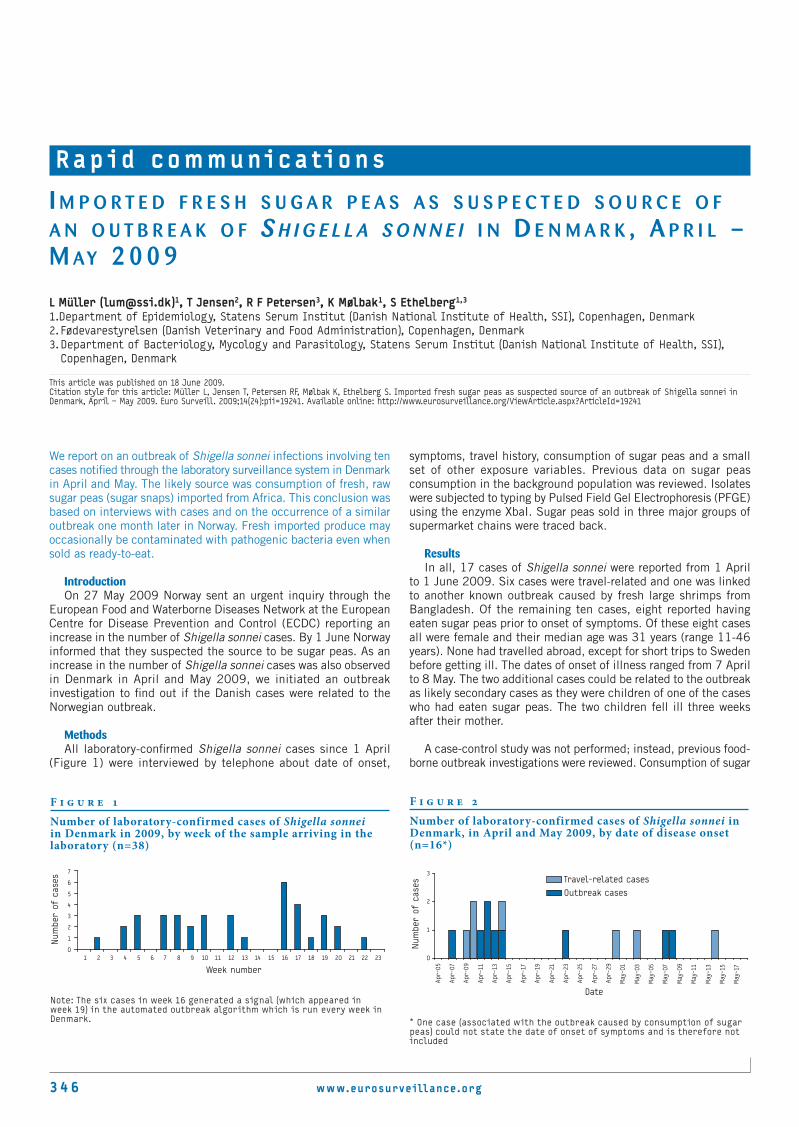
3 4 6 www.eurosurveillance.org
R ap i d com m uni ca ti on s
I m p o r t e d f r e s h s u g a r p e a s a s s u s p e c t e d s o u r c e o f a n o u t b r e a k o f S h i g e l l a S o n n e i I n d e n m a r k , a p r I l – m ay 2 0 0 9
L Müller ([email protected])1, T Jensen2, R F Petersen3, K Mølbak1, S Ethelberg1,3
1.Department of Epidemiology, Statens Serum Institut (Danish National Institute of Health, SSI), Copenhagen, Denmark2. Fødevarestyrelsen (Danish Veterinary and Food Administration), Copenhagen, Denmark3. Department of Bacteriology, Mycology and Parasitology, Statens Serum Institut (Danish National Institute of Health, SSI),
Copenhagen, Denmark
This article was published on 18 June 2009. Citation style for this article: Müller L, Jensen T, Petersen RF, Mølbak K, Ethelberg S. Imported fresh sugar peas as suspected source of an outbreak of Shigella sonnei in Denmark, April – May 2009. Euro Surveill. 2009;14(24):pii=19241. Available online: http://www.eurosurveillance.org/ViewArticle.aspx?ArticleId=19241
We report on an outbreak of Shigella sonnei infections involving ten cases notified through the laboratory surveillance system in Denmark in April and May. The likely source was consumption of fresh, raw sugar peas (sugar snaps) imported from Africa. This conclusion was based on interviews with cases and on the occurrence of a similar outbreak one month later in Norway. Fresh imported produce may occasionally be contaminated with pathogenic bacteria even when sold as ready-to-eat.
IntroductionOn 27 May 2009 Norway sent an urgent inquiry through the
European Food and Waterborne Diseases Network at the European Centre for Disease Prevention and Control (ECDC) reporting an increase in the number of Shigella sonnei cases. By 1 June Norway informed that they suspected the source to be sugar peas. As an increase in the number of Shigella sonnei cases was also observed in Denmark in April and May 2009, we initiated an outbreak investigation to find out if the Danish cases were related to the Norwegian outbreak.
MethodsAll laboratory-confirmed Shigella sonnei cases since 1 April
(Figure 1) were interviewed by telephone about date of onset,
symptoms, travel history, consumption of sugar peas and a small set of other exposure variables. Previous data on sugar peas consumption in the background population was reviewed. Isolates were subjected to typing by Pulsed Field Gel Electrophoresis (PFGE) using the enzyme XbaI. Sugar peas sold in three major groups of supermarket chains were traced back.
ResultsIn all, 17 cases of Shigella sonnei were reported from 1 April
to 1 June 2009. Six cases were travel-related and one was linked to another known outbreak caused by fresh large shrimps from Bangladesh. Of the remaining ten cases, eight reported having eaten sugar peas prior to onset of symptoms. Of these eight cases all were female and their median age was 31 years (range 11-46 years). None had travelled abroad, except for short trips to Sweden before getting ill. The dates of onset of illness ranged from 7 April to 8 May. The two additional cases could be related to the outbreak as likely secondary cases as they were children of one of the cases who had eaten sugar peas. The two children fell ill three weeks after their mother.
A case-control study was not performed; instead, previous food-borne outbreak investigations were reviewed. Consumption of sugar
F i g u r e 1
Number of laboratory-confirmed cases of Shigella sonnei in Denmark in 2009, by week of the sample arriving in the laboratory (n=38)
0
1
2
3
4
5
6
7
1 2 3 4 5 6 7 8 9 10 11 12 13 14 15 16 17 18 19 20 21 22 23
Week number
Num
ber
of c
ases
Note: The six cases in week 16 generated a signal (which appeared in week 19) in the automated outbreak algorithm which is run every week in Denmark.
F i g u r e 2
Number of laboratory-confirmed cases of Shigella sonnei in Denmark, in April and May 2009, by date of disease onset (n=16*)
* One case (associated with the outbreak caused by consumption of sugar peas) could not state the date of onset of symptoms and is therefore not included
0
1
2
3
Apr-
05
Apr-
07
Apr-
09
Apr-
11
Apr-
13
Apr-
15
Apr-
17
Apr-
19
Apr-
21
Apr-
23
Apr-
25
Apr-
27
Apr-
29
May
-01
May
-03
May
-05
May
-07
May
-09
May
-11
May
-13
May
-15
May
-17
Date
Num
ber
of c
ases
Travel-related cases
Outbreak cases

www.eurosurveillance.org 3 47
peas is among the questions included in several of the commonly used trawling questionnaires in Denmark. We looked into three different rounds of trawling questionnaire ‘studies’ performed among cases of a large outbreak of Salmonella Typhimurium U292 [1]. They were done in April, May and August 2008. In these studies 3/10, 2/17 and 0/15 cases reported consumption of sugar peas in a period of seven days prior to illness. This crude comparison indicated to us a significant association between Shigella sonnei infections and consumption of sugar peas (using the persons interviewed in April and May as community controls, comparing 8/8 exposed cases to 5/27 exposed controls, gives a Fisher p-value of < 0.0001).
Preliminary PFGE typing results of isolates from five of the 10 cases associated with sugar pea consumption suggest highly similar patterns. The PFGE patterns of the isolates from Danish patients resemble those obtained from the Norwegian patients but it is still too early to say if they are identical. Further typing results (which will include multilocus variable-number tandem-repeat analysis - MLVA typing) and comparisons between isolates from Denmark and Norway are pending.
The cases were generally able to recall in detail the type of product they had consumed and in which shop they had bought it. Six of the 10 cases associated with the outbreak reported buying sugar peas in supermarkets sharing in part the same distribution systems. Trace-back investigation of the sugar peas showed that they had been bought from a single whole-seller in the Netherlands and that they were of three different varieties which can be distinguished by their shapes, namely sugar snaps, sugar peas (snow peas) and mange touts. They originated predominantly from Kenya (from four different farms), but other batches sold in the same period came from Ethiopia and from Guatemala. The Dutch whole-seller was different from the one that supplied sugar peas to Norway. The two remaining cases may have bought their sugar peas in another group of supermarket chains which in part shares distribution systems with the supermarkets that sold the incriminated sugar peas in Norway. Further investigation into the origin of the sugar peas sold in this chain during April is still ongoing. There were no remains of the batch of sugar peas under suspicion and therefore microbiological analysis was not performed. Laboratory results from samples taken from later batches in two of the supermarket chains did not reveal contamination by either Shigella spp. or Escherichia coli (as indicator for faecal contamination).
DiscussionThe investigation points at sugar peas as the source of this
outbreak. The Danish and the Norwegian outbreaks do not appear to have been caused by the same type of peas, the batch of sugar snaps that was likely contaminated in Denmark was different from the one imported into Norway and also the Danish outbreak occurred one month earlier that the Norwegian outbreak. It is possible, though, that both outbreaks may have been a result of the same contamination event in Kenya; further investigations may cast light on this.
Outbreaks with a high ratio of females among cases may often point to fresh produce as the source. Only one previous outbreak in Denmark has been associated with sugar peas, an outbreak of Shigella flexneri in 2002 in which the epidemiological evidence pointed towards fresh imported sugar snaps of African origin (unpublished). Other fresh tropical vegetables which were eaten raw, have also caused outbreaks of shigellosis in Denmark, most
notable were two Shigella sonnei outbreaks in 2007 [2,3] and one in 1998 [4] both caused by baby corn imported from Thailand.
This outbreak underlines that some fresh vegetables imported into Europe from tropic destinations may pose a food safety hazard. In Denmark fresh imported sugar snaps are sold as a ready-to-eat product. Consumers should be aware that these types of products may pose a risk of microbiological contamination. The sugar snaps will remain crispy after being blanched or boiled shortly and it may be advisable for consumers to heat-treat fresh vegetables of this type before consumption.
References
1. Ethelberg S, Wingstrand A, Jensen T, Sørensen G, Müller L, Lisby M, Nielsen EM, Mølbak K. Large outbreaks of Salmonella Typhimurium infection in Denmark in 2008. Euro Surveill. 2008;13(44):pii=19023. Available from: http://www.eurosurveillance.org/ViewArticle.aspx?ArticleId=19023
2. Lewis HC, Ethelberg S, Olsen KE, Nielsen EM, Lisby M, Madsen SB, et al. Outbreaks of Shigella sonnei infections in Denmark and Australia linked to consumption of imported raw baby corn. Epidemiol Infect 2009;137(3):326-34.
3. Lewis HC, Kirk M, Ethelberg S, Stafford R, Olsen KE, Nielsen EM, Lisby M, Madsen SB, Mølbak K. Outbreaks of shigellosis in Denmark and Australia associated with imported baby corn, August 2007 – final summary. Euro Surveill. 2007;12(40):pii=3279. Available from: http://www.eurosurveillance.org/ViewArticle.aspx?ArticleId=3279
4. Mølbak K, Neimann J. Outbreak in Denmark of Shigella sonnei infection related to uncooked ‘baby maize’ imported from Thailand. Euro Surveill. 1998;2(33):pii=1171. Available from: http://www.eurosurveillance.org/ViewArticle.aspx?ArticleId=1171

3 4 8 www.eurosurveillance.org
R ap i d com m uni ca ti on s
O u t b r e a k O f h e pat i t i s a a m O n g m e n w h O h av e s e x w i t h m e n i n b a r c e l O n a , s pa i n , s e p t e m b e r 2008 – m a r c h 2009
C Tortajada ([email protected])1, P G de Olalla1, R M Pinto2, A Bosch2, J Caylà1
1. Public Health Agency of Barcelona, Barcelona, Spain2. Enteric Virus Laboratory of the Department of Microbiology of the University of Barcelona, Barcelona, Spain
This article was published on 16 April 2009. Citation style for this article: Tortajada C, de Olalla PG, Pinto RM, Bosch A, Caylà J. Outbreak of hepatitis A among men who have sex with men in Barcelona, Spain, September 2008 – March 2009. Euro Surveill. 2009;14(15):pii=19175. Available online: http://www.eurosurveillance.org/ViewArticle.aspx?ArticleId=19175
Between 1 September 2008 and 9 March 2009, 150 cases of hepatitis A were reported in Barcelona, representing a threefold increase compared with the same period in the previous two years. The majority of the cases occurred in adult men, including 87 who reported having sex with men. This indicated the possibility of an outbreak ongoing in the population of men who have sex with men (MSM) and emphasised the need to target this community with more effective vaccination programmes.
IntroductionIn Spain, hepatitis A is a reportable disease defined by acute
hepatitis symptoms combined with the presence of immunoglobulin M antibodies to hepatitis A virus (IgM anti-HAV) [1]. Physicians and laboratories report cases to the local public health agencies. The Public Health Agency of Barcelona is the relevant office for the city of Barcelona, covering a population of 1,600,000 inhabitants. The Health Department of the Government of Catalonia collects cases from all the regional agencies of Catalonia and reports them to the National Centre of Epidemiology in Madrid.
Since September 2008, an increase in the number of reported cases of hepatitis A in the municipality of Barcelona has been observed. Between 1 September 2008 and 9 March 2009, a total of 150 confirmed cases of hepatitis A were reported from the area. In the same period in 2006-7 and 2007-8 the numbers of notified cases were 54 and 55 respectively.
The notification data indicated that the increase may affect predominantly men who have sex with men (MSM). An outbreak alert was raised after five cases had been notified in one day, including four men aged 23-25 years of whom three were known to be MSM. For comparison, in the previous two years, the average number of notifications ranged from 0 to 12 cases per month. This prompted us to undertake a survey among the reported adult male cases, to determine whether they belonged to the group of MSM and whether they engaged in activities associated with an increased risk of hepatitis A infection [2-5].
The outbreak is still ongoing and notifications occur at a frequency of one case per day.
MethodsFor the purpose of the outbreak investigation, a case was defined
as a man over 18 years old who had sex with men, was resident in Barcelona city and had symptoms of acute hepatitis with onset from 1 September 2008 and positive result of IgM anti-HAV test.
To identify cases according to the above definition, all reported hepatitis A patients who were male and older than 18 years, resident in Barcelona city and had symptoms onset from September 2008 were interviewed with a modified questionnaire based on the standard questionnaire for hepatitis A of the Health Department of the Government of Catalonia but with additional questions on sexual behaviour. The interviews were done by telephone or e-mail. Cases that had been reported before the outbreak alert but could fulfill the case definition criteria were re-interviewed retrospectively, using the modified questionnaire.
Questions included having sex with men, number of sexual partners, visiting bathhouses, bars and discos, use of the internet to look for sexual partners, having group sex, and working as sex worker during the two months before symptoms onset, as well as hepatitis A immunisation status and infection with human immunodeficiency virus (HIV).
Contact-tracing was performed according to standard procedures, as done routinely by the local Public Health Agency for every case of hepatitis A reported. During the interview, the patient is asked to identify close contacts. These people are then contacted directly by the Agency and informed about the risk of infection and offered vaccination or postexposure prophylaxis. Vaccination and immunoglobuline is provided free of charge in the Agency offices or, in some cases, administered by healthcare workers visiting the contacts.
Sera from 14 cases who fulfilled the case definition were sent to the Enteric Virus Laboratory of the Department of Microbiology of the University of Barcelona for genetic analysis.
ResultsFrom 1 September 2008 to 9 March 2009, a total of 150
laboratory-confirmed hepatitis A cases were reported. Of the 150 cases, 137 (91%) were older than 18 years, and of these, 126

www.eurosurveillance.org 3 4 9
(84% of the total) were men and 11 (7% of the total) were women. In the equivalent period in 2006-7, of the 54 hepatitis A cases reported, 29 (54%) were older than 18 years, including 21 (39%) men. Similarly, in 2007-8, there were 55 cases in total, 24 (43%) of whom were over 18 years old, including 13 (23%) men.
Of the 126 adult male patients, 107 were interviewed using the modified questionnaire. In response, 87 (69%) declared to have had sex with men and 20 (16%) defined themselves as heterosexual. For the remaining 19 notified cases (15%) this information was not available (Figure).
As a result, 87 persons fulfilled the case definition criteria. The median age of these cases was 33 (IC 95%: 31-34) years. Ten (11%) were HIV-positive. Only one had been vaccinated against hepatitis A and another one had received only one dose of the vaccine.
A considerable proportion of MSM cases reported engaging in activities that may be associated with increased risk of infection. The mean number of sexual partners was four (IC 95%: 3-6), 14 cases (16%) used the internet to look for sexual partners, 26 (30%) frequented discos or bars and 19 (22%) visited bathhouses.
The virological analysis showed HAV genotype IA in sera obtained from 14 patients. The results of phylogenetic analysis are not available yet.
Control measuresVaccination against hepatitis A of all cases’ contacts and
postexposure prophylaxis of close contacts and sexual contacts within 15 days of the last exposure has been recommended. Vaccination and immunoglobuline is offered free of charge in the Public Health Agency of Barcelona.
We performed contact-tracing and offered vaccination and immunoglobuline to those identified. In cases when patients did not have or did not want to give this information (address or telephone), we advised them to inform their partners and close contacts to get the vaccination or immunoglobuline.
In addition, we have also strengthened the existing recommendations for vaccination of MSM by distributing fliers and posters in collaboration with the Spanish “Coordinadora Gai-Lesbiana” a federation which coordinates the activity of gay non-governmental organisations (NGO) and other associations.
The vaccination program for hepatitis A and B in gay bathhouses, which has been in place in Barcelona since 2004, has been reinforced, as well, by increasing the number of visits of healthcare workers and by covering more establishments.
To raise awareness about the possible outbreak, e-mail alerts were sent to microbiology laboratories, local practitioners and hospitals to enhance notification.
Gay organisations were informed about the hepatitis A outbreak affecting MSM, and information about the outbreak was published on some gay websites.
Discussion An increase in the number of reported hepatitis A cases in
Barcelona has been observed since September 2008. Of the 150 cases reported between 1 September 2008 and 9 March 2009, 87 were identified as MSM.
An increase in the number of notifications has recently been observed in other regions of Spain, as well. The data available are from the period between week 36 of 2008 and week 4 of
F i g u r e
Number of cases of hepatitis A among men older than 18 years, by month of onset of symptoms and sexual behaviour, Barcelona, 1 September 2008 - 9 March 2009 (n=122, preliminary data)
Source of data: Public Health Agency of Barcelona, Spain
35
30
25
20
15
10
5
1
num
ber
of c
ases
Men who have sex with men (MSM)
Unknown
Heterosexual
Sep Oct Nov Dec Jan Feb
month of symptom onset

3 5 0 www.eurosurveillance.org
2009. Andalucia has reported an increase from 175 and 125 cases for that period in 2006-7 and 2007-8, respectively, to 350 in 2008-9; Madrid has reported an increase from 95 and 75 to 230 and Castilla – La Mancha has registered an increase from 15 and 20 cases to 60 [6]. It is not clear whether these increases are due to outbreaks and whether they affect a particular risk group but investigations are ongoing.
In Spain vaccination for hepatitis A is not included in the routine immunisation schedule, but is recommended for certain risk groups, including MSM [7].
In recent years, 2002-3 and 2004, two outbreaks of hepatitis A among MSM, affecting 48 and 60 people respectively, were detected in Barcelona. Most of them (80%) were bathhouse users [data from the Public Health Agency of Barcelona, not published]. Similar venues have also been associated with hepatitis A outbreaks elsewhere in Europe [2-5]. The strain identified in the current outbreak is different from the one detected in the MSM outbreaks in 2002-3 and 2004.
Since 2004 a special vaccination programme for hepatitis A and B has been targeted at those who frequent gay bathhouses. Healthcare workers from the Public Health Agency of Barcelona visit these venues and offer information about hepatitis A, B, C and sexually transmitted infections (STI), perform rapid tests for HIV and administer vaccinations for hepatitis A and B. To date, 3,000 bathhouse guests have used this opportunity [data from the Public Health Agency of Barcelona, unpublished].
The scenario in the present outbreak seems to be different from the previous two outbreaks since only 22% of the cases identified as MSM were bathhouse users.
Interventions aimed at the sexual contacts of the cases were difficult to carry out since in a considerable proportion of the cases the partners could not be identified in the course of contact-tracing process.
All but two cases among MSM were unvaccinated. Vaccination of MSM could help to control this outbreak and is crucial in preventing future ones. Thus information campaigns and immunisation programmes which effectively reach the MSM community are needed.
References
1. Commission Decision of 28 April 2008 amending Decision 2002/253/EC laying down case definitions for reporting communicable diseases to the Community network under Decision No 2119/98/EC of the European Parliament and of the Council (notified under document number C(2008) 1589) (Text with EEA relevance). 2008/426/EC. Official Journal L 159, 18/06/2008 P. 0046 - 0090. Available from: http://eur-lex.europa.eu/LexUriServ/LexUriServ.do?uri=OJ:L:2008:159:0046:01:EN:HTML
2. Leentvaar-Kuijpers A, Kool JL, Veugelers PJ, Coutinho RA, van Griensven GJ. An outbreak of hepatitis A among homosexual men in Amsterdam, 1991-1993. Int J Epidemiol. 1995;24(1):218-22.
3. Reintjes R, Bosman A, de Zwart O, Stevens M, van der Knaap L, van den Hoek K. Outbreak of hepatitis A in Rotterdam associated with visits to ‘darkrooms’ in gay bars. Commun Dis Public Health. 1999;2(1):43-6.
4. Bell A, Ncube F, Hansell A, Davison KL, Young Y, Gilson R, et al. An outbreak of hepatitis A among young men associated with having sex in public venues. Commun Dis Public Health. 2001;4(3):163-70.
5. Mazick A, Howitz M, Rex S, Jensen IP, Weis N, Katzenstein TL, Haff J, Mølbak K. Hepatitis A outbreak among MSM linked to casual sex and gay saunas in Copenhagen, Denmark. Euro Surveill. 2005;10(5):pii=536. Available from: http://www.eurosurveillance.org/ViewArticle.aspx?ArticleId=536
6. Informe semanal de vigilancia 2 de marzo de 2009 [Weekly epidemiological report, 2 March 2009]. Red Nacional de Vigilancia Epidemiologica. Servicio Vigilancia Epidemiologica. Centro Nacional de Epidemiologia. Available from: http://www.isciii.es/htdocs/centros/epidemiologia/boletin_red/IS-090302-WEB.pdf
7. Protocol d’actuació per al control i prevenció de l’hepatitis A [Protocol of action for the control and prevention of hepatitis A]. Generalitat de Cataluña. Departament de Salut: Barcelona; 2004. Available from: http://www.gencat.cat/salut/depsalut/html/ca/dir2829/prothepatitis2009.pdf

www.eurosurveillance.org 3 51
Rap i d com m uni ca ti on s
A n A p h y l A x i s f o l l o w i n g u n n e c e s s A r y m e n i n g o c o c c A l c h e m o p r o p h y l A x i s o f A h e A lt h c A r e w o r k e r
F Giovanetti ([email protected])11. Azienda Sanitaria Locale CN2 Alba Bra, Dipartimento di Prevenzione (Local Health Unit CN2 - Alba Bra, Department of
Prevention), Alba, Piedmont Region, Italy
This article was published on 14 May 2009. Citation style for this article: Giovanetti F. Anaphylaxis following unnecessary meningococcal chemoprophylaxis of a healthcare worker. Euro Surveill. 2009;14(19):pii=19207. Available online: http://www.eurosurveillance.org/ViewArticle.aspx?ArticleId=19207
We report a case of anaphylaxis following meningococcal chemoprophylaxis of a healthcare worker, despite no history of direct contact with a patient who had meningococcal disease. The public health implications of this case are discussed.
IntroductionChemoprophylaxis of meningococcal disease is usually
recommended for close contacts, such as household members and persons directly exposed to a patient’s respiratory secretions, as these have been shown to have a higher risk of invasive meningococcal disease [1]. However, chemoprophylaxis is not recommended for low risk or indirect contacts, partly because of possible adverse outcomes. These include adverse events to prophylactic antibiotics, development of antibiotic resistance and eradication of non-pathogenic Neisseria species which may confer protection against Neisseria meningitidis [2,3]. In particular, administration of prophylactic antibiotics for healthcare workers is recommended only after an unprotected exposure to respiratory secretions of an index case. We describe a case of anaphylaxis following chemoprophylaxis of a healthcare worker with no history of direct contact with a patient who had sepsis caused by N. meningitidis. The public health implications of this case are discussed.
Case descriptionIn March 2009, a woman in her 40s was admitted to a hospital
in the Piedmont Region, Italy, with a one-day history of fever and myalgia. On admission, the patient was unconscious, with hypotension, tachycardia, acidosis and a truncal petechial rash. Clinical and laboratory features suggested a septic shock with disseminated intravascular coagulation. Her condition rapidly worsened and death occurred two hours after admission, despite resuscitation.
As the presumptive diagnosis was meningococcal disease, pathologists collected samples of blood, cerebrospinal fluid and petechial smears during the post mortem examination. PCR was subsequently performed at the Istituto Superiore di Sanità Infectious Diseases Laboratory in Rome, and N. meningitidis serogroup B was detected in all samples.
In accordance with the local public health unit, the hospital management recommended treatment with a single 500 mg dose of ciprofloxacin for two doctors who had had unprotected exposure to the respiratory secretions of the patient. Chemoprophylaxis was also administered to three healthcare workers of the hospital
staff. Outside the hospital setting, the contact tracing identified two household members as well as eight contacts who had been presumably exposed to the respiratory secretions of the patient and all took chemoprophylaxis. No secondary cases occurred in the following 30 days.
The following day, a healthcare worker in the same unit as one of the exposed doctors decided to take a single 500 mg dose of ciprofloxacin for fear of contracting meningococcal disease, although she had no history of direct contact with the index case. On this basis, she had not been classified by the hospital management as a close contact and thus had not been offered chemoprophylaxis. Approximately 20 minutes after taking ciprofloxacin at the workplace, she was admitted to the emergency room with pharyngeal oedema, tongue swelling and generalised skin rash. The patient recovered gradually after administration of adrenalin, antihistamines and corticosteroids. She had used ciprofloxacin in the past without any adverse reaction.
DiscussionInvasive meningococcal disease is uncommon in Italy.
Approximately 180 cases (0.3 per 100,000 population) are notified annually to the infectious diseases surveillance system [4]. The highest incidence is seen among children under five years old. In the Piedmont Region, an area in north-west Italy with 4.3 million inhabitants and active laboratory-based surveillance, the incidence appears to be higher: 0.4-0.7 per 100,000 population, with a constant peak during the first year of life, ranging from five to six cases per year per 100,000 population [5].
Chemoprophylaxis is recommended in Italy only for persons with close contact to the index case up to one week before the onset of the patient’s symptoms. Close contacts include: household members, contacts in child-care centres, and persons directly exposed to the patient’s oral secretions [6]. Giving chemoprophylaxis to people who have not been in close contact with an index case has not proved to be effective in preventing secondary cases and is usually not recommended [3,7].
In Italy, national guidelines on meningococcal chemoprophylaxis for healthcare workers are not available. Nevertheless, the regional health authorities as well as hospitals have developed standard operating procedures, usually based on international authoritative sources, such as the Centers for Disease Control and Prevention (CDC) in Atlanta, United States (US). In accordance with CDC guidance, the operating procedures of the local health unit involved in this case recommend chemoprophylaxis for healthcare workers

3 5 2 www.eurosurveillance.org
after an unprotected airway exposure to infectious respiratory droplets within a distance of 1 m from a probable or confirmed case of meningococcal disease; this may happen typically during mouth-to-mouth resuscitation or management of an endotracheal tube [6,8].
A study in the United Kingdom found an attack rate of 0.8 per 100,000 healthcare workers in close contact with cases of meningococcal disease, i.e. 25 times higher than in the general population [9]. The study identified three cases of meningococcal disease in healthcare workers during a period of 15 years: all had spent at least 30 minutes in contact with the index case immediately before or after hospital admission, all had been exposed to the patients’ respiratory droplets, and none had used face shields and surgical masks or taken prophylactic antibiotics.
The fluoroquinolone ciprofloxacin is often used for meningococcal chemoprophylaxis in adults because it can be given as a single oral dose, is effective in eradicating meningococcal carriage and does not interact with oral contraceptives. For the same reasons, however, unnecessary chemoprophylaxis is more likely to occur with ciprofloxacin than with other prophylactic antibiotics. Rifampicin requires a total of four doses in the course of two days and can interfere with oral contraceptives; ceftriaxone is administered as a single dose, but is not popular because it can only be administered parenterally.
Anaphylaxis following ciprofloxacin administration has been described before. In particular, three cases of anaphylactoid reactions were reported after oral administration of 500 mg ciprofloxacin to 3,200 students after two cases of meningococcal disease in the same university [10]. Limited data on the magnitude of allergic reactions following administration of drugs are available, mainly because clinical manifestations are heterogeneous (from mild to severe and potentially life-threatening) and furthermore some reactions suggesting an immunologic pathogenesis might be linked to a non-allergic mechanism. Likewise, the incidence of allergic reactions induced by oral antibiotics such as ciprofloxacin is difficult to estimate. However, according to a case/non-case study conducted on data from a passive adverse events surveillance programme, fluoroquinolones were associated with a significant increase in the reporting odds ratio of allergic reactions (2.09, 95% confidence interval (CI): 1.85-2.36) [11]. Moreover, an incidence of 5.4 (95% CI: 4.4-6.5) allergic reactions per 10,000 first administrations of ciprofloxacin has been derived from the database of a large health insurance company [12].
Another reason for concern is the potential development of antibiotic resistance. Three cases of meningococcal disease caused by a N. meningitidis serogroup B strain resistant to ciprofloxacin were recently reported in the United States [13]. The widespread use of fluoroquinolones, which are commonly prescribed in the United States [14], and the consequent emergence of resistant strains may explain these findings.
Finally, prophylactic antibiotics can eliminate carriage of N. lactamica, which leads to cross-protective immunity against N. meningitidis and therefore may confer protection against meningococcal disease [15,16]. This concerns young children in particular, since carriage of N. lactamica is inversely related to age [16].
Quantitative data about the overuse of chemoprophylaxis are scarce. A study in the United Kingdom evaluated prescribing
of chemoprophylaxis for contacts of meningococcal disease by general practitioners and hospital staff [17]. Prescribing by hospital doctors was consistent with official recommendations, whereas general practitioners prescribed 118% more chemoprophylaxis than recommended. Furthermore, the highest level of unrecommeded prescriptions was observed in regions where there were both high incidence rates and high levels of publicity surrounding the cases. Most likely, this inappropriate prescribing is client-driven because meningococcal disease raises anxiety among the involved population.
ConclusionsUse of prophylactic antibiotics against meningococcal disease
can lead to potentially severe adverse events, development of antibiotic resistance and eradication of non-pathogenic Neisseria species that may elicit cross-protective immunity. Therefore, information should be provided to the public and to healthcare workers about the potential risks from indiscriminate use of prophylactic antibiotics. Available data should be used to compare the risks related to different patterns of exposure to a case of meningococcal disease versus the possible adverse outcomes of chemoprophylaxis. To this end, thorough counselling and communication of the population-based and individual-based risk represent a key factor in the public health management of meningococcal disease cases.
References
1. Purcell B, Samuelsson S, Hahne SJ, Ehrhard I, Heuberger S, Camaroni I, et al. Effectiveness of antibiotics in preventing meningococcal disease after a case: systematic review. BMJ. 2004;328(7452):1339.
2. Kristiansen BE, Knapsog AB. Secondary prevention of meningococcal disease. BMJ. 1996;312(7031):591-2.
3. Health Protection Agency Meningococcus Forum. Guidance for public health management of meningococcal disease in the UK. August 2006. Available from: http://www.hpa.org.uk/web/HPAwebFile/HPAweb_C/1194947389261
4. Sistema Informatizzato Malattie Infettive (SIMI) [Information system for infectious diseases]. Istituto Superiore di Sanità. Dati di sorveglianza sulle meningiti [Surveillance data on meningitis]. SIMI [Accessed 25 April 2009 ]. Available from: http://www.simi.iss.it/dati.htm
5. Barale A, Criveller T, Lombardi D. Sorveglianza di laboratorio delle infezioni batteriche invasive in Regione Piemonte. Risultati attività anno 2006. [Laboratory surveillance of invasive bacterial infections in Regione Piemonte. Activity results for the year 2006]. Available from: http://epidem.asl20.piemonte.it/?sezione=323
6. Bilukha OO, Rosenstein N, National Center for Infectious Diseases, Centers for Disease Control and Prevention (CDC). Prevention and control of meningococcal disease: recommendations of the Advisory Committee on Immunization Practices (ACIP). MMWR Recomm Rep. 2005;54(RR-7):1-21.
7. American Academy of Pediatrics. Red Book 2006. Report of the Committee on Infectious Diseases. 27th ed. Elk Grove Village, IL: American Academy of Pediatrics 2006.
8. Bolyard EA, Tablan OC, Williams WW, Pearson ML, Shapiro CN, Deitchmann SD. Guideline for infection control in healthcare personnel, 1998. Hospital Infection Control Practices Advisory Committee. Infect Control Hosp Epidemiol. 1998;19(6):407-63.
9. Gilmore A, Stuart J, Andrews N. Risk of secondary meningococcal disease in health-care workers. Lancet. 2000;356(9242):1654-5.
10. Burke P, Burne SR. Allergy associated with ciprofloxacin. BMJ. 2000;320(7236):679.
11. Salvo F, Polimeni G, Cutroneo PM, Leone R, Confortic A, Moretti U, et al. Allergic reactions to oral drugs: A case/non-case study from an Italian spontaneous reporting database (GIF). Pharmacol Res. 2008;58(3-4):202-7.
12. Johannes CB, Ziyadeh N, Seeger JD, Tucker E, Reiter C, Faich G. Incidence of allergic reactions associated with antibacterial use in a large, managed care organisation. Drug Saf. 2007;30(8):705-13.
13. Wu HM, Harcourt BH, Hatcher CP, Wei SC, Novak RT, Wang X, et al. Emergence of ciprofloxacin-resistant Neisseria meningitidis in North America. N Engl J Med. 2009;360(9):886-92.

www.eurosurveillance.org 3 53
14. Steinman MA, Gonzales R, Linder JA, Landefeld CS. Changing Use of Antibiotics in Community-Based Outpatient Practice, 1991–1999. Ann Intern Med. 2003;138(7):525-33.
15. Yazdankhah SP, Caugant DA. Neisseria meningitidis: an overview of the carriage state. J Med Microbiol 2004;53(Pt 9):821–32.
16. Bennet JS, Griffiths DT, Mc Carthy ND, Sleeman KL, Jolley KA, Crook DW, et al. Genetic diversity and carriage dynamics of Neisseria lactamica in infants. Infect Immun. 2005;73(4):2424–32.
17. Marks PJ, Neal KR. Variations in chemoprophylaxis for meningococcal disease: a retrospective case note review, analysis of routine prescribing data and questionnaire of general practitioners. BMC Public Health 2001;1:16. Available from: http://www.biomedcentral.com/1471-2458/1/16

3 5 4 www.eurosurveillance.org
R ap i d com m uni ca ti on s
C h i k u n g u n ya i n f e C t i o n C o n f i r m e d i n a B e l g i a n t r av e l l e r r e t u r n i n g f r o m P h u k e t ( t h a i l a n d )
E Bottieau ([email protected])1, M Van Esbroeck1, L Cnops1, J Clerinx1, A Van Gompel1
1. Department of Clinical Sciences, Institute of Tropical Medicine, Antwerp, Belgium
This article was published on 25 June 2009. Citation style for this article: Bottieau E, Van Esbroeck M, Cnops L, Clerinx J, Van Gompel A. Chikungunya infection confirmed in a Belgian traveller returning from Phuket (Thailand). Euro Surveill. 2009;14(25):pii=19248. Available online: http://www.eurosurveillance.org/ViewArticle.aspx?ArticleId=19248
Chikungunya infection has been increasingly reported in international travellers following its epidemic re-emergence in the Indian Ocean islands in 2006 and its spread to southern Asia thereafter. We describe the first case of chikungunya in a Belgian traveller returning from Phuket, Thailand and discuss the potential implications of chikungunya cases imported to European countries for patient management and public health.
Introduction Chikungunya is a tropical arboviral disease transmitted
by mosquitoes belonging to the genus Aedes. Infection is characterised by an acute-onset fever, rash and incapacitating joint pain. Chikungunya virus belongs to the Alphavirus genus of the family Togaviridae, and was first isolated in Tanzania in 1953 [1]. Although periodic outbreaks occurred ever since throughout Africa as well as in southeast Asia, they were typically self-limiting and rarely had a broad geographic extension. After a long period of quiescence, chikungunya re-emerged in 2004 on the coast of Kenya and hit the islands of Comoros and Réunion in 2005, where high attack rates and large epidemics were reported. It spread then in a sweeping succession of outbreaks to other islands of the Indian Ocean and reached India in 2006 where more than 1,000,000 suspected cases were reported [1]. In the following years, Sri Lanka, Indonesia, Singapore and Malaysia were successively affected, including the south of Thailand in the late 2008. Since January 2009, more than 20,000 cases have been reported in Thailand, with evidence of spreading to the northern provinces [2]. We describe here the case of a Belgian traveller who presented in our centre with a chikungunya infection after having stayed exclusively in the popular tourist destination of Phuket (Thailand).
Case reportA Belgian woman in her fourties presented in mid April 2009
at the travel clinic of the Institute of Tropical Medicine, Antwerp, Belgium with symptoms of recurrent high-grade fever (up to 39°5C), headache, generalised muscle aches and skin rash for the last four days. She had returned two days before from a holiday trip to Thailand where she had stayed exclusively in Phuket for 14 days. She had consulted in a hospital in Phuket when the symptoms started and a dengue NS1 antigenic test was performed and reported as negative. In our centre, the patient presented with a slight macular skin rash on the trunk and limbs and a slightly swollen right ankle. Laboratory tests at the time of presentation showed a leucopenia (2.290 WBC/µL), a borderline thrombocytopenia (138.000 platelets/µL) and elevated alanine aminotransferase
(78 IU/L; normal 9-52 IU/L), aspartate aminotransferase (81 IU/L; normal 14-36 IU/L) and lactate dehydrogenase (742 IU/L; normal 313-618 IU/L). Blood smears for malaria and blood cultures were negative. Dengue fever was considered to be the most likely diagnosis.
Fever decreased the day following the consultation, but during the next two-three weeks, the patient developed severe joint aches in the feet, fingers and right wrist without evident swelling. Paired serology against dengue remained negative, as well as testing for leptospirosis, rickettsiosis, Q fever, West Nile virus, Toxoplasma gondii and cytomegalovirus. Chikungunya was considered as a differential diagnosis and serology by indirect immunofluorescence, adapted from Panning et al. (2008) [3], revealed a more than 4-fold increase of immunoglobulin (Ig) G titres against chikungunya (from 1/16 to 1/256 within 14 days). A real-time polymerase chain reaction testing, adapted from Panning et al. [3], of the acute-phase serum taken upon the first presentation in our clinic, was positive for the chikungunya virus (cycle threshold-value 33.48), while the serum sample taken 14 days later was negative. The patient fully recovered, but joint pain persisted until the beginning of June despite symptomatic treatment.
Upon receipt of the positive test result, national and regional health authorities were notified. A specific project called “Emerging Threats” has been indeed established in the Scientific Institute of Public Health of Belgium since September 2008. It main objective is to implement a national surveillance for tick-borne encephalitis, West Nile fever and chikungunya. Our laboratory, which is the national reference centre for tropical diseases, takes part in this project by reporting monthly all serological and/or molecular diagnoses of West Nile and chikungunya infection.
Discussion and conclusionFollowing the successive waves of outbreaks spreading from east
Africa to southeast Asia, chikungunya infection has been reported increasingly in returning western travellers or immigrants returning from visits to their home countries during the last couple of years [3-8]. In Belgium for example, 54 cases of chikungunya have been confirmed since 2006 (38 in 2006, 9 in 2007, 7 in 2008) mainly in travellers returning from countries with recent epidemics such as Mauritius (n=17), Réunion Island (n=10), Sri Lanka (n=4), Madagascar and India (n=3 for each) [unpublished data]. Compared to this, approximately 50 imported cases of dengue are diagnosed every year in our country, mainly acquired in southeast Asia/western

www.eurosurveillance.org 3 5 5
Pacific and Latin America, with Thailand, Indonesia and India being the leading countries of infection [9]. To our knowledge, this is the first imported case in Europe of chikungunya acquired undoubtedly in Phuket, Thailand. Our observation is worth reporting because this region is probably one of the most popular travel destinations in southeast Asia. We therefore expect that significant numbers of susceptible travellers might become infected in Phuket. This would result in an increase of symptomatic travellers returning from this area attending the travel or primary care settings in various western countries and make chikungunya an important differential diagnosis in these patients.
We demonstrated recently that the pre-test probability for a traveller returning from southern Asia with fever to be diagnosed with dengue was about 15% [10]. If a skin rash, a leucopenia and a thrombocytopenia are present like in the case under discussion here, with respective adjusted positive likelihood ratios of 2.8, 3.3 and 2 [10], the post-test(s) probability for dengue rises above 50%, explaining why this was the foremost diagnosis we considered. The differentiation between chikungunya and dengue infections is often difficult [4,6]. Skin rash tends to be more frequent in chikungunya patients (75-80%) than in dengue patients (about 50%) [4-8,10]. In contrast, leucopenia and thrombocytopenia seem to occur rather similarly in both diseases, although no large comparative series have been published so far. In our case, joint symptoms became prominent during the course of the disease [7,8] and paired serology against dengue remained negative. This encouraged us to look for chikungunya as an alternative diagnosis which was ultimately confirmed by further serological and molecular investigations.
Potential implications for EuropeBesides the implications for managing individual patients,
chikungunya has a potential for autochthonous transmission in Europe. This was amply demonstrated by the outbreak of chikungunya in Italy in the summer of 2007, presumably triggered by a viraemic index case – an Indian traveller returning from a visit to friends and relatives in India [11,12]. Local transmission was made possible by the presence of the receptive vector, Aedes albopictus, in Italy. This vector is established in other southern European countries as well, but not in Belgium so far although it has been sporadically introduced [13]. However, several models with different climate change scenarios predict a further spread of A. albopictus to northern Europe and consider parts of Belgium as suitable for the mosquito establishment [13]. Since the vector is sporadically introduced and might be established in Belgium in the future and since both chikungunya and dengue viruses are diagnosed repetitively in returning travellers, the risk for local epidemics, although extremely limited now, is likely to increase.
In conclusion, we have observed a case of dengue-like illness finally diagnosed as chikungunya infection and acquired in Phuket, Thailand. Phuket is a popular tourist spot in southeast Asia, increasing the likelihood of further imported cases in western countries while the local epidemic in Thailand is ongoing. Despite the similarity with dengue features, chikungunya infection should be recognised early in returning travellers because of its specific protracted morbidity and its potential for local outbreaks in European countries.
References
1. Pialoux G, Gaüzère BA, Jauréguiberry S, Strobel M. Chikungunya, an epidemic arbovirosis. Lancet Infect Dis. 2007;7(5):319-27.
2. ProMED-mail. Chikungunya: Thailand. Archive Number: 20090527.1975, 27 May 2009. Boston US: International Society for Infectious Diseases. Available from: http://www.promedmail.org
3. Panning M, Grywna K, Van Esbroeck M, Emmerich P, Drosten C. Chikungunya fever in travelers returning to Europe from the Indian Ocean region, 2006. Emerg Infect Dis. 2008;14(3):416-22.
4. Beltrame A, Angheben A, Bisoffi Z, Monteiro G, Marocco S, Calleri G, et al. Imported Chikungunya Infection, Italy. Emerg Infect Dis 2007;13(8):1264-6.
5. Hochedez P, Jauréguiberry S, Debruyne M, Bossi P, Hausfater P, Brucker, et al. Chikungunya infection in travelers. Emerg Infect Dis. 2006;12(10):1565-7.
6. Nicoletti L, Ciccozzi M, Marchi A, Fiorentini C, Martucci P, D’Ancona F, et al. Chikungunya and dengue viruses in travelers. Emerg Infect Dis. 2008;14(1):177-8.
7. Simon F, Parola P, Grandadam M, Fourcade S, Oliver M, Brouqui P, et al. Chikungunya infection: an emerging rheumatism among travelers returned from Indian Ocean islands. Report of 47 cases. Medicine (Baltimore). 2007;86(3):123-37.
8. Taubitz W, Cramer JP, Kapaun A, Pfeffer M, Drosten C, Dobler G, et al. Chikungunya fever in travelers: clinical presentation and course. Clin Infect Dis. 2007;45(1):e1-e4.
9. Scientific Institute of Public Health of Belgium, Epidemiology Unit. Annual Reports on Infectious Diseases, Dengue. [in Dutch]. Brussels. Available from: http://www.iph.fgov.be/epidemio/epinl/plabnl/plabannl/07_denn_r.pdf
10. Bottieau E, Clerinx J, Van den Enden E, Van Esbroeck M, Colebunders R, Van Gompel A, et al. Fever after a stay in the tropics: diagnostic predictors of the leading tropical conditions. Medicine (Baltimore). 2007;86(1):18-25.
11. Angelini R, Finarelli AC, Angelini P, Po C, Petropulacos K, Macini P, et al. An outbreak of chikungunya fever in the province of Ravenna, Italy. Euro Surveill. 2007;12(36):pii=3260. Available from: http://www.eurosurveillance.org/ViewArticle.aspx?ArticleId=3260
12. Rezza G, Nicoletti L, Angelini R, Romi R, Finarelli AC, Panning M, et al. Infection with chikungunya virus in Italy: an outbreak in a temperate region. Lancet. 2007;370(9602):1840-6.
13. European Centre for Disease Prevention and Control (ECDC). Development of Aedes albopictus risk maps. Stockholm: ECDC; 2009. Available from: http://www.ecdc.europa.eu/en/files/pdf/Publications/0905_TER_Development_of_Aedes_albopictus_risk_maps.pdf

3 5 6 www.eurosurveillance.org
R ap i d com m uni ca ti on s
T r e n d s i n T h e e p i d e m i o l o g y o f d e n g u e f e v e r a n d T h e i r r e l e va n c e f o r i m p o r TaT i o n To e u r o p e
T Jelinek ([email protected])11. Berlin Centre for Travel and Tropical Medicine, Berlin, Germany
This article was published on 25 June 2009. Citation style for this article: Jelinek T. Trends in the epidemiology of dengue fever and their relevance for importation to Europe. Euro Surveill. 2009;14(25):pii=19250. Available online: http://www.eurosurveillance.org/ViewArticle.aspx?ArticleId=19250
Dengue fever continues to spread globally, causing major epidemics and putting major strain on health systems in affected countries. For imported dengue in Europe, south east Asia is the most important region of origin, followed by Latin America, the Indian subcontinent, the Caribbean, and Africa. Information regarding mosquito protective measures is highly recommended for all travellers to affected areas.
Introduction Dengue fever has developed into one of the world’s major
emerging infectious diseases. The infection is by now seen as a global epidemic with recorded prevalence in more than 120 countries [1]. It appears that dengue originated from Africa and was introduced to Asia some 600 years ago. The first recognised dengue epidemics occurred almost simultaneously in Asia, Africa, and North America in the 1780s. Dengue is transmitted by Aedes mosquitoes, particularly A. aegypti and, less important, A. albopictus. These mosquitoes travel well, particularly in cargo ships and the four subtypes of dengue virus have spread to most tropical and subtropical countries in their wake. During the last 200 years, spread of the disease has increased, reaching epidemic proportions during the last three decades. Since the late 1990s, dengue is the most important mosquito-borne disease affecting humans after malaria, with around 40 million cases of dengue fever and several hundred thousand cases of dengue haemorrhagic fever (DHF) each year. The main endemic areas are Latin America, the Caribbean, Africa, south and southeast Asia, and parts of the Pacific Region. For Europe, dengue remains an imported disease, even though A. albopictus has become established in some parts of the continent.
The pathogenesis of DHF is not fully understood, but it has been well documented that secondary dengue infection is a major risk factor of the disease [2,3]. As a consequence, and maybe also under genetic control, European travellers rarely develop DHF [4]. A high percentage of dengue infections in travellers occur without any symptoms [5]. However, the important role of travellers is recognised to introduce more virulent dengue strains into endemic areas where usually only mild disease occurs [6], or into non-endemic areas but where the mosquito vector is common [7].
Recent developmentsWhile dengue activity remains quite high in Asia, Latin America
has seen a particular increase of major epidemics during the last two years. Rio de Janeiro experienced serious outbreaks in 2002 and again in 2008, each straining the health infrastructure severely
[8]. A severe epidemic developed in Bolivia in early 2009 with several 10,000 patients, prompting the government to declare a state of emergency for the nation [9]. Most recently, Argentina declared a dengue outbreak in the northern provinces of Salta, Jujuy, Catamarca, Chaco, and Corrientes with more than 26,000 cases [10]. The disease has spread as far as the capital Buenos Aires. On the other side of the Pacific Ocean, an outbreak of dengue fever erupted in December 2008 in northern Queensland, Australia. Located around a focus in Cairns, it spread to other parts of the tropical north of Australia [11].
Dengue importation into EuropeReports on dengue in international travellers have increased, too.
Both the increasing international air travel and the increasing activity of dengue in the tropics are responsible for the increased chance that healthcare providers, including those in western countries, are more and more likely to be confronted with imported dengue infections. In various studies at travel clinics, dengue infection was the most common cause for fever in returning travellers [4,12,13]. Since dengue surveillance, if performed at all, is passive, and since dengue infection presents either as a short and self-limiting viral disease or even asymptomatically, it is certainly one of the under-diagnosed tropical infections in travellers.
The European Network on Imported Infectious Disease Surveillance (TropNetEurop) was founded in 1999 to detect emerging infections of potential regional, national, or global impact at their point of importation into the European area. The network currently consists of 57 collaborating centres in 17 European countries. Annually, the collaborating centres give approximately 220,000 consultations prior to travel, and treat 57,000 patients post-travel [14]. From comparisons between national notification numbers and patients reported to the network, it can be safely assumed that TropNetEurop is covering around 12% of the European patients with imported infectious diseases. Within this network, the number of reported dengue cases increased from 64 in 1999 to a maximum of 224 in 2002 and remained at 100-170 since then. For 2008, 116 cases have been reported. The median age in this population is 38 years (range 12–73 years). The median duration of travel during which patients acquired the dengue infection decreased from 38 days in 1999 to 21 days in 2008 [15].
In 2008, 43% of the dengue cases were acquired by patients who returned from travel to countries in south east Asia, 14% were

www.eurosurveillance.org 3 57
imported from Latin America, 12% from the Indian subcontinent, 11% from the Caribbean, and 4% from Africa (Figure 1).
This distribution reflects two different aspects: worldwide dengue activity and countries’ popularity as tourist destinations. Thailand, Vietnam, and Indonesia are not only highly endemic areas for dengue viruses, but they are also very popular destinations for European tourists. Thailand alone is responsible for almost 30% of all travel associated dengue infections in our network over the past six years. Current developments mirror the epidemics in south America, with stronger reporting from Bolivia and Argentina. In addition, unusually strong signals come also from Eritrea, Jordan, Pakistan, Papua New Guinea, South Africa, Dominican Republic, and Suriname (Figure 2).
Reporting over the past years in Europe has shown that most dengue patients are European travellers (87% in 2008). Dengue
haemorrhagic fever and death have remained rare events in European travellers but are being reported almost every year. In 2008, two out of 116 patients developed complications, and one patient died of DHF.
Overall, TropNetEurop has documented a clear increase of reported dengue cases during the early 2000s, reaching a plateau since 2002. This is in line with national reporting in most European countries, as documented by the World Health Organization (WHO) Europe centralized information system for infectious diseases (CISID) database [16]. The exceptions of the rule are France with several recent dengue outbreaks in its overseas territories, and Germany, with an increase from 218 reported patients in 2002 to 263 in 2007.
Future outlookIt appears that the spread of dengue is only limited by the spread
of its vector mosquitoes, in particular A. aegypti. Since Aedes spp. has proven to be exceptionally adapted to human habitation, its global spread cannot be controlled effectively. Dengue has moved to North America, Australia, east Asia, the Pacific, and eastern Africa. Its imminent spread to Europe has to be anticipated. However, a series of phase 2 trials for efficacy and reactogenicity of dengue tetravalent vaccines has been started in early 2009 [17]. Further trials are listed to follow soon, promising the availability of effective control tools within a few years. A dengue vaccine with high protective efficacy could change the whole picture of the current epidemic. However, as long as no effective vaccine is available, dengue viruses will present a serious threat to European travellers, to European countries with growing populations of potential vector mosquitoes, and an even greater threat to those living in already endemic countries.
References
1. Halstead SB. Dengue. Lancet. 2007;370(9599):1644-52.
2. Halstead SB. Pathogenesis of dengue: challenges to molecular biology. Science. 1988;239(4839):476-81.
3. Mongkolsapaya J, Dejnirattisai W, Xu XN, Vasanawathana S, Tangthawornchaikul N, Chairunsri A, et al. Original antigenic sin and apoptosis in the pathogenesis of dengue hemorrhagic fever. Nat Med. 2003;9(7):921-7.
4. Jelinek T, Mühlberger N, Harms G, Corachán MP, Grobusch MP, Knobloch J, et al. Epidemiology and clinical features of imported dengue fever in Europe: Sentinel surveillance data from TropNetEurop. Clin Infect Dis. 2002;35(9):1047-52.
5. Cobelens FG, Groen J, Osterhaus AD, Leentvaar-Kuipers A, Wertheim-van Dillen PM, Kager PA. Incidence and risk factors of probable dengue virus infection among Dutch travellers to Asia. Trop Med Int Health. 2002;7(4):331-8.
6. Messer WB, Gubler DJ, Harris E, Sivananthan K, de Silva AM. Emergence and global spread of a dengue serotype 3, subtype III virus. Emerg Infect Dis. 2003;9(7):800-9.
7. Hanna JN, Ritchie SA, Phillips DA, Serafin IL, Hills SL, van den Hurk AF, et al. An epidemic of dengue 3 in far north Queensland, 1997-1999. Med J Aust. 2001;174(4):178-82.
8. ProMED-mail. Dengue/DHF Update 2008 (47). Archive Number: 20081104.3459, 4 November 2008. Boston US: International Society for Infectious Diseases. Available from: http://www.promedmail.org
9. ProMED-mail. Dengue/DHF Update 2009 (10). Archive Number: 20090302.0854, 2 March 2009. Boston US: International Society for Infectious Diseases. Available from: http://www.promedmail.org
10. ProMED-mail. Dengue/DHF Update 2009 (22). Archive Number: 20090601.2040, 1 June 2009. Boston US: International Society for Infectious Diseases. Available from: http://www.promedmail.org
11. ProMED-mail. Dengue/DHF Update 2009 (21). Archive Number: 20090525.1952, 25 May 2009.. Boston US: International Society for Infectious Diseases. Available from: http://www.promedmail.org
F i g u r e 1
Imported dengue fever cases in Europe in 2008 by country of origin (n=118)
Countries by pct. of total
0–2% 2–4% 4–6% 6–8% 8–10% >10%
Colourless = Cases were neither observed in the present months nor in the past reference periodMissing = Cases were observed in the past reference period but not in the present months
F i g u r e 2
Cases of imported dengue fever as of 1 June 2009, TropNetEurop data (n=1,419 entries in the database)
Probabilitymissing ≥0.1 <0.1 <0.05 <0.01 <0.001

3 5 8 www.eurosurveillance.org
12. Stephan C, Allwinn R, Brodt HR, Knupp B, Preiser W, Just-Nübling G. Travel-acquired dengue infection: clinical spectrum and diagnostic aspects. Infection. 2002;30(4):225-8.
13. O’Brien D, Tobin S, Brown GV, Torresi J. Fever in returned travelers: review of hospital admissions for a 3-year period. Clin Infect Dis. 2001;33(5):603-9.
14. European Network on Imported Infectious Disease Surveillance (TropNetEurop) [homepage on the internet]. [accessed 22 June 2009]. Available from: http://www.tropnet.net/index_2.html
15. TropNetEurop Friends & Observers Sentinel Surveillance Report: May 2009. Dengue Fever in 2008. TropNetEurop. 2009. [accessed 22 June 2009]. Available from: http://www.tropnet.eu/reports_friends/reports_friends_index.html ,
16. World Health Organization (WHO) Regional Office for Europe. Centralized Information System for Infectious Diseases (CISID). Copenhagen. 2009. Available from: http://data.euro.who.int/cisid/?TabID=67
17. United States National Institute of Health. Clinical Trials. United States. 2008. [accessed 23 June 2009]. Available from: http://clinicaltrials.gov/ct2/results?cond=%22Dengue+Virus%22

www.eurosurveillance.org 3 5 9
Rap i d com m uni ca ti on s
A n o u t b r e A k c A u s e d b y h A n tAv i r u s i n t h e b l A c k s e A r e g i o n o f t u r k e y , J A n u A r y – M Ay 2009
M Ertek on behalf of the Refik Saydam National Public Health Agency ([email protected])1, T Buzgan on behalf of the Ministry of Health2
1. Refik Saydam National Public Health Agency, Ankara, Turkey2. Ministry of Health, Ankara, Turkey
This article was published on 21 May 2009. Citation style for this article: M Ertek on behalf of the Refik Saydam National Public Health Agency , T Buzgan on behalf of the Ministry of Health. An outbreak caused by hantavirus in the Black Sea region of Turkey, January – May 2009. Euro Surveill. 2009;14(20):pii=19214. Available online: http://www.eurosurveillance.org/ViewArticle.aspx?ArticleId=19214
We present a preliminary report of 12 laboratory-confirmed cases of haemorrhagic fever with renal syndrome (HFRS) in Turkey, diagnosed between January and May 2009 according to the clinical symptoms and serological confirmation. Studies are still ongoing to better understand the dynamics of the reservoir population as well as the epidemiological characteristics and risk factors among humans.
BackgroundSince the first hantavirus, Hantaan virus (HTNV), was isolated
in 1976, many other hantaviruses have been identified, and at least 22 of them are pathogenic to humans. Hantaviruses are rodent-borne, enveloped RNA viruses with a diameter of 120 nm, belonging to the family Bunyaviridae. Each hantavirus is carried by a specific rodent species (subfamilies: Murinae, Arvicolinae, Sigmodontinae) or insectivore species and transmission to other species including humans is a “dead end” for the virus [1-4]. Transmission of hantavirus is believed to occur mainly through aerosols from infected animal excreta, i.e. saliva, urine and faeces. Although this is undoubtedly the most common route of transmission among rodents and from animals to humans, virus transmission by bite may also occur and result in both animal and human infection [1,4-6]. Hantaviruses have the potential to cause two different types of diseases in humans: haemorrhagic fever with renal syndrome (HFRS) and hantavirus pulmonary syndrome (HPS).
Outbreak investigationIn January 2009, the Ministry of Health in Turkey (MoH) was
informed by the public health authorities of provinces Zonguldak (A) and Bartın (B) about a cluster of three suspected cases of HFRS with clinical symptoms. Both provinces share a common border and have similar natural vegetation and animal diversity (Figure 1).
A blood sample was taken from only one of the three first reported cases and the laboratory investigation confirmed an infection caused by hantavirus. This was the first laboratory-confirmed case of hantavirus infection in Turkey. Therefore, an epidemiological investigation was initiated to facilitate case-finding in the affected area.
For investigation purposes, the following case definitions were adopted:
A suspected case of HFRS was defined as a patient:
• without any previously known blood or kidney disease;• who has been in a location with suspected or confirmed cases of
HFRS within the last two months before onset of illness;• with an acute illness characterised by abrupt onset with at least
two of the following criteria: fever, diarrhoea, nausea, myalgia, weakness, abdominal pain, chill, thrombocytopenia, impaired renal function.
A confirmed case was defined as a patient with IgM positive test result by using immunoblot technique in the serum sample.
In February 2009, all physicians and the local authorities in the two provinces affected were informed by the MoH about an increased risk of hantavirus infection. A case management flow chart was drawn and distributed to all healthcare facilities. It was requested that patients who meet the case definition criteria for suspected case of HFRS should be referred to the Zonguldak Karaelmas University Hospital and serum and urine samples should be sent to the Refik Saydam National Public Health Agency in Ankara.
Indirect immunoflourence assay (IFA) (hantavirus mosaic-1 (Euroimmun, Germany)) was used as diagnostic test and performed according to the manufacturers’ instructions. Result at a dilution >=1:100 was considered positive. All of the IgM IFA-positive cases were confirmed by immunoblot (Euroimmun, Germany). In addition, molecular analysis by generic hantavirus RT-PCR method was performed on samples (serum/plasma and/or urine) taken from 14 patients.
Preliminary findingsBetween 22 January and 1 May 2009, a total of 25 suspected
cases of HFRS were reported. Blood samples were taken from 23 patients and tested for hantaviruses. The remaining two patients had died before sampling, so they are considered as suspected cases. We confirmed that 12 out of 23 samples (52.2%) were positive for hantavirus in IFA and immunoblot. However, no positive result was found in the plasma/serum (n=14) and/or urine samples (n=6) by RT-PCR method.
The epidemic curve is shown in Figure 2. The mean age of laboratory-confirmed patients was 56 years (range 22-78), the male to female ratio was 6:1 All 25 suspected cases were admitted to

3 6 0 www.eurosurveillance.org
hospital. The fatality rate among these hospitalised patients was 8%.
Seroprevalence studyFrom 18 to 20 March 2009, a seropositivity study for
hantaviruses among the healthy population was carried out in province B. The aim of the study was to show the presence of hantavirus in the area and to identify the possible risk factors of infection. In the study, convenience sampling method was used, the study population consisted of six groups: four of these were at known risk for hantavirus infection (hunters, foresters, villagers involved in forestry, miners), subjects of the fifth group originated from the three villages where confirmed/suspected cases were living, and the last group was from an urban area of province B. A total of 306 sera were collected. A questionnaire was filled in for each person including demographic data, clinical symptoms (if any) and the date of onset of symptoms, diagnostic tests and treatment, and epidemiological data on housing conditions, travel history and animal exposure in the past two months.
The final results of this study are not yet available. To date, the laboratory testing has been completed but the statistical analysis is still being performed by the epidemiology unit. Preliminary results indicate that the overall seroprevalence was 5.2%.
ConclusionWe confirmed 12 cases of HFRS reported in Turkey in 2009 using
IFA and immunoblotting techniques. Our results were serologically positive for Puumala subtype, but it should be considered that
among the subtypes of hantavirus, cross-reactivity is frequently seen serologically. In addition, the generic hantavirus RT-PCR was not positive; hence, sequence analyses have not been performed. The reason for this might be that viraemia is very short in hantavirus infections. Another limitation of the study was that neutralisation tests have not been performed.
We found a 5.2% seroprevalence of hantavirus antibodies amongst the healthy but at-risk population of one of the affected provinces. These preliminary data show that the virus is circulating in the area. Until now, asymptomatic or mild infections with non-specific symptoms may have been the cause for the underestimation of the real number of hantavirus infections. It is necessary to finalise the statistical analysis of the seroepidemiological study to plan further studies and surveys in Turkey. The plan is to inventorise the local rodent species, identify circulating hantavirus serotypes in rodents, perform molecular characterisation of strains isolated from rodents and humans and compare them with strains circulating in the neighbouring countries, and investigate transmission mechanisms and the time and space-distribution of human hantavirus infections.
Hantavirus causes a significant number of human illnesses, making it a global public health threat [7]. The presence of the virus in Turkey is not surprising because it is circulating in the neighbouring countries [1,4,7]. In the affected area, a comprehensive preventive strategy against hantavirus infection, including health education and promotion activities, rodent control and surveillance, has been implemented. For example, guidelines were distributed for public on rodent proofing and trapping in and around homes, and the careful disposal of dead rodents.
AcknowledgementsWe thank all members of the Hantavirus study group in Turkey.
References
1. Zhenqiang B, Pierre BH Formenty, Roth CE. Hantavirus infection: a review and global update. J Infect Developing Countries. 2008;2(1):3-23.
2. Schmaljohn CS, Hasty SE, Dalrymple JM, LeDuc JW, von Bonsdorff CH, Brummer-Korvenkontio M, et al. Antigenic and genetic properties of viruses linked to hemorrhagic fever with renal syndrome. Science. 1985;227(4690):1041-4.
3. Goldsmith CS, Elliott LH, Peters CJ, Zaki SR. Ultrastructural characteristics of Sin Nombre virus, causative agent of hantavirus pulmonary syndrome. Arch Virol. 1995;140(12): 2107-22.
4. Vapalahti O, Mustonen J, Lundkvist A, Henttonen H, Plyusnin A, Vaheri A. Hantavirus infections in Europe. Lancet Infect Dis. 2003;3(12): 653-61.
5. Lednicky JA. Hantavirus: a short review. Arch Pathol Lab Med.2003;127(1):30-5.
6. Hart CA, Bennett M. Hantavirus infections: epidemiology and pathogenesis. Microbes and Infections. 1999; 1(14):1229-37.
7. Heyman P, Vaheri A, the ENVID members. Situation of hantavirus infections and haemorrhagic fever with renal syndrome in European countries as of December 2006. Euro Surveill. 2008;13(28):pii=18925. Available from: http://www.eurosurveillance.org/ViewArticle.aspx?ArticleId=18925
F i g u r e 2
Distribution of suspected cases of hantavirus infection reported in Turkey, from January to May 2009, by week of notification
8
7
6
5
4
3
2
1
3 4 5 6 7 8 9 10 11 12 13 14 15 16 17 18 19 20 21
Month January February March April May
laboratory-confirmed casessuspected cases (samples not taken) negative laboratory results
Num
ber
of c
ases
Calendar week
F i g u r e 1
Map of Turkey indicating the area where human cases of hantavirus infection were reported in January - May 2009

www.eurosurveillance.org 3 61
Rap i d com m uni ca ti on s
S u S ta i n e d i n t e n S i v e t r a n S m i S S i o n o f Q f e v e r i n t h e S o u t h o f t h e n e t h e r l a n d S , 2009
B Schimmer ([email protected])1, F Dijkstra1, P Vellema2, P M Schneeberger3, V Hackert4, R ter Schegget5,C Wijkmans6, Y van Duynhoven1, W van der Hoek1
1. Centre for Infectious Disease Control, (CIb), National Institute for Public Health and the Environment, RIVM, Bilthoven, the Netherlands
2. Animal Health Service (GD), Deventer, the Netherlands 3. Jeroen Bosch Hospital, ’s Hertogenbosch, The Netherlands4. Municipal Health Service South Limburg, Sittard-Geleen, the Netherlands5. Municipal Health Service Brabant-Southeast, Helmond, the Netherlands6. Municipal Health Service “Hart voor Brabant”, ’s Hertogenbosch, the Netherlands
This article was published on 14 May 2009. Citation style for this article: Schimmer B, Dijkstra F, Vellema P, Schneeberger PM, Hackert V, ter Schegget R, Wijkmans C, van Duynhoven Y, van der Hoek W. Sustained intensive transmission of Q fever in the south of the Netherlands, 2009. Euro Surveill. 2009;14(19):pii=19210. Available online: http://www.eurosurveillance.org/ViewArticle.aspx?ArticleId=19210
The Netherlands is again facing a sharp increase in Q fever notifications, after the unprecedented outbreaks of 2007 and 2008. The most affected province of Noord Brabant has a high density of large dairy goat farms, and farms with abortion waves have been incriminated. Mandatory vaccination of small ruminants has started and should have an effect in 2010. A large multidisciplinary research portfolio is expected to generate better knowledge about transmission and additional control measures.
IntroductionQ fever is a zoonosis caused by the obligate intracellular
bacterium Coxiella burnetii. Cattle, sheep and goats are the primary animal reservoir, but the causative agent has also been noted in many other animal species. Infected goats and sheep may abort, mainly in late pregnancy. The bacterium is shed in urine, faeces, milk and in especially high concentrations in placentas and birth fluids of infected animals. Bacteria are transmitted to humans mainly through the aerosol route, resulting in subclinical infection, a flu-like syndrome with abrupt onset of fever, pneumonia or hepatitis, after an incubation period of two to three weeks [1]. People with underlying conditions, especially heart valve lesions, are more susceptible to developing chronic Q fever. Endocarditis, the most common form of chronic Q fever is estimated to occur in about 1% of acute Q fever cases.
Since 1978, when Q fever in humans became a notifiable disease in the Netherlands, until 2006, the number of notifications had ranged between 1 and 32 cases annually, with an average of 17 cases per year [2]. However, in 2007, Q fever emerged as an important human and veterinary public health challenge with large epidemics in the southern part of the Netherlands [3]. In 2007, 168 human cases were notified and in 2008 exactly 1,000 human cases were registered (Figure 1). Notification criteria for acute Q fever are a clinical presentation with at least fever, or pneumonia, or hepatitis and confirmation of the diagnosis in the laboratory. Currently, the laboratory criteria are a fourfold rise in IgG antibody titre against C. burnetii in paired sera or the presence of IgM-antibodies against phase II antigen. Identification of C. burnetii in patient material with a PCR test will soon be added
to the notification criteria. Notification of probable cases, defined as clinical signs with a single high antibody titre is voluntary.
Current situationFrom April 2009, a sharp increase in Q fever was observed again,
and a total of 345 cases (including 13 probable) were notified between 1 January and 11 May 2009 (Figure 1). For 11 cases, the date of illness onset was in 2008 and one case fell ill in 2007, resulting in a total of 333 cases with confirmed or presumed illness onset in 2009. The overall male-to-female ratio for these 333 cases was 1.7:1 with a median age of 49 years (IQR 38-61 years).
The epidemic curve for 2009 shows an even steeper increase in case numbers in April-May, than in the previous two years, suggesting that an epidemic of at least the same magnitude as the one in 2008 is imminent. While most cases reside in the same region in the province of Noord-Brabant as the cases reported in 2007 and 2008 (see map in reference 3), the geographic area seems to be expanding (Figure 2).
Clinical features and diagnosticsPneumonia is the predominant clinical presentation of the
Q fever cases in the Netherlands. For those patients notified in 2008 for whom clinical details were available, 545 presented with pneumonia, 33 with hepatitis, and 115 with other febrile illness (data not yet analysed in detail). Of the 226 cases in 2009 where data regarding hospitalisation were available, 59 (26%) had been admitted to a hospital, a percentage comparable to figures in 2008, but lower than the proportion of patients hospitalised in 2007 (49%). Clinical follow-up of patients that were diagnosed with acute Q fever in 2007, shows that Q fever is not always a mild disease of short duration, as many cases still suffered from persisting fatigue several months after disease onset [4]. We have no clear information about the occurrence of other chronic sequelae, such as endocarditis at this stage.
The medical microbiology laboratories in the affected region have jointly formulated diagnostic recommendations. Cases are currently diagnosed with immunofluorescence assays (Focus

3 6 2 www.eurosurveillance.org
Diagnostics), in-house complement fixation tests or ELISA. Real-time polymerase chain reaction (PCR) tests were developed by eight medical microbiology laboratories and the most sensitive (98%) PCR has been selected and has proven a valuable additional tool for early diagnosis of acute Q fever in the time window before seroconversion.
Increased alertness of general practitioners together with easy availability of diagnostic services certainly has an impact on the number of notifications. The current epidemic curve based on week of notification reflects a more real time situation than in previous years, as the interval between date of illness onset and date of diagnosis has decreased from a median of 77 days in 2007 (IQR 40-121) and 29 days (IQR 19-45) in 2008 to 17 days in 2009 (IQR 12-24 days).
Separate clusters with multiple sources It is becoming increasingly clear that the overall outbreak
consists of at least 10 separate clusters with multiple sources, mainly in the province of Noord Brabant. For some clusters a clear epidemiological link could be established to small ruminant farms with clinical Q fever cases in animals presented as abortion waves. For other clusters such a link was less obvious. An example of the latter is a medium sized city (87,000 inhabitants) that experienced a second Q fever outbreak in 2009 similar to the one in 2008. In 2008, a dairy goat farm with abortions due to Q fever was suspected as the source, but in 2009 there were no veterinary notifications from the area. The 73 notified human cases residing in the city were clustered in the same part of the city as the cases that were notified in 2008. It remains unclear whether the same source is involved, whether the bacteria have persisted and survived in the local environment, whether the primary source in 2008 has resulted in secondary sources in 2009, or whether there is increased awareness among health professionals in this part of the city based on the 2008 experience.
In March 2009, the Animal Health Service reported a Q fever-positive farm in the province of Limburg with more than a thousand goats. The place also serves as a care farm for young people with mental disabilities who work there as part-time farmhands. Prompted by this notification, the municipal health service (MHS) South Limburg performed active laboratory screening by ELISA of the individuals affiliated to this goat farm. The screening, which involved a total of 96 people, has resulted in 28 notified symptomatic cases to date.
Veterinary situationThe total number of registered small ruminant farms in the
Netherlands is 52,000, of which 350 are professional dairy goat farms with more than 200 adult goats and 40 are professional dairy sheep farms. In 2005, Q fever was diagnosed for the first time as a cause of abortion at a dairy goat farm, using immunohistochemistry on sections of placenta [5]. A second case was diagnosed later in 2005. In 2006, 2007 and 2008, six, seven and seven new cases at dairy goat farms were confirmed, respectively, mainly in the same area where human cases occurred. In the same period, two cases of abortion caused by C. burnetii were confirmed at dairy sheep farms, one in the southern and one in the northern part of the country but these two cases do not seem to be related to human cases. Analyses of abortion outbreaks showed that the average number of goats per farm was 900 of which 20% aborted, ranging from 10-60%. The average number of sheep on both infected sheep farms was 400 and the abortion rate was 5%.
Abortion outbreaks before June 2008 were reported on a voluntary basis to the Animal Health Service and also confirmed by immunohistochemistry. Since June 2008, notification of Q fever in goats and sheep is mandatory in the Netherlands. There is a legal requirement for farmers and their private veterinary surgeons to notify the occurrence of abortion in small ruminants held in deep litter houses. For large farms (>100 animals) the notification
F i g u r e 1
Q fever notifications by week of notification, 1 January 2007 - 11 May 2009, the Netherlands (2007: n=168, 2008: n=1000, 2009 [week 1-week 19]: n=345)
0
20
40
60
80
100
120
140
160
1 4 7 10 13 16 19 22 25 28 31 34 37 40 43 46 49 52 3 6 9 12 15 18 21 24 27 30 33 36 39 42 45 48 51 1 4 7 10 13 16 19 22 25 28 31 34 37 40 43 46 49 52
2007 2008 2009
Year and week of notification
Num
ber
of n
otif
icat
ions
probable
confirmed

www.eurosurveillance.org 3 63
criterion is an abortion wave defined as an abortion percentage higher than 5% among pregnant animals. For smaller holdings, a criterion of three or more abortions in a 30-day period is used.
From January to April 2009, this new regulation has led to notification of three dairy goat farms with clinical cases of Q fever. One farm is located in the province of Overijssel (notified in February), one in the south of the province of Limburg (notified in March), and one in the province of Noord-Brabant (notified in April).
This veterinary notification can potentially facilitate the detection of related human cases or clusters. Veterinarians, physicians and the public are informed through targeted mailings, publications and the media. The exact location of animal farms with clinical Q fever is now reported to the municipal health service. In February 2009, a nationwide stringent hygiene protocol became mandatory for all professional dairy goat and sheep farms, independent of Q fever status.
Vaccination campaignsIn the fall of 2008, a voluntary vaccination campaign was
implemented in the province of Noord Brabant. In total, about 36,000 small ruminants were vaccinated in an area with a radius
of 45 kilometer around Uden, a small town in the centre of the high-risk area.
Another, mandatory vaccination campaign led by the Animal Health Service (GD) started on 21 April 2009. From April to October 2009, 200,000 small ruminants will be vaccinated in an area which includes the province of Noord-Brabant and parts of the provinces of Gelderland, Utrecht and Limburg.
Ongoing researchOngoing studies address the factors involved in the 2008
epidemic at a national, regional and local level, the efficacy of the 2008 voluntary vaccination campaign in small ruminants and the nationwide occurrence of C. burnetii antibodies in the community and in small ruminants. From the human epidemiological perspective, a case control study is currently underway in the two main affected MHS regions of 2009, ‘Hart voor Brabant’ and Brabant-Southeast. Routinely collected sera of pregnant women from the affected regions over the period June 2007 to July 2008 are retrospectively screened for Q fever to study the effect of infection on pregnancy outcome (registered in a national database). An integrated human-veterinary study was started, in which small ruminant farmers and their animals will be screened for presence of C. burnetii antibodies. In addition, environmental samples will be obtained from a subset of these farms and the role of particulate matter in relation to C. burnetii transmission will be further investigated.
ConclusionFor the third consecutive year the Netherlands is facing a large
outbreak of Q fever. The new upsurge in Q fever cases in 2009 is alarming. The mandatory vaccination campaign among small ruminants that was started in April 2009, if effective, is expected to reduce the occurrence of abortion waves and excretion of Coxiella in the lambing season 2010. There is a large portfolio of ongoing multidisciplinary research, but it will take some time before results become available that eventually will lead to the implementation of extended and improved control measures.
References
1. Maurin M, Raoult D. Q fever. Clin Microbiol Rev. 1999;12(4):518-53.
2. Gageldonk-Lafeber AB van, Koopmans MPG, Bosman A, Heijnen MA. Het vóórkomen van Q-koorts in Nederland [Occurrence of Q fever in the Netherlands]. [Article in Dutch]. Infectieziekten Bulletin. 2003;14(5):173-177. Available from: http://www.rivm.nl/infectieziektenbulletin/bul1405/art_qkoorts.html
3. Schimmer B, Morroy G, Dijkstra F, Schneeberger PM, Weers-Pothoff G, Timen A, et al. Large ongoing Q fever outbreak in the south of the Netherlands, 2008. Euro Surveill. 2008;13(31):pii=18939. Available from: http://www.eurosurveillance.org/ViewArticle.aspx?ArticleId=18939
4. Nabuurs-Franssen MH, Limonard G, Horrevorts AM, Weers-Pothoff G, Besselink R, Wijkmans C, et al. Clinical follow-up after acute Q fever. Scientific Spring Meeting of the Dutch Society for Medical Microbiology and the Dutch Society for Microbiology, 20-22 April, Arnhem, the Netherlands. International Journal of General and Molecular Microbiology. 2009;95(Supplement 1).
5. Wouda, W, Dercksen D. [Abortion and stillbirth among dairy goats as a consequence of Coxiella burnetii] [Article in Dutch]. Tijdschr Diergeneeskd. 2007;132(23):908-11.
F i g u r e 2
Notified cases of acute Q fever in the Netherlands by three-digit postal code area, 1 January – 11 May 2009 (n=344*). The black line indicates the mandatory vaccination area covering the province of Noord Brabant and parts of the provinces of Gelderland, Utrecht, and Limburg.
12-34-67-1011-2072
mandatoryvaccination area
Number of cases
Source: OSIRIS notification system. Map compiled by Ben Bom, Expertise Centre for Methodology and Information Services, RIVM* For one case the information on postal code is missing

3 6 4 www.eurosurveillance.org
R ap i d com m uni ca ti on s
T r i c h i n e l l o s i s a c q u i r e d i n s e n e g a l f r o m wa r T h o g h a m , m a r c h 2009
J Dupouy-Camet ([email protected])1, S Lecam2, H Talabani1, T Ancelle1
1.Centre National de Référence des Trichinella (National Reference Centre for Trichinella), Hôpital Cochin, Assistance Publique Hôpitaux de Paris, Descartes University, Paris, France
2. Laboratoires Biomnis, Lyon, France
This article was published on 28 May 2009. Citation style for this article: Dupouy-Camet J, Lecam S, Talabani H, Ancelle T. Trichinellosis acquired in Senegal from warthog ham, March 2009. Euro Surveill. 2009;14(21):pii=19220. Available online: http://www.eurosurveillance.org/ViewArticle.aspx?ArticleId=19220
Three confirmed and three suspected cases of trichinellosis have been reported in France with onset of symptoms in March 2009, linked to consumption of smoked warthog ham in Senegal.
Case detection and descriptionIn early May 2009, the French National Reference Centre
(NRC) for Trichinella was informed about three unrelated patients returning from Senegal who had high titres of specific anti-Trichinella antibodies (ELISA confirmed by western blot, LDBio Diagnostics, Lyon, France). Subsequently, the NRC identified a cluster of at least three confirmed cases according to the case definition criteria for trichinellosis defined in the guidelines of the Food and Agriculture Organization of the United Nations (FAO), World Health Organization (WHO) and World Organization for Animal Health (OIE) [1]. The patients were interviewed with a standard questionnaire available at the NRC web page [2]. It was established that the three patients, who lived in different regions of France, became infected after consumption of smoked warthog (Phacochoerus africanus) ham around mid-February 2009, in the same hotel in Saint–Louis (Ndar) in Senegal. The typical clinical symptoms (fever, facial and limbs oedema, myalgia) and biological signs (high eosinophilia ranging from 1 to 3.3 G/l, increased levels of muscular enzymes) appeared from early March to early April. No cardiac or neurological complications were observed. Only one patient was hospitalised, in France, for two weeks. All three patients were treated with albendazole (7.5 mg/kg twice a day for 15 days) and corticosteroids.
Outbreak investigationTrichinellosis was suspected in three additional persons. Two
of the suspected cases were the wife and the husband of two of the confirmed cases; they felt sick and tired but without typical signs. The third suspected case was a colleague of one confirmed case who presented suggestive signs (fever and diarrhoea) while still in Senegal where he lives. All three stayed in the same hotel and shared meals with the confirmed cases. Two of the suspected cases tested negative for anti-Trichinella antibodies but these tests were performed early after the suspected date of infection and no subsequent assays were performed. The three suspected cases were also treated with albendazole as they shared meals with the confirmed cases.
The hotel, in which the three confirmed and the three suspected cases stayed and were infected, hosts guests from different European countries. According to the hotel director, no other cases of trichinellosis were reported amongst the guests or staff and their families although they had also consumed warthog ham. He stated that the warthog meat is usually deep-frozen for several weeks before being processed as ham. The incriminated warthog ham was not available for parasitological examination. So far, no similar cases related to these index cases have been reported, although French and European networks of parasitologists were alerted by email. The Senegalese veterinary services were also informed about this outbreak.
DiscussionHuman trichinellosis was first reported in Senegal in the 1960s,
when an outbreak involving nine French expatriates occurred after consumption of warthog meat coming from the Senegal delta region (Boundoum) [3]. Subsequent veterinary studies reported a 4% prevalence of Trichinella infection in 450 Senegalese warthogs [4]. Pozio et al. [5] identified isolates from carnivore mammals of neighbouring Guinea as belonging to the species Trichinella britovi but could not find Trichinella in any of the 10 warthogs examined. T. britovi could also be present in Senegal and experiments have shown that this species of Trichinella is partially resistant to freezing [6]. Moreover, there is a lack of reliability and precision of the temperature in non industrial freezers. Outbreaks of human trichinellosis related to Suidae meat are not very frequent in Africa, although small outbreaks related to wild boar (Sus scrofa) have been described in French expatriates living in Algeria [7], to warthog (Phacochoerus sp.) in Ethiopia and Tanzania and to bush pigs (Potamochoerus sp.) in Kenya [8]. The French NRC also documented sporadic cases from Kenya (two infected persons) in 1995 and from Cameroon in 1999 (one infected person) [9]. In Africa, meat is usually consumed well done and pork is not consumed by the Muslims, which explains the fact that trichinellosis has been documented mostly in Europeans. Travel in endemic regions is a classical driver for acquiring trichinellosis and travellers should be informed of the risks of eating raw or rare meat products, and particularly game meat such as warthog in Africa [10].

www.eurosurveillance.org 3 6 5
AcknowledgementsMany thanks to D Chappuis (Isle d’Abeau), C Bon (La Roche sur Foron) and J Beytout (Clermont Ferrand).
References
1. Dupouy-Camet J, Murrell KD, editors. FAO/WHO/OIE Guidelines for the surveillance, management, prevention and control of trichinellosis. Food and Agriculture Organization of the United Nations (FAO), World Health Organization (WHO), World Organisation for Animal Health (OIE): Paris; 2007. Available from: ftp://ftp.fao.org/docrep/fao/011/a0227e/a0227e.pdf
2. Centre National de Référence des Trichinella [National Reference Centre for Trichinella]. Déclaration de cas [Case notification form]. 23 March 2009. Available from: http://monsite.wanadoo.fr/cnrdestrichinella/page5.html
3. Onde M, Carayon A. [Dakar cases of trichinosis]. Bull Soc Med Afr Noire Lang Fr. 1968;13(2):332-6. [in French]
4. Grétillat S, Chevalier JL. [Preliminary note on the epidemiology of trichinosis in wild animals in Western Africa]. Bull World Health Organ. 1970;43:749-57. [in French]. Available from: http://whqlibdoc.who.int/bulletin/1970/Vol43/Vol43-No5/bulletin_1970_43(5)_743-766.pdf
5. Pozio E, Pagani P, Marucci G, Zarlenga DS, Hoberg EP, De Meneghi D, et al.Trichinella britovi etiological agent of sylvatic trichinellosis in the Republic of Guinea (West Africa) and a re-evaluation of geographical distribution for encapsulated species in Africa. Int J Parasitol. 2005;35(9):955-60.
6. Pozio E, Kapel CM, Gajadhar AA, Boireau P, Dupouy-Camet J, Gamble HR. Trichinella in pork: current knowledge on the suitability of freezing as a public health measure. Euro Surveill. 2006;11(46):pii=3079. Available from: http://www.eurosurveillance.org/ViewArticle.aspx?ArticleId=3079
7. Nezri M, Ruer J, De Bruyne A, Cohen-Valensi R, Pozio E, Dupouy-Camet J. [First report of a human case of trichinellosis due to Trichinella britovi after jackal (Canis aureus) meat consumption in Algeria]. Bull Soc Pathol Exot. 2006;99(2):94-5. [in French]. Available from: http://www.pathexo.fr/documents/articles-bull/2006/2006n2/T99-2-2809-2p.pdf
8. Pozio E. Taxonomy, biology and epidemiology of Trichinella parasites. In: Dupouy-Camet J, Murrell D. Editors. FAO/WHO/OIE Guidelines for the Surveillance, Management, Prevention and Control of Trichinellosis. Food and Agriculture Organization of the United Nations (FAO), World Health Organization (WHO), World Organisation for Animal Health (OIE): Paris; 2007. Available from: ftp://ftp.fao.org/docrep/fao/011/a0227e/a0227e.pdf
9. Ancelle T, De Bruyne A, Niang M, Poisson DM2, Prazuck T, Fur A, Weinbreck P, et al. Épidémie de trichinellose à Trichinella nativa due à la consommation de viande d’ours, France 2005 [Outbreak of trichinellosis caused by Trichinella nativa due to consumption of bear meat]. BEH. 2006;14:96-98. [in French]. Available from: http://www.invs.sante.fr/beh/2006/14/beh_14_2006.pdf
10. Dupouy-Camet J. Trichinellosis: still a concern for Europe. Euro Surveill. 2006;11(1):pii=590. Available from: http://www.eurosurveillance.org/ViewArticle.aspx?ArticleId=590

3 6 6 www.eurosurveillance.org
R ap i d com m uni ca ti on s
N o r w e g i a N s a p p r o v e o f t h e h e a lt h a u t h o r i t i e s ’ s t r at e g y to c o m m u N i c at e w o r s t c a s e pa N d e m i c s c e N a r i o s
G Wøien1, K I Tonsberg ([email protected])21. Norwegian Institute of Public Health, Oslo, Norway2. Department of Communication, Norwegian Directorate of Health, Oslo, Norway
This article was published on 4 June 2009. Citation style for this article: Wøien G, Tonsberg KI. Norwegians approve of the health authorities’ strategy to communicate worst case pandemic scenarios. Euro Surveill. 2009;14(22):pii=19231. Available online: http://www.eurosurveillance.org/ViewArticle.aspx?ArticleId=19231
According to the Norwegian pandemic preparedness plans, health authorities shall assess their communication activities before and during an outbreak of infectious diseases. A survey was conducted on 29 April 2009 on acceptance of communications by the national public health authorities concerning the emerging threat from the new influenza A(H1N1) virus. The survey was similar to other surveys in 2005-6 about the avian flu. The results were not very different – the overall majority of the people interviewed were not worried and the health authorities were regarded as trustworthy.
IntroductionNorwegian media coverage (broadcast and press) of the new
influenza A(H1N1) virus outbreak in Mexico and the Unites States rose markedly in the days following the World Health Organization’s (WHO) alert on 24 April 2009 [1] and a substantial number of domestic news articles were registered. Spokespersons talking at daily news briefings on behalf of Norway’s health authorities did not rule out the worst case scenarios laid down in the National Pandemic Contingency Plan. Thus, the possibility of a severe pandemic caught the headlines which warned that the number of deaths might equal that of the Spanish flu 90 years ago. A further focus of the media reports was on public preparedness measures and advice to the public.
In order to evaluate the plans for a future communication strategy and to assess the public relations work done from 24 April to 29 April, a survey was conducted on 29 April 2009 by one of the largest public research companies in Norway, Synovate Research. The research was done on behalf of the Norwegian health authorities and it took place in the hours just before WHO raised the phase of pandemic alert level from phase 4 to phase 5.
Methods and resultsThe survey was conducted following standard procedures by
picking phone numbers randomly from the telephone directory. A total of 1,368 Norwegians were contacted and 506 (37%) interviewed, weighted according to age, sex and geographical location to make the selection representative. They were given the following possible answers to each of the six statements enumerated below:
• I completely agree or partially agree
• I neither agree nor disagree • I partially disagree or totally disagree • I don’t know / cannot answer
The following passage presents the results for each statement.
“I am not worried about catching the ‘swine flu’ now.” Eight out of 10 Norwegians stated that they are not worried.
“I feel confident that Norwegian health authorities are well prepared for a possible ‘swine flu’ outbreak with human-to-human transmission in Norway.” Eight out of 10 Norwegians are confident that the authorities are well prepared.
“Norwegian health authorities have provided good and balanced information about the ‘swine flu’.” Seven out of 10 respondents consider the authorities have provided good and balanced information.
“Norwegian health authorities have exaggerated the danger related to the ‘swine flu’.” Five out of 10 participants do not think the authorities have exaggerated the dangers.
“Outbreaks, such as the ‘swine flu’, should be taken seriously because one never knows when a dangerous flu pandemic will break out.” Nine out of 10 agree that these outbreaks should be taken seriously.
“There is too much media focus on the ‘swine flu’.” Six out of 10 Norwegians think there is too much media focus on the topic.
ConclusionsSimilar surveys on the perception of the Norwegian citizens on the
communication activities of the health authorities were conducted in 2005 and 2006 concerning the avian flu. The maximum press coverage on this public health event was in February-March 2005 with a focus on worst case pandemic scenarios. There were 20% more articles about bird flu registered in the domestic press during that period than during the influenza A(H1N1) outbreak so far. The answers were more or less in line with this year’s survey.

www.eurosurveillance.org 3 67
Our surveys are examples of what health authorities can do to monitor the impact of their communication efforts on national public opinion. As all opinion polls, they are a snapshot valid for a particular context, time and space. However, at the time of the surveys, Norwegians seemed to be open to listening to worst case scenarios and have confidence in the authorities.
The data presented from the survey allow for further comments. Surveys like these may be useful when planning risk communication strategies [2]. Further research on the topic should be inspiring for health authorities in our as well as other countries.
References
1. World Health Organization (WHO). Influenza-like illness in the United States and Mexico. Epidemic and Pandemic Alert and Response (EPR). 24 April 2009. Available from: http://www.who.int/csr/don/2009_04_24/en/index.html
2. The Peter M. Sandman Risk Communication Website [homepage on the Internet]. New York P Sandmann [updated 23 May 2009; cited 4 June 2009]. Available from: www.psandman.com/

3 6 8 www.eurosurveillance.org

www.eurosurveillance.org 3 69

370 www.eurosurveillance.org
N a ti on al B ulle ti n s
AUSTRIA
Mitteilungen der SanitätsverwaltungBundesministerium für Gesundheit Familie und Jugend, Vienna.Monthly, print only. In German.http://www.bmgfj.gv.at/cms/site/standard.html?channel=CH0954&doc=cms1229425348542
BELGIUM
Vlaams Infectieziektebulletin Department of Infectious Diseases Control, Antwerp.Quarterly, print and online. In Dutch, summaries in English. http://www.infectieziektebulletin.be
Bulletin d’information de la section d’EpidémiologieInstitut Scientifique de la Santé Publique, BrusselsMonthly, online. In French.http://www.iph.fgov.be/epidemio/epifr/episcoop/episcoop.htm
BULGARIA
Bulletin of the National Centre of Infectious and Parasitic Diseases, Sofia. Print version. In Bulgarian.http://www.ncipd.org/
CYPRUS
Newsletter of the Network for Surveillance and Control of Communicable Diseases in CyprusMedical and Public Health Services, Ministry of Health, NicosiaBiannual, print and online. In Greek. http://www.moh.gov.cy
CZECH REPUBLIC
Zprávy EM (Bulletin of epidemiology and microbiology)Státní zdravotní ústav (National Institute of Public Health), PragueMonthly, print and online. In Czech, titles in English. http://www.szu.cz/publikace/zpravy-epidemiologie-a-mikrobiologie/rocnik-18-rok-2009
Infekce v ČR - EPIDAT (Notifications of infectious diseases in the Czech Republic) Státní zdravotní ústav (National Institute of Public Health), Praguehttp://www.szu.cz/data/infekce-v-cr
DENMARK
EPI-NEWSDepartment of Epidemiology, Statens Serum Institut, Copenhagen. Weekly, print and online. In Danish and English.http://www.ssi.dk
FINLAND
KansanterveysDepartment of Infectious Disease Epidemiology, National Public Health Institute, Helsinki. Monthly, print and online. In Finnish.http://www.ktl.fi/portal/suomi/julkaisut/kansanterveyslehti
FRANCE
Bulletin épidémiologique hebdomadaireInstitut de veille sanitaire, Saint-Maurice Cedex.Weekly, print and online. In French.http://www.invs.sante.fr/beh/default.htm
GERMANY
Epidemiologisches BulletinRobert Koch-Institut, Berlin Weekly, print and online. In German.http://www.rki.de/DE/Content/Infekt/EpidBull/epid__bull__node.html
HUNGARY
Epinfo (az Országos Epidemiológiai Központ epidemiológiai információs hetilapja) National Center For Epidemiology, Budapest. Weekly, online. In Hungarian.http://www.oek.hu/oek.web?to=839,1572&nid=41&pid=9&lang=hun
ICELAND
EPI-ICELandlæknisembættiðDirectorate Of Health, Seltjarnarnes Monthly, online. In Icelandic and English.http://www.landlaeknir.is
IRELAND
EPI-INSIGHTHealth Protection Surveillance Centre, Dublin. Monthly, print and online. In English.http://www.ndsc.ie/hpsc/EPI-Insight
ITALY
Notiziario dell’Istituto Superiore di SanitàIstituto Superiore di Sanità, Reparto di Malattie Infettive, Rome.Monthly, online. In Italian. http://www.iss.it/publ/noti/index.php?lang=1&tipo=4
Bolletino Epidemiologico Nazionale (BEN)Istituto Superiore di Sanità, Reparto di Malattie Infettive, Rome.Monthly, online. In Italian.http://www.epicentro.iss.it/ben
LATVIA
Epidemiologijas BileteniSabiedribas veselibas agentura Public Health Agency, Riga.Online. In Latvian. http://www.sva.lv/epidemiologija/bileteni
LITHUANIA
Epidemiologijos žiniosUžkreciamuju ligu profilaktikos ir kontroles centrasCenter for Communicable Disease Prevention and Control, Vilnius.Online. In Lithuanian.http://www.ulpkc.lt/ulpkc.laikrastis.php
NETHERLANDS
Infectieziekten BulletinRijksinstituut voor Volksgezondheid en MilieuNational Institute of Public Health and the Environment, Bilthoven Monthly, print and online. In Dutch.http://www.rivm.nl/cib/publicaties/bulletin
NORWAY
MSIS-rapportFolkehelseinstituttet, Oslo.Weekly, print and online. In Norwegian. http://www.folkehelsa.no/nyhetsbrev/msis
POLAND
Meldunki o zachorowaniach na choroby zakazne i zatruciach w Polsce Panstwowy Zaklad Higieny, National Institute of Hygiene, Warsaw. Fortnightly, online. In Polish and English. http://www.pzh.gov.pl/epimeld/index_p.html#01
PORTUGAL
Saúde em NúmerosMinistério da Saúde,Direcção-Geral da Saúde, Lisbon. Sporadic, print only. In Portuguese. http://www.dgs.pt
ROMANIA
Info EpidemiologiaCentrul pentru Prevenirea si Controlul Bolilor Transmisibile, National Centre of Communicable Diseases Prevention and Control, Institute of Public Health, Bucharest. Sporadic, print only. In Romanian. http://www.cpcbt.ispb.ro
SLOVENIA
CNB Novice Inštitut za varovanje zdravja, Center za nalezljive bolezni, Institute of Public Health, Center for Infectious Diseases, Ljubljana. Monthly, online. In Slovene. http://www.ivz.si/index.php?akcija=podkategorija&p=89
SPAIN
Boletín Epidemiológico SemanalCentro Nacional de Epidemiología, Instituto de Salud Carlos III, Madrid. Fortnightly, print and online. In Spanish.http://www.isciii.es/jsps/centros/epidemiologia/boletinesSemanal.jsp
SWEDEN
EPI-aktuelltSmittskyddsinstitutet, Stockholm. Weekly, online. In Swedish. htpp://www.smittskyddsinstitutet.se/publikationer/smis-nyhetsbrev/epi-aktuellt
A selection of report titles from the national epidemiological bulletins in the European Union and Norway is translated and published online once a month: http://www.eurosurveillance.org

N a ti on al B ulle ti n s
A selection of report titles from the national epidemiological bulletins in the European Union and Norway is translated and published online once a month: http://www.eurosurveillance.org
UNITED KINGDOM
England and Wales Health Protection Report Health Protection Agency, London.Weekly, online only. In English.http://www.hpa.org.uk/hpr
Northern IrelandCommunicable Diseases Monthly Report Communicable Disease Surveillance Centre, Northern Ireland, Belfast.Monthly, print and online. In English.http://www.cdscni.org.uk/publications
ScotlandHealth Protection Scotland Weekly Report Health Protection Scotland, Glasgow.Weekly, print and online. In English. http://www.hps.scot.nhs.uk/ewr/index.aspx
OTHER JOURNALS
EpiNorth journalNorwegian Institute of Public Health, Folkehelseinstituttet, Oslo, NorwayPublished four times a year in English and Russian.http://www.epinorth.org
OTHER LINKS
European Union“Europa” is the official portal of the European Union. It provides up-to-date coverage of main events and information on activities and institutions of the European Union.http://europa.eu
European Commission - Public HealthThe website of European Commission Directorate General for Health and Consumer Protection (DG SANCO).http://ec.europa.eu/health/index_en.htm
Health-EU PortalThe Health-EU Portal (the official public health portal of the European Union) includes a wide range of information and data on health-related issues and activities at both European and international level.http://ec.europa.eu/health-eu/index_en.htm
Ed i t o r ial boardAustria : Reinhild Strauss, Vienna
Belgium: Koen De Schrijver, Antwerp
Bulgaria: Mira Kojouharova, Sofia
Croatia: Borislav Aleraj, Zagreb
Cyprus: Olga Poyiadji-Kalakouta, Nicosia
Czech Republic: Bohumir Križ, Prague
Denmark: Peter Henrik Andersen, Copenhagen
England and Wales: Neil Hough, London
Estonia: Kuulo Kutsar, Tallinn
Finland: Hanna Nohynek, Helsinki
France: Judith Benrekassa, Paris
Germany: Jamela Seedat, Berlin
Greece: Rengina Vorou, Athens
Hungary: Ágnes Csohán, Budapest
Iceland: Haraldur Briem, Reykjavik
Ireland: Lelia Thornton, Dublin
Italy: Paola De Castro, Rome
Latvia: Jurijs Perevoščikovs, Riga
Lithuania: Milda Zygutiene, Vilnius
Luxembourg: Robert Hemmer, Luxembourg
FYR of Macedonia: Elisaveta Stikova, Skopje
Malta: Tanya Melillo Fenech, Valletta
Netherlands: Paul Bijkerk, Bilthoven
Norway: Hilde Klovstad, Oslo
Poland: Malgorzata Sadkowska-Todys, Warsaw
Portugal: Judite Catarino, Lisbon
Romania: Daniela Pitigoi, Bucharest
Scotland: Norman Macdonald, Glasgow
Slovakia: Lukáš Murajda, Bratislava
Slovenia: Alenka Kraigher, Ljubljana
Spain: Elena Rodríguez Valín, Madrid
Sweden: Aase Sten, Stockholm
Turkey: Aysegul Gozalan, Istanbul
European Commission: Paolo Guglielmetti, Luxembourg
World Health Organization Regional Office for Europe: Nedret Emiroglu, Copenhagen
www.eurosurveillance.org 3 71

EUROPEAN CENTRE FOR DISEASE PREVENTION AND CONTROL
Visit our website at www.eurosurveillance.org
The Eurosurveillance print edition is a compilation of short and long articles that have previously been published on our website.
All the articles in this issue are available online: you can print each page separately or download the whole quarterly in pdf format.
The website archives all articles since 1995, and offers a search facility.
To receive Eurosurveillance’s free electronic releases and e-alerts by e-mail, please subscribe on our website.
Papers published in the former monthly release are indexed for MedLine since January 2001, and papers published in the weekly release from January 2005 (with the exception of short, non-scientific notices) are also indexed for MedLine.
The Index Medicus abbreviation for Eurosurveillance is Euro Surveill.
In our next issue:
•AresearchpaperonthefirstEurop-wide public information campaign on prudent antibiotic use
•Severalarticleshighlightingoutbreaks of verocytotoxin-producing Escherischia coli 0157
•Anoutbreakreportontrichinellosisin Lithuania, and
•Lookoutforthespecialissuecompiling all articles published in Eurosurveillance tracking the influenza (H1N1) 2009 pandemic
•Anotherspecialissueontraininghighlighting the European Programme for Intervention Epidemiology Training (EPIET) and its development
ISSN 1025 496X6,000 copiesGraphic design © ECDC, Stockholm
Contributions to Eurosurveillance are welcomed. Full instructions to authors are available at our website, http://www.eurosurveillance.org
TQ-AD-09-002-EN-C