Plasma levels of D‐dimer and fibrin degradation ... - Nature
Upload
independentCategory
view
0download
0
Identification of Estrogen Receptor Dimer SelectiveLigands Reveals Growth-Inhibitory Effects on Cells ThatCo-Express ERa and ERbEmily Powell1, Erin Shanle1, Ashley Brinkman1, Jun Li1¤, Sunduz Keles2, Kari B. Wisinski3, Wei Huang4,
Wei Xu1*
1 McArdle Laboratory for Cancer Research, University of Wisconsin–Madison, Madison, Wisconsin, United States of America, 2 Departments of Statistics and of Biostatistics
and Medical Informatics, University of Wisconsin–Madison, Madison, Wisconsin, United States of America, 3 UW Carbone Cancer Center, University of Wisconsin School of
Medicine and Public Health, University of Wisconsin–Madison, Madison, Wisconsin, United States of America, 4 Department of Pathology and Laboratory Medicine,
University of Wisconsin–Madison, Madison, Wisconsin, United States of America
Abstract
Estrogens play essential roles in the progression of mammary and prostatic diseases. The transcriptional effects of estrogensare transduced by two estrogen receptors, ERa and ERb, which elicit opposing roles in regulating proliferation: ERa isproliferative while ERb is anti-proliferative. Exogenous expression of ERb in ERa-positive cancer cell lines inhibits cellproliferation in response to estrogen and reduces xenografted tumor growth in vivo, suggesting that ERb might opposeERa’s proliferative effects via formation of ERa/b heterodimers. Despite biochemical and cellular evidence of ERa/bheterodimer formation in cells co-expressing both receptors, the biological roles of the ERa/b heterodimer remain to beelucidated. Here we report the identification of two phytoestrogens that selectively activate ERa/b heterodimers at specificconcentrations using a cell-based, two-step high throughput small molecule screen for ER transcriptional activity and ERdimer selectivity. Using ERa/b heterodimer-selective ligands at defined concentrations, we demonstrate that ERa/bheterodimers are growth inhibitory in breast and prostate cells which co-express the two ER isoforms. Furthermore, usingAutomated Quantitative Analysis (AQUA) to examine nuclear expression of ERa and ERb in human breast tissue microarrays,we demonstrate that ERa and ERb are co-expressed in the same cells in breast tumors. The co-expression of ERa and ERb inthe same cells supports the possibility of ERa/b heterodimer formation at physio- and pathological conditions, furthersuggesting that targeting ERa/b heterodimers might be a novel therapeutic approach to the treatment of cancers which co-express ERa and ERb.
Citation: Powell E, Shanle E, Brinkman A, Li J, Keles S, et al. (2012) Identification of Estrogen Receptor Dimer Selective Ligands Reveals Growth-Inhibitory Effectson Cells That Co-Express ERa and ERb. PLoS ONE 7(2): e30993. doi:10.1371/journal.pone.0030993
Editor: Susan Kovats, Oklahoma Medical Research Foundation, United States of America
Received August 31, 2011; Accepted December 28, 2011; Published February , 2012
Copyright: � 2012 Powell et al. This is an open-access article distributed under the terms of the Creative Commons Attribution License, which permitsunrestricted use, distribution, and reproduction in any medium, provided the original author and source are credited.
Funding: This work is supported by National Institutes of Health grants R01125387, T32ES007015, T32 CA009135 and in part by NIH/NCI P30CA014520 -University of Wisconsin Comprehensive Cancer Center Support. The funders had no role in study design, data collection and analysis, decision to publish, orpreparation of the manuscript.
Competing Interests: The authors have declared that no competing interests exist.
* E-mail: [email protected]
¤ Current address: Cancer center, Shandong Provincial Hospital, Jinan, Shandong, China
Introduction
Estrogens exert their biological effects via interaction with two
estrogen receptors (ERs), ERa and ERb [1,2]. ERs regulate key
physiological functions in the reproductive tract, breast, prostate,
bone, brain and the cardiovascular system [1,2]. In some organs,
ERa and ERb are expressed at similar levels but in different cell
types [3]. For example, in the prostate, ERa is predominately
expressed in stroma while ERb is expressed in the epithelium.
Both receptors are expressed in normal mammary epithelial cells
[4]. Studies with ERa knockout mice (aERKO) demonstrate
that ERa is essential for ductal formation and mammary gland
development [5]. Although ERb knockout mice (bERKO)
generate mild mammary phenotypes, Ki-67 expression is
increased in luminal mammary epithelial cells of bERKO mice
[6], suggesting that ERb may be important for terminal
differentiation of mammary epithelial cells. ERa and ERb are
also involved in growth and differentiation of the prostate gland
and progression of prostate disease [7,8]. A recent study showed
that stromal ERa promotes prostatic carcinogenesis [9]. More-
over, hyperplasia was observed in the prostates of bERKO mice
[10] and ERb expression was silenced in a subset of malignant
human breast and prostate cancers [11,12], suggesting that ERbplays protective roles in these diseases.
The classic mechanism through which the ERs modulate gene
expression is a cascade of events: ligand binding to ERa or ERbinduces receptor dimerization, either as homodimers (ERa/ERaor ERb/ERb) or heterodimers (ERa/ERb), translocation of
dimers to the nucleus, and recognition of Estrogen Response
Elements (EREs) on DNA. The target genes activated by these
events, and hence the physiological responses, depend on the
dimer pair activated by the ligand. Indeed, several studies have
shown that ERa and ERb exhibit opposing roles in cellular
proliferation and apoptosis, with ERa inducing the transcription of
pro-proliferative and anti-apoptotic target genes, and ERb being
anti-proliferative and pro-apoptotic [13,14,15]. In accordance
PLoS ONE | www.plosone.org 1 February 2012 | Volume 7 | Issue 2 | e30993
7
with this notion, target gene studies reveal that ERa and ERb may
have distinct biological functions; it is believed that ERa promotes
cell growth, while ERb inhibits it in breast and prostate cancer
cells [11,14,16,17,18,19]. It has thus been deduced that the role of
the ERa/a homodimer is to accelerate cellular proliferation, thus
lending to carcinogenesis and tumor progression, while conversely
the transcriptional activation from ERb/b homodimers is thought
to be protective against hormone-dependent diseases including
breast and prostate cancers [13,14,15].
ERb has well known growth modulatory activity in ERa-
positive breast cancer cells. Compared with tumors expressing
ERa alone, the co-expression of ERb has been correlated with a
more favorable prognosis [20] and decreased biological aggres-
siveness [11,21,22,23,24]. Moreover, ERb has been shown to
modulate the proliferative actions of estrogens when co-expressed
with ERa [13,19,25,26] and can be considered an endogenous
partial dominant negative receptor [27,28]. ERb is thought to
counteract the stimulatory effects of ERa through heterodimer-
ization of the two receptors [29,30]. Indeed, these heterodimers
have been shown to form and maintain function [31], and they
have been suggested to be responsible for the activation of target
genes which are distinct from those induced by either homodimer
[32,33]. The co-expression of ERb with ERa results in reduced
ERa-mediated proliferation and invasion of breast cancer cells
[11,16,17,18,19], at least in part due to ERb’s inhibition of ERaselective target gene expression. Furthermore, in the ERa/ERb-
positive mouse mammary epithelial cell line HC11, ERa drives
cellular proliferation whereas ERb contributes to growth inhibi-
tion and apoptosis in response to 17b-estradiol; (E2); the loss of
ERb in this cell line results in cellular transformation [14]. Thus,
the ERa:ERb ratio determines whether E2 will induce cellular
proliferation. Despite the fact that the ERa/b heterodimer has
been proposed to have a biological role that is unique from that of
either homodimer, the biological function of these heterodimers in
vivo has until now remained elusive, at least in part due to the
heterogeneous population of dimers existent upon the co-
expression of ERa and ERb and the lack of heterodimer-specific
compounds to elucidate their functions.
To circumvent this issue, the identification of ERa/bheterodimer-selective ligands that activate the transcriptional
effects of ERa/b heterodimers, but not that of either homodimer,
were sought in order to shed light upon the transcriptional
outcomes and biological roles of these heterodimers. To this end, a
multi-step high throughput small molecule screen for ER
transcriptional activation and dimer selectivity was developed
(Figure 1). This screening resulted in the identification of two
phytoestrogens that are transcriptionally active and ERa/bheterodimer-selective at specific concentrations. These compounds
were rigorously characterized for their biological activity in cell-
based assays (Figure 1). The results of these studies suggest that the
ERa/b heterodimer exerts growth inhibitory effects in breast and
prostate epithelial cells. These compounds may serve not only as
tools for deciphering the biological functions of the ERa/bheterodimer, but also potentially as a means for therapeutically
targeting ERa/b heterodimers in hormone-dependent diseases
including breast and prostate cancers.
Results
Characterization of Lead Compounds Cosmosiin andAngolensin Using Bioluminescence Resonance EnergyTransfer (BRET) and Reporter Assays
We developed two-step high throughput screening (HTS) for
identification of ER dimer-selective ligands (unpublished). The
primary screening and counter-screening in the presence of the
antagonist ICI 182,780 (Fulvestrant) for ER-specific transcription-
al activity was performed in T47D-KBLuc as described in the
Methods section. ER dimer selectivity of the primary hits was
assessed in secondary HTS BRET assays as described in the
Methods section and in [34]. Several compounds with dimer
selectivity were identified after performing two-step HTS on
.5200 compounds at the UWCCC Small Molecule Screening
Facility (unpublished results). Two phytoestrogens, cosmosiin
(apigenin-7-glucoside) and angolensin (R) (Fig. 2), were identified
in HTS as ER dimer selective ligands. Angolensin exists in two
enantiomeric forms; only the R form was identified and used in
this study and is thus abbreviated as angolensin hereafter. To
determine if they bind the same ligand binding pocket as
17b-estradiol and to measure their binding affinity to recom-
binant ERs, we employed in vitro Fluorescence Polarization (FP)
competition binding assays [35]. The IC50 values for cosmosiin
binding to ERa and ERb were 15.9 mM and 3.3 mM, respectively
(Fig. 2A). The IC50 values for angolensin binding to ERa and ERbwere 2.2 mM and 4.7 mM, respectively (Fig. 2B).
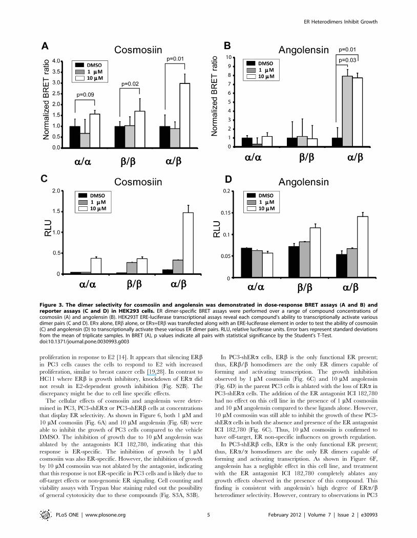
The ER dimer selectivity was validated in BRET and reporter
assays in ER-negative HEK293 cells as described [35]. While
cosmosiin exhibits preference for inducing both ERb/b homodi-
mers and ERa/b heterodimers (Fig. 3A), angolensin exhibits
ERa/b heterodimer selectivity (Fig. 3B). Neither compound shows
preference for inducing ERa/a homodimers. Because the lower
limit of detection for these compounds was 1 mM, concentrations
lower than 1 mM are not shown in this figure, although they were
tested in a range from 1 nM to 10 mM; below 1 mM, the BRET
ratios were the same as vehicle-treated. Furthermore, the ability of
these lead compounds to induce the transcriptional activity of ERaalone, ERb alone, or ERa in combination with ERb was tested at
a range of concentrations using the HEK293 ERE-luciferase
reporter assays (Fig. 3C and 3D). Although these reporter assays
do not directly examine ERa/b heterodimerization, the condition
in which ERa and ERb are cotransfected can be compared with
each receptor transfected alone.
As shown in Figure 3B, BRET assays reveal that angolensin is
capable of efficiently inducing the formation of ERa/b heterodimers
at 1 mM and 10 mM, while not inducing ERa/a or ERb/bhomodimers. ERa/b heterodimerization appears to be favored in
the presence of angolensin , and in the condition in which ERaand ERb are co-transfected for luciferase reporter assays, the
highest fold induction of transcriptional activity relative to DMSO
vehicle is observed (Fig. 3D). Thus, angolensin (R) appears to be
an ERa/b heterodimer-selective ligand at 10 mM. Cosmosiin
appears to be less selective in terms of its ability to induce ERa/bheterodimers, as ERb/b homodimers are also induced in BRET
assays; however, ERa/a homodimers are not induced by
cosmosiin (Fig. 3A). Cosmosiin at 1 mM appears to transcription-
ally activate ERb/b homodimers and ERa/b heterodimers
(Fig. 3C). At 10 mM cosmosiin, while ERa/a and ERb/bhomodimers were slightly activated, co-transfecting ERb with
ERa exhibited much stronger transcriptional activity (Fig. 3C).
Thus, cosmosiin appears to be ERb/b homodimer- and ERa/bheterodimer-selective at 1 mM.
The transcriptional activity of ERa/a homodimers treated with
10 mM cosmosiin is despite the finding that the BRET assay does
not show statistically significant ERa/a homodimerization
(Fig. 3A). The most likely explanation for this discrepancy is
differences in sensitivity between BRET and the luciferase reporter
assays. These BRET assays and luciferase reporter assays are
performed under different conditions and measure different signal
outputs: BRET captures a single moment in time in which ERa
ER Heterodimers Inhibit Growth
PLoS ONE | www.plosone.org 2 February 2012 | Volume 7 | Issue 2 | e30993
and ERb may or may not be dimerized. This moment in time was
observed after 1 hour incubation with ligand. Conversely, the
luciferase reporter assay measures an accumulation of transcrip-
tional output signal (the transcribed luciferase protein) over 18–
24 hours. Consequently, the dimerization ratios obtained via the
BRET assay do not always completely agree with the transcrip-
tional profiles obtained in the luciferase reporter assays for a given
ligand. Therefore, it is important to consider the direct dimerization
of ERa and ERb in conjunction with the transcriptional output of
these diverse dimer pairs.
Selection and generation of cell lines expressing differentamounts of ERa and ERb
In order to characterize the cellular effects of cosmosiin and
angolensin, we surveyed a variety of breast and prostate cell lines
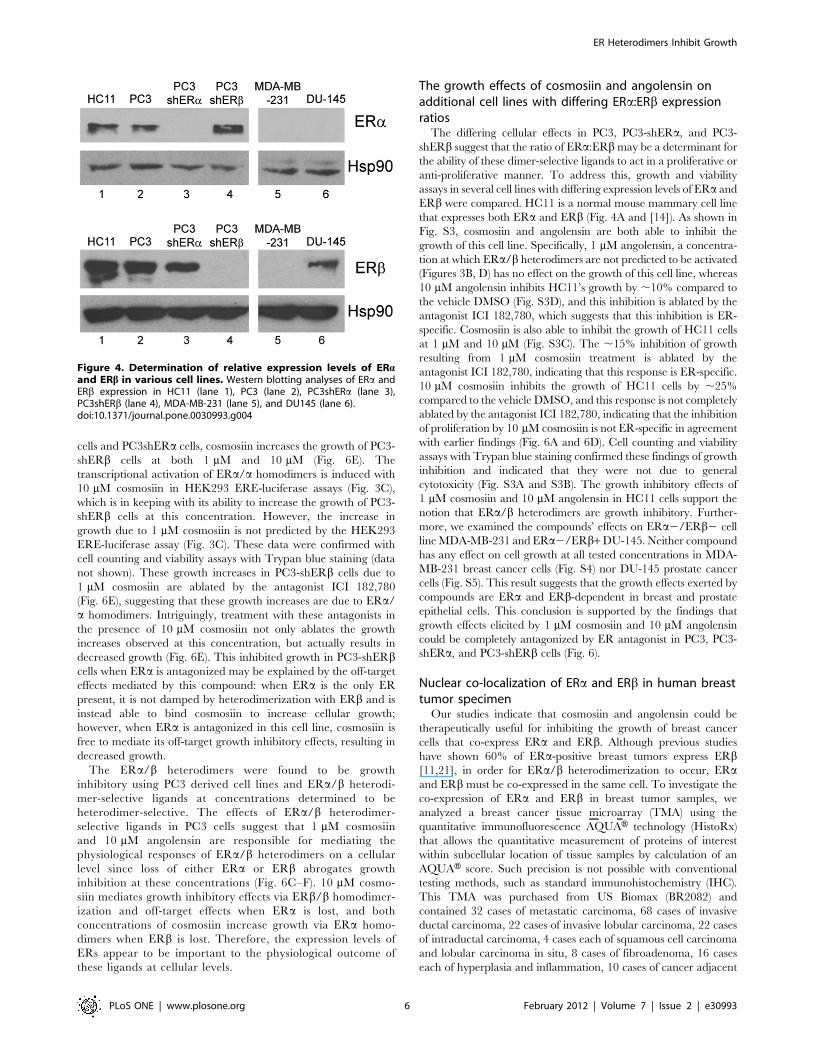
for co-expression of ERa and ERb. As shown in Fig. 4, the non-
tumorigenic mammary epithelial cell HC11 and prostate cancer
cell line PC3 were found to express both receptors (Lanes 1 and 2)
as reported by others [14,36]; in contrast, DU-145 expresses only
ERb [36] (lane 6) and MDA-MB-231 is negative for both ERaand ERb (lane 5). To delineate the functions of ERa/bheterodimers, we knocked down ERa and ERb transcript levels
in PC3 cells by means of stable transfection with specific shRNA
plasmids targeting ERa and ERb, respectively. Western blotting
results showed that ERa is selectively silenced in PC3-shERa cells
and ERb is selectively silenced in PC3-shERb cells (Fig. 4A, lanes
3 and 4). The silencing of one ER did not influence the expression
of the other. All of these characterized cell lines were subsequently
used for determination of compounds’ cellular effects.
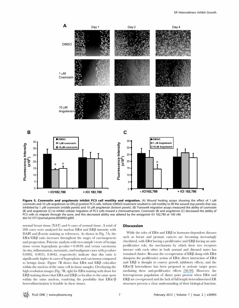
Cosmosiin and angolensin inhibit cell motility andmigration but not apoptosis in PC3
In order to examine the influences of these ERa/b heterodimer-
activating compounds on cell migration, wound healing assays
were employed using migratory PC3 cells. This assay gives a
qualitative measure of a compound’s ability to inhibit cell
migration. For these assays, 1 mM cosmosiin and 10 mM
angolensin were utilized because these are the concentrations at
which ERa/b heterodimers are most highly selectively induced by
each respective compound. As shown in Figure 5A, the vehicle
DMSO (0.1%) was unable to inhibit the migration of PC3 cells in
scratch wound healing assays: cells can be seen infiltrating the
wound 24 hours after scraping, and the wounds are completely
filled 72 hours after scraping. Conversely, both 10 mM angolensin
Figure 1. Flow scheme of high throughput screening and characterization of compounds with selectivity for ERa/ERb heterodimers.A library of .5200 small molecules was screened ER transcriptional activity using T47D-KBLuc cells. Molecules with transcriptional activity were thenscreened for ERa/a, ERa/b, or ER b/b dimerization potential using BRET assays. Two phytoestrogens, angolensin and cosmosiin, were identified as ERdimer selective ligands. These molecules were validated using in vitro binding assays and BRET and ERE-luciferase reporter assays. Heterodimerselective concentrations were identified as 10 mM angolensin and 1 mM cosmosiin. The cellular effects of these two heterodimer-selectiveconcentrations were characterized using cell migration and proliferation assays.doi:10.1371/journal.pone.0030993.g001
ER Heterodimers Inhibit Growth
PLoS ONE | www.plosone.org 3 February 2012 | Volume 7 | Issue 2 | e30993
and 1 mM cosmosiin are able to inhibit the ability of PC3 cells to
infiltrate the wounds, indicating that these compounds can hinder
cell motility.
To quantitatively measure the ability of cosmosiin and angolensin
to inhibit cell migration, transwell assays were employed. Figure 5C
shows that 10 mM angolensin can inhibit the ability of PC3 cells to
migrate through the pore, and this inhibition of migration is ablated
by the ER antagonist ICI 182,780. 1 mM angolensin, a concentra-
tion at which ERa/b heterodimers are not transcriptionally active
(Fig. 3D), has a negligible effect on cell migration. Both 1 mM and
10 mM cosmosiin can inhibit cell migration through the pore, and
this inhibition of migration is ablated by ICI 182,780 (Fig. 5B).
These results are recapitulated when the transwell is coated with
matrigel (data not shown), indicating that in addition to dampening
the ability of PC3 cells to migrate, these compounds are able to
dampen the ability of PC3 cells to invade.
The abilities of these lead compounds to influence apoptosis in
PC3 cells were next evaluated using caspase 3/7 assays. PC3 cells
were incubated with the indicated concentrations of DMSO
vehicle (0.1%), the indicated concentrations of cosmosiin or
angolensin (Fig. S1A and S1B), or the positive control cisplatin
(10 mg/mL) for 24, 48, and 72 hours. Cisplatin did not activate the
caspases 3/7 pathway at 24 hours and 48 hours (data not shown);
only at 72 hours was a weak induction of the caspases 3/7
observed (Fig. S1). At no time point did these compounds reveal
any activation of the caspase 3/7 pathway. Thus, it appears that
cosmosiin and angolensin are not strong inducers of apoptosis, at
least through the caspase 3/7 pathway.
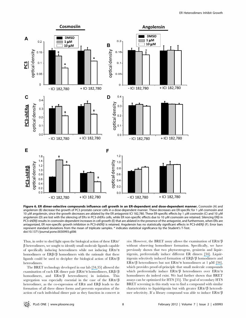
Determination of the growth effects of compounds inPC3, PC3-shERa, PC3-shERb cells
To determine if these compounds also inhibit cell proliferation
in addition to migration, MTT assays were employed. This assay
measures mitochondrial activity when yellow MTT (3-(4,5-
Dimethylthiazol-2-yl)-2,5-diphenyltetrazolium bromide) is reduced
to its purple formazan metabolic product [37]. Thus, the ability of
a cell to metabolize MTT to formazan is correlated to its
metabolic activity and cellular growth. To show that PC3 cells
express functional ERs and that E2’s cellular effects are ER-
dependent, we compared E2’s growth effects in PC3, PC3-shERa,
PC3-shERb cell lines. As shown in other ERa and ERb co-
expressing cell lines [14], E2 exhibits no effects in proliferation of
PC3 (Fig. S2A). However, when ERb expression was blocked, E2
induced proliferation (Fig. S2C) and E2’s proliferative effects were
completely abrogated by the pure ER antagonist ICI 182,780 and
the ERa selective antagonist MPP dihydrochloride (Fig. S2C,
middle and right panels). This result recapitulates the previous
finding in HC11 mammary epithelial cells that ERa drives
Figure 2. Fluorescence polarization competition binding assays for ERa and ERb. Cosmosiin (A) and angolensin (B) bind to recombinantERa and ERb with mM affinities.doi:10.1371/journal.pone.0030993.g002
ER Heterodimers Inhibit Growth
PLoS ONE | www.plosone.org 4 February 2012 | Volume 7 | Issue 2 | e30993
proliferation in response to E2 [14]. It appears that silencing ERbin PC3 cells causes the cells to respond to E2 with increased
proliferation, similar to breast cancer cells [19,28]. In contrast to
HC11 where ERb is growth inhibitory, knockdown of ERa did
not result in E2-dependent growth inhibition (Fig. S2B). The
discrepancy might be due to cell line specific effects.
The cellular effects of cosmosiin and angolensin were deter-
mined in PC3, PC3-shERa or PC3-shERb cells at concentrations
that display ER selectivity. As shown in Figure 6, both 1 mM and
10 mM cosmosiin (Fig. 6A) and 10 mM angolensin (Fig. 6B) were
able to inhibit the growth of PC3 cells compared to the vehicle
DMSO. The inhibition of growth due to 10 mM angolensin was
ablated by the antagonists ICI 182,780, indicating that this
response is ER-specific. The inhibition of growth by 1 mM
cosmosiin was also ER-specific. However, the inhibition of growth
by 10 mM cosmosiin was not ablated by the antagonist, indicating
that this response is not ER-specific in PC3 cells and is likely due to
off-target effects or non-genomic ER signaling. Cell counting and
viability assays with Trypan blue staining ruled out the possibility
of general cytotoxicity due to these compounds (Fig. S3A, S3B).
In PC3-shERa cells, ERb is the only functional ER present;
thus, ERb/b homodimers are the only ER dimers capable of
forming and activating transcription. The growth inhibition
observed by 1 mM cosmosiin (Fig. 6C) and 10 mM angolensin
(Fig. 6D) in the parent PC3 cells is ablated with the loss of ERa in
PC3-shERa cells. The addition of the ER antagonist ICI 182,780
had no effect on this cell line in the presence of 1 mM cosmosiin
and 10 mM angolensin compared to these ligands alone. However,
10 mM cosmosiin was still able to inhibit the growth of these PC3-
shERa cells in both the absence and presence of the ER antagonist
ICI 182,780 (Fig. 6C). Thus, 10 mM cosmosiin is confirmed to
have off-target, ER non-specific influences on growth regulation.
In PC3-shERb cells, ERa is the only functional ER present;
thus, ERa/a homodimers are the only ER dimers capable of
forming and activating transcription. As shown in Figure 6F,
angolensin has a negligible effect in this cell line, and treatment
with the ER antagonist ICI 182,780 completely ablates any
growth effects observed in the presence of this compound. This
finding is consistent with angolensin’s high degree of ERa/bheterodimer selectivity. However, contrary to observations in PC3
Figure 3. The dimer selectivity for cosmosiin and angolensin was demonstrated in dose-response BRET assays (A and B) andreporter assays (C and D) in HEK293 cells. ER dimer-specific BRET assays were performed over a range of compound concentrations ofcosmosiin (A) and angolensin (B). HEK293T ERE-luciferase transcriptional assays reveal each compound’s ability to transcriptionally activate variousdimer pairs (C and D). ERa alone, ERb alone, or ERa+ERb was transfected along with an ERE-luciferase element in order to test the ability of cosmosiin(C) and angolensin (D) to transcriptionally activate these various ER dimer pairs. RLU, relative luciferase units. Error bars represent standard deviationsfrom the mean of triplicate samples. In BRET (A), p values indicate all pairs with statistical significance by the Student’s T-Test.doi:10.1371/journal.pone.0030993.g003
ER Heterodimers Inhibit Growth
PLoS ONE | www.plosone.org 5 February 2012 | Volume 7 | Issue 2 | e30993
cells and PC3shERa cells, cosmosiin increases the growth of PC3-
shERb cells at both 1 mM and 10 mM (Fig. 6E). The
transcriptional activation of ERa/a homodimers is induced with
10 mM cosmosiin in HEK293 ERE-luciferase assays (Fig. 3C),
which is in keeping with its ability to increase the growth of PC3-
shERb cells at this concentration. However, the increase in
growth due to 1 mM cosmosiin is not predicted by the HEK293
ERE-luciferase assay (Fig. 3C). These data were confirmed with
cell counting and viability assays with Trypan blue staining (data
not shown). These growth increases in PC3-shERb cells due to
1 mM cosmosiin are ablated by the antagonist ICI 182,780
(Fig. 6E), suggesting that these growth increases are due to ERa/
a homodimers. Intriguingly, treatment with these antagonists in
the presence of 10 mM cosmosiin not only ablates the growth
increases observed at this concentration, but actually results in
decreased growth (Fig. 6E). This inhibited growth in PC3-shERbcells when ERa is antagonized may be explained by the off-target
effects mediated by this compound: when ERa is the only ER
present, it is not damped by heterodimerization with ERb and is
instead able to bind cosmosiin to increase cellular growth;
however, when ERa is antagonized in this cell line, cosmosiin is
free to mediate its off-target growth inhibitory effects, resulting in
decreased growth.
The ERa/b heterodimers were found to be growth
inhibitory using PC3 derived cell lines and ERa/b heterodi-
mer-selective ligands at concentrations determined to be
heterodimer-selective. The effects of ERa/b heterodimer-
selective ligands in PC3 cells suggest that 1 mM cosmosiin
and 10 mM angolensin are responsible for mediating the
physiological responses of ERa/b heterodimers on a cellular
level since loss of either ERa or ERb abrogates growth
inhibition at these concentrations (Fig. 6C–F). 10 mM cosmo-
siin mediates growth inhibitory effects via ERb/b homodimer-
ization and off-target effects when ERa is lost, and both
concentrations of cosmosiin increase growth via ERa homo-
dimers when ERb is lost. Therefore, the expression levels of
ERs appear to be important to the physiological outcome of
these ligands at cellular levels.
The growth effects of cosmosiin and angolensin onadditional cell lines with differing ERa:ERb expressionratios
The differing cellular effects in PC3, PC3-shERa, and PC3-
shERb suggest that the ratio of ERa:ERb may be a determinant for
the ability of these dimer-selective ligands to act in a proliferative or
anti-proliferative manner. To address this, growth and viability
assays in several cell lines with differing expression levels of ERa and
ERb were compared. HC11 is a normal mouse mammary cell line
that expresses both ERa and ERb (Fig. 4A and [14]). As shown in
Fig. S3, cosmosiin and angolensin are both able to inhibit the
growth of this cell line. Specifically, 1 mM angolensin, a concentra-
tion at which ERa/b heterodimers are not predicted to be activated
(Figures 3B, D) has no effect on the growth of this cell line, whereas
10 mM angolensin inhibits HC11’s growth by ,10% compared to
the vehicle DMSO (Fig. S3D), and this inhibition is ablated by the
antagonist ICI 182,780, which suggests that this inhibition is ER-
specific. Cosmosiin is also able to inhibit the growth of HC11 cells
at 1 mM and 10 mM (Fig. S3C). The ,15% inhibition of growth
resulting from 1 mM cosmosiin treatment is ablated by the
antagonist ICI 182,780, indicating that this response is ER-specific.
10 mM cosmosiin inhibits the growth of HC11 cells by ,25%
compared to the vehicle DMSO, and this response is not completely
ablated by the antagonist ICI 182,780, indicating that the inhibition
of proliferation by 10 mM cosmosiin is not ER-specific in agreement
with earlier findings (Fig. 6A and 6D). Cell counting and viability
assays with Trypan blue staining confirmed these findings of growth
inhibition and indicated that they were not due to general
cytotoxicity (Fig. S3A and S3B). The growth inhibitory effects of
1 mM cosmosiin and 10 mM angolensin in HC11 cells support the
notion that ERa/b heterodimers are growth inhibitory. Further-
more, we examined the compounds’ effects on ERa2/ERb2 cell
line MDA-MB-231 and ERa2/ERb+ DU-145. Neither compound
has any effect on cell growth at all tested concentrations in MDA-
MB-231 breast cancer cells (Fig. S4) nor DU-145 prostate cancer
cells (Fig. S5). This result suggests that the growth effects exerted by
compounds are ERa and ERb-dependent in breast and prostate
epithelial cells. This conclusion is supported by the findings that
growth effects elicited by 1 mM cosmosiin and 10 mM angolensin
could be completely antagonized by ER antagonist in PC3, PC3-
shERa, and PC3-shERb cells (Fig. 6).
Nuclear co-localization of ERa and ERb in human breasttumor specimen
Our studies indicate that cosmosiin and angolensin could be
therapeutically useful for inhibiting the growth of breast cancer
cells that co-express ERa and ERb. Although previous studies
have shown 60% of ERa-positive breast tumors express ERb[11,21], in order for ERa/b heterodimerization to occur, ERaand ERb must be co-expressed in the same cell. To investigate the
co-expression of ERa and ERb in breast tumor samples, we
analyzed a breast cancer tissue microarray (TMA) using the
quantitative immunofluorescence AQUAH technology (HistoRx)
that allows the quantitative measurement of proteins of interest
within subcellular location of tissue samples by calculation of an
AQUAH score. Such precision is not possible with conventional
testing methods, such as standard immunohistochemistry (IHC).
This TMA was purchased from US Biomax (BR2082) and
contained 32 cases of metastatic carcinoma, 68 cases of invasive
ductal carcinoma, 22 cases of invasive lobular carcinoma, 22 cases
of intraductal carcinoma, 4 cases each of squamous cell carcinoma
and lobular carcinoma in situ, 8 cases of fibroadenoma, 16 cases
each of hyperplasia and inflammation, 10 cases of cancer adjacent
Figure 4. Determination of relative expression levels of ERaand ERb in various cell lines. Western blotting analyses of ERa andERb expression in HC11 (lane 1), PC3 (lane 2), PC3shERa (lane 3),PC3shERb (lane 4), MDA-MB-231 (lane 5), and DU145 (lane 6).doi:10.1371/journal.pone.0030993.g004
ER Heterodimers Inhibit Growth
PLoS ONE | www.plosone.org 6 February 2012 | Volume 7 | Issue 2 | e30993
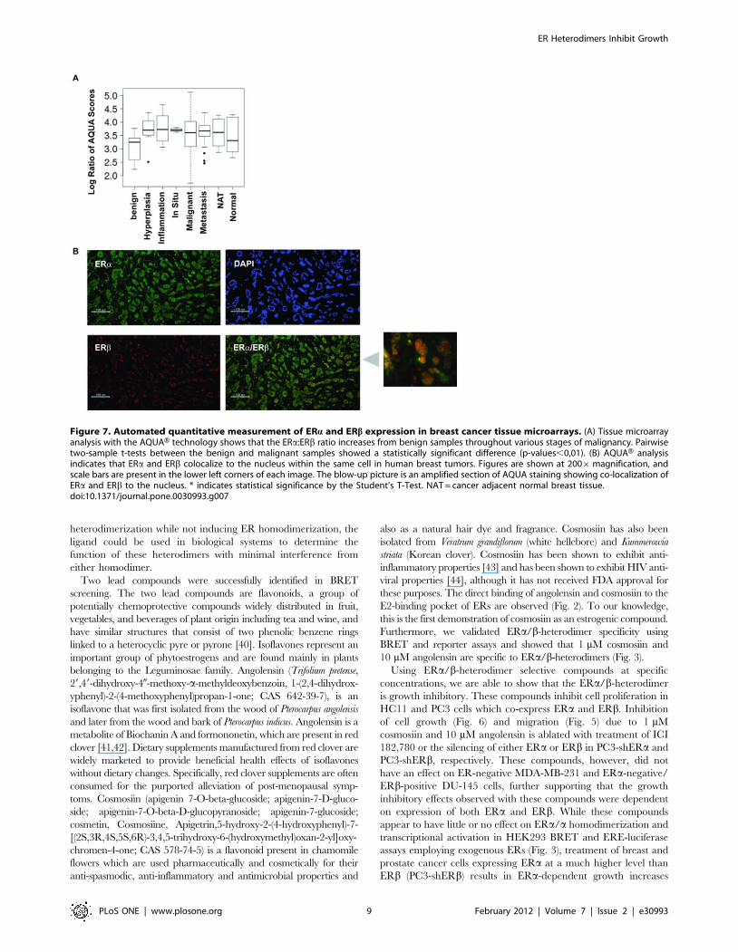
normal breast tissue (NAT) and 6 cases of normal tissue. A total of
208 cores were analyzed for nuclear ERa and ERb intensity with
DAPI and b-actin staining as references. As shown in Fig. 7A, the
ERa/ERb ratio increases throughout the stages of carcinogenesis
and progression. Pairwise analysis with two-sample t-tests of benign
tissue versus hyperplasia (p-value = 0.0039) and versus carcinoma
(in situ, inflammation, metastatic, and malignant cases with p-values
0.0092, 0.0035, 0.0042, respectively) indicate that this ratio is
significantly higher in cases of hyperplasia and carcinoma compared
to benign tissue. Figure 7B shows that ERa and ERb colocalize
within the nucleus of the same cell in tissue samples. Overlaying the
high resolution images (Fig. 7B, right) for ERa staining with those for
ERb staining shows that ERa and ERb co-localize to the same spots
within the same nucleus, rendering the possibility that ERa/bheterodimerization is feasible in these tissues.
Discussion
While the roles of ERa and ERb in hormone-dependent diseases
such as breast and prostate cancers are becoming increasingly
elucidated, with ERa having a proliferative and ERb having an anti-
proliferative role, the mechanism by which these two receptors
interact with each other in both normal and diseased states has
remained elusive. Because the co-expression of ERb along with ERadampens the proliferative action of ERa, direct interaction of ERaand ERb is thought to convey growth inhibitory effects, and the
ERa/b heterodimer has been proposed to activate target genes
mediating these anti-proliferative effects [38,39]. However, the
heterogeneous population of dimer pairs present when ERa and
ERb are co-expressed and the lack of full length heterodimerized ER
structures prevent a clear understanding of their biological function.
Figure 5. Cosmosiin and angolensin inhibit PC3 cell motility and migration. (A) Wound healing assays showing the effect of 1 mMcosmosiin and 10 mM angolensin on ERa,b-positive PC3 cells. Vehicle (DMSO) treatment resulted in cell motility to fill the wound (top panels) that wasinhibited by 1 mM cosmosiin (middle panels) and 10 mM angolensin (bottom panels). (B) Transwell migration assays measured the ability of cosmosiin(B) and angolensin (C) to inhibit cellular migration of PC3 cells toward a chemoattractant. Cosmosiin (B) and angolensin (C) decreased the ability ofPC3 cells to migrate through the pore, and this decreased ability was ablated by the antagonist ICI 182,780 at 100 nM.doi:10.1371/journal.pone.0030993.g005
ER Heterodimers Inhibit Growth
PLoS ONE | www.plosone.org 7 February 2012 | Volume 7 | Issue 2 | e30993
Thus, in order to shed light upon the biological action of these ERa/
b heterodimers, we sought to identify small molecule ligands capable
of specifically inducing heterodimers while not inducing ERa/ahomodimers or ERb/b homodimers with the rationale that these
ligands could be used to decipher the biological action of ERa/bheterodimers.
The BRET technology developed in our lab [34,35] allowed the
examination of each ER dimer pair (ERa/a homodimers, ERb/bhomodimers, and ERa/b heterodimers) in isolation. This
segregation was especially essential in the case of the ERa/bheterodimer, as the co-expression of ERa and ERb leads to the
formation of all three dimer forms and prevents separation of the
action of each individual dimer pair as they function in concert in
vivo. However, the BRET assay allows the examination of ERa/bwithout observing homodimer formation. Specifically, we have
previously shown that two phytoestrogens, genistein and liquir-
itigenin, preferentially induce different ER dimers [34]. Liquir-
itigenin selectively induced formation of ERb/b homodimers and
ERa/b heterodimers but not ERa/a homodimers at 1 mM [34],
which provides proof-of-principle that small molecule compounds
which preferentially induce ERa/b heterodimers over ERa/ahomodimers do indeed exist. We had further shown that BRET
assays can be optimized for HTS [35]. The goal of secondary HTS
BRET screening in this study was to find a compound with similar
characteristics to liquiritigenin but with greater ERa/b heterodi-
mer selectivity. If a library compound was able to induce ERa/b
Figure 6. ER dimer-selective compounds influence cell growth in an ER-dependent and dose-dependent manner. Cosmosiin (A) andangolensin (B) decrease the growth of PC3 prostate cancer cells in a dose-dependent manner. These decreases are ER-specific for 1 mM cosmosiin and10 mM angolensin, since the growth decreases are ablated by the ER antagonist ICI 182,780. These ER-specific effects by 1 mM cosmosiin (C) and 10 mMangolensin (D) are lost with the silencing of ERa in PC3-shERa cells, while ER non-specific effects due to 10 mM cosmosiin are retained. Silencing ERb inPC3-shERb results in cosmosiin-dependent increases in cell growth (E) that are ablated in the presence of the antagonist, and furthermore, when ERs areantagonized, ER non-specific growth inhibition in PC3-shERb is retained. Angolensin has no statistically significant effects in PC3-shERb (F). Error barsrepresent standard deviations from the mean of triplicate samples. * indicates statistical significance by the Student’s T-Test.doi:10.1371/journal.pone.0030993.g006
ER Heterodimers Inhibit Growth
PLoS ONE | www.plosone.org 8 February 2012 | Volume 7 | Issue 2 | e30993
heterodimerization while not inducing ER homodimerization, the
ligand could be used in biological systems to determine the
function of these heterodimers with minimal interference from
either homodimer.
Two lead compounds were successfully identified in BRET
screening. The two lead compounds are flavonoids, a group of
potentially chemoprotective compounds widely distributed in fruit,
vegetables, and beverages of plant origin including tea and wine, and
have similar structures that consist of two phenolic benzene rings
linked to a heterocyclic pyre or pyrone [40]. Isoflavones represent an
important group of phytoestrogens and are found mainly in plants
belonging to the Leguminosae family. Angolensin (Trifolium pretense,
29,49-dihydroxy-40-methoxy-a-methyldeoxybenzoin, 1-(2,4-dihydrox-
yphenyl)-2-(4-methoxyphenyl)propan-1-one; CAS 642-39-7), is an
isoflavone that was first isolated from the wood of Pterocarpus angolensis
and later from the wood and bark of Pterocarpus indicus. Angolensin is a
metabolite of Biochanin A and formononetin, which are present in red
clover [41,42]. Dietary supplements manufactured from red clover are
widely marketed to provide beneficial health effects of isoflavones
without dietary changes. Specifically, red clover supplements are often
consumed for the purported alleviation of post-menopausal symp-
toms. Cosmosiin (apigenin 7-O-beta-glucoside; apigenin-7-D-gluco-
side; apigenin-7-O-beta-D-glucopyranoside; apigenin-7-glucoside;
cosmetin, Cosmosiine, Apigetrin,5-hydroxy-2-(4-hydroxyphenyl)-7-
[(2S,3R,4S,5S,6R)-3,4,5-trihydroxy-6-(hydroxymethyl)oxan-2-yl]oxy-
chromen-4-one; CAS 578-74-5) is a flavonoid present in chamomile
flowers which are used pharmaceutically and cosmetically for their
anti-spasmodic, anti-inflammatory and antimicrobial properties and
also as a natural hair dye and fragrance. Cosmosiin has also been
isolated from Veratrum grandiflorum (white hellebore) and Kummerowia
striata (Korean clover). Cosmosiin has been shown to exhibit anti-
inflammatory properties [43] and has been shown to exhibit HIV anti-
viral properties [44], although it has not received FDA approval for
these purposes. The direct binding of angolensin and cosmosiin to the
E2-binding pocket of ERs are observed (Fig. 2). To our knowledge,
this is the first demonstration of cosmosiin as an estrogenic compound.
Furthermore, we validated ERa/b-heterodimer specificity using
BRET and reporter assays and showed that 1 mM cosmosiin and
10 mM angolensin are specific to ERa/b-heterodimers (Fig. 3).
Using ERa/b-heterodimer selective compounds at specific
concentrations, we are able to show that the ERa/b-heterodimer
is growth inhibitory. These compounds inhibit cell proliferation in
HC11 and PC3 cells which co-express ERa and ERb. Inhibition
of cell growth (Fig. 6) and migration (Fig. 5) due to 1 mM
cosmosiin and 10 mM angolensin is ablated with treatment of ICI
182,780 or the silencing of either ERa or ERb in PC3-shERa and
PC3-shERb, respectively. These compounds, however, did not
have an effect on ER-negative MDA-MB-231 and ERa-negative/
ERb-positive DU-145 cells, further supporting that the growth
inhibitory effects observed with these compounds were dependent
on expression of both ERa and ERb. While these compounds
appear to have little or no effect on ERa/a homodimerization and
transcriptional activation in HEK293 BRET and ERE-luciferase
assays employing exogenous ERs (Fig. 3), treatment of breast and
prostate cancer cells expressing ERa at a much higher level than
ERb (PC3-shERb) results in ERa-dependent growth increases
Figure 7. Automated quantitative measurement of ERa and ERb expression in breast cancer tissue microarrays. (A) Tissue microarrayanalysis with the AQUAH technology shows that the ERa:ERb ratio increases from benign samples throughout various stages of malignancy. Pairwisetwo-sample t-tests between the benign and malignant samples showed a statistically significant difference (p-values,0,01). (B) AQUAH analysisindicates that ERa and ERb colocalize to the nucleus within the same cell in human breast tumors. Figures are shown at 2006magnification, andscale bars are present in the lower left corners of each image. The blow-up picture is an amplified section of AQUA staining showing co-localization ofERa and ERb to the nucleus. * indicates statistical significance by the Student’s T-Test. NAT = cancer adjacent normal breast tissue.doi:10.1371/journal.pone.0030993.g007
ER Heterodimers Inhibit Growth
PLoS ONE | www.plosone.org 9 February 2012 | Volume 7 | Issue 2 | e30993
(Fig. 6E). This result is in agreement with the common theme that
ERa is a major growth driver, and it also implicates the
dependence of these compounds’ growth effects on the relative
expression ratio of ERa:ERb, as these compounds ablate growth
increases in PC3 and HC11, in which ERa and ERb expression
levels are relatively similar and heterodimerization may be favored
[31]. Taken together, these data suggest that the ratio of ERa:ERbin the same tumor cells is extremely important for physiological
effects of these compounds. While the data presented herein
provide initial evidence for a growth-inhibitory function of the
ERa/b heterodimer, identification of higher affinity compounds
with greater ERa/b heterodimer selectivity will be needed to
validate our findings since both compounds are weak agonists, and
cosmosiin at 10 mM appears to have off-target effects.
Compounds exhibiting ERa/b heterodimer-selectivity may
have therapeutic or preventive efficacy in hormone-dependent
diseases. A recent study shows that the tamoxifen metabolite
endoxifen is capable of degrading ERa [45], stabilizing ERb, and
inducing ERa/b heterodimerization in a concentration dependent
manner [46]. Tamoxifen is a widely-utilized FDA-approved breast
cancer treatment and prevention drug. This finding suggests that
tamoxifen’s cancer preventive effects may be mediated by
stimulation of ERa/b heterodimer formation. The possibility is
supported by the fact that both ERs are expressed in normal
mammary epithelial cells [4]. Similarly, naturally-occurring
estrogen-like compounds such as phytoestrogens, a group of
plant-derived compounds with estrogenic and/or antiestrogenic
activities hold promise for action as preventive or therapeutic ER-
regulators via their abilities to mediate estrogenic responses tissue-
specifically. Indeed, consumption of soy phytoestrogens has been
correlated with decreased breast cancer risk [47], although these
data remain somewhat controversial [48]. Furthermore, consump-
tion of genistein [49], resveratrol [50], and soy [51] has been
inversely correlated with prostate cancer risk. Although these
compounds may stimulate the proliferative action of ERa when
ERb is lost in tumors, they may have preventative effects under
normal physiological conditions when both ERs are expressed.
Furthermore, our examination of nuclear co-localization of
ERa and ERb within the same tumor cell using the AQUAHtechnology (Fig. 7) support that ERa/b heterodimerization could
potentially occur within tumor cells. Prior to these studies, the co-
localization of ERa and ERb within the same cell had not been
examined. The punctate staining pattern suggests that ERa and
ERb are co-localized on DNA, and therefore may be transcrip-
tionally active in these cells as ERa/b heterodimers. Furthermore,
AQUAH analysis showed that the ERa:ERb ratio is higher in
malignant states compared to benign tissue samples, in agreement
with the finding that ERb levels often decrease in malignant breast
cancers [52]. The growth inhibitory effects of ERa/b heterodi-
mers might due to their activation of different target genes from
their respective homodimers. Recently, global ChIP-Seq analyses
of ERa and ERb target genes show that perfectly or imperfectly
palindromic EREs are preferential binding sites for ERa/bheterodimers as compared to ERa/a or ERb/b homodimers
which are more flexible in DNA recognition [53]. This is
consistent with other reports that ERa/b heterodimers might
regulate distinct genes [32,33]. The ERa/b heterodimer-selective
ligands identified in this study will allow identification of
heterodimer target genes in cells co-expressing ERa and ERb(e.g. PC3). While our findings implicate the ERa/b heterodimer as
a putative preventative and therapeutic target for hormone-
responsive cancers, this example highlights the imminent need to
decipher the role these heterodimers in breast and prostate
cancers.
In conclusion, these data provide a proof-of-principle that ERa/
b heterodimer-selective ligands can inhibit cell growth and
migration in ERa/ERb-positive cells such as PC3 and HC11 when
ERa and ERb are expressed at similar levels. We also found that the
compounds’ growth effects depend on the relative expression levels
of ERa and ERb. Upon knockdown of ERb in PC3 cells, cosmosiin
increases PC3 cell growth in an ERa-dependent manner. Thus,
more heterodimer selective ligands need to be identified to clarify
whether the heterodimer-selective ligands become growth stimula-
tory when ERb expression is lost in human tumors. Although more
studies are needed to demonstrate the ERa/b heterodimer as a
therapeutic target, the concept of inducing ERb to pair with ERa,
thus antagonizing ERa’s proliferative function, is distinct from
existing breast cancer therapeutic strategies of targeting ERa alone.
We also suggest that the relative ERa and ERb expression levels in
patient tumors should be carefully evaluated to better understand
the ER-targeted drugs’ therapeutic performance, as many of these
drugs have not been evaluated for their dimer selectivity, and ERbexpression in patient tumors is not routinely evaluated.
Materials and Methods
High Throughput Screening MethodsAll primary and secondary screens were performed at the
University of Wisconsin Carbone Cancer Center (UWCCC) Keck
Small Molecule Screening Facility (SMSF). Ten thousand T47D-
KBLuc cells [54] were seeded into 384-well plates and allowed to
attach overnight. The next day, 0.5 ml of 1 mM compound was
added to a final concentration of 10 mM using an automated
robotic system (Beckman Biomek FX). 10 nM E2 and 1% (0.5 ml)
DMSO were used as positive and negative controls, respectively.
Cells were incubated with compound for 18 hrs at 37uC in 5%
CO2 in a cell culture incubator. On day 3, media were removed,
and 25 ml lysis buffer (Promega, cat# E2661) was added to each
well using the robot. Cells were allowed to lyse for 10 min with
constant agitation, and lysis was confirmed by microscopically
viewing a clear-bottom 384-well plate maintained in parallel under
identical conditions. 25 ml luciferase substrate (Promega, Cat#E2620) was then added, mixed for 30 seconds, and luciferase
emission was immediately detected on a Tecan Safire 2 plate
reader at 0.1 seconds per well. Counter-screening was performed
in a similar fashion in the presence and absence of the ER
antagonist ICI 182,780. Secondary Bioluminescence Resonance
Energy Transfer (BRET) screening was performed in transiently
transfected HEK293 cells (ATCC, CRL-1573). DNA encoding
BRET fusions were transfected as described in [34]. Following
24 hours of protein expression after transfection, cells were
trypsinized and counted using a Nexcelcom Cellometer, and cell
viability was determined to be .95% in each condition. Cells were
seeded at 11,000 cells per well of 384-well white-walled white-
bottom plates in 40 mL PBS. 0.2 mL of 1 mM library compound
was then added to each well using the Biomek FX Robot such that
the final concentration per well was 5 mM. Cell suspensions were
incubated with library compounds for 1 hour in a dark cabinet at
room temperature, at which point 10 mL of the Renilla Luciferase
(RLuc) substrate coelenterazine h was added to a final concentra-
tion of 5 mM. Plates were then gently shaken on a plate shaker for
10 seconds at 300 rpm, and RLuc emission was read at 460 nm
followed immediately by YFP emission at 535 nm at 0.1 second
per wavelength read per well. Each RLuc and YFP emission
measurement was taken consecutively per well before moving to
the next well. Emission values were used to calculate the BRET
ratio as described in [34]. Additional details for BRET screening
were described in [35].
ER Heterodimers Inhibit Growth
PLoS ONE | www.plosone.org 10 February 2012 | Volume 7 | Issue 2 | e30993

In vivo BRET assays to monitor ER dimer formation inliving cells
HEK293 cells (ATCC, CRL-1573) were either transfected with
a single BRET fusion plasmid (pCMX-ERa-RLuc or pCMX-
RLuc-ERb) or co-transfected with RLuc and YFP BRET fusions
(pCMX-ERa-RLuc+pCMX-YFP-ERb for ERa/ERb heterodi-
mers; pCMX-ERa-RLuc+pCMX-ERa-YFP for ERa homodimers;
or pCMX-RLuc-ERb+pCMX-YFP-ERb for ERb homodimers)
[34]. ‘‘Empty’’ expression vector pCMX-pL2 was used to keep the
total amount of transfected DNA constant. 24 hr post-transfection,
cells were trypsinized, counted, and resuspended in PBS in
quadruplicate at ,50,000 cells per well of a 96-well white-bottom
microplate. Cells were incubated with ligands for 1 hour. Coelenter-
azine h (Promega, Madison, WI) was added in PBS at a final
concentration of 5 mM, and 460 nm and 530 nm emission detection
measurements were immediately taken at 0.1 second per wavelength
read per well on a Perkin Elmer Victor 3-V plate reader.
Immunofluorescence StainingDeparaffinization and heat induced epitope retrieval were
performed simultaneously using the Lab Vision PT module
(Thermo Fisher Scientific, Fremont, CA) with Lab Vision citrate
buffer pH 8.0 at 98uC for 20 minutes. All staining was performed
at room temperature using the Lab Vision 360 automated staining
system. Endogenous peroxidase was blocked for 5 minutes with
Peroxidazed-1 (Cat.No. PX968, Biocare Medical). Non-specific
protein binding was eliminated via a 60 minute block with Biocare
Medical Sniper, and non-specific avidin was blocked using Biocare
Medical Avidin Biotin kit, incubating 15 minutes. DaVinci Green
Antibody Diluent (Cat.No. PD900L, Biocare Medical) was used
for antibody dilution. Breast TMA BR2082 containing 208 cores
was purchased from US Biomax Inc. (http://www.biomax.us/
tissue-arrays/Breast/BR2082). ERa was detected using ERarabbit mAb SP1 (1:50, 1 hr) (Thermo Fisher) and visualized with
goat anti-rabbit conjugated with Alexa Fluor 555 (Invitrogen)
secondary antibody. ERb was detected with mouse mAb 14C8
(Abcam,1:1600, 1 hr) and visualized with Alexa Fluor 647
conjugated Tyramide Signal Amplification system (Invitrogen),
which included biotinylated goat anti-mouse immunoglobulin,
streptavidin-horseradish peroxidase and Alexa Fluor 647-Tyra-
mide. Breast epithelial nuclei were masked using ProLong Gold
Antifade Reagent with DAPI mounting medium (Invitrogen).
Automated Image AcquisitionAutomated image capture was performed by the HistoRx PM-
2000 using the AQUAsition software package (New Haven, CT).
High-resolution (2048_2048 pixel, 7.4 mm), 8-bit grayscale digital
images are obtained for each area of interest resulting in 256
discrete intensity values per pixel of an acquired image [55]. The
breast epithelial nuclear compartment was defined with DAPI
(blue). The target markers (ERa and ERb) were visualized with
Alexa Fluor 555 (green) and 647 (red), respectively.
AQUA H Score GenerationSince the distributions of the original AQUAH scores exhibited
deviation from the normal distribution, we took the natural log
transformation of the original scores and then performed two
sample t-tests for pairwise comparisons among different samples.
Results from these tests were consistent with a Wilcoxon rank sum
test on the original scores. Images were evaluated before scoring.
Histospots showing ,5% tumor area, tissue folding, too much
debris, and those that were out of focus were disqualified from
scoring. Nuclear AQUAH scores for ERa and ERb for each
histospot were generated based on the unsupervised pixel-cased
clustering algorithm for optimal image segmentation for use in the
pixel-based locale assignment for compartmentalization of expres-
sion algorithm as described previously [56]. Pixels that could not
accurately be assigned to a compartment were discarded. The data
were saved and subsequently expressed as the average signal
intensity per unit of compartment area. All the signals in each
compartment were then added. The AQUAH score is expressed as
target signal intensity divided by the compartment pixel area and
is expressed on a scale of 0 to 33333 (AQUA_2.0, HistoRx). The
resultant AQUAH score is continuous and directly proportional to
the number of molecules per unit area.
Additional descriptions of cell culture, TMA and experimental
procedures can be found in Methods S1.
Supporting Information
Figure S1 Caspase 3/7 apoptosis assays showed thatcosmosiin and angolensin exhibited no apoptotic effectvia caspases 3 or 7 in PC3 cells at 96 hours. Cosmosiin (A)
and angolensin (B) modestly increase apoptosis through this
pathway in PC3 cells to a statistically non-significant level compared
to the strong apoptotic inducer cisplatin, which served as a positive
control. Statistical analysis method: Students T-Tests. Error bars
represent standard deviations from the mean of triplicate samples.
(TIF)
Figure S2 MTT assays showing the effect of 10 nM 17b-estradiol in ERa,b-positive PC3 cells and variants ofthese cells in which ERa has been silenced (PC3-shERa)or ERb has been silenced (PC3-shERb). E2 has no effect on
the proliferation of PC3 cells (A) or PC3-shERa cells (B); however,
the silencing of ERb in this cell line allows E2 to increase cellular
growth (C, left panel) by binding to ERa, since the presence of the
antagonists ICI 182,780 (C, middle panel) and MPP Dihydro-
chloride (C, right panel) ablates this increase. Statistical analysis
method: Students T-Test; * indicates p,0.05. Error bars represent
standard deviations from the mean of triplicate samples.
(TIF)
Figure S3 Cosmosiin at 1 mM and angolensin at 10 mMinhibited the growth of ERa/ERb positive HC11 cells inan ER-dependent manner. Cosmosiin (A) and angolensin (B)
had no cytotoxic effects at all tested concentrations. The growth
inhibitory effects of cosmosiin at 1 mM (C) and angolensin at 10 mM
(D) were ablated by pure ER antagonist ICI 182,780, suggesting the
growth inhibitory effects are ER-dependent. These decreases due to
10 mM cosmosiin are ER-independent since they are retained in the
presence of the antagonist ICI 182,780 (C). Statistical analysis
method: Students T-Test; * indicates p,0.05. Error bars represent
standard deviations from the mean of triplicate samples.
(TIF)
Figure S4 Neither cosmosiin (A) nor angolensin (B)influenced the growth of ER-negative MDA-MB-231breast cancer cells. Error bars represent standard
deviations from the mean of triplicate samples.
(TIF)
Figure S5 Neither cosmosiin (A) nor angolensin (B)influenced the growth of ERa-negative, ERb-positive DU-145 prostate cancer cells. Error bars represent standard
deviations from the mean of triplicate samples.
(TIF)
Methods S1 Supplemental Methods file.
(DOC)
ER Heterodimers Inhibit Growth
PLoS ONE | www.plosone.org 11 February 2012 | Volume 7 | Issue 2 | e30993
Acknowledgments
We thank Thomas Pier of UW TRIP lab for immunohistochemistry
assistance and Jiacai Wu for help with construction of PC3shERa and
PC3shERb cell lines and graphics. We thank Yidan Wang for technical
support. We are grateful to Drs. Elaine Alarid and William Ricke for
critical reading of the manuscript.
Author Contributions
Conceived and designed the experiments: EP ES AB. Performed the
experiments: EP ES AB JL WH. Analyzed the data: EP ES WH SK KW
WX. Contributed reagents/materials/analysis tools: WH JL. Wrote the
paper: EP WX.
References
1. Heldring N, Pike A, Andersson S, Matthews J, Cheng G, et al. (2007) Estrogen
receptors: how do they signal and what are their targets. Physiol Rev 87:905–931.
2. Deroo BJ, Korach KS (2006) Estrogen receptors and human disease. J Clin
Invest 116: 561–570.3. Nilsson S, Gustafsson JA (2011) Estrogen receptors: therapies targeted to
receptor subtypes. Clin Pharmacol Ther 89: 44–55.
4. Shoker BS, Jarvis C, Sibson DR, Walker C, Sloane JP (1999) Oestrogen receptorexpression in the normal and pre-cancerous breast. J Pathol 188: 237–244.
5. Hewitt SC, Harrell JC, Korach KS (2005) Lessons in estrogen biology from
knockout and transgenic animals. Annu Rev Physiol 67: 285–308.
6. Forster C, Makela S, Warri A, Kietz S, Becker D, et al. (2002) Involvement ofestrogen receptor beta in terminal differentiation of mammary gland epithelium.
Proc Natl Acad Sci U S A 99: 15578–15583.7. McPherson SJ, Ellem SJ, Patchev V, Fritzemeier KH, Risbridger GP (2006) The
role of Eralpha and ERbeta in the prostate: insights from genetic models and
isoform-selective ligands. Ernst Schering Found Symp Proc. pp 131–147.8. Imamov O, Morani A, Shim GJ, Omoto Y, Thulin-Andersson C, et al. (2004)
Estrogen receptor beta regulates epithelial cellular differentiation in the mouse
ventral prostate. Proc Natl Acad Sci U S A 101: 9375–9380.9. Ricke WA, McPherson SJ, Bianco JJ, Cunha GR, Wang Y, et al. (2008) Prostatic
hormonal carcinogenesis is mediated by in situ estrogen production and estrogenreceptor alpha signaling. FASEB J 22: 1512–1520.
10. Krege JH, Hodgin JB, Couse JF, Enmark E, Warner M, et al. (1998) Generation
and reproductive phenotypes of mice lacking estrogen receptor beta. Proc NatlAcad Sci U S A 95: 15677–15682.
11. Jarvinen TA, Pelto-Huikko M, Holli K, Isola J (2000) Estrogen receptor beta is
coexpressed with ERalpha and PR and associated with nodal status, grade, andproliferation rate in breast cancer. Am J Pathol 156: 29–35.
12. Zhu X, Leav I, Leung YK, Wu M, Liu Q, et al. (2004) Dynamic regulation of
estrogen receptor-beta expression by DNA methylation during prostate cancerdevelopment and metastasis. Am J Pathol 164: 2003–2012.
13. Chang EC, Frasor J, Komm B, Katzenellenbogen BS (2006) Impact of estrogen
receptor beta on gene networks regulated by estrogen receptor alpha in breastcancer cells. Endocrinology 147: 4831–4842.
14. Helguero LA, Faulds MH, Gustafsson JA, Haldosen LA (2005) Estrogenreceptors alfa (ERalpha) and beta (ERbeta) differentially regulate proliferation
and apoptosis of the normal murine mammary epithelial cell line HC11.
Oncogene 24: 6605–6616.15. Pettersson K, Delaunay F, Gustafsson JA (2000) Estrogen receptor beta acts as a
dominant regulator of estrogen signaling. Oncogene 19: 4970–4978.
16. Lazennec G, Bresson D, Lucas A, Chauveau C, Vignon F (2001) ER betainhibits proliferation and invasion of breast cancer cells. Endocrinology 142:
4120–4130.
17. Murphy LC, Peng B, Lewis A, Davie JR, Leygue E, et al. (2005) Inducibleupregulation of oestrogen receptor-beta1 affects oestrogen and tamoxifen
responsiveness in MCF7 human breast cancer cells. J Mol Endocrinol 34:553–566.
18. Rousseau C, Nichol JN, Pettersson F, Couture MC, Miller WH, Jr. (2004)
ERbeta sensitizes breast cancer cells to retinoic acid: evidence of transcriptionalcrosstalk. Mol Cancer Res 2: 523–531.
19. Strom A, Hartman J, Foster JS, Kietz S, Wimalasena J, et al. (2004) Estrogen
receptor beta inhibits 17beta-estradiol-stimulated proliferation of the breastcancer cell line T47D. Proc Natl Acad Sci U S A 101: 1566–1571.
20. Omoto Y, Inoue S, Ogawa S, Toyama T, Yamashita H, et al. (2001) Clinical
value of the wild-type estrogen receptor beta expression in breast cancer. CancerLett 163: 207–212.
21. Skliris GP, Carder PJ, Lansdown MR, Speirs V (2001) Immunohistochemical
detection of ERbeta in breast cancer: towards more detailed receptor profiling?Br J Cancer 84: 1095–1098.
22. Roger P, Sahla ME, Makela S, Gustafsson JA, Baldet P, et al. (2001) Decreasedexpression of estrogen receptor beta protein in proliferative preinvasive
mammary tumors. Cancer Res 61: 2537–2541.
23. Iwao K, Miyoshi Y, Egawa C, Ikeda N, Tsukamoto F, et al. (2000) Quantitativeanalysis of estrogen receptor-alpha and -beta messenger RNA expression in
breast carcinoma by real-time polymerase chain reaction. Cancer 89:
1732–1738.24. Iwao K, Miyoshi Y, Egawa C, Ikeda N, Noguchi S (2000) Quantitative analysis
of estrogen receptor-beta mRNA and its variants in human breast cancers.
Int J Cancer 88: 733–736.25. Paruthiyil S, Parmar H, Kerekatte V, Cunha GR, Firestone GL, et al. (2004)
Estrogen receptor beta inhibits human breast cancer cell proliferation and tumorformation by causing a G2 cell cycle arrest. Cancer Res 64: 423–428.
26. Williams C, Edvardsson K, Lewandowski SA, Strom A, Gustafsson JA (2008) A
genome-wide study of the repressive effects of estrogen receptor beta on estrogenreceptor alpha signaling in breast cancer cells. Oncogene 27: 1019–1032.
27. Frasor J, Chang EC, Komm B, Lin CY, Vega VB, et al. (2006) Gene expression
preferentially regulated by tamoxifen in breast cancer cells and correlations withclinical outcome. Cancer Res 66: 7334–7340.
28. Chang EC, Charn TH, Park SH, Helferich WG, Komm B, et al. (2008)
Estrogen Receptors alpha and beta as determinants of gene expression: influenceof ligand, dose, and chromatin binding. Mol Endocrinol 22: 1032–1043.
29. Saji S, Hirose M, Toi M (2005) Clinical significance of estrogen receptor beta in
breast cancer. Cancer Chemother Pharmacol 56 Suppl 1: 21–26.30. Lindberg MK, Moverare S, Skrtic S, Gao H, Dahlman-Wright K, et al. (2003)
Estrogen receptor (ER)-beta reduces ERalpha-regulated gene transcription,
supporting a ‘‘ying yang’’ relationship between ERalpha and ERbeta in mice.Mol Endocrinol 17: 203–208.
31. Cowley SM, Hoare S, Mosselman S, Parker MG (1997) Estrogen receptorsalpha and beta form heterodimers on DNA. J Biol Chem 272: 19858–19862.
32. Pettersson K, Grandien K, Kuiper GG, Gustafsson JA (1997) Mouse estrogen
receptor beta forms estrogen response element-binding heterodimers withestrogen receptor alpha. Mol Endocrinol 11: 1486–1496.
33. Tremblay GB, Tremblay A, Labrie F, Giguere V (1999) Dominant activity of
activation function 1 (AF-1) and differential stoichiometric requirements for AF-1 and -2 in the estrogen receptor alpha-beta heterodimeric complex. Mol Cell
Biol 19: 1919–1927.
34. Powell E, Xu W (2008) Intermolecular interactions identify ligand-selectiveactivity of estrogen receptor alpha/beta dimers. Proc Natl Acad Sci U S A 105:
19012–19017.
35. Powell E, Huang SX, Xu Y, Rajski SR, Wang Y, et al. (2010) Identification andCharacterization of a Novel Estrogenic Ligand Actinopolymorphol A. Biochem
Pharmacol 80: 1221–1229.
36. Lau KM, LaSpina M, Long J, Ho SM (2000) Expression of estrogen receptor(ER)-alpha and ER-beta in normal and malignant prostatic epithelial cells:
regulation by methylation and involvement in growth regulation. Cancer Res60: 3175–3182.
37. Mosmann T (1983) Rapid colorimetric assay for cellular growth and survival:
application to proliferation and cytotoxicity assays. J Immunol Methods 65:55–63.
38. Monroe DG, Secreto FJ, Subramaniam M, Getz BJ, Khosla S, et al. (2005)
Estrogen receptor alpha and beta heterodimers exert unique effects on estrogen-and tamoxifen-dependent gene expression in human U2OS osteosarcoma cells.
Mol Endocrinol 19: 1555–1568.
39. Stossi F, Barnett DH, Frasor J, Komm B, Lyttle CR, et al. (2004) Transcriptionalprofiling of estrogen-regulated gene expression via estrogen receptor (ER) alpha
or ERbeta in human osteosarcoma cells: distinct and common target genes forthese receptors. Endocrinology 145: 3473–3486.
40. Aherne SA, O’Brien NM (2002) Dietary flavonols: chemistry, food content, and
metabolism. Nutrition 18: 75–81.41. Heinonen SM, Wahala K, Adlercreutz H (2004) Identification of urinary
metabolites of the red clover isoflavones formononetin and biochanin A in
human subjects. J Agric Food Chem 52: 6802–6809.42. Pfitscher A, Reiter E, Jungbauer A (2008) Receptor binding and transactivation
activities of red clover isoflavones and their metabolites. J Steroid Biochem Mol
Biol 112: 87–94.43. Fuchs J, Milbradt R (1993) Skin anti-inflammatory activity of apigenin-7-
glucoside in rats. Arzneimittelforschung 43: 370–372.
44. Wang HK, Xia Y, Yang ZY, Natschke SL, Lee KH (1998) Recent advances inthe discovery and development of flavonoids and their analogues as antitumor
and anti-HIV agents. Adv Exp Med Biol 439: 191–225.
45. Wu X, Hawse JR, Subramaniam M, Goetz MP, Ingle JN, et al. (2009) Thetamoxifen metabolite, endoxifen, is a potent antiestrogen that targets estrogen
receptor alpha for degradation in breast cancer cells. Cancer Res 69:1722–1727.
46. Wu X, Subramaniam M, Grygo SB, Sun Z, Negron V, et al. (2011) Estrogen
receptor-beta sensitizes breast cancer cells to the anti-estrogenic actions ofendoxifen. Breast Cancer Res 13: R27.
47. Peeters PH, Keinan-Boker L, van der Schouw YT, Grobbee DE (2003)
Phytoestrogens and breast cancer risk. Review of the epidemiological evidence.Breast Cancer Res Treat 77: 171–183.
48. Ju YH, Allred KF, Allred CD, Helferich WG (2006) Genistein stimulates growth
of human breast cancer cells in a novel, postmenopausal animal model, with lowplasma estradiol concentrations. Carcinogenesis 27: 1292–1299.
49. Mentor-Marcel R, Lamartiniere CA, Eltoum IA, Greenberg NM, Elgavish A(2005) Dietary genistein improves survival and reduces expression of osteopontin
ER Heterodimers Inhibit Growth
PLoS ONE | www.plosone.org 12 February 2012 | Volume 7 | Issue 2 | e30993
in the prostate of transgenic mice with prostatic adenocarcinoma (TRAMP).
J Nutr 135: 989–995.
50. Jones SB, DePrimo SE, Whitfield ML, Brooks JD (2005) Resveratrol-induced
gene expression profiles in human prostate cancer cells. Cancer Epidemiol
Biomarkers Prev 14: 596–604.
51. Lee MM, Gomez SL, Chang JS, Wey M, Wang RT, et al. (2003) Soy and
isoflavone consumption in relation to prostate cancer risk in China. Cancer
Epidemiol Biomarkers Prev 12: 665–668.
52. Sugiura H, Toyama T, Hara Y, Zhang Z, Kobayashi S, et al. (2007) Expression
of estrogen receptor beta wild-type and its variant ERbetacx/beta2 is correlated
with better prognosis in breast cancer. Jpn J Clin Oncol 37: 820–828.
53. Grober OM, Mutarelli M, Giurato G, Ravo M, Cicatiello L, et al. (2011) Global
analysis of estrogen receptor beta binding to breast cancer cell genome reveals
an extensive interplay with estrogen receptor alpha for target gene regulation.
BMC Genomics 12: 36.54. Wilson VS, Bobseine K, Gray LE, Jr. (2004) Development and characterization
of a cell line that stably expresses an estrogen-responsive luciferase reporter for
the detection of estrogen receptor agonist and antagonists. Toxicol Sci 81:69–77.
55. Warren M, Twohig M, Pier T, Eickhoff J, Lin CY, et al. (2009) Proteinexpression of matriptase and its cognate inhibitor HAI-1 in human prostate
cancer: a tissue microarray and automated quantitative analysis. Appl
Immunohistochem Mol Morphol 17: 23–30.56. Gustavson MD, Bourke-Martin B, Reilly DM, Cregger M, Williams C, et al.
(2009) Development of an unsupervised pixel-based clustering algorithm forcompartmentalization of immunohistochemical expression using Automated
QUantitative Analysis. Appl Immunohistochem Mol Morphol 17: 329–337.
ER Heterodimers Inhibit Growth
PLoS ONE | www.plosone.org 13 February 2012 | Volume 7 | Issue 2 | e30993
Copyright © 2022 FDOKUMEN