The interferon-inducible IFI16 gene inhibits tube morphogenesis and proliferation of primary, but...
15
The interferon-inducible IFI16 gene inhibits tube morphogenesis and proliferation of primary, but not HPV16 E6/E7-immortalized human endothelial cells Ravera Raffaella, a Daniela Gioia, b Marco De Andrea, a Paola Cappello, c Mirella Giovarelli, c Peggy Marconi, d Roberto Manservigi, d Marisa Gariglio, b and Santo Landolfo a, * a Department of Public Health and Microbiology, University of Turin, 10126 Turin, Italy b Department of Medical Sciences, University of Eastern Piedmont, Novara, Italy c CERMS, San Giovanni Battista Hospital, Turin, Italy d Department of Experimental and Diagnostic Medicine, University of Ferrara, Ferrara, Italy Received 31 July 2003, revised version received 17 October 2003 Abstract Immunohistochemical analysis has demonstrated that the human IFI16 gene, in addition to the hematopoietic tissues, is highly expressed in endothelial cells and squamous stratified epithelia. In this study, we have developed a reliable HSV-derived replication-defective vector (TO-IFI16) to efficiently transduce IFI16 into primary human umbilical vein endothelial cells (HUVEC), which are usually poorly transfectable. HUVEC infection with TO-IFI16 virus suppressed endothelial migration, invasion and formation of capillary-like structures in vitro. In parallel, sustained IFI16 expression inhibited HUVEC cell cycle progression, accompanied by significant induction of p53, p21, and hypophosphorylated pRb. Further support for the involvement of these pathways in IFI16 activity came from the finding that infection with TO-IFI16 virus does not impair the in vitro angiogenic activity and cell cycle progression of HUVEC immortalized by HPV16 E6/E7 oncogenes, which are known to inactivate both p53 and pRb systems. This use of a reliable viral system for gene delivery into primary human endothelial cells assigns a potent angiostatic activity to an IFN-inducible gene, namely IFI16, and thus throws further light on antiangiogenic therapy employing IFNs. D 2003 Elsevier Inc. All rights reserved. Keywords: IFN-inducible IFI16 gene; HUVEC; Tube morphogenesis; Cell growth arrest; HPV16 E6/E7; p53; pRb Introduction Angiogenesis is mediated by multiple positive and neg- ative regulatory molecules: the balance of these mediators determines the outcome of this process [1,2]. Anti-angio- genic therapy uses negative neovascularization regulators to suppress pro-angiogenic signals or increase inhibitory sig- nals [3]. IFNs, along with their role in viral interference and cell proliferation, possess potent antiangiogenic activity [4– 6]. They down-regulate the expression of several proangio- genic molecules, such as bFGF [7], matrix metalloproteases (MMP-2 and MMP-9) [8–10], and IL-8 [11]. Binding of IFNs to their cognate cell surface receptors initiates a series of intracellular signaling cascades resulting in the activation of specific target genes, known as IFN- stimulated genes (ISGs) [12]. The proteins encoded by these genes mediate the necessary biological response [12]. Hundreds of cellular genes can be induced following IFN stimulation, whereas the molecular and biological functions of many of their products are often not known. Among these are the members of the Ifi200 family in mice (Ifi202, Ifi204, Ifi203, and D3) [13–15] and their human counterpart (HIN200 family), including IFI16 [16], MNDA (Myeloid Nuclear Differentiation Antigen) [17], and AIM2 (Absent In 0014-4827/$ - see front matter D 2003 Elsevier Inc. All rights reserved. doi:10.1016/j.yexcr.2003.10.014 Abbreviations: AIM2, absent in melanoma 2; bFGF, basic fibroblast growth factor; HSV, herpes simplex virus; hEGF, human epidermal growth factor; HUVEC, human umbilical vein endothelial cells; IFNs, interferons; IGF-1, insulin growth factor-1; ISGs, IFN-stimulated genes; MNDA, myeloid nuclear differentiation antigen; moi, multiplicity of infection; VEGF, vascular endothelial growth factor. * Corresponding author. Department of Public Health and Micro- biology, University of Turin, Via Santena 9, 10126 Turin, Italy. Fax: +39- 11-6966977. E-mail address: [email protected] (S. Landolfo). www.elsevier.com/locate/yexcr Experimental Cell Research 293 (2004) 331 – 345
-
Upload
independent -
Category
Documents
-
view
0 -
download
0
Transcript of The interferon-inducible IFI16 gene inhibits tube morphogenesis and proliferation of primary, but...
www.elsevier.com/locate/yexcr
Experimental Cell Research 293 (2004) 331–345
The interferon-inducible IFI16 gene inhibits tube morphogenesis and
proliferation of primary, but not HPV16 E6/E7-immortalized
human endothelial cells
Ravera Raffaella,a Daniela Gioia,b Marco De Andrea,a Paola Cappello,c Mirella Giovarelli,c
Peggy Marconi,d Roberto Manservigi,d Marisa Gariglio,b and Santo Landolfoa,*
aDepartment of Public Health and Microbiology, University of Turin, 10126 Turin, ItalybDepartment of Medical Sciences, University of Eastern Piedmont, Novara, Italy
cCERMS, San Giovanni Battista Hospital, Turin, ItalydDepartment of Experimental and Diagnostic Medicine, University of Ferrara, Ferrara, Italy
Received 31 July 2003, revised version received 17 October 2003
Abstract
Immunohistochemical analysis has demonstrated that the human IFI16 gene, in addition to the hematopoietic tissues, is highly expressed
in endothelial cells and squamous stratified epithelia. In this study, we have developed a reliable HSV-derived replication-defective vector
(TO-IFI16) to efficiently transduce IFI16 into primary human umbilical vein endothelial cells (HUVEC), which are usually poorly
transfectable. HUVEC infection with TO-IFI16 virus suppressed endothelial migration, invasion and formation of capillary-like structures in
vitro. In parallel, sustained IFI16 expression inhibited HUVEC cell cycle progression, accompanied by significant induction of p53, p21, and
hypophosphorylated pRb. Further support for the involvement of these pathways in IFI16 activity came from the finding that infection with
TO-IFI16 virus does not impair the in vitro angiogenic activity and cell cycle progression of HUVEC immortalized by HPV16 E6/E7
oncogenes, which are known to inactivate both p53 and pRb systems. This use of a reliable viral system for gene delivery into primary human
endothelial cells assigns a potent angiostatic activity to an IFN-inducible gene, namely IFI16, and thus throws further light on antiangiogenic
therapy employing IFNs.
D 2003 Elsevier Inc. All rights reserved.
Keywords: IFN-inducible IFI16 gene; HUVEC; Tube morphogenesis; Cell growth arrest; HPV16 E6/E7; p53; pRb
Introduction nals [3]. IFNs, along with their role in viral interference and
Angiogenesis is mediated by multiple positive and neg-
ative regulatory molecules: the balance of these mediators
determines the outcome of this process [1,2]. Anti-angio-
genic therapy uses negative neovascularization regulators to
suppress pro-angiogenic signals or increase inhibitory sig-
0014-4827/$ - see front matter D 2003 Elsevier Inc. All rights reserved.
doi:10.1016/j.yexcr.2003.10.014
Abbreviations: AIM2, absent in melanoma 2; bFGF, basic fibroblast
growth factor; HSV, herpes simplex virus; hEGF, human epidermal growth
factor; HUVEC, human umbilical vein endothelial cells; IFNs, interferons;
IGF-1, insulin growth factor-1; ISGs, IFN-stimulated genes; MNDA,
myeloid nuclear differentiation antigen; moi, multiplicity of infection;
VEGF, vascular endothelial growth factor.
* Corresponding author. Department of Public Health and Micro-
biology, University of Turin, Via Santena 9, 10126 Turin, Italy. Fax: +39-
11-6966977.
E-mail address: [email protected] (S. Landolfo).
cell proliferation, possess potent antiangiogenic activity [4–
6]. They down-regulate the expression of several proangio-
genic molecules, such as bFGF [7], matrix metalloproteases
(MMP-2 and MMP-9) [8–10], and IL-8 [11].
Binding of IFNs to their cognate cell surface receptors
initiates a series of intracellular signaling cascades resulting
in the activation of specific target genes, known as IFN-
stimulated genes (ISGs) [12]. The proteins encoded by these
genes mediate the necessary biological response [12].
Hundreds of cellular genes can be induced following IFN
stimulation, whereas the molecular and biological functions
of many of their products are often not known. Among these
are the members of the Ifi200 family in mice (Ifi202, Ifi204,
Ifi203, and D3) [13–15] and their human counterpart
(HIN200 family), including IFI16 [16], MNDA (Myeloid
Nuclear Differentiation Antigen) [17], and AIM2 (Absent In
R. Raffaella et al. / Experimental Cell Research 293 (2004) 331–345332
Melanoma 2) [18]. A highly conserved 200-amino-acid
domain present singly or in duplicate is a structural motif
found in all members and harbors an LXCXE motif that is a
potential site for binding to the retinoblastoma gene prod-
ucts [13–15].
Several reports have examined the molecular and cellular
functions of mouse Ifi200 and human HIN-200 proteins.
Overexpression of p204 in mouse embryo fibroblasts retard-
ed their proliferation, delayed G1 progression into S-phase,
and accumulated cells with a DNA content equivalent to
cells arrested in late G1 [19]. These effects were strictly
dependent on the association of progression with the Rb
proteins [20,21]. Sustained expression of p204 induced the
fusion of C2C12 myoblasts to myotubes, suggesting that it
may be involved in muscle differentiation [22]. In humans,
Johnstone et al. [23] demonstrated that IFI16 and p53
interact in vitro and in vivo to cause a dose-dependent
increase in p53-mediated transactivation. A correlation be-
tween binding of IFI16 to p53 and modulation of p53-
mediated transactivation was also observed. However, the
physiological significance of the IFI16/p53 interaction and
the precise mechanism(s) involved in the regulatory function
of IFI16 on p53 still need to be fully dissected.
Consistent with a role for p200-family proteins in cell-
growth regulation, there are indications that viral oncopro-
teins functionally inactivate p202 [24]. Expression of AIM2
is lost by frameshift mutations in colorectal tumors, and loss
of MNDA expression in prostate carcinoma is linked to
progression to more aggressive metastatic prostate cancer
[25,26]. Recent studies have revealed that increased levels of
IFI16 in prostate epithelial cells contribute to senescence-
associated irreversible cell growth arrest. Moreover, its over-
expression in human prostate cancer cell lines, which did not
express IFI16, inhibited colony formation [27]. Altogether,
these observations support the idea that the loss of function of
p200-family proteins, by providing growth advantage to the
affected cells, may contribute to the development of cancer.
Previous immunohistochemical analysis has demonstrat-
ed strong IFI16 expression in lymphocytes and monocytes
[16]. By contrast, only some resident macrophages were
stained and granulocytes were negative. In addition, in
normal adult human tissues, IFI16 is expressed in a highly
restricted pattern in selected cells within certain organs
[28,29]. Prominent IFI16 expression is seen in stratified
squamous epithelia, particularly intense in basal cells in the
proliferating compartments, whereas it gradually decreases
in the more differentiated suprabasal compartment. In addi-
tion, all vascular endothelial cells from both blood and
lymph vessels strongly expressed IFI16.
As IFI16 modulates transcriptional activation by p53 and
regulate the cell cycle through interaction with pRb, and
since it is highly expressed in endothelial cells, it is
conceivable to infer that IFI16 regulates endothelial cell
physiology through these mechanisms.
In this study, we have developed a reliable HSV-derived
replication-defective vector [30,31] to efficiently transduce
IFI16 into primary human umbilical vein embryo cells
(HUVEC), which are usually poorly transfectable by
conventional methods. Evaluation of some features of in
vitro angiogenesis, namely chemotaxis, Matrigel invasion,
tube morphogenesis, and cell cycle progression, has pro-
vided for the first time demonstration that the IFN-induc-
ible protein IFI16 impairs the tube morphogenesis and
proliferation of human endothelial cells. The observation
that HPV16 E6/E7 oncogenes abolish this activity suggests
that IFI16 directly acts on the tubulogenesis and prolifer-
ation of primary HUVEC through the interaction with p53
and/or pRb pathways.
Materials and methods
Isolation and culture of HUVECs
HUVEC were isolated from cannulated human umbilical
veins by treatment with collagenase and maintained in
complete endothelial growth medium (EGM-2, Clonetics,
San Diego, CA) containing 2% FBS, human recombinant
vascular endothelial growth factor (VEGF), basic fibroblast
growth factor (bFGF), human epidermal growth factor
(hEGF), insulin growth factor (IGF-1), hydrocortisone,
ascorbic acid, heparin, GA-1000 (gentamicin and ampho-
tericin B, 1 Ag/ml), according to the recommendations of
the supplier. Each culture was used only up to five popu-
lation doublings. The cells were seeded into 100-mm
culture dish coated with 0.2% gelatin and grown under
5% CO2 at 37jC in medium renewed every 3–4 days.
When indicated, HUVEC were growth-arrested by conflu-
ence and culturing them for 48 h in basal EBM-2 medium
containing 2% FBS and supplements, but lacking VEGF,
bFGF, hEGF, and IGF-1.
Plasmid and recombinant virus constructions
Plasmids are pUL41-ICPO-lacZ and pUL41-ICPO-
IFI16. The reporter construct consisted of the lacZ gene
upstream from the SV40 polyadenylation signal flanked by
PacI restriction sites [30,31]. In the plasmid, the reporter
gene, under the control of the herpes ICP0 promoter, was
placed within the UL41 coding sequences to allow homol-
ogous recombination of the expression cassette into the
viral genome. The full-length IFI16 cDNA (Asp718 2700-
bp fragment from pBKS-IFI16; kindly provided by Joseph
Trapani) was placed downstream from the ICP0 promoter
after digestion with EcoRI and XbaI blunt-ended to remove
the lacZ gene. The vector TOZ was created by recombining
the lacZ expression cassette described above into the UL41
locus of the replication defective herpes mutant deleted of
ICP4, ICP27, and ICP22 IE genes (T.2), and therefore
incapable of replicating in cells that do not express the
essential ICP4 and ICP27 genes in trans. Briefly, T.2
genome DNA was cotransfected with 1 Ag linearized

R. Raffaella et al. / Experimental Cell Research 293 (2004) 331–345 333
pUL41-ICP0-lacZ plasmid, containing the lacZ gene
flanked by the unique PacI sites within the UL41 viral
sequences, using the CaPO4 method into monolayers of 7B
cells in 60-mm culture plates. Recombinant viruses created
by disruption of UL41 and its replacement by the lacZ
expression cassette were identified by their blue plaque
phenotype after X-gal staining. The vector TO-IFI16 was
generated by homologous recombination between TOZ
viral DNA and the plasmid pUL41-ICP0-IFI16 using the
PacI recombination system as described above. Recombi-
nants containing IFI16 in place of the lacZ reporter gene
were identified by their clear plaque phenotype after X-gal
staining. Recombinant viruses form individual plaques
were purified to homogeneity by three rounds of limiting
dilution, and the presence of the transgene in place of lacZ
was identified by Southern blot analysis. Titers of all stocks
were determined by plaque formation in the complementing
cell line 7B with agar overlay as described by Marconi
et al. [31].
Recombinant herpes virus infection
HUVECs were infected in suspension with TOZ and TO-
IFI16 at a concentration of 1 � 106 plaques forming units
(p.f.u.)/ml in EGM-2 medium with 0.5% FBS, for 90 min at
37jC at moi 2. The infected cells were then seeded in 60-
mm culture dishes in EGM-2 medium with 2% FBS. After 6
h, the medium was discarded, and the cells were extensively
washed and cultured in fresh medium with 2% FBS. To
measure the efficiency of infection after 24-h incubation, h-galactosidase was evaluated using X-gal as substrate.
Retroviral expression vectors
The retroviral vector pBabe-puro containing the HPV16
E6/E7 ORF was kindly provided by Massimo Tommasino
(International Agency for Research on Cancer, Lyon,
France). High-titer retrovirus-containing supernatants (>5� 106 IU/ml) were generated by transient transfection of
second-generation retrovirus producer Phoenix cells and
used to infect the cells as previously described by Pear et
al. [32]. After infection, HUVEC were selected in 0.2 Ag/ml
puromycin for 10 days and designated E6/E7 HUVEC.
Western blot analysis
For crude extract preparation, cells were lysed in 3%
SDS-lysis buffer containing 125 mM Tris–HCl pH 6.8, 3%
SDS, 10 mM DTT, 10% glycerol with the addition of
protease inhibitors (0.2 mM PMSF, 1 mg/ml pepstatin, 0.1
mM benzamidine, 2 mg/ml aprotinin), and briefly sonicated
[16]. Insoluble material was removed by centrifugation at
13,000 rpm for 5 min. Protein concentration was determined
by the Bio-Rad Dc Protein Assay (Bio-Rad Laboratories).
Proteins were separated on an 8.5% or 15% SDS-polyacryl-
amide gel and transferred onto a PVDF membrane (Amer-
sham) according to the instruction manual. The membrane
was blocked in a blocking solution (10 mM Tris–HCl pH
7.5, 0.1 M NaCl, 0.1% Tween-20, 5% [w/v] nonfat dry
milk) overnight at 4jC, and incubated with affinity-purified
anti-IFI16 rabbit polyclonal antibody (diluted 1:2000), pRb,
p53, and p21WAF1 (all from Santa Cruz Biotechnology),
pRb Ser780 (Cell Signaling Technology), actin (Chemicon
International). Appropriate secondary Abs conjugated with
horseradish peroxidase conjugate were used (Sigma), and
the chemiluminescence reaction was visualized by enhance
chemiluminescence (Pierce Supersignal), according to the
manufacturer’s instructions. Densitometry was performed
by scanning the radiographs and then analyzing the bands
with Quantity One software (Bio-Rad).
Chemotaxis assays
This test was carried out in Boyden chambers as de-
scribed by Albini et al. [33]. Briefly, uninfected or infected
HUVEC were resuspended in EBM2 with 0.1% BSA. The
lower compartment of Boyden chambers was filled with
200 Al EGM2 containing VEGF and bFGF as chemo-
attractants. EBM-2 without chemoattractants added of
0.1% BSA was used as a negative control. HUVEC (1 �105/400 Al/chamber) were placed in the upper compartment.
The two compartments were separated by a polycarbonate
filter (12-Am pore size) coated with gelatin (5 mg/l) to allow
cell adhesion. The chambers were incubated for 6 h at 37jCin a humidified atmosphere containing 5% CO2. After
incubation, cells on the upper side of the filter were
removed. The cells that had migrated to the lower side of
the filter were fixed in 3% paraformaldeyde, washed in
PBS, and stained with crystal violet. Eight to ten units/field
per filter were counted at �160 magnification with a
microscope (Olympus).
Chemoinvasion assay
The invasion assays were carried out as described for the
chemotaxis assays with the following modifications. The
filter insets (upper chambers) were coated with growth
factor-depleted Matrigel (1 mg/ml), a reconstituted base-
ment membrane. This assay assesses the invasive capability
of endothelial cells by mimicking the process of extravasa-
tion through the vascular basement membrane [34].
Matrigel morphogenesis assay (tube morphogenesis)
A 24-microwell plate, prechilled at �20jC, was coated
with 300 Al/well of Matrigel Basement Membrane (5 mg/ml;
Becton and Dickinson) and then placed in an incubator at
37jC for 30 min until solidified. HUVEC in complete
medium, either uninfected or infected [8 � 104 cells/(500
Al well)], were seeded onto the matrix and allowed to
incubate at 37jC in a 5% CO2 environment. Plates were
photographed at 12 h using an Olympus inverted microscope.
l Cell Research 293 (2004) 331–345
Cell cycle analysis
Cell cycle analysis was performed using the PI staining
of DNA. Briefly, cells were harvested and fixed in 50% cold
ethanol for 60 min at 4jC. The cells were then washed twicewith PBS, incubated at 37jC in citrate buffer (0.05 M
Na2HPO4, 25 mM sodium citrate, 0.1% Triton X-100 pH
7.8), and stained with 100 Ag/ml PI for 30 min (for dye
stabilization) in the presence of RNase (100 ng/ml). Cellular
DNA content was then assessed with a FACScalibur flow
cytometer (Becton and Dickinson) and analyzed with the
ModFit LT software program (Becton and Dickinson).
R. Raffaella et al. / Experimenta334
Results
Modulation of IFI16 expression during endothelial cell
growth
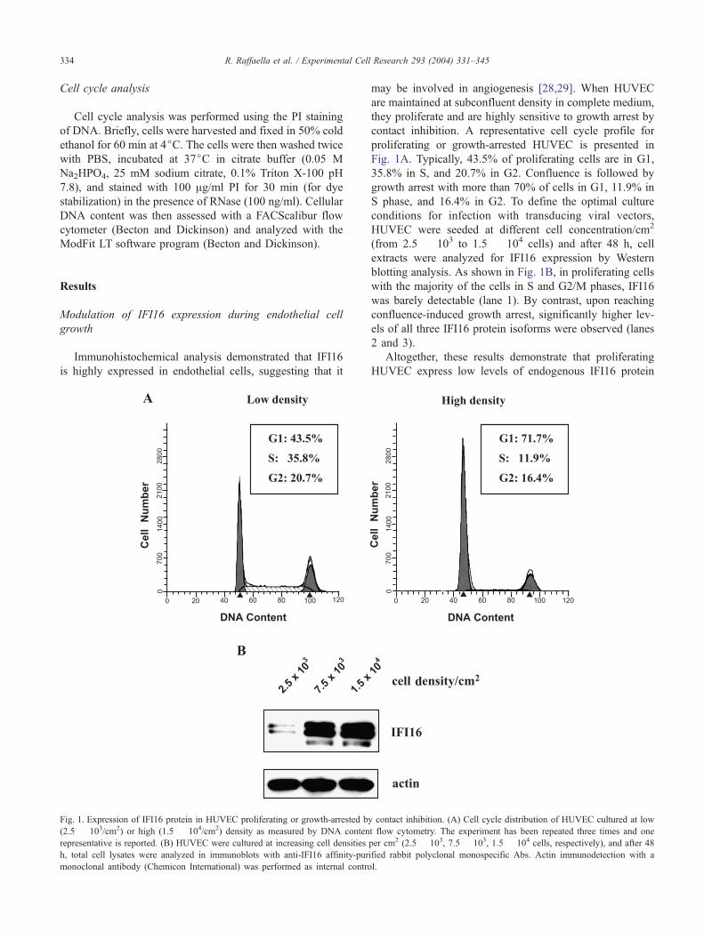
Immunohistochemical analysis demonstrated that IFI16
is highly expressed in endothelial cells, suggesting that it
Fig. 1. Expression of IFI16 protein in HUVEC proliferating or growth-arrested by
(2.5 � 103/cm2) or high (1.5 � 104/cm2) density as measured by DNA conten
representative is reported. (B) HUVEC were cultured at increasing cell densities
h, total cell lysates were analyzed in immunoblots with anti-IFI16 affinity-pur
monoclonal antibody (Chemicon International) was performed as internal contro
may be involved in angiogenesis [28,29]. When HUVEC
are maintained at subconfluent density in complete medium,
they proliferate and are highly sensitive to growth arrest by
contact inhibition. A representative cell cycle profile for
proliferating or growth-arrested HUVEC is presented in
Fig. 1A. Typically, 43.5% of proliferating cells are in G1,
35.8% in S, and 20.7% in G2. Confluence is followed by
growth arrest with more than 70% of cells in G1, 11.9% in
S phase, and 16.4% in G2. To define the optimal culture
conditions for infection with transducing viral vectors,
HUVEC were seeded at different cell concentration/cm2
(from 2.5 � 103 to 1.5 � 104 cells) and after 48 h, cell
extracts were analyzed for IFI16 expression by Western
blotting analysis. As shown in Fig. 1B, in proliferating cells
with the majority of the cells in S and G2/M phases, IFI16
was barely detectable (lane 1). By contrast, upon reaching
confluence-induced growth arrest, significantly higher lev-
els of all three IFI16 protein isoforms were observed (lanes
2 and 3).
Altogether, these results demonstrate that proliferating
HUVEC express low levels of endogenous IFI16 protein
contact inhibition. (A) Cell cycle distribution of HUVEC cultured at low
t flow cytometry. The experiment has been repeated three times and one
per cm2 (2.5 � 103, 7.5 � 103, 1.5 � 104 cells, respectively), and after 48
ified rabbit polyclonal monospecific Abs. Actin immunodetection with a
l.
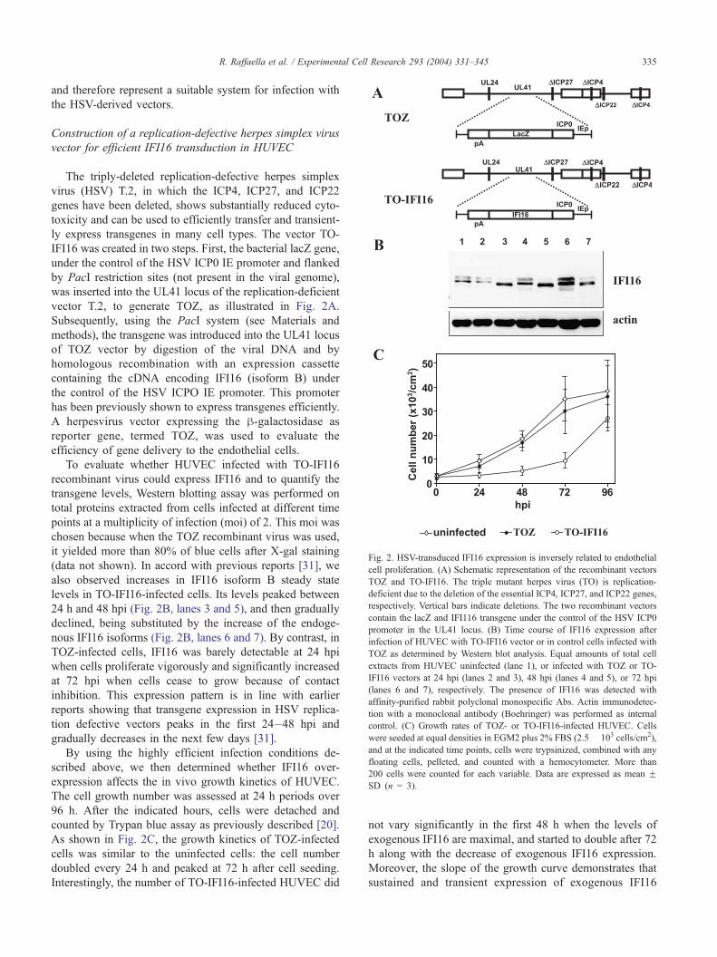
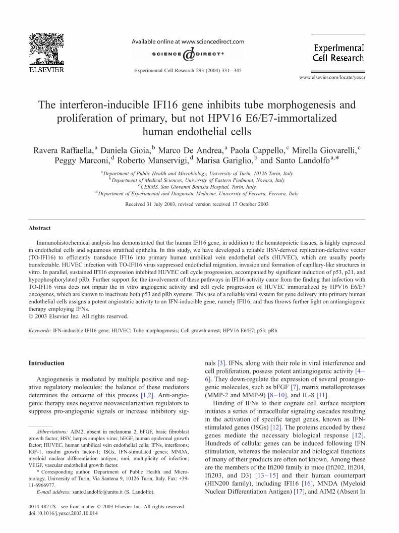
Fig. 2. HSV-transduced IFI16 expression is inversely related to endothelial
cell proliferation. (A) Schematic representation of the recombinant vectors
TOZ and TO-IFI16. The triple mutant herpes virus (TO) is replication-
deficient due to the deletion of the essential ICP4, ICP27, and ICP22 genes,
respectively. Vertical bars indicate deletions. The two recombinant vectors
contain the lacZ and IFI116 transgene under the control of the HSV ICP0
promoter in the UL41 locus. (B) Time course of IFI16 expression after
infection of HUVEC with TO-IFI16 vector or in control cells infected with
TOZ as determined by Western blot analysis. Equal amounts of total cell
extracts from HUVEC uninfected (lane 1), or infected with TOZ or TO-
IFI16 vectors at 24 hpi (lanes 2 and 3), 48 hpi (lanes 4 and 5), or 72 hpi
(lanes 6 and 7), respectively. The presence of IFI16 was detected with
affinity-purified rabbit polyclonal monospecific Abs. Actin immunodetec-
tion with a monoclonal antibody (Boehringer) was performed as internal
control. (C) Growth rates of TOZ- or TO-IFI16-infected HUVEC. Cells
were seeded at equal densities in EGM2 plus 2% FBS (2.5� 103 cells/cm2),
and at the indicated time points, cells were trypsinized, combined with any
floating cells, pelleted, and counted with a hemocytometer. More than
200 cells were counted for each variable. Data are expressed as mean FSD (n = 3).
R. Raffaella et al. / Experimental Cell Research 293 (2004) 331–345 335
and therefore represent a suitable system for infection with
the HSV-derived vectors.
Construction of a replication-defective herpes simplex virus
vector for efficient IFI16 transduction in HUVEC
The triply-deleted replication-defective herpes simplex
virus (HSV) T.2, in which the ICP4, ICP27, and ICP22
genes have been deleted, shows substantially reduced cyto-
toxicity and can be used to efficiently transfer and transient-
ly express transgenes in many cell types. The vector TO-
IFI16 was created in two steps. First, the bacterial lacZ gene,
under the control of the HSV ICP0 IE promoter and flanked
by PacI restriction sites (not present in the viral genome),
was inserted into the UL41 locus of the replication-deficient
vector T.2, to generate TOZ, as illustrated in Fig. 2A.
Subsequently, using the PacI system (see Materials and
methods), the transgene was introduced into the UL41 locus
of TOZ vector by digestion of the viral DNA and by
homologous recombination with an expression cassette
containing the cDNA encoding IFI16 (isoform B) under
the control of the HSV ICPO IE promoter. This promoter
has been previously shown to express transgenes efficiently.
A herpesvirus vector expressing the h-galactosidase as
reporter gene, termed TOZ, was used to evaluate the
efficiency of gene delivery to the endothelial cells.
To evaluate whether HUVEC infected with TO-IFI16
recombinant virus could express IFI16 and to quantify the
transgene levels, Western blotting assay was performed on
total proteins extracted from cells infected at different time
points at a multiplicity of infection (moi) of 2. This moi was
chosen because when the TOZ recombinant virus was used,
it yielded more than 80% of blue cells after X-gal staining
(data not shown). In accord with previous reports [31], we
also observed increases in IFI16 isoform B steady state
levels in TO-IFI16-infected cells. Its levels peaked between
24 h and 48 hpi (Fig. 2B, lanes 3 and 5), and then gradually
declined, being substituted by the increase of the endoge-
nous IFI16 isoforms (Fig. 2B, lanes 6 and 7). By contrast, in
TOZ-infected cells, IFI16 was barely detectable at 24 hpi
when cells proliferate vigorously and significantly increased
at 72 hpi when cells cease to grow because of contact
inhibition. This expression pattern is in line with earlier
reports showing that transgene expression in HSV replica-
tion defective vectors peaks in the first 24–48 hpi and
gradually decreases in the next few days [31].
By using the highly efficient infection conditions de-
scribed above, we then determined whether IFI16 over-
expression affects the in vivo growth kinetics of HUVEC.
The cell growth number was assessed at 24 h periods over
96 h. After the indicated hours, cells were detached and
counted by Trypan blue assay as previously described [20].
As shown in Fig. 2C, the growth kinetics of TOZ-infected
cells was similar to the uninfected cells: the cell number
doubled every 24 h and peaked at 72 h after cell seeding.
Interestingly, the number of TO-IFI16-infected HUVEC did
not vary significantly in the first 48 h when the levels of
exogenous IFI16 are maximal, and started to double after 72
h along with the decrease of exogenous IFI16 expression.
Moreover, the slope of the growth curve demonstrates that
sustained and transient expression of exogenous IFI16
R. Raffaella et al. / Experimental Cell Research 293 (2004) 331–345336
protein reversibly inhibits HUVEC proliferation without
causing cell death in accord to the results obtained with
Ifi204 overexpression in mouse fibroblasts [19,20]. These
conclusions are further supported by the finding that IFI16-
expressing HUVEC marginally undergo apoptosis as shown
by annexin V binding (data not shown).
Altogether, these results demonstrate that sustained and
transient expression of exogenous IFI16 halts growth of
proliferating HUVEC and hence that this system is suitable
for in vitro angiogenesis studies.
Inhibition of HUVEC chemotaxis, invasion, and tube
morphogenesis by IFI16 overexpression
Endothelial cell responses to angiogenic factors can be
measured in vitro by assessment of induction of endothelial
cell chemotaxis and chemoinvasion [33–36]. For this pur-
pose, HUVECs were infected with either TO-IFI16 or TOZ
viruses or left uninfected, and their migration and invasion
capabilities evaluated. Infection of endothelial cells with
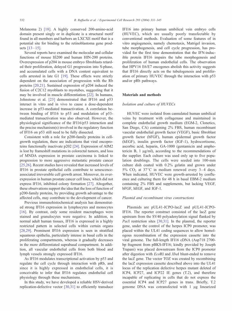
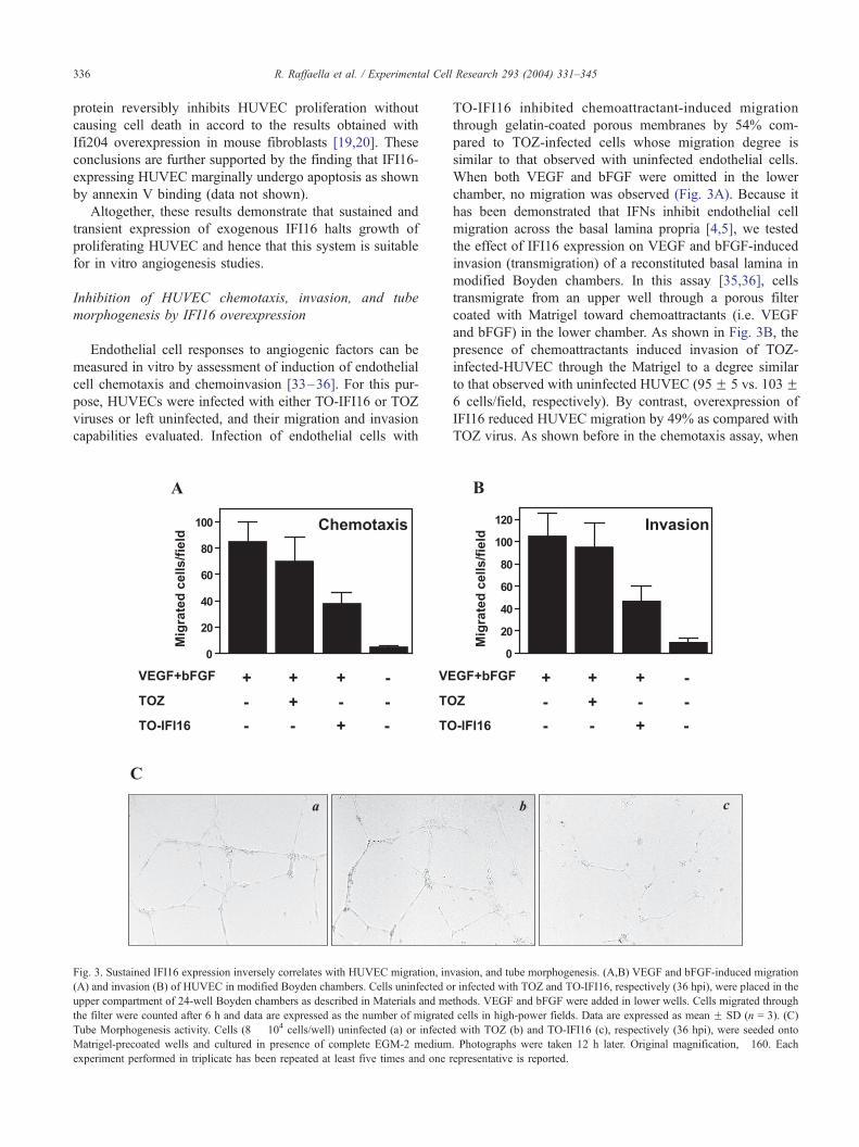
Fig. 3. Sustained IFI16 expression inversely correlates with HUVEC migration, in
(A) and invasion (B) of HUVEC in modified Boyden chambers. Cells uninfected o
upper compartment of 24-well Boyden chambers as described in Materials and me
the filter were counted after 6 h and data are expressed as the number of migrated
Tube Morphogenesis activity. Cells (8 � 104 cells/well) uninfected (a) or infecte
Matrigel-precoated wells and cultured in presence of complete EGM-2 medium
experiment performed in triplicate has been repeated at least five times and one
TO-IFI16 inhibited chemoattractant-induced migration
through gelatin-coated porous membranes by 54% com-
pared to TOZ-infected cells whose migration degree is
similar to that observed with uninfected endothelial cells.
When both VEGF and bFGF were omitted in the lower
chamber, no migration was observed (Fig. 3A). Because it
has been demonstrated that IFNs inhibit endothelial cell
migration across the basal lamina propria [4,5], we tested
the effect of IFI16 expression on VEGF and bFGF-induced
invasion (transmigration) of a reconstituted basal lamina in
modified Boyden chambers. In this assay [35,36], cells
transmigrate from an upper well through a porous filter
coated with Matrigel toward chemoattractants (i.e. VEGF
and bFGF) in the lower chamber. As shown in Fig. 3B, the
presence of chemoattractants induced invasion of TOZ-
infected-HUVEC through the Matrigel to a degree similar
to that observed with uninfected HUVEC (95F 5 vs. 103F6 cells/field, respectively). By contrast, overexpression of
IFI16 reduced HUVEC migration by 49% as compared with
TOZ virus. As shown before in the chemotaxis assay, when
vasion, and tube morphogenesis. (A,B) VEGF and bFGF-induced migration
r infected with TOZ and TO-IFI16, respectively (36 hpi), were placed in the
thods. VEGF and bFGF were added in lower wells. Cells migrated through
cells in high-power fields. Data are expressed as mean F SD (n = 3). (C)
d with TOZ (b) and TO-IFI16 (c), respectively (36 hpi), were seeded onto
. Photographs were taken 12 h later. Original magnification, �160. Eachrepresentative is reported.

Fig. 4. Sustained IFI16 overexpression inhibits HUVEC cell cycle progression. (A) Growth-arrested HUVEC (indicated as t0) were infected with TOZ or TO-
IFI 16 vectors, replated at low cell density, and then cultured in complete EGM-2 medium. At the indicated times post infection, cell cycle distribution was
evaluated. (B) Proliferating HUVEC (indicated as t0) were infected with TOZ or TO-IFI16 vectors, replated at low cell density, and at the indicated times post
infection analyzed for cell cycle distribution. Cellular DNA content was assessed with a FACScalibur flow cytometer (Becton and Dickinson) and analyzed
with the ModFit LT software program (Becton and Dickinson). The experiments were repeated three times and one representative is reported.
R. Raffaella et al. / Experimental Cell Research 293 (2004) 331–345 337
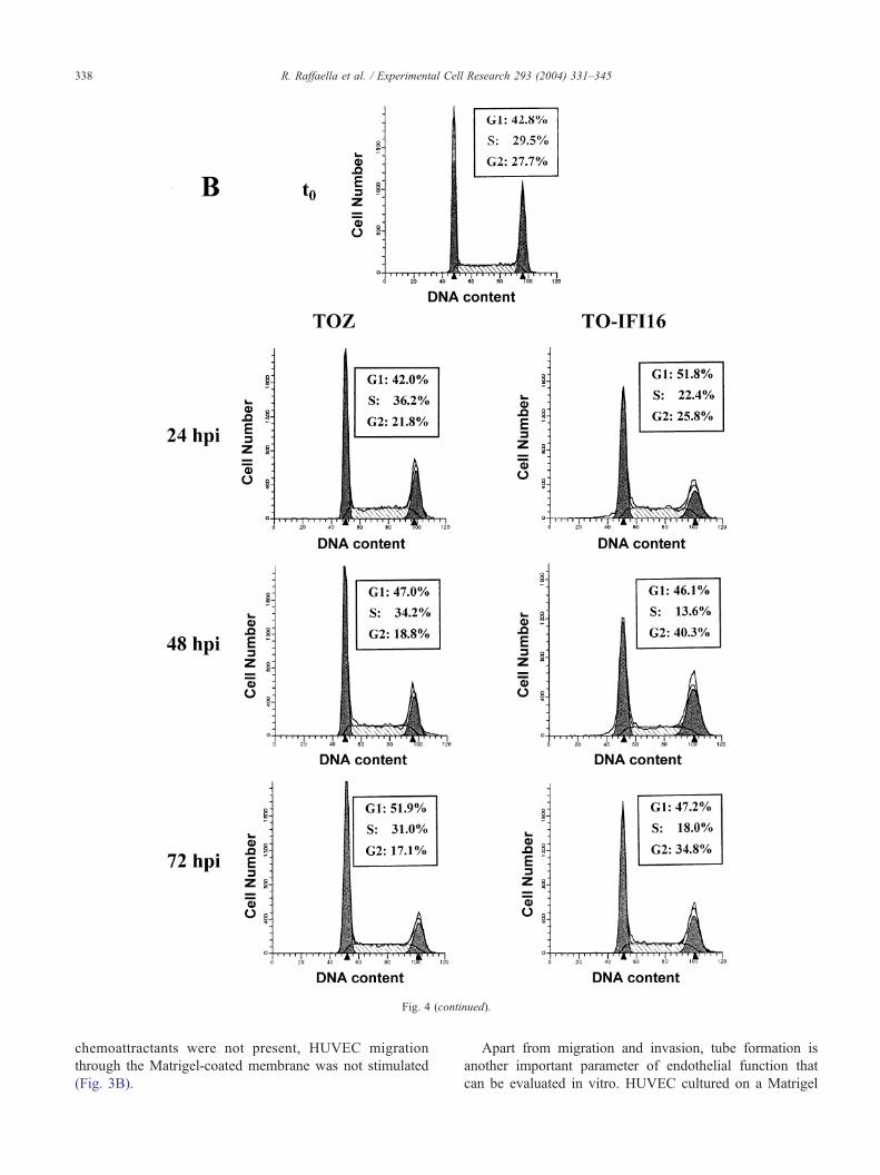
Fig. 4 (continued).
R. Raffaella et al. / Experimental Cell Research 293 (2004) 331–345338
chemoattractants were not present, HUVEC migration
through the Matrigel-coated membrane was not stimulated
(Fig. 3B).
Apart from migration and invasion, tube formation is
another important parameter of endothelial function that
can be evaluated in vitro. HUVEC cultured on a Matrigel
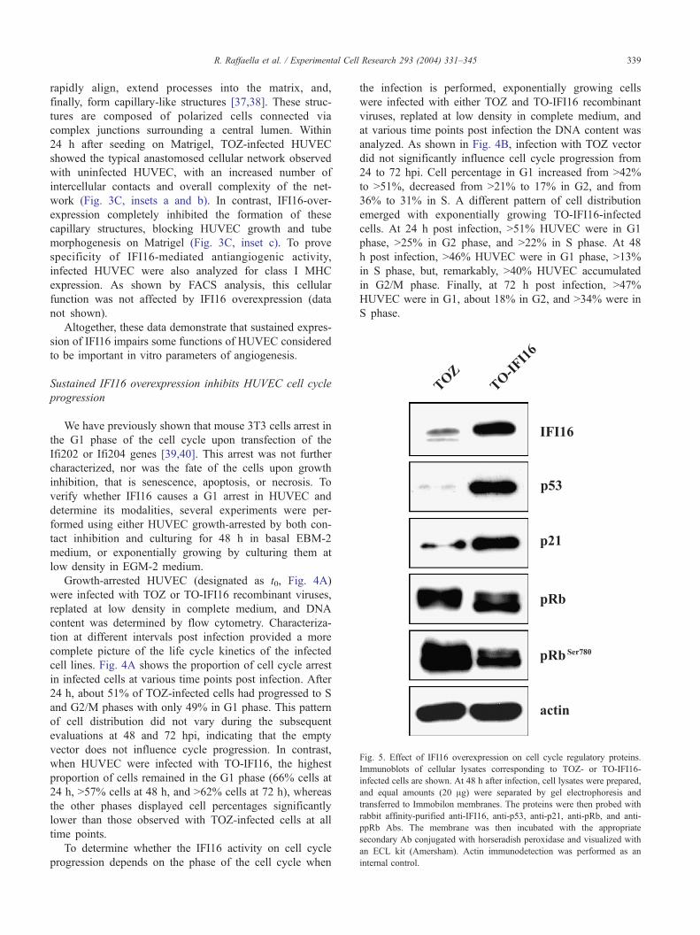
Fig. 5. Effect of IFI16 overexpression on cell cycle regulatory proteins.
Immunoblots of cellular lysates corresponding to TOZ- or TO-IFI16-
infected cells are shown. At 48 h after infection, cell lysates were prepared,
and equal amounts (20 Ag) were separated by gel electrophoresis and
transferred to Immobilon membranes. The proteins were then probed with
rabbit affinity-purified anti-IFI16, anti-p53, anti-p21, anti-pRb, and anti-
ppRb Abs. The membrane was then incubated with the appropriate
secondary Ab conjugated with horseradish peroxidase and visualized with
an ECL kit (Amersham). Actin immunodetection was performed as an
internal control.
R. Raffaella et al. / Experimental Cell Research 293 (2004) 331–345 339
rapidly align, extend processes into the matrix, and,
finally, form capillary-like structures [37,38]. These struc-
tures are composed of polarized cells connected via
complex junctions surrounding a central lumen. Within
24 h after seeding on Matrigel, TOZ-infected HUVEC
showed the typical anastomosed cellular network observed
with uninfected HUVEC, with an increased number of
intercellular contacts and overall complexity of the net-
work (Fig. 3C, insets a and b). In contrast, IFI16-over-
expression completely inhibited the formation of these
capillary structures, blocking HUVEC growth and tube
morphogenesis on Matrigel (Fig. 3C, inset c). To prove
specificity of IFI16-mediated antiangiogenic activity,
infected HUVEC were also analyzed for class I MHC
expression. As shown by FACS analysis, this cellular
function was not affected by IFI16 overexpression (data
not shown).
Altogether, these data demonstrate that sustained expres-
sion of IFI16 impairs some functions of HUVEC considered
to be important in vitro parameters of angiogenesis.
Sustained IFI16 overexpression inhibits HUVEC cell cycle
progression
We have previously shown that mouse 3T3 cells arrest in
the G1 phase of the cell cycle upon transfection of the
Ifi202 or Ifi204 genes [39,40]. This arrest was not further
characterized, nor was the fate of the cells upon growth
inhibition, that is senescence, apoptosis, or necrosis. To
verify whether IFI16 causes a G1 arrest in HUVEC and
determine its modalities, several experiments were per-
formed using either HUVEC growth-arrested by both con-
tact inhibition and culturing for 48 h in basal EBM-2
medium, or exponentially growing by culturing them at
low density in EGM-2 medium.
Growth-arrested HUVEC (designated as t0, Fig. 4A)
were infected with TOZ or TO-IFI16 recombinant viruses,
replated at low density in complete medium, and DNA
content was determined by flow cytometry. Characteriza-
tion at different intervals post infection provided a more
complete picture of the life cycle kinetics of the infected
cell lines. Fig. 4A shows the proportion of cell cycle arrest
in infected cells at various time points post infection. After
24 h, about 51% of TOZ-infected cells had progressed to S
and G2/M phases with only 49% in G1 phase. This pattern
of cell distribution did not vary during the subsequent
evaluations at 48 and 72 hpi, indicating that the empty
vector does not influence cycle progression. In contrast,
when HUVEC were infected with TO-IFI16, the highest
proportion of cells remained in the G1 phase (66% cells at
24 h, >57% cells at 48 h, and >62% cells at 72 h), whereas
the other phases displayed cell percentages significantly
lower than those observed with TOZ-infected cells at all
time points.
To determine whether the IFI16 activity on cell cycle
progression depends on the phase of the cell cycle when
the infection is performed, exponentially growing cells
were infected with either TOZ and TO-IFI16 recombinant
viruses, replated at low density in complete medium, and
at various time points post infection the DNA content was
analyzed. As shown in Fig. 4B, infection with TOZ vector
did not significantly influence cell cycle progression from
24 to 72 hpi. Cell percentage in G1 increased from >42%
to >51%, decreased from >21% to 17% in G2, and from
36% to 31% in S. A different pattern of cell distribution
emerged with exponentially growing TO-IFI16-infected
cells. At 24 h post infection, >51% HUVEC were in G1
phase, >25% in G2 phase, and >22% in S phase. At 48
h post infection, >46% HUVEC were in G1 phase, >13%
in S phase, but, remarkably, >40% HUVEC accumulated
in G2/M phase. Finally, at 72 h post infection, >47%
HUVEC were in G1, about 18% in G2, and >34% were in
S phase.
R. Raffaella et al. / Experimental Cell Research 293 (2004) 331–345340
Altogether, these results demonstrate that IFI16 gene
inhibits the progression of HUVEC through the cell cycle,
but the outcome of cell accumulation strictly depends on the
growth phase in which IFI16 overexpression occurs. When
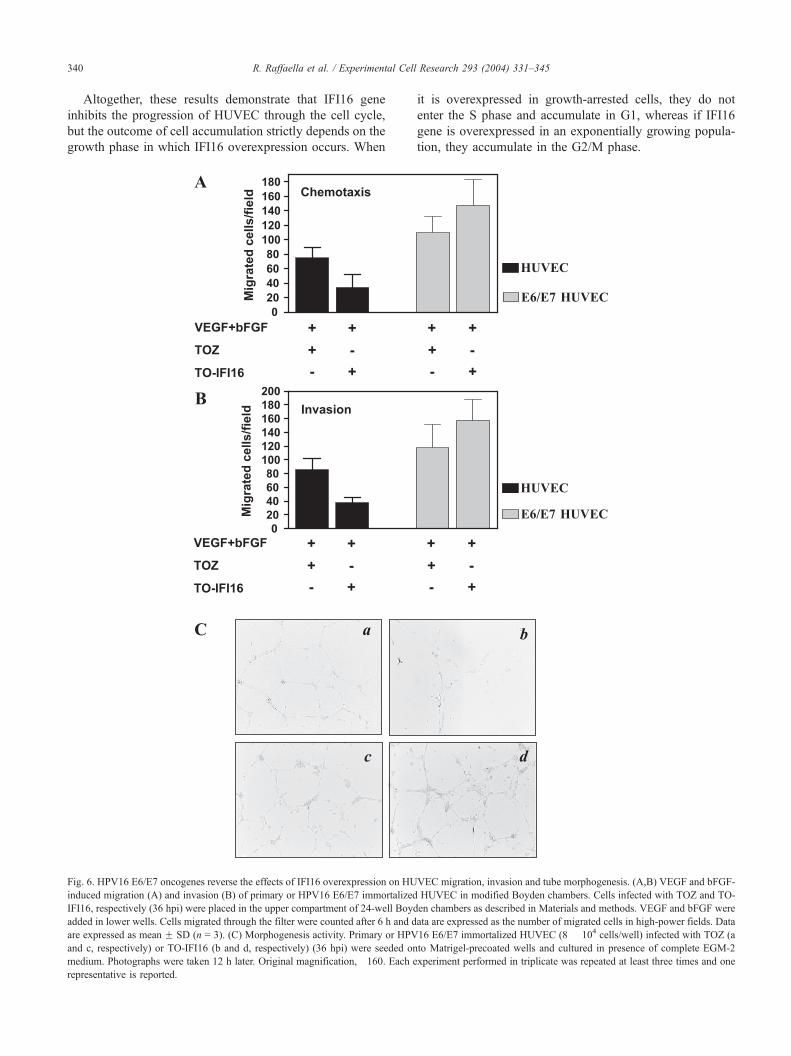
Fig. 6. HPV16 E6/E7 oncogenes reverse the effects of IFI16 overexpression on HU
induced migration (A) and invasion (B) of primary or HPV16 E6/E7 immortalized
IFI16, respectively (36 hpi) were placed in the upper compartment of 24-well Boyd
added in lower wells. Cells migrated through the filter were counted after 6 h and d
are expressed as mean F SD (n = 3). (C) Morphogenesis activity. Primary or HPV
and c, respectively) or TO-IFI16 (b and d, respectively) (36 hpi) were seeded on
medium. Photographs were taken 12 h later. Original magnification, �160. Each e
representative is reported.
it is overexpressed in growth-arrested cells, they do not
enter the S phase and accumulate in G1, whereas if IFI16
gene is overexpressed in an exponentially growing popula-
tion, they accumulate in the G2/M phase.
VEC migration, invasion and tube morphogenesis. (A,B) VEGF and bFGF-
HUVEC in modified Boyden chambers. Cells infected with TOZ and TO-
en chambers as described in Materials and methods. VEGF and bFGF were
ata are expressed as the number of migrated cells in high-power fields. Data
16 E6/E7 immortalized HUVEC (8 � 104 cells/well) infected with TOZ (a
to Matrigel-precoated wells and cultured in presence of complete EGM-2
xperiment performed in triplicate was repeated at least three times and one
R. Raffaella et al. / Experimental Cell
Earlier reports showed that 200 gene families modulate
cell growth by interacting with both p53 and pRb systems and
down-regulate transcription mediated by E2F [20,22,40]. To
further define the molecular events underlying the effects of
IFI16 on HUVEC physiology, that is migration, invasion,
tube formation, and proliferation, the steady state levels of the
tumor suppressor proteins p53 and pRb, and the p21Cip1/
WAF1 (p21WAF1), were monitored at 48 h after infection
with either TO-IFI16 or TOZ recombinant viruses, when cell
growth is arrested according to FACS analysis. As shown in
Fig. 5, the levels of both p53 and p21WAF1 were signifi-
cantly augmented upon infection with TO-IFI16 compared to
those observed with the TOZ virus. Interestingly, the increase
in p21WAF1 suggest that the induced p53 is functional [41].
When the levels of pRb expression were analyzed, although
no modulation of the Rb steady state levels was observed, the
protein appeared to be significantly hypophosphorylated,
suggesting that it is in the active form typical of cells arrested
in the G1 phase.
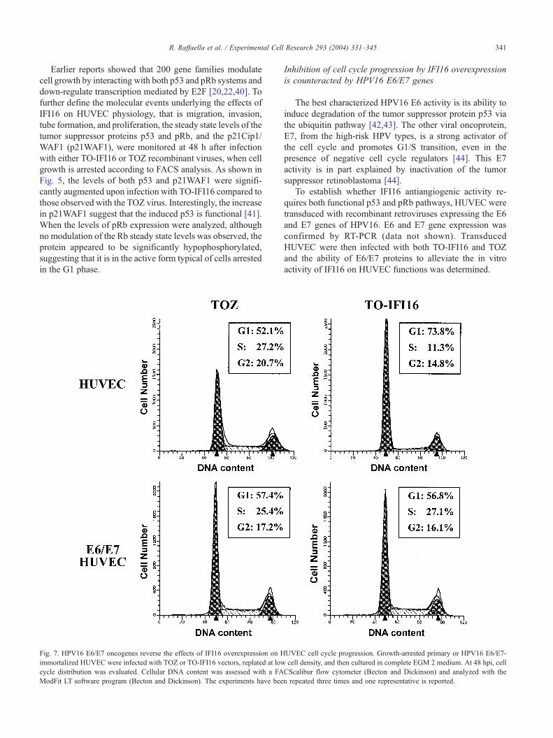
Fig. 7. HPV16 E6/E7 oncogenes reverse the effects of IFI16 overexpression on H
immortalized HUVEC were infected with TOZ or TO-IFI16 vectors, replated at low
cycle distribution was evaluated. Cellular DNA content was assessed with a FA
ModFit LT software program (Becton and Dickinson). The experiments have bee
Inhibition of cell cycle progression by IFI16 overexpression
is counteracted by HPV16 E6/E7 genes
The best characterized HPV16 E6 activity is its ability to
induce degradation of the tumor suppressor protein p53 via
the ubiquitin pathway [42,43]. The other viral oncoprotein,
E7, from the high-risk HPV types, is a strong activator of
the cell cycle and promotes G1/S transition, even in the
presence of negative cell cycle regulators [44]. This E7
activity is in part explained by inactivation of the tumor
suppressor retinoblastoma [44].
To establish whether IFI16 antiangiogenic activity re-
quires both functional p53 and pRb pathways, HUVEC were
transduced with recombinant retroviruses expressing the E6
and E7 genes of HPV16. E6 and E7 gene expression was
confirmed by RT-PCR (data not shown). Transduced
HUVEC were then infected with both TO-IFI16 and TOZ
and the ability of E6/E7 proteins to alleviate the in vitro
activity of IFI16 on HUVEC functions was determined.
Research 293 (2004) 331–345 341
UVEC cell cycle progression. Growth-arrested primary or HPV16 E6/E7-
cell density, and then cultured in complete EGM 2 medium. At 48 hpi, cell
CScalibur flow cytometer (Becton and Dickinson) and analyzed with the
n repeated three times and one representative is reported.
R. Raffaella et al. / Experimental Cell Research 293 (2004) 331–345342
For this purpose, parental- or E6/E7 immortalized-
HUVEC were infected with TOZ or TO-IFI16 and their
motility and invasion capabilities, tube morphogenesis, and
cycle progression compared. As shown in Fig. 6, infection
with TO-IFI16 strongly inhibited motility (panel A) and
invasion capabilities (panel B) along with the formation of
capillary-like structures (panel C) of primary HUVEC
compared with TOZ-infected HUVEC. In contrast, when
HUVEC immortalized with HPV16 E6/E7 oncogenes were
infected with TO-IFI16, the number of cells migrating
throughout the filters coated with gelatin (148 F 16 mi-
grating cells/field) or Matrigel (158 F 15 invading cells/
field) was significantly higher than that observed with cells
infected with TOZ (110 F 8 migrating cells/field and 118 F12 invading cells/field, respectively). In line with these
results, IFI16 overexpression in immortalized HUVEC
significantly enhanced tube formation with an increased
number of intercellular contacts and overall complexity of
the network compared to that observed with immortalized
HUVEC infected with TOZ (Fig. 6C, insets c and d vs. a
and b, respectively).
To determine whether HPV16 E6/E7 oncogenes reverse
IFI16-induced growth inhibition, parental HUVEC or
HUVEC transduced with pBabeE6/E7 (E6/E7 HUVEC)
were growth-arrested both by contact inhibition and by
culturing them in EBM-2 medium; cells were then infected
with TOZ or TO-IFI16, and at 48 hpi the cell cycle profiles
were determined by FACS analysis. As shown in Fig. 7,
E6/E7 HUVEC overexpressing IFI16 displayed a much
lower percentage of cells in G1 than that observed with
primary HUVEC infected with TO-IFI16 (56.8% vs.
73.8%, respectively), but very similar to that observed with
E6/E7 HUVEC or primary HUVEC infected with TOZ. In
contrast, an opposite effect was observed when the other
phases of the cell cycle were compared: a higher percentage
of cells in both S and G2/M was constantly observed in E6/
E7 HUVEC similar to those observed with cells infected
with TOZ.
Altogether, these results suggest that in the absence of
functional p53 and pRb systems following E6/E7 infection,
IFI16 not only loses its ability to exert antiangiogenic and
antiproliferative activities, but even enhances some param-
eters of in vivo angiogenesis, that is endothelial cell inva-
sion and tube morphogenesis.
Discussion
Earlier reports demonstrated that the mouse Ifi200
genes, namely Ifi202 and Ifi204, when overexpressed in
primary mouse embryo fibroblasts or NIH3T3 cells, arrest
their proliferation by delaying transition from G1 to S
phase [39,40]. Interaction with both pRb and p53 systems
appears to be essential for this antiproliferative activity
[19–23,45]. By contrast, less information is available
about the human homolog IFI16, probably due to the lack
of an appropriate normal cellular model in the human
system.
In this study, we have developed a suitable experimen-
tal model to test IFI16 biological functions by using
primary HUVEC, which are usually poorly transfectable
by conventional methods, transduced with a HSV-derived
replication-defective vector carrying the IFI16 cDNA. The
choice of the endothelial cell system was dictated by two
previous findings. In vivo, along with hematopoietic and
stratified squamous epithelial cells, endothelial cells spe-
cifically express IFI16, suggesting a role of this gene in the
control of their physiology. In vitro, Western blotting
analysis demonstrated that in proliferating HUVEC,
IFI16 expression is barely detectable and gradually in-
creases when cell growth halts upon reaching confluence.
It may thus be involved in the modulation of endothelium
differentiation and proliferation. The use of the HSV-
derived TO-IFI16 vector allowed overexpression of the
IFI16 protein in primary HUVEC and revealed for the first
time that sustained IFI16 expression suppresses their
growth, migration, and organization into capillary-like
structures, together with cell cycle progression. Taken
together, our results suggest that IFI16 contributes to
maintenance of a quiescent, differentiated phenotype in
HUVEC and that its expression must decrease for tube
morphogenesis to proceed.
Angiogenesis occurs by a series of sequential steps. In
response to angiogenic stimuli, endothelial cells degrade the
basement membrane by secreting proteolytic enzymes that
include metalloproteinases (MMPs) and serine proteases
[1,2,37]. The cells then migrate through the degraded base-
ment membrane and continue to break down the interstitial
stroma as they move. The endothelial cells at the tip of sprout
do not usually divide, whereas the trailing cells at the base of
the new vessel proliferate [38]. The endothelium then aligns
in a bipolar fashion to form a lumen. Given the crucial
function of MMPs in the degradation of basement membrane
in the initial steps of angiogenesis, it is therefore conceivable
that one of the mechanisms exploited by IFI16 to inhibit
chemotaxis, chemoinvasion, and tube morphogenesis is
down-regulation of MMPs secretion. We have, in fact,
observed that in zymography experiments the supernatants
from HUVEC infected with TO-IFI16 contain less proteo-
lytic activity attributable to MMP2 and MMP9 in conse-
quence of a decreased expression of MMP2 and MMP9
proteins (data not shown). This observation is consistent
with a previous report showing that another IFN-inducible
gene, namely the guanylate protein-1 GTPase, controls the
invasive and angiogenic capability of endothelial cells
through inhibition of MMP-1 expression [10].
In addition to proteolytic enzyme secretion and migra-
tion, tube morphogenesis depends on endothelial cell pro-
liferation [1,2,37,38]. Analysis of cell growth and cell cycle
profile upon TO-IFI16 infection suggests that sustained
expression of IFI16 affects HUVEC proliferation. More-
over, Western blotting analysis and IFI16 overexpression in
R. Raffaella et al. / Experimental Cell Research 293 (2004) 331–345 343
HPV16E6/E7-transduced cells revealed that its activity
depends on both functional p53 and pRb pathways. The
p53 and pRb protein families are key cell cycle regulators
that manage the response to a variety of stimuli either by
inducing cell growth arrest, senescence, or apoptosis [46–
48]. The mouse homolog p202 was found to bind the
murine homolog of the human p53-binding protein 1
(p53BP1) and regulate transcriptional activation mediated
by p53 [45]. The mouse p204 contains the pRb binding
motifs LXCXE and binds pRb [20]. Its overexpression, as
here with IFI16, delays progression of cells from the G1 to
the S phase. IFI16 directly binds to the C-terminal region of
p53 through the consensus motif MFHATVAT in the 200-
amino-acid domain a, and augments p53-mediated tran-
scriptional activation [23]. The outcome of immunoblotting
analysis confirms that sustained IFI16 expression increases
both p53 and pRb levels, the latter in its hyposphorylated
form. The molecular mechanisms underlying cell cycle
regulation by p53 have been well defined. p53 transcrip-
tionally up-regulates the p21WAF1 gene, the protein prod-
uct of which inhibits the kinase activity of cdk4/cyclin D
[49]. Activation of cdk4/cyclin D results in phosphorylation
of pRb, release of pRb from the pRb/E2F/DP-1 complex,
and progression to S phase via activation of E2F/DP-1
responsive genes [50]. Thus, p21WAF1 provides a link
between IFI16, p53, and pRb regulatory pathways. It can
act to limit the phosphorylation of pRb, thereby preventing
cells from exiting the G1 phase of the cell cycle.
The transforming proteins of DNA tumor viruses such as
SV40 large T antigen, adenovirus E1A/E1B, or HPV16 E6/
E7 extend the life span of human cells in culture [51].
Extension of life span is dependent on the ability of these
viral proteins to target the tumor suppressor proteins, p53
and pRb, and render them functionally inactive. When
HPV16 E6/E7-transduced HUVEC were analyzed to deter-
mine their tubulogenesis activity and cell cycle progression
upon IFI16 overexpression, it became evident that inacti-
vation of both p53 and pRb pathways renders them resistant
to IFI16 antitubulogenesis and antiproliferative activities. In
this context, Xin et al. [52] reported that ectopic expression
of IFI16 in prostate cancer cell lines resulted in colony
formation inhibition that differed with respect to expression
of functional pRb and p53; the maximum inhibition was
seen in LNCaP (these cells express functional Rb) and
minimum inhibition was seen in DU-145 cells (these cells
do not express functional Rb and p53). Importantly, up-
regulation of p21WAF1 and inhibition of E2F-stimulated
transcription accompanied inhibition of cell growth by
IFI16 in these prostate cancer cell lines. Moreover, John-
stone et al. [23] earlier reported that IFI16 can directly bind
to the C-terminal region of p53 and augment p53-mediated
transcriptional activation of a reporter construct containing
the promoter from the p21WAF1 gene with a p53-binding
site. Altogether, these observations supported the possibility
that IFI16-mediated antiangiogenic activity depends on the
presence of functional p53 and pRb. On the contrary, Kwak
et al. [53] reported that the inhibition of endogenous IFI16
expression by small interfering RNA (siRNA) induces
p21WAF1 mRNA and protein expression through p53,
and results in cell cycle arrest. However, there are numer-
ous differences between the experiments conducted by
Kwak et al. and the ones reported here that may affect
the cellular response to growth-inhibitory signals. For
example, they have employed a tumor cell line, U2OS,
expressing high level of endogenous IFI16, whereas we
have used primary normal endothelial cells (HUVEC)
whose basal IFI16 level is very low and highly inducible
by various stimuli. In addition, unlike our experiments, they
have inhibited endogenous IFI16 expression in U2OS cells
that constitutively display high IFI16 levels and strongly
argues against its antiproliferative activity. In light of this,
IFI16 is possibly functionally inactive in U2OS, or it may
even be the case that mutations occurred in its downstream
signaling pathways.
Our observations on the capability of IFI16 to control
some aspects of endothelial cell physiology have some
interesting implications. The first is that until now, it has
been difficult to assign a biological function to the IFI16
gene. Its homology to the mouse Ifi204 indicated its in-
volvement in cell cycle control, but the lack of efficient
transducing vectors and appropriate cell systems has ham-
pered definition of its biological function. HSV-derived
vectors overcame these obstacles and provided the first
evidence of a link between IFI16 and inhibition of cell
proliferation. The second is based on previous in vivo
findings. Immunohistochemistry revealed that IFI16 is high-
ly expressed in endothelial and epithelial cells and may be of
significance in their physiology. The use of primary HUVEC
established a link between IFI16 expression and control of
tube morphogenesis in vitro, considered to be the counterpart
of in vivo angiogenesis. The third implication is that at the
molecular level IFI16 exploits both p53 and pRb pathways to
control the cell cycle and proliferation. Thus, these data may
help to explain why in vivo transformed cells bearing
mutations at these two important regulators of cell cycle
progression become resistant to the IFN treatment because of
the acquisition to evade the IFN antiproliferative and anti-
angiogenic activities. The fourth one is that IFNs so far have
been found to have antiangiogenic properties in vivo and in
vitro through down-regulation of the expression of proan-
giogenic molecules, such as bFGF [6], MMP-2 and MMP-9
[7–9], and IL-8 [10]. Our present findings showing that
sustained expression of IFI16 impairs tube morphogenesis
and growth by inhibiting HUVEC cell cycle progression
provide a further illustration of the molecular mechanisms
exploited by IFNs to regulate angiogenesis.
Acknowledgments
We thank Filippo Reno (Novara) and Adriana Albini
(Genova) for helpful discussion and manuscript revision,
R. Raffaella et al. / Experimental Cell Research 293 (2004) 331–345344
and Barbara Azzimonti (Novara) for preparing retroviral
vectors. We thank Joseph Trapani for his generous gift of
the pBKS-IFI16 plasmid. This work was supported by
grants from Associazione Italiana per la Ricerca sul Cancro,
Special Project Oncology ‘‘Compagnia di San Paolo’’/
FIRMS, Lega Italiana per la Lotta contro i Tumori (section
of Novara) and Program 40% (MIUR).
References
[1] W. Risau, Mechanisms of angiogenesis, Nature 386 (1997) 671–674.
[2] G.D. Yancopoulos, S. Davis, N.W. Gale, J.S. Rudge, S.J. Wiegand, J.
Holash, Vascular-specific growth factors and blood vessel formation,
Nature 407 (2000) 242–248.
[3] R. Kerbel, J. Folkman, Clinical translation of angiogenesis inhibitors,
Nat. Cancer Rev. 2 (2002) 727–738.
[4] M. Iurlaro, R. Benelli, L. Masiello, M. Rosso, A. Albini, Beta inter-
feron inhibits HIV-1 Tat induced angiogenesis: synergism with 13-cis
retinoic acid, Eur. J. Cancer 34 (1998) 570–576.
[5] A. Albini, C. Marchisone, F. Del Grosso, R. Benelli, L. Masiello, L.
Tacchetti, M. Bono, M. Ferrantini, C. Rozera, M. Truini, F. Belardelli,
L. Santi, D.M. Noonan, Inhibition of angiogenesis and vascular tumor
growth by interferon-producing cells, Am. J. Pathol. 156 (2000)
1381–1393.
[6] E. Guenzi, K. Topolt, E. Cornali, C. Lubeseder-Martellato, A. Jorg,
K. Matzen, C. Zietz, E. Kremmer, F. Nappi, M. Schwemmle, C.
Hohenadl, G. Barillari, E. Tschachler, P. Monini, B. Ensoli, M.
Sturzl, The helical domain of GBP-1 mediates the inhibition of
endothelial cell proliferation by inflammatory cytokines, EMBO J.
20 (2001) 5568–5577.
[7] R.K. Singh, M. Gutman, C.D. Bucana, R. Sanchez, N. Llansa, I.J.
Fidler, Interferon alpha and beta down-regulate the expression of
basic fibroblast growth factor in human carcinomas, Proc. Natl. Acad.
Sci. U. S. A. 92 (1995) 4562–4566.
[8] K. Gohji, I.J. Fidler, R. Tsan, R. Radinsky, A.C. van Eshenbach,
T. Tsuruo, M. Nakajima, Human recombinant interferons-beta and
-gamma decrease production and invasion by human KG-2 renal
carcinoma cells, Int. J. Cancer 58 (1994) 380–384.
[9] N. Kato, A. Nawa, K. Tamakoshi, F. Kikkawa, N. Suganuma, T.
Okamoto, S. Goto, Y. Tomoda, M. Hamaguchi, M. Nakajima, Sup-
pression of gelatinase production with decreased invasiveness of cho-
riocarcinoma cells by recombinant interferon-h, Am. J. Obstet.
Gynecol. 172 (1995) 601–606.
[10] E. Guenzi, K. Topolt, C. Lubeseder-Martellaro, A. Jorg, E. Nasch-
berger, A. Albini, M. Sturzl, The guanylate binding protein-1
GTPase controls the invasive and angiogenic capability of endo-
thelial cells through inhibition of MMP-1 expression, EMBO J. 22
(2003) 3772–3782.
[11] R.K. Singh, M. Gutman, N. Llansa, I.J. Fidler, Interferon-h prevents
the upregulation of interleukin-8 in human melanoma cells, J. Inter-
feron Cytokine Res. 16 (1996) 577–584.
[12] G.R. Stark, I.M. Kerr, B.R. Williams, R.H. Silverman, R.D. Schreiber,
How cells respond to interferons, Annu. Rev. Biochem. 67 (1998)
227–264.
[13] P. Lengyel, D. Choubey, S.J. Li, B. Datta, The interferon-activatable
gene 200 cluster: from structure toward function, Semin. Virol. 6
(1995) 203–215.
[14] S. Landolfo, M. Gariglio, G. Gribaudo, D. Lembo, The Ifi 200 genes:
an emerging family of IFN-inducible genes, Biochimie 80 (1998)
721–728.
[15] R.W. Johnstone, J.A. Trapani, Transcription and growth regulatory
functions of the HIN-200 family of proteins, Mol. Cell. Biol. 19
(1999) 5833–5838.
[16] J.A. Trapani, K.A. Browne, M.J. Dawson, R.G. Ramsay, R.L. Eddy,
T.B. Show, P.C. White, B. Dupont, A novel gene constitutively ex-
pressed in human lymphoid cells is inducible with interferon-g in
myeloid cells, Immunogenetics 19 (1992) 369–376.
[17] G.R. Burrus, J.A. Briggs, R.C. Briggs, Characterization of the human
myeloid cell nuclear differentiation antigen: relationship to interferon-
induced proteins, J. Cell. Biochem. 48 (1992) 190–202.
[18] K.L. DeYoung, M.E. Ray, Y.A. Su, S.L. Anzick, R.W. Johnstone, J.A.
Trapani, P.S. Meltzer, J.M. Trent, Cloning of a novel member of the
human interferon-inducible gene family associated with control of
tumorigenicity in a model of human melanoma, Oncogene 15 (1997)
453–457.
[19] M. Lembo, C. Sacchi, C. Zappador, G. Bellomo, M. Gaboli, P.P.
Pandolfi, M. Gariglio, S. Landolfo, Inhibition of cell proliferation
by the interferon-inducible 204 gene, a member of the Ifi 200 cluster,
Oncogene 16 (1998) 1543–1551.
[20] L. Hertel, S. Rolle, M. De Andrea, B. Azzimonti, R. Osello, G.
Gribaudo, M. Gariglio, S. Landolfo, The retinoblastoma protein is
an essential mediator that links the interferon-inducible 204 gene to
cell-cycle regulation, Oncogene 19 (2000) 3598–3608.
[21] M. De Andrea, M. Ravotto, E. Noris, G.-G. Ying, D. Gioia, B. Azzi-
monti, M. Gariglio, S. Landolfo, The interferon-inducible gene,
Ifi204, acquires malignant transformation capability upon mutation
at the Rb-binding sites, FEBS Lett. 515 (2002) 51–57.
[22] C.-J. Liu, B. Ding, H. Wang, P. Lengyel, The MyoD-inducible p204
protein overcomes the inhibition of myoblasts differentiation by Id
proteins, Mol. Cell. Biol. 22 (2002) 2893–2905.
[23] R.W. Johnstone, W. Wei, A. Greenway, J.A. Trapani, Functional in-
teraction between p53 and the interferon-inducible nucleoprotein
IFI16, Oncogene 19 (2000) 6033–6042.
[24] H. Xin, S. D’Souza, L. Fang, P. Lengyel, D. Choubey, p202, an
interferon-inducible negative regulator of cell growth, is a target of
the adenovirus E1A protein, Oncogene 20 (2001) 6828–6839.
[25] J.Y. Mori, A. Rashid, B.A. Leggett, J. Young, L. Simms, P.M. Kuehl,
P. Langenberg, S.J. Meltzer, O.C. Stine, Instabilotyping: comprehen-
sive identification of frameshift mutations caused by coding region
microsatellite instability, Cancer Res. 61 (2001) 6046–6049.
[26] S. Varambally, S.M. Dhanasekaram, M. Zhou, T.R. Barrette, C. Ku-
mar-Sinha, M.G. Sanda, D. Ghosh, K.J. Pienta, R.G. Sewalt, A.P.
Otte, M.A. Rubin, A.M. Chinnaiyan, The polycomb group protein
EZH2 is involved in progression of prostate cancer, Nature 419
(2002) 624–629.
[27] H. Xin, J. Curry, R.W. Johnstone, B.J. Nickoloff, D. Choubey, Role of
IFI16, a member of the interferon-inducible p200-protein family, in
prostate epithelial cellular senescence, Oncogene 22 (31) (2003)
4831–4840.
[28] M. Gariglio, B. Azzimonti, M. Pagano, G. Palestro, M. De Andrea, G.
Valente, G. Voglino, L. Navino, S. Landolfo, Immunohistochemical
expression analysis of the human interferon-inducible gene IFI16, a
member of the HIN200 family, not restricted to hematopoietic cells,
J. Interferon Cytokine Res. 22 (2002) 815–821.
[29] W. Wei, C.J. Clarke, G.R. Somers, K.S. Cresswell, K.A. Loveland,
J.A. Trapani, R.W. Johnstone, Expression of IFI16 in epithelial cells
and lymphoid tissues, Histochem. Cell Biol. 119 (2003) 45–54.
[30] D.M. Krisky, P.C. Marconi, T.J. Oligino, R.J. Rouse, D.J. Fink, J.B.
Cohen, S.L. Watkins, J.C. Glorioso, Development of herpes simplex
virus replication-defective multigene vectors for combination gene
therapy application, Gene Ther. 5 (1998) 1517–1530.
[31] P. Marconi, M. Simonato, S. Zucchini, G. Bregola, R. Argnani, D.
Krisky, J.C. Glorioso, R. Manservigi, Replication-defective herpes
simplex virus vectors for neurotrophic factor gene transfer in vitro
and in vivo, Gene Ther. 6 (1999) 904–912.
[32] W.S. Pear, G.P. Nolan, M.L. Scott, D. Baltimore, Production of high-
titer helper-free retroviruses by transient transfection, Proc. Natl.
Acad. Sci. U. S. A. 90 (1993) 8392–8396.
[33] A. Albini, Y. Iwamato, H.K. Kleinman, G.R. Martin, S.A. Aaronson,
J.M. Kozlowski, R.N. McEwan, A rapid in vitro assay for quantitat-
R. Raffaella et al. / Experimental Cell Research 293 (2004) 331–345 345
ing the invasive potential of tumor cells, Cancer Res. 47 (1987)
3239–3245.
[34] G. Taraboletti, D. Roberts, L.A. Lotta, R. Giavazzi, Platelet throm-
bospondin modulates endothelial cell adhesion, motility and growth:
a potential angiogenesis regulatory factor, J. Cell Biol. 111 (1990)
765–772.
[35] Y. Kubota, H.K. Kleinman, G.R. Martin, T.J. Lawley, Role of laminin
and basement membrane in the morphological differentiation of hu-
man endothelial cells into capillary-like structures, J. Cell Biol. 107
(1988) 1589–1598.
[36] A. Albini, Tumor and endothelial cell invasion of basement mem-
branes, Pathol. Oncol. Res. 14 (1998) 1–12.
[37] M. Nguyen, J. Arkell, J.C. Jackson, Human endothelial gelatinases
and angiogenesis, Int. J. Biochem. Cell Biol. 33 (2001) 960–970.
[38] B. Lubarsky, M.A. Krasnow, Tube morphogenesis: making and shap-
ing biological tubes, Cell 112 (2003) 19–28.
[39] D. Lembo, A. Angeretti, S. Benefazio, L. Hertel, M. Gariglio, F.
Novelli, S. Landolfo, Constitutive expression of the interferon-indu-
cible protein p202 in NIH 3T3 cells affects cell cycle progression,
J. Biol. Regul. Homeostatic Agents 9 (1995) 42–46.
[40] G. Gribaudo, L. Riera, M. De Andrea, S. Landolfo, The antiprolifer-
ative activity of the murine interferon-inducible Ifi200 proteins de-
pends on the presence of two 200 amino acid domains, FEBS Lett.
456 (1999) 31–36.
[41] K.F. Macleod, N. Sherry, G. Hannon, D. Beach, T. Tokino, K. Kin-
zler, B. Vogelstein, T. Jacks, p53-Dependent and independent expres-
sion of p21 during cell growth, differentiation, and DNA damage,
Genes Dev. 9 (1995) 935–944.
[42] M. Thomas, D. Pim, L. Banks, The role of the E6-p53 interaction
in the molecular pathogenesis of HPV, Oncogene 18 (1999)
7690–7700.
[43] M. Tommasino, R. Accardi, S. Caldeira, W. Dong, I. Malanchi, A.
Smet, I. Zehbe, The role of TP53 in cervical carcinogenesis, Hum.
Mutat. 21 (2003) 307–312.
[44] H. Zur Hausen, Papillomavirus causing cancer: evasion from host-cell
control in early events in carcinogenesis, J. Natl. Cancer Inst. 78 (2000)
1–29.
[45] B. Datta, B. Li, D. Choubey, G. Nallur, P. Lengyel, p202, an inter-
feron-inducible modulator of transcription, inhibits transcriptional ac-
tivation by the p53 tumor suppressor protein, and a segment from the
p53-binding protein 1 that binds to p202 overcomes this inhibition,
J. Biol. Chem. 271 (1996) 27544–27555.
[46] A.J. Levine, p53, the cellular gatekeeper for growth and division, Cell
88 (1997) 323–331.
[47] C.H. Arrowsmith, Structure and function in the p53 family, Cell
Death Differ. 6 (1999) 1169–1173.
[48] N. Dyson, The regulation of E2F by pRB-family proteins, Genes Dev.
12 (1998) 2245–2262.
[49] M.G. Ritt, J. Mayor, J. Wojcieszyn, R. Smith, C. Barton, J.F. Mod-
iano, Sustained nuclear localization of p21/WAF-1 upon growth
arrest induced by contact inhibition, Cancer Lett. 158 (2000)
73–84.
[50] G. Mulligan, T. Jacks, The retinoblastoma gene family: cousins with
overlapping interests, Trends Genet. 14 (1998) 223–229.
[51] S.J. Flint, L.W. Enquist, R.M. Krug, V.R. Racaniello, A.M. Skalka
(Eds.), Principles of Virology. Molecular Biology, Pathogenesis, and
Control, ASM Press, Washington, DC, 2000.
[52] H. Xin, J. Curry, R.W. Johnstone, B.J. Nickoloff, D. Choubey, Role
of IFI16, a member of the interferon-inducible p200-protein family,
in prostate epithelial cellular senescence, Oncogene 22 (2003)
4831–4840.
[53] J.C. Kwak, P.P. Ongusaha, T. Ouchi, S.W. Lee, IFI16 as a neg-
ative regulator in the regulation of p53 and p21Waf1, J. Biol. Chem.
278 (2003) 40899–40904.