Archeologia delle produzioni di età medievale: glossario termini ceramica

Journal of CellularBiochemistry
ARTICLEJournal of Cellular Biochemistry 9999:1–12 (2012) JCB12-0282(24189)
Molecular GeneticQ1 Analysis of MSUD From India RevealsMutations Causing Altered Protein Truncation Affecting theC-Termini of E1a and E1b
A
G
A
LH
EG
*D
M
A
D
Murali D. Bashyam,1* Ajay K. Chaudhary,1 Sinha Manjari,2 H.A. Nagarajaram,2
A. Radha Rama Devi,3 Leena Bashyam,3 E. Chandrakanth Reddy,1 and Ashwin Dalal3
1Laboratory of MolecularQ2 Oncology, Centre for DNA Fingerprinting and Diagnostics, Hyderabad, India2Laboratory of Computational Biology, Centre for DNA Fingerprinting and Diagnostics, Hyderabad, India3Laboratory of Diagnostics Division, Centre for DNA Fingerprinting and Diagnostics, Hyderabad, India
ABSTRACTMaple Syrup Urine Disease is a rare metabolic disorder caused by reduced/absent activity of the branched chain a-Ketoacid dehydrogenase
enzyme complex. Mutations in BCKDHA, BCKDHB, and DBT, that encode important subunits of the enzyme complex namely E1a, E1b, and
E2, are the primary cause for the disease. We have performed the first molecular genetic analysis of MSUD from India on nine patients
exhibiting classical MSUD symptoms. BCKDHA and BCKDHBmutations were identified in four and five patients, respectively including seven
novel mutations namely the BCKDHA c.1249delC, c.1312T>C, and c.1561T>A and the BCKDHB c.401T>A, c.548G>A, c.964A>G, and
c.1065delT. The BCKDHB c.970C>T (p.R324X) mutation was shown to trigger nonsense mediated decay-based degradation of the transcript.
Seven of the total 11 mutations resulted in perturbations in the E1a or E1b C-termini either through altered termination or through an amino
acid change; these are expected to result in disruption of E1 enzyme complex assembly. Our study has therefore revealed that BCKDHA and
BCKDHB mutations might be primarily responsible for MSUD in the India population. J. Cell. Biochem. 9999: 1–12, 2012. � 2012 Wiley
Periodicals, Inc.
KEY WORDS: MSUD; MUTATION; BCKDHA; BCKDHB; TRUNCATION
Maple Syrup Urine Disease (MSUD) is a rare autosomal
recessive disorder caused due to malfunctioning of the
branched chain a-ketoacid dehydrogenase enzyme complex (BCKD)
[Chuang et al., 2006]. BCKD is responsible for the oxidative
decarboxylation of branched chain ketoacids, formed due to
transamination of branched chain amino acids including leucine,
isoleucine, and valine [Quental et al., 2008]. MSUD is an inborn error
of metabolism and can be fatal if not treated; clinical symptoms
including seizures, mental retardation, and coma are caused due to
accumulation of branched chain amino acids [Nellis et al., 2003;
dditional supporting information may be found in the online version of
rant sponsor: Department of Biotechnology, Government of India.
. Radha Rama Devi’s present address is Sandor Proteomics Pvt. Ltd., Hy
eena Bashyam’s present address is Genomics Facility, School of Lifeyderabad, India.
. Chandrakanth Reddy’s present address is Institute for Clinical Neurobioermany.
Correspondence to: Murali D. Bashyam, Laboratory of Molecular Oncoliagnostics, Hyderabad 500001, India. E-mail: [email protected]
anuscript Received: 9 April 2012; Manuscript Accepted: 7 May 2012
ccepted manuscript online in Wiley Online Library (wileyonlinelibrary.c
OI 10.1002/jcb.24189 � � 2012 Wiley Periodicals, Inc.
Chuang et al., 2006]. Patients are usually managed through diet
control including reduced intake of branched chain amino acids
[Snyderman et al., 1964].
BCKD is a large enzyme complex constituted by three catalytic
components namely a multimeric core of dihydrolipoyl acyltrans-
ferase (E2) in the form of a homo 24-mer to which are bound
multiple subunits of BCKD decarboxylase (E1) and the dihydroli-
poamide dehydrogenase (E3) as well as two regulatory subunits
namely BCKD kinase and BCKD phosphatase. The enzyme complex
is located in the mitochondria and is coded by four unlinked genes.
1
this article.
derabad, India.
Sciences, Hyderabad Central University,
logy, University of Wurzburg, Wurzburg,
ogy, Centre for DNA Fingerprinting and
om).
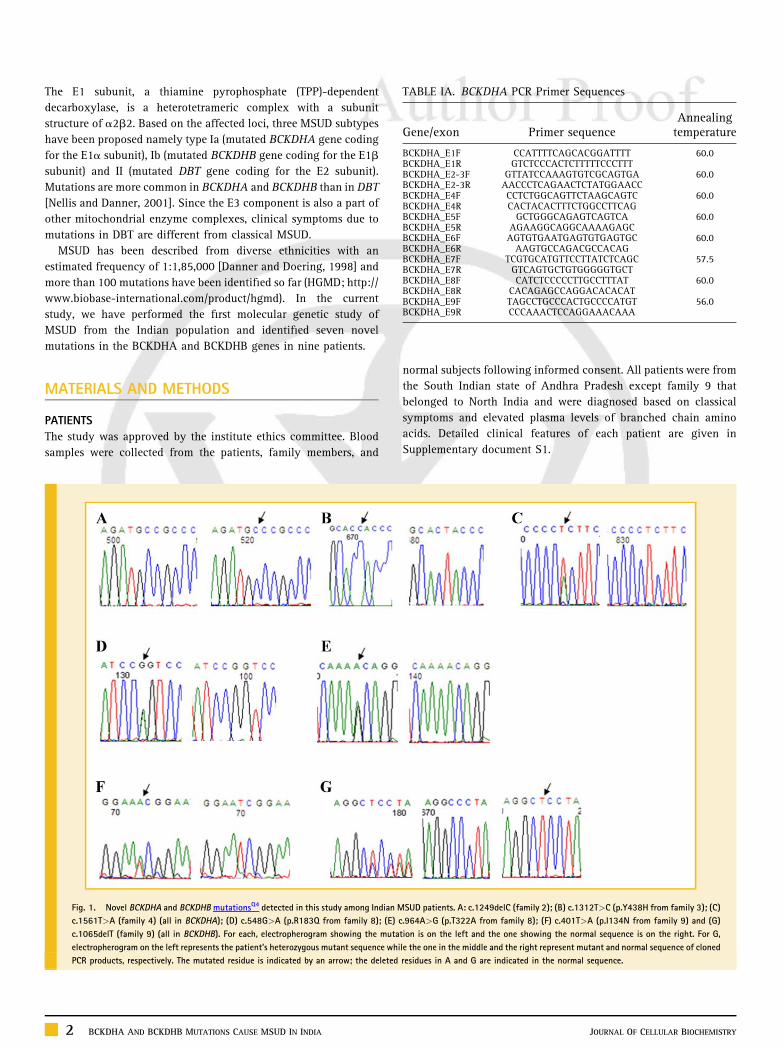
TABLE IA. BCKDHA PCR Primer Sequences
Gene/exon Primer sequenceAnnealingtemperature
BCKDHA_E1F CCATTTTCAGCACGGATTTT 60.0BCKDHA_E1R GTCTCCCACTCTTTTTCCCTTTBCKDHA_E2-3F GTTATCCAAAGTGTCGCAGTGA 60.0BCKDHA_E2-3R AACCCTCAGAACTCTATGGAACCBCKDHA_E4F CCTCTGGCAGTTCTAAGCAGTC 60.0BCKDHA_E4R CACTACACTTTCTGGCCTTCAGBCKDHA_E5F GCTGGGCAGAGTCAGTCA 60.0BCKDHA_E5R AGAAGGCAGGCAAAAGAGCBCKDHA_E6F AGTGTGAATGAGTGTGAGTGC 60.0BCKDHA_E6R AAGTGCCAGACGCCACAGBCKDHA_E7F TCGTGCATGTTCCTTATCTCAGC 57.5BCKDHA_E7R GTCAGTGCTGTGGGGGTGCTBCKDHA_E8F CATCTCCCCCTTGCCTTTAT 60.0BCKDHA_E8R CACAGAGCCAGGACACACATBCKDHA_E9F TAGCCTGCCCACTGCCCCATGT 56.0BCKDHA_E9R CCCAAACTCCAGGAAACAAA
The E1 subunit, a thiamine pyrophosphate (TPP)-dependent
decarboxylase, is a heterotetrameric complex with a subunit
structure of a2b2. Based on the affected loci, three MSUD subtypes
have been proposed namely type Ia (mutated BCKDHA gene coding
for the E1a subunit), Ib (mutated BCKDHB gene coding for the E1b
subunit) and II (mutated DBT gene coding for the E2 subunit).
Mutations are more common in BCKDHA and BCKDHB than in DBT
[Nellis and Danner, 2001]. Since the E3 component is also a part of
other mitochondrial enzyme complexes, clinical symptoms due to
mutations in DBT are different from classical MSUD.
MSUD has been described from diverse ethnicities with an
estimated frequency of 1:1,85,000 [Danner and Doering, 1998] and
more than 100 mutations have been identified so far (HGMD; http://
www.biobase-international.com/product/hgmd). In the current
study, we have performed the first molecular genetic study of
MSUD from the Indian population and identified seven novel
mutations in the BCKDHA and BCKDHB genes in nine patients.
MATERIALS AND METHODS
PATIENTS
The study was approved by the institute ethics committee. Blood
samples were collected from the patients, family members, and
Fig. 1. Novel BCKDHA and BCKDHB mutationsQ4 detected in this study among Indian M
c.1561T>A (family 4) (all in BCKDHA); (D) c.548G>A (p.R183Q from family 8); (E) c
c.1065delT (family 9) (all in BCKDHB). For each, electropherogram showing the muta
electropherogram on the left represents the patient’s heterozygous mutant sequence whi
PCR products, respectively. The mutated residue is indicated by an arrow; the deleted
2 BCKDHA AND BCKDHB MUTATIONS CAUSE MSUD IN INDIA
normal subjects following informed consent. All patients were from
the South Indian state of Andhra Pradesh except family 9 that
belonged to North India and were diagnosed based on classical
symptoms and elevated plasma levels of branched chain amino
acids. Detailed clinical features of each patient are given in
Supplementary document S1.
SUD patients. A: c.1249delC (family 2); (B) c.1312T>C (p.Y438H from family 3); (C)
.964A>G (p.T322A from family 8); (F) c.401T>A (p.I134N from family 9) and (G)
tion is on the left and the one showing the normal sequence is on the right. For G,
le the one in the middle and the right represent mutant and normal sequence of cloned
residues in A and G are indicated in the normal sequence.
JOURNAL OF CELLULAR BIOCHEMISTRY

JOURNAL OF CELLULAR BIOCHEMISTRY BCKDHA AND BCKDHB MUTATIONS CAUSE MSUD IN INDIA 3

TABLE IB. BCKDHB PCR Primer Sequences
Gene/exon Primer sequenceAnnealingtemperature
BCKDHB_E1F GCTGCATAGCCTGAGAATCC 58.5BCKDHB_E1R AATAAGCTGGGATGCAAGGABCKDHB_E2F ATTTTGCCCCATTAACAAGC 60.0BCKDHB_E2R GCTACCACAATTCAGGCACABCKDHB_E3F GACAGACCCTCACAACAAAAGA 52.8BCKDHB_E3R GCGTTGGAAATGAAAAGGAABCKDHB_E4F GACATTACTCTCATTTGCCAC 58.2BCKDHB_E4R GGAAGGGTAGCGGCAATACTBCKDHB_E5F AGGAGATTGGAAGGGAAGGA 58.5BCKDHB_E5R AACTGGGCATTGGATAGCATBCKDHB_E6F AGCCCTTCTTAGCAGCGAGT 58.2BCKDHB_E6R GGCTAGATGAATTTTTCCCAAABCKDHB_E7F TGCACAAGTGTCACCTCAGA 50.0BCKDHB_E7R GAAATTAGCATCAGTAGCACCABCKDHB_E8F ACCTTCTACATGCCATCTTTGT 56.0BCKDHB_E8R GCCAAAGGTTTCAGGGAAATBCKDHB_E9F ACCTGTCGAAAGCGAGTTGT 56.0BCKDHB_E9R TCTTCTGGAATTGGCATGTGBCKDHB_E10F AAAACTGGGATCATGCGAAC 52.8BCKDHB_E10R CGTTAATGTCAGGGGCACAT
TABLE IIA. Amino Acid Residues of a/a0 Buried Due to Subunit
Association a2b2
Amino acidresidue position
Residuetype
Percent solvent accessiblearea of residues
In thecomplexa2b2
In theisolated
a/a0 subunit
52 PRO� 16.01 19.2253 GLN� 43.85 51.4954 PHE� 3.66 44.9155 PRO� 23.28 43.8656 GLY� 1.42 23.557 ALA� 5 23.1658 SER� 15 33.5159 ALA� 4.68 22.3160 GLU� 32.24 43.5261 PHE� 19.31 47.5262 ILE� 13.13 36.6263 ASP� 12.58 36.4964 LYS� 29.16 38.2365 LEU� 13.08 48.1966 GLU� 24.47 28.8467 PHE� 6.99 56.8168 ILE� 19.63 52.8169 GLN� 38.38 45.3570 PRO� 6.78 30.9372 VAL� 15.05 19.1273 ILE� 48.57 56.0574 SER� 16.72 22.3775 GLY� 3.02 7.1876 ILE� 8.09 17.8477 PRO� 13.11 35.9478 ILE� 0.03 29.4979 TYR� 0.03 15.2180 ARG� 17.23 36.9189 ILE� 17.76 23.9890 ASN� 8.23 21.0792 SER� 29.35 29.3893 GLU� 8.84 18.19129 SER��� 11.16 26.51130 PHE��� 11.53 14.95137 GLU 1.54 1.6153 LEU�� 2.82 8.91155 PHE �� 0.51 4.36156 GLY 0.7 0.98157 GLN 2.02 2.08158 TYR��� 4.45 14.04159 ARG 13.23 20.41177 GLN��� 2.92 3.23178 CYS��� 0.37 1.45179 TYR��� 4 6.45
(Continued )
MOLECULAR GENETIC ANALYSES
Genomic DNA was isolated from blood samples as per established
protocols [Bashyam et al., 2004]. Mutations were identified by direct
polymerase chain reaction-DNA sequencing as per standard
protocols; primer sequences for each of the nine BCKDHA
and 10 BCKDHB exons are given in Tables IIA and IIB. Each
mutation was confirmed by bi-directional DNA sequencing.
Fibroblast culture from skin biopsy of proband 5 and from a
normal individual was established following informed consent and
used to quantitate ARID1B transcript levels as described in
Supplementary Methods S1.
SEQUENCE AND STRUCTURE ANALYSIS
Sequence analysis of eachmutant amino acid residue was performed
essentially as described earlier [Bashyam et al.]; detailsQ3 are given
in Supplementary Methods S1.
Fig. 2. A: Multiple sequence alignment (MSA) of human BCKDHA with homologues from other species. The MSA was performed as described in the Materials and Methods
Section. The position of each mutated residue is shown by an arrow. amino acid positions corresponding to the human sequence are indicated above each alignment. The
homologues are as follows: gij258645170jrefjNP_000700.1j, Homo sapiens; gij77736548jrefjNP_036914.1j, Rattus norvegicus; gij183396774jrefjNP_031559.3j, Mus
musculus; gij148727347jrefjNP_001092034, Pan troglodytes; gij62510814jspjQ8HXY4.1jODBA_M, Macaca fascicularis; gij297277135jrefjXP_001101959, Macaca
mulatta; gij332242782jrefjXP_003270562, Nomascus leucogenys; gij296233895jrefjXP_002762220, Callithrix jacchus; gij73947481jrefjXP_866392.1j, Canis familiaris;
gij301776619jrefjXP_002923727, Ailuropoda melanoleuca; gij338710481jrefjXP_001500344, Equus caballus; gij187607469jrefjNP_001119816, Ovis aries;
gij27806229jrefjNP_776931.1j, Bos taurus; gij178056466jrefjNP_001116555, Sus scrofa; gij291412159jrefjXP_002722340, Oryctolagus cuniculus;
gij126329384jrefjXP_001372218, Monodelphis domestica; gij327276395jrefjXP_003222955, Anolis carolinensis; gij66773104jrefjNP_001019590.1, Danio rerio;
gij195395472jrefjXP_002056360, Drosophila virilis. B: Multiple sequence alignment (MSA) of human BCKDHB with homologues from other species. The MSA was
performed as described in the Materials and Methods Section. The position of each mutated residue is shown by an arrow. amino acid positions corresponding to the human
sequence are indicated above each alignment. The homologues are as follows: gij4557353jrefjNP_000047.1j, Homo sapiens; gij162416262jspjQ6P3A8.2j, Mus musculus;
gij334324067jrefjXP_001375236, Monodelphis domestica; gij348578059jrefjXP_003474801, Caria porcellus; gij301761846jrefjXP_002916344, Ailuropoda melanoleuca;
gij348532057jrefjXP_003453523, Oreochromis niloticus; gij198285569jgbjACH85323.1j, Salmo salar; gij115502434jspjP21839.2j, Bos tarus;
gij332218346jrefjXP_003258317, Nomascus leucogenys; gij178056478jrefjNP_001116691, Sus scrofa; gij73973855jrefjXP_532213.2j, Canis lupus familiaris;
gij291396526jrefjXP_002714592, Oryctolagus cuniculus; gij158749538jrefjNP_062140.1j, Rattus norvegicus; gij344264129jrefjXP_003404146, Loxodonta africana.
4 BCKDHA AND BCKDHB MUTATIONS CAUSE MSUD IN INDIA JOURNAL OF CELLULAR BIOCHEMISTRY

TABLE IIA. (Continued )
Amino acidresidue position
Residuetype
Percent solvent accessiblearea of residues
In thecomplexa2b2
In theisolated
a/a0 subunit
180 GLY��� 0 4.27185 LEU�� 34.03 48.59186 GLY�� 4.5 13.41187 LYS��/��� 15.06 21.23188 GLY��� 0.05 8.26189 ARG��/��� 0.04 30.16190 GLN��/��� 1.67 21.28191 MET��� 1.98 29.46192 PRO��� 0 2.76196 GLY�� 3.03 6.61197 CYS�� 2.91 5.08198 LYS�� 36.96 58.16201 HIS�� 15.2 15.76202 PHE�� 0.38 1.17203 VAL�� 1.12 16.99204 THR�� 2.81 23.08205 ILE�� 0.64 0.69206 SER��/��� 0.91 14.27207 SER��� 0.48 14.58208 PRO��/��� 0.37 26.43209 LEU��� 3.09 28.68211 THR�� 0.41 12.68212 GLN�� 3.28 4.45214 PRO�� 0.06 12.94215 GLN�� 0.2 30.25217 VAL�� 0.02 1.2218 GLY�� 0 13.16219 ALA�� 0.09 7.89221 TYR�� 2.82 24.43222 ALA�� 3.14 19.08223 ALA�� 3.36 3.78224 LYS�� 16.19 16.27225 ARG�� 21.72 55.67230 ARG�� 18.36 22.5237 GLY 0.34 3.35238 GLU 0.05 0.67239 GLY��� 0 4.11240 ALA��� 0.46 1.72241 ALA� 0.15 1.79242 SER�/��� 0.18 17.53243 GLU�/��� 1.87 21.68244 GLY�/��/��� 0.1 17.84245 ASP��/��� 3.06 13.59246 ALA� 0.33 0.63247 HIS�/�� 0.95 34.9248 ALA�/�� 0.08 10.27251 ASN�/�� 0.37 17.97252 PHE�/�� 0.15 34.69254 ALA� 1.7 6.88255 THR�/�� 2.12 28.15256 LEU�� 5.94 26.75257 GLU� 20.57 27.48258 CYS�� 0 0.01265 ARG 1.62 7.03266 ASN� 0.12 0.81267 ASN� 2.12 7.7268 GLY� 0.15 6.7269 TYR� 15.94 21.58270 ALA��� 0.77 9.86271 ILE��� 21.8 55.67272 SER��� 8.18 26.47273 THR��� 1.19 10.92274 PRO��� 12.85 12.96275 THR� 2.17 6.89276 SER� 20.61 20.87277 GLU�/��� 11.34 35278 GLN�/��� 0.42 10.34279 TYR�/��� 3.12 12.69280 ARG� 26.13 59.71281 GLY� 0.91 11.64282 ASP� 2.6 4.94
(Continued )
TABLE IIA. (Continued )
Amino acidresidue position
Residuetype
Percent solvent accessiblearea of residues
In thecomplexa2b2
In theisolated
a/a0 subunit
283 GLY� 0.18 2.46286 ALA� 7.96 12.46287 ARG� 3.84 21.58289 PRO� 8.03 25.03290 GLY� 2.17 22.1291 TYR� 2.76 14.01292 GLY� 4.4 16.99293 ILE� 0.05 0.79294 MET� 4.27 26.47295 SER� 0 9.55296 ILE� 1.71 21.91297 ARG� 0.05 4.34299 ASP� 0.25 4.92301 ASN� 0.32 1.71308 ASN� 5.37 8.19309 ALA� 0 2.26312 GLU� 12.36 22.59313 ALA� 0 1.44316 ARG� 14.49 17.78324 PHE� 0 5.19325 LEU� 0.09 0.27326 ILE� 0.01 0.33329 MET� 0.03 17.21330 THR� 0.05 2.86331 TYR� 19.54 38.91332 ARG 51.01 51.11358 ASP� 22.64 22.79363 ARG� 1.91 1.97402 LYS��� 11.24 16.48403 PRO��� 1.5 4.81404 ASN��� 11.26 11.65405 PRO��� 3.01 10.86407 LEU��� 17.59 18.84408 LEU��/��� 0 34.38409 PHE��� 0.17 5.44410 SER�� 9.3 9.89411 ASP�� 4.32 21.72412 VAL��/��� 0.84 28.45413 TYR��/��� 3.51 37.63414 GLN�� 26.69 52.76415 GLU�� 25.48 34.72417 PRO�� 6.52 14.96419 GLN��/��� 12.35 37.4420 LEU��� 2.2 10.12422 LYS�� 29.84 37.08423 GLN��� 6.29 29.65426 SER��� 11.45 14.04427 LEU��� 1.36 14.62429 ARG��� 43.8 47.23430 HIS��� 4.57 21.33431 LEU��� 7 8.65433 THR��� 27.17 27.8434 TYR��� 5.66 30.77437 HIS��� 23.07 38.14438# TYR��� 4.11 25.18439 PRO��� 12.19 22.27440 LEU��� 12.31 13.69443 PHE��� 4.97 19.92
JOURNAL OF CELLULAR BIOCHEMISTRY
RESULTS
Mutations detected in the nine affected families are given in
Table IA. Four families harbored mutations in BCKDHA while the
other five harbored mutations in BCKDHB. Three of the four
BCKDHA families harbored novel mutations (Fig. 1 and Table IA);
homozygous mutations in two (family 2 and 3; Table IA) and
BCKDHA AND BCKDHB MUTATIONS CAUSE MSUD IN INDIA 5

TABLE IIB. Amino Acid ResiduesQ6 of b/b0 Buried Due to Subunit
Association a2b2
Amino acidresidue position
ResidueType
Percent Solvent Accessibleareas
In thecomplexa2b2
In theisolated
b/b0 subunit
89 PRO 32.95 36.290 THR�� 18.23 20.3892 VAL�� 1.16 7.3796 GLU��� 3.83 4.1697 ASP��� 1.13 16.299 ALA��� 5.66 6.37100 PHE��� 37.1 46.41103 VAL��� 15.04 20.75111 ARG��� 20.19 24.21116 LYS�� 44.19 44.2117 ASP�� 17.83 30.68118 ARG�� 7.57 11.51120 PHE�� 3.08 13.69121 ASN��/��� 1.96 22.26122 THR��/��� 0.36 7.34123 PRO��/��� 0.02 36.62124 LEU��� 5.65 46.51125 CYS��/��� 0.15 17.11126 GLU��� 1.5 4.96127 GLN�/��/��� 2.84 30.74128 GLY�� 0 10.14129 ILE�� 0.18 0.85131 GLY�� 0 6.44132 PHE�� 0.03 30.62134# ILE�� 0 2.1135 GLY�� 0 11.48136 ILE�� 0.64 10.46138 VAL�� 5.3 14.8139 THR�� 14.42 33.96141 ALA�� 4.89 6.87148 GLN��� 3.98 7.39151 ASP�/��� 0 1.73152 TYR�/��� 9.5 23.35154 PHE� 0 21.13155 PRO�/��� 7.88 22.2157 PHE� 0 20.47158 ASP�/�� 2.48 24.31159 GLN�� 0.5 15.82161 VAL� 0.32 10.1162 ASN�/�� 0 21.79163 GLU�/�� 0.26 13.57165 ALA�/�� 0.53 2.19166 LYS� 3.12 23.91167 TYR�� 0.54 10.35168 ARG�� 5.8 26.48169 TYR�/�� 0.02 58.35170 ARG�/�� 1.73 36.54171 SER�� 0.57 8.65172 GLY�� 1.53 19.33173 ASP�� 16.4 23.33174 LEU�/�� 10.61 45.74175 PHE�� 4.98 35.06178 GLY 0.5 0.53179 SER 6.74 6.82180 LEU 0.07 0.14181 THR 0.15 0.19191 HIS��� 25.91 38.01192 GLY��� 0.01 2.37193 ALA��� 0.5 7.2194 LEU��� 0.39 21.45195 TYR�/��� 0.81 37.68196 HIS��� 10.43 22.07198 GLN� 6.56 13.12199 SER� 2.92 4.04200 PRO� 0.23 0.35202 ALA� 2.49 7.12203 PHE� 3.05 37.26205 ALA� 0.65 5.84206 HIS� 1.79 49.82
(Continued )
TABLE IIB. (Continued )
Amino acidresidue position
ResidueType
Percent Solvent Accessibleareas
In thecomplexa2b2
In theisolated
b/b0 subunit
207 CYS� 0.31 1.44208 PRO�/�� 5.25 36.43209 GLY�� 0.32 12.94210 ILE�� 0.52 1.45211 LYS�� 1.92 9.47228 CYS 0 0.07229 ILE 2.56 2.64231 ASP 7.53 7.79232 LYS 36.33 47.22233 ASN�� 0.46 12.78308 ILE�/�� 2.65 7.39309 PRO�� 7.33 22.97310 TRP�/�� 2.64 9.25312 VAL�� 2.47 15.11313 ASP�� 29.41 31.02331 ALA� 0.67 1.93332 PRO� 7.64 17.46333 LEU�/��� 13.52 19.1334 THR� 6.59 31.49335 GLY� 5.41 17.32336 GLY� 0.28 0.94337 PHE� 1.14 2.71339# SER� 0.54 11.92340 GLU� 0.21 21.43342 SER� 0.04 5.4343 SER� 0.16 15.34344 THR�/�� 0.8 5.67346 GLN� 0.26 20.96347 GLU� 13.62 32.79348 GLU�� 23.42 29.78350 PHE� 8.74 40.53351 LEU� 45.73 53.44353 LEU� 4.86 12.12354 GLU� 28.27 29.84355 ALA� 0 1.81356 PRO� 4.8 22.1357 ILE� 0.08 7.01358 SER� 6.58 7.24359 ARG� 6.13 25.71363 TYR��� 5.29 33.69364 ASP�/��� 5.92 23.56365 THR�/��� 0.58 22.31366 PRO�/��� 1.97 30.48367 PHE�/��� 4.94 5.12368 PRO�/��� 0 7.87369 HIS��� 3.36 31.27370 ILE��� 9.9 39.64371 PHE�/��� 0 38.54373 PRO��� 16.35 27.37374 PHE��� 0 46.57375 TYR��� 0.47 2.36378 ASP��� 6.29 10.17380 TRP��� 12.3 43.92381 LYS��� 6.73 20.06383 TYR��� 3.72 7.08384 ASP��� 4.63 15.31387 ARG��� 24.52 25.9388 LYS��� 32.25 35.62392 TYR� 35.11 50.97
#, mutated residues; �, residues present at homodimers interface of a–a0 and b–b0
subunits; ��, residues present at interface of a–b; ���, residues present at a–b0 andb–a0 subunits; /, residues present at more than one interface.
6 BCKDHA AND BCKDHB MUTATIONS CAUSE MSUD IN INDIA
heterozygous in one (family 4; Table IA). We could not detect the
second mutation in proband of family 4. Since there is no earlier
evidence for autosomal dominant mode of inheritance in MSUD and
the mother of the proband harboring the c.1561T>A mutation was
JOURNAL OF CELLULAR BIOCHEMISTRY

Fig. 3. Structure analysis of missense mutations identified in this study. Panel A depicts the effect of BCKDHA p.Y438H mutation; subunit a is shown in green, b in cyan, b0 in
yellow and the mutated residue in red ball and stick model. Hydrogen bonds are denoted by red dotted line. Panels B to E depict effects of BCKDHB mutations I134N (B), R183Q
(C), T322A (D) and S339L (E); color schema is same as in panel A. Panel F shows a ribbon-plot representation of the E1 heterotetramer showing the location of mutated amino
acid residues; E1a0 is depicted in magenta. Missense mutations identified in E1a and E1b are represented by spheres. Mutations occurring at interface region are shown in red
and other mutations are in orange. The ribbon-plot was generated using PyMOL [DeLano, 2002] (DeLano Scientific, San Carlos, CA) using the Protein Data Bank entry 1� 7Y.
TABLE IIIA. Mutations Identified in MSUD Patients
Familya Gender/age Mutationb Location Mutation type Consanguinity
Family status
Mother Father
BCKDHA01 F/1 year c.1036C>T (p.R346C) Exon 8 Missense Present Carrier Carrier02 F/8 months c.1249delC Exon 9 Deletion NA NA NA03 F/10 months c.1312T>C (p.Y438H) Exon 9 Missense Absent Carrier Carrier04 F/8 months c.1561T>A 30-UTR 30-UTR NA Normal CarrierBCKDHB05 M/20days c.853C>T (p.R285X) Exon 8 Nonsense Present Carrier Carrier06 F/25 days c.970C>T (p.R324X) Exon 9 Nonsense Present NA NA07 F/1 month c.1016C>T (p.S339L) Exon 9 Missense Present Carrier Carrier08 F/5 days c.548G>A (p.R183Q); Exon 5; Missense; Present NA NA
c.964A>G (p.T322A) Exon 9 Missense09 M/9 days c.401T>A (p.I134N); Exon 4; Missense; Absent Carrier; Normal;
c.1065delT Exon 10 Deletion Normal Carrier
cDNA and amino acid nomenclature considers ‘‘A’’ of translation initiation codon (ATG) as the first nucleotide and ATG/methionine as the first codon/amino acid,respectively. Reference sequences: BCKDHA—GenBank accession no. NM_000709.3; BCKDHB—GenBank accession no. NM_000056.3.aMutations in family 1, 2, 3, 5, 6, and 7 were homozygous, in family 4 was heterozygous and in family 8 and 9 were compound heterozygous.bNovel mutations are shown in bold face; F, female; M, male; UTR, untranslated region; NA, not available.
JOURNAL OF CELLULAR BIOCHEMISTRY BCKDHA AND BCKDHB MUTATIONS CAUSE MSUD IN INDIA 7

clinically normal, it is likely that the second mutation was inherited
from the father and is expected to be located in intronic regions.
Among the five BCKDHB families, three harbored homozygous
mutation (families 5, 6, and 7; Table IA). Probands from families 8
and 9, exhibited compound heterozygosity and harbored novel
mutations (Fig. 1 and Table IA). Overall, we identified seven novel
mutations out of the total 11 mutations identified.
Multiple sequence alignments of E1a and E1b with their
respective homologues (Fig. 2A,B) as well as analysis of the
position-specific profile Gribskov’s scores (Table IB) confirmed high
evolutionary conservation of all affected residues; substitutions at
these positions are therefore expected to be unfavorable. E1a and
E1b regions located in the interface of the multimeric enzyme
complex are shown in Tables IIA and IIB, respectively. E1More
importantlyQ5, the recently developed web-server Hansa (han-
sa.cdfd.org.in:8080) [Acharya and Nagarajaram; Bashyam et al.]
predicted all missense mutations as ‘‘Disease’’ (Table IB).
The E1a R346C mutation is expected to disrupt the hydrogen
bonding network destabilizing the structure of the phosphorylation
loop [Li et al., 2004]. The p.Y438H mutation appears to preclude H-
bond interaction of Y438 with side chains of E1a H430 and E1b
D378 (Fig. 3A) which in turn is expected to destabilize the structure
of E1a as well as E1a–E1b interaction. The E1b I134 is located in a
helix (residues 126–138) where its main chain C––O and N–H groups
are involved in helical hydrogen bonds (backbone–backbone
hydrogen bonds) with other residues in the helix viz., V130,
A137, and V138 (Fig. 3B). N134 is expected to make an additional H-
bond with V130 (side chain to main chain) (Fig. 3B) thus possibly
weakening the helical H-bond which in turn can destabilize the
helix. Furthermore, this helix is at the interface region of E1a–E1b
complex and hence the mutation is expected to impede E1a–E1b
assembly. The R183 residue, located in a beta sheet that includes a
Kþ ion-binding pocket (Fig. 3C) appears to form salt bridges with
Glu239 and Glu146 (Fig. 3C). Glutamine at this position may lead to
disruption of the salt-bridges (Fig. 3C, inset) as well as Kþ binding.
The T322 residue is located at the C-terminal end of a helix (residues
312–322) and is expected to make side chain H-bonds with R334,
TABLE IIIB. Evaluation of Missense Mutations Identified in MSUD Pat
Mutationa Domain Structural explanation
BCKDHAp.R346C E1_dhb Destabilization of phosphorylation loopp.Y438H E1_dhb Disruption of side chain-side chain H-bonds
a–b0 and a0–b associationsBCKDHBp.I134N Transket_pyrc Destabilization of the helical H-bond an
destabilization of helixp.R183Q Transket_pyrc Loss of salt bridges; destabilization of bep.T322A Transketolase_Cd Loss of proton donor for H-bonds involving nei
side chain; destabilization of b subup.S339L Transketolase_Cd Loss of proton donor for H-bond involving nei
side chain; disruption of E1 b dimeriz
aNovel mutations are shown in bold face.bDehydrogenase E1 component.cTransketolase, pyrimidine binding domain.dTransketolase, C-terminal domain.eRefer to Supplementary Table S2.
8 BCKDHA AND BCKDHB MUTATIONS CAUSE MSUD IN INDIA
Y273, and S318 in addition to a main chain H-bond with S318
(Fig. 3D). All three side chain H-bonds are lost in the mutant A322
protein (Fig. 3D, inset) whichmay result in destabilization of the E1b
structure. S339 is involved in side chain-side chain H-bonding with
S339 of another E1b subunit that may be important for dimerization
(Fig. 3E). Substitution of a bulkier hydrophobic residue like leucine
at this position precludes side chain H-bonding (Fig. 3E, inset) and
hampers tight packing of the two beta subunitsQ7 (Tables IIIA
and IIIB).
Family 2 harbored the BCKDHA c.1249delC mutation (located in
the last (9th) exon); the mutation results in a change in amino acid
sequence starting from 417th residue and causes addition of 38 extra
amino acids (Fig. 4A). The mutation is expected to perturb E1a–E1b
interaction. Family 4 harbored the BCKDHA c.1561T>A heterozy-
gous mutation located in the 30-UTR (Fig. 4B). The mutation may
perturb mRNA stability, transport or translation efficiency. The
mutation does not appear to perturb any known miRNA target
sequence (data not shown). This is the first 30-UTRmutation detected
in either BCKDHA or BCKDHB though it was previously reported in
DBT [Brodtkorb et al.]. Family 5 harbored the BCKDHB c.853C>T
(p.R285X) nonsense mutation that generates a premature termina-
tion codon (PTC) 99 nucleotides upstream of the exon 8–exon 9
junction (Fig. 4C). The PTC did not alter normal splicing of the exon
(data not shown). The mutation however resulted in a significant
reduction in BCKDHB transcript level when compared with wild
type BCKDHB transcript (Fig. 4D) suggesting that the mutant
transcript was subjected to nonsense mediated decay (NMD) induced
degradation [Nagy and Maquat, 1998; Bashyam, 2009]. In addition,
the residual truncated protein (synthesized on the minor intact
mRNA fraction) would be devoid of C-terminal 107 amino acid
residues which include the E1b interface segment (aa residues 331–
392) thereby perturbing proper E1a–E1b interaction. Family 6
harbored the BCKDHB c.970C>T (p.R324X) nonsense mutation
generating a PTC located 69 bp upstream of the exon 9–exon 10
junction (Fig. 4E) which is expected to trigger NMD. Similar to the
p.R285X mutation, the truncated protein synthesized on residual
mutant transcript will be devoid of the C-terminal 68 amino acids
ients
Gribskov’sscore
Predictedmutationstatus byHansa
Percent solventaccessibility
(monomer-complex)e
region þ5.00 to �3.00 Disease –and hence þ7.00 to þ2.00 Disease 25.18–4.11 a–b0/a0–b
d hence þ4.00 to �3.00 Disease 2.10–0.00 a–b/a0–b0
ta sheet þ5.00–1.00 Disease –ghboring polarnit
þ5.00–0.00 Disease –
ghboring polaration
3.79 to �1.93 Disease 11.92–0.54 b–b0
JOURNAL OF CELLULAR BIOCHEMISTRY

JOURNAL OF CELLULAR BIOCHEMISTRY BCKDHA AND BCKDHB MUTATIONS CAUSE MSUD IN INDIA 9

and is expected to perturb E1a–E1b interaction. Family 9 harbored
the BCKDHB c.1065delT mutation located in the last exon (exon 10)
resulting in a change in amino acid sequence from position 355
(Fig. 4F) thereby perturbing the residues important for interaction
with E1a subunit. The altered reading frame results in a PTC at
amino acid position 388 (Fig. 4F).
DISCUSSION
This is the first molecular genetic analysis of MSUD from the Indian
population. The fact that we identified disease causing mutations in
all patients reveals that BCKDHA and BCKDHB could be the major
genes causing MSUD in the Indian population. Our results have
revealed an approximately equal frequency in the two genes as
reported in previous studies [Nellis and Danner, 2001; Flaschker
et al., 2007]. In addition, 64% (7/11) of mutations were novel
indicating a unique mutation pattern in the Indian population as
reported for other genetic disorders [Bashyam et al.; Bashyam et al.].
The BCKDHA R346 amino acid residue has been shown to be
affected previously in MSUD viz. p.R346H [Rodriguez-Pombo et al.,
2006] and p.R346C [Park et al.]. The BCKDHA Y438 residue is
perhaps the most frequently affected residue in MSUD patients���[Brunetti-Pierri et al.; Nellis and Danner, 2001; Henneke et al.,
2003]; though it was also incorrectly reported as Y394 [Zhang et al.,
1989] and Y393 [Fisher et al., 1991]. Similarly, the BCKDHB R183P
mutation was identified in previous studies [Edelmann et al., 2001;
Gorzelany et al., 2009] though incorrectly reported as R133P in one
[Wynn et al., 2001]. The BCKDHB p.R285Xmutation was previously
identified from Turkey [Henneke et al., 2003]. The BCKDHB R324X
mutation was identified earlier [Edelmann et al., 2001; Nellis et al.,
2003] and incorrectly reported as R274X [McConnell et al., 1997].
The BCKDHB S339L mutation was also reported previously
[Gorzelany et al., 2009] though incorrectly reported as S289L
[Wynn et al., 2001].
Absence of each novel mutation in at least 50 healthy individuals
from the local population was confirmed. Mutations occurring in
exons 6 and 7 of BCKDHA account for about half of all mutations
listed in HGMD while all BCKDHA mutations detected in this study
localized to the 8th and 9th exons and to the 30-UTR. Similarly,
exons 4, 5, and 6 of BCKDHB harbor more than 60% of mutations
listed in the HGMD, while we identified only one mutation in exon 4,
one in exon 5 and rest of the fivemutations were detected in exons 8,
9, and 10. Therefore, a majority of mutations identified in this study
Fig. 4. Depiction of effect of nonsense, single base deletion and 30-UTR mutations. Pane
acid sequences are shown. The deleted ‘‘C’’ residue is underlined in the normal sequence. T
mutant sequence. Panel B depicts effect of the BCKDHA c.1561T>A 30-UTR mutation.
codon (TGA, underlined) and the poly A sequence (AATAAA, underlined) is indicated. Pan
8 is shown. The mutated ‘‘C’’ residue is underlined; the mutation results in generation of a
the result of quantitative RT-PCR based evaluation of BCKDHB transcript level relative
normal individual and the proband from family 5 harboring the c.853C>T mutation. Th
c.970C>T mutation; the mutated ‘‘C’’ residue is underlined. The mutation results in gene
10 junction. Panel F depicts the effect of the BCKDHB c.1065delT mutation; the deleted ‘‘
amino acid residues generated due to the deletion are shown in green. The PTC generate
mutant sequence.
10 BCKDHA AND BCKDHB MUTATIONS CAUSE MSUD IN INDIA
localize to the C-terminal end of E1a and E1b resulting probably in
disruption of the a2b2 complex.
Of the 11 mutations identified, four appeared to result in a
truncated protein; one in BCKDHA and three in BCKDHB (Table IA).
Among these, two were nonsense mutations (both in BCKDHB)
while the other two were single base deletion mutations (one each
in the two genes), which generated PTC due to a change in the
reading frame. The BCKDHB c.853C>T (p.R285X) mutation
appeared to induce degradation of the transcript due to NMD
(Fig. 4C,D) and the c.970C>T (p.R324X) mutation (Fig. 4F) is also
expected to trigger NMD. There is only one previous report of
NMD in MSUD, validated in the BCKDHA [Fernandez-Guerra et al.].
The BCKDHA c.1249delC mutation located in the last exon results
in addition of 38 amino acids to the C-terminal end of the
protein (Fig. 4A). A complex BCKDHA mutation located between
nucleotide positions 1233 and 1243 was reported earlier to result in
addition of 37 extra amino acids at the C-terminus [Rodriguez-
Pombo et al., 2006].
In the current study we evaluated nine MSUD patients from India
and identified seven novel mutations. The study revealed a high
frequency of mutations causing altered protein truncation that
perturb the C-termini of E1a and E1b possibly disrupting E1
assembly. The study is the first step towards identification of
mutation spectrum in the Indian population and has important
implications for patient management and genetic counseling.
ACKNOWLEDGMENTS
We are thankful to all patients, their family members and controlsubjects for their co-operation in this study. The study wassupported by a Core grant from the Department of Biotechnology,Government of India to the Centre for DNA Fingerprinting andDiagnostics. Manjari is thankful to the Council for Scientific andIndustrial Research, Govt. of India for a Junior and Senior ResearchFellowship. Manjari is a registered Ph.D. student of ManipalUniversity, India. All authors declare no conflict of interest.
REFERENCES
Acharya V, Nagarajaram HA. Hansa: An automated method for discriminat-ing disease and neutral human nsSNPs. Hum MutatQ8.
Bashyam MD. 2009. Nonsense-mediated decay: Linking a basic cellularprocess to human disease. Expert Rev Mol Diagn 9:299–303.
l A depicts effect of the BCKDHA c.1249delC mutation; both the nucleotide and amino
he altered amino acid residues generated due to the deletion are shown in green in the
The position of the mutated ‘‘T’’ residue (underlined) with respect to the termination
el C depicts effect of the BCKDHB c.853C>T mutation; the complete sequence of exon
PTC (TGA) located 99 nucleotides upstream of exon 8–exon 9 junction. Panel D shows
to GAPDH in RNA isolated from fibroblasts derived from skin biopsy obtained from a
e P value corresponds to an unpaired t test. Panel E depicts the effect of the BCKDHB
ration of a PTC (TGA) in the 9th exon located 69 nucleotides upstream of exon 9–exon
T’’ nucleotide is underlined in the normal sequence. In the mutant sequence, the altered
d eight nucleotides upstream of the authentic termination codon is underlined in the
JOURNAL OF CELLULAR BIOCHEMISTRY

Bashyam MD, Bashyam L, Savithri GR, Gopikrishna M, Sangal V, Devi AR.2004. Molecular genetic analyses of beta-thalassemia in South India revealsrare mutations in the beta-globin gene. J Hum Genet 49:408–413.
Bashyam MD, Chaudhary AK, Reddy EC, Devi AR, Savithri GR, Ratheesh R,Bashyam L,Mahesh E, Sen D, Puri R, Verma IC, Nampoothiri S, VaidyanathanS, Chandrashekar MD, Kantheti P. Phenylalanine hydroxylase gene muta-tions in phenylketonuria patients from India: Identification of novel muta-tions that affect PAH RNA. Mol Genet Metab 100:96–99.
Bashyam MD, Chaudhary AK, Reddy EC, Reddy V, Acharya V, NagarajaramHA, Devi AR, Bashyam L, Dalal AB, Gupta N, Kabra M, Agarwal M, PhadkeSR, Tainwala R, Kumar R, Hariharan SV. An Ectodysplasin A receptorQ9
(EDAR) founder mutation results in a high frequency of the autosomalrecessive form of hypohidrotic ectodermal dysplasia in India. Br J Dermatol.
Brodtkorb E, Strand J, Backe PH, Lund AM, Bjoras M, Rootwelt T, Rootwelt H,Woldseth B, Eide L. Four novel mutations identified in Norwegian patientsresult in intermittent maple syrup urine disease when combined with theR301C mutation. Mol Genet Metab 100:324–332.
Brunetti-Pierri N, Lanpher B, Erez A, Ananieva EA, Islam M, Marini JC, SunQ, Yu C, Hegde M, Li J, Wynn RM, Chuang DT, Hutson S, Lee B. Phenylbu-tyrate therapy for maple syrup urine disease. Hum Mol Genet 20:631–640.
Chuang DT, Chuang JL, Wynn RM. 2006. Lessons from genetic disorders ofbranched-chain amino acid metabolism. J Nutr 136:243S–249S.
Danner DJ, Doering CB. 1998. Human mutations affecting branched chainalpha-ketoacid dehydrogenase. Front Biosci 3:d517–d524.
DeLano WL. 2002. Unraveling hot spots in binding interfaces: Progress andchallenges. Curr Opin Struct Biol 12:14–20.
Edelmann L, Wasserstein MP, Kornreich R, Sansaricq C, Snyderman SE, DiazGA. 2001. Maple syrup urine disease: identification and carrier-frequencydetermination of a novel founder mutation in the Ashkenazi Jewish popula-tion. Am J Hum Genet 69:863–868.
Fernandez-Guerra P, Navarrete R, Weisiger K, Desviat LR, Packman S, UgarteM, Rodriguez-Pombo P. Functional characterizationQ10 of the novel intronicnucleotide change c.288þ9C>T within the BCKDHA gene: understanding avariant presentation of maple syrup urine disease. J Inherit Metab Dis.
Fisher CR, Fisher CW, Chuang DT, Cox RP. 1991. Occurrence of a Tyr393––Asn (Y393N)mutation in the E1 alpha gene of the branched-chain alpha-ketoacid dehydrogenase complex in maple syrup urine disease patients from aMennonite population. Am J Hum Genet 49:429–434.
Flaschker N, Feyen O, Fend S, Simon E, Schadewaldt P, Wendel U. 2007.Description of the mutations in 15 subjects with variant forms of maple syrupurine disease. J Inherit Metab Dis 30:903–909.
JOURNAL OF CELLULAR BIOCHEMISTRY
Gorzelany K, Dursun A, Coskun T, Kalkanoglu-Sivri SH, Gokcay GF, Demir-kol M, Feyen O, Wendel U. 2009. Molecular genetics of maple syrup urinedisease in the Turkish population. Turk J Pediatr 51:97–102.
Henneke M, Flaschker N, Helbling C, Muller M, Schadewaldt P, Gartner J,Wendel U. 2003. Identification of twelve novel mutations in patients withclassic and variant forms of maple syrup urine disease. Hum Mutat 22:417.
Li J, Wynn RM, MachiusM, Chuang JL, Karthikeyan S, Tomchick DR, ChuangDT. 2004. Cross-talk between thiamin diphosphate binding and phosphory-lation loop conformation in human branched-chain alpha-keto acid decar-boxylase/dehydrogenase. J Biol Chem 279:32968–32978.
McConnell BB, Burkholder B, Danner DJ. 1997. Two new mutations in thehuman E1 beta subunit of branched chain alpha-ketoacid dehydrogenaseassociated with maple syrup urine disease. Biochim Biophys Acta 1361:263–271.
Nagy E, Maquat LE. 1998. A rule for termination-codon position withinintron-containing genes: When nonsense affects RNA abundance. TrendsBiochem Sci 23:198–199.
Nellis MM, Danner DJ. 2001. Gene preference in maple syrup urine disease.Am J Hum Genet 68:232–237.
Nellis MM, Kasinski A, Carlson M, Allen R, Schaefer AM, Schwartz EM,Danner DJ. 2003. Relationship of causative genetic mutations in maple syrupurine disease with their clinical expression. Mol Genet Metab 80:189–195.
Park HD, Lee DH, Hong YH, Kang DH, Lee YK, Song J, Lee SY, Kim JW, Ki CS,Lee YW. Three Korean patients with maple syrup urine disease: Four novelmutations in the BCKDHA gene. Ann Clin Lab Sci 41:167–173.
Quental S, Macedo-Ribeiro S, Matos R, Vilarinho L, Martins E, Teles EL,Rodrigues E, Diogo L, Garcia P, Eusebio F, Gaspar A, Sequeira S, Furtado F,Lanca I, Amorim A, Prata MJ. 2008. Molecular and structural analyses ofmaple syrup urine disease and identification of a founder mutation in aPortuguese Gypsy community. Mol Genet Metab 94:148–156.
Rodriguez-Pombo P, Navarrete R, Merinero B, Gomez-Puertas P, Ugarte M.2006. Mutational spectrum of maple syrup urine disease in Spain. HumMutat27:715.
Snyderman SE, Norton PM, Roitman E, Holt LE Jr. 1964. Maple syrup urinedisease, with particular reference to dietotherapy. Pediatrics 34:454–472.
Wynn RM, Chuang JL, Sansaricq C, Mandel H, Chuang DT. 2001. Biochemicalbasis of type IB (E1beta) mutations in maple syrup urine disease. A prevalentallele in patients from the Druze kindred in Israel. J Biol Chem 276:36550–36556.
Zhang B, Edenberg HJ, Crabb DW, Harris RA. 1989. Evidence for both aregulatory mutation and a structural mutation in a family with maple syrupurine disease. J Clin Invest 83:1425–1429.
BCKDHA AND BCKDHB MUTATIONS CAUSE MSUD IN INDIA 11

Q1: Author: The Journal’s copyeditors have taken care to format your authorship according to journal style (First name, Middle Initial, Surname).
In the event a formatting error escaped their inspection, or there was insufficient information to apply journal style, please take a moment to
review all author names and sequences to ensure the accuracy of the authorship in the published article. Please note that this information will
also affect external indexes referencing this paper (e.g., PubMed).
Q2: Author: Please check the given names and surnames of the authors.
Q3: Author: Please provide year of publication for the following References in the citations and in list: Bashyam et al., Acharya and Nagarajaram;
Bashyam et al., Park et al., Brunetti-Pierri et al., Brodtkorb et al. and Fernandez-Guerra et al.
Q4: Author: Figures have been saved at a low resolution. Please resupply at 600/300 dpi.
Q5: Author: Please check the sentence for clarity.
Q6: Author: Please check the table legend in Tables IIB.
Q7: Author: Tables IIIA and IIIB were not cited in the text. An attempt has been made to insert the tables into a relevant point in the text - please
check that this is OK. If not, please provide clear guidance on where it should be cited in the text.
Q8: Author: Please provide complete details in Reference Acharya and Nagarajaram.
Q9: Author: Please provide volume number and page range.
Q10: Author: Please provide volume number and page range.
12 BCKDHA AND BCKDHB MUTATIONS CAUSE MSUD IN INDIA JOURNAL OF CELLULAR BIOCHEMISTRY

USING e-ANNOTATION TOOLS FOR ELECTRONIC PROOF CORRECTION
Required software to e-Annotate PDFs: Adobe Acrobat Professional or Adobe Reader (version 8.0 or
above). (Note that this document uses screenshots from Adobe Reader X)
The latest version of Acrobat Reader can be downloaded for free at: http://get.adobe.com/reader/
Once you have Acrobat Reader open on your computer, click on the Comment tab at the right of the toolbar:
1. Replace (Ins) Tool – for replacing text.
Strikes a line through text and opens up a text
box where replacement text can be entered.
How to use it
Highlight a word or sentence.
Click on the Replace (Ins) icon in the Annotations
section.
Type the replacement text into the blue box that
appears.
This will open up a panel down the right side of the document. The majority of
tools you will use for annotating your proof will be in the Annotations section,
pictured opposite. We’ve picked out some of these tools below:
2. Strikethrough (Del) Tool – for deleting text.
Strikes a red line through text that is to be
deleted.
How to use it
Highlight a word or sentence.
Click on the Strikethrough (Del) icon in the
Annotations section.
3. Add note to text Tool – for highlighting a section
to be changed to bold or italic.
Highlights text in yellow and opens up a text
box where comments can be entered.
How to use it
Highlight the relevant section of text.
Click on the Add note to text icon in the
Annotations section.
Type instruction on what should be changed
regarding the text into the yellow box that
appears.
4. Add sticky note Tool – for making notes at
specific points in the text.
Marks a point in the proof where a comment
needs to be highlighted.
How to use it
Click on the Add sticky note icon in the
Annotations section.
Click at the point in the proof where the comment
should be inserted.
Type the comment into the yellow box that
appears.

USING e-ANNOTATION TOOLS FOR ELECTRONIC PROOF CORRECTION
For further information on how to annotate proofs, click on the Help menu to reveal a list of further options:
5. Attach File Tool – for inserting large amounts of
text or replacement figures.
Inserts an icon linking to the attached file in the
appropriate pace in the text.
How to use it
Click on the Attach File icon in the Annotations
section.
Click on the proof to where you’d like the attached
file to be linked.
Select the file to be attached from your computer
or network.
Select the colour and type of icon that will appear
in the proof. Click OK.
6. Add stamp Tool – for approving a proof if no
corrections are required.
Inserts a selected stamp onto an appropriate
place in the proof.
How to use it
Click on the Add stamp icon in the Annotations
section.
Select the stamp you want to use. (The Approved
stamp is usually available directly in the menu that
appears).
Click on the proof where you’d like the stamp to
appear. (Where a proof is to be approved as it is,
this would normally be on the first page).
7. Drawing Markups Tools – for drawing shapes, lines and freeform
annotations on proofs and commenting on these marks.
Allows shapes, lines and freeform annotations to be drawn on proofs and for
comment to be made on these marks..
How to use it
Click on one of the shapes in the Drawing
Markups section.
Click on the proof at the relevant point and
draw the selected shape with the cursor.
To add a comment to the drawn shape,
move the cursor over the shape until an
arrowhead appears.
Double click on the shape and type any
text in the red box that appears.

Color figures were included with the final manuscript files that we received for your article. Because of the high cost of color printing, we can only print figures in color if authors cover the expense. Please indicate if you would like your figures to be printed in color or black and white. Color images will be reproduced online in Wiley Online Library at no charge, whether or not you opt for color printing.
Failure to return this form will result in the publication of your figures in black and white. JOURNAL
JOURNAL OF CELLULAR BIOCHEMISTRY VOLUME
ISSUE
TITLE OF MANUSCRIPT
MS. NO.
NO. OF COLOR PAGES
AUTHOR(S)
No. Color Pages Color Charges No. Color Pages Color Charges No. Color Pages Color Charges 1 500 5 2500 9 4500 2 1000 6 3000 10 5000 3 1500 7 3500 11 5500 4 2000 8 4000 12 6000
***Please contact [email protected] for a quote if you have more than 12 pages of color***
Please print my figures in black and white
BILL TO: Purchase Name Order No. Institution Phone Address Fax E-mail
Please print my figures in color
following

Additional reprint and journal issue purchases
Should you wish to purchase additional copies of your article, please click on the link and follow the instructions provided: https://caesar.sheridan.com/reprints/redir.php?pub=10089&acro=JCB 9&acro=JCB Corresponding authors are invited to inform their co‐authors of the reprint options available.
Please note that regardless of the form in which they are acquired, reprints should not be resold, nor further disseminated in electronic form, nor deployed in part or in whole in any marketing, promotional or educational contexts without authorization from Wiley. Permissions requests should be directed to mailto: [email protected]
For information about ‘Pay‐Per‐View and Article Select’ click on the following link: http://wileyonlinelibrary.com/ppv

A. COPYRIGHT
1. The Contributor assigns to Wiley-Blackwell, during the full term of copy-right and any extensions or renewals, all copyright in and to the Contribution,and all rights therein, including but not limited to the right to publish, repub-lish, transmit, sell, distribute and otherwise use the Contribution in whole or inpart in electronic and print editions of the Journal and in derivative worksthroughout the world, in all languages and in all media of expression nowknown or later developed, and to license or permit others to do so.
2. Reproduction, posting, transmission or other distribution or use of the finalContribution in whole or in part in any medium by the Contributor as permit-ted by this Agreement requires a citation to the Journal and an appropriatecredit to Wiley-Blackwell as Publisher, and/or the Society if applicable, suitablein form and content as follows: (Title of Article, Author, Journal Title and Volume/Issue, Copyright © [year], copyright owner as specified in the Journal).Links to the final article on Wiley-Blackwell’s website are encouraged whereappropriate.
B. RETAINED RIGHTS
Notwithstanding the above, the Contributor or, if applicable, the Contributor’sEmployer, retains all proprietary rights other than copyright, such as patentrights, in any process, procedure or article of manufacture described in theContribution.
C. PERMITTED USES BY CONTRIBUTOR
1. Submitted Version. Wiley-Blackwell licenses back the following rights tothe Contributor in the version of the Contribution as originally submitted forpublication:
a. After publication of the final article, the right to self-archive on the Con-tributor’s personal website or in the Contributor’s institution’s/employer’sinstitutional repository or archive. This right extends to both intranets andthe Internet. The Contributor may not update the submission version orreplace it with the published Contribution. The version posted must containa legend as follows: This is the pre-peer reviewed version of the followingarticle: FULL CITE, which has been published in final form at [Link to finalarticle].
b. The right to transmit, print and share copies with colleagues.
2. Accepted Version. Re-use of the accepted and peer-reviewed (but notfinal) version of the Contribution shall be by separate agreement with Wiley-Blackwell. Wiley-Blackwell has agreements with certain funding agencies governing reuse of this version. The details of those relationships, and otherofferings allowing open web use, are set forth at the following website:http://www.wiley.com/go/funderstatement. NIH grantees should check thebox at the bottom of this document.
3. Final Published Version. Wiley-Blackwell hereby licenses back to the Contributor the following rights with respect to the final published version ofthe Contribution:
a. Copies for colleagues. The personal right of the Contributor only to sendor transmit individual copies of the final published version in any format tocolleagues upon their specific request provided no fee is charged, and further-provided that there is no systematic distribution of the Contribu-tion, e.g. posting on a listserve, website or automated delivery.
b. Re-use in other publications. The right to re-use the final Contribution orparts thereof for any publication authored or edited by the Contributor(excluding journal articles) where such re-used material constitutes lessthan half of the total material in such publication. In such case, any modifi-cations should be accurately noted.
c. Teaching duties. The right to include the Contribution in teaching ortraining duties at the Contributor’s institution/place of employment includ-ing in course packs, e-reserves, presentation at professional conferences,in-house training, or distance learning. The Contribution may not be usedin seminars outside of normal teaching obligations (e.g. commercial semi-nars). Electronic posting of the final published version in connection withteaching/training at the Contributor’s institution/place of employment ispermitted subject to the implementation of reasonable access controlmechanisms, such as user name and password. Posting the final publishedversion on the open Internet is not permitted.
d. Oral presentations. The right to make oral presentations based on theContribution.
4. Article Abstracts, Figures, Tables, Data Sets, Artwork and SelectedText (up to 250 words).
a. Contributors may re-use unmodified abstracts for any non-commercialpurpose. For on-line uses of the abstracts, Wiley-Blackwell encourages butdoes not require linking back to the final published versions.
b. Contributors may re-use figures, tables, data sets, artwork, and selectedtext up to 250 words from their Contributions, provided the following conditions are met:
(i) Full and accurate credit must be given to the Contribution.(ii) Modifications to the figures, tables and data must be noted.
Otherwise, no changes may be made.(iii) The reuse may not be made for direct commercial purposes, or for
financial consideration to the Contributor.(iv) Nothing herein shall permit dual publication in violation of journal
ethical practices.
COPYRIGHT TRANSFER AGREEMENT
Date: Contributor name:
Contributor address:
Manuscript number (Editorial office only):
Re: Manuscript entitled
(the “Contribution”)
for publication in (the “Journal”)
published by (“Wiley-Blackwell”).
Dear Contributor(s):Thank you for submitting your Contribution for publication. In order to expedite the editing and publishing process and enable Wiley-Blackwell to disseminate your Contribution to the fullest extent, we need to have this Copyright Transfer Agreement signed and returned as directed in the Journal’sinstructions for authors as soon as possible. If the Contribution is not accepted for publication, or if the Contribution is subsequently rejected, this Agreement shall be null and void. Publication cannot proceed without a signed copy of this Agreement.
CTA-A

D. CONTRIBUTIONS OWNED BY EMPLOYER
1. If the Contribution was written by the Contributor in the course of the Contributor’s employment (as a “work-made-for-hire” in the course ofemployment), the Contribution is owned by the company/employer whichmust sign this Agreement (in addition to the Contributor’s signature) in thespace provided below. In such case, the company/employer hereby assigns toWiley-Blackwell, during the full term of copyright, all copyright in and to theContribution for the full term of copyright throughout the world as specified inparagraph A above.
2. In addition to the rights specified as retained in paragraph B above and therights granted back to the Contributor pursuant to paragraph C above, Wiley-Blackwell hereby grants back, without charge, to such company/employer, itssubsidiaries and divisions, the right to make copies of and distribute the finalpublished Contribution internally in print format or electronically on the Com-pany’s internal network. Copies so used may not be resold or distributed externally.However the company/employer may include information and text from theContribution as part of an information package included with software orother products offered for sale or license or included in patent applications.Posting of the final published Contribution by the institution on a public accesswebsite may only be done with Wiley-Blackwell’s written permission, and paymentof any applicable fee(s). Also, upon payment of Wiley-Blackwell’s reprint fee,the institution may distribute print copies of the published Contribution externally.
E. GOVERNMENT CONTRACTS
In the case of a Contribution prepared under U.S. Government contract orgrant, the U.S. Government may reproduce, without charge, all or portions ofthe Contribution and may authorize others to do so, for official U.S. Govern-
ment purposes only, if the U.S. Government contract or grant so requires. (U.S.Government, U.K. Government, and other government employees: see notesat end)
F. COPYRIGHT NOTICE
The Contributor and the company/employer agree that any and all copies ofthe final published version of the Contribution or any part thereof distributedor posted by them in print or electronic format as permitted herein will includethe notice of copyright as stipulated in the Journal and a full citation to theJournal as published by Wiley-Blackwell.
G. CONTRIBUTOR’S REPRESENTATIONS
The Contributor represents that the Contribution is the Contributor’s originalwork, all individuals identified as Contributors actually contributed to the Con-tribution, and all individuals who contributed are included. If the Contributionwas prepared jointly, the Contributor agrees to inform the co-Contributors ofthe terms of this Agreement and to obtain their signature to this Agreement ortheir written permission to sign on their behalf. The Contribution is submittedonly to this Journal and has not been published before. (If excerpts from copy-righted works owned by third parties are included, the Contributor will obtainwritten permission from the copyright owners for all uses as set forth in Wiley-Blackwell’s permissions form or in the Journal’s Instructions for Contributors,and show credit to the sources in the Contribution.) The Contributor also warrants that the Contribution contains no libelous or unlawful statements,does not infringe upon the rights (including without limitation the copyright,patent or trademark rights) or the privacy of others, or contain material orinstructions that might cause harm or injury.
CHECK ONE BOX:
Contributor-owned work
Contributor’s signature Date
Type or print name and title
Co-contributor’s signature Date
Type or print name and title
Company/Institution-owned work
Company or Institution (Employer-for-Hire) Date
Authorized signature of Employer Date
U.S. Government work Note to U.S. Government EmployeesA contribution prepared by a U.S. federal government employee as part of the employee’s official duties, orwhich is an official U.S. Government publication, is called a “U.S. Government work,” and is in the publicdomain in the United States. In such case, the employee may cross out Paragraph A.1 but must sign (in theContributor’s signature line) and return this Agreement. If the Contribution was not prepared as part of theemployee’s duties or is not an official U.S. Government publication, it is not a U.S. Government work.
U.K. Government work Note to U.K. Government Employees(Crown Copyright) The rights in a Contribution prepared by an employee of a U.K. government department, agency or other
Crown body as part of his/her official duties, or which is an official government publication, belong to theCrown. U.K. government authors should submit a signed declaration form together with this Agreement.The form can be obtained via http://www.opsi.gov.uk/advice/crown-copyright/copyright-guidance/publication-of-articles-written-by-ministers-and-civil-servants.htm
Other Government work Note to Non-U.S., Non-U.K. Government EmployeesIf your status as a government employee legally prevents you from signing this Agreement, please contactthe editorial office.
NIH Grantees Note to NIH GranteesPursuant to NIH mandate, Wiley-Blackwell will post the accepted version of Contributions authored by NIHgrant-holders to PubMed Central upon acceptance. This accepted version will be made publicly available 12 months after publication. For further information, see www.wiley.com/go/nihmandate.
ATTACH ADDITIONAL SIGNATURE
PAGES AS NECESSARY
(made-for-hire in thecourse of employment)
CTA-A
Copyright © 2022 FDOKUMEN