Characterization of CSOC 882, a novel immortalized ovarian cancer cell line expressing EGFR, HER2,...
9
Characterization of CSOC 882, a novel immortalized ovarian cancer cell line expressing EGFR, HER2, and activated AKT Daniel R. Scoles a,b, ⁎ , James Pavelka a,b , Ilana Cass a,b , Hang Tran a , Rae L. Baldwin c , Klara Armstrong a , Beth Y. Karlan a,b a Women's Cancer Research Institute at the Samuel Oschin Comprehensive Cancer Institute and Division of Gynecologic Oncology, CSMC Burns and Allen Research Institute, Cedars-Sinai Medical Center, 8700 Beverly Boulevard, Los Angeles, CA 90048, USA b Department of Obstetrics and Gynecology, David Geffen School of Medicine, University of California at Los Angeles, Los Angeles, CA, USA c Bristol-Myers Squibb, Los Angeles, CA, USA Received 27 February 2006 Available online 7 September 2006 Abstract Objective. Only a small number of comprehensively characterized immortalized ovarian cancer cell lines are available for laboratory studies on ovarian cancer. We describe a new ovarian cancer cell line that arose from primary culture of a stage IC, grade III ovarian carcinoma, designated CSOC 882. Methods. We characterized CSOC 882 by karyotyping, growth modeling, immunohistochemical staining, immunoblotting, drug sensitivity testing, and xenografting in nude mice. Results. CSOC 882 possessed an abnormal tetraploid karyotype including loss of one copy of chromosomes 2, 17, 19, and 21, and deletion of 8p21. Growth of CSOC 882 was best modeled using the logistic growth equation revealing an average doubling time of 31 h. CSOC 882 cells expressed vimentin, cytokeratin, p53, BRCA1, EGF receptor, HER2, androgen receptor, estrogen receptor α, and progesterone receptor, while no evidence of estrogen receptor β or factor VIII was observed. Some but not all CSOC 882 cells were positive for CA125 reflecting the primary tumor, which had patchy CA125 staining. Drug sensitivity assays demonstrated that CSOC 882 was more sensitive to cisplatin and carboplatin than SKOV3 and HCC1937 while CSOC 882 and SKOV3 were both sensitive to paclitaxel unlike HCC1937. CSOC 882 xenografts retained the original characteristics of vimentin, cytokeratin, and factor VIII labeling. Conclusions. CSOC 882 is an immortalized cell line that has survived more than 130 passages in culture and retained molecular features of the primary tumor from which it was derived. Compared to the most common ovarian carcinoma cell lines, CSOC 882 is a unique resource for genetic and cellular research on ovarian cancer. © 2006 Elsevier Inc. All rights reserved. Keywords: Ovarian cancer; CSOC 882; Immortalized cell line Introduction Ovarian cancer remains a disease that has few curative treatments or reliable prognostic markers. Advances in technology for characterizing the molecular and cellular path- ways of ovarian cancer are therefore needed in order to develop improved ways for physicians to diagnose and treat women with ovarian cancer. Such advancements include the establishment of new ovarian cancer cell lines that are immortalized, tumorigenic in mice, and retain the biological properties of primary ovarian cancer cells. The availability of a wide variety of ovarian cancer cell lines that differ in their response to chemotherapeutic drugs and their expression of oncoproteins, tumor suppressors, and other signaling proteins is critical for the investigation of ovarian cancer pathways. Ideally investigators would have available a number of cell lines with differing expression patterns for proteins in signaling pathways important to ovarian cancer. Such proteins include the p53, BRCA1, and BRCA2 tumor Gynecologic Oncology 104 (2007) 120 – 128 www.elsevier.com/locate/ygyno ⁎ Corresponding author. Women's Cancer Research Institute, Cedars-Sinai Medical Center, Room 290W, 8700 Beverly Boulevard, Los Angeles, CA 90048, USA. Fax: +1 310 423 9753. E-mail address: [email protected] (D.R. Scoles). 0090-8258/$ - see front matter © 2006 Elsevier Inc. All rights reserved. doi:10.1016/j.ygyno.2006.07.023
-
Upload
independent -
Category
Documents
-
view
2 -
download
0
Transcript of Characterization of CSOC 882, a novel immortalized ovarian cancer cell line expressing EGFR, HER2,...
04 (2007) 120–128www.elsevier.com/locate/ygyno
Gynecologic Oncology 1
Characterization of CSOC 882, a novel immortalized ovarian cancer cell lineexpressing EGFR, HER2, and activated AKT
Daniel R. Scoles a,b,⁎, James Pavelka a,b, Ilana Cass a,b, Hang Tran a,Rae L. Baldwin c, Klara Armstrong a, Beth Y. Karlan a,b
a Women's Cancer Research Institute at the Samuel Oschin Comprehensive Cancer Institute and Division of Gynecologic Oncology,CSMC Burns and Allen Research Institute, Cedars-Sinai Medical Center, 8700 Beverly Boulevard, Los Angeles, CA 90048, USA
b Department of Obstetrics and Gynecology, David Geffen School of Medicine, University of California at Los Angeles, Los Angeles, CA, USAc Bristol-Myers Squibb, Los Angeles, CA, USA
Received 27 February 2006Available online 7 September 2006
Abstract
Objective. Only a small number of comprehensively characterized immortalized ovarian cancer cell lines are available for laboratory studies onovarian cancer. We describe a new ovarian cancer cell line that arose from primary culture of a stage IC, grade III ovarian carcinoma, designatedCSOC 882.
Methods. We characterized CSOC 882 by karyotyping, growth modeling, immunohistochemical staining, immunoblotting, drug sensitivitytesting, and xenografting in nude mice.
Results. CSOC 882 possessed an abnormal tetraploid karyotype including loss of one copy of chromosomes 2, 17, 19, and 21, and deletion of8p21. Growth of CSOC 882 was best modeled using the logistic growth equation revealing an average doubling time of 31 h. CSOC 882 cellsexpressed vimentin, cytokeratin, p53, BRCA1, EGF receptor, HER2, androgen receptor, estrogen receptor α, and progesterone receptor, while noevidence of estrogen receptor β or factor VIII was observed. Some but not all CSOC 882 cells were positive for CA125 reflecting the primarytumor, which had patchy CA125 staining. Drug sensitivity assays demonstrated that CSOC 882 was more sensitive to cisplatin and carboplatinthan SKOV3 and HCC1937 while CSOC 882 and SKOV3 were both sensitive to paclitaxel unlike HCC1937. CSOC 882 xenografts retained theoriginal characteristics of vimentin, cytokeratin, and factor VIII labeling.
Conclusions. CSOC 882 is an immortalized cell line that has survived more than 130 passages in culture and retained molecular features of theprimary tumor from which it was derived. Compared to the most common ovarian carcinoma cell lines, CSOC 882 is a unique resource for geneticand cellular research on ovarian cancer.© 2006 Elsevier Inc. All rights reserved.
Keywords: Ovarian cancer; CSOC 882; Immortalized cell line
Introduction
Ovarian cancer remains a disease that has few curativetreatments or reliable prognostic markers. Advances intechnology for characterizing the molecular and cellular path-ways of ovarian cancer are therefore needed in order to developimproved ways for physicians to diagnose and treat women with
⁎ Corresponding author. Women's Cancer Research Institute, Cedars-SinaiMedical Center, Room 290W, 8700 Beverly Boulevard, Los Angeles, CA90048, USA. Fax: +1 310 423 9753.
E-mail address: [email protected] (D.R. Scoles).
0090-8258/$ - see front matter © 2006 Elsevier Inc. All rights reserved.doi:10.1016/j.ygyno.2006.07.023
ovarian cancer. Such advancements include the establishment ofnew ovarian cancer cell lines that are immortalized, tumorigenicin mice, and retain the biological properties of primary ovariancancer cells.
The availability of a wide variety of ovarian cancer cell linesthat differ in their response to chemotherapeutic drugs and theirexpression of oncoproteins, tumor suppressors, and othersignaling proteins is critical for the investigation of ovariancancer pathways. Ideally investigators would have available anumber of cell lines with differing expression patterns forproteins in signaling pathways important to ovarian cancer.Such proteins include the p53, BRCA1, and BRCA2 tumor
121D.R. Scoles et al. / Gynecologic Oncology 104 (2007) 120–128
suppressors, which are often mutant in aggressive forms ofovarian cancer. HER2 and AKT activation is associated withrapamycin responsiveness and may reduce patient response tochemotherapy. Knowledge of steroid receptor status may alsobe beneficial to studies associating these to tumor progression.Because few primary cultures of ovarian carcinoma cells areefficiently grafted in mice, new characterizations of transformedcell lines that engraft more easily in mice than do primarycultures are critical for the investigation of ovarian cancerpathways in vivo. However, for ovarian cancer, there arerelatively few well-characterized cell lines available. A limitednumber of ovarian cancer related oncoproteins and tumorsuppressors have been described, and chemosensitivity hasbeen investigated for only a few of these. In contrast there areapproximately 200 cell lines that have been established fromprostate tumors [1], and while the number of breast cancer celllines has been described as few, at 65 it remains considerablyhigher than for ovarian cancer [2]. This demonstrates the needfor the acquisition of new ovarian cancer cell lines for theeffective investigation of ovarian cancer.
We regularly establish new primary ovarian carcinoma cellcultures from operative specimens at the time of our tumorbanking activities. We discovered one ovarian carcinoma cellculture that was unique from the others in its ease of culture.With successive passage, we noted that this primary culture hadcharacteristics like that of a cell line, remaining proliferative inculture beyond 130 passages. This study describes thisspontaneously immortalized cell line, designated CSOC 882,including the expression of a number of relevant proteins, therate of growth in culture, characteristics in as a xenograft tumorin nude mice, and sensitivity to cisplatin, carboplatin, andpaclitaxel. Compared to the other described and availableovarian cancer cell lines, CSOC 882 has unique featuresadvantageous for the study of ovarian cancer.
Materials and methods
Culture methods for CSOC 882
All cultures of CSOC 882 cells in the study included the use of McCoy's 5Amedium with 10% FBS and 1% penicillin/streptomycin. Standard trypsin/EDTAwas used in propagating cultures.
Rate of growth
To determine growth rate for CSOC 882, we plated equal numbers of cells in12-well plates, allowed cells to grow for various times and counted cells in threewells per time point. Harvesting was conducted by rinsing wells three times withPBS, stripping with trypsin-EDTA, neutralizing the trypsin with McCoy's 5Amedia followed by centrifugation. The pellet was resuspended in 100 μlMcCoy's 5A media plus 100 μl 0.4% Trypan blue. Live cells excluding Trypanblue were counted using a hemocytometer.
We determined the rates of growth by applying the natural and logisticgrowth models [4,5]. The logistic growth model described by the equationdPdt ¼ rP 1� P
K
� �, allows for modeling the growth of populations that are
constrained by limitations on resources including contact inhibition, availablenutrients, and available growth surface and is therefore suitable for growth ofmammalian cells in culture. The variables in the equations include number ofcells in the culture (population size) P, time t, the change in the population sizeover time dP / dt, a proportionality constant r, and the carrying capacity Kdetermined by the specific limitations on the culture. Because CSOC 882 cells
are tumor cells that are not contact inhibited, we anticipated the possibility of apoor fit to the logistic growth equation. Therefore we also fitted the data usingthe natural growth model, which lacks the modifying factor 1� P
K
� �, which
accounts for population growth constraints and from which the logistic growthmodel was derived. The natural growth model is described by the equationdPdt ¼ rP.
Antibodies
Antibodies used for immunoblotting included actin (Sigma #A-5441) and thefollowing from Cell Signaling: HER-2 (#2242), EGFR (#2232), EGFR-p(Tyr1068) (#2234), AKT (#9272), and AKT-p (Ser473) (#4058). Antibodiesused for immunohistochemical staining were mouse anti-vimentin (MAB3400,Chemicon International), mouse anti-cytokeratin AE1/AE3 (MAB3412, Che-micon International), mouse anti factor VIIIc (1199-234, Boeringher-Man-nheim), rabbit anti-Her2/neu (#60 provided by Dennis Slamon), mouse anti-estrogen receptor α (sc-8005, Santa Cruz Biotechnology), rabbit anti-estrogenreceptor β (sc-8974, Santa Cruz Biotechnology), rabbit anti progesteronereceptor (sc-5638, Santa Cruz Biotechnology), mouse anti-p53 (Ab-6) (OP43,Calbiochem), and mouse anti-CA-125 (OC-125) (617-26, Signet).
Immunohistochemical staining
Cells were cultured on fibronectin coated glass for 48 h. Cells were fixedwith methanol:acetone for 5 min then incubated with diluted primary rabbitantibodies overnight at 4°C. Visualization was accomplished by using the VectorABC elite Peroxidase Kit (Vector), enhanced by DAB enhancer, and visualizedwith diaminobenzidine (DAB) (Biomeda).
Images were taken with a 63× plan apo objective and phase contrastcondenser in place.
Immunoblotting
Cultured cells were rinsed with ice-cold PBS and harvested on ice in CellLysis Buffer (Cell Signaling Technology) containing 1× protease inhibitorcocktail (Complete Mini, Boehringer-Mannheim). Equal amounts of proteinwere loaded onto 4–15% gradient Tris–glycine Ready Gels (Bio-Rad) andelectrophoresed in XT Tricine Running Buffer (Bio-Rad). Proteins weretransferred in 1× Tris/Glycine (Bio-Rad) to Immobilon-P PVDF membrane(Millipore). Membranes were incubated with primary antibodies for 1 h at roomtemperature, and then with secondary antibodies. Antibodies were diluted inTBS (Bio-Rad) with 0.5% Tween 20 and 5% milk. Detections wereaccomplished using ECL Western Blotting Detection Reagents (Amersham)and exposure to Hyperfilm-ECL (Amersham). Secondary antibodies weredonkey anti-mouse-HRP or donkey anti-rabbit-HRP (#'s 715-035-150 and 711-035-152, respectively, Jackson ImmunoResearch Laboratories, Inc.) Note thatwhen phospho-AKT was detected, it was detected first and total AKT wasdetected after blots were stripped in stripping buffer (100 mM β-mecaptoetha-nol, 2% SDS, 62.5 mM Tris–HCl, pH 6.8) for 20 min at 60°C.
Cytogenetics
Cytogenetic analysis of CSOC 882 in passage 66 was accomplished by theG-banding method using a conventional Giemsa staining protocol [3]. Weanalyzed 10 metaphase spreads.
Xenograft analysis
We injected CSOC 882 cells into five nude mice. Injections includedapproximately 10 million cells suspended in 100 μl media (lacking matrigel) perinjection site into nude mice both intraperitoneally and subcutaneously.
Chemosensitivity
We determined the chemosensitivity of CSOC 882 to cisplatin, carboplatin,and paclitaxel comparatively to other cell lines with known chemosensitivitydata including SKOV3 and HCC 1937. We conducted these assays in 96 well
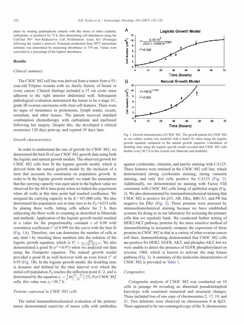
Fig. 1. Growth characteristics of CSOC 882. The growth pattern for CSOC 882in our culture system was modeled with a better fit when using the logisticgrowth equation compared to the natural growth equation. Calculation ofdoubling time using the logistic growth model revealed that CSOC 882 cellsdouble every 30.7 h in this system (see Materials and methods).
122 D.R. Scoles et al. / Gynecologic Oncology 104 (2007) 120–128
plates by treating quadruplicate cultures with like doses of either cisplatin,carboplatin, or paclitaxel for 72 h, then determining cell abundances using theCellTiter 96® Non-Radioactive Cell Proliferation Assay Kit (Promega)following the vendor's protocol. Formazan production from MTT tetrazoliumsubstrate was determined by measuring absorbance at 570 nm. Values wereconverted to a percentage of the highest absorbance.
Results
Clinical summary
The CSOC 882 cell line was derived from a tumor from a 55-year-old Filipino woman with no family history of breast orovary cancer. Clinical findings included a 15 cm cystic massadherent to the right anterior abdominal wall. Subsequentpathological evaluation determined the tumor to be a stage 1C,grade III ovarian carcinoma with clear cell features. There wereno signs of metastases in peritoneum, lymph nodes, cecum,omentum, and other tissues. The patient received standardcombination chemotherapy with carboplatin and paclitaxelfollowing her surgery. Despite this, she developed a clinicalrecurrence 120 days post-op, and expired 39 days later.
Growth characteristics
In order to understand the rate of growth for CSOC 882, wedetermined the best fit of our CSOC 882 growth data using boththe logistic and natural growth models. The observed growth forCSOC 882 cells best fit the logistic growth model, which isderived from the natural growth model by the inclusion of aterm that accounts for constraints on population growth. Inorder to fit the logistic growth model, we made the assumptionthat the carrying capacity was equivalent to the highest value weobserved for the 48-h time point when we halted the experimentwhen all wells at this time point had reached confluence. Weassigned the carrying capacity to be K=107,000 cells. We alsodetermined the population size at time zero to be P0=6333 cellsby plating three wells, letting cells adhere for 2 h, thensubjecting the three wells to counting as described in Materialsand methods. Application of the logistic growth model resultedin a value for the proportionality constant r of 0.09 withcorrelation coefficient r2 of 0.999 for the curve with the best fit(Fig. 1A). Therefore, one can determine the number of cells atany time t by inserting these numbers into the solution of thelogistic growth equation, which is Pt ¼ KP0
P0þ K�P0ð Þe�rt. We alsodemonstrated a good fit (r2 =0.97) when we analyzed our datausing the Gompertz equation. The natural growth modelprovided a good fit as well however with an even lower r2 of0.95 (Fig. 1B). In the logistic growth model, the doubling timeis dynamic and defined by the time interval over which theinitial cell population P0 reaches the inflection point K / 2, and isdetermined by the equation t2 ¼ 1
r lnK � P0
P0
� �[5]. For CSOC 882
cells, this value was t2=30.7 h.
Proteins expressed in CSOC 882 cells
The initial immunohistochemical evaluation of the primarytumor demonstrated reactivity of tumor cells with antibodies
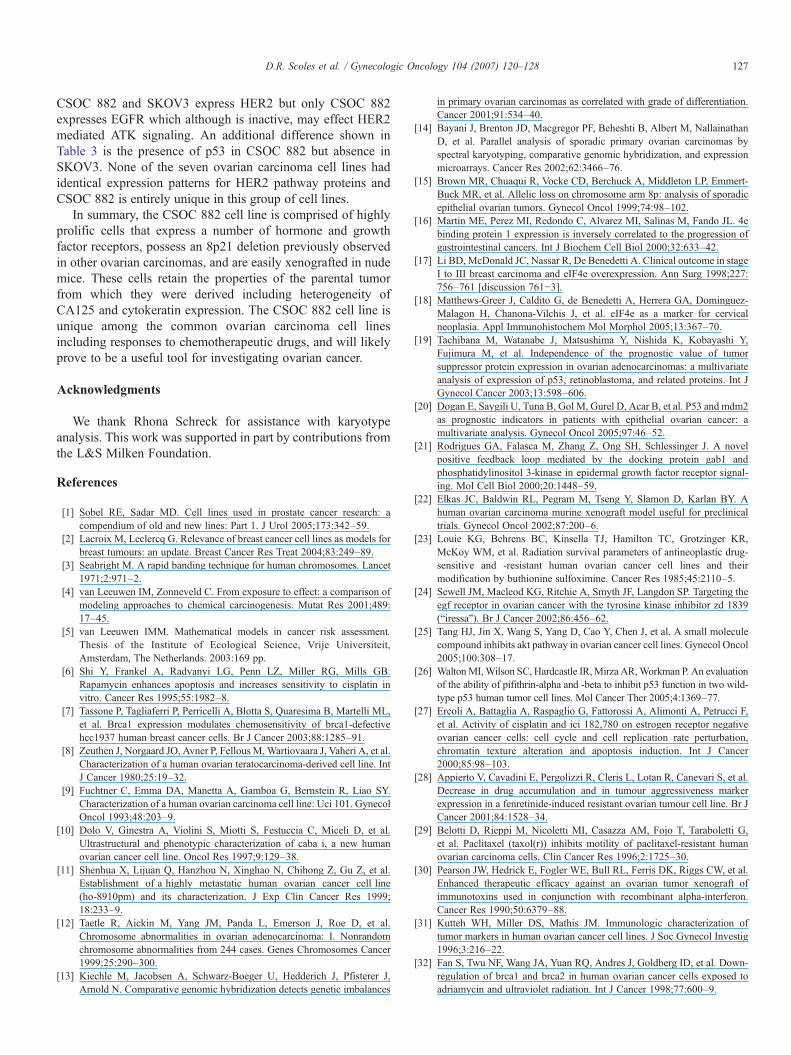
against cytokeratin, vimentin, and patchy staining with CA125.These features were retained in the CSOC 882 cell line, whichdemonstrated strong cytokeratin staining, strong vimentinstaining, and only few cells positive for CA125 (Fig. 2).Additionally, we demonstrated no staining with Factor VIIIconsistent with CSOC 882 cells being of epithelial origin (Fig.2). We also demonstrated by immunohistochemical staining thatCSOC 882 is positive for p53, AR, ERα, BRCA1, and PR butnegative for ERβ (Fig. 2). These proteins were assessed byimmunohistochemical staining because we have establishedsystems for doing so in our laboratory for screening the primarycells that we regularly bank. We conducted further testing ofHER2/AKT pathway proteins by the more sensitive method ofimmunoblotting to accurately compare the expression of theseproteins in CSOC 882 to that in a variety of other ovarian cancercell lines. Immunoblotting demonstrated that CSOC 882 cellsare positive for HER2, EGFR, AKT, and phospho-AKT, but wewere unable to detect the presence of EGFR phosphorylated ontyrosine 1068, which is known to activate the map kinasepathway (Fig. 3). A summary of the molecular characteristics ofCSOC 882 is provided in Table 1.
Cytogenetics
Cytogenetic analysis of CSOC 882 was conducted on 10cells in passage 66 revealing an abnormal pseudotetraploidkaryotype with consistent numerical and structural changes.These included loss of one copy of chromosomes 2, 17, 19, and21. Two deletions were observed on chromosome 8 at 8p21.There appeared to be one rearranged copy of the X chromosome
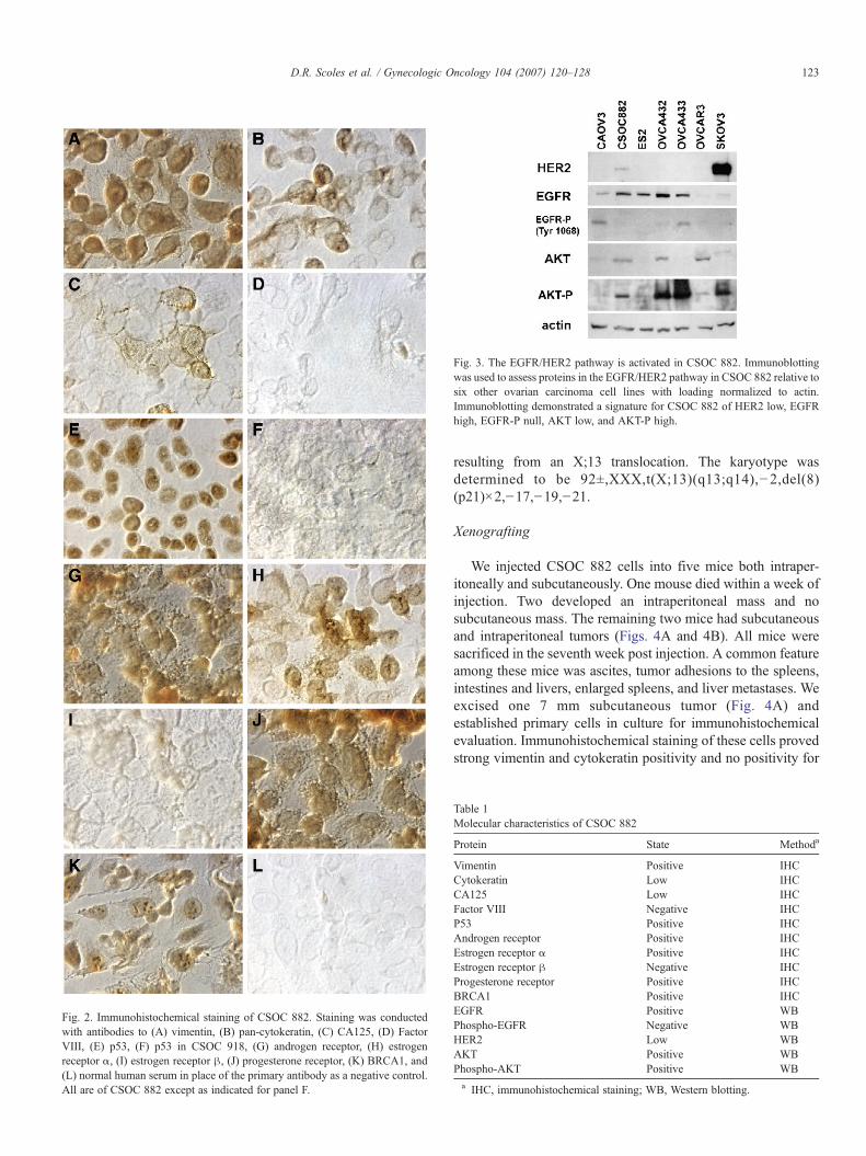
Fig. 3. The EGFR/HER2 pathway is activated in CSOC 882. Immunoblottingwas used to assess proteins in the EGFR/HER2 pathway in CSOC 882 relative tosix other ovarian carcinoma cell lines with loading normalized to actin.Immunoblotting demonstrated a signature for CSOC 882 of HER2 low, EGFRhigh, EGFR-P null, AKT low, and AKT-P high.
Fig. 2. Immunohistochemical staining of CSOC 882. Staining was conductedwith antibodies to (A) vimentin, (B) pan-cytokeratin, (C) CA125, (D) FactorVIII, (E) p53, (F) p53 in CSOC 918, (G) androgen receptor, (H) estrogenreceptor α, (I) estrogen receptor β, (J) progesterone receptor, (K) BRCA1, and(L) normal human serum in place of the primary antibody as a negative control.All are of CSOC 882 except as indicated for panel F.
123D.R. Scoles et al. / Gynecologic Oncology 104 (2007) 120–128
resulting from an X;13 translocation. The karyotype wasdetermined to be 92±,XXX,t(X;13)(q13;q14),−2,del(8)(p21)×2,−17,−19,−21.
Xenografting
We injected CSOC 882 cells into five mice both intraper-itoneally and subcutaneously. One mouse died within a week ofinjection. Two developed an intraperitoneal mass and nosubcutaneous mass. The remaining two mice had subcutaneousand intraperitoneal tumors (Figs. 4A and 4B). All mice weresacrificed in the seventh week post injection. A common featureamong these mice was ascites, tumor adhesions to the spleens,intestines and livers, enlarged spleens, and liver metastases. Weexcised one 7 mm subcutaneous tumor (Fig. 4A) andestablished primary cells in culture for immunohistochemicalevaluation. Immunohistochemical staining of these cells provedstrong vimentin and cytokeratin positivity and no positivity for
Table 1Molecular characteristics of CSOC 882
Protein State Methoda
Vimentin Positive IHCCytokeratin Low IHCCA125 Low IHCFactor VIII Negative IHCP53 Positive IHCAndrogen receptor Positive IHCEstrogen receptor α Positive IHCEstrogen receptor β Negative IHCProgesterone receptor Positive IHCBRCA1 Positive IHCEGFR Positive WBPhospho-EGFR Negative WBHER2 Low WBAKT Positive WBPhospho-AKT Positive WBa IHC, immunohistochemical staining; WB, Western blotting.

Fig. 4. CSOC 882 is a tumorigenic cell line. (A) Nude mouse with a subcutaneous CSOC 882 xenograft tumor. (B) Nude mouse with an intraperitoneal CSOC 882xenograft tumor. A subcutaneous CSOC 882 xenograft tumor was excised and cultured in vitro then subjected to immunohistochemical staining with antibodiesagainst (C) vimentin, (D) pan-cytokeratins, and (E) factor VIII.
124 D.R. Scoles et al. / Gynecologic Oncology 104 (2007) 120–128
Factor VIII (Figs. 4C–E). As our main interest was primarilytumorigenicity of CSOC 882, we only assessed these threeantigens. This result demonstrated retention of the samevimentin, cytokeratin, and Factor VIII expression profile asfor CSOC 882 (Fig. 2).
Chemosensitivity
SKOV3 is resistant to platinum chemotherapy and the breastcancer cell line HCC 1937 is resistant to paclitaxel [6,7].Therefore we chose these two lines to which to compare thechemosensitivity of CSOC 882. The cellular response was bestfit with a monophasic exponential decay model while thecellular responses to cisplatin and paclitaxel were biphasic sowe modeled the latter using a biphasic exponential decay model.To accomplish this, we used the built in equations provided withthe software package GraphPad Prism version 4. We demon-strated that SKOV3 was more resistant than HCC 1937, and thatHCC 1937 was more resistant than CSOC 882 for both cisplatinand carboplatin (Figs. 5A and B). The IC50 values for CSOC882 in either cisplatin or carboplatin were approximately half ofthat for HCC 1937, and approximately a quarter of that forSKOV3 (Table 2). However, SKOV3 and CSOC 882 were bothsensitive to paclitaxel while HCC 1937 was considerably moreresistant (Fig. 5C, Table 2).
Discussion
During the course of our regular tumor banking activities, wenoted one primary cell culture, CSOC 882, which stood outfrom the others by its aggressive growth. Further analysis of thiscell culture demonstrated that it had established into a highly
prolific cell line that retained the properties of the parentalprimary tumor, including heterogeneity of protein expressionand tumorigenicity in mice. Our characterization of CSOC 882demonstrated it to be unique among existing ovarian cancer celllines.
Sensitivity of CSOC 882 cells to chemotherapeutic drugs
We showed that CSOC 882 was considerably more sensitiveto platinum/taxol than SKOV3 with an IC50 value for cisplatinof less than a quarter of that for SKOV3. This was not expectedsince CSOC 882 was derived from a patient who eventuallysuccumbed to refractory progressive disease. The establishmentof this platinum sensitive line from a patient who did notrespond to platinum chemotherapy might be explained in termsconsistent with the cancer stem cell theory. This theoryhypothesizes that the bulk of a tumor consists of differentiatedcells that are supported by lesser abundant but more highlymetastatic undifferentiated progenitor cells [53] that may havegreater drug resistance than their differentiated tumor cellcounterparts [54]. Such progenitor cells typically have a lowrate of proliferation and require specialized conditions forculture and would not be maintained in CSOC 882 cultures. It istherefore possible that platinum sensitive CSOC 882 cells areuncharacteristic of resistant metastatic cells that account for theplatinum refractory progressive disease of this patient. How-ever, CSOC 882 is not the most sensitive cell line that we havecharacterized as in more recent work we have demonstrated forCAOV3 cells a cisplatin IC50 of 1.2 μM [55]. While CSOC 882is considerably more sensitive to cisplatin than SKOV3, itperhaps is best characterized as an ovarian carcinoma cell linewith moderate sensitivity to cisplatin.

Fig. 5. Chemosensitivity of CSOC 882. The growth response of CSOC 882 cellscultured in the presence of (A) cisplatin, (B) carboplatin, or (C) paclitaxel wasdemonstrated using MTT proliferation assays in comparison to SKOV3 andHCC 1937 cells whose responses to these drugs are known. These assaysdemonstrated that CSOC 882 cells were more sensitive to cisplatin andcarboplatin than SKOV3 and HCC 1937, and that HC1937 were highlypaclitaxel resistant unlike CSOC 882 and SKOV3. Cells were plated equallyamong the wells of a 96 well plate, recovered overnight then treated with thedrugs as indicated, in quadruplicate. The dosewise treatments included 0, 3, 6.3,12.5, 25, 50, 100, and 200 μM of cisplatin (A), 0, 10, 25, 50, 100, 250, 500 μMof carboplatin (B), and 0, 37, 75, 150, 300, 600, 1200 nM of paclitaxel (C). Datawere analyzed using monophasic (A) and biphasic (B, C) exponential decaymodels. Values of r2 ranged from 0.8 to 0.993.
Table 2IC50 values for CSOC 882, SKOV-3 and HCC 1937
Cell line IC50 valuesa when cultured with
Cisplatin Carboplatin Paclitaxel
CSOC 882 18.9 μM 49.7 μM 66.8 nMSKOV-3 79.8 μM 192.2 μM 56.3 nMHCC 1937 45.5 μM 96.3 μM Not achieved
a We defined IC50 as the inhibitory concentration effective for reducing thecell population by 50% over the 72-h culture period.
125D.R. Scoles et al. / Gynecologic Oncology 104 (2007) 120–128
Growth properties of CSOC 882 cells
CSOC 882 cells were easily cultured in standard media withno supplementary growth factors required. They appear as fast
growing cells with cuboid morphology. We investigated thegrowth properties of CSOC 882 cells by the application of threedifferent growth models (logistic, Gompertz, and natural) andfound that we could best predict growth with the logistic model.Our estimation of doubling time for CSOC 882 cells was 30.7 hand was not unlike that of other ovarian carcinoma cell linesranging from 18 to 34 h [8–11]. The growth of CSOC 882 cellsis that of an established immortalized cell line and CSOC 882cells have undergone more than 130 passages in culture.
Molecular genetics of CSOC 882 cells
Like all cancers, several molecular mechanisms are alteredduring the development of ovarian carcinomas and theirprogression to a more malignant phenotype. Such changesinclude mutations that activate oncogenes, mutations thatinactivate tumor suppressor genes, and chromosomal abnorm-alities resulting in gene deletion or loss of function. Chromo-somal alteration in ovarian cancer has been reviewed in detail[12–14]. These studies demonstrated an exceptionally highnumber of cytogenetic abnormalities in ovarian cancer includ-ing alterations located in all chromosomes. CSOC 882 cellsproved to be tetraploid with deletions of chromosomes 2, 17, 19and 21. We also observed the translocation t(X;13)(q13;14) andnoted that breakpoints at Xq13 and 13q14 have been observedpreviously in ovarian adenocarcinomas [12]. CSOC 882 cellsalso possess a deletion of 8p21. Deletions of 8p21 have beenobserved previously and are associated with increased malig-nant potential [15]. We find it of considerable interest that the4EBP1 gene is located at 8p21. When 4EBP is phosphorylatedby mTOR, 4EBP is released from eIF4e resulting in theinitiation of protein translation. Loss of 4EBP deregulates eIF4eand is inversely correlated with the progression of intestinaltumors [16], and eIF4e overexpression has been observed inother women's cancers [17,18]. More work will be required todemonstrate whether 8p21 loss could be associated to CSOC882 tumorigenesis or resistance to rapamycin.
Protein expression signature of CSOC 882
Consistent with an epithelial origin for CSOC 882 cells,100% of cultures expressed vimentin and were negative forfactor VIII. Epithelial cells are also expected to be positive forcytokeratins; however, we demonstrated only ∼40% positivityfor CSOC 882 using a pan cytokeratin antibody. Likewise,CSOC 882 cells were also only ∼20% positive for CA125. Thepattern of staining for CA125 entirely mimicked the pathology

126 D.R. Scoles et al. / Gynecologic Oncology 104 (2007) 120–128
report showing that the primary tumor from which CSOC 882cells were derived had patchy CA125 staining, and presurgicallabs showing only a moderately elevated CA125 serum level forthis patient of 54. We conclude that CSOC 882 cells areepithelial in origin and retain the protein expression signature ofthe primary ovarian carcinoma from which they were derived.
We assessed the expression of a number of tumorssuppressors and oncoproteins in CSOC 882 cells by immuno-histochemical staining. For each of p53, AR, ERα, ERβ, PR,and BRCA1, a majority of the cells in the CSOC 882 cultureshared the same pattern of staining. Each of these proteins waslocalized in the nucleus of CSOC 882 cells, except for ERβ,which was not expressed. Positive expression of the hormonereceptors AR, ERα, and PR was noted as well as positivestaining for BRCA1 suggesting that CSOC 882 cells lackBRCA1 mutations. P53 staining is normally not observed un-less the TP53 gene is mutated, and then its pattern of expres-sion is considered prognostic in most cancers including ovariancancer. Nuclear localization is associated with reduced survival[19,20] and may contribute to the aggressive growth of CSOC882 cells.
The HER2 pathway is significant to ovarian cancer biology,so we used immunoblotting to compare the expression ofHER2, total and active EGFR, and total and active AKT amongseven selected ovarian carcinoma cell lines. Immunoblottingdemonstrated that CSOC 882 cells highly expressed EGFR butweakly expressed HER2. To demonstrate constitutive activa-tion of EGFR, we assessed EGFR with an antibody directedagainst tyrosine 1068 which has been implicated in theactivation of AKT [21]. We were unable to detect phospho-EGFR in CSOC 882 cells, but we observed that AKT washighly phosphorylated.
CSOC 882 cells are tumorigenic in nude mice
CSOC 882 cells are easily engrafted in nude mice andtherefore are useful in modeling ovarian cancer in mousemodels. We previously investigated the xenografting of CSOCcell cultures in mice showing that 11 of 13 tested culturesestablished tumors in SCID mice, while we had no success
Table 3CSOC 882 compared to the five most-common ovarian carcinoma cell lines
Cell line Tumor type Molecular featuresa
CA125 p53 AR ER PR HER2 EGFR A
CSOC 882 Adenocarcinoma L P P P P L P PA2780 Adenocarcinoma N W – N – P N PCaov-3 Adenocarcinoma N M – P P N Pc LOVCAR-3 Adenocarcinoma P M P P P Ld Lc PPA-1 Teratocarcinoma P W – – – P Pe –SKOV-3 Adenocarcinoma N Null N P P P Lc P
a P, positive; N, negative; L, low; W, wild-type overexpressed; M, mutant; –, nob CS, cisplatin sensitive; CR, cisplatin resistant; PS, paclitaxel sensitive; PR, paclita
the various authors. CAOV3 was highly cisplatin sensitive in experiments conductec Fig. 3.d Constitutively phosphorylated.e Inferred by EGF binding that stimulated growth.
establishing CSOC xenografts in nude mice [22]. Compara-tively, CSOC 882 is remarkably more tumorigenic than otherprimary cultures of ovarian carcinomas that we have studied andwill likely prove to be a useful resource for investigations onovarian cancer where mouse xenografts are required. We alsoshowed that the expression of cytokeratin, vimentin, and factorVIII remained unchanged after passing CSOC 882 cells throughnude mice lending further support for that CSOC 882 cellspossess no contaminating fibroblasts.
CSOC 882 cells are unique among ovarian cell lines
While there are a number of ovarian cancer cell linesavailable to researchers, the number that are extensivelycharacterized is relatively few in comparison to other tumordisorders including prostate cancer and breast cancer. Amongthese, the common ovarian carcinoma cell lines A2780, Caov3,OVCAR-3, PA-1, and SKOV3 have been cited hundreds oftimes in the literature and are extensively characterized. Thereexist approximately 10 other lines that have been citedmoderately (20 or more times) in the ovarian cancer literaturedemonstrating a relatively significant impact on the study of thedisease, and several others that have been used sporadicallysince the early 1980s. Many of the existing ovarian carcinomacell lines were characterized by several laboratories overmultiple years while few have been presented with a singlecomprehensive characterization such as the present descriptionof the CSOC 882 cell line. None of the five most commonlystudied ovarian carcinoma cell lines had precisely the sameexpression pattern and drug sensitivity as CSOC 882 (Table 3).
Our immunoblotting and immunohistochemical stainingfurther demonstrated that CSOC 882 possess a unique patternof expression of HER2 pathway proteins and other tumorsuppressors and oncoproteins important to ovarian cancerbiology highlighting COSC 882 as a unique resource for ovariancancer research. The data that we present are valuable fordetermining whether CSOC 882 may be useful for variousmolecular studies on ovarian cancer. For example, one particularunique use for CSOC 882 might be in the investigation of theeffect of HER2 or EGFR knockdown on AKT activation. Both
Tumorgenicin mice
Chemosensitivityb No.timescited
Citations
KT-p BRCA1
P Y CS, PS N/A N/A– Y CS, PS 634 [23–29, 56]
c P Y CS, PS 162 [30–35]– Y CR, PS 497 [25,31,34,36–42]P Y CS, PS 126 [8,31,32,43–48]P Y CR, PR 553 [32,34,41,49–52]
data.xel resistant. Note that the degree of sensitivity was not always consistent amongd in our laboratory (see text).
127D.R. Scoles et al. / Gynecologic Oncology 104 (2007) 120–128
CSOC 882 and SKOV3 express HER2 but only CSOC 882expresses EGFR which although is inactive, may effect HER2mediated ATK signaling. An additional difference shown inTable 3 is the presence of p53 in CSOC 882 but absence inSKOV3. None of the seven ovarian carcinoma cell lines hadidentical expression patterns for HER2 pathway proteins andCSOC 882 is entirely unique in this group of cell lines.
In summary, the CSOC 882 cell line is comprised of highlyprolific cells that express a number of hormone and growthfactor receptors, possess an 8p21 deletion previously observedin other ovarian carcinomas, and are easily xenografted in nudemice. These cells retain the properties of the parental tumorfrom which they were derived including heterogeneity ofCA125 and cytokeratin expression. The CSOC 882 cell line isunique among the common ovarian carcinoma cell linesincluding responses to chemotherapeutic drugs, and will likelyprove to be a useful tool for investigating ovarian cancer.
Acknowledgments
We thank Rhona Schreck for assistance with karyotypeanalysis. This work was supported in part by contributions fromthe L&S Milken Foundation.
References
[1] Sobel RE, Sadar MD. Cell lines used in prostate cancer research: acompendium of old and new lines: Part 1. J Urol 2005;173:342–59.
[2] Lacroix M, Leclercq G. Relevance of breast cancer cell lines as models forbreast tumours: an update. Breast Cancer Res Treat 2004;83:249–89.
[3] Seabright M. A rapid banding technique for human chromosomes. Lancet1971;2:971–2.
[4] van Leeuwen IM, Zonneveld C. From exposure to effect: a comparison ofmodeling approaches to chemical carcinogenesis. Mutat Res 2001;489:17–45.
[5] van Leeuwen IMM. Mathematical models in cancer risk assessment.Thesis of the Institute of Ecological Science, Vrije Universiteit,Amsterdam, The Netherlands. 2003:169 pp.
[6] Shi Y, Frankel A, Radvanyi LG, Penn LZ, Miller RG, Mills GB.Rapamycin enhances apoptosis and increases sensitivity to cisplatin invitro. Cancer Res 1995;55:1982–8.
[7] Tassone P, Tagliaferri P, Perricelli A, Blotta S, Quaresima B, Martelli ML,et al. Brca1 expression modulates chemosensitivity of brca1-defectivehcc1937 human breast cancer cells. Br J Cancer 2003;88:1285–91.
[8] Zeuthen J, Norgaard JO, Avner P, FellousM,Wartiovaara J, Vaheri A, et al.Characterization of a human ovarian teratocarcinoma-derived cell line. IntJ Cancer 1980;25:19–32.
[9] Fuchtner C, Emma DA, Manetta A, Gamboa G, Bernstein R, Liao SY.Characterization of a human ovarian carcinoma cell line: Uci 101. GynecolOncol 1993;48:203–9.
[10] Dolo V, Ginestra A, Violini S, Miotti S, Festuccia C, Miceli D, et al.Ultrastructural and phenotypic characterization of caba i, a new humanovarian cancer cell line. Oncol Res 1997;9:129–38.
[11] Shenhua X, Lijuan Q, Hanzhou N, Xinghao N, Chihong Z, Gu Z, et al.Establishment of a highly metastatic human ovarian cancer cell line(ho-8910pm) and its characterization. J Exp Clin Cancer Res 1999;18:233–9.
[12] Taetle R, Aickin M, Yang JM, Panda L, Emerson J, Roe D, et al.Chromosome abnormalities in ovarian adenocarcinoma: I. Nonrandomchromosome abnormalities from 244 cases. Genes Chromosomes Cancer1999;25:290–300.
[13] Kiechle M, Jacobsen A, Schwarz-Boeger U, Hedderich J, Pfisterer J,Arnold N. Comparative genomic hybridization detects genetic imbalances
in primary ovarian carcinomas as correlated with grade of differentiation.Cancer 2001;91:534–40.
[14] Bayani J, Brenton JD, Macgregor PF, Beheshti B, Albert M, NallainathanD, et al. Parallel analysis of sporadic primary ovarian carcinomas byspectral karyotyping, comparative genomic hybridization, and expressionmicroarrays. Cancer Res 2002;62:3466–76.
[15] Brown MR, Chuaqui R, Vocke CD, Berchuck A, Middleton LP, Emmert-Buck MR, et al. Allelic loss on chromosome arm 8p: analysis of sporadicepithelial ovarian tumors. Gynecol Oncol 1999;74:98–102.
[16] Martin ME, Perez MI, Redondo C, Alvarez MI, Salinas M, Fando JL. 4ebinding protein 1 expression is inversely correlated to the progression ofgastrointestinal cancers. Int J Biochem Cell Biol 2000;32:633–42.
[17] Li BD, McDonald JC, Nassar R, De Benedetti A. Clinical outcome in stageI to III breast carcinoma and eIF4e overexpression. Ann Surg 1998;227:756–761 [discussion 761−3].
[18] Matthews-Greer J, Caldito G, de Benedetti A, Herrera GA, Dominguez-Malagon H, Chanona-Vilchis J, et al. eIF4e as a marker for cervicalneoplasia. Appl Immunohistochem Mol Morphol 2005;13:367–70.
[19] Tachibana M, Watanabe J, Matsushima Y, Nishida K, Kobayashi Y,Fujimura M, et al. Independence of the prognostic value of tumorsuppressor protein expression in ovarian adenocarcinomas: a multivariateanalysis of expression of p53, retinoblastoma, and related proteins. Int JGynecol Cancer 2003;13:598–606.
[20] Dogan E, Saygili U, Tuna B, Gol M, Gurel D, Acar B, et al. P53 and mdm2as prognostic indicators in patients with epithelial ovarian cancer: amultivariate analysis. Gynecol Oncol 2005;97:46–52.
[21] Rodrigues GA, Falasca M, Zhang Z, Ong SH, Schlessinger J. A novelpositive feedback loop mediated by the docking protein gab1 andphosphatidylinositol 3-kinase in epidermal growth factor receptor signal-ing. Mol Cell Biol 2000;20:1448–59.
[22] Elkas JC, Baldwin RL, Pegram M, Tseng Y, Slamon D, Karlan BY. Ahuman ovarian carcinoma murine xenograft model useful for preclinicaltrials. Gynecol Oncol 2002;87:200–6.
[23] Louie KG, Behrens BC, Kinsella TJ, Hamilton TC, Grotzinger KR,McKoy WM, et al. Radiation survival parameters of antineoplastic drug-sensitive and -resistant human ovarian cancer cell lines and theirmodification by buthionine sulfoximine. Cancer Res 1985;45:2110–5.
[24] Sewell JM, Macleod KG, Ritchie A, Smyth JF, Langdon SP. Targeting theegf receptor in ovarian cancer with the tyrosine kinase inhibitor zd 1839(“iressa”). Br J Cancer 2002;86:456–62.
[25] Tang HJ, Jin X, Wang S, Yang D, Cao Y, Chen J, et al. A small moleculecompound inhibits akt pathway in ovarian cancer cell lines. Gynecol Oncol2005;100:308–17.
[26] WaltonMI,Wilson SC, Hardcastle IR,Mirza AR,Workman P. An evaluationof the ability of pifithrin-alpha and -beta to inhibit p53 function in two wild-type p53 human tumor cell lines. Mol Cancer Ther 2005;4:1369–77.
[27] Ercoli A, Battaglia A, Raspaglio G, Fattorossi A, Alimonti A, Petrucci F,et al. Activity of cisplatin and ici 182,780 on estrogen receptor negativeovarian cancer cells: cell cycle and cell replication rate perturbation,chromatin texture alteration and apoptosis induction. Int J Cancer2000;85:98–103.
[28] Appierto V, Cavadini E, Pergolizzi R, Cleris L, Lotan R, Canevari S, et al.Decrease in drug accumulation and in tumour aggressiveness markerexpression in a fenretinide-induced resistant ovarian tumour cell line. Br JCancer 2001;84:1528–34.
[29] Belotti D, Rieppi M, Nicoletti MI, Casazza AM, Fojo T, Taraboletti G,et al. Paclitaxel (taxol(r)) inhibits motility of paclitaxel-resistant humanovarian carcinoma cells. Clin Cancer Res 1996;2:1725–30.
[30] Pearson JW, Hedrick E, Fogler WE, Bull RL, Ferris DK, Riggs CW, et al.Enhanced therapeutic efficacy against an ovarian tumor xenograft ofimmunotoxins used in conjunction with recombinant alpha-interferon.Cancer Res 1990;50:6379–88.
[31] Kutteh WH, Miller DS, Mathis JM. Immunologic characterization oftumor markers in human ovarian cancer cell lines. J Soc Gynecol Investig1996;3:216–22.
[32] Fan S, Twu NF, Wang JA, Yuan RQ, Andres J, Goldberg ID, et al. Down-regulation of brca1 and brca2 in human ovarian cancer cells exposed toadriamycin and ultraviolet radiation. Int J Cancer 1998;77:600–9.
128 D.R. Scoles et al. / Gynecologic Oncology 104 (2007) 120–128
[33] Yvon AM, Wadsworth P, Jordan MA. Taxol suppresses dynamics ofindividual microtubules in living human tumor cells. Mol Biol Cell1999;10:947–59.
[34] Jones J, Lagasse LD, Karlan BY. Steroid hormonal independence of her-2/neu mrna expression in four human ovarian carcinoma cell lines. GynecolOncol 1994;55:421–6.
[35] Chan MM, Fong D, Soprano KJ, Holmes WF, Heverling H. Inhibition ofgrowth and sensitization to cisplatin-mediated killing of ovarian cancercells by polyphenolic chemopreventive agents. J Cell Physiol 2003;194:63–70.
[36] Hamilton TC, Young RC, McKoy WM, Grotzinger KR, Green JA, ChuEW, et al. Characterization of a human ovarian carcinoma cell line (nih:Ovcar-3) with androgen and estrogen receptors. Cancer Res 1983;43:5379–89.
[37] Godwin AK, Meister A, O'Dwyer PJ, Huang CS, Hamilton TC, AndersonME. High resistance to cisplatin in human ovarian cancer cell lines isassociated with marked increase of glutathione synthesis. Proc Natl AcadSci U S A 1992;89:3070–4.
[38] Havrilesky LJ, Elbendary A, Hurteau JA, Whitaker RS, Rodriguez GC,Berchuck A. Chemotherapy-induced apoptosis in epithelial ovariancancers. Obstet Gynecol 1995;85:1007–10.
[39] Marth C, Lang T, Cronauer MV, Doppler W, Zeimet AG, Bachmair F, et al.Epidermal growth factor reduces her-2 protein level in human ovariancarcinoma cells. Int J Cancer 1992;52:311–6.
[40] Delord JP, Allal C, Canal M, Mery E, Rochaix P, Hennebelle I, et al.Selective inhibition of her2 inhibits akt signal transduction and prolongsdisease-free survival in a micrometastasis model of ovarian carcinoma.Ann Oncol 2005;16:1889–97.
[41] Pegues JC, Kannan B, Stromberg K. Erbb receptor expression and growthresponse to heregulin beta 1 in five ovarian carcinoma lines. Int J Oncol1999;14:1169–76.
[42] Hu L, Hofmann J, Lu Y, Mills GB, Jaffe RB. Inhibition ofphosphatidylinositol 3′-kinase increases efficacy of paclitaxel in in vitroand in vivo ovarian cancer models. Cancer Res 2002;62:1087–92.
[43] Giovanella BC, Stehlin JS, Williams Jr LJ. Heterotransplantation of humanmalignant tumors in “nude” thymusless mice: II. Malignant tumorsinduced by injection of cell cultures derived from human solid tumors.J Natl Cancer Inst 1974;52:921–30.
[44] Tainsky MA, Cooper CS, Giovanella BC, Vande Woude GF. An activated
rasn gene: detected in late but not early passage human pa1 teratocarci-noma cells. Science 1984;225:643–5.
[45] Kammerer U, Thanner F, Kapp M, Dietl J, Sutterlin M. Expression oftumor markers on breast and ovarian cancer cell lines. AntiCancer Res2003;23:1051–5.
[46] Le-Ruppert K, Masters JR, Knuechel R, Seegers S, Tainsky MA,Hofstaedter F, et al. The effect of retinoic acid on chemosensitivity ofpa-1 human teratocarcinoma cells and its modulation by an activated n-rasoncogene. Int J Cancer 1992;51:646–51.
[47] Coukos G, Makrigiannakis A, Kang EH, Rubin SC, Albelda SM, Molnar-Kimber KL. Oncolytic herpes simplex virus-1 lacking icp34.5 inducesp53-independent death and is efficacious against chemotherapy-resistantovarian cancer. Clin Cancer Res 2000;6:3342–53.
[48] Shigeta H, Taga M, Sakakibara H, Minaguchi H. Epidermal growth factorstimulates the cell growth of the pa-1 teratocarcinoma cell line in anautocrine/paracrine fashion. Eur J Endocrinol 1995;132:200–5.
[49] Fogh J, Fogh JM, Orfeo T. One hundred and twenty-seven cultured humantumor cell lines producing tumors in nude mice. J Natl Cancer Inst1977;59:221–6.
[50] Karlan BY, Jones J, Slamon DJ, Lagasse LD. Glucocorticoids stabilize her-2/neu messenger rna in human epithelial ovarian carcinoma cells. GynecolOncol 1994;53:70–7.
[51] Lamendola DE, Duan Z, Yusuf RZ, Seiden MV. Molecular description ofevolving paclitaxel resistance in the skov-3 human ovarian carcinoma cellline. Cancer Res 2003;63:2200–5.
[52] Reinartz S, Kohler S, Schlebusch H, Krista K, Giffels P, Renke K, et al.Vaccination of patients with advanced ovarian carcinoma with the anti-idiotype aca125: immunological response and survival (phase ib/ii). ClinCancer Res 2004;10:1580–7.
[53] Pardal R, Clarke MF, Morrison SJ. Applying the principles of stem-cellbiology to cancer. Nat Rev, Cancer 2003;3:895–902.
[54] Dean M, Fojo T, Bates S. Tumour stem cells and drug resistance. Nat Rev,Cancer 2005;5:275–84.
[55] Pavelka J, Scoles DR, Karlan, B. Effects of AKT signaling on the drugsensitivity of ovarian carcinoma cells [in preparation].
[56] Marth C, Zeimet AG, Widschwendter M, Ludescher C, Kaern J, Trope C,Gastl G, Daxenbichler G, Dapunt O. Paclitaxel- and docetaxel-dependentactivation of CA-125 expression in human ovarian carcinoma cells. CancerRes 1997;57(17):3818–22.