Estrogen receptor and HER2/neu status affect epigenetic differences of tumor-related genes in...
11
Open Access Available online http://breast-cancer-research.com/content/10/3/R46 Page 1 of 11 (page number not for citation purposes) Vol 10 No 3 Research article Estrogen receptor and HER2/neu status affect epigenetic differences of tumor-related genes in primary breast tumors Eiji Sunami 1 , Masaru Shinozaki 1 , Myung-Shin Sim 2 , Sandy L Nguyen 1 , Anh-Thu Vu 1 , Armando E Giuliano 3 and Dave SB Hoon 1 1 Department of Molecular Oncology, The John Wayne Cancer Institute, Saint John's Health Center, Santa Monica Blvd, Santa Monica, California 90404, USA 2 Department of Biostatistics, The John Wayne Cancer Institute, Saint John's Health Center, Santa Monica Blvd, Santa Monica, California 90404, USA 3 Department of Surgical Oncology, The John Wayne Cancer Institute, Saint John's Health Center, Santa Monica Blvd, Santa Monica, California 90404, USA Corresponding author: Dave SB Hoon, [email protected] Received: 3 Jan 2008 Revisions requested: 27 Feb 2008 Revisions received: 13 Mar 2008 Accepted: 16 May 2008 Published: 16 May 2008 Breast Cancer Research 2008, 10:R46 (doi:10.1186/bcr2098) This article is online at: http://breast-cancer-research.com/content/10/3/R46 © 2008 Sunami et al.; licensee BioMed Central Ltd. This is an open access article distributed under the terms of the Creative Commons Attribution License (http://creativecommons.org/licenses/by/2.0 ), which permits unrestricted use, distribution, and reproduction in any medium, provided the original work is properly cited. Abstract Introduction Estrogen receptor (ER)-positive breast cancers are considered prognostically more favorable than ER-negative tumors, whereas human epidermal growth factor receptor (HER)2/neu-positive breast cancers are associated with worse prognosis. The objective of the present study was to determine whether ER-positive and ER-negative status relates to epigenetic changes in breast cancer-related genes. To evaluate epigenetic differences in tumor-related genes relating to ER and HER2/neu status of primary tumors, we examined the promoter methylation status of the promoter region CpG islands of eight major breast tumor-related genes (RASSF1A, CCND2, GSPT1, TWIST, APC, NES1, RARβ2, and CDH1). Methods Paired ER-positive (n = 65) and ER-negative (n = 65) primary breast tumors (n = 130) matched for prognostic factors were assessed. DNA was extracted from paraffin-embedded tumor tissue after microdissection, and methylation-specific PCR and capillary-array electrophoresis analysis were performed. Results In early stages of tumor progression (T1 and N0), RASSF1A and CCND2 were significantly (P < 0.05) more methylated in ER-positive than in ER-negative tumors. GSTP1 hypermethylation was more frequent in the lymph node metastasis positive group than in the negative group. Double negative (ER-negative, HER2/neu-negative) breast cancers had significantly lesser frequencies of RASSF1A, GSTP1, and APC methylation (P < 0.0001, P < 0.0001, and P = 0.0035, respectively). Both ER and HER2/neu status correlated independently with these epigenetic alterations. Conclusion We demonstrated significant differences in tumor- related gene methylation patterns relevant to ER and HER2/neu status of breast tumors. This may be of significance in the assessment of targeted therapy resistance related to ER and HER2/neu status in breast cancer patients. Introduction Hypermethylation is an epigenetic change that blocks the pro- moter region of a gene and results in gene silencing. In breast cancer, tumor-related genes may be silenced by hypermethyl- ation; many hypermethylated genes have been reported, and silencing of these genes plays important roles in carcinogene- sis and tumor progression [1-3]. Identification of epigenetic changes and their correlation with other clinical factors could lead to improvements in cancer diagnosis and treatment. In patients with breast cancer, estrogen receptor (ER) status is an important treatment and prognostic factor. Breast cancer patients with ER-positive tumors generally have a more favora- ble prognosis than do those who have ER-negative tumors. These breast cancer patients can be treated successfully with APC, adenomatous polyposis coli; CCND2, cyclin D2 ; CDH1, E-cadherin; ER, estrogen receptor; GSTP1, glutathione S-transferase P1; HER2/neu, human epidermal growth factor receptor 2; LN, lymph node; MSP, methylation-specific PCR; NES1, normal epithelial cell-specific 1 or kallikrein 10; RARβ2, retinoic acid receptor- β2; RASSF1A, RAS association domain family 1A; SLN, sentinel lymph node; TWIST, human basic helix-loop-helix DNA binding protein.
-
Upload
independent -
Category
Documents
-
view
2 -
download
0
Transcript of Estrogen receptor and HER2/neu status affect epigenetic differences of tumor-related genes in...
Available online http://breast-cancer-research.com/content/10/3/R46
Open AccessVol 10 No 3Research articleEstrogen receptor and HER2/neu status affect epigenetic differences of tumor-related genes in primary breast tumorsEiji Sunami1, Masaru Shinozaki1, Myung-Shin Sim2, Sandy L Nguyen1, Anh-Thu Vu1, Armando E Giuliano3 and Dave SB Hoon1
1Department of Molecular Oncology, The John Wayne Cancer Institute, Saint John's Health Center, Santa Monica Blvd, Santa Monica, California 90404, USA2Department of Biostatistics, The John Wayne Cancer Institute, Saint John's Health Center, Santa Monica Blvd, Santa Monica, California 90404, USA3Department of Surgical Oncology, The John Wayne Cancer Institute, Saint John's Health Center, Santa Monica Blvd, Santa Monica, California 90404, USA
Corresponding author: Dave SB Hoon, [email protected]
Received: 3 Jan 2008 Revisions requested: 27 Feb 2008 Revisions received: 13 Mar 2008 Accepted: 16 May 2008 Published: 16 May 2008
Breast Cancer Research 2008, 10:R46 (doi:10.1186/bcr2098)This article is online at: http://breast-cancer-research.com/content/10/3/R46© 2008 Sunami et al.; licensee BioMed Central Ltd. This is an open access article distributed under the terms of the Creative Commons Attribution License (http://creativecommons.org/licenses/by/2.0), which permits unrestricted use, distribution, and reproduction in any medium, provided the original work is properly cited.
Abstract
Introduction Estrogen receptor (ER)-positive breast cancersare considered prognostically more favorable than ER-negativetumors, whereas human epidermal growth factor receptor(HER)2/neu-positive breast cancers are associated with worseprognosis. The objective of the present study was to determinewhether ER-positive and ER-negative status relates toepigenetic changes in breast cancer-related genes. To evaluateepigenetic differences in tumor-related genes relating to ER andHER2/neu status of primary tumors, we examined the promotermethylation status of the promoter region CpG islands of eightmajor breast tumor-related genes (RASSF1A, CCND2,GSPT1, TWIST, APC, NES1, RARβ2, and CDH1).
Methods Paired ER-positive (n = 65) and ER-negative (n = 65)primary breast tumors (n = 130) matched for prognostic factorswere assessed. DNA was extracted from paraffin-embeddedtumor tissue after microdissection, and methylation-specificPCR and capillary-array electrophoresis analysis wereperformed.
Results In early stages of tumor progression (T1 and N0),RASSF1A and CCND2 were significantly (P < 0.05) moremethylated in ER-positive than in ER-negative tumors. GSTP1hypermethylation was more frequent in the lymph nodemetastasis positive group than in the negative group. Doublenegative (ER-negative, HER2/neu-negative) breast cancers hadsignificantly lesser frequencies of RASSF1A, GSTP1, and APCmethylation (P < 0.0001, P < 0.0001, and P = 0.0035,respectively). Both ER and HER2/neu status correlatedindependently with these epigenetic alterations.
Conclusion We demonstrated significant differences in tumor-related gene methylation patterns relevant to ER and HER2/neustatus of breast tumors. This may be of significance in theassessment of targeted therapy resistance related to ER andHER2/neu status in breast cancer patients.
IntroductionHypermethylation is an epigenetic change that blocks the pro-moter region of a gene and results in gene silencing. In breastcancer, tumor-related genes may be silenced by hypermethyl-ation; many hypermethylated genes have been reported, andsilencing of these genes plays important roles in carcinogene-sis and tumor progression [1-3]. Identification of epigenetic
changes and their correlation with other clinical factors couldlead to improvements in cancer diagnosis and treatment.
In patients with breast cancer, estrogen receptor (ER) statusis an important treatment and prognostic factor. Breast cancerpatients with ER-positive tumors generally have a more favora-ble prognosis than do those who have ER-negative tumors.These breast cancer patients can be treated successfully with
Page 1 of 11(page number not for citation purposes)
APC, adenomatous polyposis coli; CCND2, cyclin D2; CDH1, E-cadherin; ER, estrogen receptor; GSTP1, glutathione S-transferase P1; HER2/neu, human epidermal growth factor receptor 2; LN, lymph node; MSP, methylation-specific PCR; NES1, normal epithelial cell-specific 1 or kallikrein 10; RARβ2, retinoic acid receptor- β2; RASSF1A, RAS association domain family 1A; SLN, sentinel lymph node; TWIST, human basic helix-loop-helix DNA binding protein.
Breast Cancer Research Vol 10 No 3 Sunami et al.
hormonal therapies such as tamoxifen and aromatase inhibi-tors [4-9]. Epidemiological studies revealed that patients withER-positive tumors have risk profiles different from patientswith ER-negative tumors. Parity and timing of births wereinversely associated with ER-positive tumors, but not with ER-negative tumors, and body mass index after menopause wasmore strongly associated with ER-positive than with ER-nega-tive tumors [10]. In addition, rates of ER-positive breast cancerincidence have been shown to increase after age 50 to 54years, whereas the rates of ER-negative breast cancer inci-dence do not [11].
Similarly, differences in the gene expression patterns of ER-positive and ER-negative tumors have been documented inmicroarray expression studies, which identified profile differ-ences in breast tumor subtypes [12,13]. These findings sug-gest that ER status of breast cancer represents distinctphenotypes. However, few studies have determined epige-netic changes in tumor-related genes in relation to ER statusin matched-paired breast cancers.
To investigate the epigenetic differences between ER-positiveand ER-negative breast cancer, we assessed the methylationfrequency of several breast tumor-related genes that areknown to undergo hypermethylated changes in breast cancer,and that play important roles in tumorigenesis and cancer pro-gression. The objective of the study was to determine theassociation between ER status and epigenetic changes inthese tumor-related genes.
The genes assessed were as follows: RASSF1A (RAS asso-ciation domain family 1A; location: 3p21.3; GenBank:AF132675), CCND2 (cyclin D2; location: 12p13; GenBank:AF518005), GSTP1 (glutathione S-transferase P1; location:11q13; GenBank: U12472), TWIST (human basic helix-loop-helix DNA binding protein; location: 7p21.2; GenBank:U80998), APC (adenomatous polyposis coli; location: 5q21-q22; GenBank: M74088), NES1 (normal epithelial cell-spe-cific 1 or kallikrein 10; location: 19q13.3-q13.4; GenBank:AF024605), RARβ2 (retinoic acid receptor-β2; location:3p24; GenBank: X07282), and CDH1 (E-cadherin; GenBank:L08599). RASSF1A is a putative tumor-suppressor gene thatis frequently inactivated epigenetically rather than in a muta-tional event [14]. A direct correlation between promoter regionmethylation and loss of expression has been shown in manytumor cell lines, including breast cancer [15-18]. RASSF1Acan exert a tumor-suppressing effect by blocking oncogene-mediated c-Jun amino-terminal kinase activation [19]. It alsoassociates with microtubules and contributes to the mainte-nance of genomic stability [20]. Loss of CCND2 expressioncaused by methylation is an early event in breast cancer tum-origenesis [21]. Methylation of CCND2 has been correlatedwith poor prognosis, implying that CCND2 has a tumor-sup-pressor function [22]. GSTP1 is one of a family of enzymesthat detoxifies hydrophobic electrophiles, and may be part of a
protection system from environmental or dietary carcinogens[23]. Our group has previously found that GSTP1 methylationcorrelates with increased tumor size and increased likelihoodof sentinel lymph node metastases [24]. TWIST induces E-cadherin mediated cell-cell adhesion and induction of cellmotility. Increased expression of TWIST correlates with tumorinvasion and metastasis [25,26]. APC gene germline muta-tions have been shown to be associated with familial adenom-atous polyposis. Hypermethylation of the APC promoter isalso associated with breast cancer, especially lobular breastcancer [27-29]. NES1 is a putative tumor suppressor genefound to be downregulated in breast cancer [30,31]. RARβ2is postulated to be a tumor suppressor gene. RARβ2 methyl-ation correlates with breast cancer metastasis [32]. It isthrough retinoic acid receptors that retinoids can prevent pri-mary tumor progression [33]. CDH1 expression reduction isregarded as one of the main molecular events involved in dys-function of the cell-cell adhesion system, triggering cancerinvasion and metastasis. Mahler-Araujo and coworkers [34]reported a correlation between negative or reduced CDH1expression and lack of ER expression in tumors from 245breast cancer patients.
This study was conducted to investigate epigenetic differ-ences in specific tumor-related genes between ER-positiveand ER-negative breast cancers. We hypothesized that ER-positive breast tumors have different epigenetic profiles oftumor-related genes during early stages of cancer progres-sion. We examined the methylation status of eight genes sus-pected of being involved in regulation of breast cancer, andinvestigated the methylation status of those genes at differentstages of tumor development. We compared the methylationstatus of these genes between ER-positive and ER-negativebreast tumors in early and advanced stages to investigatewhether epigenetic changes occur in early stages of primarytumor progression.
Human epidermal growth factor receptor (HER)2/neu is animportant factor for treatment and prognosis. HER2/neu over-expression occurs in 15% to 25% of breast tumors and isassociated with poor prognosis and resistance to hormonaltherapy [35-37]. HER2/neu and ER expression have beenreported to exhibit an inverse correlation [9,38,39]. Further-more, previous reports have demonstrated the effect of estro-gen on downregulation of HER2/neu production [40]. Weinvestigated the epigenetic differences between HER2/neu-positive and HER2/neu-negative breast tumors relative to ERstatus and further identified the epigenetic characteristics ofER-negative, HER2/neu-negative (double-negative) breasttumors.
Materials and methodsTumor and patient selectionSixty-five paraffin-embedded, invasive ER-negative and ER-positive breast tumor pairs were matched for patient age,
Page 2 of 11(page number not for citation purposes)
Available online http://breast-cancer-research.com/content/10/3/R46
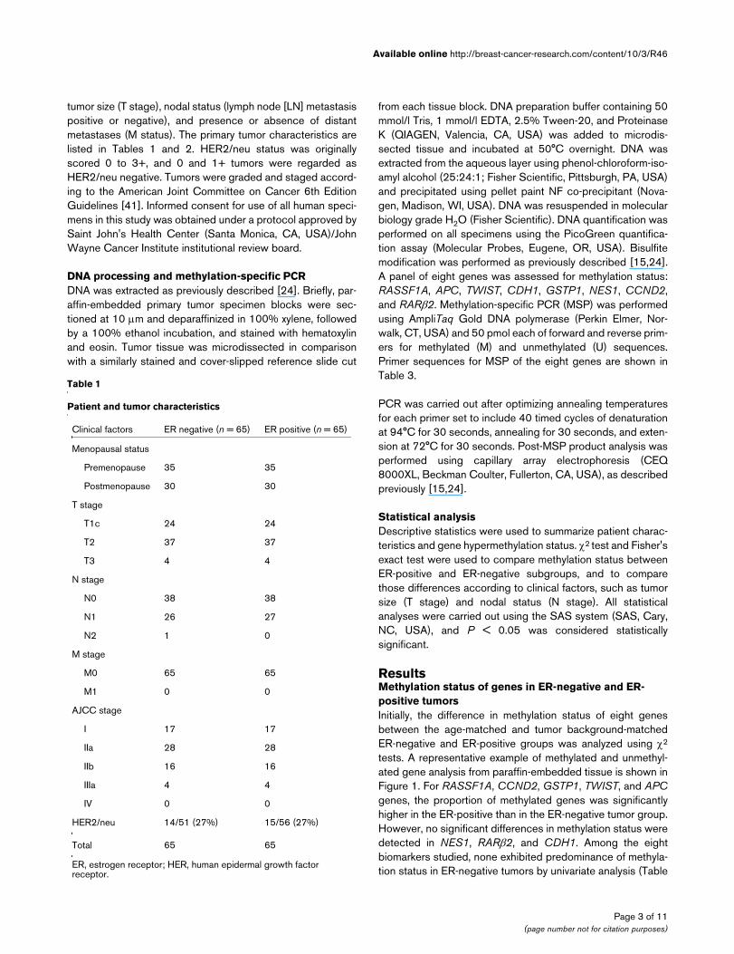
tumor size (T stage), nodal status (lymph node [LN] metastasispositive or negative), and presence or absence of distantmetastases (M status). The primary tumor characteristics arelisted in Tables 1 and 2. HER2/neu status was originallyscored 0 to 3+, and 0 and 1+ tumors were regarded asHER2/neu negative. Tumors were graded and staged accord-ing to the American Joint Committee on Cancer 6th EditionGuidelines [41]. Informed consent for use of all human speci-mens in this study was obtained under a protocol approved bySaint John's Health Center (Santa Monica, CA, USA)/JohnWayne Cancer Institute institutional review board.
DNA processing and methylation-specific PCRDNA was extracted as previously described [24]. Briefly, par-affin-embedded primary tumor specimen blocks were sec-tioned at 10 μm and deparaffinized in 100% xylene, followedby a 100% ethanol incubation, and stained with hematoxylinand eosin. Tumor tissue was microdissected in comparisonwith a similarly stained and cover-slipped reference slide cut
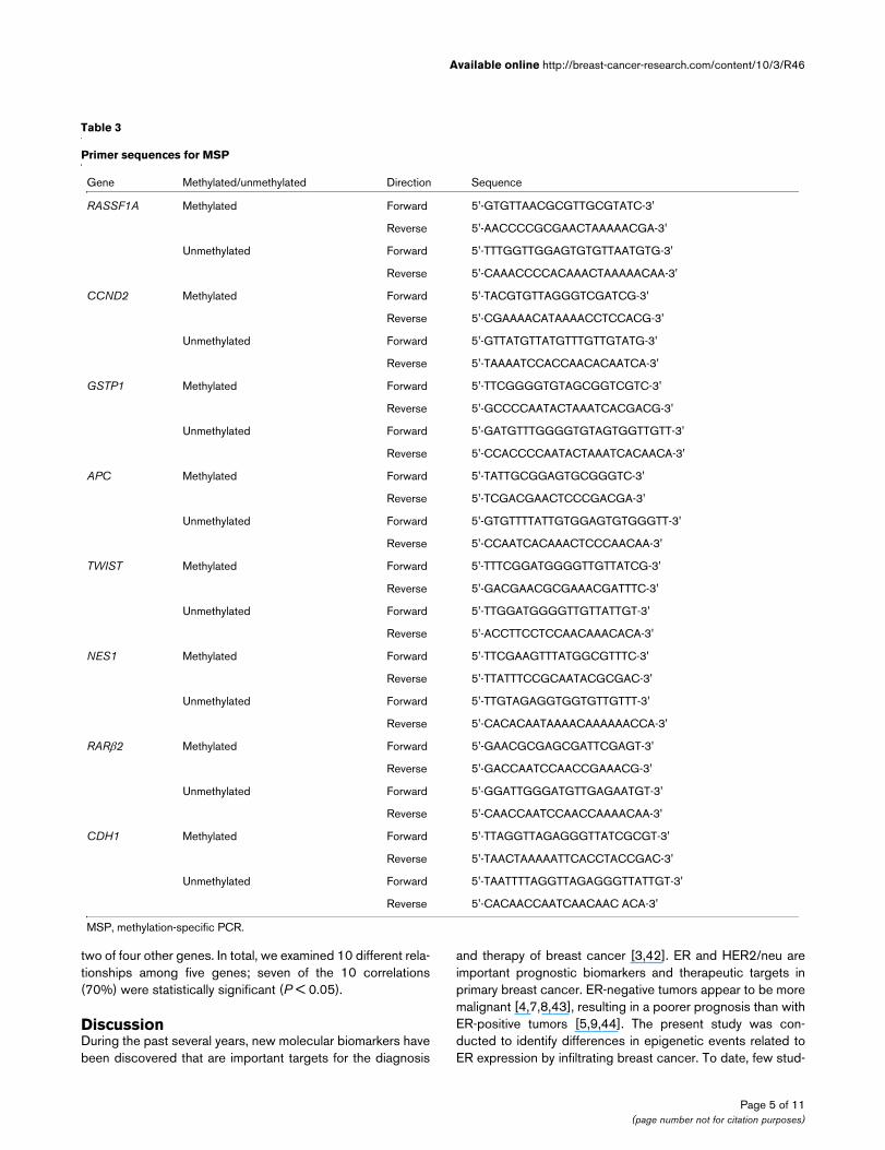
from each tissue block. DNA preparation buffer containing 50mmol/l Tris, 1 mmol/l EDTA, 2.5% Tween-20, and ProteinaseK (QIAGEN, Valencia, CA, USA) was added to microdis-sected tissue and incubated at 50°C overnight. DNA wasextracted from the aqueous layer using phenol-chloroform-iso-amyl alcohol (25:24:1; Fisher Scientific, Pittsburgh, PA, USA)and precipitated using pellet paint NF co-precipitant (Nova-gen, Madison, WI, USA). DNA was resuspended in molecularbiology grade H2O (Fisher Scientific). DNA quantification wasperformed on all specimens using the PicoGreen quantifica-tion assay (Molecular Probes, Eugene, OR, USA). Bisulfitemodification was performed as previously described [15,24].A panel of eight genes was assessed for methylation status:RASSF1A, APC, TWIST, CDH1, GSTP1, NES1, CCND2,and RARβ2. Methylation-specific PCR (MSP) was performedusing AmpliTaq Gold DNA polymerase (Perkin Elmer, Nor-walk, CT, USA) and 50 pmol each of forward and reverse prim-ers for methylated (M) and unmethylated (U) sequences.Primer sequences for MSP of the eight genes are shown inTable 3.
PCR was carried out after optimizing annealing temperaturesfor each primer set to include 40 timed cycles of denaturationat 94°C for 30 seconds, annealing for 30 seconds, and exten-sion at 72°C for 30 seconds. Post-MSP product analysis wasperformed using capillary array electrophoresis (CEQ8000XL, Beckman Coulter, Fullerton, CA, USA), as describedpreviously [15,24].
Statistical analysisDescriptive statistics were used to summarize patient charac-teristics and gene hypermethylation status. χ2 test and Fisher'sexact test were used to compare methylation status betweenER-positive and ER-negative subgroups, and to comparethose differences according to clinical factors, such as tumorsize (T stage) and nodal status (N stage). All statisticalanalyses were carried out using the SAS system (SAS, Cary,NC, USA), and P < 0.05 was considered statisticallysignificant.
ResultsMethylation status of genes in ER-negative and ER-positive tumorsInitially, the difference in methylation status of eight genesbetween the age-matched and tumor background-matchedER-negative and ER-positive groups was analyzed using χ2
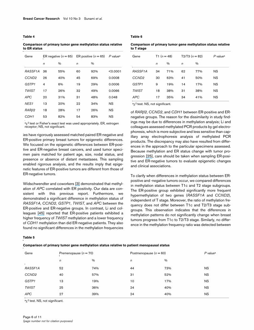
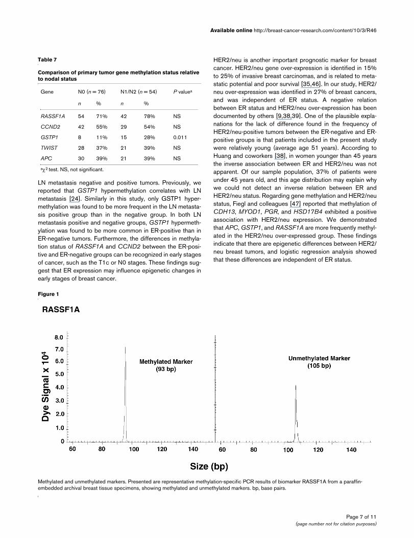
tests. A representative example of methylated and unmethyl-ated gene analysis from paraffin-embedded tissue is shown inFigure 1. For RASSF1A, CCND2, GSTP1, TWIST, and APCgenes, the proportion of methylated genes was significantlyhigher in the ER-positive than in the ER-negative tumor group.However, no significant differences in methylation status weredetected in NES1, RARβ2, and CDH1. Among the eightbiomarkers studied, none exhibited predominance of methyla-tion status in ER-negative tumors by univariate analysis (Table
Table 1
Patient and tumor characteristics
Clinical factors ER negative (n = 65) ER positive (n = 65)
Menopausal status
Premenopause 35 35
Postmenopause 30 30
T stage
T1c 24 24
T2 37 37
T3 4 4
N stage
N0 38 38
N1 26 27
N2 1 0
M stage
M0 65 65
M1 0 0
AJCC stage
I 17 17
IIa 28 28
IIb 16 16
IIIa 4 4
IV 0 0
HER2/neu 14/51 (27%) 15/56 (27%)
Total 65 65
ER, estrogen receptor; HER, human epidermal growth factor receptor.
Page 3 of 11(page number not for citation purposes)
Breast Cancer Research Vol 10 No 3 Sunami et al.
4). Based on this finding, we conducted further analysis of themethylation status of RASSF1A, CCND2, GSTP1, TWIST,and APC. Second, we analyzed the differences in the methyl-ation status of these five tumor-related genes between pre-menopausal and postmenopausal, T1 and T2/T3, and LNmetastasis negative (N0) and positive (N1/N2) subgroupsusing univariate analysis. Among these five genes, there wereno significant differences in methylation frequency betweenpremenopausal and postmenopausal subgroups and T1 andT2/T3 subgroups. Only GSTP1 exhibited significantly morefrequent methylation in the N1/N2 subgroup than in the N0subgroup (Tables 5 to 7).
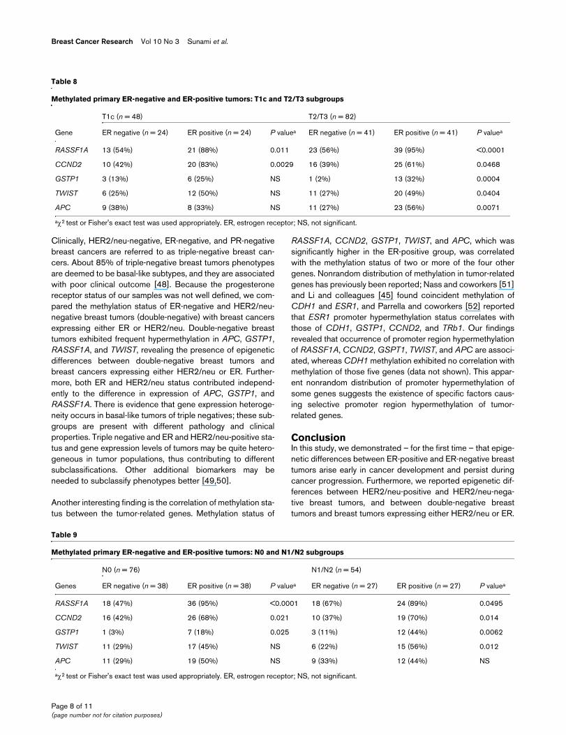
Methylation status of genes in ER-negative and ER-positive early-stage tumorsTo study further the changes in tumor-related gene methyla-tion status of both ER-positive and ER-negative breastcancers during tumor progression, we investigated the differ-ence between both ER-positive and ER-negative primarytumors based on American Joint Committee on Cancer T andN stages. First, we compared the methylation status of five dif-ferent genes in ER-negative and ER-positive tumors for T1cand T2/T3 stages (Table 8). The ER-positive group exhibitedmore frequent methylation of RASSF1A and CCND2 in bothT1c and T2/T3 stages. GSTP1 and TWIST were more meth-ylated in the ER-positive group in the T1c stage, but the differ-ences did not achieve statistical significance. However, theyshowed significant differences in T2/T3 stage. We examinedchanges in the methylation frequency ratio (methylation fre-quency of gene in the ER-positive group/methylation fre-quency of gene in the ER-negative group) for each tumor-related gene during tumor progression. No difference in themethylation frequency ratio was observed between T1c andT2/T3 stages for all tumor-related genes assessed.
Next, we investigated the difference in methylation of tumor-related genes between the ER-negative and ER-positive sub-groups of N0 and N1/N2 tumors (Table 9). The ER-positivegroup exhibited significantly (P < 0.05) more frequent methyl-ation of RASSF1A, CCND2, and GSTP1 in both N0 and N1/
N2 stages. TWIST and APC were more frequently methylatedin the ER-positive group in the N0 stage, but not significantly.Similar to subgroups divided by T stage, there were nodifferences in methylation frequency ratio for all genesassessed between the N0 and N1/N2 subgroups.
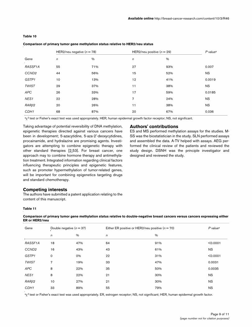
Methylation status of tumor-related genes relevant to HER2/neu statusIn addition to ER, we investigated differences in methylationstatus of the eight genes between the HER2/neu-negative andHER2/neu-positive tumor groups. First, we analyzed the rela-tion between ER status and HER2/neu status. Using matchedsamples, no difference was found in the frequency of HER2/neu-positive tumors between the ER-negative and ER-positivegroups. Then, the differences in methylation status of all eightgenes were analyzed between the HER2/neu-positive andHER2/neu-negative tumor groups. The proportion of methyl-ated RASSF1A, GSTP1, and APC genes was significantlygreater in the HER2/neu-positive than in the HER2/neu-nega-tive tumor group; no significant differences in methylation sta-tus were detected for TWIST, NES1, RARβ2, and CCND2.Among the eight biomarkers studied, CDH1 showed predom-inance of methylation status in HER2/neu-negative tumors byunivariate analysis (Table 10). Next, we compared the methyl-ation status of double-negative tumors versus breast cancersexpressing either ER or HER2/neu. The double-negativebreast cancer group showed less frequent methylation ofAPC, GSTP1, RASSF1A, and TWIST (Table 11). Logisticregression analysis revealed that both ER and HER2/neu sta-tus affect the methylation status of APC, GSTP1, andRASSF1A independently.
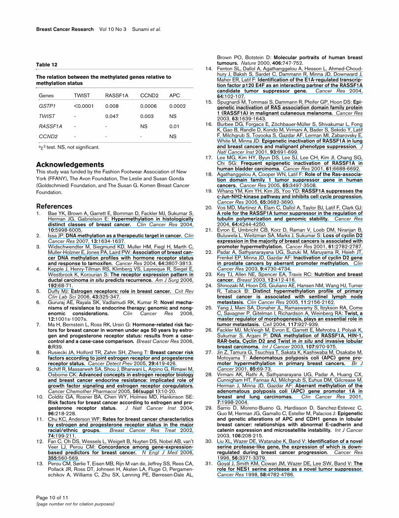
Relation between the tumor-related genes with respect to methylation statusThe relation between the genes with respect to methylationstatus is shown in Table 12. GSTP1 methylation frequency issignificantly related to methylation frequency of other genes.RASSF1A and TWIST methylation frequencies were signifi-cantly related to methylation frequency of three of four othergenes. CCND2 and APC methylation frequency related with
Table 2
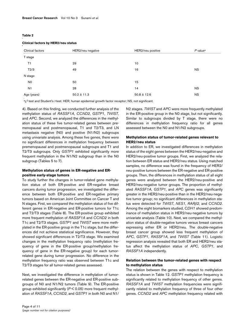
Clinical factors by HER2/neu status
Clinical factors HER2/neu negative HER2/neu positive P valuea
T stage
T1 29 10
T2/3 49 16 NS
N stage
N0 50 15
N1 28 14 NS
Age (years) 50.2 ± 11.3 50.8 ± 12.6 NS
aχ2 test and Student's t-test. HER, human epidermal growth factor receptor; NS, not significant.
Page 4 of 11(page number not for citation purposes)
Available online http://breast-cancer-research.com/content/10/3/R46
two of four other genes. In total, we examined 10 different rela-tionships among five genes; seven of the 10 correlations(70%) were statistically significant (P < 0.05).
DiscussionDuring the past several years, new molecular biomarkers havebeen discovered that are important targets for the diagnosis
and therapy of breast cancer [3,42]. ER and HER2/neu areimportant prognostic biomarkers and therapeutic targets inprimary breast cancer. ER-negative tumors appear to be moremalignant [4,7,8,43], resulting in a poorer prognosis than withER-positive tumors [5,9,44]. The present study was con-ducted to identify differences in epigenetic events related toER expression by infiltrating breast cancer. To date, few stud-
Table 3
Primer sequences for MSP
Gene Methylated/unmethylated Direction Sequence
RASSF1A Methylated Forward 5'-GTGTTAACGCGTTGCGTATC-3'
Reverse 5'-AACCCCGCGAACTAAAAACGA-3'
Unmethylated Forward 5'-TTTGGTTGGAGTGTGTTAATGTG-3'
Reverse 5'-CAAACCCCACAAACTAAAAACAA-3'
CCND2 Methylated Forward 5'-TACGTGTTAGGGTCGATCG-3'
Reverse 5'-CGAAAACATAAAACCTCCACG-3'
Unmethylated Forward 5'-GTTATGTTATGTTTGTTGTATG-3'
Reverse 5'-TAAAATCCACCAACACAATCA-3'
GSTP1 Methylated Forward 5'-TTCGGGGTGTAGCGGTCGTC-3'
Reverse 5'-GCCCCAATACTAAATCACGACG-3'
Unmethylated Forward 5'-GATGTTTGGGGTGTAGTGGTTGTT-3'
Reverse 5'-CCACCCCAATACTAAATCACAACA-3'
APC Methylated Forward 5'-TATTGCGGAGTGCGGGTC-3'
Reverse 5'-TCGACGAACTCCCGACGA-3'
Unmethylated Forward 5'-GTGTTTTATTGTGGAGTGTGGGTT-3'
Reverse 5'-CCAATCACAAACTCCCAACAA-3'
TWIST Methylated Forward 5'-TTTCGGATGGGGTTGTTATCG-3'
Reverse 5'-GACGAACGCGAAACGATTTC-3'
Unmethylated Forward 5'-TTGGATGGGGTTGTTATTGT-3'
Reverse 5'-ACCTTCCTCCAACAAACACA-3'
NES1 Methylated Forward 5'-TTCGAAGTTTATGGCGTTTC-3'
Reverse 5'-TTATTTCCGCAATACGCGAC-3'
Unmethylated Forward 5'-TTGTAGAGGTGGTGTTGTTT-3'
Reverse 5'-CACACAATAAAACAAAAAACCA-3'
RARβ2 Methylated Forward 5'-GAACGCGAGCGATTCGAGT-3'
Reverse 5'-GACCAATCCAACCGAAACG-3'
Unmethylated Forward 5'-GGATTGGGATGTTGAGAATGT-3'
Reverse 5'-CAACCAATCCAACCAAAACAA-3'
CDH1 Methylated Forward 5'-TTAGGTTAGAGGGTTATCGCGT-3'
Reverse 5'-TAACTAAAAATTCACCTACCGAC-3'
Unmethylated Forward 5'-TAATTTTAGGTTAGAGGGTTATTGT-3'
Reverse 5'-CACAACCAATCAACAAC ACA-3'
MSP, methylation-specific PCR.
Page 5 of 11(page number not for citation purposes)
Breast Cancer Research Vol 10 No 3 Sunami et al.
ies have rigorously assessed matched paired ER-negative andER-positive primary breast tumors for epigenetic differences.We focused on the epigenetic differences between ER-posi-tive and ER-negative breast cancers, and used tumor speci-men pairs matched for patient age, size, nodal status, andpresence or absence of distant metastases. This samplingenabled rigorous analysis, and the results imply that epige-netic features of ER-positive tumors are different from those ofER-negative tumors.
Widschwendter and coworkers [3] demonstrated that methyl-ation of APC correlated with ER positivity. Our data are con-sistent with this previous report. Furthermore, wedemonstrated a significant difference in methylation status ofRASSF1A, CCND2, GSTP1, TWIST, and APC between theER-positive and ER-negative groups. In contrast, Li and col-leagues [45] reported that ER-positive patients exhibited ahigher frequency of TWIST methylation and a lower frequencyof CDH1 methylation than did ER-negative patients. They alsofound no significant differences in the methylation frequencies
of RARβ2, CCND2, and CDH1 between ER-positive and ER-negative groups. The reason for the dissimilarity in study find-ings may be due to differences in methylation analysis; Li andcolleagues assessed methylated PCR products by gel electro-phoresis, which is more subjective and less sensitive than cap-illary array electrophoresis analysis of methylated PCRproducts. The discrepancy may also have resulted from differ-ences in the approach to the particular specimens assessed.Because methylation and ER status change with tumor pro-gression [25], care should be taken when sampling ER-posi-tive and ER-negative tumors to evaluate epigenetic changesand clinical associations.
To clarify when differences in methylation status between ER-positive and -negative tumors occur, we compared differencesin methylation status between T1c and T2 stage subgroups.The ER-positive group exhibited significantly more frequenthypermethylation of two genes (RASSF1A and CCND2),independent of T stage. Moreover, the ratio of methylation fre-quency does not differ between T1c and T2/T3 stage sub-groups. This observation indicates that the differences inmethylation patterns do not significantly change when breasttumors progress from T1c to T2/T3 stage. Similarly, no differ-ence in the methylation frequency ratio was detected between
Table 4
Comparison of primary tumor gene methylation status relative to ER status
Gene ER negative (n = 65) ER positive (n = 65) P valuea
n % n %
RASSF1A 36 55% 60 92% <0.0001
CCND2 26 40% 45 69% 0.0008
GSTP1 4 6% 19 29% 0.0006
TWIST 17 26% 32 49% 0.0066
APC 20 31% 31 48% 0.048
NES1 13 20% 22 34% NS
RARβ2 18 28% 17 26% NS
CDH1 53 82% 54 83% NS
aχ2 test or Fisher's exact test was used appropriately. ER, estrogen receptor; NS, not significant.
Table 6
Comparison of primary tumor gene methylation status relative to T stage
Gene T1 (n = 48) T2/T3 (n = 82) P valuea
n % n %
RASSF1A 34 71% 62 77% NS
CCND2 30 63% 41 50% NS
GSTP1 9 19% 14 17% NS
TWIST 18 38% 31 38% NS
APC 17 35% 34 41% NS
aχ2 test. NS, not significant.
Table 5
Comparison of primary tumor gene methylation status relative to patient menopausal status
Gene Premenopause (n = 70) Postmenopause (n = 60) P valuea
n % n %
RASSF1A 52 74% 44 73% NS
CCND2 40 57% 31 52% NS
GSTP1 13 19% 10 17% NS
TWIST 25 36% 24 40% NS
APC 27 39% 24 40% NS
aχ2 test. NS, not significant.
Page 6 of 11(page number not for citation purposes)
Available online http://breast-cancer-research.com/content/10/3/R46
LN metastasis negative and positive tumors. Previously, wereported that GSTP1 hypermethylation correlates with LNmetastasis [24]. Similarly in this study, only GSTP1 hyper-methylation was found to be more frequent in the LN metasta-sis positive group than in the negative group. In both LNmetastasis positive and negative groups, GSTP1 hypermeth-ylation was found to be more common in ER-positive than inER-negative tumors. Furthermore, the differences in methyla-tion status of RASSF1A and CCND2 between the ER-posi-tive and ER-negative groups can be recognized in early stagesof cancer, such as the T1c or N0 stages. These findings sug-gest that ER expression may influence epigenetic changes inearly stages of breast cancer.
HER2/neu is another important prognostic marker for breastcancer. HER2/neu gene over-expression is identified in 15%to 25% of invasive breast carcinomas, and is related to meta-static potential and poor survival [35,46]. In our study, HER2/neu over-expression was identified in 27% of breast cancers,and was independent of ER status. A negative relationbetween ER status and HER2/neu over-expression has beendocumented by others [9,38,39]. One of the plausible expla-nations for the lack of difference found in the frequency ofHER2/neu-positive tumors between the ER-negative and ER-positive groups is that patients included in the present studywere relatively young (average age 51 years). According toHuang and coworkers [38], in women younger than 45 yearsthe inverse association between ER and HER2/neu was notapparent. Of our sample population, 37% of patients wereunder 45 years old, and this age distribution may explain whywe could not detect an inverse relation between ER andHER2/neu status. Regarding gene methylation and HER2/neustatus, Fiegl and colleagues [47] reported that methylation ofCDH13, MYOD1, PGR, and HSD17B4 exhibited a positiveassociation with HER2/neu expression. We demonstratedthat APC, GSTP1, and RASSF1A are more frequently methyl-ated in the HER2/neu over-expressed group. These findingsindicate that there are epigenetic differences between HER2/neu breast tumors, and logistic regression analysis showedthat these differences are independent of ER status.
Table 7
Comparison of primary tumor gene methylation status relative to nodal status
Gene N0 (n = 76) N1/N2 (n = 54) P valuea
n % n %
RASSF1A 54 71% 42 78% NS
CCND2 42 55% 29 54% NS
GSTP1 8 11% 15 28% 0.011
TWIST 28 37% 21 39% NS
APC 30 39% 21 39% NS
aχ2 test. NS, not significant.
Figure 1
Methylated and unmethylated markersMethylated and unmethylated markers. Presented are representative methylation-specific PCR results of biomarker RASSF1A from a paraffin-embedded archival breast tissue specimens, showing methylated and unmethylated markers. bp, base pairs.
Page 7 of 11(page number not for citation purposes)
Breast Cancer Research Vol 10 No 3 Sunami et al.
Clinically, HER2/neu-negative, ER-negative, and PR-negativebreast cancers are referred to as triple-negative breast can-cers. About 85% of triple-negative breast tumors phenotypesare deemed to be basal-like subtypes, and they are associatedwith poor clinical outcome [48]. Because the progesteronereceptor status of our samples was not well defined, we com-pared the methylation status of ER-negative and HER2/neu-negative breast tumors (double-negative) with breast cancersexpressing either ER or HER2/neu. Double-negative breasttumors exhibited frequent hypermethylation in APC, GSTP1,RASSF1A, and TWIST, revealing the presence of epigeneticdifferences between double-negative breast tumors andbreast cancers expressing either HER2/neu or ER. Further-more, both ER and HER2/neu status contributed independ-ently to the difference in expression of APC, GSTP1, andRASSF1A. There is evidence that gene expression heteroge-neity occurs in basal-like tumors of triple negatives; these sub-groups are present with different pathology and clinicalproperties. Triple negative and ER and HER2/neu-positive sta-tus and gene expression levels of tumors may be quite hetero-geneous in tumor populations, thus contributing to differentsubclassifications. Other additional biomarkers may beneeded to subclassify phenotypes better [49,50].
Another interesting finding is the correlation of methylation sta-tus between the tumor-related genes. Methylation status of
RASSF1A, CCND2, GSTP1, TWIST, and APC, which wassignificantly higher in the ER-positive group, was correlatedwith the methylation status of two or more of the four othergenes. Nonrandom distribution of methylation in tumor-relatedgenes has previously been reported; Nass and coworkers [51]and Li and colleagues [45] found coincident methylation ofCDH1 and ESR1, and Parrella and coworkers [52] reportedthat ESR1 promoter hypermethylation status correlates withthose of CDH1, GSTP1, CCND2, and TRb1. Our findingsrevealed that occurrence of promoter region hypermethylationof RASSF1A, CCND2, GSPT1, TWIST, and APC are associ-ated, whereas CDH1 methylation exhibited no correlation withmethylation of those five genes (data not shown). This appar-ent nonrandom distribution of promoter hypermethylation ofsome genes suggests the existence of specific factors caus-ing selective promoter region hypermethylation of tumor-related genes.
ConclusionIn this study, we demonstrated – for the first time – that epige-netic differences between ER-positive and ER-negative breasttumors arise early in cancer development and persist duringcancer progression. Furthermore, we reported epigenetic dif-ferences between HER2/neu-positive and HER2/neu-nega-tive breast tumors, and between double-negative breasttumors and breast tumors expressing either HER2/neu or ER.
Table 8
Methylated primary ER-negative and ER-positive tumors: T1c and T2/T3 subgroups
T1c (n = 48) T2/T3 (n = 82)
Gene ER negative (n = 24) ER positive (n = 24) P valuea ER negative (n = 41) ER positive (n = 41) P valuea
RASSF1A 13 (54%) 21 (88%) 0.011 23 (56%) 39 (95%) <0.0001
CCND2 10 (42%) 20 (83%) 0.0029 16 (39%) 25 (61%) 0.0468
GSTP1 3 (13%) 6 (25%) NS 1 (2%) 13 (32%) 0.0004
TWIST 6 (25%) 12 (50%) NS 11 (27%) 20 (49%) 0.0404
APC 9 (38%) 8 (33%) NS 11 (27%) 23 (56%) 0.0071
aχ2 test or Fisher's exact test was used appropriately. ER, estrogen receptor; NS, not significant.
Table 9
Methylated primary ER-negative and ER-positive tumors: N0 and N1/N2 subgroups
N0 (n = 76) N1/N2 (n = 54)
Genes ER negative (n = 38) ER positive (n = 38) P valuea ER negative (n = 27) ER positive (n = 27) P valuea
RASSF1A 18 (47%) 36 (95%) <0.0001 18 (67%) 24 (89%) 0.0495
CCND2 16 (42%) 26 (68%) 0.021 10 (37%) 19 (70%) 0.014
GSTP1 1 (3%) 7 (18%) 0.025 3 (11%) 12 (44%) 0.0062
TWIST 11 (29%) 17 (45%) NS 6 (22%) 15 (56%) 0.012
APC 11 (29%) 19 (50%) NS 9 (33%) 12 (44%) NS
aχ2 test or Fisher's exact test was used appropriately. ER, estrogen receptor; NS, not significant.
Page 8 of 11(page number not for citation purposes)
Available online http://breast-cancer-research.com/content/10/3/R46
Taking advantage of potential reversibility of DNA methylation,epigenetic therapies directed against various cancers havebeen in development; 5-azacytidine, 5-aza-2'-deoxycytidines,procainamide, and hydralazine are promising agents. Investi-gators are attempting to combine epigenetic therapy withother standard therapies [2,53]. For breast cancer, oneapproach may to combine hormone therapy and antimethyla-tion treatment. Integrated information regarding clinical factorsinfluencing therapeutic principles and epigenetic features,such as promoter hypermethylation of tumor-related genes,will be important for combining epigenetics targeting drugsand standard chemotherapy.
Competing interestsThe authors have submitted a patent application relating to thecontent of this manuscript.
Authors' contributionsES and MS performed methylation assays for the studies. M-SS was the biostatistician in the study. SLN performed assaysand assembled the data. A-TV helped with assays. AEG per-formed the clinical review of the patients and reviewed thestudy design. DSNH was the principle investigator anddesigned and reviewed the study.
Table 10
Comparison of primary tumor gene methylation status relative to HER2/neu status
HER2/neu negative (n = 78) HER2/neu positive (n = 29) P valuea
Gene n % n %
RASSF1A 55 71% 27 93% 0.007
CCND2 44 56% 15 52% NS
GSTP1 10 13% 12 41% 0.0019
TWIST 29 37% 11 38% NS
APC 26 33% 17 59% 0.0185
NES1 22 28% 7 24% NS
RARβ2 20 26% 11 38% NS
CDH1 68 87% 20 67% 0.036
aχ2 test or Fisher's exact test was used appropriately. HER, human epidermal growth factor receptor; NS, not significant.
Table 11
Comparison of primary tumor gene methylation status relative to double-negative breast cancers versus cancers expressing either ER or HER2/neu
Gene Double negative (n = 37) Either ER positive or HER2/neu positive (n = 70) P valuea
n % n %
RASSF1A 18 47% 64 91% <0.0001
CCND2 16 43% 43 61% NS
GSTP1 0 0% 22 31% <0.0001
TWIST 7 19% 33 47% 0.0031
APC 8 22% 35 50% 0.0035
NES1 8 22% 21 30% NS
RARβ2 10 27% 21 30% NS
CDH1 33 89% 55 79% NS
aχ2 test or Fisher's exact test was used appropriately. ER, estrogen receptor; NS, not significant; HER, human epidermal growth factor.
Page 9 of 11(page number not for citation purposes)
Breast Cancer Research Vol 10 No 3 Sunami et al.
AcknowledgementsThis study was funded by the Fashion Footwear Association of New York (FFANY), The Avon Foundation, The Leslie and Susan Gonda (Goldschmied) Foundation, and The Susan G. Komen Breast Cancer Foundation.
References1. Bae YK, Brown A, Garrett E, Bornman D, Fackler MJ, Sukumar S,
Herman JG, Gabrielson E: Hypermethylation in histologicallydistinct classes of breast cancer. Clin Cancer Res 2004,10:5998-6005.
2. Issa JP: DNA methylation as a therapeutic target in cancer. ClinCancer Res 2007, 13:1634-1637.
3. Widschwendter M, Siegmund KD, Muller HM, Fiegl H, Marth C,Muller-Holzner E, Jones PA, Laird PW: Association of breast can-cer DNA methylation profiles with hormone receptor statusand response to tamoxifen. Cancer Res 2004, 64:3807-3813.
4. Kepple J, Henry-Tillman RS, Klimberg VS, Layeeque R, Siegel E,Westbrook K, Korourian S: The receptor expression pattern inductal carcinoma in situ predicts recurrence. Am J Surg 2006,192:68-71.
5. Duffy MJ: Estrogen receptors: role in breast cancer. Crit RevClin Lab Sci 2006, 43:325-347.
6. Gururaj AE, Rayala SK, Vadlamudi RK, Kumar R: Novel mecha-nisms of resistance to endocrine therapy: genomic and nong-enomic considerations. Clin Cancer Res 2006,12:1001s-1007s.
7. Ma H, Bernstein L, Ross RK, Ursin G: Hormone-related risk fac-tors for breast cancer in women under age 50 years by estro-gen and progesterone receptor status: results from a case-control and a case-case comparison. Breast Cancer Res 2006,8:R39.
8. Rusiecki JA, Holford TR, Zahm SH, Zheng T: Breast cancer riskfactors according to joint estrogen receptor and progesteronereceptor status. Cancer Detect Prev 2005, 29:419-426.
9. Schiff R, Massarweh SA, Shou J, Bharwani L, Arpino G, Rimawi M,Osborne CK: Advanced concepts in estrogen receptor biologyand breast cancer endocrine resistance: implicated role ofgrowth factor signaling and estrogen receptor coregulators.Cancer Chemother Pharmacol 2005, 56(suppl 1):10-20.
10. Colditz GA, Rosner BA, Chen WY, Holmes MD, Hankinson SE:Risk factors for breast cancer according to estrogen and pro-gesterone receptor status. J Natl Cancer Inst 2004,96:218-228.
11. Chu KC, Anderson WF: Rates for breast cancer characteristicsby estrogen and progesterone receptor status in the majorracial/ethnic groups. Breast Cancer Res Treat 2002,74:199-211.
12. Fan C, Oh DS, Wessels L, Weigelt B, Nuyten DS, Nobel AB, van'tVeer LJ, Perou CM: Concordance among gene-expression-based predictors for breast cancer. N Engl J Med 2006,355:560-569.
13. Perou CM, Sørlie T, Eisen MB, Rijn M van de, Jeffrey SS, Rees CA,Pollack JR, Ross DT, Johnsen H, Akslen LA, Fluge O, Pergamen-schikov A, Williams C, Zhu SX, Lønning PE, Børresen-Dale AL,
Brown PO, Botstein D: Molecular portraits of human breasttumours. Nature 2000, 406:747-752.
14. Fenton SL, Dallol A, Agathanggelou A, Hesson L, Ahmed-Choud-hury J, Baksh S, Sardet C, Dammann R, Minna JD, Downward J,Maher ER, Latif F: Identification of the E1A-regulated transcrip-tion factor p120 E4F as an interacting partner of the RASSF1Acandidate tumor suppressor gene. Cancer Res 2004,64:102-107.
15. Spugnardi M, Tommasi S, Dammann R, Pfeifer GP, Hoon DS: Epi-genetic inactivation of RAS association domain family protein1 (RASSF1A) in malignant cutaneous melanoma. Cancer Res2003, 63:1639-1643.
16. Burbee DG, Forgacs E, Zöchbauer-Müller S, Shivakumar L, FongK, Gao B, Randle D, Kondo M, Virmani A, Bader S, Sekido Y, LatifF, Milchgrub S, Toyooka S, Gazdar AF, Lerman MI, Zabarovsky E,White M, Minna JD: Epigenetic inactivation of RASSF1A in lungand breast cancers and malignant phenotype suppression. JNatl Cancer Inst 2001, 93:691-699.
17. Lee MG, Kim HY, Byun DS, Lee SJ, Lee CH, Kim JI, Chang SG,Chi SG: Frequent epigenetic inactivation of RASSF1A inhuman bladder carcinoma. Cancer Res 2001, 61:6688-6692.
18. Agathanggelou A, Cooper WN, Latif F: Role of the Ras-associa-tion domain family 1 tumor suppressor gene in humancancers. Cancer Res 2005, 65:3497-3508.
19. Whang YM, Kim YH, Kim JS, Yoo YD: RASSF1A suppresses thec-Jun-NH2-kinase pathway and inhibits cell cycle progression.Cancer Res 2005, 65:3682-3690.
20. Vos MD, Martinez A, Elam C, Dallol A, Taylor BJ, Latif F, Clark GJ:A role for the RASSF1A tumor suppressor in the regulation oftubulin polymerization and genomic stability. Cancer Res2004, 64:4244-4250.
21. Evron E, Umbricht CB, Korz D, Raman V, Loeb DM, Niranjan B,Buluwela L, Weitzman SA, Marks J, Sukumar S: Loss of cyclin D2expression in the majority of breast cancers is associated withpromoter hypermethylation. Cancer Res 2001, 61:2782-2787.
22. Padar A, Sathyanarayana UG, Suzuki M, Maruyama R, Hsieh JT,Frenkel EP, Minna JD, Gazdar AF: Inactivation of cyclin D2 genein prostate cancers by aberrant promoter methylation. ClinCancer Res 2003, 9:4730-4734.
23. Key TJ, Allen NE, Spencer EA, Travis RC: Nutrition and breastcancer. Breast 2003, 12:412-416.
24. Shinozaki M, Hoon DS, Giuliano AE, Hansen NM, Wang HJ, TurnerR, Taback B: Distinct hypermethylation profile of primarybreast cancer is associated with sentinel lymph nodemetastasis. Clin Cancer Res 2005, 11:2156-2162.
25. Yang J, Mani SA, Donaher JL, Ramaswamy S, Itzykson RA, ComeC, Savagner P, Gitelman I, Richardson A, Weinberg RA: Twist, amaster regulator of morphogenesis, plays an essential role intumor metastasis. Cell 2004, 117:927-939.
26. Fackler MJ, McVeigh M, Evron E, Garrett E, Mehrotra J, Polyak K,Sukumar S, Argani P: DNA methylation of RASSF1A, HIN-1,RAR-beta, Cyclin D2 and Twist in in situ and invasive lobularbreast carcinoma. Int J Cancer 2003, 107:970-975.
27. Jin Z, Tamura G, Tsuchiya T, Sakata K, Kashiwaba M, Osakabe M,Motoyama T: Adenomatous polyposis coli (APC) gene pro-moter hypermethylation in primary breast cancers. Br JCancer 2001, 85:69-73.
28. Virmani AK, Rathi A, Sathyanarayana UG, Padar A, Huang CX,Cunnigham HT, Farinas AJ, Milchgrub S, Euhus DM, Gilcrease M,Herman J, Minna JD, Gazdar AF: Aberrant methylation of theadenomatous polyposis coli (APC) gene promoter 1A inbreast and lung carcinomas. Clin Cancer Res 2001,7:1998-2004.
29. Sarrio D, Moreno-Bueno G, Hardisson D, Sanchez-Estevez C,Guo M, Herman JG, Gamallo C, Esteller M, Palacios J: Epigeneticand genetic alterations of APC and CDH1 genes in lobularbreast cancer: relationships with abnormal E-cadherin andcatenin expression and microsatellite instability. Int J Cancer2003, 106:208-215.
30. Liu XL, Wazer DE, Watanabe K, Band V: Identification of a novelserine protease-like gene, the expression of which is down-regulated during breast cancer progression. Cancer Res1996, 56:3371-3379.
31. Goyal J, Smith KM, Cowan JM, Wazer DE, Lee SW, Band V: Therole for NES1 serine protease as a novel tumor suppressor.Cancer Res 1998, 58:4782-4786.
Table 12
The relation between the methylated genes relative to methylation status
Genes TWIST RASSF1A CCND2 APC
GSTP1 <0.0001 0.008 0.0006 0.0002
TWIST - 0.047 0.003 NS
RASSF1A - - NS 0.01
CCND2 - - - NS
aχ2 test. NS, not significant.
Page 10 of 11(page number not for citation purposes)
http://www.ncbi.nlm.nih.gov/entrez/query.fcgi?cmd=Retrieve&db=PubMed&dopt=Abstract&list_uids=8764136
http://www.ncbi.nlm.nih.gov/entrez/query.fcgi?cmd=Retrieve&db=PubMed&dopt=Abstract&list_uids=8764136
http://www.ncbi.nlm.nih.gov/entrez/query.fcgi?cmd=Retrieve&db=PubMed&dopt=Abstract&list_uids=8764136
http://www.ncbi.nlm.nih.gov/entrez/query.fcgi?cmd=Retrieve&db=PubMed&dopt=Abstract&list_uids=9809976
Available online http://breast-cancer-research.com/content/10/3/R46
32. Mehrotra J, Vali M, McVeigh M, Kominsky SL, Fackler MJ, Lahti-Domenici J, Polyak K, Sacchi N, Garrett-Mayer E, Argani P, Suku-mar S: Very high frequency of hypermethylated genes inbreast cancer metastasis to the bone, brain, and lung. ClinCancer Res 2004, 10:3104-3109.
33. Zanardi S, Serrano D, Argusti A, Barile M, Puntoni M, Decensi A:Clinical trials with retinoids for breast cancerchemoprevention. Endocr Relat Cancer 2006, 13:51-68.
34. Mahler-Araujo B, Savage K, Parry S, Reis-Filho JS: Reduction ofE-cadherin expression is associated with non-lobular breastcarcinomas of basal-like and triple negative phenotype. J ClinPathol 2008, 61:615-620.
35. Francis G, Beadle G, Thomas S, Mengersen K, Stein S: Evalua-tion of oestrogen and progesterone receptor status in HER-2positive breast carcinomas and correlation with outcome.Pathology 2006, 38:391-398.
36. Ross JS, Fletcher JA: The HER-2/neu oncogene in breast can-cer: prognostic factor, predictive factor, and target for therapy.Stem Cells 1998, 16:413-428.
37. Wright C, Nicholson S, Angus B, Sainsbury JR, Farndon J, CairnsJ, Harris AL, Horne CH: Relationship between c-erbB-2 proteinproduct expression and response to endocrine therapy inadvanced breast cancer. Br J Cancer 1992, 65:118-121.
38. Huang HJ, Neven P, Drijkoningen M, Paridaens R, Wildiers H, VanLimbergen E, Berteloot P, Amant F, Vergote I, Christiaens MR:Hormone receptors do not predict the HER2/neu status in allage groups of women with an operable breast cancer. AnnOncol 2005, 16:1755-1761.
39. Balsari A, Casalini P, Tagliabue E, Greco M, Pilotti S, Agresti R,Giovanazzi R, Alasio L, Rumio C, Cascinelli N, Colnaghi MI,Ménard S: Fluctuation of HER2 expression in breast carcino-mas during the menstrual cycle. Am J Pathol 1999,155:1543-1547.
40. Read LD, Keith D Jr, Slamon DJ, Katzenellenbogen BS: Hormonalmodulation of HER-2/neu protooncogene messenger ribonu-cleic acid and p185 protein expression in human breast cancercell lines. Cancer Res 1990, 50:3947-3951.
41. Greene FL, Page DL, Fleming ID, Fritz A, Balch CM, Haller DG,Morrow M: Cancer Staging Manual 6th edition. New York:Springer; 2002.
42. D'Andrea MR, Limiti MR, Bari M, Zambenedetti P, Montagutti A,Ricci F, Pappagallo GL, Sartori D, Vinante O, Mingazzini PL: Cor-relation between genetic and biological aspects in primarynon-metastatic breast cancers and corresponding synchro-nous axillary lymph node metastasis. Breast Cancer Res Treat2007, 101:279-284.
43. Giacinti L, Claudio PP, Lopez M, Giordano A: Epigenetic informa-tion and estrogen receptor alpha expression in breast cancer.Oncologist 2006, 11:1-8.
44. Leu YW, Yan PS, Fan M, Jin VX, Liu JC, Curran EM, Welshons WV,Wei SH, Davuluri RV, Plass C, Nephew KP, Huang TH: Loss ofestrogen receptor signaling triggers epigenetic silencing ofdownstream targets in breast cancer. Cancer Res 2004,64:8184-8192.
45. Li S, Rong M, Iacopetta B: DNA hypermethylation in breast can-cer and its association with clinicopathological features. Can-cer Lett 2006, 237:272-280.
46. Slamon DJ, Godolphin W, Jones LA, Holt JA, Wong SG, Keith DE,Levin WJ, Stuart SG, Udove J, Ullrich A, et al.: Studies of theHER-2/neu proto-oncogene in human breast and ovariancancer. Science 1989, 244:707-712.
47. Fiegl H, Millinger S, Goebel G, Muller-Holzner E, Marth C, LairdPW, Widschwendter M: Breast cancer DNA methylation pro-files in cancer cells and tumor stroma: association with HER-2/neu status in primary breast cancer. Cancer Res 2006,66:29-33.
48. Bauer KR, Brown M, Cress RD, Parise CA, Caggiano V: Descrip-tive analysis of estrogen receptor (ER)-negative, progesteronereceptor (PR)-negative, and HER2-negative invasive breastcancer, the so-called triple-negative phenotype: a population-based study from the California cancer Registry. Cancer 2007,109:1721-1728.
49. Cheang MC, Voduc D, Bajdik C, Leung S, McKinney S, Chia SK,Perou CM, Nielsen TO: Basal-like breast cancer defined by fivebiomarkers has superior prognostic value than triple-negativephenotype. Clin Cancer Res 2008, 14:1368-1376.
50. Kreike B, van Kouwenhove M, Horlings H, Weigelt B, Peterse H,Bartelink H, Vijver MJ van de: Gene expression profiling and his-topathological characterization of triple-negative/basal-likebreast carcinomas. Breast Cancer Res 2007, 9:R65.
51. Nass SJ, Herman JG, Gabrielson E, Iversen PW, Parl FF, DavidsonNE, Graff JR: Aberrant methylation of the estrogen receptorand E-cadherin 5' CpG islands increases with malignant pro-gression in human breast cancer. Cancer Res 2000,60:4346-4348.
52. Parrella P, Poeta ML, Gallo AP, Prencipe M, Scintu M, Apicella A,Rossiello R, Liguoro G, Seripa D, Gravina C, Rabitti C, Rinaldi M,Nicol T, Tommasi S, Paradiso A, Schittulli F, Altomare V, Fazio VM:Nonrandom distribution of aberrant promoter methylation ofcancer-related genes in sporadic breast tumors. Clin CancerRes 2004, 10:5349-5354.
53. Yoo CB, Jones PA: Epigenetic therapy of cancer: past, presentand future. Nat Rev Drug Discov 2006, 5:37-50.
Page 11 of 11(page number not for citation purposes)
http://www.ncbi.nlm.nih.gov/entrez/query.fcgi?cmd=Retrieve&db=PubMed&dopt=Abstract&list_uids=9831867
http://www.ncbi.nlm.nih.gov/entrez/query.fcgi?cmd=Retrieve&db=PubMed&dopt=Abstract&list_uids=9831867
http://www.ncbi.nlm.nih.gov/entrez/query.fcgi?cmd=Retrieve&db=PubMed&dopt=Abstract&list_uids=1346366
http://www.ncbi.nlm.nih.gov/entrez/query.fcgi?cmd=Retrieve&db=PubMed&dopt=Abstract&list_uids=1346366
http://www.ncbi.nlm.nih.gov/entrez/query.fcgi?cmd=Retrieve&db=PubMed&dopt=Abstract&list_uids=1346366
http://www.ncbi.nlm.nih.gov/entrez/query.fcgi?cmd=Retrieve&db=PubMed&dopt=Abstract&list_uids=1972345
http://www.ncbi.nlm.nih.gov/entrez/query.fcgi?cmd=Retrieve&db=PubMed&dopt=Abstract&list_uids=1972345
http://www.ncbi.nlm.nih.gov/entrez/query.fcgi?cmd=Retrieve&db=PubMed&dopt=Abstract&list_uids=1972345
http://www.ncbi.nlm.nih.gov/entrez/query.fcgi?cmd=Retrieve&db=PubMed&dopt=Abstract&list_uids=2470152
http://www.ncbi.nlm.nih.gov/entrez/query.fcgi?cmd=Retrieve&db=PubMed&dopt=Abstract&list_uids=2470152