
promising fibre-yielding plants of the indian himalayan region

1
This research was originally published in JNM . Altai M, Wållberg H, Honarvar H, Strand J,Orlova A, Varasteh Z, Sandström M, Löfblom J, Larsson E, Strand SE, Lubberink M, Ståhl S,Tolmachev V. 188Re-ZHER2:V2, a Promising Affibody-Based Targeting Agent AgainstHER2-Expressing Tumors: Preclinical Assessment. J Nucl Med. 2014;55:1842-8.© by theSociety of Nuclear Medicine and Molecular Imaging, Inc.http://jnm.snmjournals.org/content/55/11/1842.long
188Re-ZHER2:V2, a promising affibody-based targeting agent against HER2-expressing
tumors: preclinical assessment.
Mohamed Altai1, Helena Wållberg2, Hadis Honarvar1, Joanna Strand1 , Anna Orlova3,
Zohreh Varasteh3, Mattias Sandström4,5, John Löfblom2, Erik Larsson6, Sven-Erik Strand6,
Mark Lubberink 4,5, Stefan Ståhl2, Vladimir Tolmachev1.
1 Division of Biomedical Radiation Sciences, Uppsala University, Uppsala, Sweden;2 KTH Royal Institute of Technology, School of Biotechnology, Division of ProteinTechnology, Stockholm, Sweden;3 Preclinical PET Platform, Department of Medicinal Chemistry, Uppsala University,Uppsala, Sweden;4 Nuclear medicine and PET, Uppsala University, Uppsala, Sweden;5 Medical physics, Uppsala University Hospital, Uppsala, Sweden;6 Medical Radiation Physics, Clinical Sciences, Lund University, Lund, Sweden.
This research was supported in part by grants from the Swedish Cancer Society(Cancerfonden), Swedish Research Council (Vetenskapsrådet) and Governmental Funding ofClinical Research within the National Health Service (ALF).
Corresponding author: Vladimir Tolmachev, Professor.
Biomedical Radiation Sciences, Uppsala University, Se- 75185, Uppsala; Sweden
Tel. +46184713414; Fax. +46184713432; E-mail [email protected]
First author: Mohamed Altai, PhD student,
Biomedical Radiation Sciences, Uppsala University, Se- 75185, Uppsala; Sweden
Tel. +46184713414; Fax. +46184713868; E-mail [email protected]
2
ABSTRACT
Affibody molecules are small (7 kDa) non-immunoglobulin scaffold proteins with favorable
tumor targeting properties. Studies concerning influence of chelators on biodistribution of
99mTc-labelled affibody molecules demonstrated that the variant with a C-terminal glycyl-
glycyl-glycyl-cysteine peptide-based chelator (designated ZHER2:V2) has the best
biodistribution profile in vivo and the lowest renal retention of radioactivity. The aim of this
study was to evaluate 188Re-ZHER2:V2 as a potential candidate for radionuclide therapy of
HER2-expressing tumors.
Methods. ZHER2:V2 was labelled with 188Re using a gluconate-containing kit. Targeting of
HER2-overexpressing SKOV-3 ovarian carcinoma xenografts in nude mice was studied for a
dosimetry assessment.
Results. Binding of 188Re-ZHER2:V2 to living SKOV-3 cells was demonstrated to be specific
with an affinity of 6.4±0.4 pM. The biodistribution study showed a rapid blood clearance
(1.4±0.1 %IA/g at 1h p.i.). The tumor uptake was 14±2, 12±2, 5±2 and 1.8±0.5 %IA/g at 1, 4,
24 and 48 h p.i., respectively. The in vivo targeting of HER2 expressing xenografts was
specific. Already at 4 h p.i, the tumor uptake exceeded uptake in kidneys (2.1±0.2 %IA/g).
Scintillation-camera imaging showed that tumor xenografts were the only sites with
prominent accumulation of radioactivity at 4h p.i. Based on the biokinetics, a dosimetry
evaluation for humans suggests that 188Re-ZHER2:V2 would provide an absorbed dose to tumour
of 79 Gy without exceeding absorbed doses of 23 Gy to kidneys and 2 Gy to bone marrow.
This indicates that future human radiotherapy studies may be feasible.
Conclusion. 188Re-ZHER2:V2 can deliver high absorbed doses to tumors without exceeding
kidney and bone marrow toxicity limits.
KEYWORDS
HER2, Affibody molecule, Rhenium-188, Dosimetry.

3
INTRODUCTION
Overexpression of the human epidermal growth factor receptor type-2 (HER2) is associated
with malignant transformation of cells and provides a growth advantage for tumors.
Treatment of HER2-expressing disseminated tumors with the anti-HER2 monoclonal
antibody trastuzumab improves survival of patients with metastatic breast (1) and gastric
cancer (2). However, many tumors have a primary trastuzumab resistance or develop
resistance during therapy despite preserved HER2 expression (2). Conjugation of cytotoxic
payloads (e.g. drugs or radionuclides) to tumor-targeting antibodies may enhance their anti-
tumor effects and hence improve the response duration and overall response rate (3,4). Beta-
emitting radionuclides are considered as a promising form of cytotoxic payload, because of
the cross-fire effect. The use of full-length antibodies for radionuclide therapy, however, is
associated with a number of issues. The major obstacles are insufficient penetration in the
tumor tissue, limiting the absorbed dose to tumors and slow clearance from the body causing
second organ (mainly bone marrow) toxicity (5). The use of smaller targeting agents, e.g.
antibody fragments, would provide more efficient extravasation, better tumor penetration and
more rapid clearance of an unbound tracer (6,7).
A promising approach for development of high affinity small-size tumor targeting agents is
the use of engineered scaffold proteins (8). Affibody molecules are the most studied class of
scaffold proteins for in vivo radionuclide targeting. These high affinity ligands are based on a
58 amino acid (7 kDa) triple alpha-helical scaffold derived from domain B of staphylococcal
protein A (9). An affibody with picomolar affinity (22 pM) to the extracellular domain of
HER2 has been selected earlier (10). Affibody molecules labelled with different radionuclides
have demonstrated efficient tumor targeting and high-contrast imaging of HER2-expressing
tumors (11). Two clinical studies have demonstrated that affibody molecules are non-toxic
and non-immunogenic in humans (12,13). However, affibody molecules undergo renal

4
excretion followed by efficient substantial renal re-absorption (14). The use of residualizing
labels has therefore resulted in a high retention of radionuclides in kidneys with a
radioactivity concentration exceeding several fold the radioactivity concentration in tumors.
This would make the use of majority of radiometals (e.g. 177Lu or 90Y) unsuitable for
radionuclide therapy (11). An important feature of the HER2-targeting affibody molecules is
their slow internalization by HER2-expressing cells (15). This enables the use of non-
residualizing labels without appreciable reduction of tumor retention. On the contrary, rapid
internalization of the affibody in proximal tubuli results in rapid clearance of non-
residualizing labels from kidneys (11,16).
Earlier, we have evaluated how the composition of peptide-based N3S chelators can influence
the targeting and biodistribution properties of 99mTc-labelled affibody molecules.
Mercaptoacetyl-containing chelators on N-terminus (17-20) and cysteine-containing chelators
on C-terminus (21-23) were evaluated. We have shown that biodistribution of affibody
molecules can be altered by varying the amino acid composition of such chelators.
Importantly, residualizing properties of 99mTc-label can be modulated in a wide range. For
example, 99mTc-ZHER2:V2 variant having a -GGGC peptide chelator at the C-terminus
demonstrated tumor uptake exceeding its renal uptake appreciably due to weak residualizing
properties (22).
Rhenium is a chemical analogue to technetium. The beta-emitting rhenium isotope 188Re
(T½=17.0 h; Eβ- max=2.1 MeV) has a potential for therapeutic applications. Production via a
generator with a long-lived mother nuclide 188W (T½=70 d) makes this radionuclide readily
available with high specific radioactivity. A low-abundance (15%) low-energy (155 keV)
gamma emission enables imaging of biodistribution of 188Re-labeled therapeutic agents in
5
patients for patient-specific dosimetry. We expected that substitution of 99mTc for 188Re in
affibody molecules might provide a conjugate with a high tumor uptake and low renal
retention. However, the chemical properties of rhenium and technetium are similar but not
identical and biodistribution of 99mTc- and 188Re-labelled peptides can be different (24,25).
Therefore, an evaluation of 188Re-labelled ZHER2:V2 was necessary.
The goal of the current study was to evaluate the tumor-targeting properties and biokinetics of
188Re-ZHER2:V2 in mice bearing HER2-expressing xenografts and based on dosimetry
evaluation determine whether 188Re-ZHER2:V2 might be a candidate for treatment of HER2-
expressing tumors without severe second organ toxicity.
6
MATERIALS AND METHODS
188Re was obtained as perrhenate by elution of a 188W/188Re generator (29.6 GBq) with 0.9%
sodium chloride (Polatom). Affibody molecule ZHER2:V2 was produced and purified as
described by (26). All chemicals were purchased from Sigma-Aldrich. The HER2-expressing
ovarian cancer cell line SKOV-3, 1.6x10⁶ HER2 receptors per cell (ATCC), was used in cell
studies. Cells were cultured in RPMI medium supplemented with 10% fetal calf serum, 2 mM
L-glutamine and PEST (penicillin 100 IU/ml and 100 μg/ml streptomycin).
Labelling and Stability
ZHER2:V2 affibody molecules were site-specifically labelled with 188Re using freeze-dried
labelling kits containing 5 mg of sodium α-D-gluconate, 100 μg of disodium EDTA, and 1 mg
of tin(II) chloride dihydrate. After reconstitution, labeling was performed at pH 4.2 and 90 °C
for 1h. Ascorbic acid and BSA (bovine serum albumin) are introduced as antioxidants ( see
Supplemental data).
To evaluate serum stability, freshly labelled 188Re-ZHER2:V2 (10 μL) was diluted in a serum
sample (200 μL) to a concentration similar to the concentration in blood at the moment of
injection, and incubated for 1h at 37oC. The mixture was analyzed using radio SDS-PAGE. A
sample of perrhenate was used as reference. The analysis was performed in duplicates.
In Vitro Evaluation
The specificity of binding and cellular processing of 188Re-ZHER2:V2 by SKOV-3 cells were
evaluated as described in (15). Affinity of 188Re-ZHER2:V2 binding to SKOV-3 was measured
at 4oC using LigandTracer with two concentrations of 49 pM and 98 pM (for details of in
vitro experiments see Supplement data).
7
In Vivo Studies
The animal experiments were planned and performed in accordance with national legislation
on laboratory animals’ protection. The animal study was approved by the local Ethics
Committee for Animal Research in Uppsala.
SKOV-3 cells (107 cells per mouse) were implanted on the right hind leg of female NMRI
nu/nu mice. An average animal weight was 27±2 g, and the average tumor weight was
0.8±0.2 g at the start of experiment. Six groups of mice (n=4) were injected intravenously
with 188Re-ZHER2:V2 in 100 μL PBS. The injected radioactivity was 53 kBq/mouse for six
groups. The injected protein dose was adjusted to 1 µg/mouse by adding non-labelled
ZHER2:V2. The animals were euthanized at 20 min, 1, 4, 8, 24 and 48 h p.i. One group was
injected with 140 kBq/mouse and euthanized at 48 h p.i. For specificity control, one group (n
= 3) was pre-injected with 500 µg of ZHER2:342 45 min before injection of 188Re-ZHER2:V2 and
euthanized at 4 h p.i. Organs and tissue samples (see Table 1), GI tract and the remaining
carcass were collected and weighed, and their radioactivity was measured in a NaI(Tl) well
counter (PerkinElmer). Biodistribution data were corrected for decay and self-attenuation in
samples, and organ uptake values were calculated as %IA/g except for the intestinal content,
thyroid and the carcass, which was calculated as %IA per whole sample.
In vivo imaging was performed to obtain a visual confirmation of the biodistribution data.
Three SKOV-3 bearing mice were injected with 0.7 MBq (8 µg) of 188Re-ZHER2:V2. Two mice
were injected 4 h and one mouse 1 h before imaging experiment. Immediately before
imaging, mice were sacrificed by cervical dislocation and the bladders were excised. The
imaging was performed using an Infinia scintillation camera (GE Healthcare) equipped with a

8
high-energy general purpose (HEGP) collimator. Static images (30 min) were obtained with a
zoom factor of 2 in a 256 x 256 matrix.
Dosimetry
Animal dosimetry. To evaluate feasibility of experimental therapy in mice, absorbed doses
to tumors and normal tissues were calculated using the anatomically realistic murine Moby
phantom developed by Larsson et al (27,28) based on the biokinetics results. Absorbed doses
per injected activity (Gy/MBq) for 188Re-ZHER2:V2 were calculated for three tumor
localizations: tumors located in the right hind leg, left hind leg and the left flank respectively.
Human dosimetry. For dosimetry estimations in humans, uptake was extrapolated from
animal data according to the percent kg/g method (29).
(%IA/organ)human = [(%IA/g)animal × (kgTBweight)animal × (gorgan/(kgTBweight)human]
Organ time-activity curves were calculated using organ weights of the 58 kg reference adult
female (ICRP publication 23). Residence times were calculated as the area under the curve of
bi-exponential fits to the animal organ time-activity curves. Remainder of the body residence
time was based on radioactivity in carcass. Red marrow activity concentrations were
conservatively assumed to be equal to whole blood concentrations. Absorbed doses were
estimated using OLINDA/EXM 1.0. For calculation of absorbed dose to the intestines, either
the ICRP 30 GI model was applied, using a fraction of 0.35% of the injected activity (the
measured value at 1 h p.i.) entering the small intestine, or residence times calculated as above
for small intestine and large intestine were used.
In tumor, SUV was assumed to be identical in humans as in rodents as well, and tumor
absorbed doses were calculated assuming deposition of all beta energy within the tumor,
neglecting any self- or cross-dose from gamma radiation. Dependence of absorbed dose on
tumor volume was assessed using the Olinda/EXM 1.0 sphere model.

9
RESULTS
Labelling and Stability
The labelling method provided high yield (95.8±1%) with radiocolloid content below 1%.
Purification using disposable NAP-5 columns provided radiochemical purity over 99%. Up-
scaling experiments demonstrated that specific activity of 17.5 MBq/µg (116 GBq/µmol) can
be obtained. 188Re-ZHER2:V2 was stable in the formulation containing 6 mM ascorbic acid in
PBS with 2% BSA (radiochemical purity of 99±1% during 4 h). In the absence of BSA,
radiochemical purity was 94±2% after 4 h.
SDS-PAGE analysis was performed after incubation in murine blood plasma at 37oC for 60
min. The main peak corresponded to the monomeric affibody molecule. No peaks indicating
aggregation of affibody molecules or transchelation of the radionuclide to blood proteins were
observed. The only other radioactivity peak had a path length equal to the path length of
188Re-perrhenate. This peak contained less than 2% of the total activity at the end of the
stability test (Supp. Fig 1).
In Vitro Evaluation
In vitro specificity experiment showed that binding of 188Re-ZHER2:V2 to SKOV-3 ovarian
carcinoma cells can be reduced from 52±1 to 0.84±0.05 % of added activity (p < 0.0001) by
pre-saturation of HER2 with non-labelled affibody molecule. This indicates that the binding
of 188Re-ZHER2:V2 was specific. Affinity of 188Re-ZHER2:V2 binding to SKOV3 cell at 4oC was
6.4±0.4 pM (Supp. Fig 2).
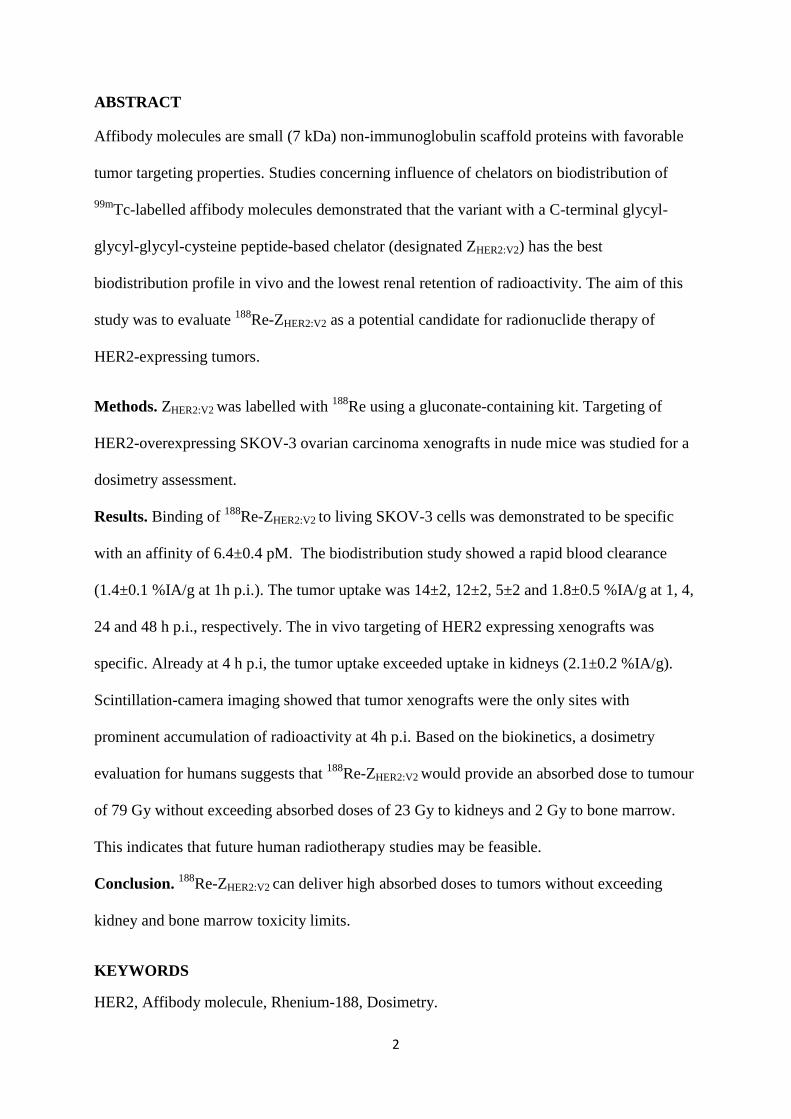
The cellular retention profile (Figure 1) showed a rapid drop of the activity during first hour
followed by a slow decline with a biological half-life of 19.6 h. The total cellular retention of

10
the radioactivity was 34.6±1.3% after 24 h of incubation at 37°C. The amount of internalized
radioactivity of 188Re-ZHER2:V2 was relatively low, less than 4% of total radioactivity after 24
h.
In Vivo Studies
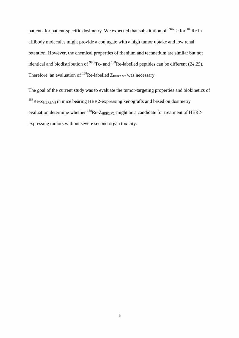
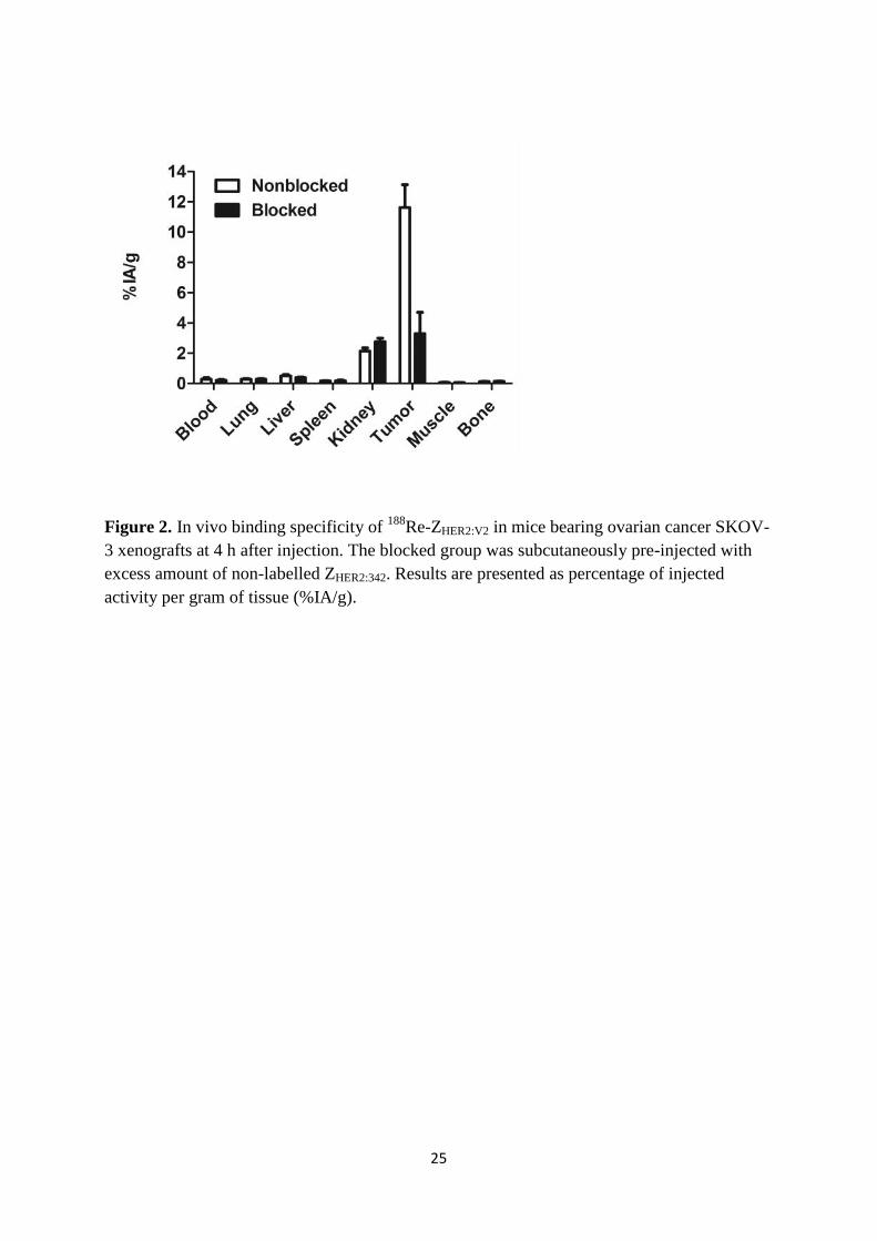
The results of in vivo specificity test (Figure 2) showed that pre-saturation of HER2-receptors
in xenografts with a parental ZHER2:342 decreased tumor uptake of 188Re-ZHER2:V2 4.5-fold (p
<0.005). This demonstrates a saturable character of the tumor accumulation and suggests its
HER2-specificity. Small but statistically significant decrease of uptake in liver and increase in
kidneys was also detected in this experiment.
Biodistribution of 188Re-ZHER2:V2 in tumor bearing mice (Table 1) was characterized by rapid
clearance of radioactivity from all organs and tissues. Low radioactivity in the content of the
gastrointestinal tract (less than 2% of injected activity, data not shown) suggested that
hepatobiliary pathway played a minor role in the excretion of 188Re-ZHER2:V2 or its
radiometabolites. Low radioactivity uptake in organs accumulating free perrhenate, i.e.
salivary gland, thyroid and stomach, suggests high stability of 188Re-ZHER2:V2 to re-oxidation
in vivo. Kidneys demonstrated high initial uptake, which decreased rapidly. At 1 h p.i., the
renal uptake was approximately equal to tumor uptake, and 4 h p.i., the tumor uptake was
five-fold higher.
The tumor uptake was prominent as early as 20 minutes p.i. and was higher than the uptake in
any normal organ except from kidneys. The tumor uptake had a maximum at 1 h p.i. (14±2
%IA/g) followed by decrease with a half-life of 15 h.
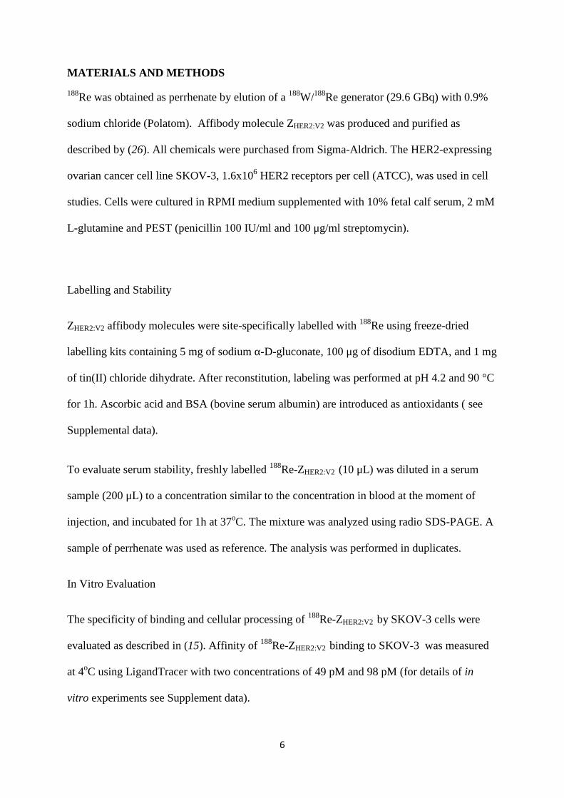
Scintillation-camera imaging, performed at 1 and 4 h after injection (Figure 3), confirmed the
results of the biodistribution experiments. The tumor xenografts were the only sites with
11
prominent accumulation of radioactivity. Kidneys were visualized at 1 h after injection, but at
4 h p.i. only the HER2-expressing tumor xenografts were clearly visible.
Dosimetry
The evaluation of absorbed doses in mice for 188Re-ZHER2:V2 is presented in Table 2. The self-
doses to the majority of organs and tissues were much smaller than absorbed doses to tumors.
However, the size of a mouse body is comparable with the range of the beta-particles emitted
by 188Re (about 10 mm). For this reason, the total absorbed doses became much higher due to
cross-irradiation. For example, the total absorbed dose to bone marrow was approximately
22% of the total absorbed tumor dose. This means that it was impossible to deliver a
therapeutically meaningful absorbed dose to tumors (above 50 Gy) without lethal absorbed
dose to the red marrow (around 8 Gy for mice). Performing therapy experiments with188Re-
ZHER2:V2 therapy in mice was thus considered unethical under these circumstances and due to
dimensional differences between mice and humans and range of beta particles, experiments in
small animal murine models will generate irrelevant results.
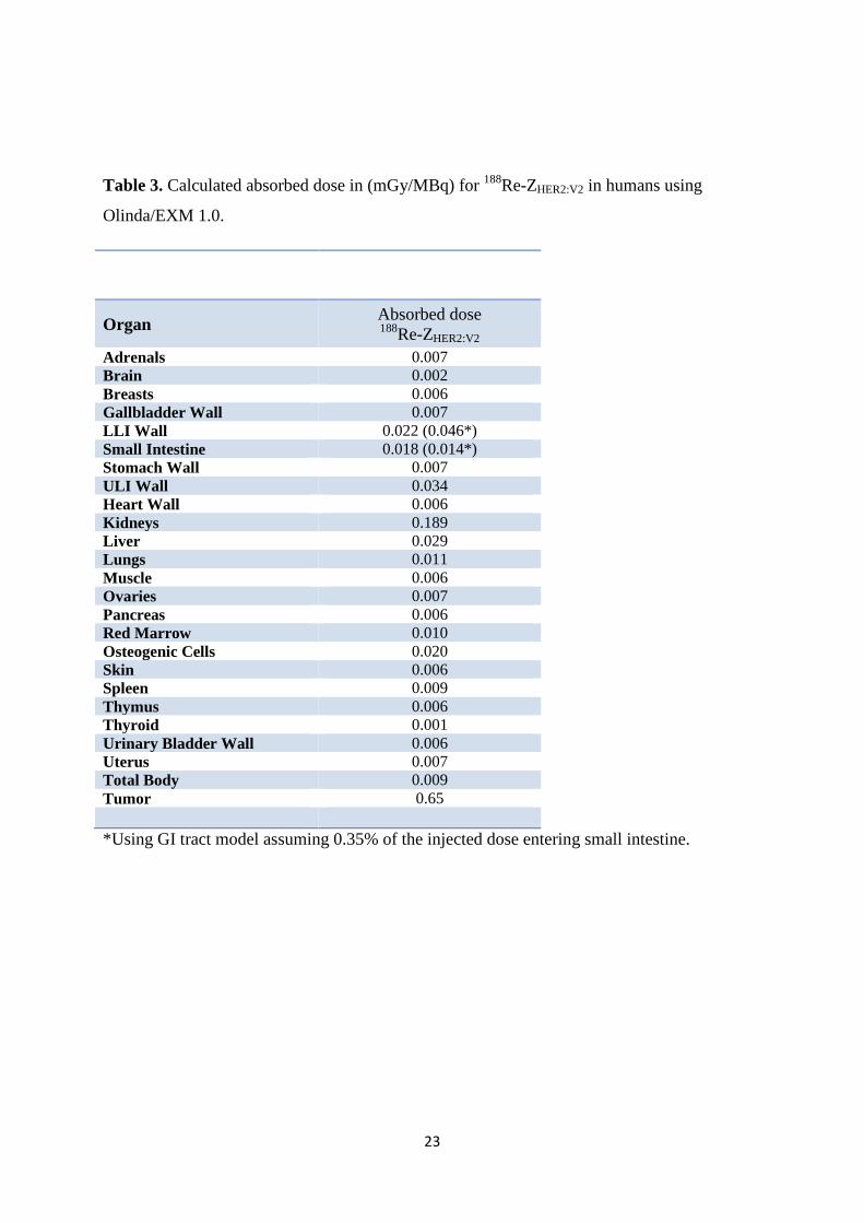
The results of estimated absorbed dose calculations of 188Re-ZHER2:V2 in humans using
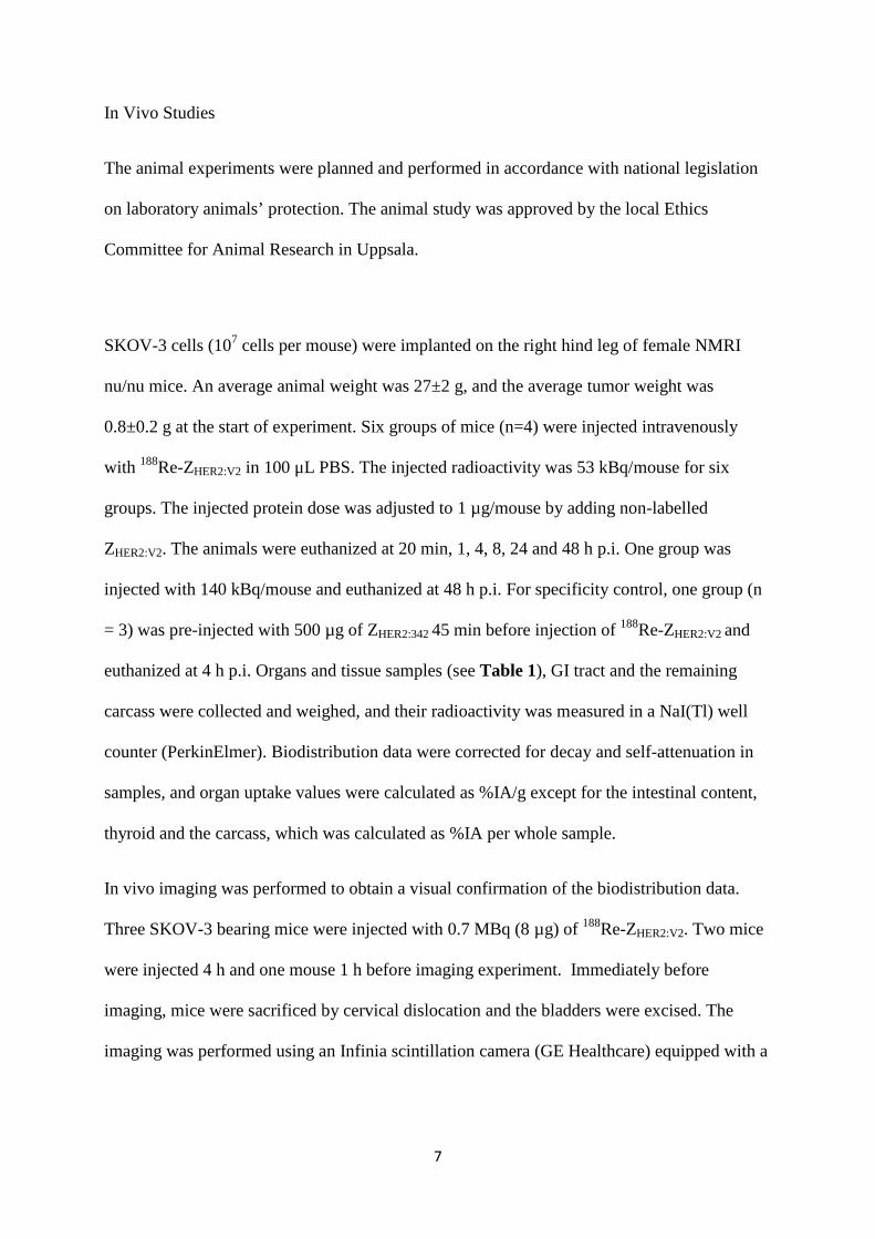
Olinda/EXM 1.0, are presented in Table 3. In brief, tumor SUV was approximately constant
after 1 h with a mean value of 3.3. Assuming local deposition of all beta energy, and no self-
or cross-dose from gamma photons, this would result in an absorbed dose to tumor of 0.65
mGy/MBq. As shown in Figure 4, absorbed doses to tumors with the weight of less than10 g
would be slightly lower due to incomplete absorption of beta particles, whilst slightly higher
absorbed doses are expected in larger tumors due to self-dose from gamma radiation.
Absorbed dose to tumor exceeded absorbed dose to kidney and bone marrow, 3.4-fold and 79-
fold, respectively.
12
DISCUSSION
Radioimmunotherapy of radiosensitive hematological malignancies has proven to be
successful, but treatment of more radioresistant solid tumors has so far been inefficient. The
main reason is a failure to reach tumor absorbed doses above 50 Gy which are typically
required to achieve response at external beam therapy (7).
Clinical experience with targeted radionuclide therapy using short peptides, particularly
somatostatin analogues, demonstrated that the use of small targeting agents can solve
problems of both poor tumor penetration and exposure of bone marrow (30). Unfortunately,
all small HER2-targeting radiolabelled agents, including antibody fragments, diabodies,
DARPins and affibody molecules undergo considerable renal reabsorption (31). In the case of
residualizing radiometal labels, the renal uptake would be higher than tumour uptake for all
these targeting proteins.
Our approach is based on the knowledge concerning cellular processing of affibody
molecules. The main hypothesis was that the use of a non-residualizing label would cause
rapid clearance from kidneys, where affibody molecules are rapidly internalized, degraded,
and their radiocatabolites are excreted from cells. At the same time, processing of affibody
molecules bound to HER2-expressing cancer cell is slow, and cellular retention is good even
in the case of non-residualizing labels. Our previous structure-property relationship studies
have demonstrated that the use of a -GGGC peptide-based chelator yields non-residualizing
99mTc (22) labels with low accumulation in kidneys. The present study has shown that
ZHER2:V2 can be efficiently labelled with 188Re. The high-fidelity refolding of affibody
molecules in physiological conditions (32) permitted their direct rhenium labelling at high
temperature with high yields. In fact, 188Re-ZHER2:V2 preserved high affinity binding (6.4±0.4
pM) to HER2 expressing cells after labelling at 90oC and pH 4.2. Although an in vitro
processing study (Figure 1) showed low intracellular retention of radiocatabolites, the overall

13
retention of radioactivity was reasonable with a biological half-life of 19.5 h. 188Re-ZHER2:V2
has shown specific uptake in HER2-expressing xenografts in vivo (Figure 2). The
biodistribution study confirmed that unbound radioactivity was cleared very quickly from
blood, and release of retained radioactivity from kidneys was much more rapid than from
tumors (Table 1). Area under curve (AUC) for tumor exceeded AUC for blood 47-fold, bone
70-fold, and kidneys 2.8-fold. This suggested that the goal of delivering an efficient absorbed
dose to tumors while sparing critical organs might be achievable.
We performed dosimetry calculations for 188Re-ZHER2:V2 in mice using the anatomically
realistic murine Moby phantom (27,28). The self-doses were in agreement with AUC data,
however that total absorbed dose to bone marrow was much higher than the self-dose. It must
be noted, that a high total absorbed dose to bone marrow is a phenomenon associated with the
use of mice as an animal model. The maximum range of beta-particles from 188Re is 10.4 mm
(7), which is comparable with the dimensions of a mouse. For this reason, the cross-dose to
bone marrow is much higher in mice than in humans. Delivery of therapeutically meaningful
absorbed doses to tumors in mice would be associated with lethal absorbed doses to bone
marrow.
Human dosimetry was much different due to the larger dimensions of the human body.
Upscaling of murine biodistribution data to human suggests that much lower absorbed doses
will be delivered to radiosensitive organs in relation to tumor absorbed doses. A maximum
absorbed dose of 2 Gy for bone marrow is generally accepted when planning radionuclide
therapy; as such absorbed dose is associated with low risk of developing leukaemia (33) and
low risk for acute bone marrow toxicity. An absorbed dose of 23 Gy is a commonly used
absorbed dose limit for kidneys in peptide receptor radionuclide therapy (34). In the case of
188Re-ZHER2:V2, an absorbed dose of 23 Gy to the kidneys would correspond to tumor absorbed
dose of 79 Gy, whilst an absorbed dose of 2 Gy to bone marrow would correspond to tumor

14
absorbed dose of 130 Gy. This would permit delivering an absorbed dose well above 50 Gy
without exceeding commonly accepted absorbed dose limits to critical organs. It has to be
mentioned that upscaling from mice to humans is associated with apparent uncertainties, and
a clinical imaging study would be required to obtain more reliable assessment of human
dosimetry.
In addition to dosimetry estimations, there are other factors to consider when planning future
therapy applications. 188Re is produced via a generator in no-carrier-added form, which
provides high specific activity. The short half-life of 188Re enables irradiation of tumors with a
high dose rate. Close matching of physical and biological half-lives and generator-mediated
production from the long-lived mother nuclide 188W (T½=70 d) permits fractionated therapy.
Both the absorbed dose rate during tumor irradiation and treatment fractionation are
considered as important radiobiological factors for increasing efficacy of targeted
radionuclide therapy (7).
One important lesson from this study concerns selection of labelling strategy. It is a common
knowledge that selection of radionuclide and chelator influences stability of nuclide
attachment to a targeting proteins, and intracellular retention of radioactivity after
internalization. Our earlier studies have demonstrated that modification of physicochemical
properties of affibody molecules by incorporation of radionuclide-chelator complex modifies
also off-target interactions, changing blood clearance rate, predominant excretion pathway
and biodistribution (11). This can be used for optimizing of targeting properties. We have
earlier shown that the 186Re-maGSG-ZHER2:342 affibody molecule having an N-terminal
mercaptoacetyl-glycyl-seryl-glycyl chelator provided, similarly to 188Re-ZHER2:V2, better
retention of radioactivity in tumors than in kidneys. However, appreciable hepatobiliary
excretion of 186Re-maGSG-ZHER2:342 (20% of injected radioactivity was measured in the
intestinal content at 4 h p.i.) caused a risk of high absorbed dose to intestines. Through re-

15
engineering of affibody molecules by modifying of the chelator and its placement to C-
terminus, as well as increasing hydrophilicity of N-terminus by amino acid substitution, we
suppressed hepatobiliary excretion of 188Re-ZHER2:V2 while keeping good tumor retention, low
renal uptake and rapid clearance of unbound radioactivity. Thus optimal molecular design,
including labelling strategy, may appreciably improve properties of scaffold-protein-based
conjugates for radionuclide therapy.
Conclusion:
The affibody molecule, ZHER2:V2 can be labelled with 188Re with a high yield with preserved
picomolar affinity to HER2. 188Re-ZHER2:V2 provides efficient targeting of HER2-expressing
xenografts, rapid blood clearance and low uptake in kidneys and bones. Dosimetry
calculations in man suggest that 188Re-ZHER2:V2 may provide absorbed dose to tumors of more
than 70 Gy while keeping absorbed dose to kidneys below 23 Gy and absorbed dose to bone
marrow below 2 Gy. Thus, 188Re-ZHER2:V2 is a promising targeting agent for radionuclide
therapy against HER2-expressing tumors.

16
ACKNOWLEDGMENTS
This research was supported in part by grants from the Swedish Cancer Society
(Cancerfonden), Swedish Research Council (Vetenskapsrådet) and Governmental Funding of
Clinical Research within the National Health Service (ALF).
17
REFERENCES
1. Slamon DJ, Leyland-Jones B, Shak S, et al. Use of chemotherapy plus a monoclonal
antibody against HER2 for metastatic breast cancer that overexpresses HER2. N Engl
J Med. 2001;344:783-792.
2. Bang YJ, Van Cutsem E, Feyereislova A, et al. Trastuzumab in combination with
chemotherapy versus chemotherapy alone for treatment of HER2-positive advanced
gastric or gastro-oesophageal junction cancer (ToGA): a phase 3, open-label,
randomised controlled trial. Lancet. 2010;376:687-697.
3. Wu AM, Senter PD. Arming antibodies: prospects and challenges for
immunoconjugates. Nat Biotechnol. 2005;23:1137-1146.
4. Diéras V, Bachelot T. The success story of trastuzumab emtansine, a targeted therapy
in HER2-positive breast cancer. Target Oncol. Jul 14, 2013[Epub ahead of print].
5. Sharkey RM, Goldenberg DM. Perspectives on cancer therapy with radiolabeled
monoclonal antibodies. J Nucl Med. 2005;46(Suppl 1):115S–127S.
6. Kenanova V, Wu AM. Tailoring antibodies for radionuclide delivery. Expert Opin
Drug Deliv. 2006;3:53-70.
7. Pouget JP, Navarro-Teulon I, Bardiès M, et al. Clinical radioimmunotherapy--the role
of radiobiology. Nat Rev Clin Oncol. 2011;8:720-734
8. Miao Z, Levi J, Cheng Z. Protein scaffold-based molecular probes for cancer
molecular imaging. Amino Acids. 2011;41:1037-1047.
9. Nygren PA. Alternative binding proteins: affibody binding proteins developed from a
small three-helix bundle scaffold. FEBS J. 2008;275:2668-2676.
10. Orlova A, Magnusson M, Eriksson TL, et al. Tumor imaging using a picomolar
affinity HER2 binding affibody molecule. Cancer Res. 2006;66:4339-4348.

18
11. Feldwisch J, Tolmachev V. Engineering of affibody molecules for therapy and
diagnostics. Methods Mol Biol. 2012;899:103-126.
12. Baum RP, Prasad V, Müller D, et al. Molecular imaging of HER2-expressing
malignant tumors in breast cancer patients using synthetic 111In- or 68Ga-labeled
affibody molecules. J Nucl Med. 2010;51:892-897.
13. Sörensen J, Sandberg D, Sandström M, et al. First-in-human molecular imaging of
HER2 expression in breast cancer metastases using the 111In-ABY-025 Affibody
molecule. J Nucl Med. 2014;55:730-735.
14. Altai M, Varasteh Z, Andersson K, Eek A, Boerman O, Orlova A. In vivo and in vitro
studies on renal uptake of radiolabeled affibody molecules for imaging of HER2
expression in tumors. Cancer Biother Radiopharm. 2013;28:187-195.
15. Wållberg H, Orlova A. Slow internalization of anti-HER2 synthetic affibody monomer
111In-DOTA-ZHER2:342-pep2: implications for development of labeled tracers.
Cancer Biother Radiopharm. 2008;23:435-442.
16. Tolmachev V, Mume E, Sjöberg S, Frejd FY, Orlova A. Influence of valency and
labelling chemistry on in vivo targeting using radioiodinated HER2-binding Affibody
molecules. Eur J Nucl Med Mol Imaging. 2009;36:692-701.
17. Engfeldt T, Tran T, Orlova A, Widström C, Karlström AE, Tolmachev V. 99mTc-
chelator engineering to improve tumour targeting properties of a HER2-specific
Affibody molecule. Eur J Nucl Med Mol Imaging. 2007;34:1843–1853
18. Tran T, Engfeldt T, Orlova A, et al. (99m)Tc-maEEE-Z(HER2:342), an Affibody
molecule-based tracer for the detection of HER2 expression in malignant tumors.
Bioconjug Chem. 2007;18:1956-1964.

19
19. Tran TA, Ekblad T, Orlova A, et al. Effects of lysine-containing mercaptoacetyl-based
chelators on the biodistribution of 99mTc-labeled anti-HER2 Affibody molecules.
Bioconjug Chem. 2008;19:2568-2576.
20. Ekblad T, Tran T, Orlova A, et al. Development and preclinical characterisation of
99mTc-labelled Affibody molecules with reduced renal uptake. Eur J Nucl Med Mol
Imaging. 2008;35:2245-2255.
21. Ahlgren S, Wållberg H, Tran TA, et al. Targeting of HER2-expressing tumors with a
site-specifically 99mTc-labeled recombinant affibody molecule, ZHER2:2395, with
C-terminally engineered cysteine. J Nucl Med. 2009;50:781-789.
22. Wållberg H, Orlova A, Altai M, et al. Molecular design and optimization of 99mTc-
labeled recombinant affibody molecules improves their biodistribution and imaging
properties. J Nucl Med. 2011;52:461-469.
23. Altai M, Wållberg H, Orlova A, et al. Order of amino acids in C-terminal cysteine-
containing peptide-based chelators influences cellular processing and biodistribution
of 99mTc-labeled recombinant Affibody molecules. Amino Acids. 2012;42:1975-
1985.
24. Cyr JE, Pearson DA, Wilson DM, et al. Somatostatin receptor-binding peptides
suitable for tumor radiotherapy with Re-188 or Re-186. Chemistry and initial
biological studies. J Med Chem. 2007;50:1354–1364.
25. Orlova A, Tran TA, Ekblad T, Karlström AE, Tolmachev V. (186)Re-maGSG-Z
(HER2:342), a potential Affibody conjugate for systemic therapy of HER2-expressing
tumours. Eur J Nucl Med Mol Imaging. 2010;37:260-269.
26. Wållberg H, Löfdal PÅ , Tschapalda K. Affinity recovery of eight HER2-binding
affibody variants using an anti-idiotypic affibody molecule as capture ligand. Protein
Expr Purif. 2011;76:127–135.

20
27. Larsson E, Strand SE, Ljungberg M, Jönsson BA. Mouse S-factors based on Monte
Carlo simulations in the anatomical realistic Moby phantom for internal dosimetry.
Cancer Biother Radiopharm. 2007;22:438-442.
28. Larsson E, Ljungberg M, Strand SE, Jönsson BA. Monte Carlo calculations of
absorbed doses in tumours using a modified MOBY mouse phantom for pre-clinical
dosimetry studies. Acta Oncol. 2011;50:973-980.
29. Stabin MG. Fundamentals of Nuclear Medicine Dosimetry. New York, NY: Springer;
2008:83-86.
30. van Essen M, Krenning EP, Kam BL, de Jong M, Valkema R, Kwekkeboom DJ.
Peptide-receptor radionuclide therapy for endocrine tumors. Nat Rev Endocrinol.
2009;5:382-393.
31. Tolmachev V. Imaging of HER-2 overexpression in tumors for guiding therapy. Curr
Pharm Des. 2008;14:2999-3019
32. Arora P, OasTG, Myers JK. Fast andfaster: a designed variant of the B domain of
protein A folds in 3 microsec. Protein Sci. 2004;13:847–853.
33. Forrer F, Krenning EP, Kooij PP, et al. Bone marrow dosimetry in peptide receptor
radionuclide therapy with [177Lu-DOTA(0),Tyr(3)]octreotate. Eur J Nucl Med Mol
Imaging. 2009;36:1138-1146.
34. Kwekkeboom DJ, de Herder WW, van Eijck CH, et al. Peptide receptor radionuclide
therapy in patients with gastroenteropancreatic neuroendocrine tumors. Semin Nucl
Med. 2010;40:78-88.

21
Table 1. Biodistribution of 188Re-ZHER2:V2 in NMRI nu/nu mice bearing HER2-expressingSKOV-3 xenografts.
Uptake *Organ 20 min. 1h 4h 8h 24h 48h
Blood 3.0±0.3 1.4±0.1 0.3±0.1 0.05±0.02 0.02±0.01 0.006±0.003Heart 1.4±0.3 0.6±0.1 0.11±0.02 0.05±0.01 0.02±0.01 NMLung 3.4±0.2 1.7±0.2 0.28±0.05 0.08±0.03 0.03±0.01 NMSalivarygland
1.6±0.5 1.1±0.3 0.2±0.1 0.06±0.03 0.02±0.01 NM
Thyroid* 0.07±0.02
0.04±0.02
0.01±0.001 0.01±0.004 0.002±0.001
NM
Liver 3.4±0.3 2.2±0.2 0.51±0.09 0.26±0.06 0.12±0.03 0.06±0.016Spleen 1.5±0.2 0.87±0.0
60.16±0.02 0.08±0.01 0.03±0.01 NM
Pancreas 0.8±0.2 0.4±0.1 0.08±0.03 0.030±0.003
0.01±0.01 NM
Stomach 2.0±0.08 1.3±0.23 0.3±0.1 0.08±0.01 0.08±0.07 NMSmallintestine
1.4±0.1 0.9±0.2 0.16±0.08 0.08±0.07 0.04±0.03 NM
Largeintestine
1.9±0.08 1.2±0.6 0.19±0.05 0.3±0.3 0.04±0.01 0.02±0.004
Kidney 47±2 18±3 2.1±0.2 1.0±0.2 0.51±0.08 0.30±0.12Tumor 8.7±0.9 14±2 12±2 8.0±0.6 5±2 1.8±0.5Skin 2.4±0.4 1.4±0.1 0.6±0.5 0.10±0.02 0.04±0.02 0.02±0.005
Muscle 0.6±0.1 0.28±0.03
0.06±0.03 0.02±0.01 0.02±0.01 NM
Bone 1.0±0.1 0.6±0.1 0.12±0.01 0.05±0.02 0.04±0.02 NMBrain 0.08±0.0
20.05±0.01
0.020±0.003
0.01±0.001 0.01±0.002 NM
Carcass** 23±2 14±4 2±1 1.1±0.9 0.35±0.09 0.19±0.04
*The uptake is expressed as %IA/g and presented as an average value from 4animals±standard deviation. Data are corrected for decay and self-attenuation in samples.*data for thyroid and carcass are presented as % of injected activity per whole sample.NM = not measurable, below detection limit.

22
Table 2. Calculated absorbed dose in (Gy/MBq) for 188Re-ZHER2:V2 in mice using murine
Moby phantom.
Organ Self-dose Total absorbed dose
Blood 0.008 0.099
Heart 0.003 0.094Lung 0.019 0.14Salivary gland 0.0062 0.11Thyroid 0.00022 0.13Liver 0.027 0.079Spleen 0.0068 0.072Pancreas 0.0024 0.075Stomach 0.014 0.061Small intestine 0.0058 0.076Large intestine 0.0051 0.11Kidney 0.17 0.23Skin 0.0038 0.081Bone 0.0019 0.10Brain 0.00073 0.026
Carcass 0.16 0.16Tumor 1* 0.40 0.43Tumor 2* 0.40 0.43Tumor 3* 0.40 0.44Bone Marrow 0.0029 0.096
Tumor 1, 2 and 3 stands for tumors located in the right hind leg, left hind leg and the left flank
respectively.
23
Table 3. Calculated absorbed dose in (mGy/MBq) for 188Re-ZHER2:V2 in humans using
Olinda/EXM 1.0.
OrganAbsorbed dose188Re-ZHER2:V2
Adrenals 0.007Brain 0.002Breasts 0.006Gallbladder Wall 0.007LLI Wall 0.022 (0.046*)Small Intestine 0.018 (0.014*)Stomach Wall 0.007ULI Wall 0.034Heart Wall 0.006Kidneys 0.189Liver 0.029Lungs 0.011Muscle 0.006Ovaries 0.007Pancreas 0.006Red Marrow 0.010Osteogenic Cells 0.020Skin 0.006Spleen 0.009Thymus 0.006Thyroid 0.001Urinary Bladder Wall 0.006Uterus 0.007Total Body 0.009Tumor 0.65
*Using GI tract model assuming 0.35% of the injected dose entering small intestine.

24
FIGURES
Figure 1. Cell-associated radioactivity as function of time after interrupted incubation ofSKOV-3 cells with 188Re-ZHER2:V2. Data are presented as average value from 3 cell dishes ±SD. Error bars might not be seen because they are smaller than point symbols.
25
Figure 2. In vivo binding specificity of 188Re-ZHER2:V2 in mice bearing ovarian cancer SKOV-3 xenografts at 4 h after injection. The blocked group was subcutaneously pre-injected withexcess amount of non-labelled ZHER2:342. Results are presented as percentage of injectedactivity per gram of tissue (%IA/g).

26
Figure 3. Imaging of HER2 expression in SKOV-3 ovarian cancer xenografts (high HER2expression) in NMRI nu/nu mice using 188Re-ZHER2:V2. Planar scintillation-camera imageswere acquired at 1 h (A) and 4 h (B) after injection. Arrows point to tumors (T) and kidneys(K).
T TK
KK
A B

27
Figure 4. Tumor absorbed dose versus mass for 188Re-ZHER2:V2.

28
SUPPLEMENTAL DATA
Labelling and stability
188Re was obtained as perrhenate by elution of a 188W/188Re generator (29.6 GBq) with 0.9%
sodium chloride (Polatom). Elution efficiency was 90%, when 4 ml of eluent was used.
For animal studies, the content of one freeze-dried kit was dissolved in100 μL 1.25 M sodium
acetate, pH 4.2, and added to 100 μg of freeze-dried ZHER2:V2. To the reaction mixture, 14-100
μL of 188Re-containing generator eluate was added under argon gas. An equivalent of 220 μg
ascorbic acid (2 mg/mL in 1.25 M sodium acetate buffer, pH 4.2) was added to the reaction
vial. The mixture was incubated at 90°C for 60 min and then cooled at room temperature for 5
min. Thereafter, the total amount of ascorbic acid in the reaction vial is adjusted to 1 mg with
a 5 mg/mL ascorbic acid solution in PBS (phosphate buffered saline) containing 2% BSA
(bovine serum albumin). 188Re-ZHER2:V2 was purified using disposable NAP-5 columns (GE
Healthcare) pre-equilibrated and eluted with PBS containing 2% BSA. The final solution was
diluted with an extra 100 μL PBS containing 2% BSA and 500 µg ascorbic acid to a final
volume of 1 mL.
In up-scaling experiments, the content of a freeze-dried labelling kit vial (4 mg tin(II) chloride
dihydrate, 400 µg disodium EDTA and 20 mg of sodium α-D-gluconate) was reconstituted in
400 µL 1.25 M sodium acetate, pH 4.2, containing 5mg/ml acid, and votrexed. The content of
the vial was transferred to another vial containing 400 µg freeze-dried ZHER2:V2 and vortexed.
To this mixture, a 1mL (~7 GBq) of 188Re-containing generator eluate was added, and the
labeling mixture was votrexed carefully and incubated at 95oC for 60 min. Further processing
was performed as described above.
For measurement of the labelling yield and radiochemical purity, samples of 188Re-ZHER2:V2
were analyzed using ITLC SG strips eluted with PBS. For measurement of reduced

29
hydrolyzed rhenium colloid levels, a pyridine:acetic acid:water (5:3:1.5) mobile phase was
used. The ITLC analysis was cross-calibrated by SDS-PAGE (Novex 4-12% Bis-Tris Gel,
MES buffer, 200 V constant).
To estimate the shelf-life, the purity of 188Re-ZHER2:V2 was measured at 1, 2 and 4h after
purification using ITLC in duplicates.
In vitro evaluation
In vitro specificity test was performed using SKOV-3 cells. Briefly, a solution of 188Re-
ZHER2:V2 (0.015 ng protein per dish, 2 nM) was added to six Petri dishes (ca. 106 cells in
each). For blocking, an excess of non-labeled recombinant ZHER2:342 (7.4 µg) was added 10
min. before 188Re-ZHER2:V2 to saturate the receptors. The cells were incubated during one hour
in a humidified incubator at 37°C. Thereafter, the media was collected, the cells were
detached by trypsin-EDTA solution, and the radioactivity in cells and media was measured to
calculate a percentage of cell-bound radioactivity.
For cellular processing SKOV-3 cells (1 × 10⁶ cells/dish) were incubated with 2 nM solution
of labeled affibody at 4°C. After 1 h incubation, the medium with the labeled compound was
removed and cells were washed three times with ice-cold serum-free medium. One mL of
complete media was added to each dish and cells were further incubated at 37°C in an
atmosphere containing 5% CO2. At designated time points (0 h, 1 h, 4 h, 8 h and 24 h), a
group of three dishes was removed from the incubator, the media was collected and cells were
washed three times with ice-cold serum-free medium. Thereafter, cells were treated with 0.5
mL 0.2 M glycine buffer, pH 2, containing 4 M urea, for 5 min on ice. The acidic solution
was collected and cells were additionally washed with 0.5 mL glycine buffer. The acidic
fractions were pooled. The cells were then incubated with 0.5 mL 1 M NaOH at 37°C for 30
min. The cell debris was collected, and the dishes were additionally washed with 0.5 mL of
30
NaOH solution. The alkaline solutions were pooled. The radioactivity in the acidic solution
was considered as membrane bound, and in the alkaline fractions as internalized.
Affinity determination using LigandTracer
SKOV-3 cells were seeded on a local area of a cell culture dish (NunclonTM, Size 100620,
NUNC A/S, Roskilde, Denmark), as described previously (1). The binding of 188Re-labeled
anti-HER2 affibody molecules to living cells was monitored in real-time at 4oC using
LigandTracer Yellow, using established methods described in Björkelund et al. In brief, the
LigandTracer records the real-time kinetics of binding and dissociation of radiolabeled tracer
in living cells. By using the TraceDrawer software, which allows the calculation of both
association and dissociation rate, it becomes possible to determine the affinity of radiolabeled
conjugate (1). In order to cover the concentration span needed for proper affinity estimation,
two increasing concentrations of 49 pM and 98 pM (selected based on previous KD values
obtained using Biacore) of each variant were added in each affinity assay Supp. Fig 2.
References
(1) Björkelund, H.; Gedda, L.; Barta, P.; Malmqvist, M.; Andersson, K. Gefitinibinduces epidermal growth factor receptor dimers which alters the interactioncharacteristics with ¹²⁵I-EGF. PLoS One. 2011, 6, e24739

31
Suppl. Figure 1. SDS–PAGE analysis of the stability of 188Re-ZHER2:V2 in murine serum. 1.
incubated in murine serum at 37oC for 1 h; 2. 188ReO4- used as a marker for low-molecular-
weight compounds. Signal, measured as digital light units, is in proportion to radioactivity in
given point of lane in SDS-PAGE gel. DLU=digital light units.
1 2
3

32
Suppl. Figure 2. LigandTracer sensorgram of interaction of 188Re-ZHER2:V2 with HER2-
expressing SKOV-3 cells. Concentrations of 188Re-ZHER2:V2 were 49 pM and 98 pM.
Copyright © 2022 FDOKUMEN