Gene amplification and protein overexpression of EGFR and ERBB2 in sinonasal squamous cell carcinoma
Upload
independentCategory
view
5download
0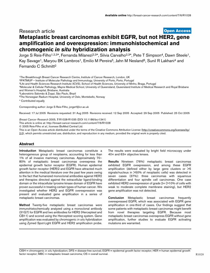
Available online http://breast-cancer-research.com/content/7/6/R1028
Open AccessVol 7 No 6Research articleMetaplastic breast carcinomas exhibit EGFR, but not HER2, gene amplification and overexpression: immunohistochemical and chromogenic in situ hybridization analysisJorge S Reis-Filho1,2,3*, Fernanda Milanezi2,3*, Silvia Carvalho2,3*, Pete T Simpson4, Dawn Steele1, Kay Savage1, Maryou BK Lambros1, Emilio M Pereira5, Jahn M Nesland6, Sunil R Lakhani4 and Fernando C Schmitt2
1The Breakthrough Breast Cancer Research Centre, Institute of Cancer Research, London, UK2IPATIMUP – Institute of Molecular Pathology and Immunology, University of Porto, Porto, Portugal3Life and Health Sciences Research Institute (ICVS), School of Health Sciences, University of Minho, Braga, Portugal4Molecular & Cellular Pathology, Mayne Medical School, University of Queensland, Queensland Institute of Medical Research and Royal Brisbane and Women's Hospital, Brisbane, Australia5Laboratório Salomão & Zoppi, São Paulo, Brazil6The Norwegian Radium Hospital, University of Oslo, Montebello, Norway* Contributed equally
Corresponding author: Jorge S Reis-Filho, [email protected]
Received: 17 Jul 2005 Revisions requested: 31 Aug 2005 Revisions received: 12 Sep 2005 Accepted: 29 Sep 2005 Published: 25 Oct 2005
Breast Cancer Research 2005, 7:R1028-R1035 (DOI 10.1186/bcr1341)This article is online at: http://breast-cancer-research.com/content/7/6/R1028© 2005 Reis-Filho et al.; licensee BioMed Central Ltd. This is an Open Access article distributed under the terms of the Creative Commons Attribution License (http://creativecommons.org/licenses/by/2.0), which permits unrestricted use, distribution, and reproduction in any medium, provided the original work is properly cited.
Abstract
Introduction Metaplastic breast carcinomas constitute aheterogeneous group of neoplasms, accounting for less than1% of all invasive mammary carcinomas. Approximately 70–80% of metaplastic breast carcinomas overexpress theepidermal growth factor receptor (EGFR). Human epidermalgrowth factor receptor (HER)2 and EGFR have attracted muchattention in the medical literature over the past few years owingto the fact that humanized monoclonal antibodies against HER2and therapies directed against the extracellular ligand-bindingdomain or the intracellular tyrosine kinase domain of EGFR haveproven successful in treating certain types of human cancer. Weinvestigated whether HER2 and EGFR overexpression waspresent and evaluated gene amplification in a series ofmetaplastic breast carcinomas.
Method Twenty-five metaplastic breast carcinomas wereimmunohistochemically analyzed using a monoclonal antibody(31G7) for EGFR and two antibodies for HER2 (Herceptest andCB11) and scored using the Herceptest scoring system. Geneamplification was evaluated by chromogenic in situ hybridizationusing Zymed Spot-Light EGFR and HER2 amplification probe.
The results were evaluated by bright field microscopy under40× and 63× objective lenses.
Results Nineteen (76%) metaplastic breast carcinomasexhibited EGFR ovexpression, and among these EGFRamplification (defined either by large gene clusters or >5signals/nucleus in >50% of neoplastic cells) was detected inseven cases (37%): three carcinomas with squamousdifferentiation and four spindle cell carcinomas. One caseexhibited HER2 overexpression of grade 2+ (>10% of cells withweak to moderate complete membrane staining), but HER2gene amplification was not detected.
Conclusion Metaplastic breast carcinomas frequentlyoverexpressed EGFR, which was associated with EGFR geneamplification in one-third of cases. Our findings suggest thatsome patients with metaplastic breast carcinomas might benefitfrom novel therapies targeting EGFR. Because mostmetaplastic breast carcinomas overexpress EGFR without geneamplification, further studies to evaluate EGFR activatingmutations are warranted.
R1028CISH = chromogenic in situ hybridization; DFS = disease-free survival; EGFR = epidermal growth factor receptor; HER = human epidermal growth factor receptor; MBC = metaplastic breast carcinoma; OS = overall survival.
Breast Cancer Research Vol 7 No 6 Reis-Filho et al.
R1029
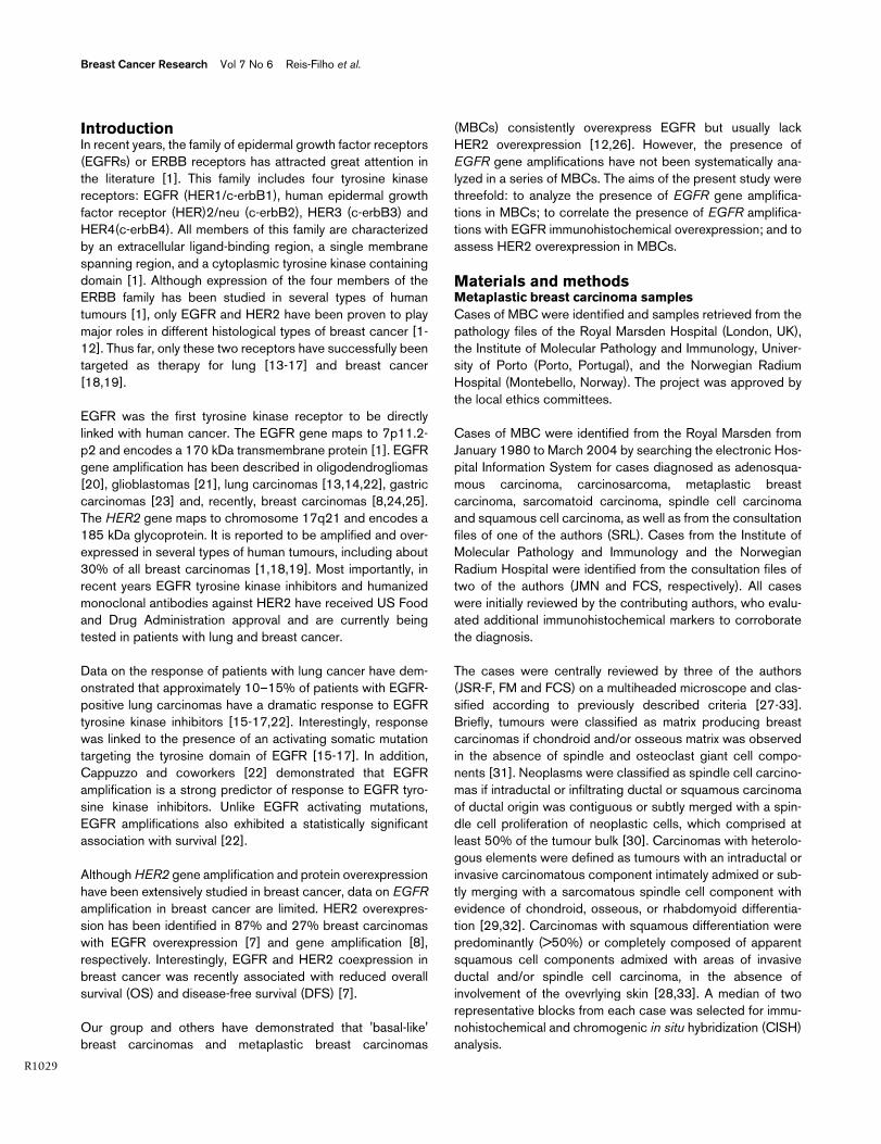
IntroductionIn recent years, the family of epidermal growth factor receptors(EGFRs) or ERBB receptors has attracted great attention inthe literature [1]. This family includes four tyrosine kinasereceptors: EGFR (HER1/c-erbB1), human epidermal growthfactor receptor (HER)2/neu (c-erbB2), HER3 (c-erbB3) andHER4(c-erbB4). All members of this family are characterizedby an extracellular ligand-binding region, a single membranespanning region, and a cytoplasmic tyrosine kinase containingdomain [1]. Although expression of the four members of theERBB family has been studied in several types of humantumours [1], only EGFR and HER2 have been proven to playmajor roles in different histological types of breast cancer [1-12]. Thus far, only these two receptors have successfully beentargeted as therapy for lung [13-17] and breast cancer[18,19].
EGFR was the first tyrosine kinase receptor to be directlylinked with human cancer. The EGFR gene maps to 7p11.2-p2 and encodes a 170 kDa transmembrane protein [1]. EGFRgene amplification has been described in oligodendrogliomas[20], glioblastomas [21], lung carcinomas [13,14,22], gastriccarcinomas [23] and, recently, breast carcinomas [8,24,25].The HER2 gene maps to chromosome 17q21 and encodes a185 kDa glycoprotein. It is reported to be amplified and over-expressed in several types of human tumours, including about30% of all breast carcinomas [1,18,19]. Most importantly, inrecent years EGFR tyrosine kinase inhibitors and humanizedmonoclonal antibodies against HER2 have received US Foodand Drug Administration approval and are currently beingtested in patients with lung and breast cancer.
Data on the response of patients with lung cancer have dem-onstrated that approximately 10–15% of patients with EGFR-positive lung carcinomas have a dramatic response to EGFRtyrosine kinase inhibitors [15-17,22]. Interestingly, responsewas linked to the presence of an activating somatic mutationtargeting the tyrosine domain of EGFR [15-17]. In addition,Cappuzzo and coworkers [22] demonstrated that EGFRamplification is a strong predictor of response to EGFR tyro-sine kinase inhibitors. Unlike EGFR activating mutations,EGFR amplifications also exhibited a statistically significantassociation with survival [22].
Although HER2 gene amplification and protein overexpressionhave been extensively studied in breast cancer, data on EGFRamplification in breast cancer are limited. HER2 overexpres-sion has been identified in 87% and 27% breast carcinomaswith EGFR overexpression [7] and gene amplification [8],respectively. Interestingly, EGFR and HER2 coexpression inbreast cancer was recently associated with reduced overallsurvival (OS) and disease-free survival (DFS) [7].
Our group and others have demonstrated that 'basal-like'breast carcinomas and metaplastic breast carcinomas
(MBCs) consistently overexpress EGFR but usually lackHER2 overexpression [12,26]. However, the presence ofEGFR gene amplifications have not been systematically ana-lyzed in a series of MBCs. The aims of the present study werethreefold: to analyze the presence of EGFR gene amplifica-tions in MBCs; to correlate the presence of EGFR amplifica-tions with EGFR immunohistochemical overexpression; and toassess HER2 overexpression in MBCs.
Materials and methodsMetaplastic breast carcinoma samplesCases of MBC were identified and samples retrieved from thepathology files of the Royal Marsden Hospital (London, UK),the Institute of Molecular Pathology and Immunology, Univer-sity of Porto (Porto, Portugal), and the Norwegian RadiumHospital (Montebello, Norway). The project was approved bythe local ethics committees.
Cases of MBC were identified from the Royal Marsden fromJanuary 1980 to March 2004 by searching the electronic Hos-pital Information System for cases diagnosed as adenosqua-mous carcinoma, carcinosarcoma, metaplastic breastcarcinoma, sarcomatoid carcinoma, spindle cell carcinomaand squamous cell carcinoma, as well as from the consultationfiles of one of the authors (SRL). Cases from the Institute ofMolecular Pathology and Immunology and the NorwegianRadium Hospital were identified from the consultation files oftwo of the authors (JMN and FCS, respectively). All caseswere initially reviewed by the contributing authors, who evalu-ated additional immunohistochemical markers to corroboratethe diagnosis.
The cases were centrally reviewed by three of the authors(JSR-F, FM and FCS) on a multiheaded microscope and clas-sified according to previously described criteria [27-33].Briefly, tumours were classified as matrix producing breastcarcinomas if chondroid and/or osseous matrix was observedin the absence of spindle and osteoclast giant cell compo-nents [31]. Neoplasms were classified as spindle cell carcino-mas if intraductal or infiltrating ductal or squamous carcinomaof ductal origin was contiguous or subtly merged with a spin-dle cell proliferation of neoplastic cells, which comprised atleast 50% of the tumour bulk [30]. Carcinomas with heterolo-gous elements were defined as tumours with an intraductal orinvasive carcinomatous component intimately admixed or sub-tly merging with a sarcomatous spindle cell component withevidence of chondroid, osseous, or rhabdomyoid differentia-tion [29,32]. Carcinomas with squamous differentiation werepredominantly (>50%) or completely composed of apparentsquamous cell components admixed with areas of invasiveductal and/or spindle cell carcinoma, in the absence ofinvolvement of the ovevrlying skin [28,33]. A median of tworepresentative blocks from each case was selected for immu-nohistochemical and chromogenic in situ hybridization (CISH)analysis.
Available online http://breast-cancer-research.com/content/7/6/R1028
R1030
Immunohistochemical and chromogenic in situ hybridization analysisImmunohistochemistry was performed with antibodies raisedagainst HER2 (Herceptest®(Dako, Glosatrup, Denmark), pol-yclonal, 1/10 epitope retrieval solution (Dako) at 98°C, predi-luted; and CB11 (Novocastra, Newcastle-upon-Tyne, UK), 2min in a pressure cooker, 1:100) and EGFR (31G7, 1:50,Zymed (South San Francisco, CA, USA)), as previouslydescribed [34]. For Her2/neu and EGFR, the Herceptest®
scoring system was applied: negative = no membrane stainingor <10% of cells stained; 1+ = incomplete membrane stainingin >10% of cells; 2+ = >10% of cells with weak to moderatecomplete membrane staining; and 3+ = strong and completemembrane staining in >10% of cells.
CISH was performed using Spot-Light amplification probesfor EGFR (Zymed) and HER2 (Zymed), in accordance with themanufacturer protocol and as previously described [34].Because the interpretation guidelines for Spot-Light EGFRand HER2 amplification probes have previously been vali-dated [8,34,35], we did not use α-satellite probes for chromo-somes 7 and 17, respectively. All cases were subjected toCISH for EGFR, and only those with HER2 grade 2+ or 3+positivity were subjected to CISH for HER2[36]. Appropriategene amplified breast tumour controls were included in eachrun. Each section was analyzed by two of the authors (FM andSC) on a multiheaded microscope. Only unequivocal signalswere counted. Signals were evaluated at 400× and 630×, andat least 60 cells were evaluated for the presence of the EGFRprobe. A given tumour was considered to be amplified forEGFR or HER2 when more than 50% of the neoplastic cells
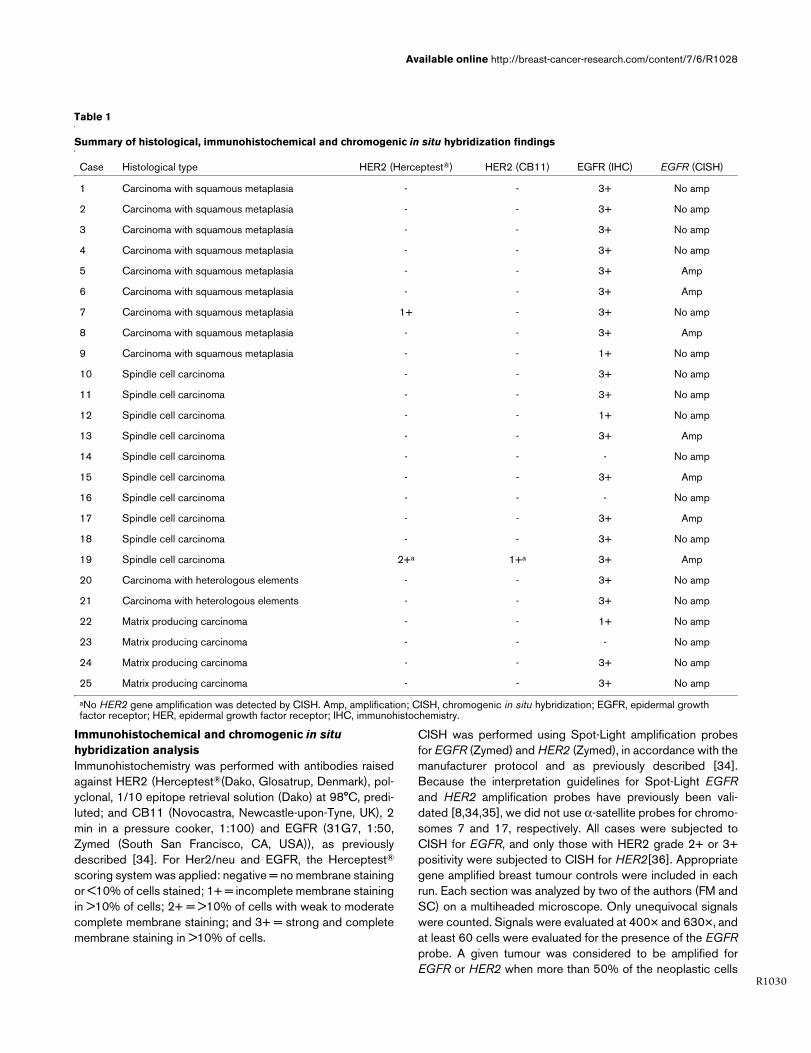
Table 1
Summary of histological, immunohistochemical and chromogenic in situ hybridization findings
Case Histological type HER2 (Herceptest®) HER2 (CB11) EGFR (IHC) EGFR (CISH)
1 Carcinoma with squamous metaplasia - - 3+ No amp
2 Carcinoma with squamous metaplasia - - 3+ No amp
3 Carcinoma with squamous metaplasia - - 3+ No amp
4 Carcinoma with squamous metaplasia - - 3+ No amp
5 Carcinoma with squamous metaplasia - - 3+ Amp
6 Carcinoma with squamous metaplasia - - 3+ Amp
7 Carcinoma with squamous metaplasia 1+ - 3+ No amp
8 Carcinoma with squamous metaplasia - - 3+ Amp
9 Carcinoma with squamous metaplasia - - 1+ No amp
10 Spindle cell carcinoma - - 3+ No amp
11 Spindle cell carcinoma - - 3+ No amp
12 Spindle cell carcinoma - - 1+ No amp
13 Spindle cell carcinoma - - 3+ Amp
14 Spindle cell carcinoma - - - No amp
15 Spindle cell carcinoma - - 3+ Amp
16 Spindle cell carcinoma - - - No amp
17 Spindle cell carcinoma - - 3+ Amp
18 Spindle cell carcinoma - - 3+ No amp
19 Spindle cell carcinoma 2+a 1+a 3+ Amp
20 Carcinoma with heterologous elements - - 3+ No amp
21 Carcinoma with heterologous elements - - 3+ No amp
22 Matrix producing carcinoma - - 1+ No amp
23 Matrix producing carcinoma - - - No amp
24 Matrix producing carcinoma - - 3+ No amp
25 Matrix producing carcinoma - - 3+ No amp
aNo HER2 gene amplification was detected by CISH. Amp, amplification; CISH, chromogenic in situ hybridization; EGFR, epidermal growth factor receptor; HER, epidermal growth factor receptor; IHC, immunohistochemistry.
Breast Cancer Research Vol 7 No 6 Reis-Filho et al.
R1031
exhibited more than five signals per nuclei or large gene signalclusters [8,34,36].
Out of the 112 cases of metaplastic breast carcinomas in ourseries, 25 had sufficient material in the blocks and were suc-cessfully analyzed by both immunohistochemistry and CISHfor EGFR and HER2.
Correlation between EGFR overexpression and amplification and clinicopathological parameters and survivalFollow-up information was available for 23 out of 25 patients,with follow-up periods ranging from 5.5 to 124.3 months(median 34.6 months, mean 51.9 months). The Statview soft-ware package was used for all calculations. Correlationsbetween categorical variables were performed using the χ2
test and Fisher's exact test. Correlations between continuousand categorical variables were performed with analysis of var-iance. DFS and OS were expressed as the number of monthsfrom diagnosis to the occurrence of an event (local recur-rence/metastasis and disease-related death, respectively).Cumulative survival probabilities were calculated using theKaplan–Meier method. Differences between survival rateswere tested using the log-rank test. All tests were two-tailed,with a confidence interval of 95%.
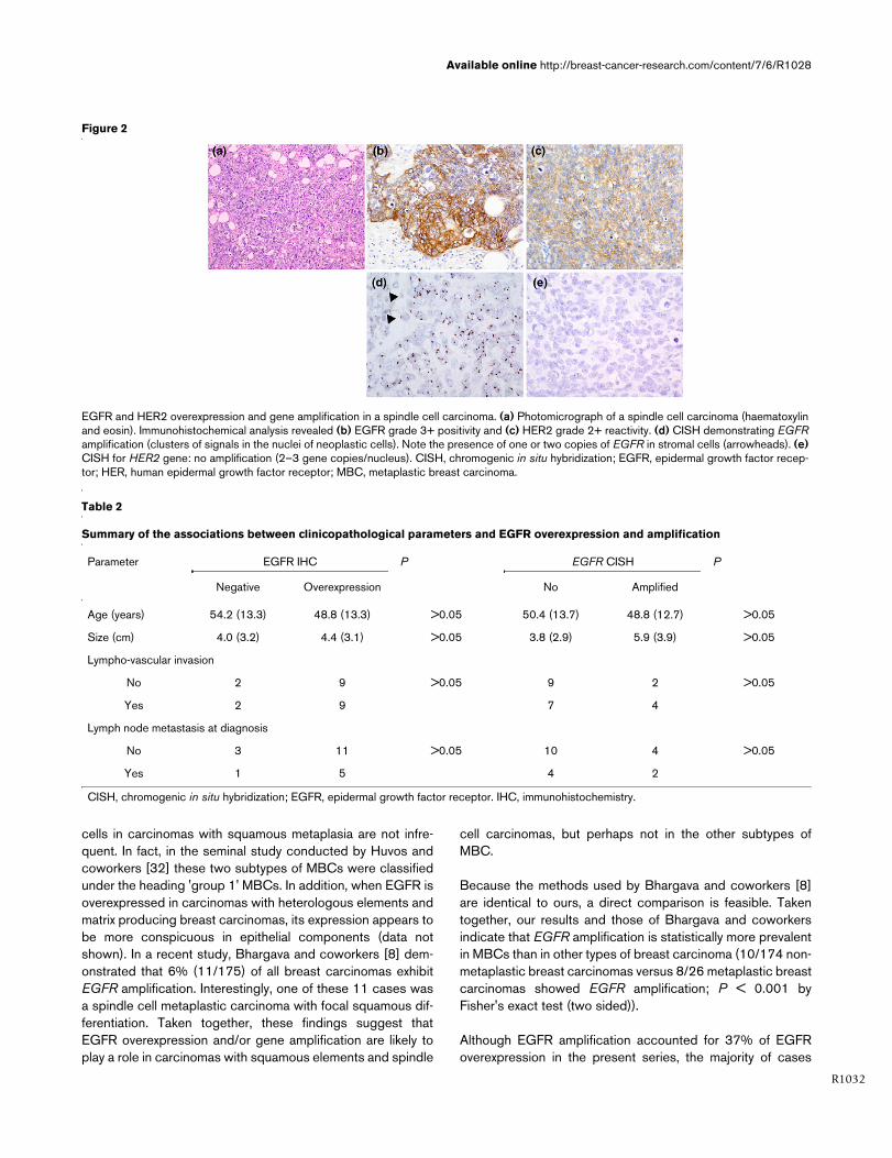
ResultsTable 1 summarizes the results of the histological, immunohis-tochemical and CISH analyses. Briefly, 19 out of 25 (76%)cases exhibited EGFR positivity of grade 3+. No samplesshowed EGFR grade 2+ positivity, whereas four cases wereEGFR 1+. Out of the 19 cases with EGFR grade 3+ expres-sion, seven (37%) exhibited EGFR gene amplification (Fig. 1).Three out of nine carcinomas with squamous metaplasia andfour out of 10 spindle cell carcinomas had EGFR amplification,whereas no matrix producing breast carcinomas showed anyamplification. Interestingly, similar numbers of EGFR signalswere observed in the epithelial and in the metaplastic ele-ments. One case exhibited HER2 grade 2+ positivity with Her-ceptest®, but HER2 gene amplification was not observed (Fig.2).
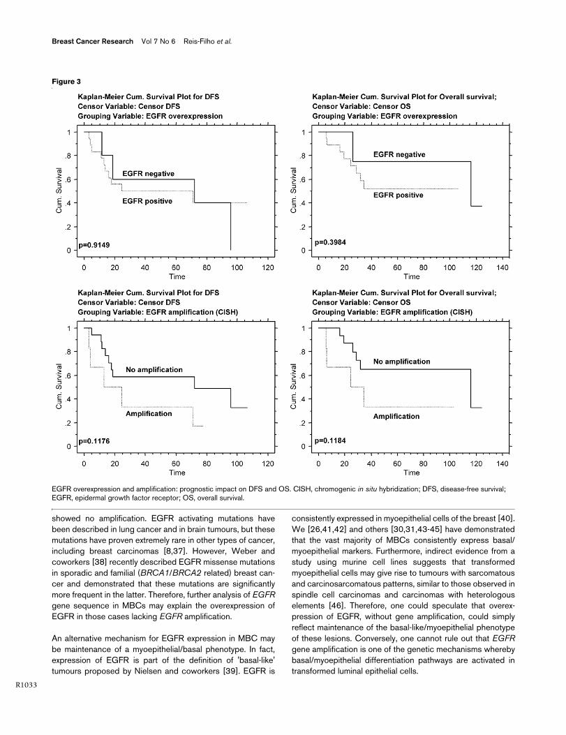
No association between EGFR overexpression or amplifica-tion and clinicopathological features was observed (Table 2).EGFR overexpression showed no association with DFS orOS. Patients with tumours harbouring EGFR amplification hada trend toward shorter DFS and OS (Fig. 3).
DiscussionIn recent studies, we and others demonstrated that MBCs fre-quently overexpress EGFR and lack HER2 overexpression. Ina previous study [26] we demonstrated that up to 83% of allMBCs show EGFR overexpression. Leibl and Moinfar [12]described positivity for EGFR in 70% of MBCs, but thoseauthors also considered cases with grade 1+ expression to be
positive. When only grade 2+ and 3+ expression was consid-ered to represent positivity, 60% (12/20) were positive. In thepresent study we demonstrated that 76% (19/25) of MBCsoverexpressed EGFR. The differences between our findingsand those of Leibl and Moinfar [12] may be related to the dif-ferent antibody clones used and different antigen retrievalmethods.
The mechanism underlying EGFR overexpression has notbeen investigated in MBCs. In the present study we demon-strated that EGFR is amplified in 28% (7/25) of MBCs and in37% (7/19) of MBCs with EGFR overexpression. Althoughonly six cases with heterologous elements (four matrix produc-ing carcinomas and two carcinomas with heterologous ele-ments) were analyxed, no amplification was found in these twosubtypes of MBC. Identification of areas of squamous differen-tiation in spindle cell carcinomas and the presence of spindle
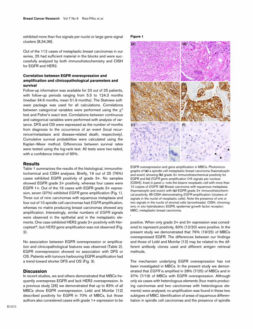
Figure 1
EGFR overexpression and gene amplification in MBCsEGFR overexpression and gene amplification in MBCs. Photomicro-graphs of (a) a spindle cell metaplastic breast carcinoma (haematoxylin and eosin) showing (b) grade 3+ immunohistochemical positivity for EGFR and (c) EGFR gene amplification (>5 signals per nucleus [CISH]). Inset in panel c: note the bizarre neoplastic cell with more than 10 copies of EGFR. (d) Breast carcinoma with squamous metaplasia (haematoxylin and eosin) with (e) EGFR grade 3+ immunohistochemi-cal positivity. (f) CISH demonstrating EGFR amplification (clusters of signals in the nuclei of neoplastic cells). Note the presence of one or two signals in the nuclei of stromal cells (arrowheads). CISH, chromog-enic in situ hybridization; EGFR, epidermal growth factor receptor; MBC, metaplastic breast carcinoma.
Available online http://breast-cancer-research.com/content/7/6/R1028
R1032
cells in carcinomas with squamous metaplasia are not infre-quent. In fact, in the seminal study conducted by Huvos andcoworkers [32] these two subtypes of MBCs were classifiedunder the heading 'group 1' MBCs. In addition, when EGFR isoverexpressed in carcinomas with heterologous elements andmatrix producing breast carcinomas, its expression appears tobe more conspicuous in epithelial components (data notshown). In a recent study, Bhargava and coworkers [8] dem-onstrated that 6% (11/175) of all breast carcinomas exhibitEGFR amplification. Interestingly, one of these 11 cases wasa spindle cell metaplastic carcinoma with focal squamous dif-ferentiation. Taken together, these findings suggest thatEGFR overexpression and/or gene amplification are likely toplay a role in carcinomas with squamous elements and spindle
cell carcinomas, but perhaps not in the other subtypes ofMBC.
Because the methods used by Bhargava and coworkers [8]are identical to ours, a direct comparison is feasible. Takentogether, our results and those of Bhargava and coworkersindicate that EGFR amplification is statistically more prevalentin MBCs than in other types of breast carcinoma (10/174 non-metaplastic breast carcinomas versus 8/26 metaplastic breastcarcinomas showed EGFR amplification; P < 0.001 byFisher's exact test (two sided)).
Although EGFR amplification accounted for 37% of EGFRoverexpression in the present series, the majority of cases
Figure 2
EGFR and HER2 overexpression and gene amplification in a spindle cell carcinomaEGFR and HER2 overexpression and gene amplification in a spindle cell carcinoma. (a) Photomicrograph of a spindle cell carcinoma (haematoxylin and eosin). Immunohistochemical analysis revealed (b) EGFR grade 3+ positivity and (c) HER2 grade 2+ reactivity. (d) CISH demonstrating EGFR amplification (clusters of signals in the nuclei of neoplastic cells). Note the presence of one or two copies of EGFR in stromal cells (arrowheads). (e) CISH for HER2 gene: no amplification (2–3 gene copies/nucleus). CISH, chromogenic in situ hybridization; EGFR, epidermal growth factor recep-tor; HER, human epidermal growth factor receptor; MBC, metaplastic breast carcinoma.
Table 2
Summary of the associations between clinicopathological parameters and EGFR overexpression and amplification
Parameter EGFR IHC P EGFR CISH P
Negative Overexpression No Amplified
Age (years) 54.2 (13.3) 48.8 (13.3) >0.05 50.4 (13.7) 48.8 (12.7) >0.05
Size (cm) 4.0 (3.2) 4.4 (3.1) >0.05 3.8 (2.9) 5.9 (3.9) >0.05
Lympho-vascular invasion
No 2 9 >0.05 9 2 >0.05
Yes 2 9 7 4
Lymph node metastasis at diagnosis
No 3 11 >0.05 10 4 >0.05
Yes 1 5 4 2
CISH, chromogenic in situ hybridization; EGFR, epidermal growth factor receptor. IHC, immunohistochemistry.
Breast Cancer Research Vol 7 No 6 Reis-Filho et al.
R1033
showed no amplification. EGFR activating mutations havebeen described in lung cancer and in brain tumours, but thesemutations have proven extremely rare in other types of cancer,including breast carcinomas [8,37]. However, Weber andcoworkers [38] recently described EGFR missense mutationsin sporadic and familial (BRCA1/BRCA2 related) breast can-cer and demonstrated that these mutations are significantlymore frequent in the latter. Therefore, further analysis of EGFRgene sequence in MBCs may explain the overexpression ofEGFR in those cases lacking EGFR amplification.
An alternative mechanism for EGFR expression in MBC maybe maintenance of a myoepithelial/basal phenotype. In fact,expression of EGFR is part of the definition of 'basal-like'tumours proposed by Nielsen and coworkers [39]. EGFR is
consistently expressed in myoepithelial cells of the breast [40].We [26,41,42] and others [30,31,43-45] have demonstratedthat the vast majority of MBCs consistently express basal/myoepithelial markers. Furthermore, indirect evidence from astudy using murine cell lines suggests that transformedmyoepithelial cells may give rise to tumours with sarcomatousand carcinosarcomatous patterns, similar to those observed inspindle cell carcinomas and carcinomas with heterologouselements [46]. Therefore, one could speculate that overex-pression of EGFR, without gene amplification, could simplyreflect maintenance of the basal-like/myoepithelial phenotypeof these lesions. Conversely, one cannot rule out that EGFRgene amplification is one of the genetic mechanisms wherebybasal/myoepithelial differentiation pathways are activated intransformed luminal epithelial cells.
Figure 3
EGFR overexpression and amplification: prognostic impact on DFS and OSEGFR overexpression and amplification: prognostic impact on DFS and OS. CISH, chromogenic in situ hybridization; DFS, disease-free survival; EGFR, epidermal growth factor receptor; OS, overall survival.
Available online http://breast-cancer-research.com/content/7/6/R1028
R1034
In the present series, there was no association between EGFRoverexpression and DFS or OS, whereas there was a trendtoward shorter DFS and OS in patients with tumours exhibit-ing EGFR amplification. Based on our results, further studiesanalyzing the prognostic impact of EGFR amplification in alarge cohort of MBCs are warranted.
The present study also confirms the results of previous analy-ses demonstrating lack of HER2 overexpression in MBCs[12,47], suggesting that humanized monoclonal antibodiesagainst HER2 play little or no role in the treatment of theselesions.
ConclusionAlthough initial studies suggested that only tumours harbour-ing EGFR mutations would be sensitive to EGFR tyrosinekinase inhibitors, there are compelling data to suggest thattumours harbouring EGFR amplification may also respond wellto these new agents [22]. Given that MBCs lack oestrogenreceptor, show EGFR overexpression and harbour EGFRamplification in up to 28% of cases, and that some MBCshave proven refractory to standard types of treatment, the useof EGFR tyrosine kinase inhibitors may represent an alternativetherapeutic regimen for patients with MBC. Further studiesaddressing the efficacy of EGFR inhibitors in this group ofbreast carcinomas are warranted.
Competing interestsThe authors declare that they have no competing interests.
Authors' contributionsJSR-F drafted the manuscript and participated in the histolog-ical review, immunohistochemical analysis and study design.FM participated in the histological review, immunohistochemi-cal and CISH analyses. SC carried out the CISH analysis. PTShelped to draft the manuscript and participated in the studydesign. DS and KS performed the immunohistochemical reac-tions. ML performed part of the CISH experiments. EMP andJN participated in the histological review. SRL and FCS con-ceived the study, participated in its design and coordination,and helped to draft the manuscript. FCS also participated inthe histological review. All authors read and approved the finalmanuscript.
AcknowledgementsThis study was supported by Breakthrough Breast Cancer and a grant Programa Operacional Ciência, Tecnologia e Inovação POCTI/CBO/45157/2002. JSR-F is partially supported by PhD Grant SFRH/BD/5386/2001 from the Fundação para a Ciência e a Tecnologia, Portugal.
References1. Hynes NE, Lane HA: ERBB receptors and cancer: the complex-
ity of targeted inhibitors. Nat Rev Cancer 2005, 5:341-354.2. Suo Z, Risberg B, Kalsson MG, Willman K, Tierens A, Skovlund E,
Nesland JM: EGFR family expression in breast carcinomas. c-erbB-2 and c-erbB-4 receptors have different effects onsurvival. J Pathol 2002, 196:17-25.
3. Suo Z, Yang H, Mei Q, Skovlund E, Cui J, Nesland JM: Type 1 pro-tein tyrosine kinases in Chinese breast carcinomas: a clinico-pathologic study. Int J Surg Pathol 2001, 9:177-187.
4. Suo Z, Nesland JM: Phyllodes tumor of the breast: EGFR familyexpression and relation to clinicopathological features.Ultrastruct Pathol 2000, 24:371-381.
5. Suo Z, Emilsen E, Tveit KM, Nesland JM: Type 1 protein tyrosinekinases in benign and malignant breast lesions. Histopathol-ogy 1998, 33:514-521.
6. Abd El-Rehim DM, Pinder SE, Paish CE, Bell JA, Rampaul RS,Blamey RW, Robertson JF, Nicholson RI, Ellis IO: Expression andco-expression of the members of the epidermal growth factorreceptor (EGFR) family in invasive breast carcinoma. Br JCancer 2004, 91:1532-42.
7. DiGiovanna MP, Stern DF, Edgerton SM, Whalen SG, Moore D II,Thor AD: Relationship of epidermal growth factor receptorexpression to ErbB-2 signaling activity and prognosis inbreast cancer patients. J Clin Oncol 2005, 23:1152-1160.
8. Bhargava R, Gerald WL, Li AR, Pan Q, Lal P, Ladanyi M, Chen B:EGFR gene amplification in breast cancer: correlation with epi-dermal growth factor receptor mRNA and protein expressionand HER-2 status and absence of EGFR-activating mutations.Mod Pathol 2005 in press.
9. Harris AL, Nicholson S, Sainsbury JR, Farndon J, Wright C: Epi-dermal growth factor receptors in breast cancer: associationwith early relapse and death, poor response to hormones andinteractions with neu. J Steroid Biochem 1989, 34:123-131.
10. Tsutsui S, Kataoka A, Ohno S, Murakami S, Kinoshita J,Hachitanda Y: Prognostic and predictive value of epidermalgrowth factor receptor in recurrent breast cancer. Clin CancerRes 2002, 8:3454-3460.
11. Walker RA, Dearing SJ: Expression of epidermal growth factorreceptor mRNA and protein in primary breast carcinomas.Breast Cancer Res Treat 1999, 53:167-176.
12. Leibl S, Moinfar F: Metaplastic breast carcinomas are negativefor Her-2 but frequently express EGFR (Her-1): potential rele-vance to adjuvant treatment with EGFR tyrosine kinaseinhibitors? J Clin Pathol 2005, 58:700-704.
13. Giaccone G: Epidermal growth factor receptor inhibitors in thetreatment of non-small-cell lung cancer. J Clin Oncol 2005,23:3235-3242.
14. Baselga J, Arteaga CL: Critical update and emerging trends inepidermal growth factor receptor targeting in cancer. J ClinOncol 2005, 23:2445-2459.
15. Pao W, Miller V, Zakowski M, Doherty J, Politi K, Sarkaria I, SinghB, Heelan R, Rusch V, Fulton L, et al.: EGF receptor gene muta-tions are common in lung cancers from 'never smokers' andare associated with sensitivity of tumors to gefitinib anderlotinib. Proc Natl Acad Sci USA 2004, 101:13306-13311.
16. Paez JG, Janne PA, Lee JC, Tracy S, Greulich H, Gabriel S, Her-man P, Kaye FJ, Lindeman N, Boggon TJ, et al.: EGFR mutationsin lung cancer: correlation with clinical response to gefitinibtherapy. Science 2004, 304:1497-1500.
17. Lynch TJ, Bell DW, Sordella R, Gurubhagavatula S, Okimoto RA,Brannigan BW, Harris PL, Haserlat SM, Supko JG, Haluska FG, etal.: Activating mutations in the epidermal growth factor recep-tor underlying responsiveness of non-small-cell lung cancer togefitinib. N Engl J Med 2004, 350:2129-2139.
18. Marty M, Cognetti F, Maraninchi D, Snyder R, Mauriac L, Tubiana-Hulin M, Chan S, Grimes D, Anton A, Lluch A, et al.: Efficacy andsafety of trastuzumab combined with docetaxel in patientswith human epidermal growth factor receptor 2-positive met-astatic breast cancer administered as first-line treatment:results of a randomized phase II trial by the M77001 studygroup. J Clin Oncol 2005, 23:4265-4274.
19. Baselga J, Gianni L, Geyer C, Perez EA, Riva A, Jackisch C: Futureoptions with trastuzumab for primary systemic and adjuvanttherapy. Semin Oncol 2004, 31:51-57.
20. Fallon KB, Palmer CA, Roth KA, Nabors LB, Wang W, CarpenterM, Banerjee R, Forsyth P, Rich K, Perry A: Prognostic value of 1p,19q, 9p, 10q, and EGFR-FISH analyses in recurrentoligodendrogliomas. J Neuropathol Exp Neurol 2004,63:314-322.
21. Marquez A, Wu R, Zhao J, Tao J, Shi Z: Evaluation of epidermalgrowth factor receptor (EGFR) by chromogenic in situ hybrid-ization (CISH) and immunohistochemistry (IHC) in archival gli-
Breast Cancer Research Vol 7 No 6 Reis-Filho et al.
R1035
omas using bright-field microscopy. Diagn Mol Pathol 2004,13:1-8.
22. Cappuzzo F, Hirsch FR, Rossi E, Bartolini S, Ceresoli GL, BemisL, Haney J, Witta S, Danenberg K, Domenichini I, et al.: Epidermalgrowth factor receptor gene and protein and gefitinib sensitiv-ity in non-small-cell lung cancer. J Natl Cancer Inst 2005,97:643-655.
23. Takehana T, Kunitomo K, Suzuki S, Kono K, Fujii H, Matsumoto Y,Ooi A: Expression of epidermal growth factor receptor in gas-tric carcinomas. Clin Gastroenterol Hepatol 2003, 1:438-445.
24. Corzo C, Tusquets I, Salido M, Corominas JM, Bellet M, Suarez M,Baro T, Fabregat X, Serrano S, Sole F: Characterization of HER1(c-erbB1) status in locally advanced breast cancer using fluo-rescence in situ hybridization and immunohistochemistry.Tumour Biol 2005, 26:25-30.
25. Al-Kuraya K, Schraml P, Torhorst J, Tapia C, Zaharieva B, NovotnyH, Spichtin H, Maurer R, Mirlacher M, Kochli O, et al.: Prognosticrelevance of gene amplifications and coamplifications inbreast cancer. Cancer Res 2004, 64:8534-8540.
26. Reis-Filho JS, Milanezi F, Simpson P, Fulford LG, Steele D, Nes-land JM, Pereira EM, Lakhani SR, Schmitt FC: Are metaplasticbreast carcinomas basal-like tumours? [abstract]. Mod Pathol2005:48A.
27. Wargotz ES, Norris HJ: Metaplastic carcinomas of the breast: V.Metaplastic carcinoma with osteoclastic giant cells. HumPathol 1990, 21:1142-1150.
28. Wargotz ES, Norris HJ: Metaplastic carcinomas of the breast.IV. Squamous cell carcinoma of ductal origin. Cancer 1990,65:272-276.
29. Wargotz ES, Norris HJ: Metaplastic carcinomas of the breast.III. Carcinosarcoma. Cancer 1989, 64:1490-1499.
30. Wargotz ES, Deos PH, Norris HJ: Metaplastic carcinomas of thebreast. II. Spindle cell carcinoma. Hum Pathol 1989,20:732-740.
31. Wargotz ES, Norris HJ: Metaplastic carcinomas of the breast. I.Matrix-producing carcinoma. Hum Pathol 1989, 20:628-635.
32. Huvos AG, Lucas JC Jr, Foote FW Jr: Metaplastic breast carci-noma. Rare form of mammary cancer. N Y State J Med 1973,73:1078-1082.
33. Oberman HA: Metaplastic carcinoma of the breast. A clinico-pathologic study of 29 patients. Am J Surg Pathol 1987,11:918-929.
34. Reis-Filho JS, Simpson PT, Jones C, Steele D, Mackay A, IravaniM, Fenwick K, Valgeirsson H, Lambros M, Ashworth A, et al.: Ple-omorphic lobular carcinoma of the breast: role of comprehen-sive molecular pathology in characterization of an entity. JPathol 2005, 207:1-13.
35. Isola J, Tanner M, Forsyth A, Cooke TG, Watters AD, Bartlett JM:Interlaboratory comparison of HER-2 oncogene amplificationas detected by chromogenic and fluorescence in situhybridization. Clin Cancer Res 2004, 10:4793-4798.
36. Bilous M, Dowsett M, Hanna W, Isola J, Lebeau A, Moreno A,Penault-Llorca F, Ruschoff J, Tomasic G, van de Vijver M: Currentperspectives on HER2 testing: a review of national testingguidelines. Mod Pathol 2003, 16:173-182.
37. Lee JW, Soung YH, Kim SY, Park WS, Nam SW, Lee JY, Yoo NJ,Lee SH: Absence of EGFR mutation in the kinase domain incommon human cancers besides non-small cell lung cancer.Int J Cancer 2005, 113:510-511.
38. Weber F, Fukino K, Sawada T, Williams N, Sweet K, Brena RM,Plass C, Caldes T, Mutter GL, Villalona-Calero MA, et al.: Variabil-ity in organ-specific EGFR mutational spectra in tumour epi-thelium and stroma may be the biological basis for differentialresponses to tyrosine kinase inhibitors. Br J Cancer 2005,92:1922-1926.
39. Nielsen TO, Hsu FD, Jensen K, Cheang M, Karaca G, Hu Z, Hern-andez-Boussard T, Livasy C, Cowan D, Dressler L, et al.: Immu-nohistochemical and clinical characterization of the basal-likesubtype of invasive breast carcinoma. Clin Cancer Res 2004,10:5367-5374.
40. Santini D, Ceccarelli C, Tardio ML, Taffurelli M, Marrano D: Immu-nocytochemical expression of epidermal growth factor recep-tor in myoepithelial cells of the breast. Appl ImmunohistochemMol Morphol 2002, 10:29-33.
41. Reis-Filho JS, Milanezi F, Paredes J, Silva P, Pereira EM, MaedaSA, de Carvalho LV, Schmitt FC: Novel and classic myoepithe-
lial/stem cell markers in metaplastic carcinomas of the breast.Appl Immunohistochem Mol Morphol 2003, 11:1-8.
42. Reis-Filho JS, Schmitt FC: p63 expression in sarcomatoid/metaplastic carcinomas of the breast. Histopathology 2003,42:94-95.
43. Leibl S, Gogg-Kammerer M, Sommersacher A, Denk H, Moinfar F:Metaplastic breast carcinomas: are they of myoepithelial dif-ferentiation?: immunohistochemical profile of the sarcoma-toid subtype using novel myoepithelial markers. Am J SurgPathol 2005, 29:347-353.
44. Dunne B, Lee AH, Pinder SE, Bell JA, Ellis IO: An immunohisto-chemical study of metaplastic spindle cell carcinoma, phyl-lodes tumor and fibromatosis of the breast. Hum Pathol 2003,34:1009-1015.
45. Popnikolov NK, Ayala AG, Graves K, Gatalica Z: Benign myoepi-thelial tumors of the breast have immunophenotypic charac-teristics similar to metaplastic matrix-producing and spindlecell carcinomas. Am J Clin Pathol 2003, 120:161-167.
46. Sapino A, Papotti M, Sanfilippo B, Gugliotta P, Bussolati G:Tumor types derived from epithelial and myoepithelial celllines of R3230AC rat mammary carcinoma. Cancer Res 1992,52:1553-1560.
47. Barnes PJ, Boutilier R, Chiasson D, Rayson D: Metaplastic breastcarcinoma: clinical-pathologic characteristics and HER2/neuexpression. Breast Cancer Res Treat 2005, 91:173-178.
Copyright © 2022 FDOKUMEN