Association of 22 cytokine gene polymorphisms with tuberculosis in Macedonians
10
ORIGINAL ARTICLE Association of 22 cytokine gene polymorphisms with rheumatoid arthritis in population of ethnic Macedonians Dejan Trajkov & Snezhana Mishevska-Perchinkova & Anzelika Karadzova-Stojanoska & Aleksandar Petlichkovski & Ana Strezova & Mirko Spiroski Received: 7 February 2009 / Revised: 28 June 2009 / Accepted: 17 July 2009 / Published online: 7 August 2009 # Clinical Rheumatology 2009 Abstract To examine the possible role of 22 cytokine gene polymorphisms in host susceptibility to or protection against RA in Macedonians. In this study, 301 healthy unrelated individuals and 85 patients with RA were studied. Cytokine genotyping was performed by PCR with sequence-specific priming (PCR–SSP) (Heidelberg kit). Results showed suscep- tible association for four cytokine alleles, six cytokine genotypes, one haplotype, and four combinations of haplo- types, while protective associations were found for four cytokine alleles, three cytokine genotypes, three haplotypes, and only one combination of haplotypes. These results suggest that IL-4 -1098, IL-4 -590, IL-10 -1082, IL-10 -819, IL-2 -330, IL-6 -174, and TNF-α -238 cytokine gene polymorphisms might be significantly associated and affect host susceptibility and/or resistance to RA in Macedonians. Keywords Cytokine polymorphism . Macedonians . Rheumatoid arthritis Introduction Rheumatoid arthritis (RA) is a complex heterogeneous chronic autoimmune disease, whereby both environmental and genetic factors contribute to the etiology and/or clinical severity that affects approximately 1% of the world’ s population, with the prevalence significantly higher in women than in men (in a 3:1 ratio). Genetics of RA, which contribution to etiology is estimated to be over 30%, is very complex involving many genes each with small individual effects [1]. RA is characterized by abnormal immune responses and inflammation in the lining of the joint capsule (synovium) and the lining of tendons. Although the exact immunopathogenesis of arthritic inflammation is still unknown, it has been postulated that clinical symptoms may reflect an imbalance in pro- and anti-inflammatory cytokines. RA represented typical T-helper 1-mediated disease in which T cells infiltrate synovial inflammatory lesions, stimulating monocytes, macrophages, and synovial fibroblasts to produce a large amount of cytokines [2]. It is already known that the number of cytokine gene polymorphisms in the cytokine gene regulatory regions correlate with their secretion [3], and that their inheritance is influenced by ethnicity [4]. These polymorphisms may result in inter-individual variation in cytokine production, which may have significant influence on disease suscepti- bility, severity, and outcome. Until now, we have only published data for the cytokine polymorphisms in healthy Macedonian population [5, 6] and for the possible association of cytokine polymorphism with bronchial asthma [7] and chronic obstructive pulmo- nary disease [8]. The aim of this study was to investigate D. Trajkov : A. Petlichkovski : A. Strezova : M. Spiroski (*) Institute of Immunobiology and Human Genetics, Faculty of Medicine, University “Ss. Kiril and Metodij”, 1109 Skopje( P.O. Box 60, Republic of Macedonia e-mail: [email protected] URL: http://www.iibhg.ukim.edu.mk/ S. Mishevska-Perchinkova : A. Karadzova-Stojanoska Clinic for Rheumatology, Clinical Center, Faculty of Medicine, University “Ss Kiril and Metodij”, Skopje, Republic of Macedonia Clin Rheumatol (2009) 28:1291–1300 DOI 10.1007/s10067-009-1238-4
Transcript of Association of 22 cytokine gene polymorphisms with tuberculosis in Macedonians

ORIGINAL ARTICLE
Association of 22 cytokine gene polymorphismswith rheumatoid arthritis in populationof ethnic Macedonians
Dejan Trajkov & Snezhana Mishevska-Perchinkova &
Anzelika Karadzova-Stojanoska &
Aleksandar Petlichkovski & Ana Strezova &
Mirko Spiroski
Received: 7 February 2009 /Revised: 28 June 2009 /Accepted: 17 July 2009 /Published online: 7 August 2009# Clinical Rheumatology 2009
Abstract To examine the possible role of 22 cytokine genepolymorphisms in host susceptibility to or protection againstRA in Macedonians. In this study, 301 healthy unrelatedindividuals and 85 patients with RA were studied. Cytokinegenotyping was performed by PCR with sequence-specificpriming (PCR–SSP) (Heidelberg kit). Results showed suscep-tible association for four cytokine alleles, six cytokinegenotypes, one haplotype, and four combinations of haplo-types, while protective associations were found for fourcytokine alleles, three cytokine genotypes, three haplotypes,and only one combination of haplotypes. These results suggestthat IL-4 −1098, IL-4 −590, IL-10 −1082, IL-10 −819,IL-2 −330, IL-6 −174, and TNF-α −238 cytokine genepolymorphisms might be significantly associated and affecthost susceptibility and/or resistance to RA in Macedonians.
Keywords Cytokine polymorphism .Macedonians .
Rheumatoid arthritis
Introduction
Rheumatoid arthritis (RA) is a complex heterogeneouschronic autoimmune disease, whereby both environmentaland genetic factors contribute to the etiology and/or clinicalseverity that affects approximately 1% of the world’spopulation, with the prevalence significantly higher inwomen than in men (in a 3:1 ratio). Genetics of RA, whichcontribution to etiology is estimated to be over 30%, is verycomplex involving many genes each with small individualeffects [1]. RA is characterized by abnormal immuneresponses and inflammation in the lining of the jointcapsule (synovium) and the lining of tendons. Althoughthe exact immunopathogenesis of arthritic inflammation isstill unknown, it has been postulated that clinical symptomsmay reflect an imbalance in pro- and anti-inflammatorycytokines. RA represented typical T-helper 1-mediateddisease in which T cells infiltrate synovial inflammatorylesions, stimulating monocytes, macrophages, and synovialfibroblasts to produce a large amount of cytokines [2].
It is already known that the number of cytokine genepolymorphisms in the cytokine gene regulatory regionscorrelate with their secretion [3], and that their inheritanceis influenced by ethnicity [4]. These polymorphisms mayresult in inter-individual variation in cytokine production,which may have significant influence on disease suscepti-bility, severity, and outcome.
Until now, we have only published data for the cytokinepolymorphisms in healthy Macedonian population [5, 6]and for the possible association of cytokine polymorphismwith bronchial asthma [7] and chronic obstructive pulmo-nary disease [8]. The aim of this study was to investigate
D. Trajkov :A. Petlichkovski :A. Strezova :M. Spiroski (*)Institute of Immunobiology and Human Genetics,Faculty of Medicine, University “Ss. Kiril and Metodij”,1109 Skopje( P.O. Box 60,Republic of Macedoniae-mail: [email protected]: http://www.iibhg.ukim.edu.mk/
S. Mishevska-Perchinkova :A. Karadzova-StojanoskaClinic for Rheumatology, Clinical Center, Faculty of Medicine,University “Ss Kiril and Metodij”,Skopje, Republic of Macedonia
Clin Rheumatol (2009) 28:1291–1300DOI 10.1007/s10067-009-1238-4

the existence of possible associations between 22 cytokinegenes polymorphisms and RA in Macedonians, in order toadd knowledge about the genetic background of thisdisease, and to provide data for meta-analysis.
Materials and methods
Population
The sample of the population of subjects from the Republic ofMacedonia included in this study comprised 301 healthyunrelated individuals and 85 patients with RA, fulfilling the1987 American College of Rheumatology criteria for RA [9].All of the patients and healthy individuals included in thisstudy participated in the study which was approved by theCommittee of the Ministry of Education and Science fromRepublic of Macedonia (No. 087405) after signing a writtenconsent. Blood samples were collected, DNA was isolatedfrom peripheral blood leukocytes by the phenol–chloroformextraction method [10], and DNA samples were stored in theMacedonian Human DNA Bank (hDNAMKD) [11].
Typing methods
Fourteen cytokine genes (22 SNP alleles) were identified asthe candidates for the cytokine polymorphism component(CPC) at the 13th International Histocompatibility Work-shop and Congress, and cytokine genotyping was per-formed by PCR with sequence-specific priming (PCR–SSP;Heidelberg kit): IL-1α −889, IL-1β −511, IL-1β +3962,IL−1R psti1970, IL-1RA mspa11100, IL-4Rα +1902,IL-12 −1188, IFNγ utr5644, TGF-β1 cdn10, TGF-β1cdn25, TNF-α −308, TNF-α −238, IL-2 −330, IL-2 +166,IL-4 −1098, IL-4 −590, IL-4 −33, IL-6 −174, IL-6 565, IL-10 −1082, IL-10 −819, and IL-10 −592. Briefly, PCR–SSPtyping Heidelberg kit consists of 48 PCR primer mixesaliquoted in 96-well PCR trays (two typings per tray).Master mix, which was supplied along with the reagents andconsisted of MgCl2, buffer, dNTPs, and glycerol, was mixedwith 1.2–3.0µg DNA and 20 U Taq polymerase anddispensed in the 48 wells [12]. Agarose gel electrophoresison a 2% gel revealed a positive or negative signal forspecific amplification in each well. Subsequently, the resultswere analyzed according to the interpretation schemeprovided with the kit [13].
Statistical methods
The population genetics analysis package, PyPop, developedby the Biostatistics Core for the Workshop [14, 15], was usedfor analysis of the cytokine data in this study. Allelefrequencies and expected Hardy Weinberg proportions
(HWP) for each SNP were determined [16]. The exact testfor genotype frequency deviation from HWP was calculatedusing the Arlequin implementation accessed via PyPop [17].Those SNPs that did not fit HWP were evaluated todetermine whether there was an excess of homozygotes orheterozygotes, or if any particular genotype frequencies weresignificantly different from the expected frequencies. Com-parisons of different genotypes for two groups were tested bythe χ2 test. Crude odds ratios (OR), as estimates of therelative risk, were calculated within 95% CI and p <0.05 wasconsidered statistically significant.
Results
Cytokine alleles
In Table 1, frequencies of polymorphic cytokine alleles,Pearson’s p value, odds ratio, and Wald’s 95% confidenceinterval in RA patients and healthy Macedonians areshown.
For the members of anti-inflammatory cytokines, wefound positive association for IL-4 −1098/T allele (p<0.001, OR=3.153, Wald’s 95% CI between 1.932 and5.148) and IL-4 −590/C allele (p=0.006, OR=1.737, Wald’s95% CI between 1.168 and 2.585), while IL-4 −1098/G andIL-4 −590/T alleles showed negative (protective) association(p<0.001, OR=0.317, Wald’s 95% CI between 0.194 and0.518 and p=0.006; OR=0.576, Wald's 95% CI between0.387 and 0.856, respectively) for RA. In the group ofproinflammatory cytokines, protective association was ob-tained for the TNFα −238/A allele (p=0.047, OR=0.257,Wald's 95% CI between 0.060 and 1.090) and IL-2 −330/Tallele (p=0.032, OR=0.681, Wald's 95% CI between 0.479and 0.969). Results showed that people with TNF-α −238/Gallele have a 3.897-fold risk to develop RA (p=0.047, Wald's95% CI between 0.917 and 16.560) in comparison to otherswith TNF-α −238/A allele. Analysis of the IL-2 poly-morphisms showed that IL-2 −330/G allele was susceptiblyassociated with RA (p=0.032, OR=1.468, Wald's 95% CIbetween 1.032 and 2.087) (Table 1).
Cytokine genotypes
Table 2 contains summarized results for different cytokinegenotypes found in our study.
We found positive (susceptible) association betweenpatients with RA and following genotypes (according thelevel of susceptibility): IL4 −1098/T:T (p<0.001), OR 51.71(Wald's 95% CI between 23.492 and 113.801); IL-2 −330/G:G (p=0.001), OR 2.815 (1.474–5.374); IL-10 −819/T:T (p=0.008), OR 2.661 (1.255–5.642); IL-6 −174/C:C (p=0.005),OR 2.598 (1.314–5.134); IL-4 −590/C:C (p<0.001), OR
1292 Clin Rheumatol (2009) 28:1291–1300

Table 1 Cytokine allele frequency, Pearson's p value, odds ratio, and Wald's 95% confidence interval in RA patients and healthy Macedonianpopulation
Cytokine polymorphism Allele RA (n=85) Control (n=301) Pearson's p value Odds ratio Wald' 95% CI
N F N F
IL-1α −889 C 134 0.848 482 0.814 0.323 1.274 0.787–2.062
T 24 0.152 110 0.186 0.785 0.485–1.270
IL-1β −511 C 118 0.702 404 0.671 0.443 1.157 0.797–1.678
T 50 0.298 198 0.329 0.865 0.596–1.254
IL-1β +3962 C 121 0.720 439 0.729 0.817 0.956 0.653–1.400
T 47 0.280 163 0.270 1.046 0.715–1.533
IL-1R psti1970 C 115 0.676 399 0.662 0.738 1.064 0.740–1.529
T 55 0.324 203 0.337 0.940 0.654–1.352
IL-1RA mspa11100 T 122 0.726 420 0.698 0.474 1.149 0.785–1.683
C 46 0.274 182 0.302 0.870 0.594–1.274
IL-4Rα +1902 A 142 0.835 502 0.834 0.965 1.010 0.639–1.598
G 28 0.165 100 0.166 0.990 0.626–1.566
IL-12 −1188 A 125 0.735 433 0.744 0.820 0.956 0.648–1.409
C 45 0.265 149 0.256 1.046 0.710–1.543
IFNγ utr5644 T 57 0.467 259 0.520 0.295 0.809 0.544–1.203
A 65 0.533 239 0.480 1.236 0.831–1.836
TGF-β1 cdn10 T 94 0.553 282 0.502 0.242 1.228 0.870–1.733
C 76 0.447 280 0.498 0.814 0.577–1.149
TGF-β1 cdn25 G 160 0.941 532 0.947 0.784 0.902 0.432–1.886
C 10 0.059 30 0.053 1.108 0.530–2.317
TNF-α −308 A 19 0.113 74 0.123 0.730 0.910 0.532–1.555
G 149 0.887 528 0.877 1.099 0.643–1.878
TNF-α −238 A 2 0.012 27 0.045 0.047b 0.257 0.060–1.090
G 166 0.988 575 0.955 3.897 0.917–16.560
IL-2 −330 G 71 0.423 191 0.332 0.032b 1.468 1.032–2.087
T 97 0.577 383 0.667 0.681 0.479–0.969
IL-2 +166 G 122 0.726 422 0.735 0.817 0.955 0.649–1.406
T 46 0.274 152 0.264 1.047 0.711–1.541
IL-4 −1098 G 21 0.124 176 0.308 <0.001b 0.317 0.194–0.518
T 149 0.876 396 0.692 3.153 1.932–5.148
IL-4 −590 C 131 0.771 377 0.659 0.006b 1.737 1.168–2.585
T 39 0.229 195 0.341 0.576 0.387–0.856
IL-4 −33 C 148 0.581 479 0.837 0.294 1.306 0.792–2.153
T 22 0.129 93 0.163 0.766 0.464–1.262
IL-6 −174 C 62 0.369 182 0.302 0.100 1.350 0.943–1.932
G 106 0.631 420 0.698 0.741 0.518–1.060
IL-6 nt565 A 59 0.352 173 0.287 0.111 1.342 0.934–1.929
G 109 0.649 429 0.713 0.745 0.518–1.071
IL-10 −1082 A 105 0.618 352 0.589 0.496 1.129 0.796–1.601
G 65 0.382 246 0.411 0.886 0.625–1.257
IL-10 −819 C 114 0.671 435 0.727 0.148 0.763 0.529–1.101
T 56 0.329 163 0.272 1.311 0.908–1.892
IL-10 −592 A 56 0.329 173 0.289 0.313 1.207 0.837–1.739
C 114 0.671 425 0.710 0.313 0.575–1.194
N absolute number, F frequency, CI confidence intervalb Statistically significant
Clin Rheumatol (2009) 28:1291–1300 1293
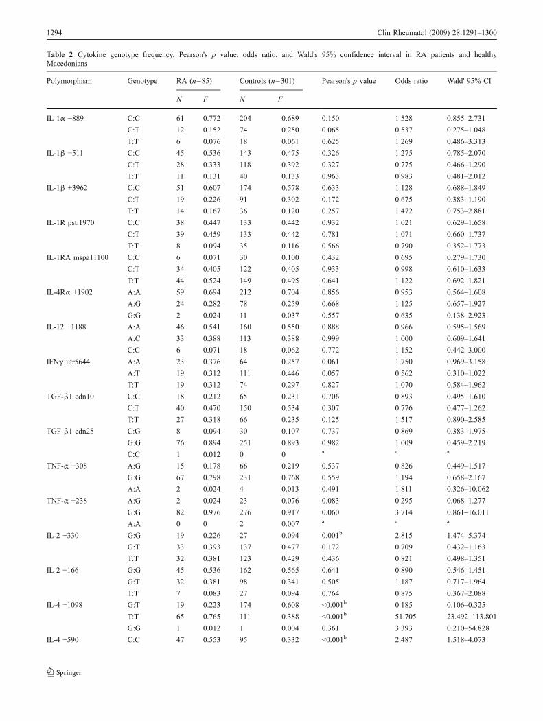
Table 2 Cytokine genotype frequency, Pearson's p value, odds ratio, and Wald's 95% confidence interval in RA patients and healthyMacedonians
Polymorphism Genotype RA (n=85) Controls (n=301) Pearson's p value Odds ratio Wald' 95% CI
N F N F
IL-1α −889 C:C 61 0.772 204 0.689 0.150 1.528 0.855–2.731
C:T 12 0.152 74 0.250 0.065 0.537 0.275–1.048
T:T 6 0.076 18 0.061 0.625 1.269 0.486–3.313
IL-1β −511 C:C 45 0.536 143 0.475 0.326 1.275 0.785–2.070
C:T 28 0.333 118 0.392 0.327 0.775 0.466–1.290
T:T 11 0.131 40 0.133 0.963 0.983 0.481–2.012
IL-1β +3962 C:C 51 0.607 174 0.578 0.633 1.128 0.688–1.849
C:T 19 0.226 91 0.302 0.172 0.675 0.383–1.190
T:T 14 0.167 36 0.120 0.257 1.472 0.753–2.881
IL-1R psti1970 C:C 38 0.447 133 0.442 0.932 1.021 0.629–1.658
C:T 39 0.459 133 0.442 0.781 1.071 0.660–1.737
T:T 8 0.094 35 0.116 0.566 0.790 0.352–1.773
IL-1RA mspa11100 C:C 6 0.071 30 0.100 0.432 0.695 0.279–1.730
C:T 34 0.405 122 0.405 0.933 0.998 0.610–1.633
T:T 44 0.524 149 0.495 0.641 1.122 0.692–1.821
IL-4Rα +1902 A:A 59 0.694 212 0.704 0.856 0.953 0.564–1.608
A:G 24 0.282 78 0.259 0.668 1.125 0.657–1.927
G:G 2 0.024 11 0.037 0.557 0.635 0.138–2.923
IL-12 −1188 A:A 46 0.541 160 0.550 0.888 0.966 0.595–1.569
A:C 33 0.388 113 0.388 0.999 1.000 0.609–1.641
C:C 6 0.071 18 0.062 0.772 1.152 0.442–3.000
IFNγ utr5644 A:A 23 0.376 64 0.257 0.061 1.750 0.969–3.158
A:T 19 0.312 111 0.446 0.057 0.562 0.310–1.022
T:T 19 0.312 74 0.297 0.827 1.070 0.584–1.962
TGF-β1 cdn10 C:C 18 0.212 65 0.231 0.706 0.893 0.495–1.610
C:T 40 0.470 150 0.534 0.307 0.776 0.477–1.262
T:T 27 0.318 66 0.235 0.125 1.517 0.890–2.585
TGF-β1 cdn25 C:G 8 0.094 30 0.107 0.737 0.869 0.383–1.975
G:G 76 0.894 251 0.893 0.982 1.009 0.459–2.219
C:C 1 0.012 0 0 a a a
TNF-α −308 A:G 15 0.178 66 0.219 0.537 0.826 0.449–1.517
G:G 67 0.798 231 0.768 0.559 1.194 0.658–2.167
A:A 2 0.024 4 0.013 0.491 1.811 0.326–10.062
TNF-α −238 A:G 2 0.024 23 0.076 0.083 0.295 0.068–1.277
G:G 82 0.976 276 0.917 0.060 3.714 0.861–16.011
A:A 0 0 2 0.007 a a a
IL-2 −330 G:G 19 0.226 27 0.094 0.001b 2.815 1.474–5.374
G:T 33 0.393 137 0.477 0.172 0.709 0.432–1.163
T:T 32 0.381 123 0.429 0.436 0.821 0.498–1.351
IL-2 +166 G:G 45 0.536 162 0.565 0.641 0.890 0.546–1.451
G:T 32 0.381 98 0.341 0.505 1.187 0.717–1.964
T:T 7 0.083 27 0.094 0.764 0.875 0.367–2.088
IL-4 −1098 G:T 19 0.223 174 0.608 <0.001b 0.185 0.106–0.325
T:T 65 0.765 111 0.388 <0.001b 51.705 23.492–113.801
G:G 1 0.012 1 0.004 0.361 3.393 0.210–54.828
IL-4 −590 C:C 47 0.553 95 0.332 <0.001b 2.487 1.518–4.073
1294 Clin Rheumatol (2009) 28:1291–1300

2.487 (1.518–4.073); and IL-10 −1082/A:A (p=0.046), OR1.694 (1.005–2.856) (Table 2).
Negative (protective) association between patients withRA and following genotypes (according to the protectivelevel) was found for: IL-4 −1098/G:T (p<0.001), OR 0.185(Wald's 95% CI between 0.106 and 0.325); IL-4 −590/C:T(p<0.001), OR 0.408 (0.249–0.668); and IL-10 −1082/A:G(p=0.007), OR 0.508 (0.309–0.833) (Table 2).
Genotype TNF-a/G:G was present only in healthyMacedonian population (Table 2).
Cytokine haplotypes
For several genes with multiple SNPs per gene (TGF-β1, TNF-α, IL-2, IL-4, IL-6, IL-10), using the Heidelberg PCR–SSP kit,we were able to detect true haplotypes. Cytokine haplotypefrequency in the RA patients and healthy Macedonians,together with the Pearson's p value, OR, and Wald's 95%CI, is shown in Table 3.
Significant association with RAwas observed in three IL-4haplotypes and one IL-2 haplotype. Positive association wasshown only for IL-4/TCC haplotype (p<0.001), OR 3.446(2.406–4.936). Negative (protective) association (according
to the protective level) was found for IL-4/GCC (p<0.001),OR 0.297 (0.176–0.500); IL-4/TTC (p=0.009), OR 0.497(0.293–0.846); and IL-2/TG haplotype (p=0.016), OR 0.641(0.445–0.922). Haplotypes IL-4/GTT and IL-4/TCT werepresent only in healthy Macedonian population (Table 3).
Cytokine diplotypes (haplotype zygosity)
Cytokine diplotypes (or haplotype zygosity) are combina-tions of haplotypes from both parents. Table 4 comprisesresults from cytokine diplotypes analysis.
Obtained results reveal that IL-4/TCC:TTC (p<0.001,OR=7.859, Wald's 95% CI between 3.058 and 20.199),IL2/GG:GG (p<0.001, OR=2.815, Wald's 95% CI between1.474 and 5.375), IL-10/ATA:ATA (p=0.020, OR=2.423,Wald's 95% CI between 1.125 and 5.218), and IL-4/TCC:TCC (p<0.001, OR=2.355, Wald's 95% CI between 1.416and 3.919) combination of haplotypes have susceptibleassociation, while only IL-4/GCC:TTC (p<0.0001, OR=0.065, Wald's 95% CI between 0.20 and 0.211) has strongprotective association with RA (Table 4).
In Table 5, we can see the summary of all susceptible andprotective cytokine polymorphisms obtained in our study.
Table 2 (continued)
Polymorphism Genotype RA (n=85) Controls (n=301) Pearson's p value Odds ratio Wald' 95% CI
N F N F
C:T 37 0.435 187 0.654 <0.001b 0.408 0.249–0.668
T:T 1 0.012 4 0.014 0.876 0.839 0.093–7.611
IL-4 −33 C:C 64 0.753 209 0.731 0.684 1.123 0.643–1.962
C:T 20 0.235 61 0.213 0.666 1.135 0.638–2.018
T:T 1 0.012 16 0.056 0.876 0.839 0.093–7.611
IL-6 −174 C:C 16 0.191 25 0.083 0.005b 2.598 1.314–5.134
C:G 30 0.357 132 0.439 0.182 0.711 0.431–1.174
G:G 38 0.452 144 0.478 0.673 0.901 0.554–1.464
IL-6 nt565 A:A 13 0.155 25 0.083 0.051 2.021 0.985–4.149
A:G 33 0.393 123 0.409 0.794 0.936 0.571–1.535
G:G 38 0.452 153 0.508 0.365 0.799 0.492–1.299
IL-10 −1082 A:A 29 0.341 70 0.234 0.046b 1.694 1.005–2.856
A:G 47 0.553 212 0.709 0.007b 0.508 0.309–0.833
G:G 9 0.106 17 0.057 0.112 1.964 0.842–4.581
IL-10 −819 C:C 42 0.494 155 0.518 0.693 0.907 0.560–1.469
C:T 30 0.353 125 0.418 0.280 0.759 0.460–1.253
T:T 13 0.153 19 0.064 0.008b 2.661 1.255–5.624
IL-10 −592 A:A 13 0.153 28 0.094 0.118 1.748 0.862–3.541
A:C 30 0.353 117 0.391 0.521 0.849 0.514–1.402
C:C 42 0.494 154 0.515 0.733 0.920 0.568–1.489
N absolute number, F frequency, CI confidence intervalaCannot be calculated because expected <5, χ2 testb Statistically significant
Clin Rheumatol (2009) 28:1291–1300 1295

Discussion
IL-4 plays an important modifying role in the pathogenesisof RA, shifting the Th1/Th2 balance toward Th2. Gene forIL-4, together with some other Th2 cytokine genes andother genes related with immune response, is located in thelong arm of the 5 chromosome. Today, several SNPs in thepromoter and the coding region of the IL-4 gene isreported, some of them with functional alterations [18]. Inthis study, we investigated alleles and genotypes of threepolymorphisms of IL-4 (at positions −1098, −590, and−33) mapped in the promoter region, as well as haplotypesand diplotypes of investigated polymorphisms. Our resultsshowed that subjects with IL-4 −1098/T allele are threetimes more susceptible for RA, while in IL-4 −1098/T:T
homozygous genotype bearers this risk rises to 51 times.We found susceptible association for IL-4 −590/C allele(OR=1.737) and IL-4 −590/C:C homozygous genotypealso. From the haplotype and diplotype analysis, we foundsusceptible association for IL-4/TCC haplotype and IL-4/TCC:TTC and IL-4/TCC:TCC diplotypes. On the otherhand, protective associations for RA were found for the IL-4 −1098/G and IL-4 −590/T allele, IL-4 −1098/G:T and IL-4/C:T genotypes, IL-4/GCC and IL-4/TTC haplotypes, andIL-4/GCC:TTC combination of haplotypes. Results ob-tained from the studies about the association of IL-4polymorphisms and RA is still controversial reflecting theinfluence of the genetic background [19, 20]. It remainsquestionable whether this observed association is solely theeffect of IL-4 polymorphisms or association of other genes
Table 3 Haplotype frequency of cytokine polymorphism, Pearson's p value, odds ratio, and Wald's 95% confidence interval in RA patients andhealthy Macedonians
Polymorphism Haplotype RA (n=85) Control (n=301) Pearson's p value Odds ratio Wald's 95% CI
N F N F
TGF-β1 CC 10 0.059 30 0.053 0.784 1.108 0.530–2.317
CG 66 0.388 250 0.445 0.192 0.792 0.558–1.124
TG 94 0.553 282 0.502 0.242 1.228 0.870–1.733
TNF-α AG 19 0.113 74 0.123 0.730 0.910 0.532–1.555
GA 2 0.012 26 0.043 0.055 0.267 0.063–1.136
GG 147 0.875 502 0.834 0.195 1.394 0.841–2.311
IL-2 GG 68 0.405 178 0.310 0.058 1.410 0.987–2.013
GT 3 0.018 14 0.024 0.619 0.727 0.207–2.561
TG 54 0.321 244 0.425 0.016b 0.641 0.445–0.922
TT 43 0.256 138 0.240 0.680 1.087 0.732–1.615
IL-4 GCC 18 0.106 163 0.285 <0.001b 0.297 0.176–0.500
GCT 2 0.012 8 0.014 0.825 0.839 0.177–3.990
GTC 1 0.006 4 0.007 0.876 0.840 0.093–7.569
GTT 0 0 1 0.002 a a a
TCC 111 0.653 202 0.353 <0.001b 3.446 2.406–4.936
TCT 0 0 4 0.007 a a a
TTC 18 0.106 110 0.192 0.009b 0.497 0.293–0.846
TTT 20 0.117 80 0.140 0.456 0.820 0.486–1.383
IL-6 CA 58 0.345 172 0.286 0.136 1.318 0.916–1.897
CG 4 0.024 9 0.150 0.431 1.607 0.489–5.285
GG 105 0.625 420 0.698 0.074 0.722 0.505–1.033
GA 1 0.006 1 0.002 0.334 3.599 0.224–57.84
IL-10 ACA 1 0.006 12 0.020 0.206 0.289 0.037–2.238
ACC 48 0.282 177 0.296 0.730 0.936 0.642–1.365
ATA 55 0.324 161 0.269 0.165 1.298 0.898–1.877
ATC 1 0.006 2 0.003 0.640 1.763 0.159–19.57
GCC 65 0.382 246 0.411 0.496 0.886 0.625–1.257
N absolute number, F frequency, CI confidence intervala Cannot be calculated because expected <5, χ2 testb Statistically significant
1296 Clin Rheumatol (2009) 28:1291–1300

Table 4 Cytokine diplotypes (haplotype zygotes), Pearson's p value, odds ratio, and Wald's 95% confidence interval in RA patients and healthyMacedonians
Polymorphism Genotype RA (n=85) Control (n=301) Pearson's p value Odds ratio Wald's 95% CI
N F N F
TGF-β1 CC:CG 4 0.047 16 0.057 0.725 0.818 0.266–2.516
CC:TG 4 0.047 14 0.050 0.918 0.942 0.302–2.941
CG:CG 13 0.153 49 0.174 0.644 0.855 0.439–1.664
CG:TG 36 0.423 136 0.484 0.328 0.783 0.480–1.278
TG:TG 27 0.318 66 0.235 0.125 1.517 0.890–2.585
CC:CC 1 0.012 0 0 a a a
TNF-α AG:GG 15 0.179 66 0.219 0.418 0.774 0.416–1.441
GA:GG 2 0.024 24 0.080 0.071 0.282 0.065–1.216
GG:GG 65 0.774 206 0.684 0.056 1.763 0.981–3.170
AG:AG 2 0.024 4 0.013 0.491 1.811 0.326–10.062
GA:GA 0 0 1 0.004 a a a
IL-2 GG:GG 19 0.226 27 0.094 <0.001b 2.815 1.474–5.375
GG:TG 17 0.202 85 0.296 0.090 0.603 0.334–1.087
GG:TT 13 0.155 38 0.133 0.629 1.183 0.598–2.340
GT:TG 3 0.036 11 0.058 0.912 0.929 0.253–3.411
TG:TG 9 0.107 50 0.174 0.139 0.569 0.267–1.211
TG:TT 16 0.191 48 0.168 0.620 1.172 0.626–2.192
TT:TT 7 0.083 25 0.087 0.914 0.953 0.397–2.287
GT:GG 0 0 1 0.003 a a a
GT:TT 0 0 2 0.007 a a a
IL-4 GCC:GCC 0 0 1 0.003 a a a
GCC:TCC 11 0.129 26 0.091 0.298 1.487 0.702–3.149
GCC:TTC 3 0.035 103 0.360 <0.001b 0.065 0.020–0.211
GCC:TTT 4 0.047 32 0.112 0.076 0.392 0.135–1.142
TCC:TCC 36 0.423 68 0.238 <0.001b 2.355 1.416–3.919
TCC:TTC 14 0.165 7 0.025 <0.001b 7.859 3.058–20.199
TCC:TTT 14 0.165 28 0.098 0.088 1.817 0.908–3.634
TTT:TTT 1 0.012 4 0.014 0.876 0.839 0.093–7.611
GCT:TTT 0 0 8 0.028 a a a
GTC:TTC 0 0 4 0.014 a a a
TCT:TTT 0 0 4 0.014 a a a
GTT:TTC 0 0 1 0.003 a a a
TTC:GCT 1 0.012 0 0 a a a
GTC:GCT 1 0.012 0 0 a a a
IL-6 CA:CA 13 0.155 25 0.083 0.051 2.021 0.985–4.149
CA:GG 29 0.345 122 0.405 0.319 0.774 0.467–1.282
CG:GG 1 0.012 9 0.030 0.359 0.391 0.049–3.130
GG:GG 37 0.440 144 0.479 0.538 0.858 0.528–1.396
GA:GG 1 0.012 1 0.003 0.333 3.615 0.224–58.407
CG:CA 3 0.036 0 0 a a a
IL-10 ACC:ACC 8 0.094 21 0.070 0.462 1.375 0.586–3.226
ACC:ATA 7 0.082 21 0.070 0.705 1.188 0.487–2.897
ACC:GCC 25 0.294 114 0.381 0.140 0.676 0.401–1.139
ATA:ATA 12 0.141 19 0.064 0.020b 2.423 1.125–5.218
ATA:GCC 22 0.259 93 0.311 0.354 0.774 0.449–1.332
GCC:GCC 9 0.106 17 0.057 0.112 1.964 0.842–4.581
Clin Rheumatol (2009) 28:1291–1300 1297

located in the long arm of the 5 chromosome may play asignificant role in pathogenesis of RA.
Another powerful anti-inflammatory cytokine that regu-lates the balance of Th1–Th2 responses is IL-10. It also playsan important role in the pathogenesis of RA. Today, severalpolymorphisms in the IL-10 gene have been described [21].Some of them have been associated with low, some withintermediate, and some with high production of IL-10. Weexamined the association of three SNPs located in thepromoter region of the gene with RA. Studied independently,there was no significant association between all threeinvestigated IL-10 alleles and RA presumably. However,analysis of genotypes showed that only A:A genotype(homozygous A allele) for the position −1082 and T:Tgenotype (homozygous T allele) for the position −819 weresusceptible for RA. Patients with these genotypes have
approximately 2.6 times bigger risk to develop the disease.This risk stays the same for IL-10/ATA:ATA diplotype. On theother hand, from all examined IL-10 polymorphisms, only−1082 A:G genotype showed protective association with RA(OR=0.5). Several studies deal with the role of IL-10polymorphisms and RA. Results are still contradictory [22,23]. One presumable explanation is that, in differentpopulations, different alleles/haplotypes may be importantin regulating the expression of IL-10.
Only a few studies about the potential role of IL-2 exist. Itsrole in the pathogenesis of RA remains obscure. Our resultshowed that only IL-2 −330 polymorphism was associatedwith RA. IL-2 −330/G allele showed susceptible association.The strength of this association rises in IL-2 −330/G:Ggenotype, but stayed the same in IL-2/GG:GG diplotype.Protective associations for RA were found for IL-2 −330/T
Table 5 Summary of all susceptible (positive) and protective (negative) cytokine polymorphisms for RA in Macedonians
Susceptible (positive) Protective (negative)
Polymorphism p Odds ratio Polymorphism p Odds ratio
Cytokine alleles TNF-α −238/G 0.047 3.897 TNF-α −238/A 0.047 0.257
IL-4 −1098/T <0.001 3.153 IL-4 −1098/G <0.001 0.317
IL-4 −590/C 0.006 1.737 IL-4 −590/T 0.006 0.576
IL-2 −330/G 0.032 1.468 IL-2 −330/T 0.032 0.681
Cytokine genotypes IL-4 −1098/T:T <0.001 51.71
IL-2 −330/G:G 0.001 2.815
IL-10 −819/T:T 0.008 2.661 IL-4 −1098/G:T <0.001 0.185
IL-6 −174/C:C 0.005 2.598 IL-4 −590/C:T <0.001 0.408
IL-4 −590/C:C <0.001 2.487 IL-10 −1082/A:G 0.007 0.508
IL-10 −1082/A:A 0.046 1.694
Cytokine haplotypes IL-4/TCC <0.001 3.446 IL-4/GCC <0.001 0.297
IL-4/TTC 0.009 0.497
IL-2/TG 0.016 0.641
Cytokine diplotypes (haplotype zygosity) IL-4/TCC:TTC <0.001 7.859 IL-4/GCC:TTC <0.001 0.065IL-2/GG:GG <0.001 2.815
IL-10/ATA:ATA 0.020 2.423
IL-4/TCC:TCC <0.001 2.355
Table 4 (continued)
Polymorphism Genotype RA (n=85) Control (n=301) Pearson's p value Odds ratio Wald's 95% CI
N F N F
ACA:GCC 0 0 3 0.010 a a a
ACA:ATA 1 0.012 9 0.030 0.349 0.384 0.048–3.071
ATC:GCC 0 0 2 0.007 a a a
ATC:ATA 1 0.012 0 0 a a a
N absolute number, F frequency, CI confidence intervala Cannot be calculated because expected <5, χ2 testb Statistically significant
1298 Clin Rheumatol (2009) 28:1291–1300
allele and IL-2/TG haplotype. Our results for IL-2/G:Ggenotype correlate with those obtained for Polish population[24], but are in disagreement with others [25]
Having in mind the fact that RA represents Th1 inflam-matory autoimmune disease, one can presume that poly-morphisms in the proinflammatory cytokine genes might playan important role in the pathogenesis. Although IL-6 has widerange of proinflammatory activities and induces secretion ofacute phase proteins, stimulate T and B cells, and stimulatesynoviocytes and osteoclasts, with resultant damage tocartilage and bone, only a few studies have analyzed thepossible relationship between IL-6 polymorphisms and RA.High level of IL-6 in serum and synovial tissue of patient withRA was found [26]. We found susceptible association onlyfor IL-6 −174/C:C genotype.
Another member of the proinflammatory cytokines thatmight play a key role in the development of RA is TNF-α.There are studies that showed no association between theTNF-a polymorphism and clinical manifestations or sever-ity of RA [27], although some meta-analysis of 14 studiesfrom different ethnic groups showed that the TNF-alpha−308 A/G polymorphism may represent a significant riskfactor for RA in Latin Americans but not in Europeans [28].Our results showed protective association for TNF-alpha−238/A allele. This contradictory finding implies that theTNF-α allele may not play or may have a limited role in thepredisposition to RA, or maybe these differences reflect notonly the genetic background but also the differences inclimate, food, and parasite exposure.
Several SNPs (IL-1α −889, IL-1β +3962, IL-2 +166,IL-4 −1098, IL-4 −590, IL-4 −33, and IL-10 −592) in thecontrol group were not in HWP (p<0.005) [12] and weshould be very careful about their associations with RA inour population. The number of patients in our study is verysmall. In the association studies, there are possibilities thatsome positive results might be spurious and some negativefindings might be a consequence of low statistical power. Itis necessary to investigate cytokine gene polymorphisms inour population in well-defined subgroups of phenotypeswith bigger number of participants in order to have moreprecise conclusions for genetic background of developmentof RA in Macedonians. Multicentric studies and/or meta-analysis of the patients with RA and association withcytokine polymorphisms should be very useful.
It can be concluded that, at least in Macedonian patientswith RA, some cytokine polymorphisms contribute to suscep-tibility/protection to disease. Ethnic factors might play a role inthe variability of results in different populations. Therefore,additional studies are needed to clarify this issue.
Acknowledgements This research is part of the project “Molecularanalysis of cytokine gene polymorphisms in the Republic of Macedonia”supported by the Ministry of Education and Science from Republic of
Macedonia (Project No. 13-874/3-05). We would like to gratefullyacknowledge Prof. G. Opelz and Dr. J. Mytilineos from the Institute ofImmunology, Department of Transplantation Immunology, University ofHeidelberg, Heidelberg, Germany for kindly supplying the HeidelbergPCR–SSP kit reagents in this project. For sample collection, technicalsupport, and laboratory direction, we thank Elena Zaharieva.
Disclosures None
References
1. MacGregor AJ, Snieder H, Rigby AS, Koskenvuo M, Kaprio J,Aho K et al (2000) Characterizing the quantitative geneticcontribution to rheumatoid arthritis using data from twins.Arthritis Rheum 43:30–37
2. Miossec P (2004) An update on the cytokine network inrheumatoid arthritis. Curr Opin Rheumatol 16:218–222
3. Warle MC, Farhan A, Metselaar HJ, Hop WC, Perrey C,Zondervan PE, Kap M, Kwekkeboom J, Ijzermans JN, TilanusHW, Pravica V, Hutchinson IV, Bouma GJ (2003) Are cytokinegene polymorphisms related to in vitro cytokine productionprofiles? Liver Transplant 9(2):170–181
4. Hoffmann SC, Stanley EM, Cox ED et al (2002) Ethnicity greatlyinfluences cytokine gene polymorphism distribution. Am JTransplant 2:560–567
5. Trajkov D, Atanasovska-Stojanovska A, Petlichkovski A, StrezovaA, Gogusev J, Hristomanova S, Djulejic E, Petrov J, Spiroski M(2008) IL-1 gene cluster polymorphisms in the Macedonianpopulation. Maced J Med Sci 1(1):21–28
6. Trajkov D, Arsov T, Petlichkovski A, Strezova A, Efinska-Mladenovska O, Gogusev J, Spiroski M (2009) Distribution ofthe 22 cytokine gene polymorphisms in healthy Macedonianpopulation. Bratisl Lek Listy 110(1):7–17
7. Trajkov D, Stojkovikj JM, Arsov T, Petlichkovski A, Strezova A,Mladenovska OE, Sandevska E, Gogusev J, Spiroski M (2008)Association of cytokine gene polymorphisms with bronchialasthma in Macedonians. Iran J Allergy Asthma Immunol 7(3):143–156
8. Trajkov D, Mirkovska-Stojkovikj J, Petlichkovski A, StrezovaA, Efinska-Mladenovska O, Sandevska E, Sibinovska O,Hristomanova S, Djulejic E, Petrov J, Gogusev J, Spiroski M(2009) Association of cytokine gene polymorphisms with chronicobstructive pulmonary disease in Macedonians. Iran J AllergyAsthma Immunol 8(1):31–42
9. Arnett FC, Edworthy SM, Bloch DA et al (1998) The AmericanRheumatism Association 1987 revised criteria for the classifica-tion of rheumatoid arthritis. Arthritis Rheum 31:315–324
10. Towner P (1995) Purification of DNA. In: Brown TA (ed)Essential molecular biology. Oxford University Press, Oxford,pp 47–54
11. Spiroski M, Arsov T, Petlichkovski A, Strezova A, Trajkov D,Efinska-Mladenovska O, Zaharieva E (2005) Case study: Macedo-nian Human DNA Bank (hDNAMKD) as a source for public healthgenetics. In: Georgieva L, Burazeri G (eds) Health determinants inthe scope of new public health. Hans Jacobs, Sofia, pp 33–44
12. Tseng LH, Chen PJ, Lin MT, Singleton K, Martin EG, Yen AH,Martin PJ, Hansen JA (2002) Simultaneous genotyping of singlenucleotide polymorphisms in the IL-1 gene complex by multiplexpolymerase chain reaction–restriction fragment length polymor-phism. J Immunol Methods 267:151–156
13. Helmberg W, Lanzer G, Zahn R, Weinmayr B, Wagner T, Albert E(1998) Virtual DNA analysis—a new tool for combination andstandardised evaluation of SSO, SSP and sequencing-based typingresults. Tissue Antigens 51:587–592
Clin Rheumatol (2009) 28:1291–1300 1299

14. Lancaster AK, Single RM, Solberg OD, Nelson MP, Thomson G(2007) PyPop update—a software pipeline for large-scale multi-locus population genomics. Tissue Antigens 69(Suppl 1):192–197
15. Single RM, Meyer D, Mack SJ, Lancaster A, Erlich HA,Thomson G (2007) 14th International HLA and ImmunogeneticsWorkshop: report of progress in methodology, data collection, andanalyses. Tissue Antigens 69(Suppl 1):185–187
16. Guo S, Thomson E (1992) Performing the exact test of HardyWeinberg proportion for multiple alleles. Biometrics 48:361
17. Schneider S, Roessli D, Excoffier L (2000) Arlequin version2.000: a software for population genetics data analysis. Geneticsand Biometry Laboratory, University of Geneva, Geneva
18. Kruse S, Japha T, Tedner M, Sparholt SH, Forster J, Kuehr J et al(1999) The polymorphisms S503P and Q576R in the interleukin-4receptor α gene are associated with atopy and influence the signaltransduction. Immunology 96:365–371
19. Moreno O, Gonzalez CI, Saaibi DL, Otero W, Badillo R, Martin J,Ramirez G (2007) Polymorphisms in the IL4 and IL4RA genes inColombian patients with rheumatoid arthritis. J Rheumatol 34:36–42
20. Huang CM, Wu MC, Wu JY, Tsai FJ (2002) No association ofinterleukin-4 gene polymorphisms in Chinese patients withrheumatoid arthritis in Taiwan. Clin Exp Rheumatol 20:871–872
21. Turner DM, Williams DM, Sankaran D, Lazarus M, Sinnott PJ,Hutchinson IV (1997) An investigation of polymorphism in theinterleukin-10 gene promoter. Eur J Immunogenet 24(1):1–8
22. Ates O, Hatemi G, Hamuryudan V, Topal-Sarikaya A (2008)Tumor necrosis factor-alpha and interleukin-10 gene promoterpolymorphisms in Turkish rheumatoid arthritis patients. ClinRheumatol 27:1243–1248. doi:10.1007/s10067-008-0893-1
23. Martinez A, Pascual M, Pascual-Salcedo D, Balsa A, Martin J,de la Concha EG (2003) Genetic polymorphisms in Spanishrheumatoid arthritis patients: an association and linkage study.Genes Immun 4:117–121. doi:10.1038/sj.gene.6363931
24. Pawlik A, Kurzawski M, Florczak M, Gawronska Szklarz B,Herczynska M (2005) IL1beta+3953 exon 5 and IL-2–330promoter polymorphisms in patients with rheumatoid arthritis.Clin Exp Rheumatol 23(2):159–164
25. Fedetz M, Matesanz F, Caliz R, Ferrer MA, Collado MD, Alcina A,Martin J (2003) Lack of association between −384 and 114 IL-2gene polymorphisms and rheumatoid arthritis. J Rheumatol 30(3):435–437
26. Wood NC, Symons JA, Dickens E, Duff GW (1992) In situhybridization of IL-6 in rheumatoid arthritis. Clin Exp Immunol87:183–189
27. Wilson AG, de Vries N, van de Putte LB, Duff GW (1995) Atumour necrosis factor alpha polymorphism is not associated withrheumatoid arthritis. Ann Rheum Dis 54:601–603
28. Lee YH, Ji JD, Song GG (2007) Tumor necrosis factor-alphapromoter −308 A/G polymorphism and rheumatoid arthritissusceptibility: a metaanalysis. J Rheumatol 34:43–49
1300 Clin Rheumatol (2009) 28:1291–1300