LEPROMATOUS AND TUBERCULOID LEPROSY: CLINICAL PRESENTATION AND CYTOKINE RESPONSES
6
Intcniationai Journal ot DL'niiatoiogy, Voi. 35, No. I 1, NovcmbL'r 1996 REPORT LEPROMATOUS AND TUBERCULOID LEPROSY: CLINICAL PRESENTATION AND CYTOKINE RESPONSES MARIA TERESA OCHOA, M.D., LILLIANA VALDERRAMA, B.S., AUGUSTO OCHOA, M.IJ., ARNOLD ZEA, M.Sc, CARLOS E. ESCOBAR, M.D., LUIS H. MORENO, M.D., AND RAFAEL EALABELLA, M.D. Abstract Objective. This study analyzes the major clinical character- istics of patients with active leprosy in relation to the in vitro immune response to the T-lymphocyte activator anti-CD3. Methods. Thirty-eight patients with an established diagno- sis of leprosy were classified according to the Ridley and Jopling table. Peripheral blood mononuclear cells from both lepromatous leprosy (LL) and tuberculoid leprosy (TL) patients and healthy controls were used to evaluate lymphocyte pro- liferation; immunoenzymatic assays were used to evaluate cytokine production (IL-I, IL-2, IL-4, IL-6, IL-IO, iFN-gamma). Results. Peripheral blood mononuclear cells from both LL and TL patients displayed blastogenic responses to anti- CD3. The cytokines IL-I|i, IL-6, IL-IO, and iFN-ywere detected in culture supernatants. Endogenous production of iL-m was significantly higher in cell cultures from patients with the lepromatous form of the disease compared to those with tuberculoid leprosy. Production of iL-6 in response to anti- CD3 was observed in a significantly higher proportion of LL than TL patients (P = 0.0025). Gamma-lnterferon production did not differ between TL and LL, but a direct correlation was observed between time of multidrug treatment and IFN pro- duction in vitro (P = 0.016). lnterleukin-10 was detected in culture supernatants of lymphocytes activated by anti-CD3 from both patient groups, but not from healthy controls. Conclusions. The findings of this study suggest that pa- tients with the two distinct forms of leprosy are capable of responding to a polyclonal T-lymphocyte stimulus such as anti-CD3 and provide evidence suggestive of alterations in From the Dermatology Service, Universidad del Valle, Cali, Colombia; the Centro Internacional de Entrenamiento e Inves- tigaciones Medicas (CIDEIM), Cali Colombia; Immunotherapy Laboratory, The National Cancer Institute, Frederick, Mary- land; and the Leprosy Clinic, Secretaria Departamental de Salud, Cali, Colombia. Support was provided by grants from the USNIAID #5P50 AI 30603-05 and Biomira Inc. Address for correspondence: Marfa Teresa Ochoa, M.D., Re- search Associate, CIDEIM, AA 5390, Cali, Colombia. the immune responses mediated by cytokines that may con- tribute to the spectrum of disease and response to treatment. Int J Dermatol 1996; 35:786-790 Leprosy is a chronic infectious disease caused by the acid-fast bacillus Mycobacterium leprae. It presents a number of clinical expressions that correlate with an immunologic spectrum allowing an association to be made among clinical, bacteriologic, and histopathologic manifestations.' These expressions of leprosy range from those of tuberculoid leprosy (TL), in which patients show a strong response with cutaneoLis, delayed-type hypersensitivity when tested with intradermal lep- romin, few lesions and few bacilli with the formation of granulomas, to lepromatous leprosy (LL), in which patients have multiple lesions with great numbers of bacilli and abundant vacuolated, bacteria-filled macro- phages. Other characteristics of LL include the absence of granulomas and lack of cutaneous delayed-type hy- persensitivity to Iepromin.- The immunologic mechanism required to produce either form of the disease and the factors responsible for nerve damage have not yet been defined.•'''' More- over, the role of cytokines that are important determi- nants of the immune response in the development of leprosy has not been established.' The cytokines that may be important in the pathogenesis of mycobacterial diseases include: (1) interleukin-1 (iL-l) that prepares T lymphocytes to respond to subsequent stimuli and to induce the synthesis and secretion of lL-2, another cy- tokine that plays an important role in the clonal ex- pansion of activated T cells;*" (2) interferon gamma (iFN-y) that increases the microbicidal activity of macrophages, but may also interfere with the produc- tion of lL-4, a cytokine known to induce B-lymphocyte proliferation;^ (3) lL-6 that promotes antibody produc- tion;* and, finally, (4) IL-JO that suppresses the T-cell response and macrophage function.^ Recent information suggests that subpopulations of T lymphocytes secrete specific patterns of cytokines that may be involved in determining the spectrum of infectious diseases, including leprosy. Thus, THl cells produce iL-2 and ii'N-Y, whereas TH2 cells produce iL-4, lL-5, lL-6, and lL-10.'"-'^ - 786
-
Upload
independent -
Category
Documents
-
view
0 -
download
0
Transcript of LEPROMATOUS AND TUBERCULOID LEPROSY: CLINICAL PRESENTATION AND CYTOKINE RESPONSES
Intcniationai Journal ot DL'niiatoiogy, Voi. 35, No. I 1, NovcmbL'r 1996
REPORT
LEPROMATOUS AND TUBERCULOID LEPROSY:CLINICAL PRESENTATION AND
CYTOKINE RESPONSES
MARIA TERESA OCHOA, M.D., LILLIANA VALDERRAMA, B.S.,
AUGUSTO OCHOA, M.IJ., ARNOLD ZEA, M.Sc, CARLOS E. ESCOBAR, M.D.,
LUIS H. MORENO, M.D., AND RAFAEL EALABELLA, M.D.
AbstractObjective. This study analyzes the major clinical character-istics of patients with active leprosy in relation to the in vitroimmune response to the T-lymphocyte activator anti-CD3.
Methods. Thirty-eight patients with an established diagno-sis of leprosy were classified according to the Ridley andJopling table. Peripheral blood mononuclear cells from bothlepromatous leprosy (LL) and tuberculoid leprosy (TL) patientsand healthy controls were used to evaluate lymphocyte pro-liferation; immunoenzymatic assays were used to evaluatecytokine production (IL-I, IL-2, IL-4, IL-6, IL-IO, iFN-gamma).
Results. Peripheral blood mononuclear cells from both LLand TL patients displayed blastogenic responses to anti-CD3. The cytokines IL-I|i, IL-6, IL-IO, and iFN-ywere detected inculture supernatants. Endogenous production of iL-m wassignificantly higher in cell cultures from patients with thelepromatous form of the disease compared to those withtuberculoid leprosy. Production of iL-6 in response to anti-CD3 was observed in a significantly higher proportion of LLthan TL patients (P = 0.0025). Gamma-lnterferon productiondid not differ between TL and LL, but a direct correlation wasobserved between time of multidrug treatment and IFN pro-duction in vitro (P = 0.016). lnterleukin-10 was detected inculture supernatants of lymphocytes activated by anti-CD3from both patient groups, but not from healthy controls.
Conclusions. The findings of this study suggest that pa-tients with the two distinct forms of leprosy are capable ofresponding to a polyclonal T-lymphocyte stimulus such asanti-CD3 and provide evidence suggestive of alterations in
From the Dermatology Service, Universidad del Valle, Cali,Colombia; the Centro Internacional de Entrenamiento e Inves-tigaciones Medicas (CIDEIM), Cali Colombia; ImmunotherapyLaboratory, The National Cancer Institute, Frederick, Mary-land; and the Leprosy Clinic, Secretaria Departamental deSalud, Cali, Colombia.
Support was provided by grants from the USNIAID #5P50AI 30603-05 and Biomira Inc.
Address for correspondence: Marfa Teresa Ochoa, M.D., Re-search Associate, CIDEIM, AA 5390, Cali, Colombia.
the immune responses mediated by cytokines that may con-tribute to the spectrum of disease and response to treatment.
Int J Dermatol 1996; 35:786-790
Leprosy is a chronic infectious disease caused by theacid-fast bacillus Mycobacterium leprae. It presents anumber of clinical expressions that correlate with animmunologic spectrum allowing an association to bemade among clinical, bacteriologic, and histopathologicmanifestations.' These expressions of leprosy range fromthose of tuberculoid leprosy (TL), in which patientsshow a strong response with cutaneoLis, delayed-typehypersensitivity when tested with intradermal lep-romin, few lesions and few bacilli with the formationof granulomas, to lepromatous leprosy (LL), in whichpatients have multiple lesions with great numbers ofbacilli and abundant vacuolated, bacteria-filled macro-phages. Other characteristics of LL include the absenceof granulomas and lack of cutaneous delayed-type hy-persensitivity to Iepromin.-
The immunologic mechanism required to produceeither form of the disease and the factors responsiblefor nerve damage have not yet been defined.•'''' More-over, the role of cytokines that are important determi-nants of the immune response in the development ofleprosy has not been established.' The cytokines thatmay be important in the pathogenesis of mycobacterialdiseases include: (1) interleukin-1 (iL-l) that prepares Tlymphocytes to respond to subsequent stimuli and toinduce the synthesis and secretion of lL-2, another cy-tokine that plays an important role in the clonal ex-pansion of activated T cells;*" (2) interferon gamma(iFN-y) that increases the microbicidal activity ofmacrophages, but may also interfere with the produc-tion of lL-4, a cytokine known to induce B-lymphocyteproliferation;^ (3) lL-6 that promotes antibody produc-tion;* and, finally, (4) IL-JO that suppresses the T-cellresponse and macrophage function.^
Recent information suggests that subpopulations ofT lymphocytes secrete specific patterns of cytokinesthat may be involved in determining the spectrum ofinfectious diseases, including leprosy. Thus, THl cellsproduce iL-2 and ii'N-Y, whereas TH2 cells produce iL-4,lL-5, lL-6, and lL-10.'"-'^ -
786
Leprosy: Clinical Presentation and CytokinesOclioa et al.
In this report, results are presented of a study of therelationship between imtnune respotise and clinicalmanifestations of the two distinct fortns of leprosy (TLand LL). These were detertnined by the ability of lytn-phocytes to proliferate and produce cytokines (tL-l, lL-2,il-N-y, lL-4, lL-6, and IL-IO) in response to the polyclonalT-cell activator anti-CD3.
Materials and MethodsPatients: A total of 38 patients with an established diag-nosis of leprosy were selected from the outpatient LeprosyClinic of the Hospital Universitario del Valle in Cali, Colom-bia. These patients were classified according to the Ridley-Jopling table based on clinical and histopathologic criteriaand on their lepromin response.̂ •̂
All of the patients were receiving multidrug therapy(dapsone, rifampicin, clofazimine) at the time of the study.Patients with reactional forms of the disease or treated withimmunosuppressive drugs were excluded. Five normalcontrols who were healthy volunteers were included in thestudy. Total blood proteins and enzyme-linked immunosor-bent assay (ELISA) for HIV were obtained for all participantsin order to exclude those subjects, who had immune distur-bances related to conditions other than leprosy.
Written consent was obtained from all the participants,in accordance with Colombian and international require-ments for investigations involving human subjects.
Lepromin: Lepromin, containing M. leprae, obtained frommacerated nodules of LL patients, was supplied by the WorldHealth Organization; 0.1 mL was injected intradermally onthe medial surface of the arm and the diameter (average ofthe horizontal and vertical diameters in mm) of the indura-tion produced was noted after 21 days (Mitsuda reaction).
Preparation of Peripheral Blood Lymphocytes (PBL): Lym-phocyte fractionation was carried out by centrifugationover Ficoll-Hypaque (Pharmacia, Uppsala, Sweden). Theconcentration of lymphocytes was then adjusted to 2 x 10"̂cells per mL and suspended in RPMI-1640 medium (Gibco,Grand Island, NY), supplemented with 10% inactivatedhuman AB-serum (Whittaker, Walkersville, MD) and antibi-otics (penicillin 100 U per mL, streptomycin 100 (jg per mL;Gibco, Grand Island, NY). The cells were then seeded insterile, 12-well culture plates (Corning, NY) in triplicate.Thirty ng per mL of monoclonal anti-CD3 (0KT3, Ortho, NJ)antibody were added to each well as a T-lymphocyte induc-er. Cultures were incubated at 37°C in a humidified atmos-phere of 5% CO2 for 48 and 96 hours. The supernatant ofeach well was recovered and preserved at -70°C.
Lymphocyte Proliferation: After recovering the 48-hoursupernatant, the culture medium was replaced by fresh RPMI1640 and subcultures of lymphocytes (4 x 10̂ per well) intriplicate were made, using 96-well microplates (Falcon,Becton Dickinson, Lincoln Park, NJ). Fifty microliters of triti-ated thymidine was added (Dupont NEN, Boston, MS) toeach well, for a final concentration of 1 |jCi per well. Cul-tures were incubated for 12 hours and then harvested in anautomatic fiberglass cell harvester (Whatman LabSales,Hillsboro, OR) before being counted in a liquid scintillationcounter (Beckman Instruments, CA).
Results were expressed as net counts per minute (cpm)(i.e., cpm of stimulated cultures minus cpm of unstimulatedcultures). For statistical purposes, the net cpm values weretransformed to logarithms to normalize the distribution ofthe
Cytokine Determination: Immunoenzymatic assays (ELISA)were used to measure each of the following cytokines: IL-1(Cistron, Pine Brook, NJ), IL-2 (Dupont Company, Wilming-ton, DE), iL-4 (R&D Systems, Minneapolis, MN), IL-6, and iL-10(Biosource International, Camarillo, CA). Radioimmunoas-says (RIA) were used to quantitate iFN-y (Centocar iFN-y-RiA,Malvern, PN).
Statistical Analysis: All data were analyzed using the"Statistical Package for Social Sciences" (SPSS). Relation-ships between qualitative variables were studied using thechi-square test; the means of data were compared usingStudent's t-test and by one- and two-way analysis of vari-ance (ANOVA). Linear regression analysis was also used todetermine the correlation between immune response andtreatment time.
RESULTS
Clinical Findings
According to the criteria of Ridley and Jopling,'^ 11 ofthe 38 patients in the study had TL and 27 had LL. Thetwo groups differed significantly with respect to treat-tnent titne but not with respect to age or sex (P = 0.01)(Table I).
Lepromin
A significant statistical difference between TL and LLpatients was found in the average induration (Mitsuda
Table ] . Correlation between Type of Leprosy, Sex, Age and Time of Treatment in 38 Patients with Leprosy
SD: Standard deviation.'Comparison of tneans.Students' t: P = O.Ot) 1.
Type of Leprosy
Tuberculoid (N =
Lepromatous (N :
11)
= 27)
No. of Woman (%)
8 (73)
12 (44)
Sex
No. of Men ("/„}
3 (27)
15 (56)
Age(Years)Mean ± SD
45 ± 17.4
38 ± 14.2
Time of Treatment'Mean ± SD
3.6 :t 2.7
12.2 ± 8
787

International Journal of DermatologyVol. 3.5, No. 11, November 1996
Table 2. Correlation between Type of Leprosy and Lep-romin Test
Type of LeprosyInduration (mm)Mean ± SD
Positive'' NegativeNo. (%) No. (%)
Tuberculoid(N= 11)
Lepromatous(N = 27)
8.0
0.3
±5.4+
± 1.3
8
1
(73)
(4)
3
26
(27)
(9(,)
'Positive > 5 mm; negative < 5 mm.t Comparison of mean. Students f. P = 0.001.SD = standard deviation.
reaction) (P = 0.001). Positive reaction to the lepromintest was seen in 73% of TL patients and 4% of LL pa-tients (Table 2).
Laboratory Studies
None of the patients had changes in their blood pro-teins and none were reactive to HIV.
Lymphocyte Transfortnation
Lymphocyte proliferation in response to stimulationwith anti-CD3 was similar in both groups of patients.There was no significant difference between Ti. patients(geometric mean Xg 159 cpm) and those with LL (Xg244 cpm).
Cytokines
Similar results were found when analyzing the averagecytokine production at 48 hours and at 96 hours (datanot presented). Thus, for the purpose of analysis, theresults of supernatants, obtained at 48 hours only,were used.
Cytokines 1, 6, 10, and ii-N-y were detected in theculture supernatants of peripheral blood mononuclearcells from patients and controls. Significantly highermean levels of these cytokines were observed in re-sponse to stimulation with anti-CD3 (P < 0.001). Nei-ther lL-2 nor iL-4 were detected.
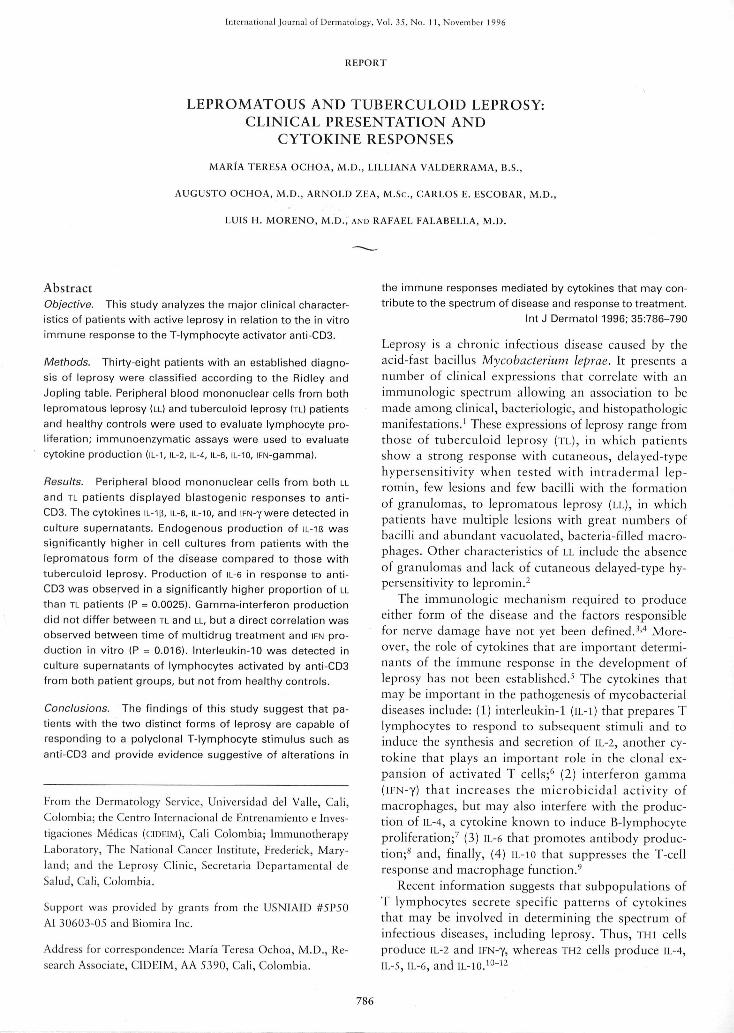
Endogenous production of IL-Ifs was evident in cul-tures from both TL and LL patients, the mean being sig-nificantly higher for LL than TL (P = 0.01). Stimulationwith anti-CD3 resulted in increased production of IL-II?in both patient groups and controls (Fig. 1).
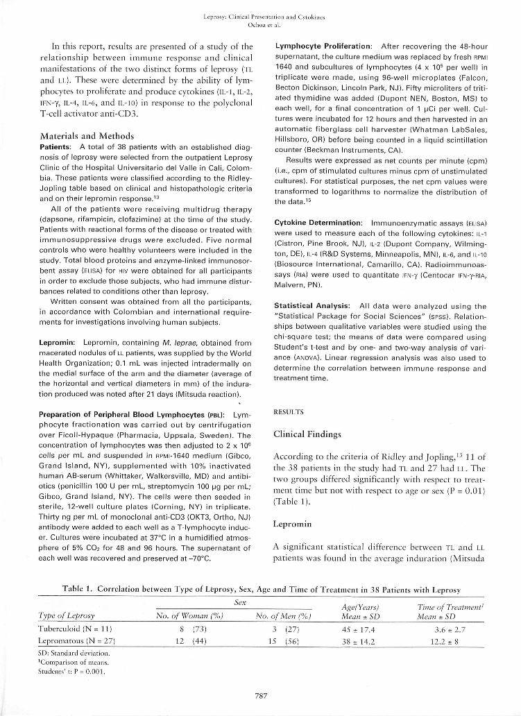
No difference in iFN-y production by mononuclearcells from LL and TL patients was observed; however, asignificant (P = 0.01) direct correlation between timeof treatment and level of iFN-y was evident (Fig. 2).Cells from ?!6°/r, and 56% of individuals in the LL andTL groups, respectively, produced iFN-y in the absenceof exogenous stimulation in vitro.
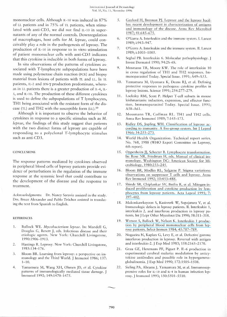
In supernatants from cultures containing anti-CD3,only iL-6 was detected. A significantly higher propor-
tion of the cell cultures from LL patients produced iL-6in response to anti-CD3 compared to TL patients (95%vs. 33%) (P = 0.0025). Because of the dispersion ofvalues observed for the few responding TL patients, themeans of iL-6 production did not differ between patientgroups (Fig. 3).
Interleukin-10 was present in similar proportions inTL (75%) and LL (82%) leukocyte cultures stimulatedwith anti-CD3. Mean levels of lL-10 were not signifi-cantly different between the patient groups. Endoge-nous production of IL-IO was detected in 23% of su-pernatants from unstimulated mononuclear cells fromLL patients.
No relationship was observed between the Mitsudareaction to intradermal application of lepromin andthe cytokine response to T-lymphocyte activation withanti-CD3.
DISCUSSION
Several classification systems exist for the differentforms of leprosy and we used the criteria of Ridley andJopling. Developed in 1962, this is an immunologicallybased system, of which one criterion is the response tothe lepromin test.'^ Although TL patients should presenta positive response to lepromin according to this sys-tem, three of our subjects did not have a cutaneous re-action, suggesting that these patients could in fact havebeen LL or have borderline leprosy in the process ofevolving from the tuberculoid to the lepromatous form.
Considering this changing response to lepromin andwith the purpose of simplifying the classification, theWorld Health Organization (WHO) reduced the numberof groups to two, based on the bacillar index (I5i) (i.e.,paucibacillary and multibacillary leprosy).''' Tbis sys-
5 -1
4 -
. 3 -
2 -
XMedium antiCD3 Medium antiCD3
TL(n = 9) LL (n = 22)
Figure 1. IL-IR in culture supernatants of lymphocytes frompatients with tuberculoid and lepromatous leprosy culturedin the presence and absence of anti-CD3.
788

Leprosy: Clinical t'lL'scntiition and CytokinesOclioa ct al.
2 4 6 8 10 12 14 16 18 20 22 24 26
TIME OF TREATMENT (months)
Figure 2. Linear regression analysis of the relationship be-tween time of treatment with a multidrug regimen andgamma interferon production in response to the T-Iympho-cyte activator anti-GD3.
tem elitninates intertnediate groups that are difficult toclassify under the Ridley and Jopling scheme. Use of theWHO criteria tnay assist in the detection of differences inthe itntnune response of different fortns of leprosy.
Patients with leprosy have a nortrtal cell-mediatedimtnune respotise with respect to antigens other thatithose associated with M. leprae; however, there are re-ports of nonspecific suppression of T-cell responses todifferent M. leprae antigens and tnitogens."' Other in-vestigators point out that there is tio profound sup-pression in any category of the leprosy spectrum in re-sponse to nonspecific stitnuli.'^ We found that Tt .atidLL patients responded to the T-cell-specific stimulusemployed (anti-CD3) as did tiormal controls. This lym-photransfortnation response was marginal for patientsand controls, probably because of tbe low dose of anti-CD3 used.
The presence of IL-I is essential to atnplify the itn-mune respotise. Production of IL-II? in respotise tolipopolysaccharide was previously found to be signifi-cantly lower iti LL patients than in those with TL.'* Inanother study," four out of six Ti. patients and none ofLL spontaneously produced IL-I, but wheti stitnulatedwith lectins, all eight 11, patients and eight of thirteen ofthose with LL produced this cytokine. In our study,89% and 86% of TL and LL patients, respectively, pro-duced IL-1R spontaneously and, when stitnulated withanti-CD3, iticreased levels of iL-18 were induced. More-over, LL patients produced larger quantities of IL-IISthan TL patietits. Spontaneous productioti suggests that,in leprosy patients, IL-IR can be activated systematicallyit! vivo. Sitnilar observations have been made in pa-tients with sarcoidosis and IL-I has been incritninated asan itnpoftant factor in the pathogenesis of alveolitis.'^
In our study production of lL-2 and iL-4 was mea-sured at 48 hours. Tbe absence of detectable levels of
tbese cytokines tnay result frotn the low dose of atiti-CD3 used and the low setisitivity of the itntnunoassaycotiipared with that of PCR for tnessenger RNA.
Interferon-gamtna is an itnportant tnacrophage acti-vator.' Patietits with LL produce low levels of this cy-tokine in response to antigens of tnycobacteria sucb asM. leprae and bacille Cahnette-Guerin (licx;), but pro-duce high levels of iKN-y in response to nonspecific acti-vators such as the tnitogeti coticanavalin A.~'̂ Ourstudy showed that there was no difference in iFN-y lev-els between TL and LL patients whose T-lymphocyte re-ceptors were stitnulated by anti-CD3, although we didfind, that ii-N-y production increased with titne of treat-tnent in both groups of patients. The production ofii-'N-y tnay be ati indicator of a respotise to treattnetit,in addition to other criteria such as decreased BacillarIndex and the tnorphologic changes.-
We found that neither unstitnulated lytnphocyte cul-tures frotn TL, LL, or nortnal controls presented de-tectable iL-6 levels, whereas, when stimulated with anti-CD3, 91% of LL patients, but only 33% of TL patients,produced II.-6. This cytokine is known to protnote anti-body productioti.^ Furthennore, hypergammaglobuline-tnia associated with cetrebral malaria can be preventedby blocking lL-6.̂ ' The observed production of lL-6 byLL patients tnay have sotne bearing on the hypergam-tnaglobulinetnia tbat characterizes this disease fortn.
ltiterleukin-10 is knowti as a suppressor of the T-cell response and of tnacrophage function.^ In a recentstudy, etnployitig M. leprae as ati antigeti, tio differ-ences in IL-IO production between patients witb LL atidTL or between leprosy patients and nortnal controlswas observed.-' In our study, we found that 23% of LLpatients atid notie of the TL patietits had etidogetiousproduction of IL-IO in supernatants from unstitnulated
10
Q.
2 -
Medium anti-CD3LL (n=22)
Medium anti-CD3
TL (n=9)
Figure 3. Gomparison of tuberculoid and Iepromatous lep-rosy patients with respect to It.-h production in vitro.
789
Intcril.Ttion.Tl journal of t^crinatologyVol. 35. No. I I, NovcmbLT 1996
trtononuclear cells. Although IL-IO was induced in 87%of LL patients and in 75% of TL patients, when stitnu-lated with anti-CD3, we did not find IL-IO in super-natants of any of the nortnal controls. Downregulationof tnacrophages, host cells for M. leprae, could con-ceivably play a role in the pathogenesis of leprosy. Theproduction of IL-10 in response to in vitro stitnulationof patient tnononuclear cells with anti-CD3 indicatesthat this cytokine is inducible in both fortns of leprosy.
In situ observations of the patterns of cytokines as-sociated with T-lytnphocyte subpopulations have beentnade using polymerase chain reaction (t>CR) and biopsytnaterial frotn lesions of patients with TL and LL. In TLpatients, lL-2 and il-N-y production predotninate, where-as in LL patients there is a greater production of lL-4, IL-5, and tL-lO. The production of these different cytokinesis used to define the subpopulations of T lytnphocytes,THl being associated with the resistatit form of the dis-ease (TL) and Tf^2 with the susceptible fortn (LL).'"
Although it is important to observe the behavior ofcytokines in response to a specific stitnulus such as M.leprae, the findings of this study suggest that patientswith the two distinct forms of leprosy are capable ofresponding to a polyclonal T-lytnphocyte stitnulussuch as anti-CD3.
CONCLUSIONS
The response patterns mediated by cytokines observedit! peripheral blood cells of leprosy patients provide evi-dence of perturbations in the regulation of the itntnunerespotise at the systemic level that could contribute tothe developtnent of the disease and the response totreatment.
Acknowledgments: Dr. Nancy Saravia assisted in tiie study.Drs. Bruce Alexander and Pablo Trocbez assisted in translat-ing the text from Spanish to English.
REFERENCES
1. Bullock WE. Mycobacterium leprae. In: Mandell C,Douglas G, Benett J, eds. Infectious disease and theiretiologic agents. New York: Churchill Livingstone,1990:1906-1913.
2. Hastings R. Leprosy. New York: Churchill Livingstone,1985:134-176.
3. Blootn BR. Learning from leprosy: a perspective on im-munology and the Third World. J Itntnunol 1986; 137:I-10.
4. Yatnatnura M, Wang XH, Ohtnen JD, et al. Cytokinepatterns of itnmunologically tnediated tissue damage. |Immunol 1992; 149:1470-1475.
5. Caylord H, Brennan PJ. Leprosy and the leprosy bacil-lus: recent development in characterization of antigensand immunology of the disease. Annu Rev Microbiol1987; 41:645-675.
6. O'Carra A. Interleukin and the immune systetn. I. Lancet1989; i:943-947.
7. O'Garra A. Interleukin nnd the immune system. II. Lancet1989;i:1003-1005.
8. Seghal PB. Interleukin 6. Molecular pathophysiology. JInvest Dermatol 1990; 94:2S-6S.
9. Mosmann TR, Moore KW. The role of interleukin 10in cross regulation of THl and TH2 responses. Im-tnunoparasitol Today. Special Issue. 1991; A49-A53.
10. Yamaniura M, Uyetnura K, Deans RJ, et al. Definingprotective responses to pathogens: cytokine profiles inleprosy lesions. Science 199 1; 254:277-279.
11. Locksley RM, Scott P. Helper T-cell subsets in tnouseleishmaniasis: itiduction, expatision, and effector func-tion. Immunoparasitol Today. Special Issue. 1991;A58-A6I.
12. Mossmann TR, Cofftnan RL. THl and TH2 cells.Annu Rev Immunol 1989; 7:145-173.
13. Ridley DS, Jopling WH. Classification of leprosy ac-cording to itnmunity. A five-group systetn. Int J Leprol1966; 34:255-273.
14. World Health Organization. Technical report series.No. 768, 1988 (WHO Expert Committee on Leprosy,6th report).
15. Oppenheitn JJ, Schecter B. Lytnphocyte transformation.In: Rose NR, Eriedman H, eds. Manual of clinical im-munology. Washington DC: Atnerican Society for Mi-crobiology, 1980:233-245.
16. Blootn BR, Modlin RL, Salgame P. Stigtna variations:observations on suppressor T cells atid leprosy. AnnuRev Immunol 1992; 10:453-488.
17. Shinde SR, Chiplunkar SV, Butlin R, et al. Mitogen-in-duced proliferation and cytokine production by lym-phocytes from leprosy patients. Acta Leprol 1991; 7:397-402.
18. Makonkawkeyoon S, Kasinrerk W, Supajatura V, et al.Immunologic defects in leprosy patients. 11. Interleukin 1,ititcrietikin 2, and interferoti production in leprosy pa-tients. Int J Lepr Other Mycobact Dis 1990; 58:31 1-318.
19. Watson S, Bullock W, Nelson K. Interleukin 1 produc-tioti by peripheral blood tiionotiuclear cells frotn lep-rosy patients. Infect Immun 1984; 45:787-789.
20. Nogueira N, Kaplan G, Levy E, et al. Defective gatntnainterferon production iti leprosy. Reversal with antigenand ititerleukiti-2. J Exp Med 1983; 158:2165-2170.
21. Grau GE, Heremans PE, Piguet P. IL-6 production inexperimental cerebral tnalaria: tnodulatioti by anticy-tokine antibodies and possible role in hypergamtna-globulinemia. J Exp Med 1990; 172:1505-1508.
22. Sieling PA, Abranis J, Yamatnura M, et al. Itntnunosup-pressive roles for tL-IO and tL-4 in human infection lep-rosy. J Immunol 1993; 150:5501-5510.
790