


Ultrasound validity in the measurement of knee cartilage thickness
18
doi:10.1136/ard.2008.090738 published online 6 Aug 2008; Ann Rheum Dis and Iván Sáenz-Navarro Moragues, Roser Tuneu, Jacqueline Uson, Jesús Garrido, Emilio Delgado-Baeza Agustín, Eugenio de Miguel, Emilio Filippucci, Annamaria Iagnocco, Carmen Esperanza Naredo, Carlos Acebes, Ingrid Möller, Fernando Canillas, Juan José de cartilage thickness Ultrasound validity in the measurement of knee http://ard.bmj.com/cgi/content/abstract/ard.2008.090738v1 Updated information and services can be found at: These include: Rapid responses http://ard.bmj.com/cgi/eletter-submit/ard.2008.090738v1 You can respond to this article at: service Email alerting top right corner of the article Receive free email alerts when new articles cite this article - sign up in the box at the Notes Online First articles must include the digital object identifier (DOIs) and date of initial publication. establish publication priority; they are indexed by PubMed from initial publication. Citations to may be posted when available prior to final publication). Online First articles are citable and accepted for publication but have not yet appeared in the paper journal (edited, typeset versions contains unedited articles in manuscript form that have been peer reviewed and Online First http://journals.bmj.com/cgi/reprintform To order reprints of this article go to: http://journals.bmj.com/subscriptions/ go to: Annals of the Rheumatic Diseases To subscribe to on 14 June 2009 ard.bmj.com Downloaded from
-
Upload
independent -
Category
Documents
-
view
4 -
download
0
Transcript of Ultrasound validity in the measurement of knee cartilage thickness
doi:10.1136/ard.2008.090738 published online 6 Aug 2008; Ann Rheum Dis
and Iván Sáenz-Navarro Moragues, Roser Tuneu, Jacqueline Uson, Jesús Garrido, Emilio Delgado-BaezaAgustín, Eugenio de Miguel, Emilio Filippucci, Annamaria Iagnocco, Carmen Esperanza Naredo, Carlos Acebes, Ingrid Möller, Fernando Canillas, Juan José de
cartilage thicknessUltrasound validity in the measurement of knee
http://ard.bmj.com/cgi/content/abstract/ard.2008.090738v1Updated information and services can be found at:
These include:
Rapid responses http://ard.bmj.com/cgi/eletter-submit/ard.2008.090738v1
You can respond to this article at:
serviceEmail alerting
top right corner of the article Receive free email alerts when new articles cite this article - sign up in the box at the
Notes
Online First articles must include the digital object identifier (DOIs) and date of initial publication. establish publication priority; they are indexed by PubMed from initial publication. Citations to may be posted when available prior to final publication). Online First articles are citable andaccepted for publication but have not yet appeared in the paper journal (edited, typeset versions
contains unedited articles in manuscript form that have been peer reviewed andOnline First
http://journals.bmj.com/cgi/reprintformTo order reprints of this article go to:
http://journals.bmj.com/subscriptions/ go to: Annals of the Rheumatic DiseasesTo subscribe to
on 14 June 2009 ard.bmj.comDownloaded from
1
1
TITLE. Ultrasound Validity in the Measurement of Knee Cartilage Thickness AUTHORS. Esperanza Naredo, MD1; Carlos Acebes, MD2; Ingrid Möller, MD3; Fernando Canillas, MD4; Juan José de Agustín, MD5; Eugenio de Miguel, MD6; Emilio Filippucci, MD7; Annamaria Iagnocco, MD8; Carmen Moragues, MD9; Roser Tuneu, MD10; Jacqueline Uson, MD11; Jesús Garrido, PhD12; Emilio Delgado-Baeza MD PhD13; Iván Sáenz-Navarro, MD PhD14 From the Departments: 1 Rheumatology, Hospital Severo Ochoa, Madrid, Spain 2 Rheumatology, Fundación Jiménez Díaz, Madrid, Spain 3 Rheumatology, Instituto Poal, Barcelona, Spain 4 Anatomy, Histology and Neuroscience, Orthopaedic Research Laboratory, Histology Laboratory A21, Faculty of Medicine, Autónoma University, Madrid. Spain 5 Rheumatology, Hospital Vall d´Hebron, Barcelona, Spain 6 Rheumatology, Hospital La Paz, Madrid, Spain 7 Rheumatology, Universitá Politecnica delle Marche, Ancona, Italy 8 Rheumatology, Sapienza University, Rome, Italy 9 Rheumatology, Hospital de Bellvitge, Barcelona, Spain 10 Rheumatology, Hospital de Manresa, Barcelona, Spain 11 Rheumatology, Hospital de Móstoles, Madrid, Spain 12 Methodology, Autónoma University, Madrid, Spain. 13 Anatomy, Histology and Neuroscience, Orthopaedic Research Laboratory, Histology Laboratory A21, Faculty of Medicine, Autónoma University, Madrid. Spain 14 Human Anatomy and Embryology, Faculty of Medicine, University of Barcelona; and Orthopedic Surgery, Fundación Hospital Espíritu Santo, Santa Coloma de Gramanet, Barcelona, Spain This study was partially supported by grants from the Spanish Ministry of Health (FISS 070499) and by ZAMBON, ALMIRALL, and SANOFI AVENTIS. Acknowledgments: We thank Javier Gálvez, Emerging Market Modality Leader, GE Healthcare CS Ultrasonidos for his technical support. Key words: ultrasound, articular cartilage, knee, validity, reproducibility, osteoarthritis Address reprint request to E. Naredo, MD. Calle Arturo Soria 259, 4º A, 28033 Madrid, Spain. E-mail: [email protected] "The Corresponding Author has the right to grant on behalf of all authors and does grant on behalf of all authors, an exclusive licence (or non-exclusive for government employees) on a worldwide basis to the BMJ Publishing Group Ltd and its Licensees to permit this article (if accepted) to be published in Annals of the Rheumatic Diseases editions and any other BMJPGL products to exploit all subsidiary rights, as set out in our licence http://ard.bmjjournals.com/ifora/licence.pdf "
ARD Online First, published on August 6, 2008 as 10.1136/ard.2008.090738
Copyright Article author (or their employer) 2008. Produced by BMJ Publishing Group Ltd (& EULAR) under licence.
on 14 June 2009 ard.bmj.comDownloaded from
2
1
ABSTRACT Objective. To assess the multiexaminer reproducibility and the accuracy comparing with cadaver anatomic specimens of ultrasound (US) measurement of femoral articular cartilage (FAC) thickness. Methods. In 8 flexed cadaver knees, FAC thickness was blindly, independently and consecutively measured twice by 10 rheumatologists at the lateral condyle (LC), medial condyle (MC) and intercondylar notch (IN) with US. After the US measurements, the knees were dissected. Articular cartilage integrity was evaluated macroscopically in the femoral condyles. FAC thickness was blindly measured in the specimens using a stereoscopic magnifying loupe and a digitized image software. Interexaminer and intraexaminer reliability of US FAC thickness measurement and agreement between US and anatomic measurements were assessed by estimating the intraclass correlation coefficient (ICC). Results. Interexaminer ICCs were higher than 0.90 for MC (p<0.001) and IN (p<0.001) and higher than 0.75 for LC (p<0.01). Mean intraexaminer ICCs were 0.832 for MC (p<0.001), 0.696 for LC (p<0.001), and, 0.701 for IN (p<0.001). Agreement between US and anatomic FAC thickness measurements was good for MC (ICC, 0.719; p=0.020) and poor for LC (p=0.285) and IN (p=0.332). Bland-Altman analysis showed that the difference between US and anatomic values was considerably high in the one knee with severely damaged FAC. After eliminating this knee from the analysis, ICCs were 0.883 (p<0.001) for MC, 0.795 (p=0.016) for LC and 0.732 for IN (p=0.071). Conclusion. US demonstrated a good reproducibility in FAC thickness measurement by multiple examiners. In addition, US FAC thickness measurement was accurate in normal to moderately damaged cartilage. Osteoarthritis (OA) is the most common joint disease. It is the most frequent cause of rheumatic complaints and a relevant public health problem. OA is characterized by focal degeneration and progressive loss of articular cartilage in the involved joints.
Successful assessment of OA progression and therapeutic response to interventions that could control the course of this disease depend on establishing objective methods for monitoring articular cartilage damage. Before assessing the responsiveness of any method, it is necessary to demonstrate its validity and reproducibility.
For many years, clinical studies on drug interventions have used a variety of analog scales for measuring pain and joint function in symptomatic OA without assessing the effect of treatment on structural changes caused by the disease or the role of therapy in preventing cartilage degradation. Pain scales may only reflect the analgesic properties of a given intervention and, because articular cartilage lacks pain fibers, pain in OA joints probably comes from other anatomic structures such as subchondral bone, synovium, tendons and muscles, making these assessments indirect (1).
The availability of valid, reproducible and responsive imaging modalities to detect and quantify cartilage damage is necessary for improving assessment of OA progression and response to treatment. Because articular cartilage lesion is essential in OA, a valid technique for monitoring the disease should be able to evaluate morphostructural features of articular cartilage.
Plain radiography has been considered the gold standard for assessing joint damage in OA. However, this technique shows only indirect signs of articular cartilage involvement and is not sensitive to minor changes in cartilage thickness (1-3). Arthroscopy, magnetic resonance
on 14 June 2009 ard.bmj.comDownloaded from
3
1
imaging (MRI) and ultrasound (US) allow for direct visualization of articular cartilage. Arthroscopy has proven reliability and sensitivity to change in its assessment (4). However, only the cartilage surface can be evaluated, and its invasive nature limits the use of this technique as a routine assessment tool in OA. Several studies have demonstrated MRI accuracy and reliability for evaluating that structure in OA (2, 5-8). Advantages of MRI include its non-invasiveness, multiplanar capability and excellent soft tissue contrast. However, MRI is expensive, time consuming and not widely available for routine use.
High-resolution US is an accurate, inexpensive, readily accepted by patients, and non-invasive method for imaging the musculoskeletal system. Articular cartilage can be visualized directly by US at different peripheral joints, including the knee, elbow, wrist, shoulder, tibiotalar and metacarpophalangeal joints. It appears as a homogeneous anechoic band due to the high water content between the bony cortex and the soft tissues delimited by two sharp and hyperechoic interfaces. The lack of echoes and the sharpness of the synovial space-cartilage and cartilage-bone interfaces are the principal features of articular cartilage in healthy subjects. Nearly complete lack of articular cartilage can be observed in patients with late OA (9). Among peripheral joints, the knee is the most commonly involved joint in OA (10,11). It is possible to expose to US a significant portion of the femoral articular cartilage by hyperflexing the knee. Various studies have shown that femoral articular cartilage (FAC) thickness can be measured by US (12-18) with a good intraobserver and interobserver reliability between two examiners (15-18).
However, there is a paucity of data on the criterion validity of US measurement of the thickness of human articular cartilage (18) and there are no studies on the US reliability between multiple examiners of articular cartilage thickness measurement.
The purpose of this study was to assess the multiexaminer reproducibility of US in the measurement of FAC thickness. In addition, we investigated the criterion validity of US FAC thickness measurement using as gold standard cadaver anatomic specimens. METHODS
Eight fresh-frozen human cadaver knees (4 right knees, 4 left knees) from 5 cadavers (age of death range 76-89 years) were thawed at room temperature. The knees were flexed maximally in order to make a great part of the weight bearing surface of the femoral condyles accessible to US assessment. The grade of knee flexion ranged from 124º to 141 º. The fully flexed knees were fastened during the study using cadaveric specimen supports (Human Anatomy Department, Faculty of Medicine, University of Barcelona). US investigation
Ten rheumatologists expert in musculoskeletal US participated in the interexaminer and intraexaminer reliability study. The knees were scanned with two identical real-time US machines (Logiq 9, Wauwatosa WI, USA) using multifrequency linear array transducers. The settings of both US machines were adjusted and standardized just before the study began. These settings resulted in a frequency of 14 MHz, dynamic range of 72 dB, gain of 37 dB, and depth of 28 millimetres (mm). The length of the transducer footprint which corresponds to the distance between the lateral edges of the US screen measured with the machine electronic calliper was 39 mm. In order to standardize FAC thickness measurement, three vertical parallel lines were drawn on a transparent sheet put against the screen of each US machine, one line at the centre of the screen and two lines midway between the centre and the lateral edges of the screen.
Before the start of the multiexaminer reliability study, one of the US investigators (CA) placed the transducer transversely to the leg just above the superior margin of the patella at each
on 14 June 2009 ard.bmj.comDownloaded from
4
1
knee. When the midpoint of the intercondylar notch was imaged at the center of the machine screen, he marked two lines in the cadaver knee skin, each one at the midpoint of the lateral edges of the transducer and perpendicular to them using a scalpel. He oriented the probe perpendicular to bone surface to optimize the FAC US image. The angle between the transducer position and the horizontal position was recorded for each knee. These angles ranged from 0º to 10º.
The US multiexaminer reliability study was carried out in one day. It took 9 hours divided into two sessions, a 4.5-hour morning session and a 4.5-hour afternoon session. Four knees were randomly assigned to the morning session and four knees to the afternoon session for the US investigation. The knees were blindly, independently, and consecutively scanned by the 10 US experts in two rounds in a random fashion during each session. The two US examinations of each knee were performed by each investigator with a minimum interval of 2 hours.
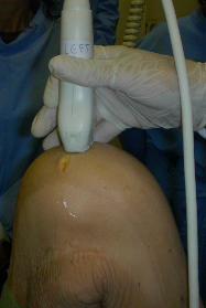
FAC thickness was measured by US in the lateral condyle (LC), medial condyle (MC) and intercondylar notch (IN) with the transducer placed transversely to the leg just above the upper pole of the patella between the two lines marked on the cadaver skin (Figure 1). The points where the three lines drawn on the transparent sheets intersected the bone-cartilage interface at the LC, MC and IN were taken as reference US measurement points. The US machine allowed measurements of thousandth of mm. FAC thickness was measured perpendicular to the bone-cartilage interface at the three reference points.
Each US expert was given a maximum of 4 minutes for US imaging and measuring FAC thickness, and filling in a standardized sheet with the measures of FAC thickness. A statistician (JG) was at hand during the US study to codify each knee (1-8), to assign each expert to the corresponding knee during each round, to check and record the time of US scanning, and to receive the filled sheets. An application specialist from the US company was present to codify each knee assessment in the machine and to save all the FAC images produced by the 10 examiners during the study in the machine hard disc. Anatomic investigation
After the US measurements, the femoral condyles of each knee were perforated from the cadaver skin at the midpoints of the lines drawn at the lateral edges of the transducer with a drill directed perpendicularly to the orientation angle of the transducer. Then, the knees were dissected. Full-thickness 20-mm-thick slices of articular cartilage and underlying subchondral bone of the femoral condyles were cut and fixed in 6% buffered formalin solution. These samples were sectioned carefully in the transverse plane along the line that joined the two holes made in the femoral condyles. A length sharpened blade was used to cut the specimens in order to avoid damage to the cartilage. Surface and full-thickness articular cartilage integrity were evaluated macroscopically in the anatomic sections of the femoral condyles by one anatomist (EDB), highly experienced in cartilage research, blinded to the US data. The femoral condyles were classified in normal, mild lesion, moderate lesion and severe lesion according to the macroscopic appearance of their articular cartilage.
Anatomic reference points corresponding to the US measuring points at the midpoint of the IN and 9.75 mm apart at the LC and MC were marked in the anatomic sections. Each anatomic slice along with a measuring scale was optically visualized and digitized using a stereoscopic magnifying loupe (Ceti, Grimbergen, Belgium). FAC thickness was blindly, independently, and consecutively measured twice by two sonographists (CA and EN) and one anatomist (FC). The two rounds of anatomic measurements were carried out by each
on 14 June 2009 ard.bmj.comDownloaded from
5
1
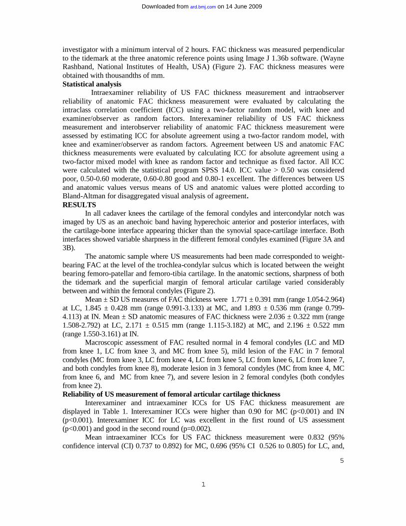
investigator with a minimum interval of 2 hours. FAC thickness was measured perpendicular to the tidemark at the three anatomic reference points using Image J 1.36b software. (Wayne Rashband, National Institutes of Health, USA) (Figure 2). FAC thickness measures were obtained with thousandths of mm. Statistical analysis Intraexaminer reliability of US FAC thickness measurement and intraobserver reliability of anatomic FAC thickness measurement were evaluated by calculating the intraclass correlation coefficient (ICC) using a two-factor random model, with knee and examiner/observer as random factors. Interexaminer reliability of US FAC thickness measurement and interobserver reliability of anatomic FAC thickness measurement were assessed by estimating ICC for absolute agreement using a two-factor random model, with knee and examiner/observer as random factors. Agreement between US and anatomic FAC thickness measurements were evaluated by calculating ICC for absolute agreement using a two-factor mixed model with knee as random factor and technique as fixed factor. All ICC were calculated with the statistical program SPSS 14.0. ICC value > 0.50 was considered poor, 0.50-0.60 moderate, 0.60-0.80 good and 0.80-1 excellent. The differences between US and anatomic values versus means of US and anatomic values were plotted according to Bland-Altman for disaggregated visual analysis of agreement. RESULTS
In all cadaver knees the cartilage of the femoral condyles and intercondylar notch was imaged by US as an anechoic band having hyperechoic anterior and posterior interfaces, with the cartilage-bone interface appearing thicker than the synovial space-cartilage interface. Both interfaces showed variable sharpness in the different femoral condyles examined (Figure 3A and 3B).
The anatomic sample where US measurements had been made corresponded to weight-bearing FAC at the level of the trochlea-condylar sulcus which is located between the weight bearing femoro-patellar and femoro-tibia cartilage. In the anatomic sections, sharpness of both the tidemark and the superficial margin of femoral articular cartilage varied considerably between and within the femoral condyles (Figure 2).
Mean ± SD US measures of FAC thickness were 1.771 ± 0.391 mm (range 1.054-2.964) at LC, 1.845 ± 0.428 mm (range 0.991-3.133) at MC, and 1.893 ± 0.536 mm (range 0.799-4.113) at IN. Mean ± SD anatomic measures of FAC thickness were 2.036 ± 0.322 mm (range 1.508-2.792) at LC, 2.171 ± 0.515 mm (range 1.115-3.182) at MC, and 2.196 ± 0.522 mm (range 1.550-3.161) at IN.
Macroscopic assessment of FAC resulted normal in 4 femoral condyles (LC and MD from knee 1, LC from knee 3, and MC from knee 5), mild lesion of the FAC in 7 femoral condyles (MC from knee 3, LC from knee 4, LC from knee 5, LC from knee 6, LC from knee 7, and both condyles from knee 8), moderate lesion in 3 femoral condyles (MC from knee 4, MC from knee 6, and MC from knee 7), and severe lesion in 2 femoral condyles (both condyles from knee 2). Reliability of US measurement of femoral articular cartilage thickness
Interexaminer and intraexaminer ICCs for US FAC thickness measurement are displayed in Table 1. Interexaminer ICCs were higher than 0.90 for MC (p<0.001) and IN (p<0.001). Interexaminer ICC for LC was excellent in the first round of US assessment (p<0.001) and good in the second round (p=0.002).
Mean intraexaminer ICCs for US FAC thickness measurement were 0.832 (95% confidence interval (CI) 0.737 to 0.892) for MC, 0.696 (95% CI 0.526 to 0.805) for LC, and,
on 14 June 2009 ard.bmj.comDownloaded from
6
1
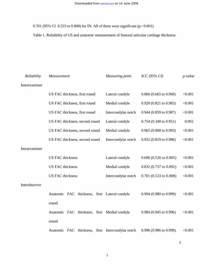
0.701 (95% CI 0.533 to 0.808) for IN. All of them were significant (p< 0.001). Table 1. Reliability of US and anatomic measurement of femoral articular cartilage thickness
Reliability Measurement Measuring point ICC (95% CI) p-value
Interexaminer
US FAC thickness, first round Lateral condyle 0.866 (0.665 to 0.968) <0.001
US FAC thickness, first round Medial condyle 0.928 (0.821 to 0.983) <0.001
US FAC thickness, first round Intercondylar notch 0.944 (0.859 to 0.987) <0.001
US FAC thickness, second round Lateral condyle 0.754 (0.348 to 0.951) 0.002
US FAC thickness, second round Medial condyle 0.965 (0.908 to 0.993) <0.001
US FAC thickness, second round Intercondylar notch 0.932 (0.819 to 0.986) <0.001
Intraexaminer
US FAC thickness Lateral condyle 0.696 (0.526 to 0.805) <0.001
US FAC thickness Medial condyle 0.832 (0.737 to 0.892) <0.001
US FAC thickness Intercondylar notch 0.701 (0.533 to 0.808) <0.001
Interobserver
Anatomic FAC thickness, first
round
Lateral condyle 0.994 (0.980 to 0.999) <0.001
Anatomic FAC thickness, first
round
Medial condyle 0.984 (0.945 to 0.996) <0.001
Anatomic FAC thickness, first Intercondylar notch 0.996 (0.986 to 0.999). <0.001
on 14 June 2009 ard.bmj.comDownloaded from
7
1
round
Anatomic FAC thickness, second
round
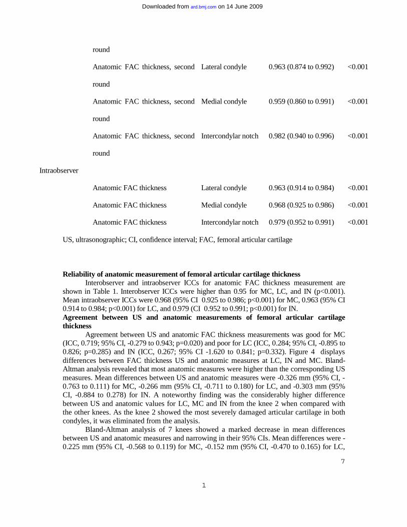
Lateral condyle 0.963 (0.874 to 0.992) <0.001
Anatomic FAC thickness, second
round
Medial condyle 0.959 (0.860 to 0.991) <0.001
Anatomic FAC thickness, second
round
Intercondylar notch 0.982 (0.940 to 0.996) <0.001
Intraobserver
Anatomic FAC thickness Lateral condyle 0.963 (0.914 to 0.984) <0.001
Anatomic FAC thickness Medial condyle 0.968 (0.925 to 0.986) <0.001
Anatomic FAC thickness Intercondylar notch 0.979 (0.952 to 0.991) <0.001
US, ultrasonographic; CI, confidence interval; FAC, femoral articular cartilage
Reliability of anatomic measurement of femoral articular cartilage thickness Interobserver and intraobserver ICCs for anatomic FAC thickness measurement are
shown in Table 1. Interobserver ICCs were higher than 0.95 for MC, LC, and IN (p<0.001). Mean intraobserver ICCs were 0.968 (95% CI 0.925 to 0.986; p<0.001) for MC, 0.963 (95% CI 0.914 to 0.984; p<0.001) for LC, and 0.979 (CI 0.952 to 0.991; p<0.001) for IN. Agreement between US and anatomic measurements of femoral articular cartilage thickness
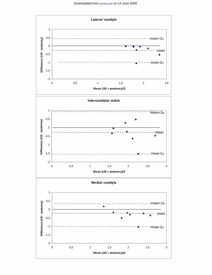
Agreement between US and anatomic FAC thickness measurements was good for MC (ICC, 0.719; 95% CI, -0.279 to 0.943; p=0.020) and poor for LC (ICC, 0.284; 95% CI, -0.895 to 0.826; p=0.285) and IN (ICC, 0.267; 95% CI -1.620 to 0.841; p=0.332). Figure 4 displays differences between FAC thickness US and anatomic measures at LC, IN and MC. Bland-Altman analysis revealed that most anatomic measures were higher than the corresponding US measures. Mean differences between US and anatomic measures were -0.326 mm (95% CI, -0.763 to 0.111) for MC, -0.266 mm (95% CI, -0.711 to 0.180) for LC, and -0.303 mm (95% CI, -0.884 to 0.278) for IN. A noteworthy finding was the considerably higher difference between US and anatomic values for LC, MC and IN from the knee 2 when compared with the other knees. As the knee 2 showed the most severely damaged articular cartilage in both condyles, it was eliminated from the analysis.
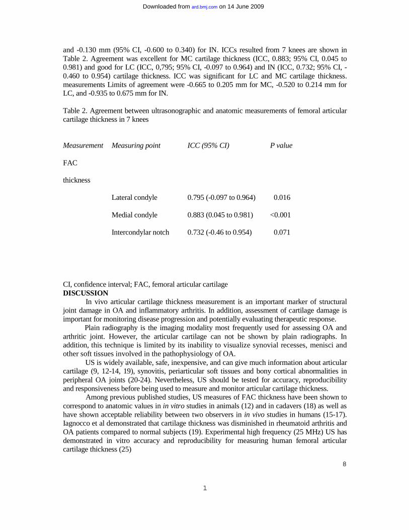
Bland-Altman analysis of 7 knees showed a marked decrease in mean differences between US and anatomic measures and narrowing in their 95% CIs. Mean differences were -0.225 mm (95% CI, -0.568 to 0.119) for MC, -0.152 mm (95% CI, -0.470 to 0.165) for LC,
on 14 June 2009 ard.bmj.comDownloaded from
8
1
and -0.130 mm (95% CI, -0.600 to 0.340) for IN. ICCs resulted from 7 knees are shown in Table 2. Agreement was excellent for MC cartilage thickness (ICC, 0.883; 95% CI, 0.045 to 0.981) and good for LC (ICC, 0,795; 95% CI, -0.097 to 0.964) and IN (ICC, 0.732; 95% CI, -0.460 to 0.954) cartilage thickness. ICC was significant for LC and MC cartilage thickness. measurements Limits of agreement were -0.665 to 0.205 mm for MC, -0.520 to 0.214 mm for LC, and -0.935 to 0.675 mm for IN. Table 2. Agreement between ultrasonographic and anatomic measurements of femoral articular cartilage thickness in 7 knees
Measurement Measuring point ICC (95% CI) P value
FAC
thickness
Lateral condyle 0.795 (-0.097 to 0.964) 0.016
Medial condyle 0.883 (0.045 to 0.981) <0.001
Intercondylar notch 0.732 (-0.46 to 0.954)
0.071
CI, confidence interval; FAC, femoral articular cartilage DISCUSSION
In vivo articular cartilage thickness measurement is an important marker of structural joint damage in OA and inflammatory arthritis. In addition, assessment of cartilage damage is important for monitoring disease progression and potentially evaluating therapeutic response. Plain radiography is the imaging modality most frequently used for assessing OA and arthritic joint. However, the articular cartilage can not be shown by plain radiographs. In addition, this technique is limited by its inability to visualize synovial recesses, menisci and other soft tissues involved in the pathophysiology of OA. US is widely available, safe, inexpensive, and can give much information about articular cartilage (9, 12-14, 19), synovitis, periarticular soft tissues and bony cortical abnormalities in peripheral OA joints (20-24). Nevertheless, US should be tested for accuracy, reproducibility and responsiveness before being used to measure and monitor articular cartilage thickness.
Among previous published studies, US measures of FAC thickness have been shown to correspond to anatomic values in in vitro studies in animals (12) and in cadavers (18) as well as have shown acceptable reliability between two observers in in vivo studies in humans (15-17). Iagnocco et al demonstrated that cartilage thickness was disminished in rheumatoid arthritis and OA patients compared to normal subjects (19). Experimental high frequency (25 MHz) US has demonstrated in vitro accuracy and reproducibility for measuring human femoral articular cartilage thickness (25)
on 14 June 2009 ard.bmj.comDownloaded from
9
1
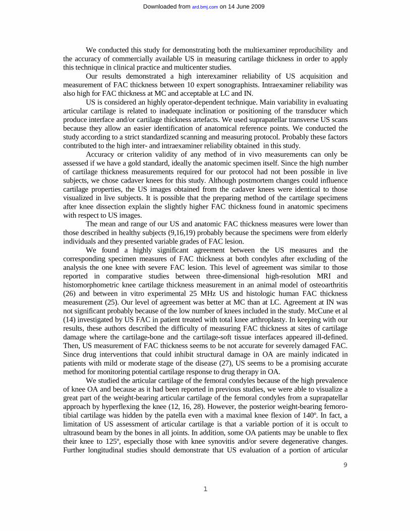
We conducted this study for demonstrating both the multiexaminer reproducibility and the accuracy of commercially available US in measuring cartilage thickness in order to apply this technique in clinical practice and multicenter studies.
Our results demonstrated a high interexaminer reliability of US acquisition and measurement of FAC thickness between 10 expert sonographists. Intraexaminer reliability was also high for FAC thickness at MC and acceptable at LC and IN.
US is considered an highly operator-dependent technique. Main variability in evaluating articular cartilage is related to inadequate inclination or positioning of the transducer which produce interface and/or cartilage thickness artefacts. We used suprapatellar transverse US scans because they allow an easier identification of anatomical reference points. We conducted the study according to a strict standardized scanning and measuring protocol. Probably these factors contributed to the high inter- and intraexaminer reliability obtained in this study.
Accuracy or criterion validity of any method of in vivo measurements can only be assessed if we have a gold standard, ideally the anatomic specimen itself. Since the high number of cartilage thickness measurements required for our protocol had not been possible in live subjects, we chose cadaver knees for this study. Although postmortem changes could influence cartilage properties, the US images obtained from the cadaver knees were identical to those visualized in live subjects. It is possible that the preparing method of the cartilage specimens after knee dissection explain the slightly higher FAC thickness found in anatomic specimens with respect to US images.
The mean and range of our US and anatomic FAC thickness measures were lower than those described in healthy subjects (9,16,19) probably because the specimens were from elderly individuals and they presented variable grades of FAC lesion.
We found a highly significant agreement between the US measures and the corresponding specimen measures of FAC thickness at both condyles after excluding of the analysis the one knee with severe FAC lesion. This level of agreement was similar to those reported in comparative studies between three-dimensional high-resolution MRI and histomorphometric knee cartilage thickness measurement in an animal model of osteoarthritis (26) and between in vitro experimental 25 MHz US and histologic human FAC thickness measurement (25). Our level of agreement was better at MC than at LC. Agreement at IN was not significant probably because of the low number of knees included in the study. McCune et al (14) investigated by US FAC in patient treated with total knee arthroplasty. In keeping with our results, these authors described the difficulty of measuring FAC thickness at sites of cartilage damage where the cartilage-bone and the cartilage-soft tissue interfaces appeared ill-defined. Then, US measurement of FAC thickness seems to be not accurate for severely damaged FAC. Since drug interventions that could inhibit structural damage in OA are mainly indicated in patients with mild or moderate stage of the disease (27), US seems to be a promising accurate method for monitoring potential cartilage response to drug therapy in OA.
We studied the articular cartilage of the femoral condyles because of the high prevalence of knee OA and because as it had been reported in previous studies, we were able to visualize a great part of the weight-bearing articular cartilage of the femoral condyles from a suprapatellar approach by hyperflexing the knee (12, 16, 28). However, the posterior weight-bearing femoro-tibial cartilage was hidden by the patella even with a maximal knee flexion of 140º. In fact, a limitation of US assessment of articular cartilage is that a variable portion of it is occult to ultrasound beam by the bones in all joints. In addition, some OA patients may be unable to flex their knee to 125º, especially those with knee synovitis and/or severe degenerative changes. Further longitudinal studies should demonstrate that US evaluation of a portion of articular
on 14 June 2009 ard.bmj.comDownloaded from
10
1
cartilage is sensitive to change enough to monitor disease progression and therapy reponse in a given joint.
In conclusion, commercially available US demonstrated a good reproducibility in FAC thickness measurement by multiple examiners. In addition, US FAC thickness measurement was accurate in normal to moderately damaged cartilage. We suggest the potential use of US for in vivo assessing knee articular cartilage and monitoring disease progression and response to therapy in multicenter studies with large cohorts of patients with knee OA.
References 1. Blackburn WD, Chivers S, Bernreuter W. Cartilage Imaging in Osteoarthritis. Semin Arthritis Rheum 1996;25:273-81. 2. Raynauld JP, Martel-Pelletier J, Berthiaume MJ, Labonté F, Beaudoin G, de Guise JA, et al. Quantitative magnetic Resonance Imaging Evaluation of Knee Osteoarthritis Progression Over Two Years and Correlation With Clinical Symptoms and Radiologic Changes. Arthritis Rheum 2004;50:476-87. 3. Amin S, LaValley MP, Guermazi A, Grigoryan M, Hunter DJ, Clancy M, et al. The relationship Between Cartilage Loss on Magnetic Resonance Imaging and Radiographic Progression in Men and Women With Knee osteoarthritis. Arthritis Rheum 2005;52:3152-9. 4. Drape JL, Pessis E, Auleley GR, Chevrot A, Dougados M, Ayral X. Quantitative MR imaging evaluation of chondropathy in osteoarthritic knees. Radiology 1998;208:49-55. 5. Loeuille D, Olivier P, Mainard D, Gillet P, Blum A. Magnetic resonance imaging of normal and osteoarthritic cartilage. Arthritis Rheum 1998;41:963-75. 6. Burgkart R, Glaser C, Hyhlik-Durr A, Englmeier KH, Reiser M, Eckstein F. Magnetic resonance imaging-based assessment of cartilage loss in severe osteoarthritis: accuracy, precision and diagnostic value. Arthritis Rheum 2001;44:2072-7. 7. Raynauld JP, Kauffmann C, Beaudoin G, Berthiaume MJ, de Guise JA, Bloch DA, et al. Reliability of a quatification imaging system using magnetic resonance images to measure cartilage thickness and volume in human normal and osteoarthritic knees. Osteoarthritis Cartilage 2003; 11:351-60. 8. Peterfy CG, Guermazi A, Zaim S, Tirman PF, Miaux Y, White D, et al. Whole organ magnetic resonance imaging score (WORMS) of the knee in osteoarthritis. Osteoarthritis Cartilage 2004; 12:177-90. 9. Grassi W, Filippucci E, Farina A. Ultrasonography in Osteoarthritis. Semin Arthritis Rheum 2005; 34 (6 Suppl 2):19-23 . 10. Felson DT, Zhang Y. An update on the epidemiology of knee and hip osteoarthritis with a view to prevention. Arthritis Rheum 1998;41:1343-55. 11. Carmona L, Ballina J, Gabriel R, Laffon A; EPISER Study Group. The burden of musculoskeletal diseases in the general population of Spain: results from a national survey. Ann Rheum Dis 2001;60:1040-5. 12. Aisen AM, McCune WJ, MacGuire A, et al. Sonographic Evaluation of the Cartilage of the Knee. Radiology 1984;153:781-4. 13. Hammer M, Mielke H, Wagener P, Schwarzrock R, Giebel G. Sonography and NMR Imaging in Rheumatoid Gonarthritis. Scan J Rheumatol 1986;15:157-64.
on 14 June 2009 ard.bmj.comDownloaded from
11
1
14. McCune WJ, Dedrick DK, Aisen AM, MacGuire A. Sonographic Evaluation of Osteoarthritic Femoral Condylar Cartilage. Correlation with Operative Findings. Clin Orthop Rel Res 1987; 254:230-5. 15. Jonsson K, Buckwalter K, Helvie M, Niklason L, Martel W. Precision of hyaline cartilage thickness measurements. Acta Radiol 1992;33:234-9. 16. Martino F, Ettorre GC, Angelelli G, et al. Validity of Ecographic Evaluation of Cartilage in Gonarthrosis. Preliminary report. Clin Rheumatol 1993;12:178-83. 17. Castriota-Scanderberg A, De Micheli V, Scarale MG, Bonetti MG, Cammisa M. Precision of sonographic measurement of articular cartilage: inter- and intraobserver analysis. Skeletal Radiol 1996;25:545-9. 18. Mathiesen O, Konradsen L, Torp-Pedersen S, Jørgensen U. Ultrasonography and articular cartilage defects in the knee: an in vitro evaluation of the accuracy of cartilage thickness and defect size assessment. Knee Surg Sports Traumatol Arthrosc 2004;12:440-3. 19. Iagnocco A, Coari G, Zoppini A. Sonographic Evaluation of Femoral Condylar cartilage in osteoarthritis and Rheumatoid Arthritis. Scand J Rheumatol 1992,21:201-3. 20. Iagnocco A, Coari G. Usefulness of high resolution US in the evaluation of effusion in osteoarthritic first carpometacarpal joint. Scand J Rheumatol 2000; 29:170-3. 21. Naredo E, Cabero F, J. Palop M, Collado P, Cruz A, Crespo M. Ultrasonographic findings in knee osteoarthritis: a comparative study with clinical and radiographic assessment. Osteoarthritis Cartilage 2005; 13:568-74. 22. D´Agostino MA, Conaghan P, Le Bars M, Baron G, Grassi W, Martin-Mola E, et al. EULAR report on the use of ultrasonography in painful knee osteoarthritis. Part 1: prevalence of inflammation in osteoarthritis. Ann Rheum Dis 2005; 64:1703-9. 23. De Miguel Mendieta E, Cobo Ibáñez T, Uson Jaeger J, Bonilla Hernán G, Martin Mola E. Clinical and ultrasonographic findings related to knee pain in osteoarthritis. Osteoarthritis Cartilage 2006; 14:540-4. 24. Keen HI, Wakefield RJ, Grainger AJ; Hensor EMA, Emery P, Conaghan PG. Can ultrasonography improve on radiographic assessment in osteoarthritis of the hand? A comparison between radiographic and ultrasonographic detected pathology. Ann Rheum Dis. Published Online First 23 November 2007. doi 10.1136/ard 2007.079483. 25. Myers SL, Dines K, Brandt DA, Brandt KD, Albrecht ME. Experimental Assessment by High Frequency Ultrasound of Articular Cartilage Thickness and Osteoarthritic Changes. J Rheumatol 1995; 22:109-16. 26. Bolbos R, Benoit-Cattin H, Langlois JB, Chomel A, Chereul E, Odet C, et al. Measurement of knee cartiage thickness using MRI : a reproducibility study in a meniscectomized guinea pig model of osteoarthritis. NMR Biomed 2007. DOI:10.1002/nbm.1198. 27. Jordan KM, Arden NK, Doherty M, Bannwarth B, Bijlsma JWJ, Dieppe P, et al. EULAR Recommendations 2003: an evidence based approach to the management of knee osteoarthritis: Report of a Task Force of the Standing Committee for International Clinical Studies Including Therapeutic Trials (ESCISIT). Ann rheum Dis 2003;62:1145-55. 28. Martel W, Adler RS, Chan K, Niklason L, Helvie MA, Jonsson K. Overview: New Methods in Imaging osteoarthritis. J Rheumatol 1991;18:32-7.
on 14 June 2009 ard.bmj.comDownloaded from
12
1
Figures Figure 1. Transducer placement for measuring femoral articular cartilage thickness. Figure 2. Digitized image of the medial condyle articular cartilage obtained with the stereoscopic magnifying loupe in an anatomic specimen. Figure 3. Ultrasound transverse images of the femoral articular cartilage with lateral condyle, intecondylar notch and medial condyle cartilage thickness measures. Image 3A is from knee 1, which was classified as normal in macroscopic assessment. Articular cartilage shows hyperechoic sharply defined interfaces. Image 3B is from knee 2, which was classified as severely damaged in macroscopic assessment. Articular cartilage appears thinner and shows less defined interfaces than knee 1. Figure 4. Agreement between ultrasonographic and anatomic measurements of femoral articular cartilage thickness. US, ultrasound
on 14 June 2009 ard.bmj.comDownloaded from
Intercondylar notch
-2
-1,5
-1
-0,5
0
0,5
1
0 0,5 1 1,5 2 2,5 3
Mean (US + anatomy)/2
Dif
fere
nce
(U
S -
an
ato
my)
mean+2σ
mean-2σ
mean
Lateral condyle
-2
-1,5
-1
-0,5
0
0,5
1
0 0,5 1 1,5 2 2,5
Mean (US + anatomy)/2
Dif
fere
nce
(U
S -
an
ato
my) mean+2σ
mean-2σ
mean
Medial condyle
-2
-1,5
-1
-0,5
0
0,5
1
0 0,5 1 1,5 2 2,5 3
Mean (US + anatomy)/2
Dif
fere
nce
(U
S -
an
ato
my) mean+2σ
mean
mean-2σ
on 14 June 2009 ard.bmj.comDownloaded from