Technical Failure of Medial Patellofemoral Ligament Reconstruction

Analysis of Tibiofemoral Cartilage Deformation inthe Posterior Cruciate Ligament-Deficient Knee
By Samuel K. Van de Velde, MD, Jeffrey T. Bingham, MS, Thomas J. Gill, MD, and Guoan Li, PhD
Investigation performed at the Bioengineering Laboratory, Department of Orthopaedic Surgery,Massachusetts General Hospital/Harvard Medical School, Boston, Massachusetts
Background: Degeneration of the tibiofemoral articular cartilage often develops in patients with posterior cruciate ligamentdeficiency, yet little research has focused on the etiology of this specific type of cartilage degeneration. In this study, wehypothesized that posterior cruciate ligament deficiency changes the location and magnitude of cartilage deformation in thetibiofemoral joint.
Methods: Fourteen patients with a posterior cruciate ligament injury inoneknee and the contralateral side intact participatedin the study. First, both knees were imaged with use of a specific magnetic resonance imaging sequence to create three-dimensional knee models of the surfaces of the bone and cartilage. Next, each patient performed a single leg lunge as imageswere recorded with a dual fluoroscopic system at 0�, 30�, 60�, 75�, 90�, 105�, and 120� of knee flexion. Finally, the three-dimensional knee models and fluoroscopic images were used to reproduce the in vivo knee position at each flexion angle withuse of a previously described image-matching method. With use of these series of knee models, the location and magnitude ofpeak tibiofemoral cartilage deformation at each flexion angle were compared between the intact contralateral and posteriorcruciate ligament-deficient knees.
Results: In the medial compartment of the posterior cruciate ligament-deficient knees, the location and magnitude ofpeak cartilage deformation were significantly changed, compared with those in the intact contralateral knees, between 75�and 120� of flexion, with a more anterior and medial location of peak cartilage deformation on the tibial plateau as well asincreased deformation of the cartilage. In the lateral compartment, no significant differences in the location or magnitudeof peak cartilage deformation were found between the intact and posterior cruciate ligament-deficient knees.
Conclusions: The altered kinematics associated with posterior cruciate ligament deficiency resulted in a shift of thetibiofemoral contact location and an increase in cartilage deformation in the medial compartment beyond 75� of knee flexion.The magnitude of the medial contact shift in the posterior cruciate ligament-deficient knee was on the same order as that ofthe anterior contact shift.
Clinical Relevance: The observed changes in the location and magnitude of cartilage deformation in the tibiofemoraljoint provide insight about the development of degeneration of the tibiofemoral joint cartilage in patients with posteriorcruciate ligament deficiency. Our data also suggest that recreating mediolateral stability of posterior cruciate ligament-deficient knees might be of importance in addition to surgically improving anteroposterior translation.
Clinically, rupture of the posterior cruciate ligament is as-sociated with posterior instability, patellar pain, and anincreased prevalence of knee osteoarthritis1-6. These com-
plications have led some clinicians and researchers in the past toadvocate surgical reconstruction of the posterior cruciate liga-
ment3,7,8. However, many studies have documented articular car-tilage lesions and premature degenerative changes after posteriorcruciate ligament reconstruction, even though posterior stabilitywas successfully restored8-12. As a result, a substantial amountof research on intact, posterior cruciate ligament-deficient, and
Disclosure: In support of their research for or preparation of this work, one or more of the authors received, in any one year, outside funding or grants inexcess of $10,000 from the National Institutes of Health (Grant R01 AR052408-02), the National Football League Charities Foundation, and the BelgianAmerican Educational Foundation. Neither they nor a member of their immediate families received payments or other benefits or a commitment oragreement to provide such benefits from a commercial entity. No commercial entity paid or directed, or agreed to pay or direct, any benefits to anyresearch fund, foundation, division, center, clinical practice, or other charitable or nonprofit organization with which the authors, or a member of theirimmediate families, are affiliated or associated.
167
COPYRIGHT � 2009 BY THE JOURNAL OF BONE AND JOINT SURGERY, INCORPORATED
J Bone Joint Surg Am. 2009;91:167-75 d doi:10.2106/JBJS.H.00177

surgically reconstructed knees has been performed, in the hopesof developing new and improved strategies for the treatment ofposterior cruciate ligament deficiency.
The posterior cruciate ligament is thought to act as theprimary restraint to posterior tibial translation of the knee at
higher flexion angles13-22. Apparently, the posterior cruciate liga-ment is not only oriented anteriorly with respect to the tibia toconstrain posterior tibial translation, it is also oriented mediallyfrom its tibial to femoral attachment region so that it also con-strains lateral translation of the tibia23,24. These findings could
Fig. 1
A: Sagittal section of a left knee, illustrating cartilage penetration (defined in this study as cartilage deformation). B: Method of measuring
cartilage thickness and penetration depth from meshed surfaces.
Fig. 2
The Cartesian coordinate system for the tibial plateau. In the anteroposterior (AP)
direction, a location anterior to the mediolateral axis was considered positive. In the
mediolateral (ML) direction, a location medial to the anteroposterior axis was considered
positive.
168
TH E J O U R N A L O F B O N E & JO I N T SU R G E RY d J B J S . O R G
VO LU M E 91-A d NU M B E R 1 d JA N UA RY 2009AN A LY S I S O F TI B I O F E M O R A L CA R T I L AG E DE F O R M AT I O N I N T H E
PO S T E R I O R CRU C I AT E LI G A M E N T-DE F I C I E N T KN E E

explain why posterior cruciate ligament deficiency not only in-creased posterior tibial translation and external tibial rotation16,25-27
but also increased lateral translation in our in vivo study ofeight patients with posterior cruciate ligament deficiency24.
Despite the progress in our understanding of the anat-omy and biomechanics of the posterior cruciate ligament, itremains unclear how and to what extent the subtle changes inin vivo joint kinematics that have been observed in posteriorcruciate ligament-deficient knees contribute to the initiation of
osteoarthritis. If a goal of the treatment of posterior cruciateligament deficiency is the prevention of destruction of thetibiofemoral cartilage, then the impact of posterior cruciateligament deficiency on the contact biomechanics of the carti-lage needs to be understood.
The objective of this study was to investigate, with the use ofa combined dual orthogonal fluoroscopic and magnetic reso-nance imaging technique, the effect of posterior cruciate ligamentdeficiency on the location of tibiofemoral cartilage contact as well
Fig. 3
Location of peak cartilage deformation on the medial tibial plateau in the anteroposterior (A) and
mediolateral (B) directions in the intact and posterior cruciate ligament (PCL)-deficient knees as a
function of knee flexion angle. A = anterior to mediolateral axis, P = posterior to mediolateral axis, M =
medial to anteroposterior axis, and L = lateral to anteroposterior axis. The values are given as the
mean and standard deviation. *P < 0.05 as determined with one-way repeated-measures analysis
of variance.
169
TH E J O U R N A L O F B O N E & JO I N T SU R G E RY d J B J S . O R G
VO LU M E 91-A d NU M B E R 1 d JA N UA RY 2009AN A LY S I S O F TI B I O F E M O R A L CA R T I L AG E DE F O R M AT I O N I N T H E
PO S T E R I O R CRU C I AT E LI G A M E N T-DE F I C I E N T KN E E
as the magnitude of in vivo cartilage contact deformation duringweight-bearing knee flexion28-30. We hypothesized that posteriorcruciate ligament deficiency changes the location and magnitudeof peak cartilage deformation in the tibiofemoral joint.
Materials and Methods
Fourteen patients (ten men and four women ranging in agefrom nineteen to sixty-four years old) with nine right and five
left knees with a posterior cruciate ligament rupture documentedby clinical examination (a positive posterior drawer test as mea-
sured by the senior author [T.J.G.]) and demonstrated by mag-netic resonance imaging were included in this study (seeAppendix). All subjects had a healthy contralateral knee. Injury toother ligaments, noticeable cartilage lesions, and injury to theunderlying bone were reasons for exclusion from the study. Pa-tients with a meniscal injury requiring removal of <50% of themeniscus were included in this study, since patients with an iso-lated posterior cruciate ligament injury and no damage to themeniscus are relatively rare and it is difficult to precisely quantifythe extent to which the meniscus is damaged without arthroscopic
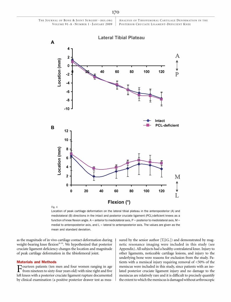
Fig. 4
Location of peak cartilage deformation on the lateral tibial plateau in the anteroposterior (A) and
mediolateral (B) directions in the intact and posterior cruciate ligament (PCL)-deficient knees as a
function of knee flexion angle. A = anterior to mediolateral axis, P = posterior to mediolateral axis, M =
medial to anteroposterior axis, and L = lateral to anteroposterior axis. The values are given as the
mean and standard deviation.
170
TH E J O U R N A L O F B O N E & JO I N T SU R G E RY d J B J S . O R G
VO LU M E 91-A d NU M B E R 1 d JA N UA RY 2009AN A LY S I S O F TI B I O F E M O R A L CA R T I L AG E DE F O R M AT I O N I N T H E
PO S T E R I O R CRU C I AT E LI G A M E N T-DE F I C I E N T KN E E

examination. The purpose of the study was explained in detail toall of the subjects at the time of recruitment. Each subject signed aconsent form that had been approved by our institutional reviewboard. Eight of these fourteen subjects had been included in ourprevious study of the six-degrees-of-freedom tibiofemoral kine-matics in patients with posterior cruciate ligament deficiency24.
Magnetic resonance and dual orthogonal fluoroscopicimaging techniques have been described in detail and validatedin previous studies28,31-33. In brief, a protocol established in ourlaboratory was used to image both the left and the right kneewith a magnetic resonance imaging scanner to create three-dimensional meshed models of the knees31. Each anatomicknee model included the osseous geometry of the femur, tibia,and fibula as well as the tibial and femoral cartilage layers. Afterthe magnetic resonance image-based computer models were
constructed, both knees of each subject were simultaneouslyimaged with use of two orthogonally placed fluoroscopes asthe patient performed a single leg quasistatic lunge at 0�, 30�,60�, 75�, 90�, 105�, and 120� of flexion while the upper bodyremained upright. Next, the fluoroscopic images were importedinto solid-modeling software and placed in the orthogonalplanes on the basis of the position of the fluoroscopes duringthe imaging of the patient. In the following step, the three-dimensional magnetic resonance image-based knee model ofthe patient was imported into the same software, viewed fromthe two orthogonal directions corresponding to the orthogonalfluoroscopic setup used to acquire the images, and indepen-dently manipulated in six degrees of freedom inside the soft-ware until the projections of the model matched the outlinesof the fluoroscopic images. When the projections match the
Fig. 5
Magnitude of peak cartilage deformation on the medial (A) and lateral (B) tibial plateaus in the intact
and posterior cruciate ligament (PCL)-deficient knees. *P < 0.05 as determined with one-way
repeated-measures analysis of variance.
171
TH E J O U R N A L O F B O N E & JO I N T SU R G E RY d J B J S . O R G
VO LU M E 91-A d NU M B E R 1 d JA N UA RY 2009AN A LY S I S O F TI B I O F E M O R A L CA R T I L AG E DE F O R M AT I O N I N T H E
PO S T E R I O R CRU C I AT E LI G A M E N T-DE F I C I E N T KN E E

outlines of the images made during in vivo knee flexion, themodel reproduces the in vivo position of the knee. Finally, therelative positions of the cartilage layers on the femur and tibiawere determined from the series of models used to reproduceknee motion. When cartilage contact occurred during kneeflexion, the articular surface meshes of the tibia and femuroverlapped.
In this study, cartilage deformation was defined for eachvertex of the articular surface mesh as the amount of penetration(Fig. 1, A) divided by the sum of the tibial and femoral cartilagesurface thicknesses33,34. The cartilage thickness was calculated byfinding the smallest Euclidian distance connecting a vertex ofthe articular surface to the cartilage-bone interface (Fig. 1, B).
The locations of peak cartilage deformation were refer-enced to Cartesian coordinate systems on the tibial plateaus, ashas been described in detail in previous publications (Fig.2)30,32,35. The origin of each coordinate system was located bythe center of a circle, which was fit to the posterior edge of eachtibial compartment. The anteroposterior and mediolateral axessplit each tibial plateau into quadrants. In the anteroposteriordirection, a location anterior to the mediolateral axis was con-sidered positive. In the mediolateral direction, a location medialto the anteroposterior axis was considered positive.
A one-way repeated-measures analysis of variance andthe Student-Newman-Keuls test were used to compare thelocation and magnitude of peak cartilage deformation of the
intact contralateral knees with those of the posterior cruciateligament-deficient knees. Differences were considered signifi-cant at the level of p < 0.05.
Source of FundingThe funding sources had no involvement in the study design;in the collection, analysis, or interpretation of the data; in thewriting of the report; or in the decision to submit the paper forpublication.
ResultsLocation of Peak Cartilage DeformationHealthy Contralateral Knees
In the medial tibial plateau, peak deformation moved pos-teromedially with flexion, from a mean (and standard de-
viation) of 6.7 ± 2.1 mm anterior to the mediolateral axis and5.1 ± 1.4 mm medial to the anteroposterior axis at 0� to a meanof 20.5 ± 0.2 mm posterior to the mediolateral axis (Fig. 3, A)and 2.0 ± 0.3 mm medial to the anteroposterior axis (Fig. 3, B)at 60�, where it remained for the rest of flexion.
In the lateral tibial plateau, peak deformation movedposteriorly throughout flexion, from a mean of 1.4 ± 2.3 mmanterior to the mediolateral axis at 0� to a mean of 27.6 ± 2.1mm posterior to the mediolateral axis at 120� (Fig. 4, A). Peakdeformation moved laterally in the lateral tibial plateau until30�, at which point it was 1.9 ± 4.4 mm from the anteropos-
Fig. 6
Color map of the cartilage deformation in the intact (left) and posterior cruciate ligament-deficient (right) knees of a typical subject
with the knees in 105� of flexion, illustrating the effect of posterior cruciate ligament deficiency on the location and magnitude of
cartilage deformation at flexion angles beyond 75�. The red X on the cartilage surfaces indicates the location of peak cartilage de-
formation. Note the more anterior (relative to the mediolateral axis) and medial (relative to the anteroposterior axis) contact location of
cartilage deformation in the medial compartment of the posterior cruciate ligament-deficient knee. The darker color depicts a higher
magnitude of peak cartilage deformation (in millimeters). The illustrative color map was created by assigning an increasingly darker
color value to each increased value of cartilage deformation for each vertex of the articular surface mesh. AP = anteroposterior, and
ML = mediolateral.
172
TH E J O U R N A L O F B O N E & JO I N T SU R G E RY d J B J S . O R G
VO LU M E 91-A d NU M B E R 1 d JA N UA RY 2009AN A LY S I S O F TI B I O F E M O R A L CA R T I L AG E DE F O R M AT I O N I N T H E
PO S T E R I O R CRU C I AT E LI G A M E N T-DE F I C I E N T KN E E
terior axis; peak deformation then moved medially until 90�,reaching a value of 4.6 ± 2.2 mm from the anteroposterior axis(Fig. 4, B).
Posterior Cruciate Ligament-Deficient KneesIn the medial compartment, posterior cruciate ligament defi-ciency significantly changed the location of peak cartilage de-formation between 75� and 120� of flexion, with the posteriorcruciate ligament-deficient knee having a more anterior andmedial cartilage contact location as compared with the intactknee. During flexion of ‡75�, contact in the medial compartmentshifted anteriorly by an average of 2.2 ± 0.4 mm (Fig. 3, A) andmedially by an average of 1.9 ± 0.4 mm (Fig. 3, B) as comparedwith the location in the intact knees. These values include thedata of the four subjects with a partial meniscectomy. Contactin the medial compartments of the four posterior cruciateligament-deficient knees with partial meniscectomy shiftedanteriorly by an average of 2.1 ± 0.5 mm and medially by anaverage of 2.0 ± 0.5 mm between 75� and 120� of flexion.
In the lateral compartment, no significant differences inthe anteroposterior and mediolateral motion of cartilage con-tact were observed between the intact and posterior cruciateligament-deficient knees (Fig. 4, A and B).
Magnitude of Peak Cartilage DeformationHealthy Contralateral KneesIn the medial compartment, peak deformation increased withflexion, from a mean of 0.17 ± 0.09 mm/mm at full extensionto a mean of 0.20 ± 0.07 mm/mm at 120� of flexion (Fig. 5, A).In the lateral compartment, a similar increase in peak defor-mation was observed, with mean values of 0.17 ± 0.08 mm/mm at full extension and 0.22 ± 0.07 mm/mm at 120� offlexion (Fig. 5, B).
Posterior Cruciate Ligament-Deficient KneesIn the medial compartment, rupture of the posterior cruciateligament caused a significant gradual increase in cartilage de-formation, as compared with that in the intact knees, from 75�to 120� of flexion (Fig. 5, A). The maximum increase in car-tilage deformation after posterior cruciate ligament ruptureoccurred at 120� of flexion (0.20 ± 0.07 mm/mm in the intactknee compared with 0.28 ± 0.08 mm/mm in the posteriorcruciate ligament-deficient knee). We did not detect significantdifferences in cartilage deformation from 0� to 60� of flexionbetween the healthy and posterior cruciate ligament-deficientknees.
In the lateral compartment, we did not detect any sig-nificant differences in cartilage deformation between the intactand posterior cruciate ligament-deficient knees throughout therange of flexion (Fig. 5, B).
Discussion
In this study, we investigated the location and magnitude oftibiofemoral cartilage deformation in posterior cruciate
ligament-deficient knees. In the medial compartment of theseknees, the location and magnitude of peak cartilage deforma-
tion were significantly changed, as compared with the findingsin the intact contralateral knees, between 75� and 120� offlexion, with a more anterior and medial location as well as anincreased magnitude. In the lateral compartment, no significantdifferences were found in the location or magnitude of peaktibiofemoral cartilage deformation between the intact andposterior cruciate ligament-deficient knees.
In our previous in vivo analysis of tibiofemoral jointkinematics, posterior cruciate ligament-deficient knees dis-played increased posterior tibial translation beyond 30� offlexion compared with that in healthy control knees24. Similarfindings have been well documented in the literature16,25-27,36.However, posterior cruciate ligament deficiency also resultedin an average 1.1-mm increase in lateral tibial translation at 90�of flexion. The findings of the present analysis of cartilagedeformation in posterior cruciate ligament-deficient knees areconsistent with these altered tibiofemoral joint kinematics, asthe increased posterior and lateral tibial translation could berelated, respectively, to the increased anterior and medial lo-cations of cartilage deformation on the medial tibial plateau.
In normal knees, cartilage is up to 50% thicker in regionswhere cartilage-to-cartilage contact is present35. Healthy cartilageis believed to adapt to mechanical stimuli37,38 and ultimately be-come dependent on the maintenance of the mechanical stimulusfor normal tissue function39. The thicker cartilage within thecartilage-to-cartilage contact area may result in a reduced contactstress, as was demonstrated by a three-dimensional finite-element analysis suggesting that thicker cartilage bears a lowerpeak contact stress than thinner cartilage under the same loadingconditions40. In the present study, we found that posterior cru-ciate ligament deficiency significantly altered tibiofemoral car-tilage contact in both the anteroposterior and the mediolateraldirection in the medial compartment of the knee at higherflexion angles. In the presence of posterior cruciate ligamentinjury, cartilage contact not only shifted anteriorly, as was ex-pected on the basis of increased posterior tibial translation, butalso medially on the surface of the tibial plateau in flexion po-sitions of ‡75�, forcing the femur to ride up the upslope of themedial tibial plateau, a region of thinner cartilage34,41. Thismedial shift of the location of cartilage contact after posteriorcruciate ligament rupture to incongruent, thinner articular re-gions increased the cartilage deformation in those regions (Fig.6). Increased cartilage deformation is associated with increasedmechanical loading of the articular contact region within theknee, which in turn has been linked to higher rates of pro-gression of osteoarthritis42. The relatively greater increase ofcartilage deformation in the medial compartment comparedwith that in the lateral compartment observed in the posteriorcruciate ligament-deficient knees in the present study is con-sistent with the reported increased development of osteoar-thritis in the medial compartment of the knee joint followingposterior cruciate ligament injury 2,4.
As the presented data were obtained during only onefunctional in vivo activity—namely, the single leg lunge—weadvise caution when extrapolating the data to other functionalactivities. Nevertheless, we believe that these findings might be
173
TH E J O U R N A L O F B O N E & JO I N T SU R G E RY d J B J S . O R G
VO LU M E 91-A d NU M B E R 1 d JA N UA RY 2009AN A LY S I S O F TI B I O F E M O R A L CA R T I L AG E DE F O R M AT I O N I N T H E
PO S T E R I O R CRU C I AT E LI G A M E N T-DE F I C I E N T KN E E

useful for the design of improved treatment protocols forposterior cruciate ligament deficiency. First, as we did notdetect differences in the cartilage biomechanics between theintact and posterior cruciate ligament-deficient knees duringthe single leg lunge between 0� and 60� of flexion, our findingssuggest that rehabilitation exercises might be safely performedin this range of flexion. On the other hand, repetitive deep kneesquats should be avoided by subjects with posterior cruciateligament deficiency, so as not to increase the tibiofemoralcartilage deformation. Second, it is interesting to note that themagnitude of medial contact shift in the posterior cruciateligament-deficient knees was on the same order as the mag-nitude of the anterior contact shift. This suggests that, when aposterior cruciate ligament reconstruction is performed witheither a single or a double-bundle graft, recreation of the me-diolateral stability of posterior cruciate ligament-deficient kneesmay be as important as the surgical improvement of antero-posterior translation. Finally, in a recent cadaver study, Giffinet al. demonstrated, with a robotic testing system, that increasingthe tibial slope with a sagittal osteotomy successfully reducedthe abnormal tibial sag in the posterior cruciate ligament-deficient knee, shifting the resting position of the tibia ante-riorly43. On the basis of our data, it could be theorized in futurestudies that the osteotomy should include a varus component,possibly reducing the abnormal medial shift following poste-rior cruciate ligament injury and thereby possibly reducing theincrease in cartilage deformation.
This study had limitations. As a result of the constraints ofthe imaging technique, motion and deformation of the me-niscus were not detectable on the fluoroscopic images. How-ever, on the basis of our previous validation of the imagingtechnique32, we do not believe that this limitation affected thecartilage-cartilage contact data, as articular surface mesh pen-etration was recorded only at the location of in vivo tibio-femoral cartilage contact. In addition, as mentioned earlier, weacquired data during only one functional activity, a single leglunge. Other in vivo activities such as walking, running, andstair climbing should be considered in future studies. Fur-thermore, it should be noted that no ground reaction forceswere measured in this study. In the future, a force-plate will beincorporated into the system. Since isolated posterior cruciateligament injuries are rare, we included some patients who hada partial tear of one of the menisci. Because there were onlyfourteen subjects in our study, there was not enough statistical
power for us to analyze the effect of partial removal of themeniscus as well. The findings from this study might thereforehave been affected by the meniscal tears. Another limitation isthat the patients were investigated at different time intervalsafter the injury. In future studies, patients in whom posteriorcruciate ligament deficiency has been treated conservativelyshould be followed for longer periods with use of a method-ology similar to that employed in our study. Tibiofemoralcontact and the health of the cartilage could therefore bemonitored over time to quantify any possible biomechanicalrelationships.
In conclusion, the altered kinematics caused by posteriorcruciate ligament deficiency resulted in a shift of the tibio-femoral contact location and an increase in the in vivo de-formation beyond 75� of flexion in the medial compartment.This injurious ‘‘jab-and-cross combo’’ provides insight aboutthe development of degeneration of tibiofemoral joint cartilagein patients with posterior cruciate ligament deficiency. If theprevention of osteoarthritis in patients with posterior cruciateligament deficiency is a goal of the treating physician, thefunction of the injured ligament should be restored as closelyto normal as possible, in both the anteroposterior and medi-olateral directions, thereby possibly better normalizing thecartilage biomechanics of the knee.
AppendixA table presenting clinical details on all fourteen studysubjects is available with the electronic versions of this
article, on our web site at jbjs.org (go to the article citation andclick on ‘‘Supplementary Material’’) and on our quarterly CD/DVD (call our subscription department, at 781-449-9780, toorder the CD or DVD). n
NOTE: The authors thank Dr. Louis DeFrate, Dr. Kyung Nha, Dr. Dain Allred, and Mr. RamprasadPapannagari for their technical assistance.
Samuel K. Van de Velde, MDJeffrey T. Bingham, MSThomas J. Gill, MDGuoan Li, PhDBioengineering Laboratory, Department of Orthopaedic Surgery,Massachusetts General Hospital/Harvard Medical School,55 Fruit Street, GRJ 1215, Boston, MA 02114.E-mail address for G. Li: [email protected]
References
1. Boynton MD, Tietjens BR. Long-term followup of the untreated isolated posteriorcruciate ligament-deficient knee. Am J Sports Med. 1996;24:306-10.
2. Clancy WG Jr, Shelbourne KD, Zoellner GB, Keene JS, Reider B, Rosenberg TD.Treatment of knee joint instability secondary to rupture of the posterior cruciateligament. Report of a new procedure. J Bone Joint Surg Am. 1983;65:310-22.
3. Cross MJ, Powell JF. Long-term followup of posterior cruciate ligament rupture: astudy of 116 cases. Am J Sports Med. 1984;12:292-7.
4. Keller PM, Shelbourne KD, McCarroll JR, Rettig AC. Nonoperatively treatedisolated posterior cruciate ligament injuries. Am J Sports Med. 1993;21:132-6.
5. Parolie JM, Bergfeld JA. Long-term results of nonoperative treatment of isolatedposterior cruciate ligament injuries in the athlete. Am J Sports Med. 1986;14:35-8.
6. Shelbourne KD, Davis TJ, Patel DV. The natural history of acute, isolated,nonoperatively treated posterior cruciate ligament injuries. A prospective study.Am J Sports Med. 1999;27:276-83.
7. Schulte KR, Chu ET, Fu FH. Arthroscopic posterior cruciate ligament recon-struction. Clin Sports Med. 1997;16:145-56.
8. Moore HA, Larson RL. Posterior cruciate ligament injuries. Results of earlysurgical repair. Am J Sports Med. 1980;8:68-78.
174
TH E J O U R N A L O F B O N E & JO I N T SU R G E RY d J B J S . O R G
VO LU M E 91-A d NU M B E R 1 d JA N UA RY 2009AN A LY S I S O F TI B I O F E M O R A L CA R T I L AG E DE F O R M AT I O N I N T H E
PO S T E R I O R CRU C I AT E LI G A M E N T-DE F I C I E N T KN E E

9. Marks PH, Cameron M, Fu FH. [Reconstruction of the cruciate ligaments withallogeneic transplants. Techniques, results and perspectives]. Orthopade.1993;22:386-91. German.
10. Hughston JC, Bowden JA, Andrews JR, Norwood LA. Acute tears of the pos-terior cruciate ligament. Results of operative treatment. J Bone Joint Surg Am.1980;62:438-50.
11. Pournaras J, Symeonides PP, Karkavelas G. The significance of the posteriorcruciate ligament in the stability of the knee. An experimental study in dogs. J BoneJoint Surg Br. 1983;65:204-9.
12. Richter M, Kiefer H, Hehl G, Kinzl L. Primary repair for posterior cruciate liga-ment injuries. An eight-year followup of fifty-three patients. Am J Sports Med.1996;24:298-305.
13. Butler DL, Noyes FR, Grood ES. Ligamentous restraints to anterior-posteriordrawer in the human knee. A biomechanical study. J Bone Joint Surg Am.1980;62:259-70.
14. Fanelli GC, Giannotti BF, Edson CJ. The posterior cruciate ligament arthro-scopic evaluation and treatment. Arthroscopy. 1994;10:673-88.
15. Fox RJ, Harner CD, Sakane M, Carlin GJ, Woo SL. Determination of the in situforces in the human posterior cruciate ligament using robotic technology. A ca-daveric study. Am J Sports Med. 1998;26:395-401.
16. Gollehon DL, Torzilli PA, Warren RF. The role of the posterolateral and cruciateligaments in the stability of the human knee. A biomechanical study. J Bone JointSurg Am. 1987;69:233-42.
17. Markolf KL, Slauterbeck JR, Armstrong KL, Shapiro MS, Finerman GA. A bio-mechanical study of replacement of the posterior cruciate ligament with a graft.Part II: forces in the graft compared with forces in the intact ligament. J Bone JointSurg Am. 1997;79:381-6.
18. Noyes FR, Stowers SF, Grood ES, Cummings J, VanGinkel LA. Posterior sub-luxations of the medial and lateral tibiofemoral compartments. An in vitro ligamentsectioning study in cadaveric knees. Am J Sports Med. 1993;21:407-14.
19. Fukubayashi T, Torzilli PA, Sherman MF, Warren RF. An in vitro biomechanicalevaluation of anterior-posterior motion of the knee. Tibial displacement, rotation,and torque. J Bone Joint Surg Am. 1982;64:258-64.
20. Race A, Amis AA. The mechanical properties of the two bundles of the humanposterior cruciate ligament. J Biomech. 1994;27:13-24.
21. Girgis FG, Marshall JL, Monajem A. The cruciate ligaments of the knee joint.Anatomical, functional and experimental analysis. Clin Orthop Relat Res.1975;106:216-31.
22. Carlin GJ, Livesay GA, Harner CD, Ishibashi Y, Kim HS, Woo SL. In-situ forcesin the human posterior cruciate ligament in response to posterior tibial loading. AnnBiomed Eng. 1996;24:193-7.
23. Papannagari R, Defrate LE, Nha KW, Moses JM, Moussa M, Gill TJ, Li G.Function of posterior cruciate ligament bundles during in vivo knee flexion. AmJ Sports Med. 2007;35:1507-12.
24. Li G, Papannagari R, Li M, Bingham JT, Nha KW, Allred D, Gill TJ. Effect ofposterior cruciate ligament deficiency on in vivo translation and rotation of the kneeduring weightbearing flexion. Am J Sports Med. 2008;36:474-9.
25. Gill TJ, DeFrate LE, Wang C, Carey CT, Zayontz S, Zarins B, Li G. The biome-chanical effect of posterior cruciate ligament reconstruction on knee joint function.Kinematic response to simulated muscle loads. Am J Sports Med. 2003;31:530-6.
26. Li G, Gill TJ, DeFrate LE, Zayontz S, Glatt V, Zarins B. Biomechanical conse-quences of PCL deficiency in the knee under simulated muscle loads—an in vitroexperimental study. J Orthop Res. 2002;20:887-92.
27. Li G, Most E, DeFrate LE, Suggs JF, Gill TJ, Rubash HE. Effect of the posteriorcruciate ligament on posterior stability of the knee in high flexion. J Biomech.2004;37:779-83.
28. Li G, Wuerz TH, DeFrate LE. Feasibility of using orthogonal fluoroscopic imagesto measure in vivo joint kinematics. J Biomech Eng. 2004;126:314-8.
29. DeFrate LE, Sun H, Gill TJ, Rubash HE, Li G. In vivo tibiofemoral contactanalysis using 3D MRI-based knee models. J Biomech. 2004;37:1499-504.
30. Li G, DeFrate LE, Park SE, Gill TJ, Rubash HE. In vivo articular cartilage contactkinematics of the knee: an investigation using dual-orthogonal fluoroscopy andmagnetic resonance image-based computer models. Am J Sports Med.2005;33:102-7.
31. Defrate LE, Papannagari R, Gill TJ, Moses JM, Pathare NP, Li G. The 6 degreesof freedom kinematics of the knee after anterior cruciate ligament deficiency: anin vivo imaging analysis. Am J Sports Med. 2006;34:1240-6.
32. Li G, Moses JM, Papannagari R, Pathare NP, DeFrate LE, Gill TJ. Anteriorcruciate ligament deficiency alters the in vivo motion of the tibiofemoral cartilagecontact points in both the anteroposterior and mediolateral directions. J Bone JointSurg Am. 2006;88:1826-34.
33. Li G, Wan L, Kozanek M. Determination of real-time in-vivo cartilage contactdeformation in the ankle joint. J Biomech. 2008;41:128-36.
34. Bingham JT, Papannagari R, Van de Velde SK, Gross C, Gill TJ, Rubash HE,Li G. In-vivo cartilage contact deformation in the healthy human tibiofemoral joint.Rheumatology (Oxford). 2008;47:1622-7.
35. Li G, Park SE, DeFrate LE, Schutzer ME, Ji L, Gill TJ, Rubash HE. The cartilagethickness distribution in the tibiofemoral joint and its correlation with cartilage-to-cartilage contact. Clin Biomech (Bristol, Avon). 2005;20:736-44.
36. Logan M, Williams A, Lavelle J, Gedroyc W, Freeman M. The effect ofposterior cruciate ligament deficiency on knee kinematics. Am J Sports Med.2004;32:1915-22.
37. Arokoski JP, Jurvelin JS, Vaatainen U, Helminen HJ. Normal and pathologicaladaptations of articular cartilage to joint loading. Scand J Med Sci Sports.2000;10:186-98.
38. Smith RL, Donlon BS, Gupta MK, Mohtai M, Das P, Carter DR, Cooke J,Gibbons G, Hutchinson N, Schurman DJ. Effects of fluid-induced shear on articularchondrocyte morphology and metabolism in vitro. J Orthop Res. 1995;13:824-31.
39. Carter DR, Beaupre GS, Giori NJ, Helms JA. Mechanobiology of skeletal re-generation. Clin Orthop Relat Res. 1998;355 Suppl:S41-55.
40. Li G, Lopez O, Rubash H. Variability of a three-dimensional finite elementmodel constructed using magnetic resonance images of a knee for joint contactstress analysis. J Biomech Eng. 2001;123:341-6.
41. Donahue TL, Hull ML, Rashid MM, Jacobs CR. A finite element model of thehuman knee joint for the study of tibio-femoral contact. J Biomech Eng.2002;124:273-80.
42. Miyazaki T, Wada M, Kawahara H, Sato M, Baba H, Shimada S. Dynamic loadat baseline can predict radiographic disease progression in medial compartmentknee osteoarthritis. Ann Rheum Dis. 2002;61:617-22.
43. Giffin JR, Stabile KJ, Zantop T, Vogrin TM, Woo SL, Harner CD. Importance oftibial slope for stability of the posterior cruciate ligament deficient knee. Am JSports Med. 2007;35:1443-9.
175
TH E J O U R N A L O F B O N E & JO I N T SU R G E RY d J B J S . O R G
VO LU M E 91-A d NU M B E R 1 d JA N UA RY 2009AN A LY S I S O F TI B I O F E M O R A L CA R T I L AG E DE F O R M AT I O N I N T H E
PO S T E R I O R CRU C I AT E LI G A M E N T-DE F I C I E N T KN E E
Copyright © 2022 FDOKUMEN