Nutrition and Immune Function
26
Nutrition and Immune Function Korinn E. Saker, MS, DVM, PhD Department of Large Animal Clinical Sciences, Virginia-Maryland Regional College of Veterinary Medicine, Blacksburg, VA 24061, USA I llness is commonly associated with anorexia and altered nutrient require- ments as a consequence of biochemical, metabolic, and pathologic abnor- malities that influence nutrient use. This complicated cascade of events directly and indirectly involves immunocompetence. Several questions of im- portance to patient management should be considered. First, should the im- mune system be enhanced during disease and/or illness? If yes, how can nutrients enhance immunocompetence during disease and/or illness and which nutrients are efficacious immunomodulators? The complex workings of the im- mune system can be simplified to the fact that disease initiation and progression are correlated with a break in immunocompetence. Something in the system went awry, and the assumption is that nutritional intervention can potentially get the system back on track to help manage, resolve, and, in the future, pre- vent the disease process. OVERVIEW OF THE IMMUNE SYSTEM The immune system is part of the host’s defense against destructive forces from outside the body, such as bacteria, viruses, and parasites, or from within, such as malignant cells or those that produce autoantibodies [1]. This system is com- posed of two components: the innate or nonspecific immune system and the adaptive or specific immune system. The system of nonspecific immunity consists of anatomic barriers and a cel- lular component. The various barriers, including skin and mucous and gastro- intestinal (GI) mucosa, are the true ‘‘first line of defense.’’ Once compromised by microorganisms, endotoxins, or any substance considered to be foreign, the complement system may be activated. The complement system is a complex cascade of proteins that promote such functions as phagocytosis, viral neutral- ization, and destruction of virus-infected cells. Complement system defects are associated with increased susceptibility to bacterial infections. Inflammatory mediators, in addition to being products of cell membrane destruction, increase vascular permeability, causing the accumulation of acute-phase proteins and immune complexes that promote the cellular phase of acute inflammation [1]. The cellular phase of the nonspecific immune system includes circulating and ‘‘fixed’’ phagocytes. Initially, neutrophils bind to pathogenic microorganisms, 0195-5616/06/$ – see front matter ª 2006 Elsevier Inc. All rights reserved. doi:10.1016/j.cvsm.2006.09.001 vetsmall.theclinics.com Vet Clin Small Anim 36 (2006) 1199–1224 VETERINARY CLINICS SMALL ANIMAL PRACTICE
Transcript of Nutrition and Immune Function

Vet Clin Small Anim 36 (2006) 1199–1224
VETERINARY CLINICSSMALL ANIMAL PRACTICE
Nutrition and Immune Function
Korinn E. Saker, MS, DVM, PhDDepartment of Large Animal Clinical Sciences, Virginia-Maryland Regional College of VeterinaryMedicine, Blacksburg, VA 24061, USA
Illness is commonly associated with anorexia and altered nutrient require-ments as a consequence of biochemical, metabolic, and pathologic abnor-malities that influence nutrient use. This complicated cascade of events
directly and indirectly involves immunocompetence. Several questions of im-portance to patient management should be considered. First, should the im-mune system be enhanced during disease and/or illness? If yes, how cannutrients enhance immunocompetence during disease and/or illness and whichnutrients are efficacious immunomodulators? The complex workings of the im-mune system can be simplified to the fact that disease initiation and progressionare correlated with a break in immunocompetence. Something in the systemwent awry, and the assumption is that nutritional intervention can potentiallyget the system back on track to help manage, resolve, and, in the future, pre-vent the disease process.
OVERVIEW OF THE IMMUNE SYSTEMThe immune system is part of the host’s defense against destructive forces fromoutside the body, such as bacteria, viruses, and parasites, or from within, suchas malignant cells or those that produce autoantibodies [1]. This system is com-posed of two components: the innate or nonspecific immune system and theadaptive or specific immune system.
The system of nonspecific immunity consists of anatomic barriers and a cel-lular component. The various barriers, including skin and mucous and gastro-intestinal (GI) mucosa, are the true ‘‘first line of defense.’’ Once compromisedby microorganisms, endotoxins, or any substance considered to be foreign, thecomplement system may be activated. The complement system is a complexcascade of proteins that promote such functions as phagocytosis, viral neutral-ization, and destruction of virus-infected cells. Complement system defects areassociated with increased susceptibility to bacterial infections. Inflammatorymediators, in addition to being products of cell membrane destruction, increasevascular permeability, causing the accumulation of acute-phase proteins andimmune complexes that promote the cellular phase of acute inflammation [1].
The cellular phase of the nonspecific immune system includes circulating and‘‘fixed’’ phagocytes. Initially, neutrophils bind to pathogenic microorganisms,
0195-5616/06/$ – see front matter ª 2006 Elsevier Inc. All rights reserved.doi:10.1016/j.cvsm.2006.09.001 vetsmall.theclinics.com

1200 SAKER
phagocytose them, and kill them. Phagocytosis is facilitated by opsonization.Opsonins activate neutrophils, resulting in an oxidative burst that includesproduction of H2O2 and O2
� free radicals. These substances kill the bacteriaand the neutrophil with release of toxic waste products. Although this re-sponse is beneficial in moderation, prolongation of the inflammatory phasecan be detrimental to the host. Monocytes and macrophages are also compo-nents of nonspecific immunity. They phagocytize antigens, process themthrough an oxidative burst reaction, and present antigen particles to T cellsvia major histocompatibility complex (MHC) class I or II receptors [2].
The specific immune system is composed of B and T cells, which are asso-ciated with humoral immunity and cell-mediated immunity. B lymphocytesmature in bone marrow and react to stimulation by certain antigens to differ-entiate into plasma cells, which synthesize and secrete antibodies commonlytermed immunoglobulins (Table 1). Cell-mediated immunity, however, relies pri-marily on T lymphocytes derived from the thymus. Antigen-presenting cells,such as macrophages, are responsible for triggering the specific immune re-sponse. Interaction of an antigen and macrophage leads to production of inter-leukin (IL)-1 by means of arachidonic acid (AA) metabolism. The IL-1produced by macrophages causes T cells to produce IL-2 and other lympho-kines. Production of IL-2 helps to stimulate T and B cells to form clonesthat carry receptors specific to the sensitizing antigen. These clones form thelong-lived memory cells, which proliferate and release lymphokines on re-expo-sure to the same antigen. These clones, in conjunction with macrophages, candestroy the antigen. Defects in cell-mediated immunity are associated with in-fections of bacteria, mycobacteria, viruses, fungi, and parasites [1,2].
T cells are not only responsible for mediating delayed hypersensitivity, graftrejection, destruction of pathogenic microorganisms, and destruction of malig-nant cells but also regulate responses of other immune cells. The subsets of T cells
Table 1Immunoglobulins
Immunoglobulin Type Role and location
IgG 4 Coats microorganisms for uptake by other cells (opsonization);crosses placenta to affect passive immunity; enhancescomplement function; primarily present in serum
IgM 2 First to respond to antigens via agglutination and bacteriolysis;present in blood
IgA 1 Protects mucous membranes by preventing bacteria fromattaching to mucosal surface; present in body fluids
IgE 1 Involved in hypersensitivity reactions and allergic responses;phagocytosis and other immunoglobulin activity; present inplasma and tissue and on surface membranes of basophilsand mast cells
IgD 1 Involved in differentiation of B lymphocytes; present in serumand in plasma membrane of B lymphocytes

1201NUTRITION AND IMMUNE FUNCTION
include helper-inducer T cells (CD4), which help plasma cells to produce anti-bodies and release lymphokines, which modulate the interaction between lym-phocytes and other cells. Cytotoxic-suppressor T cells (CD8) may destroytarget cells, inhibit antibody responses, or inhibit the inflammatory response [1,2].
NUTRIENT INFLUENCE ON SPECIFIC COMPONENTSOF THE IMMUNE SYSTEMThe small bowel contains an abundant amount of lymphoid tissue and is a pri-mary component of innate immunity through its cellular component and protec-tive barrier function. It is often considered to be the first line of defense to invasionby microbes into the systemic circulation. The physical barrier is created by thetight junctions between intact epithelial cells, gastric acid, digestive enzymes, mu-cus production, intestinal motility, and normal bacterial flora [3]. The gut-associ-ated lymphoid tissue (GALT) or intestinal immune system consists oflymphocytes and macrophages situated throughout the intestinal wall. IgA isalso secreted into the GI lumen to prevent adherence of microbes to the mucosa.By design, these two components prevent or minimize the spread of pathogens, ortheir products, across the intestinal wall into the systemic circulation, a processtermed translocation. Multiple factors promote translocation, including luminalbacterial overgrowth, impaired host defense mechanisms, protein-calorie malnu-trition (PCM), trauma, critical illness, interruption of the luminal nutrient stream,or any other process that leads to mucosal atrophy [4]. Fig. 1 depicts the potentiallife-threatening sequelae to bacterial translocation. Maintenance of an intact bar-rier and functioning GALT are imperative to preventing translocation. Certainnutrients, specifically glutamine (GLN), arginine, nucleotides, x-3 fatty acids,and dietary fiber (a source of short-chain fatty acids), are necessary for growthand normal function of the mucosal epithelial cells and the lymphoid cells ofthe GALT intestinal barrier [3,5].
MOLECULAR ASPECTS OF NUTRITIONAND IMMUNE FUNCTIONBasic ConceptsThe structural complexity of mammalian cells has its basis in regulated expres-sion of thousands of different proteins. Most of the information that definesthese molecules and structures is contained in DNA sequences within the cellnucleus. The primary focus for the technology of molecular biology is DNAand the processes that translate the informational content of DNA into cellularstructures and functions. Much of the informational content of DNA consists ofregions of nucleotide or base sequences that define or code for the amino acidsequences of proteins. Steps involved in the information transfer from DNA toproteins are illustrated in Fig. 2. The DNA nucleotide sequence is transcribedinto an mRNA nucleotide. The mRNA sequence is then translated into a pro-tein. The proteins generated in this manner form cellular structures or functionas enzymes or membrane transporters that dictate much of the overall structureand function of the cell [6].

1202 SAKER
Practical Applications to Clinical NutritionPolysomes, the ‘‘preprotein’’ molecule sequence that builds on the mRNAbackbone, can be isolated and quantified as a marker of nutrient intervention.GLN-supplemented total parenteral nutrition (TPN) versus a GLN-free controlformula was given to patients for the first 3 days after abdominal surgery. Poly-some content of the quadriceps femoris muscle obtained before and after day 3after surgery showed a significant improvement in nitrogen balance and a spar-ing of free muscle GLN in the GLN-supplemented patients [7].
A second application involves DNA cloning (cDNA). A segment of DNA, orcDNA, that encodes for a specific protein of interest can be developed and usedas a probe to assess expression of its corresponding mRNA in cells or tissues.Nutritionally relevant sequences derived from cDNA include proteins thatfunction in carbohydrate (CHO; pyruvate kinase) and lipid (apolipoproteins)metabolism, growth control (growth hormone), and specific micronutrient ac-tions (retinol-binding protein [RPB]). cDNA can be replicated multiple timesover through host bacteria cultures. These recombinant proteins can be usedto study normal and abnormal nutritionally important mechanisms of disease,including inflammation, cachexia, and oxidative stress, through such proteinsas tumor necrosis factor and ILs [6].
Fig. 2 illustrates the process of creating and transferring a protein moleculefrom within the nucleus out to the cell cytoplasm. Examples of how the tech-nologies associated with molecular biology can be used to monitor expressionof nutrition-related molecules have been summarized. Interestingly, thereseems to be a flip side of molecular nutrition, which involves the aspect of
Bacteria
Endotoxin
Intestinal
lumen
Intestinal
Barrier
Activation
Macrophages Neutrophils Complement
Microvessel thrombosis, Endothelial leak
PGE2IL-1 O2 Radicals
Multiple Organ System Failure (MOSF)
Proteases C3a C5a
Fig. 1. A summary of the stepwise progression of bacterial translocation through the mucosalbarrier resulting in multiple organ failure. PGE2, prostaglandin E2. (Modified from Rombeau JL.Enteral nutrition and critical illness. In: Borlase BC, Bell SJ, Blackburn GL, et al, editors. Enteralnutrition. New York: Chapman & Hall; 1994. p. 30; with permission.)

1203NUTRITION AND IMMUNE FUNCTION
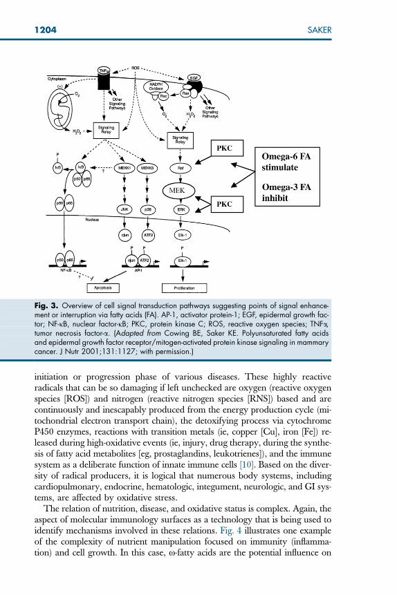
uncovering the cellular mechanisms by which nutrients can influence gene ex-pression. The transmission of signals from outside the cell into the nucleus canbe profoundly influenced by nutrients. This area of technology is helping toidentify how and which specific nutrients influence cell signal transductionand, ultimately, gene expression associated with immune function. Fig. 3 sum-marizes the complex relation between fatty acids, cell signal transduction cas-cades, apoptosis, and proliferation. This is one of numerous relations thatare being clarified through nutrition research. The topic of nutrients as cell sig-nals, although intriguing, is not meant to be a primary focus of this article. Sev-eral excellent reviews are available to develop this topic further. This level ofnutritional science is likely to be extremely influential in optimizing patientmanagement in the near future.
PATHOGENESIS OF ALTERED IMMUNOCOMPETENCEImmunoimbalance is a systemic stress response to illness-induced anorexia orhypermetabolic food deprivation. It is associated with an increased metabolicrate, protein catabolism, and a release of multiple cell mediators, including cy-tokines, prostaglandins, and leukotrienes [8]. Oxidative (immune) cell functionis escalated with the production of highly reactive radicals that damage cell con-stituents (membranes, proteins, lipids, and DNA), leading to cell death andmultiple organ dysfunction. From a biologic perspective, stress is more accu-rately defined as oxidative stress, or the imbalance between production of dam-aging free radicals and antioxidant protection [9]. Oxidative stress playsa major role in many degenerative pathologic conditions, and free radical for-mation is considered to be a pathologic biochemical mechanism involved in the
DNA
transcription
Initial RNA
transcript
Cytoplasmic
RNA translation
Protein
Nucleus
Fig. 2. Steps involved in the information transfer from DNA to protein. (Modified from SmithRJ. Molecular biology in nutrition. Nutr Clin Pract 1992;7:34; with permission from theAmerican Society for Parenteral and Enteral Nutrition [ASPEN].)
1204 SAKER
initiation or progression phase of various diseases. These highly reactiveradicals that can be so damaging if left unchecked are oxygen (reactive oxygenspecies [ROS]) and nitrogen (reactive nitrogen species [RNS]) based and arecontinuously and inescapably produced from the energy production cycle (mi-tochondrial electron transport chain), the detoxifying process via cytochromeP450 enzymes, reactions with transition metals (ie, copper [Cu], iron [Fe]) re-leased during high-oxidative events (ie, injury, drug therapy, during the synthe-sis of fatty acid metabolites [eg, prostaglandins, leukotrienes]), and the immunesystem as a deliberate function of innate immune cells [10]. Based on the diver-sity of radical producers, it is logical that numerous body systems, includingcardiopulmonary, endocrine, hematologic, integument, neurologic, and GI sys-tems, are affected by oxidative stress.
The relation of nutrition, disease, and oxidative status is complex. Again, theaspect of molecular immunology surfaces as a technology that is being used toidentify mechanisms involved in these relations. Fig. 4 illustrates one exampleof the complexity of nutrient manipulation focused on immunity (inflamma-tion) and cell growth. In this case, x-fatty acids are the potential influence on
Fig. 3. Overview of cell signal transduction pathways suggesting points of signal enhance-ment or interruption via fatty acids (FA). AP-1, activator protein-1; EGF, epidermal growth fac-tor; NF-jB, nuclear factor-jB; PKC, protein kinase C; ROS, reactive oxygen species; TNFa,tumor necrosis factor-a. (Adapted from Cowing BE, Saker KE. Polyunsaturated fatty acidsand epidermal growth factor receptor/mitogen-activated protein kinase signaling in mammarycancer. J Nutr 2001;131:1127; with permission.)

1205NUTRITION AND IMMUNE FUNCTION
the cell signal cascade and nuclear transcription through inflammatory and tu-mor cell mediators.
MALNUTRITION, NUTRIENTS, AND IMMUNITYMalnutrition, associated with a single nutrient or multiple nutrient inadequacies,is consistently associated with metabolic and clinical alterations of immunity.The association of malnutrition with reduced resistance to infection has been ob-served for centuries. Early work with children in developing countries suggestedthat the degree of immunocompromise depended on the degree of protein-en-ergy malnutrition, presence of infection, and age at the onset of malnutrition[11]. In industrialized societies, PCM has been described more frequently inthe elderly and in hospitalized patients [1]. Malnutrition should not only be con-sidered to be of protein-calorie origin, however, because vitamin and mineraldeprivation can also adversely influence the aspects of immunity. Immunocom-promise and malnutrition in hospitalized patients contribute to the developmentof infection, sepsis, organ failure, poor wound healing, and a general increase inmorbidity and mortality. Acutely ill patients with sepsis or a sepsis syndromemay exhibit immunosuppression without prior starvation [12].
Marasmus or semistarvation malnutrition is a syndrome that develops grad-ually over months to years with insufficient energy intake. The body respondsby decreasing basal energy expenditure through a decrease in thyroid and sym-pathetic nervous system activity. Additionally, there is a shift of fuel sources in
Fig. 4. Overview of the proposed relationship between inflammation and cell growth.Omega-6 and omega-3 fatty acids may influence the cell signaling cascade and nuclear tran-scription. COX-2, cyclooxygenase-2; EGFR, epidermal growth factor receptor; LOX, lipoxyge-nase; MAPK, mitogen-activated protein kinase; PKC, protein kinase C; PUFA, polyunsaturatedfatty acid; RAS, p21 ras; RAF-1, p74 raf-1; MEK, MAPK (Erk) kinase. (Adapted with modifica-tion from Cowing BE, Saker KE. Polyunsaturated fatty acids and epidermal growth factorreceptor/mitogen-activated protein kinase signaling in mammary cancer. J Nutr 2001;131:1127; with permission.)
1206 SAKER
response to the depletion rate of stored nutrients. As glucose and glycogen re-serves are depleted, protein and fat are sequestered for energy. To sustain pri-mary protein needs for as long as possible, fat becomes the predominant fuelsource. This type of malnutrition can be observed in patients with chronic dis-ease processes that adversely affect energy intake, such as cachexia (cardiac orcancer) or malassimilation disorders [13].
In contrast, hypoalbuminemic malnutrition is a manifestation of the body’sresponse to infection or inflammation with or without nutrient deprivation[14]. The association of malnutrition with metabolic and clinical alterationsof immunity is more clearly evident in this situation. Hypoalbuminemic malnu-trition is modulated by hormones and cytokines (ie, IL-1, tumor necrosis factor-a [TNFa]) secreted during the acute response to major stressors, such as sepsis,head injury, burns, or trauma [14]. It occurs quickly to deplete visceral protein(albumin) stores, and the multiplicity of sequelae to these events adversely af-fects metabolism and immune function.
The effects of PCM, regardless of the category, can be quite complex. Hu-moral immunity can be affected by PCM as a decline in the production of im-munoglobulins, secretory antibodies, and complement. Cell-mediatedimmunity is commonly affected in hypoalbuminemic or severely marasmic pa-tients. The thymus and lymphoid tissues atrophy, peripheral T lymphocytesdecrease in number, alterations in cell-mediated delayed cutaneous hypersensi-tivity and graft-versus-host reactions are apparent, there is an impaired re-sponse of lymphocytes to mitogens, and patients exhibit a poor response tocontact sensitization or inflammatory reactions as well as a depressed responseto vaccines [15]. Neutropenia may occur to varying degrees in patients withPCM. Although neutrophils seem to be morphologically normal, cell functionis decreased, specifically the capacity of neutrophils to kill phagocytosed bacte-ria or molds and to secrete chemokines [16]. Complement components ofinnate (nonspecific) immunity are depressed. Interferon production, opsoniza-tion, plasma lysosome production, and acute-phase reactants (ie, C-reactiveprotein) are adversely affected. Likewise, alterations in the anatomic barriersto infection included in the nonspecific immune system, such as atrophy ofthe skin and GI mucosa, may increase the risk of infection [13].
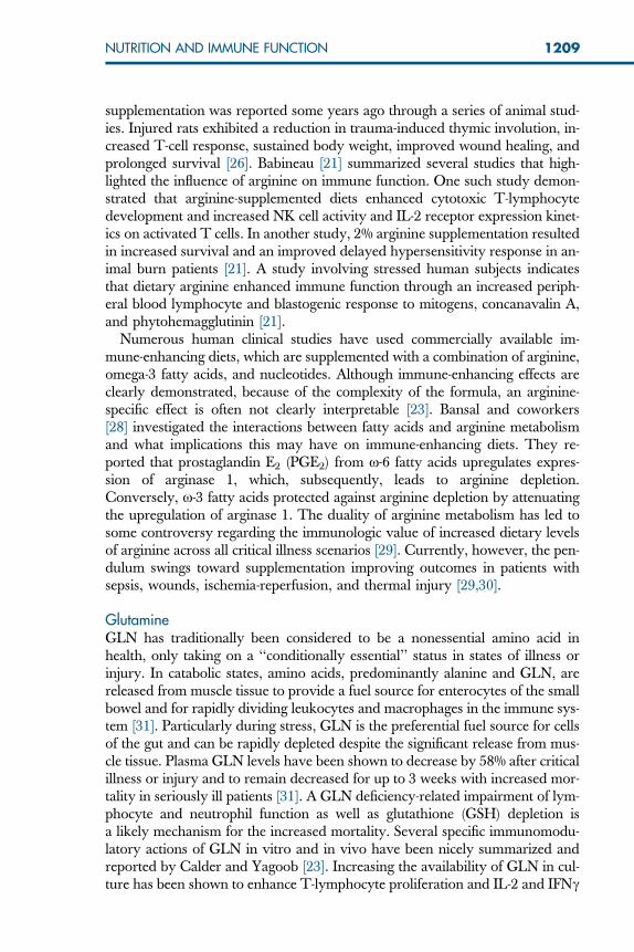
Whether correction of malnutrition improves patient outcome in all caseshas yet to be proven, although, intuitively, it is assumed to be associatedwith a beneficial effect. Several studies [17,18] that clearly demonstrate theacute impact of nutrient deprivation on immune function and the reversal ofinnate and cell-mediated immune system compromise by appropriate nutritionsupport are summarized in Table 2. Current research in nutrition support isbeginning to focus on exerting organ-specific effects by modulating metabolicprocesses rather than by simply improving nutrition. The effect of specific nu-trients on the immune system is showing great promise in this regard. Manynutrients have a role in immune function. For some, mechanisms of immuno-modulation have been clearly delineated, whereas much is yet to be learned inothers.
1207NUTRITION AND IMMUNE FUNCTION
KEY NUTRIENTS AS MODULATORS OF IMMUNE FUNCTIONThe list of key nutrients that may influence immunity seems to expand almostdaily. Several decades ago, the list was short. Protein was the key nutrient, andmicronutrient nutrition was a radical concept. Currently, a list of key nutrientsincludes specific amino acids, fatty acids, vitamins, microminerals, and nucleicacids in addition to the less well-defined micronutrients, such as flavonoids.
PROTEIN AND AMINO ACIDSAlong with total dietary protein content, form of protein delivery as an aminoacid or an intact molecule and the individual amino acid concentration in thediet have been shown to influence the immune response. Early human studiesevaluated increasing the percentage of protein in diets of children with extensiveburn injuries. An increase from 15% to 23% resulted in a twofold increase in sur-vival [19]. The higher dietary protein likewise resulted in significantly higherlevels of serum total protein, transferrin (an acute-phase protein), complementC3, and IgG in these patients. Investigators found no significant enhancementin serum protein levels, nitrogen balance, and complement C3 in animals fedfree amino acids compared with an intact whey protein source [20]. Conversely,there have been numerous reports of immune enhancement resulting from singleamino acid enrichment to patient diets, particularly with arginine or GLN.
ArginineArginine is an essential amino acid in the cat for growth and maintenance of theurea cycle. It falls into the ‘‘semiessential’’ or ‘‘conditionally essential’’ category
Table 2Differences in the monocyte phagocytic activity and CD4þ/CD8þ lymphocyte expression inresponse to nutrient deprivation and refeeding in adult catsa
Dayb
Item 4 7 11 14
Phagocytic activityc �4.1* �4.2* �5.8** �4.7**CD4þd �7.0 �9.0 �10.0 þ5.0**CD8þd þ15.0* þ9.0 þ5.0 þ25.0*Lymphocyte proliferatione �0.04 �0.09 �0.02* �0.01*
Abbreviations: Con-A, conanavalin-A; DER, daily energy requirement; FITC; fluorescein isothiocyanate.aN ¼ 23 cats.bCats underwent nutrient deprivation (excluding water) for days 1 through 7 and were then refed to
meet DER for days 8 through 14.cValues represent the change from baseline (day 0) in percentage of cells that phagocytosed fluorescent
polystyrene beads. Fluorescence detected by flow cytometry.dValues represent the change from baseline in expression of CD4/CD8 on lymphocyte cell membranes
after stimulation with con-A and staining with FITC and specific monoclonal antibody. Fluorescence detectedby flow cytometry.
eValues represent change from baseline in proliferative capacity of 3.0 � 109 cells/mL stimulated withcon-A. Proliferation determined by Alamar blue staining.
*Values differ from Day 0 (P<.01).**Values differ from Day 7 (P<.01).
1208 SAKER
under a variety of stress situations, including burns, trauma, sepsis, and rapidgrowth in other species [21]. Arginine has also been shown to play a necessaryrole in collagen synthesis for wound healing and is required for nucleotidesynthesis. Multiple secretagogue activities have been associated with argininebecause it enhances secretion of prolactin, growth hormone, and insulin-likegrowth factor-1 (IGF-1) [22]. Prolactin induces maturation of dendritic cellsby increasing the expression of antigen-presenting MHC class II and costimu-latory molecules and stimulates release of T helper (Th) 1 cytokines by T lym-phocytes. Growth hormone and IGF-1 can potentiate cytokine responses of Tcells, increase progenitor cells in the bone marrow, and increase lymphocytenumber [23].
Arginine also has documented immunoregulatory function in the stressedanimal. Overall, it augments cellular immunity, and the specific effects canbe summarized as increased thymic lymphocyte blastogenesis, responsivenessto mitogens, IL-2 production and IL-2 receptors, natural killer (NK) cells,and macrophage cytotoxicity to tumor cells and bacteria [24]. Arginine alsoseems to affect induction and development of malignant tumors through itseffects on the immune system. These actions seem to be linked to arginine-derived nitric oxide (NO) and, depending on the surrounding microenviron-ment, the net biologic effect of arginine-derived NO can inhibit or promotetumor growth [25]. Bower [1] summarized numerous studies evaluating therole of arginine in animal tumor models and clinical studies. Arginine was re-ported to decrease the incidence of tumors after exposure to carcinogens, in-crease the latency period, shorten the interval required for tumor regression,and increase host survival in animals with malignant lesions [26]. It is thoughtthat retardation of tumor growth and metastatic spread may be caused by thearginine-enhanced phagocytic function of macrophages, increased T-cell blasto-genesis, and increased IL-2 production [1]. Adults with a GI malignancy dem-onstrated a quicker and more advantageous lymphocyte proliferative responseto arginine (25 g) versus glycine (43 g) in the postoperative period [27]. Theseand numerous other studies are suggesting that the antitumorigenic effects ofarginine can be via the specific and nonspecific immune systems.
Arginine is an important substrate for the synthesis of NO. The inducibleform of nitric oxide (iNOS) is of most relevance to the immune system.iNOS expression, and hence NO production, is induced in monocytes andmacrophages in response to stimuli, particularly that of interferon-c (IFNc)and lipopolysaccharide (LPS). NO is a regulator of various immune functions,and its inhibition increases host susceptibility to infections, making it essentialfor host defense [23]. Alternatively, arginine metabolism can involve the en-zyme arginase. Arginase is increased in LPS- and cytokine-stimulated macro-phages and converts arginine to ornithine. The ornithine produced isinvolved in the synthesis of polyamines, which are required for maintenanceof cell viability. Polyamines act to facilitate DNA, RNA, and protein synthesis;therefore, inhibition of polyamine synthesis leads to a reduction in cell viabilityand cell differentiation [23]. A profound effect of dietary arginine
1209NUTRITION AND IMMUNE FUNCTION
supplementation was reported some years ago through a series of animal stud-ies. Injured rats exhibited a reduction in trauma-induced thymic involution, in-creased T-cell response, sustained body weight, improved wound healing, andprolonged survival [26]. Babineau [21] summarized several studies that high-lighted the influence of arginine on immune function. One such study demon-strated that arginine-supplemented diets enhanced cytotoxic T-lymphocytedevelopment and increased NK cell activity and IL-2 receptor expression kinet-ics on activated T cells. In another study, 2% arginine supplementation resultedin increased survival and an improved delayed hypersensitivity response in an-imal burn patients [21]. A study involving stressed human subjects indicatesthat dietary arginine enhanced immune function through an increased periph-eral blood lymphocyte and blastogenic response to mitogens, concanavalin A,and phytohemagglutinin [21].
Numerous human clinical studies have used commercially available im-mune-enhancing diets, which are supplemented with a combination of arginine,omega-3 fatty acids, and nucleotides. Although immune-enhancing effects areclearly demonstrated, because of the complexity of the formula, an arginine-specific effect is often not clearly interpretable [23]. Bansal and coworkers[28] investigated the interactions between fatty acids and arginine metabolismand what implications this may have on immune-enhancing diets. They re-ported that prostaglandin E2 (PGE2) from x-6 fatty acids upregulates expres-sion of arginase 1, which, subsequently, leads to arginine depletion.Conversely, x-3 fatty acids protected against arginine depletion by attenuatingthe upregulation of arginase 1. The duality of arginine metabolism has led tosome controversy regarding the immunologic value of increased dietary levelsof arginine across all critical illness scenarios [29]. Currently, however, the pen-dulum swings toward supplementation improving outcomes in patients withsepsis, wounds, ischemia-reperfusion, and thermal injury [29,30].
GlutamineGLN has traditionally been considered to be a nonessential amino acid inhealth, only taking on a ‘‘conditionally essential’’ status in states of illness orinjury. In catabolic states, amino acids, predominantly alanine and GLN, arereleased from muscle tissue to provide a fuel source for enterocytes of the smallbowel and for rapidly dividing leukocytes and macrophages in the immune sys-tem [31]. Particularly during stress, GLN is the preferential fuel source for cellsof the gut and can be rapidly depleted despite the significant release from mus-cle tissue. Plasma GLN levels have been shown to decrease by 58% after criticalillness or injury and to remain decreased for up to 3 weeks with increased mor-tality in seriously ill patients [31]. A GLN deficiency-related impairment of lym-phocyte and neutrophil function as well as glutathione (GSH) depletion isa likely mechanism for the increased mortality. Several specific immunomodu-latory actions of GLN in vitro and in vivo have been nicely summarized andreported by Calder and Yagoob [23]. Increasing the availability of GLN in cul-ture has been shown to enhance T-lymphocyte proliferation and IL-2 and IFNc
1210 SAKER
production by lymphocytes, B-lymphocyte differentiation into antibody-pro-ducing cells, phagocytosis, and antigen-presenting activities of macrophagesand neutrophils. Animal studies have reported that GLN enrichment of thediet increases T-lymphocyte proliferation of splenic CD4þ cells and cytokineproduction (eg, TNFa, IL-1, IL-2, IL-6, IFNc) in injury- or infection-stressedsituations. A study of critically ill patients supplemented with enteral GLN re-ported a significant decrease in the incidence of sepsis, pneumonia, and bacter-emia. The mechanism was thought to be associated with enhanced expressionof antigen-presenting receptors on the monocyte cell surface [32]. This, ofcourse, is an important aspect of innate immunity, in which phagocytic cells en-gulf and process (kill or disassemble) invading pathogens and subsequentlyjump-start other branches of the immune system for an optimal immune re-sponse. Preservation of the gut mucosal barrier to minimize intestinal perme-ability was another proposed mechanism of GLN supplementation in thisstudy. Fig. 5 summarizes potential pathways for GLN benefits in the criticallyill patient.
Interestingly, supplementation of GLN has been reported to demonstrate sig-nificant benefit and no added benefit to immunocompetence based on the route ofdelivery and study design. Hall and coworkers [33] reported that low-dose enteralGLN therapy to critically ill patients resulted in no improvement in the incidenceof sepsis, body condition, and mortality compared with unsupplemented con-trols. There are numerous studies reporting benefit and lack of benefit from par-enteral GLN in critically ill patients. Cellular mucosal and peripheral immune cellfunctions were evaluated in dogs receiving a 2% GLN-fortified parenteral admix-ture (PA) before and after intestinal resection and anastomosis surgery. PA-GLNresulted in a significantly increased helper and decreased suppressor T-cell pop-ulation, increased IgM, a mild increase in IgA, significantly less diarrhea days,and a lower hospital cost compared with those parameters in patients receivinga calorie- and GLN-free solution before and after surgery [34]. Again, dosage, tim-ing, and response indices varied and likely influenced the outcome in these stud-ies. In summary, most trials associated with GI disease indicated that larger GLNdoses were more beneficial than lower doses of GLN [31]. There is limited avail-ability of studies in critically ill or diseased companion animals supplementedwith GLN; therefore, summarizing human studies may support or refute theidea of GLN as a useful immunomodulator in the feeding management of sickpets. The results of a meta-analysis [35], including 14 randomized trials involving751 patients, of GLN administration in critically ill patients versus standardcare indicates a shorter hospital stay, lower rate of infectious complications(risk ratio [RR] ¼ 0.81), and lower mortality rate (RR ¼ 0.78). Subgroup anal-ysis revealed a treatment benefit of high-dose GLN (>0.20 g/kg of body weight[BW] per day) over low-dose GLN (<0.20 g/kg of BW per day) with regard tomortality. Table 3 summarizes only the enteral GLN studies evaluated in themeta-analysis.
GLN is also a nitrogen donor for the synthesis of purines and pyrimidines,a substrate for protein synthesis, and a precursor to glutamate, which is

1211NUTRITION AND IMMUNE FUNCTION
incorporated into the antioxidant GSH. GSH depletion was associated with di-minished IFNc production. A rise in lymphocyte GSH content was accompa-nied by an increase in mitogen-induced lymphocyte proliferation and IL-2production [23]. These studies suggest that GLN promotes a range of cell-me-diated and innate immune responses.
TaurineTaurine is another sulfur amino acid that seems to be involved in immunefunction, and several studies highlighting this relation are summarized by Cald-er and Yagood [23]. Taurine is derived from the metabolism of methionine andcysteine; thus, it is not considered to be a component of proteins. It is present inhigh concentrations in cells of the immune system, however, and accounts for50% of the free amino acid pool within lymphocytes. Although the role of tau-rine within lymphocytes is not well defined, it is reported that cats fed taurine-deficient diets exhibit atrophy of the lymph nodes and spleen, a decrease incirculating lymphocytes, and impaired oxidative burst by phagocytes. Admin-istration of taurine prevented and reversed adverse T-cell changes in mice ofvarious ages. Taurine chloramine, a complex of taurine with hypochlorousacid (HOCl), protects the host from toxic damage of HOCl derived from ox-idative processes and has also been shown to be bacteriocidal in its own right.Because taurine chloramine decreases NO, superoxide, PGE2, TNFa, and IL-6production by leukocytes, it has been proposed that taurine may offer a thera-peutic approach to acute inflammatory events.
Fig. 5. Potential mechanisms for the beneficial effects of glutamine (GLN) in critically ill pa-tients. ATP, adenosine triphosphate; NF-jB, nuclear factor-jB; NO, nitric oxide. (Adaptedfrom Wischmeyer PE. Clinical application of L-glutamine: past, present, and future. NutrClin Pract 2003;18(5):378; with permission from the American Society for Parenteral and En-teral Nutrition [ASPEN].)

1212 SAKER
LIPIDSLipid metabolism and use can not only yield a useful energy source but caninfluence metabolic and immunologic parameters during health and illness.Stored body fat is the major energy reserve for nonstressed starvation-adaptedpatients; however, under circumstances of stress, the protein-sparing effect offat oxidation is lost. Administration of relatively high levels of lipid in criticallyill patients provides a concentrated energy/calorie source and helps to avoid thecomplications associated with overfeeding CHO. Conversely, excessive fatdosing can itself lead to complications related to cardiopulmonary dysfunction,platelet dysfunction, and immune function compromise. Decreased clearanceof bacteria from phagocytosis of lipid globules, subsequently increasing therisk of bacteremia and sepsis, has been reported in human and animal patientsreceiving excessive lipid in the form of long-chain triglycerides (LCTs) [36]. Re-placement of the same fraction of the intravenous (parenteral)-delivered LCTswith medium-chain triglycerides (MCTs) seemed to protect septic patientsfrom these adverse immune system sequelae [36]. This suggests that lipid con-tent and form can influence immune function and that assessment of the pa-tient’s immunologic status is paramount when determining lipid content innutritional support protocols. Evaluation of specific immune cell functions asindicators of immunologic status in patients is realistically more suited for a re-search setting. Having said this, evaluation of bleeding time could be per-formed in a clinical setting and would give some measure of plateletfunction. With its reported relationship to omega-3 FA lipid content (LCT),it could be utilized as a means to assess adequate lipid content nutritional sup-port protocols. A full spectrum, ‘quick and dirty’ type of immunologic panel foruse in daily practice is not currently available.
Although lipids are an essential component of the body, it seems that the im-munomodulation of the specific and nonspecific immune systems is profoundly
Table 3Randomized trials of enteral glutamine in critically ill human patients
Study
Patientpopulation(no.)
Dosage L-glutamine(g/kg of BW per day)
Mortality (%)Infectiouscomplications (%)
Expt. Control Expt. Control
Houdijket al,1998
CI (78) >25 4/41(9.8)
3/39(7.7)
20/35(57.1)
26/37(70.2)
Joneset al,1999
Mixed (165) �0.16 10/26(38.5)
9/24(37.5)
— —
BrantleyandPierce,2000
CI (70) 0.50 �0/31(0.0)
0/41(0.0)
— —
Abbreviations: BW, body weight; CI, critically ill; Expt, experiment; Mixed, ICU/hospital; —, not available.

1213NUTRITION AND IMMUNE FUNCTION
influenced through action of the essential fatty acids (EFAs), omega-6 andomega-3 families. Immune cells, including monocytes, macrophages, lympho-cytes, and granulocytes, are able to synthesize non-EFAs but must rely on cir-culating blood lipids as the source of EFAs. Therefore, the lipid composition ofimmune cells reflects the fatty acid composition of lipids in the diet [1]. Thereare numerous reviews outlining the metabolism of the essential polyunsatu-rated fatty acids (PUFAs) linoleic acid (n6) and linolenic acid (n3). Briefly, lino-leic acid is converted to AA, which serves as a precursor to prostanoids(particularly PGE2), thromboxanes of the 2 series, and leukotrienes of the 4 se-ries. These compounds are largely proinflammatory and have been implicatedas mediators in the vascular component of septic shock [21]. Conversely, pro-vision of n3 fatty acids, principally from fish oil, leads to production of eicosa-pentaenoic acid (EPA) and docosahexaenoic acid (DHA). These compete withAA for cyclooxygenase (COX) and lipoxygenase enzymes, ultimately to yieldthe 3 series of prostanoids and 5 series leukotrienes, which are reported notonly to be less inflammatory and vasoactive but to possess anti-inflammatoryaction [37]. This indicates that the type of fatty acid in the diet is another ave-nue to influence immune function.
Omega-6 Fatty AcidsVegetable oils, including corn, soy, canola and safflower oils, are a primarysource of x-6 fatty acids in the diets of companion animal. Prostanoids derivedfrom metabolism of n6 PUFAs seem to have a dose effect. Extremely low con-centrations induce lymphocytes to differentiate into T cells; however, overpro-duction of PGE2 depresses measurements of T-cell function, includingresponse to mitogens, clonal proliferation, production of lymphokines, migra-tion and generation of cytotoxic T cells, and killing activity of phagocytes [1].The leukotrienes are short-turn-around activators of leukocytes. They canstimulate leukocytes to aggregate and adhere to endothelial cells. They also in-fluence NK cell activity. The n6 fatty acids, with certainty, play a significantrole in immunosuppression, tumorigenesis, and enhancing inflammation(Fig. 6) [38].
Omega-3 Fatty Acidsa-Linolenic acid (18:3x3) is an essential nutrient for companion animals,thereby necessitating its inclusion in the diet to promote normal growth anddevelopment. Dietary omega-3 fatty acids are most commonly derived frommarine fish oil, although several agricultural sources for n3 oils also exist. Infish oil–derived n3 fatty acids, EPA is the most active component. AlthoughDHA is also derived from a-linolenic acid and possesses anti-inflammatoryas well as other properties, EPA is the form of n3 fatty acid that has been re-searched most extensively. EPA and DHA attenuate the inflammatory re-sponse, stabilize the nuclear factor-jB (NF-jB) complexes, decrease plateletadhesiveness, enhance lymphocyte and neutrophil function, and aid in mem-brane stability and microvascular perfusion, whereas high levels of AA havethe inverse effect [39,40]. Clinically, n3 fatty acids have reportedly shown

1214 SAKER
benefit in a variety of disease states, including arthritis, sepsis, cardiac abnor-malities, and cancers. The primary mechanism of action underlying the valueof n3 fatty acids seems to be, directly and indirectly, their anti-inflammatoryfocus. Through cell signal cascades, n3 fatty acids influence COX-2 expressionand ultimately exhibit COX-2 inhibitor-like action to inhibit PGE2 productionand lessen the inflammatory response. The n3 fatty acids have also been dem-onstrated to decrease macrophage NF-jB nuclear translocation and subse-quently inhibit production of proinflammatory cytokines via NF-jB [40]. Ithas also been reported that n3 fatty acids alter the proteins and specific nucleartranscription factors in the mitogen-activated protein kinase (MAPK) path-ways, including activator protein-1 (AP-1). These pathways ultimately activateor inhibit cell proliferation versus apoptosis (cell death) [40,41]. Numerouscommercial (therapeutic and nontherapeutic) diets have focused on the n6:n3fatty acid ratio in an attempt to maximize immunomodulating properties ofthe n3 fatty acid family. Currently, there is a range of approximately 12:1 to1:1 depending on the marketing focus. An overall ‘‘optimal’’ ratio has yet tobe established, keeping in mind that the clinical value of dietary n6:n3 maywell be disease specific. A second approach is to enhance diets with n3 fattyacids specifically, and thereby focus on the total amount of n3 FA ratherthan the ratio.
CARBOHYDRATESPolysaccharides possess immunomodulatory properties as noted by the incor-poration of LPS as a stimulating agent in various models of immunity. LPS-associated polysaccharides are not the common CHO source in diets; rather,they are the soluble CHO component of the antigen (eg, CHO, lipid, protein)associated with endotoxins. These endotoxins are localized on the outer surfaceof gram-negative bacteria and elicit specific antibody responses. Although thethree components of endotoxin should potentially elicit antigenic diversity, ev-idence supports a primary immunodominant role for the specific polysaccha-ride component of LPS [42]. In the advent of bacterial translocation throughcompromised GI mucosa, the polysaccharide component of LPS-associated
Fig. 6. Overview of the role omega-6 fatty acids play in cell-mediated cytokine productionand inflammation. FA, fatty acids; PG, prostaglandin; LPS, lipopolysaccharide; COX-1, cyclo-oxygenase-1; COX-2, cyclooxygenase 2.

1215NUTRITION AND IMMUNE FUNCTION
bacterial endotoxins could indeed influence immunomodulation of the GALT.On a different note, Melis and coworkers [43] reported that intake of a CHO-rich beverage before surgery can prevent surgery-induced immunodepression.This is a potential avenue to reduce the risk of infectious complications andmaintain barrier and GALT functions in stressed and potentially malnourishedpatients.
Dietary CHO can be viewed from the perspective of glycemic control, par-ticularly in stressed, critically ill, diabetic, or obese patients. Associated risk fac-tors for increasing the incidence of infection include improving preoperativenutrition, choosing the optimal route of nutrient delivery and type of nutri-tional supplementation, and tight glycemic control in patients through alteringCHO intake. A landmark human study of 1548 patients requiring mechanicalventilation [44] showed that euglycemic patients had fewer episodes of acuterenal failure, fewer transfusions, and less polyneuropathy. Compared with a hy-perglycemic patient group, infectious complications were 46% lower and themortality rate was decreased by 42%. Studies have repeatedly indicated theadverse effect of modest hyperglycemia on neutrophil function, includingdecreased chemotaxis, phagocytosis, oxidative burst, and bacteriocidal capacity[45]. Additionally, modest hyperglycemia has been shown to promote theproinflammatory state through increasing levels of the inflammatory mediatorTNFa and activating NF-jB, which promotes production of TNF [46]. The ar-ticle in this issue covering critical illness provides more details on dietary CHOcontrol, especially during parenteral feeding.
MINERALS AND VITAMINSThese nutrients, although required in small concentrations in the diet, canbe considered to be the metabolic glue that is behind nearly all anabolic and cat-abolic processes in the mammalian system. Electrolytes play key roles in main-taining cell structure, and thus flow of cell regulators. Trace minerals andvitamins alike facilitate complex metabolic reactions and are key componentsof antioxidant activities, which currently are receiving attention in the scientificarena as modulators of the immunologic components of health and disease.
Copper, Zinc, and IronCu has a profound effect on many aspects of the immune system. Animalmodels of Cu deficiency have been concisely reported by Lukasewycz and Pro-haska [47] and indicate that Cu is important for antibody generation (IgG), forcell-mediated immunity, and for the generation of the inflammatory response.Laboratory animals on a low-Cu diet have demonstrated an increased suscep-tibility to bacterial infections. Domestic animals with insufficient Cu intakeshow decreased bactericidal activity, impaired macrophage phagocytosis andMHC class II expression [48], and a higher susceptibility to infection [47].Mice receiving a Cu-deficient diet showed a decreased reactivity to T- andB-cell mitogens, a low proliferative and delayed type hypersensitivity (DTH)response to stimulation, a redistribution of lymphocyte subsets, a decrease in

1216 SAKER
NK cell activity [47], and a reduced ability of T cells to produce sufficient quan-tities of IL-2 to allow cells to progress to the S phase [49]. Many of these aspectsof altered immunocompetency have reportedly been reversed by the additionof adequate Cu. Ceruloplasmin, an acute-phase protein, and superoxide dismu-tase (SOD), an endogenous antioxidant that scavenges damaging ROS in im-mune cells, are Cu-dependent enzymes whose activity and concentrationsreflects dietary Cu intake [47]. These enzyme functions help to define Cu asan integral component of phagocytic cell function.
Dietary Cu deficiency is uncommon in small animal nutrition when feedingmost commercially formulated rations. Research strongly suggests that manynutrients, including Cu, may be required in excess of current dietary recom-mendations during critical illness to compensate for losses and increased useduring acute or chronic disease challenge.
The discovery of nutritional zinc (Zn) deficiency in the pathogenesis of dis-ease provided the focus for studies to elucidate the mechanism of Zn-inducedchanges in immunity. Zn is an important cofactor for numerous enzymes in-volved in cell metabolism, but deficiency of this mineral can also result in a pro-found immunodeficiency state. Several common hallmarks of Zn deficiency arelymphopenia and thymic atrophy. Along with atrophy, a decrease in thymichormone activity is also observed. Deficiency of Zn results in depressed DTHreactions, decreased CD4 (Th) and increased CD8 (T-suppressor) cell popula-tions, a reduced proliferative response to mitogens and NK cell activity, and de-creased chemotaxis of monocytes and neutrophils [50]. Lymphopoiesis canlikewise be influenced by Zn deficiency. Fraker and King [51] reported thata 30-day period of suboptimal Zn intake in adult mice caused a 40% to 90% de-pletion of marrow cells in the B-cell compartment. Zn is also a component of theantioxidant enzyme, SOD, which scavenges damaging ROS in immune cells, de-fining Zn as an integral component of phagocytic cell function and viability [52].
Fe is probably one of the most important trace elements in the body. It playsan important role in the transport of oxygen and in the oxidation-reduction path-ways of many systems. Fe is intimately associated with malnutrition as a result ofpoor intake, poor absorption, or excessive losses. A negative Fe balance can ini-tially lead to decreased tissue stores, a decreased blood hemoglobin concentra-tion, and, finally, the appearance of microcytic hypochromic anemia.Lymphoid cells require Fe for cell division, electron transport, and oxidation-re-duction reactions. Fe bound to transferrin is the mode of transport into immunecells. It has been reported that abnormalities in cellular morphology and functionof red and white blood cells because of a negative Fe status are associated withdecreased DTH reactions, reduced T-cell mitogen response, reduced lympho-kine production by T cells, reduced antibody production and phagocytic activ-ity, and enhanced susceptibility to infection [15,53].
Selenium and Vitamin ETogether selenium (Se) and vitamin E share a unique relation through interac-tions involving the antioxidant enzyme, glutathione-peroxidase (GSH-Px).

1217NUTRITION AND IMMUNE FUNCTION
These interactions directly and indirectly influence immunity through their com-bined antioxidant functions. An in-depth discussion of antioxidants in health anddisease is presented in another article in this issue. Aside from the antioxidant ca-pacity of Se-dependent GSH-Px, this enzyme has been shown to alter immunitythrough its impact on lymphocyte differentiation; signal transduction; and regu-lation of proinflammatory cytokines, such as leukotrienes, thromboxanes, andprostaglandins [10,54]. There are five different forms of GSH-Px, which exhibittheir protective functions in unison but at different sites in the body.
Based on recently reported studies, the form of dietary Se should perhaps beconsidered. The commonly used inorganic sodium selenite is actually capable ofpromoting O2
�� formation, and ultimately enhancing oxidative stress [54]. Sele-nomethionine (Se-Met), an organic Se compound, is nontoxic and noncatalyticand does not produce the superoxide free radical [55]. Lymphocytes treatedwith Se-Met inhibited peroxyl radical formation in a dose-dependent manner[56]. Seo and coworkers [57] showed that Se-Met induced repair of damagedDNA and protected normal fibroblasts from oxidative damage at easily attain-able in vivo diet supplementation levels. Synergistic effects of Se-Met with otherantioxidants (ie, ascorbic acid, GSH, mannitol) were not appreciated in en-hanced immune cell protection [58,59]. Waters and colleagues [60] reportedthat Se-Met or Se-enriched yeast lowered the DNA damage in prostate cellsand peripheral blood lymphocytes of dogs; however, interestingly, the total ac-tivity of plasma glutathione peroxidase was not associated with DNA alterations.
Along with its well-defined role as an antioxidant, it has been reported thatvitamin E may indirectly enhance immune factors. This effect is thought tobe via inhibition of macrophage secretion of PGE2, which suppresses IL-1production, mitogen- and antigen-induced lymphocyte blastogenesis, antibodyproduction, and cytotoxic T-cell activity [61]. A limited number of compan-ion animal studies have evaluated the relation between dietary vitamin E con-centrations and immunity. The studies reported by Meydani and coworkers[62] suggested that higher (280-IU/kg diet) dietary vitamin E helped to main-tain lymphocyte proliferative activity as compared with lower (27-IU/kg diet)levels in Beagles. Diets supplying vitamin E at a rate of 250 to 500 IU/kg ofdiet seemed to provide immunologic benefit to older healthy cats [61].
Based on recent reports, perhaps the form of vitamin E supplied in the dietshould be a consideration. c-Tocopherol is the major form of vitamin E inmany plants, but a-tocopherol is the predominant form of vitamin E in tissuesand the form primarily used in supplements. Recent studies suggest that c-to-copherol has unique physiologic features that may justify its importance inhealth and disease. The c-form seems to be more effective in trapping lipophilicelectrophiles compared with a-tocopherol. It is reported to be well absorbedand stored in appreciable concentrations in tissues. Urine is the major routeof c-tocopherol metabolite excretion, and this water-soluble metabolite seemsto exhibit natriuretic activity [63]. Unlike the a-form, c-tocopherol and its ma-jor metabolite (c-carboxyethyl-hydroxychromans [CEHC]) inhibit COX activ-ity and subsequently influence anti-inflammatory properties. Jiang and

1218 SAKER
coworkers [64] reported that c-tocopherol and c-CEHC inhibit PGE2 synthesisin LPS-stimulated macrophages and in Il-1b–activated epithelial cells at signif-icantly lower concentrations than a-tocopherol. The COX-2 inhibitory effectsof the c-form versus the a-form of vitamin E indicate a nonantioxidant prop-erty of c-tocopherol that can be important in the prevention and managementof disease, such as autoimmune disease, cancer, and type-1 diabetes. Based onreported properties of a- and c-tocopherol as well as on the understanding thatlarge doses of a-tocopherol deplete plasma and tissue c-tocopherol, possible im-munomodulatory benefits of c-tocopherol, the dietary tocopherol form, shouldbe given consideration in nutritional support programs.
Vitamin CThis vitamin was mentioned previously for its role in recycling and reactivatingvitamin E. It has more direct implications in immunity as well. Decreased vita-min C (ascorbic acid) is known to be associated with depressed cell-mediatedimmunity, poor bactericidal activity, and impaired macrophage mobilization.Supplementation with vitamin C enhances T- and B-cell proliferation and bac-terial phagocytosis by macrophages [65].
Vitamin AThere are numerous well-documented vitamin A deficiency symptoms, includ-ing immune function abnormalities. Deficiency of this vitamin impairs secre-tory IgA production, decreases mucus production (a component of theinnate immune system), and leads to keratinization of secretory epithelia.When vitamin A is bound to RBP and chylomicron remnants, it modulatesnormal B-cell activation, cytokine production, antibody production, and celldifferentiation. b-Carotene, a precursor to vitamin A, has been shown to en-hance T-cell and B-cell generation in animals [65].
B-VitaminsThe B-vitamins are generally considered as a ‘‘complex,’’ (Table 4) except in spe-cific disease-associated deficiencies, such as small intestinal bacterial overgrowth(SIBO). They all are equally necessary to drive intermediary metabolism in thebody; however, with regard to having a specific impact on immune function,there is one B-vitamin in the complex that should be mentioned, pyridoxine(B6). Deficiency of this B-vitamin is associated with impaired cell-mediatedand humoral immune responses. Human beings with poor B6 intake havebeen reported to exhibit a decrease in circulating lymphocytes, reduced IgDlevels, and a decreased percentage of Th cells [65]. In the human literature, itis suggested that B6 may be needed in excess of the recommended daily allow-ances to maintain adequate immune function in compromised patients.
OTHERSNucleotidesNucleotides, which are the precursors of DNA and RNA, can be considered tobe the building blocks of life. The supply of nucleotides needed for biochemical

1219NUTRITION AND IMMUNE FUNCTION
processes is mainly supplied through de novo synthesis of purines and pyrim-idines. Dietary intake is a secondary source during health but can become ofmajor importance during illness. Animal studies have indicated that a dietaryrequirement of preformed purine or pyrimidine bases may be required for nor-mal development. Babineau [21] has summarized numerous studies of Van
Table 4Summary of immunomodulatory capabilities of minerals and vitamins
Nutrient Immunomodulation
Minerals Iron Necessary for optimum neutrophil and lymphocyte function;free iron is necessary for bacterial growth
Copper Deficiency associated with increased rate of infections,depressed RES and microbicidal activity of granulocytes,impaired antibody response, and depressed thymichormone; component of the antioxidant enzyme SOD,scavenges immune cell-damaging ROS
Zinc Deficiency associated with susceptibility to infection, abnormalcell-mediated immunity, depressed circulating thymichormones, and altered complement and phagocytic function;component of the antioxidant enzyme SOD, scavengesimmune cell-damaging ROS
Selenium Deficiency reduces antibody responses; component of theantioxidant enzyme GSH-PX, scavenges immune cell-damaging ROS
Iodine Decreased microbicidal activity of neutrophils in hypothyroidpatients, and a reversal seen with treatment
Magnesium Deficiency causes thymic hyperplasia, impaired humoral andcell-mediated immunologic responsiveness, depressedimmunoglobulins (IgG1, IgG2, IgA)
Manganese Required for normal antibody synthesis/secretion; excessinhibits antibody formation and chemotaxis and increasessusceptibility to pneumococcal infection
Sodium Brush border cells in the gastrointestinal tract are dependent onsodium for transport of glutamine, which is pivitol inmaintaining an intact gut barrier
Vitamins A Deficiency reduces lymphocyte response to mitogens andantigens; b-carotene and retinoids stimulate immuneresponses
B complex (B6) Deficiency associated with decreased antibody response andimpaired cellular immunity
C Extreme deficiency impairs phagocyte function and cellularimmunity
D Deficiency causes anergy in the delayed hypersensitivity skintest
E Deficiency decreases antibody response to T-cell–dependentantigens; effect is compounded by a Se deficiency;supplementation has been shown to enhance immuneresponses
Abbreviations: GSH-PX, glutathione peroxidase; RES, reticuloendothelial system; ROS, reactive oxygen spe-cies; Se, Selenium; SOD, superoxide dismutase.

1220 SAKER
Buren, Kulkarni, and others in investigating the role of nucleotides in immunefunction. Uracil seems to be the most important nucleic acid influencing theimmune response. Delayed cutaneous hypersensitivity, mitogen-stimulatedlymphocyte proliferation, graft rejection, and graft-versus-host disease haveall been reported as being suppressed by a diet that lacks nucleotides. Van Bu-ren and coworkers demonstrated that dietary nucleotides were able to reversemalnutrition- and starvation-induced immunosuppression in rats. They alsoshowed that helper/inducer T lymphocytes require exogenous nucleotides to re-spond normally after immune stimulation. Another study demonstrated that di-etary nucleotide restriction negatively influenced the phagocytic cell response toa bacterial sepsis challenge in mice. These studies specifically emphasize the roleof nucleotides in lymphocyte and macrophage function and metabolism, al-though dietary sources are equally as important to support the optimal growthand function of other metabolically active cells, such as intestinal cells.
GUIDELINES WITH A DISCLAIMERNutrient guidelines are available for feeding the healthy animal during variouslife stages and phases of production and/or reproduction. To date, a compre-hensive set of recommendations for nutrient support of the immune systemdoes not exist for small or large animals. It may be possible to extrapolatefrom human or animal studies, but overfeeding a specific nutrient can just aseasily diminish one or more aspects of immune function as enhance it. Inmany circumstances of immune function challenge or malnutrition, increasinga specific nutrient over maintenance recommendations is beneficial to the im-mune response. The problem lies in exactly how much to enhance. At present,a wiser and safer approach may be to prevent deficiency of identified immuno-modulating nutrients in your nutritional support plan. That said, some generalguidelines for specific nutrients have been summarized for consideration basedon extrapolation from the current literature. The reader should consider theseguidelines to be generic, however, because the optimal dose remains controver-sial. Nutrition support plans need to be individualized based on a thorough andcontinual assessment of each patient.
Suggestions for clinical use of GLN in the human literature include two cat-egories: (1) critically ill patients can be supplemented with GLN at a rate of 30to 50 g in addition to the standard enteral diet, with a goal to achieve GLN ata rate of 0.35 to 0.65 g/kg of BW per day, and (2) pre- or postsurgical patientscan be supplemented with GLN at a rate of 25 to 50 g in addition to the stan-dard enteral diet, with a goal to supply GLN at a rate of 0.30 to 0.65 g/kg of BWper day. The dose of a powder formula for the small animal patient is reportedto be 10 mg/kg/d. A suggested dose of GLN supplementation to PAs for immu-nomodulation is 2% of the total daily PA as L-GLN. The suggested goal for di-etary arginine intake of small animal patients with cancer would beapproximately 500 to 600 g per 100 kcal/d. Although values are not availablefor managing patients with severe sepsis or inflammation, studies suggesta 1.5- to 2.5-fold increase in dietary intake over the standard enteral diet.

1221NUTRITION AND IMMUNE FUNCTION
Current recommended oral doses for vitamin E in the a-tocopherol isomericform are in a range of 400 to 500 IU/d for inflammatory disease. This is a dose 2to 10 times greater than the daily requirement for dogs. Earlier studies suggestedthat a diet supplying vitamin E at a rate of approximately 250 to 500 IU/kgof diet may help to maintain lymphocyte proliferative activity in healthy catsand dogs. The suggested dose of c-tocopherol, based on scientific literature, is50 mg/kg. There are no specific recommendations reported regarding the choiceof isomeric form (a or c) for immunomodulation during critical illness or diseasestates in small animals.
Reported levels of total n3 fatty acids in commercial diets range between0.20% and 7.3% dry matter basis (DMB). An initial total dose of n3 fatty acidsat 50 to 250 mg/kg of BW per day seems to be effective in a large number ofstudies for its anti-inflammatory effect. Studies suggest a dietary n3 fatty acidconcentration of 7.3% DMB (1348 mg per 100 kcal) as most beneficial for over-all management of the canine cancer patient. A dietary n6:n3 fatty acid ratio of1:1 or less is reported to reduce tumor marker expression significantly and toinhibit tumor growth in feline models.
SUMMARYThe complexity of the immune system allows for a multitude of potential av-enues for nutrient modulation, but this also increases the challenge of produc-ing a predictable in vivo response. Numerous studies have attempted toevaluate the clinical usefulness of specific nutrient supplementation as well asthe benefit(s) of nutrient-enriched diets in modulating immunity. Because theimmune response is a cascade of biologic events, development of nutritionalsupport paradigms cannot and should not be made in a vacuum or with theexpectation of a singular response. It is absolutely imperative that the clini-cian/nutritionist understand the differences in metabolic and physiologic re-sponses to disease states (ie, shock, trauma, organ-specific dysfunction) so asto maximize immunocompetence through specialized feeding practices.
More is not always better, especially when it come to immune enhancement.Timing, dosage, and duration criteria of nutritional immunomodulation needto be identified for specific disease states rather than making blanket recom-mendations. This takes further evaluation of nutrients, diets, and disease sce-narios. Much of the human nutrition research takes this stepwise approach,and although progress is sometimes slow, nutrition support recommendationsfor disease states and health seem to be mechanism based. This level of under-standing is invaluable, especially when considering the possible benefit of nutri-ent combinations for immunomodulation.
References[1] Bower RH. Nutrition and immune function. Nutr Clin Pract 1990;5:189–95.[2] Abbas AK, Lichtman AH, Pober JS. Cells and tissues of the immune system. In: Abbas AK,
Lichtman AH, Pober JS, editors. Cellular and molecular immunology. Philadelphia: WBSaunders; 1991. p. 13–34.

1222 SAKER
[3] Shikora SA. Special nutrients for gut feeding. In: Shikora SA, Blackburn GL, editors. Nutri-tion support. Theory and therapeutics. New York: Chapman & Hall; 1997. p. 285–301.
[4] Rombeau JL. Enteral nutrition and critical illness. In: Borlase BC, Bell SJ, Blackburn GL, et al,editors. Enteral nutrition. New York: Chapman & Hall; 1994. p. 25–36.
[5] McCowen KC, Bistrain BR. Immunonutrition: problematic or problem solving? Am J ClinNutr 2003;77:764–70.
[6] Smith RJ. Molecular biology in nutrition. Nutr Clin Pract 1992;7:5–15.[7] Hammarqvist F, Wernerman J, Ali R, et al. Addition of glutamine to total parenteral nutrition
after elective abdominal surgery spares free glutamine in muscle, counteracts the fall in mus-cle protein synthesis, and improves nitrogen balance. Ann Surg 1998;209:455–61.
[8] Jabba A, Chang W, Dryden GW, et al. Gut immunology and the differential response tofeeding and starvation. Nutr Clin Pract 2003;18(6):461–82.
[9] Halliwell B. Antioxidants and human disease: a general introduction. Nutr Rev 1997;55(1 Suppl):S44–52.
[10] Surai PF. Selenium-vitamin E interactions: does 1 þ 1 equal more than 2? In: Lyons TP, Jac-ques KA, editors. Nutritional biotechnology in the feed and food industries. Proceedings ofAlltech’s 19th Annual Symposium. Nottingham (UK): Nottingham University Press; 2003.p. 59–76.
[11] Chandra RK. Nutrition, immunity and infection: present knowledge and future directions.Lancet 1983;1:688–91.
[12] Cerra FB, Holman RT, Bankley PE, et al. Nutritional pharmacology: its role in the hyperme-tabolism–organ failure syndrome. Crit Care Med 1990;18:S154–8.
[13] Still C, Apovian C, Jensen GL. Malnutrition and related complications. In: Shikora SA,Blackburn GL, editors. Nutrition support. Theory and therapeutics. New York: Chapman& Hall; 1997. p. 21–9.
[14] McMahon M, Bistrain BR. The physiology of nutritional assessment and therapy in proteincalorie malnutrition. Dis Mon 1990;36:373–417.
[15] Shronts EP. Basic concepts of immunology and its application to clinical nutrition. Nutr ClinPract 1993;8:177–83.
[16] Bistrain BR, Blackburn L, Scrimshaw NS, et al. Cellular immunity in semi-starved states in hos-pitalized patients. Am J Clin Nutr 1975;28:1148–55.
[17] Freitag KA, Saker KE, Thomas E, et al. Acute starvation and subsequent refeeding affect lym-phocyte subsets and proliferation in cats. J Nutr 2000;130:2444–9.
[18] Simon JC, Saker K, Thomas E. Sensitivity of specific immune function tests to acutenutrient deprivation as indicators of nutritional status in a feline model. Nutr Res 2000;20(1):79–89.
[19] Alexander JW, MacMillan BG, Stinnet JC, et al. Beneficial effects of aggressive proteinfeeding in severely burned children. Ann Surg 1980;192:505–7.
[20] Trocki O, Mochizuki H, Dominiono L, et al. Intact protein versus free amino acids in the nu-tritional support of thermally injured animals. JPEN J Parenter Enteral Nutr 1986;10:139–45.
[21] Babineau TJ. Specific nutrients for the gastrointestinal tract: glutamine, arginine, nucleo-tides, and structured lipids. In: Borlase BC, Bell SJ, Blackburn GL, et al, editors. Enteral nu-trition. New York: Chapman & Hall; 1994. p. 47–59.
[22] Barbul A. Arginine and immune function. Nutrition 1990;6:53–62.[23] Calder PC, Yagoob P. Amino acids and immune function. In: Cynober LA, editor. Metabolic
and therapeutic aspects if amino acids in clinical nutrition, Boca Raton (FL): CRC Press;2004. p. 305–20.
[24] Luiking YC, Poeze M, Ramsay G, et al. The role of arginine in infection and sepsis. JPEN JParenter Enteral Nutr 2005;29(1 Suppl):S70–4.
[25] Lind DS. Arginine and cancer. J Nutr 2004;134(Suppl):2837S–41S.[26] Stechmiller JK, Childress B, Porter T. Arginine immunonutrition in critically ill patients: a clin-
ical dilemma. Am J Crit Care 2004;13:17–23.

1223NUTRITION AND IMMUNE FUNCTION
[27] Daly JM, Reynolds JV, Thom A, et al. Immune and metabolic effects of arginine in the surgicalpatient. Ann Surg 1998;208:512–23.
[28] Bansal V, Syres KM, Makarenkova V, et al. Interactions between fatty acids and argininemetabolism: implications for the design of immune-enhancing diets. JPEN J Parenter EnteralNutr 2005;29(1 Suppl):S75–80.
[29] Ochoa JB, Makarenkova V, Bansal V. A rational use of immune enhancing diets; whenshould we use dietary arginine supplementation? NCP Bull 2004;19:216–25.
[30] Zaloga GP, Siddiqui R, Terry C, et al. Arginine: mediator or modulator of sepsis? NCP Bull2004;19:201–15.
[31] Wischmeyer PE. Clinical application of L-glutamine: past, present, and future. Nutr ClinPract 2003;18(5):377–85.
[32] Wischmeyer PE, Liedel JL, Lunch J, et al. Glutamine reduces gram negative bacteremia inseverely burned patients. Crit Care Med 2001;29:2075–80.
[33] Hall JC, Dobb G, Hall J, et al. A clinical trial evaluating enteral glutamine in critically ill pa-tients [abstract]. Am J Clin Nutr 2002;75(2):415S.
[34] Reitz S, Saker KE, Lanz O, et al. Evaluation of a short-term perioperative glutamine-supple-mented parenteral nutrition on mucosal and peripheral immunity in dogs. Presented at theFirst Annual AAVN Clinical Nutrition and Research Symposium. Boston, July 14, 2001.
[35] Novak F, Heyland DK, Avenell A, et al. Glutamine supplementation in serious illness: a sys-tematic review of the evidence. Crit Care Med 2002;30:2022–9.
[36] Ogawa AM. Macronutrient requirements. In: Shikora SA, Blackburn GL, editors. Nutritionsupport. Theory and therapeutics. New York: Chapman & Hall; 1997. p. 54–65.
[37] Serhan CN. Novel eicosanoid and docosanoid mediators: resolvins, docosatrienes, andneuroprotectins. Curr Opin Clin Nutr Metab Care 2005;8:115–21.
[38] Wan JM-F, Teo TC, Babayan VK, et al. Invited comment: lipids and the development of im-mune dysfunction and infection. JPEN J Parenter Enteral Nutr 1988;12(Suppl):43s–52s.
[39] Martindale R, Miles J. Is immunonutrition ready for prime time? Two points of view. Nutr ClinPract 2003;18(6):489–96.
[40] Babcock TA, Dekoj T, Espat NJ. Experimental studies defining x-3 fatty acid anti-inflamma-tory mechanisms and abrogation of tumor-related syndromes. Nutr Clin Pract 2005;20(1):62–74.
[41] Cowing BE, Saker KE. Polyunsaturated fatty acids and epidermal growth factor receptor/mitogen-activated protein kinase signaling in mammary cancer. J Nutr 2001;131:1125–8.
[42] Morrison DC, Ryan JL. Bacterial endotoxins and host immune response. In: Dixon FJ,Kunkel HG, editors. Advances in immunology. New York: Academic Press; 1980.p. 293–450.
[43] Melis GC, van Leeuwen PAM, Von Blomberg-van der Flier BME, et al. A carbohydrate-richbeverage prior to surgery prevents surgery-induced immunodepression: A randomized,controlled, clinical trial. JPEN J Parenter Enteral Nutr 2006;30:21–6.
[44] Van den Berghe G, Wouters P, Weekers F, et al. Intensive insulin therapy in the critically illpatients. N Engl J Med 2001;345:1359–67.
[45] Martindale RG, Cresci G. Preventing infectious complications with nutrition intervention.JPEN J Parenter Enteral Nutr 2005;29(1 Suppl):S53–6.
[46] McCowen KC, Bistrain BR. Hyperglycemia and nutrition support: theory and practice. NutrClin Pract 2004;19(3):235–44.
[47] Lukasewycz OA, Prohaska JR. The immune response in copper deficiency. In: Bendich A,Chandra RK, editors. Micronutrients and immune functions. Cytokines and metabolism.New York: The New York Academy of Sciences; 1990. p. 147–59.
[48] Saker KE, Allen VG, Kalnitsky J, et al. Monocyte immune cell response and copperstatus in beef steers grazed on endophyte-infected tall fescue. J Anim Sci 1998;76:2694–700.
[49] Failla ML. Nutritional and biochemical considerations of the immunosuppressive influenceof copper deficiency. Presented at the International Conference Series on Nutrition and

1224 SAKER
Health Promotion. Conference of Nutrition and Immunity. Atlanta (GA); May 5–7, 1997.p. 15.
[50] Chandra RK. Micronutrients and immune function. An overview. In: Bendich A,Chandra RK, editors. Micronutrients and immune functions. Cytokines and metabolism.New York: The New York Academy of Sciences; 1990. p. 9–16.
[51] Fraker PJ, King LA. Lymphopoiesis, myelopoiesis, and hematopoiesis in the zinc deficientrodent. Presented at the International Conference Series and Health Promotion. Conferenceof Nutrition and Immunity. Atlanta (GA); May 5–7, 1997. p. 17.
[52] Bendich A. Antioxidant micronutrients and immune responses. In: Bendich A, Chandra RK,editors. Micronutrients and immune functions. Cytokines and metabolism. New York: TheNew York Academy of Sciences; 1990. p. 168–80.
[53] Sherman AR. Influences of iron on immunity and disease resistance. In: Bendich A,Chandra RK, editors. Micronutrients and immune functions. Cytokines and metabolism.New York: The New York Academy of Sciences; 1990. p. 140–6.
[54] Surai PF. Antioxidant protection in the intestine: a good beginning is half the battle. In: LyonsTP, Jacques KA, editors. Nutritional biotechnology in the feed and food industries. Proceed-ings of Alltech’s 18th Annual Symposium. Nottingham (UK): Nottingham University Press;2002. p. 301–21.
[55] Stewart MS, Spallholz JE, Neldner KH, et al. Selenium compounds have disparate abilitiesto impose stress and induce apoptosis. Free Radic Biol Med 1999;26:42–8.
[56] Sun E, Xu H, Wen D, et al. Inhibition of lipid peroxidation. Biol Trace Elem Res 1997;59:87–92.
[57] Seo YR, Sweeney C, Smith ML. Selenomethionine induction of DNA repair response in hu-man fibroblasts. Oncogene 2001;21:3663–9.
[58] Roussyn I, Briviba K, Masumoto H, et al. Selenium-containing compounds protect DNA fromsingle-strand breaks caused by peroxynitrite. Arch Biochem Biophys 1996;330:216–8.
[59] Shen CL, Song W, Pence BC. Interactions of selenium compounds with other antioxidants inDNA damage and apoptosis in human normal keratinocytes. Cancer Epidemiol BiomarkersPrev 2001;10:385–90.
[60] Waters DJ, Shen S, Cooley DM, et al. Effects of dietary selenium supplementation on DNAdamage and apoptosis in canine prostate. J Natl Cancer Inst 2003;95:237–41.
[61] Hayek MG, Massimino SP, Burr JR, et al. Dietary vitamin E improves immune function in cats.In: Reinhart GA, Carey DP, editors. Recent advances in canine and feline nutrition. Proceed-ings of the Iams 2000 Nutrition Symposium. Wilmington (OH): Orange Frazer Press; 2000.p. 555–63.
[62] Meydani SN, Hayek MG, Wu D, et al. Vitamin E and immune response in aged dogs. In:Reinhart GA, Carey DP, editors. Recent advances in canine and feline nutrition. Proceedingsof the Iams 1998 Nutrition Symposium. Wilmington (OH): Orange Frazer Press; 2000.p. 295–303.
[63] Jiang Q, Christen S, Shigenaga MK, et al. c-Tocopherol, the major form of vitamin E in theUS diet, deserves more attention. Am J Clin Nutr 2001;74:714–22.
[64] Jiang Q, Ames BN. Gamma-tocopherol, but not alpha-tocopherol, decreases proinflamma-tory eicosanoids and inflammation damage in rats. FASEB J 2003;17(8):816–22.
[65] Baumgartner TG, Henderson G, Baumgartner SL. Micronutrients in clinical nutrition. In:Shikora SA, Blackburn GL, editors. Nutrition support. Theory and therapeutics. NewYork: Chapman & Hall; 1997. p. 66–90.