Platelet Kainate Receptor Signaling Promotes Thrombosis by Stimulating Cyclooxygenase Activation
Receptor signaling in immune cell development and function
Xiao-Ping Zhong,Department of Pediatrics, Duke University Medical Center, Durham, NC 27710, USA
Department of Immunology, Duke University Medical Center, Durham, NC 27710, USA
Department of Pediatrics-Allergy and Immunology, Duke University Medical Center, Box 2644,Durham, NC 27710, USA
Jinwook Shin,Department of Pediatrics, Duke University Medical Center, Durham, NC 27710, USA
Balachandra K. Gorentla,Department of Pediatrics, Duke University Medical Center, Durham, NC 27710, USA
Tommy O’Brien,Department of Pediatrics, Duke University Medical Center, Durham, NC 27710, USA
Department of Immunology, Duke University Medical Center, Durham, NC 27710, USA
Sruti Srivatsan,Department of Pediatrics, Duke University Medical Center, Durham, NC 27710, USA
Department of Immunology, Duke University Medical Center, Durham, NC 27710, USA
Li Xu,Department of Pediatrics, Duke University Medical Center, Durham, NC 27710, USA
Yong Chen,Department of Pediatrics, Duke University Medical Center, Durham, NC 27710, USA
Danli Xie, andDepartment of Pediatrics, Duke University Medical Center, Durham, NC 27710, USA
Hongjie PanDepartment of Pediatrics, Duke University Medical Center, Durham, NC 27710, USAXiao-Ping Zhong: [email protected]
AbstractImmune cell development and function must be tightly regulated through cell surface receptors toensure proper responses to pathogen and tolerance to self. In T cells, the signal from the T-cellreceptor is essential for T-cell maturation, homeostasis, and activation. In mast cells, the high-affinity receptor for IgE transduces signal that promotes mast cell survival and induces mast cellactivation. In dendritic cells and macrophages, the toll-like receptors recognize microbialpathogens and play critical roles for both innate and adaptive immunity against pathogens. Ourresearch explores how signaling from these receptors is transduced and regulated to betterunderstand these immune cells. Our recent studies have revealed diacylglycerol kinases andTSC1/2-mTOR as critical signaling molecules/regulators in T cells, mast cells, dendritic cells, andmacrophages.
© Springer Science+Business Media, LLC 2010Correspondence to: Xiao-Ping Zhong, [email protected].
NIH Public AccessAuthor ManuscriptImmunol Res. Author manuscript; available in PMC 2012 April 1.
Published in final edited form as:Immunol Res. 2011 April ; 49(1-3): 109–123. doi:10.1007/s12026-010-8175-9.
NIH
-PA Author Manuscript
NIH
-PA Author Manuscript
NIH
-PA Author Manuscript
KeywordsT-cell receptor; T-cell development; Anergy; Regulatory T cells; Dendritic cell (DC);Macrophages; Mast cells; Toll-like receptor (TLR); FcεRI; Diacylglycerol kinase (DGK);Mammalian target of rapamycin (mTOR); Tuberous sclerosis 1
Regulating T-cell receptor signaling for T-cell development and functionDuring T-cell maturation in the thymus, a functional T-cell receptor (TCR) is generatedthrough proper V(D)J recombination [1]. Proper TCR signaling is critical for T-cellmaturation and survival. Absence of the pre-TCR or the TCR signal results indevelopmental blockage at the CD4−CD8− double negative (DN) stage and CD4+CD8+
double positive (DP) stage, respectively [2–4]. In addition, the strength of TCR signalinfluences the outcome of thymic selections. DP thymocytes expressing TCRs with highaffinities to self-peptide MHC complexes are negatively selected due to programmed celldeath. Negative selection deletes highly self-reactive T cells to establish central tolerance.Thymocytes express TCRs with low and moderate affinities to self-peptide-MHC complexesare positively selected to mature [5]. Proper thymic selection is critical for generating arepertoire T cells for effective immune responses against foreign antigens and, at the sametime, self-tolerant. Furthermore, specialized TCRs such as the γδTCR and the invariantVα14 Jα18 TCR in mouse may transduce signals different from the conventional αβTCR todirect the differentiation of precursors to distinct T-cell lineages [6]. In the periphery, tonicT-cell receptor signal maintains normal T-cell homeostasis [7], but does not cause full T-cellactivation. In response to pathogen infection, TCR signal triggers naïve T-cell activationafter recognizing foreign peptides presented by dendritic cells and other antigen-presentingcells (APCs). During early T-cell activation and further differentiation to effector T cells, Tcells undergo drastic but characteristic changes, including enlargement of cell sizes, entryinto cell cycle, synthesis of effector molecules including cytokines and granzymes, alteredcell surface molecules, and trafficking between lymph nodes and tissues to mount effectiveimmune responses. Abnormal TCR signaling can cause severe consequences. Defects inTCR signaling can result in impairment of immune function due to failure to generate Tcells or insufficient activation of T cells [8]. It can also result in autoimmunity due toimpairment of negative selection and shift of T-cell repertoire. Abnormally elevated TCRsignaling can lead to autoimmunity due to uncontrolled T-cell activation [9]. While it isbecoming clear that proper TCR signaling is essential for normal T-cell development andfunction, the mechanisms that regulate TCR signaling remain poorly understood.
T-cell receptor signalingAn outline of signal transduction from the TCR has been illustrated through the studies bymany laboratories in the last 20 years (Fig. 1). Following TCR engagement, the Src familytyrosine kinase Lck is activated following dephosphorylation by Csk [10]. Lckphosphorylates the ITAMs on CD3, enzymes, and adaptors, leading to the activation andrecruitment of Zap70 to the TCR ζ chain and subsequent formation of multi-moleculesignaling complexes that are nucleated by LAT and SLP76 [11–14]. These proximalsignaling events lead to the activation of PLCγ1 [15]. Activated PLCγ1 hydrolyzesphosphatidylinositol 4,5-bisphosphate [PIP2] to generate two important second messengers,diacylglycerol (DAG) and inositol 1,4,5-trisphosphates (IP3), that activate multipledownstream signaling cascades [16]. IP3 binds to its receptor in the endoplasmic reticulum(ER) to trigger the release of Ca++ from the ER to the cytosol. Depletion of Ca++ in the ERinduces STIM1 conformation change and oligomerization, which further recruits Oraiprotein to open CRAC channels and Ca++ influx. Ca++ binds to calmodulin and triggers theactivation of calcineurin, which dephosphorylates NFAT. Dephosphorylated NFAT
Zhong et al. Page 2
Immunol Res. Author manuscript; available in PMC 2012 April 1.
NIH
-PA Author Manuscript
NIH
-PA Author Manuscript
NIH
-PA Author Manuscript
translocates the nuclei to induce transcription [17]. NFAT participates in transcription ofgenes involved in T-cell activation and T-cell tolerance. DAG associates with and activatesmultiple effector molecules, including the classic type protein kinase Cs (PKCs), the noveltype PKCs, protein kinases D (PKDs), RasGRPs, Munc13 s, and chimearins [18–20]. In Tcells, DAG binds to PKCθ to induce its translocation to immune synapse and activation [21].PKCθ phosphorylates Carma1 to allow recruitment of Bcl10 and Malt1 to lipid raft. Boththe Carma1/Bcl10/Malt1 complex and PKCθ associate with the IKK complex to induce itsactivation [22]. Activated IKK complex phosphorylates IκB to trigger its ubiquitination anddegradation, allowing nuclear translocation of NFκB to activate transcription [23]. Inaddition, DAG associates with RasGRP1 to activate Ras directly or by priming Sos forguanyl nucleotide-releasing activity [24]. GTP-bound Ras further activates the Raf-Mek1/2-Erk1/2-AP1 pathway. PKCθ also promotes TCR-induced Ras-Erk1/2 pathway byphosphorylating RasGRP1 at threonine 184 to increase its activity [25]. Numerous studieshave demonstrated that both PKCθ- and RasGRP1-mediated signaling pathways play criticalroles in T-cell development and peripheral T-cell function [26]. Given the importance ofDAG-mediated signaling for T cells, it is important to determine how DAG is regulated andthe importance for tight control of DAG concentration in T cells. Dysregulated DAGsignaling can be detrimental, which is suggested by the action of phorbol esters, functionalanalogues of DAG, which can cause maximal T-cell activation and are carcinogens.
Diacylglycerol kinases as critical regulators for T cellsDiaclyglycerol kinases (DGKs) are evolutionarily conserved enzymes that catalyze thephosphorylation of DAG to generate phosphatidic acid (PA). Similar to DAG, PA is also asecond messenger implicated as a mediator of the mitogenic action of various growth factorsand hormones in several types of mammalian cells [27]. PA associates with and regulatesmany effector molecules, such as SHP-1 [28], mTOR [29], PDE4 (cAMP-specificphosphodiesterase) [30], PP-1 (protein phosphatase-1) [31], PI5 K (type Iphosphatidylinositol 4-phosphate 5-kinase) [32], Sos [33], and p47phox [34]. DGKs havebeen hypothesized to play important roles in receptor signaling and cellular function byregulating DAG and PA concentration. However, the physiological functions of DGKs havebeen poorly understood. Each of the ten mammalian DGKs contains a kinase domain and atleast two cysteine-rich C1 domains that are homologous to the DAG/phorbol ester-bindingdomain of protein kinase Cs (PKCs). In addition, DGK isoforms also contain distinctstructural motifs that are used to define mammalian DGKs into five types [19, 35, 36] (Fig.2). Most DGK isoforms do not have obvious preference for specific acyl chains of DAG[37]. DGKs may associate with proteins or lipids through these distinct structural motifs tocontrol their subcellular localizations and to perform specific functions. Most DGK isoformsare expressed in multiple tissues, particularly in the hematopoietic system and the neuronalsystem. Within one tissue, multiple DGK isoforms may be present. Transcripts of at leastfive DGK isoforms can be detected in T cells, macrophages, and mast cells (our unpublisheddata).
Synergistic and crucial role of DGKα and ζ for T-cell development—The factsthat defects in generating DAG or in DAG effectors cause abnormal T-cell developmentsuggest the importance of DAG-mediated signaling. PLCγ1 deficiency in T cells results inthe impairment of both positive and negative selection [38]. The RasGRP1-Ras-Erk1/2pathway is crucial for positive selection of conventional αβ T cells but appears to bedispensable for innate CD8 T cell or Treg development [26, 39]. While the PKCθ-Carma1/Bcl10-IKK-NFκB pathway is not essential for positive selection of conventional αβ T cells[22], it plays important roles for iNKT cell and Treg generation [40]. These findings haveled us to hypothesize that DAG-mediated signaling must be tightly controlled for proper T-cell maturation.
Zhong et al. Page 3
Immunol Res. Author manuscript; available in PMC 2012 April 1.
NIH
-PA Author Manuscript
NIH
-PA Author Manuscript
NIH
-PA Author Manuscript
To test our hypothesis, we have generated and analyzed mice deficient of DGKα, ζ, or both.In the absence of either DGKα or ζ, T-cell numbers in the thymus and peripheral lymphoidorgans are not obviously altered [41, 42]. However, absence of both DGKα and ζ(DGKαζDKO) results in a severe decrease in CD4+CD8− and CD4−CD8+ SP thymocytes.Positive selection but not negative selection is impaired in DGKαζDKO mice [43]. Thesedata provide the first evidence that two DGK isoforms synergistically control a biologicalprocess. In CD4+CD8+ DP thymocytes of DGKαζDKO mice, TCR stimulation induceselevated DAG concentration and increased Ras and Erk1/2 activation as well as activation ofthe PKCθ-IKK-NFκB pathway. During in vitro thymic organ culture of DGKαζDKO thymi,addition of PA increases CD4 and CD8 SP thymocytes, revealing that DGK-derived PA isimportant for DP thymocyte maturation to the SP stage [43]. Thus, DGKs may function as asignal switch during T-cell development by turning off DAG-mediated signaling and at thesame time turning on PA-mediated signaling. Further studies should determine how DGK-derived PA exerts its function in developing thymocytes.
In addition to promoting positive selection of conventional α/β T cells, DGKα and ζ alsosynergistically promote the development of the invariant Vα14-Jα18 NKT (iNKT) celldevelopment. In the absence of either DGKα or ζ, iNKT cell numbers are not obviouslyabnormal compared to WT mice. iNKT cell numbers are drastically decreased in thethymus, spleen, and liver of DGKαζDKO mice. In contrast to conventional α/β T cells andiNKT cells, regulatory T-cell generation, γ/δ T-cell development as well as β-selectionappears not inhibited by DGKα and ζ deficiency, suggesting that DGKα and ζ isdifferentially required for the development of these distinct T-cell lineages or developmentalcheckpoints (reference 43 and unpublished data). It would be important to determine furtherwhether DGK activity does not participate for these developmental events or DGK isoformsother than DGKα and ζ may be involved.
Regulation of T-cell activation and tolerance by DGKs—T-cell-mediated immuneresponse is critical for host defense against microbial infection and for tumor surveillance.However, T-cell function also needs to be tightly regulated to ensure immune responses areproperly controlled. Dysregulated T-cell responses can be detrimental to the host [44].Similar to developing T cells in the thymus, DAG-mediated signaling is critical for T-cellactivation. Inhibition of DAG-mediated signaling pathways results in the impairment of T-cell activation and effector T-cell function [45]. In contrast, treatment of T cells withphorbol esters induces maximal T-cell activation. To determine the role of DGKs in T-cellactivation, we first used a gain-of-function approach by overexpression of DGKα or ζ in Tcell lines. Enhanced DGK function can potently inhibit TCR-induced Ras-Erk1/2-AP1activation [46]. Gajewski’s and Merida’s groups have revealed that increased DGKactivities inhibit the recruitment of RasGRP1 to the immune synapse or cytoplasmicmembrane [47, 48]. Complementary to these gain-of-function data, studies in mice deficientof either DGK α or ζ result in hyperresponsive T cells in response to TCR stimulation,correlating with decreased conversion of DAG to PA and enhanced activation of the Ras-Erk1/2-AP1 pathway [41, 42]. Although DGKα or ζ-deficient T cells are hyper-proliferativein response to TCR stimulation, they remain in a naïve state in vivo. DGKα- or ζ-deficientmice also do not manifest obvious autoimmune diseases. However, DGKαζ DKO T cellsdisplay spontaneous activation phenotypes in vivo and in vitro. They express high levels ofT-cell activation markers, display effector/memory T-cell phenotype, and readily producecytokines. DGKαζDKO T cells proliferate more vigorously than WT T cells in response toTCR stimulation and even proliferate in vitro without TCR stimulation (our unpublisheddata). Together, these observations establish that DGKα and ζ are physiological inhibitors ofTCR signaling and T-cell activation and synergistically control normal T-cell homeostasis.
Zhong et al. Page 4
Immunol Res. Author manuscript; available in PMC 2012 April 1.
NIH
-PA Author Manuscript
NIH
-PA Author Manuscript
NIH
-PA Author Manuscript
While negative selection depletes majority of self-reactive T cells, this process is notcomplete. Some self-reactive T cells escape negative selection in the thymus and populatethe periphery. Peripheral tolerance mechanisms ensure these self-reactive T cells fromcausing unnecessary tissue damage. One of the peripheral T-cell tolerance mechanisms isthe induction of anergy. For naïve T cells to become fully activated, both TCR signaling anda costimulatory signal, such as CD28, are required [49, 50]. In the absence of costimulatorysignaling, TCR signal itself can induce T cells into an anergic state [51–54]. In addition,anergic T cells can be induced by stimulation with partial agonist peptides in the presence ofco-stimulation [55], or by treatment with the Ca++ ionophore ionomycin [51, 56–58].Anergic T cells do not respond to antigen restimulation in the presence of appropriate co-stimulation. They do not make IL-2 or divide following re-stimulation, even though theyexpress all the necessary receptors [44, 59, 60].
While the mechanisms involved in anergy induction may vary among different models,anergic T cells usually manifest impaired activation of the RasGRP1-Ras-Erk1/2 pathwaybut normal Ca++ influx and NFAT activation following TCR stimulation [61–63]. Treatmentof T cells with ionomycin induces Ca++ influx, leading to activation of NFAT and anergyinduction by promoting transcription of anergy-promoting molecules, such as FasL, Erg2/3(early growth response 2/3), and several E3 ubiquitin ligases (Cbl-b, Itch, and Grail) [56, 58,64–66]. While ionomycin alone induces T-cell anergy, ionomycin plus a DAG analogue,such as PMA, induces full T-cell activation. Since DAG and IP3 are produced by PLCγ1 atan equal molar ratio following TCR engagement, it has been hypothesized that DGK activitymay contribute to T-cell anergy selectively terminating DAG-mediated signaling. Insupporting this hypothesis, DGKα and ζ are expressed in high levels in naïve and anergic Tcells but down-regulated in activated T cells. In the absence of either DGKα or ζ, T cellsdisplay resistance to anergy induction in both in vitro and in vivo, supporting an importantrole of DGK activity for T-cell anergy [42].
TSC1/2-mTOR signaling in T cellsThe mammalian target of rapamycin (mTOR) is a serine/threonine protein kinase thatintegrates numerous environmental stimuli including growth factors, nutrients, and stress-activated signals to regulate cell metabolism, survival, growth, and proliferation [67]. mTORassociates with different proteins to form two signaling complexes, mTOR complexes 1 and2 (mTORC1 and mTORC2) with different sensitivity to rapamycin and signaling properties[68]. mTORC1 is sensitive to rapamycin inhibition, while mTORC2 is resistant to acuterapamycin treatment. mTORC1 phosphorylates and activates the 70 kDa ribosomal S6kinase (S6K1) and the translational repressor 4 elongation factor binding protein 1 (4E-BP1)[69, 70] to promote cell growth and proliferation. Activated S6K1 phosphorylates S6 andother translational regulators such as eIF2 kinase and eIF-4B to regulate the translationinitiation [71, 72] and ribosomal biogenesis. Phosphorylation of 4E-BP1 releases eukaryoticinitiation factor 4E (eIF4E) to promote the recruitment of ribosome machinery in proteintranslation [73]. The mTORC2 phosphorylates Akt at serine 473 to increase Akt activity thatfurther promotes nutrient uptake and cell survival [74, 75].
Both genetic and pharmacological studies have demonstrated that mTOR plays crucial rolesduring T-cell activation, anergy, lineage commitment, and other immune responses [76]. Adecrease in mTOR signaling due to chemical inhibition or genetic manipulation has beenassociated with T-cell anergy [77], induction of regulatory T cells [78–82], impairment ofeffector T-cell generation [82, 83], and, surprisingly, enhanced memory T-cell responses tomicrobial pathogens [84]. In addition, mTOR has been shown to play an important role in T-cell trafficking in vivo by regulating the expression of CCR7 [85].
Zhong et al. Page 5
Immunol Res. Author manuscript; available in PMC 2012 April 1.
NIH
-PA Author Manuscript
NIH
-PA Author Manuscript
NIH
-PA Author Manuscript

While it is becoming clear that mTOR plays critical roles in T cells and that TCRstimulation induces the mTOR activity [86], how TCR signaling leads to mTOR activationand the importance of tight control of mTOR signaling in T cells have been not fullyunderstood. To investigate signaling pathways that are important for mTOR activation, weutilized genetically manipulated mice in the RasGRP1-Ras-Erk1/2 pathway. In the absenceof RasGRP1, T-cell development is severely blocked at the CD4+CD8+ DP stage. We havefound that in RasGRP1−/− thymocytes, TCR-induced activation of mTORC1 and mTORC2is impaired. In contrast, increased Ras activity results in elevated activation of bothmTORC1 and mTORC2 in thymocytes. Further manipulation of Mek1/2 activity inthymocytes with chemical inhibitors and in T cell lines with constitutively active anddominant negative Mek1/2 reveals that Mek1/2 activity is critical for TCR-inducedmTORC1 and mTORC2 activity (Gorentla and Zhong, unpublished data). Together, theseobservations demonstrate that the RasGRP1-Ras-Mek1/2-Erk1/2 pathway is critical forTCR-induced mTOR activation. Consistent with these observations, we have found thatDGKα and ζ deficiency causes enhanced mTOR activation that can be inhibited by Mek1/2inhibitors (Gorentla and Zhong, unpublished data). It has been reported that PA canassociate with mTOR to promote mTOR signaling. Both PLD- and DGKζ-derived PA havebeen shown to participate in mTOR activation in cell line models [29, 87]. At present, it isunclear how absence of DGK-derived PA may affect TCR-induced mTOR signaling.However, out data point to a dominant inhibitory role of DGKα and ζ in TCR-inducedmTOR activation by negative control of DAG-mediated activation of the RasGRP1-Ras-Erk1/2 pathway. As the RasGRP1-Ras-Erk1/2 pathway also controls Rsk1 activation andAP1 activity, it remains to determine how this pathway may affect T-cell biology throughthe mTOR signaling.
Since the Ras-Mek1/2-Erk1/2 pathway controls multiple effector molecules besides mTOR,neither the DGKαζDKO nor the caKRas model would provide clear information with regardto how dysregulated mTOR signaling may affect T cells. To selectively manipulate mTORsignaling, we have decided to utilize a conditional TSC1-deficient mouse model. mTORC1is activated by GTP-bound RheB (the Ras homologue enriched in brain). RheB is negativelyregulated by an upstream Tuberous sclerosis complex 1/2 (TSC1/2), a bipartite proteincomplex of Hamartin (TSC1) and Tuberin (TSC2) [88, 89]. TSC1 stabilizes TSC2 toprevent its ubiquitination and degradation. TSC2 inhibits RheB through its GAP activity andthus functions as negative regulator of mTORC1 signaling. In cell line models, the PI3 K-Akt pathway has been shown to activate mTORC1 signaling through phosphorylation ofTSC2 [90–92]. Phosphorylation of TSC2 triggers the dissociation of TSC2 from TSCcomplex, leading to the activation of mTORC1 signaling via Rheb [93]. To investigate therole of TSC1 in T cells, we have conditionally deleted TSC1 out in T cells. In TSC1-deficient T cells, TSC2 protein is hardly detectable, indicating critical role of TSC1 forTSC2 stability in T cells and a virtual TSC1/TSC2 double deficiency in TSC1 knockout Tcells. In the absence of TSC1, TCR-induced mTORC1 activity is elevated and T-cell sizesare increased. However, mTORC2 and Akt activities are decreased, leading to increased T-cell death and decreased T-cell numbers in peripheral lymphoid organs (O’Brien and Zhong,unpublished data). Thus, TSC1 differentially controls mTORC1 and mTORC2 signaling inT cells and plays important roles in maintaining normal homeostasis of T cells.
Regulation of TLR signaling and innate immunity by DGKs and TSC1/2mTOR signaling
Adaptive immune responses are tightly regulated by innate immune cells. TLRs are a familyof evolutionarily conserved receptors that recognize specific microbial PAMPs andconstitute a major mechanism to respond to microbial infection by the hosts [23, 24, 94, 95].Although diverse in specificities of ligand recognition, TLRs appear to share several
Zhong et al. Page 6
Immunol Res. Author manuscript; available in PMC 2012 April 1.
NIH
-PA Author Manuscript
NIH
-PA Author Manuscript
NIH
-PA Author Manuscript
common intracellular signaling pathways. A MyD88-dependent pathway is utilized by mostTLRs [96]. This pathway is initiated after association of MyD88 with the TLRs duringmicrobial recognition. MyD88 in turn recruits IRAK1 and 4 (IL-1R-associated kinase 1 and4), TRAF6 (TNF receptor-associated factor 6), and other signaling molecules to thecytoplasm membrane, leading to the activation of IKKα/β/γ (IκB kinase) complex [97–99].IKKα/β/γ phosphorylates IκB, causing degradation and nuclear translocation of NFκB toinduce expression of its target genes such as IL-12 and TNFα [100]. In addition, MyD88-mediated proximal signaling events also lead to the activation of JNK, p38, and Erk1/2MAPKs through Tak1 [101]. JNK and p38 activities are important for proinflammatorycytokine production [102–104]. The TRIF-dependent pathway is mediated by two TIR-domain containing adaptor molecules TRIF and TRAM [105]. This pathway is utilized byTLR3 and TLR4 [106, 107], leading to phosphorylation and activation of IRF3 (IFN-regulatory factor 3) [108]. IRF3 activates transcription of type I IFNs [109]. The class IA PI3Ks can negatively control TLR-induced responses. DCs deficient of the p85α regulatorysubunit of the class IA PI3 Ks express a high level of IL-12 following stimulation of TLR2,4, 5, and 9. Mice deficient in p85α in BALB/c background become resistant to Leishmaniamajor infection due to enhanced Th1 responses [110].
While DGKζ-deficient T cells are hyperresponsive to TCR stimulation, we were surprisedthat DGKζ-deficient mice are more susceptible to Toxoplasma gondii infection than WTcontrol mice [111]. It has been well established that host resistance to T. gondii is dependenton Th1 immune response and IFNγ. Dendritic cells play critical role in generating effectiveadaptive immune response against T. gondii through TLR-mediated recognition of thepathogen and subsequent production of IL12. As there is no intrinsic defect of DGKζ-deficient T cells to differentiate to Th1 lineage, we investigated whether DGKζ is involvedin TLR-mediated innate immunity and found that TLR-induced production ofproinflammatory cytokines such as IL12 by DGKζ-deficient DCs and BMMϕ is diminished.In DGKζ−/− bone marrow-derived macrophages (BMMϕ), PI3 K and Akt activation isenhanced following LPS stimulation. Importantly, treatment of DGKζ−/− BMMϕ with a PI3K inhibitor or phosphatidic acid can restore TLR-induced IL12 production, indicating thatDGKζ inhibits PI3 K/Akt signaling to promote TLR-induced proinflammatory cytokineproduction. It would be of interest to further determine whether PA exerts its effect byregulating PI3 K activity or through other mechanisms. These observations not onlyestablish a critical role of DGKζ in TLR-mediated innate immune responses but alsoemphasize the importance of innate immune responses in the control of adaptive immunityas enhanced activation of DGKζ−/− T cells is not sufficient to confer resistance to T. gondii.
Growing evidence has also implicated mTOR in innate immune responses. Rapamycintreatment blocks TLR9-MyD88 interaction and IRF7 phosphorylation in plasmacytoiddendritic cells (pDCs) following CpG stimulation, leading to the inhibition of type I IFNsproduction and impaired antiviral immune responses in vivo. Concordantly, 4E-BP1 and 4E-BP2 double-deficient MEF cells express high levels of IRF7 and type I IFNs, renderingresistance to viral infection [112]. However, rapamycin treatment has also been found tohave either inhibitory or stimulating effects on proinflammatory cytokine and IL-10production in human dendritic cells and monocytes [113, 114]. Using TSC1 conditionalknockout mice, we have found that TSC1 plays critical roles in mTOR signaling in dendriticcells and macrophages and controls TLR-induced proinflammatory cytokine production(Pan and Zhong, unpublished data). The study establishes a critical role of TSC1 in TLR-mediated innate immune responses.
Zhong et al. Page 7
Immunol Res. Author manuscript; available in PMC 2012 April 1.
NIH
-PA Author Manuscript
NIH
-PA Author Manuscript
NIH
-PA Author Manuscript
Diacylglycerol kinases and TSC1/2-mTOR signaling in mast cellsMast cells are the central effectors in immune pathogenesis of asthma and other allergicdisorders and play important roles in host defense against helminth infection [115–119]. Thehigh-affinity receptor for IgE FcεRI plays critical role for mast cell activation by inducingmast cell degranulation, synthesis and release of lipid mediators, and transcriptionalactivation and secretion of cytokines [120]. The paradigm of FcεRI signaling mimics TCRsignaling in many aspects, such as activation of the Src family members Lyn and Fyn, thetyrosine kinase Syk, the Tec family kinase Btk, and P I3Ks [121–130], and the formation ofmulti-molecular signaling complexes by adaptor molecules such as LAT and SLP76. Theseproximal events lead to the activation of PLCγ [131–134], PKCs [135], MAPKs [136, 137],and mTOR [138, 139]. Similar to T cells, DAG is generated following FcεRI engagementand controls mast cell function by activating multiple signaling cascades. DAG activatesPKCs and RasGRP1 that are important for mast cell degranulation and cytokine production[140–148]. Both in vitro and in vivo evidence has suggested a critical role of DAG inregulating mast cell function as defects in DAG generation or in DAG-mediated signalingpathways greatly impact mast cell function [142, 149–151].
Mast cells express at least five DGK isoforms at the mRNA levels (our unpublished data).FcεRI stimulation induces DAG phosphorylation and PA production in mast cells. In theabsence of DGKζ, FcεRI-induced PA production is decreased, indicating that DGKζ isinvolved in DAG conversion to PA. While DGKζ deficiency appears to be dispensable formast cell generation in vivo or differentiation from bone marrow progenitor cells in vitro,DGKζ differentially regulates mast cell degranulation and cytokine production. DGKζ-deficient BMMCs produce elevated amount of IL-6 following FcεRI stimulation. However,their degranulation is impaired when compared to WT BMMCs in vitro. DGKζ-deficientmice display diminished anaphylactic responses in vivo. DGKζ−/− BMMCs show enhancedErk1/2 and PKCβ activation but decreased PLCγ phosphorylation and Ca influx, which maycontribute to the differential effects DGKζ deficiency on cytokine production anddegranulation in mast cells [111].
The role of TSC1/2-mTOR signaling in mast cells has been much less clear. A few studieshave suggested potential roles of mTOR in mast cells. FcεRI stimulation inducesphosphorylation of mTOR, S6K1, and 4E-BP1 that is dependent on PI3 K activity.Rapamycin inhibits FcεRI-induced S6K1 and 4E-BP1 phosphorylation and cytokineproduction without affecting degranulation [138, 139]. We have found that FcεRIstimulation induces both mTORC1 and mTORC2 activation that are differentially regulatedby TSC1 (Shin and Zhong, unpublished data).
SummaryIn summary, recent studies have revealed that DGKs and tight regulation of mTORsignaling are important for proper immune cell development and function. Mice with alteredDGKs and the TSC1/2-mTOR pathway should provide important model systems toinvestigate the regulation of innate and adaptive cell functions and to develop new strategiesto modulate immune cell function for the treatment of autoimmune and allergic diseases andfor cancer immunotherapy.
AcknowledgmentsThe research is supported by grants from the American Heart Association, the American Cancer Society, NIH(R01-AI079088, R01-AI076357, R21-AI079873), and the Food Allergy and Anaphylaxis Network to Xiao-PingZhong.
Zhong et al. Page 8
Immunol Res. Author manuscript; available in PMC 2012 April 1.
NIH
-PA Author Manuscript
NIH
-PA Author Manuscript
NIH
-PA Author Manuscript
References1. Krangel MS. Mechanics of T cell receptor gene rearrangement. Curr Opin Immunol. 2009; 21:133–
139. [PubMed: 19362456]2. Haks MC, Oosterwegel MA, Blom B, Spits HM, Kruisbeek AM. Cell-fate decisions in early T cell
development: regulation by cytokine receptors and the pre-TCR. Semin Immunol. 1999; 11:23–37.[PubMed: 9950750]
3. Moroy T, Karsunky H. Regulation of pre-T-cell development. Cell Mol Life Sci. 2000; 57:957–975.[PubMed: 10950310]
4. Takahama Y. Journey through the thymus: stromal guides for T-cell development and selection. NatRev Immunol. 2006; 6:127–135. [PubMed: 16491137]
5. Takahama Y, Nitta T, Mat Ripen A, Nitta S, Murata S, Tanaka K. Role of thymic cortex-specificself-peptides in positive selection of T cells. Semin Immunol. 2010; 22:287–293. [PubMed:20510627]
6. Hayes SM, Love PE. Strength of signal: a fundamental mechanism for cell fate specification.Immunol Rev. 2006; 209:170–175. [PubMed: 16448542]
7. Surh CD, Sprent J. Homeostasis of naive and memory T cells. Immunity. 2008; 29:848–862.[PubMed: 19100699]
8. Elder ME, et al. Human severe combined immunodeficiency due to a defect in ZAP-70, a T celltyrosine kinase. Science. 1994; 264:1596–1599. [PubMed: 8202712]
9. Bachmaier K, et al. Negative regulation of lymphocyte activation and autoimmunity by themolecular adaptor Cbl-b. Nature. 2000; 403:211–216. [PubMed: 10646608]
10. Zikherman J, et al. CD45-Csk phosphatase-kinase titration uncouples basal and inducible T cellreceptor signaling during thymic development. Immunity. 2010; 32:342–354. [PubMed:20346773]
11. Guy CS, Vignali DA. Organization of proximal signal initiation at the TCR:CD3 complex.Immunol Rev. 2009; 232:7–21. [PubMed: 19909352]
12. Wang H, et al. ZAP-70: an essential kinase in T-cell signaling. Cold Spring Harb Perspect Biol.2010; 2 a002279.
13. Samelson LE. Signal transduction mediated by the T cell antigen receptor: the role of adapterproteins. Annu Rev Immunol. 2002; 20:371–394. [PubMed: 11861607]
14. Koretzky GA, Abtahian F, Silverman MA. SLP76 and SLP65: complex regulation of signalling inlymphocytes and beyond. Nat Rev Immunol. 2006; 6:67–78. [PubMed: 16493428]
15. Smith-Garvin JE, Koretzky GA, Jordan MS. T cell activation. Annu Rev Immunol. 2009; 27:591–619. [PubMed: 19132916]
16. Imboden JB, Stobo JD. Transmembrane signalling by the T cell antigen receptor. Perturbation ofthe T3-antigen receptor complex generates inositol phosphates and releases calcium ions fromintracellular stores. J Exp Med. 1985; 161:446–456. [PubMed: 3919143]
17. Hogan PG, Lewis RS, Rao A. Molecular basis of calcium signaling in lymphocytes: STIM andORAI. Annu Rev Immunol. 2010; 28:491–533. [PubMed: 20307213]
18. Zhong XP, Guo R, Zhou H, Liu C, Wan CK. Diacylglycerol kinases in immune cell function andself-tolerance. Immunol Rev. 2008; 224:249–264. [PubMed: 18759932]
19. Merida I, Avila-Flores A, Merino E. Diacylglycerol kinases: at the hub of cell signalling. BiochemJ. 2008; 409:1–18. [PubMed: 18062770]
20. Topham MK, Epand RM. Mammalian diacylglycerol kinases: molecular interactions andbiological functions of selected isoforms. Biochim Biophys Acta. 2009; 1790:416–424. [PubMed:19364481]
21. Isakov N, Altman A. Protein kinase C θ in T cell activation. Annu Rev Immunol. 2002; 20:761–794. [PubMed: 11861617]
22. Lee KY, D’Acquisto F, Hayden MS, Shim JH, Ghosh S. PDK1 nucleates T cell receptor-inducedsignaling complex for NF-κB activation. Science. 2005; 308:114–118. [PubMed: 15802604]
23. Vallabhapurapu S, Karin M. Regulation and function of NF-κB transcription factors in the immunesystem. Annu Rev Immunol. 2009; 27:693–733. [PubMed: 19302050]
Zhong et al. Page 9
Immunol Res. Author manuscript; available in PMC 2012 April 1.
NIH
-PA Author Manuscript
NIH
-PA Author Manuscript
NIH
-PA Author Manuscript
24. Roose JP, Mollenauer M, Ho M, Kurosaki T, Weiss A. Unusual interplay of two types of Rasactivators, RasGRP and SOS, establishes sensitive and robust Ras activation in lymphocytes. MolCell Biol. 2007; 27:2732–2745. [PubMed: 17283063]
25. Roose JP, Mollenauer M, Gupta VA, Stone J, Weiss A. A diacylglycerol-protein kinase C-RasGRP1 pathway directs Ras activation upon antigen receptor stimulation of T cells. Mol CellBiol. 2005; 25:4426–4441. [PubMed: 15899849]
26. Dower NA, et al. RasGRP is essential for mouse thymocyte differentiation and TCR signaling. NatImmunol. 2000; 1:317–321. [PubMed: 11017103]
27. English D, Cui Y, Caberlotto L. Messenger functions of phosphatidic acid. Chem Phys Lipids.1996; 80:117–132. [PubMed: 8681423]
28. Frank C, Keilhack H, Opitz F, Zschornig O, Bohmer FD. Binding of phosphatidic acid to theprotein-tyrosine phosphatase SHP-1 as a basis for activity modulation. Biochemistry. 1999;38:11993–12002. [PubMed: 10508402]
29. Fang Y, Vilella-Bach M, Bachmann R, Flanigan A, Chen J. Phosphatidic acid-mediated mitogenicactivation of mTOR signaling. Science. 2001; 294:1942–1945. [PubMed: 11729323]
30. Grange M, et al. The cAMP-specific phosphodiesterase PDE4D3 is regulated by phosphatidic acidbinding. Consequences for cAMP signaling pathway and characterization of a phosphatidic acidbinding site. J Biol Chem. 2000; 275:33379–33387. [PubMed: 10938092]
31. Jones JA, Hannun YA. Tight binding inhibition of protein phosphatase-1 by phosphatidic acid.Specificity of inhibition by the phospholipid. J Biol Chem. 2002; 277:15530–15538. [PubMed:11856740]
32. Luo B, Prescott SM, Topham MK. Diacylglycerol kinase ζ regulates phosphatidylinositol 4-phosphate 5-kinase I α by a novel mechanism. Cell Signal. 2004; 16:891–897. [PubMed:15157668]
33. Zhao C, Du G, Skowronek K, Frohman MA, Bar-Sagi D. Phospholipase D2-generatedphosphatidic acid couples EGFR stimulation to Ras activation by Sos. Nat Cell Biol. 2007; 9:706–712. [PubMed: 17486115]
34. Karathanassis D, et al. Binding of the PX domain of p47phox to phosphatidylinositol 3, 4-bisphosphate and phosphatidic acid is masked by an intramolecular interaction. EMBO J. 2002;21:5057–5068. [PubMed: 12356722]
35. Luo B, Regier DS, Prescott SM, Topham MK. Diacylglycerol kinases. Cell Signal. 2004; 16:983–989. [PubMed: 15212759]
36. Sakane F, Imai S, Kai M, Yasuda S, Kanoh H. Diacylglycerol kinases: Why so many of them?Biochim Biophys Acta. 2007; 1771:793–806. [PubMed: 17512245]
37. Tang W, Bunting M, Zimmerman GA, McIntyre TM, Prescott SM. Molecular cloning of a novelhuman diacylglycerol kinase highly selective for arachidonate-containing substrates. J Appl BiolChem. 1996; 271:10237–10241.
38. Fu G, et al. Phospholipase C γ1 is essential for T cell development, activation, and tolerance. J ExpMed. 2010; 207:309–318. [PubMed: 20123962]
39. Priatel JJ, et al. RasGRP1 regulates antigen-induced developmental programming by naive CD8 Tcells. J Immunol. 2010; 184:666–676. [PubMed: 20007535]
40. Long M, Park SG, Strickland I, Hayden MS, Ghosh S. Nuclear factor-kappaB modulatesregulatory T cell development by directly regulating expression of Foxp3 transcription factor.Immunity. 2009; 31:921–931. [PubMed: 20064449]
41. Zhong XP, et al. Enhanced T cell responses due to diacylglycerol kinase ζ deficiency. NatImmunol. 2003; 4:882–890. [PubMed: 12883552]
42. Olenchock BA, et al. Disruption of diacylglycerol metabolism impairs the induction of T cellanergy. Nat Immunol. 2006; 7:1174–1181. [PubMed: 17028587]
43. Guo R, et al. Synergistic control of T cell development and tumor suppression by diacylglycerolkinase α and ζ. Proc Natl Acad Sci USA. 2008; 105:11909–11914. [PubMed: 18689679]
44. Schwartz RH. T cell anergy. Annu Rev Immunol. 2003; 21:305–334. [PubMed: 12471050]45. Flores I, Casaseca T, Martinez AC, Kanoh H, Merida I. Phosphatidic acid generation through
interleukin 2 (IL-2)-induced alpha-diacylglycerol kinase activation is an essential step in IL-2-mediated lymphocyte proliferation. J Biol Chem. 1996; 271:10334–10340. [PubMed: 8626603]
Zhong et al. Page 10
Immunol Res. Author manuscript; available in PMC 2012 April 1.
NIH
-PA Author Manuscript
NIH
-PA Author Manuscript
NIH
-PA Author Manuscript
46. Zhong XP, Hainey EA, Olenchock BA, Zhao H, Topham MK, Koretzky GA. Regulation of T cellreceptor-induced activation of the Ras-ERK pathway by diacylglycerol kinase zeta. J Biol Chem.2002; 277:31089–31098. [PubMed: 12070163]
47. Zha Y, et al. T cell anergy is reversed by active Ras and is regulated by diacylglycerol kinase-alpha. Nat Immunol. 2006; 7:1166–1173. [PubMed: 17028589]
48. Sanjuan MA, et al. T cell activation in vivo targets diacylglycerol kinase α to the membrane: anovel mechanism for Ras attenuation. J Immunol. 2003; 170:2877–2883. [PubMed: 12626538]
49. Green JM, Karpitskiy V, Kimzey SL, Shaw AS. Coordinate regulation of T cell activation by CD2and CD28. J Immunol. 2000; 164:3591–3595. [PubMed: 10725714]
50. Lenschow DJ, Walunas TL, Bluestone JA. CD28/B7 system of T cell costimulation. Annu RevImmunol. 1996; 14:233–258. [PubMed: 8717514]
51. Jenkins MK, Pardoll DM, Mizuguchi J, Chused TM, Schwartz RH. Molecular events in theinduction of a nonresponsive state in interleukin 2-producing helper T-lymphocyte clones. ProcNatl Acad Sci USA. 1987; 84:5409–5413. [PubMed: 2955418]
52. Quill H, Schwartz RH. Stimulation of normal inducer T cell clones with antigen presented bypurified Ia molecules in planar lipid membranes: specific induction of a long-lived state ofproliferative nonresponsiveness. J Immunol. 1987; 138:3704–3712. [PubMed: 3035012]
53. Boussiotis VA, Freeman GJ, Gray G, Gribben J, Nadler LM. B7 but not intercellular adhesionmolecule-1 costimulation prevents the induction of human alloantigen-specific tolerance. J ExpMed. 1993; 178:1753–1763. [PubMed: 7901318]
54. Wells AD, Walsh MC, Sankaran D, Turka LA. T cell effector function and anergy avoidance arequantitatively linked to cell division. J Immunol. 2000; 165:2432–2443. [PubMed: 10946268]
55. Sloan-Lancaster J, Evavold BD, Allen PM. Induction of T-cell anergy by altered T-cell-receptorligand on live antigen-presenting cells. Nature. 1993; 363:156–159. [PubMed: 8483498]
56. Macian F, Garcia-Cozar F, Im SH, Horton HF, Byrne MC, Rao A. Transcriptional mechanismsunderlying lymphocyte tolerance. Cell. 2002; 109:719–731. [PubMed: 12086671]
57. Heissmeyer V, et al. A molecular dissection of lymphocyte unresponsiveness induced by sustainedcalcium signalling. Novartis Found Symp. 2005; 267:165–174. discussion 74–9. [PubMed:15999806]
58. Heissmeyer V, et al. Calcineurin imposes T cell unresponsiveness through targeted proteolysis ofsignaling proteins. Nat Immunol. 2004; 5:255–265. [PubMed: 14973438]
59. Choi S, Schwartz RH. Molecular mechanisms for adaptive tolerance and other T cell anergymodels. Semin Immunol. 2007; 19:140–152. [PubMed: 17400472]
60. Wells AD, Walsh MC, Bluestone JA, Turka LA. Signaling through CD28 and CTLA-4 controlstwo distinct forms of T cell anergy. J Clin Invest. 2001; 108:895–903. [PubMed: 11560959]
61. Kang SM, Beverly B, Tran AC, Brorson K, Schwartz RH, Lenardo MJ. Transactivation by AP-1 isa molecular target of T cell clonal anergy. Science. 1992; 257:1134–1138. [PubMed: 1509265]
62. Li W, Whaley CD, Mondino A, Mueller DL. Blocked signal transduction to the ERK and JNKprotein kinases in anergic CD4+ T cells. Science. 1996; 271:1272–1276. [PubMed: 8638107]
63. Fields PE, Gajewski TF, Fitch FW. Blocked Ras activation in anergic CD4+ T cells. Science. 1996;271:1276–1278. [PubMed: 8638108]
64. Rengarajan J, et al. Sequential involvement of NFAT and Egr transcription factors in FasLregulation. Immunity. 2000; 12:293–300. [PubMed: 10755616]
65. Safford M, et al. Egr-2 and Egr-3 are negative regulators of T cell activation. Nat Immunol. 2005;6:472–480. [PubMed: 15834410]
66. Harris JE, et al. Early growth response gene-2, a zinc-finger transcription factor, is required for fullinduction of clonal anergy in CD4+ T cells. J Immunol. 2004; 173:7331–7338. [PubMed:15585857]
67. Stanfel MN, Shamieh LS, Kaeberlein M, Kennedy BK. The TOR pathway comes of age. BiochimBiophys Acta. 2009; 1790:1067–1074. [PubMed: 19539012]
68. Kim DH, Sabatini DM. Raptor and mTOR: subunits of a nutrient-sensitive complex. Curr TopMicrobiol Immunol. 2004; 279:259–270. [PubMed: 14560962]
Zhong et al. Page 11
Immunol Res. Author manuscript; available in PMC 2012 April 1.
NIH
-PA Author Manuscript
NIH
-PA Author Manuscript
NIH
-PA Author Manuscript
69. Fingar DC, Salama S, Tsou C, Harlow E, Blenis J. Mammalian cell size is controlled by mTORand its downstream targets S6K1 and 4EBP1/eIF4E. Genes Dev. 2002; 16:1472–1487. [PubMed:12080086]
70. Sarbassov DD, Ali SM, Sabatini DM. Growing roles for the mTOR pathway. Curr Opin Cell Biol.2005; 17:596–603. [PubMed: 16226444]
71. Wang X, Li W, Williams M, Terada N, Alessi DR, Proud CG. Regulation of elongation factor 2kinase by p90(RSK1) and p70 S6 kinase. EMBO J. 2001; 20:4370–4379. [PubMed: 11500364]
72. Raught B, et al. Phosphorylation of eucaryotic translation initiation factor 4B Ser422 is modulatedby S6 kinases. EMBO J. 2004; 23:1761–1769. [PubMed: 15071500]
73. Richter JD, Sonenberg N. Regulation of cap-dependent translation by eIF4E inhibitory proteins.Nature. 2005; 433:477–480. [PubMed: 15690031]
74. Manning BD, Cantley LC. AKT/PKB signaling: navigating downstream. Cell. 2007; 129:1261–1274. [PubMed: 17604717]
75. Sarbassov DD, Guertin DA, Ali SM, Sabatini DM. Phosphorylation and regulation of Akt/PKB bythe rictor-mTOR complex. Science. 2005; 307:1098–1101. [PubMed: 15718470]
76. Mondino A, Mueller DL. mTOR at the crossroads of T cell proliferation and tolerance. SeminImmunol. 2007; 19:162–172. [PubMed: 17383196]
77. Zheng Y, et al. A role for mammalian target of rapamycin in regulating T cell activation versusanergy. J Immunol. 2007; 178:2163–2170. [PubMed: 17277121]
78. Battaglia M, Stabilini A, Roncarolo MG. Rapamycin selectively expands CD4+CD25+FoxP3+
regulatory T cells. Blood. 2005; 105:4743–4748. [PubMed: 15746082]79. Battaglia M, Stabilini A, Migliavacca B, Horejs-Hoeck J, Kaupper T, Roncarolo MG. Rapamycin
promotes expansion of functional CD4+CD25+FOXP3+ regulatory T cells of both healthy subjectsand type 1 diabetic patients. J Immunol. 2006; 177:8338–8347. [PubMed: 17142730]
80. Valmori D, et al. Rapamycin-mediated enrichment of T cells with regulatory activity in stimulatedCD4+ T cell cultures is not due to the selective expansion of naturally occurring regulatory T cellsbut to the induction of regulatory functions in conventional CD4+ T cells. J Immunol. 2006;177:944–949. [PubMed: 16818749]
81. Kang J, Huddleston SJ, Fraser JM, Khoruts A. De novo induction of antigen-specific CD4+
CD25+Foxp3+ regulatory T cells in vivo following systemic antigen administration accompaniedby blockade of mTOR. J Leukoc Biol. 2008; 83:1230–1239. [PubMed: 18270248]
82. Delgoffe GM, et al. The mTOR kinase differentially regulates effector and regulatory T celllineage commitment. Immunity. 2009; 30:832–844. [PubMed: 19538929]
83. Lee K, et al. Mammalian target of rapamycin protein complex 2 regulates differentiation of Th1and Th2 cell subsets via distinct signaling pathways. Immunity. 2010; 32:743–753. [PubMed:20620941]
84. Araki K, et al. mTOR regulates memory CD8 T-cell differentiation. Nature. 2009; 460:108–112.[PubMed: 19543266]
85. Sinclair LV, et al. Phosphatidylinositol-3-OH kinase and nutrient-sensing mTOR pathways controlT lymphocyte trafficking. Nat Immunol. 2008; 9:513–521. [PubMed: 18391955]
86. Salmond RJ, Emery J, Okkenhaug K, Zamoyska R. MAPK, phosphatidylinositol 3-kinase, andmammalian target of rapamycin pathways converge at the level of ribosomal protein S6phosphorylation to control metabolic signaling in CD8 T cells. J Immunol (Baltimore, Md : 1950).2009; 183:7388–7397.
87. Avila-Flores A, Santos T, Rincon E, Merida I. Modulation of the mammalian target of rapamycinpathway by diacylglycerol kinase-produced phosphatidic acid. J Biol Chem. 2005; 280:10091–10099. [PubMed: 15632115]
88. Zhang Y, Gao X, Saucedo LJ, Ru B, Edgar BA, Pan D. Rheb is a direct target of the tuberoussclerosis tumour suppressor proteins. Nat Cell Biol. 2003; 5:578–581. [PubMed: 12771962]
89. Huang J, Manning BD. A complex interplay between Akt, TSC2 and the two mTOR complexes.Biochem Soc Trans. 2009; 37:217–222. [PubMed: 19143635]
90. Inoki K, Li Y, Zhu T, Wu J, Guan KL. TSC2 is phosphorylated and inhibited by Akt andsuppresses mTOR signalling. Nat Cell Biol. 2002; 4:648–657. [PubMed: 12172553]
Zhong et al. Page 12
Immunol Res. Author manuscript; available in PMC 2012 April 1.
NIH
-PA Author Manuscript
NIH
-PA Author Manuscript
NIH
-PA Author Manuscript
91. Potter CJ, Pedraza LG, Xu T. Akt regulates growth by directly phosphorylating Tsc2. Nat CellBiol. 2002; 4:658–665. [PubMed: 12172554]
92. Cai SL, et al. Activity of TSC2 is inhibited by AKT-mediated phosphorylation and membranepartitioning. J Cell Biol. 2006; 173:279–289. [PubMed: 16636147]
93. Ma L, Chen Z, Erdjument-Bromage H, Tempst P, Pandolfi PP. Phosphorylation and functionalinactivation of TSC2 by Erk implications for tuberous sclerosis and cancer pathogenesis. Cell.2005; 121:179–193. [PubMed: 15851026]
94. Kumar H, Kawai T, Akira S. Toll-like receptors and innate immunity. Biochem Biophys ResCommun. 2009; 388:621–625. [PubMed: 19686699]
95. Kawai T, Akira S. The role of pattern-recognition receptors in innate immunity: update on Toll-like receptors. Nat Immunol. 2010; 11:373–384. [PubMed: 20404851]
96. Kawai T, Adachi O, Ogawa T, Takeda K, Akira S. Unresponsiveness of MyD88-deficient mice toendotoxin. Immunity. 1999; 11:115–122. [PubMed: 10435584]
97. Suzuki N, Suzuki S, Yeh WC. IRAK-4 as the central TIR signaling mediator in innate immunity.Trends Immunol. 2002; 23:503–506. [PubMed: 12297423]
98. Takeda K, Kaisho T, Akira S. Toll-like receptors. Annu Rev Immunol. 2003; 21:335–376.[PubMed: 12524386]
99. Dunne A, O’Neill LA. The interleukin-1 receptor/Toll-like receptor superfamily: signaltransduction during inflammation and host defense. Sci STKE. 2003; 2003:re3. [PubMed:12606705]
100. Takaesu G, Surabhi RM, Park KJ, Ninomiya-Tsuji J, Matsumoto K, Gaynor RB. TAK1 is criticalfor IkappaB kinase-mediated activation of the NF-kappaB pathway. J Mol Biol. 2003; 326:105–115. [PubMed: 12547194]
101. Shibuya H, et al. TAB 1: an activator of the TAK1 MAPKKK in TGF-beta signal transduction.Science. 1996; 272:1179–1182. [PubMed: 8638164]
102. Huang Q, et al. Differential regulation of interleukin 1 receptor and Toll-like receptor signalingby MEKK3. Nat Immunol. 2004; 5:98–103. [PubMed: 14661019]
103. Dumitru CD, et al. TNF-alpha induction by LPS is regulated posttranscriptionally via a Tpl2/ERK-dependent pathway. Cell. 2000; 103:1071–1083. [PubMed: 11163183]
104. Papoutsopoulou S, et al. ABIN-2 is required for optimal activation of Erk MAP kinase in innateimmune responses. Nat Immunol. 2006; 7:606–615. [PubMed: 16633345]
105. Yamamoto M, et al. Cutting edge: a novel Toll/IL-1 receptor domain-containing adapter thatpreferentially activates the IFN-beta promoter in the Toll-like receptor signaling. J Immunol.2002; 169:6668–6672. [PubMed: 12471095]
106. Yamamoto M, et al. TRAM is specifically involved in the Toll-like receptor 4-mediated MyD88-independent signaling pathway. Nat Immunol. 2003; 4:1144–1150. [PubMed: 14556004]
107. Yamamoto M, et al. Role of adaptor TRIF in the MyD88-independent toll-like receptor signalingpathway. Science. 2003; 301:640–643. [PubMed: 12855817]
108. Hoshino K, Kaisho T, Iwabe T, Takeuchi O, Akira S. Differential involvement of IFN-beta inToll-like receptor-stimulated dendritic cell activation. Int Immunol. 2002; 14:1225–1231.[PubMed: 12356687]
109. Yoneyama M, Suhara W, Fukuhara Y, Fukuda M, Nishida E, Fujita T. Direct triggering of thetype I interferon system by virus infection: activation of a transcription factor complexcontaining IRF-3 and CBP/p300. EMBO J. 1998; 17:1087–1095. [PubMed: 9463386]
110. Fukao T, et al. PI3 K-mediated negative feedback regulation of IL-12 production in DCs. NatImmunol. 2002; 3:875–881. [PubMed: 12154357]
111. Liu CH, et al. Diacylglycerol kinase zeta regulates microbial recognition and host resistance toToxoplasma gondii. J Exp Med. 2007; 204:781–792. [PubMed: 17371930]
112. Colina R, et al. Translational control of the innate immune response through IRF-7. Nature. 2008;452:323–328. [PubMed: 18272964]
113. Weichhart T, et al. The TSC-mTOR signaling pathway regulates the innate inflammatoryresponse. Immunity. 2008; 29:565–577. [PubMed: 18848473]
Zhong et al. Page 13
Immunol Res. Author manuscript; available in PMC 2012 April 1.
NIH
-PA Author Manuscript
NIH
-PA Author Manuscript
NIH
-PA Author Manuscript
114. Baker AK, Wang R, Mackman N, Luyendyk JP. Rapamycin enhances LPS induction of tissuefactor and tumor necrosis factor-alpha expression in macrophages by reducing IL-10 expression.Mol Immunol. 2009; 46:2249–2255. [PubMed: 19447494]
115. Galli SJ. Complexity and redundancy in the pathogenesis of asthma: reassessing the roles of mastcells and T cells. J Exp Med. 1997; 186:343–347. [PubMed: 9265074]
116. Benoist C, Mathis D. Mast cells in autoimmune disease. Nature. 2002; 420:875–878. [PubMed:12490961]
117. Frossi B, De Carli M, Pucillo C. The mast cell: an antenna of the microenvironment that directsthe immune response. J Leukoc Biol. 2004; 75:579–585. [PubMed: 14726495]
118. Nadler MJ, Matthews SA, Turner H, Kinet JP. Signal transduction by the high-affinity immuno-globulin E receptor Fc epsilon RI: coupling form to function. Adv Immunol. 2000; 76:325–355.[PubMed: 11079101]
119. Wedemeyer J, Tsai M, Galli SJ. Roles of mast cells and basophils in innate and acquiredimmunity. Curr Opin Immunol. 2000; 12:624–631. [PubMed: 11102764]
120. Rivera J, et al. Macromolecular protein signaling complexes and mast cell responses: a view ofthe organization of IgE-dependent mast cell signaling. Mol Immunol. 2002; 38:1253. [PubMed:12217392]
121. Zhang J, Berenstein E, Evans R, Siraganian R. Transfection of Syk protein tyrosine kinasereconstitutes high affinity IgE receptor-mediated degranulation in a Syk-negative variant of ratbasophilic leukemia RBL-2H3 cells. J Exp Med. 1996; 184:71–79. [PubMed: 8691151]
122. Parravicini V, et al. Fyn kinase initiates complementary signals required for IgE-dependent mastcell degranulation. Nat Immunol. 2002; 3:741–748. [PubMed: 12089510]
123. Hata D, et al. Involvement of Bruton’s tyrosine kinase in FcεRI-dependent mast celldegranulation and cytokine production. J Exp Med. 1998; 187:1235–1247. [PubMed: 9547335]
124. Saitoh S, et al. LAT is essential for FcεRI-mediated mast cell activation. Immunity. 2000;12:525–535. [PubMed: 10843385]
125. Pivniouk VI, Martin TR, Lu-Kuo JM, Katz HR, Oettgen HC, Geha RS. SLP-76 deficiencyimpairs signaling via the high-affinity IgE receptor in mast cells. J Clin Invest. 1999; 103:1737–1743. [PubMed: 10377180]
126. Wu JN, Jordan MS, Silverman MA, Peterson EJ, Koretzky GA. Differential requirement foradapter proteins src homology 2 domain-containing leukocyte phosphoprotein of 76 kDa andAdhesion- and degranulation-promoting adapter protein FcεRI signaling and mast cell function. JImmunol. 2004; 172:6768–6774. [PubMed: 15153494]
127. Gu H, et al. Essential role for Gab2 in the allergic response. Nature. 2001; 412:186–190.[PubMed: 11449275]
128. Xie Z-H, Ambudkar I, Siraganian RP. The adapter molecule Gab2 regulates FcεRI-mediatedsignal transduction in mast cells. J Immunol. 2002; 168:4682–4691. [PubMed: 11971018]
129. Fukao T, Terauchi Y, Kadowaki T, Koyasu S. Role of phosphoinositide 3-kinase signaling inmast cells: new insights from knockout mouse studies. J Mol Med. 2003; 81:524–535. [PubMed:12928787]
130. Manetz TS, Gonzalez-Espinosa C, Arudchandran R, Xirasagar S, Tybulewicz V, Rivera J. Vav1regulates phospholipase cgamma activation and calcium responses in mast cells. Mol Cell Biol.2001; 21:3763–3774. [PubMed: 11340169]
131. Schneider H, Cohen-Dayag A, Pecht I. Tyrosine phosphorylation of phospholipase C γ1 couplesthe Fc epsilon receptor mediated signal to mast cells secretion. Int Immunol. 1992; 4:447–453.[PubMed: 1534254]
132. Li W, Deanin GG, Margolis B, Schlessinger J, Oliver JM. Fc epsilon R1-mediated tyrosinephosphorylation of multiple proteins, including phospholipase C γ 1 and the receptor βγ 2complex, in RBL-2H3 rat basophilic leukemia cells. Mol Cell Biol. 1992; 12:3176–3182.[PubMed: 1535686]
133. Fukamachi H, Kawakami Y, Takei M, Ishizaka T, Ishizaka K, Kawakami T. Association ofprotein-tyrosine kinase with phospholipase C-γ 1 in bone marrow-derived mouse mast cells. ProcNatl Acad Sci USA. 1992; 89:9524–9528. [PubMed: 1384056]
Zhong et al. Page 14
Immunol Res. Author manuscript; available in PMC 2012 April 1.
NIH
-PA Author Manuscript
NIH
-PA Author Manuscript
NIH
-PA Author Manuscript
134. Atkinson TP, Kaliner MA, Hohman RJ. Phospholipase C-γ 1 is translocated to the membrane ofrat basophilic leukemia cells in response to aggregation of IgE receptors. J Immunol. 1992;148:2194–2200. [PubMed: 1312104]
135. Liu Y, Graham C, Parravicini V, Brown MJ, Rivera J, Shaw S. Protein kinase C θ is expressed inmast cells and is functionally involved in Fcepsilon receptor I signaling. J Leukoc Biol. 2001;69:831–840. [PubMed: 11358993]
136. Tsai M, Chen RH, Tam SY, Blenis J, Galli SJ. Activation of MAP kinases, pp90rsk and pp70–S6kinases in mouse mast cells by signaling through the c-kit receptor tyrosine kinase or FcεRI:rapamycin inhibits activation of pp70-S6 kinase and proliferation in mouse mast cells. Eur JImmunol. 1993; 23:3286–3291. [PubMed: 7504992]
137. Kimata M, Inagaki N, Kato T, Miura T, Serizawa I, Nagai H. Roles of mitogen-activated proteinkinase pathways for mediator release from human cultured mast cells. Biochem Pharmacol.2000; 60:589–594. [PubMed: 10874134]
138. Kim MS, Kuehn HS, Metcalfe DD, Gilfillan AM. Activation and function of the mTORC1pathway in mast cells. J Immunol. 2008; 180:4586–4595. [PubMed: 18354181]
139. Tsai M, Tam SY, Galli SJ. Distinct patterns of early response gene expression and proliferation inmouse mast cells stimulated by stem cell factor, interleukin-3, or IgE and antigen. Eur JImmunol. 1993; 23:867–872. [PubMed: 7681400]
140. Kim TD, Eddlestone GT, Mahmoud SF, Kuchtey J, Fewtrell C. Correlating Ca2+ Responses andSecretion in Individual RBL-2H3 Mucosal Mast Cells. J Biol Chem. 1997; 272:31225–31229.[PubMed: 9395446]
141. Blank U, Rivera J. The ins and outs of IgE-dependent mast-cell exocytosis. Trend Immunol.2004; 25:266–273.
142. Nechushtan H, Leitges M, Cohen C, Kay G, Razin E. Inhibition of degranulation andinterleukin-6 production in mast cells derived from mice deficient in protein kinase C β. Blood.2000; 95:1752–1757. [PubMed: 10688834]
143. White JR, Zembryki D, Hanna N, Mong S. Differential inhibition of histamine release from mastcells by protein kinase C inhibitors: staurosporine and K-252a. Biochem Pharmacol. 1990;40:447–456. [PubMed: 1696482]
144. Ozawa K, et al. Ca(2+)-dependent and Ca(2+)-independent isozymes of protein kinase C mediateexocytosis in antigen-stimulated rat basophilic RBL-2H3 cells. Reconstitution of secretoryresponses with Ca2+ and purified isozymes in washed permeabilized cells. J Biol Chem. 1993;268:1749–1756. [PubMed: 8420951]
145. Zhang C, Baumgartner RA, Yamada K, Beaven MA. Mitogen-activated protein (MAP) kinaseregulates production of tumor necrosis factor-α and release of arachidonic acid in mast cells.Indications of communication between p38 AND p42 map kinases. J Biol Chem. 1997;272:13397–13402. [PubMed: 9148963]
146. Ishizuka T, et al. Mast cell tumor necrosis factor alpha production is regulated by MEK kinases.PNAS. 1997; 94:6358–6363. [PubMed: 9177222]
147. Lorentz A, Klopp I, Gebhardt T, Manns MP, Bischoff SC. Role of activator protein 1, nuclearfactor-κB, and nuclear factor of activated T cells in IgE receptor-mediated cytokine expression inmature human mast cells. J Allergy Clin Immunol. 2003; 111:1062–1068. [PubMed: 12743571]
148. Turner H, Cantrell DA. Distinct ras effector pathways are involved in fcepsilon R1 regulation ofthe transcriptional activity of Elk-1 and NFAT in mast cells. J Exp Med. 1997; 185:43–54.[PubMed: 8996240]
149. Wang D, et al. Phospholipase Cgamma2 is essential in the functions of B cell and several Fcreceptors. Immunity. 2000; 13:25–35. [PubMed: 10933392]
150. Wen R, Jou S-T, Chen Y, Hoffmeyer A, Wang D. Phospholipase Cγ2 is essential for specificfunctions of FcεR and FcγR. J Immunol. 2002; 169:6743–6752. [PubMed: 12471105]
151. Leitges M, et al. Protein Kinase C-δ Is a negative regulator of antigen-induced mast celldegranulation. Mol Cell Biol. 2002; 22:3970–3980. [PubMed: 12024011]
Zhong et al. Page 15
Immunol Res. Author manuscript; available in PMC 2012 April 1.
NIH
-PA Author Manuscript
NIH
-PA Author Manuscript
NIH
-PA Author Manuscript
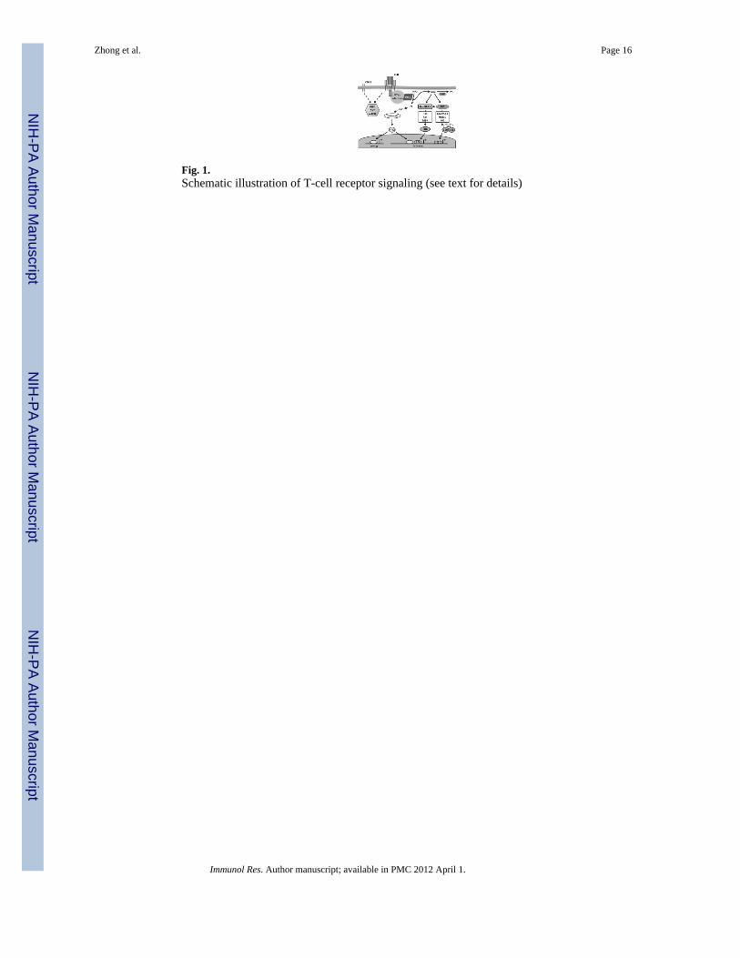
Fig. 1.Schematic illustration of T-cell receptor signaling (see text for details)
Zhong et al. Page 16
Immunol Res. Author manuscript; available in PMC 2012 April 1.
NIH
-PA Author Manuscript
NIH
-PA Author Manuscript
NIH
-PA Author Manuscript
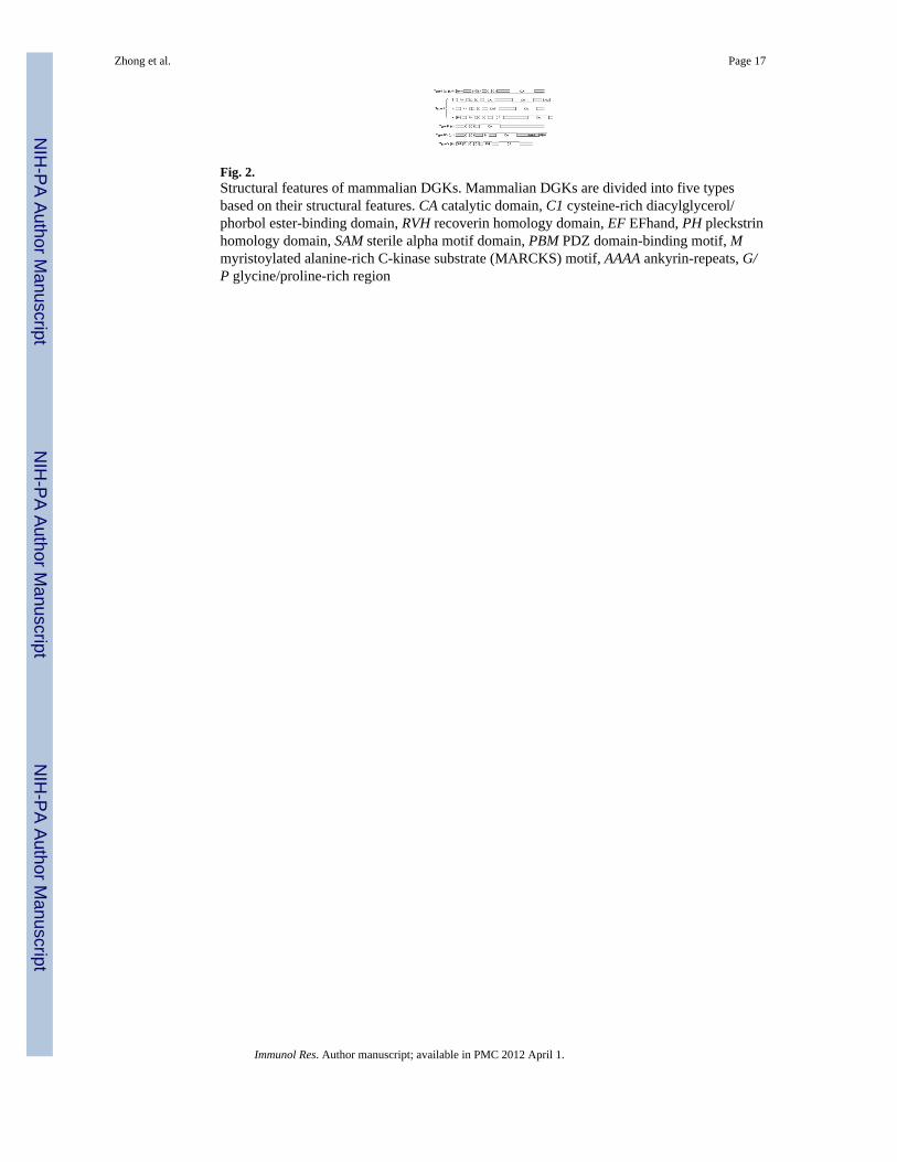
Fig. 2.Structural features of mammalian DGKs. Mammalian DGKs are divided into five typesbased on their structural features. CA catalytic domain, C1 cysteine-rich diacylglycerol/phorbol ester-binding domain, RVH recoverin homology domain, EF EFhand, PH pleckstrinhomology domain, SAM sterile alpha motif domain, PBM PDZ domain-binding motif, Mmyristoylated alanine-rich C-kinase substrate (MARCKS) motif, AAAA ankyrin-repeats, G/P glycine/proline-rich region
Zhong et al. Page 17
Immunol Res. Author manuscript; available in PMC 2012 April 1.
NIH
-PA Author Manuscript
NIH
-PA Author Manuscript
NIH
-PA Author Manuscript
Copyright © 2022 FDOKUMEN