Rational Zika vaccine design via the modulation of antigen ...
Themis controls thymocyte selection through regulation of T cellreceptor-mediated signaling
Guo Fu1,5, Sébastien Vallée1,5, Vasily Rybakin1, Marielena V. McGuire1,6, JeanetteAmpudia1, Claudia Brockmeyer3, Mogjiborahman Salek3, Paul R. Fallen1,6, John A.H.Hoerter1, Anil Munshi1, Yina H. Huang1,6, Jianfang Hu1, Howard S. Fox2,6, Karsten Sauer1,Oreste Acuto3, and Nicholas R.J. Gascoigne1,4
1Department of Immunology and Microbial Science, The Scripps Research Institute, 10550 NorthTorrey Pines Road, La Jolla, CA 92037, USA2Department of Molecular and Integrative Neurosciences, The Scripps Research Institute, 10550North Torrey Pines Road, La Jolla, CA 92037, USA3T cell Signalling Laboratory, Sir William Dunn School of Pathology, University of Oxford, SouthParks Road, Oxford OX1 3RE, UK
AbstractThemis (Thymocyte expressed molecule involved in selection), a member of a family of proteinswith unknown functions, is highly conserved among vertebrates. Here we found that Themis isexpressed in high amounts in thymocytes between the pre-T cell receptor (TCR) and positiveselection checkpoints, and in low amounts in mature T cells. Themis-deficient thymocytes exhibitdefective positive selection, which results in reduced numbers of mature thymocytes. Negativeselection is also impaired in Themis-deficient mice. A higher percentage of Themis-deficient Tcells exhibit CD4+CD25+Foxp3+ regulatory and CD62LloCD44hi memory phenotypes than inwild-type mice. Supporting a role for Themis in TCR signaling, this protein is phosphorylatedquickly after TCR stimulation, and is needed for optimal TCR-driven Ca2+ mobilization and Erkactivation.
INTRODUCTIONT cell development in the thymus follows an ordered progression from CD4−8− (doublenegative, DN, subdivided into stages DN1-4) through CD4+8+ (double positive, DP) tosingle positive (SP, CD4+8− or CD8+4−) stages that depends on a complex interplay ofsignaling pathways1,2. In DN3 cells, VDJ rearrangement and expression of the T cellreceptor (TCR) β-chain and its association with a pre-TCRα chain results in signaling thatdrives cells through the β-selection checkpoint into the TCRlo DP stage. Here, Vα-Jαrearrangement occurs and a mature αβ TCR is expressed on the cell surface. Sequentialrearrangements that delete previous rearrangements are frequently required before a TCR isproduced that has sufficient ability to interact with self MHC-peptide (pMHC) complexes toinduce the positive selection differentiation program that leads to SP thymocyte
4Correspondence should be addressed to N.R.J.G. ([email protected]).5These authors contributed equally.6Present addresses: MVM, United States Army Medical Research and Materiel Command, 1077 Patchel St., Fort Detrick, MD 21702;PRF, Oxford Gene Technology, Begbroke Science Park, Sandy Lane, Yarnton, Oxford OX5 1PF UK; YHH, Dept. of Pathology andImmunology, Washington University School of Medicine, St. Louis, MO; HSF, Dept. of Pharmacology, University of NebraskaMedical Center, Omaha, NE 68198-5800
The authors have no conflicting financial interests.
NIH Public AccessAuthor ManuscriptNat Immunol. Author manuscript; available in PMC 2010 February 1.
Published in final edited form as:Nat Immunol. 2009 August ; 10(8): 848–856. doi:10.1038/ni.1766.
NIH
-PA Author Manuscript
NIH
-PA Author Manuscript
NIH
-PA Author Manuscript

differentiation3. Cells that do not receive this positive selection signal eventually diethrough lack of stimulation, whereas those cells whose TCR binds too strongly to selfpMHC undergo activation induced apoptosis referred to as negative selection1,2.
Signaling through the TCR is tightly regulated during positive selection. Pre-selection DPcells express about 10% of the amount of cell surface TCR on post-selection TCRhi DP andSP cells, yet the pre-selection DP cells are more sensitive to TCR stimulation by weakligands than are the TCRhi cells4,5. The low TCR expression on pre-selection DP cells isactively maintained by Src-family protein tyrosine kinase (PTK) signaling andubiquitination-mediated degradation6,7, and these controls enforce TCR α-chain allelicexclusion8,9. Development through the positive selection checkpoint can be disrupted bymutation of elements of the TCR signaling cascade, such as the ZAP-70, Itk, Lck or Fynkinases or the Vav guanine nucleotide exchange factor10,11. The regulation of signalingthrough the MAP kinase Erk is extremely important in distinguishing between positive andnegative selection12–15. Similarly, mobilization of Ca2+ in response to TCR stimulation isboth regulated and related to the discrimination between positive or negativeselection4,16,17.
Here we identified a novel gene and protein involved in thymocyte positive and negativeselection. Two other groups have also identified this gene and protein, and in consensus werefer to it as thymocyte expressed molecule involved in selection (Themis)18,19. It isexpressed in a tightly regulated manner during T cell development. It is predominantlyexpressed in late DN and DP thymocytes, and is down-regulated after positive selection.Themis is tyrosine phosphorylated within seconds after TCR stimulation. It is a member of asmall gene family of unknown function, and is highly conserved amongst vertebrates. It hasno known conserved domains other than a polyproline region. We showed that Themis playsan important role in regulating thymocyte development through TCR signaling and inparticular through regulation of Ca2+ influx and phosphorylation of Erk.
RESULTSThemis encodes a novel protein
A cDNA subtraction library was constructed using cDNA from Tcra−/− thymus subtractedwith cDNA from Rag1−/− thymus20. A clone which exhibited differential expressionbetween Tcra−/− and Rag1−/− thymocyte mRNAs was analyzed further (Fig.1). Full-lengthcDNA clones were isolated from a mouse thymocyte library and from a human Jurkat celllibrary. The main open reading frame of the mouse Themis gene encodes a protein ofmolecular weight 72.8 kDa. The human THEMIS protein is 81% identical (86.4%similarity) to the mouse protein (Fig.1a). The gene is present in public databases (Mousegene #NM_178666, RIKEN cDNA E430004N04Rik; human NM_001010923), andorthologs are found across the mammals, and in birds (Gallus) and bony fish (Danio)(Supplementary Fig.1).
Although Southern blot experiments showed only a single Themis-hybridizing band inmouse genomic DNA, suggesting a single copy gene with no close relatives (data notshown), BLAST analysis of the mouse genome revealed two paralogs: the “basement-membrane-induced” gene Icb-121, and the hypothetical protein 9130404H23Rik. Themis ismore closely related to Icb-1 (32% identity) than to 9130404H23Rik (25% identity)(Supplementary Fig.2). The mouse Themis gene is on chromosome 10, location A4, andhuman THEMIS is at Chr 6q22.33 (C6orf190). The sequences of Themis, Icb-1 and9130404H23Rik revealed no identifiable conserved domains by PFAM or SMART searches,but there is a highly conserved proline rich sequence (PRS) forming three overlappingPXXP putative SH3 domain-recognition motifs near the C-terminus (residues 555–563)
Fu et al. Page 2
Nat Immunol. Author manuscript; available in PMC 2010 February 1.
NIH
-PA Author Manuscript
NIH
-PA Author Manuscript
NIH
-PA Author Manuscript

(Fig.1a; all numbering refers to mouse protein). The PRS is conserved in Icb-1 and9130404H23Rik (Supplementary Fig.2). A putative bipartite nuclear localization signal liesbetween residues 330–346.
Themis gene expression in T cell developmentThemis gene expression was analyzed by Northern blot of different tissues (Fig.1b). Themisis expressed in the thymus and to a lesser extent in the spleen, but was not detectable in non-lymphoid tissues. Of the two major transcripts, the 5.7 kb transcript was the most abundant.It was readily detectable in a number of transformed cell lines of thymic origin as well, butwas undetectable in transformed B cell lines (Supplementary Table 1). Reference to a publicgene expression atlas database (biogps.gnf.org)22 confirmed its very restricted expressionpattern, in that it was detected in high amounts in thymus, lower amounts in mature T cells,and very low amounts elsewhere. Remarkably, Icb-1 expression is restricted to B cells,macrophages and dendritic cells, and 9130404H23Rik is specifically expressed in theintestine (Supplementary Fig.3).
Real-time RT-PCR analysis of thymocyte subsets showed that Themis expression was lowin DN1 and DN2 cells and was upregulated in DN3 cells; expression remaining high inTCRlow immature pre-selection DP thymocytes, and was downregulated in post-positiveselection TCRint and TCRhi cells (Fig.1c). Themis expression remained low in mature SPsubsets. Positive selection is also marked by the migration of immature thymocytes from thethymus cortex towards the medulla, where only mature SP thymocytes that have survivedthe selection process are found. In situ hybridization showed that Themis expression wasdetectable mainly in cortical thymocytes with little expression in medullary thymocytes(Fig.1d). Thus, Themis expression is high in immature cortical thymocytes but low inmature medullary thymocytes.
These data suggested that Themis expression may be downregulated by stimulation throughthe αβ TCR. To explore this possibility we used the OT-I TCR transgenic (tg) mouse, whichexpresses a TCR recognizing a peptide derived from ovalbumin (OVA). A set of peptides isavailable with different affinities for the OT-I TCR, and positive and negative selection ofOT-I TCR tg thymocytes is blocked in mice lacking MHC class I expression (e.g.Tap1−/−)15,23–25. We therefore injected OT-I Tap1−/− mice (whose thymocytes are blockedat the pre-selection DP stage25) with peptides to stimulate the TCR in vivo26. Injection ofantigenic OVA peptide, but not a non-stimulatory VSV control peptide, resulted in down-regulation of Themis expression (Fig.1e). Basal Themis expression in Tap1-expressingthymocytes was lower than in Tap1-deficient cells, presumably due to the Themis down-regulation induced by positive selection. These data indicate that stimulation through theTCR by pMHC induces downregulation of Themis.
Defective positive selection of Themis−/− thymocytesTo determine the function of Themis in T cell development, we generated Themis-deficientmice. A construct targeting the first exon of Themis, replacing it with a neomycin cassette,was prepared and transfected into 129Sv ES cells (Supplementary Fig.4). These ES cellswere screened, selected and injected into blastocysts by standard methods. One progeny was>90% chimeric and was bred. Themis+/− offspring mice were backcrossed to B6 for ≥8generations and were then intercrossed to generate homozygous mutants (Themis−/−).Themis gene disruption resulted in intermediate or complete loss of expression of Themisprotein in Themis+/− or Themis−/− mice, respectively (Supplementary Fig. 4c).
Themis-deficient mice were viable, born at Mendelian frequency, and presented no grossabnormalities. Total thymocyte number was similar to wild-type mice (Supplementary Fig.
Fu et al. Page 3
Nat Immunol. Author manuscript; available in PMC 2010 February 1.
NIH
-PA Author Manuscript
NIH
-PA Author Manuscript
NIH
-PA Author Manuscript
5). The proportion and number of DP thymocytes was slightly but significantly increased (P< 0.05, Student’s t-test), while the proportion and number of mature SP cells—particularlyCD4 SP cells—was decreased (P < 0.01) (Fig. 2a and Supplementary Fig. 5). To identify thedevelopmental block in the Themis−/− mice, we subdivided thymocytes into five populationson the basis of differential CD3 and CD69 expression and analyzed CD4 and CD8expression (Fig.2b). Population 1, the most immature TCRloCD69lo cells, is composedmostly of DN and DP cells. This population was present in similar proportions in theThemis−/− and Themis+/+ mice. Population 2, which expresses a TCRintCD69lo phenotypeand corresponds to pre-selection DP cells, was present in slightly higher proportions inThemis−/− mice, as expected from the larger percentage of DP cells in these mice (Fig.2a).Population 3 partially upregulated CD69 and increased TCR expression relative topopulation 2, and was present in slightly lower proportions in Themis−/− mice. Population 4(TCRhiCD69hi; post-positive selection thymocytes) was markedly depleted in the Themis-deficient thymus. In the wild-type thymus, most of this population was CD4 or CD8 SP,whereas in Themis-deficient mice the cells were mostly CD4+CD8int or CD8 SP. Lastly,Themis−/− thymi contained very few TCRhiCD69lo population 5 cells, which correspond tomature SP cells ready for export to the periphery. From these data it is clear that the Themis-deficient cells are mostly blocked at the earliest stage of positive selection during whichCD8 downregulation is initiated, and that very few Themis-deficient thymocytes becomefully mature in phenotype.
To establish if Themis acts in a T cell-intrinsic manner, we performed a bone marrow (BM)reconstitution experiment (Fig.2c). BM cells from Themis+/+ Thy1.1 Ly5b mice and fromThemis−/− Thy1.2 Ly5b mice were injected into irradiated Thy1.2 Ly5a recipient mice andallowed to reconstitute the thymus for 6 weeks. Ly5b-expressing donor cells were gated andthen divided into Thy1.1+ (Themis+/+) and Thy1.2+ (Themis−/−) populations. This flowcytometric analysis demonstrated that the defect in development from DP to SP cells isintrinsic to the Themis−/− thymocytes as Themis−/− thymocytes showed reduced proportionsof CD4 SP cells and increased proportions of DP cells relative to the Themis+/+ cellsdeveloping in the same environment.
We next analyzed the effect of Themis-deficiency on thymocyte development in miceexpressing the MHC class II restricted TCR tg AND27, and the class I-restricted OT-I TCRtg24 (Fig.2d,e). The defect in thymocyte development in Themis-deficient TCR tg mice wasmore pronounced than in non-tg polyclonal Themis-deficient mice. In AND TCR tg thymi,development was blocked at the DP stage, with very few cells proceeding to CD4 SP (Fig.2d). The maturation arrest in the Themis−/− AND thymus was first manifest at the start ofthe transition from CD4hi8hi to CD4hi8int, with little or no progression to mature SP(Supplementary Fig.6a). In OT-I thymi, the developing class I-restricted cells pass throughthe CD4hi8int stage prior to upregulating CD8 and down-regulating CD428. Strong reductionof this stage in OT-I Themis−/− thymi (Fig.2e and Supplementary Fig.6b) suggests that, as inAND TCR tg and non-tg mice, Themis−/− OT-I mice show a strong defect at the earlieststage of positive selection. The architecture of the thymus was altered in Themis−/− mice,the medulla being smaller and fragmented compared to wild-type thymi (Fig.2f); this defectis indicative of impaired T cell maturation29.
Themis involvement in negative selectionTCR stimulation of DP and SP cells also induces negative selection. Thus, we investigatedthe role of Themis in negative selection in a superantigen-mediated deletion model. TheC57BL-derived mouse strains express superantigens Mtv-8 and 9, which cause deletion ofVβ5+, Vβ11+ and Vβ12+ TCR clonotypic thymocytes when the MHC molecule I–E is alsopresent30,31. To introduce I-E, which is absent on the B6-backcrossed Themis−/− mice, webred them to B10.D2 mice which express I-E. Comparison of T cell populations in H-2b/b
Fu et al. Page 4
Nat Immunol. Author manuscript; available in PMC 2010 February 1.
NIH
-PA Author Manuscript
NIH
-PA Author Manuscript
NIH
-PA Author Manuscript

(lacking I-E) and H-2b/d (expressing I-E) Themis+/− thymi showed superantigen-mediateddeletion of T cells expressing Vβ5, Vβ11, and Vβ12 (Fig.3). Vβ6+ (and Vβ7+, not shown)thymocytes do not recognize Mtv-8 and 9, and were not deleted. In fact, their proportionrose slightly in mice where deletion of the other clonotypes had occurred. In Themis−/−
mice, superantigen-mediated deletion was significantly less efficient than in Themis+/−
H-2b/d mice, although it was clearly partially active.
We also analyzed thymocyte development in mice expressing the MHC class I-restrictedHY TCR tg. Here, positive selection in females leads to a large CD8 SP population, butnegative selection predominates in males, resulting in few DP or SP cells32,33. HY TCR tgfemales lacking Themis resembled AND TCR tg and OT-I TCR tg Themis−/− mice in thatfew thymocytes progressed to become mature CD8 SP (Supplementary Fig.7a). HY TCR tgThemis+/− males showed a much reduced number of thymocytes compared to HY TCR tgfemales, and a large proportion of these cells were HY-TCR+ clonotypic cells expressinglow amounts of CD4 and CD83434. These cells are believed to escape negative selection byloss of the coreceptor. In male HY TCR tg Themis−/− thymi, negative selection wasapparently incomplete in that CD4 and CD8 expression was much lower than normal, butnot as low as in the Themis+/− males, and the clonotype-negative cells found in theThemis+/− males were not present (Supplementary Fig.7b). Thus both negative and positiveselection were reduced in the absence of Themis, suggesting that absence of Themis leads toa general defect in TCR-mediated signaling.
Peripheral T cells in Themis−/− miceThe deficiency in SP thymocytes led to a reduction in peripheral T cell numbers inThemis−/− mice (Fig.4a,b). The percentage of B cells increased due to the loss of T cells(data not shown). The deficiency in peripheral T cells was most evident in the CD4population, but both CD4 and CD8 cells were significantly reduced in number. Analysis ofTCR tg mice confirmed these data, with very few mature peripheral CD4 cells in ANDThemis−/− (Fig.4c), and few CD8 cells in OT-I Themis−/− (Fig.4d) TCR tg mice. Within theCD4+ population, cells with a regulatory T (Treg) cell phenotype (CD25+Foxp3+) wereincreased in frequency from 10% in Themis+/+ to 21% in Themis−/− (Fig.4e), though theabsolute number of Themis−/− Treg cells was ~50% of the number in Themis+/+ mice (datanot shown).
The expression of CD62L and CD44 in peripheral T cells is used to differentiate naïve frommemory T cells. There was a lower percent of CD62LhiCD44lo naïve phenotype cells, and ahigher proportion of CD62LloCD44hi memory phenotype cells in Themis−/− mice (Fig.4f).Expression of CD122+ (interleukin 2Rβ) was also higher in Themis−/− mice (data notshown). Thus the few peripheral cells in Themis-deficient mice have a phenotype redolentof memory cells, a frequent observation in partially lymphopenic mice that probably reflectshomeostatic T cell population expansion in the periphery35.
We then tested the ability of Themis−/− spleen T cells to respond to anti-CD3 stimulation(Fig.5). CD69 upregulation was strongly reduced in Themis-deficient CD4 and CD8 T cellscompared to wild-type T cells. Proliferation of both CD4 and CD8 Themis−/− T cells inresponse to anti-CD3 was very poor compared to Themis+/+ T cells. Themis+/+ andThemis−/− CD8 T cells expressed similar amounts of surface TCR, but TCR expression wasslightly reduced on the surface of Themis−/− CD4 cells (Fig.5a).
Themis in early TCR-signalingThe role of Themis in the TCR signaling cascade was investigated by immunoprecipitationand immunoblotting (Fig.6). Initially, a proteomic study of global dynamics of TCR-
Fu et al. Page 5
Nat Immunol. Author manuscript; available in PMC 2010 February 1.
NIH
-PA Author Manuscript
NIH
-PA Author Manuscript
NIH
-PA Author Manuscript

directed tyrosine phosphorylation using stable isotopic labeling of amino acids in cellculture (SILAC), identified a number of novel potential signaling components in Jurkatcells, including C6orf190 (C.B., O.A., M.S., to be published elsewhere), which was namedSPoT (for Signaling Phosphoprotein specific of T cell, which we refer to here as THEMIS).Reduction of THEMIS expression by siRNA in Jurkat cells resulted in substantial decreaseof interleukin 2 (IL-2) production induced by anti-TCR and anti-CD28 stimulation (C.B.,O.A., M.S., unpublished data). SILAC data suggested that THEMIS was phosphorylated ontyrosine as early as 30 seconds after TCR stimulation and that this phosphorylation wasreduced at 5 minutes after TCR stimulation; these phosphorylation kinetics are similar tothose of other known TCR-proximal signaling proteins (e.g., LAT, SLP-76) (data notshown).
To corroborate these data, Jurkat cells were stimulated with anti-CD3, lysates wereimmunoprecipitated with anti-phosphotyrosine (pTyr), and immunoprecipitates wereincubated with an antiserum against the C-terminal sequence of THEMIS. This treatmentrevealed a phosphoprotein of the expected size for THEMIS (~72 kD, Fig.6a) that increasedin abundance after 30 seconds of stimulation then declined. Immunoprecipitation with anti-THEMIS and immunoblotting with anti-pTyr provided direct evidence that THEMIS wasitself phosphorylated on a tyrosine residue, indicating that it is a TCR-dependent PTKsubstrate. Similar data were obtained with human CD4+ PBL T cells (Fig.6b). In mousethymocytes, Themis was tyrosine phosphorylated within 30 seconds after TCR crosslinking,and phosphorylation was undetectable after 3 minutes (Fig.6c). Together these data showthat in mouse and human cells Themis is an early target of TCR-controlled PTK(s) andsuggest that it is a component of the TCR signaling pathway whose function and-orrecruitment may depend on tyrosine phosphorylation.
Defective TCR signaling in Themis−/− thymocytesBecause the proline-rich sequence in Themis and related proteins is predicted to be abinding site for SH3 domains, we tested an SH3 domain array to screen for binding to the N-terminal and C-terminal (including the PRS) halves of Themis. The PRS-containing regionbound significantly to the phospholipase C-γ1 (PLC-γ1) SH3 domain (data not shown). Toconfirm a Themis-PLCγ1 interaction in thymocytes, we stimulated B6 thymocytes withanti-CD3, then immunoprecipitated cell lysates with Themis-specific antiserum. Althoughthere was some interaction between Themis and PLC-γ1 prior to stimulation, the amount ofPLCγ1 coprecipitated with Themis increased at 2 minutes post-stimulation, but ultimatelyreturned to a weaker interaction (Fig.6d). As Itk is a Tek-family PTK that is required foractivation of PLC-γ1 and for TCR-signaling36, we tested the possibility of an interactionbetween Itk and Themis. Anti-Itk coprecipitated both Themis and PLC-γ1 from freshlyisolated, anti-CD3-stimulated thymocytes (Fig.6e). It appeared that that Itk-Themis and Itk-PLC-γ1 interactions increased after CD3 crosslinking. To test this, we rested thymocytes inculture for 2h before activation with anti-CD3 plus anti-CD4. As previously reported37,PLC-γ1 interacted constitutively with Itk in thymocytes; after stimulation the interactionincreased slightly before declining after 30 seconds (Fig.6f). Interaction of Themis with Itkwas not constitutive, but was induced by 30 seconds after TCR stimulation.
To determine which signaling pathways are defective in Themis−/− thymocytes, we analyzedimmunoblots of anti-CD3 plus anti-CD4 activated thymocytes from Themis+/+ andThemis−/− thymocytes (Fig.6g). We found no difference in ZAP70, PLC-γ1 or Itk (or LAT,data not shown) phosphorylation in response to TCR stimulation. However, Erk showedreduced phosphorylation in Themis−/− cells. Erk1 phosphorylation (p-p44) was delayed inThemis−/− compared to Themis+/+ cells, and Erk2 phosphorylation (p-p42) was reduced inThemis−/− cells at 30 and 90 seconds after stimulation (Fig.6g, Supplementary Fig.8a), andwas not always detectable at 3 minutes. This experiment was performed three times with
Fu et al. Page 6
Nat Immunol. Author manuscript; available in PMC 2010 February 1.
NIH
-PA Author Manuscript
NIH
-PA Author Manuscript
NIH
-PA Author Manuscript

similar results (Supplementary Fig.8b). Phosphorylation of Raf, Mek and Jnk was notaffected by Themis deficiency (data not shown).
To look at other aspects of TCR signaling, we stimulated pre-selection DP thymocytes fromOT-I TCR tg Tap1−/− mice. Stimulation by anti-CD3 plus anti-CD4 or OVA-Kb tetramers(Fig.7a) induced a slightly slower and reduced initial peak of store-operated Ca2+ influx inThemis−/− cells relative to Themis+/+ cells. This difference in Ca2+ influx, while relativelysubtle, was very reproducible (a total of 9 Themis−/− versus 5 Themis+/+ in 3 separateexperiments). The release of endoplasmic reticulum Ca2+ stores was very low in thethymocytes, and we found no difference between Themis−/− cells relative to Themis+/+ cells.
Organization of the cytoskeleton at the immunological synapse is regulated by TCRsignaling38. We therefore imaged actin polymerization in OT-I Tap1−/− thymocytesexpressing or lacking Themis as they interacted with OVA peptide presented by EL4 tumorcells (Fig.7b). The actin polymerization pattern in Themis-sufficient thymocytes was similarto that in OT-I lymph node T cells, but Themis-deficient thymocytes showed a significantdefect in actin polymerization at the immunological synapse. A useful correlate ofstimulation of thymocytes through the TCR by positive or negative selecting ligands isupregulation of CD6915,25,39,40. We found that the Themis−/− OT-I Tap1−/− pre-selectionDP thymocytes were less able to upregulate CD69 in response to OVA peptide than theirThemis+/+ OT-I Tap1−/− counterparts (Fig.7c). These data suggest that Themis is involved inregulating the TCR-signaling cascade, albeit with relatively small effects on early events inthe cascade.
DISCUSSIONDevelopment of αβ T cells is critically dependent on signaling through the TCR, and can bedisrupted by deficiencies in the receptor itself and many signaling molecules and mediators.In this work, we identified a novel protein, Themis, that is required for passage through thepositive selection checkpoint.
Themis deficiency results in a defect in development that is stronger in CD4 cells than inCD8 cells. However, this defect was evident in both MHC class I and class II-restricted TCRtransgenic lines, wherein thymocyte development was severely blocked at the earliest stagesof positive selection. A stronger phenotype in TCR tg thymocytes is a common finding inmice deficient in signaling molecules, as the normal TCR repertoire is plastic enough toselect for TCRs with higher or lower affinity, thereby allowing the signal strength to bemodulated to permit selection10. The strong developmental block in the TCR tg Themis−/−
mice, together with the evidence indicating that negative selection signaling is also deficientin Themis−/− mice, suggested a role for Themis in the TCR signaling cascade rather than ina developmental program per se. Because Themis is expressed in immature thymocytes, andis downregulated by stimulation through the TCR as occurs during positive selection, thesignaling role of Themis is likely to be most important in DP thymocytes before or duringpositive selection. In agreement with this notion, Themis−/− mice pre-selection thymocytesresponded poorly compared to Themis+/+ thymocytes to TCR stimulation, as measured byCD69 upregulation. Themis−/− thymocytes also showed reduced actin polymerization at theimmunological synapse, as in Itk-deficient and Vav-deficient cells. These results areconsistent with a defect in TCR signal transduction39–42.
Themis was tyrosine phosphorylated within 30 seconds of TCR stimulation, suggesting thatit may play a role early in signal transduction through the TCR. Moreover, it interactedinducibly with Itk and PLC-γ1, although phosphorylation of these proteins was not alteredin Themis−/− cells. Deletion of Itk has a profound effect on Ca2+ signaling, but a relatively
Fu et al. Page 7
Nat Immunol. Author manuscript; available in PMC 2010 February 1.
NIH
-PA Author Manuscript
NIH
-PA Author Manuscript
NIH
-PA Author Manuscript

minor effect on thymocyte development36,41. In contrast, Themis-deficiency had a strongeffect on development but a small effect on Ca2+ signals (e.g. slightly slower and weakerCa2+ flux). The effect of loss of Themis on Erk signaling was also quite subtle, andmanifested as a reduction in the strength and duration of phosphorylation. These findingssuggest that Themis might negatively regulate phosphatases that turn off Erk signaling.Notably, similarly small differences in p-Erk induction distinguished signals that inducepositive and negative selection of thymocytes14,15, and sustained Erk signals are requiredfor positive selection2,12,14. Additionally, there is evidence that slight changes in Ca2+
signaling lead to profound differences in MAP kinase activation in non T cells43, and inpre-selection thymocytes, and that these differences distinguish the quality of TCRstimulation17. The alterations in Ca2+ and Erk signaling between Themis−/− and Themis+/+
pre-selection thymocytes suggest that Themis may be involved in setting the threshold forTCR responses to selecting signals. Such a role for Themis can account for its expressionprimarily in pre-positive selection thymocytes, and would indicate that its role in TCR-mediated signaling may not be so important in peripheral T cell activation. Study of thenormal role of Themis in peripheral T cell signaling will have to await generation of aconditional Themis−/− mouse.
When given the opportunity to produce a full TCR repertoire, some Themis-deficientthymocytes pass through the positive selection checkpoint and generate peripheral T cells.These cells respond poorly to TCR-mediated stimulation. Interestingly, in both CD4 andCD8 subsets, there was an increased prevalence of memory-phenotype(CD44hiCD62LloCD122+) cells and reduced proportions of naïve-phenotype populations.This may be the result of homeostatic population expansion in a lymphopenicenvironment35, and is similar to the phenotype seen in mice lacking Itk. Memory phenotypeT cells in Itk-deficient mice have been referred to as “innate-like” T cells44. Thisphenotypic similarity to Itk-deficient mice supports the notion that Themis is involved inTCR signal transduction via Itk.
We also noted a strongly increased proportion (though reduced absolute number) of Tregcells in Themis−/− mice. Interestingly the Themis gene was identified as one of thetranscripts most strongly downregulated by Foxp3 in response to TCR stimulation45, and itssuppression may therefore be part of the Treg cell development program.
Themis, Icb-1 and 9130404H23Rik are members of a small and essentially uncharacterizedfamily. Each has a very restricted cell-type-specific expression and is highly conserved invertebrates. Other than a PRS, they have no predicted conserved domains that are sharedwith other proteins. These properties point to the family having important and perhaps novelfunctions and structure. Here we showed that one of these family members, Themis, is a Tcell specific protein with a crucial function in positive selection through transducing TCRsignals in thymocytes.
Accession CodesMouse Themis: MGI:2443552, Human THEMIS GeneID: 387357.
METHODSMice
Tcra−/− (B6.129S2-Tcratm1Mom/J), Rag1−/− (B6.129S7-Rag1tm1Mom/J), and Ly5a congenicmice (B6.SJL-Ptprca Pep3b/BoyJ) on the B6 background were obtained from Jackson Labs.Other strains were bred at The Scripps Research Institute (TSRI) and maintained inaccordance with TSRI Animal Care and Use Committee.
Fu et al. Page 8
Nat Immunol. Author manuscript; available in PMC 2010 February 1.
NIH
-PA Author Manuscript
NIH
-PA Author Manuscript
NIH
-PA Author Manuscript

AntibodiesAntibodies recognizing CD3 (145-2C11), CD4 (RM4.4, RM4.5, GK1.5), CD8α (53–6.7),CD25 (PC61.5), CD62L (MEL14), CD44 (IM7), Itk (2F12), PLC-γ1 (10/PLCg), Foxp3(FJK-165), Vβ5 (MR9-6), Vβ6 (RR4-7), Vβ11 (CTVB11), and Vβ12 (CTVB12b), wereobtained from eBiosciences or BDBiosciences. Anti-p-Tyr (4G10) was from UpstateBiotechnology. Anti-p42/44 (3A7) and p-p42/44 (197G2) and antisera to p-ZAP70 and p-PLC-γ1 were from Cell Signaling Technology. Anti-Themis was raised in rabbits againstthe N-terminal 294 amino acids, made as a fusion to glutathione-S-transferase (GST),purified and with the GST removed. The antiserum was affinity purified. Anti-THEMISserum was made by immunizing rabbits with a peptide corresponding to the C-terminal 15amino acids of the human sequence. Specificity is demonstrated in Supplementary Fig.9.
Cloning of ThemisA subtraction library was constructed from cDNA from Tcra−/− thymus mRNA subtractedwith cDNA from Rag1−/− thymus mRNA as described20. Clones randomly picked from thelibrary were sequenced and used to search Genbank to identify potentially novel clones. Afull length clone of human THEMIS was isolated by screening a Jurkat cDNA library.
Real-time RT-PCR AnalysisReal-time PCR was performed using SYBERgreen PCR Master Mix and the Prism 7900instrument and analyzed with SDSv2.1 software (Applied Biosystems). Results werenormalized to β-actin expression. Primer sequences are shown in Supplementary Table 2).
Generation of Themis-deficient miceExon 1 of Themis was replaced by the 1.6 kb PGK neomycin cassette. The target vector(pKO Scrambler) contained the flanking sequence of exon 1: a 5′ 2.8 kb short arm and a 3′3.9 kb long arm sequence flanking the neomycin phosphotransferase gene. Embryonic stemcells (129SvEv strain) were electroporated with 25µg linearized DNA/107 cells, and culturedfor 7d in medium containing 400 µg/ml G418. Drug resistant clones were expanded andhomologous recombination confirmed by PCR and Southern blot. Positive clones wereinjected into blastocysts derived from B6 embryo donor mice mated with C6D2F1 studmales (B6 × DBA2/J hybrids) and transplanted into pseudopregnant CD-1 recipients.Chimeras were bred to C57BL/6J females in order to reveal germ-line transmission followedby backcrossing to B6 (10 generations).
Calcium fluxThymocyte suspensions were prepared and split into separate tubes. Part was loaded withCFSE (20 nM, for 10 min at 37°C) or Cy5 (1µg/ml, 5min RT), or mock treated, followed bywashing. CFSE-labeled and-or Cy5-labeled populations were mixed equally with a mock-loaded sample, for example; OT-I Tap1−/−Themis+/+ (mock) plus OT-I Tap1−/−Themis−/−
(CFSE or Cy5). In some experiments, the CFSE- and Cy5-treated and untreated pairs werereversed to ensure that the CFSE or Cy5 did not alter the results. Cells were suspended at107/ml in cRPMI (RPMI supplemented with 10% FCS, 100U/ml Penicillin, 10µg/mlStreptomycin, 292µg/ml Glutamine, 50µM 2-ME, 25mM HEPES) and were incubated with2µM Indo-1 AM (Molecular Probes) for 30 min at 37°C, 5% CO2. Cells were washed twicewith cRPMI. Cells were treated with biotin-anti-CD3 and anti-CD4 as well as PerCPCy5.5-anti-CD8 and PECy7-anti-CD4 on ice for 20min. Cells were washed once with cRPMI andonce with Ca2+ free medium (cHBSS: Ca2+ and Mg2+ free HBSS supplemented with 1%FCS, 1mM MgCl2, 1mM EGTA, and 10mM HEPES), and resuspended in cHBSS. Cellswere pre-warmed to 37°C before analysis and were kept at 37°C during event collection ona Becton-Dickinson LSRII. To stimulate cells, 10µg/ml Streptavidin (Jackson) was added to
Fu et al. Page 9
Nat Immunol. Author manuscript; available in PMC 2010 February 1.
NIH
-PA Author Manuscript
NIH
-PA Author Manuscript
NIH
-PA Author Manuscript

cross-link biotinylated antibodies; alternatively, cells were stimulated with tetramers madeas described from refolded Kb and β2m15,23, or purchased from Beckman-Coulter (iTagMHC tetramer). 5mM CaCl2 was added during analysis, and maximal Ca2+ flux obtained byadding 500ng/ml of ionomycin (Calbiochem). Mean fluorescence ratio was calculated usingFlowJo (TreeStar).
T cell activation and proliferation assayLymphocytes from Themis+/+ (Thy1.1, B6.PL) or Themis−/− (Thy1.2, B6) mice were mixed1:1. Cells were cultured in the presence of soluble anti-CD3 for 5h, stained with anti-CD69and anti-Thy1 allotypes and analyzed by FACS. For proliferation assays, cells were stainedwith 0.2µM CFSE at 37°C for 10 min. Staining was stopped by adding an equal volume ofFCS. After washing, cells were cultured with or without stimulants (e.g. anti-CD3) incRPMI for 48h. T cell proliferation was measured by CFSE dilution analyzed by FACS.
Thymocyte CD69 upregulation assayThymocytes (2–3×10⁵) from OT-I tg Tap1−/− mice (Themis+/− or −/−) were incubated with2×105 peptide-pulsed EL4 cells in round-bottom 96-well plates at 37° for 5h. Cells werestained and analyzed by FACS as described39,40. Gates defining CD69− versus CD69+
were determined from thymocytes incubated with non-peptide pulsed EL4 cells.
Immunoprecipitation and immunoblottingCells were lysed in buffer containing 20mM Tris pH7.5, 150mM NaCl, 0.5% n-Dodecyl-β-D-maltoside or NP40, 20mM NaF, 1mM Na3VO4, and protease inhibitor cocktail (Roche).Immunoprecipitation was performed on cell lysates (0.5–1 mg of protein) overnight at 4°C,then protein-A sepharose beads (Amersham) were added for 1h. After washing, sampleswere resolved on 4–12% SDS-PAGE and blotted. The membrane was blocked 1h with PBS,1% TWEEN, 5% dry milk, incubated with primary Ab for 3h at room temperature orovernight at 4°C, washed and incubated with the secondary Horseradish peroxidase-antibody for 1.5h followed by enhanced chemiluminescence detection of signal (Pierce).
In situ hybridizationThymi from 3–4 week-old mice were fixed, embedded in paraffin and 5µm sections probedwith antisense 35S-UTP-labeled Themis RNA. Sections were RNAase A-treated, washed(0.2X SSC, 42°C), dried, coated with Kodak NTB2 emulsion, exposed for 1–2 weeks,developed, and counterstained with methyl green. Controls included hybridizations withsense and unrelated probes.
Peptide stimulationThe peptides OVA (SIINFEKL) or VSV (RGYVYQGL) were injected i.v. into OT-ITap1−/− mice as described26. After 1h, the thymus was removed, RNA extracted, andsubjected to real time RT-PCR.
Statistical testingStatistical differences were calculated using the mean difference hypothesis of Student’s twotailed t-test assuming different variances and confidence level of 95%. Calculationsperformed using GraphPad Prism.S
Supplementary MaterialRefer to Web version on PubMed Central for supplementary material.
Fu et al. Page 10
Nat Immunol. Author manuscript; available in PMC 2010 February 1.
NIH
-PA Author Manuscript
NIH
-PA Author Manuscript
NIH
-PA Author Manuscript

AcknowledgmentsThis work was supported by NIH grants GM048002 and AI073870 to NRJG. SV was supported by the ConcernFoundation for Cancer Research. MVM and JAHH were supported by T32 AI07244. JAHH was supported by theIrving S. Sigal Fellowship. OA, CB and MS were supported by Wellcome Trust program grant GR076558MA andby funds from the Sir William Dunn School of Pathology. CB received the Sloane Robinson Graduate Award fromLincoln College, Oxford. We thank James Bradley, Alex Field, Yugarshi Mondal, Samuel Rose, Wayne Sakati,Vandana Sharma, Jovan Shepherd, and Jacob Vasquez for technical help. This is manuscript 19524 from TheScripps Research Institute.
REFERENCES1. Starr TK, Jameson SC, Hogquist KA. Positive and negative selection of T cells. Annu. Rev.
Immunol. 2003; 21:139–176. [PubMed: 12414722]
2. Werlen G, Hausmann B, Naeher D, Palmer E. Signaling life and death in the thymus: timing iseverything. Science. 2003; 299:1859–1863. [PubMed: 12649474]
3. Malissen, B.; Malissen, M. Allelic exclusion of T cell antigen receptor genes. In: Bell, JI.; Owen,MJ.; Simpson, E., editors. T cell receptors. Oxford: Oxford University Press; 1995. p. 352-368.
4. Davey GM. Preselection thymocytes are more sensitive to T cell receptor stimulation than mature Tcells. J. Exp. Med. 1998; 188:1867–1874. [PubMed: 9815264]
5. Lucas B, Stefanova I, Yasutomo K, Dautigny N, Germain RN. Divergent changes in the sensitivityof maturing T cells to structurally related ligands underlies formation of a useful T cell repertoire.Immunity. 1999; 10:367–376. [PubMed: 10204492]
6. D’Oro U, Vacchio MS, Weissman AM, Ashwell JD. Activation of the Lck tyrosine kinase targetscell surface T cell antigen receptors for lysosomal degradation. Immunity. 1997; 7:619–628.[PubMed: 9390686]
7. Naramura M, Kole HK, Hu R-J, Gu H. Altered thymic positive selection and intracellular signals inCbl-deficient mice. Proc. Natl. Acad. Sci. USA. 1998; 95:15547–15552. [PubMed: 9861006]
8. Alam SM, Gascoigne NRJ. Post-translational regulation of TCR Va allelic exclusion during T celldifferentiation. J. Immunol. 1998; 160:3883–3890. [PubMed: 9558094]
9. Niederberger N, et al. Allelic exclusion of the TCR a-chain is an active process requiring TCR-mediated signaling and c-Cbl; J. Immunol. 2003; 170:4557–4563.
10. Fischer A, Malissen B. Natural and engineered disorders of lymphocyte development. Science.1998; 280:237–243. [PubMed: 9535646]
11. Zamoyska R, et al. The influence of the src-family kinases, Lck and Fyn, on T cell differentiation,survival and activation. Immunol. Rev. 2003; 191:107–118. [PubMed: 12614355]
12. Mariathasan S, et al. Duration and strength of extracellular signal-regulated kinase signals arealtered during positive versus negative thymocyte selection. J. Immunol. 2001; 167:4966–4973.[PubMed: 11673503]
13. Fischer AM, Katayama CD, Pages G, Pouyssegur J, Hedrick SM. The role of erk1 and erk2 inmultiple stages of T cell development. Immunity. 2005; 23:431–443. [PubMed: 16226508]
14. McNeil LK, Starr TK, Hogquist KA. A requirement for sustained ERK signaling during thymocytepositive selection in vivo. Proc. Natl. Acad. Sci. USA. 2005; 102:13574–13579. [PubMed:16174747]
15. Daniels MA, et al. Thymic selection threshold defined by compartmentalization of Ras/MAPKsignalling. Nature. 2006; 444:724–729. [PubMed: 17086201]
16. Kane LP, Hedrick SM. A role for calcium influx in setting the threshold for CD4+CD8+ thymocytenegative selection. J.Immunol. 1996; 156:4594–4601. [PubMed: 8648101]
17. Freedman BD, Liu QH, Somersan S, Kotlikoff MI, Punt JA. Receptor avidity and costimulationspecify the intracellular Ca2+ signaling pattern in CD4+CD8+thymocytes. J. Exp. Med. 1999;190:943–952. [PubMed: 10510084]
18. Schwartz RH. THEMIS PAPER. Nature Immunol. 2009
19. Love P. THEMIS PAPER. Nature Immunol. 2009
Fu et al. Page 11
Nat Immunol. Author manuscript; available in PMC 2010 February 1.
NIH
-PA Author Manuscript
NIH
-PA Author Manuscript
NIH
-PA Author Manuscript
20. McGuire MV, Suthipinijtham P, Gascoigne NRJ. The mouse Supt16h/Fact140 gene, encoding partof the FACT chromatin transcription complex, maps close to Tcra and is highly expressed inthymus. Mamm. Genome. 2001; 12:664–667. [PubMed: 11471063]
21. Treeck O, Strunck E, Vollmer G. A novel basement membrane-induced gene identified in thehuman endometrial adenocarcinoma cell line HEC1B. FEBS Lett. 1998; 425:426–430. [PubMed:9563507]
22. Su AI, et al. A gene atlas of the mouse and human protein-encoding transcriptomes. Proc. Natl.Acad. Sci. USA. 2004; 101:6062–6067. [PubMed: 15075390]
23. Alam SM, et al. T cell receptor affinity and thymocyte positive selection. Nature. 1996; 381:616–620. [PubMed: 8637599]
24. Hogquist KA, et al. T cell receptor antagonist peptides induce positive selection. Cell. 1994;76:17–27. [PubMed: 8287475]
25. Hogquist KA, et al. Identification of a naturally occurring ligand for positive selection. Immunity.1997; 6:389–399. [PubMed: 9133418]
26. Fiorini E, et al. Peptide-induced negative selection of thymocytes activates transcription of an NF-kB inhibitor; Mol. Cell. 2002; 9:637–648.
27. Kaye J, et al. Selective development of CD4+ T cells in transgenic mice expressing a class IIMHC-restricted antigen receptor. Nature. 1989; 341:746–749. [PubMed: 2571940]
28. Gallegos AM, Bevan MJ. Central tolerance to tissue-specific antigens mediated by direct andindirect antigen presentation. J. Exp. Med. 2004; 200:1039–1049. [PubMed: 15492126]
29. van Ewijk W, Hollander G, Terhorst C, Wang B. Stepwise development of thymicmicroenvironments in vivo is regulated by thymocyte subsets. Development. 2000; 127:1583–1591. [PubMed: 10725235]
30. Irwin MJ, Gascoigne NRJ. Interplay between superantigens and the immune system. J. LeukocyteBiol. 1993; 54:495–503. [PubMed: 8228628]
31. Scherer MT, Ignatowicz L, Winslow GM, Kappler JW, Marrack P. Superantigens: Bacterial andviral proteins that manipulate the immune system. Annu. Rev. Cell Biol. 1993; 9:101–128.[PubMed: 7506550]
32. Kisielow P, Bluthmann H, Staerz UD, Steinmetz M, von Boehmer H. Tolerance in T-cell-receptortransgenic mice involves deletion of nonmature CD4+8+ thymocytes. Nature. 1988; 333:742–746.[PubMed: 3260350]
33. Teh HS, et al. Thymic major histocompatibility complex antigens and the aβ T-cell receptordetermine the CD4/CD8 phenotype of T cells. Nature. 1988; 335:229–233. [PubMed: 2970593]
34. Teh HS, Kishi H, von Boehmer H. Deletion of autospecific T cells in T cell receptor (TCR)transgenic mice spares cells with normal TCR levels and low levels of CD8 molecules. J. Exp.Med. 1989; 169:795–800. [PubMed: 2494291]
35. Ge Q, Hu H, Eisen HN, Chen J. Different contributions of thymopoiesis and homeostasis-drivenproliferation to the reconstitution of naive and memory T cell compartments. Proc. Natl. Acad.Sci. USA. 2002; 99:2989–2994. [PubMed: 11880642]
36. Berg LJ, Finkelstein LD, Lucas JA, Schwartzberg PL. Tec family kinases in T lymphocytedevelopment and function. Annu. Rev. Immunol. 2005; 23:549–600. [PubMed: 15771581]
37. Huang YH, et al. Positive regulation of Itk PH domain function by soluble IP4. Science. 2007;316:886–889. [PubMed: 17412921]
38. Burkhardt JK, Carrizosa E, Shaffer MH. The actin cytoskeleton in T cell activation. Annu. Rev.Immunol. 2008; 26:233–259. [PubMed: 18304005]
39. Yachi PP, Ampudia J, Gascoigne NRJ, Zal T. Nonstimulatory peptides contribute to antigen-induced CD8-T cell receptor interaction at the immunological synapse. Nat. Immunol. 2005;6:785–792. [PubMed: 15980863]
40. Yachi PP, Lotz C, Ampudia J, Gascoigne NRJ. T cell activation enhancement by endogenouspMHC acts for both weak and strong agonists but varies with differentiation state. J Exp Med.2007; 204:2747–2757. [PubMed: 17954567]
41. Liu KQ, Bunnell SC, Gurniak CB, Berg LJ. T cell receptor-initiated calcium release is uncoupledfrom capacitative calcium entry in Itk-deficient T cells. J. Exp. Med. 1998; 187:1721–1727.[PubMed: 9584150]
Fu et al. Page 12
Nat Immunol. Author manuscript; available in PMC 2010 February 1.
NIH
-PA Author Manuscript
NIH
-PA Author Manuscript
NIH
-PA Author Manuscript

42. Labno CM, et al. Itk functions to control actin polymerization at the immune synapse throughlocalized activation of Cdc42 and WASP. Curr. Biol. 2003; 13:1619–1624. [PubMed: 13678593]
43. Kupzig S, Walker SA, Cullen PJ. The frequencies of calcium oscillations are optimized forefficient calcium-mediated activation of Ras and the ERK/MAPK cascade. Proc. Natl. Acad. Sci.USA. 2005; 102:7577–7582. [PubMed: 15890781]
44. Berg LJ. Signalling through TEC kinases regulates conventional versus innate CD8+ T-celldevelopment. Nat. Rev. Immunol. 2007; 7:479–485. [PubMed: 17479128]
45. Marson A, et al. Foxp3 occupancy and regulation of key target genes during T-cell stimulation.Nature. 2007; 445:931–935. [PubMed: 17237765]
46. Corpet F. Multiple sequence alignment with hierarchical clustering. Nucleic Acids Res. 1988;16:10881–10890. [PubMed: 2849754]
47. Gouet P, Courcelle E, Stuart DI, Metoz F. ESPript: analysis of multiple sequence alignments inPostScript. Bioinformatics. 1999; 15:305–308. [PubMed: 10320398]
Fu et al. Page 13
Nat Immunol. Author manuscript; available in PMC 2010 February 1.
NIH
-PA Author Manuscript
NIH
-PA Author Manuscript
NIH
-PA Author Manuscript

Figure 1.Themis structure, expression and localization in wild-type mice. (a) Alignment of predictedamino acid sequence of murine (Mm) and human (Hs) Themis proteins. Sequencealignments and comparisons were performed using Multalin46, and rendered usingESPript47. Polyproline region is underlined. Amino acid identity shown as white-on-black.(b) Northern blot of Themis RNA from different tissues. Representative of threeindependent experiments. (c)Themis mRNA expression analyzed by real-time RT-PCR ofFACS-sorted subsets of B6 thymocytes. CD8m refers to mature CD8SP cells. Data shownrelative to β-actin expression. Representative of three experiments. Error bars denote s.d.
Fu et al. Page 14
Nat Immunol. Author manuscript; available in PMC 2010 February 1.
NIH
-PA Author Manuscript
NIH
-PA Author Manuscript
NIH
-PA Author Manuscript

(d)In situ hybridization of mouse thymus with Themis probe. Top left, brightfield imageshowing Themis expression as black dots. Original magnification ×10. Top right, sameimage in darkfield showing Themis expression as white dots. Bottom, brightfield image ofcorticomedullary junction (cortex on right, medulla on left). Original magnification ×40.Representative of 2 independent experiments, 3 mice each. (e) OT-I TCR tg Tap1−/− micewere injected i.v. with PBS, OVA or VSV peptide. Thymocytes were isolated and Themisexpression analyzed by real-time RT-PCR. Note that OT-I tg Tap1−/− mice do not have anymature T cells or thymocytes specific for the injected peptide, so they would not be expectedto undergo apoptosis due to cytokine release. FACS analysis did not show any increase inthe percentage of dead cells in thymocytes from the treated mice, nor was there any changein the percentage of DP or SP subsets (data not shown). Representative of 4 experiments.
Fu et al. Page 15
Nat Immunol. Author manuscript; available in PMC 2010 February 1.
NIH
-PA Author Manuscript
NIH
-PA Author Manuscript
NIH
-PA Author Manuscript
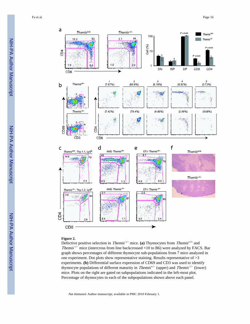
Figure 2.Defective positive selection in Themis−/− mice. (a) Thymocytes from Themis+/+ andThemis−/− mice (intercross from line backcrossed ×10 to B6) were analyzed by FACS. Bargraph shows percentages of different thymocyte sub-populations from 7 mice analyzed inone experiment. Dot plots show representative staining. Results representative of >3experiments. (b) Differential surface expression of CD69 and CD3 was used to identifythymocyte populations of different maturity in Themis+/− (upper) and Themis−/− (lower)mice. Plots on the right are gated on subpopulations indicated in the left-most plot.Percentage of thymocytes in each of the subpopulations shown above each panel.
Fu et al. Page 16
Nat Immunol. Author manuscript; available in PMC 2010 February 1.
NIH
-PA Author Manuscript
NIH
-PA Author Manuscript
NIH
-PA Author Manuscript

Representative of 2 mice each in 2 independent experiments. (c) Thymocyte sub-populationsfrom irradiated Thy1.2Ly5a mice were reconstituted with a 1:1 mixture of BM fromThy1.2,Ly5bThemis−/− and Thy1.1,Ly5bThemis+/+ mice. Donor populations were identifiedby Thy1.1 or Thy1.2 expression of Ly5b+ cells. Two chimeric mice per group were analyzedin 3 separate experiments with similar results. (d,e) Thymocytes from Themis+/+ andThemis−/− mice expressing the AND TCR27 (d) or the OT-I TCR24 (e) were analyzed byFACS. Significant differences (P < 0.0005, t-test) were found between the percentage ofCD4 SP (d) and CD8 SP (e) cells in Themis+/+ and Themis−/− thymi (data pooled from >3separate experiments each); Themis+/+ n=4 (d), n=8 (e); and Themis−/− n=6 (d), n=12 (e).(f) Thymus sections from Themis+/+ and Themis−/− mice were stained with hematoxylin.Medulla shows lighter staining. Original magnification ×10. Representative of twoexperiments.
Fu et al. Page 17
Nat Immunol. Author manuscript; available in PMC 2010 February 1.
NIH
-PA Author Manuscript
NIH
-PA Author Manuscript
NIH
-PA Author Manuscript
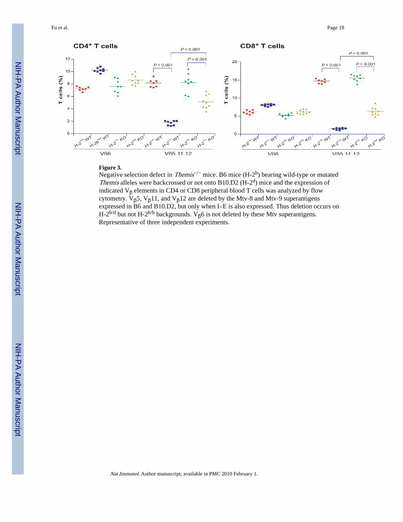
Figure 3.Negative selection defect in Themis−/− mice. B6 mice (H-2b) bearing wild-type or mutatedThemis alleles were backcrossed or not onto B10.D2 (H-2d) mice and the expression ofindicated Vβ elements in CD4 or CD8 peripheral blood T cells was analyzed by flowcytometry. Vβ5, Vβ11, and Vβ12 are deleted by the Mtv-8 and Mtv-9 superantigensexpressed in B6 and B10.D2, but only when I–E is also expressed. Thus deletion occurs onH-2b/d but not H-2b/b backgrounds. Vβ6 is not deleted by these Mtv superantigens.Representative of three independent experiments.
Fu et al. Page 18
Nat Immunol. Author manuscript; available in PMC 2010 February 1.
NIH
-PA Author Manuscript
NIH
-PA Author Manuscript
NIH
-PA Author Manuscript

Figure 4.Peripheral T cell phenotype in Themis−/− mice. (a,b) CD4 and CD8 T cells in the spleen ofB6 Themis+/+ and Themis−/− mice of indicated ages were quantified by flow cytometry. Bargraph shows average percentage of each population in seven 6 week old mice analyzed inone experiment. Dot plots show representative stainings. Data are representative of >3experiments. (c,d) CD4 and CD8 expression on spleen cells from mice expressing AND (c)and (d) OT-I TCR transgenes. Differences between AND Themis+/+ and Themis−/− CD4 Tcell percentages (P < 0.0009, n=4 mice of each genotype), and Themis+/+ and Themis−/−
OT-I CD8 T cell percentages (P < 0.003, Themis−/− n=7, Themis+/+ n=5) were significant
Fu et al. Page 19
Nat Immunol. Author manuscript; available in PMC 2010 February 1.
NIH
-PA Author Manuscript
NIH
-PA Author Manuscript
NIH
-PA Author Manuscript
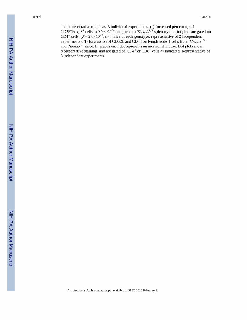
and representative of at least 3 individual experiments. (e) Increased percentage ofCD25+Foxp3+ cells in Themis−/− compared to Themis+/+ splenocytes. Dot plots are gated onCD4+ cells. (P = 2.8×10⁻⁵, n=4 mice of each genotype, representative of 2 independentexperiments). (f) Expression of CD62L and CD44 on lymph node T cells from Themis+/+
and Themis−/− mice. In graphs each dot represents an individual mouse. Dot plots showrepresentative staining, and are gated on CD4+ or CD8+ cells as indicated. Representative of3 independent experiments.
Fu et al. Page 20
Nat Immunol. Author manuscript; available in PMC 2010 February 1.
NIH
-PA Author Manuscript
NIH
-PA Author Manuscript
NIH
-PA Author Manuscript
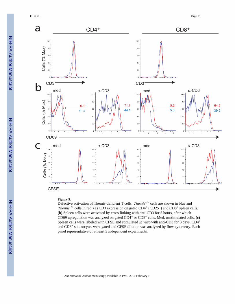
Figure 5.Defective activation of Themis-deficient T cells. Themis−/− cells are shown in blue andThemis+/+ cells in red. (a) CD3 expression on gated CD4+ (CD25−) and CD8+ spleen cells.(b) Spleen cells were activated by cross-linking with anti-CD3 for 5 hours, after whichCD69 upregulation was analyzed on gated CD4+ or CD8+ cells. Med, unstimulated cells. (c)Spleen cells were labeled with CFSE and stimulated in vitro with anti-CD3 for 3 days. CD4+
and CD8+ splenocytes were gated and CFSE dilution was analyzed by flow cytometry. Eachpanel representative of at least 3 independent experiments.
Fu et al. Page 21
Nat Immunol. Author manuscript; available in PMC 2010 February 1.
NIH
-PA Author Manuscript
NIH
-PA Author Manuscript
NIH
-PA Author Manuscript
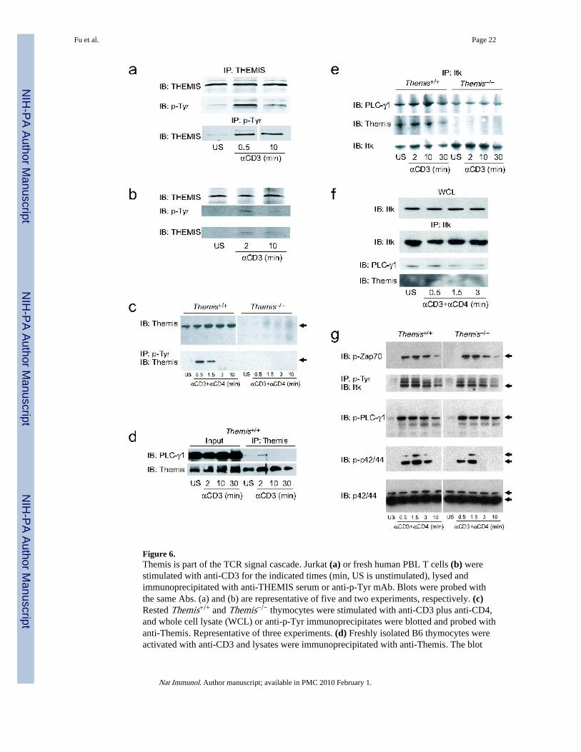
Figure 6.Themis is part of the TCR signal cascade. Jurkat (a) or fresh human PBL T cells (b) werestimulated with anti-CD3 for the indicated times (min, US is unstimulated), lysed andimmunoprecipitated with anti-THEMIS serum or anti-p-Tyr mAb. Blots were probed withthe same Abs. (a) and (b) are representative of five and two experiments, respectively. (c)Rested Themis+/+ and Themis−/− thymocytes were stimulated with anti-CD3 plus anti-CD4,and whole cell lysate (WCL) or anti-p-Tyr immunoprecipitates were blotted and probed withanti-Themis. Representative of three experiments. (d) Freshly isolated B6 thymocytes wereactivated with anti-CD3 and lysates were immunoprecipitated with anti-Themis. The blot
Fu et al. Page 22
Nat Immunol. Author manuscript; available in PMC 2010 February 1.
NIH
-PA Author Manuscript
NIH
-PA Author Manuscript
NIH
-PA Author Manuscript
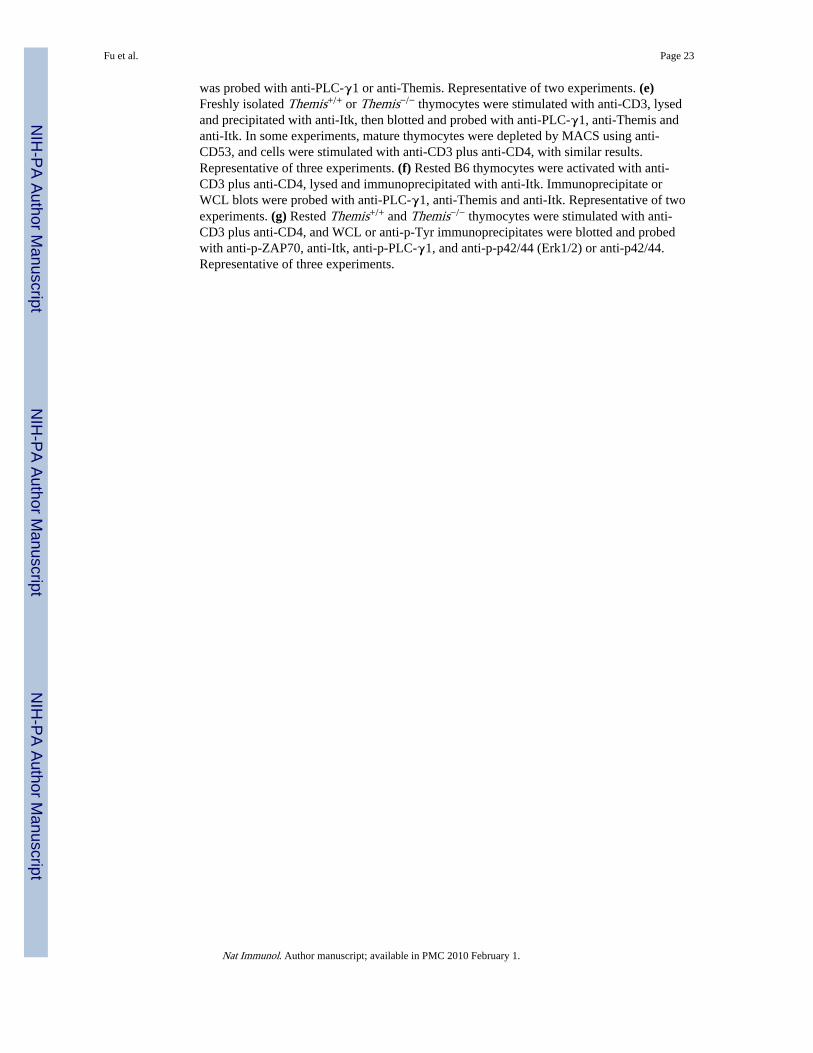
was probed with anti-PLC-γ1 or anti-Themis. Representative of two experiments. (e)Freshly isolated Themis+/+ or Themis−/− thymocytes were stimulated with anti-CD3, lysedand precipitated with anti-Itk, then blotted and probed with anti-PLC-γ1, anti-Themis andanti-Itk. In some experiments, mature thymocytes were depleted by MACS using anti-CD53, and cells were stimulated with anti-CD3 plus anti-CD4, with similar results.Representative of three experiments. (f) Rested B6 thymocytes were activated with anti-CD3 plus anti-CD4, lysed and immunoprecipitated with anti-Itk. Immunoprecipitate orWCL blots were probed with anti-PLC-γ1, anti-Themis and anti-Itk. Representative of twoexperiments. (g) Rested Themis+/+ and Themis−/− thymocytes were stimulated with anti-CD3 plus anti-CD4, and WCL or anti-p-Tyr immunoprecipitates were blotted and probedwith anti-p-ZAP70, anti-Itk, anti-p-PLC-γ1, and anti-p-p42/44 (Erk1/2) or anti-p42/44.Representative of three experiments.
Fu et al. Page 23
Nat Immunol. Author manuscript; available in PMC 2010 February 1.
NIH
-PA Author Manuscript
NIH
-PA Author Manuscript
NIH
-PA Author Manuscript

Figure 7.Signaling in Themis-deficient thymocytes. (a) Pre-selection DP thymocytes from Themis+/+
or Themis−/− OT-I Tap1−/− mice were stimulated by crosslinking biotinylated anti-CD3 and-CD4 with streptavidin (SAv) (left and center) or with OVA-Kb tetramer (right) in Ca2+-freemedium. CaCl2 was added later. Cells were surface-labeled with CFSE, Cy5 or nothing,mixed and assayed in one tube so that both Themis+/+ or Themis−/− cells were tested at thesame time in the same environment. Left; 3 samples of Themis+/+ cells tested against eachother, center and right; Themis+/+ cells tested against cells from two different Themis−/−
mice. Representative of three independent experiments. (b) OT-I T cells or pre-selection DP
Fu et al. Page 24
Nat Immunol. Author manuscript; available in PMC 2010 February 1.
NIH
-PA Author Manuscript
NIH
-PA Author Manuscript
NIH
-PA Author Manuscript

thymocytes, Themis+/+ or Themis−/−, were incubated with EL4 cells loaded with OVApeptide and stained to detect polymerized actin. Amount of f-actin in the immunologicalsynapse versus the rest of the T cell membrane was calculated from imaging data andexpressed in the left graph as the actin signal at the membrane within or outside of thesynapse (boxes, s.e.m.; crossbar, median; plus sign, mean; bars, 5–95 percentile range; dots,data points outside this range). Right graph shows ratio of f-actin in the synapse to that inthe non-synapse membrane. Each circle represents a single data point, bold lines show themean, thinner lines show ± s.e.m. ***P < 0.05. Representative of two experiments. (c) Pre-selection DP thymocytes from OT-I Tap1−/−Themis+/+ or Themis−/− mice were stimulatedin vitro with OVA presented by EL4 cells and CD69 upregulation analyzed39,40.Representative of three experiments.
Fu et al. Page 25
Nat Immunol. Author manuscript; available in PMC 2010 February 1.
NIH
-PA Author Manuscript
NIH
-PA Author Manuscript
NIH
-PA Author Manuscript
Copyright © 2022 FDOKUMEN