Concentric diversification based on technological capabilities
Upload
khangminh22Category
view
4download
0
Diversification of CD1 Molecules Shapes Lipid AntigenSelectivity
Nicole M. Paterson,1,2 Hussein Al-Zubieri,1,2 and Matthew F. Barber 1,3,*
1Institute of Ecology and Evolution, University of Oregon, Eugene, OR 97403, USA2Department of Chemistry and Biochemistry, University of Oregon, Eugene, OR 97403, USA3Department of Biology, University of Oregon, Eugene, OR 97403, USA
*Corresponding author: E-mail: [email protected]
Associate editor : Kelley Harris
Abstract
Molecular studies of host–pathogen evolution have largely focused on the consequences of variation at protein–proteininteraction surfaces. The potential for other microbe-associated macromolecules to promote arms race dynamics withhost factors remains unclear. The cluster of differentiation 1 (CD1) family of vertebrate cell surface receptors plays acrucial role in adaptive immunity through binding and presentation of lipid antigens to T-cells. Although CD1 proteinspresent a variety of endogenous and microbial lipids to various T-cell types, they are less diverse within vertebratepopulations than the related major histocompatibility complex (MHC) molecules. We discovered that CD1 genes exhibita high level of divergence between simian primate species, altering predicted lipid-binding properties and T-cell receptorinteractions. These findings suggest that lipid–protein conflicts have shaped CD1 genetic variation during primateevolution. Consistent with this hypothesis, multiple primate CD1 family proteins exhibit signatures of repeated positiveselection at surfaces impacting antigen presentation, binding pocket morphology, and T-cell receptor accessibility. Usinga molecular modeling approach, we observe that interspecies variation as well as single mutations at rapidly-evolvingsites in CD1a drastically alter predicted lipid binding and structural features of the T-cell recognition surface. We furthershow that alterations in both endogenous and microbial lipid-binding affinities influence the ability of CD1a to undergoantigen swapping required for T-cell activation. Together these findings establish lipid–protein interactions as a criticalforce of host–pathogen conflict and inform potential strategies for lipid-based vaccine development.
Key words: adaptive immunity, positive selection, antigen presentation, protein evolution.
IntroductionEarly detection of pathogen-specific molecules by the im-mune system can mean the difference between resistance,latency, or succumbing to infectious disease. Previous studieshave illustrated that host–pathogen protein interaction sur-faces are hotspots for repeated natural selection by influenc-ing resistance or susceptibility to infection (Daugherty andMalik 2012; Enard et al. 2016; van der Lee et al. 2017). Suchconflicts between hosts and pathogens can give rise to avariety of evolutionary dynamics including Red Queen armsraces (Van Valen 1973; Daugherty and Malik 2012),frequency-dependent selection (Takahata and Nei 1990),and over-dominance (Hughes and Nei 1990; Takahata andNei 1990; Nei and Rooney 2005). Although vertebrate im-mune systems are tuned to recognize a wide variety ofpathogen-associated macromolecules including DNA, RNA,lipids, and glycans, our understanding of host–pathogen evo-lutionary conflicts is largely restricted to protein–proteininteractions (Sawyer et al.2005; Elde et al. 2009; Barber andElde 2014; Choby et al. 2018). In the case of lipid and lip-opeptide antigens, the production of a functional moleculeinvolves the synthesis of precursors that are further processed
by enzymatic modifications. As such, evolutionary dynamicsinvolving these macromolecules and their host receptors maybe distinct from protein–protein interactions.
The major histocompatibility complex (MHC) superfamilycomprises a variety of cell surface proteins which present selfand foreign antigens to T-cells. Recognition of foreign anti-gens by T-cell receptors (TCRs) leads to T-cell activation andinitiation of an adaptive immune response (Frank 2002).Multiple evolutionary forces are hypothesized to contributeto the immense diversity in MHC haplotypes, including over-dominance wherein increased heterozygosity is favored byselection and polymorphisms are maintained over time(Takahata and Nei 1990). In addition to class I and class IIMHC molecules which present peptide antigens, the paralo-gous cluster of differentiation 1 (CD1) and MR1 moleculeshave been shown to present lipid and lipoprotein antigens toT-cells (Blumberg et al. 1995; Barral and Brenner 2007;Birkinshaw et al. 2015; Moriet al.2016; Zajonc and Flajnik2016). CD1 molecules display rare and infrequent polymor-phism with limited genetic diversity within humans and otherpopulations relative to class I and II MHC (Han et al. 1999;Golmogghaddam et al. 2013). The MHC and CD1 gene fam-ilies therefore appear to have experienced divergent
Article
� The Author(s) 2021. Published by Oxford University Press on behalf of the Society for Molecular Biology and Evolution.This is an Open Access article distributed under the terms of the Creative Commons Attribution License (http://creativecommons.org/licenses/by/4.0/), which permits unrestricted reuse, distribution, and reproduction in any medium, provided the original work isproperly cited. Open AccessMol. Biol. Evol. 38(6):2273–2284 doi:10.1093/molbev/msab022 Advance Access publication February 2, 2021 2273
Dow
nloaded from https://academ
ic.oup.com/m
be/article/38/6/2273/6126409 by guest on 23 January 2022

evolutionary paths after their duplication from a commonancestor with respect to antigen recognition and populationgenetic variation. CD1 paralogs are divided into groups,whereby group 1 CD1 family members (including humanCD1a, CD1b, and CD1c) present antigen primarily to cyto-toxic CD8þ T-cells (Mori et al.2016). Group 2 CD1 molecules(including CD1d in humans) present antigens to invariantnatural killer (iNK) T-cells (Pereira and Macedo 2016).
CD1 and MHC (fig. 1A) arose in jawed fishes with theadvent of the B-cell and T-cell immune receptors duringthe genesis of the adaptive immune system of vertebrates(Barral and Brenner 2007). After the gene duplications thatgave rise to ancestral MHC and CD1, vertebrate CD1 paralogsexpanded through repeated duplication events (Dascher2007). This initial expansion was followed by differential pseu-dogenization and expansion of CD1 paralogs to variousdegrees across vertebrate species (Rogers and Kaufman2016). As such, CD1 gene content varies widely across verte-brates: primates have a single copy of the CD1A paralog, micepossess none, dogs encode six, and horses possess five(Dascher 2007). In primates, CD1D is believed to representthe most deeply conserved member of the CD1 family(Salomonsen et al.2005). CD1d receptors can display antigento specialized iNKT-cells, which are able to mount an earlierresponse to infection reflecting their dual role in innate andadaptive immunity (Pereira and Macedo 2016). Current evi-dence indicates that human CD1e does not present antigen(Garcia-Alles et al. 2011) but rather assists in antigen loadingonto CD1d in lysosomes and endosomes (Cala-De Paepe et al.2012). This wide range of responsive T-cell types along withevidence that these nonclassical T-cell types mount an earlyimmune response to infection (Godfrey et al. 2015) makeshuman CD1-expressing cells surprisingly flexible respondersto infections even with their lack of exceptional sequencevariation.
CD1 molecules possess an extracellular domain containinga subsurface hydrophobic-binding pocket used to presentantigen to CD1-restricted T-cells (fig. 1B) (Barral andBrenner 2007). During the adaptive immune response, CD1on the surface of antigen-presenting cells activates T-cells bydisplaying specific classes of hydrophobic ligands to TCRs(fig. 1C) (Blumberg et al. 1995; Barral and Brenner 2007;Chancellor et al.2018). According to structural studies,CD1a has the smallest of the human CD1-binding pocketswith a volume of about 1,280 A (Ly and Moody 2014). Afterthe gene duplication event that gave rise to this paralog,CD1a likely evolved to present either self-lipids or small ex-ogenous lipopeptides (Mori et al.2016). Consistent with thishypothesis, CD1a has been crystallized in complex with self-lipids sphingomyelin, lysophosphatidylcholine (Birkinshawet al. 2015), sulfatide (Zajonc et al. 2003), as well as the my-cobacterial lipopeptideanalog didehydroxy-mycobactin(Zajonc et al. 2005). The binding pocket of human CD1a iscomposed of a double-chambered cavity termed the A0 andF0 pockets with a single (A0) portal that coordinates the pre-sentation of lipid antigen to the TCR (fig. 1B) (Zajonc et al.2005). The TCR lands just above the A0 pocket on a surfacetermed the A0 roof (Zajonc et al. 2005). The diminutive size of
the CD1a A0 pocket is thought to be formed by the electro-static interaction of the two side chains belonging to the A0
roof that also draw the two parallel alpha helices of thepocket in close proximity, whereas an amino acid sidechainblocks the base of the pocket thereby limiting size of tailgroups that can be accommodated (in human CD1a thisamino acid is valine 28) (Zajonc et al. 2003). Several otherCD1 homologs, except for CD1c, lack this roof structure(Blumberg et al. 1995). CD1a does not feature a late endo-somal targeting element and does not require low pH forantigen binding as is the case for other CD1 proteins suchas CD1b, CD1c, and CD1d (Chancelloret al.2018).
The immense diversity of the MHC family within and be-tween populations at surfaces necessary for peptide antigenrecognition has made these genes classic study systems ofadaptive protein evolution (Danchin and Pontarotti 2004;Castroet al. 2015; Grimholt 2016). CD1 molecules possesssimilar structure and function to class I and II MHC proteins,although their relative lack of diversity at the population levelhas been attributed to a lack of diversity in their cognate lipidligands. Although variation in pathogen-derived lipids hasbeen implicated in host immune recognition and virulence(Chandler et al. 2020), the potential for lipid antigens to pro-mote evolutionary conflicts with host species is unclear. In thepresent study, we used the CD1 family as a system to inves-tigate the diversity and evolution of lipid antigen recognitionby the vertebrate immune system.
Results
Diversification of the CD1 Gene Family in PrimatesA comparison of MHC class I and CD1 protein structuresillustrates the homology between these antigen presentationmolecules (fig. 1A). CD1 presents antigen to the TCR with thelipid tail groups tucked into the hydrophobic pocket andhead groups exposed where they are “read” by the TCR(fig. 1B). Distinct CD1 molecules present antigen to a widevariety of T-cell types (fig. 1C) (Godfrey et al. 2015). To assesspatterns of genetic diversity among primate CD1 familymembers, we first assembled a collection of simian primateCD1 homologs from publicly available genome databases andgenerated a phylogenetic gene tree using PhyML (fig. 1C andsupplementary fig. S1, Supplementary Material online). Thefive human CD1 paralogs are present in the majority of pri-mate genomes surveyed, allowing us to reliably comparestructural and genetic diversity within this family. A compar-ison of the sequences between CD1 orthologs revealed astriking degree of diversification, particularly in the MHC-like domain responsible for lipid antigen presentation(fig. 1D). To assess the potential consequences of this varia-tion on CD1 function, we plotted the structural conservationamong primate CD1a orthologs using color by conservation(Mura et al. 2010) (fig. 1D and E). Our analysis revealed severalhotspots of high amino acid divergence among CD1 mole-cules, focused on both interior regions of the antigen-bindingpocket as well as surface helices that are known to contactthe TCR. Together our results indicate that, despite theirlimited polymorphism within populations, CD1 paralogs
Paterson et al. . doi:10.1093/molbev/msab022 MBE
2274
Dow
nloaded from https://academ
ic.oup.com/m
be/article/38/6/2273/6126409 by guest on 23 January 2022
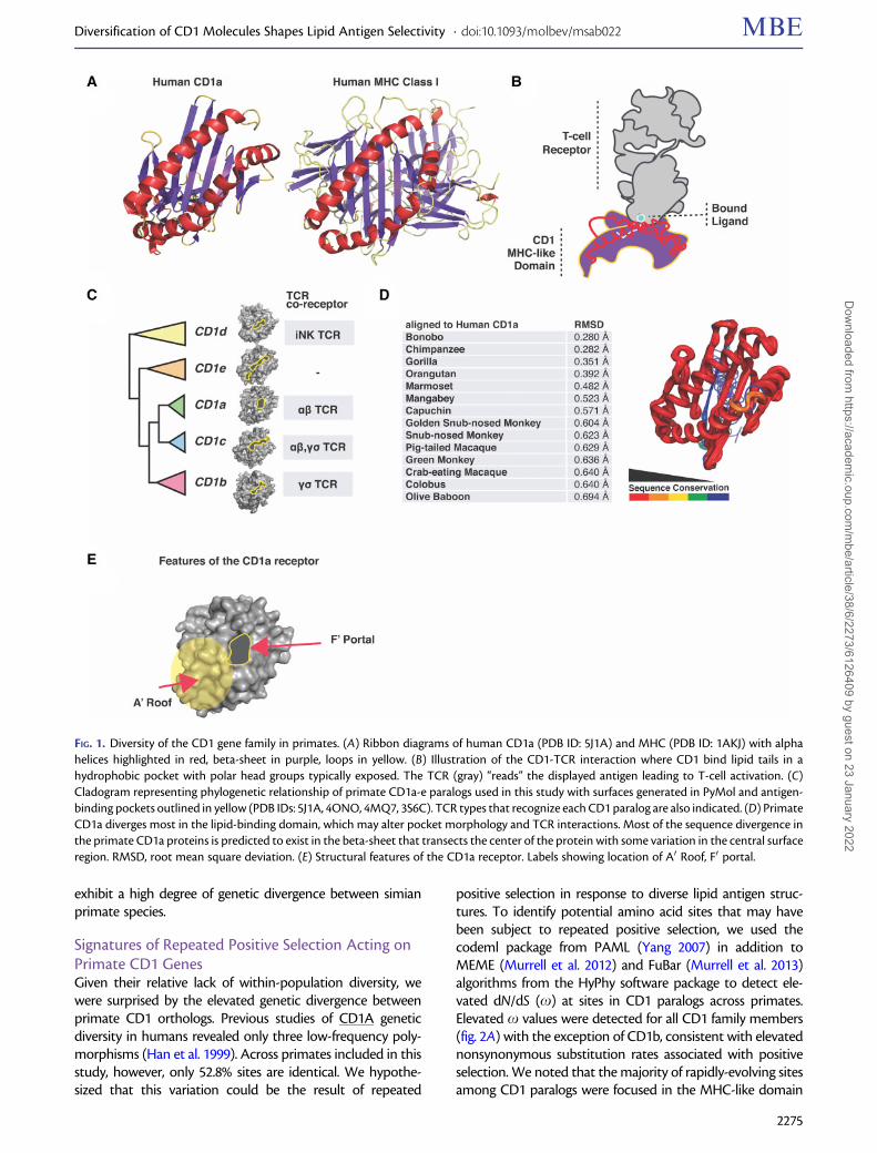
exhibit a high degree of genetic divergence between simianprimate species.
Signatures of Repeated Positive Selection Acting onPrimate CD1 GenesGiven their relative lack of within-population diversity, wewere surprised by the elevated genetic divergence betweenprimate CD1 orthologs. Previous studies of CD1A geneticdiversity in humans revealed only three low-frequency poly-morphisms (Han et al. 1999). Across primates included in thisstudy, however, only 52.8% sites are identical. We hypothe-sized that this variation could be the result of repeated
positive selection in response to diverse lipid antigen struc-tures. To identify potential amino acid sites that may havebeen subject to repeated positive selection, we used thecodeml package from PAML (Yang 2007) in addition toMEME (Murrell et al. 2012) and FuBar (Murrell et al. 2013)algorithms from the HyPhy software package to detect ele-vated dN/dS (x) at sites in CD1 paralogs across primates.Elevated x values were detected for all CD1 family members(fig. 2A) with the exception of CD1b, consistent with elevatednonsynonymous substitution rates associated with positiveselection. We noted that the majority of rapidly-evolving sitesamong CD1 paralogs were focused in the MHC-like domain
FIG. 1. Diversity of the CD1 gene family in primates. (A) Ribbon diagrams of human CD1a (PDB ID: 5J1A) and MHC (PDB ID: 1AKJ) with alphahelices highlighted in red, beta-sheet in purple, loops in yellow. (B) Illustration of the CD1-TCR interaction where CD1 bind lipid tails in ahydrophobic pocket with polar head groups typically exposed. The TCR (gray) “reads” the displayed antigen leading to T-cell activation. (C)Cladogram representing phylogenetic relationship of primate CD1a-e paralogs used in this study with surfaces generated in PyMol and antigen-binding pockets outlined in yellow (PDB IDs: 5J1A, 4ONO, 4MQ7, 3S6C). TCR types that recognize each CD1 paralog are also indicated. (D) PrimateCD1a diverges most in the lipid-binding domain, which may alter pocket morphology and TCR interactions. Most of the sequence divergence inthe primate CD1a proteins is predicted to exist in the beta-sheet that transects the center of the protein with some variation in the central surfaceregion. RMSD, root mean square deviation. (E) Structural features of the CD1a receptor. Labels showing location of A0 Roof, F0 portal.
Diversification of CD1 Molecules Shapes Lipid Antigen Selectivity . doi:10.1093/molbev/msab022 MBE
2275
Dow
nloaded from https://academ
ic.oup.com/m
be/article/38/6/2273/6126409 by guest on 23 January 2022

which is responsible for lipid binding (fig. 2A). These resultssuggest that multiple members of the CD1 family have un-dergone repeated episodes of positive selection in simianprimates specifically within regions important for lipid anti-gen presentation.
Having detected evidence of positive selection acting onseveral CD1 family members, we chose CD1a for additionalin-depth analysis. CD1a has less stringent lipid loadingrequirements than other CD1 homologs as it does not requirea reduced pH environment encountered in late endosomes,nor does it have a known adapter protein required for antigenloading (Barral and Brenner 2007). For these reasons, we an-ticipated that empirical and molecular modeling studies ofantigen recognition would be less complex for CD1a than
other paralogs. CD1a has been shown to present mycobac-terial antigens from the cell surface where it interacts withlangerin on Langerhans cells (Mizumoto and Takashima2004), a specialized dendritic cell type that surveys epithelialmonolayers for molecular indicators of infection.
To determine what domains of CD1a are subject to pos-itive selection, we mapped the sites with high x values fromour previous analysis. Results show clustering of rapidly evolv-ing sites in the MHC-like domain, similar to those observedwith other CD1 paralogs (fig. 2B and supplementary figs. S2–S4, Supplementary Material online). These sites also map toregions where CD1a is likely to interface with lipid antigen orthe TCR, suggesting that selection may have acted to alterlipid-binding and T-cell interactions. The majority of variable
FIG. 2. Evidence of repeated positive selection among primate CD1 orthologs. (A) Amino acid sites exhibiting strong signatures of positive selection(elevated dN/dS) are highlighted in teal and mapped onto corresponding crystal structures. Alpha helices are denoted in red, beta-sheet in purple.(PDB IDs: 5J1A, 4ONO, 4MQ7, 3S6C). Table summarizes positions in CD1 paralogs contributing to signatures of positive selection as well asstatistics from PAML M7-M8 model comparisons. (B) Sites with elevated dN/dS indicative of positive selection (teal) cluster in the MHC domain ofCD1a protein (PDB ID: 5J1A). Alpha helices denoted in red, beta-sheet in purple. (C) Multiple sequence alignment of primate species used tocalculate dN/dS ratios for CD1a paired with phylogenetic species tree highlighting the branches (teal) predicted by aBSREL to be undergoingepisodic positive selection.
Paterson et al. . doi:10.1093/molbev/msab022 MBE
2276
Dow
nloaded from https://academ
ic.oup.com/m
be/article/38/6/2273/6126409 by guest on 23 January 2022

sites between CD1a orthologs cluster to a region of the pro-tein near the center of the binding pocket and around theouter surface (fig. 2B). We grouped all of the rapidly-evolvingsites we identified in this study into three categories: residueslocated at or near the TCR landing site (the A0 roof in thehuman structure), residues within the binding pocket, andresidues in the N-terminus for which we have no structuralinformation. Overall, predicted structural features do not cor-relate well with phylogenetic relatedness, consistent withmultiple lineages undergoing episodic selection (figs. 2C and3A).
Accelerated Evolution of the CD1a-TCR InterfaceTo assess how variation in CD1a may influence immunefunctions in primates, we used I-TASSER to generate pre-dicted structures of several CD1a orthologs (fig. 3A). Of thehominoid structures modeled, human, bonobo, and orangu-tan have remarkably different topologies at the TCR-CD1ainterface as well as the geometry of the internal bindingpocket (fig. 3A). The morphologies of the binding pocketsvary widely, most notably in the crab-eating macaque whichis predicted to contain one main and two accessory portals,with a narrow meandering channel (fig. 3A, bottom panel).The length and volume of the pockets limit the types of lipidtail groups that can be accommodated, whereas the size andlocation of the portals have effects on how well the T-cellreceptor can read the antigen presented (Birkinshaw et al.2015).
In human CD1a, the A0 roof is hypothesized to aid indetermining whether an antigen will elicit an immune re-sponse by supporting interactions with the TCR and assistingin display of the ligand head-group (fig. 3B) (Zajonc et al. 2005;Birkinshaw et al. 2015). The predicted orangutan CD1a struc-ture lacks an A0 roof entirely (fig. 3C), whereas bonobo CD1apossesses two portals. Additionally, it has been speculatedthat disruption of hydrogen bonding between R73, R76,and E154 that form the A0 roof may indicate whether a givenligand will stimulate TCR activation (Birkinshaw et al. 2015).However, several of the CD1a structures are predicted to forman A0 roof that does not depend on this particular interaction.For example, crab-eating macaque and olive baboon CD1aare predicted to form a relatively unique A0 roof composed ofan R73/153Q linkage that does not involve R76 (fig. 3C). TheTCR does not recognize CD1a-bound ligands without ade-quate projection of lipid head groups, and it is likely thathydrogen bonding between the head groups of smallerligands and residues that make up the portal are importantfor display. Headless ligands buried in the CD1a pocket, forexample, can result in T-cell auto-reactivity (de Jong et al.2010). Site 153, which is highly variable across primates(fig. 2D) has been shown to form a hydrogen bond in humanCD1a to the head group of self antigen lysophosphatidylcho-line and sulfatide in addition to its role in forming the A0 roof(Zajonc et al. 2003; Birkinshaw et al. 2015). This site bears aglycine in orangutan, with no ability to form an A0 roof or saltbridges with ligand (fig. 3C). Together, these predicted struc-tural differences suggest that natural selection may have had
a significant impact on the ability of CD1a to display self orforeign lipid antigens across related primates.
Structural Remodeling of the CD1a Antigen-BindingPocketTo determine whether the structural differences observedacross CD1a paralogs are likely to have functional consequen-ces for antigen recognition, we applied a ligand-docking ap-proach using AutoDock Vina. We were particularly interestedto test affinity differences between endogenous and exoge-nous lipid ligands, since the current model for CD1a antigenpresentation is a swapping mechanism wherein lower affinityendogenous ligand is replaced at the cell surface by higheraffinity exogenous ligand. We used ligands previously crystal-lized in complex with human CD1a in our studies since thereis a wealth of structural information available on these par-ticular binding interactions. In our docking simulations, wefound that a single loop region is required to redock all ligandsin the CD1a-binding pocket. We assigned flexibility to thisregion for all the structures tested, as well as any nonbondedsidechains in the region of the portal (supplementary fig.S2,Supplementary Material online). We believe this is likely theregion responsible for conferring flexibility in the native CD1a,which must be flexible enough to accommodate ligands witha diversity of molecular weights (molecular weight of urushiolis 330 g/mol, dideoxy-mycobactin is 838 g/mol).
We next measured predicted CD1a-binding affinities for thepanel of lipid ligands including endogenous ligands sphingo-myelin, sulfatide, lysophosphatidylcholine, and exogenous li-gand dideoxy-mycobactin (DDM) (fig. 4A and supplementaryfigs. S5–S7, Supplementary Material online). We chose theselipids because published structural information exists for eachligand bound to the human CD1a receptor. Given that CD1a isbelieved to swap endogenous lipids for exogenous lipids basedon differences in relative binding affinities, we then estimatedthe likelihood of a lipid-swap using our panel of ligands. Wecalculated the fold differences in Kd (dissociation constant)between the highest affinity endogenous lipid and comparedthis to the Kd of the exogenous ligands. Crab-eating macaque,snub-nosed monkey, olive baboon, capuchin, mangabey, bo-nobo, and human were predicted to swap out endogenous forDDM (fig. 4B). It is worth noting in this case that we assumethe endogenous lipid with the lowest Kd is also present inabundance, which we can not know for certain in vivo. It hasbeen shown in previous studies that human CD1a moleculesbind to a diverse repertoire of lipid types in vitro (Birkinshawet al. 2015). Since lipid profiles are not available for all cell typesin the primates we studied, we chose this simplification as arough estimate for the feasibility of lipid swapping.
A notable result from these ligand docking predictions wasthat binding profiles failed to group by species phylogeny, con-sistent with branch-site test results that detected severalbranches undergoing multiple bouts of episodic positive selec-tion (supplementary fig.S5, Supplementary Material online).Unlike in humans where the largest binding pocket (CD1b)(Ly and Moody 2014) also has the most promiscuous ligand-binding profile, ligand docking predictions do not group higheraffinity binding with predicted pocket volume (fig. 4C and
Diversification of CD1 Molecules Shapes Lipid Antigen Selectivity . doi:10.1093/molbev/msab022 MBE
2277
Dow
nloaded from https://academ
ic.oup.com/m
be/article/38/6/2273/6126409 by guest on 23 January 2022

supplementary fig.S8, Supplementary Material online). Thesefindings indicate that predicted structural alterations in theCD1a ligand-binding pocket have significant impacts on recog-nition of both endogenous and pathogen-derived antigenswhich collectively shape downstream T-cell activation.
Modeling the Effects of Rapidly-Evolving Sites onAntigen Presentation by CD1aWe next assessed how variation at single rapidly-evolving posi-tions in the CD1a-binding pocket may alter lipid antigen rec-ognition. Using PyMol, we substituted single extant amino acidsfor the ancestral amino acid at sites undergoing positive selec-tion (ancestral sites predicted by DataMonkey package SLAC;Pond and Frost 2005) and used these altered structures in ourligand-docking simulation. We then tested the effects of muta-tions in positively-selected sites on crab-eating macaque CD1a.We observed that the W98G substitution (which replaces abulky tryptophan at the base of the pocket for the smallestresidue, ancestral glycine) significantly increasedbinding affinityfor endogenous lipids in crab-eating macaque, thus making itunlikely that swapping for DDM would occur (fig. 5A). Thismutation appears to have similar effects in other genetic back-grounds as well, including humans (fig. 5A and B). Analysis ofthe binding pose in crab macaque W98G bound to
lysophosphatidylcholine shows the ligand buried in the pocketwithout an exposed head group (de Jong et al. 2010) (fig. 6A).This provides a possible explanation for why the reduction inaccessible pocket volume may be beneficial, both for lipid swap-ping and TCR ligand recognition. In the human V98W muta-tion, we noticed that the tail group accesses deeper regions ofthe pocket, which may partially explain the higher affinity forDDM seen in this model (fig. 6B).
To probe our system further, we used the genetic back-ground of snub-nosed monkey to simulate the effects of muta-tions since it encodes primate consensus residues at positionswith elevated dN/dS. We mutated seven sites that appear at theinteraction interface to the ancestral sites at all loci, resulting in aprotein that is not likely to swap endogenous ligand for DDM byour predictions (supplementary fig.S6, Supplementary Materialonline). Smaller effect mutations were identified when introduc-ing combinations of mutations in crab-eating macaque at po-sition 114 where tyrosine appears to lower affinity forendogenous ligand and increases affinity for mycobacterial li-gand slightly (supplementary fig.S7, Supplementary Material on-line). Taken together, several species are predicted to bind DDMwith relatively high affinity but may not necessarily present ex-ogenous antigen due to equally or greater affinity for endoge-nous lipid. This suggests that selective pressure may exist to
FIG. 3. Structural modeling illustrates diversity at the CD1a T-cell interaction interface. (A) Predicted attributes of various primate CD1a structures.Surface characteristics across selected primates reveal differences in portal size, number of portals, and pocket morphology. Portals where T-cellreceptor “reads” head group are highlighted with gray/yellow outlines. Pocket morphologies and electrostatic properties are shown below surfacemodels. (B) PyMol generated top-view of human CD1a bound to dideoxy-mycobactin (PDB ID: 1XZO). Rapidly-evolving positions 73 and 153coordinate head groups of antigenic ligand. Note hydrogen bonding between head group and 153E. (C) Primate CD1a A0 roof predicted structureswhere CD1a interacts with TCR. Notably, the orangutan model does not form roof structure due to mutation at site under selection. Olive baboonand crab-eating macaque form A0 roof with residue of differing property at site 153.
Paterson et al. . doi:10.1093/molbev/msab022 MBE
2278
Dow
nloaded from https://academ
ic.oup.com/m
be/article/38/6/2273/6126409 by guest on 23 January 2022

decrease affinity for endogenous ligand in conjunction withincreased affinity for exogenous antigens, resulting in increasedeffectiveness of CD1a-dependent immune responses. We ob-served that even single substitutions in rapidly-evolving sitessubstantially alter both endogenous and pathogen-derived lipidantigen recognition, providing further evidence for the func-tional impact of divergence in CD1a.
DiscussionOverdominance has been proposed as an important force actingon MHC genes producing diversity across the gene family(Hughes and Nei 1990). CD1 genes exhibit limited sequencevariation within humans (Blumberg et al. 1995), which suggestsoverdominance is likely not a major factor shaping the evolutionof this family. Rather, our observations of elevated dN/dS be-tween CD1 orthologs and limited polymorphism within speciesare most consistent with a history of repeated selective sweeps
driven by positive selection. Moreover, the patterns of diver-gence in CD1, with amino acid variation enriched within theMHC-like domain, supports the hypothesis that lipid antigenrecognition and presentation are the functional drivers of thisdivergence. These patterns are also observed in MHC genes(which are also undergoing positive selection), with elevated xat hotspots in the MHC antigen recognition groove (Hughes andNei 1990; Manlik et al. 2019). The electrostatic property variationin lipid ligands is found almost exclusively in the head-groups,with differences in the tail groups restricted to length and ge-ometry of the hydrocarbon tails. As these tail groups have themost physical contact with CD1-binding pockets, amino acidschanges affecting the length and geometry of this pocket deter-mine which hydrophobic chains can be accommodated.Patterns of evolution observed in CD1 could reflect a classicalarms race in which host receptors and a subset of microbialantigens antagonistically coevolve through time. Alternatively,
FIG. 4. Divergence of CD1a shapes predicted endogenous and exogenous lipid antigen affinities. (A) Plot of relative Gibbs free energy values for allligands tested by ligand docking predictions using AutoDock Vina. Lowest energy values for each set are plotted. Sulfatide, sphingomyelin,lysophosphatidylcholine are endogenous lipid ligands. Dideoxymycobactin (DDM) is a synthetic lipid analog of Mycobacterium tuberculosissiderophore mycobactin. Urushiol is the etiological agent of poison ivy rash. (B) Lipid-swapping predictions based on predicted Kd (dissociationconstant) from docking studies. (C) Predicted pocket volume for CD1a orthologs. Legend: Hum, Human; Chp, Chimpanzee; Bon, Bonobo; Gor,Gorilla; Orn, Orangutan; Oli, Olive Baboon; Grn, Green Monkey; Mng, Mangabey; Crb, Crab-eating macaque; SNM, Snub-nosed monkey; Col,Colobus;Mrm, Marmoset;Cpn, Capuchin.
Diversification of CD1 Molecules Shapes Lipid Antigen Selectivity . doi:10.1093/molbev/msab022 MBE
2279
Dow
nloaded from https://academ
ic.oup.com/m
be/article/38/6/2273/6126409 by guest on 23 January 2022

FIG. 5. Rapidly-evolving positions in CD1a are sufficient to modulate predicted affinity for lipid antigens. (A) Mutation of site 98 to tryptophan inhuman CD1a (olive baboon and crab-eating macaque share this amino acid at this position) results in increased predicted binding affinity to DDM,with overall fold increase between endogenous ligand and DDM. Mutation of tryptophan at site 98 in crab-eating macaque to ancestral glycineresults in higher binding affinity for all endogenous ligands tested, and loss of feasible lipid-swapping and DDM presentation. (B) Mutation of site98 to tryptophan in snub-nosed monkey CD1a results in increased predicted binding affinity to DDM. Colobus, which is not predicted to swapendogenous ligand for DDM, also increases spread between binding affinities. In colobus, however, it is a decrease in affinity for endogenous lipidrather than increase in DDM affinity that is responsible for the fold change.
FIG. 6. Conceptual framework for lipid-driven diversification of CD1 molecules. (A) Crab-eating macaque CD1a, which encodes a tryptophan inposition 98, is predicted to lose the ability to present self-lipid lysophosphatidylcholine when this position is mutated to the consensus at this site,glycine. An overlay of the differences in pocket morphology shows how the tryptophan limits access to the deeper chambers of the pocket. (B)Humans possess a valine at position 98, which has been proposed to act as a barrier limiting larger ligands access to the pocket. When this residue ismutated to a tryptophan in silico, further decreasing access to the deeper chambers of the pocket, the ability to swap out endogenous forexogenous ligand is improved, suggesting that a large hydrophobic residue in this position may be beneficial in the context of Mycobacteriumtuberculosis infection in primates. Cartoons were informed by analysis of Autodock Vina docking results analyzed in PyMol. (C) Conceptualframework for lipid-driven evolution of CD1, resulting in accelerated evolution and rapid diversification of host immune receptors. Lipidbiosynthesis pathways are complex and interdependent, thereby adding levels of complexity that may slow the rate at which pathogens cansuccessfully evolve new lipid antigens. Figure created using Biorender.com.
Paterson et al. . doi:10.1093/molbev/msab022 MBE
2280
Dow
nloaded from https://academ
ic.oup.com/m
be/article/38/6/2273/6126409 by guest on 23 January 2022
selection in a fluctuating environment where the fitness benefitof recognizing a particular lipid antigen changes over time couldalso produce elevated patterns of divergence in CD1.Coevolution between lipid antigens and host proteins wouldlikely involve mutations in microbial genes responsible for lipidprocessing or modification (fig. 6C). Future studies could aid indetermining how variation in lipid-modifying genes shapes CD1-dependent immune responses to specific pathogens.
Collectively our results suggest that, for species predicted toundergo lipid swapping of endogenous lipid for mycobactin,natural selection may have acted to decrease affinity for endog-enous ligand while increasing affinity for exogenous antigen byCD1a. We observed that a single substitution can significantlyalter the predicted effects of ligand-binding affinity, with poten-tial consequences for antigen presentation (fig. 5A and B).Notably, a major effect mutation identified in species undergo-ing episodic bouts of selection has the ability to reliably increaseaffinity for DDM and/or decrease the affinity of endogenousligands by CD1a (fig. 5). Our analyses also indicate other residuesdetermining binding pocket volume in human CD1a are un-dergoing repeated positive selection across primates (fig. 2). Inparticular, valine 28 has been reported to form a molecularbarrier that acts as a size-limiting determinant for antigen bind-ing (Zajonc et al. 2003). Notably, New World monkeys encode asmaller residue (glycine) at this position. Replacement of valinewith glycine might be expected to expand the size of the bind-ing pocket. However, our molecular modeling indicates that thebinding pocket in the New World monkey lineages is predictedto be smaller than even the crab-eating macaque or mangabey,which bear an isoleucine and a threonine, respectively, at thissame site. These observations suggest that molecular determi-nants of binding pocket volume and morphology are complexand influenced by a combination of variable amino acid sub-stitutions. Additionally, the size of the binding pocket does notappear to correlate with feasibility of DDM presentation. This isnotable because in other CD1 molecules multiple lipids can beaccommodated, negating the need for a stronger binding affin-ity for exogenous ligand. In fact, other CD1 molecules such asCD1b may even require “spacer” lipids (Garcia-Alles et al. 2011).These observations may reflect selection acting to produce abinding pocket that is able to swap out endogenous ligandwithout the need for a loading protein as seen in other CD1paralogs. This feature enables CD1a to directly surveil the envi-ronment for pathogen-associated molecules, a potential advan-tage compared with the other CD1 molecules which requirelysosomal processing and accessory protein loading before an-tigen presentation can occur at the cell surface.
In order for a microbial pathogen to evolve alternative lipidantigen structures, mutations likely occur in genes responsiblefor synthesis or modification of the lipid antigen. Mutations inprocessing and production of lipids will most likely haveeffects on steps of the biosynthesis pathways that are down-stream of the mutated enzyme (fig. 6C). In the future, it wouldbe intriguing to test whether primate CD1a orthologs haveevolved to detect other lipid types or variations of mycobac-tin derived from other pathogen sources. According to datafrom NIHTPR’s AceView (Thierry-Mieg and Thierry-Mieg2006), gene expression of CD1a/c is exceptionally high in
tissues in pig-tailed macaque. Additionally, certain orthologssuch as the marmoset CD1a exhibit very low gene expression(Thierry-Mieg and Thierry-Mieg 2006) and may be undergo-ing rapid birth-and-death evolution (Nei and Rooney 2005)and eventual pseudogenization. Such observations would beconsistent with findings of dynamic CD1 gene duplicationand loss across vertebrates (Nei and Rooney 2005). The sig-nificance of changes in endogenous lipid presentation willalso be an area for important future investigation. Certainisoforms of sulfatide, for example, are associated with cancer-ous cells and when bound to CD1a can prime T-cells(Takahashi and Suzuki 2012). It has also been shownthat presentation of endogenous ceramides by CD1d is asso-ciated with the ability to detect disease (Paget et al. 2019).
CD1 molecules possess the ability to bind and present hydro-phobic antigens from a variety of pathogens, many of whichlikely remain to be described. It is notable, however, that themajority of CD1 antigens identified to date are derived frompathogenic mycobacteria including Mycobacterium tuberculosis,the causative agent of tuberculosis in humans. Tuberculosisremains a devastating human public health burden, recentlyaccounting for more deaths due to infectious disease than anyother single pathogen (Forrellad et al. 2013). It is tempting tospeculate whether mycobacterial antigens have indeed imposedparticularly strong selective pressure on CD1 molecules duringanimal evolution. Given the limited effectiveness of the currenttuberculosis vaccine (Schito et al. 2015; Gonget al.2018), additionof CD1-targeted antigens in a next-generation vaccine couldprovide one avenue for increased efficacy (Gonget al.2018).Functional characterization of diverse CD1 orthologs beyondhumans may reveal whether detection of mycobacterial antigensis a widely conserved feature in this family, as well as possibleroutes to enhance CD1-mediated immunity againstM. tuberculosis. Alternatively, evolution-guided development ofsynthetic lipid antigens that confer increased activation of CD1-responsive T-cells could provide an alternative strategy to en-hance lipid-based vaccines.
Although we focused our molecular modeling and simula-tion studies on CD1a, comparable signatures of positive selec-tion were identified in primate CD1c, CD1d, and CD1e. Furtherinvestigation of these receptors and their cognate antigenswould greatly advance our understanding of the importancefor CD1 diversity in the evolution of vertebrate immunity. Forthis study, Autodock Vina was used because published resultsshow strong correlation between docking and experimentalvalues (Trott and Olson 2009) especially when iterations areincreased (Jaghoori et al. 2016) as we did in this study. .Additionally, Autodock Vina has been reported to performwell with lipid ligands specifically (Gathiaka et al. 2013).However, there is improved reliability when comparing dock-ing results from the same receptor molecule bound to variableligands (Jaghoori et al. 2016). The main caveats of this analysisexist in the uncertainties inherent in the structural predictionmodels. I-TASSER predictions are often very good, but rely onavailability of structural information on similar molecules inthe database which may not be available (Yang and Zhang2015).
Diversification of CD1 Molecules Shapes Lipid Antigen Selectivity . doi:10.1093/molbev/msab022 MBE
2281
Dow
nloaded from https://academ
ic.oup.com/m
be/article/38/6/2273/6126409 by guest on 23 January 2022

Although lipids and other pathogen-derived macromole-cules have long been appreciated as critical targets for hostinnate and adaptive immune responses, the potential for thesefactors to promote evolutionary conflicts with host species hasbeen relatively unexplored. By combining comparative geneticsand molecular modeling approaches, this study illuminates howlipid antigens have shaped fundamental features of primateimmunity and the detection of globally devastating pathogens.
Materials and Methods
Phylogenetic AnalysesA gene tree of primate CD1 was generated with PhyML (phy-logenetics by maximum likelihood) with Bayes selection crite-rion and 1,000 bootstraps (Yang 2007). Between 18–21 primatecDNA sequences were aligned for each CD1A-E gene usingMUSCLE (supplementary fig. S8, Supplementary Material on-line), sequences were trimmed manually using the species phy-logeny as reported by Perelman et al.(2011). Our CD1A data setincluded all available nucleotide coding sequences (cDNA) for19 primate species, with areas of ambiguity and stop codonsremoved. Positively selected sites for all CD1 genes weredetected using the phylogenetic analysis by maximum likeli-hood (PAML) software package with F3X4 codon frequencymodel. Likelihood ratio tests compared pairs of site-specificmodels M1 with M2 (neutral and selection, respectively), M7with M8 (neutral, beta distribution of dN/dS< 1; selection, betadistribution dN/dS> 1, respectively). Additional tests were per-formed which account for synonymous rate variation and re-combination, including FuBAR (Murrell et al. 2013) and MEME(Murrell et al. 2012), using the HyPhy software package (Murrellet al. 2012, 2013). We chose a stringent selection criteria for thesites we focused on in this study: PAML and FuBAR posteriorprobability of greater than or equivalent to 0.9, MEME P value of0.1 or less. All sites analyzed (unless otherwise stated) fit thesecriteria under all three tests.
CD1a Structural PredictionsThe Eukaryotic Linear Motif resource (Kumar et al. 2019)(http://elm.eu.org/, last accessed February 12, 2021) wasused to identify structural motifs from the primary aminoacid sequence of CD1a. Primate CD1a structures were pre-dicted with amino acid sequences submitted to I-TASSERserver (Yang and Zhang 2015) (https://zhanglab.ccmb.med.umich.edu/, last accessed February 12, 2021) to generatestructures for analysis using PyMol, primate structural align-ment from 14 primate structures colored by conservationbased on RMSD calculations from PyMol alignment(https://pymolwiki.org/index.php/Color_by_conservation,last accessed February 12, 2021) (Mura et al. 2010), CASTp forvolume predictions, and for use in ligand docking simulations.To assess confidence in our structural predictions, isoform 1of full-length human CD1a was analyzed (there are severalcrystal structures available for this molecule) with a C-score of�0.36. C-score values vary from �5, 2 with positive valuesindicating higher confidence, and only structures with C val-ues between�1 and 2 were used for analysis. Structures wereanalyzed using PyMol (The PyMol Molecular Graphics
System, Version 2.0 Schrodinger, LLC). For binding pocketvolume predictions, CASTp was used and the radius probewas set at 0.75 A for each iTASSER-predicted structure sub-mitted for analysis. The predicted volumes for all species wereplotted using the Seaborn package in Python.
Ligand Docking with AutoDock VinaRedocking with human CD1a was first performed to identifyflexible residues required for all known ligands to redock in thesame model. We calibrated our modeling by redocking knownligands in our human CD1a iTASSER-predicted structure.According to our calculations, a comparison of CD1a crystalstructures bound to the smallest and largest ligands (PDB ID4X6D, 1XZ0) yields an RMSD of 1.23 A. This suggests there isflexibility in the CD1a pocket, supported by a number of hydro-gen bonds between residues of the main CD1a-binding domainalpha helices. Dorsal loop of alpha helix 2 was identified as re-quired and made flexible in all primate CD1a structures analyzed.Receptors with amino acid side chains that occluded the bindingpocket were also made flexible if not engaged in hydrogenbonds, and any of these additionally flexible residues (seeSupplementary Material online for details). AutoDockTools1.5.6 (Trott and Olson 2009) was used to prepare the ligandsand receptors for ligand docking. AutoDock Vina was run in thecommand line and docking results were analyzed in PyMol andplotted with the Python Seaborn package. A Python script waswritten to perform KD calculations. Details of Vina settings in-cluding exhaustiveness, grid center, and x, y, z coordinates areavailable in the Supplementary Material online.
Data AvailabilityThe data underlying this article are available in the article andin its online supplementary material.
Supplementary MaterialSupplementary data are available at Molecular Biology andEvolution online.
Author ContributionsN.M.P. and M.F.B. conceived the study. N.M.P. performed allphylogenetic analyses, structural modeling, and ligand-binding simulations with assistance from H. A-Z. N.M.P. pre-pared the original manuscript and figures with assistancefrom M.F.B. All authors reviewed and edited the manuscript.
AcknowledgmentsWe are grateful to Michael Harms, Edward Chuong, andmembers of the Barber lab, especially EmilyClare Baker, forhelpful discussions and feedback on the manuscript. Thiswork was supported by National Institutes of Health grantR35GM133652 (to M.F.B.). N.M.P. was supported by aNational Institutes of Health training grant T32HD07348.The funders played no role in study design, data collection,interpretation, or the decision to publish this study.
Paterson et al. . doi:10.1093/molbev/msab022 MBE
2282
Dow
nloaded from https://academ
ic.oup.com/m
be/article/38/6/2273/6126409 by guest on 23 January 2022

ReferencesBarber MF, Elde NC. 2014. Escape from bacterial iron piracy through
rapid evolution of transferrin. Science 346(6215):1362–1366.Barral DC, Brenner MB. 2007. CD1 antigen presentation: how it works.
Nat Rev Immunol. 7(12):929–941.Birkinshaw RW, Pellicci DG, Cheng T-Y, Keller AN, Sandoval-Romero M,
Gras S, de Jong A, Uldrich AP, Moody DB, Godfrey DI, et al. 2015. AbT cell antigen receptor recognition of CD1a presenting self lipidligands. Nat Immunol. 16(3):258–266.
Blumberg RS, Gerdes D, Chott A, Porcelli SA, Balk SP. 1995. Structure andfunction of the CD1 family of MHC-like cell surface proteins. ImmunolRev. 147(1):5–29.
Cala-De Paepe D, Layre E, Giacometti G, Garcia-Alles LF, Mori L, HanauD, de Libero G, de la Salle H, Puzo G, Gilleron M. 2012. Decipheringthe role of CD1e protein in mycobacterial phosphatidyl-myo-inositol mannosides (PIM) processing for presentation by CD1b toT lymphocytes. J Biol Chem. 287(37):31494–31502.
Castro CC, Luoma AM, Adams EJ. 2015. Coevolution of T-cell receptorswith MHC and non-MHC ligands. Immunol Rev. 267(1):30–55.
Chancellor A, Gadola SD, Mansour S. 2018. The versatility of the CD1lipid antigen presentation pathway. Immunology 154(2):196–203.
Chandler CE, Harberts EM, Pelletier MR, Thaipisuttikul I, Jones JW, HajjarAM, Sahl JW, Goodlett DR, Pride AC, Rasko DA, et al. 2020. Earlyevolutionary loss of the lipid A modifying enzyme PagP resulting ininnate immune evasion in Yersinia pestis. Proc Natl Acad Sci USA.117(37):22984–22991.
Choby JE, Buechi HB, Farrand AJ, Skaar EP, Barber MF. 2018. Molecular basisfor the evolution of species-specific hemoglobin capture byStaphylococcus aureus. MBio 9(6) e01524–18.
Danchin EGJ, Pontarotti P. 2004. Towards the reconstruction of thebilaterian ancestral pre-MHC region. Trends Genet. 20(12):587–591.
Dascher CC. 2007.Evolutionary biology of CD1. Curr Top MicrobiolImmunol. 314:3–26.
Daugherty MD, Malik HS. 2012. Rules of engagement: molecular insightsfrom host-virus arms races. Annu Rev Genet. 46(1):677–700.
de Jong A,Pe~na-Cruz V, Cheng T-Y, Clark RA, Van Rhijn I, Branch MoodyD. 2010. CD1a-autoreactive T cells are a normal component of thehuman Ab T cell repertoire. Nat Immunol. 11(12):1102–1109.
Elde NC, Child SJ, Geballe AP, Malik HS. 2009. Protein kinase R reveals anevolutionary model for defeating viral mimicry. Nature457(7228):485–491.
Enard D, Cai L, Gwennap C, Petrov DA. 2016. Viruses are a dominantdriver of protein adaptation in mammals. ELife 5:e12469.
Forrellad MA, Klepp LI, Gioffr�e A, Sabio y Garc�ıa J, Morbidoni HR,Santangelo MDLP,Cataldi AA, Bigi F. 2013. Virulence factors of theMycobacterium tuberculosis complex. Virulence 4(1):3–66.
Frank SA. 2002. Immunology and evolution of infectious disease .Princeton (NJ): Princeton University Press.
Garcia-Alles LF, Giacometti G, Versluis C, Maveyraud L, de Paepe D,Guiard J, Tranier S, Gilleron M, Prandi J, Hanau D, et al. 2011.Crystal structure of human CD1e reveals a groove suited for lipid-exchange processes. Proc Natl Acad Sci USA. 108(32):13230–13235.
Gathiaka S, Nanayakkara G, Boncher T, Acevedo O, Wyble J, Patel S, PatelA, Shane ME, Bonkowski B, Wieczorek J, et al. 2013. Design, devel-opment and evaluation of novel dual PPARd/PPARc agonists. BioorgMed Chem Lett. 23(3):873–879.
Godfrey DI, Uldrich AP, McCluskey J, Rossjohn J, Branch Moody D. 2015.The burgeoning family of unconventional T cells. Nat Immunol.16(11):1114–1123.
Golmogghaddam H, Arandi Abbas Ghaderi N, Doroudchi M. 2013.Polymorphism in exon 2 of CD1 genes in southwest of Iran. Iran JPublic Health. 42(7):775–782.
Gong W, Liang Y, Wu X. 2018. The current status, challenges, and futuredevelopments of new tuberculosis vaccines. Hum VaccinesImmunother. 14(7):1697–1716.
Grimholt U. 2016. MHC and evolution in teleosts. Biology 5(1):6.
Han M, Hannick LI, DiBrino M, Robinson MA.1999. Polymorphism ofhuman CD1 genes. Tissue Antigens 54(2):122–127.
Hughes A, Nei M. 1990.Evolutionary relationships of class II MHC genesin mammals. Mol Biol Evol . 7(December):491–514.
Jaghoori MM, Bleijlevens B, Olabarriaga SD. 2016. 1001 Ways to runAutoDock Vina for virtual screening. J Comput Aided Mol Des.30(3):237–249.
Kumar M, Gouw M, Michael S, S�amano-S�anchez H, Pancsa R, Glavina J,Diakogianni A, Valverde JA, Bukirova D, �Caly�seva J, et al. 2019. ELM –the eukaryotic linear motif resource in 2020. Nucleic Acids Res.48(D1):D296–306.
Lee R. V D,Wiel L, van Dam TJP, Huynen MA. 2017. Genome-scale de-tection of positive selection in nine primates predicts human-virusevolutionary conflicts. Nucleic Acids Res. 45(18):10634–10648.
Ly D, Moody DB. 2014. The CD1 size problem: lipid antigens, ligands, andscaffolds. Cell Mol Life Sci. 71(16):3069–3079.
Manlik O, Krutzen M, Kopps AM, Mann J, Bejder L, Allen SJ, Frere C,Connor RC, Sherwin WB. 2019. Is MHC diversity a better marker forconservation than neutral genetic diversity? A case study of twocontrasting dolphin populations. Ecol Evol. 9(12):6986–6998.
Mizumoto N, Takashima A. 2004. CD1a and Langerin: acting as morethan Langerhans cell markers. J Clin Invest. 113(5):658–660.
Mori L, Lepore M, De Libero G. 2016. The immunology of CD1- andMR1-restricted T cells. Annu Rev Immunol. 34(1):479–510.
Mura C, McCrimmon CM, Vertrees J, Sawaya MR. 2010.An introductionto biomolecular graphics. PLoS Comput Biol. 6(8):e1000918.
Murrell B, Moola S, Mabona A, Weighill T, Sheward D, Kosakovsky PondSL, Scheffler K. 2013. FUBAR: a fast, unconstrained Bayesian approx-imation for inferring selection. Mol Biol Evol. 30(5):1196–1205.
Murrell B, Wertheim JO, Moola S, Weighill T, Scheffler K, KosakovskyPond SL. 2012. Detecting individual sites subject to episodic diversi-fying selection. PLoS Genet. 8(7):e1002764.
Nei M, Rooney AP. 2005.Concerted and birth-and-death evolution ofmultigene families. Annu Rev Genet. 39(1):121–152.
Paget C, Deng S, Soulard D, Priestman DA, Speca S, von Gerichten J,Speak AO, Saroha A, Pewzner-Jung Y, Futerman AH, et al. 2019.TLR9-mediated dendritic cell activation uncovers mammalian gan-glioside species with specific ceramide backbones that activate in-variant natural killer T cells. PLoS Biol. 17(3):e3000169.
Pereira CS, Macedo MF. 2016. CD1-restricted T cells at the crossroad ofinnate and adaptive immunity. J Immunol Res. 2016:1–11.
Perelman P, Johnson WE, Roos C, Seu�anez HN, Horvath JE, MoreiraMAM, Kessing B, Pontius J, Roelke M, Rumpler Y, et al. 2011. Amolecular phylogeny of living primates. PLoS Genet. 7(3):e1001342.
Pond SLK, Frost SDW. 2005. Datamonkey: rapid detection of selective pres-sure on individual sites of codon alignments. Bioinformatics21(10):2531–2533.
Rogers SL, Kaufman J. 2016. Location, location, location: the evolutionaryhistory of CD1 genes and the NKR-P1/ligand systems. Immunogenetics68(8):499–513.
Salomonsen J, Sørensen MR, Marston DA, Rogers SL, Collen T, vanHateren A, Smith AL, Richard K, Beal K, Skjødt J, Kaufman ND.2005. Two CD1 genes map to the chicken MHC, indicating thatCD1 genes are ancient and likely to have been present in the pri-mordial MHC. Proc Natl Acad Sci U S A . 102:8668–8673.
Sawyer SL, Wu LI, Emerman M, Malik HS.2005. Positive selection ofprimate TRIM5aidentifies a critical species-specific retroviral restric-tion domain. Proc Natl Acad Sci U S A. 102:2832–2837.
Schito M, Migliori GB, Fletcher HA, McNerney R, Centis R, D’Ambrosio L,Bates M, Kibiki G, Kapata N, Corrah T, et al. 2015. Perspectives onadvances in tuberculosis diagnostics, drugs, and vaccines. Clin InfectDis. 61(Suppl 3):S102–S118.
Takahashi T, Suzuki T. 2012. Role of sulfatide in normal and pathologicalcells and tissues. J Lipid Res. 53(8):1437–1450.
Takahata N, Nei M. 1990.Allelic genealogy under overdominant andfrequency-dependent selection and polymorphism of major histo-compatibility complex loci. Genetics 124(4):967–978.
Diversification of CD1 Molecules Shapes Lipid Antigen Selectivity . doi:10.1093/molbev/msab022 MBE
2283
Dow
nloaded from https://academ
ic.oup.com/m
be/article/38/6/2273/6126409 by guest on 23 January 2022

Thierry-Mieg D, Thierry-Mieg J. 2006. AceView: a comprehensive CDNA-supported gene and transcripts annotation. Genome Biol. 7(Suppl 1):S12.
Trott O, Olson AJ. 2009. AutoDock Vina: improving the speed andaccuracy of docking with a new scoring function, efficient optimi-zation and multithreading. J Comput Chem. 31(2):61.
Van Valen L. 1973. A new evolutionary law. Evol. Theory 1: 1-30.Yang J, Zhang Y. 2015. Protein structure and function prediction using I-
TASSER. Curr Protoc Bioinformatics 52(December):5.8.1–5.815.Yang Z. 2007.PAML 4: phylogenetic analysis by maximum likelihood. Mol
Biol Evol. 24(8):1586–1591.
Zajonc DM, Crispin MDM, Bowden TA, Young DC, Cheng T-Y, Hu J,Costello CE, Rudd PM, Dwek RA, Miller MJ, et al. 2005. Molecularmechanism of lipopeptide presentation by CD1a. Immunity22(2):209–219.
Zajonc DM, Elsliger MA, Teyton L, Wilson IA. 2003. Crystal structure ofCD1a in complex with a sulfatide self antigen at a resolution of 2.15A. Nat Immunol. 4(8):808–815.
Zajonc DM, Flajnik MF.2016. CD1, MR1, NKT, and MAIT: evolution andorigins of non-peptidic antigen recognition by T lymphocytes.Immunogenetics 68(8):489–490.
Paterson et al. . doi:10.1093/molbev/msab022 MBE
2284
Dow
nloaded from https://academ
ic.oup.com/m
be/article/38/6/2273/6126409 by guest on 23 January 2022
Copyright © 2022 FDOKUMEN