CD1: From Molecules to Diseases[version 1; peer review
10
Open Peer Review F1000 Faculty Reviews are written by members of the prestigious . They are F1000 Faculty commissioned and are peer reviewed before publication to ensure that the final, published version is comprehensive and accessible. The reviewers who approved the final version are listed with their names and affiliations. Any comments on the article can be found at the end of the article. REVIEW CD1: From Molecules to Diseases [version 1; peer review: 3 approved] D. Branch Moody, Sara Suliman Division of Rheumatology, Immunology Allergy, Department of Medicine, Brigham and Women’s Hospital, Harvard Medical School, Boston, Massachusetts, USA Abstract The human cluster of differentiation (CD)1 system for antigen display is comprised of four types of antigen-presenting molecules, each with a distinct functional niche: CD1a, CD1b, CD1c, and CD1d. Whereas CD1 proteins were thought solely to influence T-cell responses through display of amphipathic lipids, recent studies emphasize the role of direct contacts between the T-cell receptor and CD1 itself. Moving from molecules to diseases, new research approaches emphasize human CD1-transgenic mouse models and the study of human polyclonal T cells or in vivo ex vivo in disease states. Whereas the high genetic diversity of major histocompatibility complex (MHC)-encoded antigen-presenting molecules provides a major hurdle for designing antigens that activate T cells in all humans, the simple population genetics of the CD1 system offers the prospect of discovering or designing broadly acting immunomodulatory agents. Keywords CD1, antigen display, TCR, T cell receptor Reviewer Status Invited Reviewers version 1 published 30 Oct 2017 1 2 3 , La Jolla Institute of Allergy & Dirk Zajonc Immunology, La Jolla, USA 1 , University of Southampton, Stephan Gadola Southampton, UK F. Hoffman-La Roche Ltd, Basel, Switzerland 2 , San Raffaele Scientific Giulia Casorati Institute, Milan, Italy 3 30 Oct 2017, (F1000 Faculty Rev):1909 ( First published: 6 ) https://doi.org/10.12688/f1000research.12178.1 30 Oct 2017, (F1000 Faculty Rev):1909 ( Latest published: 6 ) https://doi.org/10.12688/f1000research.12178.1 v1 Page 1 of 10 F1000Research 2017, 6(F1000 Faculty Rev):1909 Last updated: 17 JUL 2019
-
Upload
khangminh22 -
Category
Documents
-
view
6 -
download
0
Transcript of CD1: From Molecules to Diseases[version 1; peer review

Open Peer Review
F1000 Faculty Reviews are written by members ofthe prestigious . They areF1000 Facultycommissioned and are peer reviewed beforepublication to ensure that the final, published versionis comprehensive and accessible. The reviewerswho approved the final version are listed with theirnames and affiliations.
Any comments on the article can be found at theend of the article.
REVIEW
CD1: From Molecules to Diseases [version 1; peer review: 3approved]D. Branch Moody, Sara SulimanDivision of Rheumatology, Immunology Allergy, Department of Medicine, Brigham and Women’s Hospital, Harvard Medical School, Boston,Massachusetts, USA
AbstractThe human cluster of differentiation (CD)1 system for antigen display iscomprised of four types of antigen-presenting molecules, each with adistinct functional niche: CD1a, CD1b, CD1c, and CD1d. Whereas CD1proteins were thought solely to influence T-cell responses through displayof amphipathic lipids, recent studies emphasize the role of direct contactsbetween the T-cell receptor and CD1 itself. Moving from molecules todiseases, new research approaches emphasize human CD1-transgenicmouse models and the study of human polyclonal T cells or in vivo ex vivoin disease states. Whereas the high genetic diversity of majorhistocompatibility complex (MHC)-encoded antigen-presenting moleculesprovides a major hurdle for designing antigens that activate T cells in allhumans, the simple population genetics of the CD1 system offers theprospect of discovering or designing broadly acting immunomodulatoryagents.
KeywordsCD1, antigen display, TCR, T cell receptor
Reviewer Status
Invited Reviewers
version 1published30 Oct 2017
1 2 3
, La Jolla Institute of Allergy &Dirk Zajonc
Immunology, La Jolla, USA1
, University of Southampton,Stephan Gadola
Southampton, UKF. Hoffman-La Roche Ltd, Basel, Switzerland
2
, San Raffaele ScientificGiulia Casorati
Institute, Milan, Italy3
30 Oct 2017, (F1000 Faculty Rev):1909 (First published: 6)https://doi.org/10.12688/f1000research.12178.1
30 Oct 2017, (F1000 Faculty Rev):1909 (Latest published: 6)https://doi.org/10.12688/f1000research.12178.1
v1
Page 1 of 10
F1000Research 2017, 6(F1000 Faculty Rev):1909 Last updated: 17 JUL 2019

D. Branch Moody ( )Corresponding author: [email protected] : Conceptualization, Funding Acquisition, Resources, Supervision, Writing – Original Draft Preparation, Writing – ReviewAuthor roles: Moody DB
& Editing; : Writing – Original Draft Preparation, Writing – Review & EditingSuliman S The authors declare that they have no competing interests.Competing interests:
This work is supported by grants from the Bill and Melinda Gates Foundation and the National Institutes of Health (AI043913,Grant information:AI111224, and AR048632). The funders had no role in study design, data collection and analysis, decision to publish, or preparation of the manuscript.
© 2017 Moody DB and Suliman S. This is an open access article distributed under the terms of the Copyright: Creative Commons Attribution, which permits unrestricted use, distribution, and reproduction in any medium, provided the original work is properly cited.Licence
Moody DB and Suliman S. F1000ResearchHow to cite this article: CD1: From Molecules to Diseases [version 1; peer review: 3 approved]2017, (F1000 Faculty Rev):1909 ( )6 https://doi.org/10.12688/f1000research.12178.1
30 Oct 2017, (F1000 Faculty Rev):1909 ( ) First published: 6 https://doi.org/10.12688/f1000research.12178.1
Page 2 of 10
F1000Research 2017, 6(F1000 Faculty Rev):1909 Last updated: 17 JUL 2019

Molecules to diseaseScientific inquiry into major histocompatibility complex (MHC) and cluster of differentiation (CD)1 antigen presentation to T cells developed in separate, nearly anti-parallel tracks. Key bio-logical roles of thymic “T cells” were known fifty years ago1. The molecular identity of antigens and an understanding of how they are displayed by MHC proteins unfolded over the following three decades. Starting with clear evidence for a role of T cells in tis-sue transplantation and donor-restricted recognition of intracellular viruses2,3, subsequent experiments mapped MHC genes, detected MHC protein heterodimers, and solved the tricky business of proving that antigens bind within (rather than beside) MHC proteins4.
Whereas MHC I and II research followed a disease-to-molecules trajectory, CD1 research is unfolding as a molecules-to-disease story. At the outset, CD1 proteins were inferred to be antigen- presenting molecules on the basis of their homology to MHC I genes5, association with beta-2-microglobulin6, and restricted expression on professional antigen-presenting cells. Experimental biologists proved a role for CD1 in antigen presentation through landmark discoveries relating to identification of lipid antigens for T cells7,8, identification of CD1d as the target receptor for natural killer T (NKT) cells9, and generating crystallographic evidence that T-cell receptors (TCRs) directly contact and discriminate the structures of CD1-lipid complexes10.
This brief history is instructive because it explains why the molecular understanding of lipid antigen recognition greatly exceeds knowledge of the roles of CD1-reactive T cells in disease. Nevertheless, the CD1 system is broadly conserved in mammalian evolution, and many studies now show that CD1-reactive T cells are common in human blood11. Through new animal models and the recent development of human CD1a, CD1b, and CD1c tetramers, insight into the roles of CD1 and lipid antigens in human immunology are now emerging. The roles of CD1d, alpha-galactosyl ceramides and NKT cells in vivo have been thoroughly reviewed elsewhere12, so this summary focuses on the relatively overlooked and function-ally distinct roles of the CD1a, CD1b, and CD1c antigen display
systems. Here, we explain CD1-lipid-TCR interactions, highlight two recent examples of antigen recognition that diverge from accepted models, and summarize recent studies of disease-focused work in humans and human transgenic mice.
Head group recognition modelThe earliest analysis of CD1 sequences with Kyte-Doolittle plots predicted that CD1 proteins would fold to form hydrophobic clefts13. Crystal structures of CD1a14,15, CD1b16, CD1c17, and CD1d18,19 have subsequently demonstrated that each of these proteins has hydro-phobic clefts and pockets of different sizes, which accommodate the aliphatic hydrocarbon chains present in lipid, glycolipid, phos-pholipid, or lipopeptide antigens. Structure-function studies of these antigens initially pointed to a model in which the T-cell rec-ognition was precisely controlled by the carbohydrate, peptide, or other polar elements present in the head group moiety, but the lip-idic moieties could be changed with only a limited effect on T-cell activation20–22. Confirming these early functional results, later crystallographic studies of CD1d-glycolipid-TCR10 and CD1b- glycolipid-TCR23 revealed that the hydrophilic head groups protrude above the cleft, allowing the TCR to contact the head group moiety directly. In some cases, the TCR contacts the head group as it lies on the outer surface of CD1, and in other cases, the TCR induces substantial conformational change to the head group to press or “bulldoze” it down on CD124–26. Collectively, these studies suggested a “head group recognition” model for CD1b and CD1d that emphasizes direct and extensive contact of the TCR with anti-gen (Figure 1). These data prove that the TCR can specifically bind to and discern the structure of carbohydrate and other non-peptidic head groups, expanding prior views that TCRs react solely to peptides.
According to the head group recognition model, TCRs predomi-nantly contact a hybrid epitope formed by the outer surface of CD1 and the exposed polar head groups of carried antigens. Because the lipid tails are largely hidden within CD1, natural or experi-mental alterations do not necessarily abrogate recognition. Alter-ing the lipid anchor can produce no effect22, relatively small effects based on ease of loading27, or indirect effects based on changing
Figure 1. Three general models for CD1–lipid–T-cell receptor ternary interactions that highlight possible approaches to the design of therapeutic ligands. TCR, T-cell receptor.
Page 3 of 10
F1000Research 2017, 6(F1000 Faculty Rev):1909 Last updated: 17 JUL 2019

the overall configuration of the CD1-lipid complex. For example, longer lipid anchors or lipid side chains can move the head group of CD1b-presented sulfolipids or cause local changes in the surface of CD1d28–32.
The head group recognition model is similar to basic MHC- peptide-TCR recognition because TCR contact with the MHC-peptide surface explains high specificity of T cells for both the antigen and antigen-presenting molecule (Figure 1). Because the recognition of most known lipid antigens is very precisely controlled by the structure of the carbohydrate or peptide moieties present on gly-colipids or lipopeptides, head group recognition was thought to be the universal mechanism of antigen recognition in the CD1 system. However, recent studies of CD1a and CD1c antigen complexes pro-vide evidence for alternate models: “absence of interference” and “altered CD1”.
Absence of interference modelIsolation of autoantigens from skin and other tissues demonstrated that self-lipids like squalene could mediate CD1a autoreactivity33. Squalene is a small alkane lipid, which lacks any hydrophilic head group moieties present in the previously described CD1 antigens. Furthermore, studies of a CD1a and squalene-responsive T-cell clone known as BC2 demonstrated cross-reactivity to other small hydrophobic molecules like wax esters and free fatty acids. Thus, the key premise (hydrophilic moieties in the antigen) and the key prediction (fine specificity for the hydrophilic moieties) of the head group recognition model were not fulfilled for CD1a-presented autoantigens. These functional results from T-cell assays invited consideration of a new structural model of CD1 recognition.
Because certain CD1a autoantigens were small relative to previ-ously described antigens and the known molecular volume of CD1 clefts, an idea emerged that they might nest deeply within CD1a and allow recognition by “absence of interference” with the approach-ing TCR33–35. The first solved ternary structure of CD1a-lipid-TCR revealed that the TCR solely contacts epitopes on the CD1a protein and completely misses the carried lipids, lysophosphatidylcholine, and fatty acid. A binary structure of CD1a proteins bound to fatty acids and other molecules showed that the lipid density was seated wholly within CD1a, so fatty acids fulfill the original prediction of the absence of interference model. Lysophosphatidylcholine is not fully nested within CD1a, but instead protrudes from the cleft, but does so at an angle that pushed the head group laterally away from the TCR contact area of CD1a36. These studies identified two mechanisms by which lipids could bind CD1a yet fail to interfere with the TCR-CD1a contact: lateral escape or seating below the plane of TCR contact.
Conversely, sulfatide and sphingomyelin, which have large polar head groups, could block recognition of CD1a by CD1a- autoreactive TCRs. In crystal structures of CD1a-sphingomyelin, the sphingomyelin head group substantially disrupted a triad of res-idues located with the A′ roof of the CD1a protein, which is located under the TCR footprint36. This mechanism of interference involves alteration of CD1a itself. A somewhat different mechanism of interference likely occurs with other CD1 ligands with large head groups. In CD1d-ganglioside D3 complexes, the large head group
presumably wedges between TCR and CD1d, creating stearic inter-ference between CD1d and TCR37. Thus, the large head groups interfere with the CD1a-TCR interaction by two distinct but related mechanisms.
These studies rule in the absence of interference model for three TCRs in the CD1a system and make some general predictions about TCR contact with lipid and CD1 that are different from the head group recognition model (Figure 1). For these TCRs, small hydro-phobic ligands could function to augment response, regardless of their fine structure, so long as they can efficiently load into CD1a and fail to interfere with TCR contact with CD1a. In contrast, many phospholipids and sphingolipids, which have large head groups, would be expected to block autoreactive TCRs38. Thus, these struc-tural studies raise the possibility that readily loadable lipids with small head groups could be developed as agonists for CD1 auto-reactive T cells. Loadable lipids with large head groups might be antagonists. Currently, the absence of interference model is sup-ported by fewer examples than those underlying head group rec-ognition, but it has potential to explain immune surveillance by the high numbers of CD1a-autoreactive T cells circulating in human blood38,39.
Altered CD1 modelThe first structure of CD1c was solved in complex with a foreign lipid known as mannosyl phosphomycoketide17. This structure revealed that, rather than having a single point of access to the cleft, CD1c has a series of holes or portals, known as the F′ portal, D′ por-tal, and E′ portal. A recent article by Mansour and colleagues shows that self-cholesteryl esters trigger a major reorganization of CD1c upon binding. The cholesteryl ester triggers a motion in CD1c that is like a Venus flytrap closing on its prey40. The looser structure of CD1c, dominated by three portals, might create a situation in which the dominant effect of lipid binding is the gross alteration of the three-dimensional surface of CD1c, an effect that is in addition to, or instead of, a lipid head group protruding to the surface40.
This “altered CD1” model and the “absence of interference” model both predict that the TCR contacts solely, or mostly CD1 and not lipid. Both models predict lower TCR fine specificity for the struc-tures of the antigens bound (Figure 1). The latter emphasizes the role of blockers that prevent TCR contact of CD1a36. The former predicts that the overall shape of CD1c changes depending on the presence or absence of lipids bound. Structurally, evidence for “altered CD1” is supported by binary structures of CD1c-lipid17,40 and functional studies of antigen recognition41,42. Formal structural proof of this model awaits the solution of ternary structures of CD1c-lipid-TCR.
Human CD1 tetramersTetramers represent a major technical advance in the study of T cells43. Based on the antigen-specific nature of TCR interactions with tetramers of antigen-presenting molecules, antigen-loaded tetramers allow the direct tracking and sorting of individual antigen-specific T cells within complex mixtures of cells in ways that are otherwise not possible44. Whereas mouse and human CD1d tetram-ers have been available for more than a decade45, human CD1a, CD1b, and CD1c tetramers have only recently been validated40,42,46–49.
Page 4 of 10
F1000Research 2017, 6(F1000 Faculty Rev):1909 Last updated: 17 JUL 2019

A technical challenge has been the difficulty in loading hydropho-bic lipids into CD1 proteins in aqueous solution. However, once loading has been accomplished for a particular CD1-lipid pair, the non-polymorphic nature of CD1 proteins creates a situation in which CD1 tetramers can be used on any donor without genetic (MHC locus) matching.
In this young field, most studies have focused on mycobacterial ligands, including dideoxymycobactin47, phosphomycoketides42,48, mycolic acid32, and glucose monomycolate49, in staining T cells from the blood of patients with active or latent tuberculosis. These studies provide clear evidence for polyclonal lipid-reactive T cells in humans and certain aspects of their phenotypes, such as conserved TCRs, CD4 expression, and prominent secretion of tumor necrosis factor alpha (TNFα) and interferon gamma (IFNγ). A missing piece is the need to track CD1-reactive T cells over time during infec-tion to determine whether such cells play a role in immunological memory.
Human CD1-transgenic miceCD1a, CD1b, and CD1c are not expressed in mice and rats50, so tractable small animal models for study of these three CD1 types in vivo were limited. However, guinea pigs express CD1a, CD1b, and CD1c51, allowing analysis of mycobacterial infection and vaccination52,53. Also, transgenic mice expressing human CD1a, CD1b, and CD1c under control of endogenous human promot-ers show expression patterns similar to those of humans, allowing analysis of mycobacteria-reactive CD1-restricted T cells in vivo54. Combining human transgenic CD1 proteins and the responding TCRs, mice co-expressing a CD1b-autoreactive TCR (HJ1) and CD1b displayed intrathymic-positive selection on CD1b-expressing hematopoietic cells and peripheral activation of HJ1 transgenic T cells in a CD1b-dependent manner55. In response to CD1b expres-sion and interleukin-12 (IL-12) exposure, HJ1 T cells secreted IFNγ and IL-17, contributing to local inflammation and tissue pathology. However, HJ1 T cells also conferred some anti-microbial control against listeria challenge55, suggesting that they can be either patho-genic or protective, depending on the context.
CD1b is an attractive candidate for surveillance of pathogen-derived lipid antigens given its potent upregulation in vivo in response to bacteria56, IL-157, or granulocyte-macrophage colony-stimulating factor (GM-CSF). Moreover, CD1b shows particularly efficient trafficking through acidic endosomal and lysosomal compartments, which are the usual sites for accumulation of exogenous bacteria or foreign lipid antigens58,59. Expression of a CD1b-restricted, mycolic acid–specific transgenic TCR (DN1) in human CD1-transgenic mice offered a practical model to study CD1b-mediated mycolic acid–specific T-cell responses60. In these mice, DN1 T cells rap-idly localized to the lungs and adjacent mediastinal lymph nodes following aerosol challenge with Mycobacterium tuberculosis and provided detectable, albeit modest, protection from infection60.
Because CD1b proteins are abundantly expressed on foam cells in human atherosclerotic plaques61, Wang and colleagues asked whether cells expressing HJ1 might influence atheroscle-rosis in a model of accelerated vascular disease in mice lacking
apolipoprotein E (apoE)62. Unexpectedly, apoE knockout mice expressing the human CD1b transgene and the HJ1 TCR sponta-neously developed a severe psoriaform dermatitis62. The authors traced the disease to ectopic phospholipid deposition in skin, occur-ring as a result of hyperlipidemia. Intradermal HJ1-autoreactive T cells secreted IL-17 and IL-22, driving recruitment of neutrophils to lesional skin62. The extent to which these events mimic the pathophysiology of human psoriasis is unknown. However, human psoriatic lesions overexpress CD1b as compared with normal skin, and there is evidence for altered lipids in psoriatic skin62,63.
In vivo studies of CD1a in skin diseaseRecent studies have suggested a role for human CD1a in psoria-sis. In humans, enumeration of CD1a-autoreactive T cells ex vivo provided evidence for increased cell number in patients with pso-riasis. This work64, and related mechanistic studies65–67, revealed that CD1a-autoreactivity was associated with higher expression of lesional mast cell–derived phospholipase A
2 (PLA
2), which can
convert non-antigenic phosphatidic acid–containing molecules into lysolipid neo-antigens (Figure 2). Separately, a second-generation knockin mouse expressing the human CD1a transgene showed con-stitutive and cytokine-inducible CD1a expression at high levels68. This mouse showed that CD1a expression markedly exacerbated skin inflammation in response to imiquimod-induced dermatitis, which mimics certain downstream events in psoriasis69.
CD1a, which is constitutively expressed on epidermal Langerhans cells and is by far the most abundant CD1 antigen-presenting mol-ecule in human skin, has recently been implicated in promoting other skin diseases. Whereas all humans show immune response to bee and wasp venoms, certain patients have severe local and systemic inflammation or anaphylaxis70. In a recent study, Ogg and colleagues showed that bee and wasp venom–derived PLA
2
released antigenic free fatty acids and lysophospholipids from non-antigenic phosphodiacylglycerol substrates65. Interestingly, the PLA
2-dependent activation of CD1a-reactive T cells requires both
CD1a and a cellular membrane to provide lipid substrates. Hence, Langerhans cells may serve a dual role of antigen presentation by both providing CD1a and acting as a reservoir of membrane- tethered self-lipid antigen substrates. In this emerging model, wasp stings penetrate the skin to allow PLA enzymes to reach cellular phospholipid membranes and release self-lipids for catalysis to generate CD1a lipid neo-antigens65.
Furthermore, both venom-allergic individuals and house dust mite–allergic individuals have a higher frequency of CD1a-reactive T cells that also release IFNγ, GM-CSF and IL-13 in response to PLA
266,67. Desensitization immunotherapy for venom-allergic
patients, consisting of serial subcutaneous injections of increasing doses of wasp venom over a course of 8 weeks, initially increases the frequency of IFNγ-expressing CD1a-reactive T cells in the first 3 to 5 weeks, which decreases by the end of treatment67. Filaggrin is a skin-barrier protein, which blocks PLA
2, and is thought to limit
release of CD1a-reactive lipid neo-antigens66. Finally, transgenic expression of human CD1a in the skin of mice strongly increases immune response to the allergenic compound, found in poison ivy, known as urushiol69.
Page 5 of 10
F1000Research 2017, 6(F1000 Faculty Rev):1909 Last updated: 17 JUL 2019
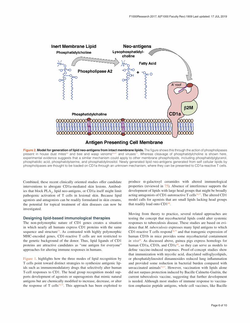
Figure 2. Model for generation of lipid neo-antigens from intact membrane lipids. The figure shows this through the action of phospholipases present in house dust mites66 and bee and wasp venoms65,67 and viruses71. Whereas cleavage of phosphatidylcholine is shown here, experimental evidence suggests that a similar mechanism could apply to other membrane phospholipids, including phosphatidylglycerol, phosphatidic acid, phosphatidylserine, and phosphatidylinositol. Newly generated lipid neo-antigens generated from self cellular lipids by phospholipases are thought to be loaded on CD1a through an unknown mechanism, where they can be presented to CD1a-reactive T cells.
Combined, these recent clinically oriented studies offer candidate interventions to abrogate CD1a-mediated skin lesions. Antibod-ies that block PLA
2, lipid neo-antigens, or CD1a itself might limit
pathogenic activation of T cells in lesional skin. Because lipid agonists and antagonists can be readily formulated in skin creams, the potential for topical treatment of skin diseases can now be investigated.
Designing lipid-based immunological therapiesThe non-polymorphic nature of CD1 genes creates a situation in which nearly all humans express CD1 proteins with the same sequence and structure72. As contrasted with highly polymorphic MHC-encoded genes, CD1-reactive T cells are not restricted to the genetic background of the donor. Thus, lipid ligands of CD1 proteins are attractive candidates as “one antigen for everyone” approaches for altering immune responses in humans.
Figure 1. highlights how the three modes of lipid recognition by T cells point toward distinct strategies to synthesize antigenic lip-ids such as immunomodulatory drugs that selectively alter human T-cell responses to CD1. The head group recognition model sup-ports development of agonists or superagonists that mimic natural antigens but are chemically modified to increase, decrease, or alter the response of T cells73,74. This approach has been exploited to
produce α-galactosyl ceramides with altered immunological properties (reviewed in 75). Absence of interference supports the development of lipids with large head groups that might be broadly acting antagonists of CD1-autoreactive T cells34,37. The altered CD1 model calls for agonists that are small lipids lacking head groups that readily load onto CD140.
Moving from theory to practice, several related approaches are testing the concept that mycobacterial lipids could alter systemic responses to tuberculosis disease. These studies are based on evi-dence that M. tuberculosis expresses many lipid antigens to which CD1-reactive T cells respond76,77 and that transgenic expression of human CD1b in mice provides some mycobacterial containment in vivo60. As discussed above, guinea pigs express homologs for human CD1a, CD1b, and CD1c50, so they can serve as models to define vaccine-induced responses. Proof-of-concept studies show that immunization with mycolic acid, diacylated sulfoglycolipids, or phosphatidylinositol dimannosides reduced lung inflammation and provided some reduction in bacterial burden compared with unvaccinated animals52,53. However, vaccination with lipids alone did not surpass protection induced by Bacille Calmette-Guérin, the current tuberculosis vaccine, suggesting that further development is needed. Although most studies of immune response to vaccina-tion emphasize peptide antigens, whole cell vaccines, like Bacille
Page 6 of 10
F1000Research 2017, 6(F1000 Faculty Rev):1909 Last updated: 17 JUL 2019

Calmette-Guérin, also contain lipid antigens, raising the possibility that generating lipid-specific immune responses occur during cur-rent vaccination approaches in ways that are not currently appreci-ated or measured78.
SummaryThe discovery of CD1 antigen presentation pathways has provided a broader perspective about the natural targets of T cell response, which normally recognize both peptide and lipid antigens. The number of known CD1-presented antigens continues to increase, and three distinct molecular models for their recognition are emerging on the basis of strong structural and functional data (Figure 1). Therefore, the key to harnessing this new basic insight that lipids are antigens for T cells will likely lie in generating better models for measuring the CD1-mediated T-cell response in vivo in animals and ex vivo in humans to understand and interrupt their disease-specific functions.
Author contributionsThe authors jointly prepared the original draft and reviewed and edited the manuscript.
Competing interestsThe authors declare that they have no competing interests.
Grant informationThis work is supported by grants from the Bill and Melinda Gates Foundation and the National Institutes of Health (AI043913, AI111224, and AR048632).
The funders had no role in study design, data collection and analysis, decision to publish, or preparation of the manuscript.
AcknowledgmentsThe authors thank Ildiko van Rhijn and Rachel Cotton for their advice.
References F1000 recommended
1. McDevitt HO, Benacerraf B: Genetic control of specific immune responses. Adv Immunol. 1969; 11: 31–74. PubMed Abstract | Publisher Full Text
2. Zinkernagel RM, Doherty PC: Restriction of in vitro T cell-mediated cytotoxicity in lymphocytic choriomeningitis within a syngeneic or semiallogeneic system. Nature. 1974; 248(5450): 701–2. PubMed Abstract | Publisher Full Text
3. Townsend AR, Gotch FM, Davey J: Cytotoxic T cells recognize fragments of the influenza nucleoprotein. Cell. 1985; 42(2): 457–67. PubMed Abstract | Publisher Full Text
4. Bjorkman PJ, Saper MA, Samraoui B, et al.: Structure of the human class I histocompatibility antigen, HLA-A2. Nature. 1987; 329(6139): 506–12. PubMed Abstract | Publisher Full Text
5. Calabi F, Milstein C: A novel family of human major histocompatibility complex-related genes not mapping to chromosome 6. Nature. 1986; 323(6088): 540–3. PubMed Abstract | Publisher Full Text
6. McMichael AJ, Pilch JR, Galfré G, et al.: A human thymocyte antigen defined by a hybrid myeloma monoclonal antibody. Eur J Immunol. 1979; 9(3): 205–10. PubMed Abstract | Publisher Full Text
7. Beckman EM, Porcelli SA, Morita CT, et al.: Recognition of a lipid antigen by CD1-restricted alpha beta+ T cells. Nature. 1994; 372(6507): 691–4. PubMed Abstract | Publisher Full Text
8. Porcelli S, Morita CT, Brenner MB: CD1b restricts the response of human CD4–8– T lymphocytes to a microbial antigen. Nature. 1992; 360(6404): 593–7. PubMed Abstract | Publisher Full Text
9. Bendelac A, Lantz O, Quimby ME, et al.: CD1 recognition by mouse NK1+ T lymphocytes. Science. 1995; 268(5212): 863–5. PubMed Abstract | Publisher Full Text
10. Borg NA, Wun KS, Kjer-Nielsen L, et al.: CD1d-lipid-antigen recognition by the semi-invariant NKT T-cell receptor. Nature. 2007; 448(7149): 44–9. PubMed Abstract | Publisher Full Text | F1000 Recommendation
11. Godfrey DI, Uldrich AP, McCluskey J, et al.: The burgeoning family of unconventional T cells. Nat Immunol. 2015; 16(11): 1114–23. PubMed Abstract | Publisher Full Text | F1000 Recommendation
12. Salio M, Silk JD, Jones EY, et al.: Biology of CD1- and MR1-restricted T cells. Annu Rev Immunol. 2014; 32: 323–66. PubMed Abstract | Publisher Full Text
13. Porcelli SA: The CD1 family: a third lineage of antigen-presenting molecules. Adv Immunol. 1995; 59: 1–98. PubMed Abstract | Publisher Full Text
14. Zajonc DM, Elsliger MA, Teyton L, et al.: Crystal structure of CD1a in complex
with a sulfatide self antigen at a resolution of 2.15 A. Nat Immunol. 2003; 4(8): 808–15. PubMed Abstract | Publisher Full Text
15. Zajonc DM, Crispin MD, Bowden TA, et al.: Molecular mechanism of lipopeptide presentation by CD1a. Immunity. 2005; 22(2): 209–19. PubMed Abstract | Publisher Full Text
16. Gadola SD, Zaccai NR, Harlos K, et al.: Structure of human CD1b with bound ligands at 2.3 A, a maze for alkyl chains. Nat Immunol. 2002; 3(8): 721–6. PubMed Abstract | Publisher Full Text | F1000 Recommendation
17. Scharf L, Li NS, Hawk AJ, et al.: The 2.5 Å structure of CD1c in complex with a mycobacterial lipid reveals an open groove ideally suited for diverse antigen presentation. Immunity. 2010; 33(8): 853–62. PubMed Abstract | Publisher Full Text | Free Full Text | F1000 Recommendation
18. Luoma AM, Castro CD, Mayassi T, et al.: Crystal structure of Vδ1 T cell receptor in complex with CD1d-sulfatide shows MHC-like recognition of a self-lipid by human γδ T cells. Immunity. 2013; 39(6): 1032–42. PubMed Abstract | Publisher Full Text | Free Full Text
19. Zeng Z, Castaño AR, Segelke BW, et al.: Crystal structure of mouse CD1: An MHC-like fold with a large hydrophobic binding groove. Science. 1997; 277(5324): 339–45. PubMed Abstract | Publisher Full Text
20. Kawano T, Cui J, Koezuka Y, et al.: CD1d-restricted and TCR-mediated activation of valpha14 NKT cells by glycosylceramides. Science. 1997; 278(5343): 1626–9. PubMed Abstract | Publisher Full Text
21. Moody DB, Young DC, Cheng TY, et al.: T cell activation by lipopeptide antigens. Science. 2004; 303(5657): 527–31. PubMed Abstract | Publisher Full Text | F1000 Recommendation
22. Moody DB, Reinhold BB, Guy MR, et al.: Structural requirements for glycolipid antigen recognition by CD1b-restricted T cells. Science. 1997; 278(5336): 283–6. PubMed Abstract | Publisher Full Text
23. Gras S, Van Rhijn I, Shahine A, et al.: T cell receptor recognition of CD1b presenting a mycobacterial glycolipid. Nat Commun. 2016; 7: 13257. PubMed Abstract | Publisher Full Text | Free Full Text
24. Pellicci DG, Clarke AJ, Patel O, et al.: Recognition of β-linked self glycolipids mediated by natural killer T cell antigen receptors. Nat Immunol. 2011; 12(9): 827–33. PubMed Abstract | Publisher Full Text | Free Full Text | F1000 Recommendation
25. Yu ED, Girardi E, Wang J, et al.: Cutting edge: structural basis for the recognition of β-linked glycolipid antigens by invariant NKT cells. J Immunol.
Page 7 of 10
F1000Research 2017, 6(F1000 Faculty Rev):1909 Last updated: 17 JUL 2019

2011; 187(5): 2079–83. PubMed Abstract | Publisher Full Text | Free Full Text
26. Shahine A, Van Rhijn I, Cheng T, et al.: A molecular basis of human T cell receptor autoreactivity toward self-phospholipids. Sci Immunol. 2017; 2(16): pii: eaao1384. PubMed Abstract | Publisher Full Text
27. Moody DB, Briken V, Cheng TY, et al.: Lipid length controls antigen entry into endosomal and nonendosomal pathways for CD1b presentation. Nat Immunol. 2002; 3(5): 435–42. PubMed Abstract | Publisher Full Text | F1000 Recommendation
28. Garcia-Alles LF, Collmann A, Versluis C, et al.: Structural reorganization of the antigen-binding groove of human CD1b for presentation of mycobacterial sulfoglycolipids. Proc Natl Acad Sci U S A. 2011; 108(43): 17755–60. PubMed Abstract | Publisher Full Text | Free Full Text
29. Girardi E, Yu ED, Li Y, et al.: Unique interplay between sugar and lipid in determining the antigenic potency of bacterial antigens for NKT cells. PLoS Biol. 2011; 9(11): e1001189. PubMed Abstract | Publisher Full Text | Free Full Text
30. Kinjo Y, Tupin E, Wu D, et al.: Natural killer T cells recognize diacylglycerol antigens from pathogenic bacteria. Nat Immunol. 2006; 7(9): 978–86. PubMed Abstract | Publisher Full Text | F1000 Recommendation
31. McCarthy C, Shepherd D, Fleire S, et al.: The length of lipids bound to human CD1d molecules modulates the affinity of NKT cell TCR and the threshold of NKT cell activation. J Exp Med. 2007; 204(5): 1131–44. PubMed Abstract | Publisher Full Text | Free Full Text
32. Van Rhijn I, Iwany SK, Fodran P, et al.: CD1b-mycolic acid tetramers demonstrate T-cell fine specificity for mycobacterial lipid tails. Eur J Immunol. 2017; 47(9): 1525–34. PubMed Abstract | Publisher Full Text
33. de Jong A, Cheng TY, Huang S, et al.: CD1a-autoreactive T cells recognize natural skin oils that function as headless antigens. Nat Immunol. 2014; 15(2): 177–85. PubMed Abstract | Publisher Full Text | Free Full Text
34. Kronenberg M, Havran WL: Immunology: oiling the wheels of autoimmunity. Nature. 2014; 506(7486): 42–3. PubMed Abstract | Publisher Full Text
35. Van Rhijn I, Godfrey DI, Rossjohn J, et al.: Lipid and small-molecule display by CD1 and MR1. Nat Rev Immunol. 2015; 15(10): 643–54. PubMed Abstract | Publisher Full Text | F1000 Recommendation
36. Birkinshaw RW, Pellicci DG, Cheng TY, et al.: αβ T cell antigen receptor recognition of CD1a presenting self lipid ligands. Nat Immunol. 2015; 16(3): 258–66. PubMed Abstract | Publisher Full Text | F1000 Recommendation
37. Park JE, Wu DY, Prendes M, et al.: Fine specificity of natural killer T cells against GD3 ganglioside and identification of GM3 as an inhibitory natural killer T-cell ligand. Immunology. 2008; 123(1): 145–55. PubMed Abstract | Publisher Full Text | Free Full Text
38. de Jong A, Peña-Cruz V, Cheng TY, et al.: CD1a-autoreactive T cells are a normal component of the human αβ T cell repertoire. Nat Immunol. 2010; 11(12): 1102–9. PubMed Abstract | Publisher Full Text | Free Full Text | F1000 Recommendation
39. de Lalla C, Lepore M, Piccolo FM, et al.: High-frequency and adaptive-like dynamics of human CD1 self-reactive T cells. Eur J Immunol. 2011; 41(3): 602–10. PubMed Abstract | Publisher Full Text
40. Mansour S, Tocheva AS, Cave-Ayland C, et al.: Cholesteryl esters stabilize human CD1c conformations for recognition by self-reactive T cells. Proc Natl Acad Sci U S A. 2016; 113(9): E1266–75. PubMed Abstract | Publisher Full Text | Free Full Text | F1000 Recommendation
41. Roy S, Ly D, Li NS, et al.: Molecular basis of mycobacterial lipid antigen presentation by CD1c and its recognition by αβ T cells. Proc Natl Acad Sci U S A. 2014; 111(43): E4648–57. PubMed Abstract | Publisher Full Text | Free Full Text
42. Roy S, Ly D, Castro CD, et al.: Molecular Analysis of Lipid-Reactive Vδ1 γδ T Cells Identified by CD1c Tetramers. J Immunol. 2016; 196(4): 1933–42. PubMed Abstract | Publisher Full Text | Free Full Text
43. Altman JD, Moss PA, Goulder PJ, et al.: Phenotypic analysis of antigen-specific T lymphocytes. Science. 1996; 274(5284): 94–6. PubMed Abstract | Publisher Full Text
44. Altman JD, Davis MM: MHC-Peptide Tetramers to Visualize Antigen-Specific T Cells. Curr Protoc Immunol. 2016; 115: 17.3.1–17.3.44. PubMed Abstract | Publisher Full Text | F1000 Recommendation
45. Sidobre S, Kronenberg M: CD1 tetramers: a powerful tool for the analysis of glycolipid-reactive T cells. J Immunol Methods. 2002; 268(1): 107–21. PubMed Abstract | Publisher Full Text
46. Kasmar AG, Van Rhijn I, Cheng TY, et al.: CD1b tetramers bind αβ T cell receptors to identify a mycobacterial glycolipid-reactive T cell repertoire in
humans. J Exp Med. 2011; 208(9): 1741–7. PubMed Abstract | Publisher Full Text | Free Full Text | F1000 Recommendation
47. Kasmar AG, Van Rhijn I, Magalhaes KG, et al.: Cutting Edge: CD1a tetramers and dextramers identify human lipopeptide-specific T cells ex vivo. J Immunol. 2013; 191(9): 4499–503. PubMed Abstract | Publisher Full Text | Free Full Text
48. Ly D, Kasmar AG, Cheng TY, et al.: CD1c tetramers detect ex vivo T cell responses to processed phosphomycoketide antigens. J Exp Med. 2013; 210(4): 729–41. PubMed Abstract | Publisher Full Text | Free Full Text
49. Van Rhijn I, Kasmar A, de Jong A, et al.: A conserved human T cell population targets mycobacterial antigens presented by CD1b. Nat Immunol. 2013; 14(7): 706–13. PubMed Abstract | Publisher Full Text | Free Full Text
50. Reinink P, Van Rhijn I: Mammalian CD1 and MR1 genes. Immunogenetics. 2016; 68(8): 515–23. PubMed Abstract | Publisher Full Text | Free Full Text
51. Hiromatsu K, Dascher CC, Sugita M, et al.: Characterization of guinea-pig group 1 CD1 proteins. Immunology. 2002; 106(2): 159–72. PubMed Abstract | Publisher Full Text | Free Full Text
52. Dascher CC, Hiromatsu K, Xiong X, et al.: Immunization with a mycobacterial lipid vaccine improves pulmonary pathology in the guinea pig model of tuberculosis. Int Immunol. 2003; 15(8): 915–25. PubMed Abstract | Publisher Full Text
53. Larrouy-Maumus G, Layre E, Clark S, et al.: Protective efficacy of a lipid antigen vaccine in a guinea pig model of tuberculosis. Vaccine. 2017; 35(10): 1395–402. PubMed Abstract | Publisher Full Text
54. Felio K, Nguyen H, Dascher CC, et al.: CD1-restricted adaptive immune responses to Mycobacteria in human group 1 CD1 transgenic mice. J Exp Med. 2009; 206(11): 2497–509. PubMed Abstract | Publisher Full Text | Free Full Text | F1000 Recommendation
55. Li S, Choi HJ, Felio K, et al.: Autoreactive CD1b-restricted T cells: a new innate-like T-cell population that contributes to immunity against infection. Blood. 2011; 118(14): 3870–8. PubMed Abstract | Publisher Full Text | Free Full Text
56. Roura-Mir C, Wang L, Cheng TY, et al.: Mycobacterium tuberculosis regulates CD1 antigen presentation pathways through TLR-2. J Immunol. 2005; 175(3): 1758–66. PubMed Abstract | Publisher Full Text
57. Yakimchuk K, Roura-Mir C, Magalhaes KG, et al.: Borrelia burgdorferi infection regulates CD1 expression in human cells and tissues via IL1-β. Eur J Immunol. 2011; 41(3): 694–705. PubMed Abstract | Publisher Full Text | Free Full Text
58. Jackman RM, Stenger S, Lee A, et al.: The tyrosine-containing cytoplasmic tail of CD1b is essential for its efficient presentation of bacterial lipid antigens. Immunity. 1998; 8(3): 341–51. PubMed Abstract | Publisher Full Text
59. Sugita M, Jackman RM, van Donselaar E, et al.: Cytoplasmic tail-dependent localization of CD1b antigen-presenting molecules to MIICs. Science. 1996; 273(5273): 349–52. PubMed Abstract | Publisher Full Text
60. Zhao J, Siddiqui S, Shang S, et al.: Mycolic acid-specific T cells protect against Mycobacterium tuberculosis infection in a humanized transgenic mouse model. eLife. 2015; 4: pii: e08525. PubMed Abstract | Publisher Full Text | Free Full Text | F1000 Recommendation
61. Melián A, Geng YJ, Sukhova GK, et al.: CD1 expression in human atherosclerosis. A potential mechanism for T cell activation by foam cells. Am J Pathol. 1999; 155(3): 775–86. PubMed Abstract | Publisher Full Text | Free Full Text
62. Bagchi S, He Y, Zhang H, et al.: CD1b-autoreactive T cells contribute to hyperlipidemia-induced skin inflammation in mice. J Clin Invest. 2017; 127(6): 2339–52. PubMed Abstract | Publisher Full Text | Free Full Text
63. Li B, Tsoi LC, Swindell WR, et al.: Transcriptome analysis of psoriasis in a large case-control sample: RNA-seq provides insights into disease mechanisms. J Invest Dermatol. 2014; 134(7): 1828–38. PubMed Abstract | Publisher Full Text | Free Full Text
64. Cheung KL, Jarrett R, Subramaniam S, et al.: Psoriatic T cells recognize neolipid antigens generated by mast cell phospholipase delivered by exosomes and presented by CD1a. J Exp Med. 2016; 213(11): 2399–412. PubMed Abstract | Publisher Full Text | Free Full Text | F1000 Recommendation
65. Bourgeois EA, Subramaniam S, Cheng TY, et al.: Bee venom processes human skin lipids for presentation by CD1a. J Exp Med. 2015; 212(2): 149–63. PubMed Abstract | Publisher Full Text | Free Full Text | F1000 Recommendation
66. Jarrett R, Salio M, Lloyd-Lavery A, et al.: Filaggrin inhibits generation of CD1a neolipid antigens by house dust mite-derived phospholipase. Sci Transl Med. 2016; 8(325): 325ra18. PubMed Abstract | Publisher Full Text | Free Full Text | F1000 Recommendation
Page 8 of 10
F1000Research 2017, 6(F1000 Faculty Rev):1909 Last updated: 17 JUL 2019

67. Subramaniam S, Aslam A, Misbah SA, et al.: Elevated and cross-responsive CD1a-reactive T cells in bee and wasp venom allergic individuals. Eur J Immunol. 2016; 46(1): 242–52. PubMed Abstract | Publisher Full Text | Free Full Text
68. Kobayashi C, Shiina T, Tokioka A, et al.: GM-CSF-independent CD1a expression in epidermal Langerhans cells: evidence from human CD1A genome-transgenic mice. J Invest Dermatol. 2012; 132(1): 241–4. PubMed Abstract | Publisher Full Text | Free Full Text
69. Kim JH, Hu Y, Yongqing T, et al.: CD1a on Langerhans cells controls inflammatory skin disease. Nat Immunol. 2016; 17(10): 1159–66. PubMed Abstract | Publisher Full Text | F1000 Recommendation
70. Elieh Ali Komi D, Shafaghat F, Zwiener RD: Immunology of Bee Venom. Clin Rev Allergy Immunol. 2017. PubMed Abstract | Publisher Full Text
71. Zeissig S, Murata K, Sweet L, et al.: Hepatitis B virus-induced lipid alterations contribute to natural killer T cell-dependent protective immunity. Nat Med. 2012; 18(7): 1060–8. PubMed Abstract | Publisher Full Text | Free Full Text
72. Van Rhijn I, Moody DB: Donor Unrestricted T Cells: A Shared Human T Cell Response. J Immunol. 2015; 195(5): 1927–32. PubMed Abstract | Publisher Full Text | Free Full Text
73. Miyamoto K, Miyake S, Yamamura T: A synthetic glycolipid prevents autoimmune encephalomyelitis by inducing TH2 bias of natural killer T cells. Nature. 2001; 413(6855): 531–4. PubMed Abstract | Publisher Full Text | F1000 Recommendation
74. Silk JD, Salio M, Reddy BG, et al.: Cutting edge: nonglycosidic CD1d lipid ligands activate human and murine invariant NKT cells. J Immunol. 2008; 180(10): 6452–6. PubMed Abstract | Publisher Full Text
75. Kharkwal SS, Arora P, Porcelli SA: Glycolipid activators of invariant NKT cells as vaccine adjuvants. Immunogenetics. 2016; 68(8): 597–610. PubMed Abstract | Publisher Full Text
76. De Libero G, Mori L: The T-Cell Response to Lipid Antigens of Mycobacterium tuberculosis. Front Immunol. 2014; 5: 219. PubMed Abstract | Publisher Full Text | Free Full Text
77. Van Rhijn I, Moody DB: CD1 and mycobacterial lipids activate human T cells. Immunol Rev. 2015; 264(1): 138–53. PubMed Abstract | Publisher Full Text | Free Full Text
78. Scriba TJ, Kaufmann SH, Henri Lambert P, et al.: Vaccination Against Tuberculosis With Whole-Cell Mycobacterial Vaccines. J Infect Dis. 2016; 214(5): 659–64. PubMed Abstract | Publisher Full Text | F1000 Recommendation
Page 9 of 10
F1000Research 2017, 6(F1000 Faculty Rev):1909 Last updated: 17 JUL 2019

Open Peer Review
Current Peer Review Status:
Editorial Note on the Review Process are written by members of the prestigious . They are commissioned andF1000 Faculty Reviews F1000 Faculty
are peer reviewed before publication to ensure that the final, published version is comprehensive and accessible.The reviewers who approved the final version are listed with their names and affiliations.
The reviewers who approved this article are:Version 1
The benefits of publishing with F1000Research:
Your article is published within days, with no editorial bias
You can publish traditional articles, null/negative results, case reports, data notes and more
The peer review process is transparent and collaborative
Your article is indexed in PubMed after passing peer review
Dedicated customer support at every stage
For pre-submission enquiries, contact [email protected]
Giulia CasoratiExperimental Immunology Unit, Division of Immunology, Transplantation and Infectious Diseases, SanRaffaele Scientific Institute, Milan, Italy
No competing interests were disclosed.Competing Interests:
1
Stephan GadolaFaculty of Medicine, University of Southampton, Southampton, UK
No competing interests were disclosed.Competing Interests:
2
Dirk ZajoncLa Jolla Institute of Allergy & Immunology, La Jolla, California, USA
No competing interests were disclosed.Competing Interests:
3
Page 10 of 10
F1000Research 2017, 6(F1000 Faculty Rev):1909 Last updated: 17 JUL 2019