![[Meticilin resistant Staphylococcus aureus and liver abscess: a retrospective analysis of 117 patients]](https://static.fdokumen.com/doc/165x107/632546fd545c645c7f099e01/meticilin-resistant-staphylococcus-aureus-and-liver-abscess-a-retrospective-analysis.jpg)
Characterisation of receptor binding by the chemotaxis inhibitory protein of Staphylococcus aureus...
23
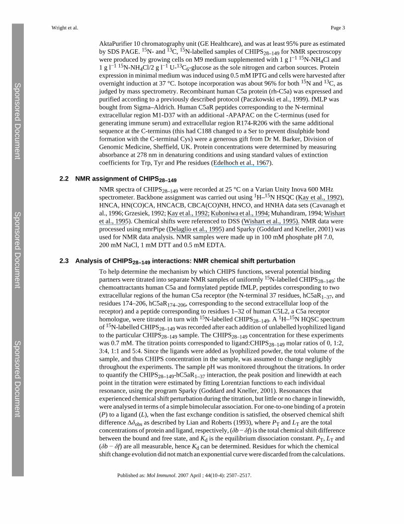
Characterisation of receptor binding by the chemotaxis inhibitory protein of Staphylococcus aureus and the effects of the host immune response Andrew J. Wright a , Adrian Higginbottom b , Didier Philippe c , Abhishek Upadhyay c , Stefan Bagby c , Robert C. Read b , Peter N. Monk b,⁎ , and Lynda J. Partridge a a Department of Molecular Biology and Biotechnology, University of Sheffield, Sheffield S10 2TN, UK. b School of Medicine and Biomedical Science, University of Sheffield, Sheffield S10 2RX, UK. c Department of Biology and Biochemistry, University of Bath, Bath BA2 7AY, UK. Abstract The chemotaxis inhibitory protein of Staphylococcus aureus (CHIPS) is reported to bind to the receptors for C5a and formylated peptides and has been proposed as a promising lead for the development of new anti-inflammatory compounds. Here we have examined the receptor specificity and mode of action of recombinant CHIPS 28–149 and also the immune response to CHIPS 28–149 in patients with S. aureus infections and in uninfected controls. Recombinant CHIPS 28–149 bound with high affinity to the human C5a receptor (C5aR), but had low affinity for the second C5a receptor, C5L2, and the formyl peptide receptor, FPR. Although ligand binding to C5aR was potently inhibited, CHIPS 28–149 had much weaker effects on ligand binding to C5L2 and FPR. Similarly, CHIPS 28–149 potently inhibited the ligand-induced activation of C5aR but was less potent at inhibition via FPR. NMR studies showed that CHIPS 28–149 bound directly to the N-terminus of C5aR but not C5L2, and CHIPS 28–149 residues involved in the interaction were identified by chemical shift analysis. All human sera examined contained high titres of IgG and IgA reactivity against CHIPS 28–149 , and no correlation was observed between infection status at the time of serum collection and antibody titre. Individual serum samples promoted or inhibited the binding of CHIPS 28–149 to C5aR, or had no effect. IgG depletion of serum samples abrogated the effects on CHIPS binding, demonstrating that these were antibody mediated. Sera from infected individuals were more likely to inhibit CHIPS 28–149 binding than sera from healthy controls. However, high antibody titres correlated well with both inhibition and enhancement of CHIPS 28–149 binding to C5aR; this suggests that the inhibitory effect relates to epitope specificity rather than greater antibody binding. We conclude that CHIPS is likely to be too immunogenic to be used as an anti-inflammatory treatment but that some antibodies against CHIPS may be useful in the treatment of S. aureus infections. © 2007 Elsevier Ltd. This document may be redistributed and reused, subject to certain conditions. ⁎Corresponding author. Tel.: +44 114 226 1312; fax: +44 114 226 1201. [email protected]. This document was posted here by permission of the publisher. At the time of deposit, it included all changes made during peer review, copyediting, and publishing. The U.S. National Library of Medicine is responsible for all links within the document and for incorporating any publisher-supplied amendments or retractions issued subsequently. The published journal article, guaranteed to be such by Elsevier, is available for free, on ScienceDirect. Sponsored document from Molecular Immunology Published as: Mol Immunol. 2007 April ; 44(10-4): 2507–2517. Sponsored Document Sponsored Document Sponsored Document
-
Upload
independent -
Category
Documents
-
view
3 -
download
0
Transcript of Characterisation of receptor binding by the chemotaxis inhibitory protein of Staphylococcus aureus...

Characterisation of receptor binding by the chemotaxis inhibitoryprotein of Staphylococcus aureus and the effects of the hostimmune response
Andrew J. Wrighta, Adrian Higginbottomb, Didier Philippec, Abhishek Upadhyayc, StefanBagbyc, Robert C. Readb, Peter N. Monkb,⁎, and Lynda J. PartridgeaaDepartment of Molecular Biology and Biotechnology, University of Sheffield, Sheffield S10 2TN,UK.bSchool of Medicine and Biomedical Science, University of Sheffield, Sheffield S10 2RX, UK.cDepartment of Biology and Biochemistry, University of Bath, Bath BA2 7AY, UK.
AbstractThe chemotaxis inhibitory protein of Staphylococcus aureus (CHIPS) is reported to bind to thereceptors for C5a and formylated peptides and has been proposed as a promising lead for thedevelopment of new anti-inflammatory compounds. Here we have examined the receptor specificityand mode of action of recombinant CHIPS28–149 and also the immune response to CHIPS28–149 inpatients with S. aureus infections and in uninfected controls. Recombinant CHIPS28–149 bound withhigh affinity to the human C5a receptor (C5aR), but had low affinity for the second C5a receptor,C5L2, and the formyl peptide receptor, FPR. Although ligand binding to C5aR was potently inhibited,CHIPS28–149 had much weaker effects on ligand binding to C5L2 and FPR. Similarly,CHIPS28–149 potently inhibited the ligand-induced activation of C5aR but was less potent atinhibition via FPR. NMR studies showed that CHIPS28–149 bound directly to the N-terminus of C5aRbut not C5L2, and CHIPS28–149 residues involved in the interaction were identified by chemical shiftanalysis. All human sera examined contained high titres of IgG and IgA reactivity againstCHIPS28–149, and no correlation was observed between infection status at the time of serumcollection and antibody titre. Individual serum samples promoted or inhibited the binding ofCHIPS28–149 to C5aR, or had no effect. IgG depletion of serum samples abrogated the effects onCHIPS binding, demonstrating that these were antibody mediated. Sera from infected individualswere more likely to inhibit CHIPS28–149 binding than sera from healthy controls. However, highantibody titres correlated well with both inhibition and enhancement of CHIPS28–149 binding toC5aR; this suggests that the inhibitory effect relates to epitope specificity rather than greater antibodybinding. We conclude that CHIPS is likely to be too immunogenic to be used as an anti-inflammatorytreatment but that some antibodies against CHIPS may be useful in the treatment of S. aureusinfections.
© 2007 Elsevier Ltd.This document may be redistributed and reused, subject to certain conditions.
⁎Corresponding author. Tel.: +44 114 226 1312; fax: +44 114 226 1201. [email protected] document was posted here by permission of the publisher. At the time of deposit, it included all changes made during peer review,copyediting, and publishing. The U.S. National Library of Medicine is responsible for all links within the document and for incorporatingany publisher-supplied amendments or retractions issued subsequently. The published journal article, guaranteed to be such by Elsevier,is available for free, on ScienceDirect.
Sponsored document fromMolecular Immunology
Published as: Mol Immunol. 2007 April ; 44(10-4): 2507–2517.
Sponsored Docum
ent Sponsored D
ocument
Sponsored Docum
ent

KeywordsComplement; Receptor; Staphylococcus aureus; Antibody; C5a
1 IntroductionC5a is the most potent pro-inflammatory mediator produced in the complement cascade (Guoand Ward, 2005), and is a potent chemoattractant for all myeloid cells (Kohl, 2001). Bindingof C5a to its receptor induces a range of inflammatory effects including leukocyte recruitmentand chemotaxis, upregulation of leukocyte adhesion molecule expression (CD18, ICAM1),release of proteolytic and reactive oxygen and nitrogen species, cytokine production, activationof the coagulation cascade, contraction of smooth muscle and changes in vascular diameterand permeability (reviewed in Gerard and Gerard, 1994; Guo and Ward, 2005). The receptorfor C5a, C5aR, belongs to the rhodopsin family of seven transmembrane G-protein coupledreceptors (Boulay et al., 1991; Gerard and Gerard, 1991). The binding of C5a to the C5aR ispostulated to occur via a ‘two-site binding’ mechanism (DeMartino et al., 1994): the basic coreof C5a is thought to interact with acidic residues in the receptor N-terminus (Mery and Boulay,1994), while the C-terminal domain of C5a binds in a pocket formed by largely hydrophobicresidues within the transmembrane helices of the C5aR (Higginbottom et al., 2005). RecentlyC5a-like receptor 2 (C5L2), which shares 35% amino acid identity with C5aR, was shown tobind both C5a and C5a-des-Arg, although with a 10-fold higher affinity for the stablemetabolite, C5a des-Arg (Cain and Monk, 2002). S. aureus supernate (SaS) containscomponents that cause a decreased chemotactic activity of neutrophils toward C5a and/or N-formyl peptides (Veldkamp et al., 2000). The factor responsible for this activity, ‘ChemotaxisInhibitory Protein of Staphylococcus aureus’ (CHIPS), is a 14.1 kDa protein (Postma et al.,2004) found in over 60% of S. aureus clinical isolates and is located on the bacteriophageencoded pathogenicity island SaPI5. It has been suggested that CHIPS could be exploited asan anti-inflammatory therapeutic agent (de Haas et al., 2004). Residues Asp10, Gly12, Asp15,and Asp18 in the N-terminal domain of C5aR are crucial for the interaction with CHIPS(Postma et al., 2005). A CHIPS31–121 fragment showed the same C5aR blocking activity asintact CHIPS although this fragment did not block FPR binding, suggesting that the FPRbinding site is at the extreme N-terminus of CHIPS (Haas et al., 2004).
We have produced recombinant CHIPS28–149 to characterise the mechanism of action ofCHIPS and to assess the antibody responses of controls and S. aureus-infected patients.CHIPS28–149 was found to be a potent competitive antagonist at C5aR with rapid bindingkinetics but was only weakly active at C5L2 or FPR. All sera tested contained high anti-CHIPS28–149 antibody titres and approximately half of these affected the binding to C5aR.Anti-CHIPS28–149 antibodies that block CHIPS binding may have therapeutic potential in thetreatment of S. aureus infections.
2 Methods and materials2.1 Proteins and peptides
DNA coding for CHIPS residues 28–149 (CHIPS28–149) was amplified from N315 MRSAstrain genomic DNA and cloned into a modified pGEX4T1 vector (Sheffield et al., 1999) using5′-CAT GCC ATG GCT TTT ACT TTT GAA CCG TTT-3′ and 5′-CCG CTC GAG CTA TTAGTA TGC GTA TTC ATT AGT TT-3′ primers. GST-CHIPS28–149 was overexpressed usingBL21 (DE3) cells with IPTG induction. Cells were lysed by sonication and GST-CHIPS28–149 was batch purified on glutathione sepharose 4B resin according to manufacturer'sinstructions (GE Healthcare). After removal of the GST carrier protein using TEV protease,CHIPS was further purified on a Mono S cation exchange column (GE Healthcare) using an
Wright et al. Page 2
Published as: Mol Immunol. 2007 April ; 44(10-4): 2507–2517.
Sponsored Docum
ent Sponsored D
ocument
Sponsored Docum
ent
AktaPurifier 10 chromatography unit (GE Healthcare), and was at least 95% pure as estimatedby SDS PAGE. 15N- and 13C, 15N-labelled samples of CHIPS28–149 for NMR spectroscopywere produced by growing cells on M9 medium supplemented with 1 g l−1 15N-NH4Cl and1 g l−1 15N-NH4Cl/2 g l−1 U-13C6-glucose as the sole nitrogen and carbon sources. Proteinexpression in minimal medium was induced using 0.5 mM IPTG and cells were harvested afterovernight induction at 37 °C. Isotope incorporation was about 96% for both 15N and 13C, asjudged by mass spectrometry. Recombinant human C5a protein (rh-C5a) was expressed andpurified according to a previously described protocol (Paczkowski et al., 1999). fMLP wasbought from Sigma–Aldrich. Human C5aR peptides corresponding to the N-terminalextracellular region M1-D37 with an additional -APAPAC on the C-terminus (used forgenerating immune serum) and extracellular region R174-R206 with the same additionalsequence at the C-terminus (this had C188 changed to a Ser to prevent disulphide bondformation with the C-terminal Cys) were a generous gift from Dr M. Barker, Division ofGenomic Medicine, Sheffield, UK. Protein concentrations were determined by measuringabsorbance at 278 nm in denaturing conditions and using standard values of extinctioncoefficients for Trp, Tyr and Phe residues (Edelhoch et al., 1967).
2.2 NMR assignment of CHIPS28–149NMR spectra of CHIPS28–149 were recorded at 25 °C on a Varian Unity Inova 600 MHzspectrometer. Backbone assignment was carried out using 1H–15N HSQC (Kay et al., 1992),HNCA, HN(CO)CA, HNCACB, CBCA(CO)NH, HNCO, and HNHA data sets (Cavanagh etal., 1996; Grzesiek, 1992; Kay et al., 1992; Kuboniwa et al., 1994; Muhandiram, 1994; Wishartet al., 1995). Chemical shifts were referenced to DSS (Wishart et al., 1995). NMR data wereprocessed using nmrPipe (Delaglio et al., 1995) and Sparky (Goddard and Kneller, 2001) wasused for NMR data analysis. NMR samples were made up in 100 mM phosphate pH 7.0,200 mM NaCl, 1 mM DTT and 0.5 mM EDTA.
2.3 Analysis of CHIPS28–149 interactions: NMR chemical shift perturbationTo help determine the mechanism by which CHIPS functions, several potential bindingpartners were titrated into separate NMR samples of uniformly 15N-labelled CHIPS28–149: thechemoattractants human C5a and formylated peptide fMLP, peptides corresponding to twoextracellular regions of the human C5a receptor (the N-terminal 37 residues, hC5aR1–37, andresidues 174–206, hC5aR174–206, corresponding to the second extracellular loop of thereceptor) and a peptide corresponding to residues 1–32 of human C5L2, a C5a receptorhomologue, were titrated in turn with 15N-labelled CHIPS28–149. A 1H–15N HQSC spectrumof 15N-labelled CHIPS28–149 was recorded after each addition of unlabelled lyophilized ligandto the particular CHIPS28–149 sample. The CHIPS28–149 concentration for these experimentswas 0.7 mM. The titration points corresponded to ligand:CHIPS28–149 molar ratios of 0, 1:2,3:4, 1:1 and 5:4. Since the ligands were added as lyophilized powder, the total volume of thesample, and thus CHIPS concentration in the sample, was assumed to change negligiblythroughout the experiments. The sample pH was monitored throughout the titrations. In orderto quantify the CHIPS28–149-hC5aR1–37 interaction, the peak position and linewidth at eachpoint in the titration were estimated by fitting Lorentzian functions to each individualresonance, using the program Sparky (Goddard and Kneller, 2001). Resonances thatexperienced chemical shift perturbation during the titration, but little or no change in linewidth,were analysed in terms of a simple bimolecular association. For one-to-one binding of a protein(P) to a ligand (L), when the fast exchange condition is satisfied, the observed chemical shiftdifference Δδobs as described by Lian and Roberts (1993), where PT and LT are the totalconcentrations of protein and ligand, respectively, (δb − δf) is the total chemical shift differencebetween the bound and free state, and Kd is the equilibrium dissociation constant. PT, LT and(δb − δf) are all measurable, hence Kd can be determined. Residues for which the chemicalshift change evolution did not match an exponential curve were discarded from the calculations.
Wright et al. Page 3
Published as: Mol Immunol. 2007 April ; 44(10-4): 2507–2517.
Sponsored Docum
ent Sponsored D
ocument
Sponsored Docum
ent

The curve fitting was done manually for each shifted residue by iterative assignment usingMicrosoft Excel.
2.4 Analysis of CHIPS interactions: isothermal titration calorimetry (ITC)ITC experiments were carried out by Margaret Nutley at the BBSRC/EPSRC BiologicalMicrocalorimetry Facility, Department of Chemistry, University of Glasgow. CHIPS28–149(0.2 mM) was titrated with the peptides hC5aR1–37 and hC5aR174–206.
2.5 Transfection and cell cultureRBL-2H3 and CHO cells were routinely cultured in Dulbecco's modified Eagle'smedium + 10% (v/v) fetal calf serum (supplemented with 400 mg/L G-418 for transfectedcells) at 37 °C, 8% CO2. The cDNA for ChemR23, FPRL1 and FPRL2 was purchased fromUMR cDNA Resource Center (www.cdna.org) in pcDNA3.1. Cells were transfected withchemoattractant receptors by electroporation, as previously described (Crass et al., 1999).
2.6 Flow cytometric receptor binding studiesFor ligand binding studies, CHO cells transfected with the appropriate receptor (50,000 perwell of a 96-well microtitre plate) were incubated with the stated concentrations of His6-taggedC5a or C5a des Arg for 30 min at 4 °C, in PBS + 0.1% BSA, 0.2% (w/v) NaN3, then washedtwice with cold PBS to remove unbound ligand (Wilken et al., 1999). The cells were incubatedwith anti-RGSHis6 antibody for 30 min at 4 °C, washed with PBS and anti-mouse IgG-FITC(Sigma) used to detect bound ligand by FACS analysis (FACSort, Becton Dickinson). Theassociation of CHIPS with C5aR was analysed by adding FITC-CHIPS28–149 to C5aR-transfected RBL cells (final concentration of 300 nM) followed by an excess of unlabelledCHIPS28–149 (14 μM). The dissociation rate, Kd, was calculated using GraphPad Prism v4.0,using the observed association rate (kobs) and the dissociation rate (koff) to firstly calculatekon: kon = kobs − koff/[labelled ligand], and secondly: Kd = kon/koff.
2.7 Effect of human antibodies on CHIPS bindingCHO or RBL2H3 cells transfected with human C5aR were used for these studies. Dilutions ofhuman serum in PBS + 0.1% BSA, 0.2% (w/v) NaN3 were pre-incubated for 45 min with afixed concentration of FITC-CHIPS28–149 (210 nm, determined to give 50% maximal binding).Binding of the FITC-CHIPS28–149 to the transfected cells was then determined by flowcytometry as described above. In some instances, human IgG purified using proteinG-Sepaharose (Amersham, UK), concentrated to ∼12 mg/ml using Vivaspin 20 columns (Pierce,UK) and deaggregated by centrifuging at 100,000 × g for 1 h, was used. In other cases, serumsamples were depleted of IgG by incubating them with an equal volume of protein G-Sepharoseon a rotary mixer overnight at 4 °C prior to use in the assay.
2.8 Inhibition of receptor activation by rCHIPS28–149Receptor activation in RBL cells was measured as the release of β-hexosaminidase fromintracellular granules, as described (Cain et al., 2000). The percentage of β-hexosaminidaserelease was calculated as a percentage of the release in the absence of CHIPS28–149. Total β-hexosaminidase content was determined following cell lysis with 0.1% NP-40. Assay of theantagonist activity was performed as described above except that the antagonists were addedat varying concentrations for 15 min before the addition of C5a or C5a des-Arg74 at a finalconcentration of 50 or 250 nM, respectively. IC50, EC50 and standard error values wereobtained by non-linear regression analysis using GraphPad Prism 4.0.
Wright et al. Page 4
Published as: Mol Immunol. 2007 April ; 44(10-4): 2507–2517.
Sponsored Docum
ent Sponsored D
ocument
Sponsored Docum
ent

2.9 ELISA titration of human serumSerum samples were obtained from infected patients or ‘normal’ volunteer donors recruitedunder the approval of the South Sheffield Research Ethics Committee (Study No. 02/299).Samples from infected patients were taken after recent (less than 4 weeks) acute S. aureusinfection, as confirmed by bacteriological culture of the infectious agent. CHIPS28–149 wascoated to 96 well microtitre-plates (Nunc MaxiSorp, Denmark) at 10 μg ml−1 in carbonate/bicarbonate buffer (pH 9.6) overnight at 4 °C. Following blocking in 0.2% gelatin/PBS, plateswere used to titrate human serum using anti-human IgG, IgM, or IgA alkaline-phosphataselabelled secondary antibodies (Sigma). Plates were developed using pNPP (Sigma, UK)substrate and OD405 nm recorded by spectrophotometry.
3 Results3.1 Production and characterisation of recombinant CHIPS
Recombinant (r)CHIPS28–149 was produced in E. coli as a fusion protein with GST, which wascleaved using TEV protease to leave three N-terminal residues, GlyAlaMet, from the vector.CHIPS is reported to bind to human C5aR and FPR (Haas et al., 2004) and so we determinedthe functionality of rCHIPS28–149 by characterising the binding of an FITC-labelled form toRBL cells transfected with human C5aR (RBL-C5aR). Unlabelled rCHIPS28–149 couldcompete with FITC-rCHIPS28–149 for binding to RBL-C5aR cells, with an IC50 value of∼69 nM (Fig. 1A). Similarly, unlabelled rhC5a could also compete with FITC-rCHIPS28–149(Fig. 1A) although even at the high dose of 250 nM, rhC5a failed to compete for more than50% of the binding sites. FITC-rCHIPS28–149 binding was quite specific for C5aR expressedon RBL and CHO cells; binding to the second C5a receptor, C5L2, and the formyl peptidereceptor, FPR, was just above the lower limit of detection at 6 μM and undetectable at lowerconcentrations whereas binding to C5aR was high, even at 0.6 μM (Fig. 1B). No binding toFPRL1, FPRL2 or ChemR23 was detected (data not shown). This result was surprising in lightof the similarity of the N-termini of C5aR and C5L2 (Cain and Monk, 2002), and the previouslyreported binding of CHIPS to FPR (Postma et al., 2004). In further experiments, the ability ofCHIPS28–149 to prevent ligand binding to C5L2 and FPR was investigated. AlthoughCHIPS28–149 could clearly inhibit the binding of C5a to C5aR (Fig. 2A, IC50 = 6.7 nM), noinhibition of C5a binding to C5L2 was observed, even at 100 μM CHIPS28–149. In contrast,the binding of C5a des Arg to C5L2 could be inhibited (Fig. 2, IC50 = 274 nM), suggestingthat CHIPS might also be active at C5L2. Using FITC-labelled formyl peptide fMLP, we couldnot detect any inhibition of binding, even at concentrations of CHIPS28–149 that completelyinhibit C5a binding to C5aR (Fig. 2). The kinetics of binding to C5aR were also examined,using flow cytometry to measure the association of FITC-CHIPS28–149 with RBL-C5aR.Binding reached maximal levels in <2 min (Fig. 3, t1/2 = 0.17 min), and the addition of anexcess of unlabelled CHIPS28–149 caused complete dissociation (t1/2 = 0.73 min). Using datafrom five separate experiments, Kd = 7.22 ± 6.66 nM, similar to the figure calculated fromcompetition experiments. Finally, rCHIPS28–149 was tested in cell activation studies, using thesecretion of β-hexosaminidase to measure the degree of inhibition of receptor activation.CHIPS28–149 could inhibit the activation of C5aR by C5a (Fig. 4A, IC50 = 2.44 μM), but notby the C-terminal C5a analog EP-54 or by phorbol ester/calcium ionophore, which directlyactivates cells (Fig. 4A). In contrast, only very weak inhibition of FPR was observed (Fig. 4A,IC50 > 1 mM), although at high rCHIPS28–149 concentrations, some non-specific inhibitioncould be observed. We further analysed the mechanism of inhibition of C5aR using Schildplots (Arunlakshana and Schild, 1959) (Fig. 4B). The relationship between the log of theconcentration of rCHIPS28–149 and the dose-ratio is linear, which suggests that inhibition isreversible and competitive. However, slopes are <1 in all cases, suggesting that negativecooperativity may be occurring. Interestingly, we also tested rCHIPS30–141, lacking two N-terminal residues, for the ability to inhibit FPR activation and found that it had 12-fold higher
Wright et al. Page 5
Published as: Mol Immunol. 2007 April ; 44(10-4): 2507–2517.
Sponsored Docum
ent Sponsored D
ocument
Sponsored Docum
ent

potency at FPR than rCHIPS28–149 (Fig. 4B). Taken together, this data suggests thatrCHIPS28–149 is highly specific for C5aR with low activity at the closely related receptor,C5L2, but no activity at FPR.
3.2 Analysis of CHIPS interactions: NMR chemical shift perturbationUsing standard triple resonance methodology (Cavanagh et al., 1996; Grzesiek, 1992; Kay etal., 1992; Kuboniwa et al., 1994; Muhandiram, 1994; Wishart et al., 1995), sequence specificbackbone resonance assignments (including amide 1H and 15N, 13Cα, 13Cβ, 13C(O) and 1Hα)were made for 95% of non-Pro, non-N-terminal amino acids in CHIPS28–149. Titrationof 15N-labelled CHIPS28–149 with either of the chemoattractants C5a and fMLP or thehC5aR174–206 peptide caused negligible change in chemical shift or intensity of the peaks inthe 1H–15N HSQC spectrum of CHIPS28–149. In contrast, titration of CHIPS28–149 with thehC5aR1–37 peptide caused significant chemical shift changes in the CHIPS28–149 1H–15NHSQC spectrum. These results indicate that CHIPS28–149 can bind to the extracellular N-terminal region of the human C5a receptor but does not show significant affinity for C5a, fMLPor the second extracellular loop of hC5aR. The interaction of CHIPS28–149 with the N-terminalregion (1–32) of the C5aR homologue, C5L2, was also investigated. Interestingly, no chemicalshift changes were observed in the CHIPS28–149 1H–15N HSQC spectrum even at a 1.5:1hC5L21–32:CHIPS28–149 ratio (data not shown), suggesting that CHIPS28–149 does not bind tothe N-terminal region of hC5L2. To map the binding site of hC5aR1–37 on CHIPS28–149, thedifferences in amide 1H and 15N chemical shifts between the free and the complexed state weremeasured. The chemical shift differences were minimized using the formula
, where ΔδNH and ΔδN are the difference in the backboneamide 1H and 15N chemical shifts between the free and complexed state. This approachprovides the actual value or an underestimate of the chemical shift perturbation. This analysisindicated that hC5aR1–37 binding mainly affected two regions of the CHIPS molecule (Fig. 5aand b): the first group of affected residues, encompassing residues 71–91 (and possibly otheramino acids beyond 91), is most strongly affected by the binding. The other region, betweenresidues 123 and 140, shows a smaller but still significant chemical shift perturbation. In orderto quantify the CHIPS28–149–hC5aR1–37 interaction, the peak position and line width at eachpoint in the titration were estimated by fitting Lorentzian functions to each individualresonance. An average Kd of 8.25 μM was obtained with a standard deviation of 8.60 μM.
3.3 Analysis of CHIPS interactions: isothermal titration calorimetry (ITC)ITC confirmed the binding of hC5aR1–37 to rCHIPS28–149, while titration of hC5aR174–206with CHIPS28–149 did not generate any response from the calorimeter (data not shown). Curveswere fitted by non-linear regression analysis: the binding data for hC5aR1–37 withCHIPS28–149 revealed a Ka of 2.5 × 10⁴ ± 1.2 × 10⁴ M−1 with an apparent stoichiometry(number of ligand binding sites) N of 1.4 ± 0.8. This corresponds to a dissociation constantKd of around 40 μM, which is in reasonable agreement with the Kd of 8.25 μM obtained byNMR chemical shift perturbation. Due to the relatively large standard deviation, the valuesderived from this ITC experiment should be interpreted with caution. Qualitatively, however,the ITC experiment confirmed the conclusions of the NMR titration.
3.4 Analysis of anti-r CHIPS28–149 antibodies in patient and control seraAs rCHIPS28–149 is clearly active at the C5aR, we were able to use this protein to analyse thelevels and functional properties of the human IgG and IgA antibody response to CHIPS from31 serum samples (7 S. aureus infected patients, 24 ‘normal’ donor individuals). Titres fromeach serum sample were determined by ELISA (Table 1). All serum samples containedrCHIPS28–149-reactive IgG and IgA but not IgM antibodies and the titres had approximatelyGaussian distributions (Fig. 6). No difference could be observed in the overall IgG or IgA titre
Wright et al. Page 6
Published as: Mol Immunol. 2007 April ; 44(10-4): 2507–2517.
Sponsored Docum
ent Sponsored D
ocument
Sponsored Docum
ent
to rCHIPS28–149 (Fig. 6), or in the IgG or IgA response to CHIPS between infected (sevensamples) or ‘normal’ donor groups (24 samples) (data not shown). To analyse the functionalproperties of these antibodies, serum samples were pre-incubated with FITC-labelledrCHIPS28–149 (210 nM—determined to give 50% maximal binding to C5aR in this instance)and the complexes assayed for binding to RBL-C5aR using flow cytometry. Some sampleswere also tested for binding to CHO-C5aR cells (since these lack Fc receptors which maypotentially cause problems when using RBL-2H3 cells). However, values obtained for eachcell line revealed comparable effects on rCHIPS28–149 binding, hence all subsequent assaysinvolved the RBL-2H3-C5aR cell line. Three distinct groups of serum samples were identified(Table 1): (1) those which inhibit rCHIPS28–149 binding (12/32, 38%), (2) those which enhancerCHIPS28–149 binding (7/32, 22%) and (3) those which have no effect on rCHIPS28–149 binding(13/32, 40%) (Table 1); examples of each group are shown in Fig. 7. To further analyse theseeffects, IgG from sample 21 was purified using protein G and concentrated to ∼12 mg ml−1
(i.e. to approximately serum levels). This purified IgG clearly showed an inhibitory effect onrCHIPS28–149 binding similar to that displayed using 21 serum. This was also observed usingpurified 29 IgG (data not shown). To confirm the role of IgG antibodies on rCHIPS28–149binding, sera from 7 samples subjects were depleted of IgG using protein G-linked sepharose.The IgG-depleted sera all showed reduced inhibitory/enhancing effects on rCHIPS28–149binding (Table 2). The biggest difference observed was in the two serum samples whichenhanced rCHIPS28–149 binding, 17 and 18; IgG depletion completely blocked the enhancingeffect of 18 serum and significantly reduced 17 mediated enhancement. Residual activityobserved following IgG depletion may be due to the presence of remaining IgA antibodies notremoved by protein G treatment. To determine if the different functional properties of serawere due simply to the titre of IgG or IgA anti-rCHIPS28–149 antibodies, we compared titresof serum groups having enhancing, inhibiting, or no effect on rCHIPS28–149 binding (Fig. 8).No significant differences were observed for either IgG or IgA titres when comparing sera withenhancement versus inhibition groups or enhancement versus no effect groups onrCHIPS28–149 binding to RBL-C5aR cells, but both IgG and IgA titres were significantlydifferent when comparing inhibition versus no effect groups. The role of titre in determiningfunctional activity was further assessed by correlating titre within enhancement or inhibitiongroups (Fig. 9). The data from three of the four plots with the exception of IgA titre in theenhancement group binding show a positive correlation between antibody titre and ability tomodify rCHIPS28–149 binding. Thus whilst the capacity to affect CHIPS activity relates toantibody titre, the inhibitory versus enhancing effects may relate to particular epitopespecificities of antibodies within the sera.
4 Discussion4.1 Structure-function studies on rCHIPS28–149
The data presented in this paper suggest that recombinant CHIPS28–149 is an effectiveantagonist of human C5aR. FITC-labelled protein was found to bind to human C5aR expressedon RBL-C5aR cells with an IC50 of ∼70 nM, which is similar to previously observed values(de Haas et al., 2004). The value we obtained for C5a-mediated inhibition of FITC-labelledCHIPS binding to the C5aR (∼250 nM) is also similar to that previously published (Postma etal., 2004), which reported 50% maximal inhibition at around ∼100 nM. The binding wasrelatively specific to C5aR, as no binding was detected to other chemoattractant receptors(ChemR23, FPRL1, FPRL2) and only low levels of binding to the recently identified secondC5a receptor, C5L2. This is surprising because of the conservation of acidic and tyrosylresidues that make up the ligand binding site in the N-terminal domains of both receptors,including the residues 10–18 within the C5aR (Postma et al., 2005) proposed to form thebinding site for CHIPS. C5L2 has been characterised as an anti-inflammatory decoy receptorthat removes C5a and C5a des Arg from the circulation, thus preventing an excessive
Wright et al. Page 7
Published as: Mol Immunol. 2007 April ; 44(10-4): 2507–2517.
Sponsored Docum
ent Sponsored D
ocument
Sponsored Docum
ent

inflammatory response (Gao et al., 2005; Huber-Lang et al., 2005). It is likely that the C5aRbinding site on CHIPS has evolved to become specific for the pro-inflammatory C5a receptor,C5aR, because activity at C5L2 would negate the antagonism at C5aR. However,rCHIPS28–149 clearly retains some activity at C5L2 and could inhibit the binding of C5a desArg but not intact C5a to C5L2. C5L2 binds both of these ligands with nearly equal affinityand so the disproportionate effect on C5a des Arg binding may suggest that C5L2 binds C5athrough a distinct binding site. However, the high concentration of rCHIPS28–149 required toinhibit C5a des Arg binding suggests that antagonism at C5L2 is unlikely to occur in vivo.
The lack of binding to, or antagonism of, FPR is almost certainly due to the three residuesadded to the N-terminus of rCHIPS28–149 following TEV protease cleavage of the GST-CHIPS28–149 fusion. We confirmed this by using rCHIPS26–141, which lacks this N-terminalextension, in a cell activation assay. Here, rCHIPS26–141 but not rCHIPS28–149 could inhibitthe activation of FPR by formyl peptide. For the first time, we have shown that inhibition byCHIPS is reversible and competitive with an indication of negative cooperativity for both C5aRand FPR. The competitive nature of CHIPS antagonism of C5aR is not surprising because theCHIPS binding site at the N-terminus includes several of the residues associated with C5abinding (Chen et al., 1998; Mery and Boulay, 1993). Negative cooperativity may occur due tothe reported dimerization of C5aR (Klco et al., 2003); if the N-termini of the dimerized C5aRare in close proximity, then binding of a second CHIPS molecule may be hindered. The kineticsof CHIPS binding to C5aR have also been analysed here for the first time, using a flowcytometric method. Binding is rapid and saturable within 2 min with a similarly fastdissociation rate; the Kd calculated from these rates is similar to that calculated from thecompetition binding studies reported here and elsewhere (Postma et al., 2004). These rapidkinetics support the notion of a reversible, competitive mode of antagonism.
The binding of rCHIPS28–149 to C5aR was further defined by structural studies. NMR chemicalshift perturbation mapping, backed up by isothermal titration calorimetry, demonstrated thatrCHIPS28–149 binds to a peptide corresponding to the extracellular N-terminal region of thehuman C5a receptor but does not bind to peptides corresponding to the second extracellularloop of hC5aR or the extracellular N-terminal region of the C5aR homologue C5L2. Whenmapped on to the structure of rCHIPS31–121 (Haas et al., 2005), the two regions ofrCHIPS28–149 identified by NMR to be most important for interaction with the hC5aR1–37peptide, residues 71–91 and 123–140, form a continuous region on the CHIPS surface (Fig.5b). This binding region comprises a central negatively charged area surrounded by positivecharges (Fig. 5b). Sequence comparison of human and mouse C5aR and human and mouseC5L2 shows that human C5aR has two unique basic residues, Lys 17 and Lys 28, within theN-terminal 30 residues. C5aR-C5a binding involves ionic interactions between negativelycharged residues on C5aR and positive charges on C5a. In the C5aR-CHIPS interaction, it ispossible that the negatively charged site at the centre of the CHIPS binding interface interactswith Lys 17 and/or Lys 28 of C5aR and that this favourable interaction helps to account forthe greater inhibitory effect of CHIPS on human C5aR over mouse C5aR (Postma et al.,2004).
4.2 Human antibody response to CHIPSA measurable IgG and IgA anti-CHIPS response was observed in all serum samples (24‘normal’ donor controls and 7 S. aureus infected samples) examined. To our knowledge thisis the first report of antibodies to CHIPS in the general sera. Rooijakkers et al. (2005a,b) haverecently reported the presence of IgG antibodies to Staphylokinase and a staphylococcalcomplement inhibitor over a wide concentration range in sera from a large number of donorand S. aureus infected samples. These antigens have also recently been identified and localisedto the same S. aureus pathogenicity island as CHIPS, SaPI5, and like CHIPS are thought to
Wright et al. Page 8
Published as: Mol Immunol. 2007 April ; 44(10-4): 2507–2517.
Sponsored Docum
ent Sponsored D
ocument
Sponsored Docum
ent

play a role in staphylococcal virulence. Statistical analysis of the data shows no significantdifferences in the titres of anti-CHIPS antibodies between donor or infected individuals. Basedon their effects on FITC-labelled rCHIPS26–141 binding to C5aR, sera were categorised intothree groups, (1) those that enhance CHIPS binding (22%), (2) those that inhibit CHIPS binding(38%), and (3) those sera that have no effect on CHIPS binding (40%). There is clear evidencethat the effects on CHIPS binding are antibody-mediated, since IgG purified from serum thatinhibited CHIPS binding to C5aR was also inhibitory and IgG depletion substantially abrogatedthe ability of serum to enhance or inhibit CHIPS binding. This indicates that specific anti-CHIPS antibodies might affect CHIPS activity in vivo. There was a significant difference inIgG and IgA titres between groups of sera that inhibited CHIPS binding and those that had noeffect. However, there was no significant difference in anti-CHIPS titres between the groupsthat enhanced or inhibited CHIPS binding. Futhermore, although there was a positivecorrelation between antibody titre and the capacity of the sera to affect CHIPS binding to C5aR,titre did not relate to whether the effects were enhancing or inhibitory. The difference betweenthese two groups of sera therefore appears to relate to the fine specificities of the anti-CHIPSantibodies they contain. Interestingly, 72% of sera from infected individuals inhibitrCHIPS26–141 binding to the C5aR, whereas only 28% of sera from ‘normal’ donors areinhibitory. Since there are only seven samples in the S. aureus infected group, further testingis clearly required to determine the immunological significance of this finding. However, wespeculate that the ability to block CHIPS binding to C5aR may provide the host with asignificant advantage as this would facilitate binding of C5a, thus stimulating leukocyterecruitment and chemotaxis towards the site of S. aureus infiltration. Antibodies that enhanceCHIPS binding have a more ambiguous role in disease aetiology. It is conceivable that someanti-CHIPS antibodies bind the C5aR binding domain of CHIPS thus blocking CHIPS binding,while others bind CHIPS at another site that facilitates binding to C5aR, perhaps throughmultimerization. The finding by Haas et al. (2005) that immunisation of mice with recombinantCHIPS produces antibodies with a range of binding specificities for the CHIPS molecule maysubstantiate this theory. It would be of considerable interest to identify the epitope(s)recognised by antibodies that inhibit CHIPS binding, since such antibodies might be developedas therapeutic reagents.
In conclusion, our studies have confirmed recent results demonstrating that CHIPS is able tobind C5aR with high affinity and specificity, and to inhibit the binding of C5a to this receptorby interacting with the receptor N-terminus. In addition, we have shown for the first time thatCHIPS is highly immunogenic, with the identification of antibodies to CHIPS throughout thegeneral population. Clearly, this questions the use of CHIPS as an anti-inflammatorytherapeutic reagent as suggested by de Haas et al. (2004). In contrast, antibodies that inhibitCHIPS binding may have potential in treating or preventing S. aureus infection.
AcknowledgmentsAJW was funded by MRC studentship G78/7822.
ReferencesArunlakshana O. Schild H.O. Some quantitative uses of drug antagonists. Br. J. Pharmacol. Chemother.
1959;14:48–58. [PubMed: 13651579]Boulay F. Mery L. Tardif M. Brouchon L. Vignais P. Expression cloning of a receptor for C5a
anaphylatoxin on differentiated HL-60 cells. Biochemistry 1991;30:2993–2999. [PubMed: 2007135]Cain S.A. Monk P.N. The orphan receptor C5L2 has high affinity binding sites for complement fragments
C5a and C5a des-Arg(74). J. Biol. Chem. 2002;277:7165–7169. [PubMed: 11773063]Cain S.A. Ratcliffe C.F. Williams D.M. Harris V. Monk P.N. Analysis of receptor/ligand interactions
using whole-molecule randomly-mutated ligand libraries. J. Immunol. Meth. 2000;245:139–145.
Wright et al. Page 9
Published as: Mol Immunol. 2007 April ; 44(10-4): 2507–2517.
Sponsored Docum
ent Sponsored D
ocument
Sponsored Docum
ent

Cavanagh, J.; Fairbrother, W.J.; Palmer, A.G.; Skelton, N.J. Academic Press; 1996. Protein NMRSpectroscopy Principles and Practice.
Chen Z. Zhang X. Gonnella N.C. Pellas T.C. Boyar W.C. Ni F. Residues 21–30 within the extracellularN-terminal region of the C5a receptor represent a binding domain for the C5a anaphylatoxin. J. Biol.Chem. 1998;273:10411–10419. [PubMed: 9553099]
Crass T. Bautsch W. Cain S.A. Pease J.E. Monk P.N. Receptor activation by human C5a des Arg74 butnot intact C5a is dependent on an interaction between Glu199 of the receptor and Lys68 of the ligand.Biochemistry 1999;38:9712–9717. [PubMed: 10423250]
de Haas C.J. Veldkamp K.E. Peschel A. Weerkamp F. Van Wamel W.J. Heezius E.C. Poppelier M.J.Van Kessel K.P. van Strijp J.A. Chemotaxis inhibitory protein of Staphylococcus aureus, a bacterialantiinflammatory agent. J. Exp. Med. 2004;199:687–695. [PubMed: 14993252]
Delaglio F. Grzesiek S. Vuister G.W. Zhu G. Pfeifer J. Bax A. NMRPipe: a multidimensional spectralprocessing system based on UNIX pipes. J. Biomol. NMR 1995;6:277–293. [PubMed: 8520220]
DeMartino J.A. Van Riper G. Siciliano S.J. Molineaux C.J. Konteatis Z.D. Rosen H. Springer M.S. Theamino terminus of the human C5a receptor is required for high affinity C5a binding and for receptoractivation by C5a but not C5a analogs. J. Biol. Chem. 1994;269:14446–14450. [PubMed: 8182049]
Edelhoch H. Brand L. Wilchek M. Fluorescence studies with tryptophyl peptides. Biochemistry1967;6:547–559. [PubMed: 6047638]
Gao H. Neff T.A. Guo R.F. Speyer C.L. Sarma J.V. Tomlins S. Man Y. Riedemann N.C. Hoesel L.M.Younkin E. Zetoune F.S. Ward P.A. Evidence for a functional role of the second C5a receptor C5L2.FASEB J. 2005;19:1003–1005. [PubMed: 15784721]
Gerard C. Gerard N.P. C5A anaphylatoxin and its seven transmembrane-segment receptor. Annu. Rev.Immunol. 1994;12:775–808. [PubMed: 8011297]
Gerard N.P. Gerard C. The chemotactic receptor for human C5a anaphylatoxin. Nature 1991;349:614–617. [PubMed: 1847994]
Goddard, T.D.; Kneller, D.G. University of California; San Francisco: 2001. SPARKY 3.Grzesiek, S.a.B.A. Improved 3D triple-resonance NMR techniques applied to a 31-kDa protein. J. Magn.
Reson. 1992;96:432–440.Guo R.F. Ward P.A. Role of C5a in inflammatory responses. Annu. Rev. Immunol. 2005;23:821–852.
[PubMed: 15771587]Haas P.J. de Haas C.J. Kleibeuker W. Poppelier M.J. van Kessel K.P. Kruijtzer J.A. Liskamp R.M. van
Strijp J.A. N-terminal residues of the chemotaxis inhibitory protein of Staphylococcus aureus areessential for blocking formylated peptide receptor but not C5a receptor. J. Immunol. 2004;173:5704–5711. [PubMed: 15494522]
Haas P.J. de Haas C.J. Poppelier M.J. van Kessel K.P. van Strijp J.A. Dijkstra K. Scheek R.M. Fan H.Kruijtzer J.A. Liskamp R.M. Kemmink J. The structure of the C5a receptor-blocking domain ofchemotaxis inhibitory protein of Staphylococcus aureus is related to a group of immune evasivemolecules. J. Mol. Biol. 2005;353:859–872. [PubMed: 16213522]
Higginbottom A. Cain S.A. Woodruff T.M. Proctor L.M. Madala P.K. Tyndall J.D. Taylor S.M. FairlieD.P. Monk P.N. Comparative agonist/antagonist responses in mutant human C5a receptors definethe ligand binding site. J. Biol. Chem.. 2005
Huber-Lang M. Sarma J.V. Rittirsch D. Schreiber H. Weiss M. Flierl M. Younkin E. Schneider M. Suger-Wiedeck H. Gebhard F. McClintock S.D. Neff T. Zetoune F. Bruckner U. Guo R.F. Monk P.N. WardP.A. Changes in the novel orphan, C5a receptor (C5L2), during experimental sepsis and sepsis inhumans. J. Immunol. 2005;174:1104–1110. [PubMed: 15634936]
Kay L.E. Keifer P. Saarinen T. Pure absorption gradient enhanced heteronuclear single quantumcorrelation spectroscopy with improved sensitivity. J. Am. Chem. Soc. 1992;114:10663–10665.
Klco J.M. Lassere T.B. Baranski T.J. C5a receptor oligomerization. I. Disulfide trapping revealsoligomers and potential contact surfaces in a G protein-coupled receptor. J. Biol. Chem.2003;278:35345–35353. [PubMed: 12835319]
Kohl J. Anaphylatoxins and infectious and non-infectious inflammatory diseases. Mol. Immunol.2001;38:175–187. [PubMed: 11532279]
Wright et al. Page 10
Published as: Mol Immunol. 2007 April ; 44(10-4): 2507–2517.
Sponsored Docum
ent Sponsored D
ocument
Sponsored Docum
ent

Kuboniwa H. Grzesiek S. Delaglio F. Bax A. Measurement of HN-H alpha J couplings in calcium-freecalmodulin using new 2D and 3D water-flip-back methods. J. Biomol. NMR 1994;4:871–878.[PubMed: 7812158]
Lian, L.Y.; Roberts, G.C.K. NMR of Macromolecules: A Practical Approach. Roberts, G.C.K., editor.Oxford University Press; New York: 1993. p. 153-182.
Mery L. Boulay F. Evidence that the extracellular N-terminal domain of C5aR contains amino-acidresidues crucial for C5a binding. Eur. J. Haematol. 1993;51:282–287. [PubMed: 8282089]
Mery L. Boulay F. The NH2-terminal region of C5aR but not that of FPR is critical for both proteintransport and ligand binding. J. Biol. Chem. 1994;269:3457–3463. [PubMed: 8106386]
Muhandiram, D.R.a.L.E.K. Gradient-enhanced triple-resonance three-dimensional NMR experimentswith improved sensitivity. J. Magn. Reson. 1994;103:203–216.
Paczkowski N.J. Finch A.M. Whitmore J.B. Short A.J. Wong A.K. Monk P.N. Cain S.A. Fairlie D.P.Taylor S.M. Pharmacological characterization of antagonists of the C5a receptor. Br. J. Pharmacol.1999;128:1461–1466. [PubMed: 10602324]
Postma B. Kleibeuker W. Poppelier M.J. Boonstra M. Van Kessel K.P. Van Strijp J.A. de Haas C.J.Residues 10-18 within the C5a receptor N terminus compose a binding domain for chemotaxisinhibitory protein of Staphylococcus aureus. J. Biol. Chem. 2005;280:2020–2027. [PubMed:15542591]
Postma B. Poppelier M.J. van Galen J.C. Prossnitz E.R. van Strijp J.A. de Haas C.J. van Kessel K.P.Chemotaxis inhibitory protein of Staphylococcus aureus binds specifically to the C5a and formylatedpeptide receptor. J. Immunol. 2004;172:6994–7001. [PubMed: 15153520]
Rooijakkers S.H. Ruyken M. Roos A. Daha M.R. Presanis J.S. Sim R.B. van Wamel W.J. van KesselK.P. van Strijp J.A. Immune evasion by a staphylococcal complement inhibitor that acts on C3convertases. Nat. Immunol. 2005;6:920–927. [PubMed: 16086019]
Rooijakkers S.H. van Wamel W.J. Ruyken M. van Kessel K.P. van Strijp J.A. Anti-opsonic propertiesof staphylokinase. Microbes Infect. 2005;7:476–484. [PubMed: 15792635]
Sheffield P. Garrard S. Derewenda Z. Overcoming expression and purification problems of RhoGDIusing a family of “parallel” expression vectors. Protein Exp. Purif. 1999;15:34–39.
Veldkamp K.E. Heezius H.C. Verhoef J. van Strijp J.A. van Kessel K.P. Modulation of neutrophilchemokine receptors by Staphylococcus aureus supernate. Infect. Immun. 2000;68:5908–5913.[PubMed: 10992501]
Wilken H.C. Rogge S. Gotze O. Werfel T. Zwirner J. Specific detection by flow cytometry of histidine-tagged ligands bound to their receptors using a tag-specific monoclonal antibody. J. Immunol. Meth.1999;226:139–145.
Wishart D.S. Bigam C.G. Holm A. Hodges R.S. Sykes B.D. 1H, 13C and 15N random coil NMR chemicalshifts of the common amino acids. I. Investigations of nearest-neighbor effects. J. Biomol. NMR1995;5:67–81. [PubMed: 7881273]
Wright et al. Page 11
Published as: Mol Immunol. 2007 April ; 44(10-4): 2507–2517.
Sponsored Docum
ent Sponsored D
ocument
Sponsored Docum
ent
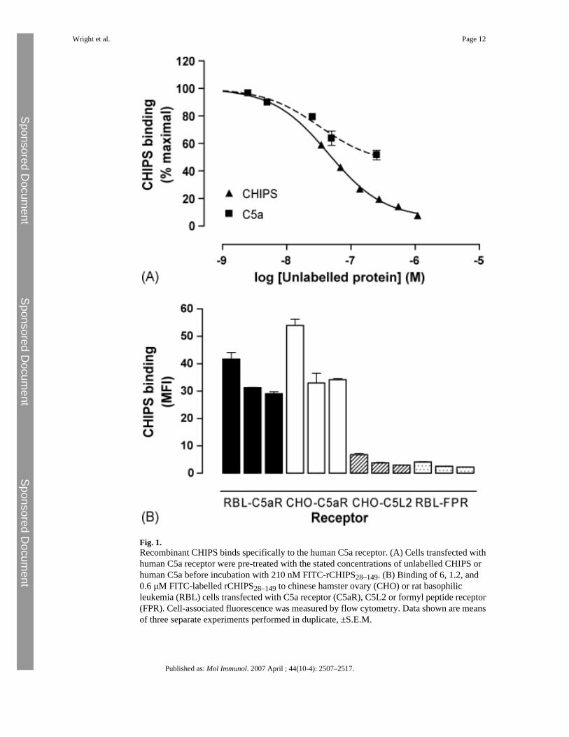
Fig. 1.Recombinant CHIPS binds specifically to the human C5a receptor. (A) Cells transfected withhuman C5a receptor were pre-treated with the stated concentrations of unlabelled CHIPS orhuman C5a before incubation with 210 nM FITC-rCHIPS28–149. (B) Binding of 6, 1.2, and0.6 μM FITC-labelled rCHIPS28–149 to chinese hamster ovary (CHO) or rat basophilicleukemia (RBL) cells transfected with C5a receptor (C5aR), C5L2 or formyl peptide receptor(FPR). Cell-associated fluorescence was measured by flow cytometry. Data shown are meansof three separate experiments performed in duplicate, ±S.E.M.
Wright et al. Page 12
Published as: Mol Immunol. 2007 April ; 44(10-4): 2507–2517.
Sponsored Docum
ent Sponsored D
ocument
Sponsored Docum
ent

Fig. 2.Inhibition of ligand binding by CHIPS. Cells transfected with C5a receptor (C5aR), C5L2 orformyl peptide receptor (FPR) were co-incubated with the stated concentrations ofrCHIPS28–149 with His6-C5a (50 nM), His6-C5a des Arg (50 nM) or FITC-formyl peptide(500 nM). Data shown are the means of three separate experiments performed in duplicate,±S.E.M.
Wright et al. Page 13
Published as: Mol Immunol. 2007 April ; 44(10-4): 2507–2517.
Sponsored Docum
ent Sponsored D
ocument
Sponsored Docum
ent

Fig. 3.Kinetic analysis of ligand binding to chemoattractant receptors. The kinetics of FITC-rCHIPS28–149 binding to cells transfected with C5a receptors was measured using flowcytometry after addition of the fluorescent ligand to RBL cells transfected with C5aR at300 nM. Dissociation was initiated by the addition of 14 μM unlabelled ligand (arrow): Dataare shown as geometric means of fluorescence values obtained in 0.1 s time windows and isone experiment, typical of five performed.
Wright et al. Page 14
Published as: Mol Immunol. 2007 April ; 44(10-4): 2507–2517.
Sponsored Docum
ent Sponsored D
ocument
Sponsored Docum
ent

Fig. 4.CHIPS inhibits the activation of C5aR and FPR. (A) RBL cells transfected with C5a receptoror formyl peptide receptor were pre-treated with rCHIPS28–149 at the stated concentrationsbefore the addition of 10 nM formyl peptide (FP), 10 nM C5a, 100 μM C5a C-terminal peptide(C-peptide) or vehicle alone (NA). Cell activation was assessed as the secretion of β-hexosaminidase, and is expressed as a percentage of the maximal response to C5a (C5a, C-terminal peptide, NA) or formyl peptide (FP). Data are means of two to three experimentsperformed in duplicate ± S.D. (B) Data from inhibition experiments was used to draw Schildplots to analyse the effects of rCHIPS28–149 on C5a and formyl peptide receptors. In a furtherset of three experiments, rCHIPS30–141 was used on formyl peptide receptors.
Wright et al. Page 15
Published as: Mol Immunol. 2007 April ; 44(10-4): 2507–2517.
Sponsored Docum
ent Sponsored D
ocument
Sponsored Docum
ent

Fig. 5.Analysis of the interaction of CHIPS with the N-terminus of C5aR. (a) Averaged chemicalshift changes of the amide cross peaks of 15N CHIPS28–149 upon complex formation withhuman C5aR1–37 peptide, corresponding to the N-terminal extracellular region of the C5areceptor. The chemical shift changes were calculated using the formula
, where the shift differences (Δδ) between free and complexedform are observed by two-dimensional 1H–15N HSQC spectra. (b) Mapping of CHIPS regionsinteracting with hC5aR1–37. Top panels: residues 71–97 (cyan) and 123–140 (magenta), shownby NMR chemical shift perturbation to be involved in interaction with hC5aR1–37, are mapped
Wright et al. Page 16
Published as: Mol Immunol. 2007 April ; 44(10-4): 2507–2517.
Sponsored Docum
ent Sponsored D
ocument
Sponsored Docum
ent

on to the structure of rCHIPS31–121 (Haas et al., 2005). The rest of the rCHIPS31–121 structureis displayed in yellow (β-strands), red (α-helices), and green (loops). Bottom panels:electrostatic surface view of the representations above. Blue and red represent positively andnegatively charged surface.
Wright et al. Page 17
Published as: Mol Immunol. 2007 April ; 44(10-4): 2507–2517.
Sponsored Docum
ent Sponsored D
ocument
Sponsored Docum
ent

Fig. 6.IgG and IgA have similar titres against CHIPS. IgG and IgA titres against recombinant CHIPSin control and patient sera were measured by ELISA and are shown as the dilution required toobtain half-maximal antibody binding measured in duplicate. The medians of the non-normallydistributed titres are shown, with interquartile range. The difference between IgG and IgA titreswas non-significant (p = 0.91) by the Mann–Whitney test.
Wright et al. Page 18
Published as: Mol Immunol. 2007 April ; 44(10-4): 2507–2517.
Sponsored Docum
ent Sponsored D
ocument
Sponsored Docum
ent

Fig. 7.Effects of human sera on CHIPS binding. Examples of the effects of human sera on FITC-CHIPS binding to human C5a receptor: (A) serum 8 (no effect), (B) 20 (enhancement ofbinding), and (C) (inhibition of binding). Diluted sera were co-incubated with 210 nM FITC-labelled rCHIPS28–149, and binding measured by flow cytometry. NA represents FITC-rCHIPS28–149 binding in the absence of human serum. Data are the means ± S.D. from twoexperiments performed in triplicate, expressed as a percentage of rCHIPS28–149 binding in theabsence of sera. Significance was assessed by t test; ***p < 0.0001.
Wright et al. Page 19
Published as: Mol Immunol. 2007 April ; 44(10-4): 2507–2517.
Sponsored Docum
ent Sponsored D
ocument
Sponsored Docum
ent

Fig. 8.Relationship between anti-CHIPS titre and antibody effects on CHIPS binding to C5a receptor.The medians and interquartile ranges of the anti-CHIPS titres of inhibitory or enhancing IgG(filled circles) and IgA (open circles) content of control and patient sera was compared usingMann–Whitney tests comparing serum IgG and IgA titre to rCHIPS28–149 in ELISA, in relationto the effects of serum on FITC-labelled rCHIPS28–149 binding to RBL-2H3-C5aR cells. Forthis purpose, sera are grouped into those having inhibiting, enhancing, or no effect onrCHIPS28–149 binding.
Wright et al. Page 20
Published as: Mol Immunol. 2007 April ; 44(10-4): 2507–2517.
Sponsored Docum
ent Sponsored D
ocument
Sponsored Docum
ent

Fig. 9.Correlation between anti-CHIPS titre and antibody effects on CHIPS binding to C5a receptor.The anti-CHIPS titres of IgG (filled circles, solid line) and IgA (open circles, broken line)plotted against half-maximal dilution factor for either the enhancement (A) or inhibition (B)of rCHIPS28–149 binding to C5a receptor. The significance of the difference of the slope valuefrom 0 is shown.
Wright et al. Page 21
Published as: Mol Immunol. 2007 April ; 44(10-4): 2507–2517.
Sponsored Docum
ent Sponsored D
ocument
Sponsored Docum
ent

Sponsored Docum
ent Sponsored D
ocument
Sponsored Docum
ent
Wright et al. Page 22
Table 1
Analysis of anti-CHIPS IgG and IgA antibody titres of serum samples
Serum Donor status IgG titre IgA titre Effect onCHIPSbinding
ChangeinCHIPSbinding(%)a
EC50/IC50 (reciprocal dilution)
1 ‘Normal’ 52 24 No effect N/A N/A
2 ‘Normal’ 116 157 No effect N/A N/A
3 ‘Normal’ 229 127 No effect N/A N/A
4 ‘Normal’ 58 70 No effect N/A N/A
5 ‘Normal’ 351 261 No effect N/A N/A
6 ‘Normal’ 40 242 No effect N/A N/A
7 ‘Normal’ 42 33 No effect N/A N/A
8 ‘Normal’ 62 44 No effect N/A N/A
9 ‘Normal’ 31 33 No effect N/A N/A
10 ‘Normal’ 195 174 No effect N/A N/A
11 ‘Normal’ 151 73 No effect N/A N/A
12 ‘Normal’ 151 97 No effect N/A N/A
13 ‘Infected’ 87 36 No effect N/A N/A
14 ‘Normal’ 178 326 Enhancing 208 7
15 ‘Normal’ 182 112 Enhancing 170 11
16 ‘Normal’ 149 45 Enhancing 135 3
17 ‘Normal’ 610 351 Enhancing 386* 118
18 ‘Normal’ 287 431 Enhancing 214* 15
19 ‘Normal’ ND ND Enhancing 245 9
2 Infected 102 88 Enhancing 155 4
21 ‘Normal’ 555 313 Inhibiting 13 12
22 ‘Normal’ 22 30 Inhibiting 66 4
23 ‘Normal’ 440 184 Inhibiting 7 4
24 ‘Normal’ 143 161 Inhibiting 32 10
25 ‘Normal’ 508 428 Inhibiting 17 4
26 ‘Normal’ 192 224 Inhibiting 26 3
27 ‘Normal’ 3097 402 Inhibiting 4 20
28 Infected 105 79 Inhibiting 27 3
29 Infected 3308 411 Inhibiting 3 38
30 Infected 105 152 Inhibiting 49 9
31 Infected 137 73 Inhibiting 12 5
32 Infected 1168 663 Inhibiting 4 56
aChange in CHIPS binding relative to control (=100%) at 1/2 dilution of serum except for * at 1/5 dilution.
Published as: Mol Immunol. 2007 April ; 44(10-4): 2507–2517.

Sponsored Docum
ent Sponsored D
ocument
Sponsored Docum
ent
Wright et al. Page 23
Table 2
Effects of IgG depletion on the ability of sera to modulate CHIPS binding to the human C5a receptor
Serum (effect on CHIPSbinding)
Change in CHIPS binding (%)a
Untreated Serum IgG-depleted serum
21 (inhibiting) 13 41
29 (inhibiting) 3 67
30 (inhibiting) 49 71
31 (inhibiting) 12 83
17 (enhancing) 386 121
18 (enhancing) 214 100
27 (inhibiting) 4 76
aChange in CHIPS binding relative to control (=100%) at 1/2 dilution of serum.
Published as: Mol Immunol. 2007 April ; 44(10-4): 2507–2517.