how Staphylococcus aureus reaches different human host ...
35
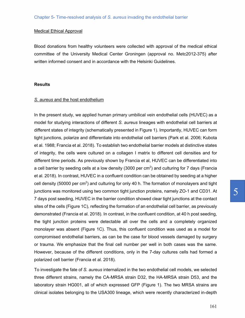
University of Groningen Breaking the barriers: how Staphylococcus aureus reaches different human host niches Raineri, Elisa DOI: 10.33612/diss.206092360 IMPORTANT NOTE: You are advised to consult the publisher's version (publisher's PDF) if you wish to cite from it. Please check the document version below. Document Version Publisher's PDF, also known as Version of record Publication date: 2022 Link to publication in University of Groningen/UMCG research database Citation for published version (APA): Raineri, E. (2022). Breaking the barriers: how Staphylococcus aureus reaches different human host niches. University of Groningen. https://doi.org/10.33612/diss.206092360 Copyright Other than for strictly personal use, it is not permitted to download or to forward/distribute the text or part of it without the consent of the author(s) and/or copyright holder(s), unless the work is under an open content license (like Creative Commons). The publication may also be distributed here under the terms of Article 25fa of the Dutch Copyright Act, indicated by the “Taverne” license. More information can be found on the University of Groningen website: https://www.rug.nl/library/open-access/self-archiving-pure/taverne- amendment. Take-down policy If you believe that this document breaches copyright please contact us providing details, and we will remove access to the work immediately and investigate your claim. Downloaded from the University of Groningen/UMCG research database (Pure): http://www.rug.nl/research/portal. For technical reasons the number of authors shown on this cover page is limited to 10 maximum. Download date: 03-06-2022
-
Upload
khangminh22 -
Category
Documents
-
view
0 -
download
0
Transcript of how Staphylococcus aureus reaches different human host ...

University of Groningen
Breaking the barriers: how Staphylococcus aureus reaches different human host nichesRaineri, Elisa
DOI:10.33612/diss.206092360
IMPORTANT NOTE: You are advised to consult the publisher's version (publisher's PDF) if you wish to cite fromit. Please check the document version below.
Document VersionPublisher's PDF, also known as Version of record
Publication date:2022
Link to publication in University of Groningen/UMCG research database
Citation for published version (APA):Raineri, E. (2022). Breaking the barriers: how Staphylococcus aureus reaches different human host niches.University of Groningen. https://doi.org/10.33612/diss.206092360
CopyrightOther than for strictly personal use, it is not permitted to download or to forward/distribute the text or part of it without the consent of theauthor(s) and/or copyright holder(s), unless the work is under an open content license (like Creative Commons).
The publication may also be distributed here under the terms of Article 25fa of the Dutch Copyright Act, indicated by the “Taverne” license.More information can be found on the University of Groningen website: https://www.rug.nl/library/open-access/self-archiving-pure/taverne-amendment.
Take-down policyIf you believe that this document breaches copyright please contact us providing details, and we will remove access to the work immediatelyand investigate your claim.
Downloaded from the University of Groningen/UMCG research database (Pure): http://www.rug.nl/research/portal. For technical reasons thenumber of authors shown on this cover page is limited to 10 maximum.
Download date: 03-06-2022

Chapter 5
Time-resolved analysis of Staphylococcus aureus invading the endothelial barrier
Elisa J.M. Raineri*, Harita Yedavally*, Anna Salvati, Jan Maarten van Dijl
*These authors contributed equally
Virulence. 2020 Dec; 11(1): 1623-1639. doi: 10.1080/21505594.2020.1844418.

152

Chapter 5- Time-resolved analysis of S. aureus invading the endothelial barrier
153
5
Abstract Staphylococcus aureus is a leading cause of infections world-wide. Once this pathogen has
reached the bloodstream, it can invade different parts of the human body by crossing the
endothelial barrier. Infected endothelial cells may be lysed by bacterial products, but the
bacteria may also persist intracellularly, where they are difficult to eradicate with antibiotics and
cause relapses of infection. Our present study was aimed at investigating the fate of methicillin
resistant S. aureus (MRSA) isolates of the USA300 lineage with different epidemiological origin
inside endothelial cells. To this end, we established two in vitro infection models based on
primary human umbilical vein endothelial cells (HUVEC), which mimic conditions of the
endothelium when infection occurs. For comparison, the laboratory strain S. aureus HG001
was used. As shown by flow-cytometry and fluorescence- or electron microscopy, differentiation
of HUVEC into a cell barrier with cell-cell junctions sets limits to the rates of bacterial
internalization, the numbers of internalized bacteria, the percentage of infected cells, and long-
term intracellular bacterial survival. Clear strain-specific differences were observed with the
HG001 strain infecting the highest numbers of HUVEC and displaying the longest intracellular
persistence, whereas the MRSA strains reproduced faster intracellularly. Nonetheless, all
internalized bacteria remained confined in membrane-enclosed LAMP-1-positive lysosomal or
vacuolar compartments. Once internalized, the bacteria had a higher propensity to persist
within the differentiated endothelial cell barrier, probably because internalization of lower
numbers of bacteria was less toxic. Altogether, our findings imply that intact endothelial barriers
are more likely to sustain persistent intracellular infection.
Keywords: Staphylococcus aureus, MRSA, endothelium, invasion, intracellular

154
Introduction S. aureus is an important pathogen that can persist for long periods of time in the human host,
be it on the skin, the mucosa or intracellularly. For most individuals the carriage of S. aureus is
asymptomatic and without major consequence. However, upon trauma, surgery or inadequate
protection by the immune defenses, S. aureus may invade the human body, reach the blood
stream and cause serious diseases that range from bacteremia to sepsis, endocarditis and
necrotizing pneumonia. The treatment of invasive S. aureus infections has always been a
challenge due to the pathogen’s ability to invade different phagocytic and non-phagocytic host
cells, and to form thick biofilms that represent a protective niche against antibiotic therapy.
Therapeutic success is nowadays also compromised as S. aureus has acquired resistance to
many different antibiotics (Archer et al. 2011; Lowy 2000; 1998). Moreover, the diversity within
the species S. aureus is enormous, which is reflected in the different epidemiological features
and pathogenicity of the clonal S. aureus lineages that we know today (Goerke et al. 2004;
Balasubramanian et al. 2017; Zhao et al. 2019; 2020).
Among the antibiotic resistant lineages, the methicillin-resistant S. aureus (MRSA) has become
notorious as it is associated with high morbidity and mortality (Ortwine et al. 2018). MRSA first
emerged in nosocomial settings, in particular causing invasive blood stream infections
(Mekonnen et al. 2017; Thurlow et al. 2012). However, in recent years MRSA infections are not
only caused by hospital-associated (HA) lineages, but also by community-associated (CA)
lineages that affect healthy individuals with no apparent hospital contact. Contrary to HA-
MRSA, the CA-MRSA lineages tend to cause soft tissue infections or more invasive infections
such as pneumonia and osteomyelitis (Thurlow et al. 2012).
Once S. aureus has gained access to the blood stream, in immunocompetent healthy
individuals it is usually cleared by phagocytic immune cells. However, some bacteria may
survive phagocytosis, disseminate in the blood, and enter the endothelium. The endothelium
has a key role in the human body as this monolayer of cells represents a barrier that can
selectively control the passage, from both the apical and basal sides, of solutes, plasma
proteins, immune cells (e.g. leukocytes), viruses and bacteria. This control is achieved by the
activation of specific pathways and expression of different sets of proteins on the apical and
the basal cell sides, and through the coordinated opening and closure of cell-cell junctions,
such as tight junctions and adherens junctions. Attached to these junctions are a variety of
adhesion molecules, including the ‘cluster of differentiation 31’ (CD31) protein (also known as
‘platelet endothelial cell adhesion molecule-1’ [PECAM-1]), which provide connections to the

Chapter 5- Time-resolved analysis of S. aureus invading the endothelial barrier
155
5
actin cytoskeleton of the cells (Daniel et al. 2013). These structures can be disrupted
temporarily, for instance in wounds, during regeneration of the endothelium, or during infection.
When the endothelial barrier integrity is compromised, both the endothelium and the underlying
tissues are more prone to infection by invasive pathogens, such as S. aureus (Lemichez et al.
2010; Dejana 2004; Sahni 2007). Once internalized, S. aureus may persist or proliferate
intracellularly for varying durations (Werdan et al. 2014; Rollin et al. 2017). Intracellular
persistence may lead to immune evasion and chronicity of infection, while intracellular
replication of S. aureus will result in endothelial host cell lysis and spread into the underlying
tissues (Werdan et al. 2014; Palma Medina et al. 2019).
The human body is a key player in determining the outcome of infection, especially since host
cell responses to close encounters with pathogens, such as S. aureus, differ per cell type.
Accordingly, S. aureus has evolved mechanisms to manipulate the different host responses in
order to survive in a wide range of hostile environments (Fraunholz et al. 2012;
Balasubramanian et al. 2017; Horn et al. 2017). A variety of virulence factors and regulators
allow S. aureus to breach cellular barriers and adapt to the intracellular environment. However,
once the intracellular environment has been reached, mutual adaptations of the pathogen and
its host will occur, as exemplified by the metabolic cross-talk that was observed upon the
invasion of lung epithelial cells by S. aureus (Palma Medina et al. 2019).
Understanding the behavior of different types of S. aureus in the intracellular compartments of
different types of host cells is fundamental for developing adequate therapeutic approaches
against chronic staphylococcal infections (Lehar, 2015). In this respect, our understanding of
these processes is currently very limited. For instance, HA- and CA-MRSA can reach the blood
stream via different routes, e.g. starting from infected surgical wounds or abscesses of the skin.
To this end, closely related HA- and CA-strains of the S. aureus USA300 lineage have evolved
different metabolic niche adaptations that favor their promulgation in blood or skin (Mekonnen
et al. 2017; 2019). Yet, we do not know how such adaptations impact on the subsequent stages
of the infection, where the bacteria enter and pass the endothelium. Therefore, the present
study was specifically aimed at investigating the fate of internalized HA- and CA-strains of the
USA300 lineage in endothelial cells. To this end, we established two in vitro infection models
that take into consideration the condition of the endothelium at the moment of invasion and the
subsequent course of infection. As a control for comparison, we included the well-defined
laboratory strain S. aureus HG001 in our analyses (Mäder et al. 2016). The course of infection
was followed quantitatively and qualitatively by flow-cytometry as well as fluorescence and
electron microscopy to determine eventual differences in the evolution of the infection over time

156
among the different strains, as well as the final fate and intracellular distribution of the
internalized bacteria.
Materials and Methods
Biological materials
The S. aureus laboratory strain HG001 (Herbert et al. 2010), and the clinical isolates D32 (CA-
MRSA) and D53 (HA-MRSA) (Mekonnen et al. 2017) were used to perform all experiments.
The HG001 strain carried plasmid pJL-sar-GFP to express the green fluorescent protein (GFP;
Liese et al., 2013), and the D32 and D53 strains (Mekonnen et al. 2017) carried plasmid pJL-
sar-GFP_redopt-cm to express GFP. The culturing was performed in Roswell Park Memorial
Institute 1640 medium (RPMI) (Gibco, New York), supplemented with 2 mM L-glutamine
(Thermo Fisher Scientific, Waltham, USA). One day prior infection, bacterial overnight cultures
were prepared in serial dilutions in RPMI (Gibco, New York) supplemented with 0.01% yeast
extract and 10 µg·ml-1 erythromycin or 10 µg·ml-1 chloramphenicol for the HG001 and USA300
strains respectively. The RPMI used for overnight precultures was supplemented with yeast
extract to facilitate the initial growth of the bacteria, while antibiotics were added to prevent the
possible loss of the GFP-encoding plasmids necessary for subsequent flow cytometry and
fluorescence microscopy. Incubation was performed at 37°C and with constant shaking (250
rpm). The following day, exponentially growing overnight cultures were used to inoculate the
main cultures for infection experiments using RPMI without yeast extract and antibiotics.
Primary human umbilical vein endothelial cells (HUVEC) from pooled donors (Lonza Cat#
C2519a Lot# 394986, Allendale, NJ, USA) were used to perform all the experiments. The
endothelial cells were grown in standard cell culture flasks (37°C, 5% CO2) in Endothelial Cell
Growth Medium 2 (Ready-to-use; PromoCell, Germany). All experiments were performed using
cells obtained from 3 to maximally 7 passages to avoid cell senescence and loss of primary
cell characteristics. The medium was changed every 48 h.
Internalization experiments and flow cytometry
Internalization experiments were performed as previously described by Pförtner et al. (2013).
Briefly, these experiments were performed using HUVEC seeded in 24-well plates (Greiner,
Germany) pre-coated with rat-tail Collagen Type-I (Corning, New York). Two different

Chapter 5- Time-resolved analysis of S. aureus invading the endothelial barrier
157
5
conditions were applied, here referred to as ‘barrier’ and ‘confluent’, following previously
established protocols (Francia et al. 2018). More in detail, to differentiate cells into a polarized
endothelial cell barrier, HUVEC were seeded at a density of 3000 cells per cm2 and cultured for
7 days prior to infection, with media exchange every 2 days. For the confluent condition,
HUVEC were seeded at a density of 50,000 cells per cm2 and cultured for 40 h prior to infection.
The numbers of cells in the barrier and confluent culture conditions were counted prior infection
with S. aureus to verify that in both conditions 100,000 cells per well of a 24-well plate were
infected.
A multiplicity of infection (MOI) of 25 was used for all internalization experiments. The bacterial
master mix for infection was prepared from exponentially growing S. aureus cells in RPMI
(OD600 of 0.4), which were counted by flow cytometry (see below), pelleted by centrifugation
and resuspended in Endothelial Cell Growth Medium 2. The bacterial master mix was added
to the HUVEC and the infected cells were incubated for 1 h (37°C, 5% CO2). Afterwards, the
medium was removed and replaced with Endothelial Cell Growth Medium 2 containing 25
µg·ml-1 of lysostaphin (AMBI Products, New York) to eliminate non-internalized bacteria bound
to the HUVEC surface. The medium was changed every 48 h.
The abundance of human and bacterial cells was measured by flow cytometry. For this
purpose, two groups of samples were collected in triplicate at different time points post infection
(p.i.) up to 6 days. One sample group was used to quantify the number of host cells by treatment
with trypsin-EDTA (Thermo Fisher Scientific, the Netherlands) during a 5 min incubation at
37°C, 5% CO2. Counting of infected HUVEC was performed with a Cytoflex S flow cytometer
(Beckman Coulter, Woerden, the Netherlands) by excitation of GFP with a 488 nm laser and
detection at 525/40 nm. Infected cells in the other sample group were lysed with 0.05% SDS
for 5 min in order to collect intracellular bacteria. Analysis of the flow cytometry data was
performed with Kaluza Analysis Software (Beckman Coulter, Woerden, the Netherlands). A
gating strategy was applied to exclude debris and select healthy cells. Distributions and
median-mean values of 20,000 cells were obtained. The relative number of infected HUVEC
was expressed as the percentage of GFP-positive cells, and the number of intracellular bacteria
was assessed by counting the number of GFP-positive events upon liberation of the
intracellular bacteria from lysed HUVEC. Statistical analyses were performed with GraphPad
Prism version 8 (GraphPad Software, La Jolla, CA, USA). Two-way Anova tests with multiple
comparisons were performed to assess the statistical significance of differences in the numbers
of internalized bacteria. P-values of ≤0.05 and a confidence of ≥95% were considered to
indicate significance.

158
Fluorescence microscopy
Immunofluorescence microscopy was performed using a Leica TCS SP8 Confocal laser
scanning microscope (Leica Microsystems, Wetzlar, Germany). The cells were seeded over
coverslips of 13 mm diameter #1.5 (Thermo Fisher Scientific, Waltham, USA). Coverslips with
infected or uninfected cells were collected at different time points p.i. until 6 days and fixed with
4% formaldehyde for 20 min at room temperature. Subsequently, the cells were permeabilized,
and blocked to avoid non-specific antibody binding, by incubation for 20 min at room
temperature with 0.5% Tween-20 in PBS, followed by overnight incubation at 4°C with 2% BSA
and 5% neutral goat serum in PBS. Additional blocking was performed by incubation with 12
µg/ml of the human monoclonal antibody 1D9 (van den Berg et al. 2015), diluted in the same
blocking solution, for 2 h at room temperature in a humidified chamber.
Subcellular localization of LAMP-1 was carried out by incubation with a primary mouse antibody
against CD107a (LAMP-1; BD, United States) at a dilution of 1:100 for 1 h at room temperature
in a humidified chamber. To detect the bound primary antibody, a secondary goat anti-mouse
antibody conjugated with Alexa Fluor 594 (Invitrogen, Netherlands) was used at a 1:500 dilution
with incubation for 1 h at room temperature. Lastly, DNA was stained with 4′,6-diamidino-2-
phenylindole (DAPI; Roche, Switzerland). The slides were mounted with Mowiol 4-88 (Merk
Millipore, USA) and stored at -20°C until microscopic visualization.
Tight junction proteins were immunostained to view their expression and distribution inside the
cells upon growth under barrier or confluent conditions by confocal microscopy. The fixation,
permeabilization and blocking procedures were carried out as described above. Subsequently,
cells were incubated for 1 h at room temperature with a polyclonal rabbit primary antibody
against the tight junction protein ZO-1 (zonula occludens-1, Life Technologies, NY, USA) at a
dilution of 1:200 and a monoclonal mouse primary antibody against CD31 (PECAM-1; Dako,
Glostrup, Denmark) at a dilution of 1:100. Bound antibodies were visualized by incubation for
1 h with a secondary goat-anti mouse antibody conjugated with Alexa Fluor 594 (Life
technologies, NY, USA) at a 1:1000 dilution, or a donkey anti-rabbit antibody conjugated with
Alexa Fluor 647 (Life technologies, NY, USA) at a 1:200 dilution. Image processing was
performed using FIJI (https://fiji.sc/).

Chapter 5- Time-resolved analysis of S. aureus invading the endothelial barrier
159
5
Transmission Electron Microscopy
Cell samples for Transmission Electron Microscopy (TEM) were collected at 2 h, 7 h and 24 h
p.i., and fixed with 0.2% glutaraldehyde and 2% paraformaldehyde in 0.1 M sodium cacodylate
buffer (pH 7.4) for 1 h. The fixed cells were rinsed twice for 5 min in 0.1 M cacodylate buffer at
room temperature followed by post-fixation in 1% osmium tetroxide, 1.5% potassium
ferrocyanide in 0.1 M sodium cacodylate at 4°C for 30 min. The cells were then washed with
Milli-Q water, dehydrated through serial incubation in a graded ethanol series (30%, 50%, 70%
and 100%) and lastly embedded in EPON resin and polymerized at 37°C for 16 h followed by
58°C for 24 h. Ultrathin sections (80 nm) were cut with an UC7 ultramicrotome (Leica, Vienna,
Austria) and contrasted using 5% uranyl acetate for 20 min, followed by Reynolds lead citrate
for 2 min. Images were recorded with a CM100 Biotwin transmission electron microscope (FEI,
Eindhoven, The Netherlands) operated at 80 kV using a Morada digital camera. Image
processing was performed with FIJI (https://fiji.sc/).
Apoptosis Assay
To measure the induction of apoptosis in infected cells, activity of the apoptotic markers
caspase-3 and -7 was measured at different time points p.i. (2 h, 7 h, 12 h, 24 h) using a
commercial Caspase-Glo 3/7 assay kit (Promega, Madison, Wisconsin, United States)
following the manufacturer's instructions. Briefly, HUVEC were collected at different time points
and 5000 cells were added to a white 96-well plate (Greiner, Germany) in duplicate. As a
positive control, cells were treated with staurosporine (0.25 µM; Biaffin GmbH & Co KG,
Germany) for 3 h. The caspase 3/7 substrates were added to each well and the plate was
shaken at 300 rpm for 2 min followed by incubation in the dark for 30 min. Luminescence was
quantified using a Synergy 2 multi-mode microplate reader (BioTek Instruments, Inc., Winooski,
VT), and the results were expressed as n-fold induction relative to the uninfected control cells.
Expression of staphylococcal Panton-Valentine Leukocidin (PVL) receptors
To investigate the expression of PVL receptors in our HUVEC infection model, we used a flow
cytometry assay based on staining with an allophycocyanin (APC)-labeled anti-human CD45
antibody and a PerCP/Cy5.5 anti-human CD88 (C5aR) antibody (BioLegend, United Sates).
HUVEC were grown under barrier or confluent conditions as described above, and 1 x 10⁶ cells
per sample were used for flow cytometry. The collected cells were incubated with the CD45- or

160
CD88-specific antibodies according to the manufacturer’s predetermined optimum
concentrations for 15-20 min at 4°C in the dark. Antibody incubation was followed by two
washing steps with 2 ml of Cell Staining Buffer (BioLegend, United States), and centrifugation
at 350 x g for 5 min. The cells were then fixed with 4% formaldehyde for 20 min at room
temperature and resuspended in PBS. Flow cytometry measurements were performed with a
Cytoflex S flow cytometer (Beckman Coulter, Woerden, the Netherlands). APC was excited
with a 638 nm laser and fluorescence was recorded at 660/20 nm, whereas PerCP/Cy5.5 was
excited with a 561 nm laser and fluorescence was recorded at 690/50 nm. Analysis of the flow
cytometry data was performed with Kaluza Analysis Software.
Previous studies have shown that neutrophils express both the CD45 and CD88 (C5aR)
receptors (Tromp et al. 2018; Spaan et al. 2013), and we therefore included neutrophils as a
positive control for our flow cytometry experiments with HUVEC. Neutrophils were freshly
isolated from healthy volunteers as previously described (Stobernack et al. 2018). Briefly, the
neutrophils were isolated from whole-blood samples using Lymphoprep buffer (StemCell
Technologies, Vancouver, Canada) and gradient centrifugation to separate different cell types.
After centrifugation, the plasma, Lymphoprep and peripheral blood mononuclear cells were
removed, and a layer of erythrocytes and neutrophils was conserved. The erythrocytes were
lysed with a red blood cell lysis buffer (10X; BioLegend, United states) followed by shaking for
10 min on ice and subsequent centrifugation. These two steps were repeated once to obtain a
pellet of purified neutrophils. 1 x 10⁶ neutrophils were resuspended in 2 ml of RPMI 1640
medium (Gibco, Waltham, MA, USA) with 2 mM L-glutamine and 10% autologous donor serum
and seeded in a 6- well plate. The neutrophils were allowed to rest on the plate at 37°C and
5% CO2 for 30 min. Subsequent flow cytometry experiments to detect CD45 and CD88, were
performed with 1 x 10⁶ neutrophils per sample following the same procedure as described
above for HUVEC. Additionally, we included the bronchial epithelial cell line 16HBE14o- in our
PVL receptor expression studies. These cells have been used in previous studies to investigate
the behavior of CA- and HA-MRSA isolates, or the control strain HG001, upon internalization
(Mekonnen et al. 2017; Palma Medina et al. 2019). The lung epithelial cell line 16HBE14o- was
cultured as described previously by (Palma Medina et al. 2019). Briefly, the cells were cultured
at 37°C in 5% CO2 in eukaryotic minimal essential medium (eMEM; 1xMEM Biochrom AG,
Germany) supplemented with 10% (v/v) fetal calf serum, 2 mM L-glutamine and 1% (v/v) non-
essential amino acids 100x (Gibco, USA). For flow cytometry experiments 1 x 10⁶ cells per
sample were used.
Chapter 5- Time-resolved analysis of S. aureus invading the endothelial barrier
161
5
Medical Ethical Approval
Blood donations from healthy volunteers were collected with approval of the medical ethical
committee of the University Medical Center Groningen (approval no. Metc2012-375) after
written informed consent and in accordance with the Helsinki Guidelines.
Results S. aureus and the host endothelium
In the present study, we applied human primary umbilical vein endothelial cells (HUVEC) as a
model for studying interactions of different S. aureus lineages with endothelial cell barriers at
different states of integrity (schematically presented in Figure 1). Importantly, HUVEC can form
tight junctions, polarize and differentiate into endothelial cell barriers (Park et al. 2006; Kubota
et al. 1988; Francia et al. 2018). To establish two endothelial barrier models at distinctive states
of integrity, the cells were cultured on a collagen I matrix to different cell densities and for
different time periods. As previously shown by Francia et al, HUVEC can be differentiated into
a cell barrier by seeding cells at a low density (3000 per cm2) and culturing for 7 days (Francia
et al. 2018). In contrast, HUVEC in a confluent condition can be obtained by seeding at a higher
cell density (50000 per cm2) and culturing for only 40 h. The formation of monolayers and tight
junctions was monitored using two common tight junction proteins, namely ZO-1 and CD31. At
7 days post seeding, HUVEC in the barrier condition showed clear tight junctions at the contact
sites of the cells (Figure 1C), reflecting the formation of an endothelial cell barrier, as previously
demonstrated (Francia et al. 2018). In contrast, in the confluent condition, at 40 h post seeding,
the tight junction proteins were detectable all over the cells and a completely organized
monolayer was absent (Figure 1C). Thus, this confluent condition was used as a model for
compromised endothelial barriers, as can be the case for blood vessels damaged by surgery
or trauma. We emphasize that the final cell number per well in both cases was the same.
However, because of the different conditions, only in the 7-day cultures cells had formed a
polarized cell barrier (Francia et al. 2018).
To investigate the fate of S. aureus internalized in the two endothelial cell models, we selected
three different strains, namely the CA-MRSA strain D32, the HA-MRSA strain D53, and the
laboratory strain HG001, all of which expressed GFP (Figure 1). The two MRSA strains are
clinical isolates belonging to the USA300 lineage, which were recently characterized in-depth

162
by comparative genome analyses and proteomics (Mekonnen et al. 2017; 2019). As shown
previously, these two strains with different epidemiology display metabolic adaptations that
have an impact on virulence factor expression and survival inside lung epithelial cells. The
HG001 control strain is a derivative of S. aureus NCTC8325, isolated from a patient with sepsis.
In S. aureus HG001 the rsbU gene, encoding a positive activator of sigma B, has been repaired
to allow its usage as a versatile model for studies on gene regulation and pathogenicity (Herbert
et al. 2010; Surmann et al. 2014).
The experimental setup used to assess the fate of the three selected S. aureus strains upon
attachment and internalization by HUVEC in the barrier or confluent states is schematically
represented in Figure 1C. Of note, studies in the barrier model were limited in time due to the
loss of a clear tight-junction organization at the interface of the cells after 48 h, also in the
absence of infecting bacteria. Thus, the bacterial fate could be followed for up to 48 h p.i. in the
barrier model, and even up to 144 h p.i. in the confluent model. Furthermore, the bacteria used
for the infection experiments were precultured in RPMI medium, in order to tune their
physiological state towards a bacteremia condition. The RPMI medium was selected based on
the results from a previous study, where S. aureus HG001 cultured under many different
conditions was analyzed by transcript profiling, showing that the gene expression signatures of
the bacteria grown in human plasma or RPMI were highly similar (Mäder et al. 2016).

Chapter 5- Time-resolved analysis of S. aureus invading the endothelial barrier
163
5
Figure 1: Schematic representation of the rationale and experimental setup to study S. aureus invading the endothelial barrier. (A) Endothelial cells are normally organized into a barrier with cell-cell junctions that control the passage of immune cells, molecules and pathogens. These cell-cell junctions include tight junctions, adherens junctions and a variety of adhesion molecules, such as ZO-1 and CD31 (PECAM-1). In a healthy condition, the barrier integrity is strictly maintained. However, endothelial cells may show loss of cell-cell junctions and this will lead to wound patches in the endothelial membrane. (B) Confocal fluorescence microscopy images of HUVEC in the barrier or confluent conditions stained with antibodies against ZO-1 (red in the merged image) and CD31 (yellow in the merged image). Blue: DAPI-stained nuclei. The micrographs present the maximum pixel value of the Z-stacks of the endothelial layers. Scale bar: 50 µm. (C) Experimental setup used during this study: two different endothelial conditions, referred to as ‘barrier’ and ‘confluent’, were used for a 1-h infection with GFP-expressing S. aureus. Extracellular bacteria were removed by a 1-h incubation with lysostaphin. Subsequently, samples were collected at different intervals for time-resolved analysis of the infectious process.
The differentiation into endothelial cell barriers determines rates of infection and bacterial
survival
HUVEC that had reached the barrier or confluent states were infected for 1 h with S. aureus
and, subsequently, any non-internalized bacteria were killed by the addition of lysostaphin. The
course of infection was then followed by flow cytometry to quantify both the host cell population

164
and the population of internalized bacteria over time (Figure 2, Supplemental Figure S1). Of
note, the presence of lysostaphin in the medium prevents bacterial survival outside cells, thus
impairing the re-infection of nearby host cells.
For the barrier infection model, samples were collected at 2, 7, 12, 24 and 48 h p.i.. During the
first 24 h of infection, the infected host cell population and the internalized bacteria showed
important changes (Figure 2A,B). While the percentage of GFP-positive infected host cells
dropped from 90% to about 60% for the HG001 and from 80% to 40% for the USA300 strains
of the total gated single cell population, the internalized bacterial population started to grow
especially during the first 7 h p.i. and remained relatively stable thereafter at a slightly lowered
level. Here it should be noticed that, in the first 2 h p.i., the number of internalized bacteria was
statistically significantly higher for the HG001 strain compared to the CA- and HA-MRSA strains
D32 and D53, respectively. However, by 7 h p.i. the intracellular D32 population had reached
comparable numbers as the HG001 population, while the D53 population remained somewhat
smaller in numbers. Consistent with the higher numbers of internalized HG001 bacteria, higher
numbers of GFP-positive HUVEC were detected over time upon infection with this strain,
particularly at 48 h p.i. (Figure 2A,B).
Compared to the barrier infection model, a very different infection dynamic was observed in the
confluent infection model. In the first place, the number of GFP-positive cells declined more
rapidly and, at 48 h p.i., merely 20% of the cell population was GFP-positive when infected with
the HG001 strain compared to over 90% observed at 2 h p.i. (Figure 2A). At this same time,
the numbers of GFP-positive HUVEC infected with the D32 or D53 strains were even close to
zero (Figure 2A). Additionally, as observed in the case of the barrier model, also in the confluent
infection model, the HG001 strain reached the highest numbers of internalized bacteria, right
from 2 h p.i. up until 96 h p.i.. Further, at 2 h p.i., higher numbers of internalized bacteria were
observed for the D32 strain compared to the D53 strain, but this difference was no longer
detectable at later time points p.i. (Figure 2B). These time-dependent changes in the
internalized bacterial populations were mirrored in the numbers of GFP-positive HUVEC
(Figure 2A).

Chapter 5- Time-resolved analysis of S. aureus invading the endothelial barrier
165
5
HG001 D32 D53
Barr
ier 2
h p.
i. Co
n!ue
nt 2
h p.
i.
C
A
B
DAPI ZO-1 CD31 GFP
HG001 D32 D53
Barr
ier 2
h p.
i. Co
n!ue
nt 2
h p.
i.
C
A
B
DAPI ZO-1 CD31 GFP

166
◄ Figure 2: The presence of intact junctions between endothelial cells determines rates of infection and bacterial survival. The progression of infection in the barrier and confluent conditions by GFP-expressing bacteria of S. aureus strains HG001, D32 or D53 was followed by flow cytometry (A and B) and fluorescence microscopy (C). (A) The % of GFP-positive host cells in the total cell population was determined over 48 h for the barrier and 144 h for the confluent condition. (B) The numbers of internalized bacteria per well of a 24-well plate with infected cells were counted over time. Two-way Anova with multiple comparisons was performed to assess the significance of differences in the bacterial numbers for different strains at particular time points of sampling. * (p<0.03), ** (p<0.002), *** (p<0.0002), **** (p<0.0001). (C) Confocal fluorescence microscopy images of HUVEC cells in barrier and confluent conditions infected with S. aureus (green). The HUVEC were stained at 2 h p.i. with anti-ZO1 (red in merged the image) and anti-CD31 (yellow in merged the image) antibodies. Blue: DAPI-stained nuclei. The micrographs present the maximum pixel value of the Z-stacks of the endothelial layers. Scale bar: 50 µm. For the unmerged images and controls with uninfected cells, see Supplemental Figure S2.
Important differences in the infection progression were observed when comparing the confluent
and barrier infection models. In particular, the number of internalized bacteria of the HG001
strain in the confluent model was nearly three-fold higher than in the barrier model at 2 h p.i.,
and it remained higher during the first 12 h p.i. (Figure 2A,B). A similar trend was observed for
the D32 and D53 strains, albeit that in both models the numbers of intracellular bacteria at 2 h
p.i. were much lower than the respective numbers of the HG001 strain. Conversely, the
intracellular numbers of the D32 and D53 strains were higher in the barrier infection model than
in the confluent model from 24 h p.i. onwards. However, at 7 and 12 h p.i., the numbers of
internalized bacteria of the D32 and D53 strains in the two infection models were more similar.
Another remarkable difference for the barrier and confluent HUVEC cell infection models was
the fact that, compared to 7 h p.i., relatively fewer bacteria survived internalization in the
confluent infection model from 24 h p.i. onwards, in comparison to what was observed in the
barrier at the same time points. In accordance with this decline in bacterial viability, the numbers
of GFP-positive cells started to decline significantly already at 12 h p.i.. This implies that in the
confluent model, the bacteria are eliminated either by the HUVEC themselves intracellularly, or
by ‘suicidal escape’ from the HUVEC and subsequent extracellular killing by lysostaphin, or
both. Altogether, these results suggest that the different states of HUVEC when grown to
confluence, or differentiated into an endothelial cell barrier, determine the rate of bacterial
internalization, the numbers of internalized bacteria, the percentage of infected cells, and the
long-term survival of the bacteria inside the HUVEC. Indeed, the differentiation of HUVEC into
a cell barrier leads to the formation of cell-cell junctions, which is a dominant phenotypic feature
of cell barriers. However, this differentiation is accompanied by other changes that can also
affect the infection and the course of its evolution. For example, endocytosis markers are
expressed differently by cells in the barrier and confluent states (Francia et al. 2018). Of note,
irrespective of the investigated strain, the long-term survival of internalized bacteria was much

Chapter 5- Time-resolved analysis of S. aureus invading the endothelial barrier
167
5
higher in the barrier infection model than in the confluent model. This implies that an intact
barrier has a higher propensity to sustain persistent intracellular infection. Yet, in both models,
the HG001 strain infected the highest numbers of HUVEC and it displayed the highest
intracellular persistence, showing that there are clear strain-specific differences in intracellular
survival. This was also clearly observed by confocal fluorescence microscopy, where the
HUVEC grown to confluence or differentiated into a barrier were immuno-stained at 2 h p.i. to
visualize ZO-1 and CD31 (Figure 2C, Supplemental Figure S2). Of note, even in the barriers
formed by the HUVEC, some ‘gaps’ and concomitant loss of tight cell-cell contacts were
detectable after bacterial infection (Figures 2C and S2). Such gaps were close to absent from
barriers formed by uninfected control cells (Figures 2C and S2), which implies that infection did
compromise the integrity of the HUVEC barrier to some extent. However, no significant
differences were detectable for the three strains, indicating that they have a comparable ability
to break this barrier.
HUVEC-internalized S. aureus resides in LAMP-1-positive membrane-enclosed compartments
Previous studies have shown that S. aureus can reside both in the cytoplasm and various other
subcellular compartments of human host cells (Grosz et al. 2014; Schröder et al. 2006;
Tranchemontagne et al. 2016; Kubica et al. 2008; Horn et al. 2017; Jarry et al. 2006). To obtain
a deeper understanding of the destiny of S. aureus inside HUVEC in the barrier and confluent
infection models, we used TEM (Figure 3, Supplemental Figure S3). Interestingly, the TEM
analyses showed that the bacteria resided exclusively in membrane-confined compartments.
These included both large electron-lucent vacuole-like structures and more electron-dense
compartments of variable size. The larger electron-dense compartments contained big clusters
of bacteria, whereas the smaller electron-dense compartments contained only one or few
bacteria. The bacteria present in big clusters frequently showed division planes, suggesting
that they were replicating inside the enclosed compartments. Judged by previous studies, the
electron-dense compartments are possibly lysosomes or phagolysosomes, where the main
degradation processes of the host cells are taking place (Palma Medina et al. 2019; Lâm et al.
2010). Since no cytosolic (i.e. ‘membrane-free’) bacteria were observed, it seems that upon
invasion of the HUVEC, the investigated S. aureus strains D32, D53 and HG001 preferentially
adapt to lysosome- or vacuole-like organelles of the host-cells, rather than to ‘escape’ to the
cytosol. However, in this respect it should be mentioned that, conceivably, different preculturing
conditions of the bacteria might tune them for a more aggressive invasive state. For example,
previous endothelial cell infection experiments with S. aureus strains precultured in tryptic soy

168
broth (TSB) or Muller Hinton (MH) broth showed phagolysosomal escape (Grosz et al. 2014;
Schröder et al. 2006; Tranchemontagne et al. 2016; Kubica et al. 2008; Horn et al. 2017; Jarry
et al. 2006). This could possibly relate to higher levels of virulence factor production in TSB or
MH broth compared to the presently used RPMI medium, but it might also relate to the use of
different cell types for the infection experiments.
Interestingly, from 2 to 6 h p.i., when the replication of internalized bacteria was highest,
bacteria of the D32 and D53 strains were mostly found in clusters within large vacuolar
structures. At later times, these bacteria were detected mostly in smaller numbers within
lysosome-like organelles. On the contrary, bacteria of the HG001 strain were detected mostly
in small numbers within small electron-dense lysosome-like organelles, right from 2 h p.i.
Importantly, this subcellular localization, i.e. inside lysosome- or vacuole-like compartments,
was observed both for the barrier and confluent infection models (Figure 3, Supplemental
Figure S3).
To further study the intracellular distribution of S. aureus, we performed confocal fluorescence
microscopy to determine eventual co-localization with LAMP-1, a protein recruited to both
lysosomal, phagolysosomal and vacuolar membranes (Saftig et al. 2009; Jarry et al. 2006;
Sinha et al. 2010). Indeed, the GFP-expressing bacteria co-localized with LAMP-1-positive
compartments over the entire time period of observation (i.e. from 2 h to 144 h p.i.), both in the
barrier and confluent infection models, and irrespective of the investigated strain (Figure 4;
Supplemental Figures S4 and S5). We therefore conclude that this subcellular localization is
typical for both investigated HUVEC models, and that it is thus not influenced by the
differentiation of cells into a polarized endothelial cell barrier.

Chapter 5- Time-resolved analysis of S. aureus invading the endothelial barrier
169
5
A
B
Barrier
Confluen
t
HG001 D32 D53
2h p
.i.6h
p.i.
24h
p.i.
2h p
.i.6h
p.i.
24h
p.i.
Scale bars – 1 µm E - Electron dense compartments V - Vacuolar structures
E
E
E
E
E E
E
E
E E E
V
V
V
V
V
V
E
E
E
E
E

170
◄ Figure 3: Bacteria internalized by endothelial cells remain localized within membrane-enclosed compartments. Transmission electron microscopy images of HUVEC infected with the S. aureus strains HG001, D32 or D53 in the barrier (A) and confluent (B) conditions. In both conditions, the investigated S. aureus strains show replication from 2 h to 7 h p.i.. Electron-dense (E) compartments of different sizes and vacuole-like structures (V) are indicated in the different panels. No cytoplasmic bacteria were detectable in both conditions. Arrows indicate replicating bacteria. For additional images, see Supplemental Figure S3.
(A) (B) Barrier Confluent
Figure 4: S. aureus internalized by endothelial cells co-localize with the lysosomal-associated membrane protein 1 (LAMP-1). Confocal fluorescence microscopy images show HUVEC infected with the S. aureus strains HG001, D32 or D53 in the barrier (A) and confluent (B) conditions stained with an anti-LAMP1 (red) antibody. Blue: DAPI-stained nuclei. Green: GFP-expressing bacteria. Co-localization with LAMP-1 was observed from the beginning of infection (2 h) until the end of the observation time (48 h for the barrier condition and 144 h for the confluent condition) for all three strains. Arrows indicate the co-localization. For enlarged images, see Supplemental Figure S4, and for images recorded at different time points p.i. see Supplemental Figure S5.
2h p.i. 48h p.i. 2h p.i. 144h p.i.
HG
001
D32
D53
Barrier Con!uent
LAMP-1 GFPDAPI
Chapter 5- Time-resolved analysis of S. aureus invading the endothelial barrier
171
5
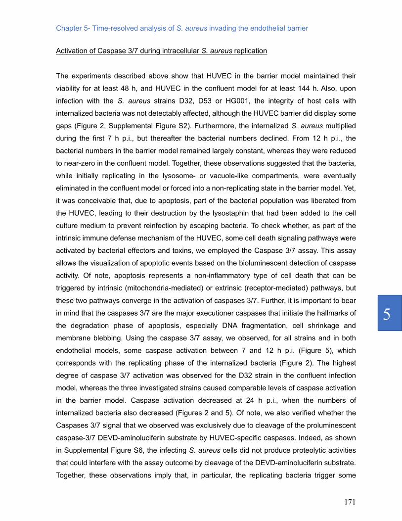
Activation of Caspase 3/7 during intracellular S. aureus replication
The experiments described above show that HUVEC in the barrier model maintained their
viability for at least 48 h, and HUVEC in the confluent model for at least 144 h. Also, upon
infection with the S. aureus strains D32, D53 or HG001, the integrity of host cells with
internalized bacteria was not detectably affected, although the HUVEC barrier did display some
gaps (Figure 2, Supplemental Figure S2). Furthermore, the internalized S. aureus multiplied
during the first 7 h p.i., but thereafter the bacterial numbers declined. From 12 h p.i., the
bacterial numbers in the barrier model remained largely constant, whereas they were reduced
to near-zero in the confluent model. Together, these observations suggested that the bacteria,
while initially replicating in the lysosome- or vacuole-like compartments, were eventually
eliminated in the confluent model or forced into a non-replicating state in the barrier model. Yet,
it was conceivable that, due to apoptosis, part of the bacterial population was liberated from
the HUVEC, leading to their destruction by the lysostaphin that had been added to the cell
culture medium to prevent reinfection by escaping bacteria. To check whether, as part of the
intrinsic immune defense mechanism of the HUVEC, some cell death signaling pathways were
activated by bacterial effectors and toxins, we employed the Caspase 3/7 assay. This assay
allows the visualization of apoptotic events based on the bioluminescent detection of caspase
activity. Of note, apoptosis represents a non-inflammatory type of cell death that can be
triggered by intrinsic (mitochondria-mediated) or extrinsic (receptor-mediated) pathways, but
these two pathways converge in the activation of caspases 3/7. Further, it is important to bear
in mind that the caspases 3/7 are the major executioner caspases that initiate the hallmarks of
the degradation phase of apoptosis, especially DNA fragmentation, cell shrinkage and
membrane blebbing. Using the caspase 3/7 assay, we observed, for all strains and in both
endothelial models, some caspase activation between 7 and 12 h p.i. (Figure 5), which
corresponds with the replicating phase of the internalized bacteria (Figure 2). The highest
degree of caspase 3/7 activation was observed for the D32 strain in the confluent infection
model, whereas the three investigated strains caused comparable levels of caspase activation
in the barrier model. Caspase activation decreased at 24 h p.i., when the numbers of
internalized bacteria also decreased (Figures 2 and 5). Of note, we also verified whether the
Caspases 3/7 signal that we observed was exclusively due to cleavage of the proluminescent
caspase-3/7 DEVD-aminoluciferin substrate by HUVEC-specific caspases. Indeed, as shown
in Supplemental Figure S6, the infecting S. aureus cells did not produce proteolytic activities
that could interfere with the assay outcome by cleavage of the DEVD-aminoluciferin substrate.
Together, these observations imply that, in particular, the replicating bacteria trigger some

172
apoptotic events, but that apoptosis cannot be the major reason why the population of
internalized bacteria decreases at late time points of infection for all strains and in both
endothelial models used. From this we infer that the bacteria are eliminated inside the
lysosome- or vacuolar-like compartments unless they reach a non-replicating state in the
barrier model.
Figure 5: Infection of endothelial cells with S. aureus induces apoptosis. The induction of apoptosis in HUVEC infected with the S. aureus strains HG001, D32 or D53 in the barrier or confluent conditions was inspected by measuring the activity of the apoptotic markers caspases 3 and -7 at different time points p.i.. The graphs show the caspase 3/7 induction fold increase over the control with uninfected cells. For additional controls, see Supplemental Figure S6.
Expression of the staphylococcal PVL receptors in HUVEC
One of the hallmarks of CA-MRSA isolates is the production of the leukotoxin PVL (Brown et
al. 2012). In contrast, the PVL-encoding lukFS genes are generally absent from HA-MRSA
isolates. In agreement with this, we have previously shown that the CA-isolate D32 used for
Caspase 3/7 activity
Caspase 3/7 activity

Chapter 5- Time-resolved analysis of S. aureus invading the endothelial barrier
173
5
this study produces PVL, whereas the HA-isolate D53 lacks the respective lukFS genes
(Mekonnen et al. 2017). Since only relatively minor differences were observed in the infection
of HUVEC by the D32 and D53 strains, we wondered whether these cells would express the
PVL receptors CD88 (C5aR1; Spaan et al. 2013) and CD45 (Tromp et al. 2018). In particular,
binding of the S-component of PVL to CD88 and CD45 was previously shown to contribute to
the cellular tropism and human specificity of this toxin (Spaan et al. 2013; Tromp et al. 2018).
This can result in pore formation in the eukaryotic cell membranes and host cell lysis.
To measure expression of the CD88 and CD45 receptors in our endothelial model systems, we
used a flow cytometry assay based on an APC-labeled anti-human CD45 antibody and a
PerCP/Cy5.5-labeled anti-human CD88 antibody (Figure 6). In addition, we used human
neutrophils as a positive control for receptor expression and 16HBE14o- lung epithelial cells
that we employed in our previous studies (Palma Medina et al. 2019; 2020) for comparison with
the HUVEC. Indeed, as previously reported (Tromp et al. 2018; Spaan et al. 2013), clear CD88
and CD45 signals were observed in neutrophils (Figure 6A). In contrast, the HUVEC in the
barrier and confluent conditions did not show a detectable fluorescence intensity shift
compared to the unstained sample for the CD45 antibody, and only a minor fluorescence
intensity shift for the CD88 antibody (Figure 6C,D). These observations imply that the CD45
receptor is absent from HUVEC and that CD88 is present only in relatively small amounts
compared to neutrophils. Similarly, CD45 was absent from the 16HBE14o- lung epithelial cells.
CD88 was however detected in the lung epithelial cells at levels that were clearly higher than
those in HUVEC, but at lower levels than the CD88 detected in neutrophils. Importantly, the
observed low-level expression of the CD88 PVL receptor in HUVEC could explain the relatively
small overall differences observed in the infection of HUVEC by the CA-MRSA isolate D32 and
the HA-MRSA isolate D53. In addition, the higher expression of CD88 in the 16HBE14o- lung
epithelial cells would be consistent with the differential behavior of these CA- and HA-MRSA
isolates in the latter infection model (Mekonnen et al. 2017).

174
Figure 6: PVL receptor expression in neutrophils, lung epithelial cells and HUVEC. To investigate the expression of receptors for the secreted staphylococcal toxin PVL, a flow cytometry-based cell staining assay was applied using APC-labelled anti-human CD45 and PerCP/Cy5.5-labelled anti-human CD88 (C5aR) antibodies. (A) Human neutrophils (positive control), (B) bronchial epithelial cell line 16HBE14o-, (C) HUVEC in the barrier condition, and (D) HUVEC in the confluent condition. HUVEC and bronchial epithelial cells tested negative for CD45-staining, but showed shifts in fluorescence intensity upon CD88 (C5aR)-staining. Neutrophils showed major shifts in fluorescence intensity upon staining for CD45 and CD88. * indicates the observed shifts in fluorescence intensity upon staining with the anti-CD45 and anti-CD88 antibodies.
A Control Unstained Control Unstained
CD45-APC stained C5aR1-PerCP stained
Neu
trop
hils
B Control Unstained Control Unstained
C5aR1-PerCP stainedCD45-APC stained
16H
BE14
o-
Control Unstained Control Unstained
HU
VEC
barr
ier
C5aR1-PerCP stainedCD45-APC stained
Control Unstained Control Unstained
C5aR1-PerCP stainedCD45-APC stained
HU
VEC
Conf
uent
DC
** *
* *

Chapter 5- Time-resolved analysis of S. aureus invading the endothelial barrier
175
5
Discussion
The present study was aimed at analysing the impact of S. aureus internalization on human
endothelial cells and the subsequent fate of the internalized bacteria. Our results show that the
bacteria are readily internalized, but get trapped in membrane-enclosed compartments where
they either fade away or reach a low/non-replicating state. This is in stark contrast with what
we previously observed for 16HBE14o- lung epithelial cells, where internalized bacteria
managed to escape to the cytoplasm and then either lysed the host or became physiologically
‘dormant’ (Palma Medina et al. 2019). It thus seems that, at least in the present experimental
setup, the employed endothelial cells (i.e. HUVEC) are much better able to contain the invading
S. aureus bacteria than 16HBE14o- lung epithelial cells. Whether the non-replicating S. aureus
bacteria inside the membrane-enclosed compartments of the HUVEC have reached a genuine
state of dormancy with an altered metabolic profile, as previously shown for S. aureus that have
escaped to the cytoplasm of lung epithelial cells, remains to be investigated in more detail.
The dynamics of S. aureus intracellular infection in the two presently implemented endothelial
models reflects well the in vivo course of infection. As shown by flow cytometry, the number of
internalized bacteria in the confluent HUVEC infection model was higher than in the barrier
model at 2 h p.i., and this difference was maintained up to 12 h p.i.. One possible explanation
of this difference relates to the fact that bacteria infecting HUVEC differentiated into a cell
barrier, first have to pass the cell-cell junctions before they can reach integrins exposed on the
HUVEC. Of note integrins are usually exposed on the basolateral side of endothelial cells and
their binding is necessary for staphylococcal invasion (Josse et al. 2017). In fact, many bacteria
produce toxins, which can kill endothelial cells, weakening their cytoskeleton and opening the
cell-cell junctions (Lubkin et al. 2017). In contrast, when the HUVEC are merely confluent, the
tight junction proteins are intracellular and do not form a clearly structured organization, which
will give the bacteria faster access to the host cells and their intracellular environment. This has
implications for the situation in the human body, where the endothelium normally controls
permeability. If the organized structure of the endothelium is lost, which can happen upon
trauma, the distribution of integrins and junction proteins that are usually present at the
basolateral side of polarized endothelial barriers is lost; thus, they can be exposed on all sides
allowing bacteria to bind and invade them at higher levels (Becker et al. 2017; Dupuy et al.
2008). Additionally, as shown by Francia et al. (2018), polarized HUVEC in a barrier display
lower mRNA expression levels of endocytic targets and lower nanoparticle uptake compared
to cells in the confluent condition. An intact barrier is, thus, not only fundamental for protection
against bacterial infection, but also sets limits to the uptake of much smaller objects, such as

176
nano-sized carriers.
Once S. aureus had reached the inside of the endothelial cells, be it under the barrier or
confluent conditions, it was retained in membrane-enclosed compartments resembling
vacuoles, phagolysosomes and lysosomes. Importantly, immunofluorescence microscopy
revealed that the GFP-positive S. aureus compartments were also LAMP-1-positive, and
colocalization of the bacteria with LAMP-1 was maintained during the entire period of
observation in both the barrier (2 days p.i.) and confluent states of the endothelial cells (6 days
p.i.). This was observed for all three investigated S. aureus strains, which implies that, once
the bacteria become internalized in endothelial cells, regardless of barrier formation, they
remain confined in LAMP-1-positive compartments. Thus, it seems that those staphylococci
that survive inside the HUVEC for extended periods of time manage to adapt to the degradative
compartments of their host cells, whereas they do not establish themselves in the cytoplasm.
At present, we cannot say whether the internalized bacteria do not reach the cytoplasm at all,
or whether some do reach the cytoplasm and then get killed in this compartment, which is
generally considered a less extreme environment than the interior of (phago)lysosomes or the
vacuole. In fact, bacterial replication was observed inside these membrane-enclosed
intracellular compartments from 2 h to 7 h p.i..
Despite the general similarities that we observed upon infection of HUVEC by the three
investigated S. aureus strains, we observed also clear strain-specific differences in terms of
internalization rate, the percentage of infected cells, and the long-term survival of the bacteria
inside the HUVEC. In particular, barriers with polarized cells and cell-cell junctions were more
resistant to infection than the confluent HUVEC but, once internalized, the bacteria had a higher
propensity to reach a state of persistence in the barrier condition. In contrast, for compromised
endothelial cell barriers, we observed much higher rates of bacterial internalization, which was
potentially more toxic for the cells. This would explain why the numbers of internalized S.
aureus dropped almost to zero over time in the confluent infection model. Lastly, although the
numbers of bacteria that entered the HUVEC were smaller for the D32 and D53 strains
compared to strain HG001, they reproduced much faster during the first 7 h p.i., especially in
HUVEC that had formed a barrier. This shows that even the internalization of very few bacteria
may turn into a rather persistent intracellular infection, and this effect was actually most
pronounced for the clinical D32 and D53 strains.
Our present observations are in accordance with previous studies in different cell types, where
intracellular replication of S. aureus in organelles, such as vacuoles, phagosomes,
phagolysosomes, and autophagosomes was observed (Flannagan et al. 2016; Kubica et al.

Chapter 5- Time-resolved analysis of S. aureus invading the endothelial barrier
177
5
2008; Horn et al. 2017; Flannagan et al. 2018). In our present experimental setup, from 24 h
p.i. onwards, the internalized bacteria seemed to stop replicating and relatively low numbers of
bacteria persisted until 6 days p.i.. Intracellular persistence has been described for several
species of bacteria and cell types, where the bacteria are able to remain viable in the host for
prolonged periods of time (Fisher et al. 2017; Lacoma et al. 2017; Löffler et al. 2014). Survival
of S. aureus in endothelial cells has been observed until 10 days p.i. (Rollin et al. 2017). At
present, we do not know what determines the duration of intracellular survival of S. aureus in
terms of strain- and host cell type-specific differences. However, at the bacterial end, we can
envisage that stress management, metabolic adaptations, and cytolytic toxin production are
prime parameters. For instance, the HG001 strain included in our present studies was
‘modified’ such that all possible defects in gene regulators, particularly rsbU related to the SigB
response, were repaired (Herbert et al. 2010). This may explain why this strain showed a
superior capability to invade the HUVEC and to survive intracellularly compared to the two
investigated clinical HA- and CA-USA300 isolates, which were previously shown to display
differential expression of the respective SigB regulons (Mekonnen et al. 2019). In addition, it
was previously shown that another S. aureus RsbU+-repaired strain (SH1000) also displayed
increased internalization and intracellular growth (Olivier et al. 2009).
For the two investigated USA300 strains, we recently identified niche-specific metabolic
adaptations priming the HA-strains for growth in nutrient-proficient environments, whereas the
CA-strains were more geared towards growth in nutrient-deplete environments (Mekonnen et
al. 2019). Such adaptations may provide also advantages for intracellular growth and survival,
because the internalized bacteria need to compete with their host for nutrients, especially when
they reside intracellularly for extended periods of time. This may explain why the CA-isolate
D32 managed to establish intracellular growth in HUVEC somewhat faster than the HA-isolate
D53, especially at early times p.i.. On the other hand, we previously showed clear differences
in the expression of cytolytic toxins by the two strains, especially for phenol-soluble modulins
(PSMs), which were produced to much higher levels by the HA-isolate D53 than the CA-isolate
D32 (Mekonnen et al. 2017). Conversely, the CA-isolate D32 expresses the Panton Valentin
Leukocidin (PVL), whereas the HA-isolate D53 lacks the lukFS genes for PVL (Mekonnen et
al. 2017). Such differences in toxin production may also impact on intracellular growth of S.
aureus, since they could for instance enhance host cell lysis, leading to the suicidal escape of
the bacteria into an extracellular environment that was supplemented with lysostaphin. Yet, the
difference in PVL production by the D32 and D53 strains is probably of minor overall importance
in the infection of HUVEC as the PVL receptors CD45 and CD88 were, respectively, absent or
present only in low amounts in these cells. Moreover, judging by the assessment of caspase

178
3/7 activation, the internalized bacteria did not contribute in major ways to caspase activity-
related cell death, except perhaps in the confluent cells, where the caspase 3/7 activation by
the PVL-proficient CA-strain D32 was higher than the activation of these enzymes by the PVL-
negative strains D53 and HG001. Yet, this difference was not detected in HUVEC that had
formed a barrier. Taken together, it seems however that effective stress management and
metabolic adaptations are more important features for intracellular growth and survival in
HUVEC than the production of cytolytic toxins.
Conclusion
Altogether, our present observations provide a better insight into how different S. aureus strains
can take advantage of endothelial cells to survive intracellularly for a prolonged period of time.
Our study in fact highlights the different dynamics of S. aureus infection in two endothelial
models, which mimic two different conditions of the endothelium. We further conclude that the
dynamics and localization of intracellular S. aureus shows important strain- and host cell type-
specific differences. Importantly, compared to our previous infection studies with the same
staphylococcal strains in lung epithelial cells, we notice clear differences in the subcellular
localization of the internalized bacteria and the duration of their intracellular survival. While the
bacteria remained enclosed in (phago)lysosomes or vacuole-like compartments of HUVEC,
they managed to escape from these compartments in the lung epithelial cells (Palma Medina
et al. 2019). In addition, we previously observed much stronger differences in the internalization
dynamics of the CA- and HA-isolates in lung epithelial cells than in the here investigated
HUVEC cells (Mekonnen et al. 2017), which may well relate to particular metabolic adaptations
(Mekonnen et al. 2019), as well as differences in the levels of the PVL receptor CD88 between
HUVEC and the lung epithelial cells. Finally, a major conclusion that can be drawn from our
present study is that the low-level invasion by S. aureus of endothelial cells in a tightly sealed
barrier state is more likely to lead to persistent staphylococcal infection than invasion of a
damaged endothelium by higher numbers of bacteria. This would be consistent with a clinical
scenario where disruption of the endothelium by trauma may lead to severe invasive S. aureus
infection of the underlying tissues, while chronic infections by S. aureus are generally
associated with low bacterial counts and less fulminant pathology.

Chapter 5- Time-resolved analysis of S. aureus invading the endothelial barrier
179
5
Acknowledgments
We thank Laura M. Palma Medina for helpful discussions and for support in establishment of
the infection models, Marines du Teil Espina for support with the neutrophil isolation, and
Francisco Romero Pastrana for producing the 1D9 antibody. Additionally, we thank Solomon
Mekonnen and Uwe Völker for providing the GFP-expressing derivatives of the USA300 D32
and D53.
Funding sources
E.J.M.R. and H.Y. received funding from CEC MSCI-ITN grant 713482 (ALERT). Fluorescence
and electron microscopies were performed at the University Medical Center Groningen Imaging
and Microscopy Center, which is sponsored by grants from the Netherlands Organisation for
Scientific Research (40-00506-98-9021, TissueFaxs, and 175.010.2009-023, Zeiss 2p).
Declaration of interest statement
E.J.M.R., H.Y., and A.S. report that they have no competing interests relevant to this study.
J.M.vD. reports that he filed a patent application on the use of 1D9, which is owned by his
employer University Medical Center Groningen.
Supporting information
Supplemental Figure S1. Flow cytometry-gating strategies for time-resolved analysis of the progression
of HUVEC infection by S. aureus.
Supplemental Figure S2. Fluorescence microscopy analysis of S. aureus-infected HUVEC.
Supplemental Figure S3. Transmission electron microscopy of of HUVEC in the barrier and confluent
conditions infected with the S. aureus strains HG001, D32 or D53.
Supplemental Figure S4. Colocalization of internalized S. aureus with LAMP-1.
Supplemental Figure S5. Colocalization of HUVEC-internalized S. aureus with LAMP-1 over time. Supplemental Figure S6. S. aureus proteases do not cleave caspase 3/7-specific substrates.

180
References
Archer, Nathan K, Mark J Mazaitis, J William
Costerton, Jeff G Leid, Mary Elizabeth Powers, and Mark E Shirtliff. 2011. “Staphylococcus aureus biofilms.” Virulence 2 (5): 445–59. https://doi.org/10.4161/viru.2.5.17724.
Balasubramanian, Divya, Lamia Harper, Bo Shopsin, and Victor J. Torres. 2017. “Staphylococcus aureus pathogenesis in diverse host environments.” Pathogens and Disease 75 (1). ftx005. https://doi.org/10.1093/femspd/ftx005.
Becker, Katrin Anne, Björn Fahsel, Hannes Kemper, Joelina Mayeres, Cao Li, Barbara Wilker, Simone Keitsch, et al. 2017. “Staphylococcus aureus Alpha-Toxin disrupts endothelial-cell tight junctions via Acid Sphingomyelinase and Ceramide.” Infection and Immunity 86 (1) e00606-17. https://doi.org/10.1128/IAI.00606-17.
Berg, Sanne van den, Hendrik P. J. Bonarius, Kok P. M. van Kessel, Goffe S. Elsinga, Neeltje Kooi, Hans Westra, Tjibbe Bosma, et al. 2015. “A human monoclonal antibody targeting the conserved Staphylococcal Antigen IsaA protects mice against Staphylococcus aureus bacteremia.” International Journal of Medical Microbiology 305 (1): 55–64. https://doi.org/10.1016/j.ijmm.2014.11.002.
Brown, Megan L., F. Patrick O’Hara, Nicole M. Close, Robertino M. Mera, Linda A. Miller, Jose A. Suaya, and Heather Amrine-Madsen. 2012. “Prevalence and sequence variation of Panton-Valentine Leukocidin in methicillin-resistant and methicillin-susceptible Staphylococcus aureus strains in the United States.” Journal of Clinical Microbiology 50 (1): 86–90. https://doi.org/10.1128/JCM.05564-11.
Daniel, Anna E., and Jaap D. van Buul. 2013. “Endothelial junction regulation: a prerequisite for leukocytes crossing the vessel wall.” Journal of Innate Immunity 5 (4): 324–35. https://doi.org/10.1159/000348828.
Dejana, Elisabetta. 2004. “Endothelial cell–cell junctions: happy together.” Nature Reviews Molecular Cell Biology 5 (4): 261–70. https://doi.org/10.1038/nrm1357.
Dupuy, A. G., and E. Caron. 2008. “Integrin-dependent phagocytosis - spreading from microadhesion to new concepts.” Journal of Cell Science 121 (11): 1773–83. https://doi.org/10.1242/jcs.018036.
Fisher, Robert A., Bridget Gollan, and Sophie Helaine. 2017. “Persistent bacterial infections and persister cells.” Nature
Reviews Microbiology 15 (8): 453–64. https://doi.org/10.1038/nrmicro.2017.42.
Flannagan, Ronald S., Bryan Heit, and David E. Heinrichs. 2016. “Intracellular replication of Staphylococcus aureus in mature phagolysosomes in macrophages precedes host cell death, and bacterial escape and dissemination: S. aureus replicates in mature phagolysosomes in macrophages.” Cellular Microbiology 18 (4): 514–35. https://doi.org/10.1111/cmi.12527.
Flannagan, Ronald S., Robert C. Kuiack, Martin J. McGavin, and David E. Heinrichs. 2018. “Staphylococcus aureus uses the GraXRS Regulatory System to sense and adapt to the acidified phagolysosome in macrophages.” MBio 9 (4): e01143-18, /mbio/9/4/mBio.01143-18.atom. https://doi.org/10.1128/mBio.01143-18.
Francia, Valentina, Aldy Aliyandi, and Anna Salvati. 2018. “Effect of the development of a cell barrier on nanoparticle uptake in endothelial cells.” Nanoscale 10 (35): 16645–56. https://doi.org/10.1039/C8NR03171A.
Fraunholz, Martin, and Bhanu Sinha. 2012. “Intracellular Staphylococcus aureus: Live-in and let die.” Frontiers in Cellular and Infection Microbiology 2: 43. https://doi.org/10.3389/fcimb.2012.00043.
Goerke, Christiane, and Christiane Wolz. 2004. “Regulatory and genomic plasticity of Staphylococcus aureus during persistent colonization and infection.” International Journal of Medical Microbiology 294 (2–3): 195–202. https://doi.org/10.1016/j.ijmm.2004.06.013.
Goldenberg, N. M., B. E. Steinberg, A. S. Slutsky, and W. L. Lee. 2011. “Broken barriers: A new take on sepsis pathogenesis.” Science Translational Medicine 3 (88): 88ps25-88ps25. https://doi.org/10.1126/scitranslmed.3002011.
Grosz, Magdalena, Julia Kolter, Kerstin Paprotka, Ann-Cathrin Winkler, Daniel Schäfer, Som Subra Chatterjee, Tobias Geiger, et al. 2014. “Cytoplasmic replication of Staphylococcus aureus upon phagosomal escape triggered by Phenol-Soluble Modulin α.” Cellular Microbiology 16 (4): 451–65. https://doi.org/10.1111/cmi.12233.
Herbert, S., A. K. Ziebandt, K. Ohlsen, T. Schafer, M. Hecker, D. Albrecht, R. Novick, and F. Gotz. 2010. “Repair of global regulators in Staphylococcus aureus 8325 and comparative analysis with other clinical isolates.” Infection and Immunity 78 (6): 2877–89. https://doi.org/10.1128/IAI.00088-10.

Chapter 5- Time-resolved analysis of S. aureus invading the endothelial barrier
181
5
Horn, Jessica, Kathrin Stelzner, Thomas Rudel, and Martin Fraunholz. 2017. “Inside job: Staphylococcus aureus host-pathogen interactions.” International Journal of Medical Microbiology 308 (6): 607-624. https://doi.org/10.1016/j.ijmm.2017.11.009.
Jarry, T. M., and A. L. Cheung. 2006. “Staphylococcus aureus escapes more efficiently from the phagosome of a cystic fibrosis bronchial epithelial cell line than from Its normal counterpart.” Infection and Immunity 74 (5): 2568–77. https://doi.org/10.1128/IAI.74.5.2568-2577.2006.
Josse, Jérôme, Frédéric Laurent, and Alan Diot. 2017. “Staphylococcal adhesion and host cell invasion: Fibronectin-binding and other mechanisms.” Frontiers in Microbiology 8: 2433. https://doi.org/10.3389/fmicb.2017.02433.
Kubota Y., Kleinman H.K., Martin G.R., Lawley T.J. 1988. “Role of laminin and basement membrane in the morphological differentiation of human endothelial cells into capillary-like structures.” 1988. The Journal of Cell Biology 107 (4): 1589–98. https://doi.org/10.1083/jcb.107.4.1589.
Kubica, Malgorzata, Krzysztof Guzik, Joanna Koziel, Miroslaw Zarebski, Walter Richter, Barbara Gajkowska, Anna Golda, et al. 2008. “A potential new pathway for Staphylococcus aureus dissemination: the silent survival of S. aureus phagocytosed by human monocyte-derived macrophages.” PLoS ONE 3 (1): e1409. https://doi.org/10.1371/journal.pone.0001409.
Lacoma, A., V. Cano, D. Moranta, V. Regueiro, D. Domínguez-Villanueva, M. Laabei, M. González-Nicolau, V. Ausina, C. Prat, and J. A. Bengoechea. 2017. “Investigating intracellular persistence of Staphylococcus aureus within a murine alveolar macrophage cell line.” Virulence 8 (8): 1761–75. https://doi.org/10.1080/21505594.2017.1361089.
Lâm, Thiên-Trí, Bernd Giese, Deepak Chikkaballi, Anika Kühn, Wanja Wolber, Jan Pané-Farré, Daniel Schäfer, Susanne Engelmann, Martin Fraunholz, and Bhanu Sinha. 2010. “Phagolysosomal integrity Is generally maintained after Staphylococcus aureus invasion of nonprofessional phagocytes but Is modulated by strain 6850.” Infection and Immunity 78 (8): 3392–3403. https://doi.org/10.1128/IAI.00012-10.
Lehar SM, Pillow T, Xu M, Staben L, Kajihara KK, Vandlen R, DePalatis L, Raab H, Hazenbos WL, Morisaki JH, Kim J, Park S, Darwish M, Lee BC, Hernandez H, Loyet KM, Lupardus P, Fong R, Yan D, Chalouni C, Luis E, Khalfin Y, Plise E, Cheong J, Lyssikatos JP, Strandh M, Koefoed K, Andersen PS, Flygare JA, Wah Tan M, Brown EJ,
Mariathasan S. 2015. "Novel antibody-antibiotic conjugate eliminates intracellular S. aureus." Nature. 527 (7578): 323-8. https://doi: 10.1038/nature16057.
Lemichez, Emmanuel, Marc Lecuit, Xavier Nassif, and Sandrine Bourdoulous. 2010. “Breaking the wall: targeting of the endothelium by pathogenic bacteria.” Nature Reviews Microbiology 8 (2): 93–104. https://doi.org/10.1038/nrmicro2269.
Liese, Jan, Suzan H. M. Rooijakkers, Jos A. G. van Strijp, Richard P. Novick, and Michael L. Dustin. 2013. “Intravital two-photon microscopy of host–pathogen interactions in a mouse model of Staphylococcus aureus skin abscess formation.” Cellular Microbiology 15 (6): 891–909. https://doi.org/10.1111/cmi.12085.
Löffler, Bettina, Lorena Tuchscherr, Silke Niemann, and Georg Peters. 2014. “Staphylococcus aureus persistence in non-professional phagocytes.” International Journal of Medical Microbiology 304 (2): 170–76. https://doi.org/10.1016/j.ijmm.2013.11.011.
Lowy, Franklin D. 1998. “Staphylococcus aureus infections.” The New England Journal of Medicine 339 (8): 520-32. https://doi.org/10.1056/NEJM199808203390806.
Lowy, Franklin D. 2000. “Is Staphylococcus aureus an intracellular pathogen?” Trends in Microbiology 8 (8): 341–43. https://doi.org/10.1016/S0966-842X(00)01803-5.
Lubkin, Ashira, and Victor J. Torres. 2017. “Bacteria and endothelial cells: A toxic relationship.” Current Opinion in Microbiology 35: 58–63. https://doi.org/10.1016/j.mib.2016.11.008.
Mäder, Ulrike, Pierre Nicolas, Maren Depke, Jan Pané-Farré, Michel Debarbouille, Magdalena M. van der Kooi-Pol, Cyprien Guérin, et al. 2016. “Staphylococcus aureus transcriptome architecture: from laboratory to infection-mimicking conditions.” PLOS Genetics 12 (4): e1005962. https://doi.org/10.1371/journal.pgen.1005962.
Mekonnen, Solomon A., Laura M. Palma Medina, Corinna Glasner, Eleni Tsompanidou, Anne de Jong, Stefano Grasso, Marc Schaffer, et al. 2017. “Signatures of cytoplasmic proteins in the exoproteome distinguish community- and hospital-associated methicillin-resistant Staphylococcus aureus USA300 Lineages.” Virulence 8 (6): 891–907. https://doi.org/10.1080/21505594.2017.1325064.
Mekonnen, Solomon A., Laura M. Palma Medina, Stephan Michalik, Marco G. Loreti, Manuela Gesell Salazar, Jan Maarten van Dijl, and Uwe Völker. 2019. “Metabolic niche adaptation of community- and hospital-associated methicillin-resistant Staphylococcus aureus.” Journal of

182
Proteomics 193: 154-161. https://doi.org/10.1016/j.jprot.2018.10.005.
Olivier, Aurélie C., Sandrine Lemaire, Françoise Van Bambeke, Paul M. Tulkens, and Eric Oldfield. 2009. “Role of RsbU and Staphyloxanthin in phagocytosis and intracellular growth of Staphylococcus aureus in human macrophages and endothelial cells.” The Journal of Infectious Diseases 200 (9): 1367–70. https://doi.org/10.1086/606012.
Ortwine, Jessica K., and Kavita Bhavan. 2018. “Morbidity, mortality, and management of methicillin-resistant S. aureus bacteremia in the USA: Update on antibacterial choices and understanding.” Hospital Practice 46 (2): 64–72. https://doi.org/10.1080/21548331.2018.1435128.
Palma Medina, Laura M., Ann-Kristin Becker, Stephan Michalik, Kristin Surmann, Petra Hildebrandt, Manuela Gesell Salazar, Solomon A. Mekonnen, Lars Kaderali, Uwe Völker, and Jan Maarten van Dijl. 2020. “Interaction of Staphylococcus aureus and host cells upon infection of bronchial epithelium during different stages of regeneration.” ACS Infectious Diseases 6 (8): 2279-2290. https://doi.org/10.1021/acsinfecdis.0c00403.
Palma Medina, Laura M., Ann-Kristin Becker, Stephan Michalik, Harita Yedavally, Elisa J.M. Raineri, Petra Hildebrandt, Manuela Gesell Salazar, et al. 2019. “Metabolic cross-talk between human bronchial epithelial cells and internalized Staphylococcus aureus as a driver for infection.” Molecular & Cellular Proteomics 18 (5): 892–908. https://doi.org/10.1074/mcp.RA118.001138.
Park, Ho-Jin, Yali Zhang, Serban P Georgescu, Kristin L Johnson, Dequon Kong, and Jonas B Galper. 2006. “Human umbilical vein endothelial cells and human dermal microvascular endothelial cells offer new insights into the relationship between lipid metabolism and angiogenesis.” Stem Cell Reviews 2: 93–101. https://doi.org/10.1007/s12015-006-0015-x
Pförtner, Henrike, Juliane Wagner, Kristin Surmann, Petra Hildebrandt, Sandra Ernst, Jörg Bernhardt, Claudia Schurmann, et al. 2013. “A proteomics workflow for quantitative and time-resolved analysis of adaptation reactions of internalized bacteria.” Methods, Quantitative Proteomics, 61 (3): 244–50. https://doi.org/10.1016/j.ymeth.2013.04.009.
Rollin, Guillaume, Xin Tan, Fabiola Tros, Marion Dupuis, Xavier Nassif, Alain Charbit, and Mathieu Coureuil. 2017. “Intracellular survival of Staphylococcus aureus in endothelial cells: a matter of growth or
persistence.” Frontiers in Microbiology 8: 1354. https://doi.org/10.3389/fmicb.2017.01354.
Saftig, Paul, and Judith Klumperman. 2009. “Lysosome biogenesis and lysosomal membrane proteins: trafficking meets function.” Nature Reviews Molecular Cell Biology 10 (9): 623–35. https://doi.org/10.1038/nrm2745.
Sahni, Sanjeev K. 2007. “Endothelial cell infection and hemostasis.” Thrombosis Research 119 (5): 531–49. https://doi.org/10.1016/j.thromres.2006.06.006.
Schröder, Andreas, Raphael Kland, Andreas Peschel, Christof von Eiff, and Martin Aepfelbacher. 2006. “Live cell imaging of phagosome maturation in Staphylococcus aureus infected human endothelial cells: Small Colony Variants are able to survive in lysosomes.” Medical Microbiology and Immunology 195 (4): 185–94. https://doi.org/10.1007/s00430-006-0015-0.
Spaan, András N., Thomas Henry, Willemien J.M. van Rooijen, Magali Perret, Cédric Badiou, Piet C. Aerts, Johan Kemmink, et al. 2013. “The staphylococcal toxin Panton-Valentine Leukocidin targets human C5a receptors.” Cell Host & Microbe 13 (5): 584–94. https://doi.org/10.1016/j.chom.2013.04.006.
Sinha B, Fraunholz M. 2010 "Staphylococcus aureus host cell invasion and post-invasion events." Int J Med Microbiol. 300 (2-3): 170-5. https://doi.org/10.1016/j.ijmm.2009.08.019.
Stobernack, Tim, Marines du Teil Espina, Lianne M. Mulder, Laura M. Palma Medina, Dillon R. Piebenga, Giorgio Gabarrini, Xin Zhao, et al. 2018. “A secreted bacterial Peptidylarginine Deiminase can neutralize human innate immune defenses.” MBio 9 (5): e01704-18, /mbio/9/5/mBio.01704-18.atom. https://doi.org/10.1128/mBio.01704-18.
Surmann, Kristin, Stephan Michalik, Petra Hildebrandt, Philipp Gierok, Maren Depke, Lars Brinkmann, Jörg Bernhardt, et al. 2014. “Comparative proteome analysis reveals conserved and specific adaptation patterns of Staphylococcus aureus after internalization by different types of human non-professional phagocytic host cells.” Frontiers in Microbiology 5: 392. https://doi.org/10.3389/fmicb.2014.00392.
Thurlow, Lance R., Gauri S. Joshi, and Anthony R. Richardson. 2012. “Virulence strategies of the dominant USA300 lineage of community-associated methicillin-resistant Staphylococcus aureus (CA-MRSA).” FEMS Immunology & Medical Microbiology 65 (1): 5–22. https://doi.org/10.1111/j.1574-695X.2012.00937.x.
Tranchemontagne, Zachary R., Ryan B. Camire, Vanessa J. O’Donnell, Jessfor Baugh, and Kristin M. Burkholder. 2016. “Staphylococcus aureus strain USA300
Chapter 5- Time-resolved analysis of S. aureus invading the endothelial barrier
183
5
perturbs acquisition of lysosomal enzymes and requires phagosomal acidification for survival inside macrophages.” Infection and Immunity 84 (1): 241–53. https://doi.org/10.1128/IAI.00704-15.
Tromp, Angelino T., Michiel Van Gent, Pauline Abrial, Amandine Martin, Joris P. Jansen, Carla J. C. De Haas, Kok P. M. Van Kessel, et al. 2018. “Human CD45 Is an F-component-specific receptor for the Staphylococcal Toxin Panton–Valentine Leukocidin.” Nature Microbiology 3 (6): 708–17. https://doi.org/10.1038/s41564-018-0159-x.
Werdan, Karl, Sebastian Dietz, Bettina Löffler, Silke Niemann, Hasan Bushnaq, Rolf-Edgar Silber, Georg Peters, and Ursula Müller-Werdan. 2014. “Mechanisms of infective endocarditis: pathogen–host interaction and risk states.” Nature Reviews Cardiology 11 (1): 35–50. https://doi.org/10.1038/nrcardio.2013.174.
Zhao, Xin, Monika A. Chlebowicz-Flissikowska, Min Wang, Elias Vera Murguia, Anne de Jong, Dörte Becher, Sandra Maaß, Girbe Buist, and Jan Maarten van Dijl. 2020. “Exoproteomic profiling uncovers critical determinants for virulence of Livestock-Associated and Human-Originated Staphylococcus aureus ST398 strains.” Virulence 11 (1): 947–63. https://doi.org/10.1080/21505594.2020.1793525.
Zhao, Xin, Laura M. Palma Medina, Tim Stobernack, Corinna Glasner, Anne de Jong, Putri Utari, Rita Setroikromo, et al. 2019. “Exoproteome heterogeneity among closely related Staphylococcus aureus T437 isolates and possible implications for virulence.” Journal of Proteome Research 18 (7): 2859–74. https://doi.org/10.1021/acs.jproteome.9b00179.