Staphylococcus aureus CcpA Affects Virulence Determinant Production and Antibiotic Resistance
13
10.1128/AAC.50.4.1183-1194.2006. 2006, 50(4):1183. DOI: Antimicrob. Agents Chemother. Markus Bischoff Christiane Wolz, Llinos Harris, Brigitte Berger-Bächi and Kati Seidl, Martin Stucki, Martin Ruegg, Christiane Goerke, Antibiotic Resistance Virulence Determinant Production and CcpA Affects Staphylococcus aureus http://aac.asm.org/content/50/4/1183 Updated information and services can be found at: These include: REFERENCES http://aac.asm.org/content/50/4/1183#ref-list-1 at: This article cites 61 articles, 45 of which can be accessed free CONTENT ALERTS more» articles cite this article), Receive: RSS Feeds, eTOCs, free email alerts (when new http://journals.asm.org/site/misc/reprints.xhtml Information about commercial reprint orders: http://journals.asm.org/site/subscriptions/ To subscribe to to another ASM Journal go to: on May 3, 2014 by guest http://aac.asm.org/ Downloaded from on May 3, 2014 by guest http://aac.asm.org/ Downloaded from
Transcript of Staphylococcus aureus CcpA Affects Virulence Determinant Production and Antibiotic Resistance

10.1128/AAC.50.4.1183-1194.2006.
2006, 50(4):1183. DOI:Antimicrob. Agents Chemother. Markus BischoffChristiane Wolz, Llinos Harris, Brigitte Berger-Bächi and Kati Seidl, Martin Stucki, Martin Ruegg, Christiane Goerke, Antibiotic ResistanceVirulence Determinant Production and
CcpA AffectsStaphylococcus aureus
http://aac.asm.org/content/50/4/1183Updated information and services can be found at:
These include:
REFERENCEShttp://aac.asm.org/content/50/4/1183#ref-list-1at:
This article cites 61 articles, 45 of which can be accessed free
CONTENT ALERTS more»articles cite this article),
Receive: RSS Feeds, eTOCs, free email alerts (when new
http://journals.asm.org/site/misc/reprints.xhtmlInformation about commercial reprint orders: http://journals.asm.org/site/subscriptions/To subscribe to to another ASM Journal go to:
on May 3, 2014 by guest
http://aac.asm.org/
Dow
nloaded from
on May 3, 2014 by guest
http://aac.asm.org/
Dow
nloaded from

ANTIMICROBIAL AGENTS AND CHEMOTHERAPY, Apr. 2006, p. 1183–1194 Vol. 50, No. 40066-4804/06/$08.00�0 doi:10.1128/AAC.50.4.1183–1194.2006Copyright © 2006, American Society for Microbiology. All Rights Reserved.
Staphylococcus aureus CcpA Affects Virulence DeterminantProduction and Antibiotic Resistance
Kati Seidl,1† Martin Stucki,1† Martin Ruegg,1 Christiane Goerke,2 Christiane Wolz,2Llinos Harris,3 Brigitte Berger-Bachi,1 and Markus Bischoff1*
Department of Medical Microbiology, University of Zurich, Zurich, Switzerland1; Institute for Medical Microbiology and Hygiene,University Hospital Tubingen, Tubingen, Germany2; and AO Research Institute, Davos, Switzerland3
Received 27 October 2005/Returned for modification 11 January 2006/Accepted 3 February 2006
Carbon catabolite protein A (CcpA) is known to function as a major regulator of gene expression in differentgram-positive organisms. Deletion of the ccpA homologue (saCOL1786) in Staphylococcus aureus was found to affectgrowth, glucose metabolization, and transcription of selected virulence determinants. In liquid culture, deletion ofCcpA decreased the growth rate and yield; however, the effect was only transient during the exponential-growthphase as long as glucose was present in the medium. Depletion of glucose and production of lactate was delayed,while the level of excretion of acetate was less affected and was even higher in the mutant culture. On solid medium,in contrast, growth of the �ccpA mutant resulted in smaller colonies containing a lower number of CFU per colony.Deletion of CcpA had an effect on the expression of important virulence factors of S. aureus by down-regulatingRNAIII, the effector molecule of the agr locus, and altering the transcription patterns of hla, encoding �-hemolysin,and spa, encoding protein A. CcpA inactivation markedly reduced the oxacillin resistance levels in the highlymethicillin-resistant S. aureus strain COLn and the teicoplanin resistance level in a glycopeptide-intermediate-resistant S. aureus strain. The presence of CcpA in the capsular polysaccharide serotype 5 (CP5)-producing strainNewman abolished capsule formation and decreased cap operon transcription in the presence of glucose. Thestaphylococcal CcpA thus not only is involved in the regulation of carbon metabolism but seems to function as amodulator of virulence gene expression as well.
Carbon catabolite repression (CCR) in bacteria is a wide-spread, global regulatory phenomenon that allows modulationof the expression of genes and operons involved in carbonutilization and metabolization in the presence of preferredcarbon source(s). In CCR, the presence of a preferred carbonsource represses the expression of genes and operons whoseproducts are involved in the metabolism of alternative, less-preferred carbon sources. In low-GC gram-positive bacteria,CCR is achieved via transcriptional control, inducer exclusion,and induction prevention (reviewed in references 55 and 60).In this group of bacteria, a common mechanism for transcrip-tional control has evolved that is mediated via the proteinsphosphotransferase HPr, the bifunctional HPr kinase-phos-phatase (HPrK/P), and the pleiotropic regulator CcpA (catab-olite control protein A). CCR in Bacillus subtilis has beenstudied extensively and is thought to serve as the prototype ofCCR-regulated gene expression in gram-positive organisms (re-viewed in reference 52). In B. subtilis, regulation of transcriptionof catabolite-repressive genes is exerted mainly through the bind-ing of CcpA to specific cis-acting DNA sequences called catabo-lite-responsive elements (CREs). The DNA-binding activity ofCcpA itself is triggered by HPr or its regulatory paralog Crh,which, in the presence of glucose, are phosphorylated by HPrK/Pon regulatory seryl residues, in which state they act as cofactorsfor CcpA. Depending on the localization of the CRE, CcpA mayfunction either as an activator or as a repressor of gene expres-
sion. Whole-transcriptome analyses suggest that 10% of all genesin B. subtilis are affected in their regulation by glucose by a factorof more than 3, with repressed genes outnumbering activatedgenes by three to one (3, 39). The majority (80%) of these genesdepend on CcpA for regulation, and a recent study indicated thatCcpA required interaction with RNA polymerase to inhibit tran-scription (31).
Although CCR by the catabolite control protein CcpA hasbeen demonstrated in Staphylococcus xylosus (reviewed in ref-erence 26), only a little is known about this element in theclosely related, pathogenic Staphylococcus aureus. However,indications that glucose affects gene expression in S. aureus(12, 24, 27, 44, 45, 49), the identification of a potential CRE inthe promoter region of the glucose-repressible pckA (49) thatis highly homologous to the CRE consensus of B. subtilis (38),and the presence of HPr (SaCOL1091), HPrK (SaCOL0825),and CcpA (SaCOL1786) homologues in S. aureus suggest thata similar mechanism might be present in this pathogen.
Site-directed inactivation of ccpA showed here that the lack ofCcpA, although causing only minor effects on growth of S. aureus,affected oxacillin and glycopeptide resistance and had a significantimpact on the ability of S. aureus to express virulence factors suchas RNAIII, hla, and spa in either the presence or absence ofglucose.
MATERIALS AND METHODS
Bacterial strains and culture conditions. The bacterial strains and relevantphenotypes are listed in Table 1. When not otherwise specified, bacteria weregrown in Luria-Bertani medium (Difco Laboratories, Detroit, Mich.) bufferedusing 50 mM HEPES (4-2-hydroxyethyl-1-piperazineethanesulfonic acid [pH7.5]) and a flask volume/culture volume ratio of 5:1 at 200 rpm and 37°C. Where
* Corresponding author. Mailing address: Department of MedicalMicrobiology, University of Zurich, Gloriastr. 32, CH-8006 Zurich,Switzerland. Phone: 41 44 634 26 70. Fax: 41 44 634 49 06. E-mail:[email protected].
† K.S. and M.S. contributed equally to the article.
1183
on May 3, 2014 by guest
http://aac.asm.org/
Dow
nloaded from

indicated, mutant strains were grown on antibiotic-supplemented media contain-ing 100 �g of ampicillin, 10 �g of erythromycin, or 10 �g of tetracycline per ml.
DNA manipulations. DNA sequencing, PCR, and plasmid isolation wereperformed using standard procedures (1) or according to manufacturers’instructions.
Construction of S. aureus �ccpA. A 2.7-kp fragment containing the ccpA geneand its flanking regions was amplified by PCR from chromosomal DNA of S.aureus COL by use of primer pair ccpABamHI-F/ccpAEcoRI-R (Table 2), di-gested, and cloned into the BamHI/EcoRI-digested vector pEC1 (4) to generateplasmid pMR1 (Fig. 1). The plasmid was used in a second step to amplify a 5.8-kb
TABLE 1. Strains and plasmids used in this study
Strain or plasmid Relevant genotype and phenotypea Reference orsource
StrainsS. aureus
COLn Tcs strain from COL, homogeneous methicillin-resistant strain, Mcr 30Newman Clinical isolate (ATCC 25904), CP5 producer 13RN4220 NCTC8325-4 r� m� rsbU 32KS30 NM143 �ccpA::tet(L), Tcr This studyMST04 RN4220 �ccpA::tet(L), Tcr This studyMST14 Newman �ccpA::tet(L), Tcr This studyMST23 COLn �ccpA::tet(L), heterogeneous methicillin-resistant strain; Mcr Tcr This studyMST31 COLn pAW17; Mcr Kanr This studyMST35 MST23 pAW17; Mcr Tcr Kanr This studyMST36 MST23 pMST1; Mcr Tcr Kanr This studyNM143 Newman GISA derivative, in vitro step-selected mutant with a teicoplanin MIC of 24 �g ml�1 N. McCallum;
unpublisheddata
Escherichia coliXL1Blue recA1 endA1 gyrA96 thi-1 hsdR17 supE44 relA1 lac �F� proAB lacIq Z�M15 Tn10 (Tcr)� Stratagene
PlasmidspAW17 E. coli-S. aureus shuttle plasmid with oris pAM�1 and ColE1, Kanr 46pEC1 pUC19 derivative containing the 1.45-kb ClaI erm(B) fragment of Tn551; Apr Emr 4pMR2 pEC1 with a 2.7-kb PCR fragment covering the ccpA flanking regions and the tet(L) cassette
fully replacing the ccpA coding region; Ampr Emr TcrThis study
pMST1 pAW17 with a 1.7-kb PCR fragment covering ccpA and its proposed promoter, Kanr This study
a Abbreviations: Apr, ampicillin resistant; CP5, capsular polysaccharide type 5; Emr, erythromycin resistant; GISA, glycopeptide intermediate resistant S. aureus;Kanr, kanamycin resistant; Mcr, methicillin resistant, Tcr, tetracycline resistant.
TABLE 2. Primers used in this study
Primer Sequence (5�–3�)a Location (GenBank accession no.NC_002951) or reference
capA167 AGGGTGACAATCCTCAGTTTATGG 153571–153594capA501 GACTTTAACTGCTGTACCGTCTGCT 153881–153905capA710 TTGAACCCAATACAGGCAATCC 154035–154056ccpA-F CAGCATCCATTGCACTATGC 1830520–1830539ccpA-R TCTCTATGGCCACAGTGTCG 1831235–1831254ccpABamHI-F gcggatccTAGAATTGCAACAGGTGACG 1829196–1829215ccpAEcoRI-R gcgaattCTGTTGCACTTAGTGATGCG 1831924–1831943ccpAko-F AATTTCCTCCTTGTAAACG 1831295–1831313ccpAko-R ATGGGTGTTGGAAGAATGCC 1830246–1830265gyr297 TTAGTGTGGGAAATTGTCGATAAT 20gyr574 AGTCTTGTGACAATGCGTTTACA 20gyr864 GTACGATTTAATACCGCCCTCATA 20hla-F AGAAAATGGCATGCACAAAAA 50hla-R TGTAGCGAAGTCTGGTGAAAA 50MST12 gagtctagaACCAACTGCGAAAGCAGC 1831994–1832011MST14 gaggatccGGCATTCTTCCAACACCCA 1830246–1830264pckA-F CCATCAACTTCTGGATCTGC 1893626–1893645pckA-R GGATGTCAGTAGACACATAC 1893129–1893148RNAIII-F GTGATGGAAAATAGTTGATGAG 5RNAIII-R GTGAATTTGTTCACTGTGTCG 5spa-F TgAATTCGTAAACTAGGTGTAGG 46spa-R cggTaCCAGGCTTGTTATTGTCTTCC 46tetL-F CCTGTTATAAAAAAAGGATC 17tetL-R CCATATTGTTGTATAAGTG 17T7-capA59 taatacgactcactatagggagAATGGAAAGTACATTAGAA 153404–153422T7-gyr taatacgactcactatagggagATTATGGTGCTGGGCAAATACA 20
a Lowercase letters represent nucleotide additions.
1184 SEIDL ET AL. ANTIMICROB. AGENTS CHEMOTHER.
on May 3, 2014 by guest
http://aac.asm.org/
Dow
nloaded from
fragment containing the pEC1 backbone and the regions flanking ccpA by use ofthe primer pair ccpAko-F/ccpAko-R. The 5.8-kb fragment was ligated to aPCR-amplified and phosphorylated 1.7-kb tet(L) cassette obtained from plasmidpBT (17) by use of primer pair tetL-F/tetL-R to generate the suicide vectorpMR2, and the plasmid was subsequently electroporated into S. aureus RN4220.Mutants with the allelic replacement were selected for tetracycline resistanceand screened for loss of erythromycin resistance, yielding MST04 (RN4220ccpA::tet[L]), which was subsequently used as a donor for transducing the ccpAdeletion into other S. aureus strains.
Construction of plasmid pMST1. A 1.76-kp fragment, covering the ccpA geneand 770 bp of its upstream region, was amplified by PCR from chromosomalDNA of S. aureus COL by use of primer pair MST12/MST14 (Table 2), digested,and cloned into the BamHI/XbaI site of vector pAW17 (46) to generate plasmidpMST1. The plasmid was first electroporated into S. aureus RN4220 and thentransduced into the various ccpA mutants.
Determination of acetate, glucose, and lactate levels. Aliquots (2 ml) of bac-terial cultures were harvested at the indicated time points and centrifuged for 2min at 16,000 g. The supernatants were incubated at 80°C for 15 min andstored at �20°C until use. Acetate, glucose, and lactate levels were determinedwith kits from R-Biopharm (Darmstadt, Germany) according to the manufac-turer’s directions.
Adherence studies. S. aureus strains were grown in brain heart infusion (BHI)at 37°C in a shaking water bath at 250 rpm for 3 h to midexponential phase (A600
of 1) and used to inoculate 1 ml prewarmed BHI in 24-well plates containingpresterilized polyethylene terephthalate (Thermanox) 13-mm disks (Life Tech-nologies, Basel, Switzerland) to a starting A600 of 0.05 and incubated without
shaking at 37°C for 15 h before fixation for scanning electron microscopy. Fix-ation and electron microscopy were carried out as described earlier (21).
Susceptibility testing. For testing antibiotic resistance on gradient plates,plates were prepared by pouring 35 ml of LB agar containing 1,000 �g ml�1
oxacillin into a rectangular inoculum dish (Dynatech, Dubendorf, Switzerland)that was reposed on one side to allow the solidifying LB agar to form a wedge.In a second step, the solidified LB agar plate was placed horizontally and 35 mlof LB agar lacking the antibiotic was poured onto the first layer and allowed tosolidify for 3 h, thereby allowing the antibiotic of the lower layer to diffuse intothe upper layer to form a gradient. Cells of the strains to be tested wereresuspended in physiological NaCl solution to a density of 0.5 McFarland (McF0.5) and swabbed onto the plate along the gradient. Growth was read after 24 hand 48 h of incubation at 35°C. For E-tests, bacterial suspensions (McF 0.5 foroxacillin and McF 2 for teicoplanin) were swabbed onto the surface of eitherMueller-Hinton agar plates supplemented with 2% NaCl (oxacillin) or BHI agarplates (teicoplanin) according to the manufacturer’s instructions (AB-Biodisk,Solna, Sweden). Determinations of MICs by broth microdilution were performedas recommended by the CLSI (formerly NCCLS) (9). For population analysisprofiles, appropriate dilutions of an overnight culture were plated on LB agarplates containing increasing concentrations of oxacillin (0 to 2,048 �g ml�1) andthe numbers of CFU were determined after 48 h of incubation at 35°C.
Northern blot analyses. For in vitro growth studies, overnight cultures of S.aureus were diluted 1:100 into fresh prewarmed, HEPES-buffered LB medium(pH 7.5). Cells were grown either with or without 10 mM glucose, samples wereremoved from the cultures after 1, 3, 5, and 8 h of growth and centrifuged at13,000 g and 4°C for 5 min, and the cell sediments were snap-frozen in liquidnitrogen. In a second approach, cells were grown in HEPES-buffered LB to anA600 of 1, the cultures were split in two, and 10 mM glucose added to one half.Aliquots were sampled at 0, 10, 20, and 30 min and harvested at 16,000 g atroom temperature for 1 min, and the cell sediments were snap-frozen in liquidnitrogen. Total RNAs were isolated according to the method of Cheung et al.(6). Blotting, hybridization and labeling were performed as previously described(17). The intensities of the 23S and 16S rRNA bands stained with ethidiumbromide were verified to be equivalent in all the samples before transfer. Primerpairs ccpA-F/ccpA-R, hla-F/hla-R, pckA-F/pckA-R, RNAIII-F/RNAIII-R, andspa-F/spa-R (Table 2) were used, respectively, to generate digoxigenin-labeledccpA-, hla-, pckA-, RNAIII-, and spa-specific probes by PCR labeling. Data shownwere confirmed in at least two independent experiments.
RNA quantification by LightCycler RT-PCR. For quantification of transcriptsby LightCycler reverse transcription-PCR (RT-PCR), RNA preparations wereperformed as described earlier (19). Briefly, approximately 109 S. aureus cellswere lysed in 1 ml TRIzol reagent (Invitrogen Life Technologies, Karlsruhe,Germany) with 0.5 ml of zirconia-silica beads (BioSpec Products, Bartlesville,OK) (0.1 mm diameter) in a high-speed homogenizer (Savant Instruments,Farmingdale, N.Y.). RNA was isolated as described in the instructions providedby the manufacturer of TRIzol. Contaminating DNA was degraded by digestingRNA samples with DNase as described before (19). Sequence-specific RNAstandards for quantitative RT-PCR were engineered as described previously (20)using primer pair T7-gyr/gyr864 for gyrB and primer pair T7-capA59/capA710 forcapA (Table 2).
LightCycler RT-PCR was carried out using a LightCycler RNA amplificationkit for hybridization probes or with a LightCycler RNA amplification kit andSYBR green I (Roche Biochemicals). Master mixes were prepared following themanufacturer’s instructions using the oligonucleotides gyr297/gyr574 for gyrB(20) and primer pair capA167/capA501 for capA (Table 2). Specific primers wereselected in such a way that they bound to an internal part of the respective RNAstandard. Standard curves were generated using 10-fold serial dilutions (104 to108 copies/�l) of the specific RNA standards. The number of copies of eachsample transcript was then determined with the aid of the LightCycler software.At least two independent RT-PCR runs were performed for each sample. Thespecificity of the PCR was verified by ethidium bromide staining on 3% agarose gels.To check for DNA contamination each sample and RNA standard was subjected toPCR using a LightCycler DNA amplification kit and SYBR green I (Roche Bio-chemicals). No amplification product was detectable in any of the cases.
Capsular polysaccharide serotype 5 (CP5) detection by indirect immunoflu-orescence. CP5 production was determined from cultures grown for 24 h in LBmedium. Slides with heat-fixed bacteria were washed three times with phosphate-buffered saline (PBS)–0.05% Tween 20 and incubated with 0.2 mg ml�1 humanimmunoglobulin (IgG) (Sigma, Deisenhofen, Germany) diluted in PBS–0.05%Tween 20 for 30 min to prevent unspecific binding of IgG by cell-wall-associatedprotein A. The slides were incubated with mouse IgM monoclonal antibodies toCP5 (22) diluted 1:50 in PBS–0.05% Tween 20 for 1 h followed by incubationwith CY3-conjugated anti-mouse F(ab)2 fragment (Dianova, Hamburg, Ger-
FIG. 1. Schematic representation of the ccpA region of S. aureusand of the strategy used to obtain MST23 (COLn �ccpA). The geneticorganization of the S. aureus COL ccpA region (A), pMR1 (B), pMR2(C), and MST23 (D) is shown. Open reading frame notations andnucleotide numbers correspond to those of the respective genomicregions of strain COL (GenBank accession no. NC_002951). Primersused to amplify the ccpA region (A), the ccpA-flanking regions includ-ing the backbone of pEC1 (B), and the respective PCR products(dotted lines) are indicated.
VOL. 50, 2006 STAPHYLOCOCCUS AUREUS CcpA 1185
on May 3, 2014 by guest
http://aac.asm.org/
Dow
nloaded from
1186 SEIDL ET AL. ANTIMICROB. AGENTS CHEMOTHER.
on May 3, 2014 by guest
http://aac.asm.org/
Dow
nloaded from

many) diluted 1:500 in PBS–0.05% Tween 20 for 1 h. Bacteria were stained with4�,6�-diamidino-2-phenylindole (DAPI) (2 �g ml�1) for 5 min, washed threetimes with water, and air dried. The slides were then mounted with fluorescentmounting medium (DakoCytomation, Hamburg, Germany), and positivelystained bacteria were detected using fluorescence microscopy.
RESULTS AND DISCUSSION
Growth and carbohydrate utilization of S. aureus �ccpAmutants. The ccpA gene was deleted in strain RN4220 byallelic replacement (Fig. 1) and transduced therefrom into thecapsular polysaccharide serotype 5 (CP5)-producing strainNewman and the methicillin-resistant strain COLn, yieldingstrains MST14 and MST23, respectively. Analysis of the tran-script sizes of the genes surrounding ccpA, i.e., acuAC, encodingacetoin utilization protein A and C, and saCOL1787, thought toencode a chorismate mutase–phospho-2-dehydro-3-deoxyhepto-nate aldolase, yielded identical patterns for Newman and its�ccpA mutant MST23, indicating that the genetic manipulationsleading to the deletion of ccpA did not affect the integrity of theadjacent genes (data not shown).
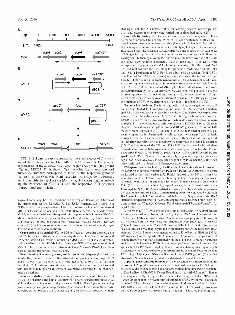
The growth characteristics and metabolite production ofstrain COLn and its �ccpA mutant MST23 were monitored inHEPES-buffered LB supplemented with 10 mM glucose. Com-parison of the wild-type and mutant cultures revealed a cleardifference only for the mid- to late-exponential-growth phases(i.e., hours 3 to 6). During these growth periods, the mutantdisplayed slower growth, yielding cell densities lagging approx-imately 1 h behind the wild-type culture densities. However,the mutant culture reached almost the same A600 values after10 h of growth (Fig. 2A), and no differences in A600 values wereobservable between the wild type and the mutant after 16 and24 h of growth (data not shown). CFU determinations of thegrowing cultures showed the same tendency, being differentonly during the mid- to late-exponential-growth phase, andagain, no differences in growth yield were observed after anincubation period of 24 h (data not shown). During the expo-nential-growth phase, differences in growth rate were also vis-ible with respect to doubling times; the wild type yielded asignificantly lower doubling time (44.34 0.65 min) thanMST23 (51.67 0.75 min; P � 0.01). A clearer difference wasobserved between the wild type and the mutant on solid media.MST23 produced significantly smaller colonies on sheep bloodagar or on Muller-Hinton plates, with lower CFU numbers percolony than COLn after 48 h of incubation (Fig. 2B). No differ-ences were observed when COLn and MST23 were checked forcell size and adherence properties (Fig. 2D), signaling that thedifferences in growth on solid media were likely to be due to areduced growth rate of the mutant.
In addition to their growth kinetics, growing COLn andMST23 cultures were further analyzed for glucose metabolismand breakdown. The glucose level of the wild-type culturevisibly decreased from hour 3 on and glucose was depleted
after hour 7. The glucose level of the MST23 culture started todecrease from hour 5 and was depleted only after 9 h of growth(Fig. 2C). Simultaneously with glucose degradation, the lactatelevel of the wild-type culture started to increase from hour 3 on,reached its maximum around hour 6, and significantly decreasedthereafter. In contrast, the lactate level of the mutant culturestarted to increase only after 7 h of growth, representing a delayof more than 2 h compared with glucose consumption, andreached its maximum after 10 h. Interestingly, only slight differ-ences were observable between the wild type and the mutant withrespect to acetate accumulation; the acetate level seen with themutant culture preceded that of the wild-type culture by approx-imately 0.5 h. After 24 h of growth, no lactate was present ineither wild-type or mutant media anymore, while acetate levelswere reduced to similar amounts, namely, 3.8 0.86 mM forCOLn and 3.75 0.76 mM for MST23, signaling that the ccpAmutation did not affect the ability of the mutant to assimilateacetate and lactate that was excreted into the media during earliergrowth stages.
Although the growth rate and yield of the ccpA mutantMST23 were only slightly affected by the deletion, the slowerglucose consumption and delayed lactate secretion of MST23,paired with the slightly increased acetate production, sug-gested that CcpA seemed to exert a positive effect on lactateproduction and secretion whereas acetate production and se-cretion seemed to be negatively affected by this protein, takinginto account that MST23 possessed lower cell densities andconsumed glucose slower than the wild type at almost all timepoints analyzed. Preliminary Northern analyses supported thehypothesis that CcpA might stimulate lactate and suppressacetate formation in the presence of glucose. Expression ofldh1 (saCOL0222), thought to encode L-lactate dehydrogenase1 (EC 1.1.1.27) that is believed to catalyze the conversion frompyruvate to lactate when S. aureus is grown in the presence ofa rapidly catabolizable carbon source under anaerobic condi-tions, was highly induced in wild-type cells grown in the pres-ence of glucose but was not detectable in the absence of glu-cose or in the �ccpA mutant under either growth condition.Expression of genes encoding enzymes involved in acetateformation, such as pdhABCD, encoding components of thepyruvate dehydrogenase multienzyme complex thought to cat-alyze the conversion of pyruvate to acetyl coenzyme A, and theadhE homologue (saCOL0135) thought to encode alcohol-acetaldehyde dehydrogenase (EC 1.2.1.10) that catalyzes theformation of acetyl-coenzyme A to acetaldehyde, on the otherhand, was found to be repressed in the wild type in the pres-ence of glucose, while no differences in expression were ob-served in the �ccpA mutant in either the presence or absenceof glucose (K. Seidl, unpublished data).
Effect of ccpA on antibiotic resistance. CcpA was among aseries of auxiliary factors reported to reduce methicillinresistance in strain COL upon Tn551 inactivation (11). We
FIG. 2. Growth characteristics of COLn (squares) and its �ccpA mutant MST23 (triangles) in LB supplemented with 10 mM glucose.(A) Absorbances at 600 nm (A600) and CFU over the growth cycle. (B) Cfu per colony of COLn and MST23 grown on sheep blood agar for 48 hat 35°C. (C) Glucose, acetate, and lactate concentrations in the culture supernatants corresponding to panel A. The data presented are mean valuesof three independent experiments. (D) Scanning electron microscopy images of COLn and MST23 adhering to polyethylene terephthalate(Thermanox) disks.
VOL. 50, 2006 STAPHYLOCOCCUS AUREUS CcpA 1187
on May 3, 2014 by guest
http://aac.asm.org/
Dow
nloaded from

demonstrated here a fourfold reduction of the oxacillin MIC forstrain COLn upon ccpA inactivation and full restoration to itsoriginal level by trans-complementation with the wild-typeccpA allele under the control of its native promoter (Fig. 3A),thus confirming experimentally that the effect was indeedsolely CcpA dependent. Moreover, the population analysisprofile (Fig. 3B) showed that the homogenous oxacillin resis-
tance of strain COLn was reduced to heterogeneous resistanceby ccpA inactivation.
Transduction of ccpA::tet(L) into NM143, the step-selectedteicoplanin-resistant derivative of strain Newman, yielding strainKS30, had a similar negative effect on teicoplanin resistance. TheccpA inactivation reduced the MIC of teicoplanin from 24 to 12�g ml�1, and the population analysis profile teicoplanin showedthat the number of more highly resistant variants was reduced bya magnitude of over 103 (Fig. 3C).
Effect of glucose and ccpA on virulence determinant production.Previous studies showed that fermentation of glucose and/orthe accompanying decrease in pH affected expression of theglobal regulator agr and of virulence factors such as �-hemo-lysin (hla) and the staphylococcal enterotoxins A, B, and C(sea, seb, and sec) (12, 24, 27, 44, 45). More recently, Weinrickand coworkers (62) showed that mildly acidic conditions (pH5.5) influenced the expression of a variety of genes, includingagr, hla, and spa, encoding protein A, and concluded thatchanges in staphylococcal gene expression formerly thought torepresent a glucose effect might be largely the result of declin-ing pH of the growth medium due to the fermentation of thesupplemented carbon source. The effect of glucose on hla andspa expression is further complicated by the fact that bothgenes are affected by a complex regulatory network includingagr and further regulatory elements such as ArlRS (15), MgrA(25), MsrR (46), Rot (37, 47), SaeRS (18, 20), SarA (reference8 and references therein), SarS (7, 36, 53), SarT (48), SvrA(16), TcaR (36), and the alternative transcription factor �B (2,20, 23), with rot, sae and tcaR expression being pH dependentas well (62).
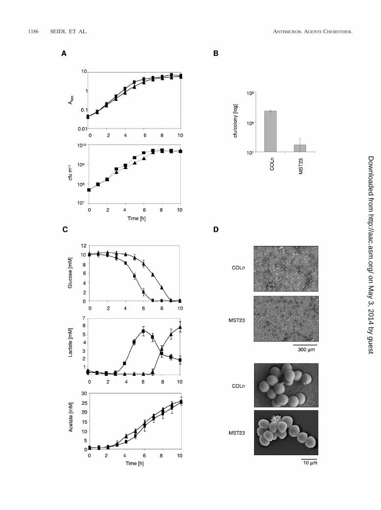
To elucidate whether agr, hla, and spa transcription wasaffected by glucose independently from the fermentation-de-pendent pH and to find out whether CcpA may be involved inmediating such a glucose effect, we monitored the expressionlevels of these genes in COLn and its ccpA mutant MST23,grown in buffered LB in the presence or absence of glucose(Fig. 4). pckA, encoding a phosphoenolpyruvate carboxyki-nase, shown to be affected by glucose, and predicted to beregulated by CcpA in S. aureus (49), was included in this studyas well. The 50 mM HEPES concentration used here to bufferthe medium to pH 7.5 had no inhibiting effect on the growthkinetics and kept the pH fairly constant (Fig. 4A), while con-centrations higher than 50 mM were growth inhibitory (datanot shown). No changes in pH were observed when COLn andMST23 were grown in LB in the absence of glucose. In theglucose-supplemented wild-type culture, the pH started todrop slightly after 5 h to a final pH of 7, while the pH of theglucose-supplemented MST23 culture dropped from hour 7 onto pH 7.25. Growth of COLn in unbuffered glucose-supple-mented LB medium would have caused a drop in pH to 5.5already after 3 h of growth (data not shown). Addition ofglucose produced a higher growth rate and growth yield in thewild type from hour 5 on (Fig. 4A). Surprisingly, glucoseseemed to have a slightly inhibiting effect on the growth rate ofthe mutant MST23 during exponential-growth phase, as seenin the lower A600 values for MST23 grown in LB supplementedwith glucose compared to those in LB alone; this effect mightbe due to a slightly increased lag phase.
Expression of ccpA, pckA, RNAIII, spa, and hla was moni-tored after 1, 3, 5, and 8 h of growth. In COLn grown in LB,
FIG. 3. Susceptibility of COLn and NM143 and their isogenic�ccpA mutants. (A) Effects of ccpA inactivation and complementationon oxacillin resistance. The methicillin-resistant strain COLn, its�ccpA mutant MST23, control strains MST31 (COLn pAW17) andMST35 [COLn �ccpA::tet(L) complemented with control plasmidpAW17], and strain MST36 [COLn �ccpA::tet(L) complemented withplasmid pMST1] were swabbed along a plate containing an oxacillingradient. The corresponding MICs of oxacillin (in micrograms permilliliter as determined by E-Test and broth microdilution) for thestrains are indicated. (B) Population analysis profiles of COLn(squares) and MST23 [COLn �ccpA::tet(L); triangles] on oxacillin.(C) Population analysis profiles of the step-selected glycopeptide-intermediate-resistant S. aureus derivative NM143 (squares) and its�ccpA mutant KS30 (triangles) on teicoplanin.
1188 SEIDL ET AL. ANTIMICROB. AGENTS CHEMOTHER.
on May 3, 2014 by guest
http://aac.asm.org/
Dow
nloaded from
expression of ccpA was found to be fairly constant at all growthstages analyzed (Fig. 4B). Supplementation of glucose seemedto increase the expression of this gene during the early growthstages (i.e., 1 to 3 h), suggesting that expression of ccpA might
be positively affected by glucose, a phenomenon that has beenobserved for ccpA in other gram-positive organisms (14). NoccpA transcripts were detectable in MST23, confirming thatthe deletion had occurred as intended. Expression of pckA
FIG. 4. Northern blot analyses of COLn and its �ccpA mutant MST23 during growth. (A) Growth characteristics of COLn (squares) and MST23(triangles) grown in HEPES-buffered LB (open symbols) and HEPES-buffered LB supplemented with 10 mM glucose (closed symbols). At 1-h intervals,an aliquot (2 ml) was removed, the absorbance at 600 nm was measured, and the pH in the culture supernatants (circles) was determined. The resultspresented are mean values of at least three independent experiments. Time points of sampling for the Northern blot analyses are indicated by arrows.(B) Transcription of ccpA, hla, pckA, RNAIII, and spa in COLn and MST23 during growth in HEPES-buffered LB (�Glc) and in HEPES-buffered LBsupplemented with 10 mM glucose (�Glc). Relevant transcript sizes and time points of sampling are indicated. Ethidium bromide-stained 16S rRNApatterns are shown as an indication of RNA loading.
VOL. 50, 2006 STAPHYLOCOCCUS AUREUS CcpA 1189
on May 3, 2014 by guest
http://aac.asm.org/
Dow
nloaded from
increased with growth and was clearly higher when COLn cellswere grown in the absence of glucose, confirming previousfindings indicating that pckA expression is negatively affectedby glucose (49). MST23, on the other hand, already producedpckA transcripts at a rather constant and high level early ingrowth. No significant differences in pckA expression wereobserved in the presence or absence of glucose, signaling thatthe effect of glucose on pckA expression was mediated viaCcpA, as has been suggested by Scovill et al. (49). The idea ofCcpA-dependent repression of pckA transcription is furthersupported by the presence of a putative CRE in the promoterregion of pckA (49) that almost perfectly matched (17/18 nt)the CRE consensus of B. subtilis (38), suggesting that the CREsequences might be similar in the two organisms.
Expression of RNAIII from the agr locus is known to in-crease during later growth stages (reference 63 and referenceswithin). Accordingly, in COLn cells grown in LB, RNAIII tran-scripts were first detected after 5 h of growth and increasedwith time. Unexpectedly, in glucose-grown cells, a strong in-crease in RNAIII expression occurred at hour 8, contrary to thefindings of Regassa et al. (45), who reported unchangedRNAIII transcript levels for S. aureus cells grown in a fermen-tor in either the presence or absence of glucose at a constantpH. However, the discrepancy in RNAIII expression might beexplained by the differing growth conditions, since Regassa andcoworkers kept the glucose concentration at a constant level of100 mM. It is noteworthy that the increase in RNAIII expres-sion was detected at a time point when all glucose was ex-hausted from the medium, indicating that the depletion ofglucose might have triggered a signal that induced RNAIIItranscription, which was not present under the conditions usedby Regassa et al. (45). The slight decrease in pH observed inthe later growth stages of the wild-type culture in response toglucose (Fig. 4A) could be excluded as a reason for the stronginduction of RNAIII transcription, since control experimentsperformed using a fermenter and an equivalent experimentalsetup allowed the pH to be kept constant and yielded the sameinduction pattern (data not shown).
No RNAIII transcripts were detectable in MST23 cellsgrown in LB, and addition of glucose yielded only traces ofRNAIII transcripts after 8 h of growth. The clear differences inRNAIII transcript levels observed for COLn and MST23 sug-gest that the presence of a functional CcpA had a positiveeffect on RNAIII expression. However, this effect was likely tobe indirect, since the screening of the agr locus did not revealany apparent CRE in this genomic region that would fit withthe CRE consensus of B. subtilis (38). Expression of spa wasnoticeably affected by glucose in the wild type. While spa tran-scripts were clearly detectable in COLn during later growthstages (hours 5 to 8) in the absence of glucose, spa transcriptswere hardly detectable in the presence of glucose (Fig. 4B).However, in MST23, spa transcripts were detectable over thewhole growth cycle independently of the presence or absenceof glucose, although the spa transcription profiles seemed todiffer between these two conditions to a certain degree. Whilespa expression in MST23 grown in LB peaked around hour 5,analogous to the situation found for the wild type, in theglucose-supplemented LB, spa transcription seemed to behighest at the latest time point monitored (i.e., hour 8). Inter-estingly, significant amounts of spa transcripts were already
detectable in MST23 during the early growth stages (hours 1 to3), irrespective of whether glucose was present in the growthmedia or not, signaling that CcpA might act as a negativeregulator for spa expression during these growth stages. Ex-pression of hla was found to be less affected by glucose and/orCcpA compared with RNAIII and spa expression results. InCOLn grown in unsupplemented LB, hla transcripts were de-tectable from hour 1 on and peaked around the transition fromlate logarithmic-growth phase to stationary phase. Supplemen-tation of glucose shifted the peak expression of hla to the lastgrowth point monitored and seemed to increase the expressionlevel at this growth stage, probably due to the action of RNAIII,which was, as shown above, found to be highly expressed underthese conditions. Essentially the same hla expression patternsas those identified in the wild type were found for MST23,although the overall amounts of hla transcripts seemed to beslightly reduced in the ccpA mutant.
Since hla and spa expression are known to be under multiplelevels of control, including that by RNAIII, it was difficult tojudge from the results described above whether the effectsobserved for hla and spa were the result of direct CcpA-me-diated regulation in response to glucose or whether they mightrepresent a secondary effect of RNAIII and other regulatoryelements that were not part of this study. To better define theimpact of glucose and, in particular, of CcpA on the expressionof these two virulence factors, we performed a second series ofNorthern blot experiments, this time monitoring the expres-sion of ccpA, hla, pckA, and spa in cells that were grown tomidexponential-growth phase (A600 1). At this time point,glucose was added to one half of the culture, and cells wereharvested in 10-min intervals from 0 to 30 min (Fig. 5). Thisprocedure was likely to abolish the effect of agr on hla and spaexpression, since RNAIII expression was barely detectable atthis time point and under these conditions (data not shown).Moreover, by analyzing the gene expression immediately afterthe addition of glucose, we assumed that we would be morelikely able to identify direct CcpA-dependent effects, sincesecondary effects were expected to occur with a certain delay.Addition of glucose to the exponentially growing cultures didnot result in a temporary growth arrest of these cultures, indi-cating that the glucose addition did not trigger any inhibition ofthe primary metabolism (data not shown).
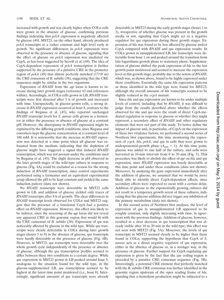
In this second series of Northern blot analyses, the level ofexpression of spa in unsupplemented LB appeared to beroughly constant, only slightly increasing with time, in agree-ment with the previous findings. Addition of glucose, however,resulted in a clear decrease in spa transcription that was al-ready visible after 10 to 20 min in the wild type; this effect wasnot seen with MST23 (Fig. 5A). Moreover, the levels of spatranscripts in MST23 seemed clearly to be higher than thosefound in COLn, supporting the hypothesis that CcpA of S.aureus acts as a direct negative regulator of spa expression,either in the absence of glucose or, in a stronger way, in thepresence of glucose. Further support for CcpA regulating spaexpression is given by the fact that the spa coding region ispreceded by a putative CRE consensus sequence (Fig. 5B).Interestingly, a potential CRE element that perfectly matchedwith the B. subtilis CRE consensus was further identified in thegenomic region upstream of the open reading frame of hla,signaling that �-hemolysin production might be subjected to a
1190 SEIDL ET AL. ANTIMICROB. AGENTS CHEMOTHER.
on May 3, 2014 by guest
http://aac.asm.org/
Dow
nloaded from
CcpA-dependent CCR as well. In line with this assumption, weidentified a slight decrease in hla transcription in COLn afterthe addition of glucose that was not detected either in the wildtype grown in unsupplemented LB or in the ccpA mutantunder both sets of conditions (Fig. 5A).
In agreement with the results of the first series of Northernblot analyses, we found pckA expression to decrease in the wildtype in response to the presence of glucose and to be constantin the absence of this sugar. Equivalent to the situation foundwith spa, expression of pckA appeared to be clearly increasedin MST23, and addition of glucose again failed to exert aneffect on pckA expression in the mutant, confirming the find-
ings of Scovill et al. (49) showing that the presence of glucoserepresses pckA transcription and supporting the hypothesisraised by these authors that this negative regulatory effect isexerted via CcpA. Interestingly, expression of ccpA itself seemsto be positively affected by glucose, since transcription of ccpAappeared to be increased in response to the addition of glu-cose, although this increase was only of a transient nature andwas only detectable at 10 min after the sugar was added (Fig.5A). A potential CRE was identifiable in the promoter regionof ccpA (Fig. 5B), sharing 15 out of 18 nucleotides with theCRE consensus of B. subtilis but lacking the palindromic na-ture of CREs that was detectable in the CRE candidates of hla,
FIG. 5. Effect of glucose addition on the gene expression of COLn and its �ccpA mutant MST23. (A) Northern blot analyses of ccpA, hla, pckA,and spa. Cells were grown in HEPES-buffered LB to midexponential-growth phase (A600 1), cultures were split in half, and 10 mM glucose wasadded to one half (�Glc) while the other half was left unchanged (�Glc). Relevant transcript sizes and time points of sampling are indicated.Ethidium bromide-stained 16S rRNA patterns are shown as an indication of RNA loading. (B) Putative CREs identified upstream of ccpA, hla,pckA, and spa. Nucleotides fitting with the CRE consensus of B. subtilis (38) are highlighted in bold type. Inverted repeats are indicated by arrows.Ambiguity codes are as follows: W denotes A or T, R denotes G or A, and N denotes A, C, G, or T.
VOL. 50, 2006 STAPHYLOCOCCUS AUREUS CcpA 1191
on May 3, 2014 by guest
http://aac.asm.org/
Dow
nloaded from

pckA, and spa and leaving the question currently open ofwhether the observed increase in transcription was mediatedvia CcpA by itself or not.
Effect of ccpA and glucose on capsular polysaccharide pro-duction. The majority of clinical isolates produce capsularpolysaccharides of serotype 5 (CP5) or serotype 8 (CP8), whichprotect S. aureus against opsonophagocytic killing by polymor-phonuclear leukocytes (28, 29, 34, 54, 59) and have been shownin a number of animal models of infection to enhance itsvirulence (40, 43, 54, 56, 61). Expression of CPs is influencedby various environmental signals in vitro and in vivo (reviewedin references 41 and 59), and transcription of the cap operonwas shown to be affected by regulatory elements such as agr,mgr, sae, sarA, and the alternative �-factor �B (2, 10, 33, 34, 35,42, 51, 57). Both biosynthetic pathways for CP5 and CP8 pro-duction utilize precursors of the cell wall, such as UDP-N-acetylglucosamine, suggesting that CP-producing proteinsmight compete with cell-wall-producing enzymes for the avail-ability of UDP-N-acetylglucosamine, thereby affecting the car-bon flux of S. aureus. We therefore analyzed whether glucose,and specifically ccpA, might affect CP synthesis, as has beenshown recently for the low G�C-content gram-positive patho-gen Clostridium perfringens (58).
Since the CP serotype 5 strain COLn was found to produceonly a little CP under the conditions tested (C. Wolz, unpub-lished data), the CP5 prototypic strain Newman and its �ccpAderivative MST14 were used to investigate the effect of glucoseand CcpA on CP formation (Fig. 6). Newman wild-type andMST14 cells were grown for 24 h in LB medium in the pres-ence or absence of 10 mM glucose, and the CP5 productionwas determined by indirect immunofluorescence using mono-clonal antibodies raised against CP5 (Fig. 6A). In the absenceof glucose, most of the wild-type cells produced CP5. However,in the presence of glucose, CP5 was abolished, indicating thatglucose repressed CP formation. Interestingly, CP5 productionin the ccpA mutant MST14 was not affected and was presentirrespective of the presence or absence of glucose in the growthmedium. The immunofluorescence data were confirmed byreal-time PCR (Fig. 6B). The expression of the cap operon wasalmost indistinguishable between the wild type and mutant inthe absence of glucose, whereas in the presence of glucose,strain Newman produced significantly fewer cap transcripts(P � 0.05) than MST14, which expressed cap in roughly the sameamounts as in the absence of glucose. Both findings stronglysuggested that the presence of glucose repressed CP formationand that this effect was, at least in part, mediated via CcpA onthe transcriptional level, adding a further regulator to the com-plex network of regulatory elements and environmental con-ditions that control cap operon expression. However, since noapparent CRE was identifiable within the genomic region en-coding the cap operon, it is again likely that the CcpA effect oncap transcription was of an indirect nature and might be me-diated by downstream regulators.
FIG. 6. Capsule production and cap expression of Newman and its�ccpA mutant MST14 in response to glucose. (A) CP5 expressiondetermined by indirect immunofluorescence of strain Newman and itsisogenic �ccpA mutant grown for 24 h at 37°C in HEPES-buffered LB(�Glc), or in HEPES-buffered LB supplemented with 10 mM glucose(�Glc). Bacteria were stained with 4�,6�-diamidino-2-phenylindole(DAPI), and marked with CP5-specific monoclonal antibodies andstained with Cy3-conjugated anti-mouse antibodies (CY3). (B) Quan-titative transcript analysis of capA by LightCycler RT-PCR of strainNewman and its isogenic �ccpA mutant grown for 8 h at 37°C inHEPES-buffered LB (�Glc) or in HEPES-buffered LB supplementedwith 10 mM glucose (�Glc). Transcripts were quantified in referenceto the transcription of gyrase (in copies per copy of gyrB). Values fromtwo separate RNA isolations and two independent RT-PCRs each
were used to calculate the mean expression (standard errors of themean). Glc, glucose; asterisk, P � 0.05 for Newman without Glc,MST14 without Glc, and MST14 with Glc.
1192 SEIDL ET AL. ANTIMICROB. AGENTS CHEMOTHER.
on May 3, 2014 by guest
http://aac.asm.org/
Dow
nloaded from

Concluding remarks. Deletion of ccpA had a clear impact onthe expression of RNAIII and on virulence factors of S. aureus,some of which have previously been shown to be affected byglucose. Interestingly, the deletion of ccpA produced an effecton gene expression not only in the presence but also in theabsence of glucose, indicating that the function of CcpA mightnot be restricted to CCR. Our findings that CcpA of S. aureusinfluenced the transcription of at least five genes and operons,with most of them being involved in virulence of this pathogen,suggests that CcpA might represent an important global reg-ulator of gene expression in S. aureus that, like that of itshomologue in B. subtilis, may not be limited to regulatingcarbon uptake and metabolization. A preliminary computa-tional screening of the S. aureus COL genome with the CREconsensus of B. subtilis (38) indeed indicated more than 110CREs to be present in the promoter or N-terminal codingregions of genes and operons encoded by S. aureus, if allowingone mismatch to occur. Whole genome and proteomic analysesare currently ongoing to identify the CcpA regulon in S. aureus.
ACKNOWLEDGMENTS
The study was supported by the Novartis Foundation, the SwissNational Science Foundation (grants 3100A0-100234 and 31-105390),the Deutsche Forschungsgemeinschaft (Wo 578/5-1), and the Euro-pean Community (grant EU-LSH-CT2003-50335) (BBW 03.0098).
We thank Jean-Michel Fournier for kindly donating the monoclonalanticapsular antibody and the AO Research Institute for providing uswith the necessary materials for the scanning electron microscopyexperiments.
REFERENCES
1. Ausubel, F. M., R. Brent, R. E. Kingston, D. D. Moore, J. G. Seidman, J. A.Smith, and K. Struhl. 1987. Current protocols in molecular biology. JohnWiley and Sons, Inc., New York, N.Y.
2. Bischoff, M., P. Dunman, J. Kormanec, D. Macapagal, E. Murphy, W.Mounts, B. Berger-Bachi, and S. Projan. 2004. Microarray-based analysis ofthe Staphylococcus aureus �B regulon. J. Bacteriol. 186:4085–4099.
3. Blencke, H. M., G. Homuth, H. Ludwig, U. Mader, M. Hecker, and J. Stulke.2003. Transcriptional profiling of gene expression in response to glucose inBacillus subtilis: regulation of the central metabolic pathways. Metab. Eng.5:133–149.
4. Bruckner, R. 1997. Gene replacement in Staphylococcus carnosus and Staph-ylococcus xylosus. FEMS Microbiol. Lett. 151:1–8.
5. Chan, P. F., and S. J. Foster. 1998. Role of SarA in virulence determinantproduction and environmental signal transduction in Staphylococcus aureus.J. Bacteriol. 180:6232–6241.
6. Cheung, A. L., K. J. Eberhardt, and V. A. Fischetti. 1994. A method to isolateRNA from gram-positive bacteria and mycobacteria. Anal. Biochem. 222:511–514.
7. Cheung, A. L., K. Schmidt, B. Bateman, and A. C. Manna. 2001. SarS, a SarAhomolog repressible by agr, is an activator of protein A synthesis in Staph-ylococcus aureus. Infect. Immun. 69:2448–2455.
8. Cheung, A. L., and A. C. Manna. 2005. Role of the distal sarA promoters inSarA expression in Staphylococcus aureus. Infect. Immun. 73:4391–4394.
9. CLSI. 2003. Methods for dilution antimicrobial susceptibility tests for bac-teria that grow aerobically. Approved standard—sixth edition, CLSI docu-ment M7-A6. CLSI, Wayne, Pa.
10. Dassy, B., T. Hogan, T. J. Foster, and J. M. Fournier. 1993. Involvement ofthe accessory gene regulator (agr) in expression of type-5 capsular polysac-charide by Staphylococcus aureus. J. Gen. Microbiol. 139:1301–1306.
11. De Lencastre, H., S. W. Wu, M. G. Pinho, A. M Ludovice, S. Filipe, S.Gardete, R. Sobral, S. Gill, M. Chung, and A. Tomasz. 1999. Antibioticresistance as a stress response: complete sequencing of a large number ofchromosomal loci in Staphylococcus aureus strain COL that impact on theexpression of resistance to methicillin. Microb. Drug Resist. 5:163–175.
12. Duncan, J. L., and G. J. Cho. 1972. Production of staphylococcal alpha toxinII. Glucose repression of toxin formation. Infect. Immun. 6:689–694.
13. Duthie, E. S., and L. L. Lorenz. 1952. Staphylococcal coagulase: mode ofaction and antigenicity. J. Gen. Microbiol. 6:95–107.
14. Egeter, O., and R. Bruckner. 1996. Catabolite repression mediated by thecatabolite control protein CcpA in Staphylococcus xylosus. Mol. Microbiol.21:739–749.
15. Fournier, B., A. Klier, and G. Rapoport. 2001. The two-component systemArlS-ArlR is a regulator of virulence gene expression in Staphylococcusaureus. Mol. Microbiol. 41:247–261.
16. Garvis, S., J. M. Mei, J. Ruiz-Albert, and D. W. Holden. 2002. Staphylococcusaureus svrA: a gene required for virulence and expression of the agr locus.Microbiology 148:3235–3243.
17. Giachino, P., S. Engelmann, and M. Bischoff. 2001. �B activity depends onRsbU in Staphylococcus aureus. J. Bacteriol. 183:1843–1852.
18. Giraudo, A. T., A. L. Cheung, and R. Nagel. 1997. The sae locus of Staphy-lococcus aureus controls exoprotein synthesis at the transcriptional level.Arch. Microbiol. 168:53–58.
19. Goerke, C., S. Campana, M. G. Bayer, G. Doring, K. Botzenhart, and C.Wolz. 2000. Direct quantitative transcript analysis of the agr regulon ofStaphylococcus aureus during human infection in comparison to the expres-sion profile in vitro. Infect. Immun. 68:1304–1311.
20. Goerke, C., U. Fluckiger, A. Steinhuber, V. Bisanzio, M. Ulrich, M. Bischoff,J. M. Patti, and C. Wolz. 2005. Role of Staphylococcus aureus global regu-lators sae and SigmaB in virulence gene expression during device-relatedinfection. Infect. Immun. 73:3415–3421.
21. Harris, L. G., S. Tosatti, M. Wieland, M. Textor, and R. G. Richards. 2004.Staphylococcus aureus adhesion to titanium oxide surfaces coated with non-functionalized and peptide-functionalized poly(L-lysine)-grafted-poly(ethyl-ene glycol) copolymers. Biomaterials 25:4135–4148.
22. Hoeger, P. H., W. Lenz, A. Boutonnier, and J. M. Fournier. 1992. Staphylo-coccal skin colonization in children with atopic dermatitis: prevalence, per-sistence, and transmission of toxigenic and nontoxigenic strains. J. Infect.Dis. 165:1064–1068.
23. Horsburgh, M. J., J. L. Aish, I. J. White, L. Shaw, J. K. Lithgow, and S. J.Foster. 2002. SigmaB modulates virulence determinant expression and stressresistance: characterization of a functional rsbU strain derived from Staph-ylococcus aureus 8325-4. J. Bacteriol. 184:5457–5467.
24. Iandolo, J. J., and W. M. Shafer. 1977. Regulation of staphylococcal entero-toxin B. Infect. Immun. 16:610–616.
25. Ingavale, S., W. van Wamel, T. T. Luong, C. Y. Lee, and A. L. Cheung. 2005.Rat/MgrA, a regulator of autolysis, is a regulator of virulence genes inStaphylococcus aureus. Infect. Immun. 73:1423–1431.
26. Jankovic, I., and R. Bruckner. 2002. Carbon catabolite repression by thecatabolite control protein CcpA in Staphylococcus xylosus. J. Mol. Microbiol.Biotechnol. 4:309–314.
27. Jarvis, A. W., R. C. Lawrence, and G. G. Pritchard. 1975. Glucose repressionof enterotoxins A, B and C and other extracellular proteins in staphylococciin batch and continuous culture. J. Gen. Microbiol. 86:75–87.
28. Kampen, A. H., T. Tollersrud, and A. Lund. 2005. Staphylococcus aureuscapsular polysaccharide types 5 and 8 reduce killing by bovine neutrophils invitro. Infect. Immun. 73:1578–1583.
29. Karakawa, W. W., A. Sutton, R. Schneerson, A. Karpas, and W. F. Vann.1988. Capsular antibodies induce type-specific phagocytosis of capsulatedStaphylococcus aureus by human polymorphonuclear leukocytes. Infect. Im-mun. 56:1090–1095.
30. Katayama, Y., H. Z. Zhang, and H. F. Chambers. 2004. PBP 2a mutationsproducing very-high-level resistance to beta-lactams. Antimicrob. AgentsChemother. 48:453–459.
31. Kim, J. H., Y. K. Yang, and G. H. Chambliss. 2005. Evidence that Bacilluscatabolite control protein CcpA interacts with RNA polymerase to inhibittranscription. Mol. Microbiol. 56:155–162.
32. Kreiswirth, B. N., S. Lofdahl, M. J. Betley, M. O’Reilly, P. M. Schlievert,M. S. Bergdoll, and R. P. Novick. 1983. The toxic shock syndrome exotoxinstructural gene is not detectably transmitted by a prophage. Nature 305:709–712.
33. Luong, T., S. Sau, M. Gomez, J. C. Lee, and C. Y. Lee. 2002. Regulation ofStaphylococcus aureus capsular polysaccharide expression by agr and sarA.Infect. Immun. 70:444–450.
34. Luong, T. T., and C. Y. Lee. 2002. Overproduction of type 8 capsular poly-saccharide augments Staphylococcus aureus virulence. Infect. Immun. 70:3389–3395.
35. Luong, T. T., S. W. Newell, and C. Y. Lee. 2003. mgr, a novel global regulatorin Staphylococcus aureus. J. Bacteriol. 185:3703–3710.
36. McCallum, N., M. Bischoff, H. Maki, A. Wada, and B. Berger-Bachi. 2004.TcaR, a putative MarR-like regulator of sarS expression. J. Bacteriol. 186:2966–2972.
37. McNamara, P. J., K. C. Milligan-Monroe, S. Khalili, and R. A. Proctor.2000. Identification, cloning, and initial characterization of rot, a locus en-coding a regulator of virulence factor expression in Staphylococcus aureus. J.Bacteriol. 182:3197–3203.
38. Miwa, Y., A. Nakata, A. Ogiwara, M. Yamamoto, and Y. Fujita. 2000. Eval-uation and characterization of catabolite-responsive elements (cre) of Bacil-lus subtilis. Nucleic Acids Res. 28:1206–1210.
39. Moreno, M. S., B. L. Schneider, R. R. Maile, W. Weyler, and M. H. Saier, Jr.2001. Catabolite repression mediated by the CcpA protein in Bacillus subtilis:novel modes of regulation revealed by whole-genome analyses. Mol. Micro-biol. 39:1366–1381.
VOL. 50, 2006 STAPHYLOCOCCUS AUREUS CcpA 1193
on May 3, 2014 by guest
http://aac.asm.org/
Dow
nloaded from
40. Nilsson, I.-M., J. C. Lee, T. Bremell, C. Ryden, and A. Tarkowski. 1997. Therole of staphylococcal polysaccharide microcapsule expression in septicemiaand septic arthritis. Infect. Immun. 65:4216–4221.
41. O’Riordan, K., and J. C. Lee. 2004. Staphylococcus aureus capsular polysac-charides. Clin. Microbiol. Rev. 17:218–234.
42. Pohlmann-Dietze, P., M. Ulrich, K. B. Kiser, G. Doring, J. C. Lee, J. M.Fournier, K. Botzenhart, and C. Wolz. 2000. Adherence of Staphylococcusaureus to endothelial cells: influence of the capsular polysaccharide, the globalregulator agr, and the bacterial growth phase. Infect. Immun. 68:4865–4871.
43. Portoles, M., K. B. Kiser, N. Bhasin, K. H. N. Chan, and J. C. Lee. 2001.Staphylococcus aureus Cap5O has UDP-ManNAc dehydrogenase activityand is essential for capsule expression. Infect. Immun. 69:917–923.
44. Regassa, L. B., J. L. Couch, and M. J. Betley. 1991. Steady-state staphylo-coccal enterotoxin type C mRNA is affected by a product of the accessorygene regulator (agr) and by glucose. Infect. Immun. 59:955–962.
45. Regassa, L. B., R. P. Novick, and M. J. Betley. 1992. Glucose and nonmain-tained pH decrease expression of the accessory gene regulator (agr) inStaphylococcus aureus. Infect. Immun. 60:3381–3388.
46. Rossi, J., M. Bischoff, A. Wada, and B. Berger-Bachi. 2003. MsrR, a putativecell envelope-associated element involved in Staphylococcus aureus sarAattenuation. Antimicrob. Agents Chemother. 47:2558–2564.
47. Said-Salim, B., P. M. Dunman, F. M. McAleese, D. Macapagal, E. Murphy, P. J.McNamara, S. Arvidson, T. J. Foster, S. J. Projan, and B. N. Kreiswirth. 2003.Global regulation of Staphylococcus aureus genes by Rot. J. Bacteriol. 185:610–619.
48. Schmidt, K. A., A. C. Manna, S. Gill, and A. L. Cheung. 2001. SarT, arepressor of alpha-hemolysin in Staphylococcus aureus. Infect. Immun. 69:4749–4758.
49. Scovill, W. H., H. J. Schreier, and K. W. Bayles. 1996. Identification andcharacterization of the pckA gene from Staphylococcus aureus. J. Bacteriol.178:3362–3364.
50. Senn, M. M., M. Bischoff, C. von Eiff, and B. Berger-Bachi. 2005. �B activityin a Staphylococcus aureus strain Newman hemB mutant. J. Bacteriol. 187:7397–7406.
51. Steinhuber, A., C. Goerke, M. G. Bayer, G. Doring, and C. Wolz. 2003.Molecular architecture of the regulatory locus sae of Staphylococcus aureusand its impact on expression of virulence factors. J. Bacteriol. 185:6278–6286.
52. Stulke, J., and W. Hillen. 2000. Regulation of carbon catabolism in Bacillusspecies. Annu. Rev. Microbiol. 54:849–880.
53. Tegmark, K., A. Karlsson, and S. Arvidson. 2000. Identification and char-acterization of SarH1, a new global regulator of virulence gene expression inStaphylococcus aureus. Mol. Microbiol. 37:398–409.
54. Thakker, M., J.-S. Park, V. Carey, and J. C. Lee. 1998. Staphylococcus aureusserotype 5 capsular polysaccharide is antiphagocytic and enhances bacterialvirulence in a murine bacteremia model. Infect. Immun. 66:5183–5189.
55. Titgemeyer, F., and W. Hillen. 2002. Global control of sugar metabolism: agram-positive solution. Antonie Leeuwenhoek 82:59–71.
56. Tzianabos, A. O., J. Y. Wang, and J. C. Lee. 2001. Structural rationale for themodulation of abscess formation by Staphylococcus aureus capsular polysac-charides. Proc. Natl. Acad. Sci. USA 98:9365–9370.
57. van Wamel, W., Y. Q. Xiong, A. S. Bayer, M. R. Yeaman, C. C. Nast, andA. L. Cheung. 2002. Regulation of Staphylococcus aureus type 5 capsularpolysaccharides by agr and sarA in vitro and in an experimental endocarditismodel. Microb. Pathog. 33:73–79.
58. Varga, J., V. L. Stirewalt, and S. B. Melville. 2004. The CcpA protein isnecessary for efficient sporulation and enterotoxin gene (cpe) regulation inClostridium perfringens. J. Bacteriol. 186:5221–5229.
59. Voyich, J. M., K. R. Braughton, D. E. Sturdevant, A. R. Whitney, et al. 2005.Insights into mechanisms used by Staphylococcus aureus to avoid destructionby human neutrophils. J. Immunol. 175:3907–3919.
60. Warner, J. B., and J. S. Lolkema. 2003. CcpA-dependent carbon cataboliterepression in bacteria. Microbiol. Mol. Biol. Rev. 67:475–490.
61. Watts, A., D. Ke, Q. Wang, A. Pillay, A. Nicholson-Weller, and J. C. Lee.2005. Staphylococcus aureus strains that express serotype 5 or serotype 8capsular polysaccharides differ in virulence. Infect. Immun. 73:3502–3511.
62. Weinrick, B., P. M. Dunman, F. McAleese, E. Murphy, S. J. Projan, Y. Fang,and R. P. Novick. 2004. Effect of mild acid on gene expression in Staphylo-coccus aureus. J. Bacteriol. 186:8407–8423.
63. Xiong, Y. Q., W. van Wamel, C. C. Nast, M. R. Yeaman, A. L. Cheung, andA. S. Bayer. 2002. Activation and transcriptional interaction between agrRNAII and RNAIII in Staphylococcus aureus in vitro and in an experimentalendocarditis model. J. Infect. Dis. 186:668–677.
1194 SEIDL ET AL. ANTIMICROB. AGENTS CHEMOTHER.
on May 3, 2014 by guest
http://aac.asm.org/
Dow
nloaded from