An Electrochemical and Photophysical Study of a Covalently Linked Inorganic-Organic Dyad
Upload
hms-harvardCategory
view
0download
0
Chemistry & Biology, Vol. 12, 1041–1048, September, 2005, ©2005 Elsevier Ltd All rights reserved. DOI 10.1016/j.chembiol.2005.06.013
Vancomycin Covalently Bonded to Titanium BeadsKills Staphylococcus aureus
Binoy Jose,1,4 Valentin Antoci, Jr.,2 Allen R. Zeiger,1
Eric Wickstrom,1,3,5,* and Noreen J. Hickok1,2
1Department of Biochemistry and Molecular Biology2Department of Orthopaedic Surgery3Department of Microbiology and ImmunologyThomas Jefferson UniversityPhiladelphia, Pennsylvania 19107
Summary
Periprosthetic infections are life-threatening compli-cations that occur in about 6% of medical device in-sertions. Stringent sterile techniques have reducedthe incidence of infections, but many implant patientsare at high risk for infection, especially the elderly,diabetic, and immune compromised. Moreover, be-cause of low vascularity at the site of the new implant,antibiotic prophylaxis is often not effective. To ad-dress this problem, we designed a covalent modifica-tion to titanium implant surfaces to render them bac-tericidal. Specifically, we aminopropylated titanium, awidely used implant material and extended a tetherby solid phase coupling of ethylene glycol linkers, fol-lowed by solid phase coupling of vancomycin. Vanco-mycin covalently attached to titanium still bound sol-uble bacterial peptidoglycan, reduced Staphylococcusaureus colony-forming units by 88% ± 16% over 2 hr,and retained antibacterial activity upon a repeatedchallenge.
Introduction
Implant-associated infections are serious complica-tions of medical device insertions [1–3]. An estimated11 million people in the United States alone have re-ceived at least one implanted medical device [4]. Suf-fering and additional health costs due to implant infec-tions are a major public health problem. Such infectionsare difficult to treat for two principal reasons. The firstof these is that the implant is surrounded by a fibrouscoating, as is typical in the response to a foreign body.This encapsulation provides a protected environmentfor bacterial growth that is largely inaccessible to thehost immune system. The second reason is that bacte-ria such as Staphylococcus aureus (S. aureus) adherereadily to implant surfaces. Thus the implant surface,which is coated with a host of extracellular matrix se-rum proteins, provides an ideal environment to facilitatebacterial adhesion and proliferation to ultimately resultin a bacterial population encased in a polysaccharideglycocalyx, termed a biofilm. At this point, the biofilm-coated surface is largely resistant to pharmacologicalagents as well as host defenses [5, 6]. Implant-associ-ated infections can arise in the weeks immediately fol-
*Correspondence: [email protected]
4 Present address: SK Biopharmaceuticals, Fairfield, NJ 07004. 5 Lab address: http://tesla.jci.tju.edulowing surgery due to contamination; at later times, in-fections can arise from hematogenous sources, suchas urinary tract infections and periodontal disease. Injoint replacements, treatment entails aggressive sys-temic and local antibiotic treatment, debridement, and,in many cases, implant removal and reimplantation. De-spite aggressive antibiotic treatment, eradication ofestablished implant-associated infections often fails [3,7–9]. Difficult cases lead to arthrodesis, amputation,or death.
Current clinical practice relies on insertion of antibi-otic-impregnated bone cement, usually in the form ofspacer blocks [10]. Such blocks suffer from poor elu-tion profiles, material properties, and the requirementfor surgical removal. Controlled-release, antibiotic-impreg-nated biodegradable materials have been developed forthis application [11–16]. However, they suffer from fra-gility and/or undesirable modification of the implant en-vironment during their degradation. Importantly, thesematerials deliver active prophylaxis only from the timeof implantation and last at most only through the imme-diate postoperative period [17]. Thus, delivery of localantibiotic prophylaxis is less than ideal with the materi-als described above. A new paradigm to interdict deep-seated bone infections both in the immediate postop-erative period and throughout the life of the implantwould have great clinical importance for implant surgery.
We hypothesized that permanent covalent bondingof drugs to implants would provide the desired protec-tion. For the first test of the permanent covalentlybonded strategy, we examined titanium (Ti), a metalwith excellent biocompatibility that is commonly usedeither in its pure state or as an alloy in the fabricationof orthopaedic and dental implants. Ti biocompatibilityappears to depend not on its bulk properties but on theTi-OH surface layer, which displays a dielectric con-stant of 80 or more [18], very close to that of water.Importantly, Ti and its alloys osseointegrate, that is,bone and its matrix come into stable association withthe metal implant, thus allowing stable fixation of theimplant.
Osseointegration of Ti implants has been enhancedthrough altering surface topography, as well as throughsurface adsorption or surface modification with bioac-tive molecules to enhance osseointegration [19–21], forexample, fabrication of an interconnected porous Tisurface, or adsorption of calcium phosphate [22]. TheTi-OH surface layer is sufficiently reactive to allow si-lanization [23]. Because the 3d2 outer shell orbitals oftitanium are similar in reactivity to the 3p2 orbitals ofsilicon, the Ti-OH outer layer of Ti metal can be amino-propylated just like Si-OH to permit permanent attach-ment of interesting molecules. This approach has beenused to attach bioactive peptides, enzymes, and pro-teins to glass, titanium, quartz, and silicone aminopro-pylated surfaces [23–30].
Although antibiotics have not yet been covalentlybonded to Ti implant devices, coupling to Ti throughaminopropylation seemed conducive to antibiotic at-
Chemistry & Biology1042
tachment. We hypothesized that the antibiotic implantproducts would be bacteriostatic or bactericidal, creat-ing a stable microenvironment with a similar spectrumas the soluble antibiotics. Such self-protecting implantsrepresent the beginning of a new generation of pros-thetic devices that could lower the incidence of im-plant-associated infections, prevent bacterial biofilmformation, and ultimately decrease revision surgeriesdue to infection-associated implant loosening. In thismodel, antibiotic protection of implant surfaces wouldnot be compromised by bone implant cement becausebacterial colonization of the metal surface beneath thecement constitutes the central problem.
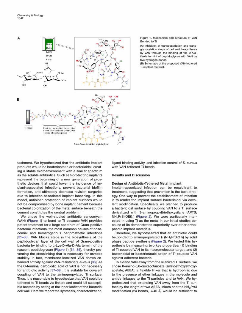
We chose the well-studied antibiotic vancomycin(VAN) (Figure 1) to bond to Ti because VAN providespotent treatment for a large spectrum of Gram-positivebacterial infections, the most common causes of noso-comial and hematogenous periprosthetic infections[31–33]. VAN blocks steps in the biosynthesis of thepeptidoglycan layer of the cell wall of Gram-positivebacteria by binding to L-Lys-D-Ala-D-Ala termini of thenascent peptidoglycan (Figure 1) [34, 35], thereby pre-venting the crosslinking that is necessary for osmoticstability. In fact, membrane-localized VAN shows en-hanced activity against VAN-resistant S. aureus [36]. Asthe C-terminal carboxylic acid of VAN is not necessaryfor antibiotic activity [37–39], it is suitable for covalentcoupling of VAN to the aminopropylated Ti surface.Thus, it is reasonable to hypothesize that VAN could betethered to Ti beads via linkers and could kill suscepti-ble bacteria by acting at the inner leaflet of the bacterialcell wall. Here we report the synthesis, characterization,
lw
R
DIteiladNecp
bppoba
catapfm
Figure 1. Mechanism and Structure of VANBonded to Ti
(A) Inhibition of transpeptidation and trans-glycosylation steps of cell wall biosynthesisby VAN through the binding of the D-Ala-D-Ala termini of peptidoglycan with VAN byfive hydrogen bonds.(B) Schematic of the proposed VAN-tetheredTi implant material.
igand binding activity, and infection control of S. aureusith VAN-tethered Ti beads.
esults and Discussion
esign of Antibiotic-Tethered Metal Implantmplant-associated infection can be recalcitrant toreatment, suggesting that prevention is the best strat-gy. One way to prevent the establishment of infection
s to render the implant surface bactericidal via cova-ent modification. Specifically, we planned to produce
bactericidal surface by coupling VAN to a Ti surfaceerivatized with 3-aminopropyltriethoxysilane (APTS;H2PrSi(OEt)3) (Figure 2). We were particularly inter-sted in using Ti as the metal in our initial studies be-ause of its demonstrated superiority over other ortho-aedic implant materials.Therefore, we hypothesized that an antibiotic could
e bonded to aminopropylated Ti (NH2PrSiOTi) by solidhase peptide synthesis (Figure 2). We tested this hy-othesis by measuring two key properties: (1) bindingf Ti-coupled VAN to its macromolecular target; and (2)actericidal or bacteriostatic action of Ti-coupled VANgainst adherent bacteria.To extend VAN away from the silanized Ti surface, we
hose 8-amino-3,6-dioxaoctanoate (aminoethoxyethoxy-cetate; AEEA), a flexible linker that is hydrophilic dueo the presence of ether linkages in the molecule andmide linkages to the Ti particles and to VAN. We hy-othesized that extending VAN away from the Ti sur-
ace by the length of two AEEA linkers and the NH2PrSiodification (24 bonds, z40 Å) would be sufficient to
Vancomycin-Titanium Beads1043
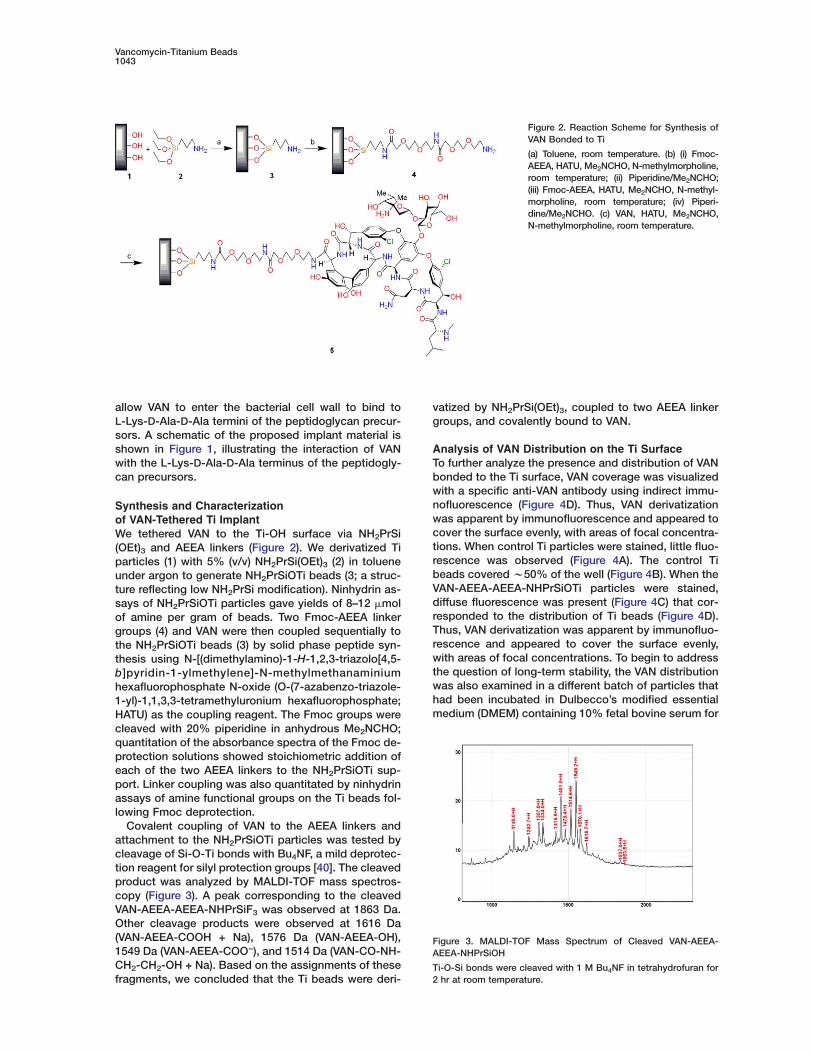
Figure 2. Reaction Scheme for Synthesis ofVAN Bonded to Ti
(a) Toluene, room temperature. (b) (i) Fmoc-AEEA, HATU, Me2NCHO, N-methylmorpholine,room temperature; (ii) Piperidine/Me2NCHO;(iii) Fmoc-AEEA, HATU, Me2NCHO, N-methyl-morpholine, room temperature; (iv) Piperi-dine/Me2NCHO. (c) VAN, HATU, Me2NCHO,N-methylmorpholine, room temperature.
allow VAN to enter the bacterial cell wall to bind toL-Lys-D-Ala-D-Ala termini of the peptidoglycan precur-sors. A schematic of the proposed implant material isshown in Figure 1, illustrating the interaction of VANwith the L-Lys-D-Ala-D-Ala terminus of the peptidogly-can precursors.
Synthesis and Characterizationof VAN-Tethered Ti ImplantWe tethered VAN to the Ti-OH surface via NH2PrSi(OEt)3 and AEEA linkers (Figure 2). We derivatized Tiparticles (1) with 5% (v/v) NH2PrSi(OEt)3 (2) in tolueneunder argon to generate NH2PrSiOTi beads (3; a struc-ture reflecting low NH2PrSi modification). Ninhydrin as-says of NH2PrSiOTi particles gave yields of 8–12 �molof amine per gram of beads. Two Fmoc-AEEA linkergroups (4) and VAN were then coupled sequentially tothe NH2PrSiOTi beads (3) by solid phase peptide syn-thesis using N-[(dimethylamino)-1-H-1,2,3-triazolo[4,5-b]pyridin-1-ylmethylene]-N-methylmethanaminiumhexafluorophosphate N-oxide (O-(7-azabenzo-triazole-1-yl)-1,1,3,3-tetramethyluronium hexafluorophosphate;HATU) as the coupling reagent. The Fmoc groups werecleaved with 20% piperidine in anhydrous Me2NCHO;quantitation of the absorbance spectra of the Fmoc de-protection solutions showed stoichiometric addition ofeach of the two AEEA linkers to the NH2PrSiOTi sup-port. Linker coupling was also quantitated by ninhydrinassays of amine functional groups on the Ti beads fol-lowing Fmoc deprotection.
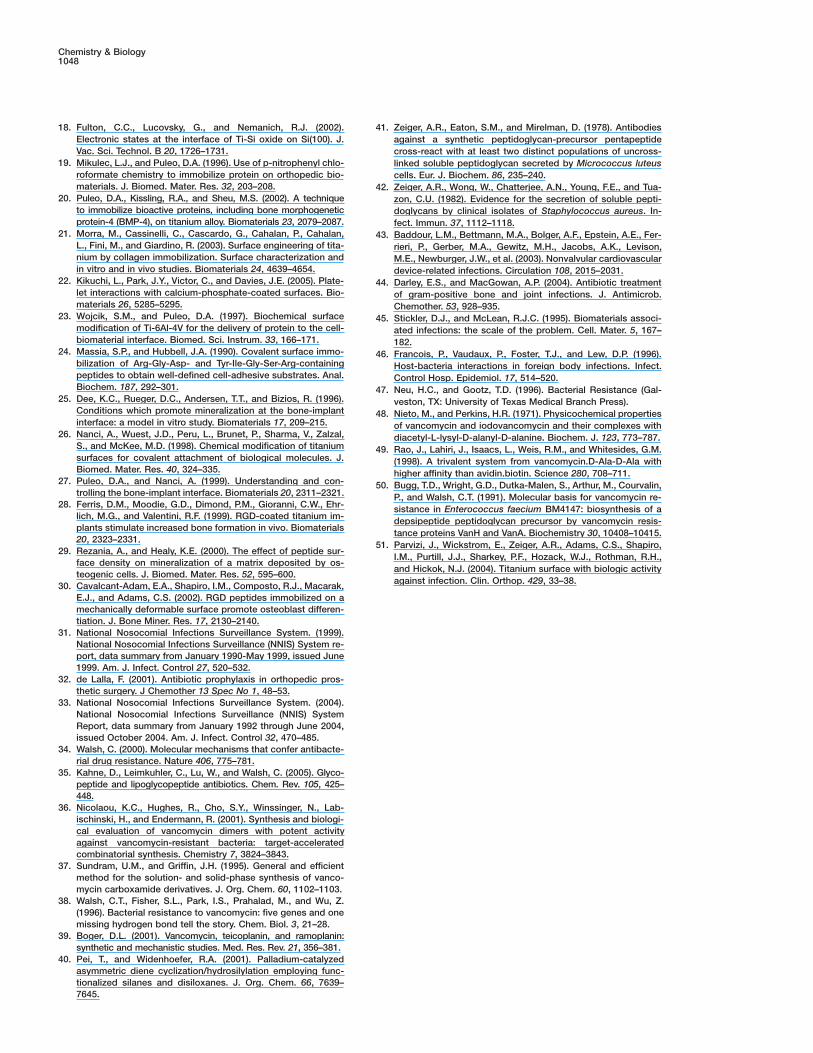
Covalent coupling of VAN to the AEEA linkers andattachment to the NH2PrSiOTi particles was tested bycleavage of Si-O-Ti bonds with Bu4NF, a mild deprotec-tion reagent for silyl protection groups [40]. The cleavedproduct was analyzed by MALDI-TOF mass spectros-copy (Figure 3). A peak corresponding to the cleavedVAN-AEEA-AEEA-NHPrSiF3 was observed at 1863 Da.Other cleavage products were observed at 1616 Da(VAN-AEEA-COOH + Na), 1576 Da (VAN-AEEA-OH),1549 Da (VAN-AEEA-COO−), and 1514 Da (VAN-CO-NH-CH2-CH2-OH + Na). Based on the assignments of thesefragments, we concluded that the Ti beads were deri-
medium (DMEM) containing 10% fetal bovine serum for
Figure 3. MALDI-TOF Mass Spectrum of Cleaved VAN-AEEA-AEEA-NHPrSiOH
Ti-O-Si bonds were cleaved with 1 M Bu4NF in tetrahydrofuran for2 hr at room temperature.
vatized by NH2PrSi(OEt)3, coupled to two AEEA linkergroups, and covalently bound to VAN.
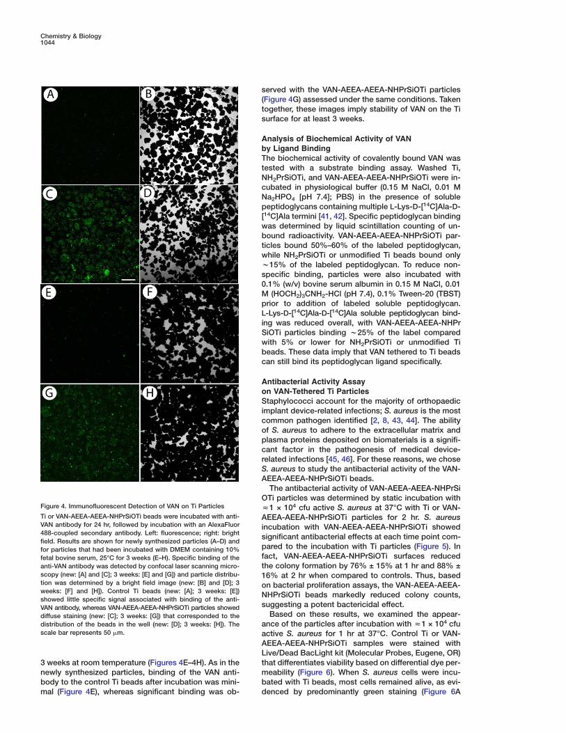
Analysis of VAN Distribution on the Ti SurfaceTo further analyze the presence and distribution of VANbonded to the Ti surface, VAN coverage was visualizedwith a specific anti-VAN antibody using indirect immu-nofluorescence (Figure 4D). Thus, VAN derivatizationwas apparent by immunofluorescence and appeared tocover the surface evenly, with areas of focal concentra-tions. When control Ti particles were stained, little fluo-rescence was observed (Figure 4A). The control Tibeads covered w50% of the well (Figure 4B). When theVAN-AEEA-AEEA-NHPrSiOTi particles were stained,diffuse fluorescence was present (Figure 4C) that cor-responded to the distribution of Ti beads (Figure 4D).Thus, VAN derivatization was apparent by immunofluo-rescence and appeared to cover the surface evenly,with areas of focal concentrations. To begin to addressthe question of long-term stability, the VAN distributionwas also examined in a different batch of particles thathad been incubated in Dulbecco’s modified essential
Chemistry & Biology1044
Figure 4. Immunofluorescent Detection of VAN on Ti Particles
Ti or VAN-AEEA-AEEA-NHPrSiOTi beads were incubated with anti-VAN antibody for 24 hr, followed by incubation with an AlexaFluor488-coupled secondary antibody. Left: fluorescence; right: brightfield. Results are shown for newly synthesized particles (A–D) andfor particles that had been incubated with DMEM containing 10%fetal bovine serum, 25°C for 3 weeks (E–H). Specific binding of theanti-VAN antibody was detected by confocal laser scanning micro-scopy (new: [A] and [C]; 3 weeks: [E] and [G]) and particle distribu-tion was determined by a bright field image (new: [B] and [D]; 3weeks: [F] and [H]). Control Ti beads (new: [A]; 3 weeks: [E])showed little specific signal associated with binding of the anti-VAN antibody, whereas VAN-AEEA-AEEA-NHPrSiOTi particles showeddiffuse staining (new: [C]; 3 weeks: [G]) that corresponded to thedistribution of the beads in the well (new: [D]; 3 weeks: [H]). Thescale bar represents 50 �m.
s(ts
AbTtNcNp[wbtwws0MpLiSwbc
AoSicopcrSA
OzAispft1oNs
aaAL
3 weeks at room temperature (Figures 4E–4H). As in thenewly synthesized particles, binding of the VAN anti-body to the control Ti beads after incubation was mini-mal (Figure 4E), whereas significant binding was ob-
tmbd
erved with the VAN-AEEA-AEEA-NHPrSiOTi particlesFigure 4G) assessed under the same conditions. Takenogether, these images imply stability of VAN on the Tiurface for at least 3 weeks.
nalysis of Biochemical Activity of VANy Ligand Bindinghe biochemical activity of covalently bound VAN wasested with a substrate binding assay. Washed Ti,H2PrSiOTi, and VAN-AEEA-AEEA-NHPrSiOTi were in-ubated in physiological buffer (0.15 M NaCl, 0.01 Ma2HPO4 [pH 7.4]; PBS) in the presence of solubleeptidoglycans containing multiple L-Lys-D-[14C]Ala-D-
14C]Ala termini [41, 42]. Specific peptidoglycan bindingas determined by liquid scintillation counting of un-ound radioactivity. VAN-AEEA-AEEA-NHPrSiOTi par-icles bound 50%–60% of the labeled peptidoglycan,hile NH2PrSiOTi or unmodified Ti beads bound only15% of the labeled peptidoglycan. To reduce non-
pecific binding, particles were also incubated with.1% (w/v) bovine serum albumin in 0.15 M NaCl, 0.01
(HOCH2)3CNH2-HCl (pH 7.4), 0.1% Tween-20 (TBST)rior to addition of labeled soluble peptidoglycan.-Lys-D-[14C]Ala-D-[14C]Ala soluble peptidoglycan bind-
ng was reduced overall, with VAN-AEEA-AEEA-NHPriOTi particles binding w25% of the label comparedith 5% or lower for NH2PrSiOTi or unmodified Tieads. These data imply that VAN tethered to Ti beadsan still bind its peptidoglycan ligand specifically.
ntibacterial Activity Assayn VAN-Tethered Ti Particlestaphylococci account for the majority of orthopaedic
mplant device-related infections; S. aureus is the mostommon pathogen identified [2, 8, 43, 44]. The abilityf S. aureus to adhere to the extracellular matrix andlasma proteins deposited on biomaterials is a signifi-ant factor in the pathogenesis of medical device-elated infections [45, 46]. For these reasons, we chose. aureus to study the antibacterial activity of the VAN-EEA-AEEA-NHPrSiOTi beads.The antibacterial activity of VAN-AEEA-AEEA-NHPrSiTi particles was determined by static incubation with1 × 104 cfu active S. aureus at 37°C with Ti or VAN-
EEA-AEEA-NHPrSiOTi particles for 2 hr. S. aureusncubation with VAN-AEEA-AEEA-NHPrSiOTi showedignificant antibacterial effects at each time point com-ared to the incubation with Ti particles (Figure 5). In
act, VAN-AEEA-AEEA-NHPrSiOTi surfaces reducedhe colony formation by 76% ± 15% at 1 hr and 88% ±6% at 2 hr when compared to controls. Thus, basedn bacterial proliferation assays, the VAN-AEEA-AEEA-HPrSiOTi beads markedly reduced colony counts,uggesting a potent bactericidal effect.Based on these results, we examined the appear-
nce of the particles after incubation with z1 × 104 cfuctive S. aureus for 1 hr at 37°C. Control Ti or VAN-EEA-AEEA-NHPrSiOTi samples were stained withive/Dead BacLight kit (Molecular Probes, Eugene, OR)hat differentiates viability based on differential dye per-eability (Figure 6). When S. aureus cells were incu-ated with Ti beads, most cells remained alive, as evi-enced by predominantly green staining (Figure 6A

Vancomycin-Titanium Beads1045
Figure 5. Ti-Tethered VAN Inhibits Proliferation of S. aureus
Control Ti (solid bars) or VAN-AEEA-AEEA-NHPrSiOTi (Ti-VAN,open bars) particles were incubated with S. aureus under staticconditions for up to 120 min. Colony counts were determined bydirect counting after serial dilution plating. Incubation of S. aureuswith VAN-AEEA-AEEA-NHPrSiOTi resulted in significantly de-creased colony counts at all times sampled. S. aureus in controlcultures maintained colony counts without robust proliferation aswould be expected in static cultures. Normalized data are shownas a percentage of Ti control at 30 min. The asterisks imply statisti-cal significance in reference to the corresponding time control atp < 0.05.
VAN-resistant mutant bacteria. An environment that is
Figure 6. Ti-Tethered VAN Kills AdherentS. aureus
Ti (A–C) or VAN-AEEA-AEEA-NHPrSiOTi(D–F) particles were incubated with S. aureusfor 1 hr. In the control Ti sample, green stain-ing of live cells (A) was more intense thanthe parallel red signal for dead cells (B). Incontrast, the VAN-AEEA-AEEA-NHPrSiOTibeads displayed only scattered green stain-ing for live cells (D) with the greatest redstaining for dead cells (E). (C) and (F) showthe corresponding bright field images of theparticle distribution in each assay. The scalebar represents 100 �m.
mide), yielding a measured KD of (4 ± 1) × 10 M [49].
versus 6B; bright field image of the particles is shown inFigure 6C). In contrast, S. aureus incubated with VAN-AEEA-AEEA-NHPrSiOTi beads were predominantly dead,as indicated by red staining (Figure 6D versus 6E; brightfield in Figure 6F). When beads that had been exposedthe day before to S. aureus were washed, rechallenged,and stained, VAN-AEEA-AEEA-NHPrSiOTi retained itsability to kill S. aureus preferentially (not shown). Thesestaining and proliferation assay results imply that theVAN-AEEA-AEEA-NHPrSiOTi surface actively killedS. aureus.
Antibiotic ResistanceBefore introducing a new and stable form of VAN invivo, one must consider the possibility of selection for
permissive for the selection of resistant organisms re-lies on (1) the presence of millions of bacteria, charac-teristically encountered in the gut and the oral cavityand (2) prolonged exposure to concentrations of antibi-otics sufficient to suppress growth of sensitive organ-isms but insufficient to suppress insensitive organisms[47]. In the first case, the number of bacteria that initiateperiprosthetic infections is small and as the antibioticis constrained on the Ti surface, the number of bacteriaexposed to the antibiotic is smaller still. Under thesecircumstances, the probability of a mutation that con-fers a selective growth advantage to a low number ofbacteria is vanishingly small. With respect to the sec-ond possibility, the dissociation constant of VAN fromL-Lys-D-Ala-D-Ala in solution is 1 × 10⁻⁶ M [48]; thus,at limiting concentrations of soluble VAN monomers,enough peptidoglycan precursors are unoccupied atany moment for some bacteria to survive and growslowly, providing an ideal situation to select for a mu-tant peptidoglycan with a terminal D-Ala-D-Lac thatbinds weakly to VAN [11].
That situation is much less likely for a solid phasematerial bearing multiple VAN residues. In suspensionculture, a single bacterium presents thousands ofL-Lys-D-Ala-D-Ala precursor peptidoglycan bindingsites for soluble VAN over the surface of its cell wall.For a bacterium sitting on a bed of multiple solid phaseVAN moieties, bound by each of thousands of accessi-ble peptidoglycans to thousands of VAN residues onthe Ti surface, dissociation of that bacterium would re-quire simultaneous cooperative undocking of all pepti-doglycans from all VAN-AEEA-AEEA-NHPrSiOTi sites atthe same instant. The dissociation constant for multiplepeptidoglycan-VAN complexes is the product of themultiple dissociation constants. That is, the KD for twopeptidoglycans from two solid phase VAN moieties is(10−6 × 10⁻⁶ M) = 10−12 M. For three peptidoglycanssimultaneously escaping from three VAN residues, onecalculates similarly KD = 10−18 M. This analysis was vali-dated by a specific test of 1,3,5-tris(CONH(N-acetyl)-L-Lys-D-Ala-D-Ala) benzene binding to tris(VAN carboxa-
−17

Chemistry & Biology1046
For as few as 100 peptidoglycan precursors on a bacte-rium binding to 100 VAN on a Ti surface, KD rises to10−600 M, implying no detectable escape of any boundbacterium until the dead fragments dissociate one byone. In other words, simultaneous binding to thousandsof solid phase VAN moieties has an effect equivalent toan enormous solution concentration of free VAN. Thisphenomenon potentiates the bactericidal action ofVAN.
The most common VAN resistance mutation is achange of peptidoglycan target from D-Ala-D-Ala toD-Ala-D-Lac. The dissociation constant of VAN from themutant peptidoglycan will increase to 10−3 M [50]. Inthat situation, we could extrapolate from the hypotheti-cal example of 100 VAN binding to 100 mutant pepti-doglycan precursor sites a KD of 10−300 M, but this re-mains hypothetical in the absence of experiment. Thus,both because the surface will be exposed to relativelysmall numbers of microorganisms in vivo, and becauseVAN will show an increased potency because of its sur-face immobilization, occurrence of bacterial resistanceshould be extremely unlikely.
Implications for Management of Implant InfectionsThe covalently tethered antibiotic approach is uniquelysuited to the eradication of a nascent infection and thuscould reduce the risk of implant-associated infections.Bacterial infection in surrounding tissue and bone can-not be controlled entirely by an antibiotic covalentlybonded to an implant surface, but might be managedby a secondary route of release of soluble antibiotic ondemand. Moreover, this new paradigm could be easilyextended to other agents and materials. Applications ofsuch surfaces extend beyond orthopaedics. Indwellingdevices tend to serve as ideal surfaces for establish-ment of infections. Thus, surface modifications thatrender devices antibacterial for the long term offergreat promise for ameliorating the complications inher-ent in implant-associated infections and their treat-ment. Direct chemical modification of implant surfaceswith bactericidal agents could provide a new genera-tion of implants that are able to combat implant-associ-ated infection and its devastating consequences.
Significance
Bacterial adhesion to implanted biomaterial surfacesis a critical step in the pathogenesis of infection. Theformation of a complex biofilm at the interface be-tween a biomaterial and the biological environment isthought to be responsible for sustaining and ex-acerbating inflammation and for the ultimate failureof biomedical devices. In this report, we have takenthe first steps toward the goal of producing a bacte-ria-resistant implant. First, these studies demonstratefor the first time, to our knowledge, that an antibioticcan be covalently attached to a biocompatible implantmaterial. The synthesis was validated by step-by-stepanalysis and by MALDI-TOF mass spectra of the finalproduct. Second, the solid phase antibiotic not onlyretained the activity of the soluble antibiotic, but mayimprove on the latter activity by preventing biofilmformation at the site of the implant. Activity of the co-
vcgwlboiaaetcUfsvoi
E
CAAA(
DTtptfoct(wwaMToslifq1
SNweai2ooFscwor1
CMs
alently bound antibiotic was shown at the biochemi-al level by specificity against the secreted peptido-lycan target of the antibiotic. The tethered antibioticas also demonstrated to be active at the microbio-
ogical level by inhibition of bacterial proliferation andiofilm formation. Finally, the covalently bound antibi-tic maintained its stability as demonstrated by activ-
ty after a repeat challenge, and by its ability to bindnti-VAN antibody after 3 weeks in culture mediumt room temperature. The titanium surface was thusmpowered to protect itself from colonization by bac-eria. These experiments demonstrate the potential ofovalent implant modification for infection control.nlike a noncovalent coating that slowly releases a
ree antibiotic, Ti-bonded VAN was not lost from theurface during incubation with the bacteria. Thus, co-alent binding can extend the microbiological activityf VAN in precisely the microenvironment in which it
s most needed.
xperimental Procedures
hemicalsll solvents were purchased from Fisher Scientific (Fair Lawn, NJ).ll reagents for coupling reactions were purchased from Sigma-ldrich (St. Louis, MO). Ti particles (350 mesh) were from Alfa Aesar
Ward Hill, MA).
erivatization of Ti Surfaces with NH2PrSi(OEt)3
i beads (5–10 g of 350 mesh) were cleaned with MeOH/concen-rated HCl (1:1, 5 ml) for 20 min at room temperature with 20 seceriods of sonication at 0, 5, 12, 15, and 20 min. The cleaned par-
icles were washed twice with double-deionized water (5 ml), thenour times with anhydrous Me2NCHO (5 ml). Particles were driedvernight under vacuum and placed in the argon atmospherehamber of an MO-20M glove box (Vacuum Atmospheres, Haw-horne, CA). Particles were washed twice with anhydrous toluene10 ml) by suspension with a stainless steel spatula, then reactedith 5% NH2PrSi(OEt)3 in anhydrous toluene (v/v, 5 ml) for 60 minith resuspension every 10 min. The beads were washed twice withnhydrous toluene (10 ml) as above, washed twice with anhydrouse2NCHO (10 ml) as above, and dried under vacuum overnight.
he beads were then baked at 100°C for 15 min in a convectionven. Aliquots of z100 mg were assayed for amine content byuspension in 0.35 mM SnCl2, 0.1 M Na citrate (pH 5; 1 ml), fol-
owed by the addition of 4% ninhydrin in EtOH (w/v, 1 ml). Aftermmersion in boiling water for 15 min., the solution was cooledor 2 min, diluted with EtOH/H2O (3:2, v/v, 5 ml), and amines wereuantitated by measuring the absorbance, A570, using �570 = 1.5 ×04/M·cm.
olid Phase Coupling of VAN to the Ti SurfaceH2PrSiOTi beads (1–2 g, 10–20 �mol amine) were washed twiceith anhydrous Me2NCHO (5 ml), then coupled with a 4-fold molarxcess of Fmoc-8-amino-3,6-dioxaoctanoate (aminoethoxyethoxy-cetate; AEEA) linker, activated with a 4-fold molar excess of HATU
n anhydrous Me2NCHO (5 ml), followed by Fmoc deprotection with0% piperidine in anhydrous Me2NCHO (5 ml). The completenessf deprotection was determined by Kaiser’s test. Similarly, a sec-nd AEEA linker group was subsequently coupled to the beads. Allmoc deprotection solutions and washes were collected for analy-is. Finally, a 4-fold excess of VAN was coupled by the same proto-ol. After the VAN coupling step, the Ti beads were washed twiceith anhydrous Me2NCHO (5 ml), and then dried under vacuumvernight. Absorbance spectra of the deprotection solutions wereecorded and yields were calculated using A570 and �301 = 7.78 ×03/M·cm.
leavage of Si-O Bond by Bu4NF Reaction and MALDI-TOF MSatrix-assisted laser desorption ionization time of flight mass
pectrometry (MALDI-TOF) was used to characterize the molecular

Vancomycin-Titanium Beads1047
species covalently attached to the Ti surface. Specifically, the VAN-AEEA-AEEA-NHPrSiOTi particles (z100 mg) were suspended inMe2NCHO (0.5 ml), to which was added 1 M Bu4NF in tetrahydrofu-ran (4 ml) with stirring for 2 hr at room temperature to cleave theSi-O bonds. After filtering to remove Ti particles, the filtrate solutionwas analyzed by MALDI-TOF mass spectrometry.
VAN Antibody BindingTi and VAN-AEEA-AEEA-NHPrSiOTi particles were washed twicewith PBS. The material was blocked for 30 min using 10% fetalbovine serum in distilled, deionized water (dH2O, blocking buffer).The beads were incubated with mouse anti-VAN IgG (1:300; U.S.Biologicals, Swampscott, MA) in blocking buffer at 4°C for 12 hr,washed with PBS, and incubated with AlexaFluor 488-coupleddonkey anti-mouse IgG (Molecular Probes, Eugene, OR) diluted1:300 in blocking buffer at room temperature for 1 hr. Both Ti andVAN-AEEA-AEEA-NHPrSiOTi particles were washed three timeswith PBS, followed by two 15 min incubations in PBS. The particleswere then visualized using a Fluoview 300 confocal laser micro-scope (Olympus, Melville, NY). For long-term experiments, thebeads were incubated in DMEM/10% fetal bovine serum for 3weeks at room temperature, washed four times with PBS, followedby incubation with blocking buffer for 15 min and staining as above.
Soluble Peptidoglycan Binding Assay for VANTo determine the biological activity of VAN covalently bound to theTi particles, specific binding of VAN-AEEA-AEEA-NHPrSiOTi to theVAN ligand L-Lys-D-Ala-D-Ala present on soluble peptidoglycanwas determined [41, 42]. Samples of Ti, NH2PrSiOTi, and VAN-AEEA-AEEA-NHPrSiOTi beads (100 mg each) were washed by sus-pension in PBS (1 ml) in a polypropylene vial, with rocking for 1 hrat room temperature. The wash supernatant was removed, and theparticles were incubated with 2.5 nCi of L-Lys-D-[14C]Ala-D-[14C]Alasoluble peptidoglycan, z10 Ci/mol, prepared as previously de-scribed [41, 42] for 2 hr with rocking. Unbound soluble peptidogly-cans in the supernatants were collected, and quantitated by liquidscintillation counting in Aquasol II (PerkinElmer Life Sciences, Shel-ton, CT; 10 ml) in an LS 6000SC scintillation spectrometer (Beck-man Instruments, Fullerton, CA). To control for nonspecific binding,the above beads were also analyzed using 0.1% (w/v) bovine se-rum albumin in TBST in the washing step with samples rocked for4 hr at room temperature, followed by incubation with 2.5 nCi ofL-Lys-D-[14C]Ala-D-[14C]Ala soluble peptidoglycan with rockingovernight at 4°C and quantitation as above.
Bacterial AssaysThe control Ti and VAN-AEEA-AEEA-NHPrSiOTi particles were ster-ilized by incubation in 70% ethanol, followed by five PBS washes.The beads were resuspended in 1% dextrose/PBS, and 30 �l ofthe suspension, corresponding to z40 mg of particles, were dis-pensed into each well of an eight-well chamber slide (Nunc, Roch-ester, NY). S. aureus subspecies aureus Rosenbach ATCC strain25923 was cultured in trypticase soy broth for 12 hr. The culturewas pelleted by centrifugation at 3200 × g for 5 min, and resus-pended in 1% dextrose/PBS. Using the McFarland standard, anearly log-phase aliquot of S. aureus containing z1 × 104 cfu in 170�l was inoculated in each well. The cultures were incubated at 37°Cfor 30, 60, 90, and 120 min under static conditions.
Quantitative culture was performed at 30, 60, 90, and 120 min byserially diluting 200 �l of sample in 1.8 ml PBS, then plating 200 �lof the relevant dilutions in triplicate on Todd-Hewitt agar plates.The number of cfu per ml was determined by counting the numbersof colonies on plates after 24 hr, determining the mean per sample,and adjusting for the dilution. Plates showing no growth after theinitial 24 hr incubation were kept for an additional 24 hr to confirmthe absence of growth.
For microscopy, the samples were washed twice with PBS toremove planktonic bacteria. Viability of bacteria was determined at1 hr by confocal laser microscopy (Olympus Fluoview 300) afterstaining using the Live/Dead BacLight Viability kit (MolecularProbes) that functions based on the differential membrane perme-ability to Cyto9 and propidium iodide. After staining, upon excita-
tion, live bacteria show an intense green fluorescence, while deadbacteria fluoresce red.
Acknowledgments
We thank Dr. Christopher Adams and Dr. Irving Shapiro for criticalreading of the manuscript and valuable discussions. We thank Dr.Michele Marcolongo, Drexel University, for the gift of the Ti par-ticles. A preliminary account of this work was presented at theAmerican Academy of Orthopaedic Surgeons, 71st Annual Meet-ing, San Francisco, CA, March 10–14, 2004 [51]. This work wassupported in part by DoD grant DAMD-17-03-1-0713.
Received: April 29, 2005Revised: June 28, 2005Accepted: June 30, 2005Published: September 23, 2005
References
1. Garvin, K.L. (1994). Two-stage reimplantation of the infectedhip. Semin. Arthroplasty 5, 142–146.
2. An, Y.H., and Friedman, R.J. (1996). Prevention of sepsis in totaljoint arthroplasty. J. Hosp. Infect. 33, 93–108.
3. Garvin, K.L., and Hanssen, A.D. (1995). Infection after total hiparthroplasty: past, present, and future. J. Bone Joint Surg. 77A,1576–1588.
4. Moss, A.J., Hamburger, S., Moore, R.M., Jr., Jeng, L.L., andHowie, L.J. (1991). Use of selected medical device implants inthe United States, 1988. Adv. Data 77A, 1–24.
5. Stewart, P.S., and Costerton, J.W. (2001). Antibiotic resistanceof bacteria in biofilms. Lancet 358, 135–138.
6. Dunne, W.M., Jr. (2002). Bacterial adhesion: seen any good bio-films lately? Clin. Microbiol. Rev. 15, 155–166.
7. Duggan, J.M., Georgiadis, G.M., and Kleshinski, J.F. (2001).Management of prosthetic joint infections. Infect. Med. 18,534–541.
8. Zimmerli, W., Trampuz, A., and Ochsner, P.E. (2004). Prosthetic-joint infections. N. Engl. J. Med. 351, 1645–1654.
9. Haddad, F.S., Masri, B.A., Garbuz, D.S., and Duncan, C.P.(1999). The treatment of the infected hip replacement. Thecomplex case. Clin. Orthop. 369, 144–156.
10. Haddad, F.S., Masri, B.A., Campbell, D., McGraw, R.W.,Beauchamp, C.P., and Duncan, C.P. (2000). The PROSTALACfunctional spacer in two-stage revision for infected knee re-placements. Prosthesis of antibiotic-loaded acrylic cement. J.Bone Joint Surg. Br. 82, 807–812.
11. Loll, P.J., and Axelsen, P.H. (2000). The structural biology ofmolecular recognition by vancomycin. Annu. Rev. Biophys. Bi-omol. Struct. 29, 265–289.
12. van de Belt, H., Neut, D., Schenk, W., van Horn, J.R., van derMei, H.C., and Busscher, H.J. (2001). Infection of orthopedicimplants and the use of antibiotic-loaded bone cements. A re-view. Acta Orthop. Scand. 72, 557–571.
13. Frazier, D.D., Lathi, V.K., Gerhart, T.N., and Hayes, W.C. (1997).Ex vivo degradation of a poly(propylene glycol-fumarate) bio-degradable particulate composite bone cement. J. Biomed.Mater. Res. 35, 383–389.
14. Otsuka, M., Matsuda, Y., Suwa, Y., Fox, J.L., and Higuchi, W.I.(1994). A novel skeletal drug delivery system using a self-set-ting calcium phosphate cement. 5. Drug release behavior froma heterogeneous drug-loaded cement containing an anticancerdrug. J. Pharm. Sci. 83, 1565–1568.
15. Wang, C.J., Huang, T.W., Wang, J.W., and Chen, H.S. (2002).The often poor clinical outcome of infected total kneearthroplasty. J. Arthroplasty 17, 608–614.
16. Wright, T.M., Sullivan, D.J., and Arnoczky, S.P. (1984). The ef-fect of antibiotic additions on the fracture properties of bonecements. Acta Orthop. Scand. 55, 414–418.
17. Lucke, M., Schmidmaier, G., Sadoni, S., Wildemann, B., Schil-ler, R., Haas, N.P., and Raschke, M. (2003). Gentamicin coatingof metallic implants reduces implant-related osteomyelitis inrats. Bone 32, 521–531.
Chemistry & Biology1048
18. Fulton, C.C., Lucovsky, G., and Nemanich, R.J. (2002).Electronic states at the interface of Ti-Si oxide on Si(100). J.Vac. Sci. Technol. B 20, 1726–1731.
19. Mikulec, L.J., and Puleo, D.A. (1996). Use of p-nitrophenyl chlo-roformate chemistry to immobilize protein on orthopedic bio-materials. J. Biomed. Mater. Res. 32, 203–208.
20. Puleo, D.A., Kissling, R.A., and Sheu, M.S. (2002). A techniqueto immobilize bioactive proteins, including bone morphogeneticprotein-4 (BMP-4), on titanium alloy. Biomaterials 23, 2079–2087.
21. Morra, M., Cassinelli, C., Cascardo, G., Cahalan, P., Cahalan,L., Fini, M., and Giardino, R. (2003). Surface engineering of tita-nium by collagen immobilization. Surface characterization andin vitro and in vivo studies. Biomaterials 24, 4639–4654.
22. Kikuchi, L., Park, J.Y., Victor, C., and Davies, J.E. (2005). Plate-let interactions with calcium-phosphate-coated surfaces. Bio-materials 26, 5285–5295.
23. Wojcik, S.M., and Puleo, D.A. (1997). Biochemical surfacemodification of Ti-6Al-4V for the delivery of protein to the cell-biomaterial interface. Biomed. Sci. Instrum. 33, 166–171.
24. Massia, S.P., and Hubbell, J.A. (1990). Covalent surface immo-bilization of Arg-Gly-Asp- and Tyr-Ile-Gly-Ser-Arg-containingpeptides to obtain well-defined cell-adhesive substrates. Anal.Biochem. 187, 292–301.
25. Dee, K.C., Rueger, D.C., Andersen, T.T., and Bizios, R. (1996).Conditions which promote mineralization at the bone-implantinterface: a model in vitro study. Biomaterials 17, 209–215.
26. Nanci, A., Wuest, J.D., Peru, L., Brunet, P., Sharma, V., Zalzal,S., and McKee, M.D. (1998). Chemical modification of titaniumsurfaces for covalent attachment of biological molecules. J.Biomed. Mater. Res. 40, 324–335.
27. Puleo, D.A., and Nanci, A. (1999). Understanding and con-trolling the bone-implant interface. Biomaterials 20, 2311–2321.
28. Ferris, D.M., Moodie, G.D., Dimond, P.M., Gioranni, C.W., Ehr-lich, M.G., and Valentini, R.F. (1999). RGD-coated titanium im-plants stimulate increased bone formation in vivo. Biomaterials20, 2323–2331.
29. Rezania, A., and Healy, K.E. (2000). The effect of peptide sur-face density on mineralization of a matrix deposited by os-teogenic cells. J. Biomed. Mater. Res. 52, 595–600.
30. Cavalcant-Adam, E.A., Shapiro, I.M., Composto, R.J., Macarak,E.J., and Adams, C.S. (2002). RGD peptides immobilized on amechanically deformable surface promote osteoblast differen-tiation. J. Bone Miner. Res. 17, 2130–2140.
31. National Nosocomial Infections Surveillance System. (1999).National Nosocomial Infections Surveillance (NNIS) System re-port, data summary from January 1990-May 1999, issued June1999. Am. J. Infect. Control 27, 520–532.
32. de Lalla, F. (2001). Antibiotic prophylaxis in orthopedic pros-thetic surgery. J Chemother 13 Spec No 1, 48–53.
33. National Nosocomial Infections Surveillance System. (2004).National Nosocomial Infections Surveillance (NNIS) SystemReport, data summary from January 1992 through June 2004,issued October 2004. Am. J. Infect. Control 32, 470–485.
34. Walsh, C. (2000). Molecular mechanisms that confer antibacte-rial drug resistance. Nature 406, 775–781.
35. Kahne, D., Leimkuhler, C., Lu, W., and Walsh, C. (2005). Glyco-peptide and lipoglycopeptide antibiotics. Chem. Rev. 105, 425–448.
36. Nicolaou, K.C., Hughes, R., Cho, S.Y., Winssinger, N., Lab-ischinski, H., and Endermann, R. (2001). Synthesis and biologi-cal evaluation of vancomycin dimers with potent activityagainst vancomycin-resistant bacteria: target-acceleratedcombinatorial synthesis. Chemistry 7, 3824–3843.
37. Sundram, U.M., and Griffin, J.H. (1995). General and efficientmethod for the solution- and solid-phase synthesis of vanco-mycin carboxamide derivatives. J. Org. Chem. 60, 1102–1103.
38. Walsh, C.T., Fisher, S.L., Park, I.S., Prahalad, M., and Wu, Z.(1996). Bacterial resistance to vancomycin: five genes and onemissing hydrogen bond tell the story. Chem. Biol. 3, 21–28.
39. Boger, D.L. (2001). Vancomycin, teicoplanin, and ramoplanin:synthetic and mechanistic studies. Med. Res. Rev. 21, 356–381.
40. Pei, T., and Widenhoefer, R.A. (2001). Palladium-catalyzedasymmetric diene cyclization/hydrosilylation employing func-tionalized silanes and disiloxanes. J. Org. Chem. 66, 7639–7645.
4
4
4
4
4
4
4
4
4
5
5
1. Zeiger, A.R., Eaton, S.M., and Mirelman, D. (1978). Antibodiesagainst a synthetic peptidoglycan-precursor pentapeptidecross-react with at least two distinct populations of uncross-linked soluble peptidoglycan secreted by Micrococcus luteuscells. Eur. J. Biochem. 86, 235–240.
2. Zeiger, A.R., Wong, W., Chatterjee, A.N., Young, F.E., and Tua-zon, C.U. (1982). Evidence for the secretion of soluble pepti-doglycans by clinical isolates of Staphylococcus aureus. In-fect. Immun. 37, 1112–1118.
3. Baddour, L.M., Bettmann, M.A., Bolger, A.F., Epstein, A.E., Fer-rieri, P., Gerber, M.A., Gewitz, M.H., Jacobs, A.K., Levison,M.E., Newburger, J.W., et al. (2003). Nonvalvular cardiovasculardevice-related infections. Circulation 108, 2015–2031.
4. Darley, E.S., and MacGowan, A.P. (2004). Antibiotic treatmentof gram-positive bone and joint infections. J. Antimicrob.Chemother. 53, 928–935.
5. Stickler, D.J., and McLean, R.J.C. (1995). Biomaterials associ-ated infections: the scale of the problem. Cell. Mater. 5, 167–182.
6. Francois, P., Vaudaux, P., Foster, T.J., and Lew, D.P. (1996).Host-bacteria interactions in foreign body infections. Infect.Control Hosp. Epidemiol. 17, 514–520.
7. Neu, H.C., and Gootz, T.D. (1996). Bacterial Resistance (Gal-veston, TX: University of Texas Medical Branch Press).
8. Nieto, M., and Perkins, H.R. (1971). Physicochemical propertiesof vancomycin and iodovancomycin and their complexes withdiacetyl-L-lysyl-D-alanyl-D-alanine. Biochem. J. 123, 773–787.
9. Rao, J., Lahiri, J., Isaacs, L., Weis, R.M., and Whitesides, G.M.(1998). A trivalent system from vancomycin.D-Ala-D-Ala withhigher affinity than avidin.biotin. Science 280, 708–711.
0. Bugg, T.D., Wright, G.D., Dutka-Malen, S., Arthur, M., Courvalin,P., and Walsh, C.T. (1991). Molecular basis for vancomycin re-sistance in Enterococcus faecium BM4147: biosynthesis of adepsipeptide peptidoglycan precursor by vancomycin resis-tance proteins VanH and VanA. Biochemistry 30, 10408–10415.
1. Parvizi, J., Wickstrom, E., Zeiger, A.R., Adams, C.S., Shapiro,I.M., Purtill, J.J., Sharkey, P.F., Hozack, W.J., Rothman, R.H.,and Hickok, N.J. (2004). Titanium surface with biologic activityagainst infection. Clin. Orthop. 429, 33–38.
Copyright © 2022 FDOKUMEN


![Drug Delivery System Based on Covalently Bonded Poly[N-Isopropylacrylamide-co-2-Hydroxyethylacrylate]-Based Nanoparticle Networks](https://static.fdokumen.com/doc/165x107/6340d5f6e0dac3b265042228/drug-delivery-system-based-on-covalently-bonded-polyn-isopropylacrylamide-co-2-hydroxyethylacrylate-based.jpg)


![Fluorescent cellulose aerogels containing covalently immobilized (ZnS) ₓ (CuInS ₂) ₁₋ ₓ/ZnS (core/shell) quantum dots [2013]](https://static.fdokumen.com/doc/165x107/63372dc94554fe9f0c05b209/fluorescent-cellulose-aerogels-containing-covalently-immobilized-zns-cuins.jpg)