High prevalence of ST‐78 infection‐associated vancomycin‐resistant Enterococcus faecium from...
109
Antimicrobial Resistance Spread and the Role of Mobile Genetic Elements
-
Upload
independent -
Category
Documents
-
view
5 -
download
0
Transcript of High prevalence of ST‐78 infection‐associated vancomycin‐resistant Enterococcus faecium from...

Antimicrobial Resistance Spread and the Role of
Mobile Genetic Elements

ISBN 978-90-8559-944-9
© Mushtaq Ahmad Khan, 2010.
All rights reserved. No part of this thesis may be reproduced or transmitted in any form or by
any means, electronic or mechanical, without the prior written permission of the author.
Printed by: Optima Grafische Communicatie, Rotterdam, The Netherlands.
Cover Design: Michiel Leendert Bexkens and Mushtaq Ahmad Khan

Antimicrobial Resistance Spread and the Role of
Mobile Genetic Elements
Antimicrobiële resistentie verspreiding en de rol van mobiele genetische
elementen
THESIS
To obtain the degree of Doctor from the Erasmus University Rotterdam
by command of the
rector magnificus
Prof.dr. H.G. Schmidt
and in accordance with the decision of the Doctorate Board.
The public defence shall be held on
Wednesday, March 17th, 2010 at 15:30 hours
by
Mushtaq Ahmad Khan
born in Pulwama, Kashmir, India

Doctoral Committee
Promoter: Prof.dr.dr. A.F. van Belkum
Other members: Prof.dr. H.A. Verbrugh
Prof.dr. F. Baquero
Prof.dr. A.G. Vulto
Co-promoter: Dr. J.P. Hays
The research described in this thesis was financially supported by a European Union FP6
grant (DRESP2).

“Somewhere, something incredible is waiting to be known.”
Dr. Carl Sagan (1934-1996)
To family, friends and relatives


Contents Chapter 1 General Introduction 9 Chapter 2 bro β-lactamase and Antibiotic Resistances in a Global Cross-Sectional 33
Study of Moraxella catarrhalis from Children and Adults
Chapter 3 Dominance of CTX-M-2 and CTX-M-56 among extended-spectrum 51
β-lactamases produced by Klebsiella pneumoniae and
Escherichia coli isolated in hospitals in Paraguay
Chapter 4 Important methodological considerations with respect to the 59
differentiation of CTX-M-15 and CTX-M-28
extended-spectrum β-lactamases
Chapter 5 Molecular characterization of ESBL producing Klebsiella Pneumoniae 65
isolated from blood cultures in Pondicherry, India
Chapter 6 Molecular characterization of antimicrobial resistance in non-typhoidal 75
salmonellae from India
Chapter 7 Analysis of VanA vancomycin-resistant Enterococcus faecium isolates 91
from Saudi Arabian hospitals reveals the presence of clonal
cluster 17 and two new Tn1546 lineage types
Chapter 8 High prevalence of ST-78 infection-associated vancomycin-resistant 99
Enterococcus faecium from hospitals in Asunción, Paraguay
Chapter 9 Molecular epidemiology of high-level aminoglycoside resistant 107
Enterococcus feacalis from patients during

and after hospitalisation
Chapter 10 Summary and conclusive remarks / Nederlandse samenvatting 123 Acknowledgements 144
Curriculum Vitae 145
List of publications 146
PhD portfolio 147

Chapter 1
General IntroductionGeneral Introduction


Chapter 1 General Introduction
A brief history of antimicrobial resistance
Alexander Fleming discovered the first antimicrobial agent, penicillin (a β-lactam), in
1928 in the mold Penicillium notatum. Penicillin was initially found to be active against staphylococcal strains, which at that time were a major source of infectious diseases. Indeed, the mortality rate of individuals with severe Staphylococcus aureus infection in the early 1940’s was about 80% [1]. The discovery of penicillin gave clinicians an effective means to combat fatal microbial infections for the first time. Since that time, many different types of antimicrobial agents have been discovered, including aminoglycosides, macrolides and cephalosporins to name but a few. Further, current advances in biochemistry and engineering now mean that novel antimicrobial agents can be produced by 1) synthetically altering the structure of known antimicrobial agents, or by 2) high throughput screening strategies. However, even as new antibiotics are being discovered, bacteria are acquiring mechanisms to neutralise their effect.
Even at the time that penicillin was being commercially made available in 1945, reports of enzymes possessing β-lactamase activity had already been published by Abraham and Chain [2], and we now know that antimicrobial resistance mechanisms have been around for many millions of years, even before the large-scale use of antibiotic therapy in human populations began. For example, ampicillin (a derivative of penicillin) resistant bacteria estimated to be approximately 2000 years old have been recovered from glacial samples obtained from the Canadian Arctic Archipelago, while a more recent study detected TEM-type β-lactamases from a metagenomic library of cold seep deep-sea segments estimated to be approximately 10,000 years old [3, 4]. In fact, it is now known that many environmental organisms produce antimicrobial compounds in order to limit competition for nutrients or chemically alternative niches in defined niches. However, in response, bacteria have evolved (and are continuing to evolve) mechanisms to counteract these lethal antimicrobial compounds, in order to protect themselves from attack. It is these mechanisms that eventually create problems with respect to antimicrobial resistance and human health.
During the period after Alexander Fleming’s discovery of penicillin, academia and the pharmaceutical industry discovered and developed many new antimicrobial agents. However, after each new class of antimicrobial agent was brought to market, new resistance mechanisms appeared that reduced the efficacy of the new antibiotics, seriously limiting patient treatment options. For example, one new class of antimicrobial agents discovered were broad-spectrum β-lactam antibiotics, the cephalosporins. These antibiotics have an extended-spectrum of action against both Gram-positive and Gram-negative bacteria and were originally extensively used for the treatment of serious infections caused by Gram-negative bacteria in the 1980s [5]. However, resistance to cephalosporins rapidly developed, creating new problems for the treatment of infectious diseases. This race between the introduction of new antimicrobial agents and the appearance of associated resistance mechanisms in microorganisms is still a major problem globally, limiting the successful treatment of infectious diseases by health care professionals. It is essential therefore to monitor the spread of existing, new, and emerging antimicrobial mechanisms in the world. The goal of this thesis is to provide further data that will help monitor existing antimicrobial resistances in order to add information to global antimicrobial resistance monitoring campaigns, whilst also helping to predict future trends.
11

Chapter 1 General Introduction
Overview of antimicrobial resistance in microorganisms of major clinical importance
Both Gram-negative and Gram-positive microorganisms are associated with antimicrobial resistance in the clinical (and nosocomial) environments.
A. Gram-negative microorganisms:
Currently, the most important clinically relevant antimicrobial resistances among Gram-negative bacteria are associated with the enterobacteriaceae, namely:
1. Extended-spectrum β-lactam resistance in Klebseilla pneumoniae, Escherichia coli and Salmonella species.
2. Fluoroquinolone resistance in Salmonella species.
1. β-lactam resistance in E. coli was first identified in the early 1920’s. Subsequent studies showed that β-lactam resistance among enterobacteriaceae was caused by the so-called TEM-1 and SHV-1 β-lactamases, which possess an amino-acid similarity of 68%. As the discovery of new antibiotics quickened, a new class of antibiotics, the broad-spectrum β-lactam antibiotics (cephalosporins), were introduced due to their improved spectrum of activity against both Gram-negative and Gram-positive bacteria. Coincidently, β-lactam resistant strains of enterobacteriaceae were also found to be susceptible to these broad-spectrum antibiotics. However, soon after the introduction of the cephalosporins, resistant enterobacterial isolates began to emerge. The mechanism facilitating this particular resistance was found to be a mutation in either TEM or SHV β-lactamase genes, which lead to changes in the relevant β-lactamase amino-acid sequence, giving rise to different phenotypic variants that possessed “extended” β-lactamase activities. The fact that the new TEM and SHV type β-lactamases possessed “extended” activity against β-lactam antibiotics, meant that they were named “extended-spectrum β-lactamases” or ESBLs [6]. The first ESBL (SHV-type) reported to cause the inactivation of extended spectrum β-lactam antibiotics was reported in Germany in 1983 [7]. However, since 1995, the rate of dissemination and reporting of ESBLs among different bacterial isolates has increased rapidly in most parts of the world [8-10]. In fact, the increased frequency of microbial isolates carrying these mutation events most probably occurred via an increase in “selection pressure” after the introduction and large-scale use of cephalosporins, which removed cephalosporin sensitive bacterial isolates from the clinical environment, thereby allowing cephalosporin resistant isolates to take advantage of the newly available niches [11-14].
Untill the 1990’s, ESBLs found in clinical isolates of bacteria were mostly identified as TEM and SHV-type enzymes. However, a different class of ESBL enzymes (called CTX-M) began to emerge after this date. CTX-M enzymes are a new class of β-lactamase that have become increasingly prevalent in the clinical and nosocomial environments, affecting all populated continents. Indeed, since first being reported in Germany in 1990 [15], several different classes of CTX-M enzyme groups have emerged, with CTX-M carrying enterobacterial isolates having quickly become the most widespread and clinically relevant group of ESBL enzymes. To illustrate this point, to date, more than 50 different CTX-M enzymes have been reported, which are grouped into five main subgroups (CTX-M-1, CTX-M-2, CTX-M-8, CTX-M-9 and CTX-M-25) based on their amino acid sequence similarities [10].
12

Chapter 1 General Introduction
2. Fluoroquinolone resistance within the enterobacteriaceae (and in particular within the Salmonella species) is also of great economic and nosocomial importance. Fluoroquinolone antibiotics have been available since the 1960s, when nalidixic acid was introduced. Subsequent structural modifications to the quinolone molecule led to the development of newer compounds such as ciprofloxacin and norfloxacin, which provided better results in the treatment of severe (enterobacterial) infections. However, microorganisms including salmonellae rapidly developed resistance to these fluoroquinolone compounds, mainly due to binding-site mutations in the DNA gyrase (the enzyme affected by fluoroquinolones that is required for correct DNA synthesis) and due to active efflux system. Fluoroquinolone resista- ance in enterobacteriaceae is currently one of the main concerns associated with the developing world.
The major aspect causing concern with respect to treatment failures in Gram-negative bacterial infections currently, is the in-vivo acquisition of both extended-spectrum β-lactamase (ESBL) AND fluoroquinolone resistance genes. This characteristic has now been well-established in isolates from several countries [16-18], especially in countries where "over-the-counter" antibiotics are available without a general practitioner’s prescription. In India, this problem tends to be particularly associated with (non-typhoidal) salmonellae isolates [19]. B. Gram-positive microorganisms:
The most important clinically relevant antimicrobial resistances in Gram-positive bacteria are:
1. Methicillin resistance in Staphylococcus aureus. 2. Vancomycin resistance in Staphylococcus aureus and Enterococcus faecium. 3. High-level gentamicin resistance (HLGR) in Enterococcus faecalis.
1. Among Gram-positive bacteria probably the most important and worrying aspect of antimicrobial resistance relates to the development of resistance in Staphylococcus aureus. S. aureus lives as the part of the normal skin flora in the nose or on the skin, and is considered to be a major pathogen capable of causing relatively mild infections such as furuncles, post-operative wound infections up to life-threatening invasive infections such as sepsis and endocarditis [20]. As previously mentioned, infections with S. aureus were a major cause of morbidity and mortality in the 1930s and 1940s, though the introduction of penicillin initially greatly decreased the mortality rate. However, by the 1950s penicillinase-producing hospital strains of S. aureus (that were coincidently also resistant to other commonly available antimicrobials of the time) were becoming a major problem, responsible for significant numbers of infections [21]. Further, though the introduction of new antimicrobial agents such as methicillin, and the synthetic penicillins, cloxacillin and flucloxacillin, initially helped combat the problems conferred by penicillinase positive S. aureus [22], resistance to these new antibiotics was quickly reported, with methicillin resistance being reported in S. aureus (MRSA) isolates from England as early as 1960 [23]. 2. In subsequent decades, nosocomial infections caused by methicillin resistant S. aureus (MRSA) continued to increase and an intense search was made for new antibiotics that were effective against MRSA isolates. In this respect, vancomycin, a glycopeptide antimicrobial agent was introduced to combat the treatment problems associated with MRSA. Again, though initially successful, intermediate vancomycin resistant S. aureus isolates were soon
13

Chapter 1 General Introduction
reported worldwide [23], with the fear that transfer of high-level vancomycin resistance, via the vanA gene complex of glycopeptide-resistant enterococci into S. aureus, would occur. This fear became a reality when small numbers of clinical isolates of vancomycin resistant S. aureus (VRSA), carrying the vanA gene complex, were reported from the USA between 2002 and 2007. Other high-level VRSA isolates were subsequently reported in India [24-26], with the vanA gene complex found to be carried on transposon Tn1546 [24], a transposon reported in vanA positive E. faecium [27]. In fact, the treatment of nosocomial VRSA infections remains problematic, with the wide scale use of antibiotics effective against VRSA most probably leading to the acquisition of new resistance phenotypes.
Vancomycin resistance is also a problem often associated with nosocomial infections by enterococci. Enterococci are prevalent in environmental, human and nosocomial settings, being found as commensals of the gastrointestinal tract of both humans and animals [28-31], and generally exhibiting only low virulence. Nevertheless, colonization followed by infection can readily occur in immuno-compromised patients and those with serious injuries within the nosocomial environment, particularly if the patient has previously been treated with antibiotics (for example in an attempt to treat an infection caused by an unrelated bacterial species). Among the genus Enterococcus, Enterococcus faecalis and Enterococcus faecium are the species most commonly found to be associated with human nosocomial infection [32], with E. faecalis accounting for approximately 75% of all enterococcal infections, and E. faecium accounting for the majority of the remainder [33]. These are particularly associated with infections such as urinary tract infections, bacteremia, intra-abdominal infections, skin and soft tissue infections and endocarditis [34-37]. The most important aspect of clinically relevant antimicrobial resistance among enterococci was the appearance of vancomycin-resistant Enterococcus faecium in 1986, which lead to clonal expansion of this highly resistant and potentially difficult-to-treat pathogen across the world [38]. Further, the spread of nosocomial vancomycin resistant enterococci has been promoted via the extensive use of vancomycin within hospitals [39, 40], as well as the use of the animal growth promoter avoparcin in Europe. With respect to phenotype, enterococci may actually exhibit intrinsic (VanC) or acquired resistance to vancomycin, with 6 acquired vancomycin resistance genotypes reported namely vanA, vanB, vanD, vanE, vanG and vanL [41, 42] ]. Of these subtypes, the vanA genotype (VanA phenotype) is the most common type of acquired glycopeptide resistance found in vancomycin resistant enterococci within hospitals. Further, it is generally accepted that a single focal group of vanA-type VRE (clonal complex 17 or CC17) is particularly associated with nosocomial infections [43]. 3. High-level gentamicin resistant Enterococcus faecalis (HLGRE) were first reported in 1979 in France [44]. Subsequently, HLGRE were described from clinical samples of hospitalized patients in several countries [45, 46]. High-level aminoglycoside resistance in enterococci is mediated by aminoglycoside-modifying enzymes (AMEs) and may be facilitated by several related AMEs that are expressed from a series of aminoglycoside resistance genes, usually carried on transposons. HLGRE is mediated by the AME aac (6’)-Ie-aph (2”)-Ia, aph (2”)-Ib, aph (2”)-Ic, and aph (2”)-Id [47, 48]. E. faecalis is capable of acquiring and mobilizing transposon-related drug resistance determinants from, and into, other bacterial species [49, 50]. Clinically, it is particularly important to investigate the mechanism and prevalences of high-level aminoglycoside resistance in E. faecalis, as aminoglycosides are important antimicrobial agents when administered in combination with other cell wall-active agents such as β-lactams, this produces a synergistic bactericidal effect especially useful in nosocomial infections associated with bacteremia and endocarditis.
14

Chapter 1 General Introduction
“Biological” mechanisms of antimicrobial resistance
Generally, microorganisms exhibit two types of antimicrobial resistance mechanisms i.e. intrinsic and acquired resistance: intrinsic resistance is a natural phenomenon that is exhibited by many microorganisms and is conferred by the analogous genetic make-up of a particular species. Acquired resistance is the result of active or passive acquisition of genes that encode antimicrobial proteins, which then protect the host bacterium from the effects of antibiotics. In some cases of acquired resistance, the genes involved in antibiotic resistance arise by mutation of a regulatory gene and in others, they are acquired from bacteria co-habitating within their environment that are already resistant to antibiotics [51].
There are 6 “biological” mechanisms associated with intrinsic and acquired antimicrobial resistance (Figure 1).
These are: a) altered target sites, b) bypass pathways, c) decreased uptake, d) active efflux system, e) enzymatic inactivation or modification, and f) overproduction of target [52].
a) Alteration in antibiotic target sites are usually brought about by point mutations at the regions of a gene necessary for antibiotic activity, resulting in lowered binding affinity between the antibiotic and its target. For example, point mutations in the gene encoding DNA gyrase can alter the binding efficiency of quinolone antibiotics, thereby reducing their efficacy. In addition, not only single mutations, but multiple point mutations may also occur, eventually leading to higher levels of resistance, as observed for the quinolone resistance-determining region (QRDR) of DNA topoisomerase genes (gyrA, gyrB, and parC), where multiple mutations lead to higher minimum inhibitory concentrations (MICs) and therefore decreased susceptibility to quinolone antimicrobial agents [53]. b) Bypass pathways play an important role in developing antimicrobial resistance as they allow bacterial cells to protect themselves by the production of an alternative target (usually an enzyme) that is resistant to inhibition by a particular antibiotic, while still continuing to produce the original sensitive target. One well-known example of this particular mechanism is vancomycin resistance in enterococci, which are normally susceptible to vancomycin. The normal target of vancomycin is a cell wall precursor that contains a pentapeptide that has a D-alanine-D-alanine terminus to which the vancomycin binds, thereby preventing cell wall synthesis. When an enterococcus acquires the vanA gene cluster, it generates an alternative cell wall precursor ending in D-alanine-D-lactate, to which vancomycin can not bind [54]. c) A decreased uptake of antimicrobial agents across the bacterial cell membrane is usually caused by lowered cell permeability; in a bacterial cell, outer membrane proteins (OMPs) provide channels of entry for molecules to the cell membrane (including antimicrobial agents) based on charge, shape, and size of the entering molecule. Loss of function of one of these porins due to a mutation event can cause loss of function of that particular porin, possibly leading to antibiotic resistance [55-57]. For example, loss of the D2 porin due to mutation causes imipenem resistance in Pseudomonas aeruginosa [58]. d) Enzymatic inactivation or modification of antimicrobial agents is a common mechanism of resistance that reduces or eliminates antimicrobial activity. One of the most common mechanisms involved in enzymatic modification of antimicrobial agents is acetylation. One classical example of such modification involves aminoglycoside acetyltransferase, that
15

Chapter 1 General Introduction
acetylates and inactivates aminoglycosides such as gentamicin [59]. In addition, this mechanism is also involved in the chloramphenicol resistance [60]. e) The overproduction of an antibiotic binding target protein. One example of such type of antimicrobial resistance is β-lactam resistance in enterococci. Enterococci can overproduce low-affinity penicillin-binding proteins, leading to high-level resistance to β-lactam antibiotics. f) The active efflux of antimicrobial agents is associated with enzymes that possess the ability to bind to antimicrobial agents and pump them out of the bacterial cell before they can reach their site of action. Examples of active efflux mediated antimicrobial resistance include, resistance to tetracycline and fluoroquinolones in enterobacteriaceae and high intrinsic resistance to penem antibiotics in Pseudomonas aeruginosa due to the composition of the cellular membrane and an active efflux system [61, 62].
f Active effluxf Active efflux
Figure 1. Different mechanisms involved in antimicrobial resistance. (Adapted with permission from: Coates A, Hu Y, Bax R et al. The future challenges facing the development of new antimicrobial drugs. Nat Rev Drug Discov 2002; 1:895-910). Types of mobile genetic elements involved in antimicrobial resistance
Several different “molecular platforms” facilitate the spread of antimicrobial resistance genes among microorganisms. The different platforms associated with the transmission of antimicrobial resistances in both Gram-negative and Gram-positive bacteria are described briefly below:
16
Chapter 1 General Introduction
Plasmids:
Plasmids are extra-chromosomal, circular, double-stranded units of DNA, that are transferable and capable of autonomous replication within a suitable host [63, 64]. They have proven to be the ideal vehicle for the dissemination of genes responsible for antimicrobial resistance. Plasmids usually do not contain any of the core genes needed by a bacterial cell for basic growth and multiplication, instead they tend to carry genes that enable the cell to circumvent particular unfavorable environmental conditions, for example when in the presence of a potentially lethal antibiotics or heavy metals. Basically, there are many types of plasmids, which are widespread in all bacterial populations studied, and which may generally be divided, into five main classes: a) Fertility or F-plasmids, are capable of conjugation i.e. transfer of genetic material between bacteria via a conjugation tube. An F-plasmid contains tra (for "transfer") genes and a number of other genes responsible for compatibility and replication. b) Resistance or R-plasmids contain genes that code for resistance to antibiotics. R-plasmids tend to harbour a variety of genes encoding not only resistance to a wide spectrum of antimicrobial agents, but also resistance to heavy metals, mutagenic agents such as ethidium bromide, and disinfectant agents including formaldehyde [65, 66]. Some examples of R-plasmid mediated antimicrobial resistance include those that mediate resitance to cephalosporins, fluoroquinolones, glycopeptides and aminoglycosides. For instance, A/C and FIA plasmids harbour genes responsible for extended-spectrum β-lactam antibiotics in enterobacteriaceae [67], whilst, pMG252 is responsible for carrying qnr genes, which are involved in fluoroquinolone resistance in enterobacteriaceae [68]. Additionally, pLRM19 carries vanB resistance genes in enterococci [69], and pRE25 harbours resistance genes against 12 antibiotics including aminoglycosides [70]. c) Col-plasmids contain genes that code for bacteriocins, i.e. bacterial proteins that possess the capacity to kill bacteria of different species. d) Degradative plasmids enable the digestion of unusual aromatic substances e.g. toluene or salicylic acid. e) Virulence plasmids enable bacteria to increase their pathogenic potential.
However, this division of plasmids into functional classes has tended to be replaced by a system relying on different Inc groups, involving characterization of plasmid-associated recombinases (Rec) and relaxases (Rel). More recent plasmid classification systems include factors relating to copy number (cop) and maintenance (toxin-antitoxin, or post segregational killing (PSK), systems) [71]. Figure 2. shows the typical structure of the simple resistance plasmid R100 from E. coli.
17

Chapter 1 General Introduction
Plasmid R100
IS1mer
sul
str
cat
IS1
IS10IS10
repori T
tra
REPLICATIONFUNCTIONS
Streptomycin resistance gene
Sulphonamide resistance gene
Chloramphenicol resistance gene
Insertion sequences
Mercury ion resistance gene
Plasmid R100
IS1mer
sul
str
cat
IS1
IS10IS10
repori T
tra
REPLICATIONFUNCTIONS
Streptomycin resistance gene
Sulphonamide resistance gene
Chloramphenicol resistance gene
Insertion sequences
Mercury ion resistance gene
Figure 2. Typical structure of a simple antibiotic resistance plasmid R100 Transposons:
Transposons were discovered by Barbara McClintock in the 1940s [72], she was subsequently awarded a Nobel prize in 1983. Transposons belong to the set of mobile genetic elements called transposable elements. In bacteria, transposons can “jump” from chromosomal DNA to plasmid DNA and back, allowing the transfer and permanent addition of accessory genes, such as those encoding antimicrobial resistance, between bacterial species. This process may lead to the development of multi-antimicrobial resistant bacterial strains. Bacterial transposons can be divided in to four classes, Class I (composite transposons). The mobility of these transposons is associated with plasmids or phages. Common examples of Class I transposons are, Tn5, Tn1525 and Tn10. Class II transposons are more frequently found in enterobacteriaceae, but are not directly transmissible. The typical example is Tn3. Class III transposons (Transposons of the family Tn5090-Tn7) are phage-associated. One typical example is Tn552 from S. aureus. Fourth group of transposons are also called as “conjugative transposons”, or ICEs (integrative and conjugative elements) or CONSTINs (conjugal self-transmissible, integrating elements), to stress its frequent interaction with the bacterial chromosome. Typical example is Tn916.
Class I mobile genetic elements or retrotransposons are first transcribed to RNA, and then reverse transcribed back to DNA by reverse transcriptase, before being inserted at another position in the genome. Class II transposons move directly from one position to another using a transposase to "cut and paste" themselves within the genome. Class II transposon are directly transmissible between bacteria, particularly between Gram-negatives. Class III transposons consist of bacteriophage Mu and related temperate phages. The entire phage genome functions as a transposon, and replication of the phage DNA during vegetative
18

Chapter 1 General Introduction
growth occurs by replicative transposition facilitated by a transposase. Example of a Class II transposon, Tn3 is shown in Figure 3. Tn3 contains terminal inverted repeats, a transposase, a resolvase and ampicillin resistance genes. The inverted repeat sequences are recognised by the transposase and act as recognition sequences for transposase digestion. The transposase enzyme is responsible for the excision and re-integration of transposed DNA. Resolvase enzyme is involved in the separation of transposons into their component replicons during copying, cutting and pasting.
Resolvase Transposase (ampicillin resistance)
Ap
Inverted repeats
Figure 3. Tn3 transposon found in enterobacteriaceae
Integrons:
The number of platforms involved in antibiotic resistance dissemination was further expanded by the discovery of a novel genetic system for the movement of antibiotic resistance genes called the “integron” [73]. Integrons are mobile DNA elements with a specific structure consisting of two conserved segments flanking a central region in which antimicrobial resistance “gene cassettes” are integrated [73, 74]. The important components of an integron include, an int gene which codes for a site-specific recombinase (involved in recombination), an adjacent recognition site for integrase called attI (also acts as a receptor site for the gene cassettes), and a promoter required for expression of the gene cassette. The gene cassettes are mobile elements that usually carry an antibiotic resistance gene, and an integrase-specific recombination site, a 59-base element [75]. There are four classes of integrons currently described that encode antibiotic resistance genes. The majority of integrons described belong to class 1 (Figure 4), and are associated with sulI gene conferring resistance to sulphonamides) [75]. The most important epidemiological feature of class 1 integrons is their association with Tn3 transposons. Class 2 integrons are associated with Tn7-family of transposons. Class 3 integrons are similar to class 2, and are associated with conjugative plasmids. An example of a Class 3 integron consists of an intI3 integrase gene, two (Pc and Pint) promoter regions, an attI3 recombination site, a blaGES-1 gene cassette, and a fused blaOXA-10-type/aac (6’)-Ib gene cassette [76]. Class 4 integrons are part of the SXT element encoding antibiotic-resistance in Vibrio cholerae [77].
Integrons are widespread versatile genetic elements, their ability to integrate gene cassettes (and especially gene cassettes encoding resistance to antimicrobial agents) makes them key elements in the dissemination of antibiotic resistance. Since many integrons possess multiple resistance genes, selection for one antimicrobial resistance determinant (via antibiotic treatment), may actually select for a range of antibiotic resistances.
19

Chapter 1 General Introduction
sulIintlI
Pant
5’-conserved segment 3’-conserved segment
Gene cassettes
59-base element
Figure 4. Schematic representation of sulI associated integron. Adapted from: Hall RM and Collis CM. Mobile gene cassettes and integrons: capture and spread of genes by site-specific recombination. Mol Microbiol 1995; 15:593-600 [75]. Chromosomes:
Antibiotic resistance genes present on chromosomes may transfer to other bacterial isolates via a process called transformation, in which naked DNA of a lysed bacterium is taken up across the cell membrane of another “competent” bacterial cell. This new DNA may then be incorporated into the chromosome at regions of sequence homology via a process called “homologous recombination”. Antibiotic resistant genes present on bacterial chromosomes are usually mutated versions of normal, chromosomal bacterial genes. For example, a mutation may alter the ribosomal structure and hence alter the active site of action for an antimicrobial agent such as erythromycin making this particular antimicrobial agent no longer effective. Additionally, resistance to β-lactam antibiotics in some naturally transformable bacterial pathogens has arisen by inter-species recombinational events that have generated hybrid penicillin-binding proteins with reduced affinity for β-lactam antibiotics. This type of resistance is of particular concern in pneumococci, with resistance to β-lactam antibiotics rapidly increasing worldwide [78]. However, chromosomally resistant mutants are typically resistant to only a single type of antibiotic.
Modes of antimicrobial resistance transfer and dispersal
The dissemination of mobile genetic element platforms (plasmids, transposons, and integrons) has significantly contributed to the rapid spread of antimicrobial resistance. These platforms utilize 3 different modes of transmission to facilitate their dispersal, namely i) transformation, ii) conjugation and iii) transduction [79]. These modes of dispersal are shown in Figure 5.
20

Chapter 1 General Introduction
Transduction Conjugation
Transformation
Transduction Conjugation
Transformation
Figure 5. Different modes of antimicrobial resistance transfer (Adapted with permission from: Levy SB and Marshall B. Antibacterial resistance worldwide: causes, challenges and responses. Nat Med 2004; 10:S122-129).
i) Bacterial transformation involves a stable genetic change brought about by the uptake of naked DNA (DNA without associated cells or proteins). In the transformation process, so called “competent” bacteria acquire and incorporate DNA segments from bacteria that have died, lysed, and released their DNA into the environment ready for uptake. Clinically important examples of transformation-mediated antibiotic resistance include the acquisition of penicillin and quinolone resistance genes by pneumococci [80, 81].
ii) During conjugation, a bacterium may transfer plasmid DNA to an adjacent bacterium, via an elongated proteinaceous structure termed a “conjugation tube”, which is attached to, and couples together two separate bacterial cells [82]. Conjugation among Gram-positive bacteria is usually initiated by production of sex pheromones, which facilitate the clumping of donor and recipient organisms, as well as conjugation tube attachment. Examples of conjugation-mediated antimicrobial resistance in Gram-negative bacteria include extended-spectrum β-lactam resistance [83], and in Gram-positive bacteria, high level vancomycin resistance, which is carried by F-plasmids in both enterococci and staphylococci [84].
iii) During transduction, genes are transferred from one bacterium to another via a bacteriophage (bacterial virus), though this is now thought to be a relatively rare event. Genetic exchange via transduction involves bacteriophage infection of a bacterium, phage replication, and packaging of some of the bacterial DNA (including antibiotic resistance determinants) within the phage DNA. This is followed by lysis of the bacterium and infection
21

Chapter 1 General Introduction
of subsequent bacteria by new phages, with subsequent transfer of antibiotic resistance genes to newly infected bacteria [85].
Causes of antimicrobial resistance Antimicrobial resistance is a natural biological phenomenon that can be accelerated by repeated exposure to antibiotics, with several studies having confirmed that increased human and veterinary antibiotic consumption has led to the emergence of antibiotic resistance worldwide [5, 6, 86-91]. Repeated exposure to antibiotics, results in the selection of pre-existing resistant variants that ultimately become fixed in the population. In addition, antibiotic pressure may also select for bacteria with an increased frequency of mutation (hypermutators) [92], that tend to possess an increased frequency of recombination. Hypermutation has only an increased frequency of homologous recombination (among not too distant sequences). Probably the fact that in most cases, hypermutation is due to defects in the MMR (methyl-mismatch-repair system). Hypermutators are selected by antibiotic exposure by “hitch-hiking”, that is, hypermutators have more frequently mutations leading to antibiotic-resistance [92-96]. This selection occurs because “mutator clones” have a higher probability of producing mutants that survive the strong selection pressure of antibiotic administration. This allows their frequency to increase during treatment, which is particularly important in chronic infections when repeated antibiotic administrations are required [92, 94, 95]. In addition to antibiotic prescription policies, factors contributing to the emergence of resistance include dose and duration of antibiotic therapy and cross-selection between different antibiotic classes [97].When antimicrobial agents are used incorrectly, for too short a time, at too low a dose, at inadequate potency, or for the wrong diagnosis, the likelihood that bacteria will adapt and replicate, rather than be killed, is greatly enhanced. In fact, the World Health Organization (WHO) estimates that 50 percent of all medicines are inappropriately prescribed, dispensed, or sold, and that 50 percent of all patients fail to take their medicine properly [98]. Other major factors identified by WHO in initiating and promoting antimicrobial resistance include: a) The unnecessary use of antibiotics by humans b) The misuse of antibiotics by health professionals c) Over-the-counter availability of antibiotics in many countries d) Patient failure to follow the prescribed course of treatment e) The use of antibiotics in animal feeds as growth hormones Consequences of antimicrobial resistance
The consequences of antimicrobial resistance not only have a profound impact on healthcare systems as a whole, but also on patients, society and the general economy.
The major problem associated with the development of antimicrobial resistance is treatment failure which in turn leads to increased patient morbidity, mortality and cost of therapy [99, 100]. Treatment failure delays the successful response to antibiotics, and results in longer length of disease, increased patient morbidity as well as costs. It has been shown that the primary cause of initial treatment failure is suboptimal dosing [101], often compounded by treatment failure due to inappropriate choice of antibiotic therapy. For example, if a hospital pharmacy only offers a limited range of antimicrobial agents to clinicians for treatment, subsequent treatment failures may occur due to the lack of (more expensive) appropriate antibiotics. Thus healthcare policies and directives may influence
22

Chapter 1 General Introduction
antimicrobial treatment options if they prefer older and more selective antimicrobial agents in order to reduce the cost of therapy. These economical and selective antimicrobial agents may be effective against non-resistant microorganisms [102], but may not be effective against existing or newer and emerging resistant strains.
Another consequence of antimicrobial resistance is that those infected with resistant bacterial isolates may have a prolonged illness with longer periods of infection, increasing exposure of others to drug-resistant strains of the disease. One of the best examples is tuberculosis (TB). While drug-susceptible TB can be cured within six months by the use of standard antimicrobial agents such as isoniazid, rifampicin and ethambutol, drug-resistant TB requires extensive chemotherapy for as long as two years [103]. Not only are increased costs associated with prolonged care, but patients are also at an increased risk of dying due to treatment failure. A patient infected with multi-drug-resistant TB can be expected to receive additional expensive diagnostic evaluation including extra cultures and laboratory studies, and will likely require a more potent, and usually more expensive, antimicrobial treatment regimen. For example, the drugs that are needed to treat multidrug-resistant forms of tuberculosis (streptomycin, isonicotinyl hydrazine, rifampin, ethambutol, pyrazinamide, moxifloxacin and cycloserine) are over 100 times more expensive than the first-line drugs that are used to treat the non-resistant forms of TB. In addition, if appropriate, ICU and overall hospital length-of-stay will almost certainly be extended [104, 105] if resistant TB is diagnosed. In fact, in many developing countries including Russia, the high cost of such replacement drugs is unaffordable, with the result that some diseases can no longer be treated in areas where resistance to first-line drugs is widespread. Surveillance studies
Epidemiological surveillance is the systematic collection, analysis and dissemination of health data for the planning, implementation and evaluation of public health programmes. In this respect, antimicrobial resistance surveillance studies are one of the main means of gathering important information about:
a) Existing trends in pathogen incidence and antimicrobial resistance mechanisms. b) The appearance of novel resistance types. c) The prediction of future trends in antimicrobial resistance.
To be most effective, surveillance studies should be conducted at local, national and
international levels. Local surveillance studies are the most important studies for clinicians needing guidance for empirical therapy and in the management of antimicrobial resistance in the nosocomial environment. Global surveillance of antibiotic resistance on the other hand is necessary, since antimicrobial resistance genes are able to cross both national and international boundaries. For example, the spread of drug-resistant tuberculosis, penicillin-resistant pneumococci and ESBLs are just a few examples of this phenomenon.
Currently, several international surveillance studies have been established. These include:
1) European Antimicrobial Resistance Surveillance System (EARSS): EARSS is the first publicly funded monitoring system for antimicrobial resistance in the European region
23

Chapter 1 General Introduction
established in January 1999. EARSS is a European surveillance and information system that provides validated data on the prevalence and spread of major disease-causing bacteria with resistance to one or more antibiotics. Antimicrobial resistance is monitored by a network of national centres in 31 European countries. These countries sytematically collect data from clinical laboratories in their own countries. The national centres send this data to the EARSS Management Team at National Institute for Public Health and the Environment (RIVM), The Netherlands, who check them for consistency and publish, approved results. The guidelines set for EARSS were published in 2001 [106].
2) The Alexander project, established in 1992 to examine antimicrobial susceptibilities in bacterial isolates from community-acquired infections of the lower respiratory tract. Isolates of S. pneumoniae, H. influenzae and M. catarrhalis were collected from geographically separated areas in countries in the European Union and various states in the USA, Mexico, Brazil, Saudi Arabia, South Africa and Hong Kong. Testing of a range of compounds was undertaken in a central laboratory funded by the pharmaceutical company “GlaxoSmithKline Beecham”.
3) The PROTEKT study, a longitudinal, global, multicenter surveillance study involving 69 centers from 29 countries, which was started in 1999. Antimicrobial resistance profiles were determined in microorganisms involved in community-acquired respiratory tract infections. The different classes of antimicrobial agents were used to determine antibiotic susceptibility profiles, among them the β-lactams, cephalosporins, quinolones and glycopeptides. The PROTEKT surveillance study is funded by Aventis pharma [107].
Examples of national surveillance studies include: 1) The Danish Integrated Antimicrobial Resistance Monitoring and Research Program (DANMAP). 2) NETHMAP (The Netherlands antimicrobial surveillance program).
With respect to this particular thesis, a surveillance program exclusively concentrating on the molecular mechanisms and epidemiology involved in the spread of antimicrobial resistance in clinically important bacteria was started in 2006. This surveillance program, called DRESP2 for “Drug Resistance Spread 2” was funded by a European Union grant (FP6), and involves 10 academic research institutions and 1 small medium enterprise (SME) across the European Union. The main objective of the project was to provide information on the mechanisms and “signatures” associated with the molecular mechanisms (platforms) responsible for the spread of antimicrobial resistance. This information was then to be used in predicting future trends in antimicrobial resistance. Aim and outline of this thesis
The aim of the research performed in this thesis is to increase our current knowledge regarding the molecular mechanisms involved in the spread of antimicrobial resistance. The focus of the research was fixed on the characterization of antimicrobial resistance in clinically relevant Gram-negative and Gram-positive bacteria, in order to identify new molecular “signatures” and antibiotic resistance profiles that could provide information on the future impact of antibiotic resistances. Geographical regions were particularly targeted, where information on the molecular mechanisms and fingerprints of existing antimicrobial resistances is currently limited. The Medical Microbiology and Infectious Diseases department of the Erasmus University Medical Center was one of the partners in the
24

Chapter 1 General Introduction
collaboration. This thesis forms part of the results obtained by the DRESP2 consortium (http:// www.dresp2.com/).
The following topics were specifically addressed in this thesis.
• β-lactamase MIC distribution and correlation to other antibiotic resistances in Moraxella catarrhalis isolates.
In this study we investigated correlation of different antibiotics in 1440 global M. catarrhalis isolates obtained from 7 world regions (chapter 2). • Production of extended-spectrum β-lactamses (ESBLs) in enterobacteriaceae from
Paraguay and India. We characterized ESBL types in E. coli and K. pneumoniae from Paraguay (chapter 3), as well as the prevelance and characterization of ESBLs among, K. peumoniae and non-typhoidal salmonellae from India (chapter 4, 5 and 6). • High-level vancomycin resistance in Enterococcus faecium isolates from Saudi
Arabia and Paraguay. High-level vancomycin resistant E. faecium (vanA type), isolates from Saudi Arabia and Paraguay (chapter 7 and 8) were studied. • High-level aminoglycoside resistance in Enterococcus faecalis from patients
attending a Dutch hospital. High-level gentamicin resistance among E. faecalis isolates obtained from patients during and after hospitalization in a Dutch teaching hospital were studied (chapter 9).
25

Chapter 1 General Introduction
References 1. Skinner D and Keefer SC. Significance of bacteremia caused by Staphylococcus
aureus. Arch Intern Med 1941; 68:851-875. 2. Abraham EP and Chain E. An enzyme from bacteria able to destroy penicillin.
1940. Rev Infect Dis 1988; 10:677-678. 3. Song JS, Jeon JH, Lee JH et al. Molecular characterization of TEM-type β-
lactamases identified in cold-seep sediments of Edison Seamount (south of Lihir Island, Papua New Guinea). J Microbiol 2005; 43:172-178.
4. Dancer SJ, Shears P and Platt DJ. Isolation and characterization of coliforms from glacial ice and water in Canada's High Arctic. J Appl Microbiol 1997; 82:597-609.
5. Bradford PA. Extended-spectrum β-lactamases in the 21st century: characterization, epidemiology, and detection of this important resistance threat. Clin Microbiol Rev 2001; 14:933-951, table of contents.
6. Isturiz R. Global resistance trends and the potential impact on empirical therapy. Int J Antimicrob Agents 2008; 32 Suppl 4:S201-206.
7. Knothe H, Shah P, Krcmery V et al. Transferable resistance to cefotaxime, cefoxitin, cefamandole and cefuroxime in clinical isolates of Klebsiella pneumoniae and Serratia marcescens. Infection 1983; 11:315-317.
8. Lavilla S, Gonzalez-Lopez JJ, Miro E et al. Dissemination of extended-spectrum β-lactamase-producing bacteria: the food-borne outbreak lesson. J Antimicrob Chemother 2008; 61: 1244-51.
9. Moor CT, Roberts SA, Simmons G et al. Extended-spectrum β-lactamase (ESBL)-producing enterobacteria: factors associated with infection in the community setting, Auckland, New Zealand. J Hosp Infect 2008; 68: 355-62.
10. Bonnet R. Growing group of extended-spectrum β-lactamases: the CTX-M enzymes. Antimicrob Agents Chemother 2004; 48:1-14.
11. Payne DJ, Marriott MS and Amyes SG. Mutants of the TEM-1 β-lactamase conferring resistance to ceftazidime. J Antimicrob Chemother 1989; 24:103-110.
12. Gutmann L, Kitzis MD, Billot-Klein D et al. Plasmid-mediated β-lactamase (TEM-7) involved in resistance to ceftazidime and aztreonam. Rev Infect Dis 1988; 10:860-866.
13. Kliebe C, Nies BA, Meyer JF et al. Evolution of plasmid-coded resistance to broad-spectrum cephalosporins. Antimicrob Agents Chemother 1985; 28:302-307.
14. Urbanek K, Kolar M, Loveckova Y et al. Influence of third-generation ephalosporin utilization on the occurrence of ESBL-positive Klebsiella pneumoniae strains. J Clin Pharm Ther 2007; 32:403-408.
15. Bauernfeind A, Grimm H and Schweighart S. A new plasmidic cefotaximase in a clinical isolate of Escherichia coli. Infection 1990; 18:294-298.
16. Whichard JM, Gay K, Stevenson JE et al. Human Salmonella and concurrent decreased susceptibility to quinolones and extended-spectrum cephalosporins. Emerg Infect Dis 2007; 13:1681-1688.
17. Yan JJ, Chiou CS, Lauderdale TL et al. Cephalosporin and ciprofloxacin resistance in Salmonella, Taiwan. Emerg Infect Dis 2005; 11:947-950.
18. Jin Y and Ling JM. CTX-M-producing Salmonella spp. in Hong Kong: an emerging problem. J Med Microbiol 2006; 55:1245-1250.
19. Prabha Adhikari MR and Baliga S. Ciprofloxacin-resistant typhoid with incomplete response to cefotaxime. J Assoc Physicians India 2002; 50:428-429.
26

Chapter 1 General Introduction
20. Lowy FD. Staphylococcus aureus infections. N Engl J Med 1998; 339:520-532. 21. Hawkey PM. The growing burden of antimicrobial resistance. J Antimicrob
Chemother 2008; 62 Suppl 1:i1-9. 22. Parker MT. Staphylococci endemic in hospitals. Sci Basis Med Annu Rev 1966; 157-
173. 23. Colley EW, McNicol MW and Bracken PM. Methicillin-Resistant Staphylococci in
a General Hospital. Lancet 1965; 1:595-597. 24. Clark NC, Weigel LM, Patel JB et al. Comparison of Tn1546-like elements in
vancomycin-resistant Staphylococcus aureus isolates from Michigan and Pennsylvania. Antimicrob Agents Chemother 2005; 49:470-472.
25. Ghoshal U, Garg A, Tiwari DP et al. Emerging vancomycin resistance in enterococci in India. Indian J Pathol Microbiol 2006; 49:620-622.
26. Sung JM and Lindsay JA. Staphylococcus aureus strains that are hypersusceptible to resistance gene transfer from enterococci. Antimicrob Agents Chemother 2007; 51:2189-2191.
27. Arthur M, Molinas C, Depardieu F et al. Characterization of Tn1546, a Tn3-related transposon conferring glycopeptide resistance by synthesis of depsipeptide peptidoglycan precursors in Enterococcus faecium BM4147. J Bacteriol 1993; 175:117-127.
28. Bonten MJ, Hayden MK, Nathan C et al. Epidemiology of colonisation of patients and environment with vancomycin-resistant enterococci. Lancet 1996; 348:1615-1619.
29. Kuhn I, Iversen A, Burman LG et al. Epidemiology and ecology of enterococci, with special reference to antibiotic resistant strains, in animals, humans and the environment. Example of an ongoing project within the European research programme. Int J Antimicrob Agents 2000; 14:337-342.
30. Donskey CJ, Helfand MS, Pultz NJ et al. Effect of parenteral fluoroquinolone administration on persistence of vancomycin-resistant Enterococcus faecium in the mouse gastrointestinal tract. Antimicrob Agents Chemother 2004; 48:326-328.
31. Heuer OE, Hammerum AM, Collignon P et al. Human health hazard from antimicrobial-resistant enterococci in animals and food. Clin Infect Dis 2006; 43:911-916.
32. Witte W, Wirth R and Klare I. Enterococci. Chemotherapy 1999; 45:135-145. 33. Huycke MM, Sahm DF and Gilmore MS. Multiple-drug resistant enterococci: the
nature of the problem and an agenda for the future. Emerg Infect Dis 1998; 4:239-249. 34. Richards MJ, Edwards JR, Culver DH et al. Nosocomial infections in combined
medical-surgical intensive care units in the United States. Infect Control Hosp Epidemiol 2000; 21:510-515.
35. Megran DW. Enterococcal endocarditis. Clin Infect Dis 1992; 15:63-71. 36. Jett BD, Huycke MM and Gilmore MS. Virulence of enterococci. Clin Microbiol
Rev 1994; 7:462-478. 37. Desai PJ, Pandit D, Mathur M et al. Prevalence, identification and distribution of
various species of enterococci isolated from clinical specimens with special reference to urinary tract infection in catheterized patients. Indian J Med Microbiol 2001; 19:132-137.
38. Uttley AH, George RC, Naidoo J et al. High-level vancomycin-resistant enterococci causing hospital infections. Epidemiol Infect 1989; 103:173-181.
27

Chapter 1 General Introduction
39. Harbarth S, Cosgrove S and Carmeli Y. Effects of antibiotics on nosocomial epidemiology of vancomycin-resistant enterococci. Antimicrob Agents Chemother 2002; 46:1619-1628.
40. Murray BE. Vancomycin-resistant enterococcal infections. N Engl J Med 2000; 342:710-721.
41. Boyd DA, Willey BM, Fawcett D et al. Molecular characterization of Enterococcus faecalis N06-0364 with low-level vancomycin resistance harboring a novel D-Ala-D-Ser gene cluster, vanL. Antimicrob Agents Chemother 2008; 52:2667-2672.
42. Courvalin P. Vancomycin resistance in gram-positive cocci. Clin Infect Dis 2006; 42 Suppl 1:S25-34.
43. Willems RJ, Top J, van Santen M et al. Global spread of vancomycin-resistant Enterococcus faecium from distinct nosocomial genetic complex. Emerg Infect Dis 2005; 11:821-828.
44. Horodniceanu T, Bougueleret L, El-Solh N et al. High-level, plasmid-borne resistance to gentamicin in Streptococcus faecalis subsp. zymogenes. Antimicrob Agents Chemother 1979; 16:686-689.
45. van Den Braak N, van Belkum A, Kreft D et al. The prevalence and clonal expansion of high-level gentamicin-resistant enterococci isolated from blood cultures in a Dutch university hospital. J Antimicrob Chemother 1999; 44:795-798.
46. Udo EE, Al-Sweih N, John P et al. Characterization of high-level aminoglycoside-resistant enterococci in Kuwait hospitals. Microb Drug Resist 2004; 10:139-145.
47. Vakulenko SB and Mobashery S. Versatility of aminoglycosides and prospects for their future. Clin Microbiol Rev 2003; 16:430-450.
48. Vakulenko SB, Donabedian SM, Voskresenskiy AM et al. Multiplex PCR for detection of aminoglycoside resistance genes in enterococci. Antimicrob Agents Chemother 2003; 47:1423-1426.
49. Christie PJ, Korman RZ, Zahler SA et al. Two conjugation systems associated with Streptococcus faecalis plasmid pCF10: identification of a conjugative transposon that transfers between S. faecalis and Bacillus subtilis. J Bacteriol 1987; 169:2529-2536.
50. Dunny GM and Clewell DB. Transmissible toxin (hemolysin) plasmid in Streptococcus faecalis and its mobilization of a noninfectious drug resistance plasmid. J Bacteriol 1975; 124:784-790.
51. Davies JE. Origins, acquisition and dissemination of antibiotic resistance determinants. Ciba Found Symp 1997; 207:15-27; discussion 27-35.
52. Coates A, Hu Y, Bax R et al. The future challenges facing the development of new antimicrobial drugs. Nat Rev Drug Discov 2002; 1:895-910.
53. Chen FJ and Lo HJ. Molecular mechanisms of fluoroquinolone resistance. J Microbiol Immunol Infect 2003; 36:1-9.
54. Leclercq R and Courvalin P. Resistance to glycopeptides in enterococci. Clin Infect Dis 1997; 24:545-554; quiz 555-546.
55. Achouak W, Heulin T and Pages JM. Multiple facets of bacterial porins. FEMS Microbiol Lett 2001; 199:1-7.
56. Poole K. Outer membranes and efflux: the path to multidrug resistance in Gram-negative bacteria. Curr Pharm Biotechnol 2002; 3:77-98.
57. Gootz TD. The forgotten Gram-negative bacilli: what genetic determinants are telling us about the spread of antibiotic resistance. Biochem Pharmacol 2006; 71:1073-1084.
28

Chapter 1 General Introduction
58. Livermore DM. Interplay of impermeability and chromosomal β-lactamase activity in imipenem-resistant Pseudomonas aeruginosa. Antimicrob Agents Chemother 1992; 36:2046-2048.
59. Wright GD. Bacterial resistance to antibiotics: enzymatic degradation and modification. Adv Drug Deliv Rev 2005; 57:1451-1470.
60. Suzuki Y and Okamoto S. The enzymatic acetylation of chloramphenicol by the multiple drug-resistant Escherichia coli carrying R factor. J Biol Chem 1967; 242:4722-4730.
61. Okamoto K, Gotoh N and Nishino T. Pseudomonas aeruginosa reveals high intrinsic resistance to penem antibiotics: penem resistance mechanisms and their interplay. Antimicrob Agents Chemother 2001; 45:1964-1971.
62. Li XZ, Livermore DM and Nikaido H. Role of efflux pump(s) in intrinsic resistance of Pseudomonas aeruginosa: resistance to tetracycline, chloramphenicol, and norfloxacin. Antimicrob Agents Chemother 1994; 38:1732-1741.
63. Klein RD, Geary TG, Gibson AS et al. Reconstitution of a bacterial/plant polyamine biosynthesis pathway in Saccharomyces cerevisiae. Microbiology 1999; 145 ( Pt 2):301-307.
64. Thomas CM and Nielsen KM. Mechanisms of, and barriers to, horizontal gene transfer between bacteria. Nat Rev Microbiol 2005; 3:711-721.
65. Kummerle N, Feucht HH and Kaulfers PM. Plasmid-mediated formaldehyde resistance in Escherichia coli: characterization of resistance gene. Antimicrob Agents Chemother 1996; 40:2276-2279.
66. Foster TJ. Plasmid-determined resistance to antimicrobial drugs and toxic metal ions in bacteria. Microbiol Rev 1983; 47:361-409.
67. Carattoli A. Resistance plasmid families in Enterobacteriaceae. Antimicrob Agents Chemother 2009; 53:2227-2238.
68. Martinez-Martinez L, Pascual A and Jacoby GA. Quinolone resistance from a transferable plasmid. Lancet 1998; 351:797-799.
69. Rice LB, Carias LL, Donskey CL et al. Transferable, plasmid-mediated vanB-type glycopeptide resistance in Enterococcus faecium. Antimicrob Agents Chemother 1998; 42:963-964.
70. Schwarz FV, Perreten V and Teuber M. Sequence of the 50-kb conjugative multiresistance plasmid pRE25 from Enterococcus faecalis RE25. Plasmid 2001; 46:170-187.
71. Couturier M, Bex F, Bergquist PL et al. Identification and classification of bacterial plasmids. Microbiol Rev 1988; 52:375-395.
72. Mc CB. The origin and behavior of mutable loci in maize. Proc Natl Acad Sci U S A 1950; 36:344-355.
73. Stokes HW and Hall RM. A novel family of potentially mobile DNA elements encoding site-specific gene-integration functions: integrons. Mol Microbiol 1989; 3:1669-1683.
74. Collis CM and Hall RM. Gene cassettes from the insert region of integrons are excised as covalently closed circles. Mol Microbiol 1992; 6:2875-2885.
75. Hall RM and Collis CM. Mobile gene cassettes and integrons: capture and spread of genes by site-specific recombination. Mol Microbiol 1995; 15:593-600.
76. Correia M, Boavida F, Grosso F et al. Molecular characterization of a new class 3 integron in Klebsiella pneumoniae. Antimicrob Agents Chemother 2003; 47:2838-2843.
29

Chapter 1 General Introduction
77. Amita, Chowdhury SR, Thungapathra M et al. Class I integrons and SXT elements in El Tor strains isolated before and after 1992 Vibrio cholerae O139 outbreak, Calcutta, India. Emerg Infect Dis 2003; 9:500-502.
78. Levy SB and Marshall B. Antibacterial resistance worldwide: causes, challenges and responses. Nat Med 2004; 10:S122-129.
79. McManus MC. Mechanisms of bacterial resistance to antimicrobial agents. Am J Health Syst Pharm 1997; 54:1420-1433; quiz 1444-1426.
80. Dowson CG, Coffey TJ and Spratt BG. Origin and molecular epidemiology of penicillin-binding-protein-mediated resistance to β-lactam antibiotics. Trends Microbiol 1994; 2:361-366.
81. Balsalobre L, Ferrandiz MJ, Linares J et al. Viridans group streptococci are donors in horizontal transfer of topoisomerase IV genes to Streptococcus pneumoniae. Antimicrob Agents Chemother 2003; 47:2072-2081.
82. Bennett PM. Plasmid encoded antibiotic resistance: acquisition and transfer of antibiotic resistance genes in bacteria. Br J Pharmacol 2008; 153 Suppl 1:S347-357.
83. Paterson DL and Bonomo RA. Extended-spectrum β-lactamases: a clinical update. Clin Microbiol Rev 2005; 18:657-686.
84. Weigel LM, Clewell DB, Gill SR et al. Genetic analysis of a high-level vancomycin-resistant isolate of Staphylococcus aureus. Science 2003; 302:1569-1571.
85. Frost LS, Leplae R, Summers AO et al. Mobile genetic elements: the agents of open source evolution. Nat Rev Microbiol 2005; 3:722-732.
86. Malhotra-Kumar S, Lammens C, Coenen S et al. Effect of azithromycin and clarithromycin therapy on pharyngeal carriage of macrolide-resistant streptococci in healthy volunteers: a randomised, double-blind, placebo-controlled study. Lancet 2007; 369:482-490.
87. Vander Stichele RH, Elseviers MM, Ferech M et al. European surveillance of antimicrobial consumption (ESAC): data collection performance and methodological approach. Br J Clin Pharmacol 2004; 58:419-428.
88. Wilcox MH. The tide of antimicrobial resistance and selection. Int J Antimicrob Agents 2009; 34 Suppl 3:S6-10.
89. Barlow M. What antimicrobial resistance has taught us about horizontal gene transfer. Methods Mol Biol 2009; 532:397-411.
90. Spellberg B. Antibiotic resistance and antibiotic development. Lancet Infect Dis 2008; 8:211-212; author reply 212-214.
91. Ghuysen JM. Serine β-lactamase s and penicillin-binding proteins. Annu Rev Microbiol 1991; 45:37-67.
92. Mao EF, Lane L, Lee J et al. Proliferation of mutators in A cell population. J Bacteriol 1997; 179:417-422.
93. Rayssiguier C, Thaler DS and Radman M. The barrier to recombination between Escherichia coli and Salmonella typhimurium is disrupted in mismatch-repair mutants. Nature 1989; 342:396-401.
94. Taddei F, Radman M, Maynard-Smith J et al. Role of mutator alleles in adaptive evolution. Nature 1997; 387:700-702.
95. Shaver AC, Dombrowski PG, Sweeney JY et al. Fitness evolution and the rise of mutator alleles in experimental Escherichia coli populations. Genetics 2002; 162:557-566.
96. Blazquez J. Hypermutation as a factor contributing to the acquisition of antimicrobial resistance. Clin Infect Dis 2003; 37:1201-1209.
30

Chapter 1 General Introduction
97. Goossens H. Antibiotic consumption and link to resistance. Clin Microbiol Infect 2009; 15 Suppl 3:12-15.
98. World Health Organization (WHO) MSCatC-, " accessed online at www.who.int, on Jan. 12, 2006.
99. Grossman RF. The value of antibiotics and the outcomes of antibiotic therapy in exacerbations of COPD. Chest 1998; 113:249S-255S.
100. Pechere JC and Lacey L. Optimizing economic outcomes in antibiotic therapy of patients with acute bacterial exacerbations of chronic bronchitis. J Antimicrob Chemother 2000; 45:19-24.
101. Forrest A, Nix DE, Ballow CH et al. Pharmacodynamics of intravenous ciprofloxacin in seriously ill patients. Antimicrob Agents Chemother 1993; 37:1073-1081.
102. Hillman A. Cost-effectiveness opportunities for new antibiotics. Pharmacoeconomics 1994; 5:40-43.
103. WHO. Overcoming Microbial Resistance. Report 14: 2006 104. Saravolatz LD, Markowitz N, Arking L et al. Methicillin-resistant Staphylococcus
aureus. Epidemiologic observations during a community-acquired outbreak. Ann Intern Med 1982; 96:11-16.
105. Holmberg SD, Solomon SL and Blake PA. Health and economic impacts of antimicrobial resistance. Rev Infect Dis 1987; 9:1065-1078.
106. Bax R, Bywater R, Cornaglia G et al. Surveillance of antimicrobial resistance--what, how and whither? Clin Microbiol Infect 2001; 7:316-325
107. Felmingham D. The need for antimicrobial resistance surveillance. J Antimicrob Chemother 2002; 50 Suppl S1:1-7
31


Chapter 2
bro β-lactamase and antibiotic resistances in a global cross-
sectional study of Moraxella catarrhalis from children and adults
Mushtaq A. Khan
John Blackman Northwood
Foster Levy
Suzanne J. C. Verhaegh
David J. Farrell
Alex Van Belkum
John P. Hays
Journal of Antimicrobial Chemotherapy
2010 Jan; 65(1): 91-7.

Chapter 2 BRO β-lactamase
Abstract
Objectives: To compare and contrast the geographic and demographic distribution of bro β-lactamase and antibiotic MIC50/90 for 1440 global Moraxella catarrhalis isolates obtained from children and adults between 2001 and 2002. Methods: One thousand four hundred and forty M. catarrhalis isolates originating from seven world regions were investigated. The isolates were recovered from 411 children <5 years of age and 1029 adults >20 years of age. PCR-restriction fragment length polymorphism (RFLP) was performed to determine bro prevalence and to distinguish between bro types. MIC values of 12 different antibiotics were determined using the CLSI (formerly NCCLS) broth microdilution method. Results: Of the 1440 isolates, 1313 (91%) possessed the bro-1 gene and 64 (4%) possessed the bro-2 gene. Additionally, the prevalence of bro positivity between the child and adult age groups was significantly different (P<0.0001), though bro-1 and bro-2 prevalences within age groups were not significantly different. Consistently higher β-lactam MICs were observed for M. catarrhalis isolates originating in the Far East. Significant correlations in MICs were observed for several antibiotic combinations, including all five β-lactams with each other, and among the two quinolones. Conclusions: The worldwide prevalence of bro gene carriage in clinical isolates of M. catarrhalis is now approaching 95%, with children significantly more likely to harbour bro-positive isolates than adults. Further, statistically significant differences in the distribution of β-lactam MICs were observed between different world regions, particularly with respect to the Far East.
34

Chapter 2 BRO β-lactamase
Introduction
Moraxella catarrhalis is a Gram-negative diplococcal commensal of the respiratory tract of humans that is also frequently implicated as a pathogen in human disease [1, 2]. The species is frequently associated with respiratory tract infections, including acute and chronic otitis media, sinusitis, acute bronchitis and pneumonia [3]. However, a general division tends to exist between the frequency of M. catarrhalis isolation in upper and lower respiratory tract infection between children and adults, e.g. otitis media predominates in children compared with exacerbations of chronic obstructive pulmonary disease (COPD) in adults [4].
The first β-lactamase-producing M. catarrhalis isolate was reported from Sweden in 1976, and in the last 30 years there has been a dramatic increase in the percentage of β-lactamase positive M. catarrhalis clinical isolates. Indeed, although M. catarrhalis is still susceptible to the majority of antimicrobial agents, >95% of global clinical isolates are now resistant to penicillin [5-8]. In addition, M. catarrhalis resistance to cefaclor and cefuroxime has recently been reported, with 80% of M. catarrhalis isolates tested from the UK and Ireland showing resistance to cefaclor and 5% showing resistance to cefuroxime [9].
In 1977, it was reported that M. catarrhalis produces a single β-lactamase, designated BRO, which was subsequently found to be located on the bacterial chromosome [10]. Later research indicated the presence of two BRO types, named BRO-1 and BRO-2, encoded by bro-1 and bro-2 genes. The two BRO enzymes are distinguishable by their level of β-lactamase production, and different isoelectric focusing patterns (resulting from a single amino acid substitution). The bro-2 gene has a 21 bp deletion in its promoter region compared with bro-1 [3, 10, 11]. Further genetic analysis of the bro gene indicated that it may have a Gram-positive origin, due to the fact that there is no significant sequence similarity between bro and the β-lactamase genes of other Gram-negative bacteria, and that the gene possesses an LPXTG signal sequence motif, characteristic for Gram-positive bacteria [12, 13]. This finding, coupled with the relatively conserved nature of the immediate sequences flanking the bro gene, suggests that the gene may have been fortuitously acquired by M. catarrhalis via interspecies gene transfer, with ‘promoter-up’ mutations leading to the appearance and eventual dissemination of the bro-1 variant [12]. However, this interpretation of the appearance and spread of the bro genes is still open to debate [14].
At the clinical level, β-lactamase production by M. catarrhalis has been reported to indirectly benefit other bacterial species present in mixed bacterial infections (e.g. Streptococcus pneumoniae and Haemophilus influenzae), by virtue of production of a β-lactamase whose activity allows susceptible isolates to evade antibiotic therapy with β-lactam antibiotics (specifically penicillin), possibly facilitating treatment failure [15, 16].
The aim of this study was to determine the distribution of antibiotic MIC50/90 in a cross-sectional population of global M. catarrhalis isolates obtained from children and adults between 2001 and 2002. Special emphasis was placed on β-lactam antibiotic resistance phenotypes and bro β-lactamase genetic variation due to the clinical importance of β-lactamase resistance within this species. Antimicrobial MICs of 12 different antibiotics for a set of 1440 global M. catarrhalis isolates were obtained. Information was also collected regarding age (of patients from which the specimen was obtained), world region and specimen type.
Materials and methods Bacterial strains
35

Chapter 2 BRO β-lactamase
A total of 1440 M. catarrhalis isolates were used in the analysis, comprising 411 isolates obtained from children (<5 years of age) and 1029 isolates obtained from adults (>20 years of age) during the years 2001–2002 (see Table 1). Samples were collected consecutively within countries, but collection times were not globally uniform; thus, the study shares characteristics with a global ‘point prevalence’ study where the ‘point’ is 1–2 years. The isolates originated from a total of 30 countries, namely Argentina (34), Australia (36), Austria (9), Belgium (40), Brazil (25), Canada (160), China (8), Ecuador (15), Eire (45), France (36), Germany (158), Hong Kong (24), Hungary (15), Italy (70), Japan (224), the Netherlands (18), Peru (7), Poland (19), Portugal (20), Russia (1), South Africa (115), South Korea (12), Spain (127), Sweden (23), Switzerland (23), Taiwan (44), Turkey (3), the UK (32), the USA (86) and Venezuela (11), where the numbers in parentheses represent the number of isolates. With respect to specimen type, the isolates were cultured from bronchiolar lavages (125), blood (9), ear swabs (48), middle ear fluids (17), nasopharyngeal swabs (199), sinus fluids (68), sputa (950) and throat swabs (20). Four isolates were cultured from unknown specimen types. Isolates were received and tested at a central laboratory. All isolates were collected as part of the PROTEKT study using sampling and identification methods as published by Felmingham [17]. There was a significant difference in sample type (lower respiratory tract versus upper respiratory tract) between the age groups (Fisher’s exact test P<0.0001), which reflected the difference in disease states associated with M. catarrhalis between the two age groups. bro gene PCR-restriction fragment length polymorphism (RFLP) screening and β-lactamase testing
To differentiate between bro-1 and bro-2 isolates, a PCR was first performed using primer pair BROF 50-TRGTGAAGTGATTTT KRRMTTG-30 and BROR 50-GCAATTTATTAACTGGATGTA-30, which yielded amplicons differing in size by 21 bp (bro-1 165 bp and bro-2 144 bp). The PCR thermocycler parameters used were 94ºC for 5 min, followed by 94ºC for 30 s, 51ºC for 30 s and 72ºC for 30 s for 35 cycles, and then 72ºC for 7 min. To confirm bro-1 or bro-2, 5 µL of PCR product was digested using 2.5 U of the restriction enzyme Tsp509i (New England Biolabs, MA, USA) for 30 min at 65ºC. Electrophoresis was performed on the enzyme digests using 3.5% agarose at 6 V/cm for 25 min. Tsp509i cleaved the bro-1 region of interest into two visible fragments of 55 and 91 bp, while bro-2 was left with a visible fragment of 91 bp. The agarose gel was stained with ethidium bromide and visualized on a UV transilluminator. All isolates (including bro-negative isolates) were investigated for the presence of β-lactamase production using the nitrocefin disc test (Oxoid, Basingstoke, UK).
Antibiotic resistance testing
MIC values were determined using the CLSI (formerly NCCLS) broth microdilution method with lyophilized microtitre plates (Sensititre system, Trek Diagnostics) with an inoculum of 3-7x10⁴ cfu in 100 µL of medium [17, 18].
Statistical methods
The Pearson correlation coefficient was used to assess associations of log2 MICs among antibiotics. Differences in frequencies were assessed using the Fisher’s exact or x2 tests. Multiway analysis of variance (ANOVA) was performed in a linear regression framework to test the contribution of potential predictor variables to ampicillin MICs. β-
36

Chapter 2 BRO β-lactamase
lactamase-positive isolates were included in the analysis (n=1377). The analysis was based on log2-transformed MICs using the General Linear Model procedure in SAS version 9.1. Four main factors were included in the model: age; source; infection type; and region. First, an analysis was carried out on a model that included all main effects, the nested effect of countries within regions, and all two- three- and four-way interactions. As none of the three- or four-way interactions was significant, they were removed from subsequent analyses. For countries with samples sizes of ≥25 isolates, separate ANOVAs were conducted to compare differences among countries within regions followed by the Student–Newman–Keuls a posteriori test to compare means. Results Distribution of bro β-lactamase types
The results of PCR-RFLP screening of 1440 global M. catarrhalis isolates revealed that 1377 (96%) isolates contained the bro β-lactamase gene, whilst 63 (4%) isolates were PCR and β-lactamase negative (Table 1). Among the 1377 bro-positive isolates, 1313 (95%) isolates were found to carry the bro-1 gene and 64 (5%) isolates were found to carry the bro-2 gene. With respect to age, the prevalence of bro-1, bro-2 and bro-negative isolates in the child group was 96% (396/411), 3% (12/411) and 1% (3/411), respectively; whilst for the adult group the prevalence was 89% (917/1029), 5% (52/1029) and 6% (60/1029). The prevalence of bro positivity between the two age groups was significantly different (x2 P<0.0001), with a relatively increased number of bro-2 and bro-negative isolates being found in isolates cultured from the adult group. However, the prevalence of bro-1 and bro-2 alone between the age groups was not significant (Fisher’s exact test P = 0.067). All isolates found to be bro PCR positive were also found to produce β-lactamase enzyme using the nitrocefin disc test. All nitrocefin disc test positive isolates were found to be bro PCR positive.
Individual antimicrobial MICs
The MIC50 and MIC90 values obtained for 1440 M. catarrhalis isolates from seven world regions using 12 antibiotics, and divided into bro-1, bro-2 and bro-negative types, are shown in Tables 2, S1 and S2 [Tables S1 and S2 are available as Supplementary data at JAC Online (http://jac.oxfordjournals.org/)], respectively. For antibiotics with more than minimal variation in MICs, isolates from the Far East had consistently higher 50% and 90% MICs of β-lactam antibiotics compared with the rest of the world. This was particularly pronounced for cefaclor where the 90% MIC was 32 mg/L in the Far East, but only 4 mg/L in all other regions tested. Unlike β-lactams, no difference was observed in non-β-lactam antibiotic MIC50/90 ranges in any geographical region, except for the 90% MICs of tetracycline observed in Eastern European isolates and 90% MICs of telithromycin observed in Far East and South African isolates. Though relatively few isolates were tested from Eastern European countries (which could have influenced the statistical validity of the results), Eastern European MICs of the remaining 12 antibiotics tested were similar to those observed for the majority of other regions tested.
Inter-relationships between antibiotics
Significant correlations were observed between: (i) all five of the β-lactam antibiotics (all 15 pairs of antibiotic combinations in both children and adults at P<0.0001); (ii) the quinolones ciprofloxacin and levofloxacin (P<0.0001) in both child and adult age groups;
37

Chapter 2 BRO β-lactamase
(iii) all five β-lactams and erythromycin, tetracycline, chloramphenicol and sulfamethoxazole/trimethoprim (all at P<0.01 in children and adults); (iv) tetracycline, chloramphenicol and sulfamethoxazole/trimethoprim (all at P<0.05) with each other; and (v) sulfamethoxazole/trimethoprim and telithromycin (P=0.01). With particular respect to worldwide patterns, log2 MICs of ampicillin were significantly correlated with: (i) tetracycline (r=0.12), erythromycin (r=0.18) and sulfamethoxazole/trimethoprim (r=0.14) in the Far East; (ii) chloramphenicol (r=0.21), erythromycin (r=0.17) and sulfamethoxazole/ trimethoprim (r=0.20) in North America; (iii) sulfamethoxazole/ trimethoprim (r=0.23) and telithromycin (r=0.20) in Latin America; (iv) tetracycline (r=0.46) and chloramphenicol (r=0.37) in Eastern Europe; and (iv) erythromycin (r=0.13) and sulfamethoxazole/trimethoprim (r=0.09) in Western Europe, where, interestingly, there was significant (P<0.01) negative correlation between log2 ampicillin MICs and telithromycin (r=20.11) as well as the quinolones, ciprofloxacin (r=20.10) and levofloxacin (r=-0.15).
With regard to ampicillin MICs, there were significant differences among regions (P=0.04), among countries within regions (P<0.0001) as well as ‘age x source’ (P<0.01) and ‘age x region’ (P=0.03) interactions. The ‘age x source’ interaction reflects differences between children and adults in carriage sites for M. catarrhalis; children are more likely to have M. catarrhalis associated with the inner ear compared with the more prevalent colonization of the lower respiratory tract observed in adults. The ‘age x region’ interaction was caused by a higher proportion of the South African isolates being derived from children (66%), compared with isolates from all other regions (18%–36%). The Far East region had significantly higher ampicillin MICs than all other regions (1.67 mg/L versus 0.28–0.83 mg/L). Significant differences among countries within regions were present in the Far East (P<0.01) where Hong Kong had the lowest MICs, North America (P<0.001) where MICs were significantly higher in the USA compared with Canada, and Latin America where MICs were significantly higher in Brazil compared with Argentina (P<0.05).
For non-β-lactam antibiotics, differences among countries within regions were present in: the Far East where there were significantly higher MICs in Taiwan of sulfamethoxazole/trimethoprim (P<0.0001), tetracycline (P<0.0001) and the quinolones (P<0.001), and Hong Kong had significantly lower MICs of chloramphenicol (P<0.0001) and the cephalosporins cefaclor P<0.01), cefpodoxime (P<0.0001) and cefuroxime P<0.0001); Western Europe where MICs of sulfamethoxazole/trimethoprim (P=0.001), levofloxacin (P=0.02), cefazolin (P=0.001) and tetracycline (P=0.03) were lowest in France; and in Latin America where Argentina had higher MICs of tetracycline (P<0.001) and levofloxacin (P=0.01) compared with Brazil.
38

Chapter 2 BRO β-lactamase
Table 1. Distribution of bro-1, bro-2 and bro-negative isolates in 1440 M. catarrhalis isolates obtained from children and adults in seven world regions during the years 2001 and 2002
Children Adults Region bro-1 bro-2 bro-neg total bro-1 bro-2 bro-neg totalAustralia 12 - - 12 24 - - 24Eastern Europe 6 1 - 7 24 4 3 31Far East 111 2 - 113 176 12 11 199Latin America 28 1 - 29 55 6 2 63North America 51 1 1 53 176 5 12 193South Africa 74 2 - 76 31 8 - 39Western Europe 114 5 2 121 431 17 32 480Total 396 12 3 411 917 52 60 1029
Children Adults Region bro-1 bro-2 bro-neg total bro-1 bro-2 bro-neg totalAustralia 12 - - 12 24 - - 24Eastern Europe 6 1 - 7 24 4 3 31Far East 111 2 - 113 176 12 11 199Latin America 28 1 - 29 55 6 2 63North America 51 1 1 53 176 5 12 193South Africa 74 2 - 76 31 8 - 39Western Europe 114 5 2 121 431 17 32 480Total 396 12 3 411 917 52 60 1029
Children=isolates from children < 5 years of age. Adults=isolates from adults > 20 years of age. The prevalence of bro types between age groups was significantly different (P<0.0001). Discussion
There has been a rapid rise in the prevalence of worldwide β-lactamase-resistant M. catarrhalis isolates (which now approaches 90%–99% of all clinical isolates), since the first reports appeared in the late 1970s. This is considered to be the fastest dissemination of any known bacterial β-lactamase [13]. In the current analysis, >90% of isolates were found to be β-lactamase and bro gene positive, with the great majority of isolates carrying the bro-1 type (increased β-lactamase production compared with bro-2), a finding consistent with previous studies. However, when taking account of demographics, a significant difference in the distribution of bro types was observed, with adults being more likely to be colonized by bro-2 or bro-negative isolates. This observation may reflect increased global β-lactam use in children compared with adults, or indicate that different genotypes (associated with particular bro phenotypes) circulate within child and adult populations. However, previous studies have reported no linkage between M. catarrhalis genotypes and bro type, with at least one study reporting ampicillin MIC variation in apparently identical M. catarrhalis isolates cultured from an otitis media patient in Japan [19]. Further, a recent global survey of 195 M. catarrhalis isolates revealed no association between M. catarrhalis genotypes and child or adult age groups [4]. Perhaps differences in the global prescribing of β-lactam antibiotics between child and adult age groups are responsible for the difference in bro type carriage observed in this study.
39

Chapter 2 BRO β-lactamase
The statistical interpretation of our results must be viewed cautiously because the sample isolates were not randomly selected. While use of convenience samples in surveillance studies is not uncommon, its impact is most probably greatest within countries of small geographical extent and those with minimal socioeconomic differentiation where residents have equal access to healthcare. With geographic and cultural homogeneity, clonal outbreaks are more likely to dominate a convenience sample. Conversely, the timing of outbreaks and the underlying genotypes involved are much less likely to be coincident across large regions and, thus, regional patterns of differentiation should be viewed with more confidence. With respect to geographical region, it is obvious from this study that consistently increased bro-1 β-lactam MICs are observed in M. catarrhalis isolates
40

Chapter 2 BRO β-lactamase
originating from the Far East, and especially Japan (which provided the majority of isolates collected from the Far East in this study). Though some of these results have been published separately by Inoue et al. [20] and Canton [21], we are the first to show in detail that bro-1-positive M. catarrhalis isolates from the Far East represent a phenotypically distinct subset of global isolates.
Mechanisms not associated with the bro gene per se may be involved in the increased β-lactamase resistance observed in isolates from the Far East carrying the bro-1 gene. To support this hypothesis, a study of 818 M. catarrhalis isolates from five European countries and the USA, collected between 1992 and 1993, indicated 8% penicillin and 4% amoxicillin resistance in β-lactamase-negative M. catarrhalis isolates, though this activity was limited to isolates from Europe alone [22]. Another study of 90 M. catarrhalis isolates from Turkey and published in 2007 found a single BRO-negative isolate (1/5) that exhibited penicillin resistance [23]. A third study reported that 16% of BRO negative isolates collected from a single hospital over a 10 year period (1984–1994) in Tennessee, USA, exhibited reduced susceptibility to penicillin [14]. Finally, Jetter et al. [24] recently published evidence linking the M. catarrhalis outer membrane porin M35 to aminopenicillin susceptibility. Taken together, these results indicate that non-bro-related mechanisms of β-lactamase resistance exist within some isolates of the M. catarrhalis species, and that these mechanisms may be associated with geographical and/or temporal factors.
The question remains as to whether regional differences in β-lactam MICs are actually clinically relevant. In this respect, it has been reported that bro-positive M. catarrhalis isolates may protect penicillin-susceptible Streptococcus pneumoniae isolates from antibiotic therapy [15]. Therefore, It would be interesting to investigate whether the prevalence of S. pneumoniae β-lactam treatment failures in the Far East could be related to co-colonization between S. pneumoniae and bro-positive M. catarrhalis isolates, and whether any increased prevalence in treatment failure was due to increased β-lactam MICs in the Far East.
Interestingly, studies have indicated an increase in MICs of both cefuroxime and cefaclor, as well as an overall decrease in susceptibility to cephems [20, 22, 25-28], though in our study this effect was observed for Far East isolates only. Based on past experience, with respect to the rapid dissemination of penicillin resistance in M. catarrhalis, it seems logical that the global monitoring of these antibiotic resistances in M. catarrhalis should continue.
The majority of global M. catarrhalis isolates circulating during the years 2001–2002 were susceptible to most non-β-lactam antibiotics. These results are consistent with previous studies investigating antimicrobial susceptibility from M. catarrhalis isolates cultured in 1999–2000 and 1999–2007 [9, 20, 29]. Occasionally, resistance to tetracycline, erythromycin, streptomycin and fluoroquinolones has been reported [30], though in this study the only exception to non-β-lactam antibiotic susceptibility appeared to be a relative 8-fold increase in tetracycline MICs to 4 mg/L (intermediate resistance) at the 90% inhibition level, for isolates recovered from Eastern Europe. However, the number of isolates tested from Eastern Europe was relatively low, which could have affected the statistical reliability of our results. In this respect it is worthwhile noting that the MICs obtained for all other antibiotics (including β-lactam antibiotics) in this region matched the MICs obtained for the majority of other geographical regions. Further analysis of interregional differences revealed that 30% (6/20) of isolates from Poland possessed tetracycline MICs of 4 mg/L, with neither Hungary nor Turkey (the other countries included in this global region) possessing tetracycline MICs >1 mg/L. Further research is therefore necessary to determine whether tetracycline use in Poland is particularly high compared with the rest of the world, or whether our finding represents a statistical anomaly, perhaps resulting from a sample with over-representation of a single clone.
41

Chapter 2 BRO β-lactamase
Not unexpectedly, strong correlations in MICs were found among functionally related antibiotics, i.e. β-lactams or quinolones. MIC correlations for antibiotics with a shared mechanism action are most probably caused by a specific resistance determinant. Thus, the action of a β-lactamase is the likely explanation for worldwide correlations among the five β-lactam agents. Similarly, several known resistance factors can account for the correlations between the two quinolones. While correlations in MICs were stronger among antibiotics with shared mechanisms of action, significant correlations were prevalent even among functionally and structurally unrelated antibiotics. An association of susceptibility phenotypes for antibiotics whose modes of action have no evident relationship is more likely to be caused by genetic linkage of resistance determinants it may derive from a non-specific resistance mechanism, such changes in efflux pumps or membrane permeability. Either may explain the correlations between β-lactam agents and four antibiotics possessing diverse mechanisms of action. However, M. catarrhalis, resistance to non-β-lactam antibiotics is rare, and plasmids, particularly multidrug resistance plasmids, are uncommon. Thus, the significant correlations involving non-β-lactam agents most probably represent small differences in susceptibility to the non-β-lactam agents. Such incremental differences that influence susceptibility to multiple agents may be caused by genetic mutations whose individual phenotypic effects may be small but that in concert with other factors may bring about marked reductions in susceptibility. Again, these multifactorial effects may arise from changes in the efficiency and specificity of efflux pumps and membrane permeability. In this study, significant correlations were observed between tetracycline, chloramphenicol, sulfamethoxazole/trimethoprim and telithromycin, as well as β-lactam MICs. These significant correlations may reflect the preferred antibiotic prescribing practices associated with the treatment of M. catarrhalis infections in different world regions, or possibly the preferred antibiotic prescribing policies for infections where M. catarrhalis is a co-colonizing bacterium. In any case, β-lactam antibiotics, with or without the inclusion of a β-lactamase inhibitor (e.g. amoxicillin/clavulanate), remain the antimicrobial drugs of choice for treating M. catarrhalis infections [31]. Finally, it would be interesting to know whether differences in β-lactam prescribing policies in the Far East (compared with the rest of the world) are providing the selection pressure towards increased β-lactam MICs in this world region, as β-lactams are commonly used for the empirical treatment of community-acquired respiratory tract infections in the Far East [32]. If this is indeed the case, then changes in antibiotic prescribing policy in the Far East may be warranted.
42
Chapter 2 BRO β-lactamase
References 1. Verduin CM, Hol C, Fleer A et al. Moraxella catarrhalis: from emerging to
established pathogen. Clin Microbiol Rev 2002; 15: 125–44. 2. Hays JP. The genus Moraxella. In: Dworkin M, Falkow S, Rosenberg E et al., eds.
The Prokaryotes. 3rd edn. New York: Springer, 2006; 958–87. 3. Wallace RJ Jr, Steingrube VA, Nash DR et al. BRO β-lactamases of Branhamella
catarrhalis and Moraxella subgenus Moraxella, including evidence for chromosomal β-lactamase transfer by conjugation in B. catarrhalis, M. nonliquefaciens, and M. lacunata. Antimicrob Agents Chemother 1989; 33: 1845–54.
4. Verhaegh SJ, Streefland A, Dewnarain JK et al. Age-related genotypic and phenotypic differences in Moraxella catarrhalis isolates from children and adults presenting with respiratory disease in 2001-2002. Microbiology 2008; 154: 1178–84.
5. Malmvall BE, Brorsson JE, Johnsson J. In vitro sensitivity to penicillin V and β-lactamase production of Branhamella catarrhalis. J Antimicrob Chemother 1977; 3: 374–5.
6. Fritsche TR, Sader HS, Stilwell MG et al. Antimicrobial activity of tigecycline tested against organisms causing community-acquired respiratory tract infection and nosocomial pneumonia. Diagn Microbiol Infect Dis 2005; 52: 187–93.
7. Johnson DM, Sader HS, Fritsche TR et al. Susceptibility trends of Haemophilus influenzae and Moraxella catarrhalis against orally administered antimicrobial agents: five-year report from the SENTRY Antimicrobial Surveillance Program. Diagn Microbiol Infect Dis 2003; 47: 373–6.
8. Deshpande LM, Sader HS, Fritsche TR et al. Contemporary prevalence of BRO β-lactamases in Moraxella catarrhalis: report from the SENTRY antimicrobial surveillance program (North America, 1997 to 2004). J Clin Microbiol 2006; 44: 3775–7.
9. Morrissey I, Maher K, Williams L et al. Non-susceptibility trends among Haemophilus influenzae and Moraxella catarrhalis from community-acquired respiratory tract infections in the UK and Ireland, 1999–2007. J Antimicrob Chemother 2008; 62 Suppl 2: ii97–103.
10. Eliasson I, Kamme C, Vang M et al. Characterization of cellbound papain-soluble β-lactamases in BRO-1 and BRO-2 producing strains of Moraxella (Branhamella) catarrhalis and Moraxella nonliquefaciens. Eur J Clin Microbiol Infect Dis 1992; 11: 313–21.
11. Bootsma HJ, van Dijk H, Verhoef J et al. Molecular characterization of the BRO β-lactamase of Moraxella (Branhamella) catarrhalis. Antimicrob Agents Chemother 1996; 40: 966–72.
12. Bootsma HJ, Aerts PC, Posthuma G et al. Moraxella (Branhamella) catarrhalis BRO β-lactamase: a lipoprotein of grampositive origin? J Bacteriol 1999; 181: 5090–3.
13. Bootsma HJ, van Dijk H, Vauterin P et al. Genesis of BRO β-lactamase-producing Moraxella catarrhalis: evidence for transformation mediated horizontal transfer. Mol Microbiol 2000; 36: 93–104.
14. Levy F, Walker ES. BRO β-lactamase alleles, antibiotic resistance and a test of the BRO-1 selective replacement hypothesis in Moraxella catarrhalis. J Antimicrob Chemother 2004; 53: 371–4.
15. Hol C, Van Dijke EE, Verduin CM et al. Experimental evidence for Moraxella-induced penicillin neutralization in pneumococcal pneumonia. J Infect Dis 1994; 170: 1613–6.
43

Chapter 2 BRO β-lactamase
16. Wardle JK. Branhamella catarrhalis as an indirect pathogen. Drugs 1986; 31 Suppl 3: 93–6.
17. Felmingham D. The need for antimicrobial resistance surveillance. J Antimicrob Chemother 2002; 50 Suppl S1: 1–7.
18. National Committee for Clinical Laboratory Standards. Methods for Dilution Antimicrobial Susceptibility Tests for Bacteria That Grow Aerobically—Fifth Edition: Approved Standard M7-A5. NCCLS, Wayne, PA, USA, 2000.
19. Yokota S, Harimaya A, Sato K et al. Colonization and turnover of Streptococcus pneumoniae, Haemophilus influenzae, and Moraxella catarrhalis in otitis-prone children. Microbiol Immunol 2007; 51: 223–30.
20. Inoue M, Kaneko K, Akizawa K et al. Antimicrobial susceptibility of respiratory tract pathogens in Japan during PROTEKT years 1–3 (1999–2002). J Infect Chemother 2006; 12: 9–21.
21. Canton R. Resistance trends in Moraxella catarrhalis (PROTEKT years 1–3 [1999–2002]). J Chemother 2004; 16 Suppl 6: 63–70.
22. Berk SL, Kalbfleisch JH. Antibiotic susceptibility patterns of community-acquired respiratory isolates of Moraxella catarrhalis in western Europe and in the USA. The Alexander Project Collaborative Group. J Antimicrob Chemother 1996; 38 Suppl A: 85–96.
23. Esel D, Ay-Altintop Y, Yagmur G et al. Evaluation of susceptibility patterns and BRO β-lactamase types among clinical isolates of Moraxella catarrhalis. Clin Microbiol Infect 2007; 13: 1023–5.
24. Jetter M, Heiniger N, Spaniol V et al. Outer membrane porin M35 of Moraxella catarrhalis mediates susceptibility to aminopenicillins. BMC Microbiol 2009; 9: 188.
25. Kadry AA, Fouda SI, Elkhizzi NA et al. Correlation between susceptibility and BRO type enzyme of Moraxella catarrhalis strains. Int J Antimicrob Agents 2003; 22: 532–6.
26. Walker ES, Neal CL, Laffan E et al. Long-term trends in susceptibility of Moraxella catarrhalis: a population analysis. J Antimicrob Chemother 2000; 45: 175–82.
27. Reinert RR, Rodloff AC, Halle E et al. Antibacterial resistance of community-acquired respiratory tract pathogens recovered from patients in Germany and activity of the ketolide telithromycin: results from the PROTEKT surveillance study (1999–2000). Chemotherapy 2004; 50: 143–51.
28. Jacobs MR, Felmingham D, Appelbaum PC et al. The Alexander Project 1998-2000: susceptibility of pathogens isolated from community-acquired respiratory tract infection to commonly used antimicrobial agents. J Antimicrob Chemother 2003; 52: 229–46.
29. Mendes C, Marin ME, Quinones F et al. Antibacterial resistance of community-acquired respiratory tract pathogens recovered from patients in Latin America: results from the PROTEKT surveillance study (1999-2000). Braz J Infect Dis 2003; 7: 44-61.
30. McGregor K, Chang BJ, Mee BJ et al. Moraxella catarrhalis: clinical significance, antimicrobial susceptibility and BRO β-lactamases. Eur J Clin Microbiol Infect Dis 1998; 17: 219–34.
31. Wong DM, Blumberg DA, Lowe LG. Guidelines for the use of antibiotics in acute upper respiratory tract infections. Am Fam Physician 2006; 74: 956–66.
32. Baba H, Inoue M, Farrell DJ. Increasing prevalence of β-lactam resistant Haemophilus influenza in Japan: in vitro activity of telithromycin and β-lactam antimicrobials over 4 years. In: Abstracts of the Fifteenth European Congress of Clinical Microbiology and Infectious Diseases, Copenhagen, Denmark, 2005.
44

Chapter 2 BRO β-lactamase
Abstract 1134_04_123. European Society of Clinical Microbiology and Infectious Diseases, Basel, Switzerland.
45

Supplementary data
Table S1. Antibiotic MICs obtained for 64 bro-2-positive M. catarrhalis isolates originating from seven world regions during the years 2001 and 2002
MIC (mg/L)
Country (number of isolates) MIC50 MIC90 range
Australia (0)
AMP - - -
AMC - - -
CXM - - -
CEC - - -
CPD - - -
ERY - - -
CIP - - -
LVX - - -
TET - - -
CHL - - -
SXT - - -
TEL - - -
Eastern Europe (5)
AMP 2 8 1-8
AMC 0.12 0.12 0.12-0.12
CXM 0.5 0.5 0.5-0.5
CEC 1 2 0.5-1
CPD 0.25 0.25 0.12-0.25
ERY 0.25 0.5 0.25-0.5
CIP 0.03 0.06 0.03-0.06
LVX 0.03 0.06 0.015-0.06
TET 0.5 1 0.25-1
CHL 0.5 1 0.5-1
SXT 0.5 4 0.25-4
TEL 0.06 0.12 0.06-0.12
Far East (14)
AMP 2 4 0.5-4
AMC 0.12 0.25 0.12-0.25
CXM 0.5 2 0.25-2
CEC 1 2 0.5-8
CPD 0.25 0.5 0.06-0,05
ERY 0.25 0.25 0.25-0.25
CIP 0.03 0.06 0.015-0.5
LVX 0.03 0.06 0.015-1
TET 0.25 0.5 0.25-0.5
CHL 0.5 0.5 0.5-0.5
SXT 0.12 0.25 0.03-0.25
TEL 0.06 0.06 0.06-0.12
Latin America (7)

AMP 1 8 0.5-8
AMC 0.12 0.25 0.12-0.25
CXM 0.5 1 0.25-1
CEC 1 2 0.5-2
CPD 0.12 0.25 0.12-0.25
ERY 0.25 0.25 0.25-0.5
CIP 0.03 0.03 0.015-0.03
LVX 0.03 0.06 0.03-0.06
TET 0.25 1 0.25-1
CHL 0.5 0.5 0.5-0.5
SXT 0.25 4 0.12-4
TEL 0.06 0.12 0.03-0.12
North America (6)
AMP 0.5 4 0.25-4
AMC 0.12 0.25 0.12-0.25
CXM 0.5 1 0.25-1
CEC 0.5 1 0.5-1
CPD 0.12 0.25 0.12-0.25
ERY 0.25 0.25 0.25-0.5
CIP 0.03 0.06 0.015-0.06
LVX 0.03 0.06 0.03-0.06
TET 0.25 0.5 0.25-0.5
CHL 0.5 0.5 0.25-0.5
SXT 0.12 0.25 0.12-0.25
TEL 0.06 0.06 0.03-0.6
South Africa (10)
AMP 1 2 0.25-4
AMC 0.12 0.12 0.12-0.25
CXM 0.5 0.5 0.25-1
CEC 1 2 0.5-2
CPD 0.12 0.25 0.12-0.5
ERY 0.25 0.25 0.25-0.25
CIP 0.03 0.03 0.015-0.03
LVX 0.03 0.03 0.015-0.06
TET 0.25 0.5 0.25-0.5
CHL 0.5 0.5 0.5-0.5
SXT 0.25 0.25 0.12-1
TEL 0.06 0.06 0.03-0.12
Western Europe (22)
AMP 1 2 0.25-4
AMC 0.12 0.12 0.12-0.25
CXM 0.5 1 0.25-2
CEC 1 2 0.5-2
CPD 0.12 0.5 0.12-0.5
ERY 0.25 0.25 0.25-0.25
CIP 0.03 0.06 0.03-0.06
LVX 0.03 0.06 0.03-0.06
TET 0.25 1 0.25-1

CHL 0.5 1 0.25-1
SXT 0.25 0.5 0.06-1
TEL 0.06 0.12 0.03-0.12
AMP, ampicillin; AMC, amoxicillin/clavulanate; CXM, cefuroxime; CEC, cefaclor; CPD, cefpodoxime; ERY, erythromycin; CIP, ciprofloxacin; LVX, levofloxacin; TET, tetracycline;
CHL, chloramphenicol; SXT, sulfamethoxazole/trimethoprim; TEL, telithromycin.
MIC50 = MIC where 50% of the isolates were inhibited.
MIC90 = MIC where 90% of the isolates were inhibited.
Range = range of MICs.
Table S2. Antibiotic MICs obtained for 63 bro-negative M. catarrhalis isolates originating from seven world regions during the years 2001 and 2002
MIC (mg/L)
Country (number of isolates) MIC50 MIC90 range
Australia (0)
AMP - - -
AMC - - -
CXM - - -
CEC - - -
CPD - - -
ERY - - -
CIP - - -
LVX - - -
TET - - -
CHL - - -
SXT - - -
TEL - - -
Eastern Europe (3)
AMP 0.12 0.12 0.12-0.12
AMC 0.12 0.12 0.12-0.25
CXM 0.25 0.5 0.25-0.5
CEC 0.5 0.5 0.5-0.5
CPD 0.12 0.12 0.06-0.12
ERY 0.25 0.25 0.25-0.25
CIP 0.03 0.06 0.03-0.06
LVX 0.03 0.06 0.03-0.06
TET 0.25 0.25 0.25-0.25
CHL 0.5 0.5 0.25-0.5
SXT 0.12 0.25 0.06-0.25
TEL 0.015 0.03 0.008-0.03
Far East (11)
AMP 0.12 0.12 0.12-0.12
AMC 0.12 0.12 0.12-0.12
CXM 0.5 0.5 0.25-0.5
CEC 0.5 1 0.5-1
CPD 0.12 0.12 0.06-0,12
ERY 0.25 0.25 0.25-0.25
CIP 0.03 0.03 0.015-0.03

LVX 0.03 0.06 0.03-0.06
TET 0.25 0.25 0.12-0.5
CHL 0.5 0.5 0.5-0.5
SXT 0.12 0.12 0.06-0.25
TEL 0.06 0.06 0.06-0.12
Latin America (2)
AMP 0.12 0.12 0.12-0.12
AMC 0.12 0.12 0.12-0.12
CXM 0.5 0.5 0.5-0.5
CEC 0.5 1 0.5-1
CPD 0.12 0.12 0.06-0,12
ERY 0.25 0.25 0.25-0.25
CIP 0.03 0.03 0.03-0.03
LVX 0.03 0.06 0.015-0.06
TET 0.25 0.5 0.25-0.5
CHL 0.5 0.5 0.5-0.5
SXT 0.06 0.12 0.06-0.12
TEL 0.06 0.06 0.06-0.06
North America (13)
AMP 0.12 0.12 0.12-0.12
AMC 0.12 0.12 0.12-0.12
CXM 0.25 0.5 0.25-0.5
CEC 0.5 1 0.5-2
CPD 0.12 0.25 0.12-0.25
ERY 0.25 0.25 0.25-0.25
CIP 0.03 0.03 0.015-0.03
LVX 0.03 0.06 0.015-0.06
TET 0.25 0.5 0.25-0.5
CHL 0.5 0.5 0.25-0.5
SXT 0.12 0.25 0.06-0.5
TEL 0.06 0.06 0.04-0.06
South Africa (0)
AMP - - -
AMC - - -
CXM - - -
CEC - - -
CPD - - -
ERY - - -
CIP - - -
LVX - - -
TET - - -
CHL - - -
SXT - - -
TEL - - -
Western Europe (34)
AMP 0.12 0.12 0.12-0.12
AMC 0.12 0.12 0.12-0.12

CXM 0.5 0.5 0.12-2
CEC 0.5 1 0.5-1
CPD 0.12 0.25 0.06-0.5
ERY 0.25 0.25 0.25-0.25
CIP 0.03 0.06 0.015-0.06
LVX 0.03 0.06 0.03-0.06
TET 0.25 0.5 0.12-1
CHL 0.5 1 0.25-1
SXT 0.12 0.25 0.06-01
TEL 0.06 0.12 0.04-0.12
AMP, ampicillin; AMC, amoxicillin/clavulanate; CXM, cefuroxime; CEC, cefaclor; CPD, cefpodoxime; ERY, erythromycin; CIP, ciprofloxacin; LVX, levofloxacin; TET, tetracycline;
CHL, chloramphenicol; SXT, sulfamethoxazole/trimethoprim; TEL, telithromycin.
MIC50 = MIC where 50% of the isolates were inhibited.
MIC90 = MIC where 90% of the isolates were inhibited.
Range = range of MICs.
Chapter 3
Dominance of CTX-M-2 and CTX-M-56 among extended-
spectrum β-lactamases produced by Klebsiella pneumoniae and
Escherichia coli isolated in hospitals in Paraguay
Mushtaq A. Khan
Nicole Lemmens
Esteban Riera
Tamara Blonk
Janet Goedhart
Alex Van Belkum
Wil Goessens
John P. Hays
Mireille Van West
Journal of Antimicrobial Chemotherapy
2009 Dec; 64(6): 1330-2.


Chapter 3 Dominance of CTX-M-2 and CTX-M-56
Sir, Extended-spectrum β-lactamases (ESBLs) have been reported from all the continents
of the world including South America [1, 2]. However, there is currently a lack of knowledge regarding ESBL carriage in many South American countries, including Paraguay. Therefore, the aim of this study was to investigate the molecular characteristics and clonality of ESBL-positive Klebsiella pneumoniae and Escherichia coli isolates from Asunción, Paraguay.
One hundred and eight clinically relevant ESBL-positive K. pneumoniae (83) and E. coli (25) isolates, obtained from a total of 1006 K. pneumoniae (428) and E. coli (578), were collected during an 8 week period from April to June 2005 from 12 collaborating laboratories representing different major semi-public and private hospitals in the region of Asunción.
Isolates were initially tested for resistance to ceftazidime and cefotaxime using CLSI guidelines [3], and antibiotic susceptibility profiling was performed using the VITEK 2 system (bioMérieux, Marcy l’Éetoile, France). Among the 83 K. pneumoniae isolates tested, the proportions resistant to gentamicin, ciprofloxacin, piperacillin/tazobactam and co-trimoxazole were 75%, 72%, 99% and 55%, respectively. Among the 25 ESBL-positive E. coli, the proportions resistant to the same antibiotics were 72%, 68%, 96% and 56%. As well as resistance to ceftazidime and cefotaxime, 58% (48/83) of the Paraguayan ESBL-positive K. pneumoniae and 44% (11/25) of E. coli isolates were found to be resistant to both ciprofloxacin and gentamicin. Multidrug resistance (i.e. expression of both fluoroquinolone and aminoglycoside resistance) among ESBL-positive K. pneumoniae and E. coli is already well documented [4]. All of the ESBL-positive isolates tested were susceptible to imipenem.
Genotyping by PFGE revealed a lack of clonality, and a high degree of genotypic diversity in both K. pneumoniae and E. coli isolates, with no association between genotype and hospital of isolation, type of ESBL or associated antibiotic resistance profile (Figure 1).
PCR screening and sequencing of β-lactamase genes of the ESBL-positive isolates were performed using standard methods and primers [Table S1, available as Supplementary data at JAC Online (http://jac.oxfordjournals.org/)] [5]. With respect to the 83 ESBL-positive K. pneumoniae isolates, 83% produced CTX-M-2 and 11% produced CTX-M-56. Interestingly, both CTX-M-2 and CTX-M-56 belong to the same group of β-lactamase gene variants, the ‘CTX-M-2 group’. Among the CTX-M-2 isolates, 88% also produced SHV-1 while 32% were positive for TEM-1, while 56% of the isolates producing CTX-M-56 were also positive for SHV-1 and TEM-1. Five (6%) ESBL-positive K. pneumoniae isolates lacked genes encoding CTX-M enzymes but were positive for SHV-2. Among the 25 isolates of ESBL-positive E. coli, 16 (64%) produced CTX-M-2 (10 of which also produced TEM-1) and 9 (36%) produced CTX-M-56, of which 8-produced TEM-1. These data show that 100% of clinical ESBL E. coli and 94% of clinical ESBL K. pneumoniae isolates cultured in Asunción, Paraguay carried CTX-M-2 group ESBL enzymes, which is higher than the previously published data from Brazil, Bolivia and Peru [1, 6, 7].
Plasmids were characterized using the method of Carattoli et al. [8]. Primer pairs used to detect A/C, FIA, I1, F1B and F11s replicons are listed in Table S1. Plasmid analysis revealed that 48% of K. pneumoniae isolates carried A/C plasmids, with 52%, 24% and 12% of E. coli isolates carrying A/C, FIA and both A/C and FIA plasmids, respectively. In this study, 44% of CTX-M-2-positive isolates and 78% of CTX-M-56-positive isolates were associated with possession of an A/C type plasmid. The association between CTX-M-56 (but not CTX-M-2) and A/C plasmid carriage was significant (Fisher’s exact test P=0.01 versus P=0.1, respectively). To our knowledge, this is the first report associating CTX-M-56 ESBL resistance with A/C replicon type plasmid carriage. This study highlights the important role played by CTX-M enzymes in ESBL phenotype Enterobacteriaceae within Paraguayan hospitals.
53

Chapter 3 Dominance of CTX-M-2 and CTX-M-56
Acknowledgements We would like to acknowledge the Collaborating Laboratories of the Paraguay Network, which includes: Laboratorio Central de Salud Publica, Asunción; Institute de Prevision Social, Asunción; Laboratorio Curie, Asunción; Hospital Nacional, Itagua; Hospital de Clinicas, Asunción; Instituto de Medicina Tropical, Asunción; Centro Medico Bautista, Asunción; Hospital General Pediatrico, Asunción; Meyer Lab Asunción; Ineram, Asunción; Saint America, Asunción; and Saint Carla, Asunción.
54

Chapter 3 Dominance of CTX-M-2 and CTX-M-56
Figure 1. PFGE patterns obtained from 83 ESBL-positive K. pnumoniae (lef) and E. coli (right) isolates obtained from Paraguay during 8 weeks from April to June2005. Cluster analysis was performed using the method of Dice with UPGMA (‘unweighted pair group method with arithmetic mean’), with band tolerance set to1.5%. All isolates were resistant to ampicillin, amoxicillin/clavulanic acid, cefotaxime, ceftazidime, cefazolin and piperacillin. All isolates were sensitive to imipenem. IPS, Instituto de Prevision Social; HNA, Hospital Nacional; HCL, Hospital de Clinicas; LCSP, Laboratorio Central de Salud Publica; IMT, Instituto deMedicina Tropical; IRM, Ineram; CMB, Centro Me´dico Bautista; HPD, Hospital General Pedia´trico; ML, Meyer Lab; CU, Laboratorio Curie; SC, Saint Carla; SA, Saint America; CIP, ciprofloxacin; GEN, gentamicin; TZP, piperacillin/tazobactam; TET, tetracycline; SXT, co-trimoxazole. *Non-typeable isolates.
100
908070605040
Isolate Hospital CTX-M SHV TEM Plasmid CIP GEN TZP TET SXT
M069M129M103M062M094M054M124M082M080M105M109M118M066M048M135M073M099M125M132M134M095M096M097M071M075M036
M038M079M092M117M115M045M050M051M128M035M122M159M136M137M032M108M111M058M086M084M085M076M087M106M113M120M112M037M040M041M121M055M077M116M098M046M127M091M102M060M061M039M093M123M133M064M065M063M052M057M104M074M089M047M059M130M049
MLHNAIPSHCLHNAIPSHCLHCLIPSIPSLCSPHNAHCLLCSPIPSHNAHCLHCLIMTIPSHNAHCLHCLHNAHCLHCL
HCLHCLIPSIPSIRMHCLIPSIPSHNAHNAHNAIPSIPSIPSIPSIPSLCSPIPSIPSIPSIPSCMBIPSIPSHNAHNAHNAHCLHCLHCLHNAIPSIPSIPSHCLIPSIPSIPSIPSMLHCLHCLIPSHPDIPSHCLHCLHCLIPSIPSIPSHCLIPSLCSPMLHNA
IPS
56
56
56
56
56
56
56
56
56
2
2
2
2
2
22 222
22222222222
22
222
222222
222222
2
22222222
2
22
2222222
22222222
2
2222
2222
RRSSSSSSR
SSSRRSRSSSRRRRRR
RRRRRRRRRRRRRRRRRRRRRRRRRRRRRRRRRRRRRRRSSS
SSRRR
RSSSS
S
RRSRRSRRSSRRRSSRSRSSRRRRRR
RRRRRSRRRR
RRRRR
RRRRRRRRRRRRRRRRRRRRRRSSSSSSRRRSSSRSRSS
R
RRSSSSSRSSRRSSISRIRSIIIIIR
RRRRRRRRIIRIIIIRRSSIRSIISSSIIIIIIISIIRISSSSRRIRISRSSSSRR
S
RRRSSSSSSSRRSSSSRSRSRRRSSS
RRRRRSRSRRRRRSRRRRRRSRRRRRRRRRRRRSRSSRRSSSSSSSRRSSSSSSRR
S
I
I
I
II
I
-
-
-
-
-
11
112
12
2
2
2
2
1
111
1
1
1111
1111
1
1
1
1
11
1
1
1
1
1
1
111
111111
1111
111111
111111
11111-11111111
111111111111111111-1
1111111
1-
11
---
-
--
--
-
--
-
------
-----
----
-1 -
-----------
----------
---
-
A/C
A/CA/C
A/C
A/CA/C
A/C
A/C
A/CA/CA/CA/C
A/CA/CA/C
A/CA/C
A/CA/CA/CA/CA/CA/CA/CA/C
A/CA/C
A/CA/C
A/C
A/CA/CA/C
A/C
A/C
A/C
A/C
A/C
A/C
A/C
-
-
-
---
--
-
--
--
------
-----
---
-
--------
---
-
-
-
-
RRRRRRRRRR RRRRRRRRRRRRRRRR
RRRRRRRRRRRRRRRRRRRRRRRRRRRRRRRRRRRRRRRRRRRRRRRRRRSRRRRR
R
-
Isolate Hospital CTX-M TEM Plasmid CIP GEN TZP TET SXT
167
70
162
140
42
43
53
119
139
142
143
151
161
138
163
56
144
152
67
168
34
68
IPS
ML
IPS
IPS
HCl
HCl
IPS
HNA
IPS
CU
SC
IPS
IPS
IPS
IPS
IPS
SA
IPS
HCl
IPS
IPS
HNA
*131
*153*155
ML
IPSIPS
2 1
2 1
2 1
-
56 -
56 1
2 -
2 1
56 1
56 1
2 1
56 1
56 1
2 1
2 1
2 1
56 1
2 -
56 1
56 1
2 -
2 1
S
R
S
R
R
R
S
S
S
R
R
R
R
R
R
S
R
R
S
R
R
R
A/C
A/C/FIA
A/C
A/C
A/C
A/C
A/C
A/C
A/C
A/C
FIA
FIA
A/C/FIA
A/C/FIA
FIA
-
-
-
-
-
-
-
2 1 - R
2 - - S 2 - - R
R
R
R
S
S
R
I
R
R
R
R
R
R
R
R
R
S
S
R
R
S
R
R
S
S
R
R
R
R
R
R
R
R
R
R
R
R
R
R
S
R
R
R
R
R
R
R
R
R
R
S
R
S
R
R
R
S
R
S
R
S
R
R
R
R
S
R
R
S
R
R
R
R
R
R
R
R
S
R
R
R
S
S
S
R
R
S
S
R
S
S
R
R
S
R
R
R
R
S
S
100
908070605040
2
S
100
908070605040
Isolate Hospital CTX-M SHV TEM Plasmid CIP GEN TZP TET SXT
M069M129M103M062M094M054M124M082M080M105M109M118M066M048M135M073M099M125M132M134M095M096M097M071M075M036
M038M079M092M117M115M045M050M051M128M035M122M159M136M137M032M108M111M058M086M084M085M076M087M106M113M120M112M037M040M041M121M055M077M116M098M046M127M091M102M060M061M039M093M123M133M064M065M063M052M057M104M074M089M047M059M130M049
MLHNAIPSHCLHNAIPSHCLHCLIPSIPSLCSPHNAHCLLCSPIPSHNAHCLHCLIMTIPSHNAHCLHCLHNAHCLHCL
HCLHCLIPSIPSIRMHCLIPSIPSHNAHNAHNAIPSIPSIPSIPSIPSLCSPIPSIPSIPSIPSCMBIPSIPSHNAHNAHNAHCLHCLHCLHNAIPSIPSIPSHCLIPSIPSIPSIPSMLHCLHCLIPSHPDIPSHCLHCLHCLIPSIPSIPSHCLIPSLCSPMLHNA
IPS
56
56
56
56
56
56
56
56
56
2
2
2
2
2
22 222
22222222222
22
222
222222
222222
2
22222222
2
22
2222222
22222222
2
2222
2222
RRSSSSSSR
SSSRRSRSSSRRRRRR
RRRRRRRRRRRRRRRRRRRRRRRRRRRRRRRRRRRRRRRSSS
SSRRR
RSSSS
S
RRSRRSRRSSRRRSSRSRSSRRRRRR
RRRRRSRRRR
RRRRR
RRRRRRRRRRRRRRRRRRRRRRSSSSSSRRRSSSRSRSS
R
RRSSSSSRSSRRSSISRIRSIIIIIR
RRRRRRRRIIRIIIIRRSSIRSIISSSIIIIIIISIIRISSSSRRIRISRSSSSRR
S
RRRSSSSSSSRRSSSSRSRSRRRSSS
RRRRRSRSRRRRRSRRRRRRSRRRRRRRRRRRRSRSSRRSSSSSSSRRSSSSSSRR
S
I
I
I
II
I
-
-
-
-
-
11
112
12
2
2
2
2
1
111
1
1
1111
1111
1
1
1
1
11
1
1
1
1
1
1
111
111111
1111
111111
111111
11111-11111111
111111111111111111-1
1111111
1-
11
---
-
--
--
-
--
-
------
-----
----
-1 -
-----------
----------
---
-
A/C
A/CA/C
A/C
A/CA/C
A/C
A/C
A/CA/CA/CA/C
A/CA/CA/C
A/CA/C
A/CA/CA/CA/CA/CA/CA/CA/C
A/CA/C
A/CA/C
A/C
A/CA/CA/C
A/C
A/C
A/C
A/C
A/C
A/C
A/C
-
-
-
---
--
-
--
--
------
-----
---
-
--------
---
-
-
-
-
RRRRRRRRRR RRRRRRRRRRRRRRRR
RRRRRRRRRRRRRRRRRRRRRRRRRRRRRRRRRRRRRRRRRRRRRRRRRRSRRRRR
R
-
Isolate Hospital CTX-M TEM Plasmid CIP GEN TZP TET SXT
167
70
162
140
42
43
53
119
139
142
143
151
161
138
163
56
144
152
67
168
34
68
IPS
ML
IPS
IPS
HCl
HCl
IPS
HNA
IPS
CU
SC
IPS
IPS
IPS
IPS
IPS
SA
IPS
HCl
IPS
IPS
HNA
*131
*153*155
ML
IPSIPS
2 1
2 1
2 1
-
56 -
56 1
2 -
2 1
56 1
56 1
2 1
56 1
56 1
2 1
2 1
2 1
56 1
2 -
56 1
56 1
2 -
2 1
S
R
S
R
R
R
S
S
S
R
R
R
R
R
R
S
R
R
S
R
R
R
A/C
A/C/FIA
A/C
A/C
A/C
A/C
A/C
A/C
A/C
A/C
FIA
FIA
A/C/FIA
A/C/FIA
FIA
-
-
-
-
-
-
-
2 1 - R
2 - - S 2 - - R
R
R
R
S
S
R
I
R
R
R
R
R
R
R
R
R
S
S
R
R
S
R
R
S
S
R
R
R
R
R
R
R
R
R
R
R
R
R
R
S
R
R
R
R
R
R
R
R
R
R
S
R
S
R
R
R
S
R
S
R
S
R
R
R
R
S
R
R
S
R
R
R
R
R
R
R
R
S
R
R
R
S
S
S
R
R
S
S
R
S
S
R
R
S
R
R
R
R
S
S
100
908070605040
2
S
55

Chapter 3 Dominance of CTX-M-2 and CTX-M-56
References 1. Villegas MV, Kattan JN, Quinteros MG et al. Prevalence of extended-spectrum β-
lactamases in South America. Clin Microbiol Infect 2008; 14 Suppl 1: 154–8. 2. Hawkey PM, Jones AM. The changing epidemiology of resistance. J Antimicrob
Chemother 2009; 64 Suppl 1: i3–10. 3. Clinical and Laboratory Standards Institute. Performance Standards for
Antimicrobial Susceptibility Testing: Fifteenth Informational Supplement M100-S15. CLSI, Wayne, PA, USA, 2005.
4. Villegas MV, Correa A, Perez F et al. Prevalence and characterization of extended-spectrum β-lactamases in Klebsiella pneumoniae and Escherichia coli isolates from Colombian hospitals. Diagn Microbiol Infect Dis 2004; 49: 217–22.
5. Woodford N, Kaufmann ME, Karisik E et al. Molecular epidemiology of multiresistant Escherichia coli isolates from community-onset urinary tract infections in Cornwall, England. J Antimicrob Chemother 2007; 59: 106–9.
6. Minarini LA, Climaco EC, Guimaraes DB et al. Clonal transmission of ESBL-producing Klebsiella spp. at a university hospital in Brazil. Curr Microbiol 2008; 56: 587–91.
7. Pallecchi L, Bartoloni A, Fiorelli C et al. Rapid dissemination and diversity of CTX-M extended-spectrum β-lactamase genes in commensal Escherichia coli isolates from healthy children from low-resource settings in Latin America. Antimicrob Agents Chemother 2007; 51: 2720–5.
8. Carattoli A, Bertini A, Villa L et al. Identification of plasmids by PCR-based replicon typing. J Microbiol Methods 2005; 63: 219–28.
56
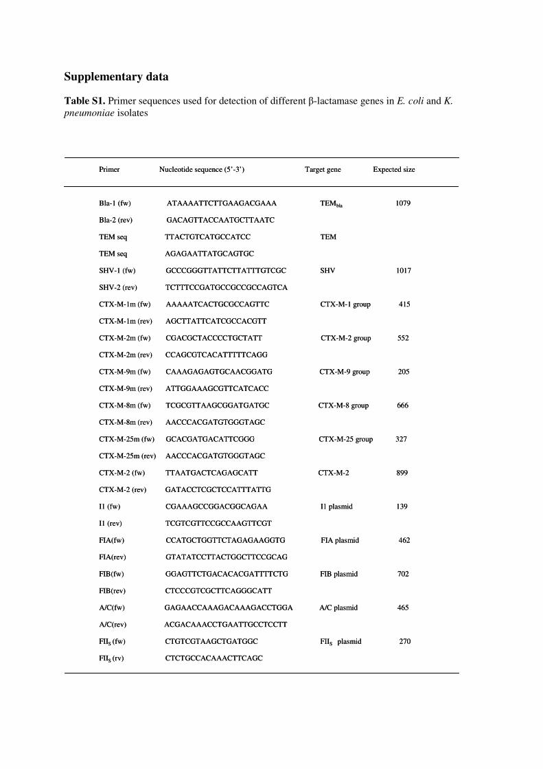
Supplementary data
Table S1. Primer sequences used for detection of different β-lactamase genes in E. coli and K.
pneumoniae isolates
Primer Nucleotide sequence (5’-3’) Target gene Expected size
Bla-1 (fw) ATAAAATTCTTGAAGACGAAA TEMbla 1079
Bla-2 (rev) GACAGTTACCAATGCTTAATC
TEM seq TTACTGTCATGCCATCC TEM
TEM seq AGAGAATTATGCAGTGC
SHV-1 (fw) GCCCGGGTTATTCTTATTTGTCGC SHV 1017
SHV-2 (rev) TCTTTCCGATGCCGCCGCCAGTCA
CTX-M-1m (fw) AAAAATCACTGCGCCAGTTC CTX-M-1 group 415
CTX-M-1m (rev) AGCTTATTCATCGCCACGTT
CTX-M-2m (fw) CGACGCTACCCCTGCTATT CTX-M-2 group 552
CTX-M-2m (rev) CCAGCGTCACATTTTTCAGG
CTX-M-9m (fw) CAAAGAGAGTGCAACGGATG CTX-M-9 group 205
CTX-M-9m (rev) ATTGGAAAGCGTTCATCACC
CTX-M-8m (fw) TCGCGTTAAGCGGATGATGC CTX-M-8 group 666
CTX-M-8m (rev) AACCCACGATGTGGGTAGC
CTX-M-25m (fw) GCACGATGACATTCGGG CTX-M-25 group 327
CTX-M-25m (rev) AACCCACGATGTGGGTAGC
CTX-M-2 (fw) TTAATGACTCAGAGCATT CTX-M-2 899
CTX-M-2 (rev) GATACCTCGCTCCATTTATTG
I1 (fw) CGAAAGCCGGACGGCAGAA I1 plasmid 139
I1 (rev) TCGTCGTTCCGCCAAGTTCGT
FIA(fw) CCATGCTGGTTCTAGAGAAGGTG FIA plasmid 462
FIA(rev) GTATATCCTTACTGGCTTCCGCAG
FIB(fw) GGAGTTCTGACACACGATTTTCTG FIB plasmid 702
FIB(rev) CTCCCGTCGCTTCAGGGCATT
A/C(fw) GAGAACCAAAGACAAAGACCTGGA A/C plasmid 465
A/C(rev) ACGACAAACCTGAATTGCCTCCTT
FIIS (fw) CTGTCGTAAGCTGATGGC FIIS plasmid 270
FIIS (rv) CTCTGCCACAAACTTCAGC


Chapter 4
Important methodological considerations with respect to the
differentiation of CTX-M-15 and CTX-M-28 extended-spectrum
β-lactamases
Godfred A. Menezes
Mushtaq A. Khan,
John P. Hays
Indian Journal of Medical Microbiology
Accepted for publication


Chapter 4 Important methodological considerations
Dear Editor,
The acquisition and expression of β-lactamases by bacteria is a major health concern in the treatment of infectious disease, with the increase in carriage of extended-spectrum β-lactamases (ESBLs) being of particular concern. CTX-M β-lactamases constitute one family of the these rapidly disseminating ESBL enzymes, having been identified in numerous countries within Africa, Asia, Europe, South America and the USA, also including India [1, 2]. Phylogenic studies have revealed 5 major groups of acquired CTX-M enzymes, CTX-M-1, CTX-M-2, CTX-M-8, CTX-M-9 and CTX-M-25, with CTX-M-15 and CTX-M-28 (the subject of this letter) belonging to the CTX-M-1 group [3]. More specifically, CTX-M-15 has been frequently isolated from India [4], whilst CTX-M-28 is much less common, only recently having been identified in India and Tunisia [5, 6]. Further, these two enzymes only differ by a single amino acid substitution, which is present at the C-terminus of the protein (3’-end of the CTX-M gene), and it is usual to distinguish between these two CTX-M types (as well as other CTX-M types) using nucleotide sequencing (see GenBank accession numbers AY044436 and AJ549244).
In this respect, we would like to caution researchers to use at least one CTX-M flanking gene primer (3’-end) in order to obtain sequencing products that accurately differentiate between CTX-M-15 and CTX-M-28. In particular, if nucleotide polymorphisms exist in the PCR primers used to amplify CTX-M genes, and the resultant PCR products are subsequently sequenced, then these nucleotide polymorphisms will be present in any subsequent sequence data obtained from the original PCR product. For example, Achour et al. [5] used four CTX-M group-specific primer sets to screen for CTX-M genes, and then cloned and sequenced the resulting PCR products. However, the use of primers CTX-1F and CTX-1R generates a PCR product with 5’- and 3’- termini comprising CTX-M-28-specific sequences (Figure 1). Moreover, even if the gene present was actually CTX-M-15, subsequent PCR screening, cloning and sequencing would generate a CTX-M-28 sequence, due to the use of non-homologous primers. On the other hand, the use of CTX-M-15 specific primers and sequencing of subsequent PCR products will mis-identify any CTX-M-28 sequences (Figure 1). This may have been the case in publications by Moubareck et al. (2005) [6] and Weill et al. (2004) [7].
In fact, the mis-identification of CTX-M-15 and CTX-M-28 enzymes is due to the fact that the nucleotide sequences are very similar, with only 2 single nucleotide substitutions. Moreover, these sequence differences occur close to the 5’-end (position 21) and 3’-end (position 865) of the CTX-M gene, the 5’-end and 3’-end of genes usually being chosen as sites to design “specific” PCR screening primers (Figure 1).
We therefore caution researchers against using CTX-M gene 3’-end PCR screening primers to generate PCR product that will be later used in sequencing reactions, especially in regions where CTX-M- 15 and CTX-M-28 are present.
61

Chapter 4 Important methodological considerations
References 1. Tzouvelekis LS, Tzelepi E, Tassios PT et al. CTX-M-type β-lactamases: an
emerging group of extended-spectrum enzymes. Int J Antimicrob Agents 2000; 14:137–42.
2. Padmini BS, Raju AB, Mani KR. Detection of Enterobacteriaceae producing CTX-M extended-spectrum β-lactamases from a tertiary care hospital in south India. Indian J Med Microbiol 2008; 26:163-6.
3. Bonnet R. Growing group of extended-spectrum β-lactamases: the CTX-M enzymes. Antimicrob Agents Chemother 2004;48:1–14.
4. Muzaheed, Doi Y, Adams-Haduch JM et al. High prevalence of CTX-M-15-producing Klebsiella pneumoniae among inpatients and outpatients with urinary tract infection in Southern India. J Antimicob Chemother 2008; 61(6): 1393-4.
5. Achour NB, Mercuri PS, Power P et al. First detection of CTX-M-28 in a Tunisian hospital from a cefotaxime-resistant Klebsiella pneumoniae strain. Pathol Biol 2008; Sep 30. [Epub ahead of print].
6. Moubareck C, Daoud Z, Hakimé NI et al. Countrywide spread of community- and hospital-acquired extended-spectrum β-lactamase (CTX-M-15)-producing Enterobacteriaceae in Lebanon. J Clin Microbiol 2005;43(7):3309-13.
7. Weill, F-X, Perrier-Gros-Claude JD et al. Characterization of extended-spectrum-β-lactamase (CTX-M-15)-producing strains of Salmonella enterica isolated in France and Senegal. FEMS Microbiol Letters 2004; 238(2):353-8.
62

Chapter 4 Important methodological considerations
Figure 1. Problems associated with using non-homologous PCR primers to generate CTX-M sequencing products to differentiate between CTX-M-15 and CTX-M-28. CTX-M-15, reference CTX-M-15 sequence (GenBank Accession number AY044436). CTX-M28, reference CTX-M-28 sequence (GenBank Accession number AJ549244). CTX-1F and CTX-1R, PCR primers used by Achour et al. (2008) [5]. CTX-M-1-F and CTX-M-15-R, PCR primers used by Moubarek et al. (2005) [6] and Weill et al. (2004) [7]. Arrows represent direction of PCR amplification. Underlined nucleotides are the stop codon. The cytosine to thymidine nucleotide substitution (position 21) is silent. NB: The reverse primers have been reverse complemented to match the direction of the gene sequence.
63


Chapter 5
Molecular characterization of extended-spectrum β-lactamase
producing Klebsiella pneumoniae isolated from blood cultures in
Pondicherry, India
Mohamudha R. Parveen
Mushtaq A. Khan
Godfred A. Menezes
Belgode N. Harish
Subhash C. Parija
John P. Hays
Submitted for publication
Med. Library Erasmus MC
EMBARGO
EMBARGO


Chapter 6
Molecular characterization of antimicrobial resistance in
non-typhoidal salmonellae from India
Godfred. A. Menezes
Mushtaq. A. Khan
Belgode. N. Harish
Subhash. C. Parija
Wil. Goessens
John. P. Hays
Submitted for publication
Med. Library Erasmus MC
EMBARGO
EMBARGO

Chapter 7
Analysis of VanA vancomycin-resistant Enterococcus faecium
isolates from Saudi Arabian hospitals reveals the presence of
clonal cluster 17 and two new Tn1546 lineage types
Mushtaq. A. Khan
Martin van der Wal
David J. Farrell
Luke Cossins
Alex van Belkum
Alwaleed Alaidan
John. P. Hays
Journal of Antimicrobial Chemotherapy
(2008) 62, 279–283

Chapter 7 Vancomycin resistant E. faecium from Saudi Arabia
Abstract Objectives: The aim of this study was to characterize 34 vancomycin-resistant VanA Enterococcus faecium isolates obtained from two hospitals in Saudi Arabia and to assess Tn1546 variation within these isolates. Methods: PFGE and multilocus sequence typing (MLST) genotypes, antibiotic susceptibility patterns, the presence of enterococcal surface protein (esp) and hyaluronidase (hyl) genes and conjugation frequencies were determined. In addition, Tn1546 elements were characterized. Results: PFGE and MLST analysis revealed the presence of 31 and 6 different genotypes, respectively. Further, three new ST types were discovered. Ninety-seven percent (33/34) of the isolates were associated with clonal complex 17 (CC17), with all isolates but one being resistant to ampicillin and all isolates being susceptible to linezolid. The esp and hyl genes were found in 44% (15/34) and 53% (18/34) of the isolates, respectively. Tn1546 analysis revealed that the isolates belonged to five different groups, including two new lineages. The IS-element insertions described did not abolish the transfer of VanA resistance. Conclusions: VanA vancomycin-resistant E. faecium isolates obtained from Saudi Arabian hospitals include CC17 MLST types, a clonal cluster associated with E. faecium nosocomial infection worldwide. Novel E. faecium MLST types are circulating in Saudi Arabia, as well as novel Tn1546 types. It seems likely that CC17 E. faecium isolates may be distributed throughout the Middle East as well as Europe, America, Africa and Australia.
92

Chapter 7 Vancomycin resistant E. faecium from Saudi Arabia
Introduction
Enterococci and vancomycin-resistant enterococci (VRE) are often found in the human and animal gut and have become increasingly responsible for nosocomial infections, particularly in the USA. In Europe, however, VRE have tended to be associated with community carriage and occasional nosocomial outbreaks, although the incidence of VRE infection may be changing [1]. Enterococci often express high-level resistance to glycopeptides and aminoglycosides, with vancomycin resistance having been promoted via the extensive use of vancomycin in hospitals, as well as the animal growth promoter avoparcin.
Six different vancomycin resistance types have so far been described in enterococci, namely VanA, VanB, VanC, VanD, VanE and VanG. The VanA type is characterized by both high-level and inducible resistance to vancomycin (MICs 64–1024 mg/L) and teicoplanin (MICs 16–512 mg/L) and has been shown to be directly facilitated by the carriage of transposon Tn1546, a transposon widely disseminated in humans, animals and the environment [2].
Currently, there is a distinct lack of data regarding the molecular analysis of VanA Enterococcus faecium Tn1546 carriage in VRE isolates originating from the Middle East, including Saudi Arabia. Therefore, the aim of this study was to determine E. faecium genotypic variation and VanA Tn1546 transposon diversity in VRE isolates recovered from the Kingdom of Saudi Arabia. Materials and methods Bacterial isolates
Thirty-four vancomycin-resistant E. faecium isolates were cultured from clinical specimens obtained from two large tertiary-care hospitals in the Kingdom of Saudi Arabia, namely the King Faisal Specialist Hospital and Research Centre, Riyadh (KFSH&RC), and the King Fahad National Guard Hospital, King Abdulaziz Medical City, Riyadh (KFNGH). Isolates were collected between 2000 and2003. Two pairs of isolates (25/29 and 17/32) were isolated from two different patients, but on separate occasions. PFGE and multilocus sequence typing (MLST)
PFGE was performed using SmaI-digested fragments of bacterial chromosomal DNA, with fragment separation achieved in 0.8% agarose. Electrophoresis conditions comprised using a constant voltage of 6 V/cm at 148C and pulse times of 3.5–25 s increased linearly over 12 h (block 1), followed by 1–5 s increased over 8 h. Gel patterns were analysed using BioNumerics software (Applied Maths) with the band tolerance set at 1.0%. MLST was performed using internal fragments from seven housekeeping genes. Allele numbers and sequence types were assigned after reference to the online E. faecium MLST database at http://efaecium.mlst.net/.
Antibiotic susceptibility testing
Antibiotic susceptibility testing was performed using the VITEK 2 automated identification and susceptibility system (bioMérieux, Marcy l’Étoile, France) according to the manufacturer’s instructions.
93

Chapter 7 Vancomycin resistant E. faecium from Saudi Arabia
PCR characterization of Tn1546 elements and screening for virulence genes
Primer pairs used to characterize Tn1546 were based on those published by Miele et al. [3]. In addition, primer walking was utilized to define insertion site-specific sequences. PCR screening for the enterococcal surface protein (esp) and hyaluronidase (hyl) virulence genes used three different primer pairs. For the esp gene, primer pairs esp11 (50-TTGCTAATGCTAGTCCACGACC-30)/esp12 (50-GCGTCAACACTTGCATTGCCGAA-30) and 14F (50-AGATTTCATCTTTGATTCTTGG-30)/12R (50-AATTGATTCTTTAGCATCTGG- 30) were used, and for the hyl gene, primer pair HYL n1 (50-ACAGAAGAGCTGCAGGAAATG-30)/HYL n2 (50-GACTGACGTCCAAGTTTCCAA-30). A touchdown PCR protocol was used for all primer pairs, comprising an initial annealing temperature of 65ºC, which was initially reduced by 18C per cycle over 15 cycles, and then followed by 20 cycles of amplification using an annealing temperature of 55ºC. All PCRs were performed using Taq DNA polymerase (EP0402, Fermentas).
Conjugation frequency
Conjugation frequency was determined using a standard methodology [4], with E. faecium GE1 being used as the recipient strain, and antibiotic concentrations of 6 mg/L vancomycin, 64 mg/L rifampicin and 10 mg/L fusidic acid being used. Conjugation frequencies were calculated with reference to the donor isolate.
Results and discussion PFGE and MLST genotyping
PFGE identified 18 ‘possibly related’ PFGE genotypes based on a 70% similarity cut-off value (approximately equivalent to four to six PFGE fragment differences) [5], with all isolates being genotypically related at a similarity of 34% (Figure 1). MLST genotyping revealed six ST types, with three new ST genotypes (ST 358, 359 and 360) and 59% (20/34) of the isolates being ST 17 genotypes. Further, the three new ST types (four isolates) and ST 16 (two isolates) were exclusively associated with the KFSH&RC, whereas ST 18 (eight isolates) was exclusively associated with the KFNGH. In contrast, ST 17 was recovered from both hospitals. Ninety-seven percent (33/34) of the isolates belonged to clonal complex 17 (CC17), a cluster of E. faecium MLST genotypes previously associated with global nosocomial infections [1]. CC17 has been previously associated with ampicillin resistance and carriage of the enterococcal surface protein pathogenicity island. In Saudi isolates, ampicillin resistance and esp gene carriage were found in 99% and 45% of the isolates, respectively, indicating that CC17 isolates from Saudi Arabia possess similar characteristics to those obtained from other well documented regions of the world. Antibiotic susceptibility patterns
All isolates but one were resistant to vancomycin and ampicillin, and all isolates were susceptible to linezolid (Figure 1). No pattern was observed with respect to the general expression of multiple antibiotic resistances and PFGE genotype.
Characterization of Tn1546 elements
94

Chapter 7 Vancomycin resistant E. faecium from Saudi Arabia
In total, five different Tn1546 types were observed within our 34 E. faecium isolates (Figure 2). In eight isolates (numbers 2, 4, 5, 6, 12, 34, 35 and 41), the PCR amplicon size analysis revealed no major insertions or deletions, compared with theoretical amplicon products generated using reference Tn1546 BM4147 sequence. In isolates where Tn1546 polymorphisms were found, the majority of polymorphic events involved the insertion of IS1216V within the intergenic regions of either vans and vanH or vanX and vanY genes, along with other less frequent insertions of IS1485 and IS1251. In fact, IS1216V is one of the most common insertion sequences found in Tn1546-bearing vancomycin-resistant enterococci worldwide. Further, IS1251 has previously been demonstrated to be present in Tn1546 transposons present within human clinical E. faecium isolates from Brazil, Korea, the USA, Norway and Ireland. Our Tn1546 results allowed us to add two new lineages to a previously published evolutionary scheme [6], namely lineage III (IS1485 insertion in orf1) and lineage IV (IS1216V insertion between vanS and vanH). Prevalence of esp and hyl virulence genes
Using PCR screening, the enterococcal surface protein (esp) gene was found to be present in 44% (15/34) and the hyaluronidase (hyl) gene in 53% (18/34) of the isolates tested. esp-positive isolates made up 46% (10/22) of the isolates from the KFNGH and 41% (5/12) of the isolates from the KFSH&RC. In contrast, hyl-positive isolates made up 41% (9/22) of the isolates from the KFNGH but 75% (9/12) of the isolates from the KFSH&RC. Only four esp-positive isolates were also found to be hyl-positive, whereas 14 hyl-positive isolates were found to be esp-negative. A significant difference in the prevalence of esp and hyl-positive isolates was observed with respect to PFGE clusters (Figure 1), with 71% (10/14) of the esp-positives belonging to the cluster bordered by isolates 19 and 40 (Fisher’s exact test P = 0.02), yet 75% (15/20) of the hyl-positives belonging to the cluster bordered by isolates 36 and 12 (Fisher’s exact test P = 0.004). The distribution of the two hospitals between the PFGE clusters defined earlier was not significant (Fisher’s exact test P = 0.066). The expression of enterococcal surface protein (Esp) has been associated with both biofilm formation and nosocomial infection in E. faecium [7], as well as CC17 isolates [8]. Further, a recent publication indicated that clinical E. faecium isolates carrying the esp gene possess an increased conjugation frequency with respect to the ability to acquire VanA vancomycin resistance [9]. The prevalence of the esp gene within our 33 CC17 isolates was somewhat lower than previously reported [1], although all isolates were ampicillin-resistant. However, 91% (30/33) of the CC17 isolates possessed either the esp or hyl virulence genes. The hyaluronidase protein (Hyl) is a virulence trait also associated with clinical E. faecium isolates, with a ratio of 2:1 for esp:hyl-positive isolates reported in a sample of 577 isolates worldwide [10]. This compares with a ratio of 1:1.2 in our smaller group. Conjugation frequency
Conjugation frequencies of representative isolates from each transposon group ranged from 2.2 x 10⁻⁴ to 1.9 x 10⁻⁶. These frequencies lie within the range of previously published transformation frequencies observed for other global VanA-resistant, Tn1546-carrying E. faecium isolates.
Conclusions
In this study, we investigated 34 vancomycin-resistant VanA E. faecium isolates cultured from two hospitals situated in Riyadh, Saudi Arabia. The majority of the isolates
95

Chapter 7 Vancomycin resistant E. faecium from Saudi Arabia
tested belonged to CC17 and were resistant to ampicillin, suggesting that E. faecium MLST types associated with nosocomial infection are also circulating in the Middle East. The Tn1546 analysis revealed two new lineages, including two previously undescribed IS1485 and IS1216V insertions.
Acknowledgements We would like to acknowledge the routine microbiology laboratory of Erasmus MC with respect to the VITEK 2 system. E. faecium isolate GE1 was kindly supplied by Dr Ana Freitas, REQUIMTE Lab. Microbiologia, Faculdade de Farmacia, Universidade do Porto, Porto, Portugal.
Isolate Hosp. esp hyl VAN AMP GEN TET CIP Q/D LZD MLST 100
806040
192316172829263225273314140363735393038691418710118134534212
NNNNNNNNNNNKKNNNNNNNKKNNKKKKNKKNKK
RRRRRRR
RRRRRRRRRRRRRRRRRRRRRRRRRR
RRRRRRR
RRSRRRSSSSSSRRRRRRRRRSSSSS
RRRRRRR
RRSSRRSSSSSSRRRRRRRRRSSRRS
SSSSSSS
SSSISSSSSSSSSSSSSSSSSSSSSS
RRRRRRR
RRRIRRRRRRRRRRIRRRRRRRRRRI
R RR SR
------+
--+-+-++++++++--++++---+++
-
17171717171717
17171835836017181817181818359359181817171717171616171717
17
+-+++++
+---++------++------+++---
+
RRRRRRR
RRRRRRRRRRRRRRSRRRRRRRRRRR
R
SSSSSSS
SSSSSSSSSSSSSSSSSSSSSSSSSS
S
Isolate Hosp. esp hyl VAN AMP GEN TET CIP Q/D LZD MLST 100
806040 100
806040
192316172829263225273314140363735393038691418710118134534212
NNNNNNNNNNNKKNNNNNNNKKNNKKKKNKKNKK
RRRRRRR
RRRRRRRRRRRRRRRRRRRRRRRRRR
RRRRRRR
RRSRRRSSSSSSRRRRRRRRRSSSSS
RRRRRRR
RRSSRRSSSSSSRRRRRRRRRSSRRS
SSSSSSS
SSSISSSSSSSSSSSSSSSSSSSSSS
RRRRRRR
RRRIRRRRRRRRRRIRRRRRRRRRRI
R RR SR
------+
--+-+-++++++++--++++---+++
-
17171717171717
17171835836017181817181818359359181817171717171616171717
17
+-+++++
+---++------++------+++---
+
RRRRRRR
RRRRRRRRRRRRRRSRRRRRRRRRRR
R
SSSSSSS
SSSSSSSSSSSSSSSSSSSSSSSSSS
S
96

Chapter 7 Vancomycin resistant E. faecium from Saudi Arabia
Figure 1. PFGE patterns obtained from 34 vancomycin-resistant E. faecium isolates from Saudi Arabia. Cluster analysis was performed using the method of DICE with UPGMA with band tolerances set to 1.0%. Hosp. = hospital, where K = King Faisal Specialist Hospital and Research Centre and N = King Fahad National Guard Hospital. esp, enterococcal surface protein PCR; hyl, hyaluronidase PCR; AMP, ampicillin; GEN, gentamicin; TET, tetracycline; Q/D, quinupristin/dalfopristin; LZD, linezolid; CIP, ciprofloxacin; R, resistant; I, intermediate resistance; S, susceptible.
orf1 orf2 vanR vanS vanH vanA vanX vanY vanZ
5823 5816IS1216V
J* (6)
B (14)
8731 8724IS1216V
631 629IS1485
H *(1)
A1 (8)
IS1251
F (5)
5820 5813889 bp deletion
IS1216V
167bp pEFNP1-fragment insertion
orf1 orf2 vanR vanS vanH vanA vanX vanY vanZ
5823 5816IS1216V
J* (6)
B (14)
8731 8724IS1216V
8731 8724IS1216V
8731 8724IS1216V
631 629IS1485
H *(1)
A1 (8)
IS1251
F (5)
5820 5813889 bp deletion
IS1216V
167bp pEFNP1-fragment insertion
Figure 2. Genetic maps of Tn1546 types associated with 34 E. faecium isolates originating from Saudi Arabia. The unbroken black lines represent reference Tn1546 sequence BM4147. Unfilled block arrows represent the positions of genes and open reading frames (orf1 and orf2). Filled boxes represent IS elements and arrows their direction of transcription. Dotted lines represent deletions. The unfilled box represents a sequence similar to plasmid pEFNP1 (Losteinkit et al., 2000; GenBank accession number AB038522, unpublished results). The positions of the first nucleotides upstream and downstream of the insertion sequence are shown. Group definitions (right-hand side) are based on those published by Schouten et al. [6]. Asterisk indicates novel Tn1546 group. Numbers in parentheses indicate how many isolates belonged to each Tn1546 grouping.
97

Chapter 7 Vancomycin resistant E. faecium from Saudi Arabia
References 1. Willems RJ, Top J, van Santen M et al. Global spread of vancomycin-resistant
Enterococcus faecium from distinct nosocomial genetic complex. Emerg Infect Dis 2005; 11: 821–8.
2. Biavasco F, Foglia G, Paoletti C et al. VanA-type enterococci from humans, animals, and food: species distribution, population structure, Tn1546 typing and location, and virulence determinants. Appl Environ Microbiol 2007; 73: 3307–19.
3. Miele A, Bandera M, Goldstein BP. Use of primers selective for vancomycin resistance genes to determine van genotype in enterococci and to study gene organization in VanA isolates. Antimicrob Agents Chemother 1995; 39: 1772–8.
4. Novais C, Coque TM, Costa MJ et al. High occurrence and persistence of antibiotic-resistant enterococci in poultry food samples in Portugal. J Antimicrob Chemother 2005; 56: 1139–43.
5. Tenover FC, Arbeit RD, Goering RV et al. Interpreting chromosomal DNA restriction patterns produced by pulsed-field gel electrophoresis: criteria for bacterial strain typing. J Clin Microbiol 1995; 33: 2233–9.
6. Schouten MA, Willems RJ, Kraak WA et al. Molecular analysis of Tn1546-like elements in vancomycin-resistant enterococci isolated from patients in Europe shows geographic transposon type clustering. Antimicrob Agents Chemother 2001; 45: 986–9.
7. Van Wamel WJ, Hendrickx AP, Bonten MJ et al. Growth condition-dependent Esp expression by Enterococcus faecium affects initial adherence and biofilm formation. Infect Immun 2007; 75: 924–31.
8. Willems RJ, Homan W, Top J et al. Variant esp gene as a marker of a distinct genetic lineage of vancomycin-resistant Enterococcus faecium spreading in hospitals. Lancet 2001; 357: 853–5.
9. Lund B, Billstrom H, Edlund C. Increased conjugaon frequencies in clinical Enterococcus faecium strains harbouring the enterococcal surface protein gene esp. Clin Microbiol Infect 2006; 12: 588–91.
10. Rice LB, Carias L, Rudin S et al. A potential virulence gene, hylEfm, predominates in Enterococcus faecium of clinical origin. J Infect Dis 2003; 187: 508–12.
98

Chapter 8
High prevalence of ST-78 infection-associated vancomycin-
resistant Enterococcus faecium from hospitals in Asunción,
Paraguay
M. A. Khan
J. B. Northwood
R. G. J. Loor
A. T. R. Tholen
E. Riera
M. Falcon
Paraguayan Antimicrobial Network
A. van Belkum
M. van Westreenen
J. P. Hays
Clin Microbiol Infect. 2009 Jul 20.
[Epub ahead of print]

Chapter 8 Vancomycin-resistant Enterococcus faecium from Paraguay
Abstract
Forty infection-associated VanA-type vancomycin-resistant Enterococcus faecium (VRE) strains obtained from five collaborating hospitals in Asunción, Paraguay were investigated. Genotyping using pulsed-field gel electrophoresis (PFGE) and multilocus sequence typing revealed the presence of 17 cluster types and four STs, with 93% (37/40) of isolates comprising ST type 78. Other ST types included ST-132, ST-210 and one new ST type (ST-438). All but one isolate (ST-438) were associated with clonal complex 17 (CC17), and 97% of the total isolates carried the esp gene. Three Tn1546 variants were found, including a new lineage containing an ISEfa5 insertion in an existing IS1251 element.
100

Chapter 8 Vancomycin-resistant Enterococcus faecium from Paraguay
Vancomycin-resistant Enterococci (VRE) are nosocomial pathogens widely disseminated within hospitals [1], with the vanA genotype being the most common type of acquired glycopeptide resistance found in these enterococci. Although the first clinical isolate of VRE was detected in Europe in 1986, studies have now shown that clinical VRE may be isolated in several world regions [2]. Further, it is currently accepted that a single focal group of vanA-type VRE (clonal complex 17 or CC17) is particularly associated with nosocomial infections [3]. However, there is some evidence to suggest that this grouping may be influenced by the high rates of recombination associated with E. faecium [4].
In 1988, French researchers identified a small mobile genetic element in VanA-type Enterococcus faecium called Tn1546, which is involved in vancomycin resistance [5], and there are currently five other acquired vancomycin resistance genotypes reported in enterococci, namely vanB, vanD, vanE, vanG and vanL [5, 6].
Currently, there exist limited data regarding the incidence and types of clinical vancomycin-resistant E. faecium from countries in South America. Therefore, the objective of this study was to investigate the complexity of clinical E. faecium isolates from Paraguay, by determining E. faecium genotypes and VanA Tn1546 transposon diversity, and to compare the results with isolates from both South America and the rest of the world.
Two hundred and twenty-one VanA vancomycin-resistant E. faecium isolates were collected between January 2005 and March 2007 from five collaborating hospitals in Asunción Paraguay. These five hospitals, the Instituto de Previsión Social (IPS), Hospital de Clinicas (HCL), Hospital Nacional (HNA), Centro Medico de Bautista (CMB) and Instituto de Medicina Tropical (IMT), are representative of the national healthcare system in Paraguay. A subset of 40 isolates was chosen for further analysis, based on their site of isolation and association with disease. These infection-associated isolates were cultured from abscess material, blood, tissue and needle aspirates, representing 7% (7/97), 20% (1/5) and 27% (32/117) of the isolates obtained from HCL, HNA, and IPS, respectively. No infection-associated isolates were obtained from CMB (0/1) and IMT (0/1) hospitals.
Antibiotic resistance profiling was performed using the VITEK 2 system (BioMérieux, Marcy l’Étoile, France) and E-test (amoxicillin/clavulanic acid), according to CLSI guidelines. All 40 infection-associated isolates tested in this study were resistant to ampicillin, amoxicillin/clavulanic acid, ciprofloxacin, erythromycin, imipenem, penicillin, teicoplanin and vancomycin. All isolates were sensitive to linezolid and tetracycline. Forty-three per cent (17/40) of the isolates were resistant to gentamicin (Figure. 1). The enterococcal surface protein gene (esp) was detected by PCR screening [7], revealing that all isolates were esp positive with the exception of one isolate (ST132). The prevalence of esp in these isolates is higher than previously reported for European CC17 isolates [8].
Pulsed-field gel electrophoresis (PFGE) and multi locus sequence typing (MLST) genotyping were performed as previously described [9]. PFGE results revealed 17 cluster types with no identifiable yearly pattern per cluster. Cluster analysis was performed using the method of DICE with UPGMA, with band tolerance set at 1.5% and the threshold cut-off value at 80% [10, 11]. The results of MLST genotyping revealed three known (ST-78, ST-132 and ST-210) and one novel (ST-438) ST type, with 93% (36/40) of isolates comprising ST-78 genotypes. These ST-78 isolates comprised 100% of all infection-associated isolates from the HCL and HNA hospitals, and 87% (28/32) of the isolates from the IPS. Although this ST type has previously been reported in Europe, it has not been reported in South America, the USA or Spain, approximately 95% of Paraguayans are of mixed Spanish and Amerindian descent. Therefore, the origins of ST-78 in Paraguay remain unexplained, although it appears that the ST-78 strain has spread to many hospitals in Asunción.
In total, 97% (39/40) of the isolates tested belonged to MLST clonal complex 17 (CC17), which comprises a cluster of E. faecium genotypes previously associated with
101
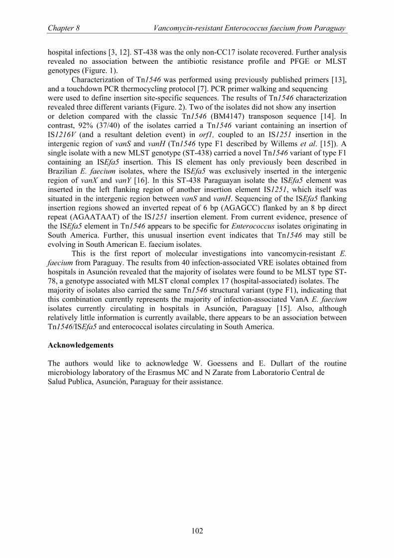
Chapter 8 Vancomycin-resistant Enterococcus faecium from Paraguay
hospital infections [3, 12]. ST-438 was the only non-CC17 isolate recovered. Further analysis revealed no association between the antibiotic resistance profile and PFGE or MLST genotypes (Figure. 1).
Characterization of Tn1546 was performed using previously published primers [13], and a touchdown PCR thermocycling protocol [7]. PCR primer walking and sequencing were used to define insertion site-specific sequences. The results of Tn1546 characterization revealed three different variants (Figure. 2). Two of the isolates did not show any insertion or deletion compared with the classic Tn1546 (BM4147) transposon sequence [14]. In contrast, 92% (37/40) of the isolates carried a Tn1546 variant containing an insertion of IS1216V (and a resultant deletion event) in orf1, coupled to an IS1251 insertion in the intergenic region of vanS and vanH (Tn1546 type F1 described by Willems et al. [15]). A single isolate with a new MLST genotype (ST-438) carried a novel Tn1546 variant of type F1 containing an ISEfa5 insertion. This IS element has only previously been described in Brazilian E. faecium isolates, where the ISEfa5 was exclusively inserted in the intergenic region of vanX and vanY [16]. In this ST-438 Paraguayan isolate the ISEfa5 element was inserted in the left flanking region of another insertion element IS1251, which itself was situated in the intergenic region between vanS and vanH. Sequencing of the ISEfa5 flanking insertion regions showed an inverted repeat of 6 bp (AGAGCC) flanked by an 8 bp direct repeat (AGAATAAT) of the IS1251 insertion element. From current evidence, presence of the ISEfa5 element in Tn1546 appears to be specific for Enterococcus isolates originating in South America. Further, this unusual insertion event indicates that Tn1546 may still be evolving in South American E. faecium isolates.
This is the first report of molecular investigations into vancomycin-resistant E. faecium from Paraguay. The results from 40 infection-associated VRE isolates obtained from hospitals in Asunción revealed that the majority of isolates were found to be MLST type ST-78, a genotype associated with MLST clonal complex 17 (hospital-associated) isolates. The majority of isolates also carried the same Tn1546 structural variant (type F1), indicating that this combination currently represents the majority of infection-associated VanA E. faecium isolates currently circulating in hospitals in Asunción, Paraguay [15]. Also, although relatively little information is currently available, there appears to be an association between Tn1546/ISEfa5 and enterococcal isolates circulating in South America. Acknowledgements The authors would like to acknowledge W. Goessens and E. Dullart of the routine microbiology laboratory of the Erasmus MC and N Zarate from Laboratorio Central de Salud Publica, Asunción, Paraguay for their assistance.
102

Chapter 8 Vancomycin-resistant Enterococcus faecium from Paraguay
Dice (Tol 1.5%-1.5%) (H>0.0% S>0.0%) [0.0%-100.0%]Paraguay VRE 24.02.09
1987654Paraguay VRE 24.02.09 Key
11464749
2042114212
11022156
58529
7111312326
6551130
111118105
210211117107
110059106120
2131042
4312127
18914
MLST
7878132
78787878
787878
787878
78787878
787878
7843878
78787878
78787878
787878
7878132
2107878
1146474920421142121102
2156585297111312326
6551130111118105210211
117107110059106120213104
2431212718914
78781327878787878
787878787878787878
78787878438*
787878
7878787878787878
7878781322107878
HCLIPSIPSIPSIPSIPSHCLIPS
IPSIPSIPSIPSIPSIPSIPSIPSIPS
HNAIPSIPSHCLIPSIPSHCLHCL
HCLIPSIPSIPSIPSIPSHCLIPS
IPSIPSIPSIPSIPSIPSIPS
2006-112005-092005-102005-042005-062006-052007-042006-10
2005-042005-112005-112005-032005-052005-032006-102006-072005-05
2005-032005-112006-112006-052006-072006-022007-042007-04
2006-072006-022006-102005-112006-022006-072007-042006-02
2005-022005-062006-072005-052006-102005-022005-03
RIRRRRRR
RRRRRRRRI
RRRIRRRR
RRRRRRRR
RRRRRRR
RSSRRSRR
RSSRRSSSS
RSRSRRSR
RSRSSSSS
SSSRRSS
RRIRRRRR
RIRRRRRSR
RRRRRRRR
RRRRRRRR
RRRIRIR
ISRISIIS
IRSISIISS
ISISIIIS
IIIIIIII
ISIRSSI
RSRRRRRR
RRRRRRRRS
RRRRRRRR
RRRRRRRR
RRRRRRR
STIsolate Hospital ERY GEN NIT Q/D STRDate
100
908070605040
Dice (Tol 1.5%-1.5%) (H>0.0% S>0.0%) [0.0%-100.0%]Paraguay VRE 24.02.09
1987654Paraguay VRE 24.02.09 Key
11464749
2042114212
11022156
58529
7111312326
6551130
111118105
210211117107
110059106120
2131042
4312127
18914
MLST
7878132
78787878
787878
787878
78787878
787878
7843878
78787878
78787878
787878
7878132
2107878
1146474920421142121102
2156585297111312326
6551130111118105210211
117107110059106120213104
2431212718914
78781327878787878
787878787878787878
78787878438*
787878
7878787878787878
7878781322107878
HCLIPSIPSIPSIPSIPSHCLIPS
IPSIPSIPSIPSIPSIPSIPSIPSIPS
HNAIPSIPSHCLIPSIPSHCLHCL
HCLIPSIPSIPSIPSIPSHCLIPS
IPSIPSIPSIPSIPSIPSIPS
2006-112005-092005-102005-042005-062006-052007-042006-10
2005-042005-112005-112005-032005-052005-032006-102006-072005-05
2005-032005-112006-112006-052006-072006-022007-042007-04
2006-072006-022006-102005-112006-022006-072007-042006-02
2005-022005-062006-072005-052006-102005-022005-03
RIRRRRRR
RRRRRRRRI
RRRIRRRR
RRRRRRRR
RRRRRRR
RSSRRSRR
RSSRRSSSS
RSRSRRSR
RSRSSSSS
SSSRRSS
RRIRRRRR
RIRRRRRSR
RRRRRRRR
RRRRRRRR
RRRIRIR
ISRISIIS
IRSISIISS
ISISIIIS
IIIIIIII
ISIRSSI
RSRRRRRR
RRRRRRRRS
RRRRRRRR
RRRRRRRR
RRRRRRR
STIsolate Hospital ERY GEN NIT Q/D STRDate
100
908070605040 100
908070605040
Figure 1. Pulsed-field gel electrophoresis (PFGE) patterns obtained from 40 infection-associated vancomycin-resistant Enterococcus faecium isolates obtained from Paraguay between 2005 and 2007. Cluster analysis was performed using the method of DICE with UPGMA with band tolerance set to 1.5%.
103

Chapter 8 Vancomycin-resistant Enterococcus faecium from Paraguay
orf1 orf2 vanR vanS vanH vanA vanX vanY vanZ
A1
F1*
BM4147
F1
IS12515820 5813
889 bp deletion
IS1216V
619bp pEFNP1-fragment
889 bp deletion
IS1216V
619bp pEFNP1-fragment
Group
(2)
(37)
(1)
IS12515820 5813
45 38ISEfa5
No of isolates
orf1 orf2 vanR vanS vanH vanA vanX vanY vanZ
A1
F1*
BM4147
F1
IS12515820 5813
889 bp deletion
IS1216V
619bp pEFNP1-fragment
889 bp deletion
IS1216V
619bp pEFNP1-fragment
Group
(2)
(37)
(1)
IS12515820 5813
45 38ISEfa5
No of isolates
Figure 2. PCR and sequence-based characterization of Tn1546 in 40 infection-related Enterococcus faecium isolates from Paraguay. Tn1546 reference sequence BM4147 is represented by the unbroken black lines. The position of different genes and open reading frames (orf1 and orf2) are shown by filled block arrows. IS elements are shown by filled boxes and their direction of transcription is represented by arrows. Deletions are represented by dotted lines. The positions of the first nucleotides upstream and downstream of the insertion sequences are shown. The groups are based on the published results of Willems et al. [15]. *A novel F1 variant of Tn1546. Numbers on the left indicate how many isolates were found to belong to each Tn1546 variant.
104

Chapter 8 Vancomycin-resistant Enterococcus faecium from Paraguay
References 1. Bonten MJ, Willems R, Weinstein RA. Vancomycin-resistant enterococci: why are
they here, and where do they come from? Lancet Infect Dis 2001; 1: 314–325. 2. Top J, Willems R, Bonten M. Emergence of CC17 Enterococcus faecium: from
commensal to hospital-adapted pathogen. FEMS Immunol Med Microbiol 2008; 52: 297–308.
3. Willems RJ, Top J, van Santen M et al. Global spread of vancomycin-resistant Enterococcus faecium from distinct nosocomial genetic complex. Emerg Infect Dis 2005; 11: 821–828.
4. Turner KM, Hanage WP, Fraser C et al. Assessing the reliability of eBURST using simulated populations with known ancestry. BMC Microbiol 2007; 7: 30.
5. Courvalin P. Vancomycin resistance in gram-positive cocci. Clin Infect Dis 2006; 42 (suppl 1): S25–S34.
6. Boyd DA, Willey BM, Fawcett D et al. Molecular characterization of Enterococcus faecalis N06-0364 with low-level vancomycin resistance harboring a novel D-Ala-D-Ser gene cluster, vanL. Antimicrob Agents Chemother 2008; 52: 2667–2672.
7. Khan MA, van der Wal M, Farrell DJ et al. Analysis of VanA vancomycin-resistant Enterococcus faecium isolates from Saudi Arabian hospitals reveals the presence of clonal cluster 17 and two new Tn1546 lineage types. J Antimicrob Chemother 2008; 62: 279–283.
8. Top J, Willems R, van der Velden S et al. Emergence of clonal complex 17 Enterococcus faecium in The Netherlands. J Clin Microbiol 2008; 46: 214–219.
9. Homan WL, Tribe D, Poznanski S et al. Multilocus sequence typing scheme for Enterococcus faecium. J Clin Microbiol 2002; 40: 1963–1971.
10. Tenover FC, Arbeit RD, Goering RV et al. Interpreting chromosomal DNA restriction patterns produced by pulsed-field gel electrophoresis: criteria for bacterial strain typing. J Clin Microbiol 1995; 33: 2233–2239.
11. Carrico JA, Pinto FR, Simas C et al. Assessment of band-based similarity coefficients for automatic type and subtype classification ofmicrobial isolates analyzed by pulsed-field gel electrophoresis. J Clin Microbiol 2005; 43: 5483–5490.
12. Leavis HL, Bonten MJ, Willems RJ. Identification of high-risk enterococcal clonal complexes: global dispersion and antibiotic resistance. Curr Opin Microbiol 2006; 9: 454–460.
13. Miele A, Bandera M, Goldstein BP. Use of primers selective for vancomycin resistance genes to determine van genotype in enterococci and to study gene organization in VanA isolates. Antimicrob Agents Chemother 1995; 39: 1772–1778.
14. Arthur M, Molinas C, Depardieu F et al. Characterization of Tn1546, a Tn3-related transposon conferring glycopeptide resistance by synthesis of depsipeptide peptidoglycan precursors in Enterococcus faecium BM4147. J Bacteriol 1993; 175: 117–127.
15. Willems RJ, Top J, van den Braak N et al. Molecular diversity and evolutionary relationships of Tn1546-like elements in enterococci from humans and animals. Antimicrob Agents Chemother 1999; 43: 483–491.
16. Camargo IL, Zanella RC, Brandileone MC et al. Occurrence of insertion sequences within the genomes and Tn1546-like elements of glycopeptide-resistant enterococci isolated in Brazil, and identification of a novel element, ISEfa5. Int J Med Microbiol 2005; 294: 513–519.
105


Chapter 9
Molecular epidemiology of high-level aminoglycoside resistant
Enterococcus faecalis patients during and after hospitalisation
Mushtaq. A. Khan
Saswati Sinha
Hubert P. Endtz
John Blackman Northwood
Alex van Belkum
John. P. Hays
Submitted for publication
Med. Library Erasmus MC
EMBARGO
EMBARGO

Chapter 10
Summary and concluding remarksSummary and concluding remarks
Nederlandse samenvatting


Chapter 10 Summary and concluding remarks
The growing problem of antimicrobial resistance involves practically all types of pathogens and remains a significant public health concern worldwide [1, 2]. Further, one of the most alarming trends is the development of antimicrobial resistance for virtually all currently available antimicrobial drugs, sometimes even combined in a single organism. Indeed, both the Infectious Diseases Society of America and the European Society of Clinical Microbiology and Infectious Diseases have highlighted their concerns regarding the large percentage of clinical isolates that are resistant to most antimicrobial agents [3, 4]. In particular, the bacterial pathogens Staphylococcus aureus, Enterococcus species, Escherichia coli, Acinetobacter species, Pseudomonas species, Streptococcus pneumoniae and Klebsiella species are currently most associated with this problem [5-9]. This thesis provides further data on the clinical relevance and importance of antimicrobial resistance in six bacterial species, including several of the ones mentioned above. Resistance to β-lactam antibiotics
β-lactam antibiotics are a broad class of antibiotics that include penicillins and their derivatives including cephalosporins, monobactams, and carbapenems. They are one of the most important groups of antimicrobial agents currently used in the treatment of infectious diseases caused by both Gram-negative and Gram-positive bacteria. However, the development of resistance to these agents (by both Gram-negative and Gram-positive bacterial pathogens) has created great concern among health care professionals.
The main mechanism of β-lactam resistance, particularly amongst Gram-negative bacteria, is the production of β-lactamases, hydrolytic enzymes whose activity causes disruption of the amide bond of the four-membered β-lactam ring, leading to hydrolysis of the β-lactam ring, and inactivation of the β-lactam antibiotic. In fact, there are several types of β-lactamases known that confer resistance to β-lactam antibiotics. A few clinically relevant examples of these are discussed in the following section. The BRO- β-lactamase
Moraxella catarrhalis has been recognized as an important human pathogen, because it can cause upper respiratory tract infections (e.g. acute otitis media) in children and lower respiratory tract infections (e.g. chronic obstructive pulmonary disease, COPD) in adults with pre-existing and chronic pulmonary disease. Although M. catarrhalis strains are generally susceptible to most antibiotics used for the treatment of respiratory infections, they tend to be resistant to penicillin because of the widespread production of BRO β-lactamase, a unique β-lactamase first described in Sweden in 1976 [10, 11]. Subsequently, two types of BRO, named BRO-1 and BRO-2, coded by the bro-1 and bro-2 genes respectively, have been reported. The two BRO enzymes are distinguishable by their level of β-lactamase production (BRO-1> BRO-2) and different iso-electric focusing patterns (resulting from a single amino-acid substitution). Further, the bro-2 gene also has a 21 bp deletion in its promoter region compared to bro-1 [12]. Despite some initial confusion, it is now accepted that the bro gene is located on the bacterial chromosome [13], and that it may originally have been acquired from an unknown Gram-positive bacterium, due to the fact that there is no significant sequence similarity between bro and the β-lactamase genes of other Gram-negative bacteria, and that the gene possesses an LPXTG signal sequence motif, which is characteristic for Gram-positive bacteria [14, 15].
Since 1976, there has been a rapid rise in the prevalence of worldwide clinical β-lactamase resistant M. catarrhalis isolates (which now approaches 90–99% of all clinical
125

Chapter 10 Summary and concluding remarks
isolates), considered to be the fastest dissemination rate of any known bacterial β-lactamase gene [15].
In Chapter 2, we show that more than 90% of a selection of global M. catarrhalis isolates appear to be β-lactamase and bro gene positive. With respect to geographical region, increased bro-1 β-lactam MICs were observed in M. catarrhalis isolates originating from the Far East, and especially Japan, possibly due to differences in geographical antibiotic prescribing policies. Additionally, the prevalence of bro positivity between the two-age groups (children <5 years and adults > 20 years) was significantly different, which again may be due to differences in antibiotic prescribing policies (i.e. differences in age group specific prescription). Further, no clonal isolates, linked to a geographical region (including Japan and/or the Far East) were found (data not shown). Previous studies have also reported a lack of genotypic linkage between M. catarrhalis genotype and bro type, with at least one study reporting ampicillin MIC variation in apparently identical M. catarrhalis isolates cultured from an otitis media patient in Japan [16]. Further, a recent global survey of 195 M. catarrhalis isolates revealed no association between M. catarrhalis genotypes in child or adult age groups [17]. However, recent research has identified that mechanisms not associated with the bro gene per se may be involved in β-lactamase resistance, which may possibly have an effect on the results we obtained from the Far East and between children or adults [18]. Further research in this field is necessary.
The results from our study are worrying, because at the clinical level, β-lactamase production by M. catarrhalis has been reported to indirectly benefit other bacterial species present within mixed bacterial infections (e.g. S. pneumoniae and H. influenzae), as production of BRO β-lactamase may allow β-lactam susceptible bacterial isolates to evade antibiotic therapy, possibly facilitating treatment failure [19, 20].
Interestingly, recent studies have indicated an increase in MIC levels to both cefuroxime and cefaclor, as well as an overall decrease in susceptibility to cephems [21-26], though in our study, this effect was observed for Far East isolates only. Based on past experience, with respect to the rapid dissemination of penicillin resistance in M. catarrhalis, it seems logical that the global monitoring of these antibiotic resistances in this bacterial species should continue.
Extended spectrum β-lactamases
TEM and SHV-type extended-spectrum β-lactamases (ESBLs) were the most prevalent ESBLs in clinical isolates until the 1990s. However, since then CTX-M ESBL enzymes have started becoming increasingly prevalent in both clinical and nosocomial environments. Initially, clinical problems associated with ESBL-positive bacterial isolates tended to be associated with nosocomial outbreaks of. K. pneumoniae, mostly in intensive care settings [27]. However, community acquired E. coli has now become the main pathogen associated with ESBL-type carriage and infection [28, 29], with recent studies stressing the importance of the influx of community-acquired CTX-M-positive bacterial isolates into the hospital environment [30]. Further, in contrast to infection caused by ESBL-producing K. pneumoniae, approximately one-half of infections caused by ESBL-producing E. coli affects non-hospitalized patients [31].
The presence of ESBL-carrying bacteria, including CTX-M types, has now been documented worldwide including in various South American countries such as Argentina, Brazil, Uruguay, Peru [32-37], and India [38, 39]. In fact, the prevalence of ESBL producing Klebsiella species and E. coli from South America is reported to be amongst the highest in the world, estimated to be in the range of approximately 45-52% and 8-18%, respectively [40], with reports of high ESBL prevalence in this region dating back to the late 1980s [37].
126

Chapter 10 Summary and concluding remarks
These statistics match the tendency towards higher infection rates by ESBL producing bacteria in countries that are less heavily industrialized. For example, the prevalence of ESBL-carrying bacteria in Sweden is approximately 3%, but >25% in Turkey and Portugal, and >30% in South America [40]. Based upon the data currently available on the prevalence of ESBL types, it appears that CTX-M is now the most predominant ESBL reported worldwide [41], including in the South American continent.
In Chapter 3, we show the predominance of CTX-M-2 and CTX-M-56 carriage (though with no genotypic clonality), in ESBL producing isolates of the enterobacteriaceae K. pneumoniaea and E. coli from Paraguay. The prevalence of the CTX-M-2 group has been previously reported to be high in South America, with CTX-M-2 group enzymes predominating in this continent [34]. Therefore, this study reiterates the current importance of CTX-M-2 (as well as CTX-M-56) ESBL enzymes in South America, and indicates that urgent action is required to combat the prevalence and spread of ESBL-carrying enterobacteriaceae in Paraguay, as well as in South America as a whole.
Similar to many other countries, ESBL carrying enterobacteriaceae in India are becoming a major problem. For example, a study from North India reported the prevalence of ESBL as 63.6% in E. coli and 66.7% in K. pneumoniae isolates in 2003 [42]. From India, in the past, TEM and SHV type ESBLs were predominant clinical K. pneumoniae and E. coli isolates. However, in the recent past CTX-M type ESBLs were reported in several parts of India, particularly the comparatively new variant CTX-M-15, which is now the most common CTX-M type reported worldwide (reported in Portugal, Korea, Western Africa, Bulgaria, Canada, France, Italy, Japan, Poland, Romania, Russia and Turkey [34, 37, 43-48]). Indeed, this particular CTX-M ESBL was first identified in enterobacterial isolates from India in 1999 [49]. The rapid dissemination of the CTX-M-15 gene in enterobacterial isolates is enhanced by its plasmid location, and likely, by the insertion sequence ISEcp1 that may act as a transposition platform [49].
In chapter 5, we found a very high incidence of ESBL-gene carriage among K. pneumoniae isolates obtained from an NICU and other wards from a single tertiary hospital in India, with the most notable result being the presence of CTX-M-15 in 92% of isolates (36/39). The isolates in this study were found to possess a range of genotypes, and were not clonal in nature, with no relationship between clustering and type of infection, or antibiotic resistance profile. This study emphasises the major role that CTX-M-15 plays in facilitating antimicrobial resistance in Indian hospitals. Extended-spectrum β-lactam and fluoroquinolone resistance in Salmonella species
In addition to K. pneumoniae and E. coli, antimicrobial resistance in other enterobacterial species such as non-typhoidal salmonellae has become a major problem worldwide. Non-typhoidal salmonellae are one of the principal pathogens implicated in food-borne gastroenteritis worldwide, with treatment failures due to the development of resistance to extended-spectrum β-lactams and/or fluoroquinolones in non-typhoidal Salmonella species being well documented [50-52]. In India, non-typhoidal salmonellae constitute approximately 20% of the Salmonella serovars currently circulating [53], and dissemination of isolates resistant to extended-spectrum β-lactamases is frequently observed.
In chapter 6, we observed 52% of non-typhoidal salmonellae isolates harbouring ESBL genes and exhibiting an ESBL phenotype. Though several different serovars were identified, the great majority of ESBL producers belonged to the Salmonella Agona serovar, which also represented the most abundant serovar isolated in this study in different hospitals. Further, multidrug-resistant S. Agona has been responsible for at least two hospital outbreaks in paediatric wards in Brazil [54, 55]. In both cases, the strains were found to harbour large
127

Chapter 10 Summary and concluding remarks
plasmids that conferred resistance to multiple antibiotics. At the current moment in time, there are very few studies that have investigated antibiotic resistance in non-typhoidal salmonellae [56], though, from our results, it appears that S. Agona may also play an important role in non-typhoidal salmonellae ESBL-mediated resistance in India. More importantly, of particular concern is the fact that two isolates of S. Agona recovered in this study possessed both ESBL and fluoroquinolone resistance genes, potentially seriously limiting antimicrobial treatment options, and possibly providing a reservoir for the spread of this combination of antibiotic resistances to other pathogenic bacteria. Additionally, 48% of the non-typhoidal Salmonella isolates in this study were found to possess the SHV-12 ESBL phenotype and gene. SHV-12 is currently one of the most common non-CTX-M ESBLs circulating and has been identified in many Gram-negative species, including Salmonella species [57, 58]. The ESBL enzymes that tend to be responsible for cephalosporin resistance in the genus Salmonella are TEM and SHV β-lactamase derivatives, though CTX-M type ESBLs have recently (though infrequently) been reported. In a study by Rotimi et al., 3.4% of Salmonella spp. were found to be CTX-M-15 positive [59]. The presence of ESBL and fluoroquinolone antibiotic resistance in non-typhoidal Salmonella isolates within resource poor countries is jeopardizing classical antibiotic therapy. Continued surveillance of circulating ESBL producing, as well as fluoroquinolone resistant, salmonellae is therefore warranted in India. Vancomycin resistance in Enteroccus faecium
Enterococci are an important group of bacteria especially involved in several types of
nosocomial infections such as urinary tract infections, bacteremia, intra-abdominal infections, skin, soft tissue infections and endocarditis. Enterococci are found in both the “healthy” environment and hospital settings, frequently expressing glycopeptide/high-level aminoglycoside resistance. The intrinsic low-level resistance of enterococci to many commonly used antimicrobial agents e.g. β-lactams and glycopeptides may have allowed them to survive in hospital environments and successfully acquire other antibiotic resistance genes involved in, for example high-level resistance to glycopeptides (vancomycin and teicoplanin in E. faecium), and high-level aminoglycosides (gentamicin resistance in E. faecalis).
In enterococci, seven different vancomycin resistance types have so far been described, namely VanA, VanB, VanC, VanD, VanE, VanG and VanL [60]. Though, vancomycin resistance is usually more predominant in E. faecium than E. faecalis, with VanA type being the most important from the clinical point of view. This is because it is the most frequently found type of vancomycin resistance in clinical enterococcal isolates, characterized by both high-level and inducible resistance to vancomycin and teicoplanin. Vancomycin resistant E. faecium has been reported worldwide, having been promoted via the extensive use of vancomycin in hospitals, as well as the animal growth promoter avoparcin (an analogue of vancomycin). In E. faecium, VanA type resistance has been shown to be directly facilitated by the carriage of transposon Tn1546 [61], a transposon widely disseminated in humans, animals and the environment [60]. In fact, significant diversity has
now been identified within Tn1546-related elements since it was first described by Arthur et al. in Enterococcus faecium BM4147 in1993 [61]. This variation, in the form of point mutations, insertion sequence (IS) elements, and deletions, has been reported in several epidemiological studies [62-65]. Based on these studies, Schouten et al. (2001) proposed an evolutionary scheme comprising 2 lineages in Tn1546, based on the nature of these mutations and insertion sequences [66].
128

Chapter 10 Summary and concluding remarks
In chapter 7, we describe two extra lineages associated with vanA positive E. faecium isolates from hospitals in Saudi Arabia. In the study, we found that the majority of polymorphic events in Tn1546 involved the insertion of IS1216V within the intergenic regions of either vanS and vanH, or vanX and vanY genes, along with other less frequent insertions of IS1485 and IS1251. IS1216V insertions are one of the most common events in Tn1546-bearing vancomycin-resistant enterococci worldwide. The IS1485 insertion was first described in enterococcus species by Cheng et al. in 1999 [67]. However, the “Saudi isolate insertion events” were new to this study.
In chapter 8, we reported on a novel Tn1546 variant containing an ISEfa5 insertion in vanA E. faecium isolates obtained from hospitals in Paraguay. This IS element has only previously been described in Brazilian E. faecium isolates, where the ISEfa5 was exclusively inserted in the intergenic region of vanX and vanY [64]. In our Paraguayan isolates the ISEfa5 insertion was found between vanS and vanH genes. Currently, the ISEfa5 insert has been described only in South America, and it is interesting to speculate whether this particular event is limited to South American vanA E. faecium isolates only. Our results provide further evidence for the evolving nature of Tn1546 in vanA E. faecium isolates. Additionally, surveillance of vanA type E. faecium and the diversity of Tn1546 resistances is important because vancomycin resistant S. aureus isolates have been shown to carry Tn1546 including the insertion of IS1251 elements [68], and Tn1546 surveillance will help to trace the dissemination of Tn1546 from E. faecium to S. aureus allowing predictions to be made regarding the mechanisms on future spread of this important antimicrobial resistance element.
Finally, in recent years, a clonal complex of vanA type E. faecium isolates (CC17) has been identified that is particularly associated with global nosocomial-infections [69] (though there is some evidence to suggest that this grouping may be influenced by the high rates of recombination associated with the E. faecium species [70]). Our results from Saudi Arabia and Paraguay (countries not previously reporting studies on vanA resistance in E. faecium) support the high prevalence of “CC17” clonal complex isolates in global nosocomial settings, as more than 90% of vanA positive E. faecium isolates from Saudi Arabia and from Paraguay belonged to CC17.
High-level aminoglycoside resistance in Enterococcus faecalis
As mentioned above, the intrinsic low-level resistance of enterococci to many commonly used antimicrobial agents may have helped them to survive in hospital environments and successfully acquire several different antibiotic resistance genes. E. faecalis accounts for approximately 75% of all enterococcal infections [71], and this species is generally resistant to many commonly used antimicrobial agents, including aminoglycosides, aztreonam, cephalosporins, clindamycin, the semi-synthetic penicillins, nafcillin and oxacillin, and trimethoprim-sulfamethoxazole. High-level resistance to aminoglycosides (e.g. gentamicin) is one possible cause of treatment failure in nosocomial infections caused by E. faecalis, as aminoglycosides are usually administered along with other groups of antimicrobial agents to generate a synergistic effect and help ensure successful antimicrobial therapy. Aminoglycoside resistance in E. faecalis is conferred by genes that encode various aminoglycoside-modifying enzymes (AMEs), e.g. aac (6’)-Ie-aph (2”)-Ia, aph (2”)-Ib, aph (2”)-Ic, and aph (2”)-Id. These AMEs are becoming widespread in the hospital environment, resulting in bacterial pathogens that possess high-level resistance to aminoglycosides, especially with respect to the high-level gentamicin resistant (HLGR) E. faecalis. E. faecalis readily acquire transposon Tn4001, a transposon that possess a bifunctional enzyme, 6’-acetyltransferase-2”-phospho-transferase encoded by the fused aac6-aph2 gene [72-74]. Further, an association between the emergence of HLGR strains and the
129

Chapter 10 Summary and concluding remarks
use of other non-aminoglycoside antimicrobial agents, such as glycopeptides, cephalosporins, quinolones and carbapenems, is now well documented [75-77].
In chapter 9, results obtained from HLGR positive E. faecalis isolates cultured from Erasmus MC showed that all of the HLGR isolates were resistant to ciprofloxacin and cefuroxime. Previous studies have reported that 2 clonal complexes (CC2 and CC9) and the single ST16 of E. faecalis are especially associated with high-level gentamicin resistance in the nosocomial setting, with the results from our study being comparable, further verifying the importance of CC2 and ST-16 genotypes in nosocomial infection [78-80]. Of the major ST types circulating in Erasmus MC, ST-2 isolates tended to be tetracycline susceptible but resistant to levofloxacin and streptomycin, whilst ST-6 isolates tended to be resistant to tetracycline and levofloxacin but susceptible to streptomycin. Further, ST-16 isolates tended to be susceptible to levofloxacin. The investigation of resistance and susceptibility profiles for these antibiotics in the above ST types is important, because these antibiotics are vital in the treatment of E. faecalis mediated nosocomial infections. Though all of the HLGR isolates isolated at the Erasmus MC were susceptible to ampicillin, the latest NETHMAP report of 2009 [81], concerning the consumption of antimicrobial agents and antimicrobial resistance among medically important bacteria in The Netherlands, showed that there has been an increase in the number of β-lactamase positive E. faecalis isolates in Dutch hospitals. Though not yet a problem at Erasmus MC, this issue may become a problem in the future and needs to be closely monitored.
Research has shown that the majority of HLGR E. faecalis isolates possess a plasmid that contains the transposon Tn4001 [80, 82]. Casetta et al. [83] suggested that Tn4001 may be stabilized when incorporated into the E. faecalis chromosome, with a loss of flanking IS256 insertion sequences being associated with carriage on plasmids. Interestingly, all of the major HLGR E. faecalis ST types in our study, possessed deletion events in one or more of the Tn4001 IS256 flanking sequences. This may suggest that plasmids play an important role in facilitating high-level gentamicin resistance within Erasmus MC. However, further research is necessary to determine whether a single plasmid type or multiple plasmid types are involved in the transmission of high-level gentamicin resistance between bacterial species within Erasmus MC or in other hospitals where HLGR E. faecalis is found.
Concluding remarks
Antimicrobial resistance is a continuing and increasing threat to both global public health and global healthcare systems, meaning that the control of antimicrobial resistance is a major issue faced by society today. International surveillance and epidemiological studies of antimicrobial resistance are continuing to show increasing resistance against the commonly used antimicrobial agents. Further, the fact that globalization is booming, means that it is important to understand international patterns of resistance spread, as global travel and the concomitant spread of antibiotic resistant microorganisms is becoming much easier. Additionally, selective pressure facilitated by the inappropriate and widespread use of antibiotics is a major contributor to the problem of antibiotic resistance and should be addressed both at the national level (via governmental legislation) and at the local level (via hospital prescription guidelines).
The evolution shown by the different “platforms” involved in antimicrobial resistance has significantly reduced the effectiveness of policies adapted for tackling antimicrobial resistance. In this respect, another important aspect of antibiotic resistance is to understand the mechanisms involved in the spread of antimicrobial resistance on a global scale. This may be achieved via the setting up of active surveillance programs, as surveillance programs
130

Chapter 10 Summary and concluding remarks
provide important information on antimicrobial resistance trends, allowing predictions to be made.
Finally, the following steps would be extremely helpful in tackling antimicrobial resistance:
• A reduction in the misuse of antimicrobial agents by designing and implementing standard and strict prescription practices.
• Development of novel antibiotics (10 x '20). [Www.idsociety.org]. • Using novel antimicrobials selectively, i.e. only when other therapies have failed. • The development of rapid, inexpensive, and accurate (point-of-care) diagnostic
methods to allow rapid decision-making regarding accurate antimicrobial therapy. • The improvement of public and healthcare worker education. • The improvement of hygiene in hospitals and in the community. • A ban on the non-therapeutic use of antimicrobial agents in food and cattle. • A reduction in antimicrobial usage via alternative approaches, such as vaccines,
phages, and probiotics. • Development of better alternative methods for anti-infection therapy circumventing
chemotherapy and thereby preventing the development of resistance. However, it remains to be seen whether governments, hospitals and individuals are
able (or willing) to implement these strategies. Only a combined and global effort will ultimately limit the spread of antimicrobial resistance and the damaging consequences of a lack of action.
131

Chapter 10 Summary and concluding remarks
Nederlandse samenvatting
Het groeiende probleem van antimicrobiële resistentie is te detecteren in allerlei ziekteverwekkers en veroorzaakt wereldwijd een significante belasting van de zorginstellingen in de gezondheidszorg [1, 2]. Eén van de meest alarmerende tendensen is de ontwikkeling van antimicrobiële resistentie voor vrijwel alle nu verkrijgbare antibiotica. Zowel de Amerikaanse vereniging voor infectieziekten als de Europese vereniging van Klinische Microbiologie en Besmettelijke Ziekten, hebben allebei hun zorg geuit ten aanzien van het grote percentage klinische isolaten dat bestand is geworden tegen de meeste antibiotica [3, 4]. Momenteel zijn de Stafylokokken, Enterokokken, Escherchia coli, Acinetobacter, Pseudomonas, Streptokok pneumoniae en Klebsiella soorten die het meest geassocieerd worden met dit probleem [5-9]. Dit proefschrift bevat studies die data verlenen en de klinische relevantie weergeven van 6 verschillende bacteriële soorten die antibiotica resistent zijn, met inbegrip van verscheidene van de hierboven vermelde soorten. De weerstand tegen β-lactam antibiotica
β-lactam antibiotica is een brede klasse van antibiotica’s en omvatten penicillines en hun derivaten met inbegrip van, cephalosporins, monobactams, en carbapenems. Zij zijn één van de belangrijkste groepen antibiotica die momenteel in de behandeling van besmettelijke ziekten worden gebruikt, zowel veroorzaakt door Gram negatieve als Gram positieve bacteriën. De ontwikkeling van resistentie tegen deze antibiotica (door zowel Grampositieve als Gramnegatieve bacteriële ziekteverwekkers) heeft tot grote zorg geleid onder gezondheidszorgpersoneel.
Het belangrijkste mechanisme van β-lactam weerstand is de productie van β-lactamases. Dat zijn hydrolytische enzymen die verstoring van de amideband teweeg brengen in de β-lactam ring. Deze verstoring leidt tot het openen van de β-lactam ring wat inactivering van het β-lactam antibioticum veroorzaakt. In feite, zijn er verscheidene bekende soorten β-lactamases die resistentie tegen β-lactam antibiotica genereren. Een paar klinisch relevante voorbeelden hiervan worden besproken in de volgende sectie.
BRO- β-lactamase
Moraxella catarrhalis wordt gezien als een belangrijke menselijke ziekteverwekker, omdat het besmettingen veroorzaakt in de bovenste delen van de luchtwegen in kinderen (bijvoorbeeld oorontsteking) en onderste delen van de luchtwegen bij volwassenen (b.v. chronische obstructieve longziekte, COPD) die al reeds een bestaande of chronische longziekte doormaken. Hoewel M. catarrhalis over het algemeen vatbaar is voor de antibiotica die gebruikt worden voor luchtweginfectie, neigen zij steeds vaker resistent te worden tegen penicilline wegens de steeds wijder verspreide productie van BRO β-lactamases in deze bacterie. BRO β-lactamase is een unieke lactamase die voor het eerst in Zweden in 1976 wordt beschreven [10, 11]. Latere studies melden dat er twee soorten BRO zijn, bro-1 en bro-2, gecodeerd door bro-1 en bro-2 genen. De twee enzymen BRO zijn te onderscheiden door hun niveau van β-lactamase productie (BRO-1> BRO-2). Daarnaast hebben de twee BRO lactamases verschillende iso-electrische patronen (als gevolg van één enkele aminozuur substitutie). Verder, heeft gen bro-2 ook een 21 bp deletie in zijn promotorgebied in vergelijking met de promotor van bro-1 [12].
Ondanks wetenschappelijke discussies, is het nu duidelijk geworden dat het bro gen in het bacteriële genoom ligt [13]. Dit gen is zeer waarschijnlijk verkregen via een tot nu toe onbekende Gram positieve bacterie, omdat er tot nu toe geen significante vergelijkenissen
132

Chapter 10 Summary and concluding remarks
zijn gevonden tussen het bro gen en verschillende andere β-lactamase genen die aanwezig zijn in Gram negatieve bacteriën. Daarnaast bevat het bro gen een LPXTG signaal motief voor uitscheiding van eiwitten karakteristiek voor Gram positieve bacteriën [14, 15].
Sinds 1976 is er een snelle wereldwijde groei van M. catarrhalis stammen die klinische β-lactamase resistentie bezitten. Op dit moment is 90-99% van alle klinische isolaten resistent en is bro gecodeerde lactamase daarmee de snelst verspreidde resistentie ten opzichte van elke ander bekende bacteriële β-lactamase [15].
In Hoofdstuk 2, tonen wij aan dat meer dan 90% van een wereldwijde M. catarrhalis isolaten collectie, β-lactamase en bro gen positief zijn. Met betrekking tot geografische gebieden, werden verhoogde bro-1 β-lactam MICs waargenomen in M. catarrhalis isolaten die komen uit het Verre Oosten, en vooral Japan. Deze waarneming kan misschien verklaard worden door een ander antibioticum beleid in deze gebieden. Daarnaast, bleek de aanwezigheid van het bro gen tussen de twee leeftijdsgroepen (kinderen < 5 jaren en volwassenen > 20 jaar) significant te verschillen. Dit is zeer waarschijnlijk opnieuw toe te schrijven aan verschillen in het antibioticum beleid (d.w.z. verschillen in antibiotica verstrekking in deze leeftijdsgroepen in geografische gebieden). Verder werden er geen clonale isolaten gevonden, met betrekking tot een geografisch gebied (met inbegrip van Japan en/of het Verre Oosten) (resultaten niet weergegeven). Dit is in overeenstemming met eerdere studies die ook geen genotypische (clonale link) tussen M. catarrhalis genotype en bro type vonden. Er is echter wel 1 Japanse studie die ampenicilline MIC variatie vond in M. catarrhalis stammen, geïsoleerd uit patiënten met een oorontsteking, die ogenschijnlijk genotypisch identiek leken te zijn aan elkaar [16]. Verder, blijkt uit een recent overzicht van 195 M. catarrhalis wereldwijd geïsoleerde stammen dat ook hier geen associatie was tussen M. catarrhalis genotypen gevonden in kinderen of volwassen. De stammen collectie is dus heterogeen [17]. Niettemin heeft zeer recent onderzoek ook aangetoond dat er mechanismen zijn die niet verbonden zijn aan het bro gen, maar wel invloed hebben op β-lactamase resistentie. Dit kan van effect zijn op de resultaten die wij hebben verkregen met stammen uit het Verre Oosten en het significante verschil van aanwezigheid van het bro gen tussen kinderen en volwassenen [18]. Verder onderzoek op dit gebied is dus noodzakelijk.
De resultaten van onze studie zijn erg verontrustend, omdat op klinisch niveau, β-lactamase productie door M. catarrhalis indirect invloed heeft op aan andere bacteriële soorten. Bijvoorbeeld een M. catarrhalis menginfectie met een Streptokok pneumoniae of Haemophilus influenzae bacterie. Met een dergelijke menginfectie kan de productie van BRO β–lactamase, β-lactam gevoelige bacteriën laten ontsnappen aan deze antibiotica therapie. Hierdoor heeft een behandeling van zo’n infectie een verhoogd risico op mislukking [19, 20].
Interessant is daarnaast dat recente studies een verhoging van de MIC niveaus in M. catarrhalis vonden voor antibiotica cefuroxime en cefaclor. Een algemene daling van gevoeligheid tegen cephems werd ook geconstateerd [21-26]. In onze studie werd dit effect alleen waargenomen voor de stammen die uit het Verre Oosten zijn geïsoleerd. Gebaseerd op ervaringen uit het verleden, met betrekking tot de snelle verspreiding van penicilline resistentie in M. catarrhalis, lijkt het niet meer dan logisch dat het wereldwijd monitoren op resistentie door moet gaan. Breed spectrum β -lactamases
TEM en SHV-type breed spectrum β-lactamases (ESBLs) waren de meest voorkomende ESBL klinische isolaten uit de jaren '90. Sindsdien is er een toename van de enzymen CTX-M ESBL geconstateerd in ziekenhuis omgevingen. Eerst werden de klinische problemen gerelateerd aan ESBL-positieve K. pneumoniae op de intensieve zorg afdelingen in ziekenhuizen [27]. Ondertussen is nu E. coli de belangrijkste pathogeen geworden die
133

Chapter 10 Summary and concluding remarks
geassocieerd is met ESBL dragerschap en infectie [28, 29]. Recente studies benadrukken het probleem van de toevloed via de gemeenschapverworvene CTX-M-positieve bacteriële isolaten naar het ziekenhuismilieu [30]. In tegenstelling tot besmetting die door ESBL K. pneumoniae veroorzaakt worden, vind ongeveer de helft van besmettingen die door ESBL E. coli worden veroorzaakt plaats buiten het ziekenhuis [31]. De aanwezigheid van ESBL bevattende bacteriën met daarnaast de CTX-M types worden nu wereldwijd gedocumenteerd, met inbegrip diverse Zuid-Amerikaanse landen zoals Argentinië, Brazilië, Uruguay, Peru [32-37], en India [38, 39]. In feite, is de aanwezigheid van ESBL producerende Klebsiella en E. coli soorten geïsoleerd in Zuid-Amerika een van de hoogste in de wereld. De omvang van positieve isolaten wordt geschat op 45-52% en 8-18% [40]. Vroegere publicaties geven aan dat ESBL aanwezigheid in dit gebied ook al hoog was in de jaren '80 voor ESBLs [37]. Deze statistieken markeren de tendens naar hogere infectiegraden door ESBL producerende bacteriën in landen die minder zwaar geïndustrialiseerd zijn. Bijvoorbeeld, is de aanwezigheid van ESBL positieve bacteriën in Zweden ongeveer 3%, maar > 25% in Turkije en Portugal, en > 30% in Zuid-Amerika [40]. Gebaseerd op de huidige gegevens over de aanwezigheid van ESBL, blijkt het dat CTX-M nu de meest overheersende ESBL type is [41], inclusief het Zuid-Amerikaanse continent. In Hoofdstuk 3, tonen wij de overheersende aanwezigheid van CTX-M-2 en CTX-M-56 (hoewel zonder genotypische clonality) aan in ESBL producerend isolaten van Enterobacteriaceae, K. pneumoniaea en E.coli soorten geïsoleerd in Paraguay. De hoge aanwezigheid van de CTX-M-2 groep is eerder gerapporteerd in Zuid-Amerika [34]. Deze studie bevestigt het huidige probleem van CTX-M-2 (evenals CTX-M-56) ESBL enzymen in Zuid-Amerika, en wijst erop dat er dringend actie ondernomen moet worden om het overwicht en de verspreiding van deze types onder controle te krijgen. Ook in India, lijken ESBL positieve bacteriën een belangrijk probleem aan het worden. Als voorbeeld, toonde een studie uit 2003 aan, uitgevoerd in Noord-India, dat de aanwezigheid van ESBL in 63.6% in E. coli en 66.7% in K. pneumoniae kon worden aangetoond [42]. TEM en SHV was het overheersende type ESBLs aanwezig in klinische K. pneumoniae en E. coli isolaten in India. Recent is in verschillende delen van India het CTX-M type ESBLs waargenomen, in het bijzonder een betrekkelijk nieuwe variant CTX-M-15, die nu wereldwijd het meest gemelde type is (gerapporteerd in Portugal, Korea, Westelijk Afrika, Bulgarije, Canada, Frankrijk, Italië, Japan, Polen, Roemenië, Rusland en Turkije [34, 37, 43-48]). Dit bepaalde type CTX-M ESBL werd als eerste geïdentificeerd in Enterobacteriele isolaten vanuit India in 1999 [49]. De snelle verspreiding van dit CTX-M gen in Enterobacteriele isolaten wordt veroorzaakt door de plaats van dit gen op een plasmide met daarbij waarschijnlijk geholpen in de verspreiding door de insertie sequentie ISEcp1 die dienst kan doen als transpoase [49].
In hoofdstuk 5, vonden wij een zeer hoge aanwezigheid van ESBL dragerschap onder K. pneumoniae isolaten verkregen uit NICU en andere afdelingen van één enkel tertiair ziekenhuis in India, met het opmerkelijke resultaat dat CTX-M-15 in 92% van de isolaten aanwezig was (36/39). Genotypisch werd in deze studie aangetoond dat deze stammen heterogeen zijn. Er werd geen verband gevonden tussen groeper en type van besmetting, of antibiotisch resistentie. Deze studie benadrukt dus de belangrijke rol die CTX-M-15 kan spelen in het vergemakkelijken van het verkrijgen van antibiotica resistentie in de Indische ziekenhuizen.
Wijdverbreide β-lactam en flouroquinolone resistentie in de soorten van Salmonella's
Naast K. pneumoniae en E. coli, is antibiotica resistentie in andere Enterobacteriële soorten zoals niet typhoide Salmonella's wereldwijd een belangrijk probleem geworden. De niet typhoide Salmonella's zijn één van de belangrijkste ziekteverwekkers die geïsoleerd
134

Chapter 10 Summary and concluding remarks
worden na voedsel gerelateerde infecties. De huidige behandelingsbelemmeringen voor deze infecties zijn op dit moment toe te schrijven aan de ontwikkeling van resistentie tegen breed spectrum van β-lactams en/of fluoroquinolones bij de niet typhoide soorten van Salmonella [50-52]. In India, vormen de niet typhoide Salmonella's ongeveer 20% van het totaal aantal geïsoleerde Salmonella’s serovars [53], en verspreiding van isolaten resistent tegen breed spectrum β-lactamases wordt steeds vaker geobserveerd.
In hoofdstuk 6, namen wij waar dat 52% van de niet typhoide Salmonella’s ESBL genen bevatten, daarnaast werd bij deze stammen ook het ESBL fenotype waargenomen. Hoewel verscheidene verschillende serovars werden geïdentificeerd, behoorde de grote meerderheid van de ESBLs tot de Salmonella's serovar Agona. Serovar Agona representeert het type Salmonella dat ook het meest uit de verschillende ziekenhuizen werd geïsoleerd. Verder blijkt dat de Multiresistente S. Agona, ten minste verantwoordelijk blijkt te zijn voor twee ziekenhuisuitbraken op kindergeneeskunde afdelingen in Brazilië [54, 55]. In beide gevallen werden grote plasmiden gedetecteerd die resistentie tegen veelvoudige antibiotica verleenden. Op dit moment zijn er zeer weinig studies die antibiotische resistentie in niet typhoide Salmonella’s hebben bestudeerd [56]. Uit onze resultaten blijkt dat S. Agona ook een belangrijke bijdrage lijkt te leveren in de niet typhoide Salmonella ESBL-gereguleerde resistentie in India. Wat nog belangrijker en een bron van zorg is, is het feit dat twee S. Agona isolaten in deze studie genen bevatten van zowel ESBL als van fluoroquinolone resistentie. Dit dragerschap kan potentieel ernstige infectie behandelingsopties beperken, en daarnaast als een reservoir dienen voor de verdere verspreiding van deze combinatie resistentie genen onder andere pathogene bacteriën. Bovendien bleek in deze studie 48% van de niet typhoide isolaten van Salmonella's het SHV-12 ESBL gen te bezitten met daarnaast het corresponderend fenotype. SHV-12 is momenteel één van de meest voorkomende niet CTX-M ESBLs en wordt in vele Gram negatieve soorten geïdentificeerd, met inbegrip van Salmonella [57, 58]. De ESBL enzymen die verantwoordelijk lijken te zijn voor cephalosporin resistentie in de soort Salmonella zijn TEM en SHV β-lactamase derivaten, hoewel CTX-M type ESBLs onlangs ook (niet in grote aantallen) werd gerapporteerd. In een studie door Rotimi et al., bleek 3.4% van de Salmonella’s positief te zijn voor CTX-M-15 [59]. De aanwezigheid van ESBL en fluoroquinolone resistentie in niet typhoide Salmonella’s brengt in de arme landen de klassieke antibiotische therapie in gevaar. Het voortdurende toezicht op het doorgeven van ESBL, evenals fluoroquinolone resistentie aanwezig in Salmonella is daarom ook in India noodzakelijk. Vancomycin resistentie in Enteroccus faecium
Enterococci is een belangrijke groep bacteriën vooral betrokken bij verscheidene soorten ziekenhuis besmettingen zoals urineweg infecties, bacteremia, introabdominale infecties, huid, zachte weefselbesmettingen en endocarditis. Enterococci worden gevonden in zowel de samenleving als in het ziekenhuis gevonden en hebben vaak een hoge glycopeptides/aminoglycosides resistentie. De intrinsiek gevonden lage resistentie van Enterococci tegen veel algemeen gebruikte anti-microbiotica bv. β-lactams en glycopeptides kunnen eraan hebben bijgedragen dat deze bacterie met succes kan overleven in een ziekenhuis omgevingen. De al aanwezige lage resistentie tegen vele antibiotica heeft er zeer waarschijnlijk toe bij gedragen dat andere antibiotisch resistentie genen werden verworven, bijvoorbeeld resistentie op hoog niveau tegen glycopeptides (vancomycin en teicoplanin in E. faecium), en aminoglycosides (gentamicin weerstand in E. faecalis). In Enterococcen zijn tot dusver zeven verschillende vancomycine resistentie types beschreven, namelijk VanA, VanB, VanC, VanD, Vin, VanG en VanL [60]. Vancomycin resistentie gewoonlijk meer aanwezig is in E. faecium dan in E. faecalis, waarbij VanA het
135

Chapter 10 Summary and concluding remarks
belangrijkst gen is vanuit het klinische oogpunt gezien. VanA wordt namelijk het vaakst gevonden bij resistenties in klinische Enterococcen isolaten, karakteristiek met hun hoge en induceerbare resistentie tegen vancomycine en teicoplanine. Vancomycine resistentie in E. faecium wordt wereldwijd gemeld, en wordt geïnduceerd door wijdverbreid gebruik van vancomycine in de ziekenhuizen, evenals het dierlijke avoparcin een groeipromotor die analoog is aan vancomycine). In E. faecium, wordt VanA resistentie gefaciliteerd door transposon Tn1546 [61]. Dit is een transposon die wijdverspreid voorkomt in mensen, dieren en het leefmilieu [60]. In feite, is de significante diversiteit gerelateerd aan het Tn1546 transposon, die voor het eerst werd beschreven door Arthur et al., gevonden in Enterococcus faecium BM4147 in 1993 [61]. De variaties in dit transposon gevonden in de vorm van puntmutaties, insertie sequentie elementen (IS) en deleties, zijn ook beschreven in verschillende andere epidemiologische studies [62-65]. Gebaseerd op deze studies, stelde Schouten et a., (2001) een evolutie schema voor bestaand uit 2 lijnen gebaseerd op de aard van deze mutaties en insertie sequenties in dit Transposon [66].
In hoofdstuk 7, beschrijven wij twee extra lijnen geassocieerd met VanA positieve E. faecium isolaten, geïsoleerd vanuit de ziekenhuizen in Saudi-Arabië. In deze studie, vonden wij dat de meerderheid van de polymorfe veranderingen in Tn1546 veroorzaakt werden door de insertie van IS1216V aanwezig binnen een DNA stretch van de genen vanS, vanH, of vanX en vanY genen, samen met ander minder frequente gevonden inserties van IS1485 en IS1251. Insertie van IS1216V is één van de meest voorkomende gebeurtenissen in transposon Tn1546 gevonden in vancomycin resistente Enterococcen. De insertie IS1485 werd voor het eerst beschreven in de Enterococcus soorten door Cheng et al. in 1999 [67], de gevonden insertie aanwezig in een isolaat uit Saudi-Arabië was een nieuwe bevinding.
In hoofdstuk 8, beschrijven wij een nieuwe variant van Tn1546 die een nieuwe insertie genaamd ISEfa5 bevat, geïsoleerd uit een VanA postieve E. faeciums aanwezig in ziekenhuizen in Paraguay. Dit IS element is slechts alleen eerder beschreven in een Braziliaanse E. faecium isolaat, waar ISEfa5 uitsluitend in een DNA stretch gebied van VanX en VanY [64] werd gevonden. In onze Paraguayanse isolaten werd de insertie van ISEfa5 gevonden tussen de VanS en VanH genen. Momenteel, is de insertie ISEfa5 alleen beschreven in Zuid-Amerika. Het is dus interessant om te speculeren of deze insertie alleen beperkt is tot Zuid-Amerikaanse VanA E. faecium isolaten. Onze studie levert daarnaast meer bewijs over de evolutie van Tn1546 in VanA E. faecium positieve isolaten. Daarnaast is het belangrijk om de diversiteit van Tn1546 VanA E. faecium te monitoren, omdat er voor vancomycin resistente S. aureus isolaten is aangetoond dat deze ook drager zijn van Tn1546 met inbegrip van het IS1251 element [68]. Het monitoren van de verspreiding van Tn1546 van E. faecium en overdracht van S. aureus Tn1546 aan E. faecium zal helpen om voorspellingen te kunnen doen over de verspreiding, van dit belangrijke resistentie element, nu en in de toekomst.
Tot slot, melden we dat de laatste jaren een clonale vorm van het VanA type Enterococcus faecium isolaten (CC17) is geïdentificeerd. Dit type is geassocieerd met ziekenhuisinfecties [69]. Voor dit type is er bewijsmateriaal die de suggestie wekt dat deze groepering door een hoge recombinatie frequentie, geassocieerd aan de E. faecium soorten, wordt bewerkstelligd [70]. Onze resultaten verkregen met stammen uit Saudi-Arabië en Paraguay, landen die geen eerder studies hebben gerapporteerd over VanA resistentie in E. faecium, ondersteunen de hoge aanwezigheid van het CC17 klonale isolaten in ziekenhuizen. Meer dan 90% van de VanA positieve E. faecium VanA isolaten van Saudi-Arabië en Paraguay behoord tot het klonale complex CC17.
De aminoglycosideresistentie is op hoog niveau in Enterococcus faecalis
136

Chapter 10 Summary and concluding remarks
Zoals hierboven vermeld, de al aanwezige lage resistentie van Enterococcen tegen veel algemeen gebruikte antibiotica helpen deze bacterie om in ziekenhuizen te overleven. En verwerft met succes nu ook verschillende andere antibiotica resistentie genen. E. faecalis veroorzaakt 75% van alle Enterococcoide besmettingen [71]. Deze soort is over het algemeen bestand tegen vele algemeen gebruikte antibiotica, met inbegrip van aminoglycosides, aztreonam, cephalosporins, clindamycin, de halfsynthetische penicilline, nafcillin en oxacillin, en het trimethoprim -trimethoprim-sulfamethoxazole. Resistentie is op hoog niveau tegen aminoglycosides (b.v. gentamicin) is een mogelijke oorzaak van de behandeling- belemmeringen in ziekenhuis besmettingen die door E. faecalis worden veroorzaakt. Aminoglycosides worden gewoonlijk samen met andere groepen antibiotica’s gegeven om een synergetisch effect te produceren om hiermee een succesvol antibiotica therapie te verzekeren. De weerstand van Aminoglycoside resistentie in E. faecalis wordt geregeld door genen die diverse aminoglycoside-modificerende enzymen (AMEs) coderen, b.v. aac (6') - d.w.z. -d.w.z.-aph (2“) - Ia, aph (2“) - Ib, aph (2“) - Ic, en aph (2“)-Id. Deze AMEs wordt wijdverspreid in ziekenhuizen gevonden. Dit resulteert in bacteriële ziekteverwekkers die zeer resistent zijn tegen aminoglycosides, vooral met betrekking tot gentamicin resistentie in E. faecalis. E. faecalis verwerft daarnaast het transposon Tn4001 gemakkelijk, een transposon die codeert voor een bifunctional enzyme, 6’-acetyltransferase-2’- phosphotransferase, die door het gen aac6-aph2 wordt gecodeerd [72-74]. Verder, is er een associatie tussen de aanwezigheid van HLGR stammen en het gebruik van andere niet-aminoglycoside antibiotica, zoals glycopeptides, cephalosporins, quinolones en carbapenems, nu goed gedocumenteerd [75-77].
In hoofdstuk 9, worden de resultaten getoond die uit de HLGR positieve E. faecalis isolaten werden verkregen, gekweekt in het Erasmus MC. De HLGR positieve E. faecalis stammen bleken allemaal bestand te zijn tegen ciprofloxacin en cefuroxime. Eerdere studies hebben gerapporteerd dat 2 klonale types (CC2 en CC9) en de enige ST16 type van E. faecalis kloon sterk geassocieerd worden met een hoge mate van gentamicin resistentie. Deze resistentie wordt voornamelijk gevonden in ziekenhuizen, waarbij onze studie vergelijkbare resultaten laat zien. Onze studie bevestigt daarmee de associatie van deze klonale stammen CC2/CC9 en ST16 genotypen in ziekenhuis infecties [78-80]. Van de belangrijkste ST types die in Erasmus MC circuleren, blijkt ST-2 vatbaar te zijn voor tetracycline, maar daarnaast resistent te zijn tegen levofloxacin en streptomycine. Terwijl ST6 resistent blijkt te zijn tegen tetracycline en levofloxacin, maar gevoelig is voor streptomycine. Verder blijken ST16 isolaten vatbaar te zijn voor levofloxacin. Het onderzoek van resistentie en gevoeligheidsprofielen voor deze antibiotica in de bovengenoemde ST types is belangrijk, omdat deze antibiotica in de behandeling van E. faecalis essentieel zijn. Alhoewel alle HLGR isolaten die in het Erasmus MC worden geïsoleerd gevoelig blijken te zijn voor ampicilline, blijkt uit het recentste Nethmap- rapport van 2009 [81], betreffende het gebruik van antibiotica en resistentie onder medisch belangrijke bacteriën in Nederland, een toename te zijn van het aantal van β-lactamase positieve E. faecalis isolaten in de Nederlandse ziekenhuizen. Niettemin is dit bij het Erasmus MC nog niet het geval, om te voorkomen dat dit ook hier in de toekomst een probleem wordt, moet men dit goed in de gaten houden.
Onderzoek heeft aangetoond dat de meerderheid van HLGR E. faecalis isolaten een plasmide bezitten die een transposon Tn4001 bevat [80, 82]. Casetta et a.l [83], vond dat Tn4001 wordt gestabiliseerd wanneer deze wordt opgenomen in het E. faecalis chromosoom, waarbij het verlies van flankerende sequenties van IS256 geassocieerd zijn met de aanwezigheid op plasmiden. Interessant, alle belangrijke HLGR E. faecalis ST types in onze studie, bezaten één of meer deleties van de flankerende Tn4001 IS256 sequenties. Dit kan er op wijzen dat plasmiden een belangrijke rol spelen in het verkrijgen van gentamicin resistentie binnen het Erasmus MC. Verder onderzoek is noodzakelijk om te bepalen of één
137

Chapter 10 Summary and concluding remarks
enkel plasmide type of meerdere plasmide types betrokken zijn in de transmissie van gentamicin resistentie tussen bacteriële soorten binnen Erasmus MC of in andere ziekenhuizen waar HLGR E. faecalis geïsoleerd worden.
Conclusie en opmerkingen
Antibiotica resistentie is een toenemende en voortdurende bedreiging voor zowel globale volksgezondheid als globale gezondheidszorgsystemen. Controle op antibiotica resistentie is daarom een belangrijke kwestie die voor de maatschappij van vandaag onder ogen moet worden gezien. Internationaal toezicht en epidemiologische studies tonen aan dat antibiotica resistentie een stijgende trend laat zien tegen de algemeen gebruikte antibiotica. Verder, betekent het feit dat de globalisering een hoge vlucht neemt, het belangrijk is om de internationale patronen van antibiotica resistentie te begrijpen en te volgen. Het internationale reizen draagt bij aan het feit dat antibiotica resistentie van micro-organismen steeds makkelijker wordt verkregen.
Bovendien, is de selectieve druk sterk door het vele ongepaste en algemene gebruik van antibiotica. Dit is een ander belangrijke bijdrager aan het probleem van antibiotica resistentie en zou zowel op het nationale niveau (via de regeringswetgeving) en op het plaatselijke niveau (via de richtlijnen van het ziekenhuisvoorschrift) moeten worden aangepakt.
Het ontstaan van verschillende „platforms“ die betrokken zijn bij het monitoren van antibiotica resistentie hebben duidelijk de doeltreffendheid van het antibiotica beleid aangetast. Een ander belangrijk aspect van antibiotica resistentie is het begrijpen van de mechanismen die betrokken zijn bij de verspreiding van resistentie op internationaal niveau. Dit kan via het opzetten van actieve toezichtprogramma's worden bewerkstelligd, aangezien de toezichtprogramma's belangrijke informatie kunnen verstrekken over de tendensen die er zijn in het ontstaan van antibiotica resistentie. Deze informatie zou kunnen worden gebruikt om toekomstige voorspellingen te kunnen doen over de verspreiding van antibiotica resistentie.
Tot slot zouden de volgende stappen in het aanpakken van antibiotica resistentie uiterst nuttig zijn: • Een vermindering van het misbruik van antibiotica door standaard en strikte
voorschriftpraktijken te ontwerpen en uit te voeren. • Ontwikkeling van nieuwe antibiotica’s (10 x '20). [www.idsociety.org]. • Nieuwe antibiotica’s selectief gebruiken, d.w.z. slechts wanneer andere therapie geen
effect meer heeft. • De ontwikkeling van snelle, goedkope, en nauwkeurige diagnostische methodes om
snelle en nauwkeurige antibiotica therapie toe te staan. • De verbetering van scholing van burgers en gezondheidsmedewerkers t.a.v.
antibiotica gebruik. • De verbetering van de hygiëne in ziekenhuizen en in de samenleving. • Een verbod op het niet-geneeskundige gebruik van antibiotica in de voedselindustrie
en veehouderij • Een vermindering van antibiotica gebruik via alternatieve benaderingen, zoals
vaccins, bacteriofagen, en Probiotica. • Ontwikkeling van betere alternatieve methodes voor anti-infectie therapie om hiermee
de chemotherapy te omzeilen, zodat resistentie voorkomen kan worden.
138

Chapter 10 Summary and concluding remarks
Op dit moment valt het te bezien of overheden, ziekenhuizen en individuen deze strategieën kunnen en willen toepassen. Slechts een gezamenlijke en internationale aanpak zal uiteindelijk alleen de verspreiding van antibiotica resistentie kunnen voorkomen.
References 1. Levy SB and Marshall B. Antibacterial resistance worldwide: causes, challenges and
responses. Nat Med 2004; 10:S122-129. 2. Zhang R, Eggleston K, Rotimi V et al. Antibiotic resistance as a global threat:
evidence from China, Kuwait and the United States. Global Health 2006; 2:6. 3. Struelens MJ and Van Eldere J. Conclusion: ESCMID declaration on meeting the
challenges in clinical microbiology and infectious diseases. Clin Microbiol Infect 2005; 11 Suppl 1:50-51.
4. Talbot GH, Bradley J, Edwards JE, Jr. et al. Bad bugs need drugs: an update on the development pipeline from the Antimicrobial Availability Task Force of the Infectious Diseases Society of America. Clin Infect Dis 2006; 42:657-668.
5. Johnson DM, Stilwell MG, Fritsche TR et al. Emergence of multidrug-resistant Streptococcus pneumoniae: report from the SENTRY Antimicrobial Surveillance Program (1999-2003). Diagn Microbiol Infect Dis 2006; 56:69-74.
6. Hoffman-Roberts HL, E CB and Mitropoulos IF. Investigational new drugs for the treatment of resistant pneumococcal infections. Expert Opin Investig Drugs 2005; 14:973-995.
7. Peterson LR. Squeezing the antibiotic balloon: the impact of antimicrobial classes on emerging resistance. Clin Microbiol Infect 2005; 11 Suppl 5:4-16.
8. Styers D, Sheehan DJ, Hogan P et al. Laboratory-based surveillance of current antimicrobial resistance patterns and trends among Staphylococcus aureus: 2005 status in the United States. Ann Clin Microbiol Antimicrob 2006; 5:2.
9. Thomson JM and Bonomo RA. The threat of antibiotic resistance in Gram-negative pathogenic bacteria: β-lactams in peril! Curr Opin Microbiol 2005; 8:518-524.
10. Malmvall BE, Brorsson JE and Johnsson J. In vitro sensitivity to penicillin V and β-lactamase production of Branhamella catarrhalis. J Antimicrob Chemother 1977; 3:374-375.
11. McGregor K, Chang BJ, Mee BJ et al. Moraxella catarrhalis: clinical significance, antimicrobial susceptibility and BRO β-lactamases. Eur J Clin Microbiol Infect Dis 1998; 17:219-234.
12. Bootsma HJ, van Dijk H, Verhoef J et al. Molecular characterization of the BRO β-lactamase of Moraxella (Branhamella) catarrhalis. Antimicrob Agents Chemother 1996; 40:966-972.
13. Eliasson I, Kamme C, Vang M et al. Characterization of cell-bound papain-soluble β-lactamases in BRO-1 and BRO-2 producing strains of Moraxella (Branhamella) catarrhalis and Moraxella nonliquefaciens. Eur J Clin Microbiol Infect Dis 1992; 11:313-321.
14. Bootsma HJ, Aerts PC, Posthuma G et al. Moraxella (Branhamella) catarrhalis BRO β-lactamase: a lipoprotein of gram-positive origin? J Bacteriol 1999; 181:5090-5093.
15. Bootsma HJ, van Dijk H, Vauterin P et al. Genesis of BRO β-lactam ase-producing Moraxella catarrhalis: evidence for transformation-mediated horizontal transfer. Mol Microbiol 2000; 36:93-104.
139

Chapter 10 Summary and concluding remarks
16. Yokota S, Harimaya A, Sato K et al. Colonization and turnover of Streptococcus pneumoniae, Haemophilus influenzae, and Moraxella catarrhalis in otitis-prone children. Microbiol Immunol 2007; 51:223-230.
17. Verhaegh SJ, Streefland A, Dewnarain JK et al. Age-related genotypic and phenotypic differences in Moraxella catarrhalis isolates from children and adults presenting with respiratory disease in 2001-2002. Microbiology 2008; 154:1178-1184.
18. Jetter M, Heiniger N, Spaniol V et al. Outer membrane porin M35 of Moraxella catarrhalis mediates susceptibility to aminopenicillins. BMC Microbiol 2009; 9:188.
19. Hol C, Van Dijke EE, Verduin CM et al. Experimental evidence for Moraxella-induced penicillin neutralization in pneumococcal pneumonia. J Infect Dis 1994; 170:1613-1616.
20. Wardle JK. Branhamella catarrhalis as an indirect pathogen. Drugs 1986; 31 Suppl 3:93-96.
21. Inoue M, Kaneko K, Akizawa K et al. Antimicrobial susceptibility of respiratory tract pathogens in Japan during PROTEKT years 1-3 (1999-2002). J Infect Chemother 2006; 12:9-21.
22. Kadry AA, Fouda SI, Elkhizzi NA et al. Correlation between susceptibility and BRO type enzyme of Moraxella catarrhalis strains. Int J Antimicrob Agents 2003; 22:532-536.
23. Walker ES, Neal CL, Laffan E et al. Long-term trends in susceptibility of Moraxella catarrhalis: a population analysis. J Antimicrob Chemother 2000; 45:175-182.
24. Reinert RR, Rodloff AC, Halle E et al. Antibacterial resistance of community-acquired respiratory tract pathogens recovered from patients in Germany and activity of the Ketolide Telithromycin: results from the PROTEKT surveillance study (1999-2000). Chemotherapy 2004; 50:143-151.
25. Berk SL and Kalbfleisch JH. Antibiotic susceptibility patterns of community-acquired respiratory isolates of Moraxella catarrhalis in western Europe and in the USA. The Alexander Project Collaborative Group. J Antimicrob Chemother 1996; 38 Suppl A:85-96.
26. Jacobs MR, Felmingham D, Appelbaum PC et al. The Alexander Project 1998-2000: susceptibility of pathogens isolated from community-acquired respiratory tract infection to commonly used antimicrobial agents. J Antimicrob Chemother 2003; 52:229-246.
27. Livermore DM. Bacterial resistance: origins, epidemiology, and impact. Clin Infect Dis 2003; 36:S11-23.
28. Rodriguez-Bano J, Navarro MD, Romero L et al. Epidemiology and clinical features of infections caused by extended-spectrum β-lactamase-producing Escherichia coli in nonhospitalized patients. J Clin Microbiol 2004; 42:1089-1094.
29. Pitout JD, Nordmann P, Laupland KB et al. Emergence of Enterobacteriaceae producing extended-spectrum β-lactamases (ESBLs) in the community. J Antimicrob Chemother 2005; 56:52-59.
30. Ben-Ami R, Schwaber MJ, Navon-Venezia S et al. Influx of extended-spectrum β-lactamase-producing enterobacteriaceae into the hospital. Clin Infect Dis 2006; 42:925-934.
31. Rodriguez-Bano J, Navarro MD, Romero L et al. Bacteremia due to extended-spectrum β-lactamase-producing Escherichia coli in the CTX-M era: a new clinical challenge. Clin Infect Dis 2006; 43:1407-1414.
140

Chapter 10 Summary and concluding remarks
32. Garcia Dde O, Doi Y, Szabo D et al. Multiclonal outbreak of Klebsiella pneumoniae producing extended-spectrum β-lactamase CTX-M-2 and novel variant CTX-M-59 in a neonatal intensive care unit in Brazil. Antimicrob Agents Chemother 2008; 52:1790-1793.
33. Pallecchi L, Lucchetti C, Bartoloni A et al. Population structure and resistance genes in antibiotic-resistant bacteria from a remote community with minimal antibiotic exposure. Antimicrob Agents Chemother 2007; 51:1179-1184.
34. Bonnet R. Growing group of extended-spectrum β-lactamases: the CTX-M enzymes. Antimicrob Agents Chemother 2004; 48:1-14.
35. Canton R and Coque TM. The CTX-M β-lactamase pandemic. Curr Opin Microbiol 2006; 9:466-475.
36. Quinteros M, Radice M, Gardella N et al. Extended-spectrum β-lactamases in enterobacteriaceae in Buenos Aires, Argentina, public hospitals. Antimicrob Agents Chemother 2003; 47:2864-2867.
37. Radice M, Power P, Di Conza J et al. Early dissemination of CTX-M-derived enzymes in South America. Antimicrob Agents Chemother 2002; 46:602-604.
38. Jemima SA and Verghese S. Multiplex PCR for bla(CTX-M) & bla(SHV) in the extended spectrum β-lactamase (ESBL) producing gram-negative isolates. Indian J Med Res 2008; 128:313-317.
39. Grover SS, Sharma M, Chattopadhya D et al. Phenotypic and genotypic detection of ESBL mediated cephalosporin resistance in Klebsiella pneumoniae: emergence of high resistance against cefepime, the fourth generation cephalosporin. J Infect 2006; 53:279-288.
40. Villegas MV, Kattan JN, Quinteros MG et al. Prevalence of extended-spectrum β-lactamases in South America. Clin Microbiol Infect 2008; 14 Suppl 1:154-158.
41. Paterson DL and Bonomo RA. Extended-spectrum β-lactamases: a clinical update. Clin Microbiol Rev 2005; 18:657-686.
42. Rodrigues C, Joshi P, Jani SH et al. Detection of β -lactamases in nosocomial gram negative clinical isolates. Indian J Med Microbiol 2004; 22:247-250.
43. Conceicao T, Brizio A, Duarte A et al. First description of CTX-M-15-producing Klebsiella pneumoniae in Portugal. Antimicrob Agents Chemother 2005; 49:477-478.
44. Kim J, Lim YM, Jeong YS et al. Occurrence of CTX-M-3, CTX-M-15, CTX-M-14, and CTX-M-9 extended-spectrum β-lactamases in Enterobacteriaceae clinical isolates in Korea. Antimicrob Agents Chemother 2005; 49:1572-1575.
45. Soge OO, Queenan AM, Ojo KK et al. CTX-M-15 extended-spectrum (beta)-lactamase from Nigerian Klebsiella pneumoniae. J Antimicrob Chemother 2006; 57:24-30.
46. Poirel L, Gniadkowski M and Nordmann P. Biochemical analysis of the ceftazidime-hydrolysing extended-spectrum β-lactamase CTX-M-15 and of its structurally related β-lactamase CTX-M-3. J Antimicrob Chemother 2002; 50:1031-1034.
47. Poirel L, Kampfer P and Nordmann P. Chromosome-encoded Ambler class A β-lactamase of Kluyvera georgiana, a probable progenitor of a subgroup of CTX-M extended-spectrum β-lactamases. Antimicrob Agents Chemother 2002; 46:4038-4040.
48. Walther-Rasmussen J and Hoiby N. Cefotaximases (CTX-M-ases), an expanding family of extended-spectrum β-lactamases. Can J Microbiol 2004; 50:137-165.
49. Karim A, Poirel L, Nagarajan S et al. Plasmid-mediated extended-spectrum β-lactamase (CTX-M-3 like) from India and gene association with insertion sequence ISEcp1. FEMS Microbiol Lett 2001; 201:237-241.
141

Chapter 10 Summary and concluding remarks
50. Jin Y and Ling JM. CTX-M-producing Salmonella spp. in Hong Kong: an emerging problem. J Med Microbiol 2006; 55:1245-1250.
51. Whichard JM, Gay K, Stevenson JE et al. Human Salmonella and concurrent decreased susceptibility to quinolones and extended-spectrum cephalosporins. Emerg Infect Dis 2007; 13:1681-1688.
52. Yan JJ, Chiou CS, Lauderdale TL et al. Cephalosporin and ciprofloxacin resistance in Salmonella, Taiwan. Emerg Infect Dis 2005; 11:947-950.
53. Kumar Y, Sharma A, Sehgal R et al. Distribution trends of Salmonella serovars in India (2001-2005). Trans R Soc Trop Med Hyg 2009; 103:390-394.
54. Asensi MD, Solari CA and Hofer E. A Salmonella agona outbreak in a pediatric hospital in the city of Rio de Janeiro, Brazil. Mem Inst Oswaldo Cruz 1994; 89:1-4.
55. Vicente AC and de Almeida DF. Identification of multiple-resistance (R) and colicinogeny (Col) plasmids in an epidemic Salmonella agona serotype in Rio de Janeiro. J Hyg (Lond) 1984; 93:79-84.
56. Jesudason M, Kurian T, Pereira SM et al. Isolation of Salmonella agona (4, 12:f, g, s:) for the first time in India. Indian J Pathol Microbiol 1988; 31:303-305.
57. Weill FX, Demartin M, Tande D et al. SHV-12-like extended-spectrum-β-lactam ase-producing strains of Salmonella enterica serotypes Babelsberg and Enteritidis isolated in France among infants adopted from Mali. J Clin Microbiol 2004; 42:2432-2437.
58. Naiemi NA, Duim B, Savelkoul PH et al. Widespread transfer of resistance genes between bacterial species in an intensive care unit: implications for hospital epidemiology. J Clin Microbiol 2005; 43:4862-4864.
59. Rotimi VO, Jamal W, Pal T et al. Emergence of CTX-M-15 type extended-spectrum β-lactamase-producing Salmonella spp. in Kuwait and the United Arab Emirates. J Med Microbiol 2008; 57:881-886.
60. Biavasco F, Foglia G, Paoletti C et al. VanA-type enterococci from humans, animals, and food: species distribution, population structure, Tn1546 typing and location, and virulence determinants. Appl Environ Microbiol 2007; 73:3307-3319.
61. Arthur M, Molinas C, Depardieu F et al. Characterization of Tn1546, a Tn3-related transposon conferring glycopeptide resistance by synthesis of depsipeptide peptidoglycan precursors in Enterococcus faecium BM4147. J Bacteriol 1993; 175:117-127.
62. Garcia-Migura L, Hasman H, Svendsen C et al. Relevance of hot spots in the evolution and transmission of Tn1546 in glycopeptide-resistant Enterococcus faecium (GREF) from broiler origin. J Antimicrob Chemother 2008; 62:681-687.
63. Novais C, Freitas AR, Sousa JC et al. Diversity of Tn1546 and its role in the dissemination of vancomycin-resistant enterococci in Portugal. Antimicrob Agents Chemother 2008; 52:1001-1008.
64. Camargo IL, Zanella RC, Brandileone MC et al. Occurrence of insertion sequences within the genomes and Tn1546-like elements of glycopeptide-resistant enterococci isolated in Brazil, and identification of a novel element, ISEfa5. Int J Med Microbiol 2005; 294:513-519.
65. Willems RJ, Top J, van den Braak N et al. Molecular diversity and evolutionary relationships of Tn1546-like elements in enterococci from humans and animals. Antimicrob Agents Chemother 1999; 43:483-491.
66. Schouten MA, Willems RJ, Kraak WA et al. Molecular analysis of Tn1546-like elements in vancomycin-resistant enterococci isolated from patients in Europe shows geographic transposon type clustering. Antimicrob Agents Chemother 2001; 45:986-989.
142

Chapter 10 Summary and concluding remarks
67. Cheng SQ, Musso RE, Liu R et al. Cloning of insertion sequence IS1485 from Enterococcus species. Plasmid 1999; 42:42-44.
68. Perichon B and Courvalin P. VanA-type vancomycin-resistant Staphylococcus aureus. Antimicrob Agents Chemother 2009; 53:4580-4587.
69. Willems RJ, Top J, van Santen M et al. Global spread of vancomycin-resistant Enterococcus faecium from distinct nosocomial genetic complex. Emerg Infect Dis 2005; 11:821-828.
70. Turner KM, Hanage WP, Fraser C et al. Assessing the reliability of eBURST using simulated populations with known ancestry. BMC Microbiol 2007; 7:30.
71. Huycke MM, Sahm DF and Gilmore MS. Multiple-drug resistant enterococci: the nature of the problem and an agenda for the future. Emerg Infect Dis 1998; 4:239-249.
72. Vakulenko SB and Mobashery S. Versatility of aminoglycosides and prospects for their future. Clin Microbiol Rev 2003; 16:430-450.
73. Vakulenko SB, Donabedian SM, Voskresenskiy AM et al. Multiplex PCR for detection of aminoglycoside resistance genes in enterococci. Antimicrob Agents Chemother 2003; 47:1423-1426.
74. Chow JW. Aminoglycoside resistance in enterococci. Clin Infect Dis 2000; 31:586-589.
75. Caballero-Granado FJ, Cisneros JM, Luque R et al. Comparative study of bacteremias caused by Enterococcus spp. with and without high-level resistance to gentamicin. The Grupo Andaluz para el estudio de las Enfermedades Infecciosas. J Clin Microbiol 1998; 36:520-525.
76. Noskin GA, Till M, Patterson BK et al. High-level gentamicin resistance in Enterococcus faecalis bacteremia. J Infect Dis 1991; 164:1212-1215.
77. Vigani AG, Oliveira AM, Bratfich OJ et al. Clinical, epidemiological, and microbiological characteristics of bacteremia caused by high-level gentamicin-resistant Enterococcus faecalis. Braz J Med Biol Res 2008; 41:890-895.
78. Ruiz-Garbajosa P, Bonten MJ, Robinson DA et al. Multilocus sequence typing scheme for Enterococcus faecalis reveals hospital-adapted genetic complexes in a background of high rates of recombination. J Clin Microbiol 2006; 44:2220-2228.
79. Kawalec M, Pietras Z, Danilowicz E et al. Clonal structure of Enterococcus faecalis isolated from Polish hospitals: characterization of epidemic clones. J Clin Microbiol 2007; 45:147-153.
80. Mato R, Almeida F, Pires R et al. Assessment of high-level gentamicin and glycopeptide-resistant Enterococcus faecalis and E. faecium clonal structure in a Portuguese hospital over a 3-year period. Eur J Clin Microbiol Infect Dis 2009; 28:855-859.
81. NETHMAP. Consumption of antimicrobial agents and antimicrobial resitance among medically important bacteria in the Netherlands. 2009;
82. Daikos GL, Bamias G, Kattamis C et al. Structures, locations, and transfer frequencies of genetic elements conferring high-level gentamicin resistance in Enterococcus faecalis isolates in Greece. Antimicrob Agents Chemother 2003; 47:3950-3953.
83. Casetta A, Hoi AB, de Cespedes G et al. Diversity of structures carrying the high-level gentamicin resistance gene (aac6-aph2) in Enterococcus faecalis strains isolated in France. Antimicrob Agents Chemother 1998; 42:2889-2892.
143

Acknowledgements
The studies described in this thesis would have not been possible without the support of many people. Firstly, I would like to express my earnest thanks to my promoter Prof.dr.dr. Alex van Belkum. Alex selected me for this research project and often gave me the advice during my work about keeping a right track and not getting lost in the ocean of antimicrobial resistance. Alex always provided the proper guidance in success of my research project. In addition, Alex also helped me in many aspects other than the research. For example helping me in getting my accommodation via stads wonen and my work permit. I would like to say that Alex is one of the best persons to work with. Thank you very much for all your support and help Alex.
Secondly, I would like to thank my supervisor Dr. John Hays. John is a wonderful person, always busy and ready to help. I have learnt a lot from John in these three years. When I joined this department, I was not having much experience in the Microbiology research, but John took extra efforts to help me to bring my level up. Thank you for all your help John.
Next, I would like to thank Dr. Wil Goessens who always gave me inputs and suggestions about my project. Thank you Wil for all the support. I would also like to thank Dr. Mireille Van Westreenen who helped me to work on two research projects from Paraguay. Thank you Mireille.
I would like to thank Rogier and Astrid for being my paranymphs, also thank you Rogier for taking your precious time for writing “Nederlandse samenvattting” of my thesis. Also I would like to thank Michiel for helping me in designing the cover page of my thesis. I would also like to thank colleagues, Willem vL, Willem vW, Irma Bakker, Nicole, Helene, Suzanne, Susan, Rene, Saskia, Sana, Wendy, Gerjo, Kimberly, Mehri, Deborah, Godfred, Saswati, Corne, Jasper and Marian for helping me in lab work as well as some comments and suggestions regarding my work. I would also like to thank Diagnostic department of Medical Microbiology and Infectious Diseases, ErasmusMC for helping me to perform the antibiotic susceptibility testing work.
For a foreign student it is not easy to get the administrative requirements done quickly in Netherlands. For me the ride was a bit easy. For this, I would like to thank Lydia Wielemaker who helped me many times sorting out my visa, work permit, health insurance and the list goes on. Thank you Lydia for all your help.
Last, but not least, I would like to thank my parents, sisters, family, Nighat (my wife) and Eshal (my daughter) for the help and support. Thank you all for your precious support.
Curriculum Vitae
Mushtaq Khan was born in Pulwama, Kashmir, India on 31st October 1972 and
completed his secondary education at M. M. Higher secondary school Pulwama in 1992.
Subsequently, finished the Bachelor’s degree in Pharmacy from Oxford college of Pharmacy
Bangalore (Bangalore University) India in 2000. Following to the pharmacy degree, worked
in the pharmaceutical industry in sales and marketing for 4 years in India. Afterwards, joined
the Masters degree course in “Molecular Life Sciences” in 2005 at HAN-University,
Nijmegen, The Netherlands. After successfully finishing the Masters degree, eventually
joined the department of Medical Microbiology and Infectious Diseases at the Erasmus
University Medical Centre, Rotterdam, The Netherlands, in 2007, researching into the
antimicrobial resistance spread and the role of mobile genetic elements in few clinically
important bacteria under the supervision of Dr. John. P Hays.

List Of Publications
1) Mushtaq A. Khan, Martin van der Wal, David J. Farrell, Luke Cossins, Alex van
Belkum, Alwaleed Alaidan and John P. Hays. Analysis of VanA vancomycin-resistant Enterococcus faecium isolates from Saudi Arabian hospitals reveals the presence of clonal cluster 17 and two new Tn1546 lineage types. J Antimicrob Chemother 2008; 62: 279–283.
2) M. A. Khan, J. B. Northwood, R. G. J. Loor, A. T. R. Tholen, E. Riera, M. Falcon,
Paraguayan Antimicrobial Network, A. van Belkum, M. van Westreenen and J. P. Hays. High prevalence of ST-78 infection-associated vancomycin-resistant Enterococcus faecium from hospitals in Asunción, Paraguay. Clin Microbiol Infect 2009 Jul 20. [Epub ahead of print].
3) Mushtaq A. Khan, Nicole Lemmens, Esteban Riera, Tamara Blonk, Janet Goedhart,
Alex Van Belkum, Wil Goessens, John P. Hays and Mireille Van Westreenen. Dominance of CTX-M-2 and CTX-M-56 among extended-spectrum β-lactamases produced by Klebsiella pneumoniae and Escherichia coli isolated in hospitals in Paraguay. J Antimicrob Chemother. 2009 Dec; 64(6): 1330-2.
4) Mushtaq A. Khan, John Blackman Northwood, Foster Levy, Suzanne J. C.
Verhaegh, David J. Farrell, Alex Van Belkum and John P. Hays. bro β-lactamase and antibiotic resistances in a global cross-sectional study of Moraxella catarrhalis from children and adults. J Antimicrob Chemother. 2010 Jan; 65(1): 91-7.
5) Godfred A. Menezes, Mushtaq A. Khan and John P. Hays. Important methodological considerations with respect to the differentiation of CTX-M-15 and CTX-M-28 extended-spectrum β-lactamases. Indian Journal of Medical Microbiology. In press.
6) Mohamudha R. Parveen, Mushtaq A. Khan, Godfred A. Menezes, Belgode N.
Harish, Subhash C. Parija and John P. Hays. Molecular characterization of extended-spectrum β-lactamase producing Klebsiella pneumoniae isolated from blood cultures in Pondicherry, India. Submitted.
7) Godfred. A. Menezes, Mushtaq. A. Khan, Belgode. N. Harish, Subhash. C. Parija,
Wil. Goessens and John. P. Hays. Molecular characterization of antimicrobial resistance in non-typhoidal salmonellae from India. Submitted.
8) Mushtaq. A. Khan, Saswati Sinha, Hubert P. Endtz, John Blackman Northwood,
Alex van Belkum and John. P. Hays. Molecular epidemiology of high-level aminoglycoside resistant Enterococcus faecalis patients during and after hospitalisation. Submitted.

PhD Portfolio
Summary of PhD training and teaching
PhD student: Mushtaq Ahmad Khan Department: Medical Microbiology and Infectious Diseases, ErasmusMC, Rotterdam, The Netherlands Research School: Post-graduate Molecular Medicine
PhD period: 2007-2010 Promotor(s): Prof.dr.dr. A. van Belkum Supervisor: Dr.J.P. Hays
PhD training Year General courses - The Workshop on Phylogeny and Genetics in Microbiology and Virology - The Workshop on Basic data analysis on gene expression arrays I - Biomedical English Writing and Communication
2008 2008 2009
International Scietific Presentations - DRESP2 Scientific meeting, Budapest, Hungary (oral presentation) - DRESP2 Scientific meeting, Catania, Italy (oral presentation) - DRESP2 Scientific meeting, Stockholm, Sweden (oral presentation) - 19th European Congress of Clinical Microbiology and Infectious Diseases, Helsinki, Finland
2007 2007 2008 2009
National Scietific Presentations - 1st International Workshop on Haemophilus Influenzae and Moraxella Catarrhalis (HinMax
2008), Rotterdam, The Netherlands (oral presentation) - The 2nd Symposium and Workshop on Molecular Microbiology of Infectious Diseases, ErasmusMC,
Rotterdam, The Netherlands (oral presentation) - 11th ANNUAL Molecular Medicine Day - 12th ANNUAL Molecular Medicine Day (poster presentation) - 13th ANNUAL Molecular Medicine Day (poster presentation) - Scientific meeting Nederlandse Vereniging Voor Microbiologie (NVVM) Arnhem, The Netherlands - Scientific meeting Nederlandse Vereniging Voor Microbiologie (NVVM) Arnhem, The Netherlands
(poster presentation) - Scientific meeting Nederlandse Vereniging Voor Microbiologie (NVVM) Arnhem, The Netherlands
(poster presentation)
2008
2008
2007 2008 2009 2007 2008
2009