Generic Vancomycin Products Fail In Vivo despite Being Pharmaceutical Equivalents of the Innovator
Upload
independentCategory
view
0download
0
CASE REPORT Open Access
Heterogeneous vancomycin-intermediatesusceptibility in a community-associatedmethicillin-resistant Staphylococcus aureusepidemic clone, in a case of InfectiveEndocarditis in ArgentinaClaudia Sola1*, Ricardo O Lamberghini2, Marcos Ciarlantini2, Ana L Egea1, Patricia Gonzalez3, Elda G Diaz2,Vanina Huerta1, Jose Gonzalez2, Alejandra Corso4, Mario Vilaro5, Juan P Petiti6, Alicia Torres6, Ana Vindel7 andJose L Bocco1
Abstract
Background: Community-Associated Methicillin Resistant Staphylococcus aureus (CA-MRSA) has traditionally beenrelated to skin and soft tissue infections in healthy young patients. However, it has now emerged as responsiblefor severe infections worldwide, for which vancomycin is one of the mainstays of treatment. Infective endocarditis(IE) due to CA-MRSA with heterogeneous vancomycin-intermediate susceptibility-(h-VISA) has been recentlyreported, associated to an epidemic USA 300 CA-MRSA clone.
Case Presentation: We describe the occurrence of h-VISA phenotype in a case of IE caused by a strain belongingto an epidemic CA-MRSA clone, distinct from USA300, for the first time in Argentina. The isolate h-VISA (SaB2) wasrecovered from a patient with persistent bacteraemia after a 7-day therapy with vancomycin, which evolved tofatal case of IE complicated with brain abscesses. The initial isolate-(SaB1) was fully vancomycin susceptible (VSSA).Although MRSA SaB2 was vancomycin susceptible (≤2 μg/ml) by MIC (agar and broth dilution, E-test and VITEK 2),a slight increase of MIC values between SaB1 and SaB2 isolates was detected by the four MIC methods, particularlyfor teicoplanin. Moreover, Sab2 was classified as h-VISA by three different screening methods [MHA5T-screeningagar, Macromethod-E-test-(MET) and by GRD E-test] and confirmed by population analysis profile-(PAP). In addition,a significant increase in cell-wall thickness was revealed for SaB2 by electron microscopy. Molecular typing showedthat both strains, SaB1 and SaB2, belonged to ST5 lineage, carried SCCmecIV, lacked Panton-Valentine leukocidin-(PVL) genes and had indistinguishable PFGE patterns (subtype I2), thereby confirming their isogenic nature. Inaddition, they were clonally related to the epidemic CA-MRSA clone (pulsotype I) detected in our country.
Conclusions: This report demonstrates the ability of this epidemic CA-MRSA clone, disseminated in some regionsof Argentina, to produce severe and rapidly fatal infections such as IE, in addition to its ability to acquire low-levelvancomycin resistance; for these reasons, it constitutes a new challenge for the Healthcare System of this country.
* Correspondence: [email protected] de Investigaciones en Bioquímica Clínica e Inmunología (CIBICI-CONICET); Departamento de Bioquímica Clínica; Facultad de CienciasQuímicas; Universidad Nacional de Córdoba; Córdoba, ArgentinaFull list of author information is available at the end of the article
Sola et al. Annals of Clinical Microbiology and Antimicrobials 2011, 10:15http://www.ann-clinmicrob.com/content/10/1/15
© 2011 Sola et al; licensee BioMed Central Ltd. This is an Open Access article distributed under the terms of the Creative CommonsAttribution License (http://creativecommons.org/licenses/by/2.0), which permits unrestricted use, distribution, and reproduction inany medium, provided the original work is properly cited.
BackgroundCommunity-associated methicillin resistant Staphylococ-cus aureus-(CA-MRSA) has rapidly become the maincause of S. aureus infections worldwide since 1993.These non-multiresistant strains frequently harbor sta-phylococcal-cassette-chromosome-(SCCmec) types IV orV and Panton-Valentine leukocidin-(PVL) genes. Theworldwide increase of CA-MRSA infections is due tothe dissemination of some epidemic CA-MRSA clonesharboring pvl genes (PVL+), with a specific geographicalpattern, these are frequently designated by their multilo-cus sequence type (MLST) or by their pulsed field gelelectrophoresis (PFGE) pattern (ST1, ST8-USA300,ST30, ST59, ST93 and ST80) [1-4].Although CA-MRSA was initially related to skin and
soft tissue infections, it has now emerged as a cause ofsevere infections worldwide [1]. Vancomycin has tradi-tionally been the mainstay of therapy for these seriousMRSA infections, thereby the emergence of resistanceto this agent is of great concern [5,6]. Heterogeneousvancomycin intermediate S. aureus (h-VISA) is an iso-late of S. aureus with a vancomycin MIC within the sus-ceptible range (≤2 μg/ml) when tested by routinemethods, containing subpopulations of cells (typically ata frequency of ≤10-5 to 10-6) with intermediate resis-tance to vancomycin (VISA: MIC 4-8 μg/ml) [5]. Thistype of resistance is associated with a thickened cell-wallthat prevents vancomycin action over its target withinthe cytoplasmic membrane. The exact mechanisms lead-ing to this thickening have not been determined yet [5].Lately, cases of infective endocarditis (IE) caused by
CA-MRSA, mainly associated with the USA300 geno-type have been reported [7-9]. More recently, phenotypeh-VISA related to USA300 CA-MRSA clone has alsobeen described [3,10-12]. On the other hand, the emer-gence of a ST5-IV clone PVL+ among CA-MRSA strainsin Argentina was detected in 2005 [4,13].We report the occurrence of h-VISA phenotype in a
case of IE caused by this epidemic CA-MRSA clone forthe first time in this country.
Case PresentationOn April 4, 2009, a 73-year-old female with suddenweakness in the extremities was admitted at InstitutionA. On admission, she was afebrile and the head com-puted tomography (CT) was normal. She had underlyingchronic rheumatic valve disease and chronic atrial fibril-lation. She had no previous exposure to healthcare per-sonnel or hospital environment during the previousyear. Forty-eight hours after admission, the patientdeveloped fever (38°C) and depression of the sensorium;hence, she was transferred to the Intensive Care Unit(ICU). Abdominal CT scan and echo-Doppler of thecarotid vessels were normal.
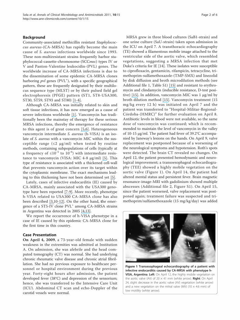
MRSA grew in three blood cultures (SaB1-strain) andone urine culture (SaU-strain) taken upon admission inthe ICU on April 7. A transthoracic echocardiography(TE) showed a filamentous mobile image attached to theventricular side of the aortic valve, which resembledvegetations, suggesting a MRSA infection that metDuke’s criteria for IE [14]. These isolates were susceptibleto ciprofloxacin, gentamicin, rifampicin, tetracycline, tri-methoprim-sulfamethoxazole-(TMP-SMX) and linezolidby disk diffusion and broth microdilution methods (seeAdditional file 1, Table S1) [15] and resistant to erythro-mycin and clindamycin (inducible resistance, D-test posi-tive) [15]. In addition, vancomycin MIC was 1 μg/ml bybroth dilution method [15]. Vancomycin treatment (15mg/kg every 12 h) was initiated on April 7 and thepatient was transferred to “Hospital-Militar-Regional-Córdoba-(HMRC)” for further evaluation on April 8.Antibiotic levels in blood were not available, so the samedose of vancomycin was continued; which is recom-mended to maintain the level of vancomycin in the valleyof 10-15 μg/ml. The patient had fever of 39.2°C accompa-nied by Janeway’s lesions on the hands. On April 9, valvereplacement was postponed because of a worsening ofthe neurological symptoms and hypotension. Roth’s spotswere detected. The brain CT revealed no changes. OnApril 12, the patient presented hemodynamic and neuro-logical improvement; a transoesophageal echocardiogra-phy (TEE) showed a highly mobile vegetation on theaortic valve (Figure 1). On April 14, the patient hadaltered mental status and persistent fever. Brain magneticresonance-image-MRI with gadolinium showed multipleabscesses (Additional file 2, Figure S1). On April 15,since the patient worsened, valve replacement was post-poned again; treatment failure was suspected and tri-methoprim/sulfamethoxazole (15 mg/kg/day) was added
Figure 1 Transesophageal echocardiography of a patient withinfective endocarditis caused by CA-MRSA with phenotype h-VISA, Argentina. Left: On April 12, the highly mobile vegetation onthe aortic valve (AV) of 20 × 41 mm (white arrow). Right: On April24, slight decrease in the aortic valve (AV) vegetation (white arrow)and a new vegetation on the mitral valve (MV) (10 × 4.6 mm) oflow motility (white arrow).
Sola et al. Annals of Clinical Microbiology and Antimicrobials 2011, 10:15http://www.ann-clinmicrob.com/content/10/1/15
Page 2 of 6
to vancomycin. On April 17, the patient remained febrile,three new samples for blood culture taken on April 14revealed MRSA (strain SaB2) with the same susceptibilityto antibiotics, except for vancomycin, which reached aMIC of 2 μg/ml (broth dilution [15]. On April 20, thepatient was afebrile. Three additional sets of blood cul-tures were drawn providing negative results. On April 24,a control TEE (Figure 1) showed a slight decrease of thesize of the aortic valve vegetation and a new vegetationon the mitral valve. The patient had evident deteriorationof mental and physical status and died on April 25 beforesurgical intervention.The emergence of h-VISA phenotype in a CA-MRSAwas suspected based on the rapid and fatal outcome ofthe patient, the facts that the IE was caused by MRSAwithout multiresistance to antibiotics, failure of vanco-mycin therapy (defined as persistent fever and bacterae-mia longer than 7 days after onset of therapy) andincrease of vancomycin MICs values (1 to 2 μg/ml)within therapy. This prompted us to further analyzethese strains. The blood (SaB1 and SaB2) and urine(SaU) MRSA isolates were sent to the CIBICI ("Centrode Investigaciones en Bioquimica Clinica e Inmunolo-gia"; UNC-CONICET); for additional analysis includingmolecular and genetic characterization.
Phenotypic and genotypic characterizations of MRSAThe slight increase of MIC values between the initial(SaU and SaB1) and subsequent (SaB2) isolates wasdetected by four MICs methods [15], particularly for tei-coplanin (Table 1). The isolates were also sent to the“Instituto-Nacional-de-Enfermedades-Infecciosas Dr.Carlos G. Malbrán” to independently confirm the MICresults for vancomycin and teicoplanin. Moreover, SaB2was classified as h-VISA by three screening methods,MHA5T-screening agar, Macromethod-E-test-(MET)and GRD E-test (Table 1) [5]. Although MRSA SaB2was vancomycin susceptible by MIC, it grew in BHI
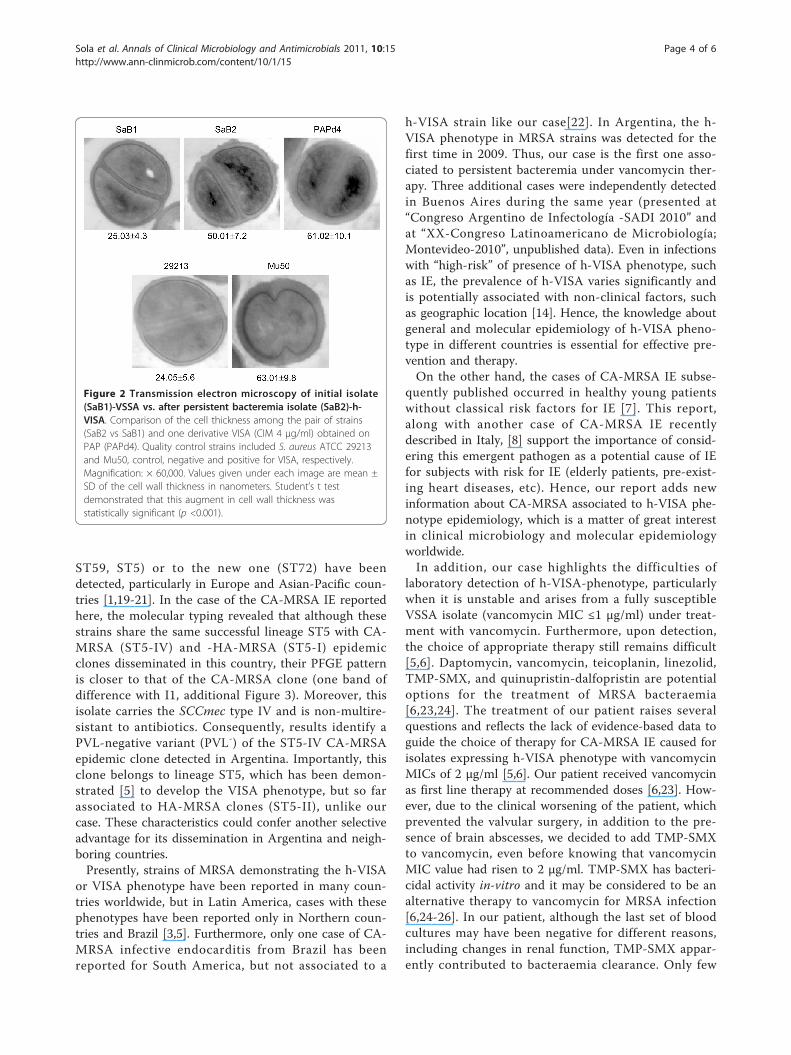
agar containing 4.0 μg/mL of vancomycin at a frequencyof 1.6 × 10⁻⁶ on population analysis profile-(PAP)[16,17], which is consistent with h-VISA (Additional file3, Figure S2). Vancomycin MICs by broth-dilution; agar-dilution and E-test were determined for these derivativesand all were between 4 and 8 μg/ml. Neither MRSASaB1 isolate nor S. aureus ATCC 29213 grew in BHIagar with 4 μg/ml. In addition, electron microscopyexamination [5,18] of SaB2 and one derivative VISAobtained on PAP, revealed that the cell-wall of both iso-lates was thicker (p <0.001) than the parent strain SaB1(Figure 2), a feature observed for most strains withVISA and h-VISA phenotypes. Moreover, the h-VISAphenotype found in strain SaB2 was unstable, since van-comycin MICs in all isolates with MICs ≥2 μg/mlreverted to MICs ≤1 μg/ml (susceptible phenotype-VSSA) after multiple subcultures in absence of vanco-mycin. Importantly, the detection by PCR [11] of vanAand vanB genes was negative.The clinical isolates (SaU, SaB1 and SaB2) and the deri-vatives obtained on PAP were analyzed by PFGE,MLST, SCCmec and spa typing, in addition to thedetection of virulence genes by PCR, as previouslyreported [4]. Molecular typing revealed that all theseisolates had indistinguishable pulsotypes (subtype I2)(Additional file 4, Figure S3) and were characterized asST5, agr-2, spa-t045, associated to SCCmec-IV. The egc(seg, sei, sem, sen and seo) and lukED genes weredetected among all the virulence genes analyzed by PCRin these three isolates. They do not harbor pvl and seagenes. The results confirmed the isogenic nature ofthese isolates and their clonality with the CA-MRSAclone (pulsotype I) detected in our country (Additionalfile 4, Figure S3).
DiscussionCurrently, PVL negative CA-MRSA strains belonging tosome successful PVL positive CA-MRSA clones (ST1,
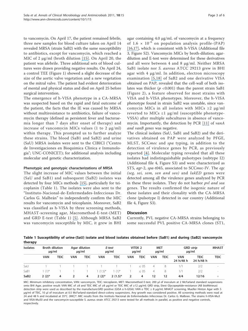
Table 1 Susceptibility of urine-(SaU) isolate and blood isolates obtained before (SaB1) and during (SaB2) vancomycintherapy
Isolates Broth dilutionμg/ml
Agar dilutionμg/ml
E-testμg/ml
VITEK 2μg/ml
METμg/ml
GRD stripμg/ml
MHA5T
VAN TEIC VAN TEIC VAN TEIC VAN TEIC VAN TEIC VAN24 h/48 h
TEIC24 h/48 h
TEIC
SaU 1 1 1 1 1 1 1 ≤ 05 4 8 1/1 2/2 -
SaB1 1 (1)* 1 1 1 1 (1.5)* 1 (1)* 1 ≤ 05 4 8 1/1 2/2 -
SaB2 2 (2)* 4 2 4 2 (2)* 2 (1.5)* 2 4 12 12 4/4 12/16 +
MIC: Minimum inhibitory concentration, VAN: vancomycin, TEIC: teicoplanin, MET: Macromethod E-test, 200 μl of inoculum at 2 McFarland standard suspensionsonto BHI Agar, positive result: VAN MIC of ≥8 and TEIC MIC of ≥8 μg/ml or TEIC MIC of ≥12 μg/ml; GRD strip, Etest Glycopeptide-resistance (AB bioMérieux)detection strip were used as described by the manufacture:GRD positive (GISA o h-GISA): VAN o TEIC ≥ 8 μg/ml; MHA5T screening, Mueller Hinton Agar with 5μg/ml of TEIC, 10 μl of inoculum at 0.5 McFarland-standard direct-colony suspensions. Any growth was considered positive. All screening methods were read at24 and 48 h and incubated at 35°C. (MIC)*: MIC results from the Instituto Nacional de Enfermedades Infecciosas Dr. Carlos G. Malbran. The strains h-VISA-Mu3and VISA-Mu50 and the vancomycin-susceptible S. aureus strain ATCC 29213 were tested for all methods in parallel, as positive and negative controls,respectively.
Sola et al. Annals of Clinical Microbiology and Antimicrobials 2011, 10:15http://www.ann-clinmicrob.com/content/10/1/15
Page 3 of 6
ST59, ST5) or to the new one (ST72) have beendetected, particularly in Europe and Asian-Pacific coun-tries [1,19-21]. In the case of the CA-MRSA IE reportedhere, the molecular typing revealed that although thesestrains share the same successful lineage ST5 with CA-MRSA (ST5-IV) and -HA-MRSA (ST5-I) epidemicclones disseminated in this country, their PFGE patternis closer to that of the CA-MRSA clone (one band ofdifference with I1, additional Figure 3). Moreover, thisisolate carries the SCCmec type IV and is non-multire-sistant to antibiotics. Consequently, results identify aPVL-negative variant (PVL-) of the ST5-IV CA-MRSAepidemic clone detected in Argentina. Importantly, thisclone belongs to lineage ST5, which has been demon-strated [5] to develop the VISA phenotype, but so farassociated to HA-MRSA clones (ST5-II), unlike ourcase. These characteristics could confer another selectiveadvantage for its dissemination in Argentina and neigh-boring countries.Presently, strains of MRSA demonstrating the h-VISA
or VISA phenotype have been reported in many coun-tries worldwide, but in Latin America, cases with thesephenotypes have been reported only in Northern coun-tries and Brazil [3,5]. Furthermore, only one case of CA-MRSA infective endocarditis from Brazil has beenreported for South America, but not associated to a
h-VISA strain like our case[22]. In Argentina, the h-VISA phenotype in MRSA strains was detected for thefirst time in 2009. Thus, our case is the first one asso-ciated to persistent bacteremia under vancomycin ther-apy. Three additional cases were independently detectedin Buenos Aires during the same year (presented at“Congreso Argentino de Infectología -SADI 2010” andat “XX-Congreso Latinoamericano de Microbiología;Montevideo-2010”, unpublished data). Even in infectionswith “high-risk” of presence of h-VISA phenotype, suchas IE, the prevalence of h-VISA varies significantly andis potentially associated with non-clinical factors, suchas geographic location [14]. Hence, the knowledge aboutgeneral and molecular epidemiology of h-VISA pheno-type in different countries is essential for effective pre-vention and therapy.On the other hand, the cases of CA-MRSA IE subse-
quently published occurred in healthy young patientswithout classical risk factors for IE [7]. This report,along with another case of CA-MRSA IE recentlydescribed in Italy, [8] support the importance of consid-ering this emergent pathogen as a potential cause of IEfor subjects with risk for IE (elderly patients, pre-exist-ing heart diseases, etc). Hence, our report adds newinformation about CA-MRSA associated to h-VISA phe-notype epidemiology, which is a matter of great interestin clinical microbiology and molecular epidemiologyworldwide.In addition, our case highlights the difficulties of
laboratory detection of h-VISA-phenotype, particularlywhen it is unstable and arises from a fully susceptibleVSSA isolate (vancomycin MIC ≤1 μg/ml) under treat-ment with vancomycin. Furthermore, upon detection,the choice of appropriate therapy still remains difficult[5,6]. Daptomycin, vancomycin, teicoplanin, linezolid,TMP-SMX, and quinupristin-dalfopristin are potentialoptions for the treatment of MRSA bacteraemia[6,23,24]. The treatment of our patient raises severalquestions and reflects the lack of evidence-based data toguide the choice of therapy for CA-MRSA IE caused forisolates expressing h-VISA phenotype with vancomycinMICs of 2 μg/ml [5,6]. Our patient received vancomycinas first line therapy at recommended doses [6,23]. How-ever, due to the clinical worsening of the patient, whichprevented the valvular surgery, in addition to the pre-sence of brain abscesses, we decided to add TMP-SMXto vancomycin, even before knowing that vancomycinMIC value had risen to 2 μg/ml. TMP-SMX has bacteri-cidal activity in-vitro and it may be considered to be analternative therapy to vancomycin for MRSA infection[6,24-26]. In our patient, although the last set of bloodcultures may have been negative for different reasons,including changes in renal function, TMP-SMX appar-ently contributed to bacteraemia clearance. Only few
Figure 2 Transmission electron microscopy of initial isolate(SaB1)-VSSA vs. after persistent bacteremia isolate (SaB2)-h-VISA. Comparison of the cell thickness among the pair of strains(SaB2 vs SaB1) and one derivative VISA (CIM 4 μg/ml) obtained onPAP (PAPd4). Quality control strains included S. aureus ATCC 29213and Mu50, control, negative and positive for VISA, respectively.Magnification: × 60,000. Values given under each image are mean ±SD of the cell wall thickness in nanometers. Student’s t testdemonstrated that this augment in cell wall thickness wasstatistically significant (p <0.001).
Sola et al. Annals of Clinical Microbiology and Antimicrobials 2011, 10:15http://www.ann-clinmicrob.com/content/10/1/15
Page 4 of 6
cases of patients with persistent bacteraemia have beentreated with TMP-SMX in addition to vancomycin, latein the course of bacteriemia, but these are not enoughto assess the clinical outcome [27]. Daptomycin was notconsidered due to the potential risks in patients withbacteremia, in which a deep focus of infection has notbeen surgically debrided, as well as for patients withleft-side endocarditis [28], two characteristics seen inour patient.One of the limitations of this report is the absence of
measurements of vancomycin serum concentrations dueto technical problems of the clinical laboratory at thattime. Hence, although we think that, considering theclinical characteristics of the patient, the dose of vanco-mycin that she received should have been enough toreach or even exceed the recommended therapeuticlevel [6,27], we cannot demonstrate it. In summary, thefailure of vancomycin to eradicate this CA-MRSA strainwas likely due to the presence of several important fac-tors: I) high-bacterial-load during infection, combinedwith the impossibility to perform the cardiac surgery, II)slow bactericidal mechanism of vancomycin, III) theability of this CA-MRSA clone to develop h-VISA phe-notype and IV) the virulence of the CA-MRSA strain,possibly related to the metastatic complications andrapid progression [5,23].
ConclusionsThis report demonstrates the ability of this epidemicCA-MRSA clone, disseminated in some regions ofArgentina, to cause severe and rapidly fatal infectionssuch as infective endocarditis, in addition to its capacityto acquire low-level vancomycin resistance; for this rea-son, it has become a challenge for the healthcare systemin our country. Therefore, systematic surveillance alongto molecular epidemiology and quick diffusion of theresults in each region are crucial for proper manage-ment of these infections.
ConsentWritten informed consent was obtained from thepatient’s son for publication of this case report and anyaccompanying images. A copy of the written consent isavailable for review by the Editor-in-Chief of this journal
Additional material
Additional file 1: Table S1: Antimicrobial susceptibility profile ofurine-(SaU) isolate and blood isolates obtained before-(SaB1) andduring-(SaB2) vancomycin therapy. CLSI: Clinical and LaboratoryStandards Institute, MIC: Minimum inhibitory concentration by brothmicrodilution (VITEK 2) per CLSI guideline, R*: Inducible ClindamycinResistance.
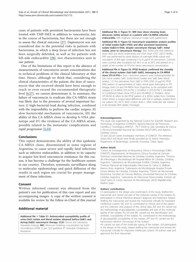
Additional file 2: Figure S1: MRI (two slices) showing brainabscesses (white arrows) in a patient with CA-MRSA infectiveendocarditis. MRI: magnetic resonance image (with gadolinium).
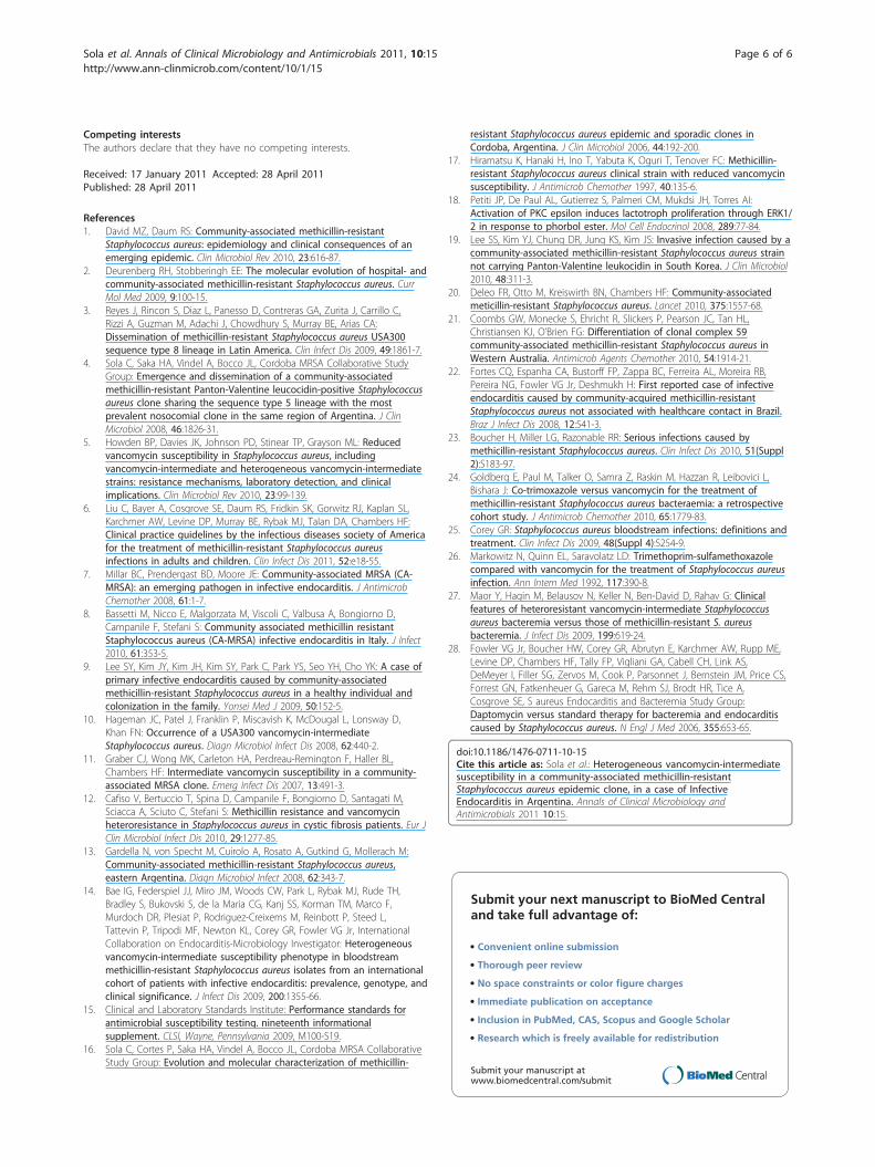
Additional file 3: Figure S2: Vancomycin population analysis profilesof initial isolate-(SaB1)-VSSA and after persistent bacteremiaisolate-(SaB2)-h-VISA, despite vancomycin therapy SaB1: initialisolate, prior to vancomycin therapy. Also, hVISA and VISA referencestrain Mu3 and Mu50 respectively and VSSA strain ATCC 29213. Briefly,PAP was performed by serial dilution of overnight BHIB culture andinoculation of BHI agar containing 0 to 8 μg/ml of vancomycin. Colonieswere counted after incubation for 48 h in air at 35°C and plotted on agraph of the number of CFU/ml versus vancomycin concentration.
Additional file 4: Figure S3: PFGE-Analysis confirmed the clonalityof the clinical isolates (SaU-SaB1-SaB2), belonging to CA-MRSAclone ST5-IV-PVL+. Sma I restriction patterns were indistinguishable forSaU (urine isolate), SaB1 (initial blood isolate) and SaB2 (later bloodisolate), 1-5: five derivatives from SaB2 in PAPs (CIM ≥4 μg/ml), PFGEDNA pattern of representative of major clonal types belonging to ST5lineage, both CA and HA-MRSA from Argentina, to be compared withsubtype I2 CA-MRSA (ST5-IV-PVL¯): CA-MRSA I1 (ST5-IV-PVL+), HA-MRSAA1-Cordobes/Chilean clone (ST5-I) and HA-MRSA C1 Pediatric clone(ST100-IV). I6 refers to the PFGE pattern of a MSSA isolate from Córdoba,which shows a unique band difference with the I2 subtype, isolated fromour patient. NC: NCTC 8325 control strain L: DNA molecular size markersin kb (lambda DNA ladder, Promega).
AcknowledgementsThis study was supported by the National Council for Scientific Research andTechnology of Argentina (CONICET), Agencia Nacional de PromociónCientífica y Tecnológica (ANPCyT - PICT 01630 to JLB), Secretaría de Cienciay Técnica-Universidad Nacional de Córdoba (SECyT-UNC) and AgenciaCórdoba Ciencia.CS and JLB are career investigator members of CONICET. The referencestrains Mu3 and Mu50 were kindly provided by Dr. Keiichi Hiramatsu,Department of Bacteriology, Juntendo University, Tokyo, Japan.
Author details1Centro de Investigaciones en Bioquímica Clínica e Inmunología (CIBICI-CONICET); Departamento de Bioquímica Clínica; Facultad de CienciasQuímicas; Universidad Nacional de Córdoba; Córdoba, Argentina. 2Serviciode Infectología y Microbiología del Hospital Militar de Córdoba, Córdoba,Argentina. 3Laboratorio de Microbiología SUOEM, Córdoba, Argentina.4Instituto Nacional de Enfermedades Infecciosas Dr. Carlos G. Malbrán,Buenos Aires, Argentina. 5Laboratorio de Microbiología Hospital PrivadoCentro Médico de Córdoba, Córdoba, Argentina. 6Centro de MicroscopíaElectrónica, Facultad de Ciencias Médicas, Universidad Nacional de Córdoba,Córdoba, Argentina. 7Laboratorio de Infecciones Nosocomiales, Instituto deSalud Carlos III, Centro Nacional de Microbiología, 28220 Majadahonda,Madrid, Spain.
Authors’ contributionsCS participated in the design and coordination of the study, drafted themanuscript and carried out part of the molecular typing of the isolates. RL:participated in clinical infectious disease’s diagnosis and treatment, helpeddrafting the manuscript and revised the manuscript critically for importantintellectual content. MC and JG: contributed to clinical care of the patientand the collection and analyses of the clinical data. ALE and VH: carried outthe methodology of glycopeptides susceptibility and part of the moleculartyping of the isolates. PG, ED and MV: carried out the identification andantibiotic susceptibility of the isolates. AC: contributed to the methodologyof glycopeptides susceptibility of the isolates. JPP and AT: carried out thetransmission electron microscopy of the isolates.AV: carried out part of the molecular typing of the isolates. JLB participatedin the design of the study, helped drafting the manuscript and revised themanuscript critically for important intellectual content. All authors read andapproved the final manuscript.
Sola et al. Annals of Clinical Microbiology and Antimicrobials 2011, 10:15http://www.ann-clinmicrob.com/content/10/1/15
Page 5 of 6
Competing interestsThe authors declare that they have no competing interests.
Received: 17 January 2011 Accepted: 28 April 2011Published: 28 April 2011
References1. David MZ, Daum RS: Community-associated methicillin-resistant
Staphylococcus aureus: epidemiology and clinical consequences of anemerging epidemic. Clin Microbiol Rev 2010, 23:616-87.
2. Deurenberg RH, Stobberingh EE: The molecular evolution of hospital- andcommunity-associated methicillin-resistant Staphylococcus aureus. CurrMol Med 2009, 9:100-15.
3. Reyes J, Rincon S, Diaz L, Panesso D, Contreras GA, Zurita J, Carrillo C,Rizzi A, Guzman M, Adachi J, Chowdhury S, Murray BE, Arias CA:Dissemination of methicillin-resistant Staphylococcus aureus USA300sequence type 8 lineage in Latin America. Clin Infect Dis 2009, 49:1861-7.
4. Sola C, Saka HA, Vindel A, Bocco JL, Cordoba MRSA Collaborative StudyGroup: Emergence and dissemination of a community-associatedmethicillin-resistant Panton-Valentine leucocidin-positive Staphylococcusaureus clone sharing the sequence type 5 lineage with the mostprevalent nosocomial clone in the same region of Argentina. J ClinMicrobiol 2008, 46:1826-31.
5. Howden BP, Davies JK, Johnson PD, Stinear TP, Grayson ML: Reducedvancomycin susceptibility in Staphylococcus aureus, includingvancomycin-intermediate and heterogeneous vancomycin-intermediatestrains: resistance mechanisms, laboratory detection, and clinicalimplications. Clin Microbiol Rev 2010, 23:99-139.
6. Liu C, Bayer A, Cosgrove SE, Daum RS, Fridkin SK, Gorwitz RJ, Kaplan SL,Karchmer AW, Levine DP, Murray BE, Rybak MJ, Talan DA, Chambers HF:Clinical practice guidelines by the infectious diseases society of Americafor the treatment of methicillin-resistant Staphylococcus aureusinfections in adults and children. Clin Infect Dis 2011, 52:e18-55.
7. Millar BC, Prendergast BD, Moore JE: Community-associated MRSA (CA-MRSA): an emerging pathogen in infective endocarditis. J AntimicrobChemother 2008, 61:1-7.
8. Bassetti M, Nicco E, Malgorzata M, Viscoli C, Valbusa A, Bongiorno D,Campanile F, Stefani S: Community associated methicillin resistantStaphylococcus aureus (CA-MRSA) infective endocarditis in Italy. J Infect2010, 61:353-5.
9. Lee SY, Kim JY, Kim JH, Kim SY, Park C, Park YS, Seo YH, Cho YK: A case ofprimary infective endocarditis caused by community-associatedmethicillin-resistant Staphylococcus aureus in a healthy individual andcolonization in the family. Yonsei Med J 2009, 50:152-5.
10. Hageman JC, Patel J, Franklin P, Miscavish K, McDougal L, Lonsway D,Khan FN: Occurrence of a USA300 vancomycin-intermediateStaphylococcus aureus. Diagn Microbiol Infect Dis 2008, 62:440-2.
11. Graber CJ, Wong MK, Carleton HA, Perdreau-Remington F, Haller BL,Chambers HF: Intermediate vancomycin susceptibility in a community-associated MRSA clone. Emerg Infect Dis 2007, 13:491-3.
12. Cafiso V, Bertuccio T, Spina D, Campanile F, Bongiorno D, Santagati M,Sciacca A, Sciuto C, Stefani S: Methicillin resistance and vancomycinheteroresistance in Staphylococcus aureus in cystic fibrosis patients. Eur JClin Microbiol Infect Dis 2010, 29:1277-85.
13. Gardella N, von Specht M, Cuirolo A, Rosato A, Gutkind G, Mollerach M:Community-associated methicillin-resistant Staphylococcus aureus,eastern Argentina. Diagn Microbiol Infect 2008, 62:343-7.
14. Bae IG, Federspiel JJ, Miro JM, Woods CW, Park L, Rybak MJ, Rude TH,Bradley S, Bukovski S, de la Maria CG, Kanj SS, Korman TM, Marco F,Murdoch DR, Plesiat P, Rodriguez-Creixems M, Reinbott P, Steed L,Tattevin P, Tripodi MF, Newton KL, Corey GR, Fowler VG Jr, InternationalCollaboration on Endocarditis-Microbiology Investigator: Heterogeneousvancomycin-intermediate susceptibility phenotype in bloodstreammethicillin-resistant Staphylococcus aureus isolates from an internationalcohort of patients with infective endocarditis: prevalence, genotype, andclinical significance. J Infect Dis 2009, 200:1355-66.
15. Clinical and Laboratory Standards Institute: Performance standards forantimicrobial susceptibility testing. nineteenth informationalsupplement. CLSI, Wayne, Pennsylvania 2009, M100-S19.
16. Sola C, Cortes P, Saka HA, Vindel A, Bocco JL, Cordoba MRSA CollaborativeStudy Group: Evolution and molecular characterization of methicillin-
resistant Staphylococcus aureus epidemic and sporadic clones inCordoba, Argentina. J Clin Microbiol 2006, 44:192-200.
17. Hiramatsu K, Hanaki H, Ino T, Yabuta K, Oguri T, Tenover FC: Methicillin-resistant Staphylococcus aureus clinical strain with reduced vancomycinsusceptibility. J Antimicrob Chemother 1997, 40:135-6.
18. Petiti JP, De Paul AL, Gutierrez S, Palmeri CM, Mukdsi JH, Torres AI:Activation of PKC epsilon induces lactotroph proliferation through ERK1/2 in response to phorbol ester. Mol Cell Endocrinol 2008, 289:77-84.
19. Lee SS, Kim YJ, Chung DR, Jung KS, Kim JS: Invasive infection caused by acommunity-associated methicillin-resistant Staphylococcus aureus strainnot carrying Panton-Valentine leukocidin in South Korea. J Clin Microbiol2010, 48:311-3.
20. Deleo FR, Otto M, Kreiswirth BN, Chambers HF: Community-associatedmeticillin-resistant Staphylococcus aureus. Lancet 2010, 375:1557-68.
21. Coombs GW, Monecke S, Ehricht R, Slickers P, Pearson JC, Tan HL,Christiansen KJ, O’Brien FG: Differentiation of clonal complex 59community-associated methicillin-resistant Staphylococcus aureus inWestern Australia. Antimicrob Agents Chemother 2010, 54:1914-21.
22. Fortes CQ, Espanha CA, Bustorff FP, Zappa BC, Ferreira AL, Moreira RB,Pereira NG, Fowler VG Jr, Deshmukh H: First reported case of infectiveendocarditis caused by community-acquired methicillin-resistantStaphylococcus aureus not associated with healthcare contact in Brazil.Braz J Infect Dis 2008, 12:541-3.
23. Boucher H, Miller LG, Razonable RR: Serious infections caused bymethicillin-resistant Staphylococcus aureus. Clin Infect Dis 2010, 51(Suppl2):S183-97.
24. Goldberg E, Paul M, Talker O, Samra Z, Raskin M, Hazzan R, Leibovici L,Bishara J: Co-trimoxazole versus vancomycin for the treatment ofmethicillin-resistant Staphylococcus aureus bacteraemia: a retrospectivecohort study. J Antimicrob Chemother 2010, 65:1779-83.
25. Corey GR: Staphylococcus aureus bloodstream infections: definitions andtreatment. Clin Infect Dis 2009, 48(Suppl 4):S254-9.
26. Markowitz N, Quinn EL, Saravolatz LD: Trimethoprim-sulfamethoxazolecompared with vancomycin for the treatment of Staphylococcus aureusinfection. Ann Intern Med 1992, 117:390-8.
27. Maor Y, Hagin M, Belausov N, Keller N, Ben-David D, Rahav G: Clinicalfeatures of heteroresistant vancomycin-intermediate Staphylococcusaureus bacteremia versus those of methicillin-resistant S. aureusbacteremia. J Infect Dis 2009, 199:619-24.
28. Fowler VG Jr, Boucher HW, Corey GR, Abrutyn E, Karchmer AW, Rupp ME,Levine DP, Chambers HF, Tally FP, Vigliani GA, Cabell CH, Link AS,DeMeyer I, Filler SG, Zervos M, Cook P, Parsonnet J, Bernstein JM, Price CS,Forrest GN, Fatkenheuer G, Gareca M, Rehm SJ, Brodt HR, Tice A,Cosgrove SE, S aureus Endocarditis and Bacteremia Study Group:Daptomycin versus standard therapy for bacteremia and endocarditiscaused by Staphylococcus aureus. N Engl J Med 2006, 355:653-65.
doi:10.1186/1476-0711-10-15Cite this article as: Sola et al.: Heterogeneous vancomycin-intermediatesusceptibility in a community-associated methicillin-resistantStaphylococcus aureus epidemic clone, in a case of InfectiveEndocarditis in Argentina. Annals of Clinical Microbiology andAntimicrobials 2011 10:15.
Submit your next manuscript to BioMed Centraland take full advantage of:
• Convenient online submission
• Thorough peer review
• No space constraints or color figure charges
• Immediate publication on acceptance
• Inclusion in PubMed, CAS, Scopus and Google Scholar
• Research which is freely available for redistribution
Submit your manuscript at www.biomedcentral.com/submit
Sola et al. Annals of Clinical Microbiology and Antimicrobials 2011, 10:15http://www.ann-clinmicrob.com/content/10/1/15
Page 6 of 6
Copyright © 2022 FDOKUMEN